Note: Descriptions are shown in the official language in which they were submitted.
WO 2021/160838
PCT/EP2021/053516
LILRB3 ANTIBODY MOLECULES AND USES THEREOF
FIELD OF THE INVENTION
The present invention relates to novel antibody molecules that specifically
bind to
LILRB3 (ILT5). The invention also relates to the use of such novel antibody
molecules or
other antibody molecules that specifically bind to LILRB3 (ILT5) in treatment
of graft re-
jection, an autoimmune disorder and/or an inflammatory disorder.
BACKGROUND OF THE INVENTION
lo The
family of human leukocyte immunoglobulin (1g)-like receptors (LI LRs), also
called human immunoglobulin-like transcripts (ITLs), comprises six activating
(LILRA1-6)
and five inhibitory (LILRB1-5) LILRs that regulate immune responses (1, 2).
Both recep-
tor subtypes display two, or four, homologous C-2 type immunoglobulin (1g)-
like extracel-
lular domains, but differ in their transmembrane and cytoplasmic regions (3,
4). LILRAs
have short truncated cytoplasmic domains with charged arginine residues in
their trans-
membrane domains, allowing them to associate with the v-chain of ITAM-bearing
FccR
to propagate activating signaling cascades (5). Conversely, LILRB have long
cytoplasmic
domains that contain multiple ITIM domains, which recruit phosphatases such as
SHP-1
and SHIP-1 that elicit inhibitory signaling (3, 4). Located at human
chromosome 19q13.4,
these polygenic receptors demonstrate significant allelic variation, with
LILRB3
(IL15/CD85a) and LILRB4 (ILT3/CD85k) displaying at least 15 different variants
(3. 6).
The inhibitory LILRBs are proposed to act as immune checkpoints serving to con-
trol and limit overt immune responses (1, 2). In agreement with this, LILRB
expression is
increased in suppressive (also referred to as alternatively activated or M2)
macrophages
and tolerogenic dendritic cells (DCs) (7-10). On monocytes, co-ligation of
LILRB1
(ILT2/CD85j) and LILRB2 (IL14/CD85d) with Fcylil (CD64) results in SHP-1
activation,
decreasing downstream phosphorylation events and intracellular calcium
mobilization
(11). Upon ligation with HLA class I (H LA-I) ligands, LILRB1 and LILRB2
prevent migra-
tion of DCs, and promote their release of anti-inflammatory cytokines (1, 12).
Similarly,
engagement of LILRB1 on macrophages by the common HLA-I subunit [32-
microglobulin
on malignant cells limits their phagocytic potential (13). LILRBs have also
been shown to
render DCs tolerogenic both in vitro and in vivo, subsequently inhibiting T
cell responses
(7, 8, 12, 14, 15). As such, the engagement of HLA-G with LILRB1 and LILRB2 is
an im-
portant immunosuppressive pathway at the fetal-maternal interface during
pregnancy
(16-18). LILRB1 is also expressed on NK cells and has been reported to inhibit
NK cell
cytotoxicity (19).
1
CA 03167424 2022- 8-9
WO 2021/160838
PCT/EP2021/053516
Although mice do not express LILRBs, the orthologous paired lg-like receptor
(PIR)-B regulates various arms of the immune system. PI R-B regulates priming
of cyto-
toxic T-lymphocytes by DCs via interaction with MHC class I expressed on CD8
cells
(20); and negatively influences integrin signaling in neutrophils and
macrophages (21).
Furthermore, PI R-B regulates the differentiation of myeloid-derived
suppressor cells
(MDSCs) that aid in tumor progression (22). Similar to FIR-B, the interaction
between
HLA-G and LILRB1 supports allotransplant engraftment through expansion of
potent
MDSC (23, 24).
Among the inhibitory LILRBs, LILRB3 (I LT5/LIR3/CD85a), containing 4
intracellu-
lar ITIM motifs, presents an attractive immunomodulatory target due to its
relative re-
striction to, and high expression on, myeloid cells (2). Despite its discovery
in the late
1990's, its exact functions and immunomodulatory potential have not been fully
deter-
mined, due to the lack of specific reagents and model systems
SUMMARY OF THE INVENTION
To investigate the potential immunomodulatory capacity of LILRB3, a panel of
LILRB3-specific monoclonal antibodies (mAb) was generated using Biolnvent
Interna-
tional AB's proprietary n-CoDeRED and F.I.R.S.TTm platform technology. The
antibodies
bound to two major but discrete epitopes in Ig-like domains 2 and 4. LILRB3
ligation on
primary human monocytes and macrophages resulted in phenotypic and functional
changes and potent inhibition of immune responses in vitro, including
significant reduc-
tion in phagocytosis of opsonized cancer cells and T cell proliferation.
Importantly, target-
ing of LILRB3 in humanized mice induced a tolerogenic status and permitted
enhanced
engraftment of allogeneic human lymphoma cells. Our findings reveal
immunoregulatory
functions of human LILRB3 and identify its potential as an important myeloid
immune
checkpoint, with potential roles in transplantation, infection and
autoimmunity.
The work leading to the present invention comprised the following:
= generation and characterization of a panel of human monoclonal anti-
LILRB3
antibodies agonistic activity,
= demonstrating that ligation of LILRB3 on human myeloid cells induces an anti-
inflammatory phenotype, leading to subsequent inhibition of T cell
proliferation
= demonstrating that LILRB3 ligation on human macrophages inhibits phagocy-
tosis of opsonized target cells
= demonstrating that agonistic anti-LILRB3 antibodies induced tolerance in
hu-
manized mice, permitting successful engraftment of allogeneic cells.
2
CA 03167424 2022- 8-9
WO 2021/160838
PCT/EP2021/053516
Thus, the present invention relates to antibody molecules that bind
specifically to
LILRB3 (ILT5) for use in treatment of graft rejection, autoimmune disorders
and/or in-
flammatory disorders.
The present invention also relates to specific antibody molecules that bind
specifi-
cally to LILRB3 (IL15) selected from the group consisting of antibody
molecules compris-
ing 1-6 of the CDRs VH-CDR1, VH-CDR2, VH-CDR3, VL-CDR1, VL-CDR2 and VL-
CDR3,
wherein VH-CDR1, if present, is selected from the group consisting of SEQ. ID.
NOs: 1, 9, 17 and 25;
lo wherein VH-CDR2, if present, is selected from the group consisting of
SEQ. ID.
NOs: 2, 10, 18 amd 26;
wherein VH-CDR3, if present, is selected from the group consisting of SEQ. ID.
NOs: 3, 11 and 19 and 27;
wherein VL-CDR1, if present, is selected from the group consisting of SEQ. ID.
NOs: 4, 12, 20 and 28;
wherein VL-CDR2, if present, is selected from the group consisting of SEQ. ID.
NOs: 5, 13, 21 and 29; and
wherein VL-CDR3, if present, is selected from the group consisting of SEQ. ID.
NOs: 6, 14, 22 and 30.
The present invention also relates to isolated nucleotide sequences encoding
at
least one of the above antibody molecules.
The present invention also relates to plasmids comprising at least one of the
above nucleotide sequences.
The present invention also relates to cells comprising at least one of the
above
nucleotide sequences, or at least one of the above plasmids.
The present invention also relates to the above antibody molecules, nucleotide
sequences, plasmids and/or cells for use in medicine.
The present invention also relates to the above antibody molecules, nucleotide
sequences, plasmids and/or cells for use in the treatment of graft rejection.
The present invention also relates to the above antibody molecules, nucleotide
sequences, plasmids and/or cells for use in the treatment of an autoimmune
disorder
(also denoted autoimmunity).
The present invention also relates to the above antibody molecules, nucleotide
sequences, plasmids and/or cells for use in the treatment of an inflammatory
disorder.
The present invention also relates to the use of the above antibody molecules,
nucleotide sequences, plasmids and/or cells for use in the treatment of graft
rejection.
3
CA 03167424 2022- 8-9
WO 2021/160838
PCT/EP2021/053516
The present invention also relates to the use of the above antibody molecules,
nucleotide sequences, plasmids and/or cells for use in the treatment of an
autoimmune
disorder.
The present invention also relates to the use of the above antibody molecules,
nucleotide sequences, plasmids and/or cells for use in the treatment of an
inflammatory
disorder.
The present invention also relates to pharmaceutical compositions comprising
or
consisting of at least one of the above antibody molecules, nucleotide
sequences, plas-
mids and/or cells, and optionally a pharmaceutically acceptable diluent,
carrier, vehicle
and/or excipient. Such a pharmaceutical composition may be used in the
treatment of
graft rejection. Such a pharmaceutical composition may also or alternatively
be used in
the treatment of an autoimmune disorder. Such a pharmaceutical composition may
also
or alternatively be used in the treatment of an inflammatory disorder.
The present invention also relates to methods for treatment of graft rejection
in a
patient comprising administering to the patient a therapeutically effective
amount of at
least one of the above antibody molecules, nucleotide sequences, plasmids
and/or cells.
The present invention also relates to methods for treatment of an autoimmune
disorder in a patient comprising administering to the patient a
therapeutically effective
amount of at least one of the above antibody molecules, nucleotide sequences,
plasmids
and/or cells.
The present invention also relates to methods for treatment of an inflammatory
disorder in a patient comprising administering to the patient a
therapeutically effective
amount of at least one of the above antibody molecules, nucleotide sequences,
plasmids
and/or cells.
The present invention also relates to antibody molecules, antibody molecules
for
use, isolated nucleotide sequences, isolated nucleotide sequences for use,
plasmids,
plasmids for use, cells, cells for use, uses, pharmaceutical compositions and
methods of
treatment as described herein with reference to the accompanying description,
examples
and/or figures.
DETAILED DESCRIPTION OF THE INVENTION
Thus, the present invention concerns antibody molecules that bind specifically
to
LI LRB3 (ILT5). In this context, the term "antibody molecule that specifically
binds
LI LRB3" can be used interchangeably with the term "anti-LI LRB3 antibody
molecule (or
"antibody molecule that specifically binds I LT5" and "anti-I LT5 antibody
molecule, re-
spectively) refers to an antibody molecule that specifically binds to at least
one epitope in
4
CA 03167424 2022- 8-9
WO 2021/160838
PCT/EP2021/053516
the extracellular domain of LILRB3 (ILT5). Cell surface antigen and epitope
are terms
that would be readily understood by one skilled in immunology or cell biology.
Methods of assessing protein binding are known to the person skilled in
biochem-
istry and immunology. It would be appreciated by the skilled person that those
methods
could be used to assess binding of an antibody to a target; as well as the
relative
strength, or the specificity, or the inhibition, or prevention, or reduction
in those interac-
tions. Examples of methods that may be used to assess protein binding are, for
example,
immunoassays, Biacore, western blots, radioimmunoassay (RIA) and enzyme-linked
im-
munosorbent assays (ELISAs) and Flow cytometry (FACS). See Fundamental Immuno1-
ogy Second Edition, Raven Press, New York at pages 332-336 (1989) for a
discussion
regarding antibody specificity.
The target cells expressing the LILRB3 to which the antibody molecule bind in
ac-
cordance with the present invention can be any LILRB3 expressing cells, such
as human
myeloid cells, including monocytes and macrophages.
Without being bound to any specific mechanism, one hypothesis is that the
effect
of the binding of the antibody molecules according to the invention to LILRB3
may be
that it leads to the phosphorylation of the ITIM domains. LILRB3 contains four
intracellu-
lar ITIMs. This, in turn, inhibits cellular activation and induces the
production of immuno-
suppressive genes by the myeloid cells. This is evident from the example below
showing
RNAseq analysis of human monocytes.
In some embodiments, the agonistic activity may be improved by the antibody
molecule binding to an Fcy receptor in addition to binding to LILRB3. In some
such em-
bodiments, the agonistic non-blocking LILRB3 antibody molecules bind with
higher affin-
ity to inhibitory Fcy receptors than to activating Fcy receptors. With higher
affinity to in-
hibitory Fcy receptors than to activating Fcy receptors, we include the
meaning of vari-
ants that bind with higher affinity to inhibitory Fcy receptors compared with
individual acti-
vating Fcy receptors, e.g. compared with either of FcyRIIA, FcyRIIIA and
FcyRI.
The relatively high homology between mouse and human FcyR systems ac-
counts for many of the general aspects of conserved FcyR mediated mechanisms
be-
tween the species. However, mouse and human IgG subclasses differ in their
affinities
for their cognate FcyRs, making it important when translating FcyR-mediated
observa-
tions in the mouse system into human IgG-based therapeutics to choose an
antibody,
antibody subclass and/or engineered subclass variant, that shows appropriate
binding to
human activating vs inhibitory FcyRs. The affinity and/or avidity of human
antibody mole-
cules for individual human FcyRs can be determined using surface plasmon
resonance
(SPR).
5
CA 03167424 2022- 8-9
WO 2021/160838
PCT/EP2021/053516
In some embodiments, the binding to an Fc receptor occurs through normal inter-
action between the Fc region of the agonistic antibody molecule and the Fc
receptor. In
some such embodiments the antibody molecule is an IgG, which has an Fc region
bind-
ing to an Fcy receptor. In some such embodiments, the anti-LILRB3 antibody is
of hu-
man IgG2 isotype, which has similar intermediate affinity for human inhibitory
FcyRIIB
and human activating FcyRI IA and FcyRIIIA, but does not productively engage
with hu-
man activating FcyRI. In some embodiments the anti-LILRB3 antibody is of human
IgG1
isotype, which binds FcyRIIB with higher affinity compared with IgG2, but also
binds acti-
vating human activating FcyRIIA, FcyRIIIA with higher affinity, and
additionally binds acti-
vating FcyRI with high affinity. In other embodiments, the anti-LILRB3
antibody is a hu-
man IgG engineered for enhanced binding to FcyRIIB e.g. the "SELF" mutation
(Chu et
al. "Inhibition of B cell receptor-mediated activation of primary human B
cells by coen-
gagement of CD19 and FcgammaRI lb with Fc-engineered antibodies." Mol Immunol.
2008 Sep;45(15):3926-33), and/or engineered for relative enhanced binding to
FcyRIIB
compared to activating FcyRs e.g. V9 or V11 mutations (Mimoto et al.
"Engineered anti-
body Fc variant with selectively enhanced FcyRIlb binding over both
FcyRIlaR131 and
FcyRIla"131".Protein Eng Des Sel. 2013 Oct; 26(10): 589-598.). Such IgG
variants engi-
neered for enhanced binding to inhibitory FcyRIIB, or specifically enhanced
binding affin-
ity specifically to inhibitory FcyRIIB but not activating EcyRIIA, have been
shown to in-
crease in vivo agonist activity, and therapeutic activity, of the CD40 agonist
antibody CP-
870,893 in animals humanized for activating and inhibitory FcyRs (Dahan et al.
2016.
'Therapeutic Activity of Agonistic, Human Anti-CD40 Monoclonal Antibodies
Requires
Selective FcgammaR Engagement, Cancer Cell, 29: 820-31).
The Fc receptor to which agonistic antibody molecule may bind in addition to
LILRB3 is a receptor found on the surface of cells of myeloid origin, such as
macro-
phages, monocytes, MDCSs, neutrophils, mast cells, basophils, or dendritic
cells, or on
the surface of lymphocytes such as NK cells, B cells, or certain T cells.
In other embodiments, the antibody molecules may comprise a modified Fc re-
gion having decreased binding to Fcy receptors, such as a deglycosylated or
aglycosyl-
ated variant of an IgG1 antibody molecule. Such aglycosylation may for example
be
achieved by an amino acid substitution of the asparagine in position 297
(N297X) in the
antibody chain. The substation may be with a glutamine (N2970), or with an
alanine
(N297A), or with a glycine (N297G), or with an asparagine (N297D), or by a
serine
(N297S). Other substitutions have e.g. been described by Jacobsen FW et al.,
JBC
2017, 292, 1865-1875, (see e.g. Table 1); such additional substitutions
include L242C,
V259C, A287C, R292C, V302C, L306C, V323C, I332C, and/or K334C.
6
CA 03167424 2022- 8-9
WO 2021/160838
PCT/EP2021/053516
Antibodies are well known to those skilled in the art of immunology and
molecular
biology. Typically, an antibody comprises two heavy (H) chains and two light
(L) chains.
Herein, we sometimes refer to this complete antibody molecule as a full-size
or full-
length antibody. The antibody's heavy chain comprises one variable domain (VH)
and
three constant domains (CH1, CH2 and CH3), and the antibody's molecule light
chain
comprises one variable domain (VL) and one constant domain (CL). The variable
do-
mains (sometimes collectively referred to as the Fv region) bind to the
antibody's target,
or antigen. Each variable domain comprises three loops, referred to as
complementary
determining regions (CDRs), which are responsible for target binding. The
constant do-
mains are not involved directly in binding an antibody to an antigen, but
exhibit various
effector functions. Depending on the amino acid sequence of the constant
region of their
heavy chains, antibodies or immunoglobulins can be assigned to different
classes. There
are five major classes of immunoglobulins: IgA, IgD, IgE, IgG and IgM, and in
humans
several of these are further divided into subclasses (isotypes), e.g., IgG1,
IgG2, IgG3,
and IgG4; IgA1 and IgA2.
Another part of an antibody is the Fc region (otherwise known as the fragment
crystallizable domain), which comprises two of the constant domains of each of
the anti-
body's heavy chains. As mentioned above, the Fc region is responsible for
interactions
between the antibody and Fc receptor.
The term antibody molecule, as used herein, encompasses full-length or full-
size
antibodies as well as functional fragments of full length antibodies and
derivatives of
such antibody molecules.
Functional fragments of a full-size antibody have the same antigen binding
char-
acteristics as the corresponding full-size antibody and include either the
same variable
domains (i.e. the VH and VL sequences) and/or the same CDR sequences as the
corre-
sponding full-size antibody. A functional fragment does not always contain all
six CDRs
of a corresponding full-size antibody. It is appreciated that molecules
containing three or
fewer CDR regions (in some cases, even just a single CDR or a part thereof)
are capable
of retaining the antigen-binding activity of the antibody from which the
CDR(s) are de-
rived. For example, in Gao et al., 1994, J. Biol. Chem., 269: 32389-93 it is
described that
a whole VL chain (including all three CDRs) has a high affinity for its
substrate.
Molecules containing two CDR regions are described, for example, by Vaughan &
Sollazzo 2001, Combinatorial Chemistry & High Throughput Screening, 4: 417-
430. On
page 418 (right column ¨3 Our Strategy for Design) a minibody including only
the H1
and H2 CDR hypervariable regions interspersed within framework regions is
described.
The minibody is described as being capable of binding to a target. Pessi
etal., 1993, Na-
ture, 362: 367-9 and Bianchi etal., 1994, J. Mol. Biol., 236: 649-59 are
referenced by
7
CA 03167424 2022- 8-9
WO 2021/160838
PCT/EP2021/053516
Vaughan & Sollazzo and describe the H1 and H2 minibody and its properties in
more de-
tail. In Qiu etal., 2007, Nature Biotechnology, 25:921-9 it is demonstrated
that a mole-
cule consisting of two linked CDRs are capable of binding antigen. Quiocho
1993, Na-
ture, 362: 293-4 provides a summary of "minibody" technology. Ladner 2007,
Nature Bio-
technology, 25:875-7 comments that molecules containing two CDRs are capable
of re-
taining antigen-binding activity.
Antibody molecules containing a single CDR region are described, for example,
in Laune etal., 1997, J BC, 272: 30937-44, in which it is demonstrated that a
range of
hexapeptides derived from a CDR display antigen-binding activity and it is
noted that
synthetic peptides of a complete, single, CDR display strong binding activity.
In Monnet
etal., 1999, JBC, 274: 3789-96 it is shown that a range of 12-mer peptides and
associ-
ated framework regions have antigen-binding activity and it is commented on
that a
CDR3-like peptide alone is capable of binding antigen. In Heap et at., 2005,
J. Gen. Vi-
ral., 86: 1791-1800 it is reported that a "micro-antibody" (a molecule
containing a single
CDR) is capable of binding antigen and it is shown that a cyclic peptide from
an anti-HIV
antibody has antigen-binding activity and function. In Nicaise etal., 2004,
Protein Sci-
ence, 13:1882-91 it is shown that a single CDR can confer antigen-binding
activity and
affinity for its lysozyme antigen.
Thus, antibody molecules having five, four, three or fewer CDRs are capable of
retaining the antigen binding properties of the full-length antibodies from
which they are
derived.
The antibody molecule may also be a derivative of a full-length antibody or a
frag-
ment of such an antibody. When a derivative is used it should have the same
antigen
binding characteristics as the corresponding full-length antibody in the sense
that it binds
to the same epitope on the target as the full-length antibody.
Thus, by the term "antibody molecule", as used herein, we include all types of
an-
tibody molecules and functional fragments thereof and derivatives thereof,
including:
monoclonal antibodies, polyclonal antibodies, synthetic antibodies,
recombinantly pro-
duced antibodies, multi-specific antibodies, bi-specific antibodies, human
antibodies, an-
tibodies of human origin, humanized antibodies, chimeric antibodies, single-
chain Fvs
(scFv), Fab fragments, F(ab')2 fragments, F(ab') fragments, disulfide-linked
Fvs (sdFv),
antibody heavy chains, antibody light chains, homo-dimers of antibody heavy
chains,
homo-dimers of antibody light chains, heterodimers of antibody heavy chains,
heterodi-
mers of antibody light chains, antigen binding functional fragments of such
homo- and
heterodimers.
8
CA 03167424 2022- 8-9
WO 2021/160838
PCT/EP2021/053516
Further, the term "antibody molecule", as used herein, includes all classes of
anti-
body molecules and functional fragments, including: IgG, IgG1, IgG2, IgG3,
IgG4, IgA,
IgM, IgD, and IgE, unless otherwise specified.
In some embodiments, the antibody molecule is a human antibody molecule, a
humanized antibody molecule or an antibody molecule of human origin. In this
context, a
humanized antibody molecule means an originally non-human antibody that has
been
modified to increase its similarity to a human antibody. Humanized antibody
molecules
may, for example, originally be murine antibodies or lama antibodies. In this
context, an
antibody molecule of human origin means an originally human antibody molecule
that
has been modified.
In some embodiments, the antibody molecule is an IgG antibody.
In some embodiments, the antibody molecule is a wild-type IgG antibody.
In some embodiments, the antibody molecule is an Fc engineered IgG antibody,
such as the ones mentioned above, including aglycosylated or deglycosylated
IgG anti-
body molecules, such as those including a substitution of the asparagine in
position 297,
such as for example a N297Q or N297A substitution.
In some embodiments, the antibody molecule is a human IgG1 antibody. Human
IgG1 antibodies correspond to murine IgG2a antibodies, so if a murine
surrogate to a hu-
man IgG1 is to be used, for example for in vivo studies, a murine IgG2a format
is used.
In some embodiments, the antibody molecule is a human IgG2 antibody. Human
IgG2 antibodies correspond to murine IgG3 antibodies, so if a murine surrogate
to a hu-
man IgG2 is to be used, for example for in vivo studies, a murine IgG3 format
is used.
In some embodiments, the antibody molecule is a human IgG4 antibody. Human
IgG4 antibodies correspond to murine IgG1 antibodies, so if a murine surrogate
to a hu-
man IgG4 is to be used, for example for in vivo studies, a murine IgG1 format
is used.
The Fc modifications may vary between human and murine antibody molecules;
for example a murine N297A IgG2a antibody molecule can be used as a surrogate
of a
human N297Q IgG1 antibody molecule.
In some embodiments, the anti-LILRB3 antibody is a monoclonal antibody.
In some embodiments, the anti-LILRB3 antibody is a polyclonal antibody.
As outlined above, different types and forms of antibody molecules are encom-
passed by the invention, and would be known to the person skilled in
immunology. It is
well known that antibodies used for therapeutic purposes are often modified
with additional
components which modify the properties of the antibody molecule.
Accordingly, we include that an antibody molecule described herein or an anti-
body molecule used as described herein (for example, a monoclonal antibody
molecule,
9
CA 03167424 2022- 8-9
WO 2021/160838
PCT/EP2021/053516
and/or polyclonal antibody molecule, and/or bi-specific antibody molecule)
comprises a
detectable moiety and/or a cytotoxic moiety.
By "detectable moiety", we include one or more from the group comprising of:
an
enzyme; a radioactive atom; a fluorescent moiety; a chemiluminescent moiety; a
biolumi-
nescent moiety. The detectable moiety allows the antibody molecule to be
visualized in
vitro, and/or in vivo, and/or ex vivo.
By "cytotoxic moiety", we include a radioactive moiety, and/or enzyme, for
exam-
ple wherein the enzyme is a caspase, and/or toxin, for example wherein the
toxin is a
bacterial toxin or a venom; wherein the cytotoxic moiety is capable of
inducing cell lysis.
lo We
further include that the antibody molecule may be in an isolated form and/or
purified form, and/or may be PEGylated. PEGylation is a method by which
polyethylene
glycol polymers are added to a molecule such as an antibody molecule or
derivative to
modify its behavior, for example to extend its half-life by increasing its
hydrodynamic
size, preventing renal clearance.
As discussed above, the CDRs of an antibody bind to the antibody target. The
as-
signment of amino acids to each CDR described herein is in accordance with the
defini-
tions according to Kabat EA et al. 1991, In "Sequences of Proteins of
Immunological In-
terest" Fifth Edition, NI H Publication No. 91-3242, pp xv- xvii.
As the skilled person would be aware, other methods also exist for assigning
amino acids to each CDR. For example, the International ImMunoGeneTics
information
system (IMGT(R)) (http://www.imgt.org/ and Lefranc and Lefranc "The
Immunoglobulin
FactsBook" published by Academic Press, 2001).
In some embodiments, the antibody molecule that specifically binds LI LRB3 com-
prises one of the VH-CDR1 sequences listed in Table 1 below.
In some embodiments, the antibody molecule that specifically binds LI LRB3 com-
prises one of the VH-CDR2 sequences listed in Table 1 below.
In some embodiments, the antibody molecule that specifically binds LI LRB3 com-
prises one of the VH-CDR3 sequences listed in Table 1 below.
In some embodiments, the antibody molecule that specifically binds LI LRB3
corn-
s() prises one of the VL-CDR1 sequences listed in Table 1 below
In some embodiments, the antibody molecule that specifically binds LI LRB3 com-
prises one of the VL-CDR2 sequences listed in Table 1 below.
In some embodiments, the antibody molecule that specifically binds LILRB3 com-
prises one of the VL-CDR3 sequences listed in Table 1 below.
In some embodiments, the anti-LILRB3 antibody molecule is an antibody mole-
cule selected from the group consisting of antibody molecules wherein the
three CDRs in
the variable heavy chain (VH) are selected from the group consisting of:
CA 03167424 2022- 8-9
WO 2021/160838
PCT/EP2021/053516
SEQ. ID. NO: 1, SEQ. ID. NO: 2 and SEQ. ID. NO: 3;
SEQ. ID. NO: 9, SEQ. ID. NO: 10 and SEQ. ID. NO: 11;
SEQ. ID. NO: 17, SEQ. ID. NO: 18 and SEQ. ID. NO: 19; and
SEQ. ID. NO: 25, SEQ. ID. NO: 26 and SEQ. ID. NO: 27.
In some embodiments, the anti-LILRB3 antibody molecule is an antibody mole-
cule selected from the group consisting of antibody molecules wherein the
three CDRs in
the variable light chain (VL) are selected from the group consisting of:
SEQ. ID. NO: 4, SEQ. ID. NO: 5 and SEQ. ID. NO: 6;
SEQ. ID. NO: 12, SEQ. ID. NO: 13 and SEQ. ID. NO: 14;
SEQ. ID. NO: 20, SEQ. ID. NO: 21 and SEQ. ID. NO: 22; and
SEQ. ID. NO: 28, SEQ. ID. NO: 29 and SEQ. ID. NO: 30.
In some embodiments, the anti-LILRB3 antibody molecule is an antibody mole-
cule selected from the group consisting of antibody molecules comprising a VH
selected
from the group consisting of SEQ. ID. NOs: 7, 15, 23 and 31.
In some embodiments, the anti-LILRB3 antibody molecule is an antibody mole-
cule selected from the group consisting of antibody molecules comprising a VL
selected
from the group consisting of SEQ. ID. NOs: 8, 16, 24 and 32.
In some embodiments the anti-LILRB3 antibody molecule comprises a CH having
SEQ. ID. NO: 41.
In some embodiments the anti-LILRB3 antibody molecule comprises a CL having
SEQ. ID. NO: 42.
Table 1: Specific sequences of anti-LILRB3 antibody molecules (in the VH and
VL se-
quences, the CDR sequences are marked in bold text)
Antibody Region Sequence
SEQ.
clone
ID. NO:
Al VH-C D R1 FSSYAMSVVVRQAPG
1
VH-CDR2 SAISGSGGSTYYADSVKGR
2
VH-CDR3 ARRKKRERGFSGNDPVGAI DV
3
VL-CDR1 CTGSSSN I GAGYDVH
4
VL-CDR2 GNTNRPS
5
VL-CDR3 CSAWDDSLSGVV
6
VH EVQLLESGGGLVQPGGSLRLSCAASGFTFSSYAMSVWR- 7
QAPG KG LEVVVSAISGSGGSTY-
YADSVKGRFTISRDNSKNTLYLQM NSLRAEDTAVYYCARR
KKRERGFSGNDPVGAIDVWGQGTLVTVSS
11
CA 03167424 2022- 8-9
WO 2021/160838
PCT/EP2021/053516
VL QSVLTQPPSASGTPG QRVTISCTGSSSN I-
8
GAGYDVHVVYQQLPGTAPKLLIYGNTN RPSGVP-
DRFSGSKSGTSASLAI SG LRSEDEADYYCSAWDDSLSGVV
FGGGTKLTVLG
A16 VH-C D R1 FSSYVVMSVVVRQAPG
9
VH-CDR2 SR INTHGTNIDYADSVKGR
10
VH-CDR3 VGVAGTGWFDP
11
VL-CDR1 CTGSSSN I GAGYDVH
12
VL-CDR2 GNNNRPS
13
VL-CDR3 CQSYDTSLSGSV
14
VH EVQLLESGGGLVQPGGSLRLSCAASGFTFSSYWMSVVVR- 15
QAPGKGLEVVVSRINTHGTNIDY-
ADSVKGRFTISRDNSKNTLYLQMNSLRAEDTAVYYCVGVA
GTGWFDPWGQGTLVTVSS
VL QSVLTQPPSASGTPG QRVTISCTGSSSN I-
16
GAGYDVHVVYQQLPGTAPKLLIYGNNNRPSGVP-
DRFSGSKSGTSASLAISGLRSEDEADYYCQSYDTSLSGSV
FGGGTKLTVLG
A20 VH-C D R1 FSSYSIVINWVRQAPG
17
VH-CDR2 SAISGSGGSTYYADSVKGR
18
VH-CDR3 ARGLATYGLDV
19
VL-CDR1 CSGSSSNIGRHHVY
20
VL-CDR2 SNSLRPS
21
VL-CDR3 CAAWDDSLSGVVV
22
VH EVOLLESGGGLVQPGGSLRLSCAASGFTFSSYSMNWVR- 23
QAPGKGLEWVSAISGSGGSTY-
YADSVKGRFTISRDNSKNTLYLQMNSLRAEDTAVYYCARG
LATYGLDVWGQGTLVTVSS
VL QSVLTQPPSASGTPGQRVTISCSGSSSNIGRHHVY-
24
VVYQQLPGTAPKLLIYSNS LRPSGVP-
DRFSGSKSGTSASLAI SG LRSEDEADYYCAAWDDSLSGW
VFGGGTKLTVLG
A28 VH-C D R1 FSSYSMNWVRQAPG
25
VH-CDR2 AN I KQDGTENYYVD SVEGR
26
VH-CDR3 ARDGDWGWGFDY
27
VL-CDR1 CTGSSSN I GAGYDVH
28
VL-CDR2 ENNKRPS 29
12
CA 03167424 2022- 8-9
WO 2021/160838
PCT/EP2021/053516
VL-CDR3 CAAWDDS LSGWV
30
VH
EVQLLESGGGLVQPGGSLRLSCAASGFTFSSYSMNVVVR- 31
QAPGKGLEVVVANIKQDG-
TENYYVDSVEGRFTISRDNSKNTLYLQMNSLRAEDTAVYY
CARDGDWGVVGFDYVVGQGTLVTVSS
VL QSVLTQPPSASGTPGQRVTISCTGSSSNI-
32
GAGYDVHVVYQQLPGTAPKLLIYENNKRPSGVP-
DRFSGSKSGTSVSLAISGLRSEDEADYYCAAWDDSLSGW
VFGGGTKLTVLG
The sequences in Table 1 above are all of human origin and derived from the n-
CoDeR library, as explained in detail in Example 1.
In some embodiments, the antibody molecules that specifically bind LILRB3 de-
scribed herein may also comprise one or both of the constant regions (CH
and/or CL)
listed in Table 2 below.
Table 2:
Region Sequence
SEQ.
ID. NO:
CH ASTKG PSVFPLAPSSKSTSGGTAALGC LVKDYFPEPVTVSWNSGAL-
33
TSGVHTFPAVLQSSG LYSLSSVVTVPSSSLGTQTYICNVNHKPSNT-
KVDKKVEPKSCDKTHTCPPCPAPELLGGPSVFLFPPKPKDTLMISRTPE-
VTCVVVDVSH E DPEVKFNVVYVDGVEVH NAKTKPREEQYNSTYRVVS-
VLTVLHQDVVLNGKEYKCKVSNKALPAPIEKTISKAKGQPREPQVY-
TLPPSRDELTKNQVSLTCLVKGFYPSD IAVEVVESNGQPEN-
NYKTTPPVLDSDGSFFLYS KLTVDKSRWQQG NVFSCSVM H EALH N HY-
TQKSLSLSPGK
CL QPKAAPSVTLFPPSSEELQANKATLVCLISDFYPGAVTVAVVKADSSPV- 34
KAGVETTTPSKQSNNKYAASSYLSLTPEQVVKSHRSYSCQVTHEGSTVE
KTVAPTECS
The CH (SEQ. ID. NO: 33) and the (SEQ. ID. NO: 34) sequences in Table 2
above are of human origin.
As mentioned above, in some embodiments, the antibody molecules bind human
LILRB3). In some such embodiments, it is preferred that the antibody molecules
binds
strongly to human LILRB3, i.e. that they have a low EC50 value.
In some embodiments, it is advantageous that the antibody molecule binds both
to human LILRB3 and to cynomologous monkey LILRB3 (cm LILRB3 or cyno LILRB3).
13
CA 03167424 2022- 8-9
WO 2021/160838
PCT/EP2021/053516
Cross-reactivity with LILRB3 expressed on cells in cynomologous monkey, also
called
crab-eating macaque or Macaca fascicularis, may be advantageous since this
enables
animal testing of the antibody molecule without having to use a surrogate
antibody, with
particular focus on tolerability.
In some embodiments, it is necessary to use a surrogate antibody to test an
anti-
body molecule's functional activity in relevant in vivo models in mice. To
ensure the com-
parability between the antibody molecule's effect in humans and the in vivo
results for
the surrogate antibody in mice, it is essential to select a functionally
equivalent surrogate
antibody having the same in vitro characteristics as the human antibody
molecule.
lo In some embodiments, the antibody molecule of the present invention
or used ac-
cording to the invention is an antibody molecule that is capable of competing
with the
specific antibodies provided herein, for example capable of competing with
antibody mol-
ecules comprising a VH selected from the group consisting of SEQ. ID. NOs: 7,
15, 23
and 31; and/or a VL selected from the group consisting of SEQ. ID. NOs: 8, 16,
24 and
32, for binding to LILRB3.
By "capable of competing for" we mean that the competing antibody is capable
of
inhibiting or otherwise interfering, at least in part, with the binding of an
antibody mole-
cule as defined herein to the specific target LILRB3.
For example, such a competing antibody molecule may be capable of inhibiting
the binding of an antibody molecule described herein to LILRB3 by at least
about 10%;
for example at least about 20%, or at least about 30%, at least about 40%, at
least about
50%, at least about 60%, at least about 70%, at least about 80%, at least
about 90%, at
least about 95%, or about 100%.
Competitive binding may be determined by methods well known to those skilled
in the art, such as Enzyme-linked immunosorbent assay (ELISA).
ELISA assays can be used to evaluate epitope-modifying or blocking antibodies.
Additional methods suitable for identifying competing antibodies are disclosed
in Antibod-
ies: A Laboratory Manual, Harlow & Lane, which is incorporated herein by
reference (for
example, see pages 567 to 569, 574 to 576, 583 and 590 to 612, 1988, CSHL, NY,
ISBN
0-87969-314-2).
In some embodiments, it is of interest to use not the anti-LILRB3 antibody
mole-
cule itself but a nucleotide sequence encoding such an antibody molecule. The
present
invention thus encompasses nucleotide sequences encoding the above anti-LILRB3
anti-
body molecules.
14
CA 03167424 2022- 8-9
WO 2021/160838
PCT/EP2021/053516
The above described antibody molecules and nucleotide sequences, or other
anti-LILRB3 antibody molecules or nucleotide sequences encoding such antibody
mole-
cules, can be used in medicine, and then such an antibody molecule and/or
nucleotide
sequence can be included in a pharmaceutical composition, as discussed further
below.
The anti-LI LRB3 antibody molecules, nucleotide sequences and/or pharmaceuti-
cal compositions can be used in the treatment of graft rejection, as discussed
further be-
low.
The anti-LI LRB3 antibody molecules, nucleotide sequences and/or pharmaceuti-
cal compositions can be used in the treatment of an autoimmune disorder, as
discussed
further below.
The anti-LI LRB3 antibody molecules, nucleotide sequences and/or pharmaceuti-
cal compositions can be used in the treatment of an inflammatory disorder, as
discussed
further below.
The anti-LI LRB3 antibody molecules and/or nucleotide sequences can be used in
the manufacture of a pharmaceutical composition for use in the treatment of
graft rejec-
tion
The anti-LI LRB3 antibody molecules and/or nucleotide sequences can be used in
the manufacture of a pharmaceutical composition for use in the treatment of an
autoim-
mune disorder.
The anti-LI LRB3 antibody molecules and/or nucleotide sequences can be used in
the manufacture of a pharmaceutical composition for use in the treatment of an
inflam-
matory disorder.
The anti-LI LRB3 antibody molecules, nucleotide sequences and/or pharmaceuti-
cal compositions can be used in treatment of graft rejection an autoimmune
disorder
and/or an inflammatory disorder in a patient, wherein a therapeutically
effective amount
of an anti-LILRB3 antibody molecule, nucleotide sequence and/or pharmaceutical
com-
position is administered to the patient.
Examples of graft rejection that can be treated as disclosed herein include
rejec-
tion in connection with an organ transplant or organ transplantation, such as
transplanta-
tion of kidney, liver, heart, lungs, pancreas and intestines from a donor to a
recipient, in
cases where the recipient suffers from a disease or an injury that affects an
organ that is
replaced in the transplantation. Another example of graft rejection that can
be treated as
disclosed herein include rejection of an allogeneic transplant wherein stem
cells, such as
hematopoietic stem cells (HSCs) are collected from a matching donor and
transplanted
into the patient to suppress a disease and restore the patient's immune
system.
CA 03167424 2022- 8-9
WO 2021/160838
PCT/EP2021/053516
At least in some embodiments, the recipient of the graft should undergo pre-
con-
ditioning by administrating agonistic LI LRB3 mAb prior to transplantation. In
some em-
bodiments, the recipient of the graft should also undergo treatment with
agonistic LILRB3
mAb after transplantation.
The antibody molecules, pharmaceutical compositions and treatments described
herein can be used to prevent, treat or minimize rejection of the new organ or
other
transplant by the recipient.
Examples of autoimmune disorder, or autoimmunity, that can be treated as dis-
closed herein include celiac disease, diabetes mellitus type 1, sarcoidosis,
systemic lu-
pus erythematosus (SLE), Sjogren's syndrome, eosinophilic granulomatosis with
polyan-
giitis, Hashimoto's thyroiditis, Graves' disease, idiopathic thrombocytopenic
purpura, Ad-
dison's disease, rheumatoid arthritis (RA), ankylosing spondylitis,
polymyositis (PM), der-
matomyositis (DM) and multiple sclerosis (MS).
Examples of inflammatory disorders that can be treated as disclosed herein in-
clude both chronic inflammatory disorders, such as rheumatoid arthritis (RA),
systemic
lupus erythematosus (SLE) and multiple sclerosis (MS), and acute inflammatory
disor-
ders, such as sepsis.
It would be known to the person skilled in medicine, that medicines can be
modi-
fied with different additives, for example to change the rate in which the
medicine is ab-
sorbed by the body; and can be modified in different forms, for example to
allow for a
particular administration route to the body.
Accordingly, we include that the antibody molecules, nucleotide sequences,
plas-
mids and/or cells described herein may be combined with a pharmaceutically
acceptable
excipient, carrier, diluent, vehicle and/or adjuvant into a pharmaceutical
composition. In
this context, the term pharmaceutical composition can be used interchangeably
with the
terms pharmaceutical preparation, pharmaceutical formulation, therapeutic
composition,
therapeutic preparation, therapeutic formulation and therapeutic entity.
The pharmaceutical compositions described herein may comprise, or in some
embodiments consist of, antibody molecules, nucleotide sequences, plasmids or
cells.
The pharmaceutical compositions described herein may in some embodiments
consist of or comprise plasmids comprising nucleotide sequences encoding the
above
described antibody molecules or comprising the above described nucleotide
sequences.
The invention also comprises other therapeutic modalities, or "shapes" of
drugs,
such as antibody drug conjugates, fusion proteins etc, and pharmaceutical
composition
comprising such therapeutic modalities.
16
CA 03167424 2022- 8-9
WO 2021/160838
PCT/EP2021/053516
The antibody molecules, nucleotide sequences, plasmids, cells and/or pharma-
ceutical compositions described herein may be suitable for parenteral
administration in-
cluding aqueous and/or non-aqueous sterile injection solutions which may
contain anti-
oxidants, and/or buffers, and/or bacteriostats, and/or solutes which render
the formula-
tion isotonic with the blood of the intended recipient; and/or aqueous and/or
non-aqueous
sterile suspensions which may include suspending agents and/or thickening
agents. The
antibody molecules, nucleotide sequences, plasmids, cells and/or
pharmaceutical com-
positions described herein may be presented in unit-dose or multi-dose
containers, for
example sealed ampoules and vials, and may be stored in a freeze-dried (i.e.
lyophilized)
condition requiring only the addition of the sterile liquid carrier, for
example water for in-
jections, immediately prior to use.
Extemporaneous injection solutions and suspensions may be prepared from ster-
ile powders, and/or granules, and/or tablets of the kind previously described.
For parenteral administration to human patients, the daily dosage level of the
anti-LILRB3 antibody molecule will usually be from 1 mg/kg bodyweight of the
patient to
mg/kg, or in some cases even up to 100 mg/kg administered in single or divided
doses. Lower doses may be used in special circumstances, for example in
combination
with prolonged administration. The physician in any event will determine the
actual dos-
age which will be most suitable for any individual patient and it will vary
with the age,
20 weight and response of the particular patient. The above dosages are
exemplary of the
average case. There can, of course, be individual instances where higher or
lower dos-
age ranges are merited and such are within the scope of this invention.
Typically, a pharmaceutical composition (or medicament) described herein com-
prising an antibody molecule will contain the anti-LILR B3 antibody molecule
at a concen-
tration of between approximately 2 mg/ml and 150 mg/ml or between
approximately 2
mg/ml and 200 mg/ml.
Generally, in humans, oral or parenteral administration of the antibody
molecules,
nucleotide sequences, plasmids, cells and/or pharmaceutical corn positions
described
herein is the preferred route, being the most convenient. For veterinary use,
the antibody
molecules, nucleotide sequences, plasmids, cells and/or pharmaceutical
compositions
described herein are administered as a suitably acceptable formulation in
accordance
with normal veterinary practice and the veterinary surgeon will determine the
dosing regi-
men and route of administration which will be most appropriate for a
particular animal.
Thus, the present invention provides a pharmaceutical formulation comprising
an amount
of an antibody molecule, nucleotide sequence, plasmid and/or cell of the
invention effec-
tive to treat various conditions (as described above and further below).
Preferably, the
17
CA 03167424 2022- 8-9
WO 2021/160838
PCT/EP2021/053516
antibody molecules, nucleotide sequences, plasmids, cells and/or
pharmaceutical com-
positions described herein is adapted for delivery by a route selected from
the group
comprising: intravenous (IV or i.v.); intramuscular (IM or i.m.) or
subcutaneous (SC or
s.c.).
The present invention also includes antibody molecules, nucleotide sequences,
plasmids, cells and/or pharmaceutical compositions described herein comprising
phar-
maceutically acceptable acid or base addition salts of the target binding
molecules or
parts of the present invention. The acids which are used to prepare the
pharmaceutically
acceptable acid addition salts of the aforementioned base compounds useful in
this in-
vention are those which form non-toxic acid addition salts, i.e. salts
containing pharma-
cologically acceptable anions, such as the hydrochloride, hydrobromide,
hydroiodide, ni-
trate, sulphate, bisulphate, phosphate, acid phosphate, acetate, lactate,
citrate, acid cit-
rate, tartrate, bitartrate, succinate, maleate, fumarate, gluconate,
saccharate, benzoate,
methanesulphonate, ethanesulphonate, benzenesulphonate, p- toluenesulphonate
and
pamoate [i.e. 1 ,1'-methylene-bis-(2-hydroxy-3 naphthoate)] salts, among
others. Phar-
maceutically acceptable base addition salts may also be used to produce
pharmaceuti-
cally acceptable salt forms of the agents according to the present invention.
The chemi-
cal bases that may be used as reagents to prepare pharmaceutically acceptable
base
salts of the present agents that are acidic in nature are those that form non-
toxic base
salts with such compounds. Such non-toxic base salts include, but are not
limited to
those derived from such pharmacologically acceptable cations such as alkali
metal cati-
ons (e.g. potassium and sodium) and alkaline earth metal cations (e.g. calcium
and mag-
nesium), ammonium or water-soluble amine addition salts such as N-
methylglucamine-
(meglumine), and the lower alkanolammonium and other base salts of
pharmaceutically
acceptable organic amines, among others. The antibody molecules, nucleotide se-
quences, plasmids and/or cells described herein may be lyophilized for storage
and re-
constituted in a suitable carrier prior to use. Any suitable lyophilization
method (e.g.
spray drying, cake drying) and/or reconstitution techniques can be employed.
It will be
appreciated by those skilled in the art that lyophilization and reconstitution
can lead to
varying degrees of antibody activity loss (e.g. with conventional
immunoglobulins, IgM
antibodies tend to have greater activity loss than IgG antibodies) and that
use levels may
have to be adjusted upward to compensate. In one embodiment, the lyophilized
(freeze
dried) polypeptide binding moiety loses no more than about 20%, or no more
than about
25%, or no more than about 30%, or no more than about 35%, or no more than
about
40%, or no more than about 45%, or no more than about 50% of its activity
(prior to ly-
ophilization) when re-hydrated.
18
CA 03167424 2022- 8-9
WO 2021/160838
PCT/EP2021/053516
The anti-LILRB3 antibody molecules, nucleotide sequences and pharmaceutical
compositions described herein can be used use in the treatment of graft
rejection or au-
toimmunity in a subject or patient. Herein, the terms subject and patient are
used inter-
changeably
"Patient" (or subject) as the term is used herein refers to an animal,
including hu-
man, that has been diagnosed as suffering from graft rejection or autoimmunity
and/or
that exhibits symptoms of suffering from graft rejection or autoimmunity.
In some embodiments, the patient (or subject) is an animal, including human,
that
has been diagnosed as suffering from graft rejection or autoimmunity. In some
embodi-
ments, the patient (or subject) is an animal, including human, that will
undergo a trans-
plantation and therefore being in the risk of graft rejection; the treatment
discussed
herein is then a carried out as a preventive treatment or for preventive
purposes.
In some embodiments, the patient (or subject) is a mammalian or non-mamma-
lian animal, including human, that has been diagnosed as having graft
rejection or auto-
immunity and/or that exhibits symptoms of graft rejection or autoimmunity.
The treatments may be administered as a course of treatment, which is to say
that the therapeutic agent is administered over a period of time. The length
of time of the
course of treatment will depend on a number of factors, which could include
the type of
therapeutic agent being administered, the type of disease or condition being
treated, the
severity of the disease or condition being treated, and the age and health of
the patient,
amongst others reasons.
By "during the treatment", we include that the patient is currently receiving
a
course of treatment, and/or receiving a therapeutic agent, and/or receiving a
course of a
therapeutic agent.
BRIEF DESCRIPTION OF THE DRAWINGS
In the examples below, reference is made to the following figures:
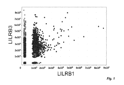
Figure 1. Generation of fully human mAb against human LILRB3. Fig. 1 A:
Screening of generated LILRB3 clones. FMAT was performed and scFv clones
screened
against LILRB3 target and LILRB1 non-target-transfected cells. MFI was
calculated, with
target-specific scFvs depicted in lighter color and non-target scFvs in darker
color. Fig. 1
B: Screening of LILRB3 mAb by flow cytometry. Peripheral blood mononuclear
cells
(PBMCs) or LILR-transfected CHO-S cells were incubated with His-tagged scFv
superna-
tants, followed by anti-His-AF647 staining. Where transfected CHO-S cells were
used,
LILRB1- and LILRB2-transfected CHO-S cells were used as non-targets for
LILRB3. Anti-
body clones were compared against both gated monocytes and target transfected
CHO-S
19
CA 03167424 2022- 8-9
WO 2021/160838
PCT/EP2021/053516
cells using the TI BOO Spotfire software, with LILRB3 specific clones
highlighted in light
grey, non-specific clones in dark grey, and the irrelevant isotype control in
grey. Fig. 1 C:
Specificity of LILRB3 clones against human LI LR-transfected 2B4 cells. LI
LRB3 mAb were
tested against cells transfected with the indicated LILR family members by
flow cytometry;
a representative clone (A16) is presented. Fig. 1 D and E: Testing the
specificity of LI LRB
clones against primary cells by flow cytometry. PBMCs (Fig. 1 D) or whole
blood (Fig. 1 E)
stained with either APC-labelled LILRB3 (clone A16) or hIgG1 isotype as well
as various
leukocyte surface markers, as indicated. Histograms are representative plots
of multiple
donors: monocytes and B cells (n=12), T cells and NK cells (n=12) and
neutrophils (n=6).
lo
Figure 2. Characterization of LILRB3 antibodies. Fig. 2 A: LILRB3 mAb affinity
assessed by SPR. LI LRB3-hFc recombinant protein was immobilized and various
LILRB3
mAb flowed across the chip. KD values were calculated using the BiacoreTM T100
Evalu-
ation Software. Representative LILRB3 clones are shown. Fig. 2 B: Ability of
generated
mAb to cross-block binding of LILRB3 mAb (a commercial LILRB3 mAb;(clone
222821,
R&D Systems, UK)). PBMCs were stained with unconjugated LILRB3 antibody clones
and
subsequently stained with a directly-conjugated commercial LILRB3 mAb and
analyzed by
flow cytometry; representative clones displayed, as indicated. The isotype
control (iso ctrl)
is shaded in grey, clone 222821 alone in black and in combination with
indicated LILRB3
clones in grey line. Fig. 2 C: LILRB3 domain epitope mapping. HEK293F cell
transfected
with either VVT LILRB3 (full-length extracellular portion), LILRB3-D1-3,
LILRB3-D1-2 or
LILRB3-D1 were stained with LILRB3 clones, followed by an anti-hIgG-PE
secondary an-
tibody staining. Schematic of domain constructs generated and restriction
digest of each
construct shown. Histograms showing staining of two representative clones
differentially
binding to \AfT (D4), D3, D2 and D1-expressing cells, as indicated (n=3
independent ex-
periments). Fig. 2 D: LILRB3 2B4 reporter cells were treated with 10 pg/ml
LILRB3 anti-
bodies overnight to assess agonism or antagonism. GFP expression was then
measured
by flow cytometry; representative clones shown.
Figure 3. LILRB3 ligation regulates T cell activation and proliferation. CFSE-
labelled PBMCs were stimulated with antibodies against human CD3 and CD28 in
the
presence or absence of isotype control (iso ctrl) or LILRB3 mAb (10 pg/ml) and
proliferation
measured through CFSE dilution after 3-5 days. Fig. 3 A: Assessing T cell
activation and
proliferation following treatment. Light microscopy images following PBMC
stimulation in
culture. CD8+ T cell proliferation was assessed through CFSE dilution;
representative his-
tograms shown. Fig. 3 B: LILRB3 mAb were deglycosylated (Degly) through PNGase-
treatment, as confirmed by SDS-PAGE; representative clones shown. Fig. 3 C:
Assessing
the effects of deglycosylated LILRB3 mAb on T cell proliferation. CFSE
dilution of CD8+ T
cells, treated with various LILRB3 mAb was assessed by flow cytometry. Data
normalized
CA 03167424 2022- 8-9
WO 2021/160838
PCT/EP2021/053516
to anti-CD3/CD28-treated samples and mean represented by solid line;
representative
clones shown. Two-tailed paired T-test performed and stars represent level of
significant
difference compared to iso ctrl (*** p<0.005); n=13-20 independent donors
(each dot rep-
resents an individual donor).
Figure 4. LILRB3 ligation modulates macrophage phagocytosis. Fig. 4 A: Hu-
man MDMs were stained with anti-CD14 and anti-LILRB3 (A16) and analyzed by
flow cy-
tometry. Fig. 4 B: MDMs were treated with deglycosylated isotype control (iso
ctrl) or
LILRB3 mAb (10 pg/ml) prior to co-culture with CFSE+ rituximab-opsonized
target cells;
and phagocytosis was defined as the number of gated live cells that were
double positive
(CD16+ CFSE+ cells), as a percentage of total MDMs (CD16+ cells), using the
following
equation:
(Double positive MDM / Total MDM) x 100 = % positive MDMs
Fig. 4 C: The effect of deglycosylated LILRB3 mAb on phagocytosis. Each donor
was
performed in triplicate and the mean is represented by a solid line (n=4-6
healthy donors);
representative clones shown. Two-tailed paired T-test was performed and stars
represent
level of significant difference compared to isotype control (* p<0.05, ***
p<0.0005). Fig. 4
D: The effect of deglycosylated LILRB3 mAb on phagocytosis assessed by
confocal mi-
croscopy. LILRB3-treated MDMs (grey) were co-cultured with CFSE-labelled B
cells (light
grey), fixed in 4% PFA and membrane glycoproteins stained with biotinylated
WGA. Cells
were then incubated with a secondary streptavidin-conjugated AF635 and
analyzed by
confocal microscopy.
Figure 5. LILRB3 ligation induce induces tolerance in vivo. Fully
reconstituted
humanized mice (50% circulating hCD45+ leukocytes) were generated and the
expres-
sion of human LILRB3 was confirmed on CD14+ myeloid cells. Fig. 5 A:
Representative
flow cytometry histogram showing LILRB3 expression on hCD45+ bone marrow
hCD14+
myeloid cells; isotype control in solid dark grey, LILRB3 in solid light grey.
Fig. 5 B: Hu-
manized mice were injected with 200 pg LILRB3 mAb (clone Al) or an isotype-
matched
(hIgG1) control mAb on day 0 and 4, i.v. and intraperitoneal (i.p.),
respectively. On day 7,
mice were injected i.p. with 1x10⁷ non-autologous luciferase# human lymphoma
cells. Lym-
phoma cell growth was monitored over time using an IVIS imager, representative
images
from 3 independent experiments shown (n=3 mice/group).
Figure 6. Human LILRB3 ligation reprograms human primary myeloid cells.
Freshly isolated human peripheral CD14+ monocytes were treated with an isotype
control
(iso ctrl) or a human LILRB3 mAb (clone Al). Fig. 6 A: Monocyte morphology
following
treatment. Light microscopy images following overnight treatment of freshly-
isolated
CD14+ monocytes with indicated mAb in culture. Fig. 6 B: Transcriptomic
analysis of
LILRB3-treated monocytes. RNA was extracted from cells following mAb treatment
(-18
21
CA 03167424 2022- 8-9
WO 2021/160838
PCT/EP2021/053516
hours) and subjected to RNA sequencing. The left panel depicts a list of genes
that were
significantly upregulated and the right panel depicts genes that were
significantly down-
regulated compared to iso ctrl treated-cells (n=4; each row represents an
individual donor).
Fig. 6 C: Ligation of LILRB3 on primary human CD14+ monocytes induces M2-
polarized
genes. GSEA graphs showing a significant enrichment for M2-polarizing genes in
LILRB3-
treated monocytes versus isotype control, respectively. UP; upregulated, NES;
normalized
enrichment score = -1.68; FWER; familywise-error rate p <0.001. Fig. 6 D: qPCR
analysis
of selected genes following LILRB3 ligation on monocytes. Data were normalized
to
GAPDH mRNA levels and standardized to the levels of isotype control-treated
monocytes.
Fold difference data were 10g10 transformed. One-way ANOVA with Bonferroni's
multiple
comparisons test was performed, n=3 independent donors (** p < 0.005, *** p <
0.0005).
Fig. 6 E: GSEA analysis showing negative correlation with `IFN-y' (NES=-2.17;
FWER p
<0.001), FN-a' (NES=-2.3; FWER p <0.001) and allograft rejection' (NES=-1.58;
FWER
p =0.14) signaling elements and positive correlation with 'oxidative
phosphorylation'
(NES=2; FWER p <0.001). Fig. 6 F: Schematic diagram demonstrating the
immunosup-
pressive function of LILRB3 following ligation on APCs.
EXAMPLES
Specific, non-limiting examples which embodies certain aspects of the
invention
will now be described.
Materials and Methods
Ethics Statement
All research with human samples and mice was performed in compliance with in-
stitutional guidelines, the Declaration of Helsinki and the US Department of
Health and
Human Services Guide for the Care and Use of Laboratory Animals. The Committee
on
Animal Care at Massachusetts Institute of Technology (MIT) reviewed and
approved the
studies described here. All human samples (adult peripheral blood and fetal
liver) were
collected anonymously with informed consent by a third party and purchased for
research.
For human peripheral blood, ethical approval for the use of clinical samples
was obtained
by the Southampton University Hospitals NHS Trust; from the Southampton and
South
West Hampshire Research Ethics Committee following provision of informed
consent. Pri-
mary chronic lymphocytic leukemia (CLL) samples were released from the Human
Tissue
Authority licensed University of Southampton, Cancer Science Unit Tissue Bank
as part of
the LPD study LREC number 228/02/T.
22
CA 03167424 2022- 8-9
WO 2021/160838
PCT/EP2021/053516
Hematopoietic stem/progenitor cells (FISPCs) isolation and generation of human-
ized mice
Human fetal livers were obtained from aborted fetuses at 15-23 weeks of gesta-
tion, in accordance with the institutional ethical guidelines (Advanced
Bioscience Re-
sources, Inc., CA, USA). All women gave written informed consent for the
donation of their
fetal tissue for research. Fetuses were collected within 2 hours of the
termination of preg-
nancy. Fetal liver tissue was initially cut into small pieces and digested
with collagenase
VI (2 mg/ml in Dulbecco's modified Eagle's medium [DMEM]) for 30 minutes at 37
C with
periodic mixing. Single-cell suspensions were prepared by passing the digested
tissue
through a 100 pm cell strainer (BD Biosciences, NJ, USA). CD34+ cells were
purified with
the use of a CD34 selection kit (Stem Cell Technologies, Vancouver, BC,
Canada); the
purity of CD34+ cells was 90%-99%. Viable cells were counted by trypan blue
exclusion
of dead cells. All cells were isolated under sterile conditions.
NSG mice were purchased from the Jackson Laboratories (Bar Harbor, Maine,
USA) and maintained under specific pathogen-free conditions in the animal
facilities at
MIT. To reconstitute mice, newborn pups (less than 2 days old) were irradiated
with 100
cGyusing a Gamma radiation source injected intracardially with CD34+CD133+
cells (ap-
proximately 2 x 10⁵ cells/recipient), as reported previously (25). Around 12
weeks of age,
human leukocyte reconstitution was determined by flow cytometry of peripheral
blood
mononuclear cells (PBMCs). Chimerism, or the level of human leukocyte
reconstitution,
was calculated as follows: % 0D45+ human cell / (% CD45+ human cell + %
C045+ mouse cell). Mice with 40% human CD45+ leukocytes were used in the
study.
Cell culture
Cell lines were grown at 37 C in either RPM! 1640 medium supplemented with
10% fetal calf serum (FCS) (Sigma-Aldrich, UK), 100 Wm! Penicillin-
Streptomycin, 2 mM
glutamine and 1 mM pyruvate (Thermo Fisher Scientific, UK) in a humidified
incubator with
5% 002, Freestyle 293F media, in 8% 002, shaking at 130 rpm, or Freestyle CHO
media
(Thermo Fisher Scientific, UK) with 8 mM glutamine, in 8% CO2, shaking at 140
rpm.
Antibody generation and production
Generation of LILRB3 antibodies.
Selection of various LILRB3-specific mAb was performed using the n-CoDeR
phage display library (26). Three consecutive panning rounds were performed,
as well as
a pre-panning step. In the panning, Fc fusion proteins containing the
extracellular domains
of LILRB1, LILRB2 or LILRB3 (LILRB-Fc) were used as non-targets or targets,
respec-
tively. These proteins were produced in transiently transfected HEK293 cells
followed by
23
CA 03167424 2022- 8-9
WO 2021/160838
PCT/EP2021/053516
purification on protein A, as described previously (27). CHO-S cells
transiently transfected
to express the various LILRB proteins were also used as targets/non-targets in
the pan-
ning.
In panning 1, Biolnvent n-CoDeRe' scFv were selected using biotinylated in-
house
produced recombinant LILRB-human (h) Fc recombinant fusion proteins (captured
with
streptavidin-coated Dynabeads ) with or without competition or LILRB-hFc
coated to
etched polystyrene balls (Polysciences, US)/ plastic immunotubes. Binding
phages were
eluted by trypsin digestion and amplified on plates using standard procedures
(28). The
amplified phages from panning 1 were used for panning 2, the process repeated,
and the
amplified phages from panning 2 used in panning 3. In the third panning round
however,
amplified phages from all 3 strategies were combined and selected against
LILRB transi-
ently transfected CHO-S cells.
Next, the LILRB3-positive scFv cassettes from the enriched phage repertoires
from
panning 3 were re-cloned to allow soluble scFv expression in E. coli. The
soluble scFv
fragments expressed by individual clones were tested for binding against LILRB-
trans-
fected CHO-S cells using Flourometric Microvolume Assay Technology (FMAT), and
re-
combinant LILRB protein by Enzyme-linked immunosorbent assay (ELISA). This
allowed
the identification of clones binding specifically to LILRB3. Clones were then
further reduced
in a tertiary screen against CHO-S cells expressing LI LRB1-3 and primary
cells (PBMCs).
Clones showing specific patterns of binding to a single LILRB were sequenced,
yielding
LI LRB1-3-specific mAb.
Production of full-length IgG's.
The unique scFv identified above were cloned into a eukaryotic expression
system
allowing transient expression of full-length IgG in HEK293-EBNA cells. The
antibodies
were then purified from the culture supernatants using Protein A-based
affinity chromatog-
raphy as previously described (29).
Production of deglycosylated IgG.
To allow dissection of Fc- and Fab-dependent effector functions, IgG were
degly-
cosylated using PNGase F (Promega) with 0.05 U of PNGase/pg of IgG, at 37 C
for at
least 15 hours. Deglycosylation was confirmed by reduction in size of the
heavy chain on
SDS-PAGE.
Production of domain mutant constructs
Using wild-type LILRB3 cDNA isolated from a healthy donor PBMCs, a series of
domain mutant DNA constructs were generated by overlap PCR to express 1, 2 or
3
24
CA 03167424 2022- 8-9
WO 2021/160838
PCT/EP2021/053516
LILRB3 Ig-like domains (with domains identified based on annotations in
Uniprot). The
gene constructs were then cloned into pcDNA3.
Cell Transfections
10X106 HEK293F cells were transiently transfected with 10 pg of plasmid DNA by
lipofection using 233 fectin with Optimem 1 Media (Thermo Fisher Scientific,
UK).
Preparation of human leukocytes and generation of monocyte-derived macro-
phages (MDMs)
lo
Whole blood was acquired with informed consent from healthy volunteers. PBMC
were isolated from leukocyte blood cones (Blood Transfusion Services,
Southampton
General Hospital). Isolation was performed by gradient density centrifugation
using lym-
phoprep (Axis Shield, UK). MDMs were generated from healthy peripheral blood
human
monocytes as before (30). Briefly, PBMCs were plated at 2x10⁷ cells/well in a
6-well plate
(Corning, UK) with 1% human AB serum (Sigma-Aldrich, UK) and incubated at 37 C
for 2
hours. Non-adherent cells were washed away and the adherent monocytes (>90%
CD14+)
were incubated at 37 C overnight with 5% CO2. The following day 100 ng/ml
human re-
combinant M-CSF (in house) was added to each well. Media and cytokine were
replen-
ished twice during culture and cells were then harvested on day 7-8.
Macrophage phagocytosis assay
Human MDMs generated as described above, were plated at 1x10⁵ cells/well in a
96-well flat-bottom plate. MDMs were treated with 10 pg/ml LILRB3 antibodies
for 2 hours
and washed. Primary chronic lymphocytic leukemia (CLL) cells, labelled with
5pM CFSE
(Sigma-Aldrich, UK), were opsonized with rituximab for 25 minutes at 4 C (or
herceptin as
an isotype control). MDMs and target CLL cells were then co-cultured for 1
hour at 37 C,
at a 1:5 ratio, respectively, before staining with 10 pg/ml CD16-APC
(BioLegend, UK) for
15 minutes at room temperature in the dark. Cells were washed, harvested,
analyzed by
flow cytometry and % phagocytosis calculated as follows: (c/o double-positive
MDM) / (c/0
total MDM) x 100.
Flow cytometry
For cell surface staining of PBMCs or whole blood, cells were blocked with 2%
human AB serum (Sigma-Aldrich, UK) for 10 minutes on ice and then stained with
the
relevant APC-labelled mAb or hIgG1 isotype (Biolnvent, Sweden), alongside the
following
cell surface markers: CD14-PE (eBioscience, UK), CD20-A488 (fluorescent
labelled ritux-
imab, in house), CD3-PE-Cy7, CD56-APC-Cy7 or CD15-Pacific Blue and CD66B-FITC
CA 03167424 2022- 8-9
WO 2021/160838
PCT/EP2021/053516
mAb (all BioLegend, UK). Cells were stained for 30 minutes at 4 C and then
were washed
twice, first in 10% red blood cell (RBC) lysis buffer (Serotec, UK) and then
FAGS wash
(PBS, 1% BSA, 10 mM NaN3), before acquisition on a FACSCalibur or FACSCanto II
(BD
Biosciences, USA) and analyzed with FCS Express V3 (De Novo Software).
For assays to determine if mAb bound to similar cross-blocking epitopes 1x10⁶
PBMCs were blocked with 2% human AB serum for 10 minutes and stained with 10
pg/ml
unconjugated LILRB3 mAb for 30 minutes at 4 C. The cells were then stained
with directly-
conjugated commercial LILRB3 mAb (clone 222821; R&D Systems, UK) for 20
minutes at
4 C, before washing and acquisition using a FACSCalibur.
lo For
LILRB3 epitope mapping studies, LILRB3-domain mutant-transfected
HEK293F cells were stained with the relevant LILRB3 mAb for 25 minutes at 4 C,
washed
twice, stained with an anti-human-PE secondary (Jackson I mmunoResearch, USA)
for 20
minutes at 4 C, before washing and acquisition using a FACSCalibur.
For staining of 264 reporter cells expressing LI LR-A1, -A2, -A5, -B1, -B2, -
63, -64,
or -B5 (or non-transfected controls) cells were stained with 10 pg/ml LILRB
mAb and incu-
bated at 37 C with 5% CO2, overnight. The following day, the cells were washed
and
stained with a secondary anti-hIgG antibody (Jackson ImmunoResearch, USA) at 4
C, for
45 minutes. The cells were washed and acquisition performed using a FACScan
(BD Bio-
sciences, USA) and analysis using Cell Quest (BD Biosciences, USA).
Flow cytometry data were analyzed with FCS Express V3 (De Novo Software) and
FlowJo.
Surface plasmon resonance (SPR)
SPR was performed with the Biacore T100 (GE Healthcare, UK) as per the manu-
facturer's instructions. LI LRB3-hFc recombinant protein (the extracellular
LILRB3 domain
with a human Fc tag) was used as the ligand and immobilized by amine coupling
onto a
series S sensor chip (CM5). Various LILRB3 mAb were used as "analytes" and
flowed
across the chip, and SPR measured. KD values were calculated from the'
Univalent' model
of 1:1 binding by Kd [1/5] / Ka [1/Ms], using the Biacore TM T100 Evaluation
Software (GE
Healthcare, UK).
T cell Proliferation assay
PBMCs (1-2 x 10⁷) were labelled with 2 pM CSFE at room temperature for 10
minutes. Cells were subsequently resuspended in serum-free CTL medium
(Immunospot,
Germany) and plated at 1x10⁵ cells/well in a 96-well round-bottom plate
(Corning, UK).
Cells were then stimulated with 0.02 pg/ml CD3 (clone OKT3, University of
Southampton),
26
CA 03167424 2022- 8-9
WO 2021/160838
PCT/EP2021/053516
pg/ml CD28 (clone CD28.2; BioLegend, UK) and 10 pg/ml LILRB3 antibodies or a
rele-
vant isotype. Plates were then incubated at 37 C for 4 days, after which time
cells were
stained with 5 pg/ml CD8-APC (clone SK1; BioLegend, UK), harvested and CSFE
dilution
measured by flow cytometry, as a readout for T cell proliferation.
5
In vivo allograft assay
Fully reconstituted humanized mice (40 70 circulating hCD45+ leukocytes) were
injected with 200 pg LILRB3 mAb (clone Al) or an isotype-matched (hIgG1)
control on day
0 and day 4, iv. and i.p, respectively. On day 7 cohorts of mice were injected
i.p. with
1x10⁷ luciferase-positive human 'double-hit' B cell lymphomas (25, 31),
derived from un-
related unmatched donors. Lymphoma cell growth was monitored over time using
an IVIS
Spectrum-bioluminescent imaging system, as before (25).
Transcriptome analysis
To assess LILRB3-mediated transcriptional changes on monocytes, human periph-
eral blood monocytes were isolated from freshly prepared PBMCs taken from
healthy do-
nors using an EasySep TM Human Monocyte Enrichment Kit (negative selection
cell; Stem-
Cell Technologies, USA). Cells were incubated in CTL medium supplemented with
100
Wm! Penicillin-Streptomycin, 2 mM Glutamine and HEPES buffer and treated with
10
pg/ml of an isotype control or an agonistic LILRB3 mAb (clone Al; hIgG1). 18
hours later
cells were lysed in RLT lysis buffer containing 8-mercaptoethanol and total
RNA extracted
using the RNeasy micro kit (Qiagen, USA). Total RNA was assessed for quality
and quan-
tified using a total RNA 6000 Nano LabChip on a 2100 Bioanalyzer (Agilent
Inc., USA) and
cDNA libraries prepared and sequenced according to the Illumina TruSeq RNA
Sample
Preparation Guide for SMARTer Universal Low Input RNA Kit (Clontech, USA) and
a
HiSeq 2000 system (Illumina, USA). RNAseq outputs were aligned to hg19, using
Bowtie2
v2.2.3 (32). The number of mapped reads were quantified by RSEM v1.2.15 (33).
Differ-
ential expression analysis between paired samples before and after treatment
was per-
formed using edgeR (34) with p <0.05 and >2 fold-change cut-offs.
Differentially expressed
genes were annotated using online functional enrichment analysis tool DAVID
(http://da-
vid.ncifcrf.gov/) (35). Gene set enrichment analysis (GSEA) was performed
using Broad
Institute Software (36), with the gene list pre-ranked according to logFC
values from the
edgeR output. For comparison of gene-set expression, M1 and M2 macrophage gene
sets
(37) were obtained from the Molecular Signature Database
(http://software.broadinsti-
tute.org/gsea/msigdb/). Heatmaps were visualized with MeV (38).
Quantitative PCR (qPCR)
27
CA 03167424 2022- 8-9
WO 2021/160838
PCT/EP2021/053516
Probe-based qPCR was used to amplify cDNA in 20 pl reactions performed in trip-
licate for each sample condition in a 96-well PCR plate (Bio-Rad, UK). Each
reaction com-
prised of 48 ng cDNA, 10 pl platinum qPCR mix (Life Technologies, UK), 8 pl
DEPC water
and 1pl of gene-specific 20x PrimeTime probe/primer mix, as per manufacturer's
protocol.
The 96-well plate containing the PCR reagents were run in a C1000 Thermal
Cycler
CFX96 Real-time System PCR machine (Bio-Rad, Kidlington, UK). The CFX manager
software (Bio-Rad, Kidlington, UK), was used for data acquisition and analysis
of gene
expression initially recorded as cycle threshold values (Ct). The Ct values
were normalized
to housekeeping gene Glyceraldehyde 3-phosphate dehydrogenase (GAPDH) and
stand-
ardized to gene expression levels in isotype control-treated cells.
Statistics
Paired two-tailed T-tests were performed for both the phagocytosis and T cell
pro-
liferation data; straight bars indicate median values. On bar graphs, where at
least 3 ex-
periments were performed, error bars represent standard deviation. One-way
ANOVA with
Bonferroni's multiple comparisons test was performed for qPCR data analysis.
Statistical
analysis was performed using GraphPadPrism (v5 or 6).
Results
Generation of a panel of specific LILRB3 mAb
To study the protein expression and function of human LILRB3, antibodies
against
LILRB3 were identified using a human phage display library. Selection was
performed us-
ing target LILRB3 protein (in solution, coated to a plastic surface or
expressed on cells)
and by selecting against homologous non-target LI LRB1 and LILRB2 proteins.
After three
rounds of phage panning and enrichment, successful selection of clones
specific for
LILRB3 was confirmed by flow cytometry and ELISA (data not shown).
Subsequently, tar-
get-specific phage-bound scFy clones were converted to soluble scFv and
screened by
FMAT and ELISA. Fluorescent intensity for each clone was plotted and target
versus non-
target specificity displayed (Figure 1A). Successful clones were selected
based on binding
LILRB3 and lack of cross-reactivity to LILRB1 and LILRB2. Selected clones were
then
sequenced and tested for binding against primary cells and transfectants
(Figure 1B).
Once the target-specific clones were chosen and converted to IgG, specificity
was recon-
firmed by screening against a panel of LI LR-expressing 2B4 reporter cell
lines (Figure 1C).
In total 16 LILRB3-specific antibodies were identified for further study.
Staining of PBMCs
or whole blood with these LILRB3 mAb showed predominant staining of monocytes
(Figure
1D) and neutrophils (Figure 1E), in agreement with previous reports (39).
LILRB3-specific
28
CA 03167424 2022- 8-9
WO 2021/160838
PCT/EP2021/053516
clones were further tested and confirmed to have no cross reactivity to the
mouse
orthologue, PI R-B (data not shown).
LILRB3 mAb bind with high affinity and map to different epitopes
The LILRB3-specific mAb were then tested for their binding properties. SPR
anal-
ysis showed that all LILRB3 clones bound to recombinant LI LRB3-hFc protein in
a dose-
dependent manner (Figure 2A) and displayed a range of affinities, represented
by A16
(8.16 x10⁻¹⁰). Interestingly, all of the mAb had similar association rates (-
105), but varied
in their dissociation rates by three orders of magnitude (- 1 0-3 - 1 0-6) .
Epitope mapping studies were then performed. Some mAb were able to block the
binding of a commercial LILRB3 mAb (e.g., A35), suggesting a shared or
proximally-re-
lated epitope; whilst others could not (e.g., Al), indicating binding
elsewhere (Figure 2B).
Binding specificities were further confirmed with a series of LILRB3 domain
(Figure 2 D)
mutants displaying either all four extracellular domains (WT), three, two or
one domain,
transiently transfected into HEK293F cells. Binding to these cells showed two
groups of
mAb: those that bound to the VVT, 03 and D2 expressing cells; and those that
bound only
the VVT-transfected cells (Figure 2B), indicating binding within D4
(exemplified by Al), re-
spectively (Figure 2C). These data demonstrate that highly specific, fully
human IgG1 mAb
were raised against LILRB3. Epitope mapping revealed that, although conserved
residues
seem to be present in all 4 domains, LILRB3 mAb bind to either of the two
distinct extra-
cellular dominant epitopes, located within D2 and 04, respectively.
Furthermore, reporter cells transfected with the extracellular domain of
LILRB3,
fused with the human CD3C cytoplasmic domain were used to investigate whether
the
generated mAb were able to crosslink the receptor. Signaling through these
hybrid cells
results in the expression of GFP under the NEAT promoter (40). We were able to
identify
two distinct groups of LILRB3 mAb, those capable of inducing signaling (e.g.
Al) and those
being inert (e.g. A28) upon binding to the receptor (Figure 2D).
LILRB3 ligation modulates T cell activation and proliferation
Next, we sought to investigate the effect of these mAb on cellular effector
functions.
LILRB1 has previously been shown to inhibit T cell responses; either by
causing
dephosphorylation in the CD3 signaling cascade, or competing with CD8 for HLA-
I binding
(41, 42). LILRBs have also been shown to inhibit T cell responses indirectly
by rendering
antigen-presenting cells (APC), such as monocytes and DCs tolerogenic, through
the in-
duction of CD8+ T suppressor cells (10, 12, 43). In order to investigate the
immunomodu-
latory potential of LILRB3 and its ability to regulate adaptive immune
responses, we tested
LILRB3 mAb in PBMC assays, measuring T cell proliferation in response to anti-
29
CA 03167424 2022- 8-9
WO 2021/160838
PCT/EP2021/053516
CD3/CD28 stimulation. T cell activation and proliferation was successfully
driven by CD3
and CD28 antibodies, demonstrated by cell clustering and CFSE dilution (Figure
3 A).
Fcy receptors (FcyRs) are known to mediate the effects of human IgG (29, 44-
46),
therefore, to study the direct F(ab):receptor-mediated effects of the LILRB3
mAb on T cell
proliferation, they were first deglycosylated to eliminate effects mediated by
FcyR-IgG in-
teractions (47). SOS-PAGE confirmed a decrease in molecular weight of
deglycosylated
mAb (Degly) compared to wild-type (WT) controls, indicative of successful
deglycosylation
(Figure 3 B). The mAb were then introduced to the T cell proliferation assay
detailed above.
Successful T cell proliferation driven by CD3 and CD28 antibodies was assessed
in 20
different donors, showing a significant increase in CD8+ T cell proliferation,
compared to
controls (p<0.0001) (Figure 3 C). The majority of the LILRB3 mAb significantly
inhibited
CD8+ T cell proliferation, represented by clone Al, when compared to the human
IgG1
isotype control (p=0.0001; Figure 3 C). A28 also exhibited a trend for
inhibited proliferation,
but A16 appeared to have no inhibitory effect. These data demonstrate that
targeting
LILRB3 can modulate T cell responses in either direction in a clear mAb-
specific manner,
with some delivering LI LR3B-agonistic properties (enhanced inhibition) such
as Al. VVhen
the assay was repeated with isolated T cells in the same manner, no inhibition
was seen
confirming that the APCs within the PBMCs, most likely monocytes, were
responsible for
the effects observed (data not shown). This result was expected, given the
lack of expres-
sion of LI LR3B on T cells.
LILRB3 mAb modulate macrophage effector function
The above findings indicated that the LILRB3 mAb were able to agonize or antag-
onize LILRB3 to regulate T cell proliferation, likely through regulating APC
function. Hence,
as macrophages also express high levels of LILRB, and are known to be
regulated by
them (13), the effects of LILRB3 ligation on macrophage phagocytosis were
studied. Stain-
ing with representative LILRB3 mAb confirmed high expression levels of LILRB3
on human
MDMs (Figure 4 A). To assess any modulation of their effector function, CFSE-
labelled
primary CLL B cells were opsonized with anti-CD20 mAb (rituximab) and used as
targets
for macrophages in a phagocytosis assay (Figure 4 B-C). The deglycosylated
anti-LILRB3
clones significantly decreased the extent of phagocytosis (p<0.05 in all
cases) (Figure 4
C). These findings were further confirmed by confocal microscopy, showing
lower number
of CFSE+ target cells in LILRB3-treated macrophages, compared to isotype
control (Fig-
ure 4 D). These data demonstrate that the majority of LILRB3 mAb delivered
inhibitory
signals to reduce macrophage effector function. Importantly, the LILRB3 mAb
were degly-
cosylated, capable of mediating only Fab-dependent effects without
complications arising
as a result of Fc:FcyR interactions (48).
CA 03167424 2022- 8-9
WO 2021/160838
PCT/EP2021/053516
LILRB3 ligation induces immune tolerance in humanized mice
Given these data showing both adaptive (T cells) and innate (myeloid)
activities
can be suppressed following LILRB3 ligation, we next tested the possible
effects of LILRB3
modulation in an allogeneic engraftment model using humanized mice
(reconstituted with
primary human HSC). Characterization of the humanized mice demonstrated that
LILRB3
was expressed on the myeloid cells in a similar manner to human peripheral
blood (Figure
5 A). Allogeneic human lymphoma cells are readily rejected in humanized mice
due to the
HLA mismatch (data not shown; 49). To test the potential of LILRB3 ligation to
suppress
the allogeneic immune response, we pre-treated reconstituted adult humanized
mice with
an agonistic LILRB3 mAb (Al) and assessed the engraftment of allogeneic human
'dou-
ble-hit' B cell lymphoma cells (31, 50) derived from unrelated donors. LILRB3
mAb treat-
ment was able to induce a state of tolerance in the mice and led to a
successful engraft-
ment of human lymphoma cells (Figure 5 B). LILRB3-treated tumor-bearing mice
had to
be humanely culled due to high tumor burden, whereas, isotype control-treated
mice read-
ily rejected the lymphoma cells without any morbidity. These observations
further corrob-
orate our in vitro functional assays and identify LILRB3 a key regulator of
myeloid cells
during an immune response.
LILRB3 ligation leads to transcriptional modifications and M2-skewing of human
APCs
To investigate the pathways and factors involved in LILRB3-mediated immunosup-
pression we investigate the transcriptomic changes in primary APCs following
LILRB3 en-
gagement. Short-term (-18 hour) in vitro treatment of isolated human
peripheral CD14+
monocytes with agonistic LILRB3 mAb (Al) caused a dramatic shift in their
phenotype
(Figure 6 A), with the cells displaying an elongated morphology resembling
"M2", immuno-
suppressed 1L4/IL-13 treated macrophages (51). RNAseq analysis revealed that
ligation
of LILRB3 on monocytes induced a signature resembling "M2"-skewed
immunosuppres-
sive macrophages (Figure 6 B). Likewise, the expression of genes associated
with "Ml"-
so skewed immunostimulatory macrophages was downregulated in LILRB3 mAb-
treated
compared to isotype control-treated monocytes (Figure 6 B-C). We confirmed
these data
by performing qPCR on a further 3 donors for a select number of differentially
regulated
genes (Figure 6 D). Treatment of monocytes with a less/non-agonistic LILRB3
mAb (A28)
did not affect monocyte phenotype and gene expression (data not shown and
Figure 6 C).
Gene-set enrichment analysis (GSEA) showed a positive correlation with gene
signatures
reported for suppressive macrophages, e.g., oxidative phosphorylation (52).
Conversely,
LILRB3-ligated monocyte gene signatures negatively correlated with the gene
signatures
31
CA 03167424 2022- 8-9
WO 2021/160838
PCT/EP2021/053516
reported for inflammatory macrophages, e.g., IFN-y and IFN-a responsive
elements, as
well as allograft rejection (Figure 6 E). Taken together, these data confirm
that LILRB3
activation results in significant phenotypic and transcriptional alterations
in APCs, such as
monocytes, leading to potent inhibition of downstream immune responses (Figure
6 F).
Discussion
We previously demonstrated that ligation of LILRB1 on human monocytes induces
a tolerogenic phenotype, subsequently hindering T cell responses (12, 53). In
this study,
we investigated another LILR family member, LILRB3, whose function is not yet
deter-
mined, due to lack of suitable reagents and experimental systems. We,
therefore, gener-
ated and characterized an extensive panel of fully human mAb with specificity
for LILRB3.
Staining of different leukocyte populations with the specific mAb confirmed
that LILRB3 is
mainly restricted to human myeloid cells (3). This was confirmed in several
independent
donors, suggesting that although these receptors are polymorphic (LILRB3 has
at least
ten variants (3, 54)), the antibodies recognize many if not all variants,
which is important
for the development of these reagents for therapeutic use. Subsequent analysis
showed
that the LILRB3 mAb displayed a range of affinities, all of which were in the
nanomolar
(nM) range with similar on-rates, but off-rates differing over three orders of
magnitude. KD
values that are of low nM range are generally considered to be viable drug
candidates;
rituximab, for example, has an 8 nM affinity for its target, CD20 (55). This
suggests that
the LILRB3 mAb generated here have potential as therapeutic agents. Some of
the se-
lected LILRB3 clones showed unexpected cross-reactivity to other human LI LR-
transfect-
ants and were excluded from subsequent analysis. However, it should be noted
that as
LILR3B shares >95% sequence homology in its extracellular domain with LILRA6,
LILRB3
mAb may well interact with LILRA6 if co-expressed (56). Furthermore, epitope
mapping
experiments revealed that the specific LILRB3 mAb were generated against two
distinct
epitopes, as they bound to either Ig-like extracellular domain two or four.
None of the gen-
erated LILRB3 mAb bound to domains one or three, suggesting that these domains
may
not contain conserved unique epitopes.
The ability of the LI LRB3 mAb to influence T cell responses was observed
through
either inhibition or enhancement of proliferation, indicating agonistic or
antagonistic prop-
erties, respectively. Similar to LILRB1 (12, 13, 57), this is likely through
an effect on APCs,
as they are the only cells expressing LILRB3 in the culture. Unlike LILRB1
(42, 53, 583,
59), LILRB3 is not expressed on T cells, and can only affect T cell responses
indirectly.
Based on the LILRB3 expression pattern and frequency of the cells within the
PBMC cul-
ture, monocytes represent the most likely cell type influenced. In support of
this, agonistic
32
CA 03167424 2022- 8-9
WO 2021/160838
PCT/EP2021/053516
LILRB3 mAb did not suppress T cell proliferation in the absence of monocytes.
Binding
epitopes influence the ability of mAb to modulate receptor function in many
systems (29,
60) and so it was unsurprising to see LILRB3 mAb capable of opposing
functions. The
majority of LILRB3 mAb that bound to the second lg-like domain of LILRB3 were
able to
inhibit T cell proliferation. Conversely, some clones that bound to domain
four enhanced
proliferation. However, a D4-binding mAb (Al) was one of the strongest
inhibitors of pro-
liferation and another D4-binding (A28) induced less inhibitory effect.
Therefore, domain-
specific epitopes do not seem to correlate directly with LILRB3 mAb-mediated
effector cell
functions.
Although the LILRB3 mAb showed variation in their ability to inhibit or
enhance T
cell proliferation, the majority of clones inhibited phagocytosis by
macrophages or had no
effect. This suggests that the majority of mAb are agonistic in this context,
stimulating
inhibitory signaling and suppressing effector function, akin to the inhibition
of T cell re-
sponses.
Our observations demonstrating immunoinhibitory activities downstream of
LILR3B
were further confirmed in the reconstituted humanized mouse model. In this
system, where
LILRB3 is present only on the monocytic cells, ligation of LILRB3 with an
agonistic LILRB3
mAb prior to engraftment of allogeneic lymphoma cells (31) induced tolerance
in vivo and
enabled subsequent tumor growth. This demonstrates the capacity of LI LRB3 to
exert pro-
found immunosuppressive effects that may be exploited in therapeutic settings,
such as
autoimmunity and transplantation, where induction of immune tolerance will be
beneficial.
Although regarded as an orphan receptor, it has been suggested that LILRB3
associates
with cytokeratin (CK)-associated proteins (exposed on necrotic cancer cells),
angiopoietin-
like protein 5 and bacteria, such as Staphylococcus aureus (S. aureus)(40, 61,
62). There-
fore, our functional data suggest that certain pathogens (61) may be able to
subvert
immune responses by actively ligating LILRB3 during an active response..
To investigate the pathways and factors involved in LILR83-mediated immunosup-
pression, we investigated the transcriptomic changes in isolated peripheral
myeloid cells
following LILRB3 activation. Over one hundred genes were differentially
regulated in pri-
mary human monocytes following LILRB3 ligation, some of which are known to be
modu-
lated in M2 macrophages and TAMs. Amphiregulin was among the genes whose
expres-
sion was significantly upregulated in LILRB3-ligated monocytes. Amphiregulin
is an epi-
dermal growth factor-like growth factor, responsible for inducing tolerance
and immuno-
suppression, via various mechanisms including enhancement of Treg activity
(63). Fur-
thermore, amphiregulin is overexpressed in tumor-associated DCs (64) and
suppres-
sive/M2 macrophages (65) and has been suggested to play a crucial role in
immunosup-
pression and cancer progression (66). Such LILRB3-inducible factors may be
responsible
33
CA 03167424 2022- 8-9
WO 2021/160838
PCT/EP2021/053516
for the suppression observed in our T cell assays. Our ongoing efforts aim to
test this and
fully understand the mechanisms responsible for LILRB3-mediated suppression of
myeloid
cells. A recent study investigating the mode of action of Glatiramer acetate
(Copaxone), a
peptide-based drug used to treat patients with the relapsing-remitting form of
multiple scle-
rosis that ameliorates autoimmunity, identified LILRB2 and LILRB3 as potential
ligands
(67). Targeting human LILRB2 with antagonistic mAb on human myeloid cells is
able to
promote their pro-inflammatory activity and enhance antitumor responses in
vivo (13). Fur-
thermore, recent data by Zhang and colleagues suggests that LILRB4 signaling
in leuke-
mia cells mediates T cell suppression of supports tumor cell dissemination to
distal organs
(68). These data further support our findings, demonstrating that activation
of human LIL-
RBs induce immunosuppression via reprogramming myeloid cells (i.e., reducing
M1-like
maturation and promoting MDSC-suppressive function).
Together the findings presented here show that LI LRB3 activation on primary
hu-
man myeloid cells exerts potent immunoinhibitory functions and that LILRB3-
specific
mAb are potentially powerful immunomodulatory agents, with a broad application
ranging
from transplantation to autoimmunity to inflammatory disorders, and beyond.
References
1. K. J. Anderson, R. L. Allen, Regulation of 1-cell immunity by leucocyte
immunoglobulin-like receptors: innate immune receptors for self on antigen-
presenting cells. Immunology 127, 8-17 (2009).
2. W. van der Touw, H. M. Chen, P. Y. Pan, S. H. Chen, LILRB receptor-
mediated
regulation of myeloid cell maturation and function. Cancer Immunol lmmunother
66, 1079-1087 (2017).
3. M. Colonna etal., A common inhibitory receptor for major
histocompatibility
complex class I molecules on human lymphoid and myelomonocytic cells. J Exp
Med 186, 1809-1818 (1997).
4. L. Borges, M. L. Hsu, N. Fanger, M. Kubin, D. Cosman, A family of human
lymphoid and myeloid Ig-like receptors, some of which bind to MHC class I
molecules. J Immunol 159, 5192-5196 (1997).
5. H. Nakajima, J. Samaridis, L. Angman, M. Colonna, Human myeloid cells
express
an activating ILT receptor (I LT1) that associates with Fc receptor gamma-
chain. J
Immunol 162, 5-8 (1999).
6. C. C. Chang et at., Polymorphism and linkage disequilibrium of
immunoglobulin-
like transcript 3 gene. Hum Immunol 69, 284-290 (2008).
34
CA 03167424 2022- 8-9
WO 2021/160838
PCT/EP2021/053516
7. F. W. Velten, K. Duperrier, J. Bohlender, P. Metharom, S.
Goerdt, A gene
signature of inhibitory MHC receptors identifies a BDCA3(+) subset of IL-10-
induced dendritic cells with reduced allostimulatory capacity in vitro. Eur J
Immunol 34, 2800-2811 (2004).
8. C. C. Chang et at., Tolerization of dendritic cells by T(S) cells: the
crucial role of
inhibitory receptors I LT3 and ILT4. Nat Immunol 3, 237-243 (2002).
9. M. Beyer et at., High-resolution transcriptome of human macrophages.
PLoS One
7, e45466 (2012).
10. J. S. Manavalan etal., High expression of I LT3 and IL14 is a general
feature of
lo tolerogenic dendritic cells. Transpl Immunol 11, 245-258 (2003).
11. N. A. Fenger et al., The MHC class I binding proteins LIR-1 and LIR-2
inhibit Fc
receptor-mediated signaling in monocytes. Eur J Immunol 28, 3423-3434 (1998).
12. N. T. Young etal., The inhibitory receptor LILRB1 modulates the
differentiation
and regulatory potential of human dendritic cells. Blood 111, 3090-3096
(2008).
13. A. A. Barkal etal., Engagement of MHC class I by the inhibitory
receptor LILRB1
suppresses macrophages and is a target of cancer immunotherapy. Nat Immunol
19, 76-84 (2018).
14. M. K. Rochat etal., Maternal vitamin D intake during
pregnancy increases gene
expression of ILT3 and ILT4 in cord blood. Clin Exp Allergy 40, 786-794
(2010).
15. M. Brenk et at., Tryptophan deprivation induces inhibitory receptors
ILT3 and
ILT4 on dendritic cells favoring the induction of human CD4+CD25+ Foxp3+ T
regulatory cells. J Immunol 183, 145-154 (2009).
16. M. G. Petroff, P. Sedlmayr, D. Azzola, J. S. Hunt, Decidual macrophages
are
potentially susceptible to inhibition by class la and class lb HLA molecules.
J
Reprod Immunol 56, 3-17 (2002).
17. R. Apps, L. Gardner, A. M. Sharkey, N. Holmes, A. Moffett, A
homodimeric
complex of HLA-G on normal trophoblast cells modulates antigen-presenting
cells
via LILRB1. Eur J Immunol 37, 1924-1937 (2007).
18. L. Lombardelli et at., HLA-G5 induces IL-4 secretion critical for
successful
pregnancy through differential expression of ILT2 receptor on decidual CD4(+)
T
cells and macrophages. J Immunol 191, 3651-3662 (2013).
19. B. Favier, J. Lemaoult, E. Lesport, E. D. Carosella, ILT2/HLA-G
interaction
impairs NK-cell functions through the inhibition of the late but not the early
events
of the NK-cell activating synapse. FASEB J 24, 689-699 (2010).
20. S. Endo, Y. Sakamoto, E. Kobayashi, A. Nakamura, T. Takai, Regulation
of
cytotoxic T lymphocyte triggering by PIR-B on dendritic cells. Proceedings of
the
CA 03167424 2022- 8-9
WO 2021/160838
PCT/EP2021/053516
National Academy of Sciences of the United States of America 105, 14515-14520
(2008).
21. S. Pereira, H. Zhang, T. Takai, C. A. Lowell, The inhibitory receptor
FIR-B
negatively regulates neutrophil and macrophage integrin signaling. J Immunol
173, 5757-5765 (2004).
22. N. S. Wilson et at., An Fcgamma receptor-dependent mechanism drives
antibody-mediated target-receptor signaling in cancer cells. Cancer Cell 19,
101-
113 (2011).
23. W. Zhang, S. Liang, J. Wu, A. Horuzsko, Human Inhibitory Receptor ILT2
lo Amplifies CD11b(+)Gr1(+) Myeloid-Derived Suppressor Cells that
Promote Long-
Term Survival of AIlografts. Transplantation 86, 1125-1134 (2008).
24. J. Wu et al., Isoforms of human leukocyte antigen-G and their
inhibitory receptors
in human kidney allograft acceptancet. Human Immunology 70, 988-994 (2009).
25. A. Roghanian etal., Cyclophosphamide Enhances Cancer Antibody
Immunotherapy in the Resistant Bone Marrow Niche by Modulating
Macrophage FcgammaR Expression. Cancer Immunol Res 7, 1876-1890
(2019).
26. E. Soderlind et al., Recombining germline-derived CDR sequences for
creating
diverse single-framework antibody libraries. Nature biotechnology 18, 852-856
(2000).
27. A. Roghanian etal., Antagonistic human FcgammaRlIB (CD32B)
antibodies have anti-tumor activity and overcome resistance to antibody
therapy in vivo. Cancer Cell 27, 473-488 (2015).
28. N. Olsson et al., Proteomic Analysis and Discovery Using Affinity
Proteomics and
Mass Spectrometry. Mo/ Cell Proteomics 10, (2011).
29. L. N. Dahal, A. Roghanian, S. A. Beers, M. S. Cragg, FcgammaR
requirements
leading to successful immunotherapy. Immunological reviews 268, 104-122
(2015).
30. A. Roghanian etal., Filament-associated TSGA10 protein is expressed in
professional antigen presenting cells and interacts with vimentin. Cell
Immunol
265, 120-126 (2010).
31. I. Leskov et at., Rapid generation of human B-cell lymphomas via
combined
expression of Myc and BcI2 and their use as a preclinical model for biological
therapies. Oncogene 32, 1066-1072 (2013).
32. B. Langmead, C. Trapnell, M. Pop, S. L. Salzberg, Ultrafast and memory-
efficient
alignment of short DNA sequences to the human genonne. Genome Biol 10, R25
(2009).
36
CA 03167424 2022- 8-9
WO 2021/160838
PCT/EP2021/053516
33. B. Li, C. N. Dewey, RSEM: accurate transcript quantification from RNA-
Seq data
with or without a reference genome. BMC Bioinformatics 12, 323 (2011).
34. M. D. Robinson, D. J. McCarthy, G. K. Smyth, edgeR: a Bioconductor
package
for differential expression analysis of digital gene expression data.
Bioinformatics
26, 139-140 (2010).
35. D. W. Huang etal., DAVID Bioinformatics Resources: expanded annotation
database and novel algorithms to better extract biology from large gene lists.
Nucleic Acids Res 35, W169-175 (2007).
36. A. Subramanian etal., Gene set enrichment analysis: a knowledge-based
lo approach for interpreting genome-wide expression profiles. Proc Nat!
Acad Sci U
SA 102, 15545-15550 (2005).
37. F. 0. Martinez, S. Gordon, M. Locati, A. Mantovani, Transcriptional
profiling of the
human monocyte-to-macrophage differentiation and polarization: new molecules
and patterns of gene expression. J Immunol 177, 7303-7311 (2006).
38. A. I. Saeed etal., TM4: a free, open-source system for microarray data
management and analysis. Biotechniques 34, 374-378 (2003).
39. N. Tedla etal., Activation of human eosinophils through leukocyte
immunoglobulin-like receptor 7. Proceedings of the National Academy of
Sciences of the United States of America 100, 1174-1179 (2003).
40. D. C. Jones etal., Allele-specific recognition by LI LRB3 and LILRA6 of
a
cytokeratin 8-associated ligand on necrotic glandular epithelial cells.
Oncotarget
7, 15618-15631 (2016).
41. D. Saverino etal., The CD85/LIR-1/ILT2 inhibitory receptor is expressed
by all
human T lymphocytes and down-regulates their functions. J Immunol 165, 3742-
3755 (2000).
42. M. Shiroishi etal., Human inhibitory receptors IQ-like transcript 2
(IL12) and ILT4
compete with CD8 for MHC class I binding and bind preferentially to HLA-G.
Proceedings of the National Academy of Sciences of the United States of
America 100, 8856-8861 (2003).
43. C. C. Chang et at., BCL6 Is Required for Differentiation of Ig-Like
Transcript 3-Fc-
Induced CD8(+) T Suppressor Cells. J Immunol 185, 5714-5722 (2010).
44. R. A. Clynes, T. L. Towers, L. G. Presta, J. V. Ravetch, Inhibitory Fc
receptors
modulate in vivo cytotoxicity against tumor targets. Nature medicine 6, 443-
446
(2000).
45. C. S. Lee etal., Expression of the inhibitory Fc gamma receptor II B
(FCGR2B,
CD32B) on follicular lymphoma cells lowers the response rate to rituximab
monotherapy (SAKK 35/98). British journal of haematology 168, 145-148 (2015).
37
CA 03167424 2022- 8-9
WO 2021/160838
PCT/EP2021/053516
46. C. E. Hargreaves etal., Fcgamma receptors: genetic variation, function,
and
disease. Immunological reviews 268, 6-24 (2015).
47. Y. Kaneko, F. Nimmerjahn, E. V. Ravetch, Anti-inflammatory activity of
immunoglobulin G resulting from Fc sialylation. Science 313, 670-673 (2006).
48. P. M. Hogarth, G. A. Pietersz, Fc receptor-targeted therapies for the
treatment of
inflammation, cancer and beyond. Nat Rev Drug Discov 11, 311-331 (2012).
49. A. Roghanian et al., Cyclophosphamide Enhances Cancer Antibody
Immunotherapy in the Resistant Bone Marrow Niche by Modulating Macrophage
FcgammaR Expression. Cancer Immunol Res, (2019).
50. C. P. Pallasch et at., Sensitizing protective tumor microenvironments
to antibody-
mediated therapy. Cell 156, 590-602 (2014).
51. F. Y. McWhorter, T. Wang, P. Nguyen, T. Chung, W. F. Liu, Modulation of
macrophage phenotype by cell shape. Proceedings of the National Academy of
Sciences of the United States of America 110, 17253-17258 (2013).
52. S. Galvan-Pena, L. A. O'Neill, Metabolic reprograming in macrophage
polarization. Front Immunol 5, 420 (2014).
53. R. C. Khanolkar et at., Leukocyte Ig-Like receptor B1 restrains
dendritic cell
function through increased expression of the NF-kappaB regulator ABIN1/TNIP1.
J Leukoc Biol, (2016).
54. A. A. Bashirova etal., Diversity of the human LILRB3/A6 locus encoding
a
myeloid inhibitory and activating receptor pair. lmmunogenetics 66, 1-8
(2014).
55. M. D. Pescovitz, Rituximab, an anti-cd20 monoclonal antibody: history
and
mechanism of action. Am J Transplant 6, 859-866 (2006).
56. M. R. Lopez-Alvarez, D. C. Jones, W. Jiang, J. A. Traherne, J.
Trowsdale, Copy
number and nucleotide variation of the LILR family of myelomonocytic cell
activating and inhibitory receptors. lmmunogenetics 66, 73-83 (2014).
57. C. S. Wagner etal., Human cytomegalovirus-derived protein UL18 alters
the
phenotype and function of monocyte-derived dendritic cells. J Leukoc Biol 83,
56-
63 (2008).
58. J. Dietrich, M. Cella, M. Colonna, Ig-like transcript 2
(ILT2)/leukocyte lg-like
receptor 1 (URI) inhibits TCR signaling and actin cytoskeleton reorganization.
J
Immunol 166, 2514-2521 (2001).
59. F. Ketroussi etal., Lymphocyte Cell-Cycle Inhibition by HLA-G Is
Mediated by
Phosphatase SHP-2 and Acts on the mTOR Pathway. Plos One 6, (2011).
60. X. Yu et at., Complex Interplay between Epitope Specificity and lsotype
Dictates
the Biological Activity of Anti-human CD40 Antibodies. Cancer Cell 33, 664-675
e664 (2018).
38
CA 03167424 2022- 8-9
WO 2021/160838
PCT/EP2021/053516
61. M. Nakayama et a/., Paired Ig-like receptors bind to bacteria and shape
TLR-
mediated cytokine production. J lmmunol 178, 4250-4259 (2007).
62. J. Zheng et al., Inhibitory receptors bind ANGPTLs and support blood
stem cells
and leukaemia development. Nature 485, 656-660 (2012).
63. D. M. W. Zaiss, W. C. Gause, L. C. Osborne, D. Artis, Emerging
functions of
amphiregulin in orchestrating immunity, inflammation, and tissue repair.
Immunity
42, 216-226 (2015).
64. Y. L. Hsu et al., Lung tumor-associated dendritic cell-derived
amphiregulin
increased cancer progression. J Immunol 187, 1733-1744 (2011).
65. P. Vlaicu etal., Monocytes/macrophages support mammary tumor invasivity
by
co-secreting lineage-specific EGFR ligands and a STAT3 activator. BMC Cancer
13, 197 (2013).
66. B. Busser, L. Sancey, E. Brambilla, J. L. Coll, A. Hurbin, The multiple
roles of
amphiregulin in human cancer. Biochim Biophys Acta 1816, 119-131 (2011).
67. W. van der Touw eta!, Glatiramer Acetate Enhances Myeloid-Derived
Suppressor Cell Function via Recognition of Paired Ig-like Receptor B. J
Immunol
201, 1727-1734 (2018).
68. M. Deng etal., LI LRB4 signalling in leukaemia cells mediates T cell
suppression
and tumour infiltration. Nature 562, 605-609 (2018).
39
CA 03167424 2022- 8-9