Note: Descriptions are shown in the official language in which they were submitted.
WO 2021/178636
PCT/US2021/020825
WIRELESS HEART PRESSURE SENSOR SYSTEM AND METHOD
CROSS-REFERENCE TO RELATED APPLICATION
[0001] This application claims the benefit of Provisional
Application No.
62/986,355, filed March 6, 2020, which is incorporated herein by reference in
its entirety
for all purposes.
FIELD
[0002] This disclosure relates generally to systems and methods for sensing
heart chamber pressures and related diagnostic and treatment methods.
Disclosed
embodiments include implantable wireless sensors and methods for obtaining
left and
right heart pressures.
BACKGROUND
[0003] During the past decade, the number of coronary deaths in
the United
States has steadily decreased thanks to advancements in medical science and
treatment, but the relative number of heart failure deaths has increased,
indicating that
more people are living with a high risk of heart failure than ever before.
Generally, heart
failure occurs when the heart cannot supply enough blood to the body. As a
result,
lower volume output leads to a higher filling pressure in the left heart to
help
compensate for the lack of output. Lower volume output also causes lower organ
perfusion, including a reduction in kidney or renal perfusion. Reduced kidney
perfusion
can result in a retention of excess fluid. An acute decompensation episode is
when fluid
levels rise and/or vascular blood distribution declines to a state that causes
the patient
to experience fatigue and dyspnea (trouble breathing), thus presenting to the
hospital.
If left untreated, this may result in serious complications and ultimately
death.
[0004] It has been observed that heart failure primarily
initiates as a result of
left-side heart issues. In a normal healthy heart, oxygenated blood is first
carried from
the pulmonary veins, through the left atrium, into the left ventricle, and
into the aorta,
after which the blood is carried throughout the body. Thereafter, deoxygenated
blood is
carried from the two vena cava into the right atrium, through the right
ventricle, and into
the pulmonary arteries, which then carry the blood into the lungs for
oxygenation. The
pumping performance of the left ventricle can be affected by the
thickening/thinning of
the left ventricular wall or by the aortic/mitral valve damage, causing less
blood to be
pumped to the rest of the body.
1
CA 03167547 2022- 8- 10
WO 2021/178636
PCT/US2021/020825
[0005] There are at least two categories of heart failures:
HFrEF (heart failure
with reduced ejection fraction) and HFpEF (heart failure with preserved
ejection
fraction). In HFrEF, the left ventricle fills with enough blood, but cannot
pump enough
blood out due to poor contraction of the heart muscle. This is also called
systolic heart
failure. In HFpEF, the heart can pump blood out normally, but the left
ventricle fills with
less blood due to poor relaxation of the heart muscle creating less blood
volume in the
ventricle. This is also called diastolic heart failure. In either case, there
generally is not
enough blood being pumped to the body. Less commonly, biventricular failure
can
occur, which is when the left heart cannot pump enough blood out to the body
and the
right heart cannot pump enough blood to the lungs.
[0006] Pharmacological treatments are commonly employed to
reduce heart
pressure and prevent acute decompensation episodes. Remotely, the particular
drug
used is often determined by a trial and error approach using sign/symptoms
such as
weight gain, or by a singular intra-cardiac blood pressure measurement.
Medications
that are used today to reduce heart pressure and prevent acute decompensation
episodes primarily include diuretics and vasodilators (nitrates, hydralazine,
ace
inhibitors, etc.) while other medications can be beta-blockers, inotropes, and
more.
Diuretics primarily target excess fluid buildup (fluid retention) and work by
making the
kidney release more sodium into the urine. The sodium then takes water with it
from
the bloodstream, thereby decreasing the amount of fluid flowing through the
blood
vessels and ultimately reducing intra-cardiac blood pressure. Loop diuretics,
which are
common in chronic heart failure, are also known to have a vasodilator effect
on the
venous vasculature, causing an increase in venous capacitance. Therefore,
diuretics
primarily help lower the preload on the heart by reducing blood volume from
circulation.
[0007] Vasodilators are medications that open or dilate blood
vessels, which can
include nitrates, hydralazine, ace-inhibitors, and angiotensin receptor
blockers, to name
a few. As a result, blood flows more easily through the vessels, primarily
arterial
resistance vessels, and the heart does not need to pump as hard, thereby
reducing
intra-cardiac blood pressure. Nitrates, for example, are venous dilators at
very low
initial doses, but primarily increasingly affect arterial dilation in moderate
to high doses
(typical dosage of heart failure). Unlike diuretics, vasodilator therapy is
primarily used to
help reduce vascular resistance and afterload on the heart, which enhances
stroke
volume and cardiac output and leads to secondary decreases in left ventricular
preload
and venous pressures resulting in lower left sided filling pressure. Beta-
blockers work
2
CA 03167547 2022- 8- 10
WO 2021/178636
PCT/US2021/020825
to make the heart pump slower, i.e. induces lower heart rate, and with less
force,
thereby reducing intra-cardiac blood pressure. Inotropes work to increase the
strength
of ventricular contraction and therefore increase the heart rate. This
medication may be
used in severe cases where extremely poor perfusion exists and a ventricular
assist
device (VAD) or heart transplant is needed.
[0008] Remote pulmonary artery pressure monitoring and a
corresponding
medication treatment algorithm utilizing guideline medications has been proven
to be
effective in reducing hospitalizations due to heart failure. As shown in FIG.
1A, by
monitoring the correct predictive biomarkers and performing the appropriate
early
interventions, the risk of hospitalization in a patient is significantly
lowered. For
example, in the earliest stages preceding a potential hospitalization event,
measurement devices that measure an increase in the filling pressure of the
heart can
allow for timely treatment, resulting in a prevention of the pending
hospitalization. After
increased filing pressures occur, when the heart experiences pre-symptomatic
congestion, the intrathoracic impedance changes. Later, other signs like a
sudden
weight gain, swelling in the feet and ankles, weakness or shortness of breath
(dyspnea),
and changes in the frequency of urination show that the body is retaining
fluid. At these
points, however, the congestion is typically at a later stage that is
dangerously close to
a decompensation episode. Therefore, it is best to treat the earliest
indications because
by the time later symptoms occur prior to a decompensation episode develop, it
may
already be too late as permanent damage may have already been done to the
organs.
[0009] To understand and treat a patient's heart failure, the
hospital performs
many acute analyses using various means of measurements. These include
noninvasive measurements as well as invasive ones so that the medical service
providers can get a better understanding of the patient's disease. Noninvasive
measurements include: echocardiogram, which is used to diagnose the disease,
monitor blood flow, and visualize changes in physiology; weight gain, which
determines
changes in fluid retention; visual inspection of the jugular vein, which
determines fluid
retention status; blood pressure readings, which estimate the blood flow of
the body;
heart rate; electrocardiography (ECG); and oxygen saturation. Invasive
measurements
include: right heart catheterization and left heart catheterization.
[00010] Right heart catheterization, which is performed using Swan-Ganz
catheterization, can measure the central venous pressure, right atrial
pressure (RAP),
right ventricular diastolic and systolic pressures, pulmonary arterial
diastolic and systolic
3
CA 03167547 2022- 8- 10
WO 2021/178636
PCT/US2021/020825
pressures, and pulmonary artery wedge pressure (PAWP). Also, this method can
measure the oxygen status, temperature, and heart rate of the patient, as well
as
calculate the cardiac output, systemic vascular resistance, and pulmonary
vascular
resistance. The right heart catheterization is primarily used to check
pressures, cardiac
output, resistance, and fluid status in the heart. Left heart catheterization
can measure
the left atrial pressure as well as the left ventricular diastolic and
systolic pressures.
The right heart catheter can be left in a patient for a few days while the
medical service
providers attempt to reduce the patient's intracardiac blood filling pressure
back to
acceptable levels using medications. This is an effective practice in an acute
setting.
During the ESCAPE clinical trial, the use of pressure measurements was
determined as
a viable means to improve a patient's overall status in the acute setting, for
example by
targeting a RAP of 8 mm Hg and a PAWP of 15 mm Hg. However, it was not an
ongoing solution, and therefore did not prevent hospitalizations because the
pressures
were assumed to change relatively shortly after leaving the hospital.
Therefore, a right
heart catheter is primarily used to guide therapy to reduce symptoms and
pressure in
the acute setting.
[00011] Current diagnostic approaches can be divided into two broad settings:
acute and remote. The acute setting occurs when a patient is assessed at the
hospital
using various methods (invasive or noninvasive). The remote setting
corresponds to
patient physiological parameters taken remotely, outside the hospital.
[00012] In the acute setting, a right heart catheterization may
be used to give the
medical service providers information for selecting appropriate medications.
Generally,
a right heart catheterization is viewed as useful for separating effects of
volume and
vascular resistance (e.g., by observing both PAWP and right atrial pressures).
Medical
service providers will look at the absolute values and ratios to distinguish
between the
two issues, particularly in the left heart failure, such that they know when
fluid is
offloaded and are then able to determine the status of the blood distribution.
In current
practice, the acute setting typically allows for more accurate measurement of
the heart's
health because pressure readings from different locations within the heart are
taken into
consideration simultaneously.
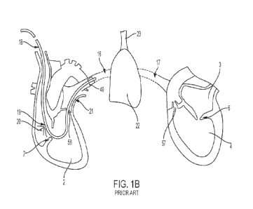
[00013] FIG. 1B is illustrative of the implementation of a right
heart
catheterization. The measurement device 40 is attached to the end of a
pulmonary
artery catheter 18 which passes through the right atrium 1, the tricuspid
valve 7, the
right ventricle 2, through the pulmonary valve 58, and into the pulmonary
artery 16
4
CA 03167547 2022- 8- 10
WO 2021/178636
PCT/US2021/020825
where the device 40 takes measurement of the blood pressure as deoxygenated
blood
is carried into the lung 22. Then, fresh air is carried into the lung 22 from
the trachea 23
after which oxygenated blood is carried through the pulmonary vein 17, the
left atrium 3,
the mitral valve 6, the left ventricle 4, and the aortic valve 57. The
catheter 18 also has
a proximal injection port which injects cold saline bolus 20 into the right
atrium, and a
thermistor 21 located at a distal end of the catheter to measure the
temperature of the
blood in the pulmonary artery 16. This method of measurement is known as
thermodilution, which measures the blood flow based on the premise that when
the cold
saline bolus is added to the circulating blood, the rate of blood flow is
inversely
proportional to the rate of change in blood temperature resulting from the
cold saline
bolus over time. This provides a measure of cardiac output.
[00014] Pulmonary artery wedge pressure and pulmonary artery
diastolic
pressure may be used as surrogate measurements for the pressure within the
left
atrium and the filling pressure of the left ventricle, which is a typical area
of concern in
heart failure. It has been shown that the pulmonary artery and left
ventricular filling
pressures correlate on most occasions except for certain comorbidities such as
primary
pulmonary arterial hypertension. Such pressures change because of circulating
volume
increase (e.g. fluid retention) or declining pumping efficiency of the left
ventricle (e.g.,
thickening, dilation, or vasoconstriction of the peripheral resistance
vessels).
[00015] Various attempts have been made to remotely monitor
cardiac pressures
in order to identify more effective pharmacological treatment programs. These
systems
seek to monitor increases in intracardiac pressures to provide an early
predictor of an
impending acute decompensation for a patient with prior history of heart
failure (e.g., as
a much more reliable indicator than other measurements such as weight gain,
thoracic
impedance, etc.) For example, the CardioMEMSTm heart failure monitoring system
by
Abbott resides in the pulmonary artery and seeks to effectively monitor
pulmonary artery
pressures as a surrogate for left atrial pressure. Other examples of remote
monitoring
systems include: Chronicle by Medtronic and HeartPODTM by Abbott/St. Jude.
The
CardioMEMS, Chronicle and HeartPOD devices are described generally in the De
Rosa
et al. paper entitled Transcatheter Implantable Devices To Monitoring Of
Elevated Left
Atrial Presses In Patients With Chronic Heart Failure, Universita degli Studi
di Salerno,
Translational Medicine, 2017, 17(4): 19-21 (ISSN 2239-9747).
[00016] With Chronicle , the measurement device resides in the
right ventricle
and reports an estimated pulmonary artery diastolic pressure (ePAD) to a
receiving
CA 03167547 2022- 8- 10
WO 2021/178636
PCT/US2021/020825
device. It has been stated that the measurements showed a correlation between
right
ventricular diastolic pressure, right ventricular systolic pressure, and ePAD,
with the
increase in all these pressure readings acting as indicators of an impending
hospitalization.
[00017] HeartPOD TM uses a lead-and-can design with delivery of
a measurement
device by septal puncture method, with the measurement device remaining in the
atrial
septum and measuring left atrial pressure.
[00018] Another example includes the Vectorious TM left atrial
pressure (LAP)
monitoring system by Vectorious Medical Technologies which uses a pressure
sensor
to measure the blood pressure within the left atrium.
[00019] Over the past several decades, the development of remote
systems has
focused on finding a reliable predictor of upcoming hospitalization events.
Measuring
left sided filling pressure and surrogates have shown to be the most reliable,
predictive,
and effective form of remote monitoring. However, these systems show less
information than acute right heart catheterization, as such systems provide
limited data
for accurately detecting root causes of the rise in pressure. One effect of
limited data,
whether in the remote or acute setting, is that medical service providers are
required to
utilize trial and error medication techniques for patient treatment. This
remote trial and
error practice can result in potential unnecessary harm to the patient,
including kidney
damage, further heart failure disease progression, or undetected
comorbidities. For this
reason, physicians are careful with their titration increases (slow
increases/decreases),
use creatinine lab testing as a lagging metric to detect kidney damage due to
over-
diuresis, and are worried about arising comorbidities (such as undetected
right heart
failure), and bring the patient into the office for further analysis, which
may include the
need for a right heart catheterization in order to determine a safe and
effective
treatment change.
[00020] For example, a medical service provider may first try
diuretics to reduce
the monitored blood pressure, if they assume that the pressure increase is due
to a fluid
retention issue. If this does not work, they may increase the dosage of
diuretics again.
If this still does not work, the medical service provider may decide that the
problem is
not in the fluid retention, but vascular resistance, after which an attempt
may be made
to use medications such as vasodilators. Lab creatinine testing may further
reveal that
over-diuresis (hypovolemia) led to increased damage of the kidneys. In other
words,
treatment methods often rely heavily on an individual medical service
provider's
6
CA 03167547 2022- 8- 10
WO 2021/178636
PCT/US2021/020825
personal experiences and intuition, which not only vary from provider-to-
provider and
patient-to-patient but may also extend the time needed to reliably arrive at a
correct
diagnosis.
[00021] There remains need for improved devices, systems and methods for
physiologic measurements and associated diagnostic and treatment regimens for
patients at risk of heart failure.
SUMMARY
[00022] Disclosed herein are methods and medical devices, such as implantable
measurement devices, for performing measurements in a heart.
[00023] One exemplary embodiment is a medical system for determining a
treatment regimen for a patient with a condition. The medical system comprises
a
sensing device including a pressure sensor for monitoring and providing RVP
information representative of right ventricle heart pressures over a period of
time,
wherein at least the pressure sensor is configured for implantation into a
right ventricle
of the patient's heart; one or more processors, coupled to receive the RVP
information,
configured to: determine a right atrial filling pressure based on the RVP
information;
and determine a left atrial filling pressure based on the RVP information; and
optionally
a display device to display the right atrial filling pressure and the left
atrial filling
pressure, wherein the condition is at least one selected from the group of:
left heart
failure, right heart failure, and primary pulmonary disorder.
[00024] Embodiments of the medical system may further comprise a memory unit
configured to store the right atrial filling pressure, the left atrial filling
pressure and the
condition of the patient; and wherein the one or more processors are
configured to
determine, based on the right atrial filling pressure, the left atrial filling
pressure and the
condition of the patient, the treatment regimen for the patient. The one or
more
processors may be coupled to receive the RVP information from the sensing
device by
a wireless communication link.
[00025] The sensing device of the medical system may comprise a wireless
transmitter to wirelessly transmit the RVP information; and the one or more
processors
may be coupled to receive the RVP information wirelessly transmitted by the
sensing
device. The sensing device may comprise a housing configured for attachment to
a
wall (optionally free wall, apex, septum or outflow tract) in the right
ventricle of the heart.
The sensing device may comprise an anchor for attaching the sensing device to
the wall
7
CA 03167547 2022- 8- 10
WO 2021/178636
PCT/US2021/020825
of the right ventricle of the heart, and wherein the anchor optionally
includes one or
more of a coiled spring or a barbed hook. The sensing device may be configured
to be
entirely located in the right ventricle. The sensing device may comprise an
antenna to
receive electromagnetic energy; a wireless transmitter; and wherein the
sensing device
may be configured to be energized by electromagnetic energy received by the
antenna,
and to transmit the RVP information by the wireless transmitter when
energized. In
embodiments, the sensing device does not transmit the RVP information until it
is
energized.
[00026] In embodiments, the one or more processors of the medical system are
configured to determine the right atrial filling pressure using the RVP
information as a
surrogate for the for the right atrial filling pressure. In embodiments, the
one or more
processors are configured to determining the right atrial filling pressure
based on heart
pressure information consisting of the RVP information. In embodiments, the
one or
more processors are configured to determine the right atrial filling pressure
based on
the RVP information at end diastole (e.g., using right ventricle end diastolic
pressure as
a surrogate for the right atrial filling pressure). In embodiments, the one or
more
processors are configured to receive electrical information, optionally ECG
information,
representative of electrical activity of the heart; identify a time of end
diastole of the
heart based on the electrical information; and determine the right atrial
filling pressure
based on the RVP information at the identified time of end diastole of the
heart. The
one or more processors may be configured to determine the left atrial filling
pressure
using the RVP information as a surrogate for the left atrial filling pressure.
The one or
more processors may be configured to determine the left atrial filling
pressure based on
a slope, and optionally a maximum or peak of the slope, of the RVP
information. The
one or more processors may be configured to determine the left atrial filling
pressure
using the right ventricular pressure represented by the RVP information at a
time
corresponding to the maximum or peak slope of the RVP information as a
surrogate for
estimated pulmonary artery diastolic pressure, and using the estimated
pulmonary
artery diastolic pressure as a surrogate for the left atrial filling pressure.
The one or
more processors may be configured to determine the left atrial filling
pressure based on
heart pressure information consisting of the RVP information. In embodiments,
the
system is configured to determine the right atrial filling pressure without
directly
monitoring pressure in the right atrium, and to determine the left atrial
filling pressure
without directly monitoring pressure in the left atrium. The sensing device
may be
8
CA 03167547 2022- 8- 10
WO 2021/178636
PCT/US2021/020825
configured to provide the RVP information over one or more cycles of diastole
and
systole. The one or more processors may be remote from a patient's body
including a
heart associated with the RVP information in embodiments. In embodiments, the
sensing device acquires the RVP information at a frequency greater than 100
Hz, and
optionally greater than 200 Hz.
[00027] In embodiments of the medical system, to determine the treatment
regimen of the patient, the one or more processors may be configured to
compare the
right atrial filling pressure to a baseline right atrial pressure; and compare
the left atrial
filling pressure to a baseline left atrial pressure. To determine the
treatment regimen of
the patient, the one or more processors may be configured to compare the right
atrial
filling pressure and the left atrial filling pressure. To determine the
treatment regimen
for the patient, the one or more processors may be configured to provide a
notification
to increase the dosage of the treatment regimen. To determine the treatment
regimen
for the patient, the one or more processors may be configured to provide a
notification
to decrease the dosage of the treatment regimen.
[00028] In embodiments of the medical system, the one or more processors are
incorporated into an implantable medical device. The one or more processors
may be
incorporated into a device located external to the patient. To determine the
treatment
regimen of the patient, the one or more processors may be configured to
provide a
notification to increase at least one treatment selected from the following
group of
treatments: vasodilators, diuretics, pulmonary vasodilators, neurohormonal
antagonists,
beta blockers, and inotropes. To determine the treatment regimen of the
patient, the
one or more processors may be configured to provide a notification to decrease
at least
one treatment selected from the following group of treatments: vasodilators,
diuretics
pulmonary vasodilators, neurohormonal antagonists, beta blockers, and
inotropes.
[00029] Another exemplary embodiment is a computer-implemented method for
determining a treatment regimen for a patient with a condition. Embodiments of
the
method comprise operating a sensing device including a pressure sensor located
in a
right ventricle of the patient's heart to monitor pressure in the right
ventricle and to
transmit RVP information representative of the pressure in the right ventricle
over a
period of time, optionally including implanting the pressure sensor in the
right ventricle;
processing the RVP information by one or more processors to determine a right
atrial
filling pressure for the patient based on the RVP information and a left
atrial filling
pressure for the patient based on the RVP information; and optionally
displaying on a
9
CA 03167547 2022- 8- 10
WO 2021/178636
PCT/US2021/020825
display device the right atrial filling pressure and the left atrial filling
pressure, wherein
the condition is at least one selected from the group of: left heart failure,
right heart
failure, and primary pulmonary disorder. In embodiments, the method further
comprises
determining the treatment regimen for the patient based on the right atrial
filling
pressure, the left atrial filling pressure, and the condition of the patient.
[00030] In embodiments, determining the right atrial filling
pressure may comprise
using the RVP information as a surrogate for the for the right atrial filling
pressure.
Determining the right atrial filling pressure may comprise determining the
right atrial
filling pressure based on heart pressure information consisting of the RVP
information.
Determining the right atrial filling pressure may comprise determining the
right atrial
filling pressure based on the RVP information at end diastole (e.g., using
right ventricle
end diastolic pressure as a surrogate for the right atrial filling pressure).
[00031] Embodiments of the method further comprise receiving electrical
information, optionally ECG information, representative of electrical activity
of the heart;
and identifying a time of end diastole of the heart based on the electrical
information;
and determining the right atrial filling pressure comprises determining the
right atrial
filling pressure based on the RVP information at the identified time of end
diastole of the
heart (e.g., using right ventricle end diastolic pressure as a surrogate for
the right atrial
filling pressure). Determining the left atrial filling pressure may comprise
using the RVP
information as a surrogate for the left atrial filling pressure. Determining
the left atrial
filling pressure may comprise determining the left atrial filling pressure
based on a
slope, and optionally a maximum or peak of the slope, of the RVP information.
Determining the left atrial filling pressure may comprise using the right
ventricular
pressure represented by the RVP information at a time corresponding to the
maximum
or peak slope of the RVP information as a surrogate for estimated pulmonary
artery
diastolic pressure, and using the estimated pulmonary artery diastolic
pressure as a
surrogate for the left atrial filling pressure. Determining the left atrial
filling pressure may
comprise determining the left atrial filling pressure based on heart pressure
information
consisting of the RVP information. Operating the sensing device to monitor
pressure
includes acquiring the RVP information at a frequency greater than 100 Hz, and
optionally greater than 200 Hz, in embodiments.
[00032] In embodiments of the method, receiving the RVP information comprises
receiving the RVP information from a sensing device comprising a single
pressure
sensor in the right ventricle. Receiving the RVP information may comprise
receiving the
CA 03167547 2022- 8- 10
WO 2021/178636
PCT/US2021/020825
RVP information from a sensing device entirely located in the right ventricle.
Determining the left atrial filling pressure may comprise determining the
right atrial filling
pressure and determining the left atrial filling pressure based on the RVP
information
received from the single pressure sensor. In embodiments, receiving the RVP
information comprises wirelessly receiving the RVP information.
[00033] In embodiments of the method, determining the right
atrial filling pressure
comprises determining the right atrial filling pressure without directly
monitoring
pressure in the right atrium; and determining the left atrial filling pressure
comprises
determining the left atrial filling pressure without directly monitoring
pressure in the left
atrium. The method may further comprise energizing an implanted sensing device
comprising a pressure sensor located in the right ventricle, and wherein the
energized
sensing device transmits the RVP information. In embodiments, the implanted
sensing
device does not transmit the RVP information until it is energized. Receiving
the RVP
information representative of a right ventricle heart pressure over a period
of time may
comprise receiving the RVP information representative of a right ventricle
heart
pressure over one or more cycles of diastole and systole. In embodiments, the
one or
more processors may be remote from a patient's body including a heart
associated with
the RVP information.
[00034] In embodiments of the method, determining the treatment regimen of the
patient may comprise comparing the right atrial filling pressure to a baseline
right atrial
pressure; and comparing the left atrial filling pressure to a baseline left
atrial pressure.
Determining the treatment regimen of the patient may comprise comparing the
right
atrial filling pressure and the left atrial filling pressure. Determining the
treatment
regimen for the patient may comprise providing a notification to increase the
dosage of
the treatment regimen. Determining the treatment regimen for the patient may
comprise
providing a notification to decrease the dosage of the treatment regimen.
Determining
the treatment regimen of the patient may comprise providing a notification to
increase at
least one treatment selected from the following group of treatments:
vasodilators,
diuretics, pulmonary vasodilators, neurohormonal antagonists, beta blockers,
and
inotropes. Determining the treatment regimen of the patient may comprise
providing a
notification to decrease at least one treatment selected from the following
group of
treatments: vasodilators, diuretics, and pulmonary vasodilators, neurohormonal
antagonists, beta blockers, and inotropes.
[00035] Yet other exemplary embodiments includes a monitoring system,
11
CA 03167547 2022- 8- 10
WO 2021/178636
PCT/US2021/020825
comprising a receiver configured to receive RVP information associated with a
right
ventricle heart pressure over a period of time, wherein the RVP information is
received
from a sensing device including a pressure sensor located in the right
ventricle of the
heart; a memory unit configured to store the received RVP information;
optionally a
display device; and one or more processors configured to determine a right
atrial filling
pressure based on the RVP information and a left atrial filling pressure based
on the
RVP information; compare the right atrial filling pressure and the left atrial
filling
pressure; and output, to the display device, the comparison. In embodiments,
the one
or more processors are further configured to determine, based on the
comparison, a
treatment regimen for the patient. The receiver receives RVP information
acquired at a
frequency greater than 100 Hz, and optionally greater than 200 Hz, in
embodiments.
[00036] In embodiments of the monitoring system the one or more processors
may be configured to determine the right atrial filling pressure using the RVP
information as a surrogate for the for the right atrial filling pressure. The
one or more
processors may be configured to determine the right atrial filling pressure
based on
heart pressure information consisting of the RVP information. The one or more
processors may be configured to determine the right atrial filling pressure
based on the
RVP information at end diastole (e.g., using right ventricle end diastolic
pressure as a
surrogate for the right atrial filling pressure). The one or more processors
may be
configured to receive electrical information, optionally ECG information,
representative
of electrical activity of the heart; identify a time of end diastole of the
heart based on the
electrical information; and determine the right atrial filling pressure based
on the RVP
information at the identified time of end diastole of the heart.
[00037] In embodiments of the monitoring system, the one or more processors
may be configured to determine the left atrial filling pressure using the RVP
information
as a surrogate for the left atrial filling pressure. The one or more
processors may be
configured to determine the left atrial filling pressure based on a slope, and
optionally a
maximum or peak of the slope, of the RVP information. The one or more
processors
may be configured to determine the left atrial filling pressure using the
right ventricular
pressure represented by the RVP information at a time corresponding to the
maximum
or peak slope of the RVP information as a surrogate for estimated pulmonary
artery
diastolic pressure, and to use the estimated pulmonary artery diastolic
pressure as a
surrogate for the left atrial filling pressure. In embodiments, the one or
more processors
are configured to determine the left atrial filling pressure based on heart
pressure
12
CA 03167547 2022- 8- 10
WO 2021/178636
PCT/US2021/020825
information consisting of the RVP information. The system may be configured to
determine the right atrial filling pressure without direct information about
monitored
pressure in the right atrium, and to determine the left atrial filling
pressure without direct
information about monitored pressure in the left atrium.
[00038] In embodiments of the monitoring system, the treatment comprises at
least treatment selected from the following group of treatments: diagnosis,
medication
titrations, advanced therapy, IV medications, lifestyle changes, intra-atrial
shunts, valve
repair/replace, ICDs, CRTs, and ablation. The medication titrations may
comprise at
least one titration selected from the following group of titrations.
vasodilators, diuretics,
pulmonary vasodilators, neurohormonal antagonists, beta blockers, and
inotropes. The
advanced therapy may comprise one or more selected from the group of:
implanting a
ventricular assist device (VAD), implanting a mechanical circulator support
(MCS), a
transplant, or both. The lifestyle changes may comprise at least one lifestyle
change
selected from the following group of lifestyle changes: a change in diet,
increased
activity, or both.
[00039] In embodiments of the monitoring system, the one or more processors
are further configured to output the determined treatment to the display
device. The
one or more processors may be further configured to diagnose, based on the
comparison, the patient. In embodiments, the monitoring system is a closed
loop
system where trend data of the measurements inform changes to an automated
dispensing of a medicine. The medicine may be a diuretic, vasodilator, or
both. In
embodiments, the monitoring system is a closed loop system where trend data of
the
measurements inform changes to a ventricular assist device. The monitoring
system
may be used to determine RPM changes in a ventricular assist device.
[00040] In embodiments, the one or more processors may use machine learning
to modify the treatment regimen. The processing device may be further
configured to
output to the display device one or more of the following: left atrial
pressure, left atrial
pressure averages, right atrial pressure, right atrial pressure averages,
trend arrows of
the measurements, line graphs over time of the measurements, waveforms of the
measurements, and one or more medications of a patient associated with the
measurements.
BRIEF DESCRIPTION OF THE DRAWINGS
[00041] The accompanying drawings are included to provide a further
13
CA 03167547 2022- 8- 10
WO 2021/178636
PCT/US2021/020825
understanding of the disclosure and are incorporated in and constitute a part
of this
specification, illustrate embodiments, and together with the description serve
to explain
the principles of the disclosure.
[00042] FIG. 1A is a graph showing the utility in data from a surrogate
measurement of left atrial pressure in reducing hospitalizations due to heart
failure;
[00043] FIG. 1B is a schematic diagram of a heart and a lung of a patient
using
the prior-art measurement device (Swan Ganz right heart catheter) as discussed
herein;
[00044] FIG. 2 is a diagrammatic illustration of a medical system according to
some embodiments;
[00045] FIG. 3 is an illustration of a sensing device in
accordance with
embodiments implanted in a right ventricle of a patient's heart;
[00046] FIG. 4 is a diagrammatic illustration of a sensing device in
accordance
with embodiments;
[00047] FIG. 5 is a diagrammatic illustration of a monitoring
device in accordance
with embodiments;
[00048] FIG. 6 is an illustration of an external charger and communication
relay in
accordance with embodiments;
[00049] FIG. 7A is a flow diagram describing a method for determining right
atrial
filling pressures from right ventricle pressure information in accordance with
embodiments;
[00050] FIG. 7B is a flow diagram describing a method for determining left
atrial
filling pressures from right ventricle pressure information in accordance with
embodiments;
[00051] FIG. 8 illustrates a block diagram of a method to determine actions
that
need to be taken based on pressure measurements according to some embodiments;
[00052] FIG. 9 illustrates a medication administration reference
table using two
sets of measurement data as implemented by the method in FIG. 8;
[00053] FIG. 10 illustrates a flow diagram of a method for determining a
treatment
regimen for a patient according to some embodiments;
[00054] FIG. 11 illustrates an exemplary diagnostic table and an exemplary
treatment regimen table referenced by the method in FIG. 10 for a patient
diagnosed
with left heart failure;
[00055] FIG. 12 illustrates an exemplary diagnostic table and an exemplary
treatment regimen table referenced by the method in FIG. 10 for a patient
diagnosed
14
CA 03167547 2022- 8- 10
WO 2021/178636
PCT/US2021/020825
with right heart failure;
[00056] FIG. 13 illustrates an exemplary diagnostic table and an exemplary
treatment regimen table referenced by the method in FIG. 10 for a patient
diagnosed
with primary pulmonary disorder;
[00057] FIG. 14 illustrates an exemplary diagnostic table and an exemplary
treatment regimen table referenced by the method in FIG. 10 for a patient
diagnosed
with left heart failure and right heart failure;
[00058] FIG. 15 illustrates an exemplary diagnostic table and an exemplary
treatment regimen table referenced by the method in FIG. 10 for a patient
diagnosed
with left heart failure and primary pulmonary disorder;
[00059] FIG. 16 illustrates an exemplary diagnostic table and an exemplary
treatment regimen table referenced by the method in FIG. 10 for a patient
diagnosed
with right heart failure and primary pulmonary disorder;
[00060] FIG. 17 illustrates an exemplary diagnostic table and an exemplary
treatment regimen table referenced by the method in FIG. 10 for a patient
diagnosed
with left heart failure, right heart failure, and primary pulmonary disorder;
[00061] FIG. 18 is a graph of an exemplary RVP (right ventricular pressure)
over
several diastolic and systolic heart cycles, and associated pulmonary arterial
pressure
(PAP), RV dp/dt (right ventricular pressure changes over time), and ECG
(electrocardiogram); and
[00062] FIG. 19 is an illustration of another sensing device
including an
attachment structure in accordance with embodiments.
DETAILED DESCRIPTION
[00063] This disclosure is not meant to be read in a restrictive manner. For
example, the terminology used in the application should be read broadly in the
context
of the meaning those in the field would attribute such terminology.
[00064] As the terms are used herein with respect to ranges of measurements
"about" and "approximately" may be used, interchangeably, to refer to a
measurement
that includes the stated measurement and that also includes any measurements
that
are reasonably close to the stated measurement, but that may differ by a
reasonably
small amount such as will be understood, and readily ascertained, by
individuals having
ordinary skill in the relevant arts to be attributable to measurement error,
differences in
measurement and/or manufacturing equipment calibration, human error in reading
CA 03167547 2022- 8- 10
WO 2021/178636
PCT/US2021/020825
and/or setting measurements, adjustments made to optimize performance and/or
structural parameters in view of differences in measurements associated with
other
components, particular implementation scenarios, imprecise adjustment and/or
manipulation of objects by a person or machine, and/or the like.
[00065] Certain terminology is used herein for convenience only. For example,
words such as "top", "bottom", "upper," "lower," "left," "right,"
"horizontal," "vertical,"
"upward," and "downward" merely describe the configuration shown in the
figures or the
orientation of a part in the installed position. Indeed, the referenced
components may be
oriented in any direction. Similarly, throughout this disclosure, where a
process or
method is shown or described, the method may be performed in any order or
simultaneously, unless it is clear from the context that the method depends on
certain
actions being performed first.
[00066] Various embodiments are directed toward implantable medical devices
such as device for performing physiologic measurements to obtain information
regarding characteristics in the left and right sides of the heart. In certain
instances, the
various aspects of the present disclosure relate to methods and devices for
performing
pressure measurements. Additionally, the present disclosure also includes a
medical
treatment system for determining administration of medications to a patient
based on
the measurements performed.
[00067] FIG. 2 is a diagrammatic illustration of a medical system 60 in
accordance with embodiments for determining a treatment regimen for a patient
with a
heart condition such as left heart failure, right heart failure or primary
pulmonary
disorder. As shown, the medical system 60 includes a sensing device 61 (i.e.,
a
measurement device) and a monitoring system 62. As described in greater detail
below, the sensing device 61 includes a pressure sensor (not shown in FIG. 2)
configured to be implanted in a right ventricle of a patient's heart. FIG. 3
is an
illustration of an embodiment of the sensing device 61 where the entire
sensing device
is located and implanted in a right ventricle of a patient's heart. For
example the
sensing device 61, or at least the pressure sensor, may be implanted in the
right
ventricle free wall, right ventricle apex, right ventricle septum or right
ventricle outflow
tract. The sensing device can be delivered and implanted into the right
ventricle using
conventional methods such as trans-catheter delivery (e.g., by a delivery
catheter 59
through vasculature including the vena cava, right atrium, pulmonary valve and
into the
right ventricle), or open heart surgical approaches. Following implantation of
the
16
CA 03167547 2022- 8- 10
WO 2021/178636
PCT/US2021/020825
sensing device 61, room may remain in the right ventricle for other structures
such as
pacing leads, wireless pacemakers, CRT leads, ICD leads, leadless pacemakers,
etc.,
or combined with the leads of wireless pacemakers. These and other structures
of
these types can be used in combination with the sensing device 61 and methods
described herein. For example, pacemakers and/or ICDs may be used as an input
for
the ECG signal used to identify right ventricular end diastolic (RVEDP) and
right atrial
pressures in accordance with embodiments described herein.
[00068] The sensing device 61 monitors pressures in the patient's right
ventricle
over periods of time (e.g., one or more heartbeats or cycles of diastole and
systole).
Monitoring system 62 is coupled to receive data or information representative
of the
measured right ventricle pressures (referred to as RVP information in this
description).
In embodiments, monitoring system 62 is configured to wirelessly receive the
RVP
information transmitted by the sensing device 61. Monitoring system 62 is also
configured to process the received RVP information, and to determine a right
atrial filling
pressure (RAP) of the patient's heart based on the RVP information, and to
determine a
left atrial filling pressure (LAP) of the patient's heart based on the RVP
information. As
described in greater detail below, embodiments of the monitoring system 62
determine
both the right atrial filling pressure and the left atrial filling pressure of
the patient's heart
using the right ventricular pressure represented by the RVP information as a
surrogate.
Embodiments of the monitoring system 62 include a display device (not shown in
FIG.
2) that displays the determined right atrial filling pressure and the
determined left atrial
filling pressure. Embodiments of the monitoring system 62 may be configured to
determine and display the heart condition of the patient and/or a treatment
regimen for
the patient based at least in part on the determined right and left atrial
filling pressures
of the patient.
[00069] FIG. 4 is a diagrammatic illustration of a sensing device 61 in
accordance
with embodiments. The illustrated embodiments include a housing 63 enclosing a
controller unit 64 coupled to components including pressure sensor 65, power
source
66, transmitter 67, memory 68, charging coil 69 and electrical sensor 70. An
attachment structure 71 on the housing 63 may be used to anchor the sensing
device
61 to tissues of the patient's heart (e.g., at a bottom portion of the right
ventricle as
shown in FIG. 3). Housing 63 can be formed of appropriate known or otherwise
conventional materials such as biocompatible metal (e.g. stainless steel or
titanium)
and/or polymers. Controller unit 64 may be embodied in suitable known or
otherwise
17
CA 03167547 2022- 8- 10
WO 2021/178636
PCT/US2021/020825
conventional electronics structures such as discrete circuit components,
application
specific integrated circuits (ASICs) or programmed processors. Similarly,
memory 68
may be embodied in suitable known or otherwise conventional structures
configured for
operation with the controller unit 64. Pressure sensor 65 may, for example,
incorporate
MEMS technology such as but not limited to capacitive or piezoelectric sensors
or other
pressure measurement technologies suitable for measurement of intracardiac
pressure
levels. Signals or other information representative of pressures monitored by
the
pressure sensor 65 (e.g., the RVP information) are coupled to controller unit
64 and
may optionally be stored in the memory 68. In other embodiments (not shown),
components of the sensing device 61 other than the pressure sensor 65 may be
located
outside of the patient's heart, (e.g., in a housing located under the skin in
the patient's
chest) and coupled (e.g., by leads) to an implanted pressure sensor 65.
[00070] Embodiments of the transmitter 67 include an antenna (not separately
shown) to wirelessly transmit the RVP information provided by the controller
unit 64
(e.g., by radio frequency (RE)). Embodiments of transmitter 67 may, for
example,
include near field (e.g., Bluetooth) or other suitable known or conventional
technologies.
Power source 66 may be any suitable source. In embodiments, the power source
66
includes the charging coil 69 coupled to an energy storage device to enable
inductive
charging of the power source by an external device. In embodiments including
such an
inductive power source 66, the sensing device 61 can be energized by the
external
device and thereby operated to measure the right ventricular pressure and
transmit the
RVP information. In inductive charging embodiments of this type the sensing
device 61
may measure pressure and transmit the RVP information only when the power
source
66 is energized. An advantage of such an inductive charging power source 66 is
that
the need to exchange the power source when it runs out of power is reduced.
The
charging coil 69 and the antenna of the transmitter 67 may be the same
structure in
embodiments having an inductive charging power source 66. Alternatively or in
addition, embodiments of power source 66 may include a battery.
[00071] The illustrated embodiment of sensing device 61 includes electrical
sensor 70. The electrical sensor 70 is configured to measure and provide
signals
representative of electrical activity of the heart. In embodiments, for
example, electrical
sensor 70 may measure and provide information representative of
electrocardiogram
(ECG) signals in the patient's heart. Embodiments of the electrical sensor 70
may
include anode and cathode terminals on the housing 63 (not separately shown).
The
18
CA 03167547 2022- 8- 10
WO 2021/178636
PCT/US2021/020825
electrical information measured by the electrical sensor 70 is coupled to the
controller
unit 64 and may be transmitted by the transmitter 67. The electrical
information
measured by electrical sensor 70 may also be stored in the memory 68. Other
embodiments of sensing device 61 and/or the medical system 60 do not include
an
electrical sensor such as 70. As described below, some embodiments of medical
system 60 do not make use of electrical information such as the ECG of the
heart. Yet
other embodiments of medical system 60 make use of electrical information such
as the
ECG of the patient's heart that are obtained from other sources (e.g.,
electrodes on the
patient's body and/or other implanted devices in the patient).
[00072] Attachment structure 71 may include known or otherwise conventional
structures to anchor the sensing device 61 (or pressure sensor 65 in
embodiments)
within the right ventricle of the patient's heart. In the embodiments shown in
FIG. 3, for
example, attachment structure 71 includes helically coiled springs configured
to engage
and enter the heart tissue upon rotation of the sensing device 61. Other
embodiments
of attachment structure 71 include other structures, such as for example one
or more
hooks optionally including barbs. FIG. 19, for example, illustrates a sensing
device 61'
including a plurality of anchors 71' (two are shown for purposes of example)
configured
to secure the sensing device under a tissue surface in a patient's right
ventricle. In the
illustrated embodiments each of anchors 71' includes a substantially linear
section 71a'
extending from a distal end of the housing 63' generally parallel to a
longitudinal axis of
the housing, and a curved section 71b' extending from the substantially linear
section.
The curved section 71b' of each anchor 71' may be configured to align with the
substantially linear section 71a' relative to the longitudinal axis of the
housing 63' in a
delivery configuration, and curve radially outwardly relative to the
longitudinal axis and
toward the distal end of the housing in the deployed configuration.
[00073] FIG. 5 is a diagrammatic illustration of a monitoring
system 62 in
accordance with embodiments. The illustrated embodiments include a processing
system 76 coupled to a receiver 77, memory 78 and display 79. Processing
system 76
is a programmable microprocessor-based system in embodiments. Memory 78, which
can for example include ROM and RAM, is coupled to the processing system 76
and
can store data and information such as programs executed by the processing
system.
For example, and as described in greater detail below, processing system 76
can
execute programs stored in memory 78 that characterize methods or algorithms
to
generate the right atrial filling pressure and the left atrial filling
pressure in the patient's
19
CA 03167547 2022- 8- 10
WO 2021/178636
PCT/US2021/020825
heart based on the RVP information transmitted by the sensing device 61.
Processing
system 76 may also execute programs stored in memory 78 to determine heart
conditions and treatment regimens based on information such as the right
atrial filing
pressure and left atrial filling pressure of the patient's heart in accordance
with methods
and algorithms described below. Alternatively or in addition, processing
system 76 can
be implemented by other suitable structures such as discrete circuit elements
and
ASICs. In embodiments, monitoring system 62 can be embodied as an app (i.e.,
application software) in a conventional mobile device such as a smartphone or
tablet.
In other embodiments all or components of monitoring system 62 (e.g.,
processing
system 76, memory 78 and display 79) can be embodied as a "desktop" computer
system coupled to a receiver 77 (e.g., over a communications network). Yet
other
embodiments of monitoring system 62 include computing components in the cloud
coupled to a user's device, such as a mobile phone or tablet, including a
display.
[00074] Receiver 77 is configured to receive information such as the RVP
information from the sensing device 61, and to couple the received information
to the
processing system 76. Receiver 77 wirelessly receives the information in
embodiments
(e.g., by RF). In embodiments that make use of ECG or other electrical
information of
the patient's heart, the electrical information may also be received by and
coupled to the
processing system 76 by the receiver 77.
[00075] Display 79 can be operated by the processing system 76 to display
information received by and/or generated by the processing system. In
embodiments,
for example, the display 79 can display one or more of the RVP information,
the right
atrial filling pressure and/or left atrial filling pressure, determined heart
conditions,
determined treatment regimens and/or heart electrical information. Display 79
can also
be configured to display other information measured or otherwise obtained from
the
patient, such as for example blood pressure, temperature and/or oxygen
saturation. If
the patient is visually impaired or prefers audio notifications, the
monitoring system 62
can provide audio output to alert the patient if measurements indicate the
patient's heart
may be a risk of acute decompensation episodes, so that the patient can go to
a
hospital for further examination. The monitoring system 62 can also upload the
measured and/or generated data and information onto a remote server (not
shown) to
be collected by medical service providers or a database to remotely monitor
the
conditions of the patient's heart.
[00076] FIG. 6 shows an example of an external charger and communications
CA 03167547 2022- 8- 10
WO 2021/178636
PCT/US2021/020825
relay 80 according to some examples. As shown, the external charger and
communications relay 80 is a device which can charge or power a power source
such
as 66 of the sensing device 61 (for example, a battery or capacitor) via
electromagnetic
induction, as well as to communicate with the sensing device to obtain
measurement
data or information such as the RVP information. In one example, the external
charger
and communications relay 80 is a device which inductively couples with the
sensing
device 61 to directly power the sensing device such that an on-board power
source, for
example a battery, is not required. In one example, the external charger and
communications relay 80 wirelessly powers the sensing device 61 via
radiofrequency
(RF) electromagnetic radiation. The external charger and communications relay
80 may
be worn (e.g., using a harness 81) such that the location of the charger and
relay 80 is
placed at an operable location for the charger and relay to charge and obtain
data from
the sensing device 61. Monitoring system 62 can be used by the patient or
other party
(e.g., medical service provider or remote monitoring facility) to receive
information
regarding the measurement data via the external charger and communications
relay 80.
In other embodiments data and other information measured by the sensing device
61,
including the RVP information, can be transmitted by the sensing device
directly to the
monitoring system 62 (e.g., if the sensing device is battery powered).
[00077] FIGs. 7A and 7B are flowcharts illustrating methods and algorithms
that
can be implemented by the monitoring system 62 using the RVP information, and
optionally the heart electrical information, to generate or determine the
patient's right
atrial filling pressure and left atrial filling pressure. FIG. 18 is a graph
of an example of
right ventricular pressures (RVP) monitored within a patient over a period of
time
including two diastolic and systolic cycles. The RVP information used by the
methods
of FIGs. 7A and 7B can be data or other information representative of the
illustrated
right ventricular pressure. FIG. 18 also illustrates exemplary pulmonary
artery
pressures (PAP), changes in the right ventricular pressures over time (i.e.,
slopes) (RV
dP/dt) and ECG signals of the patient, that are associated with and correspond
to the
right ventricular pressure RVP. The pressures, changes in pressures and
electrical
signals shown in FIG. 18 are used in connection with the description of the
methods
shown in FIGs. 7A and 7B. In embodiments, the data acquisition frequency of
sensing
device 61 is greater than 100 Hz to determine the maximum dP/dt for purposes
of
obtaining estimated pulmonary artery diastolic pressure (ePAD). In
embodiments, for
example, the data acquisition frequency is 200 Hz ¨ 250 Hz, or even greater.
If the data
21
CA 03167547 2022- 8- 10
WO 2021/178636
PCT/US2021/020825
acquisition frequency is too low, accuracy of the determinations or locations
for ePAD
on the right ventricular pressure waveform, as represented by the RVP
information, may
be detrimentally impacted.
[00078] Method 110 illustrated in FIG. 7A uses the right ventricular pressures
RVP, as represented by the RVP information, as surrogates for determining the
right
atrial filling pressure. Right atrial filling pressure or right atrial
pressure (RAP) is
generally equal to the right ventricle end diastolic pressure (RVEDP), which
is the right
ventricle pressure at the end of the diastolic cycle of the heart (e.g., in
the absence of
tricuspid valve issues). Accordingly, by method 110 the monitoring system 62
monitors
the RVP information as shown by step 112, and determines the end time of the
diastolic
cycle as shown by step 114. The end of the diastolic cycle during which the
right
ventricle fills with blood defines the beginning of the systolic cycle during
which the
heart contracts to pump deoxygenated blood from the right ventricle through
the
pulmonary valve toward the lungs. Accordingly, and as shown in FIG. 18 at the
times
corresponding generally to 0.25 sec. and 1.0 sec., the right ventricular
pressure RVP,
and therefore the RVP information, relatively quickly and substantially
increase
immediately following end diastole. As is also shown in FIG. 18, the ECG
signal
relatively quickly and substantially decreases at the beginning of the
systolic cycle.
Method 110 can make use of these physiologic and/or electrical characteristics
of the
heart in connection with step 114.
[00079] In one embodiment the monitoring system 62 monitors the slope of the
RVP information to determine the end time of the diastolic cycle as shown by
step 114.
For example, the monitoring system 62 can identify the end time of the
diastolic cycle as
the time that the slope of the RVP information increases by a predetermined
amount
(e.g., exceeds a threshold value) within a predetermined time period. As shown
by
steps 116 and 118, monitoring system 62 then determines the right ventricular
pressure
at the determined end time of the diastolic cycle, and uses the right
ventricular pressure
at the end of the diastolic cycle as the right atrial pressure. By this
embodiment, the
monitoring system 62 can determine the right atrial filling pressure without
the use of the
ECG or other electrical information. This embodiment can thereby be
implemented
using a sensing device 61 that does not include an electrical sensor such as
70 (as
shown for example in the embodiment in FIG. 4).
[00080] In embodiments where the monitoring system 62 receives heart
electrical
information such as the ECG (e.g., embodiments having a sensing device 61
including
22
CA 03167547 2022- 8- 10
WO 2021/178636
PCT/US2021/020825
electrical sensor 70), monitoring system 62 may use the electrical information
to
determine the end time of the diastolic cycle by step 114. For example, the
monitoring
system 62 can identify the end time of the diastolic cycle as the time that
the slope of
the ECG information decreases by a predetermined amount (e.g., exceeds a
threshold
value) within a predetermined time period. Monitoring system 62 then
determines the
right ventricular pressure at the determined end time of the diastolic cycle,
and uses the
right ventricular pressure at the determined end time of the diastolic cycle
as the right
atrial pressure as shown by steps 116 and 118. Conventional signal processing
approaches including slope determinations and detection, filtering,
comparisons and
thresholding can be used in connection with these embodiments of method 110.
By this
method 110 the monitoring system 62 determines the right atrial filling
pressures without
directly monitoring pressure in the right atrium (e.g., there is no pressure
sensor in the
right atrium). Instead, the right atrial pressures are determined using heart
pressure
information consisting only of the RVP information. Other embodiments may use
other
signal processing approaches and algorithms to determine the right atrial
filling
pressures based on the RVP information. For example, in other embodiments,
obtaining
right atrial filling pressures based on the RVP waveforms may be performed
using the
systems and methods described in U.S. Pat. No. 6,915,162, entitled,
"Implantable
Medical Device For Measuring Ventricular Pressure," and issued on July 5,
2005, the
entire contents of which is incorporated herein in its entirety for all
purposes.
Additionally, or alternatively, in other embodiments, obtaining right atrial
filling pressures
based on the RVP waveforms and/or ECG information may be performed using the
systems and methods described in U.S. Pat. No. 5,368,040, entitled, "Apparatus
And
Method For Determining A Plurality Of Hemodynamic Variables From A Single,
Chronically Implanted Absolute Pressure Sensor," and issued on November 29,
1994,
the entire contents of which is incorporated herein in its entirety for all
purposes.
[00081]
Method 120 illustrated in FIG. 7B uses the right ventricular pressures
RVP, as represented by the RVP information, as surrogates for determining the
left
atrial filling pressures (LAP). The left atrial pressure is generally equal to
the estimated
pulmonary artery diastolic pressure (ePAD or PADP). The pulmonary artery
diastolic
pressure is generally equal to the right ventricular pressure at the time of
the pulmonary
valve opening. The pulmonary valve opens at a time generally corresponding to
the
time of maximum or peak increasing pressure change or slope in the right
ventricular
pressure during systole. Accordingly, by method 120, the monitoring system 62
23
CA 03167547 2022- 8- 10
WO 2021/178636
PCT/US2021/020825
monitors the RVP information as shown by step 122, and determines the time at
which
the RVP has its maximum increasing change or increasing slope (dP/dt) during
the
systolic cycle as shown by step 124. As shown by steps 126 and 128, the
monitoring
system 62 then determines the right ventricular pressure at the time of
maximum
change of the RVP slope (which corresponds to the pulmonary artery diastolic
pressure
at the time of the pulmonary valve opening), and uses that right ventricular
pressure as
the left atrial pressure LAP. In embodiments, the right ventricular pressure
RVP at the
time at which the RVP has its minimum slope (dP/dt) during the systolic cycle
can also
be used as a surrogate for the left atrial filling pressure LAP (e.g., in
addition to or as an
alternative to the approaches described above). FIG. 18 is annotated, for
example, to
show a minimum slope of RVP (dP/dt min) and the associated RVP. Conventional
signal processing approaches including slope determinations and detection,
filtering,
comparisons and thresholding can be used in connection with these embodiments
of
method 120. By this method the monitoring system 62 determines the left atrial
filling
pressures without directly monitoring pressure in the left atrium (e.g.,
without the use of
a pressure sensor in the left atrium). Instead, the left atrial pressures are
determined
using heart pressure information consisting only of the RVP information. Other
embodiments may use other signal processing approaches and algorithms to
determine
the left atrial filling pressures based on the RVP information.
[00082] In other embodiments, the sensing device 61 is configured to determine
the right atrial pressures and left atrial pressures using the RVP
information.
Embodiments of a sensing device 61 of these types can, for example, include a
processing system such as 76 that processes the RVP information in accordance
with
methods 110 and 120. In embodiments of these types, the sensing device 61 can
transmit or otherwise couple the determined right and left atrial pressures to
the
monitoring system 62.
[00083] In embodiments, the sensing device 61 and/or monitoring system 62 can
be used in combination with other medical devices. Examples of such medical
devices
include, but are not limited to, blood pressure cuffs, pulse-oximeters,
scales, creatinine
testing devices, smart devices, and wearable medical tracking devices, to name
a few.
The sensing device 61 can also be combined with other implantable devices,
such as
for example a ventricular assist device (VAD), drug delivery shunt or system.
The
sensing device 61 may provide feedback to the other implantable device(s), as
part of a
closed loop or open loop feedback system. The VAD may be a right VAD, a left
VAD,
24
CA 03167547 2022- 8- 10
WO 2021/178636
PCT/US2021/020825
or a bi VAD.
[00084] The pressure measurement data obtained using the sensing device 61
as described herein can be used to perform pulse-contour method, which is
another
method that is used to measure the cardiac output of the patient. This method
uses the
continuous pressure measurement data to plot a pressure-versus-time graph for
the
patient's heart, after which the pressure integral, i.e. the area beneath the
plotted line on
the pressure-versus-time graph, is used to determine the stroke volume (SV) of
the
portion of the heart that is being measured. The value of SV multiplied by the
heart rate
is the cardiac output.
[00085] FIG. 8 is a flow chart showing a remote medical treatment monitoring
method 99 that can be implemented using one or more electronic devices, such
as the
monitoring system 62, using measurement data received from the sensing device
61,
for example, or any of the sensor elements described herein (e.g., RAPs and
LAPs
determined as described herein) . In some examples, the method 99 is used for
patients with a history of left heart failure (LHF), to determine treatment
protocols guided
by measured right and left heart physiologic metrics (e.g., pressure,
temperature, and/or
oxygen saturation).
[00086] Regardless, in some embodiments, in an optional first step 90 the
service
provider determines if the patient receiving treatment has a history of either
left heart
(LH) or right heart (RH)/biventricular failure. The method 99 may be used for
patients
with a risk of LH or RH/biventricular failure as determined by the medical
service
providers, regardless of history. In optional step 91, the medical service
provider set a
baseline "normal" level for applicable physiologic metrices (e.g., the left
and right atrial
pressures) in the acute setting by performing various tests on the patient to
determine,
based on the current condition of the patient, what normal levels (pressure,
cardiac
output, and/or oxygen saturation) would be. Baseline values can then be
entered into
the system which transfers the data to the monitoring system 62. In the
example
illustrated in this figure, the pressures being measured are the left atrial
pressure (LAP)
and the right atrial pressure (RAP). Other embodiments may include other
measurements of other parts of the heart, as deemed appropriate by the medical
service provider.
[00087] In some examples, the monitoring system 62 receives or determines
RAP and LAP measurements in step 92. In one implementation, the measurements
include whether the pressure values of the right atrium and the left atrium
are trending
CA 03167547 2022- 8- 10
WO 2021/178636
PCT/US2021/020825
below, at, or above the normal level. In another example, the method may also
consider whether the pressure values are increasing, decreasing, or staying
steady as
an additional input into the overall assessment.
[00088] In optional step 93, the monitoring system 62 confirms whether the
patient has a history of LH or RH/biventricular failure. The monitoring system
62
optionally uses a medication administration reference table 100 in FIG. 9 to
determine
and indicate if dosage of certain medications needs to be increased or
reduced, in step
94. Alternatively, a medical service provider (e.g., physician) optionally
uses the data
directly to assess what treatment regimen (e.g., pharmacological) is
appropriate based
upon the data using the methodology of table 100.
[00089] As shown, the table 100 has three columns and three rows, where the
columns pertain to "RAP trending below normal" 101, "RAP trending normal" 102,
and
"RAP trending above normal" 103, and the columns pertain to "LAP trending
below
normal" 104, "LAP trending normal" 105, and "LAP trending above normal" 106.
For
example, if the RAP is trending below normal but the LAP is trending above
normal, the
method would include the step of "Increase Vasodilators" according to the
table 100. If
automated, a consistent "message" or communication could be relayed to a user
of the
monitoring system. On the other hand, if the RAP is also trending above
normal, the
method would include the step of "Increase Diuretics". Again, if automated, a
consistent
"message" or communication could be relayed to a user of the monitoring
system. It
should be noted that when the LAP and RAP values are both in the normal level
(i.e. the
box defined by the "LAP normal" row and "RAP normal" column), one method would
include not altering any medications.
[00090] After the initial medication is administered, the method 99 includes
verifying to see if the RAP is still trending above normal and if the RAP
value is
unaffected by diuretics, in step 95. This may occur in the second example
shown
above, where the LAP and RAP are both trending above normal, so the amount of
diuretics administered to the patient is increased, but a subsequent
measurement of the
RAP shows that this pressure is still above normal. In this instance, the
monitoring
system 62 could display an indication in step 96 instructing the medical
service provider
to bring the patient in for a potential diagnosis of RH failure (or the
medical service
provider could carry out the step 96 based upon the data). Among other
possible
causes of high RAP is primary pulmonary arterial hypertension. When the
medical
service provider tests the patient for possible diagnosis of these conditions,
the medical
26
CA 03167547 2022- 8- 10
WO 2021/178636
PCT/US2021/020825
service provider can set a new baseline value range for the "RAP normal" level
and
update the patient's status as having a history of RH/biventricular failure so
that moving
forward, the method will proceed to step 97 instead of step 95 in the future.
Otherwise,
if the RAP decreases to the normal level, the monitoring system 62 optionally
goes back
to step 92 to take subsequent RAP and LAP measurements.
[00091] Returning to step 93, if the monitoring system 62 (or the medical
service
provider) confirms that the patient has a history of RH/biventricular failure,
the method
99 proceeds to step 97 after determining which medication to increase or
decrease
based on analysis outlined in table 100. In step 97, the method 99 includes
determining
if the medication administered in step 94 is effective. For example, the
method 99 may
include comparing the previous LAP and RAP values with the new LAP and RAP
values
taken after the medication is administered. If the comparison shows that there
is an
insufficient change in the status in a way that indicates that the
administered medication
is ineffective (for example, if the LAP or RAP is still below normal and the
medication is
not causing it to increase toward normal level, or if the LAP or RAP is still
above normal
and the medication is not causing it to decrease toward normal level, etc.)
the medical
service provider may bring the patient in for adjusted treatment and/or the
monitoring
system 62 may provide a message or other communication indicating that further
diagnosis / treatment is warranted in step 98. The possible lack of efficacy
of the
medications may be a sign of increased exigency or that immediate medical
attention is
otherwise warranted. Otherwise, if the administered medication is showing
apparent
efficacy in moving LAP and RAP toward nominal or desired levels, the method
returns
to step 92 and the monitoring system 62 continues to receive and evaluate new
measurements for assessing patient health.
[00092] Use of at least two sets of measurement data (in this example, LAP and
RAP measurements) obtained by the devices and methods described herein in
assessing cardiac function is advantageous over prior-art methods with only
one set of
measurement data for a variety of reasons, including that the second set helps
facilitate
more accurate root cause diagnosis and treatment.
[00093] In another embodiment, the method 99 may be programmed so that
instead of using the actual measured LAP and RAP values, a ratio of LAP to RAP
(or a
ratio of RAP to LAP) may be used to determine which medications to administer
and
how much. This methodology may be based on the understanding that the
pressures
within the left and right atria should correspond to a desired ratio (e.g.,
2:1 LAP:RAP) in
27
CA 03167547 2022- 8- 10
WO 2021/178636
PCT/US2021/020825
a healthy heart, therefore the ideal ratio of LAP to RAP can be determined
(e.g., an
ideal ratio of 2:1 pressures are desired), and any ratio that is significantly
smaller or
larger than the desired ratio (e.g., 2:1) would pose a threat to the patient's
health.
[00094] In some examples, if the ratio of LAP to RAP is above a threshold
value
(i.e. the LAP is much higher than the RAP) and keeps increasing in a patient
with a
history of LH failure, the method may include a determination that the amount
of
vasodilators being administered should be increased. The threshold ratio value
of LAP
to RAP which triggers such a determination may be determined and updated
periodically by the medical service provider (e.g., after examination
performed on the
patient). In other words, various methods include one or more medical service
providers determining the range of "normal" baseline ratios, which will then
be used in
the medication administration reference table. Alternatively, a generalized
set of
guidelines may be provided to medical service providers regarding an
appropriate
baseline.
[00095] The method 99 can be adjusted to be more specific in terms of how
much a pharmacological, or medication regimen needs to be increased or
reduced,
which can be varied based on how much the LAP and RAP are trending above or
below
the normal level. This may be done by implementing another table or set of
guidelines
within the table 100 that indicates the amount of medication to be
administered (e.g., so
that a treatment dosage may be adjusted for a patient without requiring direct
medical
service provider intervention). The table 100 can include any of a variety of
medical
recommendations / indications, such as beta-blockers and inotropes, for
example, as
indicated by a particular set of physiologic measurements and associated
guidance of
the table 100. Furthermore, to inform the patient on which medication to
choose and its
dosage, the type of medication (e.g. diuretic or vasodilator) that needs to be
administered and the dosage thereof can be displayed on, for example, the
screen of a
computer or a display of a smart device used by the patient.
[00096] As referenced above, the measurement data and associated monitoring
and treatment methodology is not necessarily limited to LAP and RAP
measurements.
In some examples, additional or alternative locations (e.g., pulmonary
arteries,
ventricles, pulmonary veins, aorta, and others) and/or additional or
alternative metrics
(e.g., temperature and/or oxygen saturation) may be utilized in implementing a
monitoring and treatment method such as the method 99.
[00097] As explained above, the method 99 may be performed manually or may
28
CA 03167547 2022- 8- 10
WO 2021/178636
PCT/US2021/020825
be partially or completely automated using any device capable of receiving and
processing the measurement data from the sensing device 61. For example, the
method 99 may be implemented entirely in the monitoring system 62 (e.g., such
as a
smart device), which performs all the comparisons, calculations, and
determinations
after receiving the LAP and RAP measurement data from the sensing device 61.
In
some examples, the method may be implemented partially in the monitoring
system 62
and partially in the communications relay 80 which may include a processing
unit to
receive the LAP and RAP measurement data from the pressure sensor, determine
whether the LAP and RAP are above/at/below normal level and
decreasing/steady/increasing, then relay this information to the monitoring
system 62 to
perform the rest of the method. In yet another example, the sensing device 61
may
have appropriate structure and be programmed to perform a portion or the
entirety of
the method.
[00098] In still further examples, the method 99 may be implemented in a
device
with a user interface allowing the patient to administer medications according
to the
results of the method. The method may also be implemented in the medical
service
providers' electronic health record (EHR) or electronic medical record (EMR)
systems
which keep track of the necessary records of each patient. As such, the EHR or
EMR
systems may use local or remote database to access, among other things, the
patient's
history of LH or RH/biventricular failure and whether the medical service
providers have
deemed the patient to be at a risk of such failure. The resulting data from
the method
may be displayed on a dashboard of the user interface with multiple options
for the user
(e.g. patient and medical service providers), which may include: LAP and RAP
averages, trend arrows, line graphs over time, and waveforms, as well as a
history of
the medications taken by the patient, etc. The dashboard may also be
configured such
that the user can first pull up the most meaningful information, such as the
averages
and trends, then dig in further for a more detailed analysis, such as the
waveforms.
This may be implemented by organizing the multiple options in a hierarchical
manner
based on the importance of each option. In one example, this hierarchical
order of the
options is customizable according to the user's preference, such that the most
preferred
information can be pulled up first.
[00099] FIG. 10 illustrates a flow diagram of a method 200 for determining a
treatment regimen for a patient according to some embodiments. In at least
some
embodiments, the treatment regimen for the patient may include maintaining a
dosing
29
CA 03167547 2022- 8- 10
WO 2021/178636
PCT/US2021/020825
regimen for the patient, increasing the dosing regimen for the patient,
decreasing the
dosing regimen for the patient, changing the type of medication for the
patient,
confirming and/or checking the heart waveform for the patient, and/or
suggesting the
patient visit a medical professional for further testing and/or diagnosis. In
at least some
embodiments, the treatment regimen for the patient may be communicated to the
patient by the monitoring system 62. In some embodiments, the embodiments
described below for changing the type of medication for the patient may be
determined
by a processing device using machine learning techniques.
[000100] For example, the method 200 may be implemented using one or more
electronic devices, such as the monitoring system 62, using measurement data
received from, for example, the sensing device 61 or any of the sensor
elements
described herein. In at least some embodiments, the method 200 may be used for
patients with a history of LHF, right heart failure (RHF), and/or primary
pulmonary
disorder to determine a treatment regimen guided by sensed left heart pressure
measurements (e.g., left atrial pressure measurements) and/or right heart
pressure
measurements (e.g., right atrial pressure measurements).
[000101] In at least some embodiments, the method 200 (and/or algorithm 99)
may be used in a closed loop system (e.g., diuretic and/or vasodilator pump)
to reduce
the need to rely on patient compliance. Additionally, or alternatively, the
method 200
(and/or algorithm 99) may be used with and/or incorporated into a therapy
device (e.g.,
VAD) to adjust device settings (e.g., VAD RPMs) in addition to medications.
[000102] In some embodiments, the method 200 includes determining if the
patient has LHF, RHF, and/or primary pulmonary disorder condition (block 202).
In at
least some embodiments, a medical service professional may make the
determination
based on one or more of patient history, family history, a physical
examination, chest
radiography, electrocardiography, and/or the like. In embodiments, the method
200
may include inputting and/or communicating the condition of the patient to one
or more
devices (block 204). For example, the condition may be input into the
monitoring
system 62.
[000103] Some embodiments of the method 200 may also include determining one
or more baseline heart pressure measurements (block 206). For example, in at
least
some embodiments, the baseline heart pressure measurements may be baseline
left
heart pressure measurements and/or baseline right heart pressure measurements.
For
example, the left heart pressure measurements may be left atrial pressure
CA 03167547 2022- 8- 10
WO 2021/178636
PCT/US2021/020825
measurements (LAP) and the right heart pressure measurements may be right
atrial
pressure measurements (RAP). Additionally, or alternatively, other
measurements may
be sensed to represent the left heart and/or right heart pressures. For
example,
surrogates for the left heart pressure may be used as described above. As
another
example, surrogates for the right heart pressure may be used as described
above.
[000104] In at least some embodiments, a Valsalva pressure measurement
technique (remote or in office) may be used to re-calibrate the pressure
sensing device
61 if the pressure reading is suspect to sensor drift. For example, the
Valsalva airway
pressure will equalize with RVEDP of the sensing device 61 and can be compared
for
recalibration.
[000105] In at least some embodiments, the baseline heart pressure
measurements may be determined based on healthy heart pressure measurements.
For example, a healthy heart may have left heart pressure measurements that
are
approximately Li and right heart pressure measurements that are approximately
Ri. In
embodiments, the baseline heart pressure measurements may be set to Li and Ri
+/-
an appropriate variation. In at least some embodiments, the variation may be
+/-10%,
20%, etc. As such, the baseline heart pressure measurements may be set to Li
and Ri
+/- 10%, 20%, etc.
[000106] Additionally, or alternatively, the baseline heart pressure
measurements
may be determined by a medical service provider based on the condition of the
patient.
For example, a medical service provider may assign baseline heart pressure
measurements based on the condition of the patient (e.g., LHR, RHF, and/or
primary
pulmonary disorder) and/or may test the baseline heart pressure measurements
of the
patient in an acute setting by performing various tests on the patient to
determine,
based on the current condition of the patient, what normal heart pressure
measurements would be. The baseline heart pressure measurements can then be
input into and received by the monitoring system 62 (block 208).
[000107] The method 200 may also include sensing or determining the LAP and/or
the RAP after baseline pressures are established (block 210). In at least some
embodiments, the LAP and/or the RAP are sensed or determined at regular
intervals.
For example, the LAP and/or the RAP may be sensed every minute, every hour,
every
day, every week, every month, etc. As used throughout this description, either
or both
of LAP and RAP, including those used in connection with method 200 and/or
algorithm
99, can be pressures determined by the methods described herein (e.g., by the
use of
31
CA 03167547 2022- 8- 10
WO 2021/178636
PCT/US2021/020825
sensing device 61 and monitoring system 62).
[000108] The method 200 may further include determining whether the sensed or
determined LAP and/or the sensed or determined RAP vary from the baseline
heart
pressure measurements (block 214). For example, the monitoring system 62 may
compare the LAP and/or the RAP sensed in block 210 with the baseline
measurements
established in block 208. The monitoring system 62 can then determine whether
the
LAP and/or RAP are below, at, or above the baseline heart pressure
measurements
established in block 208. For example, if the LAP and/or the RAP are within a
threshold
of the baseline heart pressure measurements, then the method 200 may proceed
back
to block 210 and continue to monitor the LAP and/or the RAP. Alternatively, if
the LAP
and/or the RAP are above or below the baseline heart pressure measurements by
a
threshold, then the method 200 may proceed to block 216.
[000109] In at least some embodiments, the threshold may be a percentage
difference of the baseline heart pressure measurements. In some embodiments,
the
percentage difference may be input into the monitoring system 62. For example,
the
threshold may be +/- 5%, +/- 10%, +/- 15%, +/- 20%, +/- 25%, etc. of the
baseline heart
pressure measurements. And, once a percentage threshold is selected and input
into
the monitoring system 62, if the sensed LAP and/or the sensed RAP are within
the
selected percentage of the baseline heart pressure measurements, then the
method
200 may proceed to block 210. Alternatively, if the sensed LAP and/or the
sensed RAP
differ by the selected percentage or differ by more than the selected
percentage from
the baseline heart pressure measurements, then the method 200 may proceed to
block
216.
[000110] In at least some other embodiments, the threshold may be a constant.
In
some embodiments, the percentage difference may be input into the monitoring
system
62. For example, the threshold may be xi millimeters of mercury (mmHg). And,
if the
sensed LAP and/or the sensed RAP are within xi of the baseline heart pressure
measurements, then the method 200 may proceed to block 210. Alternatively, if
the
sensed LAP and/or the sensed RAP differ by xi or differ by more than xi than
the
baseline heart pressure measurements, then the method 200 may proceed to block
216.
[000111] Additionally, or alternatively, the method 200 may also include
determining a trend of the sensed LAP and/or the sensed RAP. For example, the
monitoring system 62 may determine whether the pressure values are increasing,
32
CA 03167547 2022- 8- 10
WO 2021/178636
PCT/US2021/020825
decreasing, or staying steady as an additional input into the overall
assessment.
[000112] In the event the method 200 proceeds to block 216, the method 200
proceeds to appropriate figure of FIGs. 11-17, based on the patient's
condition. That is,
if the method 200 determines the patient has only LHF at block 202, then the
method
200 proceeds to tables 300, 350 illustrated in FIG. 11. If the method 200
determines
the patient has only RHF at block 202, then the method 200 proceeds to table
400, 450
illustrated in FIG. 12. If the method 200 determines the patient has only
primary
pulmonary disorder at block 202, then the method 200 proceeds to table 500,
550
illustrated in FIG. 13. If the method 200 determines the patient has only LHF
and RHF
at block 202, then the method 200 proceeds to table 600, 650 illustrated in
FIG. 14. If
the method 200 determines the patient has only LHF and primary pulmonary
disorder at
block 202, then the method 200 proceeds to table 700, 750 illustrated in FIG.
15. If the
method 200 determines the patient has only RHF and primary pulmonary disorder
at
block 202, then the method 200 proceeds to table 800, 850 illustrated in FIG.
16. And,
if the method 200 determines the patient has LHF, RHF, and primary pulmonary
disorder at block 202, then the method 200 proceeds to table 900, 950
illustrated in FIG.
17. Once the appropriate table is referenced, the method 200 determines a
treatment
regimen for the patient based on the recommended treatment regimen from the
table
using the sensed LAP and/or the sensed RAP (block 218). In at least some
embodiments, the monitoring system 62 may reference the appropriate table of
the
tables illustrated in FIGs. 11-17 and instruct (via a notification and/or
other
communication) the patient and/or medical professional to follow the treatment
regimen
proposed by the appropriate table. Once the treatment regimen is determined,
administered, and/or communicated at block 218, the method 200 may return to
block
210 to sense the LAP and/or RAP and continue through method 200 to determine
whether the treatment regimen is effective. In at least some embodiments
treatments
may include, but are not limited to, diagnosis, medication titrations,
advanced therapy,
IV medications, lifestyle changes, intra-atrial shunts, valve repair/replace,
ICDs, CRTs,
and ablation. Exemplary medication titrations may include, but are not limited
to,
vasodilators, diuretics, pulmonary vasodilators, neurohormonal antagonists,
beta
blockers, and inotropes. Exemplary advanced therapies may include, but are not
limited to, implanting a ventricular assist device (VAD), a transplant, or
both. Exemplary
lifestyle changes may include, but are not limited to, a change in diet,
increased activity,
or both.
33
CA 03167547 2022- 8- 10
WO 2021/178636
PCT/US2021/020825
[000113] The method 200 may be adjusted to be more specific in terms of how
much a pharmacological, or medication regimen needs to be increased or
reduced,
which can be varied based on how much the LAP and RAP are above or below the
baseline levels. This may be done by implementing another table or set of
guidelines
within the tables illustrated in FIGs. 1-17 that indicates the amount of
medication to be
administered (e.g., so that a treatment dosage regimen may be adjusted for a
patient
without requiring direct medical service provider intervention). Furthermore,
to inform
the patient on which medication to choose and its dosage, the type of
medication (e.g.
diuretic or vasodilator) that needs to be administered and the dosage thereof
can be
displayed on, for example, the screen of a computer or a display of a smart
device used
by the patient.
[000114] As explained above, the method 200 may be performed manually or may
be partially or completely automated using any device capable of receiving and
processing the measurement data from the sensing device 61. For example, the
method 200 may be implemented entirely in the monitoring system 62 (e.g., such
as a
smart device), which performs all the comparisons, calculations, and
determinations
after receiving the LAP and RAP measurement data from the sensing device 61.
In
some examples, the method 200 may be implemented partially in the monitoring
system
62 and partially in the communications relay 80 which may include a processing
unit to
receive the LAP and RAP measurement data from the sensors, determine whether
the
LAP and RAP are above/at/below normal level and decreasing/steady/increasing,
then
relay this information to the monitoring system 62 to perform the rest of the
method. In
yet another example, the sensing device 61 may be suitably structured and
programmed to perform a portion or the entirety of the method 200.
[000115] In still further examples, the method 200 may be implemented in a
device
with a user interface allowing the patient to administer medications according
to the
results of the method 200. The method 200 may also be implemented in the
medical
service providers' electronic health record (EHR) or electronic medical record
(EMR)
systems which keep track of the necessary records of each patient. As such,
the EHR
or EMR systems may use local or remote database to access, among other things,
the
patient's history of LHF or RHF and whether the medical service providers have
deemed the patient to be at a risk of such failure. The resulting data from
the method
200 may be displayed on a dashboard of the user interface with multiple
options for the
user (e.g. patient and medical service providers), which may include: LAP and
RAP
34
CA 03167547 2022- 8- 10
WO 2021/178636
PCT/US2021/020825
averages, trend arrows, line graphs over time, and waveforms, as well as a
history of
the medications taken by the patient, etc. The dashboard may also be
configured such
that the user can first pull up the most meaningful information, such as the
averages
and trends, and a more detailed analysis can then be displayed, such as the
waveforms. This may be implemented by organizing the multiple options in a
hierarchical manner based on the importance of each option. In one example,
this
hierarchical order of the options is customizable according to the user's
preference,
such that the most preferred information can be pulled up first.
[000116] Referring to FIG. 11, an exemplary diagnostic regimen lookup table
300
and an exemplary treatment regimen lookup table 350 are illustrated for a
patient
diagnosed with the condition of LHF. In particular, the table 300 has three
columns 302,
304, 306 and three rows 308, 310, 312 and each cell illustrates a pathology of
the
patient based on corresponding sensed heart pressure measurements for the
patient.
Similarly, the table 350 has three columns 352, 354, 356 and three rows 358,
360, 362
and each cell illustrates a treatment regimen for the patient based on
corresponding
sensed pressure measurements and the identified pathology in table 300. That
is,
columns 302, 352 pertain to the RAP sensed at block 210 (of FIG. 10) being
lower than
the baseline RAP determined at block 206 (of FIG. 10) by a threshold. Columns
304,
354 pertain to the RAP sensed at block 210 (of FIG. 10) being within a
threshold of the
baseline RAP determined at block 206 (of FIG. 10). Columns 306, 356 pertains
to the
RAP sensed at block 210 (of FIG.10) being greater than the baseline RAP
determined
at block 206 (of FIG. 10) by a threshold. Rows 308, 358 pertain to the LAP
sensed at
block 210 (of FIG. 10) being higher than the baseline LAP determined at block
206 (of
FIG. 10) by a threshold. Rows 310, 360 pertain to the LAP sensed at block 210
(of FIG.
10) being within a threshold of the baseline LAP determined at block 206 (of
FIG. 10).
And, rows 312, 362 pertain to the LAP sensed at block 210 (of FIG. 10) being
less than
the baseline LAP determined at block 206 (of FIG. 10) by a threshold.
[000117] Referring to columns 302, 352, the sensed RAP is lower than the
baseline RAP by a threshold. In the event the sensed LAP is higher than the
baseline
LAP by a threshold, which corresponds to rows 308, 358, then it is likely the
systemic
vascular resistance (SVR) of the patient has increased so the treatment
regimen for the
patient is to increase the vasodilator dosing regimen of the patient according
to table
350. As another example and still referring to columns 302, 352, if the sensed
LAP is
within a threshold of the baseline LAP, which corresponds to rows 310, 360,
then it is
CA 03167547 2022- 8- 10
WO 2021/178636
PCT/US2021/020825
likely the SVR of the patient has increased and the intravascular volume of
the patient
has decreased, so the vasodilator dosing regimen of the patient is increased
while the
diuretic dosing regimen of the patient is decreased. As even another example
and still
referring to columns 302, 352, if the sensed LAP is lower than the baseline
LAP by a
threshold, which corresponds to rows 312, 362, then it is likely the
intravascular volume
of the patient has decreased, and the corresponding treatment regimen
indicated in
table 350 is to decrease the diuretic dosing regimen of the patient.
[000118] Referring to columns 304, 354, the sensed RAP is within a threshold
of
the baseline RAP. In the event the sensed LAP is higher than the baseline LAP
by a
threshold, then it is likely the SVR of the patient has increased and the
intravascular
volume of the patient has increased. In this case, the vasodilator dosing
regimen of the
patient is increased, and the diuretic dosing regimen of the patient is
increased. As
another example and still referring to columns 304, 354, if the sensed LAP is
within a
threshold of the baseline LAP, then it is likely the dosing regimen of the
patient is
effective, and the current treatment regimen is maintained. As even another
example
and still referring to columns 304, 354, if the sensed LAP is lower than the
baseline LAP
by a threshold, then it is likely the pulmonary vascular resistance (PVR) of
the patient
has increased, or the patient is experiencing RHF and the intravascular volume
of the
patient has decreased. In this case, the monitoring system 62 may suggest the
patient
visit a medical professional for further testing and/or diagnosis.
[000119] Referring to columns 306, 356, the sensed RAP is greater than the
baseline RAP by a threshold. In the event the sensed LAP is higher than the
baseline
LAP by a threshold, then it is likely the intravascular volume of the patient
has
increased, so the diuretic dosing regimen of the patient is increased. As
another
example and still referring to columns 306, 356, if the sensed LAP is within a
threshold
of the baseline LAP, then the patient is likely experiencing RHF and increased
intravascular volume, or the PVR of the patient has increased and the
intravascular
volume of the patient has increased. In this case, the monitoring system 62
may
suggest the patient visit a medical professional for further testing and/or
diagnosis.
Additionally, or alternatively, the diagnosis may be performed automatically
by the
monitoring system 62. As even another example and still referring to columns
306, 356,
if the sensed LAP is lower than the baseline LAP by a threshold, then it is
likely the
patient is experiencing RHF. In this case, the monitoring system 62 may
suggest the
patient visit a medical professional for further testing and/or diagnosis.
Additionally, or
36
CA 03167547 2022- 8- 10
WO 2021/178636
PCT/US2021/020825
alternatively, the diagnosis may be performed automatically by the monitoring
system
62. For any of these dosing regimen changes, the monitoring system 62 may send
a
notification and/or send corresponding instructions to a therapy device.
[000120] Referring to FIG. 12, an exemplary diagnostic regimen lookup table
400
and an exemplary treatment regimen lookup table 450 are illustrated for a
patient
diagnosed with the condition of RHF. In particular, the table 400 has three
columns
402, 404, 406 and three rows 408, 410, 412 and each cell illustrates a
pathology of the
patient based on corresponding sensed heart pressure measurements for the
patient.
Similarly, the table 450 has three columns 452, 454, 456 and three rows 458,
460, 462
and each cell illustrates a treatment regimen for the patient based on
corresponding
sensed pressure measurements and the identified pathology in table 400. That
is,
columns 402, 452 pertain to the RAP sensed at block 210 (of FIG. 10) being
lower than
the baseline RAP determined at block 206 (of FIG. 10) by a threshold. Columns
404,
454 pertain to the RAP sensed at block 210 (of FIG. 10) being within a
threshold of the
baseline RAP determined at block 206 (of FIG. 10). Columns 406, 456 pertains
to the
RAP sensed at block 210 (of FIG. 10) being greater than the baseline RAP
determined
at block 206 (of FIG. 10) by a threshold. Rows 408, 458 pertain to the LAP
sensed at
block 210 (of FIG. 10) being higher than the baseline LAP determined at block
206 (of
FIG. 10) by a threshold. Rows 410, 460 pertain to the LAP sensed at block 210
(of FIG.
10) being within a threshold of the baseline LAP determined at block 206 (of
FIG. 10).
And, rows 412, 462 pertain to the LAP sensed at block 210 (of FIG. 10) being
less than
the baseline LAP determined at block 206 (of FIG. 10) by a threshold.
[000121] Referring to columns 402, 452, the sensed RAP is lower than the
baseline RAP by a threshold. In the event the sensed LAP is higher than the
baseline
LAP by a threshold, which corresponds to rows 408, 458, then it is likely the
patient is
experiencing LHF. In this case, the monitoring system 62 may suggest the
patient visit
a medical professional for further testing and/or diagnosis. Additionally, or
alternatively,
the diagnosis may be performed automatically by the monitoring system 62. As
another
example and still referring to columns 402, 452, if the sensed LAP is within a
threshold
of the baseline LAP, which corresponds to rows 410, 460, then it is likely the
patient is
experiencing LHF and the intravascular volume of the patient has decreased. In
this
case, the monitoring system 62 may suggest the patient visit a medical
professional for
further testing and/or diagnosis. Additionally, or alternatively, the
diagnosis may be
performed automatically by the monitoring system 62. As even another example
and
37
CA 03167547 2022- 8- 10
WO 2021/178636
PCT/US2021/020825
still referring to columns 402, 452, if the sensed LAP is lower than the
baseline LAP by
a threshold, which corresponds to rows 412, 462, then it is likely the
intravascular
volume of the patient has decreased, and the corresponding treatment regimen
is to
decrease the diuretic dosing regimen of the patient
[000122] Referring to columns 404, 454, the sensed RAP is within a threshold
of
the baseline RAP. In the event the sensed LAP is higher than the baseline LAP
by a
threshold, then it is likely the patient is experiencing LHF and increased
intravascular
volume. In this case, the monitoring system 62 may suggest the patient visit a
medical
professional for further testing and/or diagnosis. Additionally, or
alternatively, the
diagnosis may be performed automatically by the monitoring system 62. As
another
example and still referring to columns 404, 454, if the sensed LAP is within a
threshold
of the baseline LAP, then it is likely the dosing regimen of the patient is
effective, and
the current treatment regimen is maintained. As even another example and still
referring to columns 404, 454, if the sensed LAP is lower than the baseline
LAP by a
threshold, then it is likely the patient's RHF is getting worse and the
intravascular
volume of the patient has decreased. In this case, the corresponding treatment
regimen
is to increase the pulmonary vasodilators treatment regimen and decrease the
diuretic
treatment regimen.
[000123] Referring to columns 406, 456, the sensed RAP is greater than the
baseline RAP by a threshold. In the event the sensed LAP is higher than the
baseline
LAP by a threshold, then it is likely the intravascular volume of the patient
has increased
so the corresponding treatment regimen indicated in table 450 is to increase
the diuretic
dosing regimen of the patient. As another example and still referring to
columns 406,
456, if the sensed LAP is within a threshold of the baseline LAP, then the
patient's RHF
is likely worsening and the intravascular volume of the patient has increased.
In this
case, the corresponding dosing regimen indicated in table 450 is to increase
pulmonary
vasodilators and increase diuretics. As even another example and still
referring to
columns 406, 456, if the sensed LAP is lower than the baseline LAP by a
threshold,
then it is likely the patient's RHF is worsening. In this case, the
corresponding
treatment regimen indicated in table 450 is to increase the pulmonary
vasodilators. For
any of these dosing regimen changes, the monitoring system 62 may send a
notification
and/or send corresponding instructions to a therapy device.
[000124] Referring to FIG. 13, an exemplary diagnostic regimen lookup table
500
and an exemplary treatment regimen lookup table 550 are illustrated for a
patient
38
CA 03167547 2022- 8- 10
WO 2021/178636
PCT/US2021/020825
diagnosed with the condition of primary pulmonary disorder. In particular, the
table 500
has three columns 502, 504, 506 and three rows 508, 510, 512 and each cell
illustrates
a pathology of the patient based on corresponding sensed heart pressure
measurements for the patient. Similarly, the table 550 has three columns 552,
554, 556
and three rows 558, 560, 562 and each cell illustrates a treatment regimen for
the
patient based on corresponding sensed pressure measurements and the identified
pathology in Table 500. That is, columns 502, 552 pertain to the RAP sensed at
block
210 (of FIG. 10) being lower than the baseline RAP determined at block 206 (of
FIG.
10) by a threshold. Columns 504, 554 pertain to the RAP sensed at block 210
(of FIG.
10) being within a threshold of the baseline RAP determined at block 206 (of
FIG. 10).
Columns 506, 556 pertains to the RAP sensed at block 210 (of FIG. 10) being
greater
than the baseline RAP determined at block 206 (of FIG. 10) by a threshold.
Rows 508,
558 pertain to the LAP sensed at block 210 (of FIG. 10) being higher than the
baseline
LAP determined at block 206 (of FIG. 10) by a threshold. Rows 510, 560 pertain
to the
LAP sensed at block 210 (of FIG. 10) being within a threshold of the baseline
LAP
determined at block 206 (of FIG. 10). And, rows 512, 562 pertain to the LAP
sensed at
block 210 (of FIG. 10) being less than the baseline LAP determined at block
206 (of
FIG. 10) by a threshold.
[000125] Referring to columns 502, 552, the sensed RAP is lower than the
baseline RAP by a threshold. In the event the sensed LAP is higher than the
baseline
LAP by a threshold, which corresponds to rows 508, 558, then it is likely the
patient is
experiencing LHF. In this case, the monitoring system 62 may suggest the
patient visit
a medical professional for further testing and/or diagnosis. Additionally, or
alternatively,
the diagnosis may be performed automatically by the monitoring system 62. As
another
example and still referring to columns 502, 552, if the sensed LAP is within a
threshold
of the baseline LAP, which corresponds to rows 510, 560, then it is likely the
patient is
experiencing LHF and the intravascular volume of the patient has decreased. In
this
case, the monitoring system 62 may suggest the patient visit a medical
professional for
further testing and/or diagnosis. Additionally, or alternatively, the
diagnosis may be
performed automatically by the monitoring system 62. As even another example
and
still referring to columns 502, 552, if the sensed LAP is lower than the
baseline LAP by
a threshold, which corresponds to rows 512, 562, then it is likely the
intravascular
volume of the patient has decreased, and the corresponding treatment regimen
is to
decrease the diuretic dosing regimen of the patient.
39
CA 03167547 2022- 8- 10
WO 2021/178636
PCT/US2021/020825
[000126] Referring to columns 504, 554, the sensed RAP is within a threshold
of
the baseline RAP. In the event the sensed LAP is higher than the baseline LAP
by a
threshold, then it is likely the patient is experiencing LHF and increased
intravascular
volume. In this case, the monitoring system 62 may suggest the patient visit a
medical
professional for further testing and/or diagnosis. Additionally, or
alternatively, the
diagnosis may be performed automatically by the monitoring system 62. As
another
example and still referring to columns 504, 554, if the sensed LAP is within a
threshold
of the baseline LAP, then it is likely the dosing regimen of the patient is
effective, and
the current treatment regimen is maintained. As even another example and still
referring to columns 504, 554, if the sensed LAP is lower than the baseline
LAP by a
threshold, then it is likely the patient's PVR is worsening. In this case, the
corresponding treatment regimen is to increase the pulmonary vasodilators
treatment
regimen.
[000127] Referring to columns 506, 556, the sensed RAP is greater than the
baseline RAP by a threshold. In the event the sensed LAP is higher than the
baseline
LAP by a threshold, then it is likely the intravascular volume of the patient
has increased
so the corresponding treatment regimen indicated in table 550 is to increase
the diuretic
dosing regimen of the patient. As another example and still referring to
columns 506,
556, if the sensed LAP is within a threshold of the baseline LAP, then the
patient is
experiencing RHF and the intravascular volume of the patient has increased, or
the
patients PVR is worsening and the intravascular volume of the patient has
increased. In
this case, the corresponding dosing regimen indicated in table 550 is to
increase
pulmonary vasodilators and increase diuretics. As even another example and
still
referring to columns 506, 556, if the sensed LAP is lower than the baseline
LAP by a
threshold, then it is likely the patient is experiencing RHF. In this case,
the monitoring
system 62 may suggest the patient visit a medical professional for further
testing and/or
diagnosis. Additionally, or alternatively, the diagnosis may be performed
automatically
by the monitoring system 62. For any of these dosing regimen changes, the
monitoring
system 62 may send a notification and/or send corresponding instructions to a
therapy
device.
[000128] Referring to FIG. 14, an exemplary diagnostic regimen lookup table
600
and an exemplary treatment regimen lookup table 650 are illustrated for a
patient
diagnosed with the condition of LHF and RHF. In particular, the table 600 has
three
columns 602, 604, 606 and three rows 608, 610, 612 and each cell illustrates a
CA 03167547 2022- 8- 10
WO 2021/178636
PCT/US2021/020825
pathology of the patient based on corresponding sensed heart pressure
measurements
for the patient. Similarly, the table 650 has three columns 652, 654, 656 and
three rows
658, 660, 662 and each cell illustrates a treatment regimen for the patient
based on
corresponding sensed pressure measurements and the identified pathology in
Table
600. That is, columns 602, 652 pertain to the RAP sensed at block 210 (of FIG.
10)
being lower than the baseline RAP determined at block 206 (of FIG. 10) by a
threshold.
Columns 604, 654 pertain to the RAP sensed at block 210 (of FIG. 10) being
within a
threshold of the baseline RAP determined at block 206 (of FIG. 10). Columns
606, 656
pertains to the RAP sensed at block 210 (of FIG. 10) being greater than the
baseline
RAP determined at block 206 (of FIG. 10) by a threshold. Rows 608, 658 pertain
to the
LAP sensed at block 210 (of FIG. 10) being higher than the baseline LAP
determined at
block 206 (of FIG. 10) by a threshold. Rows 610, 660 pertain to the LAP sensed
at
block 210 (of FIG. 10) being within a threshold of the baseline LAP determined
at block
206 (of FIG. 10). And, rows 612, 662 pertain to the LAP sensed at block 210
(of FIG.
10) being less than the baseline LAP determined at block 206 (of FIG. 10) by a
threshold.
[000129] Referring to columns 602, 652, the sensed RAP is lower than the
baseline RAP by a threshold. In the event the sensed LAP is higher than the
baseline
LAP by a threshold, which corresponds to rows 608, 658, then it is likely the
SVR of the
patient has increased so the treatment regimen for the patient is to increase
the
vasodilator dosing regimen of the patient, as indicated in table 650. As
another
example and still referring to columns 602, 652, if the sensed LAP is within a
threshold
of the baseline LAP, which corresponds to rows 610, 660, then it is likely the
SVR of the
patient has increased and the intravascular volume of the patient has
decreased, so the
corresponding treatment regimen indicated in table 650 is to increase the
vasodilator
dosing regimen of the patient while decreasing the diuretic dosing regimen of
the
patient. As even another example and still referring to columns 602, 652, if
the sensed
LAP is lower than the baseline LAP by a threshold, which corresponds to rows
612,
662, then it is likely the intravascular volume of the patient has decreased,
and the
corresponding treatment regimen indicated in table 650 is to decrease the
diuretic
dosing regimen of the patient.
[000130] Referring to columns 604, 654, the sensed RAP is within a threshold
of
the baseline RAP. In the event the sensed LAP is higher than the baseline LAP
by a
threshold, then it is likely the SVR of the patient has increased and the
intravascular
41
CA 03167547 2022- 8- 10
WO 2021/178636
PCT/US2021/020825
volume of the patient has increased. In this case, the vasodilator dosing
regimen of the
patient is increased, and the diuretic dosing regimen of the patient is
increased. As
another example and still referring to columns 604, 654, if the sensed LAP is
within a
threshold of the baseline LAP, then it is likely the dosing regimen of the
patient is
effective, and the current treatment regimen is maintained. As even another
example
and still referring to columns 604, 654, if the sensed LAP is lower than the
baseline LAP
by a threshold, then it is likely the patient's RHF is worsening and the
intravascular
volume of the patient has decreased. In this case, the treatment regimen
indicated in
table 650 is to increase the pulmonary vasodilators dosing regimen and
decrease the
diuretic dosing regimen.
[000131] Referring to columns 606, 656, the sensed RAP is greater than the
baseline RAP by a threshold. In the event the sensed LAP is higher than the
baseline
LAP by a threshold, then it is likely the intravascular volume of the patient
has increased
so the corresponding treatment regimen indicated in table 650 is to increase
the diuretic
dosing regimen of the patient. As another example and still referring to
columns 606,
656, if the sensed LAP is within a threshold of the baseline LAP, then the
patient's RHF
is likely worsening and the intravascular volume of the patient has increased.
In this
case, the corresponding dosing regimen indicated in table 650 is to increase
pulmonary
vasodilators and increase diuretics. As even another example and still
referring to
columns 606, 656, if the sensed LAP is lower than the baseline LAP by a
threshold,
then it is likely the patient's RHF is worsening. In this case, the
corresponding
treatment regimen indicated in table 650 is to increase the pulmonary
vasodilators. For
any of these dosing regimen changes, the monitoring system 62 may send a
notification
and/or send corresponding instructions to a therapy device.
[000132] Referring to FIG. 15, an exemplary diagnostic regimen lookup table
700
and an exemplary treatment regimen lookup table 750 are illustrated for a
patient
diagnosed with the condition of LHF and primary pulmonary disorder. In
particular, the
table 700 has three columns 702, 704, 706 and three rows 708, 710, 712 and
each cell
illustrates a pathology of the patient based on corresponding sensed heart
pressure
measurements for the patient. Similarly, the table 750 has three columns 752,
754, 756
and three rows 758, 760, 762 and each cell illustrates a treatment regimen for
the
patient based on corresponding sensed pressure measurements and the identified
pathology in Table 700. That is, columns 702, 752 pertain to the RAP sensed at
block
210 (of FIG. 10) being lower than the baseline RAP determined at block 206 (of
FIG.
42
CA 03167547 2022- 8- 10
WO 2021/178636
PCT/US2021/020825
10) by a threshold. Columns 704, 754 pertain to the RAP sensed at block 210
(of FIG.
10) being within a threshold of the baseline RAP determined at block 206 (of
FIG. 10).
Columns 706, 756 pertains to the RAP sensed at block 210 (of FIG. 10) being
greater
than the baseline RAP determined at block 206 (of FIG. 10) by a threshold.
Rows 708,
758 pertain to the LAP sensed at block 210 (of FIG. 10) being higher than the
baseline
LAP determined at block 206 (of FIG. 10) by a threshold. Rows 710, 760 pertain
to the
LAP sensed at block 210 (of FIG. 10) being within a threshold of the baseline
LAP
determined at block 206 (of FIG. 10). And, rows 712, 762 pertain to the LAP
sensed at
block 210 (of FIG. 10) being less than the baseline LAP determined at block
206 (of
FIG. 10) by a threshold.
[000133] Referring to columns 702, 752, the sensed RAP is lower than the
baseline RAP by a threshold. In the event the sensed LAP is higher than the
baseline
LAP by a threshold, which corresponds to rows 708, 758, then it is likely the
SVR of the
patient has increased so the treatment regimen for the patient is to increase
the
vasodilator dosing regimen of the patient, as indicated in table 750. As
another
example and still referring to columns 702, 752, if the sensed LAP is within a
threshold
of the baseline LAP, which corresponds to rows 710, 760, then it is likely the
SVR of the
patient has increased and the intravascular volume of the patient has
decreased, so the
corresponding treatment regimen indicated in table 750 is to increase the
vasodilator
dosing regimen of the patient while decreasing the diuretic dosing regimen of
the
patient. As even another example and still referring to columns 702, 752, if
the sensed
LAP is lower than the baseline LAP by a threshold, which corresponds to rows
712,
762, then it is likely the intravascular volume of the patient has decreased,
and the
corresponding treatment regimen indicated in table 750 is to decrease the
diuretic
dosing regimen of the patient.
[000134] Referring to columns 704, 754, the sensed RAP is within a threshold
of
the baseline RAP. In the event the sensed LAP is higher than the baseline LAP
by a
threshold, then it is likely the SVR of the patient has increased and the
intravascular
volume of the patient has increased. In this case, the corresponding treatment
regimen
indicated in table 750 is to increase the vasodilator dosing regimen for the
patient and
increase the diuretic dosing regimen of the patient. As another example and
still
referring to columns 704, 754, if the sensed LAP is within a threshold of the
baseline
LAP, then it is likely the dosing regimen of the patient is effective, and the
current
treatment regimen is maintained. As even another example and still referring
to
43
CA 03167547 2022- 8- 10
WO 2021/178636
PCT/US2021/020825
columns 704, 754, if the sensed LAP is lower than the baseline LAP by a
threshold,
then it is likely the patient's PVR is worsening. In this case, the treatment
regimen
indicated in table 750 is to increase the pulmonary vasodilators dosing
regimen for the
patient.
[000135] Referring to columns 706, 756, the sensed RAP is greater than the
baseline RAP by a threshold. In the event the sensed LAP is higher than the
baseline
LAP by a threshold, then it is likely the intravascular volume of the patient
has increased
so the corresponding treatment regimen indicated in table 750 is to increase
the diuretic
dosing regimen of the patient. As another example and still referring to
columns 706,
756, if the sensed LAP is within a threshold of the baseline LAP, then the
patient's PVR
is likely worsening and the intravascular volume of the patient has increased.
In this
case, the corresponding dosing regimen indicated in table 750 is to increase
pulmonary
vasodilators and increase diuretics. As even another example and still
referring to
columns 706, 756, if the sensed LAP is lower than the baseline LAP by a
threshold,
then it is likely the patient is experiencing RHF. In this case, the
monitoring system 62
may suggest the patient visit a medical professional for further testing
and/or diagnosis.
Additionally, or alternatively, the diagnosis may be performed automatically
by the
monitoring system 62. For any of these dosing regimen changes, the monitoring
system 62 may send a notification and/or send corresponding instructions to a
therapy
device.
[000136] Referring to FIG. 16, an exemplary diagnostic regimen lookup table
800
and an exemplary treatment regimen lookup table 850 are illustrated for a
patient
diagnosed with the condition of RHF and primary pulmonary disorder. In
particular, the
table 800 has three columns 802, 804, 806 and three rows 808, 810, 812 and
each cell
illustrates a pathology of the patient based on corresponding sensed heart
pressure
measurements for the patient. Similarly, the table 850 has three columns 852,
854, 856
and three rows 858, 860, 862 and each cell illustrates a treatment regimen for
the
patient based on corresponding sensed pressure measurements and the identified
pathology in Table 800. That is, columns 802, 852 pertain to the RAP sensed at
block
210 (of FIG. 10) being lower than the baseline RAP determined at block 206 (of
FIG.
10) by a threshold. Columns 804, 854 pertain to the RAP sensed at block 210
(of FIG.
10) being within a threshold of the baseline RAP determined at block 206 (of
FIG. 10).
Columns 806, 856 pertains to the RAP sensed at block 210 (of FIG. 10) being
greater
than the baseline RAP determined at block 206 (of FIG. 10) by a threshold.
Rows 808,
44
CA 03167547 2022- 8- 10
WO 2021/178636
PCT/US2021/020825
858 pertain to the LAP sensed at block 210 (of FIG. 10) being higher than the
baseline
LAP determined at block 206 (of FIG. 10) by a threshold. Rows 810, 860 pertain
to the
LAP sensed at block 210 (of FIG. 10) being within a threshold of the baseline
LAP
determined at block 206 (of FIG. 10). And, rows 812, 862 pertain to the LAP
sensed at
block 210 (of FIG. 10) being less than the baseline LAP determined at block
206 (of
FIG. 10) by a threshold.
[000137] Referring to columns 802, 852, the sensed RAP is lower than the
baseline RAP by a threshold. In the event the sensed LAP is higher than the
baseline
LAP by a threshold, which corresponds to rows 808, 858, then it is likely the
patient is
experiencing LHF. In this case, the monitoring system 62 may suggest the
patient visit
a medical professional for further testing and/or diagnosis. Additionally, or
alternatively,
the diagnosis may be performed automatically by the monitoring system 62. As
another
example and still referring to columns 802, 852, if the sensed LAP is within a
threshold
of the baseline LAP, which corresponds to rows 810, 860, then it is likely the
patient is
experiencing LHF and the intravascular volume of the patient has decreased. In
this
case, the monitoring system 62 may suggest the patient visit a medical
professional for
further testing and/or diagnosis. Additionally, or alternatively, the
diagnosis may be
performed automatically by the monitoring system 62. As even another example
and
still referring to columns 802, 852, if the sensed LAP is lower than the
baseline LAP by
a threshold, which corresponds to rows 812, 862, then it is likely the
intravascular
volume of the patient has decreased, and the corresponding treatment regimen
is to
decrease the diuretic dosing regimen of the patient.
[000138] Referring to columns 804, 854, the sensed RAP is within a threshold
of
the baseline RAP. In the event the sensed LAP is higher than the baseline LAP
by a
threshold, then it is likely the patient is experiencing LHF and increased
intravascular
volume. In this case, the monitoring system 62 may suggest the patient visit a
medical
professional for further testing and/or diagnosis. Additionally, or
alternatively, the
diagnosis may be performed automatically by the monitoring system 62. As
another
example and still referring to columns 804, 854, if the sensed LAP is within a
threshold
of the baseline LAP, then it is likely the dosing regimen of the patient is
effective, and
the current treatment regimen is maintained. As even another example and still
referring to columns 804, 854, if the sensed LAP is lower than the baseline
LAP by a
threshold, then it is likely the patient's PVR is worsening or the patient's
RHF is
worsening and the intravascular volume of the patient has increased. In this
case, the
CA 03167547 2022- 8- 10
WO 2021/178636
PCT/US2021/020825
corresponding treatment regimen illustrated in table 850 is to increase the
pulmonary
vasodilators treatment regimen and decrease the diuretic treatment regimen for
the
patient.
[000139] Referring to columns 806, 856, the sensed RAP is greater than the
baseline RAP by a threshold. In the event the sensed LAP is higher than the
baseline
LAP by a threshold, then it is likely the intravascular volume of the patient
has increased
so the corresponding treatment regimen indicated in table 850 is to increase
the diuretic
dosing regimen of the patient. As another example and still referring to
columns 806,
856, if the sensed LAP is within a threshold of the baseline LAP, then the
patient's RHF
is likely worsening and the intravascular volume of the patient has increased
or the
patient's PVR is worsening and the intravascular volume of the patient has
increased.
In this case, the corresponding dosing regimen indicated in table 850 is to
increase
pulmonary vasodilators and increase diuretics. As even another example and
still
referring to columns 806, 856, if the sensed LAP is lower than the baseline
LAP by a
threshold, then it is likely the patient's RHF is worsening. In this case, the
corresponding treatment regimen indicated in table 850 is to increase the
pulmonary
vasodilators. For any of these dosing regimen changes, the monitoring system
62 may
send a notification and/or send corresponding instructions to a therapy
device.
[000140] Referring to FIG. 17, an exemplary diagnostic regimen lookup table
900
and an exemplary treatment regimen lookup table 950 for a patient diagnosed
with the
condition of LHF, RHF, and primary pulmonary disorder are illustrated. In
particular, the
table 900 has three columns 902, 904, 906 and three rows 908, 910, 912 and
each cell
illustrates a pathology of the patient based on corresponding sensed heart
pressure
measurements for the patient. Similarly, the table 950 has three columns 952,
954, 956
and three rows 958, 960, 962 and each cell illustrates a treatment regimen for
the
patient based on corresponding sensed pressure measurements and the identified
pathology in Table 900. That is, columns 902, 952 pertain to the RAP sensed at
block
210 (of FIG. 10) being lower than the baseline RAP determined at block 206 (of
FIG.
10) by a threshold. Columns 904, 954 pertain to the RAP sensed at block 210
(of FIG.
10) being within a threshold of the baseline RAP determined at block 206 (of
FIG. 10).
Columns 906, 956 pertains to the RAP sensed at block 210 (of FIG. 10) being
greater
than the baseline RAP determined at block 206 (of FIG. 10) by a threshold.
Rows 908,
958 pertain to the LAP sensed at block 210 (of FIG. 10) being higher than the
baseline
LAP determined at block 206 (of FIG. 10) by a threshold. Rows 910, 960 pertain
to the
46
CA 03167547 2022- 8- 10
WO 2021/178636
PCT/US2021/020825
LAP sensed at block 210 (of FIG. 10) being within a threshold of the baseline
LAP
determined at block 206 (of FIG. 10). And, rows 912, 962 pertain to the LAP
sensed at
block 210 (of FIG. 10) being less than the baseline LAP determined at block
206 (of
FIG. 10) by a threshold.
[000141] Referring to columns 902, 952, the sensed RAP is lower than the
baseline RAP by a threshold. In the event the sensed LAP is higher than the
baseline
LAP by a threshold, which corresponds to rows 908, 958, then it is likely the
SVR of the
patient has increased so the treatment regimen for the patient is to increase
the
vasodilator dosing regimen of the patient, as indicated in table 950. As
another
example and still referring to columns 902, 952, if the sensed LAP is within a
threshold
of the baseline LAP, which corresponds to rows 910, 960, then it is likely the
SVR of the
patient has increased and the intravascular volume of the patient has
decreased, so the
corresponding treatment regimen indicated in table 950 is to increase the
vasodilator
dosing regimen of the patient while decreasing the diuretic dosing regimen of
the
patient. As even another example and still referring to columns 902, 952, if
the sensed
LAP is lower than the baseline LAP by a threshold, which corresponds to rows
912,
962, then it is likely the intravascular volume of the patient has decreased,
and the
corresponding treatment regimen indicated in table 950 is to decrease the
diuretic
dosing regimen of the patient.
[000142] Referring to columns 904, 954, the sensed RAP is within a threshold
of
the baseline RAP. In the event the sensed LAP is higher than the baseline LAP
by a
threshold, then it is likely the SVR of the patient has increased and the
intravascular
volume of the patient has increased. In this case, the vasodilator dosing
regimen of the
patient is increased, and the diuretic dosing regimen of the patient is
increased. As
another example and still referring to columns 904, 954, if the sensed LAP is
within a
threshold of the baseline LAP, then it is likely the dosing regimen of the
patient is
effective, and the current treatment regimen is maintained. As even another
example
and still referring to columns 904, 954, if the sensed LAP is lower than the
baseline LAP
by a threshold, then it is likely the patient's PVR has increased, or the
patient's RHF is
worsening and the intravascular volume of the patient has decreased. In this
case, the
treatment regimen indicated in table 950 is to increase the pulmonary
vasodilators
dosing regimen and decrease the diuretic dosing regimen.
[000143] Referring to columns 906, 956, the sensed RAP is greater than the
baseline RAP by a threshold. In the event the sensed LAP is higher than the
baseline
47
CA 03167547 2022- 8- 10
WO 2021/178636
PCT/US2021/020825
LAP by a threshold, then it is likely the intravascular volume of the patient
has increased
so the corresponding treatment regimen indicated in table 950 is to increase
the diuretic
dosing regimen of the patient. As another example and still referring to
columns 906,
956, if the sensed LAP is within a threshold of the baseline LAP, then the
patient's RHF
is likely worsening and the intravascular volume of the patient has increased,
or the
patient's PVR has increased and the intravascular volume of the patient has
increased.
In this case, the corresponding dosing regimen indicated in table 950 is to
increase
pulmonary vasodilators and increase diuretics. As even another example and
still
referring to columns 906, 956, if the sensed LAP is lower than the baseline
LAP by a
threshold, then it is likely the patient's RHF is worsening. In this case, the
corresponding treatment regimen indicated in table 950 is to increase the
pulmonary
vasodilators. For any of these dosing regimen changes, the monitoring system
62 may
send a notification and/or send corresponding instructions to a therapy
device.
[000144] In some embodiments, the method 200 (and/or algorithm 99) may
incorporate additional metrics such as systemic blood pressure and heart rate
to
determine the addition of other medications beyond diuretics, vasodilators,
and
pulmonary vasodilators to address a rise in pressure. For example, an increase
in heart
rate may determine the need for an increase in dosage of beta blockers instead
of
vasodilators in order to reduce a high LAP pressure. As another example, a
very low
blood pressure combined with high LAP and high RAP may determine the need for
inotropes instead of diuretics. Additionally, in at least some embodiment, the
method
200 (and/or algorithm 99) may be used to indicate the need for treatments
beyond
medications titrations including lifestyle changes (diet, activity), advanced
therapy
(VADs, transplant), or therapeutic interventions (intra-atrial shunts, CRTs,
ICDs, valve
repair/replacement, ablations, etc.)
[000145] The disclosed embodiments offer enhanced efficacy and other benefits.
For example, no left heart procedure or implant or right atrial or procedure
or implant
may be needed. Embodiments with the single pressure sensing device can be
efficaciously implanted. The methods provide enhanced efficacy. Also, avoiding
direct
measurements of the atrial septum leaves the atrial septum open or available
for
procedures such as an atrial shunt, occlusion, left atrial appendage
occlusion, mitral
valve repair/replacement, mitral chordae repair/replacement and/or afib
ablation.
[000146] Persons skilled in the art will readily appreciate that various
aspects of
the present disclosure can be realized by any number of methods and apparatus
48
CA 03167547 2022- 8- 10
WO 2021/178636
PCT/US2021/020825
configured to perform the intended functions. It should also be noted that the
accompanying drawing figures referred to herein are not necessarily drawn to
scale but
may be exaggerated to illustrate various aspects of the present disclosure,
and in that
regard, the drawing figures should not be construed as limiting.
[000147] The invention of this application has been described above both
generically and with regard to specific embodiments. It will be apparent to
those skilled
in the art that various modifications and variations can be made in the
embodiments
without departing from the scope of the disclosure. Thus, it is intended that
the
embodiments cover the modifications and variations of this invention provided
they
come within the scope of the appended claims and their equivalents.
[000148] In embodiments, methods described herein may be used in connection
with disease states that may be treated with medications. Before using methods
of the
types described herein, it may be advantageous to rule out the presence of
disease
states or comorbidities that may cause pressure increases such as mitral valve
or
tricuspid valve regurgitation or atrial fibrillation, that may benefit from
other treatment
approaches or procedures such as surgery, rather than medication.
49
CA 03167547 2022- 8- 10