Note: Descriptions are shown in the official language in which they were submitted.
WO 2021/167651
PCT/US2020/050606
High Efficiency External Counter Pulsation System And Method of Treatment
Using the System
[0003] Field of the Invention
[0004] The present invention relates to a highly efficient external counter
pulsation
system and method of treatment using the system. Specifically, the present
invention
comprises one or more air bladders that utilize helical geometry of the major
veins and
arteries in users' thigh to achieve high efficiency.
[0005]
[0006] Background of the Invention
[0007] External Counter Pulsation (ECP) is a clinically proven
treatment system for
various diseases such as refractory angina, acute myocardial infarction,
congestive
heart failure and ischemia related diseases by using air bladders on the leg
to modulate
hemodynamic characteristics. Other applications are currently being explored
in
neurology and nephrology. However, current ECP systems are expensive, large,
heavy
and stationary. One reason is that high powered air compressors are required
to operate
the systems. Therefore, only hospitals and clinics are able to purchase and
house them,
requiring patients to travel to receive ECP treatments.
110008] The design of the air bladders can substantially
influence efficiency of an ECP
system, including machine dimension and electrical power consumption. This
invention discloses a novel helix air bladder-based high efficiency ECP
system, in
which the helix air bladder takes advantage of the helical manner that the
major
arteries and veins in the thigh winds around the femur to efficiently modulate
blood
flow by pressing on the major arteries and veins against the femur. Therefore
the
artery will be pressed by both the action force of helix air bladder and by
the reaction
1
CA 03167610 2022- 8- 10
WO 2021/167651
PCT/US2020/050606
force of the femur to make most use of applied air pressure. We add an adjunct
bladder on one end of the helix air bladder to further confine the pressed
artery blood
moving towards the desired direction. Special cuffs to accommodate the air
bladders
are designed to ensure high air pressure transfer efficiency to artery. The
invention
discloses the whole air piping loop and the relevant control method to realize
a high
efficiency ECP system. The efficiency realized by the present invention using
the
novel helical air bladders substantially reduces air compressor power
requirements and
thereby reduces the cost as well as size and weight of the ECP system of the
present
invention so that owning and running the ECP system of the present invention
in house
is possible.
[0009] Summary of the Invention
[00010] The present invention relates to an external counter
pulsation (ECP) device
comprising an air bladder system comprising one or more helix air bladders and
one or
more adjunct air bladders wherein each helix air bladder is shaped such that,
when
attached to a user's thigh, the helix air bladder forms a helix around the
thigh that
closely follow the major arteries and veins that wrap around femur bone in a
manner
that enables the helix air bladder to efficiently exert pressure on the major
arteries
and/or veins against the femur bone when the helix bladder is pressurized to
effect
blood flow modulation within the major arteries and veins; a valve and fluid
system
pneumatically connected to the air bladder system wherein the valve and fluid
system
is configured to pressurize and depressurize the helix air bladder and the
adjunct air
bladder; and a control system comprising a processor and one or more PPG
sensors
and one or more ECG sensors wherein the PPG and ECG sensors are connected to
the
user to collect PPG and ECG signals from the user and wherein the control
system_ is
electronically connected to the valve and fluid system to control the valve
and fluid
2
CA 03167610 2022- 8- 10
WO 2021/167651
PCT/US2020/050606
system to pressurize or depressurize the air bladders of the air bladder
system based on
signals detected by the sensors.
[00011] In an embodiment, the dimensions of the helix air
bladder are determined by
the anatomy of the user so that the helix air bladder can closely follow the
major
arteries and veins of the user's thigh. In another embodiment, helix air
bladder's
length L in cm is defined as height of the user/3.2 -b where b is between 15
cm to 30
cm, helix air bladder's top width and helix air bladder's lower width are
about 14 cm
and helix angle is about 55 . In yet another embodiment, the wattage of the
valve
system is less than about 1500 Watts.
[00012] In an embodiment, the helix air bladder length L does not exceed 50
cm, W1
and W2 do not exceed 25 cm and 1250 cm2in area. In another embodiment, the
pressure within the helix air bladder does not exceed 350 mmHg when fully
pressurized. In another embodiment, the pressure of the helix air bladder is
not below
about 150 mmHg when fully pressurized. In yet another embodiment, the ratio of
the
top width of the helix air bladder W1 to the bottom width W2 of the helix air
bladder
is about from 1:1 to 2:1.
[00013] In an embodiment, the helix angle of the helix air
bladder is between about 30
and 75 . In another embodiment, the adjunct air bladder is positioned at the
lower end
of the helix air bladder with the adjunct air bladder overlapping the helix
air bladder.
In yet another embodiment, the adjunct air bladder is positioned at the lower
end of the
helix air bladder without the adjunct air bladder overlapping the helix air
bladder.
[00014] In an embodiment, the adjunct bladder is positioned at
the upper end of the
helix bladder with the adjunct air bladder overlapping the helix air bladder.
In another
embodiment, the adjunct bladder is positioned at the upper end of the helix
bladder
3
CA 03167610 2022- 8- 10
WO 2021/167651
PCT/US2020/050606
without the adjunct air bladder overlapping the helix air bladder. In yet
another
embodiment, the adjunct bladder and the helix bladder are in one single cuff.
[00015] The present invention also relates to a method for
providing external counter
pulsation treatment using ECP device of the present invention comprising the
steps of
detecting R peak of a user's heartbeat, instituting a delay of about 10 ms to
250 ms
from the R peak, pressurizing the adjunct air bladder, instituting a delay of
about 20
ms to 100 ms, pressurizing the helix air bladder for a therapeutically
effective amount
of time of about 200 ms to 600 ms, depressurizing both the adjunct air bladder
and
helix air bladder at about the same time, and repeating steps a-f for a
therapeutically
effective amount of time.
[00016] Brief Description of the Drawings
[00017] Figure 1 is a diagram illustrating the helical geometry of major
arteries and
veins in the thigh of a user and an embodiment of the helix air bladder of the
present
invention 10.
[00018] Figure 2 is a high level block diagram of the present invention 10.
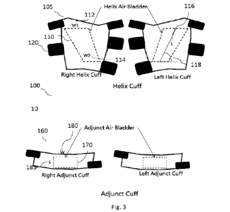
[00019] Figure 3 is a detailed diagram of an embodiment of helix cuff 105 and
adjunct
cuff 160 of the present invention.
[00020] Figures 4a and 4b are diagrams illustrating two different possible
placements
of the helix cuff 105 and adjunct cuff 160 of the present invention.
[00021] Figures 4c and 4c1 are diagrams illustrating two different possible
placements
of the helix air bladder 110 and adjunct air bladder 170 when they are both
constructed
within one cuff.
[00022] Figures 5a and 513 are diagrams showing the pressurization and
depressurization cycles of the ECP device of the present invention 10 as they
relate to
4
CA 03167610 2022- 8- 10
WO 2021/167651
PCT/US2020/050606
cardiac cycles and blood flow when the adjunct air bladder 170 is located at
the lower end
of the helix air bladder 110.
[00023] Figures 6a and 6b are diagrams showing the pressurization and
depressurization cycles of the ECP device of the present invention 10 as they
relate to
cardiac cycles and blood flow when the adjunct air bladder 170 is located at
the upper end
of the helix air bladder 110.
[00024] Figure 7 is a block diagram illustrating an embodiment of the ECP
control
unit 200 of the present invention.
[00025] Figure 8 is a block diagram depicting an embodiment of the ECP control
unit
200 and the fluid and valve system 300 of the present invention.
[00026] Figure 9 is a block diagram depicting an embodiment of the fluid and
valve
system 300 of the present invention.
[00027] Figure 10 is a flow chart illustrating an embodiment to of the method
of using
the ECP device of the present invention 10.
[00028] Figure 11 is a combination of PPG and ECG charts with charts showing
pressurization and depressurization of helix air bladder 110 and adjunct air
bladder 170
wherein the diagrams are synched in ti me to illustrate an embodiment of the
method of
treatment using the ECP device of the present invention 10.
[00029] Figures 12a and 12b illustrate an exemplary PPG and ECG signals of a
user
before and during treatment using the ECP device of the present invention 10,
respectively.
[00030] Figure 13 is a drawing of an exemplary embodiment of the ECP control
system 200 and fluid and valves system 300 of the ECP device of the present
invention 10.
5
CA 03167610 2022- 8- 10
WO 2021/167651
PCT/US2020/050606
[00031]
[00032] Detailed Description of the Present Invention
[00033]
As used in this specification and in claims which follow, the singular
forms
"a", "an" and "the" include plural referents unless the context clearly
indicates otherwise.
Thus, for example, reference to "an ingredient" includes mixtures of
ingredients,
reference to 'San active pharmaceutical agent" includes more than one active
pharmaceutical agent, and the like.
[00034] As used herein, the tcrm "about" as a modifier to a quantity is
intended to
mean + or - 5% inclusive of the quantity being modified.
[00035] The term "effective amount of time" or "a therapeutically effective
amount of
time" of a treatment is intended to mean a nontoxic/unharmful but sufficient
amount of
time required for providing the desired therapeutic effect. The time period
that is
"effective- may vary from subject to subject, depending on the age and general
condition
of the individual, the particular conditions, and the like.
[00036] The ECP device 10 of the present invention is capable of achieving
miniaturization, low energy consumption and low device cost as compared to
prior art
ECP devices. This is possible because the ECP device 10 of the present
invention takes
advantage of the geometry of major arteries and veins in the thigh which wind
around the
femur in a helical manner shown in figure 1. Specifically, as shown in figure
1, the major
arteries and veins start in front of the femur in front of the pelvic bone,
winds around the
femur in a helical fashion towards the inner thigh and ends up behind the
femur behind
the knee before travelling farther down the leg. As shown in figure 1, the
helix air
bladder 110 is specifically shaped to take advantage of this particular
anatomy so that air
6
CA 03167610 2022- 8- 10
WO 2021/167651
PCT/US2020/050606
bladder 110 focuses its energy only on the area of the thigh required to press
the major
veins and arteries against the femur bone in order to modulate blood flow of
the major
arteries and veins while wasting little energy on other areas of the thigh.
The efficiency
achieved means that a much smaller and less powerful air compressor is
required for
achieving the same or better therapeutic results compared to traditional ECP.
For
example, the power consumption of the present invention is below about 500,
600, 700,
800, 900, 1000, 1250 or 1500 Watts as compared to around 2500 Watts energy
consumption of the typical commercial units currently available. In addition,
the
miniaturization renders the ECP device of the present invention 10 easy to
handle, even
portable, at less than about 20, 25 or 30 kg, and cost substantially less than
existing ECP
devices which are typically so heavy that they are made to be stationary. This
means that
users may easily own and operate the ECP device of the present invention 10 at
home
rather than having to travel to a clinic for treatment.
[000371 Figure 2 is a high level depiction of the ECP device 10 of the present
invention 10 comprising an air bladder system 100, a control system 200 and a
valve and
fluid system 300.
[00038] Figure 3 depicts an embodiment of the cuff system 100. As shown in
figure 3,
the helix cuff system 100 comprises a helix cuff 105 and a helix air bladder
110. In an
embodiment, the helix air bladder 110 comprises an upper width W1 112, lower
width
W2 114, length L 116 and helix angle 118, where length L 116 is the straight
line
between the midpoint of width WI 112 and midpoint of width W2 114. Helix angle
is the
angle between length L 116 and horizontal line parallel to the level ground
when the cuff
is worn by the user standing upright. If W2 is designed as parallel to this
horizontal line
as illustrated in figure 3, the helix angle would be the angle between width
W2 and length
7
CA 03167610 2022- 8- 10
WO 2021/167651
PCT/US2020/050606
L 116. In another embodiment, the helix cuff 105 further comprises one or more
cuff
fasteners 120.
[00039] In an embodiment as described above, the helix air bladder 110 is
placed over
major artery and or vein of the inner thigh such that the helix shape of the
air bladder 110
follows the major arteries and veins that wrap around the femur as shown in
figure 1.
Upper width W1 112, lower width W2 114, length L 116 and helix angle 118 may
differ
depending on factors such as location of placement and biometrics of the user
such as sex,
height, weight, BMI, age, etc... in order to better conform to the major
arteries and or
veins around the femur. In one embodiment, the helix angle 118 is about
between 300 to
75 , 40 to 650 or about 55 . In one embodiment, the helix air bladder's
upper width WI
112 is wider than the lower width W2 114 as shown in figure 3. In another
embodiment,
the ratio of W1 112 :W2 114 is between about 2:1 to 1:1, about 1.9:1 to 1.1:1,
about 1.8
to 1.2:1, about 1.7:1 to 1.3:1, about 1.6:1 to 1.4:1 or about 1.5:1. The fact
that W1 112 is
wider than W2 114 helps to direct the blood upwards towards the torso when the
helix air
bladder 110 is inflated as shown in fig.4a and 4c. In one embodiment, the
ratio of the
width W1 112 of the top of the helix air bladder 110 to the length of the air
bladder L 116
is about 1:2 to 1:4 or about 1:3.
[00040] In one embodiment, the helix air bladder 110 only covers the thigh
area. In
another embodiment, the helix air bladder 110 does not cover the entire thigh
but just
enough area over the major artery or vein over the femur as necessary to
therapeutically
effectively modulate blood flow so that the cuff system 100 and ECP device 10
overall
may be miniaturized. In an embodiment, L 116, W1 112 and W2 114 are dependent
on
the biometrics of a user such as height and/or weight of the user. For
example, in an
embodiment: L= (User Height/3.2) ¨ b where b is between about 15 cm to about
30 cm.
8
CA 03167610 2022- 8- 10
WO 2021/167651
PCT/US2020/050606
So that if user height is 165 cm, L can be between about 21.6 cm and 36.6 cm
and W1
112 is about 14 cm and W2 114 is about 14 cm.
[00041] In one embodiment, as shown in fig. 3, the ECP device of the present
invention 100 further comprises an adjunct cuff 160. In an embodiment, the
adjunct cuff
160 comprises an adjunct air bladder 170 which assists the helix air bladder
110 in
modulating blood flow towards or away from the user's torso as will be
described in
further detail below in connection with figs. 4-6. As with the design
considerations for
the helix air bladder 110, width W 180 and length L 185 of the adjunct air
bladder 170
should be minimized to reduce power and size requirements of the air
compressor used to
pressurize it but still provide adequate assistance to the helix air bladder
110 as described
further in connection with figure 4. However, the width W 180 of the adjunct
air bladder
170 should be wide enough to fully encompass the W2 114 width of the helix air
bladder
110 if the adjunct cuff 160 is placed at the lower end of the helix cuff 105
as illustrated on
figs. 4a and c or the W1 width 112 if the adjunct cuff 160 is placed at the
upper end of the
helix cuff 105 as illustrated on figs. 4b and d. In addition, the length L 185
and width W
180 of the adjunct air bladder 170 should be sized and the pressure provided
by the air
compressor should be high enough to provide adequate force in assisting the
helix air
bladder 110 in modulating blood flow towards the desired direction as
described in
different configurations below in connection with figure 4. In one embodiment,
the ratio
of the width W 180 to length L 185 of the adjunct air bladder 170 is about
1.5:1 to 4:1,
about 2:1 to 3:1 or about 2.5:1. In another embodiment, the width W 180 of the
adjunct
air bladder is about 8 cm to 30 cm, about 16 to 24 cm or about 22 cm.
9
CA 03167610 2022- 8- 10
WO 2021/167651
PCT/US2020/050606
[00042] In one embodiment as illustrated in figures 4a-d, the adjunct air
bladder 170
may be positioned over the helix air bladder 110 in different configurations.
In one
embodiment, the adjunct air bladder 170 may be positioned over the lower end
of the
helix air bladder 110 as shown in figure 4a. In an embodiment, the adjunct air
bladder
170 abuts the lower end of the helix air bladder at the bottom of W2 114 and
overlaps the
helix air bladder 110. In yet another embodiment, the helix air bladder 110
and adjunct
air bladder 170 may be built in one single cuff as shown in fig. 4c.
[00043] In another embodiment, as shown in fig. 4b, the adjunct air bladder
170 may
be positioned over the upper end of the helix air bladder 110. In an
embodiment, the
adjunct air bladder 170 abuts the upper end of the helix air bladder at the
top of WI 112
and overlaps the helix air bladder 110. In another embodiment, the helix air
bladder 110
and adjunct air bladder 170 may be built in one single cuff as shown in fig.
4d.
[00044] In an embodiment, the adjunct air bladder 170 is pressurized before
the helix
air bladder 170 so as to affect direction of blood flow when the helix air
bladder 110 is
subsequently pressurized. Specifically, as shown in fig. 5, when the adjunct
air bladder
170 is positioned at the lower end of the helix air bladder 110 and the
adjunct air bladder
170 is pressurized before the helix air bladder 110, the direction of the
majority of blood
flow caused by the cuff system 100 is first upwards towards the torso of the
user since
adjunct air bladder 170 prevents downward blood flow. In an embodiment, the
adjunct
air bladder 170 should be wide enough to at least cover the entire width W1
112 of the
helix air bladder. In addition, air pressure of adjunct air bladder 170 should
be high
enough to stop over about 90%, about 80%, about 70% or about 60% of the blood
flow
downwards when pressurized. In an embodiment, the pressure in the air bladders
110 and
170 is about 150 mmHg to 350 mmHg, about 200 mmHg to 300 mmHg or about 250
CA 03167610 2022- 8- 10
WO 2021/167651
PCT/US2020/050606
mmHg. Subsequently, when both air bladders 170 and 110 depressurize, majority
of
blood flow caused by the cuff system 100 is downwards away from the torso of
the user.
And in figure 6 in an embodiment in which the adjunct air bladder 170 is
positioned at the
upper half of the helix air bladder 110 and when the adjunct air bladder 170
is pressurized
before the helix air bladder 110, the direction of the majority of blood flow
caused by the
cuff system 100 is first downwards towards the feet of the user since adjunct
air bladder
170 prevents upward blood flow. Subsequently, when both air bladders 170 and
110
depressurize at about the same time, the majority of blood flow caused by the
cuff system
100 is upwards towards the torso of the user. Therefore, air pressure of the
adjunct air
bladder 170 should be high enough to stop over 90%. about 80%, about 70% or
about
60% of the blood flow upwards when pressurized. In an embodiment, the pressure
in the
air bladders 110 and 170 is about 150 mmHg to 350 minHg, 200 to 300 or about
250
mmHg. As illustrated in figures 5 and 6, placement of the air bladders 110 and
170
allows the user to target different areas of the body at different strengths
of treatment.
[00045] In an embodiment, as shown in figure 7, the ECP device 10 of the
present
invention further comprises an ECP controller system 200 configured to control
various
aspects of the ECP device 10 of the present invention including but not
limited to
interaction with the user, collection and analysis of user's biometric data
and interaction
with valve system 300 to pressurize/depressurize air bladders 110 and 170. In
an
embodiment, the ECP controller system 200 preferably comprises an ECP
processor 210,
one or more heartbeat sensors 220, 230, 240 and an interactive display unit
250. In an
embodiment, the ECP processor 210 is connected to the interactive display unit
250, the
heartbeat sensors 220, 230 and 240 via electronic connection 260. In addition,
in an
embodiment, the ECP processor 210 is further connected to a valve and fluid
system 300
11
CA 03167610 2022- 8- 10
WO 2021/167651
PCT/US2020/050606
via electronic connection 260 as described in further details below in
connection with figs.
8 and 10.
[00046] In an embodiment, the ECP processor 210 preferably comprises a
processor
configured to send, receive and process signals including but not limited to
signals to and
from the user via interactive display 250, signals to and from the valve and
fluid system
300 as well as signals related to biometric data of the user such as heartbeat
information
collected by the heartbeat sensors 220, 230 and 240. In this way, the ECP
processor 210
is configured to control various aspects of the ECP device 10 of the present
invention
such as pressure in the helix air bladder 110 and the adjunct air bladder 170
based on the
various signals processed.
[00047] As shown in figure 7, in an embodiment, the PPG heartbeat sensor 220
may
comprise a finger sensor 220a and two toe sensors 220b and 220c. In an
embodiment, the
ECG heartbeat sensor 230 may comprise a right and left chest sensor 230a and
230b. In
addition, the ECG heartbeat sensor 230 may further comprise leg sensors 230e.
In an
embodiment, the continuous blood pressure sensor 240 comprises a blood
pressure cuff
over the arm of the user.
[00048] The display 250 is preferably a touchscreen that allows the user to
interact
with the ECP device 10 of the present invention such as triggering ECP
treatment, input
user information, system settings, etc.... User information may comprise
biometric
information of the user such as sex, height, weight, BMI, age, etc. Input
information may
also comprise systems settings such as type of ECP treatment and time period
of
treatment, maximum and/or minimum pressure, etc.... Output of information may
comprise type of treatment, progress of treatment, etc....
12
CA 03167610 2022- 8- 10
WO 2021/167651
PCT/US2020/050606
[00049] In an embodiment, as shown in Figs. 8 and 9, the ECP device of the
present
invention 10 further comprises a valve and fluid system 300 that works with
the ECP
controller system 200 to control pressure within the ECP device of the present
invention
10, including pressure within the valve and fluid system 300 as well as the
cuff system
100. In an embodiment, the valve and fluid system 300 comprises one or more
air
bladder valves 310, a post adjustment air compartment 320, an air pressure
ratio
adjustment valve 330, an air compressor air compartment 340, an air compressor
350, air
pressure to electric signal transducer 360, air inlet valve 370, air inlet 375
and a series of
large airways 380 and small airways 390 and 395. In an embodiment, the large
airways
380, which are used for establishing negative pressure, have diameters of
about 1 cm to
10 cm, and the small airways 390 and 395 have diameters of about 0_4 crn to 2
crn_
[00050] In an embodiment, the air bladder valves 310 each preferably comprises
a
valve configured to regulate pressure of the cuff system 100 based on
electronic signals
received from the ECP control system 100. In an embodiment, air bladder valves
310 are
solenoid valves. The post adjustment air compartment 320 preferably comprises
an air
compartment capable of storing pressurized air for pressurizing the air
bladders 110 and
170. In an embodiment, the pressure within the air compartment 320 is from 150
mmHg
to 350 mmHg, 200 mmHg to 300 mmHg or about 250 mmHg. Each valve 310a and 310b
is connected on one side to the post adjustment air compartment 320 via airway
390 and
to helix air bladder 110 and adjunct air bladder 170 via airway 395 on the
other side of
the valve. Each valve 310 is additionally connected to air inlet valve 370 and
compressor
350 via airway 380 through which air bladders 110 and 170 may be
depressurized.
Moreover, each valve 310 is electronically connected to ECP processor 210 via
electronic
connection 260 so that ECP processor 210 may electronically trigger valves 310
to
pressurize and depressurize air bladders 110 and 170.
13
CA 03167610 2022- 8- 10
WO 2021/167651
PCT/US2020/050606
[00051] In an embodiment, the air compressor air compartment 340 comprises an
air
compartment that connects to the post adjustment air compartment 320 via the
air
pressure ratio adjustment valve 330. In an embodiment, the air pressure ratio
adjustment
valve 330 further connects to the ECP processor 210 via electronic connection
260. In
this way, the air pressure ratio adjustment valve 330 is configured to
maintain air pressure
within the two air storages 320 and 340 based on signals from the ECP
processor 210. In
an embodiment, the air pressure in the post adjustment air storage 320 is
maintained at
between about from 150 mmHg to 350 mmHg, 200 mmHg to 300 mmHg, or about 250
mmHg while the air pressure within the compressor air storage 340 is
maintained at about
4 kgf to 8 kgf, about 5 kgf to 7 kgf or about 6 kgf.
[00052] In an embodiment, the air compressor 350 comprises an air compressor
configured to provide positive pressure to airway 390 when air inlet valve 370
is open
and negative pressure to airway 380 when air inlet valve 370 is closed to air
inlet 375 in
order to facilitate replenishing air to the air compartment 340 and
depressurizing the air
bladders 110 and 170, respectively. In an embodiment, the air compressor 350
is capable
of running at about 1700 rpm at about 130 L/m of flux at pressure up to about
8 kgf. The
air inlet valve 370 preferably comprises a valve that connects to the
compressor 350 via
airway 380 on one end and to an air inlet 375 on the other end. In an
embodiment, the air
inlet valve 370 comprises a solenoid valve.
[00053] Lastly, transducers 360 preferably comprises transducers that each
translates
pressure to electric signal. Each transducer 360 preferably connects on one
side to one of
the air compartments 340 and 320, respectively, via airway 390 and to the ECP
controller
processor 210. In this way, the valve system 300 is configured to transmit air
pressure
14
CA 03167610 2022- 8- 10
WO 2021/167651
PCT/US2020/050606
information to ECP controller system 200 via transducers 360. As mentioned
above, the
ECP controller processor 210 is connected to solenoid valve 310, the air
pressure ratio
adjustment valves 330, air inlet valve 370 and air compressor 350 so that the
ECP
controller system 200 is configured to send electronic signals to control the
valves 330
and 370 and air compressor 350 based on air pressure information from
transducers 360a
and 360b.
[00054] In an embodiment, when ECP processor 210 send a signal to valve 310,
valve
310 pressurizes air bladders 110 and 170 by connecting them to post adjustment
air
compartment 320, supplying pressurized air to the air bladders 110 and 170. To
depressurize air bladders 110 and 170, ECP processor 210 stops any signal to
valve 310
so that valve 310 defaults to disconnecting the air bladders 110 and 170 from
air
compartment 320 and connecting them instead to airway 380. In addition, ECP
processor
210 also sends a signal to air inlet valve 370 to close the air inlet so that
compressor 350
is able to establish negative pressure in airway 380 to rapidly depressurize
the air bladders
110 and 170. In an embodiment, the negative air pressure in airway 380 is
about 80
mmHg to 120 mmHg, about 90 mmHg to 110 mmHg or about 100 mmHg.
[000551 Figure 10 illustrates the method for providing External Counter
Pulsation of
the present invention 1000. The method of the present invention may be
provided to treat
diseases such as stroke, dementia, and arteriosclerosis but may also be
provided merely to
improve blood flow in general. As illustrated in fig. 11, in step 1100, the
method of the
present invention is triggered to begin. In one embodiment, step 1100 may be
manually
triggered by a person such as the user via interactive display 250. In another
embodiment,
step 1100 may be triggered automatically by signals from the heart rate
sensors 220, 230,
CA 03167610 2022- 8- 10
WO 2021/167651
PCT/US2020/050606
240. Next in step 1105, the ECP processor 210 reads and analyzes various
biometric data
such as but not limited to those input by the user via display as well as ECG
220, PPG
230, continuous blood pressure sensor 240, etc.... in order to determine the R
peak of the
user's heart rate. Once the R peak has been determined in step 1105 using
various
methods well known to persons in the art, the ECP processor 210 institutes a
delay of
between about 10 ms to about 250 ms, about 50 ms to about 200 ms or about 100
ms to
about 150 ms from the R peak in step 1110. During the delay, in step 1115, a
determination is made as to whether additional air is required in the valve
and fluid
system 300. In one embodiment, step 1115 is performed by the ECP processor 210
based
on air pressure signals from transducers 360a and 360b which provide air
pressure
information for the air compartments 320 and 340, respectively. If in step
1115 it is
determined that additional air is not required in either air compartments 320
and 340, for
example, if the pressure within air compartments 320 and 340 is maintained
between
about 150 mmHg to 350 mmHg, 200 mmHg to about 300 mmHg, or about 250 mmHg,
then no additional air is required, in step 1120, the ECP processor 210
triggers valve 310b
to connect adjunct air bladder 170 to air compartment 320 to pressurize
adjunct air
bladder 170. In addition, in step 1220, the ECP processor 210 triggers valve
310a to
connect helix air bladder 110 to air compartment 320 to pressurize helix air
bladder 110
using air from air compartment 320. In an embodiment, step 1120 is performed
before
step 1220 wherein a delay of about 30 to 70 ms, or about 40 to 60 ms or about
50 ms is
instituted between steps 1120 and 1220.
[00056] In step 1125, the end of the adjunct air bladder 170 pressurization
period is
reached. In an embodiment, the pressurization time period is about 200 ins to
600 ins,
250 ms to 550 ms, 300 ms to 500 ms or about 400 ms. In an embodiment, the
16
CA 03167610 2022- 8- 10
WO 2021/167651
PCT/US2020/050606
pressurization time period maybe determined based upon heart rate according to
the table
below:
Table 1
;' =:;.* F 7.`':'k *,:>, ,;,,,`:.:".,"'"-criil
\ tv õ.:;=*, :'' \ik, ,N., , '-. A i., i i: :I.
\\Vµ vS\ about 280
about 300
=`.. -:.= -\1. , about 320
,4, k-.., about 340
-s;
\\\
about 360 \\N
\
about 400
\
::,,,,% =,:.= ..\,õ about 440
\\\N_NTiVAN about 460
about 480
\ about 500
\
[00057]
[00058] In an embodiment, the ECP processor 210 performs step 1125 by keeping
track of this time period. Next in step 1130, the adjunct air bladder 170 is
depressurized.
In an embodiment, the depressurization is performed by the ECP processor 210
sending a
signal to valve 310b to disconnect air bladder 170 from air compartment 320 to
connect
air bladder 170 to airway 380 as well as to close air inlet valve 370 to allow
air
compressor 350 to establish negative pressure in airway 380 to facilitate
rapid
depressurization of the adjunct air bladder 170. Next, in step 1135, the end
of adjunct air
bladder 170 depressurization period is reached. In an embodiment, the ECP
processor
210 performs step 1135 by keeping account of this time period. In step 1140,
adjunct air
bladder 170 depressurization process is stopped and the process repeats from
step 1105 if
therapeutic effect has not been fully realized.
[000591
Similarly, after maintaining air pressure in the helix air bladder 110 for
a
preset time period the end of the helix cuff 105 pressurization period in step
1225. In an
17
CA 03167610 2022- 8- 10
WO 2021/167651
PCT/US2020/050606
embodiment, the pressurization time period is about from 200 ms to 600 ms, 250
ms to
550 ms, 300 ms to 500 ms or about 400 ms. In another embodiment, the air
pressure in
the helix air bladder 110 is maintained according to the user's heart rate
according to
Table 1 minus any delay institute between steps 1120 and 1220 as discussed. In
an
embodiment, the ECP processor 210 performs step 1225 by keeping track of this
time
period. In step 1230, the helix air bladder 110 is depressurized. In an
embodiment, the
depressurization is performed by the ECP processor 210 sending a signal valve
310a to
disconnect helix air bladder 110 from air compartment 320 to connect helix air
bladder
110 to airway 380 in which negative pressure is established to depressurize
the air bladder
by closing air inlet valve 370 while compressor 350 is running. Next, in step
1235, the
end of helix air bladder 110 depressurization period is reached. In an
embodiment, the
ECP processor 210 performs step 1235 by keeping track of this time period. In
step 1240,
the helix air bladder 110 depressurization process is stopped, and the process
repeats from
step 1105.
[00060] In an embodiment steps 1125 and 1225 are performed about the same
time,
and steps 1130 and 1230 are also performed about the same time so that both
air bladders
110 and 170 are depressurized about the same time. In another embodiment, step
1125 is
performed before step 1225, and step 1130 is performed before step 1230 so
that the
adjunct air bladder 170 is depressurized before the helix air bladder 110. In
this
embodiment, the delay is about from 20 ms to 100 ms, 30 ms to 90 ms, 40 ms to
80 ms or
about 60 ms. In another embodiment, step 1125 is performed after step 1225,
and step
1130 is performed after step 1230 so that the adjunct air bladder 170 is
depressurized
after the helix air bladder 110. In this embodiment, the delay is about from
20 ins to 100
ms, 30 ms to 90 ms, 40 ms to 80 ms or about 60 ms.
18
CA 03167610 2022- 8- 10
WO 2021/167651
PCT/US2020/050606
[00061] If in step 1115 ECP processor 210 determines that air replenishment is
required in the air compartments 320 and 340, steps 1120 to 1140 and 1220 to
1240 are
performed as described, but steps 1305 to 1315 are also performed to add more
air into
the system. Specifically, in step 1305, the ECP processor 210 signals valve
370 to open
to air inlet 375 and ensures that compressor 350 is running to replenish air
to air
compartment 340. The air pressure ratio adjustment valve 330 in turn adds air
to air
compartment 320. In step 1310 as the system reaches end of air replenishment
period, in
an embodiment, the ECP processor 210 keeps track of the air replenishment
period in step
1310. In step 1315, the ECP processor 210 sends signals to close air inlet
valve 370 to
stop adding air into the valve system.
In an embodiment, since air bladder
depressurization period requires that valve 370 to he closed so that negative
pressure can
be established in airway 380, steps 1305 to 1315 are performed concurrently
with steps
1120 to 1125 and 1220 to 1225, before steps 1120 and 1220 or after steps 1140
and 1240
are completed.
[00062] Figs.11 illustrate graphically the method of the present invention
1000. As
shown in fig. 11, the R peak is detected in step 1105 and a delay of about 10
ms to about
250 ms, about 50 ms to about 200 ms or about 100 ms to about 150 ms is
instituted in
step 1110 before the adjunct air bladder 170 is pressurized in step 1120.
After the adjunct
air bladder 170 is pressurized, the helix air bladder 110 is subsequently
pressurized in
step 1220 after a delay of about 50 ms. Also seen in figs. 11, if
replenishment of air is
required as determined in step 1115, it is done in steps 1305 to 1315 about
the same time
as the start of the adjunct air bladder 170 pressurization for about 50 ms to
about 100 ms.
19
CA 03167610 2022- 8- 10
WO 2021/167651
PCT/US2020/050606
Subsequently both the adjunct air bladder 170 and the helix air bladder 110
are
depressurized at about the same time in steps 1125 to 1140 and in steps 1225
to 1240.
[00063]
Fig. 12 illustrates the therapeutic effects of the ECP device 10 of the
present
invention. As seen in fig. 12a, prior to the treatment. PPG signal of a user
is weak.
During the treatment, the PPG signal of the user is much more regular and
maintained at a
constant strength as shown in figs. 12b.
[00064] It is to be understood that both the foregoing general description and
the
following detailed description are exemplary and explanatory only and are not
restrictive
of the invention, as claimed.
[00065] These and other changes can be made to the technology in light of the
detailed
description. In general, the terms used in the following disclosure should not
be
construed to limit the technology to the specific embodiments disclosed in the
specification, unless the above detailed description explicitly defines such
terms.
Accordingly, the actual scope of the technology encompasses the disclosed
embodiments
and all the equivalent ways of practicing or implementing the technology.
CA 03167610 2022- 8- 10