Note: Descriptions are shown in the official language in which they were submitted.
SURGICAL DEPTH INSTRUMENT HAVING
NEUROMONITORING CAPABILITIES
CROSS-REFERENCE TO RELATED APPLICATIONS
This application claims the benefit of, and priority to, U.S. Provisional
Application No.
62/417,046, filed November 3, 2016, U.S. Provisional Application No.
62/471,873, filed March
15, 2017, and U.S. Provisional Application No. 62/554,470, filed September 5,
2017.
FIELD
The present disclosure relates generally to medical devices, and, more
particularly, to a
measuring instrument for use in a bone implant fixation procedure, the
measuring instrument
including a combination of a bone probe allowing for physical examination of a
hole drilled in a
bone and a depth gauge member for determining a depth of the hole and
providing a digital
measurement of the depth.
BACKGROUND
Orthopedics is a medical specialty concerned with the correction of
deformities or
functional impairments of the skeletal system, especially the extremities and
the spine, and
associated structures, such as muscles and ligaments. Some orthopedic surgical
procedures
require surgeons to secure a device to one or more bones of a patient. For
example, in some
procedures, the surgeon may span and secures one or more bones, or pieces of a
single bone,
using a bone plate and one or more fasteners, such as screws. Other bone-
related surgical
procedures, however, may not require a bone plate and may instead solely rely
on the use of one
or more screws (e.g., securing a transplanted tendon).
In such bone-related surgical procedures, before an implant or plate, or
simply the screw
itself, can be attached to bone, an opening is typically drilled into the bone
to accommodate the
screw. With a hole in place, the surgeon can more easily select a screw of the
appropriate length.
However, selecting a screw of appropriate length is critical. For example, if
the selected screw is
too long, the distal end of the screw may pass through the end of the drilled
hole and cause
damage to the bone and/or protrude entirely through the bone, which can have
deleterious
1
Date Regue/Date Received 2022-07-14
effects, such as damage to surrounding tissue and/or pain and discomfort, or
more serious
complications, for the patient. For example, in some instances, the bone may
abut against soft
tissues that may be harmed if the screw is too long and may result in
irritation of or damage to
the soft parts. Additionally, a screw that protrudes through the bone may be
tactilely felt by the
patient, may prevent soft tissues (e.g., tendons, ligaments, or muscles) from
moving over the
bone surface as intended, or may even pierce the skin, which can lead to
serious infection and
complications.
The selection of an appropriate length screw is particularly important in
spinal fixation
procedures, such as lumbar sacral fusion and the correction of spinal
deformities such as
scoliotic curves. As an example, a screw mounted in the pedicle portion of the
human spine
should not extend to a point where the screw contacts the spinal cord itself,
an event that can
cause irreparable nervous system damage including paralysis. Accordingly, the
determination of
a length of the hole is important for choosing the appropriate length screw.
During drilling, the surgeon is typically capable of recognizing the
resistance on the drill
in order to determine when the drill has penetrated through the bone. Because
the simple act of
drilling does not provide an exact measurement of the depth of the bone
itself, a depth gauge is
commonly employed for directly measuring the depth of the hole from the top,
drilling side to
the bottom, opposite side of the hole.
Currently, many designs are known and utilized for measuring the depth of a
hole or bore
in a portion of a bone. Generally speaking, these designs utilize a central
probe member having
a barb at a distal end, and a sleeve or channel member. The probe member is
inserted into the
pilot hole while the surgeon attempts to find the surface with the barb. More
specifically, the
probe member is inserted to a depth greater than the depth of the pilot hole
so that the barb is
beyond the opposite side, at which point the surgeon finds the surface by
hooking the barb to the
opposite side.
The probe member is received in the sleeve or channel member and may
reciprocate
relative thereto. The channel member has graduated markings along a portion of
its length,
typically in inches and/or millimeters. A marker is laterally secured to the
probe member such
that, as the probe member shifts relative to the channel member, the marker
indicates the relative
shift between the probe member and the channel member. Accordingly, once the
probe member
has been secured to the opposite side of the bone, the channel member is
shifted relative to the
2
Date Regue/Date Received 2022-07-14
probe member and toward the bone until the channel member abuts the surface of
the bone. The
depth gauge is then read by examining graduated markings indicated by the
probe member
marker.
A number of problems are experienced with this depth gauge. As an initial
point, the
components are typically made with surgical-grade stainless steel, and the
graduated markings
are embossed therein. Therefore, the brightness of the operating room lights
on the highly
reflective surface can make the markings difficult to read. The markings are
commonly in small
increments, such as millimeters, and surgeons often have trouble
differentiating between the
markings, or noting partial increments. Reading these gauges, then, often
requires carefully
.. holding the depth gauge as the reading is taken, and a surgeon's effort to
closely examine the
reading may result in a loss of securement or purchase of the barb on the
bone, thus necessitating
a re-measurement and a loss of time.
Furthermore, proper reading of the markings requires a surgeon's eyes to be
properly
aligned with the markings. That is, a proper view of the measurement requires
the surgeon to
view the gauge from a lateral point of view so that the view of the probe
marker aligned with the
graduated markings is proper not distorted by the surgeon's elevated, standing
perspective.
Therefore, it is often necessary for the surgeon to bend over while using
these gauges to view an
accurate reading. If the depth gauge is tilted in order to make the reading,
the sleeve will shift
relative to the probe, thus making the measurement inaccurate and possibly
causing the barb to
become unsecured, as described above. In addition, removal of the depth gauge
often causes the
measurement to be lost. As the bone is essentially clamped, by light pressure,
between the distal
end of the channel member and the distal barb of the probe member, it is often
necessary to
retract the channel member from the bone surface in order to extract the probe
from the pilot
hole.
SUMMARY
The present disclosure is a medical device for use in a bone implant fixation
procedure.
The device is configured to provide a faster and more accurate measure of
depth. In particular,
the device includes a combination of a bone probe allowing for physical
examination of a hole
drilled in a bone and a depth gauge member for determining a depth of the hole
and providing a
digital measurement of the depth. Accordingly, the device of the present
disclosure is capable of
3
Date Regue/Date Received 2022-07-14
digitally measuring the depth of an opening in a bone during the same surgical
step that a
surgeon probes and inspects the interior of the opening.
During a bone-related procedure involving placement of a screw, or other
fastener, it may
be desirable to determine whether drilling of the hole resulted in any cracks
or openings, either
along an interior side wall of the hole or at the base of the hole. Ensuring
the integrity of the
drilled hole is important because unintended cracks, openings, or
irregularities can increase the
risk that the screw will either not securely attach itself within the hole or
may result in chipping
or fragmenting of bone during fastening of the screw within the hole. It is
generally not possible
for a surgeon to visual examine the integrity of the drilled hole due to a
limited field of view
within the hole (drilled holes can be relatively small in width, such as 5 mm
or less in some
instances).
The device of the present disclosure includes a bone probe that allows for a
surgeon to
feel the interior side walls of the hole to locate any cracks or other
unintended openings or
irregularities along the interior of the hole and to further determine the
exit point of the hole (i.e.,
for a hole that has been drilled entirely through the bone for subsequent
placement of a bicortical
screw or other fastener). The bone probe generally includes an elongated shaft
slidably mounted
within a body of the device serving as a handle adapted for manual
manipulation. The elongated
shaft of the probe includes a distal end configured to extend from the body of
the device during
use. The distal end includes a probing tip for contacting an interior portion
of the hole. At least
a portion of the elongated shaft may be substantially flexible or semi-rigid
to provide a proper
"feel" to the surgeon during examination of the hole in the bone. For example,
the shaft of the
bone probe may be substantially non-elastic such that the surgeon can apply
pressure against the
interior wall of the hole to feel for irregularities or the base of the hole
via tactile feedback
provided by the shaft. In some embodiments, the shaft may be tapered such that
the shaft
narrows in width or thickness in a direction towards the probing distal tip.
In this manner, the
flexibility of the shaft may increase along the shaft in a direction toward
the probing tip.
The probing tip may include at least a first portion having a shape or contour
that aids the
surgeon in detecting surface irregularities (e.g., cracks, crevices, openings,
etc.) on the interior
surface of the hole. For example, in some embodiments the first portion may
have a
substantially arcuate or curved shape. The arcuate or curved portion may also
aid the surgeon in
locating the exit point (i.e., second opening) the hole so as to allow for the
probing tip to be
4
Date Regue/Date Received 2022-07-14
accurately placed and secured along an edge of the exit point so that the hole
can be measured
via the depth gauge member. The arcuate or curved shape of the first portion
of the probing tip
may generally lessen risk of tissue irritation that may otherwise occur along
the interior surface
of the hole, which is usually soft and easily penetrable with less curved and
more abrupt surfaces
(with sharp or distinct edges). In some embodiments, the first portion may
have a general
spherical shape. In other embodiments, the first portion may be substantially
planar with
rounded edges.
The probing tip may also include a second portion positioned opposite the
first portion,
wherein the second portion includes an engagement surface configured to pierce
or otherwise
establish purchase with an exterior portion of bone immediately adjacent to
the exit point of the
hole (i.e., along the edge of the hole). In particular, upon locating the exit
point or second
opening of the hole, the surgeon may then extend the probing tip through the
exit point and then
position the bone probe shaft against the interior surface of hole and pull
back on the bone probe
shaft so as to draw the probing tip, specifically the engagement surface, back
towards, and into
engagement with, the exterior surface of the bone along the edge of the exit
point of the hole.
Upon sufficient application of pressure (i.e., sufficient retraction of the
bone probe shaft), the
engagement surface of the probing tip engages and establishes purchase with
the hone
immediately adjacent the hole. Upon establishing engagement, the medical
device may be
stabilized in position, at which point, the depth gauge member can be used for
measuring the
depth of the hole. In some embodiments, the engagement surface may include
surface texturing
to enhance friction between the engagement surface and a portion of bone. For
example, in some
procedures in which a plate or implants is to be secured with screws through a
bicortical drill
hole, the probing tip may extend entirely through the hole (from one side of
the bone to the
other), at which point the surgeon may pull the bone probe back towards the
hole such that the
engagement surface of the second portion of the probing tip establishes
purchase with one side of
the bone, and the surface texturing enhances friction between the engagement
surface and bone
to reduce risk of slippage.
The depth gauge member generally includes a hollow elongated body slidably
mounted
within the body of the device and includes a distal end configured to extend
from the first end of
the body during use. The hollow elongated body includes a lumen in which at
least a portion of
5
Date Regue/Date Received 2022-07-14
the bone probe shaft is received within such that the bone probe and depth
gauge member are
independently slidable relative to one another and the body of the device.
The device further includes at least one sensor configured to generate an
electronic signal
indicative of a depth of the hole as a result of sensing a distance between
the first end of the
device body and the distal end of the depth gauge member. For example, in one
embodiment,
upon establishing purchase with an exterior surface of bone generally
providing an edge of the
exit point of the drilled (or otherwise pierced hole) via the probing tip, a
surgeon need only move
the device handle (i.e., device body) in a direction towards the bone such
that the first end of the
handle contacts a surface of the bone proximate the first opening of the hole.
The surgeon may
then advance the depth gauge member towards hole, such that the distal end of
the depth gauge
member extends from the first end of the device handle and advances into the
hole, sliding over
the bone probe. While the bone probe is maintained in engagement with an
exterior surface of
bone on the opposing side of the hole via the probing tip, the depth gauge
member may be
advanced in a direction towards the exit point of the hole until the distal
end of the depth gauge
member makes contact with a distal end of the probing tip, which sits at the
exit point of the
hole. The bone probe essentially acts as a guide upon which the depth gauge
member slide over
when advancing to the end of the hole.
The sensor is configured to generate an electronic signal based on a distance
between the
first end of the body and the distal end of the depth gauge member, wherein
the electronic signal
is indicative of at least a depth of the hole. In particular, the sensor may
include inductive or
capacitive elements or assemblies configured to sense the location of the
distal end of the depth
gauge member relative to the first end of the device body, and, as a result,
generate an electronic
signal representing the distance there between. Accordingly, the sensed
distance between the
first end of the device handle (when abutting the bone surface) and the distal
end of the depth
gauge member (when abutting the end of the hole) is the depth of the hole.
It should be noted that the device may include logic or allow for adjustment
to the
sensing capabilities so as to program the sensor to account for other
variables when sensing the
depth of the hole. For example, in some embodiments, certain procedures
require fixing a plate
or implant to the bone via screws. Accordingly, the screw length must not only
be sufficient to
fill the hole but also long enough to account for the thickness of a plate or
implant through which
it passes when engaging the hole. Accordingly, in some embodiments, the sensor
may be
6
Date Regue/Date Received 2022-07-14
programmed so as to account for the thickness of the plate or implant and will
further include
that thickness in the electronic signal produced, such that the electronic
signal is indicative of the
total depth that a corresponding screw length will need to cover, including
the depth of the hole
in the bone in addition to the thickness of the plate or implant through which
the screw will pass
through and the screw head will engage.
Furthermore, in some instances, first end of the device handle will be
directly abutting a
surface of the plate or implant, which is directly abutting the surface of the
bone, when the
surgeon is measuring the depth. Thus, in this case, the sensor is still able
to sense a distance
between the first end of the device handle and the distal end of the depth
gauge member, which
will provide an overall depth, rather than just a depth of the hole in the
bone.
Accordingly, the digital sensing of the hole depth provides a much more
accurate
measurement than conventional analog depth gauges and also requiring very
little, if any, input
or interpretation from the surgeon. Accordingly, by providing a much more
accurate
measurement of a hole depth, the surgeon is able to select the correct length
screw for any given
hole so as to improve the chances of a successful surgery.
In some embodiments, the device may further include a display provided on the
body and
configured to visually provide a digital readout of a depth measurement of the
hole based on the
electronic signal from the sensor. In other embodiments, the device may be
configured to
wirelessly communicate and exchange data with a separate display or computing
device, such as,
for example, a monitor or panel display, a PC, a notebook, a tablet computer,
a smartphone, or
other wireless computing device.
Upon receiving the electronic signal from the sensor, the separate display or
computing
device may be configured to visually provide the depth measurement of the hole
based on the
electronic signal from the sensor. Furthermore, in some embodiments. the
computing device
may include a specific software application that may be directed to
maintaining a record of the
hole measurements and/or provide an interactive user interface in which
multiple holes can be
mapped to a particular plate or implant and the depth of each hole (including
the thickness of the
plate or implant) can be included and stored for records.
In some embodiments, the device may further include a sensor configured to
sense strain
of the bone probe shaft. In particular, the sensor may include a strain gauge
or the like
configured to determine a strain of the bone probe shaft, which may be useful
for alerting the
7
Date Regue/Date Received 2022-07-14
surgeon of an amount of resistance that the distal probing tip is encountering
during probing of
the interior of the hole. For example, while a surgeon may be able to "feel"
the interior surface
and further have a sense of when the probing tip actually makes contact with
the exit point of the
hole, the strain sensor may further generate an electronic signal based on a
sensed strain of the
shaft which may then be used to provide an audible and/or visual alert to the
surgeon indicating
that the probing tip is in fact positioned at the end of the hole. For
example, the resistance
encountered when the probing tip engages the exit point or second opening of
the hole may have
a certain strain value (i.e., above a certain threshold) which may be
different than a resistance
encountered with the sidewalls of the hole (which may have a softer, spongier
tissue).
Accordingly, the audible and/or visual alert may confirm to a surgeon whether
they are in fact
positioned at the end of the hole or if too much pressure is being placed
against the interior
surface such that they risk possibly inadvertently piercing the interior
surface.
In some embodiments, the device may further be compatible with other medical
devices
so as to provide additional features, in additional bone probing and depth
measurement. For
example, in some embodiments, the bone probe shaft may include an electrically
conductive
material (e.g., a metal such as stainless steel, nitinol, or aluminum),
wherein a portion of the bone
probe shaft may be exposed, or otherwise accessible, along a portion of the
device handle. In
particular, the device handle may include an aperture, window, or the like,
that provides access
to an interior of the handle, particularly providing access to an exposed
portion of the bone probe
shaft. Thus, in some embodiments, an electrical current from a separate device
may be supplied
to the bone probe shaft via the access region (e.g., slide a working tip of an
electrocautery device
into the access region to make contact with bone probe shaft). As a result of
being made from a
conductive material, the bone probe shaft may carry the electrical current to
the distal probe tip,
which may then be used to deliver energy to a desired target (e.g., interior
surface of hole of the
bone) as a result of the electrical current applied thereto. Similarly, a
separate nerve
sensing/stimulation device may be coupled to the conductive bone probe shaft
via the access
region, such that the distal probe tip essentially acts as an extension to the
nerve
sensing/stimulation device and may be used to sense/stimulate nerves within
the bone.
Yet still, in another embodiment, the handle may include a port in
communication with a
portion of the bone probe shaft. The port may provide access from an exterior
of the handle to
an interior of the handle and to the bone probe shaft. The port may be
configured to receive and
8
Date Regue/Date Received 2022-07-14
place an input connector of a second medical device, such as a neuromonitoring
device, for nerve
sensing and/or nerve stimulation, into electrical communication with the bone
probe shaft, such
that the bone probe shaft can be used to carry electrical signals to and from
the input connector
of the second medical device.
BRIEF DESCRIPTION OF THE DRAWINGS
Features and advantages of the claimed subject matter will be apparent from
the
following detailed description of embodiments consistent therewith, which
description should be
considered with reference to the accompanying drawings, wherein:
FIG. 1 is top view of one embodiment of a medical device consistent with the
present
disclosure;
FIG. 2 is a cross-sectional view of the medical device of FIG. 1 illustrating
the hollow
interior of the handle and arrangement of the bone probe and depth gauge
member relative to one
another;
FIGS. 3A and 3B are enlarged front and side views, respectively, of one
embodiment of a
probing tip defined on the distal end of the bone probe shaft;
FIGS. 3C and 3D are enlarged front and side views, respectively, of another
embodiment
of a probing tip defined on the distal end of the bone probe shaft;
FIG. 4 is a perspective view of another embodiment of a bone probe compatible
for use
with the medical device of FIG. 1, illustrating another embodiment of a
probing tip defined on a
distal end of the bone probe shaft;
FIGS. 5 and 6 are front and side views, respectively, of the bone probe of
FIG. 4;
FIG. 7 is an enlarged side view of the probing tip of FIG. 4;
FIGS. 8 and 9 are enlarged perspective views of the probing tip of FIG. 4;
FIGS. 10A and 10B illustrate retraction of the bone probe within the handle
member and
subsequent compression of a spring assembly upon movement of the handle
towards the bone
when the probing tip of the distal end of the bone probe shaft is in contact
with the bottom of the
drilled hole in the bone;
FIG. 11 is a side view of the medical device of FIG. 1 including a strain
sensor sensing
strain upon the bone probe shaft and providing an electronic signal indicative
of the strain to an
audio or visual component for providing an audible or visual alert;
9
Date Regue/Date Received 2022-07-14
FIGS. 12A-12F illustrate a series of steps for perfonning a procedure of
probing a drilled
hole and subsequently obtaining a depth measurement using another embodiment
of a medical
device consistent with the present disclosure;
FIGS. 13A-13C illustrate a series of steps for performing a procedure of
probing a fully
.. drilled hole (i.e., a hole extending entirely through a bone for receipt of
a bicortical bone screw)
with the bone probe of FIG. 4 and further establishing purchase of the probing
tip of the bone
probe with a side of the bone adjacent to the bicortical drilled hole to
secure the bone probe in
place and allow the depth gauge member to be used for measuring the depth of
the bicortical
drilled hole.
FIG. 14 is another embodiment of a medical device consistent with the present
disclosure
having a display for providing a digital readout of a depth measurement of the
hole;
FIG. 15 is another embodiment of a medical device consistent with the present
disclosure
configured to wirelessly communicate with and transmit depth measurement data
to a wireless
computing device to record, store, and/or visually display measured depths;
FIGS. 16 and 17 illustrate the compatibility of a medical device of the
present disclosure
with other medical devices so as to provide additional features, in additional
bone probing and
depth measurement, such as energy emission (FIG. 16) and sensing capabilities
(FIG. 17);
FIG. 18 is a perspective view of a medical device consistent with the present
disclosure
and having a neuromonitoring port configured to receive a corresponding input
connector from a
nerve sensing/nerve stimulation device and provide an electrical pathway to
the bone probe;
FIG. 19 is a side view, partly in section, of the medical device of FIG. 18
illustrating the
configuration of the bone probe shaft to carry electrical signals to and from
the nerve
sensing/nerve stimulation device;
FIGS. 20A, 20B, 20C illustrate the transmission of a signal from bone probe to
a screw
positioned within a hole in a vertebra for neuromonitoring capabilities; and
FIG. 21 illustrates an angle guide for use with the medical device of the
present
disclosure.
For a thorough understanding of the present disclosure, reference should be
made to the
following detailed description, including the appended claims, in connection
with the above-
.. described drawings. Although the present disclosure is described in
connection with exemplary
embodiments, the disclosure is not intended to be limited to the specific
forms set forth herein. It
Date Regue/Date Received 2022-07-14
is understood that various omissions and substitutions of equivalents are
contemplated as
circumstances may suggest or render expedient.
DETAILED DESCRIPTION
By way of overview, the present disclosure is generally directed to a medical
device for
use in a bone implant fixation procedure and configured to provide a faster
and more accurate
measure of depth. In particular, the device includes a combination of a bone
probe allowing for
physical examination of a hole drilled in a bone and a depth gauge member for
determining a
depth of the hole and providing a digital measurement of the depth.
Accordingly, the device of
the present disclosure is capable of digitally measuring the depth of an
opening in a bone during
the same surgical step that a surgeon probes and inspects the interior of the
opening.
The device of the present disclosure includes a bone probe that allows for a
surgeon to
feel the interior side walls of the hole to locate any cracks or other
unintended openings or
irregularities along the interior of the hole and to further determine the
exit point of the hole (i.e.,
for a hole that has been drilled entirely through the bone for subsequent
placement of a bicortical
screw or other fastener). The bone probe generally includes an elongated shaft
slidably mounted
within a body of the device serving as a handle adapted for manual
manipulation. The elongated
shaft of the probe includes a distal end configured to extend from the body of
the device during
use. The distal end includes a probing tip for contacting an interior portion
of the hole. At least
a portion of the elongated shaft may be substantially flexible or semi-rigid
to provide a proper
"feel" to the surgeon during examination of the hole in the bone. For example,
the shaft of the
bone probe may be substantially non-elastic such that the surgeon can apply
pressure against the
interior wall of the hole to feel for irregularities or the base of the hole
via tactile feedback
provided by the shaft. In some embodiments, the shaft may be tapered such that
the shaft
narrows in width or thickness in a direction towards the probing distal tip.
In this manner, the
flexibility of the shaft may increase along the shaft in a direction toward
the probing tip.
The probing tip may include at least a first portion having a shape or contour
that aids the
surgeon in detecting surface irregularities (e.g., cracks, crevices, openings,
etc.) on the interior
surface of the hole. For example, in some embodiments the first portion may
have a
substantially arcuate or curved shape. The arcuate or curved portion may also
aid the surgeon in
locating the exit point (i.e., second opening) the hole so as to allow for the
probing tip to be
11
Date Regue/Date Received 2022-07-14
accurately placed and secured along an edge of the exit point so that the hole
can be measured
via the depth gauge member. The arcuate or curved shape of the first portion
of the probing tip
may generally lessen risk of tissue irritation that may otherwise occur along
the interior surface
of the hole, which is usually soft and easily penetrable with less curved and
more abrupt surfaces
(with sharp or distinct edges). In some embodiments, the first portion may
have a general
spherical shape. In other embodiments, the first portion may be substantially
planar with
rounded edges.
The probing tip may also include a second portion positioned opposite the
first portion,
wherein the second portion includes an engagement surface configured to pierce
or otherwise
establish purchase with an exterior portion of bone immediately adjacent to
the exit point of the
hole (i.e., along the edge of the hole). In particular, upon locating the exit
point or second
opening of the hole, the surgeon may then extend the probing tip through the
exit point and then
position the bone probe shaft against the interior surface of hole and pull
back on the bone probe
shaft so as to draw the probing tip, specifically the engagement surface, back
towards, and into
.. engagement with, the exterior surface of the bone along the edge of the
exit point of the hole.
Upon sufficient application of pressure (i.e., sufficient retraction of the
bone probe shaft), the
engagement surface of the probing tip engages and establishes purchase with
the hone
immediately adjacent the hole. Upon establishing engagement, the medical
device may be
stabilized in position, at which point, the depth gauge member can be used for
measuring the
.. depth of the hole. In some embodiments, the engagement surface may include
surface texturing
to enhance friction between the engagement surface and a portion of bone. For
example, in some
procedures in which a plate or implants is to be secured with screws through a
bicortical drill
hole, the probing tip may extend entirely through the hole (from one side of
the bone to the
other), at which point the surgeon may pull the bone probe back towards the
hole such that the
engagement surface of the second portion of the probing tip establishes
purchase with one side of
the bone, and the surface texturing enhances friction between the engagement
surface and bone
to reduce risk of slippage.
The depth gauge member generally includes a hollow elongated body slidably
mounted
within the body of the device and includes a distal end configured to extend
from the first end of
the body during use. The hollow elongated body includes a lumen in which at
least a portion of
12
Date Regue/Date Received 2022-07-14
the bone probe shaft is received within such that the bone probe and depth
gauge member are
independently slidable relative to one another and the body of the device.
The device further includes at least one sensor configured to generate an
electronic signal
indicative of a depth of the hole as a result of sensing a distance between
the first end of the
device body and the distal end of the depth gauge member. For example, in one
embodiment,
upon establishing purchase with an exterior surface of bone generally
providing an edge of the
exit point of the drilled (or otherwise pierced hole) via the probing tip, a
surgeon need only move
the device handle (i.e., device body) in a direction towards the bone such
that the first end of the
handle contacts a surface of the bone proximate the first opening of the hole.
The surgeon may
then advance the depth gauge member towards hole, such that the distal end of
the depth gauge
member extends from the first end of the device handle and advances into the
hole, sliding over
the bone probe. While the bone probe is maintained in engagement with an
exterior surface of
bone on the opposing side of the hole via the probing tip, the depth gauge
member may be
advanced in a direction towards the exit point of the hole until the distal
end of the depth gauge
member makes contact with a distal end of the probing tip, which sits at the
exit point of the
hole. The bone probe essentially acts as a guide upon which the depth gauge
member slide over
when advancing to the end of the hole.
The sensor is configured to generate an electronic signal based on a distance
between the
first end of the body and the distal end of the depth gauge member, wherein
the electronic signal
is indicative of at least a depth of the hole. In particular, the sensor may
include inductive or
capacitive elements or assemblies configured to sense the location of the
distal end of the depth
gauge member relative to the first end of the device body, and, as a result,
generate an electronic
signal representing the distance there between. Accordingly, the sensed
distance between the
first end of the device handle (when abutting the bone surface) and the distal
end of the depth
gauge member (when abutting the end of the hole) is the depth of the hole.
Accordingly, the digital sensing of the hole depth provides a much more
accurate
measurement than conventional analog depth gauges and also requiring very
little, if any, input
or interpretation from the surgeon. Accordingly, by providing a much more
accurate
measurement of a hole depth, the surgeon is able to select the correct length
screw for any given
hole so as to improve the chances of a successful surgery.
13
Date Regue/Date Received 2022-07-14
FIG. 1 is top view of one embodiment of a medical device 100 consistent with
the present
disclosure and FIG. 2 provides a cross-sectional view of the medical device
100. As shown, the
medical device 100 includes a body 102 having a first end 104 and an opposing
second end 106
and is generally hollow. The body 102 is configured as a handle and generally
adapted for
manual manipulation. Accordingly, the body will be referred to a "handle 102"
hereinafter for
ease of explanation.
The device 100 further includes a bone probe 108 slidably mounted within the
handle
102. The bone probe 108 includes a shaft 110 having a distal end 112
configured to extend from,
and retract towards, the first end 104 of the handle 102 during use, as will
be described in
greater detail herein. The distal end 112 further includes a probing tip 114,
which is useful for
examination and inspection of interior surfaces of a drilled hole in bone, as
will be described in
FIGS. 3A and 3B.
The device 100 further includes a depth gauge member 116 slidably mounted
within the
handle 102. The depth gauge member 116 generally includes a hollow elongated
body 118
having a distal end 120 configured to extend from, and retract towards, the
first end of the handle
102 during use, similar to the bone probe shaft 110, as will be described
herein. The hollow
elongated body 118 has a lumen in which at least a portion of the bone probe
shaft 110 is
received such that the bone probe 108 and depth gauge member 116 are
independently slidable
relative to one another and the handle 102. The device 100 further includes
one or more depth
measurement sensors 122 configured to generate an electronic signal indicative
of a depth of at
least the hole, wherein the electronic signal varies in relation to a distance
between the first end
104 of the handle 102 and the distal end 120 of the depth gauge member 116, as
will be
described in greater detail herein.
The bone probe 108 and depth gauge member 116 may each be coupled to separate
slider
members for allowing a surgeon to manually control movement of the bone probe
108 and depth
gauge member 116 independent of one another. For example, as shown in FIG. 1,
a first slider
124 may be coupled to at least the bone probe shaft 110 and is slidable along
a longitudinal axis
of the handle 102, which such movement of the first slider 124 causes
corresponding movement
of the bone probe shaft 110. Although not shown in FIGS. 1 and 2, a second
slider may be
coupled to the depth gauge member 116 and is similarly slidable along the
longitudinal axis of
14
Date Regue/Date Received 2022-07-14
the handle 102, such that movement of the second slider causes corresponding
movement of the
depth gauge member 116.
The device 100 may further include a spring assembly 126 coupled to at least
one of the
bone probe 108 and depth gauge member 116. The spring assembly 126 may be
configured to
provide a biasing force upon at least one of the bone probe 108 and depth
gauge member 116 so
as to maintain either the bone probe 108 or depth gauge member 116 in a
default extended
position. For example, as shown in FIGS. 1 and 2, the bone probe 108 is
generally positioned in
an extended configuration (probing tip 114 extended out of first end 104 of
handle 102), in
which a surgeon may now examine an interior surface of a drilled hole, as is
shown in FIGS.
10A and 10B.
During a bone-related procedure involving placement of a screw, or other
fastener, it may
be desirable to detettnine whether drilling of the hole resulted in any cracks
or openings, either
along an interior side wall of the hole or at the base of the hole. Ensuring
the integrity of the
drilled hole is important because unintended cracks, openings, or
irregularities can increase the
risk that the screw will either not securely attach itself within the hole or
may result in chipping
or fragmenting of bone during fastening of the screw within the hole. It is
generally not possible
for a surgeon to visual examine the integrity of the drilled hole due to a
limited field of view
within the hole (drilled holes can be relatively small in width, such as 5 mm
or less in some
instances).
The bone probe 108 allows for a surgeon to feel the interior side walls and
bottom of a
drilled hole so as to locate any cracks or other unintended openings or
irregularities along the
interior of the hole. For example, probing tip 114 is configured for
contacting an interior portion
of the hole and at least a portion of the elongated shaft 110 may be
substantially flexible or semi-
rigid to provide a proper "feel" to the surgeon during examination of the hole
in the bone. For
example, the shaft 110 of the bone probe 108 may be substantially non-elastic
such that the
surgeon can apply pressure against the interior wall of the hole to feel for
irregularities or the
base of the hole via tactile feedback provided by the shaft 110. In some
embodiments, the shaft
110 may be tapered such that the shaft narrows in width or thickness in a
direction towards the
probing distal tip. In this manner, the flexibility of the shaft may increase
along the shaft in a
direction toward the probing tip 114.
Date Regue/Date Received 2022-07-14
FIGS. 3A and 3B are enlarged front and side views, respectively, of one
embodiment of a
probing tip 114a defined on the distal end 112 of the bone probe shaft 110. As
shown, the
probing tip 114a may include an arcuate first portion 128 shaped and
configured to contact an
interior surface of the hole with little or no resistance and provide tactile
feedback of the interior
surface to the surgeon. For example, as shown, the first portion 128 is
substantially curved or
spherical so as to prevent or minimize the risk that the probing tip 114a
would penetrate or
otherwise engage of portion of the interior surface of the hole. Rather, the
first portion 128 is
shaped so as to glide or easily slide along the interior surface, while still
allowing sufficient
contact to provide tactile feedback to the surgeon. Accordingly, the arcuate
first portion 128
may lessen or eliminate tissue irritation that may otherwise occur when a
sharper object is used
to probe the bone opening.
The probing tip 114a further includes a second portion 130 having an
engagement surface
shaped and configured to establish purchase with a portion of the interior
surface of the hole and
associated with a bottom of the hole upon sufficient application of force to
the shaft. The
engagement surface may be a substantially abrupt edge of the probing tip 114,
in which the
transition between the first portion 128 and second portion 130 is sudden
(e.g., sharp corner or
edge). Accordingly, upon sufficient pressure, the engagement surface is
configured to pierce or
establish purchase with tissue in the interior of the hole. Thus, the probing
tip 114a is
multifunctional in that the first portion 128 allows for probing of the
interior surfaces to provide
a surgeon with a "feel" for examination purposes and to further locate the
bottom of the hole and
the second portion 130 allows for the surgeon to establish purchase at the
desired site (i.e., the
bottom of the hole) so as to stabilize the bone probe in the desired position,
at which point, the
depth gauge member can be used for measuring the depth of the hole.
In some embodiments, the engagement surface of the second portion 130 may
include
surface texturing to enhance friction between the engagement surface and a
portion of bone. For
example, in some procedures in which a plate or implants is to be secured with
screws through a
bicortical drill hole, the probing tip may extend entirely through the hole
(from one side of the
bone to the other), at which point the surgeon may pull the bone probe back
towards the hole
such that the engagement surface of the second portion of the probing tip
establishes purchase
with one side of the bone, and the surface texturing enhances friction between
the engagement
surface and bone to reduce risk of slippage.
16
Date Regue/Date Received 2022-07-14
FIGS. 3C and 3D are enlarged front and side views, respectively, of another
embodiment
of a probing tip 114b defined on the distal end 112 of the bone probe shaft
110. As shown, the
probing tip 114b may include a first portion 129 shaped and configured to
contact an interior
surface of the hole with little or no resistance and provide tactile feedback
of the interior surface
to the surgeon. For example, as shown, the first portion 129 has a
substantially planar or flat
surface with rounded edges so as to prevent or minimize the risk that the
probing tip 114b would
penetrate or otherwise engage of portion of the interior surface of the hole.
Rather, the rounded
edges of the first portion 129 are shaped so as to glide or easily slide along
the interior surface,
while still allowing sufficient contact to provide tactile feedback to the
surgeon. The
substantially planar surface may yield a more accurate depth measurement than
a full radius
bottom in that, in some circumstances, the flat surface may provide better
engagement and sit
more flush with the bottom of the hole than the full radius first portion 128
of probing tip 114a
(in FIGS. 3A and 3B). It should be noted, however, that the round edges may
still provide
enough edge to serve as an engagement surface for establishing purchase with a
portion of the
interior surface of the hole and associated with a bottom of the hole upon
sufficient application
of force to the shaft. The second portion 131 of probing tip 114b may be
substantially curved or
spherical.
FIG. 4 is a perspective view of another embodiment of a bone probe 208
compatible for
use with the medical device 100 consistent with the present disclosure.
Similar to the bone probe
108 previously described herein, the bone probe 208 allows for a surgeon to
feel the interior side
walls of a hole to locate any cracks or other unintended openings or
irregularities along the
interior of the hole and, in combination with the depth gauge member 116, the
bone probe 208
further allows for depth measurements of the hole. In particular, as described
in greater detail
herein, the bone probe 208 is configured for assisting in measuring of a
drilled hole extending
entirely through a bone (i.e., a bicortical drilled hole) in which a
bicortical screw or other
bicortical fastener is to be placed. Accordingly, unlike the bone probe 108,
which has a bone
probing tip generally configured to locate the base or bottom of a drilled
hole in bone that does
not extend entirely through the bone, the bone probe 208 includes a bone
probing tip specifically
configured to be extended entirely through a drilled hole (from one side of
the bone to the other),
at which point the surgeon may pull the bone probe back towards the hole such
that an
engagement surface of the bone probing tip establishes purchase with one side
of the bone,
17
Date Regue/Date Received 2022-07-14
thereby anchoring or securing the bone probe 208 in place and allowing
subsequent depth
measurement of the hole via the depth gauge member in a manner described
previously herein.
The bone probe 208 includes a shaft 210 having a proximal end 211 and an
opposing
distal end 212 configured to extend from, and retract towards, the first end
104 of the handle 102
during use, as will be described in greater detail herein. The proximal end
211 may further
include a cut out portion (or notch) 213 allowing for the bone probe shaft 210
to be physically
coupled to a control mechanism or the like (e.g., the slider 124) for
extending/retracting the shaft
210. The distal end 212 includes a probing tip 214, which is useful for
examination and
inspection of interior surfaces of a drilled hole in bone in a similar manner
as the probing tip 114.
The bone probe 208 allows for a surgeon to feel the interior side walls of a
drilled hole so
as to locate any cracks or other unintended openings or irregularities along
the interior of the
hole. For example, probing tip 214 is configured for contacting an interior
portion of the hole
and at least a portion of the elongated shaft 210 may be substantially
flexible or semi-rigid to
provide a proper "feel" to the surgeon during examination of the hole in the
bone. For example,
the shaft 210 of the bone probe 208 may be substantially non-elastic such that
the surgeon can
apply pressure against the interior wall of the hole to feel for
irregularities or the base of the hole
via tactile feedback provided by the shaft 210.
In some embodiments, the shaft 210 may be tapered such that the shaft narrows
in width
or thickness in a direction towards the probing distal tip 214. In this
manner, the flexibility of
the shaft may increase along the shaft 210 in a direction toward the probing
tip 214. For
example, in the illustrated embodiment, the shaft 210 may have a generally
cylindrical geometry
along a majority of its length and may include a substantially planar portion
formed along a
length thereof and tapered in a direction towards the distal end 212. For the
purposes of
discussion, and ease of description, the following description refers to the
shaft 210 as having a
first side 216 including the cylindrical shape and a second side 218 that is
substantially planar
and extends along length of the shaft 210, the shaft tapering in thickness
(i.e., transitioning from
greater thickness to less thickness along length of the shaft 210) from the
proximal end 211 to
the distal end 212, as illustrated in FIGS. 5 and 6.
In particular, FIG. 5 is a front view (i.e., facing in a direction towards the
second side 218
of the shaft) of the bone probe 208 and FIG. 6 is a side view of the bone
probe 208. As shown in
FIG. 5, the overall width of the shaft 210 remains relatively constant from
the proximal end 211
18
Date Regue/Date Received 2022-07-14
to the distal end 212, while the thickness of the shaft 210 tapers from the
proximal end 211
towards the distal end 212, as shown in FIG. 6. For example, the bone probe
208 may be formed
from a single cylindrical piece of medical grade material (e.g., a rod of a
metal such as stainless
steel, nitinol, or aluminum). The second side 218 may be formed by way of a
subtractive
manufacturing process, such as grinding, milling, or the like, to thereby
remove material from
the shaft 210 to form the substantially planar surface of the second side 218.
Furthermore, the
probing tip 214 is further formed by way of grinding, milling, or other
technique for removing
material from the shaft 210 so as to form the hook-like design, as will be
described with
reference to FIGS. 7, 8, and 9 in greater detail herein. Accordingly, as shown
in FIG. 5, the
width W1 at the proximal end 211 is approximately equal to the width W7 at the
distal end 212
and the probing tip 214. As shown in FIG. 6, the thickness T1 at the proximal
end 211 is greater
than the thickness T2 at the distal end 212, while thickness Ti is
approximately equal to the
thickness T3 at the probing tip 214. Accordingly, the tapering in thickness of
the shaft 210
occurs along the substantially planar second side 218 as a result of the
formation of the second
side 218 (i.e., machining to remove shaft material and create the
substantially planar surface).
FIG. 7 is an enlarged side view of the probing tip 214 and FIGS. 8 and 9 are
enlarged
perspective views of the probing tip 214. As shown, the probing tip 214 may
generally resemble
a hook or the like extending from the distal end 212 of the probe shaft 210
and oriented at an
angle relative to the shaft 210, wherein such angle may be approximately
perpendicular to the
longitudinal axis of the shaft 210. However, it should be noted that the
probing tip 214 may be
oriented at obtuse angle or an acute angle relative to the longitudinal axis
of the shaft 210. The
probing lip 214 may include a base portion 220 shaped and configured to
contact an interior
surface of the hole with little or no resistance and provide tactile feedback
of the interior surface
to the surgeon. For example, as shown, the base portion 220 may have
substantially curved or
arcuate edges so as to prevent or minimize the risk that the probing tip 214
would penetrate or
otherwise engage of portion of the interior surface of the hole. Rather, the
base portion 220 may
be shaped so as to glide or easily slide along the interior surface, while
still allowing sufficient
contact to provide tactile feedback to the surgeon. Accordingly, the base
portion 220 may lessen
or eliminate tissue irritation that may otherwise occur when a sharper object
is used to probe the
bone opening.
19
Date Regue/Date Received 2022-07-14
The probing tip 214 further includes a top portion 222 having a substantially
planar
surface that is oriented at a first angle 01 relative to a longitudinal axis A
of the shaft 210 and
further oriented at a second angle 02 relative to a plane 221 along which the
base portion 220 is
substantially parallel to. In some embodiments, the surface of the top portion
222 may be
substantially perpendicular to axis A, and thus the angle 01 may be
approximately 90 degrees.
However, in some embodiments, the surface of the top portion 222 may be
oriented at an angle
offset relative to axis A. For example, as shown in FIGS. 7-9, the angle 01
may be acute (i.e.,
less than 90 degrees). In some embodiments, the angle 01 may be between 1 and
89 degrees. In
some embodiments, the angle 01 may be between 5 and 25 degrees. However, in
some
embodiments, the angle 01 may be obtuse (i.e., greater than 90 degrees). In
some embodiments,
the angle 01 may be between 91 and 179 degrees. In some embodiments, the angle
01 may be
between 95 and 115 degrees. With reference to second angle 02, in some
embodiments, the
surface of the top portion 222 may be substantially parallel to the plane 221,
and thus the angle
02 is approximately 0 degrees. However, in some embodiments, the surface of
the top portion
222 may be oriented at an angle offset relative to plane 221. For example, as
shown in FIGS. 7-
9, the surface of the top portion 222 may be offset relative to the plane 221
and thus the angle 02
may be between approximately l and 89 degrees. In some embodiments, the angle
0/ is may be
between approximately 5 and 25 degrees.
The probing tip 214 further includes a groove or notch 224 formed adjacent to
the distal
end 212 of the probe shaft 210, thereby resulting in less shaft material
present at the junction
between the probing tip 214 and the distal end 212 of the shaft 210, which
allows for increased
deflection of the tip 214 relative to the shaft 210 for improving the
purchasing the tip 214 with a
portion of the bone, as will be described in greater detail herein. The
probing tip 214 further
includes an engagement surface 226, in the fonti of an edge, defined along the
perimeter of the
top portion 222. The engagement surface 226 is shaped and configured to
establish purchase
with a portion of the bone, specifically a side of the bone immediately
adjacent to an opening of
the drilled hole through which the probing tip has passed. In particular, as
will be described in
greater detail herein, upon an operator extending the probing tip 214 entirely
through a bicortical
drilled hole (i.e., a drilled hole extending entirely from one side of the
bone through to the
opposing side of the bone), the engagement surface 226 is shaped and
configured to establish
purchase with a portion of the opposing side of the bone immediately adjacent
to the opening of
Date Regue/Date Received 2022-07-14
the drilled hole in response to manipulation from the surgeon. The engagement
surface 226 may
be a substantially abrupt edge of the probing tip 214, in which the transition
between the base
portion 220 and the top portion 222 is sudden (e.g., sharp corner or edge).
Accordingly, upon
sufficient pressure, the engagement surface 226 is configured to pierce or
establish purchase with
a portion of the opposing side of bone, thereby securing the bone probe shaft
210 in place for
subsequent depth measurements.
Thus, the probing tip 214 is multifunctional in that the base portion 220
allows for
probing of the interior surfaces to provide a surgeon with a "feel" for
examination purposes and
to further locate the opposing side of the bone and the top portion 222 allows
for the surgeon to
establish purchase at the desired site (i.e., portion of the opposing side of
the bone adjacent to the
opening of the drilled hole) so as to stabilize the bone probe in the desired
position, at which
point, the depth gauge member can be used for measuring the entire depth of
the hole. In some
embodiments, the engagement surface 226 of the top portion 222 may include
surface texturing
to enhance friction between the engagement surface 226 and the portion of bone
to reduce risk of
slippage during bicortical depth measurements.
Furthermore, as previously described, the groove 224 present at the junction
between the
distal end 212 of the probe shaft 210 and the probing tip 214 allows for
increased deflection of
the tip 214 relative to the shaft 210 for improving the purchasing of the
portion of bone adjacent
to the hole opening with the tip 214. For example, upon advancing the probing
tip 214 entirely
through the hole, the surgeon may then position the substantially planar
second side 218 against
the interior surface of the drilled hole and then retract (i.e., pull back)
the probe shaft 210 such
that the top portion 222 of the probing tip 214 comes into contact with a
portion of the opposing
side of the bone immediately adjacent to the opening of the hole. As the
surgeon is pulling the
bone probe shaft 210 back towards the hole, the groove 224 will allow for
additional flexing of
the probing tip 214 relative to the remainder of the probe shaft 210 due to
less material at the
junction between the shaft 210 and the tip 214 at the groove 224, which will
improve the
purchasing or grabbing of the opposing side of the bone with the engagement
surface 226 of the
top portion 222 of the probing tip. Furthermore, the tapered thickness of the
shaft 210, provided
by the substantially planar second side 218, allows for deflection or bending
of the shaft 210 on
one axis, such that, if the probing tip 214 is substantially perpendicular to
shaft 210, as generally
shown, application of pressure upon the shaft 210 results in deflection of the
probing tip 214,
21
Date Regue/Date Received 2022-07-14
particularly the engagement surface 226, to become angled upward, thereby
enabling a superior
purchase or gripping of the outer surface of the opposing side of the bone.
It should be noted that the bone probe 208 may also be used for obtaining
depths of
drilled holes that are not bicortical (i.e., that do not extend entirely
through the bone from one
side to the other side). For example, the engagement surface 226 may establish
purchase with a
portion of the interior surface of the hole and associated with a bottom of
the hole upon sufficient
application of force to the shaft 210 and subsequently the tip 214. The
engagement surface 226
may be a substantially abrupt edge of the probing tip 114, in which the
transition between the
base portion 220 and the top portion 222 is sudden (e.g., sharp corner or
edge). Accordingly,
.. upon sufficient pressure. the engagement surface 226 is configured to
pierce or establish
purchase with tissue in the interior of the hole. Accordingly, upon placement
of force against the
probing tip 214, such as when a surgeon presses the probing tip 214 against an
interior portion of
the hole, the groove 224 will allow for additional flexing of the probing tip
214 relative to the
remainder of the probe shaft 210 due to less material at the junction between
the shaft 210 and
the tip 214 at the groove 224, which will improve the purchasing or grabbing
of a surface of the
hole via the engagement surface 226. Furthermore, the tapered thickness of the
shaft 210,
provided by the substantially planar second side 218, allows for deflection or
bending of the
shaft 210 on one axis, such that, if the probing tip 214 is substantially
perpendicular to shaft 210,
as generally shown, application of pressure upon the shaft 210 results in
deflection of the probing
.. tip 214, particularly the engagement surface 226, to become angled upward,
thereby enabling a
superior purchase or gripping of the interior surface of the hole.
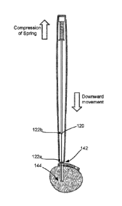
FIGS. 10A and 10B illustrate an initial process of examining, via the bone
probe 108, a
drilled hole 134 in a bone 132. For example, as previously described herein,
the biasing force
from the spring assembly 126 may be sufficient so as to maintain the bone
probe 108 in the
extended position while the surgeon probes an interior surface 136 of the
drilled hole 134 and
locates the bottom 138 of the hole 134. However, as shown in FIG. 10B, the
biasing force may
be overcome upon a surgeon moving the handle 102 in a direction towards the
hole 134 once the
desired target site is located, such as locating the bottom 138 of the hole
134. The surgeon can
move the handle 102 until the first end 104 of the handle 102 abuts either the
surface of the bone
.. 132 or a surface of a plate or implant 140, as indicated by arrow 142,
thereby resulting in
compression of the spring assembly 126 while maintaining placement of the
probing tip 114 at
22
Date Regue/Date Received 2022-07-14
the bottom 138 of the hole 134, as indicated by arrow 144. At this point, the
depth gauge
member 116 can be advanced in a direction towards the hole 134, such that the
hollow shaft 118
slides over the bone probe shaft 110, wherein the bone probe shaft 110
generally acts as a guide
and holding position as a result of the engagement surface of the second
portion 130 of the
probing tip 114 having established purchase with the bottom 138 of the hole
134. The depth
gauge member 116 can be extended down into the hole 134 until the distal end
120 of the depth
gauge member 116 abuts the bottom 138 of the hole 134. Accordingly, the one or
more depth
measurement sensors 122 can then generate an electronic signal in relation to
a distance between
the first end 104 of the handle 102 and the distal end 120 of the depth gauge
member 116,
wherein the electronic signal is indicative of the depth of the hole 134 and
the thickness of the
plate or implant 140.
The device 100 of the present disclosure may include a variety of different
sensing
devices suitable for determining a length or depth of the drilled hole or bore
to be measured. For
example, the one or more depth measurement sensors 122 may include, but are
not limited to, an
electromechanical or electronic sensor, such as a linear encoder, and may
employ any one or
more of acoustic, ultrasound, capacitive, electric field, inductive,
electromagnetic (e.g.. Hall
effect-type) and optical components for determining relative or absolute
distance measurements.
In some embodiments, the sensors 122 may be configured to measure, sense,
discriminate, or
otherwise determine a length or distance between at least the first end 104 of
the handle 102 and
the distal end 120 of the depth gauge member 116.
For example, in one embodiment, as shown in FIGS. 10A and 10B, at least a
first sensor
element 122a is positioned proximate to the first end 104 of the handle 102
and a second sensor
element 122b is positioned on the depth gauge shaft 118 proximate the distal
end 120. The
sensor elements 122a, 122b are configured to measure at least one of relative,
absolute and
incremental movement (e.g., distance, speed, etc.) of the depth gauge shaft
118 with respect to
the first end 104 of the handle 102 during a measurement procedure. For
example, in one
embodiment, the sensor elements 122a, 122b may be used for measure an absolute
distance that
the depth gauge 116 distal end 120 is moved relative to the fixed reference
point such as, for
example the first end 104 of the handle 102.
The first sensor element 122a may be an active inductive, capacitive or
optical element
that is in communication with circuitry (e.g., a controller) of a user
interface portion of the
23
Date Regue/Date Received 2022-07-14
device (e.g., a GUI display or the like with user inputs). The first sensor
element 122a may
include one or more longitudinally-extending conductors that are wires, cables
or traces on a
printed circuit board such as, for example, a flex-circuit or the like.
Furthermore, the first sensor
element 122a may further include a plurality of inductive, capacitive or
optical elements that
may be coupled with and disposed on the longitudinally-extending conductors.
The second
sensor element 122b may be configured on the depth gauge shaft 118 in manner
so as to
cooperate with the first sensor element 122a proximate the first end 104 of
the handle 102. For
example, the second sensor element 122b may be a generally passive element
such as a
permanent magnet, optical element (e.g., indicia) or the like that is
configured to cooperate,
communicate or otherwise interact with the first sensor element 122a. For
example, during a
measurement procedure, movement of the depth gauge 116 out of the device
handle 102 results
in interaction between the first and second sensor elements 122a, 122b. In
particular, as the
depth gauge 116 extends from the device handle 102, the first and second
sensor elements 122a,
122b move relative to one another (i.e., second sensor element 122b moves past
first sensor
element 122a and, in combination with one another, provide signals (e.g.,
pulses, etc.) to the
circuitry, which processes the signals and displays a distance measurement on
a display and/or
transmits the signals to separate computing devices.
In various embodiments of the present invention, the one or more sensors 122
may he
connected with a microprocessor and/or other digital electronic device in
order to produce an
output for an electronic display, such as a liquid crystal display or light-
emitting diode display,
and or for wireless/wired transmission of electronic signals, comprising the
measurement data, to
a wireless compatible computing device. For example, in some embodiments, the
microprocessor or other digital electronic device may be connected to a
wireless transmitter for
wireless transmission of electronic signals. In some embodiments, a signal
conditioning circuit
may interpose the inductive or capacitive elements of the electronic sensor
and the
microprocessor or other digital electronic device used to drive the display,
thus ensuring that
correct input current and voltage levels are provided to the various
components. The device may
further include a power source, such as a primary or secondary battery, may be
connected to the
signal conditioning circuit or to the microprocessor directly.
It should be noted that the device 100 of the present disclosure may include a
variety of
different electronic sensor and circuitry assemblies for determining and
transmitting depth
24
Date Regue/Date Received 2022-07-14
measurements, including the sensors and systems discussed in U.S. Patent Nos.:
7,165,336;
7,444,756; 7,493,703; 7,607,238; 7,676,943; 7,685,735; 7,730,629; 7,895,762;
7,895,767.
FIG. 11 is a side view of the medical device 100 including a strain sensor 146
for sensing
strain upon the bone probe shaft 110 as a result of probing the interior
surface of a drilled hole.
The sensor 146 may include a strain gauge or the like configured to determine
a strain of the
bone probe shaft 110, which may be useful for alerting the surgeon of an
amount of resistance
that the distal probing tip 114 is encountering during probing of the interior
of the hole. For
example, while a surgeon may be able to "feel" the interior surface and
further have a sense of
when the probing tip 114 actually makes contact with the bottom of the hole,
the strain sensor
146 may further generate an electronic signal based on a sensed strain of the
shaft 110 which
may then be used to provide an audible and/or visual alert, via a device 148
(i.e., speaker or
lights) to the surgeon indicating that the probing tip 116 is in fact
positioned at the bottom of the
hole.
For example, the resistance encountered when the probing tip 116 engages the
bottom of
the hole may have a certain strain value (i.e., above a certain threshold)
which may be different
than a resistance encountered with the sidewalls of the hole (which may have a
softer, spongier
tissue). Accordingly, the audible and/or visual alert may confirm to a surgeon
whether they are
in fact positioned at the bottom of the hole or if too much pressure is being
placed against the
interior surface such that they risk possibly inadvertently piercing the
interior surface.
FIGS. 12A-12F illustrate a series of steps for performing a procedure of
probing a drilled
hole and subsequently obtaining a depth measurement using another embodiment
of a medical
device 300 consistent with the present disclosure. As shown, the device 300
may be similarly
configured as device 100 previously described herein. However, as shown in
FIG. 12A, both the
bone probe 108 and depth gauge member 116 may both be completely withdrawn
into the handle
102 until either a first slider 324 is moved, resulting in corresponding
movement of the bone
probe 108, or a second slider 350 is moved, resulting in corresponding
movement of the depth
gauge member 116, as shown in FIG. 12E.
In addition to including sliders for allowing independent movement of the bone
probe
and depth gauge member, the device 300 further includes a locking member 352
for locking a
position of at least the bone probe 108. As shown, the locking member 352 is
coupled to the first
Date Regue/Date Received 2022-07-14
end 104 of the handle 102 and is associated with at least the bone probe 108
in such as manner so
as to allow/prevent movement of the bone probe 108. For example, the locking
member 352 has
an unlocked configuration and a locked configuration, wherein, in the unlocked
configuration,
the locking member 352 allows the bone probe 108 to freely move and, when in
the locked
configuration, the locking member 352 prevents movement of the bone probe 108.
For example, upon extending the bone probe 108, a surgeon may then place the
locking
member 352 in a locked configuration, as shown in FIG. 12C, in which the
locking member 352
is configured to provide sufficient contact with the bone probe shaft 110 so
as to prevent, or
make difficult, the movement of the bone probe shaft 110 relative to the first
end 104 of the
handle 102, thereby providing an amount of rigidity to the probe shaft 110.
Accordingly, a
surgeon may now perform examination of a drilled hole without concern of the
bone probe 108
withdrawing back into the handle 102 or being loose.
Upon locating the base or bottom of the hole, the surgeon may then apply
sufficient force
upon the bone probe shaft 110 so that the engagement surface of the second
portion of the
probing tip engages and establishes purchase with the bottom of the hole, or a
sidewall
immediately adjacent to the bottom, as shown in FIG. 12D. Upon establishing
engagement, the
surgeon may then place the locking member 352 in an unlocked configuration,
now that the bone
probe shaft 110 is in a stabilized in position. The surgeon may then move the
handle in a
directions towards the bone until the first end of the handle abuts the
surface of the bone or the
surface of the plate/implant, as shown in FIG. 12E, at which point, the depth
gauge member 116
can be used for measuring the depth of the hole. As shown in FIG. 12F, the
surgeon may then
advance the depth gauge member 116 towards hole, via the second slider 350,
such that the distal
end 120 of the depth gauge member shaft 118 extends from the first end of the
device handle and
advances into the hole, sliding over the bone probe 108. While the bone probe
108 is maintained
in engagement with the bottom of the hole via the probing tip, the depth gauge
member may be
advanced in a direction towards the bottom of the hole until the distal end of
the depth gauge
member makes contact with the bottom of the hole. The bone probe essentially
acts as a guide
upon which the depth gauge member slide over when advancing to the bottom of
the hole.
The sensor is configured to generate an electronic signal based on a distance
between the
first end of the body and the distal end of the depth gauge member, wherein
the electronic signal
is indicative of at least a depth of the hole. In particular, the sensor may
include inductive or
26
Date Regue/Date Received 2022-07-14
capacitive elements or assemblies configured to sense the location of the
distal end of the depth
gauge member relative to the first end of the device body, and, as a result,
generate an electronic
signal representing the distance there between. Accordingly, the sensed
distance between the
first end of the device handle (when abutting the bone surface) and the distal
end of the depth
gauge member (when abutting the bottom of the hole) is the depth of the hole.
It should be noted that the device may include logic or allow for adjustment
to the
sensing capabilities so as to program the sensor to account for other
variables when sensing the
depth of the hole. For example, in some embodiments, certain procedures
require fixing a plate
or implant to the bone via screws. Accordingly, the screw length must not only
be sufficient to
fill the hole but also long enough to account for the thickness of a plate or
implant through which
it passes when engaging the hole. Accordingly, in some embodiments, the sensor
may be
programmed so as to account for the thickness of the plate or implant and will
further include
that thickness in the electronic signal produced, such that the electronic
signal is indicative of the
total depth that a corresponding screw length will need to cover, including
the depth of the hole
in the bone in addition to the thickness of the plate or implant through which
the screw will pass
through and the screw head will engage.
Furthermore, in some instances, first end of the device handle will be
directly abutting a
surface of the plate or implant, as shown in FIG. 12F, which is directly
abutting the surface of
the bone, when the surgeon is measuring the depth. Thus, in this case, the
sensor is still able to
sense a distance between the first end of the device handle and the distal end
of the depth gauge
member, which will provide an overall depth, rather than just a depth of the
hole in the bone.
FIGS. 13A-13C illustrate a series of steps for performing a procedure of
probing a fully
drilled hole (i.e., a hole extending entirely through a bone for receipt of a
bicortical bone screw)
with the bone probe 208 and further establishing purchase of the probing tip
212 of the bone
probe 208 with a side of the bone adjacent to the bicortical drilled hole to
secure the bone probe
208 in place and allow the depth gauge member to be used for measuring the
depth of the
bicortical drilled hole. It should be noted that he bone probe 208 of FIGS. 4-
9 is compatible for
use with either of devices 100 and 300 and may be extended and retracted, and
otherwise
manipulated for subsequent probing and depth measurements, therefrom in a
similar manner as
bone probe 108 previously described herein.
27
Date Regue/Date Received 2022-07-14
As shown in FIGS. 13A-13C, the hole is drilled entirely through the bone
(i.e., bicortical
drill hole), and thus a surgeon will need to not only probe the interior
surface of the hole, and
possible obtain neuromonitoring data (i.e., determine whether there are any
nearby nerves which
may be affected by placement of a screw within the hole), but further obtain
an accurate
.. measurement of the depth of the entire hole.
As shown in FIG. 13A, a surgeon may first perform examination of the drilled
hole with
the probing tip 214 by advancing the bone probe 208 into the drilled hole. The
surgeon may
simply apply slight pressure such that the base portion 220 of the probing tip
214 contacts an
interior surface of the hole and, in return, provides tactile feedback of the
interior surface to the
.. surgeon. The base portion 220 is shaped so as to glide or easily slide
along the interior surface,
while still allowing sufficient contact to provide tactile feedback to the
surgeon. The surgeon
may then advance the probing tip 214 entirely through the hole, at which
point, the base portion
220 will cease contact with the interior surface and the surgeon will sense
(via tactile feedback)
that the end of the hole has been reached (shown in FIG. 13B).
At this point, upon the surgeon extending the probing tip 214 entirely through
a bicortical
drilled hole, the surgeon can then establish purchase between the top portion
222 of the probing
tip 214 and a portion of an opposing side of bone so as to secure the bone
probe shaft 210 in
place for subsequent depth measurements with the depth gauge member. For
example, as shown
in FIG. 13C, the surgeon may simply position the substantially planar second
side 218 against
.. the interior surface of the drilled hole and then retract (i.e., pull back)
the probe shaft 210 such
that the engagement surface 226 of the top portion 222 of the probing tip 214
comes into contact
with a portion of the opposing side of the bone immediately adjacent to the
opening of the hole.
The engagement surface 226 may be a substantially abrupt edge of the probing
tip 214, in which
the transition between the base portion 220 and the top portion 222 is sudden
(e.g., sharp corner
or edge). Accordingly, as the surgeon is pulling the bone probe shaft 210 back
towards the hole,
the engagement surface 226 will begin to contact the bone. In some
embodiments, the
engagement surface 226 may include surface texturing to enhance friction
between the
engagement surface 226 and the portion of bone to reduce risk of slippage
during bicortical
depth measurements. Furthermore, groove 224 (present at the junction between
the distal end
.. 212 of the probe shaft 210 and the probing tip 214) will allow for
additional flexing of the
probing tip 214 relative to the remainder of the probe shaft 210 due to less
material at the
28
Date Regue/Date Received 2022-07-14
junction between the shaft 210 and the tip 214 at the groove 224, which will
improve the
purchasing or grabbing of the opposing side of the bone with the engagement
surface 226 of the
top portion 222 of the probing tip. Furthermore, the tapered thickness of the
shaft 210, provided
by the substantially planar second side 218, allows for deflection or bending
of the shaft 210 on
one axis, such that, if the probing tip 214 is substantially perpendicular to
shaft 210, as generally
shown, application of pressure upon the shaft 210 results in deflection of the
probing tip 214,
particularly the engagement surface 226, to become angled upward, thereby
enabling a superior
purchase or gripping of the outer surface of the opposing side of the bone.
Upon securing the
bone probe 208 in place, depth measurements may take place with the depth
gauge member in a
manner similar to that of bone probe 108 previously described herein.
FIG. 14 is another embodiment of a medical device 400 consistent with the
present
disclosure having a display 454 for providing a digital readout of a depth
measurement of the
hole based on the electronic signal from the sensor. The display 454 may
include a liquid crystal
display or an LED display, for example.
FIG. 15 is another embodiment of a medical device 500 consistent with the
present
disclosure configured to wirelessly communicate with and transmit depth
measurement data to a
wireless computing device 600 over a network, to record, store, and/or
visually display measured
depths based on electronic signals from the sensor for determining depth of
drilled holes. For
example, the device 500 may include a wireless transmitter 556 configured to
wireless
communicate and exchange information, including the electronic signal, with a
wireless display
or computing device 600 for at least visually providing a depth measurement of
the hole based
on the electronic signal from the sensor. The separate display or computing
device 600 may
include, but is not limited to, a monitor or panel display, a PC, a notebook,
a tablet computer, a
smartphone, or other computing device configured to wirelessly communicate
with the wireless
transmitter 556.
The network may be any network that carries data. Non-limiting examples of
suitable
networks that may be used as network include WiFi wireless data communication
technology,
the internet, private networks, virtual private networks (VPN), public switch
telephone networks
(PSTN), integrated services digital networks (ISDN), digital subscriber link
networks (DSL).
various second generation (2G), third generation (3G), fourth generation (4G)
cellular-based data
communication technologies, Bluetooth radio, Near Field Communication (NFC),
the most
29
Date Regue/Date Received 2022-07-14
recently published versions of IEEE 802.11 transmission protocol standards,
other networks
capable of carrying data, and combinations thereof.
Furthermore, in some embodiments, the computing device 600 may include a
specific
software application that may be directed to maintaining a record of the hole
measurements
and/or provide an interactive user interface (GUI) in which multiple holes can
be mapped to a
particular plate or implant and the depth of each hole (including the
thickness of the plate or
implant) can be included and stored for records.
FIGS. 16 and 17 illustrate the compatibility of a medical device of the
present disclosure
with other medical devices so as to provide additional features, in additional
bone probing and
depth measurement, such as energy emission (FIG. 16) and sensing capabilities
(FIG. 17). For
example, in some embodiments, the bone probe shaft 110, 210 may include an
electrically
conductive material (e.g., a metal such as stainless steel, nitinol, or
aluminum), wherein a portion
of the bone probe shaft 110, 210 may be exposed, or otherwise accessible,
along a portion of the
device handle. In particular, the device handle may include an access region
158 that may be in
the form of an aperture, window, or the like, that provides access to an
interior of the handle,
particularly providing access to an exposed portion of the bone probe shaft.
Thus, in some
embodiments, an electrical current from a separate device 700, 800 may be
supplied to the hone
probe shaft via the access region 158 (e.g., slide a working tip 702 of an
electrocautery device
700 into the access region 158 to make contact with bone probe shaft 110,
210). Accordingly, as
a result of being made from a conductive material, the bone probe shaft 110,
210 may carry the
electrical current to the distal probe tip, which may then be used to deliver
energy to a desired
target (e.g., interior surface of hole of the bone) as a result of the
electrical current applied
thereto. Similarly, a separate nerve sensing/stimulation device 800 (shown in
FIG. 17) may be
coupled to the conductive bone probe shaft via the access region (i.e., slide
a working tip 802 of
the device 800 into the access region 158), such that the distal probe tip
essentially acts as an
extension to the nerve sensing/stimulation device and may be used to
sense/stimulate nerves
within the bone. The separate sensing/nerve stimulation device or system 800
may include, for
example, existing capital equipment or a handheld battery-powered
neuromonitoring device.
FIG. 18 is a perspective view of a medical device 100 having a port 160
provided on the
proximal, or second end 106, of the device body 102. The port 160 is
configured to receive a
corresponding input connector from a nerve sensing/nerve stimulation device
800. The port 160
Date Regue/Date Received 2022-07-14
(hereinafter referred to as "neuromonitoring port 160) is coupled to the bone
probe shaft 110,
210 and is configured to provide an electrical pathway from the nerve
sensing/nerve stimulation
device 700 to the bone probe 108, 208 upon insertion of the input connector
into the
neuromonitoring port 160. As previously described, the bone probe shaft 110,
210 may include
an electrically conductive material (e.g., a metal such as stainless steel,
nitinol, or aluminum) and
thus may carry an electrical signal. Thus, in some embodiments, an electrical
signal from the
nerve sensing/nerve stimulation device 800 may be supplied to the bone probe
shaft 110, 210 via
the neuromonitoring port 160. Accordingly, as a result of being made from a
conductive
material, the bone probe shaft 110, 210 may carry the electrical signal to the
distal probe tip 114,
214, which may then be used to sense/stimulate nerves adjacent or in close
proximity to the
drilled hole in the bone, either when the bone probe 108, 208 is directly
placed within the drilled
hole or when the bone probe 108, 208 is in contact with a screw placed within
the drilled hole.
FIG. 19 is a side view, partly in section, of the medical device 100 of FIG.
18 illustrating
the configuration of the bone probe shaft 110 for carrying electrical signals
to and from the nerve
sensing/nerve stimulation device. It should be noted that bone probe shaft 210
is also compatible
with the nerve sensing/nerve stimulation device and can function in a similar
manner as bone
probe shaft 110 described herein. Upon insertion of the electrical connector
into the
neuromonitoring port 160, a pathway is provided between the nerve
sensing/nerve stimulation
device 700 and the bone probe 108. The bone probe shaft 108 generally includes
a soft coil
portion 162 configured to allow conduction of an electrical signal provided by
the nerve
sensing/stimulation device 800 while the shaft 110 moves between fully
retracted and fully
extended positions and intermediate positions there between, particularly when
measuring the
depth of the drilled hole 134. In some embodiments, a portion of the distal
end 112 of the bone
probe 108, particularly the exposed portion of the shaft 110 extendable
outside of device body
102 may include an insulating material 164, while the distal probing tip 114
is free of insulating
material.
FIGS. 20A, 20B, and 20C illustrate the transmission of a signal from bone
probe 108 to a
screw positioned within a hole in a vertebra for neuromonitoring capabilities.
As shown in FIG.
20A, upon coupling the nerve sensing/nerve stimulation device 700 to the
medical device 100
(e.g., inserting the electrical connector into the neuromonitoring port 160),
a surgeon can begin a
neuromonitoring procedure to determine whether there are any critical
neurological structures
31
Date Regue/Date Received 2022-07-14
adjacent to or within an unsafe proximity to the drilled hole and screw. In
particular, a surgeon
can perform neuromonitoring procedure by placing the bone probe 108 directly
within the drilled
hole prior to screw placement, in which the distal probing tip 114 can be
placed in direct contact
with the interior of the hole and transmit the electrical signal from the
nerve sensing/nerve
stimulation device 800 to the bone tissue and will subsequently receive a
response signal to then
be carried back to the nerve sensing/nerve stimulation device 700 for
processing. In another
method, as shown in FIGS. 20A, 20B, and 20C, the surgeon is performing the
neuromonitoring
procedure once the screw is already in place (e.g., already fitted within the
drilled hole) by
placing the distal probing tip 114 in direct contact with the screw, which, in
turn, will act as a
conduit and carry electrical signals to and from the distal probing tip 114
and the nerve
sensing/nerve stimulation device 900.
Accordingly, the medical device consistent with the present disclosure is a
three-in-one
single use device designed to more accurately and safely measure the screw
hole pathway. For
example, the probing tip of the bone probe provides a user (e.g., surgeon)
with superior tactile
feedback to assist the surgeon in confirming a safe pathway within the bone.
The electronic
measurement/digital sensing is designed to provide more accurate depth
measurement for the
screw pathway. The neuromonitoring feature is used to stimulate the pathway
and/or screw,
ensuring the screw is safely positioned away from any critical neurological
structures. Overall,
the medical device of the present disclosure is a faster, safer, more accurate
and user-friendly
solution for surgeons when placing bone screws, particularly pedicle screws
during spinal fusion
surgery, thereby minimizing spine surgery complications and reducing overall
healthcare costs.
FIG. 21 illustrates an angle guide 900 for use with the medical device of the
present
disclosure. In some instances, holes may be drilled into bone at an angle.
Accordingly, the
angle guide may be useful in providing a surgeon with a visual guide as to the
correct angle at
which to position the device when attempting to examine the hole and further
locate the bottom
of the hole to carry out the depth measurements.
Reference throughout this specification to "one embodiment" or "an embodiment"
means
that a particular feature, structure, or characteristic described in
connection with the embodiment
is included in at least one embodiment. Thus, appearances of the phrases "in
one embodiment"
or "in an embodiment" in various places throughout this specification are not
necessarily all
32
Date Regue/Date Received 2022-07-14
referring to the same embodiment. Furtheimore, the particular features,
structures, or
characteristics may be combined in any suitable manner in one or more
embodiments.
The terms and expressions which have been employed herein are used as terms of
description and not of limitation, and there is no intention, in the use of
such terms and
expressions, of excluding any equivalents of the features shown and described
(or portions
thereof), and it is recognized that various modifications are possible within
the scope of the
claims. Accordingly, the claims are intended to cover all such equivalents.
33
Date Regue/Date Received 2022-07-14