Note: Descriptions are shown in the official language in which they were submitted.
WO 2021/168455
PCT/US2021/019126
METHODS OF SPATIALLY RESOLVED SINGLE CELL RNA SEQUENCING
CROSS-REFERENCE TO RELATED APPLICATIONS
This application claims priority to U.S. Provisional Application No.
62/979,235 filed on
February 20, 2020, which is incorporated by reference herein in its entirety.
FIELD OF THE INVENTION
The present disclosure generally relates to spatial detection of a nucleic
acid, such as a
genomic DNA or a RNA transcript, in a cell comprised in a tissue sample. The
present disclosure
provides methods for detecting and/or analyzing nucleic acids, such as
chromatin or RNA
transcripts, so as to obtain spatial information about the localization,
distribution or expression of
genes in a tissue sample. The present disclosure thus provides a process for
performing "spatial
transcriptomics" or "spatial genomics," which enables the user to determine
simultaneously the
expression pattern, or the location/distribution pattern of the genes
expressed or genes or genomic
loci present in a single cell while retaining information related to the
spatial location of the cell
within the tissue architecture.
BACKGRO UND
Over the past decade, massively-parallel single cell RNA-sequencing (scRNA-
seq) has
emerged as a powerful approach to catalogue the remarkable cellular
heterogeneity in complex
tissues (1, 2). While scRNA-seq can profile the transcriptomes of thousands of
cells in a single
experiment, it requires the dissociation of tissue into single cell
suspensions prior to library
preparation and sequencing, eliminating any spatial information (3-6). Several
strategies have
emerged to obtain molecular and spatial information simultaneously from
complex tissue.
Imaging-based strategy combines high resolution microscopy with fluorescent in
situ
hybridization (FISH) to achieve subcellular resolution and could profile the
entire transcriptome
(7-10), but this requires lengthy iterative microscopy workflows and large
probe panels. Another
approach is to hybridize RNA directly from tissue slices onto a microarray
containing spatially-
barcoded oligo(dT) spots or beads to encode location information into RNA-
sequencing libraries.
These approaches can sample the entire transcriptome without the need for
iterative rounds of
hybridization (11) and recent improvements using DNA-barcoded beads (FIDST and
Slide-
1
CA 03168485 2022- 8- 18
WO 2021/168455
PCT/US2021/019126
seqvl/v2) report spatial resolutions at or below the diameter of a single cell
(12-14). However,
because of the low numbers of mRNA molecules captured per bead, these spatial
transcriptomic
approaches often aggregate neighboring beads prior to downstream analysis,
resulting in lower
effective resolution and averaging of transcript abundances from multiple
cells. As a result,
annotation of specific cell types present within each spatial unit of analysis
is accomplished by
aggregating gene sets computationally defined from orthogonal scRNA-seq
datasets (15, 16).
While integration methods have demonstrated the ability to localize cell types
within the spatial
organization of complex tissue, they rely on having available data from two
independent assays
and have limited ability to infer how spatial context influences the cell
state of individual cell
types.
SUMMARY OF THE INVENTION
To address these drawbacks, we have developed XYZeq, a method that expands on
recent
methods of split-pool indexing (17, 18) for single cell sequencing to enable
simultaneous recording
of spatial information. At the heart of the approach is a strategy that
integrates split-pool indexing
and spatial barcoding to enable the profiling, such as transcriptomic
profiling or chromatin
accessibility profiling, of tens of thousands of single cells and the
resolution of cells to thousands
of spatial wells. Cellular transcripts, for instance, are spatially encoded in
situ with barcoded oligos
in an array containing microwells. A tissue slice is placed on an array
containing barcoded oligo
d(T) primers containing a unique molecular identifier and a PCR handle. This
is followed by
reverse transcription, split-pool step to introduce a second round of
barcoding by PCR, and
tagmentation to generate single cell RNA-sequencing libraries. Similar
methodology can be used
to spatially profile chromatin accessibility. XYZeq compares favorably to both
image-based and
array- or bead-based methods in its ability to target the genome-wide
chromatin or the entire
transcriptome and simultaneous estimate single cell gene transcription or
expression profiles
enabling the detecting of rare and transient transcriptional states.
Accordingly, in one aspect, the present disclosure relates to a method for
spatial detection
of a nucleic acid within a sample comprising cells, said method comprising
identifying presence,
absence or quantity of a combination of a spatial barcode domain and a
cellular barcode domain
in a nucleic acid of the sample.
2
CA 03168485 2022- 8- 18
WO 2021/168455
PCT/US2021/019126
In some embodiments, the method comprises contacting an array comprising a
plurality of
microwells with the sample comprising cells such that the sample contacts a
plurality of
microwells at their distinct positions on the array, wherein each microwell
occupies a distinct
position on the array and comprises a different spatial index primer
comprising a nucleic acid
molecule comprising, from 5' to 3':
a) an annealing domain comprising a nucleotide sequence that is recognized
by a
first sequencing primer,
b) a spatial barcode domain comprising a nucleotide sequence that is unique
to each
microwell; and
c) a capture domain comprising a polythymidine sequence;.
In some embodiments, the method further comprises allowing a time period to
elapse in
physiologically acceptable conditions, the time period sufficient to allow
hybridization of one or
more message RNAs (mRNAs) present in one or more cells located in each
microwell to the
capture domain of the spatial index primer unique to said microwell. In some
embodiments, this
step may comprise performing a reverse transcription reaction to obtain a
first strand of the cDNA
molecules.
In some embodiments, the method further comprises performing reverse
transcription to
generate one or more cDNA molecules corresponding to the one or more mRNAs
present in said
microwell. In some embodiments, the method further comprises pooling cells
present in each
microwell of the array and sorting into a multiwell plate comprising a
plurality of wells. In some
embodiments, the method further comprises performing an amplification reaction
with a cellular
index primer comprising a nucleic acid molecule comprising, from 5' to 3':
a) an annealing domain comprising a nucleotide sequence that is recognized
by a
second sequencing primer; and
b) a cellular barcode domain comprising a nucleotide sequence that is
unique to each
well of the multiwell plate.
In some embodiments, the method further comprises sequencing amplification
reaction
products obtained in the above step using the first sequencing primer and the
second sequencing
primer. In some embodiments, the method further comprises detecting the
presence of a nucleotide
sequence of a given spatial barcode domain and a nucleotide sequence of a
given cellular barcode
domain, or sequences complementary to a given spatial barcode domain and a
given cellular
3
CA 03168485 2022- 8- 18
WO 2021/168455
PCT/US2021/019126
barcode domain. In some embodiments, the method further comprises a step of
providing an array
comprising a plurality of microwells prior to contacting each subsample to
each spatial index
primer. In some embodiments, the method further comprises permeabilizing cells
comprised in
the tissue sample prior to performing the hybridization. In some embodiments,
the method further
comprises imaging the array with the sample overlaid after contacting the
array with the sample.
In some embodiments, the method further comprises lysing the cells after the
cells are sorted into
the multiwell plate. In some embodiments, the method further comprises
generating sequencing
libraries from the cDNA molecules generated by tagmentation. In some
embodiments, the method
further comprises performing an amplification reaction following tagmentation.
In some embodiments, the method further comprises determining which genes are
expressed in the cell at a particular distinct location of the tissue sample
by a method comprising
determining the sequences of the cDNA molecules comprising the same nucleotide
sequence of a
spatial barcode domain, or sequence complementary thereto, and the same
nucleotide sequence of
a cellular barcode domain, or sequence complementary thereto. In some
embodiments, the method
further comprises correlating the nucleotide sequence of a spatial barcode
domain unique to a
given particular microwell of the array, or the sequence complementary
thereto, present in the
cDNA molecules to a position in the tissue sample. In some embodiments, the
method further
comprises correlating the nucleotide sequence of a spatial barcode domain
unique to a given
particular microwell of the array, or the sequence complementary thereto,
present in the cDNA
molecules to an image of the tissue sample.
In any of the aforementioned methods, the presence of a particular nucleotide
sequence of
the spatial barcode domain unique to a given particular microwell of the
array, or the sequence
complementary thereto, and the presence of a particular nucleotide sequence of
the cellular
barcode domain, or the sequence complementary thereto, indicates that the cDNA
molecules are
obtained from mRNAs present in one single cell comprised in the sample at the
distinct position
where the sample contacted said particular microwell of the assay.
In another aspect, the present disclosure relates to a method of generating a
single cell
transcriptome profile or RNA library of a sample, the method comprising
identifying presence,
absence or quantity of a combination of a spatial barcode domain and a
cellular barcode domain
in a nucleic acid of the sample.
4
CA 03168485 2022- 8- 18
WO 2021/168455
PCT/US2021/019126
In some embodiments, the method comprises contacting an array comprising a
plurality of
microwells with the sample comprising cells such that the sample contacts a
plurality of
microwells at their distinct positions on the array, wherein each microwell
occupies a distinct
position on the array and comprises a different spatial index primer
comprising a nucleic acid
molecule comprising, from 5' to 3':
a) an annealing domain comprising a nucleotide sequence that is recognized
by a
first sequencing primer,
b) a spatial barcode domain comprising a nucleotide sequence that is unique
to each
microwell; and
c) a capture domain comprising a polythymidine sequence;.
In some embodiments, the method further comprises allowing a time period to
elapse in
physiologically acceptable conditions, the time period sufficient to allow
hybridization of one or
more message RNAs (mRNAs) present in one or more cells located in each
microwell to the
capture domain of the spatial index primer unique to said microwell. In some
embodiments, this
step may comprise performing a reverse transcription reaction to obtain a
first strand of the cDNA
molecules.
In some embodiments, the method further comprises performing reverse
transcription to
generate one or more cDNA molecules corresponding to the one or more mRNAs
present in said
microwell. In some embodiments, the method further comprises pooling cells
present in each
microwell of the array and sorting into a multiwell plate comprising a
plurality of wells. In some
embodiments, the method further comprises performing an amplification reaction
with a cellular
index primer comprising a nucleic acid molecule comprising, from 5' to 3':
a) an annealing domain comprising a nucleotide sequence that is recognized
by a
second sequencing primer; and
b) a cellular barcode domain comprising a nucleotide sequence that is
unique to each
well of the multiwell plate.
In some embodiments, the method further comprises sequencing amplification
reaction
products obtained in the above step using the first sequencing primer and the
second sequencing
primer. In some embodiments, the method further comprises detecting the
presence of a nucleotide
sequence of a given spatial barcode domain and a nucleotide sequence of a
given cellular barcode
domain, or sequences complementary to a given spatial barcode domain and a
given cellular
CA 03168485 2022- 8- 18
WO 2021/168455
PCT/US2021/019126
barcode domain. In some embodiments, the method further comprises a step of
providing an array
comprising a plurality of microwells prior to contacting each subsample to
each spatial index
primer. In some embodiments, the method further comprises permeabilizing cells
comprised in
the tissue sample prior to performing the hybridization. In some embodiments,
the method further
comprises imaging the array with the sample overlaid after contacting the
array with the sample.
In some embodiments, the method further comprises lysing the cells after the
cells are sorted into
the multiwell plate. In some embodiments, the method further comprises
generating sequencing
libraries from the cDNA molecules generated by tagmentation. In some
embodiments, the method
further comprises performing an amplification reaction following tagmentation.
In some embodiments, the method further comprises determining which genes are
expressed in the cell at a particular distinct location of the tissue sample
by a method comprising
determining the sequences of the cDNA molecules comprising the same nucleotide
sequence of a
spatial barcode domain, or sequence complementary thereto, and the same
nucleotide sequence of
a cellular barcode domain, or sequence complementary thereto. In some
embodiments, the method
further comprises correlating the nucleotide sequence of a spatial barcode
domain unique to a
given particular microwell of the array, or the sequence complementary
thereto, present in the
cDNA molecules to a position in the tissue sample. In some embodiments, the
method further
comprises correlating the nucleotide sequence of a spatial barcode domain
unique to a given
particular microwell of the array, or the sequence complementary thereto,
present in the cDNA
molecules to an image of the tissue sample.
The disclosure relates to a method of obtaining the transcriptome of a single
cell
comprising:
(i) contacting a sample to an array, said array comprising multiple wells
comprising
(ii) isolating RNA from the sample in each well;
(iii) performing quantitative PCR on the isolated RNA by amplification of the
RNA by
the primer or primers in each well;
(iv) correlating the amplification product of the RNA with a cell at a
position that
corresponds to the position within the sample.
In some embodiments the cell is a mesenchymal cell, a cancer cell, a
hepatocyte or a splenocyte.
In any of the aforementioned methods, the presence of a particular nucleotide
sequence of
the spatial barcode domain unique to a given particular microwell of the
array, or the sequence
6
CA 03168485 2022- 8- 18
WO 2021/168455
PCT/US2021/019126
complementary thereto, and the presence of a particular nucleotide sequence of
the cellular
barcode domain, or the sequence complementary thereto, indicates that the cDNA
molecules were
obtained from mRNAs present in one single cell comprised in the subsample at
the distinct position
where the subsample is positioned in said particular microwell of the assay.
In yet another aspect, the present disclosure relates to a method of
generating high-
resolution spatial positioning of a nucleic acid expression in a cell within a
sample, the method
comprising identifying presence, absence or quantity of a combination of a
spatial barcode domain
and a cellular barcode domain in a nucleic acid of the sample.
In some embodiments, the method comprises contacting an array comprising a
plurality of
microwells with the sample comprising cells such that the sample contacts a
plurality of
microwells at their distinct positions on the array, wherein each microwell
occupies a distinct
position on the array and comprises a different spatial index primer
comprising a nucleic acid
molecule comprising, from 5' to 3':
a) an annealing domain comprising a nucleotide sequence that is recognized
by a
first sequencing primer;
b) a spatial barcode domain comprising a nucleotide sequence that is unique
to each
microwell; and
c) a capture domain comprising a polythymidine sequence,.
In some embodiments, the method further comprises allowing a time period to
elapse in
physiologically acceptable conditions, the time period sufficient to allow
hybridization of one or
more message RNAs (mRNAs) present in one or more cells located in each
microwell to the
capture domain of the spatial index primer unique to said microwell. In some
embodiments, this
step may comprise performing a reverse transcription reaction to obtain a
first strand of the cDNA
molecules.
In some embodiments, the method further comprises performing reverse
transcription to
generate one or more cDNA molecules corresponding to the one or more mRNAs
present in said
microwell. In some embodiments, the method further comprises pooling cells
present in each
microwell of the array and sorting into a multiwell plate comprising a
plurality of wells. In some
embodiments, the method further comprises performing an amplification reaction
with a cellular
index primer comprising a nucleic acid molecule comprising, from 5' to 3 ' :
7
CA 03168485 2022- 8- 18
WO 2021/168455
PCT/US2021/019126
a) an annealing domain comprising a nucleotide sequence that is recognized
by a
second sequencing primer; and
b) a cellular barcode domain comprising a nucleotide sequence that is
unique to each
well of the multiwell plate.
In some embodiments, the method further comprises sequencing amplification
reaction
products obtained in the above step using the first sequencing primer and the
second sequencing
primer. In some embodiments, the method further comprises detecting the
presence of a nucleotide
sequence of a given spatial barcode domain and a nucleotide sequence of a
given cellular barcode
domain, or sequences complementary to a given spatial barcode domain and a
given cellular
barcode domain. In some embodiments, the method further comprises a step of
providing an array
comprising a plurality of microwells prior to contacting each subsample to
each spatial index
primer. In some embodiments, the method further comprises permeabilizing cells
comprised in
the tissue sample prior to performing the hybridization. In some embodiments,
the method further
comprises imaging the array with the sample overlaid after contacting the
array with the sample.
In some embodiments, the method further comprises lysing the cells after the
cells are sorted into
the multiwell plate. In some embodiments, the method further comprises
generating sequencing
libraries from the cDNA molecules generated by tagmentation. In some
embodiments, the method
further comprises performing an amplification reaction following tagmentation.
In some embodiments, the method further comprises determining which genes are
expressed in the cell at a particular distinct location of the tissue sample
by a method comprising
determining the sequences of the cDNA molecules comprising the same nucleotide
sequence of a
spatial barcode domain, or sequence complementary thereto, and the same
nucleotide sequence of
a cellular barcode domain, or sequence complementary thereto. In some
embodiments, the method
further comprises correlating the nucleotide sequence of a spatial barcode
domain unique to a
given particular microwell of the array, or the sequence complementary
thereto, present in the
cDNA molecules to a position in the tissue sample. In some embodiments, the
method further
comprises correlating the nucleotide sequence of a spatial barcode domain
unique to a given
particular microwell of the array, or the sequence complementary thereto,
present in the cDNA
molecules to an image of the tissue sample.
In any of the aforementioned methods, the presence of a particular nucleotide
sequence of
the spatial barcode domain unique to a given particular microwell of the
array, or the sequence
8
CA 03168485 2022- 8- 18
WO 2021/168455
PCT/US2021/019126
complementary thereto, and the presence of a particular nucleotide sequence of
the cellular
barcode domain, or the sequence complementary thereto, indicates that the cDNA
molecule was
obtained from the nucleic acid expressed in one single cell comprised in the
subsample at the
distinct position where the subsample is positioned in said particular
microwell of the assay.
In one further aspect, the present disclosure relates to a method of
quantifying gene
expression in a tissue sample on a single cell level, the method comprising
identifying presence,
absence or quantity of a combination of a spatial barcode domain and a
cellular barcode domain
in a nucleic acid of the sample.
In some embodiments, the method comprises contacting an array comprising a
plurality of
microwells with the sample comprising cells such that the sample contacts a
plurality of
microwells at their distinct positions on the array, wherein each microwell
occupies a distinct
position on the array and comprises a different spatial index primer
comprising a nucleic acid
molecule comprising, from 5' to 3':
a) an annealing domain comprising a nucleotide sequence that is recognized
by a
first sequencing primer;
b) a spatial barcode domain comprising a nucleotide sequence that is unique
to each
microwell; and
c) a capture domain comprising a polythymidine sequence,.
In some embodiments, the method further comprises allowing a time period to
elapse in
physiologically acceptable conditions, the time period sufficient to allow
hybridization of one or
more message RNAs (mRNAs) present in one or more cells located in each
microwell to the
capture domain of the spatial index primer unique to said microwell. In some
embodiments, this
step may comprise performing a reverse transcription reaction to obtain a
first strand of the cDNA
molecules.
In some embodiments, the method further comprises performing reverse
transcription to
generate one or more cDNA molecules corresponding to the one or more mRNAs
present in said
microwell. In some embodiments, the method further comprises pooling cells
present in each
microwell of the array and sorting into a multiwell plate comprising a
plurality of wells. In some
embodiments, the method further comprises performing an amplification reaction
with a cellular
index primer comprising a nucleic acid molecule comprising, from 5' to 3 ' :
9
CA 03168485 2022- 8- 18
WO 2021/168455
PCT/US2021/019126
a) an annealing domain comprising a nucleotide sequence that is recognized
by a
second sequencing primer; and
b) a cellular barcode domain comprising a nucleotide sequence that is
unique to each
well of the multiwell plate.
In some embodiments, the method further comprises sequencing amplification
reaction
products obtained in the above step using the first sequencing primer and the
second sequencing
primer. In some embodiments, the method further comprises detecting the
presence of a nucleotide
sequence of a given spatial barcode domain and a nucleotide sequence of a
given cellular barcode
domain, or sequences complementary to a given spatial barcode domain and a
given cellular
barcode domain. In some embodiments, the method further comprises a step of
providing an array
comprising a plurality of microwells prior to contacting each subsample to
each spatial index
primer. In some embodiments, the method further comprises permeabilizing cells
comprised in
the tissue sample prior to performing the hybridization. In some embodiments,
the method further
comprises imaging the array with the sample overlaid after contacting the
array with the sample.
In some embodiments, the method further comprises lysing the cells after the
cells are sorted into
the multiwell plate. In some embodiments, the method further comprises
generating sequencing
libraries from the cDNA molecules generated by tagmentation. In some
embodiments, the method
further comprises performing an amplification reaction following tagmentation.
In some embodiments, the method further comprises determining which genes are
expressed in the cell at a particular distinct location of the tissue sample
by a method comprising
determining the sequences of the cDNA molecules comprising the same nucleotide
sequence of a
spatial barcode domain, or sequence complementary thereto, and the same
nucleotide sequence of
a cellular barcode domain, or sequence complementary thereto. In some
embodiments, the method
further comprises correlating the nucleotide sequence of a spatial barcode
domain unique to a
given particular microwell of the array, or the sequence complementary
thereto, present in the
cDNA molecules to a position in the tissue sample. In some embodiments, the
method further
comprises correlating the nucleotide sequence of a spatial barcode domain
unique to a given
particular microwell of the array, or the sequence complementary thereto,
present in the cDNA
molecules to an image of the tissue sample.
In any of the aforementioned methods, the presence of a particular nucleotide
sequence of
the spatial barcode domain unique to a given particular microwell of the
array, or the sequence
CA 03168485 2022- 8- 18
WO 2021/168455
PCT/US2021/019126
complementary thereto, and the presence of a particular nucleotide sequence of
the cellular
barcode domain, or the sequence complementary thereto, indicates that the cDNA
molecules were
obtained from the genes expressed in one single cell comprised in the
subsample at the distinct
position where the subsample is positioned in said particular microwell of the
assay.
In another aspect, the present disclosure relates to a method of spatial
detection of a nucleic
acid within a sample comprising cells, the method comprising identifying
presence, absence or
quantity of a combination of a spatial barcode domain and a cellular barcode
domain in a nucleic
acid of the sample.
In some embodiments, the method further comprises contacting an array
comprising a
plurality of microwells with the sample comprising cells such that the sample
contacts a plurality
of microwells at their distinct positions on the array, wherein each microwell
occupies a distinct
position on the array and comprises an insertional enzyme and a different
spatial index adaptor
comprising a nucleic acid molecule comprising, from 5' to 3':
a) an annealing domain comprising a nucleotide sequence that is recognized
by a
first sequencing primer; and
b) a spatial barcode domain comprising a nucleotide sequence that is unique
to each
microwell.
In some embodiments, the method further comprises allowing a time period to
elapse in
physiologically acceptable conditions, the time period sufficient to allow the
insertional enzyme
to produce fragments of genomic DNA in one or more cells located in each
microwell and tag the
fragments of genomic DNA with the spatial index adaptor unique to said
microwell
In some embodiments, the method further comprises pooling cells present in
each
microwell of the array and sorting into a multiwell plate comprising a
plurality of wells.
In some embodiments, the method further comprises performing an amplification
reaction
with a cellular index primer comprising a nucleic acid molecule comprising,
from 5' to 3':
a) an annealing domain comprising a nucleotide sequence that is recognized
by a
second sequencing primer; and
b) a cellular barcode domain comprising a nucleotide sequence that is
unique to each
well of the multiwell plate.
In some embodiments, the method further comprises sequencing amplification
reaction
products obtained in step d) using the first sequencing primer and the second
sequencing primer.
11
CA 03168485 2022- 8- 18
WO 2021/168455
PCT/US2021/019126
In some embodiments, the method further comprises detecting the presence of a
nucleotide
sequence of a given spatial barcode domain and a nucleotide sequence of a
given cellular barcode
domain, or sequences complementary to a given spatial barcode domain and a
given cellular
barcode domain. In some embodiments, the method further comprises a step of
providing an array
comprising a plurality of microwells prior to contacting each subsample to
each spatial index
primer.
In some embodiments, the insertional enzyme used in any of aforementioned
methods is a
transposase. In some embodiments, the transposase is Tn5 transposase or MuA
transposase.
In any of the aforementioned methods, the presence of a particular nucleotide
sequence of
the spatial barcode domain unique to a given particular microwell of the
array, or the sequence
complementary thereto, and the presence of a particular nucleotide sequence of
the cellular
barcode domain, or the sequence complementary thereto, indicates that the
fragments of genomic
DNAare obtained from one single cell comprised in the sample at the distinct
position where the
sample contacted said particular microwell of the assay.
In some embodiments, the one or more cells located in each microwell of the
array used in
the methods according to the present disclosure are tagged with an antibody.
In some
embodiments, the methods according to the present disclosure further comprises
sorting the one
or more cells by the antibody.
In some embodiments, the array used in the methods of the present disclosure
comprises
at least about 10, 50, 100, 200, 500, 1000, 2000 or 4000 microwells. In some
embodiments, the
array comprises at least about 768 microwells. In some embodiments, each
microwell in the array
of the present disclosure is triangle shaped, square shaped, pentagon shaped,
hexagon shaped, or
round shaped. In some embodiments, each microwell in the array is pentagon
shaped.
In some embodiments, each microwell in the array used in the methods of the
present
disclosure is from about 50 to about 500 microns in depth. In some
embodiments, each microwell
in the array is about 400 microns in depth.
In some embodiments, the microwells in the array use in the methods of the
present
disclosure are from about 50 microns to about 500 microns center-to-center
space. In some
embodiments, the microwells in the array are about 200 microns center-to-
center spaced. In some
embodiments, the microwells in the array are about 500 microns center-to-
center spaced.
12
CA 03168485 2022- 8- 18
WO 2021/168455
PCT/US2021/019126
In some embodiments, the multiwell plate used in the methods of the present
disclosure
comprises about 24, 48, 96, 192, 384 or 768 wells. In some embodiments, the
multiwell plate
comprises about 96 wells. In some embodiments, the multiwell plate comprises
about 384 wells.
In some embodiments
In some embodiments, about 10 to about 100 cells are sorted into each well of
the multiwell
plate used in the methods of the present disclosure. In some embodiments,
about 20 to about 50
cells are sorted into each well of the multiwell plate.
In some embodiments, the spatial barcode domain comprised in the spatial index
primer
used in the methods of the present disclosure comprises from about 10 to about
30 nucleotides. In
some embodiments, the polythymidine sequence comprised in the spatial index
primer used in the
methods of the present disclosure comprises from about 10 to about 30
deoxythymidine residues.
In some embodiments, the cellular barcode domain comprised in the cellular
index primer used in
the methods of the present disclosure comprises from about 10 to about 30
nucleotides.
In some embodiments, the sample used in the methods of the present disclosure
is a tissue
section or a cell suspension. In some embodiments, the sample is a tissue
section. In some
embodiments, the tissue section is prepared using a fixed tissue, a formalin-
fixed paraffin-
embedded (FFPE) tissue, or deep-frozen tissue. In some embodiments, the sample
is from a
subject having, diagnosed with, or suspected of having a tumor.
In another aspect, the present disclosure relates to a system comprising one
or a plurality
of arrays, each array comprising one or a plurality of microwells, each
microwell occupying a
distinct position on the array and comprising a spatial index primer
comprising a nucleic acid
molecule comprising, in 5' to 3' orientation:
i) an annealing domain comprising a nucleotide sequence that is recognized
by a
first sequencing primer;
ii) a spatial barcode domain comprising a nucleotide sequence that is
unique to each
microwell; and
iii) a capture domain comprising a polythymidine sequence.
In some embodiments, each array of the system according to the present
disclosure
comprises at least about 10, 50, 100, 200, 500, 1000, 2000 or 4000 microwells.
In some
embodiments, each array comprises at least about 768 microwells. In some
embodiments, each
13
CA 03168485 2022- 8- 18
WO 2021/168455
PCT/US2021/019126
microwell in the array is triangle shaped, square shaped, pentagon shaped,
hexagon shaped, or
round shaped. In some embodiments, each microwell in the array is pentagon
shaped.
In some embodiments, each microwell in the array of the system according to
the present
disclosure is from about 50 to about 500 microns in depth. In some
embodiments, each microwell
in the array is about 400 microns in depth. In some embodiments, the
microwells in the array are
from about 50 microns to about 500 microns center-to-center spaced. In some
embodiments, the
microwells in the array are about 200 microns center-to-center spaced. In some
embodiments,
wherein the microwells in the array are about 500 microns center-to-center
spaced.
In some embodiments, the system according to the present disclosure further
comprises
one or a plurality of multiwell plates, each multiwell plate comprising one or
a plurality of wells,
each well occupying a distinct position on the multiwell plate and comprising
a cellular index
primer comprising a nucleic acid molecule comprising, from 5' to 3 ' :
i) an annealing domain comprising a nucleotide sequence that is recognized
by a
second sequencing primer; and
ii) a cellular barcode domain comprising a nucleotide sequence that is
unique to each
well of the multiwell plate.
In some embodiments, the multiwell plate of the system according to the
present disclosure
comprises about 24, 48, 96, 192, 384 or 768 wells. In some embodiments, the
multiwell plate
comprises about 96 wells. In some embodiments, the multiwell plate comprises
about 384 wells.
In some embodiments, the spatial barcode domain comprised in the spatial index
primer
used in the array of the system according to the present disclosure comprises
from about 10 to
about 30 nucleotides. In some embodiments, the polythymidine sequence
comprised in the spatial
index primer comprises from about 10 to about 30 deoxythymidine residues. In
some
embodiments, the cellular barcode domain comprised in the cellular index
primer comprises from
about 10 to about 30 nucleotides.
BRIEF DESCRIPTION OF THE DRAWINGS
Features of the present disclosure will be understood from the description
provided herein,
together with the Figures, wherein:
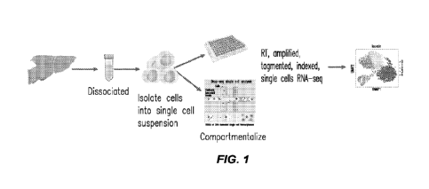
FIG. I depicts a general workflow of single cell RNAseq. This platform is
typically used
to study tissue transcriptomes of homogenized biopsies, which results in
averaged transcriptome
14
CA 03168485 2022- 8- 18
WO 2021/168455
PCT/US2021/019126
and loss of spatial information. However, the positional context of gene
expression is of key
importance to understanding tissue functionality and pathological changes.
FIG. 2 depicts the combinatorial indexing schematic of XYZeq. The combination
of
spatially informative RT-index and split-pool PCR-index makes it possible to
simultaneously
obtain transcriptome data at single cell resolution and assign each cell to a
specific well in the
array. Using two rounds of combinatorial barcoding, for example, first with
768 positional RT-
indices and second with 384 PCR-indices, up to 294,912 barcode combinations
can be generated.
FIG. 3 depicts the process by which the array for XYZeq is fabricated.
FIG. 4A-4C depict arrays with hexagonal shaped microwells used for the spatial
sequencing platform of the present disclosure. FIG. 4A: Array with 500-micron
microwells; FIG.
4B: array with 200-micron microwells; and FIG. 4C: array on a histology slide.
FIG. 5A-5E illustrate that XYZeq enables single cell and spatial transcriptome
profiling
simultaneously. FIG. SA: Schematic of the XYZeq workflow. FIG. 5B: Schematic
of XYZeq
sequencing library structure. P5 and P7: Illumina adaptors. bp: base pairs. R1
and R2: annealing
sites for Illumina sequencing primers. FIG. SC: Schematic representation of
the mixed species
cell gradient pattern printed on the chip with 11 unique cell proportion
ratios (see Methods in
Example 8 for specific cell proportion ratios). FIG. SD: Scatter plot of mouse
(x-axis) and human
(y-axis) UMI counts detected from a mixture of HEK293T and NII-I3 T3 cells
after computational
decontamination. Dark gray refers to human cells (n=4,182), gray refers to
mouse cells (n=2,220),
and light gray refers to collisions (n=45). FIG. 5E: Proportion of HEK293T
(blue) cells, NIH/3T3
(gray) cells or collisions (light gray) detected by XYZeq for each column of
the microwell array.
FIG. 6A-6C illustrate the high-resolution spatial resolution single cell RNA
capture from
tissue using XYZeq. FIG. 6A. Scatter plot of transcripts from human (n¨XX) and
mouse cells
(n=XX); FIG. 6B: Violin plot showing the number of detected UMIs and genes per
cell; FIG. 6C:
Cell distribution spatial map of human and mouse cells in the microarray.
FIG. 7A-7F show the quantification of specific cell types and gene expression
in tissue.
FIG. 7A: Annotated cell-identity clusters found by Louvain clustering
visualized in a UMAP
representation; cell expression to identify hepatocytes (Apoal), tumor (Plec),
macrophages
(Cd74), liver sinusoidal endothelial cells (Stab2), lymphocytes (Skapl),
Kupffer cells (Cd51), from
low expression (darker gray) to high expression (light gray). Marker genes may
be expressed also
in other cell identity populations as shown for macrophages and Kupffer cells;
FIG. 7B:
CA 03168485 2022- 8- 18
WO 2021/168455
PCT/US2021/019126
Correlation plot comparing XYZeq to 10X chromium; FIG. 7C: Violin plot
comparing UMI and
gene counts per cell for XYZeq and 10X; FIG. 7D: Heat map representation of
cell populations
between XYZeq and 10x; FIG. 7E: Spatial density plot showing localization of
each cell cluster
in the spatial array; FIG. 7F: Spatial pie chart representation that show the
ratio of each cell type
that occupy each well.
FIG. 8A-8B show identification of distinct cell populations found in liver
tumor model.
FIG. 8A. Annotated cell-identity clusters found by Leiden clustering
visualized in a UMAP
representation; FIG. 8B: Visualization of the overlap of gene expression
across the cell
populations (the size of the bubble for each gene correlates to the degree of
expression for the cell
type).
FIG. 9 shows a heat map representing genes that are differentially expressed
between cell-
type clusters with a log-fold change of at least 1.5. The colored bars on the
Y axis correspond to
the group of genes representative of that cellType cluster.
FIG. 10A-10G show gene information obtained from the spatial single cell data.
The
genes tested are a few top marker for lymphocytes and macrophages that showed
spatial variation.
FIG. 10C, FIG. 10D and FIG. 10G show psuedo time trajectory plots. Each dot
represents the
macrophage cells. Y axis is the log expression of the gene: in this case TGFbi
(FIG. 10C), CCR5
(FIG. 10D) or Tox (FIG. 10G). The horizontal dots on the bottom of FIG. 10C,
FIG. 10D and
FIG. 10G indicate macrophages that do not express that gene (the macrophages
with 0 counts for
Tgfbi). The line describes the trend of Tgfb expression across the distance
variable. Thus, it is
higher at the 0 distance (tumor), then decreases as it moves away (liver). The
purple and yellow
bar in FIG. 10A and FIG. 10E represents distance, which corresponds to the
spatial plot shown in
FIG. 10B and FIG. 10F. Yellow is liver, and purple and green are tumor
regions. The purple to
yellow bar in FIG. 10A and FIG. 10E is the scale/axis for the gene-expression
bars above (blue
to white). The purple to yellow is a representation the spatial map and the
dark blue to white is a
representation of the expression of genes in relation to space (specifically
tumor to liver).
FIG. 11A-11D show spatially resolved single cell transcriptomes captured from
tissue.
FIG. 11A: Scatter plot of mouse (x-axis) and human (y-axis) UI\4I counts
detected from
liver/tumor tissues (n=4) at 500 UMI cutoff after decontamination processing.
Dark gray on the
y-axis refers to human cells (n=2,657) and dark gray on the x-axis refers to
mouse cells (n=5,707)
and light gray refers to collisions (n=382). FIG. 11B: Violin plots showing
the number of detected
16
CA 03168485 2022- 8- 18
WO 2021/168455
PCT/US2021/019126
UMIS (left) and genes (right) per mouse and human cell. Median UMI counts for
human cells:
1,596; mouse cells: 1,009. Median gene counts for human cells: 629; mouse
cells: 456 across all
liver/tumor slices. FIG. 11C: Hematoxylin and eosin (H&E) stained image of the
liver/tumor
tissue slice. Tumor region (dark gray with light gray dotted outlines); Liver
region (light gray).
Scale showing 2mm. FIG. 110: Visualization of human (gray and dark gray) and
mouse (dark
gray) cell distribution on the XYZeq array overlayed on the H&E stained slice.
FIG. 12A-12F show frequency and spatial mapping of single cell clusters from
tissue.
FIG. 12A: t-distributed stochastic neighbor embedding (tSNE) visualization of
the cell types
identified from liver/tumor tissue. 6,623 total cells plotted. FIG. 12B: Heat
map of scaled marker
gene expression and hierarchical clustering of genes that define each cell
type from liver/tumor
tissue. Reference for grayscale bar in FIG. 12A. FIG. 12C: Correlations of
pseudobulk
expression values for matching cell types between XYZeq and 10X Genomics
Chromium. FIG.
12D: Spatial localization of hepatocytes, MC38 and myeloid cells overlaid on
brightfield image of
tissue. Light gray dotted outline indicates tumor regions. FIG. 12E: Pie chart
of cell type
composition for each XYZeq well from a representative liver/tumor tissue slice
(top panel) and
bar chart illustrating combined cell type composition across all four slices
of liver/tumor tissue,
which tracks with proximity to the tumor (bottom panel) (see Methods in
Example 8 for proximity
score). FIG. 12F: Pairplot showing the frequency of hepatocytes, MC38, and
myeloid cells in
each well. Scatter plots show the colocalization of two cell types in each
well. Histograms show
the distribution of number of cells (x-axis) per well (y-axis) for each cell
type. Pearson correlation
(r) and p values are annotated.
FIG. 13A-13F show expression of gene modules in space that track with cellular
composition. FIG. 13A. Projection of average expression of hepatocyte-enriched
module (LM14)
in tSNE space. Each dot is a cell and colored by the average expression of top
contributing module
genes (see Methods in Example 8). FIG. 13B: Spatial expression of hepatocyte-
enriched module
(LM14). Each spatial well is colored by the average expression of top
contributing module genes
weighted by the number of cells per well. Wells are binarized into high (above
weighted average)
versus low (all other non-zero expression). Light gray dotted outlines
indicate tumor regions.
FIG. 13C: Heat map representing the number of overlapping genes between each
pair of modules
in liver/tumor and spleen/tumor. Each row is a liver module and each column is
a spleen module.
FIG. 13D: tSNE projection of XYZeq scRNA-seq data grayscaled by annotated cell
types in
17
CA 03168485 2022- 8- 18
WO 2021/168455
PCT/US2021/019126
liver/tumor (top left) and spleen/tumor (bottom left) and mean gene expression
of the top
overlapping modules between liver/tumor (top row) and spleen/tumor (bottom
row). Tumor
response modules correspond to LM5 and SM12 and immune regulation modules
correspond to
LM19 and SM7. Projection in spatial coordinates the mean expression of the
tumor response
modules (FIG. 13E) corresponding to LM5 and SM12; and the immune regulation
modules (FIG.
13F) corresponding to LM19 and SM7. Each well in (FIG. 13E, FIG. 13F) are
grayscaled by the
average gene expression of each module weighted by the number of cells per
well (high vs low)
and light gray dotted outline indicates tumor regions. Wells are binarized
into high (above
weighted average) versus low (all other non-zero expression).
FIG. 14A-14F show differential gene expression within MSCs associated with
their spatial
proximity to tumor. FIG. 14A: Average expression of the cell migration modules
(LM10 and
SM17) in tSNE space. Each dot is a cell grayscaled by its mean expression of
top module genes
between corresponding liver and spleen modules. FIG. 14B: XYZeq array
grayscaled by the
tumor proximity score. Values near 1 (dark gray) indicate regions rich in
tumor, values near 0
(black) indicate regions rich in non-tumor cells, and wells capturing the
border between the two
tissue types take on values around 0.5 (draker gray). FIG. 14C: MSCs
grayscaled by the cell-
specific proximity score in tSNE space. FIG. 14D: Row-clustered heat map
showing the scaled,
mean gene expression in MSCs of genes enriched in three spatial regions (intra-
tumor, boundary,
intra-tissue) along the 1-dimensional proximity score. For spleen/tumor,
statistically significant
genes enriched in the tumor and non-tumor regions are highlighted. FIG. 14E:
Log expression
(y-axis) of Csnidl (left) and 1Shz2 (right) along the proximity score (x-
axis). Each dot corresponds
to one MSC cell and the regression line is fitted using the negative binomial
distribution (see
Methods in Example 8). FIG. 14F: Projection in space of mean expression of
Csincil (left) and
Tshz2 (right) in MSCs. Light gray dotted outline indicates tumor region.
FIG. 15A-15B show that single cell mixed species experiment reveals strong
correlation
to estimated cell gradient proportions. FIG. 15A: Scatter plot of mouse and
human UNII counts
detected from a mixture of HEK293T and N1H3T3 cells. Darker gray on the y-axis
refers to human
cells (n=4,389) and gray on the x-axis refers to mouse cells (n=1,728) and
light gray refers to
collisions (n=330). FIG. 15B: Scatter plot revealing high concordance between
observed and
expected cell type proportions in each column of the XYZeq array (Lin's
Concordance Correlation
= 0.91).
18
CA 03168485 2022- 8- 18
WO 2021/168455
PCT/US2021/019126
FIG. 16A-16C show quantification of cells captured per well from liver/tumor
tissue.
FIG. 16A: Image of the liver/tumor tissue slice on top of the XYZeq frozen
microarray with wells
with reagents spotted (white). FIG. 16B: Scatter plot of transcripts (n=4)
from human (draker
gray on the y-axis: n=2,667), mouse cells (gray on the x-axis: n=6,854), and
collisions (light gray:
n=747). FIG. 16C: Median cell number in wells across XYZeq array for HEK293T
human (top)
and liver/tumor mouse (bottom) cells.
FIG. 17A-17F show distinct cell types clusters identified from XYZeq of
liver/tumor
tissue. FIG. 17A: tSNE visualization of Leiden cluster to annotated cell
types. FIG. 17B:
Correlation of mean chromosomal expression of MC38 cells observed in XYZeq
compared to
MC38 cells from a Efremova, et al. (25), hepatocytes from Tabula Muris (26),
and immune cells
enriched from liver/tumor from an independent internal experiment (3). Both
the x-axis and y-
axis denotes average expression of all genes on a given chromosome. FIG. 17C:
Violin plot
representing estimated contamination fraction for each cell type from our
liver/tumor XYZeq data
(FIG. 17D, FIG. 17E) Violin plot showing the number of detected UIVIIs and
genes per cell cluster.
Median UI\4I counts (log) and gene counts for each cell cluster: hepatocytes
(3.04 and 552),
Kupffer cells (2.92 and 420), lymphocytes (2.97 and 454), MSCs (3.08 and 594),
macrophages
(3.03 and 511), MC38 (3.22 and 851), and LSECs (2.94 and 431). FIG. 17F:
Annotated cell-
identity clusters; Feature plot of cells that are positive for each individual
marker gene to identify
Hepatocytes (Cps], Glut), MC38 (Plec), macrophages (Cd 1 b , (]d74), liver
sinusoidal endothelial
cells (Stab2, Ptprb), lymphocytes (Cd8b, Il I 8r I), Kupffer cells (Cd51,
Timd4), m e sen ch ym al stem
cells (1?bms3, Tshz2), pericentral hepatocytes (Gin!, Ghia, Oat) from low
expression (black) to
high expression (light gray).
FIG. 18A-18B show reproducibility of XYZeq across tissue slices. Four non-
sequential
z-layer slices of liver/tumor tissue processed with XYZeq (with HEK293T cells
spiked-in as
control). FIG. 18A: Pairplot showing the expression of common genes between
different slices
of liver/tumor. Scatter plots show the UMI counts for common expressed (UMIs >
0) genes.
Histograms show the distribution of number of UMIs (x-axis) per gene (y-axis)
for each slice.
FIG. 18B: tSNE visualization of Leiden clusters across four slices.
FIG. 19A-19B show that cell type clusters captured from XYZeq found comparable
to
10X Genomics platform. FIG. 19A: tSNE representation of liver/tumor tissue
data generated with
the 10X Chromium V3 kit. 2,703 total cells were plotted. FIG. 19B: Scatter
plot comparing the
19
CA 03168485 2022- 8- 18
WO 2021/168455
PCT/US2021/019126
proportion of each cell type found in XYZeq and 10X Chromium V3. Lin's
concordance
coefficient of 0.988.
FIG. 20A-20B show distinct spatial localization pattern across tissue for each
cell type
cluster. FIG. 20A: Spatial density plot showing localization of lymphocytes,
MSCs, Kupffer cells
and LSECs in the spatial array. Light gray dotted outline indicates tumor
region. FIG. 20B:
Pairplot showing the frequency of cell types found in each well across the
XYZeq array. Scatter
plots show the co-localization of the cell types in each well. Histograms show
the distribution for
number of cells (x-axis) per well (y-axis) for each cell type. r and p values
annotated.
FIG. 21A-21F show that XYZeq of spleen/tumor tissue reveals comparable data
quality to
liver/tumor tissue. FIG. 21A: Scatter plot of mouse and human UMI counts
detected from
spleen/tumor tissues (n=4). Drak gray on the y-axis refers to human cells
(n=4,007) and gray on
the X-axis refers to mouse cells (n=3,394) and light gray refers to collisions
(n=104). FIG. 21B:
Violin plot showing the number of detected UMIs and genes per cell. Median UMI
counts for
human cells: 1,312; mouse cells: 1,169. Median gene counts for human cells:
661; mouse cells:
577. FIG. 21C: H&E stained image of the spleen/tumor tissue slice. Tumor
region (gray area
with light gray dotted outline); spleen region (darker gray with dark gray
features). Scale showing
2 mm. FIG. 21D: Image of spleen/tumor tissue on frozen XYZeq microarray with
reagents in
wells (white). FIG. 21E: Visualization of human (gray and dark gray) and mouse
(gray and dark
gray) cell distribution on the XYZeq array with 500 UMI cutoff overlaid on the
image of H&E
stained tissue slice. FIG. 21F: Median cell number in wells across XYZeq array
for HEK293T
human (top) and spleen/tumor mouse (bottom) cells
FIG. 22A-22D show identification and spatial mapping of cell type clusters
from
spleen/tumor tissue. FIG. 22A: tSNE projection of the spleen/tumor XYZeq data.
3,394 total
cells were plotted. FIG. 22B: tSNE visualization of Leiden cluster to annotate
cell types for
spleen/tumor. FIG. 22C: Heat map of a scaled expression of marker genes and
hierarchical
clustering that define each cell type from XYZeq spleen/tumor tissue. FIG.
22D: Image of
spleen/tumor tissue overlaid with spatial plot of the XYZeq array showing
localization of cell type
clusters from (FIG. 22A) with 500 UMI cutoff Light gray dotted outline
indicates tumor region.
FIG. 23A-23D show cell type contribution and functional annotation of gene
modules.
FIG. 23A: Barplot showing percent fraction of overlapping genes in liver/tumor
modules
compared to corresponding spleen/tumor modules. Dotted line represents the
threshold used to
CA 03168485 2022- 8- 18
WO 2021/168455
PCT/US2021/019126
determine significant overlap between the modules. FIG. 23B: Pie chart
representation of cell
type fractions that make up each module (see Methods in Example 8). LM denotes
liver/tumor
module (FIG. 23C, FIG. 23D). GO annotations for tumor response modules (FIG.
23C) and
immune regulation modules (FIG. 23D). GO enrichment analysis for immune
response module
is represented by LM19. p-values computed using GOrilla (50) and adjusted by
Benjamini-
Hochberg correction.
FIG. 24A-24B show expression of the cell migration gene module enriched in
MSCs.
FIG. 24A: Matrix plot of top overlapping genes in the cell migration module
(LM10) across all
cell types in liver/tumor. FIG. 24B: GO annotation for cell migration module
from LM10 and
SM17. p-values computed using GOrilla (50) and adjusted by Benjamini-Hochberg
correction.
FIG. 25A-25E show tumor proximity score defined for both liver and spleen
tissue. FIG.
25A: The proximity scores for each tissue relied on the annotation of
successive concentric layers
of neighbors for a well in question. FIG. 25B: The set of wells neighboring
each well in the array
were tabulated for up to 10 layers. FIG. 25C: The cell-containing wells of
representative
spleen/tumor slice, where white to lighter gray indicates a higher proportion
of tumor cells, and
darker gray indicates a higher proportion of non-tumor cells. The wells
selected for setting the
proximity score to I are outlined in white. FIG. 25D: The cell containing
wells of a representative
liver/tumor slice. Light gray indicates higher proportion of tumor cells, gray
to darker gray
indicates higher proportion of hepatocytes. FIG. 25E: The proximity score
values annotated on
each well (left), where lighter gray is closer to the minimum value and darker
gray is closer to the
maximum value. The scores are visualized for different values of 1 and d. The
values of 1 = 10
and d = 1.05 were chosen as they rendered the distribution of scores (right)
more uniform across
all wells.
DETAILED DESCRIPTION OF EMBODIMENTS
The present disclosure can be understood more readily by reference to the
following
detailed description of embodiments, the figures and the examples included
herein.
Before the present methods and compositions are disclosed and described, it is
to be
understood that they are not limited to specific synthetic methods unless
otherwise specified, or to
particular reagents unless otherwise specified, as such may, of course, vary.
It is also to be
understood that the terminology used herein is for the purpose of describing
particular aspects only
21
CA 03168485 2022- 8- 18
WO 2021/168455
PCT/US2021/019126
and is not intended to be limiting. Although any methods and materials similar
or equivalent to
those described herein can be used in the practice or testing of the present
invention, example
methods and materials are now described.
Moreover, it is to be understood that unless otherwise expressly stated, it is
in no way
intended that any method set forth herein be construed as requiring that its
steps be performed in
a specific order. Accordingly, where a method claim does not actually recite
an order to be
followed by its steps or it is not otherwise specifically stated in the claims
or descriptions that the
steps are to be limited to a specific order, it is in no way intended that an
order be inferred, in any
respect. This holds for any possible non-express basis for interpretation,
including matters of logic
with respect to arrangement of steps or operational flow, plain meaning
derived from grammatical
organization or punctuation, and the number or type of aspects described in
the specification.
All publications mentioned herein are incorporated herein by reference to
disclose and
describe the methods and/or materials in connection with which the
publications are cited. The
publications discussed herein are provided solely for their disclosure prior
to the filing date of the
present application. Nothing herein is to be construed as an admission that
the present invention
is not entitled to antedate such publication by virtue of prior invention.
Further, the dates of
publication provided herein can be different from the actual publication
dates, which can require
independent confirmation.
Definitions
Unless defined otherwise, all technical and scientific terms used herein have
the same
meaning as commonly understood by one of ordinary skill in the art to which
the invention
pertains.
As used in the specification and in the claims, the term "comprising" can
include the
aspects "consisting of" and "consisting essentially of." Comprising can also
mean "including but
not limited to.-
As used in the specification and the appended claims, the singular forms "a,"
"an" and
"the" can include plural referents unless the context clearly dictates
otherwise. Thus, for example,
reference to "a compound" includes mixtures of compounds; reference to "a
pharmaceutical
carrier" includes mixtures of two or more such carriers, and the like.
The word "or" as used herein means any one member of a particular list and
also includes
any combination of members of that list.
22
CA 03168485 2022- 8- 18
WO 2021/168455
PCT/US2021/019126
The term "about" is used herein to mean within the typical ranges of
tolerances in the art.
For example, "about" can be understood as about 2 standard deviations from the
mean. According
to certain embodiments, when referring to a measurable value such as an amount
and the like,
"about" is meant to encompass variations of 20%, 10%, 5%, 1%, 0.9%,
0.8%, 0.7%,
0.6%, 0.5%, 0.4%, 0.3%, 0.2% or 0.1% from the specified value as such
variations are
appropriate to perform the disclosed methods. When "about" is present before a
series of numbers
or a range, it is understood that "about" can modify each of the numbers in
the series or range.
As used herein, the term "activated substrate" relates to a material on which
interacting or
reactive chemical functional groups were oxidated or reduced or otherwise
funtionalized by
exposure to reagents known to the person skilled in the art to prime the
surface for a reaction at
the functional group. For example, a substrate comprising carboxyl groups has
to be activated
before use. Furthermore, there are substrates available that contain
functional groups that can react
with specific moieties already present in the nucleic acid primers.
As used herein the term "a plurality of' or "multiple" means two or more, or
at least two,
such as 3, 5, 10, 15, 20, 30, 40, 50, 60, 70, 80, 90, 100, 150, 200, 400, 500,
1000, 2000, 5000,
10,000, or more. Thus, for example, the number of microwells on an array or
the number of wells
on a multiwell plate may be any integer in any range between any two of the
aforementioned
numbers.
As used herein, a "cellular index primer" refers to a primer or an oligo for
amplifying the
cDNA molecules obtained from reverse transcription and labelling each of the
amplified cDNA
molecules with a second index barcode that is unique to each well of a
multiwell plate (defined
herein as cellular barcode domains).
As used herein, a "spatial index primer" refers to a primer or an oligo for
capturing and
labelling transcripts from all of the single cells located at a distinct
position in the tissue sample,
such as a thin tissue sample slice, or "section."
An "array," as that term is used herein, typically refers to an arrangement of
entities in
spatially discrete locations with respect to one another, and usually in a
format that permits
simultaneous exposure of the arranged entities to potential interaction
partners (e.g., cells) or other
reagents, substrates, etc. In some embodiments, an array comprises a solid
substrate such as a
plastic comprising adjacently arranged microwells in spatially discrete
locations on the solid
support. In some embodiments, spatially discrete locations on an array are
termed "microwells"
23
CA 03168485 2022- 8- 18
WO 2021/168455
PCT/US2021/019126
or "spots" (regardless of their shape). In some embodiments, spatially
discrete locations on an
array are arranged in a regular pattern with respect to one another (e.g., in
a grid). In some
embodiments, the array comprise from about 90 to about 400 micrwells arranged
in adjacent
positions along the planar surface of a solide substrate. In some embodiments,
the array is a
mi croarray plate.
The term "barcode" as used herein refers to any unique, non-naturally
occurring, nucleic
acid sequence capable of identifying the originating source of a nucleic acid
fragment. In some
embodiments the basrcode is a unique, non-naturally occurring, nucleic acid
sequence
corresponding to at least one spatial position on an array, such that the
barcodes position on the
array also corresponds with a position of the cell or cells in contact with
that position.
The term "binding" isused broadly throughout this disclosure to refer to any
form of
attaching or coupling, either non-covalently or covalently, two or more
components, entities, or
objects. For example, two or more components may be bound to each other via
chemical bonds,
covalent bonds, ionic bonds, hydrogen bonds, electrostatic forces, Watson-
Crick hybridization,
etc.In the context of complenmentary nucleic acid seqeunces, two complementary
strands bind to
form a hydrogen bound duplex of nucleic acid.
The terms "polynucleotide," "oligo", "oligonucleotide" and "nucleic acid" are
used
interchangeably throughout and include DNA molecules (e.g., cDNA or genomic
DNA), RNA
molecules (e.g., mRNA), analogs of the DNA or RNA generated using nucleotide
analogs (e.g.,
peptide nucleic acids and non-naturally occurring nucleotide analogs), and
hybrids thereof, The
nucleic acid molecule can be single-stranded or double-stranded. In some
embodiments, the
nucleic acid molecules of the disclosure comprise a contiguous open reading
frame encoding an
antibody, or a fragment thereof, as described herein. "Nucleic acid" or
"oligonucleotide" or
"polynucleotide" as used herein may mean at least two nucleotides covalently
linked together. The
depiction of a single strand also defines the sequence of the complementary
strand. Thus, a nucleic
acid also encompasses the complementary strand of a depicted single strand.
Many variants of a
nucleic acid may he used for the same purpose as a given nucleic acid. Thus, a
nucleic acid also
encompasses substantially identical nucleic acids and complements thereof. A
single strand
provides a probe that may hybridize to a target sequence under stringent
hybridization conditions.
Thus, a nucleic acid also encompasses a probe that hybridizes under stringent
hybridization
conditions. Nucleic acids may be single stranded or double stranded, or may
contain portions of
24
CA 03168485 2022- 8- 18
WO 2021/168455
PCT/US2021/019126
both double stranded and single stranded sequence. The nucleic acid may be
DNA, both genomic
and cDNA, RNA, or a hybrid, where the nucleic acid may contain combinations of
deoxyribo- and
ribo-nucleotides, and combinations of bases including uracil, adenine,
thymine, cytosine, guanine,
inosine, xanthine hypoxanthine, isocytosine and isoguanine Nucleic acids may
be obtained by
chemical synthesis methods or by recombinant methods. A nucleic acid will
generally contain
phosphodiester bonds, although nucleic acid analogs maybe included that may
have at least one
different linkage, e.g., phosphoramidate, phosphorothioate,
phosphorodithioate, or o-
methylphosphoroamidite linkages and peptide nucleic acid backbones and
linkages. Other analog
nucleic acids include those with positive backbones, non-ionic backbones, and
non-ribose
backbones, including those described in U.S. Pat. Nos. 5,235,033 and
5,034,506, which are
incorporated by reference in their entireties. Nucleic acids containing one or
more non-naturally
occurring or modified nucleotides are also included within one definition of
nucleic acids. The
modified nucleotide analog may be located for example at the 5'-end and/or the
3'-end of the
nucleic acid molecule. Representative examples of nucleotide analogs may be
selected from
sugar- or backbone-modified ribonucleotides. It should be noted, however, that
also nucleobase-
modified ribonucleotides, i.e. ribonucleotides, containing a non-naturally
occurring nucleobase
instead of a naturally occurring nucleobase such as uridines or cytidines
modified at the 5-position,
e.g. 5-(2-amino)propyl uridine, 5-bromo uridine; adenosines and guanosines
modified at the 8-
position, e.g. 8-bromo guanosine; deaza nucleotides, e.g. 7-deaza-adenosine; o-
and N-alkylated
nucleotides, e.g. N6-methyl adenosine are suitable. The 2'-OH-group may be
replaced by a group
selected from H, OR, R, halo, SH, SR, NH2, NUR, N2 or CN, wherein R is C -C6
alkyl, alkenyl or
alkynyl and halo is F, Cl, Br or I. Modified nucleotides also include
nucleotides conjugated with
cholesterol through, e.g., a hydroxyprolinol linkage as described in
Krutzfeldt et al., Nature (Oct.
30, 2005), Soutschek et al., Nature 432:173-178 (2004), and U.S. Patent
Publication No.
20050107325, which are incorporated herein by reference in their entireties.
Modified nucleotides
and nucleic acids may also include locked nucleic acids (LNA), as described in
U.S. Patent No.
20020115080, which is incorporated herein by reference. Additional modified
nucleotides and
nucleic acids are described in U.S. Patent Publication No. 20050182005, which
is incorporated
herein by reference in its entirety. Modifications of the ribose-phosphate
backbone may be done
for a variety of reasons, e.g., to increase the stability and half-life of
such molecules in
physiological environments, to enhance diffusion across cell membranes, or as
probes on a
CA 03168485 2022- 8- 18
WO 2021/168455
PCT/US2021/019126
biochip. Mixtures of naturally occurring nucleic acids and analogs may be
made; alternatively,
mixtures of different nucleic acid analogs, and mixtures of naturally
occurring nucleic acids and
analogs may be made. In some embodiments, the expressible nucleic acid
sequence is in the form
of DNA. In some embodiments, the expressible nucleic acid is in the form of
RNA with a sequence
that encodes the polypeptide sequences disclosed herein and, in some
embodiments, the
expressible nucleic acid sequence is an RNA/DNA hybrid molecule that encodes
any one or
plurality of polypeptide sequences disclosed herein.
The "percent identity" or "percent homology" of two polynucleotide or two
polypeptide
sequences is determined by comparing the sequences using the GAP computer
program (a part of
the GCG Wisconsin Package, version 10.3 (Accelrys, San Diego, Calif.)) using
its default
parameters. "Identical" or "identity" as used herein in the context of two or
more nucleic acids or
amino acid sequences, may mean that the sequences have a specified percentage
of residues that
are the same over a specified region. The percentage may be calculated by
optimally aligning the
two sequences, comparing the two sequences over the specified region,
determining the number
of positions at which the identical residue occurs in both sequences to yield
the number of matched
positions, dividing the number of matched positions by the total number of
positions in the
specified region, and multiplying the result by 100 to yield the percentage of
sequence identity. In
cases where the two sequences are of different lengths or the alignment
produces one or more
staggered ends and the specified region of comparison includes only a single
sequence, the residues
of single sequence are included in the denominator but not the numerator of
the calculation When
comparing DNA and RNA, thymine (T) and uracil (U) may be considered
equivalent. Identity
may he performed manually or by using a computer sequence algorithm such as
BLAST or BLAST
2Ø Briefly, the BLAST algorithm, which stands for Basic Local Alignment
Search Tool is
suitable for determining sequence similarity. Software for performing BLAST
analyses is publicly
available through the National Center for Biotechnology Information
(ncbi.nlm.nih.gov). This
algorithm involves first identifying high scoring sequence pair (HSPs) by
identifying short words
of length Win the query sequence that either match or satisfy some positive-
valued threshold score
T when aligned with a word of the same length in a database sequence. T is
referred to as the
neighborhood word score threshold (Altschul et al.). These initial
neighborhood word hits act as
seeds for initiating searches to find HSPs containing them. The word hits are
extended in both
directions along each sequence for as far as the cumulative alignment score
can be increased.
26
CA 03168485 2022- 8- 18
WO 2021/168455
PCT/US2021/019126
Extension for the word hits in each direction are halted when: 1) the
cumulative alignment score
falls off by the quantity X from its maximum achieved value; 2) the cumulative
score goes to zero
or below, due to the accumulation of one or more negative-scoring residue
alignments; or 3) the
end of either sequence is reached. The Blast algorithm parameters W, T and X
determine the
sensitivity and speed of the alignment. The Blast program uses as defaults a
word length (W) of
11, the BL0S1J1V162 scoring matrix (see Henikoff et al., Proc. Natl. Acad.
Sci. USA, 1992, 89,
10915-10919, which is incorporated herein by reference in its entirety)
alignments (B) of 50,
expectation (E) of 10, M=5, N=4, and a comparison of both strands. The BLAST
algorithm (Karlin
et al., Proc. Natl. Acad. Sci. USA, 1993, 90, 5873-5787, which is incorporated
herein by reference
in its entirety) and Gapped BLAST perform a statistical analysis of the
similarity between two
sequences. One measure of similarity provided by the BLAST algorithm is the
smallest sum
probability (P(N)), which provides an indication of the probability by which a
match between two
nucleotide sequences would occur by chance. For example, a nucleic acid is
considered similar to
another if the smallest sum probability in comparison of the test nucleic acid
to the other nucleic
acid is less than about 1, less than about 0.1, less than about 0.01, and less
than about 0.001. Two
single-stranded polynucleotides are "the complement" of each other if their
sequences can be
aligned in an anti-parallel orientation such that every nucleotide in one
polynucleotide is opposite
its complementary nucleotide in the other polynucleotide, without the
introduction of gaps, and
without unpaired nucleotides at the 5' or the 3' end of either sequence. A
polynucleotide is
"complementary" to another polynucleotide if the two polynucleoti des can
hybridize to one
another under moderately stringent conditions. Thus, a polynucleotide can be
complementary to
another polynucleotide without being its complement.
By "substantially identical" is meant nucleic acid molecule (or polypeptide)
exhibiting at
least 50% identity to a reference amino acid sequence (for example, any one of
the amino acid
sequences described herein) or nucleic acid sequence (for example, any one of
the nucleic acid
sequences described herein). Preferably, such a sequence is at least 60%, more
preferably 80% or
85%, and more preferably 90%, 95% or even 99% identical at the amino acid
level or nucleic acid
to the sequence used for comparison.
The term "hybridization" or "hybridizes" as used herein refers to the
formation of a duplex
between nucleotide sequences that are sufficiently complementary to form
duplexes via Watson-
Crick base pairing. Two nucleotide sequences are "complementary" to one
another when those
27
CA 03168485 2022- 8- 18
WO 2021/168455
PCT/US2021/019126
Molecules share base pair organization homology. "Complementary" nucleotide
sequences will
combine with specificity to form a stable duplex under appropriate
hybridization conditions. For
instance, two sequences are complementary when a section of a first sequence
can bind to a section
of a second sequence in an anti-parallel sense wherein the 3'-end of each
sequence binds to the 5'-
end of the other sequence and each A, T(U), G and C of one sequence is then
aligned with a T(U),
A, C and G, respectively, of the other sequence. RNA sequences can also
include complementary
G=U or U=G base pairs. Thus, two sequences need not have perfect homology to
be
"complementary." Usually two sequences are sufficiently complementary when at
least about
90% (preferably at least about 95%) of the nucleotides share base pair
organization over a defined
length of the molecule. In the present disclosure, the capture domain of each
spatial index primer
comprises a region of complementarity for the nucleic acid, e.g. RNA
(preferably mRNA) of the
tissue sample. In some embodiments, such a region of complementarity comprised
in the capture
domain of each spatial index primer comprises a polythymidine sequence to
capture mRNA via
the poly-A tail.
As used herein, the term "sample" refers to a biological sample obtained or
derived from a
source of interest, as described herein. In some embodiments, a source of
interest comprises an
organism, such as an animal or human. In some embodiments, a biological sample
comprises
biological tissue or bodily fluid. In some embodiments, a biological sample
may be or comprise
bone marrow; blood; blood cells; ascites; tissue or fine needle biopsy
samples; cell-containing
body fluids; free floating nucleic acids; sputum; saliva; urine; cerebrospinal
fluid, peritoneal fluid;
pleural fluid; feces; lymph; gynecological fluids; skin swabs; vaginal swabs;
oral swabs; nasal
swabs; washings or lavages such as a ductal lavages or broncheoalveolar
lavages; aspirates;
scrapings, bone marrow specimens, tissue biopsy specimens, surgical specimens;
other body
fluids, secretions, and/or excretions; and/or cells therefrom, etc. In some
embodiments, a
biological sample is or comprises cells obtained from an individual. In some
embodiments, a
sample is a "primary sample" obtained directly from a source of interest by
any appropriate means.
For example, in some embodiments, a primary biological sample is obtained by
methods selected
from the group consisting of biopsy (e.g., fine needle aspiration or tissue
biopsy), surgery,
collection of body fluid (e.g., blood, lymph, feces etc.), etc. In some
embodiments, as will be clear
from context, the term "sample" refers to a preparation that is obtained by
processing (e.g., by
removing one or more components of and/or by adding one or more agents to) a
primary sample.
28
CA 03168485 2022- 8- 18
WO 2021/168455
PCT/US2021/019126
For example, filtering using a semi-permeable membrane. Such a "processed
sample- may
comprise, for example nucleic acids or proteins extracted from a sample or
obtained by subjecting
a primary sample to techniques such as amplification or reverse transcription
of mRNA, isolation
and/or purification of certain components, such as organelles, nucleic acid or
membrane-bound
proteins. In some embodiments, sample is a tissue comprising a plurality of
cell types. In some
embodiments, sample is connective tissue, muscle tissue, nervous tissue, or
epithelial tissue.
The term "amplification reaction" as used herein refers to a reaction by which
the number
of copies of a nucleic acid is increased. This may be conducted through
methods such as
polymerase chain reaction (PCR), including but not limited to qPCR, RT-qPCR,
RACE-PCR and
RT-LAMP, ligase chain reaction (LCR), transcription-mediated amplification,
and nicking
enzyme amplification reaction (NEAR). Any variation of the aforementioned
methodologies for
amplifying a nucleic acid is also encompassed by this term.
As used herein, the term "insertional enzyme" refers to an enzyme capable of
inserting a
nucleic acid sequence into a polynucleotide. In some cases, the insertional
enzyme can insert the
nucleic acid sequence into the polynucleotide in a substantially sequence-
independent manner.
The insertional enzyme can be prokaryotic or eukaryotic. Examples of
insertional enzymes
include, but are not limited to, transposases, HER_MES, and HIV integrase. The
transposase can
be a Tn transposase (e.g., Tn3, Tn5, Tn7, Tn10, Tn552, Tn903), a MuA
transposase, a Vibhar
transposase (e.g., from Vibrio harveyi), Ac-Ds, Ascot-1, Bsl, Cin4, Copia,
En/Spm, F element,
hobo, Hsmarl, Hsmar2, IN (HIV), IS1, IS2, IS3, IS4, IS5, IS6, IS10, IS21,
IS30, IS50, IS51,
IS150, IS256, IS407, IS427, IS630, IS903, IS911, IS982, IS1031, ISL2, Li,
Mariner, P element,
Tam3, Tcl, Tc3, Tel, THE-1, Tn/O, TnA, Tn3, Tn5, Tn7, Tn10, Tn552, Tn903,
Toll, To12, Tnl 0,
Tyl, any prokaryotic transposase, or any transposase related to and/or derived
from those listed
above. In certain instances, a transposase related to and/or derived from a
parent transposase can
comprise a peptide fragment with at least about 50%, about 55%, about 60%,
about 65%, about
70%, about 75%, about 80%, about 85%, about 90%, about 91%, about 92%, about
93%, about
94%, about 95%, about 96%, about 97%, about 98%, or about 99% amino acid
sequence homology
to a corresponding peptide fragment of the parent transposase. The peptide
fragment can be at
least about 10, about 15, about 20, about 25, about 30, about 35, about 40,
about 45, about 50,
about 60, about 70, about 80, about 90, about 100, about 150, about 200, about
250, about 300,
about 400, or about 500 amino acids in length. For example, a transposase
derived from Tn5 can
29
CA 03168485 2022- 8- 18
WO 2021/168455
PCT/US2021/019126
comprise a peptide fragment that is 50 amino acids in length and about 80%
homologous to a
corresponding fragment in a parent Tn5 transposase. In some cases, the
insertion can be facilitated
and/or triggered by addition of one or more cations. The cations can be
divalent cations such as,
for example, Ca2 , Mg2+ and Mn2 .
In some embodiments, the transposase is a DDE motif transposase such as a
prokaryotic
transposase from ISs, Tn3, Tn5, Tn7, orTn10; a bacteriophage transposase from
phage Mu; or a
eukaryotic "cut and paste" transposase. U.S. Pat. Nos. 6,593,113; 9,644,199;
Yuan and Wessler
(2011) Proc Natl Acad Sci USA 108(19).7884-7889. In some embodiments, the
transposase
includes a retroviral transposase, such asHIV. Rice and Baker (2001) Nat
Struct Biol. 8: 302-307.
In some embodiments, the transposase is a member of the IS50 family of
transposases,
such as Tn5 transposase or variants of Tn5 transposase. Tn5 transposase is
derived from the Tn5
transposon, a bacterial transposon that can encode antibiotic resistance
genes. The activity of Tn5
transposase can be increased with the point mutations E54K and/or L372P. In
particular
embodiments, the transposase is a E54K/L372P mutant of Tn5 transposase, which
has increased
transposase activity. An exemplary E54K/L372P Tn5 transposase comprises the
following
sequence:
MITSALHRAADWAKSVFSSAALGDPRRTARLVNVAAQLAKYSGKSITISS
EGSKAMQEGAYRFMNPNVSAEAIRKAGAMQTVKLAQEFPELLAIEDTT S
LSYRHQVAEELGKLGSIQDKSRGWWVHSVLLLEATTFRTVGLLHQEWW
MRPDDP AD ADEKES GKWL A A A AT SRLRMG SMM SNVIA VCDRE A DIH A Y
LQDKLAFINERFVVRSKHPRKDVESGLYLYDHLKNQPELGGYQISIPQKG
VVDKRGKRKNRPARKASL SLR S GRITLKQGNITLNAVLAEEINPPKGETPL
KWLLLT SEPVESLAQALRVIDIYTHRWRIEEFHKAWKTGAGAERQRMEEP
DNLERMVSIL S FVAVRLLQLRE SF TPPQALRAQGLLKEAEHVE S Q SAETV
LTPDEC QLLGYLDKGKRKRKEKAGSLQWAYMAIARLGGFMD SKRTGIAS
WGALW (SEQ ID NO: 42)
Other mutations to increase the activity of Tn5 transposase are disclosed in
U.S. Pat. Nos.
5,965,443; 6,406,896; 7,608,434; and Reznikoff (2003) Molecular Microbiology
47(5): 1199-
1206, all of which are expressly incorporated by reference herein. In some
embodiments, the Tn5
transposase is a mutant transposase (Tn5-059) with a lowered GC insertion
bias. Kia et al. (2017)
BMC Biotechnology 17: 6.
CA 03168485 2022- 8- 18
WO 2021/168455
PCT/US2021/019126
Methods
As mentioned above, methods of the present disclosure relate to a method of
the integration
of split-pool indexing and spatial barcoding. Thus, the present disclosure
uses a set of barcoded
index primers to obtained single cell gene expression profiling or
transcriptomes from a tissue
sample while preserving their corresponding spatial information.
The present disclosure thus relates to a method of spatial recognition of gene
expression,
the method comprising identifying the presence, absence or quantity of a
combination of a spatial
barcode domain and a cellular barcode domain in a nucleic acid sample by
dtetcing the domain or
domains in a sample. In some embodiments, the method further comprises
correlating the
presence, absence or quantity of the spatial barcode domain and the cellular
barcode domain to a
spatial position of a cell in a tissue sample on an array.
The present disclosure also relates to a method of identifying a cell type in
a sample based
on spatial gene expressing profiling, the method comprising detecting the
presence, absence or
quantity of a combination of a spatial barcode domain and a cellular barcode
domain in a sample.
In some embodiments, the method further comprises correlating the presence,
absence or quantity
of the spatial barcode domain and the cellular barcode domain to a spatial
position of a cell in a
tissue sample on an array. In some embodiments, the step detecting the
presence, absence or
quantity of a combination of a spatial barcode domain and a cellular barcode
domain in a sample
comprises annealing one or a plurality of complemtary nucleic acids to the
cellular barcode domain
and/or the spatial barcode domain and performing a polymerase chain reaction
on the sequences
to identify the presence or quantity of the one or both domains.
The present disclosure further relates to a method of identifying chromatin
accessibility in
a cell of a sample, the method comprising identifying the presence, absence or
quantity of a
combination of a spatial barcode domain and a cellular barcode domain in a
nucleic acid sample.
In some embodiments, the method further comprises correlating the presence,
absence or quantity
of the spatial barcode domain and the cellular barcode domain to a spatial
position of a cell in a
tissue sample on an array.
The present disclosure additionally relates to a method of spatially barcoding
a single cell
in a tissue, the method comprising identifying or detcting the presence,
absence or quantity of a
combination of a spatial barcode domain and a cellular barcode domain in a
nucleic acid sample.
31
CA 03168485 2022- 8- 18
WO 2021/168455
PCT/US2021/019126
In some embodiments, the method further comprises correlating the presence,
absence or quantity
of the spatial barcode domain and the cellular barcode domain to a spatial
position of a cell in a
tissue sample on an array. In some embodiments, the step of detecting
comprises detecting a
tluorcent signal or probe covalently or non-covalently bound to one or both
domains; or detecting
one or a plurality of copes of
The present disclosure also relates to a method of spatially identifying a
cell population
within a tissue, the method comprising identifying the presence, absence or
quantity of a
combination of a spatial barcode domain and a cellular barcode domain in a
nucleic acid sample.
In some embodiments, the method further comprises correlating the presence,
absence or quantity
of the spatial barcode domain and the cellular barcode domain to a spatial
position of a cell in a
tissue sample on an array.
The present disclosure further relates to a method of detecting gene
expression in a single
cell in a tissue, the method comprising identifying the presence, absence or
quantity of a
combination of a spatial barcode domain and a cellular barcode domain in a
nucleic acid sample.
In some embodiments, the method further comprises correlating the presence,
absence or quantity
of the spatial barcode domain and the cellular barcode domain to a spatial
position of a cell in a
tissue sample on an array.
The present disclosure also relates to a method of isolating cells
corresponding to a spatial
position within a tissue, the method comprising identifying the presence,
absence or quantity of a
combination of a spatial barcode domain and a cellular barcode domain in a
nucleic acid sample
In some embodiments, the method further comprises correlating the presence,
absence or quantity
of the spatial barcode domain and the cellular barcode domain to a spatial
position of the cell in
the tissue on an array.
The present disclosure additionally relates to a method of detecting a
meschymal stem cell
in an organ, the method comprising identifying the presence, absence or
quantity of a combination
of a spatial barcode domain and a cellular barcode domain in a nucleic acid
sample. In some
embodiments, the method further comprises correlating the presence, absence or
quantity of the
spatial barcode domain and the cellular barcode domain to a spatial position
of a meschymal stem
cell in a tissue sample of the organ on an array.
The present disclosure further relates to a method of quantifying RNA
expression in a
single cell, the method comprising identifying the presence, absence or
quantity of a combination
32
CA 03168485 2022- 8- 18
WO 2021/168455
PCT/US2021/019126
of a spatial barcode domain and a cellular barcode domain in a nucleic acid
sample. In some
embodiments, the method further comprises correlating the presence, absence or
quantity of the
spatial barcode domain and the cellular barcode domain to a spatial position
of the single cell in a
tissue sample on an array.
The present disclosure also relates to a method of quantifying RNA expression
corresponding to a spatial position within a tissue sample, the method
comprising identifying the
presence, absence or quantity of a combination of a spatial barcode domain and
a cellular barcode
domain in a nucleic acid sample. In some embodiments, the method further
comprises correlating
the presence, absence or quantity of the spatial barcode domain and the
cellular barcode domain
to a spatial position of the RNA expression in a tissue sample on an array.
The present disclosure also relates to a method of preparing a nucleic acid of
a single cell
within a tissue sample, the method comprising identifying the presence,
absence or quantity of a
combination of a spatial barcode domain and a cellular barcode domain in a
nucleic acid sample.
In some embodiments, the method further comprises correlating the presence,
absence or quantity
of the spatial barcode domain and the cellular barcode domain to a spatial
position of the nucleci
acid sample in the tissue sample on an array.
The disclosure relates to a method of obtaining the transcriptome of a single
cell
comprising:
(a) contacting a sample to an array, said array comprising multiple wells
comprising one
or a plurality of spatial primers and/or barcodes;
(b) isolating RNA from the sample in each well;
(c) performing quantitative PCR on the isolated RNA by amplification of the
RNA by the
annealing th eprimer or primers in each well with the isolated RNA,
(d) correlating the amplification product of the isolated RNA with a cell at a
position that
corresponds to the position within the sample.
In some embodiments, the cell is a mesenchymal cell, a cancer cell, a
hepatocyte or a splenocyte.
In some embodiments, the well comprises 1, 2, 3, 4, 5, 6, 7, 8, 9, or 10
cells. In some embodiments,
the method further comprises repeating the steps over each well to create an
expression profile;
and calculating an average of mean expression across an expression profile for
each well weighted
by the number of cells in each well.
33
CA 03168485 2022- 8- 18
WO 2021/168455
PCT/US2021/019126
In some embodiments, the methods further comprise a step of calculating a
proximity
score. In some embodiments, the step of calculating the proximity score
comprises performing the
analysis on page 88 of the specification. In some embodiments, the methods
further comprise
perfroming a trajectory interference anaylsi s.
The disclosure relates to a method of obtaining the transcriptome of a single
cell
comprising:
(a) contacting a sample to an array, said array comprising multiple wells
comprising
(b) isolating RNA from the sample in each well,
(c) performing quantitative PCR on the isolated RNA by amplification of the
RNA by the
primer or primers in each well;
(d) correlating the amplification product of the RNA with a cell at a position
that
corresponds to the position within the sample;
wherein each well comprises barcode and a primer that correspond to the
position of the barcode
and the primer within the array.
The term "barcode" as used herein refers to any unique, non-naturally
occurring, nucleic
acid sequence capable of identifying the originating source of a nucleic acid
fragment. The
barcode sequence provides a high-quality individual read of a barcode
associated with, for
instance, DNA, RNA, cDNA, cell or nuclei, such that multiple species can be
sequenced together.
Barcoding may be performed based on any of the compositions or methods
disclosed in
patent publication WO 2014/047561 Al, which is incorporated herein by
reference in its entirety.
Not being bound by a theory, amplified sequences from single cells or nuclei
can be sequenced
together and resolved based on the barcode associated with each cell or
nuclei. Other barcoding
designs and tools have also been described (see e.g., Birrell et al., (2001)
Proc. Natl. Acad. Sci.
USA 98:12608-12613; Giaever, et al., (2002) Nature 418: 387-391; Winzeler et
al., (1999) Science
285:901-906; and Xu et al., (2009) Proc. Natl. Acad. Sci. USA. 106:2289-2294).
A first barcoded index primer of the present disclosure is called "spatial
index primer." As
used herein, a "spatial index primer" refers to a primer or an oligo for
capturing and labelling
transcripts from all of the single cells located at a distinct position in the
tissue sample, such as a
thin tissue sample slice, or "section." The tissue samples or sections for
analysis are produced in
a highly parallelized fashion, such that the spatial information in the
section is preserved. The
captured RNA molecules, preferably mRNAs, for each cell, or "transcriptomes,"
are subsequently
34
CA 03168485 2022- 8- 18
WO 2021/168455
PCT/US2021/019126
transcribed into cDNA molecules and the resultant cDNA molecules are analyzed,
for example,
by high throughput sequencing. The resultant data may be correlated to images
of the original
tissue samples, such as sections, through the barcode sequences (or ID tags,
defined herein as
spatial barcode domains) incorporated into the arrayed nucleic acids via the
spatial index primers.
To accomplish all of these functions, each "spatial index primer," according
to the present
disclosure, comprises at least two domains, a capture domain and a spatial
barcode domain (or
spatial tag). The spatial index primer may further comprise a universal domain
as defined further
below.
In some embodiments, the capture domain is located at the 3' end of the
spatial index
primer and comprises a free 3' end that can be extended by, for example,
template dependent
polymerization. The capture domain comprises a nucleotide sequence that is
capable of
hybridizing to a nucleic acid, e.g. RNA (preferably mRNA), present in the
cells of the tissue sample
contact with the array. In some embodiments where transcriptional profiling is
preferred, the
capture domain may comprise a polythymidine sequence, such as a poly-T (or a
"poly-T-like")
oligonucleotide, alone or in conjunction with a random oligonucleotide
sequence. The random
oligonucleotide sequence, if used, may for example be located 5' or 3' of the
poly-T sequence,
such as at the 3' end of the spatial index primer.
In some embodiments, the spatial barcode domain (or spatial tag) of the
spatial index
primer comprises a nucleotide sequence which is unique to each microwell of an
array and acts as
a positional or spatial marker (the identification tag). In this way, each
region or domain of the
tissue sample, e.g. each cell in the tissue, will be identifiable by spatial
resolution across the array
linking the nucleic acid, such as RNAs or transcripts, from a certain cell to
a unique spatial barcode
domain sequence in the spatial index primer. By virtue of the spatial barcode
domain, a spatial
index primer in the array may be correlated to a position in the tissue
sample, for instance, it may
be correlated to a cell in the tissue sample. In some embodiments, the spatial
resolution at a
particular position is from about 0.1 1.1m2 to about 1 cm'. In some
embodiments, the spatial
resolution at a particular position is about 0.1 pm'. In some embodiments, the
spatial resolution
at a particular position is about 0.2 ium2. In some embodiments, the spatial
resolution at a particular
position is about 0.5 1.1m2. In some embodiments, the spatial resolution at a
particular position is
about 0.75 p.m'. In some embodiments, the spatial resolution at a particular
position is about 1
lam'. In some embodiments, the spatial resolution at a particular position is
about 2 p.m'. In some
CA 03168485 2022- 8- 18
WO 2021/168455
PCT/US2021/019126
embodiments, the spatial resolution at a particular position is about 5 pm2.
In some embodiments,
the spatial resolution at a particular position is about 10 pm2. In some
embodiments, the spatial
resolution at a particular position is about 20 pm2. In some embodiments, the
spatial resolution at
a particular position is about 30 pm2. In some embodiments, the spatial
resolution at a particular
position is about 50 pm2. In some embodiments, the spatial resolution at a
particular position is
about 80 pm2. In some embodiments, the spatial resolution at a particular
position is about 100
pm'. In some embodiments, the spatial resolution at a particular position is
about 150 pm'. In
some embodiments, the spatial resolution at a particular position is about 200
pm2. In some
embodiments, the spatial resolution at a particular position is about 500 pm2.
In some
embodiments, the spatial resolution at a particular position is about 750 pm2.
In some
embodiments, the spatial resolution at a particular position is about 1 cm2.
Any suitable sequence may be used as the spatial barcode domain in the spatial
index
primer according to the present disclosure. By a suitable sequence, it is
meant that the spatial
barcode domain does not interfere with (i.e. inhibit or distort) the
interaction between the RNA of
the tissue sample and the capture domain of the spatial index primer. For
example, the spatial
barcode domain should be designed such that nucleic acid molecules in the
tissue sample do not
hybridize specifically or substantially to the spatial barcode domain or a
complementary portion
thereof. In some embodiments, the nucleotide sequence of the spatial barcode
domain of the
spatial index primer, or the complementary thereof, has less than about 80%
sequence identity
across a substantial part of the nucleic acid molecules in the tissue sample.
In some embodiments,
the nucleotide sequence of the spatial barcode domain of the spatial index
primer, or the
complementary thereof, has less than about 70% sequence identity across a
substantial part of the
nucleic acid molecules in the tissue sample. In some embodiments, the
nucleotide sequence of the
spatial barcode domain of the spatial index primer, or the complementary
thereof, has less than
about 60% sequence identity across a substantial part of the nucleic acid
molecules in the tissue
sample. In some embodiments, the nucleotide sequence of the spatial barcode
domain of the
spatial index primer, or the complementary thereof, has less than about 50%
sequence identity
across a substantial part of the nucleic acid molecules in the tissue sample.
In some embodiments,
the nucleotide sequence of the spatial barcode domain of the spatial index
primer, or the
complementary thereof, has less than about 40% sequence identity across a
substantial part of the
36
CA 03168485 2022- 8- 18
WO 2021/168455
PCT/US2021/019126
nucleic acid molecules in the tissue sample. Sequence identity may be
determined by any
appropriate method known in the art, such as using the BLAST alignment
algorithm.
The nucleotide sequence of the spatial barcode domain of the spatial index
primer may be
generated using random sequence generation. The randomly generated sequences
may be
followed by stringent filtering by mapping to the genomes of all common
reference species and
with pre-set Tm intervals, GC content and a defined distance of difference to
the other barcode
sequences to ensure that the barcode sequences will not interfere with the
capture of the nucleic
acid, e.g. RNA from the tissue sample, and will be distinguishable from each
other without
difficulty.
As mentioned above, in some embodiments, the spatial index primer further
comprises a
universal domain. In some embodiments, the universal domain of the spatial
index primer is
located directly or indirectly upstream, i.e. closer to the 5' end of the
spatial index primer, of the
spatial barcode domain. In some embodiments, the universal domain is directly
adjacent to the
spatial barcode domain, i.e. there is no intermediate sequence between the
spatial barcode domain
and the universal domain. In embodiments where the spatial index primer
comprises a universal
domain, the domain can form the 5' end of the spatial index primer, which may
be immobilized
directly or indirectly on the substrate of the array.
As described elsewhere herein, the cDNA molecules obtained from the RNA
molecules,
preferably mRNAs, captured by the capture domains of the spatial index primers
are subsequently
sequenced and analyzed. Thus, in some embodiments, the universal domain
comprised in the
spatial index primer may comprise an annealing domain comprising a nucleotide
sequence that is
recognized by a first sequencing primer. To sequence and analyze the cDNA
molecules in a high-
throughput manner, in some embodiments, the annealing domain in each spatial
index primer
preferably comprises the same nucleotide sequence.
Any suitable sequence may be used as the annealing domain in the spatial index
primers
of the present disclosure. By a suitable sequence, it is meant that the
annealing domain should not
interfere with (i.e. inhibit or distort) the interaction between the nucleic
acid, e.g. RNA of the tissue
sample, and the capture domain of the spatial index primer. Furthermore, the
annealing domain
should comprise a nucleotide sequence that is not the same or substantially
the same as any
sequence in the nucleic acid, e.g. RNA of the tissue sample, such that the
primer used for the
37
CA 03168485 2022- 8- 18
WO 2021/168455
PCT/US2021/019126
sequencing can hybridized only to the annealing domain under the conditions
used for the
sequencing.
For example, the annealing domain should be designed such that nucleic acid
molecules in
the tissue sample do not hybridize specifically to the annealing domain or the
complementary
thereof. In some embodiments, the nucleotide sequence of the annealing domain
of the spatial
index primer, or the complementary thereof, has less than about 80% sequence
identity across a
substantial part of the nucleic acid molecules in the tissue sample. In some
embodiments, the
nucleotide sequence of the annealing domain of the spatial index primer, or
the complementary
thereof, has less than about 70% sequence identity across a substantial part
of the nucleic acid
molecules in the tissue sample. In some embodiments, the nucleotide sequence
of the annealing
domain of the spatial index primer, or the complementary thereof, has less
than about 60%
sequence identity across a substantial part of the nucleic acid molecules in
the tissue sample. In
some embodiments, the nucleotide sequence of the annealing domain of the
spatial index primer,
or the complementary thereof, has less than about 50% sequence identity across
a substantial part
of the nucleic acid molecules in the tissue sample. In some embodiments, the
nucleotide sequence
of the annealing domain of the spatial index primer, or the complementary
thereof, has less than
about 40% sequence identity across a substantial part of the nucleic acid
molecules in the tissue
sample. Sequence identity may be determined by any appropriate method known in
the art, such
as using the BLAST alignment algorithm.
The second barcoded index primer of the present disclosure is called "cellular
index
primer." As used herein, a "cellular index primer" refers to a primer or an
oligo for amplifying
the cDNA molecules obtained from reverse transcription and labelling each of
the amplified cDNA
molecules with a second index barcode that is unique to each well of a
multiwell plate (defined
herein as cellular barcode domains). As described elsewhere herein, this step
of PCR amplification
to amplified the cDNA molecules obtained from reverse transcription is
performed on a multiwell
plate instead of the array on which the first barcoded index primer of the
present disclosure is
incorporated into arrayed nucleic acids via the spatial index primers.
According to the present disclosure, each "cellular index primer" comprises at
least one
domain called "cellular barcode domain" (or cellular tag). The cellular index
primer may further
comprise a universal domain as defined further below.
38
CA 03168485 2022- 8- 18
WO 2021/168455
PCT/US2021/019126
The cellular barcode domain (or cellular tag) of the cellular index primer
comprises a
nucleotide sequence which is unique to each well of the multiwell plate and
acts as an identification
tag for the cells located in any given well of the multiwell plate. In this
way, all the PCR products
obtained from the PCR amplification in each well are labelled with the same
cellular barcode
domain. Transcripts of a single cell at a particular location on the array can
thus be identified
based on the combination of a specific spatial barcode domain and a specific
cellular barcode
domain. The disclosure relates to a method of spatial recognition of gene
expression comprising
identifying a spatial barcode domain and a specific cellular barcode domain.
Any suitable sequence may be used as the cellular barcode domain in the
cellular index
primer according to the present disclosure. By a suitable sequence, it is
meant that, for example,
the cellular barcode domain is designed such that cDNA molecules obtained from
reverse
transcription do not hybridize specifically or substantially to the cellular
barcode domain or a
complementary thereof. In some embodiments, the nucleotide sequence of the
cellular barcode
domain of the cellular index primer, or the complementary thereof, has less
than about 80%
sequence identity across a substantial part of the cDNA molecules obtained
from reverse
transcription. In some embodiments, the nucleotide sequence of the cellular
barcode domain of
the cellular index primer, or the complementary thereof, has less than about
70% sequence identity
across a substantial part of the cDNA molecules obtained from reverse
transcription. In some
embodiments, the nucleotide sequence of the cellular barcode domain of the
cellular index primer,
or the complementary thereof, has less than about 60% sequence identity across
a substantial part
of the cDNA molecules obtained from reverse transcription In some embodiments,
the nucleotide
sequence of the cellular barcode domain of the cellular index primer, or the
complementary
thereof, has less than about 50% sequence identity across a substantial part
of the cDNA molecules
obtained from reverse transcription. In some embodiments, the nucleotide
sequence of the cellular
barcode domain of the cellular index primer, or the complementary thereof, has
less than about
40% sequence identity across a substantial part of the cDNA molecules obtained
from reverse
transcription. Sequence identity may be determined by any appropriate method
known in the art,
such as using the BLAST alignment algorithm.
The nucleotide sequence of the cellular barcode domain of the cellular index
primer may
be generated using random sequence generation. The randomly generated
sequences may be
followed by stringent filtering by mapping to the genomes of all common
reference species and
39
CA 03168485 2022- 8- 18
WO 2021/168455
PCT/US2021/019126
with pre-set Tm intervals, GC content and a defined distance of difference to
the other barcode
sequences to ensure that the barcode sequences will not hybridize to the cDNA
molecules obtained
from reverse transcription and will be distinguishable from each other without
difficulty.
As mentioned above, the cellular index primer may also comprise a universal
domain. The
universal domain of the cellular index primer is located directly or
indirectly upstream, i.e. closer
to the 5' end of the cellular index primer, of the cellular barcode domain. In
some embodiments,
the universal domain is directly adjacent to the cellular barcode domain, i.e.
there is no
intermediate sequence between the cellular barcode domain and the universal
domain. In
embodiments where the cellular index primer comprises a universal domain, the
domain will form
the 5' end of the cellular index primer, which may be immobilized directly or
indirectly on the
substrate of the multiwell plate.
As described elsewhere herein, the cDNA molecules obtained from reverse
transcription
followed by PCR amplification are subsequently sequenced and analyzed. Thus,
in some
embodiments, the universal domain comprised in the cellular index primer may
comprise an
annealing domain comprising a nucleotide sequence that is recognized by or
complentary to a
second sequencing primer. To sequence and analyze the cDNA molecules in a high-
throughput
manner, in some embodiments, the annealing domain in each cellular index
primer preferably
comprises the same nucleotide sequence.
Any suitable sequence may be used as the annealing domain in the cellular
index primers
of the present disclosure. By a suitable sequence, it is meant that, for
example, the annealing
domain of any given cellular index primer should comprise a nucleotide
sequence that is not the
same or not substantially the same as any sequence in the cDNA molecules
obtained from reverse
transcription such that the primer used for the sequencing can hybridized only
to the annealing
domain under the conditions used for the sequencing.
For example, the annealing domain should be designed such that nucleic acid
molecules in
the tissue sample do not hybridize specifically to the annealing domain or the
complementary
sequence thereof In some embodiments, the nucleotide sequence of the annealing
domain of the
cellular index primer, or the complementary thereof, has less than about 90%,
85%, 80%. 75% or
70% sequence identity across a substantial part of the nucleic acid molecules
in the tissue sample.
In some embodiments, the nucleotide sequence of the annealing domain of the
cellular index
primer, or the complementary sequence thereof, has less than about 70%
sequence identity across
CA 03168485 2022- 8- 18
WO 2021/168455
PCT/US2021/019126
a substantial part of the nucleic acid molecules in the tissue sample. In some
embodiments, the
nucleotide sequence of the annealing domain of the cellular index primer, or
the complementary
thereof, has less than about 60% sequence identity across a substantial part
of the nucleic acid
molecules in the tissue sample. In some embodiments, the nucleotide sequence
of the annealing
domain of the cellular index primer, or the complementary thereof, has less
than about 50%
sequence identity across a substantial part of the nucleic acid molecules in
the tissue sample. In
some embodiments, the nucleotide sequence of the annealing domain of the
cellular index primer,
or the complementary thereof, has less than about 40% sequence identity across
a substantial part
of the nucleic acid molecules in the tissue sample. Sequence identity may be
determined by any
appropriate method known in the art, such as using the BLAST alignment
algorithm.
The array, or microwell array, according to the present disclosure may contain
multiple or
a plurality of microwells. A microwell may be defined by a volume, area or
distinct position on
the array. In some embodiments, a single species of spatial index primer is
immobilized or in
solution. In some embodiments, the disclosure relates to a system comprising
an array, wherein
the array comprises 6, 12, 24, 48, 96, 192 or more microwells. In some
embodiments, each
microwell will comprise a multiplicity of spatial index primer molecules of
the same species. It
will be understood in this context that, while it is encompassed that each
spatial index primer of
the same species may have the same sequence, this need not necessarily be the
case. In some
embodiments, each species of spatial index primer will have the same spatial
barcode domain (i.e.
each member of a species and thus each primer in a microwell will be
identically "tagged"), but
the sequence of each member of the microwell (species) may differ, because the
sequence of a
capture domain may differ. As described above, random nucleic acid sequences
may be included
in the capture domains.
In some embodiments, the spatial index primers within a microwell may comprise
different
random sequences. The number and density of the microwells on the array will
determine the
resolution of the array, i.e. the level of detail at which the transcriptome
of the tissue sample can
be analyzed. A higher density of microwells will typically increase the
resolution of the array. As
mentioned above, the methods of the present disclosure provide a spatial
recognition of gene
expression based on a specific combination of a spatial barcode domain and a
cellular barcode
domain, the present disclosure provides a resolution at a single cell level.
However, the tissue
resolution will depend on the size of microwells. Accordingly, in some
embodiments, the array
41
CA 03168485 2022- 8- 18
WO 2021/168455
PCT/US2021/019126
comprises a plurality of microwells, each microwell equidistant from each
other and comprising a
volume of from about 100 to 400 microliters. In some embodiments, the array
comprises a
plurality of microwells, each microwell equidistant from each other (as
measured by the center of
each well) and comprise a volume of from about 100 to 400 microliters. In some
embodiments,
the array comprises a plurality of microwells, each microwell equidistant from
each other (as
measured by the center of each well) and comprise a volume of from about 10 to
400 microliters.
In some embodiments, the array comprises a plurality of microwells, each
microwell equidistant
from each other (as measured by the center of each well) and comprise a volume
of from about 20
to about 400 microliters. In some embodiments, the array comprises a plurality
of microwells,
each microwell equidistant from each other (as measured by the center of each
well) and comprise
a volume of from about 50 to about 400 microliters. In some embodiments, the
array comprises a
plurality of microwells, each microwell equidistant from each other (as
measured by the center of
each well) and comprise a volume of from about 75 to about 350 microliters. In
some
embodiments, the array comprises a plurality of microwells, each microwell
equidistant from each
other (as measured by the center of each well) and comprise a volume of from
about 100 to 370
microliters. In some embodiments, the array comprises a plurality of
microwells, each microwell
equidistant from each other (as measured by the center of each well) and
comprise a volume of
from about 300 to about 375 microliters. In some embodiments, the array
comprises a plurality of
microwells, each microwell equidistant from each other (as measured by the
center of each well)
and comprise a volume of from about 340 to about 360 microliters. In some
embodiments, the
array comprises a plurality of microwells, each microwell equidistant from
each other (as
measured by the center of each well) and comprise a volume of from about 5 to
about 100
microliters. In some embodiments, the array comprises a plurality of
microwells, each microwell
equidistant from each other (as measured by the center of each well) and
comprises a barcode
index primer immobilized on the bottom of each microwell of the array.
In some embodiments, the methods are capable of detecting and expression
profle with a
spatial resolution at a particular position of a sample from about 0.1 pin2 to
about 1 cm2 of the
sample. In some embodiments, the spatial resolution at a particular position
of the sample is about
0.1 lam2. In some embodiments, the spatial resolution at a particular position
of the sample is about
0.2 pm2. In some embodiments, the spatial resolution at a particular position
of the sample is about
0.5 [1.1112. In some embodiments, the spatial resolution at a particular
position of the sample is about
42
CA 03168485 2022- 8- 18
WO 2021/168455
PCT/US2021/019126
0.75 p,m2. In some embodiments, the spatial resolution at a particular
position of the sample is
about 1 pm2. In some embodiments, the spatial resolution at a particular
position of the sample is
about 2 pm2. In some embodiments, the spatial resolution at a particular
position of the sample is
about 5 pm2. In some embodiments, the spatial resolution at a particular
position of the sample is
about 10 pm2. In some embodiments, the spatial resolution at a particular
position of the sample
is about 20 pm2. In some embodiments, the spatial resolution at a particular
position of the sample
is about 30 pm'. In some embodiments, the spatial resolution at a particular
position of the sample
is about 50 pm2. In some embodiments, the spatial resolution at a particular
position of the sample
is about 80 pm2. In some embodiments, the spatial resolution at a particular
position of the sample
is about 100 pm2. In some embodiments, the spatial resolution at a particular
position of the sample
is about 150 p,m2. In some embodiments, the spatial resolution at a particular
position of the sample
is about 200 pm2. In some embodiments, the spatial resolution at a particular
position of the sample
is about 500 pm'. In some embodiments, the spatial resolution at a particular
position of the sample
is about 750 p,m2. In some embodiments, the spatial resolution at a particular
position of the sample
is about 1 cm2.
As mentioned above, the size and number of the microwells on the array of the
present
disclosure will depend on the nature of the sample and required resolution.
For example, if the
sample contains large cells, then the number and/or density of microwells on
the array may be
reduced (i.e. lower than the possible maximum number of microwells) and/or the
size of the
microwells may be increased (i.e. the area of each microwell may be greater
than the smallest
possible microwell), such as an array comprising few large microwells.
Alternatively, if it is
desirable to increase the resolution or the tissue sample contains small
cells, it may be necessary
to use the maximum number of microwells possible, which would necessitate
using the smallest
possible microwell size, such as an array comprising many small microwells.
Accordingly, in some embodiments, an array of the present disclosure may
contain at least
about 2, about 5, about 10, about 50, about 100, about 500, about 750, about
1000, about 1500,
about 2000, about 2500, about 3000, about 3500, about 4000, about 4500 or
about 5000
microwells. In other embodiments, arrays with microwells in excess of about
5000 may be
prepared and such arrays are envisaged and within the scope of the present
disclosure. As noted
above, microwell size may be decreased and this may allow greater numbers of
microwells to be
accommodated within the same or a similar area. By way of example, these
microwells may be
43
CA 03168485 2022- 8- 18
WO 2021/168455
PCT/US2021/019126
comprised in an area of less than about 20 cm2, about 10 cm2, about 5 cm2,
about 1 cm2, about 1
mm2, or about 100 gm2.
Depending on the size of the microwells and the area in which they are
comprised, the
microwells of the present disclosure may be from about 50 microns to about 500
microns center-
to-center spaced. In some embodiments, the microwells are about 50 microns
center-to-center
spaced. In some embodiments, the microwells are about 100 microns center-to-
center spaced. In
some embodiments, the microwells are about 150 microns center-to-center
spaced. In some
embodiments, the microwells are about 200 microns center-to-center spaced. In
some
embodiments, the microwells are about 250 microns center-to-center spaced. In
some
embodiments, the microwells are about 300 microns center-to-center spaced. In
some
embodiments, the microwells are about 350 microns center-to-center spaced. In
some
embodiments, the microwells are about 400 microns center-to-center spaced. In
some
embodiments, the microwells are about 450 microns center-to-center spaced. In
some
embodiments, the microwells are about 500 microns center-to-center spaced.
The microwells of the present disclosure may be in any desired shape,
including but not
limited to stacked planar triangles, squares, pentagons, hexagons, or are
cylindrical. In some
embodiments, the microwells are triangle shaped. In some embodiments, the
microwells are
square shaped. In some embodiments, horizontal planes of the microwells are
pentagon-shaped.
In some embodiments, the microwells are hexagonal. In some embodiments, the
microwells ar
cylindrical with round bottom sat the base.
As illustrated in the accompanied drawings, in some embodiments, the
microwells
according to the present disclosure have a 3-dimensional structure rather than
a 2-dimensional, flat
surface. In some embodiments, the microwells of the present disclosure have a
depth of about 5
gm, about 10 gm, about 50 gm, about 100 gm, about 150 gm, about 200 gm, about
250 gm, about
300 gm, about 350 gm, about 400 gm, about 450 gm, or about 500 gm. In other
embodiments,
depending on the application and the tissue sample, arrays with microwells
having a depth of more
than about 500 gm may be prepared and such arrays are envisaged and within the
scope of the
present disclosure. In some embodiments, the depth is from about 1 gm to about
1000 gm.
The array, or microwell array, according to the present disclosure may be
fabricated using
any suitable material known to the person skilled in the art. Typically, a
positive mold and a
negative mold will be needed to fabricate the microwell array. In some
embodiments, a negative
44
CA 03168485 2022- 8- 18
WO 2021/168455
PCT/US2021/019126
mold, which is the reverse template of the microwells, can be fabricated
using, for example, silicon
wafer with microwells. Microwells with desired size, shape and spacing are
then fabricated on a
solid support, such as glass, plastic or silicon chip or slide, using the
resultant negative mold. A
non-limiting example of microwell array fabrication is provided in the
examples below and
illustrated in FIG. 3.
The multiwell plate according to the present disclosure, by definition,
contains multiple or
a plurality of wells. In some embodiments, the multiwell plate of the present
disclosure contains
about 4, about 16, about 32, about 48, about 96, about 192, about 384, about
768 or about 1536
wells. In other embodiments, multiwell plate with wells in excess of about
1536 may be used and
such multiwell plates are envisaged and within the scope of the present
disclosure. In some
embodiments, the multiwell plate of the present disclosure is a microplate or
microtiter plate.
Similar to the microwell described above, each well of the multiwell plate may
be defined
as an area or distinct position on the microwell plate at which a single
species of cellular index
primer is immobilized. Thus, each well will comprise a multiplicity of
cellular index primer
molecules of the same species. It will be understood in this context that,
whilst it is encompassed
that each cellular index primer of the same species may have the same
sequence, this need not
necessarily be the case. Each species of cellular index primer will have the
same cellular barcode
domain (i.e. each member of a species and thus each primer in a well will be
identically "tagged"),
but the sequence of each member of the well (species) may differ. As described
above, the cellular
index primer may comprise a universal domain, which can be directly or
indirectly adjacent to the
cellular barcode domain. Thus, the cellular index primers within a particular
well may comprise
different intermediate sequence in between the cellular barcode domain and the
universal domain.
The spatial index primers and cellular index primers may be attached to the
microwells of
the array or the wells of the multiwell plate, respectively, by any suitable
means. In some
embodiments, the spatial index primers and cellular index primers are
immobilized to the
microwells or wells by chemical immobilization. This may be an interaction
between the substrate
(support material) of the array or plate and the spatial index primer or
cellular index primer based
on a chemical reaction. Such a chemical reaction typically does not rely on
the input of energy
via heat or light, but can be enhanced by either applying heat, e.g. a certain
optimal temperature
for a chemical reaction, or light of certain wavelength. For example, a
chemical immobilization
may take place between functional groups on the substrate and corresponding
functional elements
CA 03168485 2022- 8- 18
WO 2021/168455
PCT/US2021/019126
on the spatial index primer or cellular index primer. Such corresponding
functional elements in
the spatial index primer or cellular index primer may either be an inherent
chemical group of the
primer, e.g. a hydroxyl group or be additionally introduced. An example of
such a functional
group is an amine group. Typically, the spatial index primer or cellular index
primer to be
immobilized comprises a functional amine group or is chemically modified in
order to comprise a
functional amine group. Means and methods for such a chemical modification are
well known.
The localization of such a functional group within the spatial index primer or
cellular index
primer to be immobilized may be used in order to control and shape the binding
behavior and/or
orientation of the primer, e.g. the functional group may be placed at the 5'
or 3' end of the spatial
index primer or cellular index primer or within sequence of the primer. A
typical substrate for a
spatial index primer or cellular index primer to be immobilized comprises
moieties which are
capable of binding to such primers, e.g. to amine-functionalized nucleic
acids. Examples of such
substrates are carboxy, aldehyde or epoxy substrates. Such materials are known
to the person
skilled in the art. Functional groups, which impart a connecting reaction
between primers which
are chemically reactive by the introduction of an amine group, and array
substrates are known to
the person skilled in the art.
Alternative substrates on which spatial index primers or cellular index
primers may be
immobilized may have to be chemically activated, e.g. by the activation of
functional groups,
available on the array substrate or plate substrate. The term "activated
substrate" relates to a
material in which interacting or reactive chemical functional groups were
established or enabled
by chemical modification procedures as known to the person skilled in the art.
For example, a
substrate comprising carboxyl groups has to be activated before use.
Furthermore, there are
substrates available that contain functional groups that can react with
specific moieties already
present in the nucleic acid primers.
Typically, the substrate is a solid support and thereby allows for an accurate
and traceable
positioning of the nucleic acid primers on the substrate. An example of a
substrate is a solid
material or a substrate comprising functional chemical groups, e.g. amine
groups or amine-
functionalized groups. A substrate envisaged by the present disclosure is a
non-porous substrate.
Preferred non-porous substrates are glass, silicon, poly-L-lysine coated
material, nitrocellulose,
polystyrene, cyclic olefin copolymers (COCs), cyclic olefin polymers (COPs),
polypropylene,
polyethylene and polycarbonate.
46
CA 03168485 2022- 8- 18
WO 2021/168455
PCT/US2021/019126
Any suitable material known to the person skilled in the art may be used.
Typically, glass
or polystyrene is used. Polystyrene is a hydrophobic material suitable for
binding negatively
charged macromolecules because it normally contains few hydrophilic groups.
For nucleic acids
immobilized on glass slides, it is furthermore known that by increasing the
hydrophobicity of the
glass surface the nucleic acid immobilization may be increased Such an
enhancement may permit
a relatively more densely packed formation. In addition to a coating or
surface treatment with
poly-L-lysine, the substrate, in particular glass, may be treated by
silanation, e.g. with epoxy-silane
or amino-silane or by silynation or by a treatment with polyacrylamide.
It will be evident that a tissue sample from any organism could be used in the
methods of
the present disclosure. The array of the present disclosure allows the capture
of any nucleic acid,
such as mRNA molecules, which are present in cells of a sample and are capable
of transcription
and/or translation. The arrays and methods of the present disclosure are
particularly suitable for
isolating and analyzing the transcriptome of cells within a sample, wherein
spatial resolution of
the transcriptomes is desirable, such as where the cells are interconnected or
in contact directly
with a plurality of cells. However, it will be apparent to a person of skill
in the art that the methods
of the present disclosure may also be useful for the analysis of the
transcriptome of different cells
or cell types within a sample even if said cells do not interact directly,
such as a blood sample. In
other words, the cells do not need to present in the context of a tissue and
can be applied to the
array as single cells (e.g. cells isolated from a non-fixed tissue). Such
single cells, while not
necessarily fixed to a certain position in a tissue, are nonetheless applied
to a certain position on
the array and can be individually identified. Thus, in the context of
analyzing cells that do not
interact directly, or are not present in a tissue context, the spatial
properties of the described
methods may be applied to obtaining or retrieving unique or independent
spatial transcriptome
information from individual cells. The disclosure relates to a method of
identifying spatial
expression of a nucleic acid or protein in a sample comprising identifying an
interaction or binding
event between a primer and/or an endogenous nucleic acid in the sample.
The sample may be a harvested or biopsied tissue sample, or possibly a
cultured sample.
Representative samples include clinical samples, such as whole blood or blood-
derived products,
blood cells, tissues, biopsies, or cultured tissues or cells, including cell
suspensions. Artificial
tissues may for example be prepared from cell suspension (including for
example blood cells).
Cells may be captured in a matrix (for example a gel matrix such as agar,
agarose, etc.) and may
47
CA 03168485 2022- 8- 18
WO 2021/168455
PCT/US2021/019126
then be sectioned in a conventional way. Such procedures are known in the art
in the context of
immunohistochemistry (see e.g. Andersson et al 2006, J. Histochem. Cytochem.
54(12): 1413-23.
Epub 2006 Sep. 6).
The mode of tissue preparation and how the resulting sample is handled may
affect the
transcriptomic analysis of the methods of the present disclosure. Moreover,
various tissue samples
will have different physical characteristics and it is well within the skill
of a person in the art to
perform the necessary manipulations to yield a tissue sample for use with the
methods of the
present disclosure. However, it is evident from the disclosures herein that
any method of sample
preparation may be used to obtain a tissue sample that is suitable for use in
the methods of the
present disclosure. For instance, any layer of cells with a thickness of
approximately 1 cell or less
may be used in the methods of the present disclosure. In some embodiments, the
thickness of the
tissue sample may be less than about 0.9, 0.8, 0.7, 0.6, 0.5, 0.4, 0.3, 0.2 or
0.1 of the cross-section
of a cell. However, since as noted above, the present disclosure is not
limited to single cell
resolution and hence it is not a requirement that the tissue sample has a
thickness of one cell
diameter or less; thicker tissue samples may if desired be used. For example,
cryostat sections
may be used, which may be from about 10 to about 50 p.m thick. In some
embodiments, the sample
is about 5 pm thick. In some embodiments, the sample is about 10 pm thick. Ti
some
embodiments, the sample is about 20 p.m thick. In some embodiments, the sample
is about 30 p.m
thick. In some embodiments, the sample is about 40 p.m thick. In some
embodiments, the sample
is about 50 pm thick. In some embodiments, the sample is about 60 pm thick. In
some
embodiments, the sample is about 70 pm thick. In some embodiments, the sample
is about 80 pm
thick. In some embodiments, the sample is about 90 pm thick. In some
embodiments, the sample
is about 100 pm thick.
The tissue sample may be prepared in any convenient or desired way and the
present
disclosure is not restricted to any particular type of tissue preparation.
Fresh, frozen, fixed or
unfixed tissues may be used. Any desired convenient procedure may be used for
fixing or
embedding the tissue sample, as described and known in the art. Thus, any
known fixatives or
embedding materials may be used.
In one representative example of a tissue sample for use in the present
disclosure, the tissue
may be prepared by deep freezing at temperature suitable to maintain or
preserve the integrity (i.e.
the physical characteristics) of the tissue structure, such as less than about
-20 C, -25 C, -30 C, -
48
CA 03168485 2022- 8- 18
WO 2021/168455
PCT/US2021/019126
40 C, -50 C, -60 C, -70 C or -80 C. The frozen tissue sample may be sectioned,
i.e. thinly sliced,
onto the array surface by any suitable means. For example, the tissue sample
may be prepared
using a chilled microtome, a cryostat, set at a temperature suitable to
maintain both the structural
integrity of the tissue sample and the chemical properties of the nucleic
acids in the sample, such
as to less than about -15 C, -20 C or -25 C. Thus, the sample should be
treated so as to minimize
the degeneration or degradation of the nucleic acids, such as mRNA in the
tissue. Such conditions
are well-established in the art and the extent of any degradation may be
monitored through nucleic
acid extraction, for example, total RNA extraction and subsequent quality
analysis at various
stages of the preparation of the tissue sample.
In another representative example, the tissue may be prepared using standard
methods of
formalin-fixation and paraffin-embedding (FFPE), which are well-established in
the art.
Following fixation of the tissue sample and embedding in a paraffin or resin
block, the tissue
samples may sectioned, i.e. thinly sliced, onto the array. As noted above,
other fixatives and/or
embedding materials can be used.
It will be apparent that the tissue sample section will need to be treated to
remove the
embedding material, such as to deparaffinize to remove the paraffin or resin,
from the sample prior
to carrying out the methods of the present disclosure. This may be achieved by
any suitable method
and the removal of paraffin or resin or other material from tissue samples is
well established in the
art, such as by incubating the sample (on the surface of the array) in an
appropriate solvent, for
example xylene, followed by an ethanol rinse, such as about 99.5% ethanol for
about 2 minutes,
about 96% ethanol for about 2 minutes, and about 70% ethanol for about 2
minutes.
The thickness of the tissue sample section for use in the methods of the
present disclosure
may be dependent on the method used to prepare the sample and the physical
characteristics of the
tissue. Thus, any suitable section thickness may be used in the methods of the
present disclosure.
In some embodiments, the thickness of the tissue sample section may be at
least about 0.1 p.m, 0.2
pm, 0.3 pm, 0.4 pm, 0.5 pm, 0.7 pm, 1.0 pm, 1.5 pm, 2 pm, 3 pm, 4 pm, 5 pm, 6
pm, 7 pm, 8 pm,
9 pm or 10 pm. In other embodiments, the thickness of the tissue sample
section is at least about
pm, 11 pm, 12 pm, 13 pm, 14 pm, 15 pm, 20 pm, 25 pm, 30 pm, 35 pm, 40 pm, 45
pm or 50
p.m. However, these are representative values only. Thicker samples may be
used if desired or
convenient, such as about 70 p.m or 100 p.m or more. Typically, the thickness
of the tissue sample
section is from about 1 to about 100 pm, from about 1 to about 50 pm, from
about 1 to about 30
49
CA 03168485 2022- 8- 18
WO 2021/168455
PCT/US2021/019126
pm, from about 1 to about 25 gm, from about 1 to about 20 p.m, from about 1 to
about 15 gm, from
about 1 to about 10 pm, from about 2 to about 8 pm, from about 3 to about 7 pm
or from about 4
to about 6 pm, but as mentioned above thicker samples may be used.
In order to correlate the sequence analysis or transcriptome information
obtained from each
microwell of the array with the region (i.e. an area or cell) of the tissue
sample, the tissue sample
is oriented in relation to the microwells on the array. In other words, the
tissue sample is placed
on the array such that the position of a spatial index primer on the array may
be correlated with a
position in the tissue sample. Thus, it may be identified where in the tissue
sample the position of
each species of spatial index primer (or each microwell of the array)
corresponds. In other words,
it may be identified to which location in the tissue sample the position of
each species of spatial
index primer corresponds. This may be done by virtue of positional markers
present on the array,
as described below. Conveniently, but not necessarily, the tissue sample may
be imaged following
its contact with the array. This may be performed before or after the nucleic
acids of the tissue
sample is processed, such as before or after the cDNA generation step of the
method, in particular
the step of generating the first strand cDNA by reverse transcription. In some
embodiments, the
tissue sample is imaged prior to the reverse transcription step. In other
embodiments, the tissue
sample is imaged after the nucleic acids of the tissue sample have been
processed, such as after
the reverse transcription step. Generally speaking, imaging may take place at
any time after
contacting the tissue sample with the array, but before any step which
degrades or removes the
tissue sample. As noted above, this may depend on the tissue sample.
Advantageously, the array may comprise markers to facilitate the orientation
of the tissue
sample or the image thereof in relation to the microwells of the array. Any
suitable means for
marking the array may be used such that they are detectable when the tissue
sample is imaged.
For instance, a molecule, such as a fluorescent molecule, that generates a
signal, preferably a
visible signal, may be immobilized directly or indirectly on the surface of
the array. In some
embodiments therefore, the array may comprise at least two markers in distinct
positions on the
surface of the array. In other embodiments, more than two markers, such as at
least about 3, 4, 5,
6, 7, 8, 9, 10, 12, 15, 20, 30, 40, 50, 60, 70, 80, 90 or 100 markers, can
also be used. Conveniently
several hundred or even several thousand markers may be used. The markers may
be provided in
a pattern, for example make up an outer edge of the array, such as an entire
outer row of the
microwells of an array. Other informative patterns may be used, such as lines
sectioning the array.
CA 03168485 2022- 8- 18
WO 2021/168455
PCT/US2021/019126
This may facilitate aligning an image of the tissue sample to an array, or
indeed generally in
correlating the microwells of the array to the tissue sample. Thus, the marker
may be an
immobilized molecule to which a signal giving molecule may interact to
generate a signal. In
some embodiments, the marker may be detected using the same imaging conditions
used to
visualize the tissue sample.
The tissue sample may be imaged using any convenient histological means known
in the
art, such as light, bright field, dark field, phase contrast, fluorescence,
reflection, interference,
confocal microscopy or a combination thereof. Typically, the tissue sample is
stained prior to
visualization to provide contrast between the different regions, such as
cells, of the tissue sample.
The type of stain used will be dependent on the type of tissue and the region
of the cells to be
stained. Such staining protocols are known in the art. In some embodiments,
more than one stain
may be used to visualize (image) different aspects of the tissue sample, such
as different regions
of the tissue sample, specific cell structures (e.g. organelles) or different
cell types. In other
embodiments, the tissue sample may be visualized or imaged without staining
the sample, such as
if the tissue sample contains already pigments that provide sufficient
contrast or if particular forms
of microscopy are used. In some embodiments, the tissue sample is visualized
or imaged using
fluorescence microscopy.
In some embodiments, a gasket sheet is used to seal the tissue sample onto the
array
following the step of contacting the array with the tissue sample. The use of
a gasket sheet further
provides force sufficient to allow cells in the tissue sample to drop into the
microwells of the array.
Depending on the dimension of the microwells in the array, different amount of
cells will be forced
into each individual microwell. In some embodiments, each individual microwell
of the array
comprises from about 1 to about 100 cells. In some embodiments, each
individual microwell of
the array comprises from about 1 to about 90 cells. In some embodiments, each
individual
microwell of the array comprises from about 1 to about 80 cells. In some
embodiments, each
individual microwell of the array comprises from about 1 to about 70ce11s. In
some embodiments,
each individual microwell of the array comprises from about 1 to about 60
cells. In some
embodiments, each individual microwell of the array comprises from about 1 to
about 50 cells. In
some embodiments, each individual microwell of the array comprises from about
1 to about 40
cells. In some embodiments, each individual microwell of the array comprises
from about 1 to
about 30 cells. In some embodiments, each individual microwell of the array
comprises from
51
CA 03168485 2022- 8- 18
WO 2021/168455
PCT/US2021/019126
about 1 to about 20 cells. In some embodiments, each individual microwell of
the array comprises
from about 1 to about 10 cells. In some embodiments, each individual microwell
of the array
comprises from about 1 to about 5 cells. In some embodiments, each individual
microwell of the
array comprises from about 5 to about 10 cells.
In some embodiments, each individual microwell of the array comprises an
average of
about 50 cells. In some embodiments, each individual microwell of the array
comprises an average
of about 40 cells. In some embodiments, each individual microwell of the array
comprises an
average of about 30 cells. In some embodiments, each individual microwell of
the array comprises
an average of about 20 cells. In some embodiments, each individual microwell
of the array
comprises an average of about 15 cells. In some embodiments, each individual
microwell of the
array comprises an average of about 10 cells. In some embodiments, each
individual microwell
of the array comprises an average of about 9 cells. In some embodiments, each
individual
microwell of the array comprises an average of about 8 cells. In some
embodiments, each
individual microwell of the array comprises an average of about 7 cells. In
some embodiments,
each individual microwell of the array comprises an average of about 6 cells.
In some
embodiments, each individual microwell of the array comprises an average of
about 5 cells. In
some embodiments, each individual microwell of the array comprises an average
of less than about
cells.
Following the step of contacting the array with a tissue sample and allowing
the cells to
fall into the mi crowells, under conditions sufficient to all ow hybridization
to occur between the
nucleic acids, such as mRNAs, of the tissue sample to the spatial index
primers, the step of securing
(acquiring) the hybridized nucleic acids takes place. Securing or acquiring
the captured nucleic
acid involves a covalent attachment of a complementary strand of the
hybridized nucleic acid to
the spatial index primer (i.e. via a nucleotide bond, a phosphodiester bond
between juxtaposed 3'-
hydroxyl and 5'-phosphate termini of two immediately adjacent nucleotides),
thereby tagging or
marking the captured nucleic acid with the spatial barcode domain specific to
the microwell on
which the nucleic acid is captured.
In some embodiments, securing the hybridized nucleic acid, such as a single
stranded
nucleic acid, may involve extending the spatial index primer to produce a copy
of the captured
nucleic acid, such as generating cDNA from the captured (hybridized) RNA. It
will be understood
that this refers to the synthesis of a complementary strand of the hybridized
nucleic acid, such as
52
CA 03168485 2022- 8- 18
WO 2021/168455
PCT/US2021/019126
generating cDNA based on the captured RNA template (the RNA hybridized to the
capture domain
of the spatial index primer). Thus, in an initial step of extending the
spatial index primer, i.e. the
cDNA generation, the captured (hybridized) nucleic acid, such as RNA, acts as
a template for the
extension in a reverse transcription step.
Reverse transcription concerns the step of synthesizing cDNA from RNA,
preferably
mRNA (messenger RNA), by reverse transcriptase. Thus, cDNA can be considered
to be a copy
of the RNA present in a cell at the time at which the tissue sample was taken,
i.e. it represents all
or some of the genes that were expressed in that cell at the time of
isolation.
The spatial index primer, specifically the capture domain of the spatial index
primer, acts
as a primer for producing the complementary strand of the nucleic acid
hybridized to the spatial
index primer, e.g., a primer for reverse transcription. Hence, the nucleic
acid, such as cDNA,
molecules generated by the extension reaction (reverse transcription
reaction), incorporate the
sequence of the spatial index primer, i.e. the extension reaction (reverse
transcription reaction)
may be seen as a way of labelling indirectly the nucleic acid, such as
transcripts, of the tissue
sample that are in contact with each microwell of the array. As mentioned
above, each species of
spatial index primer comprises a spatial barcode domain (microwell
identification tag) that
represents a unique sequence for each microwell of the array. Thus, all of the
nucleic acid, such
as cDNA, molecules synthesized at a specific microwell will comprise the same
nucleic acid "tag."
cDNA molecules synthesized at each microwell of the array may represent the
genes
expressed from the region or area of the tissue sample in contact with that
microwell, such as a
tissue or cell type or group or sub-group thereof, and may further represent
genes expressed under
specific conditions, such as at a particular time, in a specific environment,
at a stage of
development or in response to stimulus etc. Thus, the cDNA at any single
microwell may represent
the genes expressed in a single cell, or if the microwell is in contact with
the sample at a cell
junction, the cDNA may represent the genes expressed in more than one cell.
Similarly, if a single
cell is in contact with multiple microwells, then each microwell may represent
a proportion of the
genes expressed in that cell.
The step of extending the spatial index primer, i.e. reverse transcription,
may be performed
using any suitable enzymes and protocol of which many exist in the art, as
described in detail
below. However, it will be evident that it is not necessary to provide a
primer for the synthesis of
53
CA 03168485 2022- 8- 18
WO 2021/168455
PCT/US2021/019126
the first cDNA strand because the capture domain of the spatial index primer
acts as the primer for
the reverse transcription.
After the first cDNA strand is synthesized, the cells in the array are pooled
using any
methods known in the art, such as centrifugation. However, the force of the
centrifugation, or any
other method used to collect the cells, should be such that the integrity of
each cell be preserved.
The cells thus collected are then sorted into one or a plurality of multiwell
plates as described
herein elsewhere for a secondary tagging. Typically, more than one cell are
sorted into one single
well of the multiwell plate. In some embodiments, at least about two cells are
sorted into the same
well. In other embodiments, more than two cells, such as at least about 3, 4,
5, 6, 7, 8, 9, 10, 12,
15, 20, 30, 40, 50, 60, 70, 80, 90 or 100 cells are sorted into the same well.
In some embodiments,
each well of the multiwell plate contains from about 2 to about 100, from
about 5 to about 80, from
about 10 to about 60 or from about 25 to about 50 cells. In some embodiments,
each well of the
multiwell plate individually contains about 5 cells. In some embodiments, each
well of the
multiwell plate individually contains about 10 cells. In some embodiments,
each well of the
multiwell plate individually contains about 15 cells. In some embodiments,
each well of the
multiwell plate individually contains about 20 cells. In some embodiments,
each well of the
multiwell plate individually contains about 25 cells. In some embodiments,
each well of the
multiwell plate individually contains about 30 cells. In some embodiments,
each well of the
multiwell plate individually contains about 35 cells. In some embodiments,
each well of the
multiwell plate individually contains about 40 cells. In some embodiments,
each well of the
multiwell plate individually contains about 45 cells. In some embodiments,
each well of the
multiwell plate individually contains about 50 cells. However, the number of
cells contained in
each well of the multiwell plate does not have to be the same. As described
above, each well of
the multiwell plate comprises a specific cellular index primer with a cellular
barcode domain,
which tags the cells located in the same well with a sequence unique to that
well.
The cells may be sorted into the one or plurality of multiwell plates by any
methods known
in the art, such as FACS (fluorescent activated cell sorting) and MACS
(magnetic activated cell
sorting). Methods other than FACS and MACS may also be used. In some
embodiments, the cells
are sorted using FACS. In other embodiments, the cells are sorted using MACS.
Once the cells are sorted into the multiwell plate, a method of the disclosure
comprises a
step of second strand cDNA synthesis. In some embodiments, the cDNA synthesis
takes place in
54
CA 03168485 2022- 8- 18
WO 2021/168455
PCT/US2021/019126
situ on the plate. In some embodiments, second strand cDNA synthesis may use a
method of
template switching, such as using the SMARTTm technology from Clontech . SMART
(Switching Mechanism at 5' End of RNA Template) technology is well established
in the art and
is based on the discovery that reverse transcriptase enzymes, such as
Superscript II (Invitrogen),
are capable of adding one, two, three or more nucleotides at the 3' end of an
extended cDNA
molecule, i.e. to produce a DNA/RNA hybrid with a single stranded DNA overhang
at the 3' end.
In some embodiments, the overhang is 1, 2, 3, 4, 5, 6, 7, 8, 9, 10, 11, 12 or
more nucleotides in
length. The DNA overhang may provide a target sequence to which an
oligonucleotide probe can
hybridize to provide an additional template for further extension and/or
amplification of the cDNA
molecule. Advantageously, the oligonucleotide probe that hybridizes to the
cDNA overhang
contains an amplification domain sequence, the complement of which can be
found in the cellular
index primer. This way, the resultant cDNA molecules may be further amplified
and enriched
using the cellular index primers while, at the same time, being tagged with a
second unique, well-
specific barcode (i.e. cellular barcode). This method avoids the need to
ligate adaptors to the 3'
end of the cDNA first strand. Whilst template switching was originally
developed for full-length
mRNAs, which have a 5' cap structure, it has since been demonstrated to work
equally well with
truncated mRNAs without the cap structure. Thus, template switching may be
used in the methods
of the present disclosure to generate full length and/or partial or truncated
cDNA molecules. In
some embodiments therefore, the second strand synthesis may utilize, or be
achieved by, template
switching.
Following the reverse transcription, the cDNA molecules are enhanced, enriched
and/or
amplified using cellular index primers. As discussed above, each cellular
index primer comprises
a cellular barcode domain comprising a nucleotide sequence that is unique to
each well of the
multiwell plate. Thus, all the cDNAs located in one particular well of the
plate are tagged with
the same nucleotide sequence corresponding to the unique cellular barcode
domain. Conditions
for performing such PCR amplifications are well known in the art.
It will be apparent from the above description that the cDNA molecules from a
single array
that have been synthesized by the methods of the present disclosure may all
comprise the same
annealing domain that is recognized by a first sequencing primer and the same
annealing domain
that is recognized by a second sequencing primer. Consequently, the cDNA
molecules can be
massively quantified and analyzed using any sequencing platforms known in the
art, such as any
CA 03168485 2022- 8- 18
WO 2021/168455
PCT/US2021/019126
next generation sequencing technologies. In some embodiments therefore, the
cDNA molecules
are quantified and analyzed using Illumina sequencing by first generating
Illumina sequencing
compatible libraries by tagmentation followed by PCR amplification.
Amplifiable fragments will
preferably contain both barcode domains (i.e. spatial barcode domain and
cellular barcode domain)
added during cDNA preparation.
The step of sequence analysis will identify or reveal a portion of captured
RNA sequence
and the sequences of both barcode domains (i.e. spatial barcode domain and
cellular barcode
domain). The sequence of the spatial barcode domain will identify the
microwell to which the
mRNA molecule was captured. The sequence of the captured RNA molecule may be
compared
with a sequence database of the organism from which the sample originated to
determine the gene
to which it corresponds. By determining which region of the tissue sample was
in contact with the
microwell, it is possible to determine which region of the tissue sample was
expressing said gene.
As it is possible that a given region of the tissue sample in contact with a
given microwell may
contain more than one cell, the sequence of the cellular barcode domain will
allow differentiating
captured RNA molecules with the same spatial barcode domain at the cellular
level. This analysis
may be achieved for all of the cDNA molecules generated by the methods of the
present disclosure
yielding a spatial transcriptome of the tissue sample in a single-cell
fashion.
By way of a representative example, sequencing data may be analyzed to sort
the sequences
into specific species of spatial index primer, i.e. according to the sequence
of the spatial barcode
domain. This may be achieved by using, for example, the FastX toolkit FASTQ
Barcode splitter
tool to sort the sequences into individual files for the respective spatial
index primer's spatial
barcode domain sequence. The sequences of each species, i.e. from each
microwell, may be
analyzed to determine the identity of the transcripts. For instance, the
sequences may be identified
using Blastn software, to compare the sequences to one or more genome
databases, such as the
database for the organism from which the tissue sample was obtained. The
identity of the database
sequence with the greatest similarity to the sequence generated by the methods
of the present
disclosure will be assigned to that sequence. In general, only hits with a
certainty of at least about
le-6, about le-7, about le-8, or about le-9 will be considered to have been
successfully identified.
It will be apparent that any nucleic acid sequencing method may be utilized in
the methods
of the present disclosure. However, the so-called "next generation sequencing"
techniques will
find particular utility in the present disclosure. High-throughput sequencing
is particularly useful
56
CA 03168485 2022- 8- 18
WO 2021/168455
PCT/US2021/019126
in the methods of the present disclosure because it enables a large number of
nucleic acids to be
partially sequenced in a very short period of time. In view of the recent
explosion in the number
of fully or partially sequenced genomes, it is not essential to sequence the
full length of the
generated cDNA molecules to determine the gene to which each molecule
corresponds. For
example, the first about 100 nucleotides from each end of the cDNA molecules
should be sufficient
to identify both the microwell to which the mRNA was captured (i.e. its
location on the array) at
the cellular level and the gene expressed.
As a representative example, the sequencing reaction may be based on
reversible dye-
terminators, such as used in the llluminaTM technology. For example, DNA
molecules are first
attached to primers on, for example, a glass or silicon slide and amplified so
that local clonal
colonies are formed (bridge amplification). Four types of ddNTPs are added,
and non-incorporated
nucleotides are washed away. Unlike pyrosequencing, the DNA can only be
extended one
nucleotide at a time. A camera takes images of the fluorescently labelled
nucleotides then the dye
along with the terminal 3' blocker is chemically removed from the DNA,
allowing a next cycle.
This may be repeated until the required sequence data is obtained Using this
technology,
thousands of nucleic acids may be sequenced simultaneously on a single slide.
Other high-throughput sequencing techniques may be equally suitable for the
methods of
the present disclosure, e.g. pyrosequencing. In this method, the DNA is
amplified inside water
droplets in an oil solution (emulsion PCR), with each droplet containing a
single DNA template
attached to a single primer-coated bead that then forms a clonal colony. The
sequencing machine
contains many picoliter-volume wells each containing a single bead and
sequencing enzymes.
Pyrosequencing uses luciferase to generate light for detection of the
individual nucleotides added
to the nascent DNA and the combined data are used to generate sequence read-
outs.
It is clear that future sequencing formats are slowly being made available,
and with shorter
run times as one of the main features of those platforms, it will be evident
that other sequencing
technologies will be useful in the methods of the present disclosure.
An essential feature of the present disclosure, as described above, is any
method disclosed
herein comprising a step of securing a complementary strand of the captured
RNA molecules to
the spatial index primer by, for example, reverse transcribing the captured
RNA molecules. The
reverse transcription reaction is well known in the art and in representative
reverse transcription
reactions, the reaction mixture includes a reverse transcriptase, dNTPs and a
suitable buffer. The
57
CA 03168485 2022- 8- 18
WO 2021/168455
PCT/US2021/019126
reaction mixture may comprise other components, such as RNase inhibitor(s).
The primers and
template are the capture domain of the spatial index primer and the captured
RNA molecules are
described above. In the subject methods, each dNTP will typically be present
in an amount ranging
from about 10 to about 5000 p.M, usually from about 20 to about 1000 pM.
The desired reverse transcriptase activity may be provided by one or more
distinct
enzymes, wherein suitable examples are: M-MLV, MuLV, AMV, HIV, ArrayScriptTM,
MUlti SCrlbeTM, ThermoScriptTm, and SuperScripte I, II, and III enzymes.
The reverse transcriptase reaction may be carried out at any suitable
temperature, which
will be dependent on the properties of the enzyme. Typically, reverse
transcriptase reactions are
performed between about 37 to about 55 C, although temperatures outside of
this range may also
be appropriate. The reaction time may be as little as about 1, 2, 3, 4 or 5
minutes or as much as
about 48 hours. Typically, the reaction will be carried out for between about
5 to about 120
minutes, such as from about 5 to about 60 minutes, from about 5 to about 45
minutes, from about
to about 30 minutes, from about 1 to about 10 minutes, or from about 1 to
about 5 minutes
according to choice. The reaction time is not critical and any desired
reaction time may be used.
As indicated above, certain embodiments of the methods include an
amplification step,
where the copy number of generated cDNA molecules is increased, such as to
enrich the sample
to obtain a better representation of the transcripts captured from the tissue
sample. The
amplification may be linear or exponential, as desired, where representative
amplification
protocols of interest include, but are not limited to, polymerase chain
reaction (PCR) and
isothermal amplification, etc.
In preparing the reverse transcriptase, DNA extension or amplification
reaction mixture of
the steps of the subject methods, the various constituent components may be
combined in any
convenient order. For example, in the amplification reaction, the buffer may
be combined with
primer, polymerase and then template DNA, or all of the various constituent
components may be
combined at the same time to produce the reaction mixture.
By way of a representative example, any method of the present disclosure may
comprise
the following steps:
(a) contacting an array with a tissue sample, wherein the
array comprises a substrate
on which multiple species of spatial index primers are direcsuch that each
species occupies a
58
CA 03168485 2022- 8- 18
WO 2021/168455
PCT/US2021/019126
distinct position on the array and is oriented to have a free 3' end, wherein
each species of said
spatial index primer comprises a nucleic acid molecule comprising, from 5' to
3':
i) an annealing domain comprising a nucleotide sequence that is recognized
by a first sequencing primer;
ii) a spatial barcode domain comprising a nucleotide sequence that is
unique
to each microwell; and
iii) a capture domain comprising a polythymidine sequence,
such that nucleic acid sequence or sequences of the tissue sample hybridizes
to said spatial
index primers;
(b) imaging the tissue sample on the array;
(c) reverse transcribing the captured mRNA molecules to
generate cDNA molecules;
(d) pooling cells from the array and sorting into one or more
96-well plates;
(e) lysing cells and performing second strand cDNA synthesis
to incorporate a 5- PCR
handle by template switching;
(f) amplifying cDNA molecules to incorporate a cellular index
primer into each cDNA
molecule, each cellular index primer comprises a nucleic acid molecule
comprising, from 5' to 3':
i) an annealing domain comprising a nucleotide sequence that is recognized
by a second sequencing primer; and
ii) a cellular barcode domain comprising a nucleotide sequence that is
unique
to each well of the 96-well plate;
and
(g) analyzing the sequence and/or position (e.g., sequencing)
of the cDNA molecules.
The present disclosure includes any suitable combination of the steps in the
above
described methods. It will be understood that the present disclosure also
encompasses variations
of these methods, for example, where amplification is performed in situ on the
plate. Also
encompassed are methods which omit the imaging step.
The present disclosure also relates to a method of capturing mRNA from a
tissue sample
that is contacted with said array; or a method of determining and/or analyzing
(e.g., partial or
global) transcriptomes of a tissue sample, said methods comprising
immobilizing multiple species
of spatial index primers to an array substrate, wherein each species of said
spatial index primers
comprises a nucleic acid molecule, from 5' to 3':
59
CA 03168485 2022- 8- 18
WO 2021/168455
PCT/US2021/019126
i) an annealing domain comprising a nucleotide sequence that is recognized
by a
first sequencing primer;
ii) a spatial barcode domain comprising a nucleotide sequence that is
unique to each
microwell; and
iii) a capture domain comprising a polythymidine sequence.
In some embodiments, the disclosure relates to a method of producing an array
of the
present disclosure such that each species of spatial index primer is
immobilized as a microwell on
the array. In some embodiments, the disclosure relates to a method of
producing an array
comprising: immobilizing multiple species of spatial index primers to an array
substrate, wherein
each species of said spatial index primers comprises a nucleic acid molecule,
from 5' to 3':
i) an annealing domain comprising a nucleotide sequence that is recognized
by a
first sequencing primer;
ii) a spatial barcode domain comprising a nucleotide sequence that is
unique to each
microwell; and
iii) a capture domain comprising a polythymidine sequence.
The present disclosure may further relates to method for making or producing a
multiwell
plate for use in determining and/or analyzing (e.g., partial or global)
transcriptomes of a tissue
sample, said method comprising immobilizing, directly or indirectly, multiple
species of cellular
index primers to a multiwell plate substrate, wherein each species of said
cellular index primer
comprises a nucleic acid molecule comprising, from 5' to 3':
i) an annealing domain comprising a nucleotide sequence that is recognized
by a
second sequencing primer; and
ii) a cellular barcode domain comprising a nucleotide sequence that is
unique to each
well of the multiwell plate.
The method of producing a multiwell plate of the present disclosure may be
further defined
such that each species of cellular index primer is immobilized as a well on
the plate.
The method of immobilizing the spatial index primers on the array or the
cellular index
primers on the plate may be achieved using any suitable means as described
herein. Where the
spatial index primers or cellular index primers are immobilized on the array
or plate, respectively,
indirectly, they may be synthesized on the array or plate. For example, the
spatial index primers
CA 03168485 2022- 8- 18
WO 2021/168455
PCT/US2021/019126
or cellular index primers may be synthesized directly on the array or plate,
respectively, using an
automated dispensing system, such as Scienion sciFLEXARRAYER S3 printer.
The sequence analysis (e.g., sequencing) information obtained in step (g) may
be used to
obtain spatial information as to the nucleic acid in the sample at the
cellular level. In other words,
the sequence analysis information may provide information as to the location
of the nucleic acid
in the tissue sample in a single-cell fashion. This spatial information may be
derived from the
nature of the sequence analysis information obtained, such as from a sequence
determined or
identified, for example it may reveal the presence of a particular nucleic
acid molecule which may
itself be spatially informative in the context of the tissue sample used,
and/or the spatial
information (e.g. spatial localization) may be derived from the position of
the tissue sample on the
array, coupled with the sequence analysis information. However, as described
above, spatial
information may conveniently be obtained by correlating the sequence analysis
data to an image
of the tissue sample.
Accordingly, in some embodiments, a method of the present disclosure comprises
a step
of:
(h) correlating said sequence analysis information with an
image of said tissue sample,
wherein the tissue sample is imaged before or after step (b).
In some embodiments, the methods of the present disclosure can be used to
perform
chromatin sequencing, namely ATAC-seq (assay for transposase-accessible
chromatin seq) at a
single cell resolution. To do so, the same microwell array is used, but
instead of having oligo-dT
printed in the microwells, barcoded Transposase (TN5) is used, which will tag
the open chromatin
and allow ATAC-seq libraries to be generated.
In some embodiments, methods of the present disclosure can be used to perform
TCR-seq.
Because the library provided in the methods of the present disclosure is
generated via template
switching, full length cDNAs are generated, which makes spatial single cell
TCR seq possible. To
do so, single cell cDNAs are spatially barcoded. A TCR enrichment PCR is then
performed with
primers that binds to the variable region of the TCR alpha and beta chain. The
primer has a Nextera
R2 handle which allows a nested PCR to be performed to finish the seq library
with an Illumina
p5 primers.
In some embodiments, methods of the present disclosure can be used to perform
cell-
specific spatial transcriptomic profiling. This is made possible because the
methods of the present
61
CA 03168485 2022- 8- 18
WO 2021/168455
PCT/US2021/019126
disclosure include a cell sorting step in between the first barcoding and the
second barcoding steps.
The cells may be tagged with a cell-specific antibody during the first
barcoding step and then only
cells of interest are sorted for the second barcoding step.
Systems
The disclosure further relates to a system comprising one or a plurality of
arrays disclosed
herein. In some embodiments, each of such arrays comprises one or a plurality
of microwells,
each microwell occupying a distinct position on the array and comprising any
of the spatial index
primers disclosed herein elsewhere. In some embodiments, each of such spatial
index primers
comprises a nucleic acid molecule comprising, in 5' to 3' orientation:
i) an annealing domain comprising a nucleotide sequence that is recognized
by a
first sequencing primer;
ii) a spatial barcode domain comprising a nucleotide sequence that is
unique to each
microwell; and
iii) a capture domain comprising a polythymidine sequence.
In some embodiments, each array of the disclosed system individually comprises
at least
about 10 microwells. In some embodiments, each array of the disclosed system
individually
comprises at least about 50 microwells. In some embodiments, each array of the
disclosed system
individually comprises at least about 100 microwells. In some embodiments,
each array of the
disclosed system individually comprises at least about 200 microwells. In some
embodiments,
each array of the disclosed system individually comprises at least about 500
microwells. In some
embodiments, each array of the disclosed system individually comprises at
least about 1000
microwells. In some embodiments, each array of the disclosed system
individually comprises at
least about 2000 microwells. In some embodiments, each array of the disclosed
system
individually comprises at least about 4000 microwells.
In some embodiments, each array of the disclosed system individually comprises
at least
about 16 microwells. In some embodiments, each array of the disclosed system
individually
comprises at least about 32 microwells. In some embodiments, each array of the
disclosed system
individually comprises at least about 64 microwells. In some embodiments, each
array of the
disclosed system individually comprises at least about 128 microwells. In some
embodiments,
each array of the disclosed system individually comprises at least about 256
microwells. In some
62
CA 03168485 2022- 8- 18
WO 2021/168455
PCT/US2021/019126
embodiments, each array of the disclosed system individually comprises at
least about 512
microwells. In some embodiments, each array of the disclosed system
individually comprises at
least about 768 microwells. In some embodiments, each array of the disclosed
system individually
comprises at least about 1024 microwells.
In some embodiments, each microwell in the array of the disclosed system is
triangle
shaped. In some embodiments, each microwell in the array of the disclosed
system is square
shaped. In some embodiments, each microwell in the array of the disclosed
system is pentagon
shaped. In some embodiments, each microwell in the array of the disclosed
system is hexagon
shaped. In some embodiments, each microwell in the array of the disclosed
system is round
shaped.
In some embodiments, each microwell in the array of the disclosed system is
from about
25 gm to about 800 1AM in depth. In some embodiments, each microwell in the
array of the
disclosed system is from about 1 gm to about 1000 gm in depth. In some
embodiments, each
microwell in the array of the disclosed system is from about 50 to about 500
microns in depth. In
some embodiments, each microwell in the array of the disclosed system is from
about 75 gm to
about 250 gm in depth. In some embodiments, each microwell in the array of the
disclosed system
is about 5 gm, about 10 gm, about 50 gm, about 100 gm, about 150 gm, about 200
gm, about 250
gm, about 300 gm, about 350 gm, about 400 gm, about 450 gm, about 500 gm, or
about 1000 gm
in depth. In some embodiments, each microwell in the array of the disclosed
system is about 400
microns in depth.
In some embodiments, the microwells in the array of the disclosed system are
from about
50 microns to about 500 microns center-to-center spaced. In some embodiments,
the microwells
are about 50 microns center-to-center spaced. In some embodiments, the
microwells are about
100 microns center-to-center spaced. In some embodiments, the microwells are
about 150 microns
center-to-center spaced. In some embodiments, the microwells are about 200
microns center-to-
center spaced. In some embodiments, the microwells are about 250 microns
center-to-center
spaced. In some embodiments, the microwells are about 300 microns center-to-
center spaced. In
some embodiments, the microwells are about 350 microns center-to-center
spaced. In some
embodiments, the microwells are about 400 microns center-to-center spaced. In
some
embodiments, the microwells are about 450 microns center-to-center spaced. In
some
embodiments, the microwells are about 500 microns center-to-center spaced.
63
CA 03168485 2022- 8- 18
WO 2021/168455
PCT/US2021/019126
In some embodiments, the disclosed system further comprises one or a plurality
of the
multiwell plates disclosed herein. In some embodiments, each of the multiwell
plates comprises
one or a plurality of wells, each well occupying a distinct position on the
multiwell plate and
comprising any onr or plurality of the cellular index primers disclosed
herein. In some
embodiments, each of such cellular index primers comprises a nucleic acid
molecule comprising,
from 5' to 3':
i) an annealing domain comprising a nucleotide sequence that is recognized
by a
second sequencing primer; and
ii) a cellular barcode domain comprising a nucleotide sequence that is
unique to each
well of the multiwell plate.
In some embodiments, each multiwell plate of the disclosed systems
individually
comprises about 24 wells. In some embodiments, each multiwell plate of the
disclosed systems
individually comprises about 48 wells. In some embodiments, each multiwell
plate of the
disclosed systems individually comprises about 96 wells. In some embodiments,
each multiwell
plate of the disclosed systems individually comprises about 192 wells. In some
embodiments,
each multiwell plate of the disclosed systems individually comprises about 384
wells. In some
embodiments, each multiwell plate of the disclosed systems individually
comprises about 768
wells.
In some embodiments, the spatial barcode domains of the disclosed systems
individually
comprise from about 8 to about 50 nucleotides. In some embodiments, the
spatial barcode domains
of the disclosed systems individually comprise from about 9 to about 40
nucleotides. In some
embodiments, the spatial barcode domains of the disclosed systems individually
comprise from
about 10 to about 30 nucleotides. In some embodiments, the spatial barcode
domains of the
disclosed systems individually comprise from about 12 to about 25 nucleotides.
In some
embodiments, the spatial barcode domains of the disclosed systems individually
comprise about
8, about 9, about 10, about 11, about 12, about 13, about 14, about 15, about
16, about 17, about
18, about 19, about 20, about 21, about 22, about 23, about 24, about 25,
about 26, about 27, about
28, about 29, about 30, about 35, about 40, about 45, or about 50 nucleotides.
In some
embodiments, the spatial barcode domains of the disclosed systems individually
comprise about
16 nucleotides.
64
CA 03168485 2022- 8- 18
WO 2021/168455
PCT/US2021/019126
In some embodiments, the polythymidine sequences in the capture domain of the
disclosed
systems individually comprise from about 8 to about 50 deoxythymidine
residues. In some
embodiments, the polythymidine sequences in the capture domain of the
disclosed systems
individually comprise from about 9 to about 40 deoxythymidine residues. In
some embodiments,
the polythymidine sequences in the capture domain of the disclosed systems
individually comprise
from about 10 to about 30 deoxythymidine residues. In some embodiments, the
polythymidine
sequences in the capture domain of the disclosed systems individually comprise
from about 12 to
about 25 deoxythymidine residues. In some embodiments, the polythymidine
sequences in the
capture domain of the disclosed systems individually comprise about 8, about
9, about 10, about
11, about 12, about 13, about 14, about 15, about 16, about 17, about 18,
about 19, about 20, about
21, about 22, about 23, about 24, about 25, about 26, about 27, about 28,
about 29, about 30, about
35, about 40, about 45, or about 50 deoxythymidine residues. In some
embodiments, the
polythymidine sequences in the capture domain of the disclosed systems
individually comprise
about 18 deoxythymidine residues.
In some embodiments, the cellular barcode domain of the disclosed systems
individually
comprise from about 8 to about 50 nucleotides. In some embodiments, the
cellular barcode
domains of the disclosed systems individually comprise from about 9 to about
40 nucleotides. In
some embodiments, the cellular barcode domains of the disclosed systems
individually comprise
from about 10 to about 30 nucleotides. In some embodiments, the cellular
barcode domains of the
disclosed systems individually comprise from about 12 to about 25 nucleotides.
In some
embodiments, the cellular barcode domains of the disclosed systems
individually comprise about
8, about 9, about 10, about 11, about 12, about 13, about 14, about 15, about
16, about 17, about
18, about 19, about 20, about 21, about 22, about 23, about 24, about 25,
about 26, about 27, about
28, about 29, about 30, about 35, about 40, about 45, or about 50 nucleotides.
In some
embodiments, the cellular barcode domains of the disclosed systems
individually comprise about
16 nucleotides.
In some embodiments, the disclosed systems further comprise one or a plurality
of gasket
sheets. Such gasket sheets can be used to force cells in a sliced tissue to
drop into the microwells
of the disclosed array by placing the gasket sheet on top of the sliced
tissue. Gasket sheets may
be made of any known material. In some embodiments, the gasket sheets of the
disclosed system
are made of silicone. In some embodiments, the disclosed systems further
comprise materials and
CA 03168485 2022- 8- 18
WO 2021/168455
PCT/US2021/019126
reagents adapted for tissue digestion. In some embodiments, the disclosed
systems further
comprise materials and reagents adapted for permeabilization. In some
embodiments, the
disclosed systems further comprise materials and reagents adapted for reverse
transcription (RT).
In some embodiments, the disclosed systems are in form of a kit with
instructions for suitable
operational parameters in the form of a label or product insert.
Aspects and embodiments of the present disclosure will now be illustrated, by
way of
example, with reference to the accompanying tables and figures. Further
aspects and embodiments
will be apparent to those skilled in the art. All documents mentioned in this
text are incorporated
herein by reference in their entireties.
EXAMPLE S
Example 1: General Overview of the Methodology
XYZeq uses a modified combinatorial indexing approach, similar to methods
published as
sci-RNA-seq (for single-cell combinatorial-indexing RNA-sequencing analysis;
23) and SPLiT-
seq (for split-pool ligation-based transcriptome sequencing; 24) in 2017.
Briefly, a 500-micron
hexagonal well array is fabricated from Norland Optical Adhesive 81 (N0A81) on
a generic
histology slide using a Polydimethylsiloxane (PDMS) mold as a template. Each
well is then
spotted with spatially defined, barcoded oligo(dT)18 primers and dried down.
On day of experiment, the well array slide is spotted with a mixture of tissue
digestion,
permeabilization, and reverse transcription (RT) reagents, over which a fixed,
frozen tissue section
is overlaid. The array is clamped with a silicon gasket and placed in a slide
microarray
hybridization chamber (Agilent G2534A) to ensure microwell sealing during the
short in-situ RT
reaction. After reaction, the array slide is removed and placed in a 50-ml
conical tube filled with
1X SSC buffer and 10% FCS. The tube with slide is vortexed for 15 seconds to
dislodge cells
from the wells and spun down for 10 minutes at 700 ref to pellet the cells.
After removing all but
1-2 ml from the 50-ml conical tube, cells are filtered through 70-micron cell
strainer, stained with
antibody, and 25-50 cells are sorted into 96-well plates that have 5 ul of
second RT mix in the
wells. At this point, the cells are lysed with the addition of DTT that is
included in the second RT
mix and a standard 1.5-hour reverse transcription and template switching
reaction is performed at
42 C followed by PCR, where barcoded Illumina P5 primers are used for
secondary indexing.
Barcoded cDNA is pooled together from all the wells into a 2-ml tube and
cleaned and
66
CA 03168485 2022- 8- 18
WO 2021/168455
PCT/US2021/019126
concentrated using Solid Phase Reversible Immobilization (SPRI) beads. The
cDNA is eluted in
15 quantified and checked for appropriate size distribution.
Illumina compatible sequencing
libraries are then generated from the cDNA by tagmentation followed by PCR,
such that both
combinatorial barcodes are retained on sequenced fragments.
Example 2: Fabrication of Microwell Array Chips for XYZeq
The array fabrication for XYZeq involves positive mold design and fabrication
as well as
production of negative PDMS mold. For the positive mold, the microwell array
was designed as
a hexagonal pack of 500 tm wells (measured center to center), spaced by 10 nm.
The array design
included corner fiducial markers for accurate alignment and reagent dispensing
by a Scienion
sciFLEXARRAYER S3. A UV mask of the microwell design was obtained from CAD/Art
Services (Bandon, Oregon). A 100 mm silicon wafer was spin-coated with SU-8
2150 photoresist
at 2000 rpm for 30 seconds, soft-baked at 95 C for 2 hours, UV exposed with
mask for 30 minutes,
post-baked at 95 C for 20 minutes, then developed for 1 hour.
The negative PDMS mold was produced as follows. PDMS (Sylgard 184) comes in
two
liquid components: component A is the base and component B is the curing
agent. Using a
weighing scale, added 30 grams of the component A and then added component B
which is 1:10
of component A into a 100-mm petri dish. Mixed the two components with a
plastic swab. Placed
the silicon wafer positive array mold into the dish and then degassed for 30
minutes to an hour in
a vacuum desiccator until no bubbles remain. Centrifuged the dish with silicon
wafer at 1000 rcf
for 10 minutes to bring the wafer down to bottom and remove any remaining
bubbles. Cured the
PDMS in a 70 C oven overnight. Peeled the PDMS from the wafer and then cut out
the molds
using a razor blade.
The microwell array chips were fabricated as follows. Heated hot plate to 100
C. Added
150 pi of NOA81 to the PDMS mold and spread it to cover the entire array.
Placed a histology
slide on top of the PDMS mold and place a transparent 20 g weight on top of
slide. UV cured the
NOA81 for 2 minutes on one side, then 1 minute on the back side without
weight. Cooled briefly
and then peeled the PDMS mold off the NOA81 array to complete the fabrication
process.
Microwell array chips were printed with spatially barcoded oligo(dT)18 primers
using a
Scienion sciFLEXARRAYER S3 printer. In the particular experiment performed,
the array was
printed with 768 uniquely barcoded oligo(dT)18 primers. The S3 printer was
housed in a chilled
67
CA 03168485 2022- 8- 18
WO 2021/168455
PCT/US2021/019126
and humidity-controlled chamber so that during the printing process, the
source plate did not
evaporate. The oligos were dried in the chip and stored until day of
experiment.
Example 3: Validation of XYZeq Platform Using Cell Lines
The feasibility of XYZeq platform was validated using cell lines from two
different species
mixed at concentrations determined by the relative spatial location of each
well. The capability of
XYZeq platform to identify unique cellular populations with distinct spatial
organization within
the intact tissue was also validated using a murine heterotopic liver tumor
model.
XYZeq expands on recent methods of split-pool indexing (17, 18) for single
cell
sequencing to enable simultaneous recording of spatial information. Cellular
transcripts are
spatially encoded in situ by barcoded oligos in 250 p.m from center of
hexagonal microwell arrays.
Cells were spotted into wells, permeabilized, and indexed with well-specific
barcoded oligo d(T)
primers (RT-index) containing a unique molecular identifier and a PCR handle.
This is followed
by reverse transcription, a second round of barcoding by PCR, and tagmentation
to generate single
cell RNA-sequencing libraries (FIG. SA). The combination of spatially
informative RT-index and
split-pool PCR-index allows us to obtain single-cell transcriptome data and
simultaneously assign
each cell to a specific well in the array. With two rounds of combinatorial
barcoding, first with
768 positional RT-indices and second with 384 PCR-indices, up to 294,912
barcode combinations
can be generated.
In order to validate that XYZeq generates interpretable single cell
transcriptom es, we
performed a mixed species experiment where a mixture of 80 human (HEK293T) and
mouse
(NIH/3T3) cells were deposited into 768 barcoded microwells at various
different ratios. We
demonstrate the feasibility of XYZeq using cell lines from two different
species mixed at
concentrations determined by the relative spatial location of each well. Each
column in the
microwell array had either descending or ascending concentrations of human or
mouse cells that
were mixed together at a gradient (FIG. 5B). The cells from the microwell chip
were pooled and
FACS sorted into each well of four 96-well plates at a concentration of 25
cells/well. We obtained
a total of 4,871 uniquely barcoded cells where the reads were subsequently
aligned to the mouse
or human genome. Our data revealed a clear separation of reads between species
where each cell
was explicitly assigned to a single species (>90% of the reads aligned to a
single genome) with
only 8.4% collision rate where the cells mapped to both human and mouse, which
is consistent
68
CA 03168485 2022- 8- 18
WO 2021/168455
PCT/US2021/019126
with the expected barcode collision rate using these parameters (FIG. 5C). We
obtained a median
of 939 UMIs and 439 genes per human cell and 816 UMIs and 336 genes per mouse
cell (FIG.
5D). Additionally, the ratio of human to mouse cells in each column was
consistent with the
expected ratio of cells printed on the gradient pattern (FIG. 5E). These
results suggest that there
was very little transfer of barcodes between wells when cells are pooled
before reverse
transcription and that XYZeq produces high quality scRNA-seq libraries
Example 4: Validation of XYZeq Platform Using Fixed Tissue Section
Whether XYZeq could generate single cell RNA-seq libraries from a fixed tissue
section
was next determined. This requires tissue digestion, cell permeabilization and
spatial indexing in
the microwells. To test this, we used a heterotopic murine tumor model that is
established by
intrahepatic injections of a syngeneic colon adenocarcinoma cell line, MC38,
into
immunocompetent mice. The MC38 was tagged with a luciferase (MC38-Luc) to
permit
visualization of the tumor growth in the liver to determine the correct
timeframe to sac the animal.
When tumors grew to approximately 5 mm in diameter by bioluminescence imaging
(day 10-12
post injection), mice were sacrificed and livers bearing the tumor nodule were
harvested, fixed,
and frozen in the embedding matrix cartridge. We selected the liver tumor
model because clear
margins define the tumor/liver boundary and MC38 tumor is immunogenic (30).
MC38 tumor
also has immunomodulating properties with immune cells accumulating at the
tumor/tissue
interface. Previous data have shown ¨15-20% of all cells in the tumor
approximately 12 days post
tumor inoculation are infiltrating immune cells (23, 24) Thus, we predicted
that our XYZeq data
may capture both tissue resident and infiltrating cell populations with
distinct spatial organizations
during disease progression.
We adapted the XYZeq platform for studies of intact tissue sections. To ensure
again that
transcriptomes could be assigned to discrete single cells, fixed human HEK293T
cells were spotted
into a barcoded microwell array at an average of 58 cells per well and then
frozen at -80 C to
provide a control for detecting mixing within spatial or PCR wells. Next, a 25
p.m slice of fixed
frozen liver/tumor tissue from a C57BL/6 mouse was placed on top of the pre-
frozen -80 C
microwell array while a sequential 10 um slice was taken and fixed for
immunohistochemical
staining. An image of the tissue on the array is captured to determine the
gross orientation of the
tissue on the array. After imaging, the array is sealed with a silicone gasket
then clamped down
69
CA 03168485 2022- 8- 18
WO 2021/168455
PCT/US2021/019126
in an Agilent Microarray Hybridization slide chamber. The Microarray
Hybridization chamber
serves two purposes: 1) mechanical pressure to force the tissues into the
wells and 2) to prevent
evaporation during the 42 C incubation when tissue digestion, cellular
permeabilization, in situ
oligo(dT) annealing, and reverse transcription (RT) were performed (FIG. 5A).
The tissue-based protocol generated data with high single cell integrity 56%
of cells
mapping to mouse and 34% to human with 9.6% collision rate (FIG. 6A). At a
sequencing
saturation of 46%, we detected a median of 1596 transcript UIVIIs and 629
unique genes per
HEK293T cells and 1009 UMI transcripts and 456 unique genes per cell from the
heterotopic
murine tumor model (FIG. 6B). Image of the tissue taken from the array as well
as the hemoxylin
and eosin (H&E) immunohistochemical staining of the tissue reveals distinct
boundaries of the
tumor and liver tissue (FIG. 6C). Reconstructing the spatial arrangement of
cells from the single
cell data revealed human cells scattered across the entire array and mouse
cells sequestered to
wells that were overlaid with tissue (FIG. 6D). Importantly, these results
demonstrate that XYZeq
can generate spatially-resolved single cell RNA-seq data from frozen tissue.
It is important to note that, in order to achieve high quality RNA from fixed
frozen tissue,
the Microarray Hybridization Chamber housing the slide had to undergo gradual
step-wise
temperature increase from -80 C, -20 C, 4 C, 25 C to 42 C. In the absence of
this step-wise
temperature change, RNA extracted from the array was severely degraded (data
not shown).
Example 3: Identification of Distinct Cell Populations Found in Liver Tumor
Model
In a tissue section processed with XYZeq, we generated a total of 26,436
unique barcode
combinations, with an average of 456 unique genes detected for the 4,788
barcodes expressing at
least 500 UMIs which we filtered as cell containing compartments. Unsupervised
Leiden
clustering revealed seven distinct cell populations in our scRNAseq dataset:
including HEK293T,
MC38 tumor, macrophages, Kupffer cells, liver sinusoidal endothelial cells
(LSEC), lymphocytes,
and hepatocytes (FIG. 7A). Each cluster could be defined by a distinct gene
expression profile
including Plec for Mc38 tumor, ,S'tab2 for LSECs, Dpyd for hepatocytes, Cd51
for Kupffer cells,
Cd74 for macrophages, and Skap I for lymphocytes (FIG. 7B). Using Harmony, an
algorithm that
can normalize datasets to integrate data from cells across multiple
experiments with diverse
experimental and biological factors, we were able to merge XYZeq dataset with
10X Chromium
(v3) to determine how the metrics compare. Cells for 10X Chromium were
processed from
CA 03168485 2022- 8- 18
WO 2021/168455
PCT/US2021/019126
previously fixed, frozen, and sliced heterotropic liver tumors that were
pooled together into single
cell suspension and sorted prior to library generation using 10X Chromium
manufacturer's
protocol. To merge the datasets, the raw counts matrices for XYZeq and 10X
were filtered only
for the final set of cell barcodes, while retaining all possible mouse genes,
and combined into a set
of 5453 cells across 22374 genes Data were normalized to 1 million counts per
cell, logged, and
then scaled to a mean of zero and variance of 1, per gene. Data were
preprocessed using PC,
followed by Harmony. Visualization was done with UMAP and clustering was done
with Leiden
and a resolution of 0.2 (FIG. 8A).
To determine how well the two platforms correlated, cells were filtered for
the 2500 cell
barcodes expressing the most U1VIIs. Using the annotations from the merged
dataset, the
proportion of cells from each method and belonging to each cell type was
calculated. Proportions
for each cell type were plotted, and the coefficient of determination was
calculated by fitting to the
model that assumes proportions are equal between the two methods. Using this
metric, correlation
between the clusters from the 10X data to XYZeq was high at the r^2 value
between the two
different single cell platforms was 0.961, with cluster composition that was
similar between the
two platforms. (FIG. 7B). The median number of U1VIIs that were detected from
the 10X
Chromium (v3) was 1805 and 857 genes per cell. Conversely, the single cell
metrics that were
recovered from our aggregated data of 6 tissue slices were processed by XYZeq
platform using
fixed frozen tissue slices detected 1124 UMIs and 468 genes per cell (FIG.
7C). Comparative
analysis allowed us to reveal the heterogeneity within each population that
differed in gene
expression profile, function, and organization. Tiling distinct expression
profiles based on known
representative maker genes across the 7 cell types, we were able to visualize
the overlap of gene
expression across the cell populations (FIG. 8B). The size of the bubble for
each gene correlates
to the degree of expression for the cell type.
To determine the degree of concordance between the XYZeq and 10X genomics
platform,
we tried to visualize via a heatmap, where we correlated the scaled gene
expression between
clusters generated from our assay and those generated from the 10x genomics
platform (FIG. 7D).
All of the clusters found in our assay correlate with all but one of the
corresponding cell types
found using the 10X platform, the sole exception being a small population of B
cells. These cells
did not separately cluster in the XYZeq data but are likely being captured, at
least in part, by the
lymphocyte population. Other correlations are observed among the immune cell
types, and notably
71
CA 03168485 2022- 8- 18
WO 2021/168455
PCT/US2021/019126
between the two clusters of macrophages, those marked by Cd74 and Tgfbr 1
indicating infiltration
from the periphery, and the others marked by Clec4f and Timd4 suggesting they
are the tissue-
resident Kupffer cells of non-hematopoetic origin. These data show the high
concordance between
the XYZeq method and the 10X genomics platform.
Example 4: Gene Expression Profiles of Lymphocytes Reveal Tissue Specific
Adaptation
The 10X Chromium can generate a comprehensive dataset of gene expression
profiles and
cell types, it cannot spatially localize the cells within the context of the
tissue. To determine
whether XYZeq's single cell data can faithfully reconstruct the spatial
histological features of our
liver tumor tissue, we explored the localization of our single cell data
clusters to our spatial array.
Grossly, the density heatmap of hepatocytes and tumor cells across the spatial
wells overlaps the
hemoxylin and eosin (H&E) immunohistochemical staining of a serial section
(outlined as a gray
dotted line) (FIG. 7D and FIG. 7E). Projection of other cell types revealed
distinct spatial
organization pattern for lymphocytes, macrophages, Kupffer, hepatocytes, MC38
and LSECs with
distinct density pattern scattered throughout the array (FIG. 7E). In
particular, the lymphocyte
distribution overlaps with both hepatocytes and tumor, while macrophages seem
to be sequestered
to the tumor region. LSEC wells also overlap with the tumor and hepatocytes
region while Kupffer
cells, expectedly overlap only with the hepatocyte defined wells. Consistent
with the enrichment
of cell-type specific markers in the UMAP projection, expression of Plec
spatially col ocalized with
tumor cells, Stab2 with lymphocytes, Dpyd with hepatocytes, Cd5I with Kupffer
cells, Cd74 with
macrophages, and Slav] with LSECs (FIG. 8). However, the density spatial map
revealed spatial
overlap of multiple different cell types suggesting potential hotspots of
cellular interaction. To
quantify the composition of cells that occupy each spatial well, we utilized
our single cell data to
generate a well-specific pie chart that delineates the ratio of cellular
subgroups that are present in
each well (FIG. 7F). The pie chart-based analysis revealed a co-localization
of immune cells that
were enriched in the liver/tumor interface ¨ information that would not be
available in a scRNA-
seq platform that dissociates tissue. Quantification of one column on the
spatial array is
represented as a bar plot. Similar to our visual analysis of the spatial
density plot, macrophages
are sequestered in tumor areas while lymphocytes are co-detected in both
hepatocyte and tumor
regions, suggesting distinct spatial organization occurs within intact tissue
These experiments
72
CA 03168485 2022- 8- 18
WO 2021/168455
PCT/US2021/019126
demonstrated that XYZeq can profile single cell transcriptomes in tissue and
can generate
comparable metrics to other high throughput in situ based scRNAseq platforms,
while mapping
cells types to specific regions within the tissue microenvironment.
Spatially-resolved sequencing permits expression analysis in the context of
the tissue
architecture that is not possible with current single cell sequencing methods.
The lack of spatial
information with the methods prevents the analysis of how changes in cell
state affect neighboring
cells in the tissue microenvironment. XYZeq is foremost a new scRNA-seq
workflow that retains
spatial information, thereby allowing us to recapitulate the gross
organizational layout of the tissue
section for cellular proportion and heterogeneity, while also allowing us to
discern the location
and gene expression of each single cell residing within the tissue
microenvironment. With XYZeq,
we can begin to decipher the intercellular dynamics that underlie the function
of normal and
aberrant tissues. While FISH imaging-based methods also offer true single cell
spatial resolution,
they are limited in terms of throughput and the creation of custom probes. As
a sequencing-based
approach, XYZeq leverages the enormous technical development in the NGS field,
benefiting from
increased throughput and decreasing cost per data point While it is too early
to predict if spatially
resolved transcriptomics will find integration into routine clinical
pathology, it can at a minimum,
can begin to map large scale transcriptomic data within the context of tissues
and organisms.
Example 5: Use of XYZeq for Cell-Specific Spatial Transcriptomics Profiling
XYZeq can be used to study cell-specific spatial transcriptomic profiling. To
do so, at the
step where RT buffer is spotted to the microwell array, antibody of interest
can be added to the
first RT mix. This will then allow for the antibody tagging of cells of
interest be sorted. Non-
limiting examples of antibodies that may be used are provided in Table 1.
Table 1. Examples of antibodies for use in cell-specific spatial
transcriptomics profiling
with XYZeq.
Cell Type Antibodies
4-1BBL
(CD 13 7L) mouse TKS-1; mouse LOB12.3; mosue 3H3
BTLA mouse PI1 96; mouse PK1 8 6
CD11a human R7-1
CD11b mouse M1/70
73
CA 03168485 2022- 8- 18
WO 2021/168455
PCT/US2021/019126
CD11b mouse M1/70
CD137 (4-1BB) mouse 3H3
CD154 (CD4OL) mouse MR-1; mouse 9H10
CD19 mouse 1D3
CD19 mosue ID3
CD20 mouse AISB12
mouse PC-61.5.3; mouse 7D4; human 7G7B6; mouse PC-61; human
CD25 (IL-2Ra) mA251; human 2A3
CD28 human 9.3; mouse PV-1
CD3c mouse 145-2C11; human OKT-3
CD4 mouse GK1.5; mouse YTS177; mouse YTS191
CD40 mouse FGK4.5/FGK45; human G28.5
CD44 human Hermes-1; mouse/human IM&
CD45RB mouse HB220
CD80 mouse 16-10A1; mouse GL-1
CD8a mouse 2.43; mouse 53-6.72; mouse YTS169.4; human OKT-
8
CSF1 mouse 5A1
CTLA-4 (CD152) mouse 9D9; human BN13; mouse UC10-4F10-11; mouse 9h10
Endothelial cell
antigen mouse MECA-32
F4/80 mouse CI:A3-1
GM-CSF mouse MP1-22E9
ILA-DQ human HLADQ1
HLA-DR Human L243
ICOS mouse 17G9; mouse 27Al2
ICOSL mouse HK5.3
IFNy mouse XMG1.2; human B27; human B133.5
IL-10R mouse 1B1.3A
11-12 mouse R1-5D9
IL-12 p40 mouse C17.8
11-17 mouse 17F3
IL-21R mouse 4A9
IL-4 Ra mouse 11B11
ill0 mouse JES5-2A5
ILI() mouse JES5-2A5
1112p70 mouse 20C2
Illalpha mouse ALF-161
Illbeta mouse B122
LAG3 mouse C9B7W
LFA-1 mouse M17/4; human Ts-1/22.1.1.13
74
CA 03168485 2022- 8- 18
WO 2021/168455
PCT/US2021/019126
LPAM-1 mouse DATK32
Ly6c mouse HK1.4
Ly6G (Gr-1) mouse RB6-8C5; mouse 1A8
MAdCAM-1 mouse MECA-367
NK1.1 mouse PK136
NKG2D mouse HMG2D
PD-1 (CD279) mouse J43; mouse R1VIP1-14; mouse 29F.1Al2; human
J110; human J116
PD-Li (B7-H1) mouse 10F.9G2
PD-L2 (B7-DC) mouse Ty25
TCRbeta mouse HB218/ H57-597
TGFI3 mouse/human 1D11.16.8
Thyl (CD90) mouse M5/49.4.1; mouse T24/31
Thy1.1 (cd90.1) mouse 19E12
Thy1.2 (CD90.2) mouse 30H12
thy1.2 (CD90.2) mouse 30H12
TIG1T mouse IG9
TIM-3 mouse B8.2C12; mouse RMT3-23
TNFa mouse TN3-19.12; mouse XT3.11
Example 6: Use of XYZeq for Spatial TCR-seq
First part of the library preparation is the same as described above up to the
generation of
cDNAs. Then this is followed by PCR amplification of TCRa and TCR13 genes by a
cocktail of
TCRa and TCRI3 variable region primers that binds to the end of the V segment
for a semi-nested
PCR. A list of non-limiting exemplary multiplex primer sequences for spatial
TCR-seq using
XYZeq is provided in Table 2.
Table 2. Examples of multiplex primer sequences for spatial TCR-seq using
XYZeq.
Primer Sequence
SEQ ID NO:
GTCTCGTGGGCTCGGAGATGTGTATAAGAGACAGCAGGG
Travl TGTGGAGCAGCCTGCCAA
1
GTCTCGTGGGCTCGGAGATGTGTATAAGAGACAGATCTA
Trav2/21 TTGGTACCGACAGGTTCC
2
GTCTCGTGGGCTCGGAGATGTGTATAAGAGACAGGGCGA
Trav3 GCAGGTGGAGCAGCGC
3
GTCTCGTGGGCTCGGAGATGTGTATAAGAGACAGTCTGCT
Trav4 CTGAGATGCAATTTT
4
GTCTCGTGGGCTCGGAGATGTGTATAAGAGACAGCTACTT
Trav5 CCCTTGGTATAAGCAAGA
5
CA 03168485 2022- 8- 18
WO 2021/168455
PCT/US2021/019126
GTC TCGT GGGC TCGGAGATGT GTATAAGAGAC AGACC CA
Trav6 ACTCTKTTCTGGTATGT
6
GTC TCGTGGGCTCGGAGATGTGTATAAGAGACAGAAGGT
Trav7 ACAGC AGAGC CC AGAATC
7
GTCTCGTGGGCTCGGAGATGTGTATAAGAGACAGCCTGA
Trav8 GCATC CAC GAGGGT GAA
8
GTCTCGTGGGCTCGGAGATGTGTATAAGAGACAGAGCTG
Trav9 AGATGCAASTATTCCT
9
GTC TCGT GGGC TCGGAGATGT GTATAAGAGAC AGC AT GG
Trav10 AGAGAAGGTCGAGCAACA
10
GTC TCGTGGGCTCGGAGATGTGTATAAGAGACAGAAGAC
Travll CCAAGTGGAGCAGAGTC
11
GTC TCGTGGGCTCGGAGATGTGTATAAGAGACAGGTGAC
Trav12 CCAGACAGAAGGCCTGG
12
GTCTCGTGGGCTCGGAGATGTGTATAAGAGACAGGTCCTT
Trav13 GGTTCTGCAGGAGG
13
GTC TCGT GGGC TCGGAGATGT GTATAAGAGAC AGC AGC A
Trav14 GCAGGTGAGACAAAG
14
GTC TCGTGGGCTCGGAGATGTGTATAAGAGACAGCTGGA
Trav15 C T GT TC ATAT GAGAC AAGT
15
GTCTCGTGGGCTCGGAGATGTGTATAAGAGACAGAGAAG
Trav16 GTAACACAGACTCAGAC
16
GTC TCGTGGGCTCGGAGATGTGTATAAGAGACAGCAGTC
Trav17 C GT GGAC C AGC C T GAT GC
17
GTC TCGTGGGCTCGGAGATGTGTATAAGAGACAGGAGCA
Trav18 GAGTCCTCGGTTTCTGAG
18
GTCTCGTGGGCTCGGAGATGTGTATAAGAGACAGCCAGC
Trav19 AAGTTAAACAAAGCTCTCC
19
GTCTCGTGGGCTCGGAGATGTGTATAAGAGACAGCCTCC
Trav23 GTTTCTCGGCTCCTGG
20
GTC TCGTGGGCTCGGAGATGTGTATAAGAGACAGGTGAC
Trbv01 T TTGCTGGAGCAAAAC CC
21
GTCTCGTGGGCTCGGAGATGTGTATAAGAGACAGGACCC
Trb v02 GAAAATTATCCAGAAACC
22
GTC TCGTGGGCTCGGAGATGTGTATAAGAGACAGGGACC
Trbv03 CAAAGTCTTACAGATCCC
23
GTC TC GT GGGC TC GGAGATGT GTATAAGAGAC AGGAGAC
Trbv04 GGCTGTTTTCCAGACTCC
24
GTC TCGTGGGCTCGGAGATGTGTATAAGAGACAGAACAC
Trbv05 TAAAATTACTCAGTCACC
25
GTC TC GTGGGC TCGGAGAT GT GTATAAGAGACAG
Trbv12 GATTCTGGGGTTGTCCAGTCTCC
26
GTC TCGTGGGCTCGGAGATGTGTATAAGAGACAGGAGGC
Trbv13-1+2 TGCAGTCACCCAAAGCCC 27
GTCTCGTGGGCTCGGAGATGTGTATAAGAGACAGGAGGC
Trbv13-3 TGCAGTCACCCAAAGTCC 28
76
CA 03168485 2022- 8- 18
WO 2021/168455
PCT/US2021/019126
GTCTCGTGGGCTCGGAGATGTGTATAAGAGACAGGAAGC
Trbv14 TGGAGTCACCCAGTCTCC
29
GTCTCGTGGGCTCGGAGATGTGTATAAGAGACAGGATGC
Trbv15 TGGAGTTACCCAGACACC
30
GTCTCGTGGGCTCGGAGATGTGTATAAGAGACAGAATGC
Trbv16 TGGTGTCATCCAAACACC
31
GTCTCGTGGGCTCGGAGATGTGTATAAGAGACAGGATAC
Trbv17 TACGGTTAAGCAGAACCC
32
GTCTCGTGGGCTCGGAGATGTGTATAAGAGACAGGGTGG
Trbv19 CATCATTACTCAGACACC
33
GTCTCGTGGGCTCGGAGATGTGTATAAGAGACAGGGAGC
Trbv20 ACTCGTCTATCAATATCC
34
GTCTCGTGGGCTCGGAGATGTGTATAAGAGACAGGACTC
Trbv21 TGGGGTTGTCCAGAATCC
35
GTCTCGTGGGCTCGGAGATGTGTATAAGAGACAGGATGC
Trbv23 TGCAGTTACACAGAAGCC
36
GTCTCGTGGGCTCGGAGATGTGTATAAGAGACAGGTTGC
Trbv24 TGGAGTAACCCAGACTCC
37
GTCTCGTGGGCTCGGAGATGTGTATAAGAGACAGAATTC
Trbv26 AAAAGTCATTCAGACTCC
38
GTCTCGTGGGCTCGGAGATGTGTATAAGAGACAGGACAT
Trbv29 GAAAGTAACCCAGATGCC
39
GTCTCGTGGGCTCGGAGATGTGTATAAGAGACAGAGTGT
Trbv30 CCTCCTCTACCAAAAGCC
40
GTCTCGTGGGCTCGGAGATGTGTATAAGAGACAGGCTCA
Trbv31 GACTATCCATCAATGGCC
41
A first PCR was performed in a tube with a Hotstart PCR mix for 50 cycles to
enrich the
TCR. Then a second PCR was performed using an Illumina P5 primer and to add
the library index
using a P7 primer. Briefly, 1 ng of cDNA was added with Qigen ix HotStar Taq
buffer, 10 nM
of mixed TCRa, and TCRI3 V segment primers, 1 pl of each dNTP, and 1 tl
HotStar Taq and H20
to make final volume 100 pl. The PCR cycle was as follows: 94 C for 10 minutes
followed by 50
cycles of 94 C for 40 seconds, 62 C for 45 seconds, 30 cycles of 94 C for 40
seconds, 62 C for
45 seconds, 72 C for 1 minute, and a final incubation at 72 C for 1 minute.
The PCR products
were cleaned up with Ampure bead and eluted to 25 1. The second PCR was
performed using 5x
Kapa Mg7+ buffer, 1 jil DNTP, 1 jil KAPA HIFI enzyme, 0.2 ul IFC-F primer, 0.2
ul N7XX
primer, H20 to make final volume 50 pi in the following cycle:
Kapa AMP
77
CA 03168485 2022- 8- 18
WO 2021/168455
PCT/US2021/019126
Step 1 72 C 3 minutes
Step 2 95 C 10 seconds
Step 3 95 C 30 seconds
Step 4 66 C 30 seconds
Step 5 72 C 1 minute
Step 6 Go to step 3 14 times
Step 7 72 C 5 minutes
Step 8 4 C Forever
The PCR products were again cleaned up using Ampure bead and eluted to 15 tl
for Qubit
quantification and size analyzed by bioanalyzer before sequencing on the
Illumina Miseq (2 x 300
bp reads). The end result is a spatial single cell TCR-seq library that can
(theoretically) map TCR
clones back to regions in the tissue.
Example 7: Use of XYZeq for Spatial ATAC-seq
The basic protocol is the same as XYZeq RNAseq protocol with reaction mix in
the wells
that will spatially barcode, then the entire chip is frozen to -80 C so that
the tissue can be place on
top, after incubating for reaction, cells are taken out and then sorted into
96 well plates for second
barcoding via PCR. Library is indexed and sequenced. An exemplary procedure is
as follows:
1. Reaction mix consists of 5x DMF-TAPS buffer, 30 custom and uniquely
indexed
single sided Tn5 transposomes (10 ligated with barcoded P5 adaptor and 20
ligated with barcoded
P7 adaptor), digitonin (tissue digestion reagent), and H20. By spotting TN5-P5
along the rows
and Tn5-P7 along columns, it is possible to get 200 wells that will have
unique barcoded Tn5
combinations.
2. The microwell array was sealed and incubated at 55 C for 30 minutes and
37 C for
15 minutes.
3. Following tagmentation, the microwell array was placed in a 50 ml
conical tube
with 40 mM EDTA (supplemented with 1 mM Spermidine, 20% FCS, and PBS) added to
stop the
reaction and vortexed. Cells in the conical tube were spun down, resuspended
in 1 ml, filtered,
and stained with DAPI. 25 DAPI+ cells were sorted into each well of 96-well
plates that contained
12.5 pl lysis buffer (11 p1 of EB buffer, 0.5 pl of 100X BSA, and 1 p1 of
DTT).
78
CA 03168485 2022- 8- 18
WO 2021/168455
PCT/US2021/019126
4. After sorting, indexed PCR primer to each well (0.5 1.04 final
concentration),
polymerase master mix was added to each well. Tagmented DNA is then PCR
amplified.
5. After PCR amplification, DNA was cleaned up using lx Ampure beads
(Agencourt) and eluted in 15 IA of EB buffer, then quantified.
6. The concentration and quality of the libraries was determined using the
BioAnalyzer.
Example 8: XYZeq Reveals Expression Heterogeneity in the Tumor
Microenvironment
Single-cell RNA-sequencing (scRNA-seq) of tissues has revealed remarkable
heterogeneity of cell types and states but does not directly provide
information on the spatial
organization of cells within complex tissue architecture. To better understand
how individual cells
function within an anatomical space, we developed XYZeq, a novel workflow that
encodes spatial
metadata into scRNA-seq libraries. We used XYZeq to profile heterotopic mouse
liver and spleen
tumor models to capture transcriptomes from tens of thousands of cells across
eight tissue slices.
Analyses of these data revealed the spatial distribution of distinct cell
types and a cell migration-
associated transcriptomic program in tumor-associated mesenchymal stem cells
(MSCs).
Furthermore, we identify localized expression of tumor suppressor genes by
MSCs that vary with
respect to proximity to the tumor core. We demonstrate XYZeq can be used to
simultaneously
map the transcriptom e and spatial localization of individual cells in situ to
reveal how cell
composition and cell states can be affected by location within complex
pathological tissue.
1. Materials and Methods
i. Mice, tumor cell line, and tumor inoculation
6-12 weeks old C57BL/6 female mice were purchased from Jackson Laboratories
and
housed in specific pathogen free conditions. MC38 colon adenocarcinoma cell
line was cultured
in complete cell culture medium (RPMI 1640 with GlutaMAX, penicillin,
streptomycin, sodium
pyruvate, HEPES, NEAA, and 10% fetal bovine serum (FBS). Cell lines were
routinely tested for
mycoplasma contamination. For experiments, mice were given an anesthetic
cocktail of
Buprenorphine (300u1) and Meloxiacam (300u1) 30 minutes prior to the
procedure. At the time of
surgery, 1 drop of Bupivacaine was administered and mice were anesthetized
with isoflurane prior
to intrahepatic (or intrasplenic) injection of MC38 colon adenocarcinoma cells
(50 pl at 10x106
79
CA 03168485 2022- 8- 18
WO 2021/168455
PCT/US2021/019126
cells/nil) using a 30 1/2 gauge needle. Incision was stapled closed and post-
operative care was
given to the mice. All experiments were conducted in accordance with the
animal protocol
approved by the University of California, San Francisco IACUC committee.
ii. Cancer model system
Intrahepatic and intrasplenic cancer model that we used for the paper is
described in great
detail in recently published report, Lee et al. 2020 (21). Briefly,
intrahepatic and intrasplenic
tumors were generated by subcapsular injection of the tumor cells directly
into the organs. To
establish the ideal time point for sacrificing the mice, in vivo imaging was
done on tumor
inoculated mice. Intra-organ injected MC38 cells were modified to express the
firefly luciferase.
Mice were intraperitoneally infected with D-luciferin (150 mg/kg; Gold
Biotechnology) 7 minutes
prior to imaging with the Xenogen IVIS Imaging system. Mice with detectable
tumor nodules
with at least 5 mm fluorescence were sacrificed for tissue harvesting. Organs
to be used for XYZeq
were fixed with dithiobis(succinimidyl propionate) (DSP) (Thermo Scientific)
and cryopreserved
while organs used for 10X Genomics Chromium Single cell sequencing were
digested in RPMI
complete medium that were supplemented with collagenase D (125 U/ml; Roche)
and
deoxyribonuclease I (20 mg/ml; Roche) then processed for single cell
suspension using the
gentleMACS tissue dissociator per manufacturer's protocol (Miltenyi).
iii. 10X Genomic Chromium platform
Cells isolated from tissue were washed and resuspended in PBS with 0.04% BSA
at 1000
cells/Ill and loaded on the 10X Genomics Chromium platform per manufacturer's
instructions and
sequenced on NovaSeq or HiSeq 4000 (I1lumina).
iv. Tissue harvesting and cryopreservation
At day 10 post tumor inoculation, mice were sacrificed and harvested for the
tumor injected
liver (or spleen) and incubated for 30 minutes in ice cold DMSO-free freezing
media (Bulldog
Bio). This was followed by 30 minutes incubation in ice cold DSP (Thermo
Scientific)
supplemented with 10% FCS, then neutralized in ice cold 20 mM of Tris-HCl, pH
7.5. The organs
were placed in a cryomold, sealed airtight, and slowly frozen overnight in -80
C.
v. Cells and reagent dispensing into array
The sciFLEXARRAYER S3 (Scienion AG) was used to dispense cells and reagents to
the
microwell arrays. Drop stability and array quality were assessed for each
experiment. Prior to
dispensing into the microwell arrays slides, Autodrop detection was used to
assess drop stability
CA 03168485 2022- 8- 18
WO 2021/168455
PCT/US2021/019126
and quantify the velocity, deviations, and drop volume for each reagent.
Volume entry was used
to determine the number of drops required to reach the total designated well
volume. Each well
oligo(dT) primer 5' CTACACGACGCTCTTCCGATCTNNNNNNNNNN[16bp unique spatial
barcode] TTTTTTTTTTTTTTTTTT-3', where "N" is any base; SEQ ID NO: 43; IDT)
were
spotted. During barcoding, the dewpoint control software monitored the ambient
temperature and
humidity allowing dynamic control of the temperature of the source plate to
maintain nominal
oligo concentrations through the duration of the run. Barcoded slides were
dried in the wells prior
to storage. Reaction mix (Thermo Fisher Scientific) were added to wells and
automated with a
10% bleach wash between each probe to eliminate carry over contamination.
Dissociation/permeabilization buffer was printed into each well on day of
experiment and tissue
section was loaded onto the microwell array slides. For all tissue
experiments, DSP fixed
HEK293T cells were added at 5 ul (@ 10x106 cells/m1) to the RT digestion mix
before being
dispensed across all the wells in the microarray. The average number of
HEK293T cells were 58
cells/well, however, the absolute number of cells per well likely varied
across the array due to the
cells being in suspension inside the dispensing nozzle. Cells harvested from
the array after
incubation was analyzed on ARIA (BD biosciences) and datasets were analyzed
using FlowJo
software (Tree Star Inc.).
vi. Array fabrication
Photoresist masters are created by spinning on a layer of photoresist SU-8
2150 (Fisher
Scientific) onto a 3-inch silicon wafer (University Wafer) at 1500 rpm, then
soft baking at 95 C
for 2 hours. Then photoresist-layered silicon wafer is exposed to ultraviolet
light (UV) for 30
minutes over a photolithography masks (CAD/Art Sciences, USA) that was printed
at 12,000 DPI.
After ultraviolet exposure, the wafers are hard baked at 95 C for 20 minutes
then developed for 2
hours in fresh solution of propylene glycol monomethyl ether acetate (Sigma
Aldrich) to develop,
followed by a manual rinse with fresh propylene glycol monomethyl ether
acetate then baked at
95 C for 2 minutes to remove residual solvent. Polymethylsiloxane (PDMS)
mixture (Sylgard 184,
Dow Corning Midland) with pre-polymer:curing-agent ratios of 10:1 was poured
over the SU-8
silicon wafer master. This was placed in a 100 mm petri dish and was cured
overnight in a 70 C
oven This PDMS negative mold was peeled off the SIJ-8 silicon master the
following day PDMS
block was placed on a flat surface and Norland Optical Adhesive 81(N0A81)
(Thorlabs) was
poured into the mold to cover the entire surface. A slide was placed on top of
the NOA-poured
81
CA 03168485 2022- 8- 18
WO 2021/168455
PCT/US2021/019126
PDMS mold, and a transparent weight was placed on top. NOA was cured for 2
minutes under
UV light, flipping once half way thru the UV curing time. Finally, PDMS mold
was detached
from the cured NOA microwell array slide (referred to as microwell array
chips). The dimensions
of each hexagonal well is approximately 400 um in height and 500 um in
diameter with the volume
of 0.04 mm3 which can hold 40 nl of liquid.
vii. XYZeq methodology
Liver/tumor organ was mounted on a Cyrostat (Leica) and sliced at 25 um for
use as a
XYZeq experimental sample or mounted on a histology slide at 10 um for
immunohistochemical
staining. On the day of experiment, XYZeq microwell array chips were spotted
with reverse
transcription cocktail mix that were spiked in with fixed FIEK293T cells. The
microwell array
chips were brought down to -80 C and tissue slice was placed on top of the
array. A digital image
was taken to document the orientation of the tissue before sandwiching a
silicone gasket sheet
between the XYZeq microwell array chip and a blank histology slide. The chip
was placed in a
Microarray Hybridization Chamber (Agilent) to ensure an air tight seal while
undergoing tissue
digestion and reverse transcription. In order to recover high quality RNA from
fixed frozen tissue,
the Microarray Hybridization Chamber housing the chip had to undergo a gradual
step-wise
temperature increase to 42 C before the 20 minutes incubation to undergo
reverse transcription.
The chip was removed from the chamber and placed in a 50 ml conical tube with
50 ml of lx SSC
buffer and 25% FCS. The tube was vortexed and spun down at 1000rcf for 10
minutes. Excess
volume was removed and cells were filtered and stained for DAPI (Life
Technologies) prior to
sorting (BD Aria) into 96 well plates preloaded with 5 1,11 of second RT mix.
Plates were reverse
transcribed for 1.5 hours at 42 C, followed by PCR using 2x Kapa Hotstart
Readymix (Kapa
Biosystems). PCR amplification was performed with indexing primer
(5' -
AATGATACGGCGACCACCGAGATCTACAC [i5]ACACTCTTTCCCTACACGACGC TC TT
CCGATCT-3'; SEQ ID NO: 44; IDT). Contents of the PCR plate were pooled into 2
ml Eppendorf
tubes and cDNA was purified with AMpure XP SPRI bead (Beckman). cDNA was
tagmented and
amplified with Illumina Nextera library p7 index (IDT). Final library was
analyzed by
BioAnalyzer (Agilent) and quantified by Qubit (Invitrogen) and sequenced on a
NovaSeq or HiSeq
4000 (Illumina) (read 1:26 cycles, read 2: 98 cycles, index 1: 8 cycles, index
2: 8 cycles).
82
CA 03168485 2022- 8- 18
WO 2021/168455
PCT/US2021/019126
viii. XYZeq decontamination analysis
In our analysis, we recognized some reads aligning to the mouse genes were
present in
cells that otherwise had high alignment to the human genome. We suspected
these reads were
ambient RNA contamination, and sought to remove them. We first removed mouse-
aligned
transcripts with an extremely high expression in human cell population (n =
59, log(counts + 1) >
6). The human cell population was considered a control in the contamination
detection, because
any ambient RNA from lysed cells was expected to contaminate both mouse and
human cells.
DecontX (2) was then performed to estimate the contamination rate for
different cell populations
using the human-mouse mixture dataset, and therefore derive a decontaminated
count matrix from
the raw data. Briefly, the algorithm applies variational inference to model
the observed counts of
each cell as a mixture of true gene expression of its corresponding cell
population and the
contamination signature (from other cell populations), and then subtracts the
contamination
signature (FIG. 17C). By considering the human-mouse mixed species experiment,
we could
remove those counts potentially contributing to collision, and effectively
account for all potential
transcripts in the lysed cells which contribute to ambient RNA. In FIG. 17C,
the initial estimated
contamination rate for each mouse cell type are plotted with the median
estimates ranging from
0.06% -0.31% with the highest seen in the hepatocyte cell cluster with 2.18%
initial contamination
fraction. All the downstream analysis was performed based on the
decontaminated data after
contamination removal.
ix. How distinctions were made between collision rate and contamination rate
The collision rate is directly calculated from the gene expression of human-
mouse mixture
dataset based on the ratio between mouse-aligned and human-aligned
transcripts, while the
contamination rate for each cell is estimated as a cell-specific parameter in
the Bayesian
hierarchical model via variational inference from DecontX. In order to specify
the contamination
rate, each cell has a beta-distributed parameter modeling its proportion of
transcript counts which
come from its native expression distribution. The estimated contamination rate
for each cell is the
proportion of transcript counts which come from contamination in the Bayesian
model. Each
transcript in a cell follows a multinomial distribution parameterized by the
native expression
distribution of its cell population or contamination from all the other cell
populations, given a
Bernoulli hidden state, indicating whether the transcript comes from its
native expression
distribution or from the contamination distribution.
83
CA 03168485 2022- 8- 18
WO 2021/168455
PCT/US2021/019126
x. Cell species mixing experiment
Mixture of HEK293T and NIH/3T3 cells were deposited into wells in a gradient
pattern
across the columns of the array with a total of 11 distinctive cell proportion
ratios. Specifically
columns on the array was spotted with human cells to mouse cells ratio of
100/0- 90/10; 80/20;
70/30; 60/40; 50/50; 40/60; 30/70; 20/80; 10/90; 0/100; 10/90; 20/80; 30/70;
40/60; 50/50; 60/40;
70/30; 80/20; 90/10; 100/0, with only human cells flanking the end columns and
only mouse cells
in the center columns. The ratio of UNIT de-duplicated reads aligning to
either human or mouse
reference genomes were calculated for each cell, and those with less than 66%
aligning to a single
species were deemed barcode collision cells.
xi. XYZeq single cell analysis
Single cell RNA sequence data processing was performed where sequencing reads
were
processed as previously described (17). Briefly, raw base calls were converted
to FASTQ files
and demultiplexed on the second combinatorial index using bc12fastq v2.20.
Reads were trimmed
using trim galore v0.6.5, aligned to a mixed human (GRCh38) mouse (mm10)
reference genome
and UMI deduplicated. Reads were then assigned to single cells by
demultiplexing on the first
combinatorial index, prior to the construction of a gene by cell count matrix.
The count matrix
was processed using the Scanpy toolkit. Cells with less than 500 UMIs and
greater than 10000
UMIs, as well as cells expressing less than 100 unique genes or more than
15000, were discarded.
Cells with more than 1% mitochondrial read percentage were also discarded.
Gene counts were
normalized to 10,000 per cell, log transformed, and further filtered for high
mean expression and
high dispersion using the filter genes dispersion function, with a minimum
mean of 0.35, maximum
mean of 7, and minimum dispersion of 1. Gene counts were then corrected using
the regress out
function with total counts per cell and the percentage mitochondrial UMIs per
cell as covariates.
Subsequent dimensionality reduction was done by scaling the gene counts to a
mean of 0 and unit
variance, followed by principal component analysis, computing of a
neighborhood graph, and t-
distributed stochastic neighbor embedding (tSNE). Leiden clustering was
performed with a
resolution of 0.8, and cells were grouped to reveal distinct murine cell types
and human HEK293T
cells.
xii. 10X data processing
Counts matrices were generated using the "count" tool from Cellranger version
3.1.0, using
the combined human and mouse reference dataset (version 3.1.0) and the
"chemistry" flag set to
84
CA 03168485 2022- 8- 18
WO 2021/168455
PCT/US2021/019126
"fiveprime." The count matrix was processed using the Scanpy toolkit. Cells
with less than 500
UMIs and greater than 75,000 UMIs, as well as cell expressing less than 100
unique genes and
greater than 10,000, were discarded. Cells with more than 7.5% mitochondrial
read percentage
were also discarded. Gene counts were normalized to 10,000 per cell, log
transformed, and further
filtered for high mean expression and high dispersion using the filter genes
dispersion function,
with a minimum mean of 0.2, maximum mean of 7, and minimum dispersion of 1
Gene counts
were then corrected using the regress out function with total counts per cell
and the percentage
mitochondrial UMIs per cell as covariates. Subsequent dimensionality reduction
was done by
scaling the gene counts to a mean of 0 and unit variance, followed by
principal component analysis,
computing of a neighborhood graph, and tSNE. Leiden clustering was performed
with a resolution
of 1, and cells were grouped to reveal major murine cell types and human
11EK293T cells.
xiii. Heatmap for XYZeq
Mouse cells were subsetted from the XYZeq processed data matrix. The processed
gene
expression values were plotted in a heatmap with a minimum fold change of 1.5
and hierarchically
clustered using the heatmap function from Scanpy, with the default settings of
Pearson correlation
method and complete linkage.
xiv. XYZeq gene pairplot
Four slices of liver/tumor tissue were processed using the XYZeq assay (with
FIEK293T
cells spiked-in) and aligned to a joint human and mouse reference. All genes
with at least one
count in each slice were kept, and the counts across the common set of genes
between pairwise
slices were plotted in the lower triangle, with the Spearman correlation for
the data shown in the
upper triangle. Along the diagonal, histograms were plotted showing the
distribution of counts
per gene for all the non-zero genes for each slice.
xv. XYZeq cell/well pairplot
Pairplot showing the number of microwells containing pairwise combinations of
cell types.
For scatter plots, each point in the plot represents a well, and its
coordinate positions indicates the
number of cells of each cell type present in that well. Every dot on the
scatter plot is a gene
representing mean per gene for common genes across all cells in the slices.
Along the diagonal of
the figure are histograms, showing the univariate distribution of cell number
per well for the given
cell type.
CA 03168485 2022- 8- 18
WO 2021/168455
PCT/US2021/019126
xvi. Heat map comparing 10X to XYZeq
Mouse cells were subsetted from each of the processed data matrices. For
pairwise mouse
Leiden clusters found between XYZeq and 10X, the scaled and log transformed
gene expression
values of common genes were plotted. For each comparison, a Pearson
correlation was calculated
and plotted in the heatmap. Row/column labels were ordered according to their
corresponding cell
types.
xvii. Correlation plot
Mouse cells were subsetted from each of the processed data matrices.
Proportions for each
cell type (as determined by the Leiden clustering and visualized using tSNE)
were plotted, and the
coefficient of determination was calculated by fitting to the model that
assumes proportions are
equal between the two assays.
xviii. Gene module analysis of top contributing genes
In order to identify gene modules using non-negative matrix factorization
genes expressed
in fewer than 5 cells, and cells expressing fewer than 100 genes were filtered
out. Variance
stabilizing transformation was performed on count data, and confounding
covariates including
number of counts per cell, batch, and mitochondrial read percentage were
regressed out by a
regularized negative binomial regression model using the SCTransform (48)
function in the Seurat
R package. Pearson residual values from the regression model were centered,
and all negative
values were converted to zero. Non-smooth non-negative matrix factorization
(nsNMF) was
performed on the resulting expression data with a rank value of 20 using the
nmf (49) function in
NMT R package. In each module, genes were sorted by their magnitude in the
corresponding
coefficient matrix in a descending order. Gene ontology enrichment analysis
was performed for
the sorted genes in each module using GOrilla (50). For each module, the top
consecutive genes
with higher coefficients in this module compared to all the other modules were
further selected as
genes contributing the most to the module (51) in the tissue-specific
analysis. Binary spatial plots
were generated by first calculating the median expression across all the cells
for each well within
each batch based on the log-normalized gene expression data. We then extracted
the mean
expression across all the genes within one module for each well and calculated
the average of mean
expression across selected module genes for each well weighted by the number
of cells in each
well. The wells with a mean expression across genes above the weighted average
were labeled as
highly expressing for that gene module, and all the other wells with non-zero
expression of those
86
CA 03168485 2022- 8- 18
WO 2021/168455
PCT/US2021/019126
selected module genes were labeled as lowly expressing that gene module. tSNE
plots representing
the gene modules were colored by their mean expression of genes within the
annotated module.
xix. Overlapping analysis between the gene modules identified in liver/tumor
and
spleen/tumor
Gene modules were first identified using nsNMF with a rank value of 20 for the
two tissues,
liver/tumor and spleen/tumor, respectively. The top 200 genes in each sorted
gene list for a module
were selected as having high association with the module. For each module in
the liver/tumor
tissue, the spleen/tumor module with the largest gene overlap was initially
matched as functionally
similar. We then removed those matched pairs with fewer than 25% overlapping
genes out of top
200 genes in the liver/tumor module. In order to calculate cell type fractions
that make up each
module, the average gene expression for each gene across all the cells was
calculated. Median
expression across all the overlapping genes for each cell type was further
computed, which was
later transformed into fractions by dividing by the sum of median expression
across all the cell
types.
xx. Defining the proximity score by wells
We sought to define a score for each well of the hexagonal well array that
would capture
how centrally located a well was within either the tumor or non-tumor tissue
domains. Central to
the method was the determination of successive concentric "layers" of wells
that were adjacent to
a well in question: those corresponding to its immediate neighbors (layer 1),
those wells exactly 2
wells away (layer 2), and so on, for n layers. In the spleen/tumor, we
selected several wells on the
far side of the tumor region and set the score of these wells to 1. We then
took 10 successive layers
of wells and decreased the score linearly with each layer, with the wells in
layers 10 and beyond
set to 0. In the liver, MC38 cells were found in different locations, and
therefore, unlike the spleen,
there was no single unidirectional spatial dimension to place all MC38 cells
at one end and all non-
tumor tissue cells at the other. Therefore, we used an alternative approach to
calculate these scores
in the liver/tumor tissue. For each well wxy, annotated by their x,y position
on the hexagonal
well array, we calculated the proportion of hepatocytes, pxy, since the
hepatocytes were the most
abundant parenchymal cell type in, and strictly associated with, the non-tumor
liver tissue:
txy = # of total hepatocytes and MC38 cells in wx,),
hx3, = # of hepatocytes in wx,3,
87
CA 03168485 2022- 8- 18
WO 2021/168455
PCT/US2021/019126
hxy
Px,y =
Then, for each well in question w we tabulated the surrounding wells in
each of the successive
concentric 10 layers. We denote these wells wx' y, to differentiate from the
well in question. For
each of those layers 1, we took its constituent wells' px,y, and calculated a
cell number-weighted
average pxy,/ :
wxy,/ = twx'y' E layer 1 of wx,y}
= # of total hepatocytes and MC38 cells in wxy,i
wx.37,1
xr
Px,y,t =2 t P x',y'
x y"
Then, for the well in question wx,y , we calculated a distance weighted
average of all the
pxy,/, and this became the proximity score sxy for the well in question. The
distance weights for
each layer, ut, were based on an exponential decay, terminated to 10 terms and
then normalized to
1 by dividing by the sum of all weights us. We give equal weight to pxy and
the value for the
layer 1 neighbors pxy,i. A decay factor d of 1.05 was chosen empirically, as
it seemed to create
the most uniform-like distribution of the scores across all wells.
1(:)
1
d = 1.05,
/-1
cTi
u/ = (
us
io
sxy = uipxy + uipxy,/
These calculations were repeated for all wells containing at least 1 murine
cell.
xxi. Trajectory inference analysis
Genes expressed in fewer than 5 cells, and cells expressing fewer than 100
genes were
excluded. Variance stabilizing transformation was performed using the
SCTransform (48)
function in the R Seurat package. The resulting corrected count data in MSC in
one tissue was
used as the count matrix input in trajectory inference analysis, using the
tradeSeq (41) package in
88
CA 03168485 2022- 8- 18
WO 2021/168455
PCT/US2021/019126
R. Genes whose expression is associated with the proximity score were
identified by the
associationTest function in tradeSeq, based on a Wald test under the negative
binomial generalized
additive model. The p-values were corrected using Benjamimi-Hochberg multiple
testing
procedure, and genes with corrected p-values smaller than 0.05 were considered
to be significantly
associated with the proximity score.
2. Results
We have developed XYZeq, a method that uses two rounds of split-pool indexing
to encode
the spatial location of each cell from a tissue sample into combinatorially-
indexed scRNA-seq
libraries (17, 18). Critical for the performance of XYZeq, we fixed tissue
slices with dithio-
bis(succinimidyl propionate) (DSP), a reversible cross-linking fixative that
has been shown to
preserve histological tissue morphology while maintaining RNA integrity for
single cell
transcriptomics (19). In the first round of indexing, a fixed and cryo-
preserved tissue section is
placed on and sealed into an array of microwells spaced 500 m center-to-
center. The microwells
contain distinctly barcoded reverse transcription (RT) primers (spatial
barcode). This step
physically partitions intact cells from tissue into distinct in situ barcoding
reactions. After reverse
transcription, intact cells are removed from the array, pooled, and
distributed into wells for a
second round of PCR indexing, imparting each single cell with a combinatorial
barcode (FIG. 5A
and FIG. 5B). After sequencing and demultiplexing, the spatial barcode maps
each cell back to
its physical location in the array (FIG. 5B). This combinatorial barcoding
strategy theoretically
could enable spatial transcriptomic analysis of large sets of single cells
¨with two rounds of split-
pool indexing, 768 spatial RT-barcodes and 384 PCR-barcodes, up to 294,912
unique single-cell
barcodes can be generated.
In order to determine whether XYZeq can assign transcriptomes to single cells,
we
performed a mixed species experiment where a total of 11 distinct ratios of
DSP-fixed human
(HEK293T) and mouse (NIH/3T3) cell mixtures were deposited into each of the
768 barcoded
microwells, creating a cell proportion gradient along the columns of the array
(FIG. 5C and
Methods). XYZeq was used to generate scRNA-seq data for 6,447 cells. 94.8% of
cell barcodes
were assigned to a single species with an estimated barcode collision rate of
5.1% based on the
percentage of cell barcodes with reads mapping to both human and mouse
transcriptomes (FIG.
15A). We hypothesized that a portion of collisions were due to contamination
from ambient RNA
released by damaged cells. Using DeconX (20), a hierarchical Bayesian method
that assumes the
89
CA 03168485 2022- 8- 18
WO 2021/168455
PCT/US2021/019126
observed transcript counts of a cell is a mixture of counts from two binomial
distributions, we
removed contaminating transcripts, reducing the collision rate to 0.7% (FIG.
5D and Methods).
After computational decontamination and removal of collision events, we
obtained a median of
939 UMIs and 439 genes per human cell and 816 UMIs and 336 genes per mouse
cell. Mapping
each single cell to its originating microwell, we observed a high concordance
between the observed
and expected cell type proportions along the columns of the wells (Lin's
Concordance Correlation
Coefficient = 0.91, FIG. 5E and FIG. 15B). Together, these results demonstrate
that a minimal
amount of barcode contamination takes place from single cells in each well and
between
neighboring wells on the array after pooling, indicating that the XYZeq
workflow successfully
produces spatially resolved scRNA-seq libraries.
We next applied XYZeq to a fixed and cryopreserved heterotopic murine tumor
model
established by intrahepatic injections of a syngeneic colon adenocarcinoma
cell line, MC38, into
immunocompetent mice. This model mimics tissue infiltrating features of
metastatic cancer, and
more importantly, is associated with a relatively well-defined tumor boundary
(21, 22). MC38
tumor cells also have immunomodulating properties with previous data showing
immune cells
infiltrating the tumor/tissue interface approximately 10 days post tumor
inoculation (23, 24). Thus,
we predicted that XYZeq could simultaneously capture the gene expression
states and spatial
organization of parenchymal liver cells, cancer cells, and tumor-associated
immune cell
populations. A 25 pm slice of fixed frozen liver/tumor tissue from a C57BL/6
mouse was placed
on top of the pre-frozen microwell array while a sequential 10 lam slice was
fixed for
immunohistochemical staining (FIG. 16A and Methods). We also deposited fixed
human
HEK293T cells into the same array at an average of 58 cells per well to serve
as a mixed-species
internal control to experimentally quantify collision rates. We performed
XYZeq and observed an
initial collision rate of 7.3% based on comparing the ratio of human versus
mouse transcripts (FIG.
16B). After computational decontamination and further quality control, which
includes filtering
cells based on cell counts and mitochondrial expression, the collision rate
was reduced to 4.4%
(FIG. 11A and Methods). After removing collisions, we obtained a total of
8,746 cells and
detected a median of 1,596 UMIs and 629 unique genes per HEK293T cell and
1,009 UMIs and
456 unique genes per cell from the heterotopic murine tumor model at 46%
sequencing saturation
(FIG. 11B). A hematoxylin and eosin (H&E) stained serial section of the tissue
revealed a
histological boundary between the tumor and adjacent liver/tumor tissue (FIG.
11C). As expected,
CA 03168485 2022- 8- 18
WO 2021/168455
PCT/US2021/019126
we observed BEK293T human cells distributed across the entire array, while
mouse cells were
sequestered within the boundary of the murine tissue (FIG. 11D). Note, empty
spatial wells with
no cells detected were likely due to a limited number of cells targeted for
sequencing (-40,000).
We obtained a median of 3 human cells/well and 9 mouse cells/well with a total
of 13 cells/well
expected (FIG. 16C).
XYZeq revealed distinct cell types within the murine liver and tumor. Semi-
supervised
Leiden clustering revealed thirteen cell populations in the murine tumor model
(FIG. 17A), from
which seven cell types were annotated based on markers that define each
population: hepatocytes,
cancer cells (MC38), Kupffer cells, liver sinusoidal endothelial cells
(LSECs), mesenchymal stem
cells (MSCs), lymphocytes, and myeloid cells (FIG. 12A). The annotation of
MC38 tumor cells
was supported by a high correlation of chromosomal copy numbers estimated from
XYZeq
scRNA-seq data and publicly available MC38 cytogenetic data (Pearson r = 0.78)
(25). Notably,
a partial amplification of chromosome 15 and a partial deletion of chromosome
14 observed in the
XYZeq data were consistent with common chromosomal abnormalities seen in MC38
cells (FIG.
17B). As a negative control, we saw low chromosomal copy number correlation
when comparing
MC38 cells to hepatocytes (26) and immune cells (21) (Pearson r ¨ 0.05 and r ¨
0.17 respectively)
(FIG. 17B). A heatmap showing differentially expressed genes across seven cell
types uncovered
distinct clusters of cells defined by expression of canonical genes that are
relatively exclusive to
each cell type (FIG. 12B). Note, we estimated uniformly low rates of
contamination of each cell
cluster (median under 1%) with the exception of hepatocytes, which had a
slightly higher rate at
2.2% (FIG. 17C and Methods). We found comparable median UMIs and genes
detected across
all cell clusters including immune cell populations that have been difficult
to profile using other
combinatorial indexing methods (27) (FIG. 17D and FIG. 17E). Cell types
expected in non-tumor
bearing liver were identified using markers previously described, which
included hepatocytes,
Kupffer cells, and LSECs (26). Consistent with the known heterogeneity of
hepatocytes, we
identified hepatocyte subsets annotated by the expression of pericentral
markers (Glul, Oat and
Gulo) (26) (FIG. 17F). MC38 adenocarcinoma cells comprised a large uniform
cluster and were
distinguished by the expression of the known marker Plec (22). Myeloid cells
were defined by
canonical markers Cdl lb and Cd74 (28), but other non-canonical markers were
also observed,
including Myolf (29) and Tgfb (30). Lymphocytes showed a similar mix of broad
and specific
expression patterns of cell type markers, with expression of pan-lymphocyte
marker Ii18r1, T-
91
CA 03168485 2022- 8- 18
WO 2021/168455
PCT/US2021/019126
lymphocyte marker Prkcq and cytotoxic T-cell marker Cd8b (31-33). Lastly, we
detected a cluster
of mesenchymal stem/stromal cells which expressed both broad mesenchymal cell
markers Rbms3
and Tshz2 and stem/stromal cell markers Prkgl and Gpc6 (34-38) (FIG. 17F).
We next assessed the reproducibility of XYZeq while comparing changes in the
transcriptional landscape across the z-layer of the organ. Four non-sequential
25 jam tissue slices
from the same frozen liver/tumor sample block were processed and analyzed. The
average
expression over all cells for genes detected across all slices were highly
correlated between each
pair of slices (average pairwise Spearman r = 0.93) (FIG. 18A). We noted that
among the four
tissue sections, slice 1 and slice 2, which were the two most proximal slices
in their z-coordinates
(separated by 80 [tm), had the highest expression correlation (Spearman r =
0.96). In contrast,
slice 1 and slice 4, which were the most distal in z-coordinates (separated by
830 m), had the
lowest correlations (Spearman r = 0.91). Further, clusters jointly annotated
across all four slices
consisted of cells from each slice, suggesting the observed heterogeneity is
not due to batch effects
(FIG. 18B).
We further compared the quality of the scRNA-seq data generated by XYZeq to
another
single cell technology that is commercially available. To accomplish this, we
compared the cell
type clusters identified from XYZeq to those identified from an independent
scRNA-seq dataset
of the same liver/tumor model generated using the 10X Genomics droplet-based
Chromium
system. Most cell populations detected by 10X were also observed by XYZeq,
except neutrophils,
erythroid progenitors, and plasma cells (FIG. 12C and FIG. 19A), immune cell
populations known
to be sensitive to the cryopreservation (39) required for XYZeq.
Interestingly, 10X did not capture
MSCs even though cells were isolated from fresh liver/tumor samples. In
addition, B cells
identified using the 10X platform correlated with the myeloid population
detected by XYZeq,
likely due to the transcript capture of 486, Cd74 and several Class II
histocompatibility antigen
genes (e.g. H2ab 1 or H2dmb 1). For the six cell types identified in both the
10X and XYZeq data,
we observed high correlations in both the cell-type proportions (Lin's CCC =
0.99; FIG. 19B) and
the pseudobulk expression profiles of each cell type (Pearson r = 0.64 - 0.86,
p < 0.01, FIG. 12C).
Next, we turned to the critical question of whether XYZeq can determine the
spatial
location of each cell. To do this, we compared the spatial localization of
each cell cluster to the
images of H&E-stained sequential slices. First to determine that we could
accurately define liver
from tumor tissue, we confirmed that the density of hepatocytes and cancer
cells across the spatial
92
CA 03168485 2022- 8- 18
WO 2021/168455
PCT/US2021/019126
wells overlap with the histological annotation of the adjacent section (FIG.
12D). Projection of
other cell types revealed distinct spatial organization patterns for myeloid
cells, lymphocytes,
Kupffer cells, MSCs and LSECs (FIG. 12D and FIG. 20A). Quantification of
cellular
composition occupying each spatial well revealed MSCs, lymphocytes, and
myeloid cells were
co-localized with cancer cells, while Kupffer cells and LSECs co-localized
with hepatocytes,
suggesting potential regions of cellular interaction in tumor infiltrated
tissue (FIG. 12E and
Methods). These qualitative observations were confirmed by pairwise
correlation analysis of cell
type proportion across all the wells (0.37 Pearson r0.77, p<0.05; FIG. 12F and
FIG. 20B).
To assess the generalizability of XYZeq to other tissues, we processed samples
from the
same heterotopic murine tumor model in the spleen. We recovered a total of
7,505 cells at a
median of 1,312 UMIs and 661 unique genes per HEK293T cell and 1,169 UMIs and
577 unique
genes per mouse cell at an estimated collision rate of 1.36% (FIG. 21A and
FIG. 21B). Similar
to the liver/tumor model, XYZeq was able to reconstruct the boundaries of the
splenic mouse tissue
with the MC38 tumor region annotated on a sequential H&E-stained slice (FIG.
21C to FIG.
21E). A median of 4 human cells/well and 7 mouse cells/well were detected
(FIG. 21F). Semi-
supervised Leiden clustering revealed six distinct cell populations for the
spleen/tumor model
including: B cells, T cells, myeloid cells, MSCs, endothelial cells, and MC38
tumor cells (FIG.
22A) We observed that all four spleen/tumor slices contributed to each cell
type cluster,
suggesting that the annotated clusters are not due to batch effects (FIG. 22B)
A heatmap showing
differentially expressed genes across the six cell types revealed distinct
clusters of cells expressing
canonical genes that are relatively exclusive to each type (FIG. 22C). Cells
from each type could
be spatially mapped across the tissue (FIG. 22D). Collectively, these results
demonstrate that
XYZeq can generate spatially resolved single cell RNA-seq data from different
fixed frozen
tissues.
The ability to obtain spatial and single-cell transcriptomic data
simultaneously allowed us
to assess the effects of cellular composition on gene expression patterns
across space. We applied
non-negative matrix factorization (NNIF) to both the liver/tumor and
spleen/tumor scRNA-seq
data to define modules of co-expressed genes and associated the expression of
each module in
each cell type with its expression across spatial wells. Using our approach,
we identified twenty
modules of co-expressed genes in each tissue (Methods). As a proof of
principle of the approach,
we first identified liver module (LM) 14 from the liver/tumor data, which was
predominantly
93
CA 03168485 2022- 8- 18
WO 2021/168455
PCT/US2021/019126
expressed by the hepatocyte cluster in the tSNE space (FIG. 13A). As expected,
the highest LM14
expressing wells were enriched for hepatocytes suggesting that the spatial
variability of this
module is largely driven by the frequency of hepatocytes (FIG. 13B).
Next, we reasoned that because both the liver and spleen were injected with
the same tumor
cell line, the invading tumors may induce a shared gene expression profile
that vary over space,
driven in part, by the cellular composition of the tumor microenvironment. To
test this hypothesis,
we first identified pairs of matching gene modules between the two tissues
from the NMF analysis
(Methods). We found four distinct liver modules (LM) that had at least 25% of
genes overlapping
with spleen/tumor modules (SM) (FIG. 13C and FIG. 23A). Gene Ontology (GO)
analysis of the
modules revealed the enrichment of genes implicated in tumor-response, immune
regulation, and
cell migration (FIG. 23B and FIG. 23C; and FIG. 24B). Consistent with the
enrichment analysis,
many of the genes from these modules have been implicated in tumorogenesis
(complete gene lists
in Table 3). Unlike LM14, further analysis of these matching modules revealed
a heterogeneous
composition of cell populations that contributed to the expression of specific
module genes (FIG.
23D and Methods). For example, the tumor response module LM5 and its matching
modules SM2
and SM12 (FIG. 13C and FIG. 23A), consisted of genes predominantly expressed
in MC38 tumor
cells with some expression in myeloid cells and lymphocytes (FIG. 13D; FIG.
23D; and Methods).
The immune regulation modules, LM13 and LM19 (matched with SM7 and SM20),
consisted of
genes expressed primarily in both conventional (e.g. myeloid and lymphocytes)
and non-
conventional (e.g., Kupffer cells from liver samples) immune cells (FIG. 13C
and FIG. 130; and
FIG. 230). The expression of these overlapping modules were highest in regions
densely
infiltrated with cancer cells (FIG. 13E and FIG. 13F). Collectively, these
results show that the
joint analysis of scRNA-seq and spatial metadata from XYZeq can identify
spatially variable gene
modules due to differences in cellular composition across tissue samples.
Table 3. Lists of overlapping genes among top 200 contributing genes between
liver and
spleen.
Modules Overlapping genes
Tumor response Lgalsl Rp18 Rpsa Ly6e Rps12 S100a4 Rplpl Tapbp
S100a6 Rp126 Ptma
modules Rp114 Rp138 Rps24 Rps8 Vim Hsp90ab1 B2m Gnb211
Rp121 Ptrf Rp1p0
94
CA 03168485 2022- 8- 18
WO 2021/168455
PCT/US2021/019126
(128 genes, LM5) Col3a1 Rp130 Rps20 Rps26 Rps19 Npml Rp113 Rp137
Rps5 Rps29 Ly6a
Rp132 Pfnl Rps15 Rps15a Pfdn5 Ncl Tubalc Rp123a Ahnak Rp113a
Rps21 Arpclb Eeflg Rp141 Cct7 Rp137a Rp123 Calml Myh9 Psmc5 Hdgf
Msn Pkm Rp112 Fau Rps27 Rps14 Serpinhl Cd63 Rp19 Pdia3 Eef2 Rp117
Arhgdia Cfll Rp14 Rpn2 Edfl Ccndl Ftll Sparc Rps25 Hspa8 Rp1p2 Plec
Rps10 Scd2 Cyb5r3 Serbpl Rps17 Cct5 Rcnl Rp15 Rp119 H1f0 Gm
Rps27a Rp111 Tubal a P4hb Rp136 Hnmpab Rp136a Pabpcl Eif5a Eef1b2
Hspa9 Bsg Rp127 Rps3 Rp122 Ybxl Psmb10 Tmsb10 Col6a1 Ecml Fthl
Set Caldl Canx Tubb5 Cox4i1 Vcp Eeflal 5100a10 Rp134 Ldha Gabarap
Rp118a Hintl Ppplca Kpnbl Tcpl Rps28 Pdapl
Immune regulation Ptprj Fyb Mertk Aoah Adap2 Pou2f2 Adgrel Myo9a Unc93b1
Dock10
modules Frmd4b Zdhhc14 Myolf Prkcb Zeb2 Tmcc3 Zfp710 Slc9a9
Itga9 Lcp2
(75 genes, LM13) Hck Fcerlg Mrcl Rreb 1 Spi 1 Ccr5 1700112E06Rik
Elmol Marchl
Abcg3 1rpm2 Lyn Gm5150 Slc8a1 Nrros Ctsc Gab2 Sirpa Ly86 P2rx4
Pla2g4a Entpdl Cd84 Fgr Acer3 Cd3001f Cadml Tmsb4x Msrl Maf Mitf
Lgmn Csflr Ncf2 Ptprc Gmip Pik3apl Ctsb Laccl Arhgap15 Dock8 Plc12
Shtnl Prexl Diaph2 Exoc6 Dock4 Mgat5 Slc43a2 Arrb2 Ppmlh Slcl 5a3
Pld4 Tbxasl Vrk2
Immune regulation Tgfbi Cd74 Arhgap15 Ccr5 Ctss Ly86 Myolf 1700112E06Rik
Dock10
modules Elmol Tgfbrl Inpp5d Epsti 1 Zeb2 Ccr2 H2-Ab 1 Ptprc
Slc8a1 Dock2
(119 genes, LM19) Dock8 Laptm5 Arhgap30 Hck Ms4a6c Cd84 Lyn Ctsc Cyth4 Fyb
Itga4
Pld4 Gab2 Lcpl Cybb Unc93b1 Tbxasl Gpr141 Hmhal Clqa Tmsb4x
Plcg2 Marchl Cx3crl Gm4955 Ctsb Pde7b Cd3001f Sirpa Dock4 Zfp710
Nlrplb Pik3cd Fam49b Klra2 Ms4a6b Slfn5 H2-Eb 1 Aoah Lcp2 Csflr
Arhgap24 Abr Cers6 Entpdl H2-Aa Ptk2b Sp100 H2-DMb1 Slfn2 Mpegl
Aim2 Apobecl Rreb 1 Etv6 Adap2 Irf7 Spi 1 Ms4a4c Gm2a Lyz2 Lgmn
Statl Ms4a7 Tnfrsfl la Apbal Pik3apl Fam105a Nckapll Psap Arid5b
Mndal Slfn8 Trim30a Lain l Picalm lllOra Ncfl Ccdc88b Corol a Clqc
Gatm Tgm2 Ctsh Clqb Pip4k2a Chd7 Mxl Samhdl Fcgr2b Diaph2 Mitf
Mef2a Pou2f2 Pidl 1fi203 Wdfy4 Fnbpl Denndlb Pik3r5
CA 03168485 2022- 8- 18
WO 2021/168455
PCT/US2021/019126
Cell
migration Tshz2 Col 1a2 Rbms3 Igfbp7 Kalrn Prkgl Ccdc80 Rora Palm2
Timp3
modules
Pdzrn3 Pde3a Adamts2 Ron l Sulfl Palld Pcdh7 C1qtnf7 Tmtcl Pdela
(73 genes, LM10)
Slit3 Adam12 Col 1 4a1 Col5a2 Casp4 Col 1 al Ank3 Biccl Col4a2 PIxdc2
Illrl 1700112E06Rik Lhfp Rnf150 Pard3b Cacnalc Criml Itgal Itga9
9530026P05Rik Gucy1a2 Col4a1 Pdgfrb Nhs Hivep3 Fndcl Pricklel
Chstll Rarb Robol 1133 Gpm6b Itprl Bgn Mecom Slc7a7 Atrnll Rbpms
Hmcnl Pappa Lpp Phldb2 Dmd Antxrl Lama2 St6galnac3 Fam78b Nrpl
Setbpl Eln Nhs12 Prickle2 Glis3
We next focused our analysis on matching modules LM10 and SM15/SM17, which are
primarily expressed by MSCs and enriched for genes involved in cell migration
(FIG. 13C; FIG.
14A; FIG. 23D; FIG. 24A; and FIG. 24B). Because MSCs are known to possess
homing abilities
to injured or inflamed sites (40), we hypothesized that LMIO could be
differentially expressed in
MSCs based on their proximity to the tumor. rt o test this hypothesis, we
first computed a tumor
proximity score for each well based on the composition of and distance from
nearby wells (FIG.
14B; see Methods and FIG. 25 for score definition). Projecting the proximity
score onto MSCs
in tSNE space revealed that the transcriptional heterogeneity of the
population is associated with
spatial proximity to tumor (FIG. 14C). We then analyzed the MSC expression
profiles using
tradeSeq (41) to identify differentially expressed genes that tracked with the
proximity score. We
identified and clustered 177 genes from the liver/tumor tissue (p < 0.05) and
66 genes from the
spleen/tumor tissue (p < 0.05) that are associated with the continuous, one-
dimensional proximity
score (FIG. 14D). The genes were broadly divided into three groups based on
the proximity cells
to tumor: intra-tumor, tumor-tissue boundary, and intra-tissue with
statistically significant genes
highlighted for the spleen/tumor tissue (Benjamini-Hochberg FDR < 0.05) (FIG.
14D).
Interestingly, for MSCs found in the intratumor regions of the spleen/tumor,
many of the
differentially expressed genes are reported to regulate the extracellular
matrix (ECM) (FIG. 14D,
right panel) (42-45), suggesting that MC38 cells may induce a local gene
expression program in
neighboring MSCs that could contribute to malignant remodeling of the ECM.
Finally, we leveraged the scRNA-seq data from XYZeq to visualize how
individual MSCs
expressed Tshz2 and Csmdl, two genes of divergent function that are spatially
variable with
respect to the tumor in the spleen. Both genes are characterized as tumor
suppressor genes and are
96
CA 03168485 2022- 8- 18
WO 2021/168455
PCT/US2021/019126
often silenced in cancer cells to promote malignant growth and metastasis (36,
46, 47). However,
we found spleen/tumor MSCs expressed lower levels of Csindi but higher levels
of Tshz2 in closer
proximity to the tumor (FIG. 14E). Importantly, the mean differential
expression of these genes
was specific to splenic MSCs and not expressed by MC38 tumor cells. The
expression pattern of
each of these genes in space revealed a pattern consistent with the
aforementioned spatial trajectory
analysis, suggesting that their heterogeneous expression in MSCs may be
determined by the
location of the cells with respect to tumor (FIG. 14F). Taken together, these
results reveal that
joint analysis of spatial and single-cell transcriptomic data from XYZeq can
detect
transcriptionally variable genes within specific cell types (e.g. MSCs) driven
by their location
within the complex tissue architecture.
3. Discussion
We introduce XYZeq, a new single-cell RNA-sequencing workflow that encodes
spatial
meta information at 500 p.m resolution. XYZeq enables unbiased single-cell
transcriptomic
analysis to capture the full spectrum of cell types and states while
simultaneously placing each cell
within the spatial context of complex tissue. In murine tumor models, we
demonstrate that XYZeq
identifies both spatially variable patterns of gene expression determined by
cellular composition
and heterogeneity within a cell type determined by spatial proximity. Looking
forward, XYZeq
provides a scalable workflow that can be adapted to multiple z-layers of
tissue and can potentially
facilitate analysis of entire organs. Large scale integrated profiling of
multiple modalities of single
cells mapped to the structural features of their tissue will enable greater
understanding of how the
tissue microenvironment affect cellular infiltration and interaction in health
and disease.
REFERENCES:
1. A. P. Patel et at., Single-cell RNA-seq highlights intratumoral
heterogeneity in primary
glioblastoma. Science 344, 1396-1401(2014).
2. S. V. Puram et al., Single-Cell Transcriptomic Analysis of Primary and
Metastatic Tumor
Ecosystems in Head and Neck Cancer. Cell 171, 1611-1624 e1624 (2017).
3. C. Ziegenhain et al., Comparative Analysis of Single-Cell RNA Sequencing
Methods. Mol
Cell 65, 631-643 e634 (2017).
4. I. C. Macaulay, C. P. Ponting, T. Voet, Single-Cell Multiomics: Multiple
Measurements
from Single Cells. Trends Genet 33, 155-168 (2017).
97
CA 03168485 2022- 8- 18
WO 2021/168455
PCT/US2021/019126
5. M. L. Suva, I. Tirosh, Single-Cell RNA Sequencing in Cancer: Lessons
Learned and
Emerging Challenges. Mot Cell 75, 7-12 (2019).
6. V. Svensson, R. Vento-Tormo, S. A. Teichmann, Exponential scaling of
single-cell RNA-
seq in the past decade. Nat Protoc 13, 599-604 (2018).
7. K. H. Chen, A. N. Boettiger, J. R. Moffitt, S. Wang, X. Zhuang, RNA
imaging. Spatially
resolved, highly multiplexed RNA profiling in single cells. Science 348,
aaa6090 (2015).
8. A. Raj, P. van den Bogaard, S. A. Rifkin, A. van Oudenaarden, S. Tyagi,
Imaging
individual mRNA molecules using multiple singly labeled probes. Nat Methods 5,
877-879 (2008).
9. C. L. Eng et al., Transcriptome-scale super-resolved imaging in tissues
by RNA seqFISH.
Nature 568, 235-239 (2019).
10. S. Shah, E. Lubeck, W. Zhou, L. Cai, seqFISH Accurately Detects
Transcripts in Single
Cells and Reveals Robust Spatial Organization in the Hippocampus. Neuron 94,
752-758 e751
(2017).
11. P. L. Stahl et al., Visualization and analysis of gene expression in
tissue sections by spatial
transcriptomics. Science 353, 78-82 (2016).
12. S. G. Rodrigues et al., Slide-seq: A scalable technology for measuring
genome-wide
expression at high spatial resolution. Science 363, 1463-1467 (2019).
13. S. Vickovic et al., High-definition spatial transcriptomics for in situ
tissue profiling. Nat
Methods 16, 987-990 (2019).
14. R. R. Stickel s et al., Highly sensitive spatial transcriptomics at
near-cellular resolution with
Slide-seqV2. Nat Biotechnol, (2020).
15. K. Achim et al., High-throughput spatial mapping of single-cell RNA-seq
data to tissue of
origin. Nat Biotechnol 33, 503-509 (2015).
16. R. Satij a, J. A. Farrell, D. Gennert, A. F. Schier, A. Regev, Spatial
reconstruction of single-
cell gene expression data. Nat Biotechnol 33, 495-502 (2015).
17. J. Cao et al., Comprehensive single-cell transcriptional profiling of a
multicellular
organism. Science 357, 661-667 (2017).
18. A. B. Rosenberg et al., Single-cell profiling of the developing mouse
brain and spinal cord
with split-pool barcoding. Science 360, 176-182 (2018).
19. M. Attar et al., A practical solution for preserving single cells for
RNA sequencing. Sci
Rep 8, 2151 (2018).
98
CA 03168485 2022- 8- 18
WO 2021/168455
PCT/US2021/019126
20. S. Yang et at., Decontamination of ambient RNA in single-cell RNA-seq
with DecontX.
Genoine Blot 21, 57 (2020).
21. J. C. Lee et at., Regulatory T cell control of systemic immunity and
immunotherapy
response in liver metastasis. ,S'ci lmmunol 5, (2020).
22. M. Yadav et at., Predicting immunogenic tumour mutations by combining
mass
spectrometry and exome sequencing. Nature 515, 572-576 (2014).
23. K. N. Kodumudi et at., Immune Checkpoint Blockade to Improve Tumor
Infiltrating
Lymphocytes for Adoptive Cell Therapy. PLoS One 11, e0153053 (2016).
24. H. Tang et at., PD-Li on host cells is essential for PD-Li blockade-
mediated tumor
regression. J Clin Invest 128, 580-588 (2018).
25. M. Efremova et at., Targeting immune checkpoints potentiates
immunoediting and
changes the dynamics of tumor evolution. Nat Commun 9, 32 (2018).
26. C. Tabula Muris et at., Single-cell transcriptomics of 20 mouse organs
creates a Tabula
Muris. Nature 562, 367-372 (2018).
27. J. Ding et at., Systematic comparative analysis of single cell RNA-
sequencing methods.
bioRxiv, 632216 (2019).
28. M. J. C. Jordao et at., Single-cell profiling identifies myeloid cell
subsets with distinct fates
during neuroinflammation. Science 363, (2019).
29. S. V. Kim et at., Modulation of cell adhesion and motility in the
immune system by Myolf.
Science 314, 136-139 (2006).
30. X. Yu et at., The Cytokine TGF-beta Promotes the Development and
Homeostasis of
Alveolar Macrophages. Immunity 47, 903-912 e904 (2017).
31. H. Helgeland et at., Transcriptome profiling of human thymic CD4+ and
CD8+ T cells
compared to primary peripheral T cells. BMC Genomics 21, 350 (2020).
37. 0. J. Harrison et at., Epithelial-derived IL-18 regulates Th17
cell differentiation and
Foxp3(+) Treg cell function in the intestine. AllICOSal 11111111111018, 1226-
1236 (2015).
33. N. Isakov, A. Altman, PKC-theta-mediated signal delivery from the
TCR/CD28 surface
receptors. Front Immunol 3, 273 (2012).
34. L. E. Oikari et at., Cell surface heparan sulfate proteoglycans as
novel markers of human
neural stem cell fate determination. Stem Cell Res 16, 92-104 (2016).
99
CA 03168485 2022- 8- 18
WO 2021/168455
PCT/US2021/019126
35. D. Fritz, B. Stefanovic, RNA-binding protein RBMS3 is expressed in
activated hepatic
stellate cells and liver fibrosis and increases expression of transcription
factor Prx 1. .l Mot Blot
371, 585-595 (2007).
36. M. Riku et at., Down-regulation of the zinc-finger homeobox protein
TSHZ2 releases GLI1
from the nuclear repressor complex to restore its transcriptional activity
during mammary
tumorigenesis. Oncotarget 7, 5690-5701 (2016).
37. H. Kalyanaraman, N. Schall, R. B. Pilz, Nitric oxide and cyclic GMP
functions in bone.
Nitric Oxide 76, 62-70 (2018).
38. N. Schall c/at., Protein kinase G1 regulates bone regeneration and
rescues diabetic fracture
healing. JCI Insight 5, (2020).
39. J. Baboo et at., The Impact of Varying Cooling and Thawing Rates on the
Quality of
Cryopreserved Human Peripheral Blood T Cells. Sci Rep 9, 3417 (2019).
40. Q. Wang, T. Li, W. Wu, G. Ding, Interplay between mesenchymal stem cell
and tumor and
potential application. Hum Cell 33, 444-458 (2020).
41. K. Van den Berge et at., Trajectory-based differential expression
analysis for single-cell
sequencing data. Nat C01111111111 11, 1201 (2020).
42. J. Soikkeli et at., Metastatic outgrowth encompasses COL-I, FN1, and
POSTN up-
regulation and assembly to fibrillar networks regulating cell adhesion,
migration, and growth. Am
.1 Pathol 177, 387-403 (2010).
43. Y. Wang, H. Xu, B. Zhu, Z. Qiu, Z. Lin, Systematic identification of
the key candidate
genes in breast cancer stroma. Cell Mol Biol Lett 23, 44 (2018).
44. J. Li et at., Stromal microenvironment promoted infiltration in
esophageal adenocarcinoma
and squamous cell carcinoma. a multi-cohort gene-based analysis. Sci Rep 10,
18589 (2020).
45. Y. Gao, S. P. Yin, X. S. Xie, D. D. Xu, W. D. Du, The relationship
between stromal cell
derived SPARC in human gastric cancer tissue and its clinicopathologic
significance. Oncotarget
8, 86240-86252 (2017).
46. A. Escudero-Esparza et at., Complement inhibitor CSMD1 acts as tumor
suppressor in
human breast cancer. Oncotarget 7, 76920-76933 (2016).
47. S. Ropero et at., Epigenetic loss of the familial tumor-suppressor gene
exostosin-1 (EXT1)
disrupts heparan sulfate synthesis in cancer cells. Hum Mot Genet 13, 2753-
2765 (2004).
100
CA 03168485 2022- 8- 18
WO 2021/168455
PCT/US2021/019126
48. C. Hafemeister, R. Satij a, Normalization and variance stabilization of
single-cell RNA-seq
data using regularized negative binomial regression. Genome Biol 20, 296
(2019).
49. R. Gaujoux, C. Seoighe, A flexible R package for nonnegative matrix
factorization. BMC
Bioinformatics 11, 367 (2010).
50. E. Eden, R. Navon, I. Steinfeld, D. Lipson, Z. Yakhini, GOrilla: a tool
for discovery and
visualization of enriched GO terms in ranked gene lists. BMC Bioinformatics
10, 48 (2009).
51. P. Carmona-Saez, R. D. Pascual-Marqui, F. Tirado, J. M. Carazo, A.
Pascual-Montano,
Biclustering of gene expression data by Non-smooth Non-negative Matrix
Factorization. BMC
Bioir!formatics 7, 78 (2006).
52. C. Giesen et al., Highly multiplexed imaging of tumor tissues with
subcellular resolution
by mass cytometry. Nat Methods 11, 417-422 (2014).
53. Y. Goltsev et al., Deep Profiling of Mouse Splenic Architecture with
CODEX Multiplexed
Imaging. Cell 174, 968-981 e915 (2018).
101
CA 03168485 2022- 8- 18