Note: Descriptions are shown in the official language in which they were submitted.
CA 03169213 2022-07-25
WO 2021/155096
PCT/US2021/015624
- 1 ¨
ARTICLES AND METHODS FOR BLOOD SEPARATION
RELATED APPLICATIONS
This application claims priority under 35 U.S.C. 119(e) to U.S. Provisional
Patent Application No. 62/967,808, filed January 30, 2020, which is hereby
incorporated
by reference in its entirety.
TECHNICAL FIELD
Articles and methods for blood separation are generally described.
SUMMARY
Disclosed herein are articles and methods for blood separation. For example,
inventive articles and methods that remove red blood cells from blood samples
are
described. In some embodiments, the article comprises a first layer that
removes red
blood cells and a second layer that further removes red blood cells. In some
embodiments, the first layer and/or second layer removes red blood cells with
size
exclusion and/or electrostatic interactions. In some embodiments, the article
comprises a
third layer that absorbs the purified blood (e.g., purified blood plasma). In
some
embodiments, the first layer, second layer, and third layer are vertically
stacked. The
subject matter of the present invention involves, in some cases, interrelated
products,
alternative solutions to a particular problem, and/or a plurality of different
uses of one or
more systems and/or articles.
Some embodiments relate to articles. In some embodiments, the article
comprises: a first layer, wherein the first layer is porous and has a first
mode pore size
that is greater than or equal to 1 micron and less than or equal to 30
microns; a second
layer having a first surface and a second surface, wherein the second layer is
porous and
greater than or equal to 20% of the pores of the second layer have a pore size
of less than
or equal to 20 microns; and a third layer, wherein the third layer is porous
and has an
absorbency of greater than or equal to 80 microliters/cm2 and less than or
equal to 600
microliters/cm2; and wherein the second layer is positioned between the first
layer and
the third layer.
Some embodiments relate to methods. In some embodiments, the method
comprises: passing a blood sample across a first layer to produce a blood
sample with
CA 03169213 2022-07-25
WO 2021/155096
PCT/US2021/015624
¨ 2 ¨
reduced red blood cells, passing the blood sample with reduced red blood cells
across a
second layer to produce a blood sample with further reduced red blood cells;
and passing
the blood sample with further reduced red blood cells into a third layer that
has an
absorbency of greater than or equal to 80 microliters/cm2 and less than or
equal to 500
microliters/cm2; wherein the first layer, the second layer, and the third
layer are porous.
Other advantages and novel features of the present invention will become
apparent from the following detailed description of various non-limiting
embodiments of
the invention when considered in conjunction with the accompanying figures. In
cases
where the present specification and a document incorporated by reference
include
conflicting and/or inconsistent disclosure, the present specification shall
control.
BRIEF DESCRIPTION OF THE DRAWINGS
Non-limiting embodiments of the present invention will be described by way of
example with reference to the accompanying figures, which are schematic and
are not
intended to be drawn to scale. In the figures, each identical or nearly
identical
component illustrated is typically represented by a single numeral. For
purposes of
clarity, not every component is labeled in every figure, nor is every
component of each
embodiment of the invention shown where illustration is not necessary to allow
those of
ordinary skill in the art to understand the invention. In the figures:
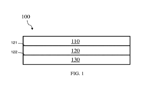
FIG. 1 is, in accordance with some embodiments, a schematic illustration of an
article comprising a first layer, a second layer, and a third layer.
FIG. 2 is a schematic of a deconstructed article, according to one set of
embodiments.
FIG. 3 shows a method of separating blood, according to one set of
embodiments.
FIG. 4 is a plot of the recovered plasma volume as a function of separation
time,
according to one set of embodiments. The large plasma separation device (1.6
cm
diameter) was used. The sample input volume (250 i.1.1_,) and hematocrit (ca.
45%) were
constant. Each data point represents the average of three replicates and error
bars
represent the standard error of the mean.
FIG. 5 is a bar graph showing the separation efficiency of devices of various
sizes with various sample input volumes, according to one set of embodiments.
The
CA 03169213 2022-07-25
WO 2021/155096
PCT/US2021/015624
¨ 3 ¨
separation time (10 mins) and hematocrit (ca. 45%) were constant. Each column
represents the average (N=5) and error bars represent the standard error of
the mean.
FIGs. 6A-6C show a comparison of plasma quality for samples prepared using
plasma separation devices (N=20), in accordance with some embodiments, or a
centrifuge (N=20).
FIG. 7A is a schematic of positive (test and control lines present) and
negative
(only control line present) results for a tetanus lateral flow test.
FIG. 7B shows images of a reference plasma sample collected via centrifugation
of whole blood (positive control), a plasma sample recovered from a plasma
separation
device in accordance with some embodiments (collected plasma), a plasma sample
recovered from a plasma separation device in accordance with some embodiments
after
drying at room temperature for 16 hours and elution with buffer (rehydrated
plasma), and
a buffered sample without tetanus antibody (negative control).
FIG. 7C shows replicate images of lateral flow tests with plasma samples
recovered from a plasma separation device in accordance with some embodiments
and
directly applied to the lateral flow test without centrifugation (N=5).
FIG. 8 shows the dimensions for various acrylic scaffolds, according to one
set
of embodiments.
FIG. 9 shows the quantitation of total protein, where FIG. 9A shows the
calibration curve used and FIG. 9B shows the replicate data for plasma
obtained from a
device in accordance with some embodiments compared to the plasma obtained
from
centrifugation (N=20, p-value = 0.0001).
FIG. 10 shows the calibration data for purity assessment, where FIG. 10A is a
plot of four calibration curves used for h-IgG, and FIG. 10B shows the
calibration plot
for hemoglobin.
DETAILED DESCRIPTION
Disclosed herein are articles and methods for blood separation. For example,
inventive articles and methods that remove red blood cells from blood samples
are
described. In some embodiments, blood separation (e.g., removal of red blood
cells from
a blood sample) is desired, as removal of the cellular components (e.g., red
and white
blood cells) from whole blood can improve sensitivity of some clinical assays
and/or
reduce degradation of analytes of interest in plasma. However, this separation
can be
CA 03169213 2022-07-25
WO 2021/155096
PCT/US2021/015624
¨ 4 ¨
challenging, as the red blood cells in whole blood are numerous and may clog
separation
devices, and red blood cells are fragile and may rupture, contaminating the
plasma.
Moreover, this separation can be expensive, as it may require expensive high-
speed
centrifuges or constant manual operation, and it may produce only low volumes
of
plasma for large separation devices and/or long separation times. In some
embodiments,
the articles and/or methods described herein provide improved articles and/or
methods
for blood separation.
In some embodiments, the article comprises a first layer, a second layer, and
a
third layer. In some embodiments, the first layer is a pre-filter layer that
quickly
.. removes a significant portion of the red blood cells (and/or white blood
cells) from
whole blood, such that the second layer is less likely to get clogged and/or
can have a
higher loading capacity. In some embodiments, the second layer further removes
red
blood cells (and/or white blood cells). In some embodiments, the second layer
has a
gradient in pore size (e.g., with larger pores on the surface of the second
layer adjacent to
the first layer), such that the second layer is less likely to get clogged
and/or is less likely
to rupture the red blood cells. In some embodiments, the third layer is
absorbent, so that
it can absorb the purified blood. In some embodiments, the purified blood in
the third
layer can be used immediately (e.g., collected from and/or used directly from
the third
layer) or it can be stored long term (e.g., dried in the third layer). In some
embodiments,
the first layer, second layer, and third layer are vertically stacked.
Articles are described herein. In accordance with some embodiments, articles
are
illustrated schematically in FIGS. 1-2.
In some embodiments, the article comprises one or more layers. In some
embodiments, the article comprises greater than or equal to 1 layer, greater
than or equal
.. to 2 layers, or greater than or equal to 3 layers. In some embodiments, the
article
comprises less than or equal to 10 layers, less than or equal to 7 layers,
less than or equal
to 5 layers, less than or equal to 4 layers, or less than or equal to 3
layers. Combinations
of these ranges are also possible (e.g., greater than or equal to 1 layer and
less than or
equal to 4 layers). In some embodiments, the article comprises a first layer,
a second
layer, and a third layer. For example, in some embodiments, article 100 in
FIG. 1
comprises first layer 110, second layer 120, and third layer 130. Similarly,
in some
embodiments, the article in FIG. 2 comprises first layer 200, second layer
202, and third
layer 205.
CA 03169213 2022-07-25
WO 2021/155096
PCT/US2021/015624
¨ 5 ¨
In some embodiments, the article comprises a first layer. In some embodiments,
the first layer comprises a pre-filter. In some embodiments, the first layer
comprises
fiberglass, polyester, a fibrous membrane (e.g., polyether sulfone), and/or
mesh (e.g.,
polyester and/or nylon). In some embodiments, the polyester comprises a
treated
polyester, such as Leukosorb. In some embodiments, the first layer comprises a
mesh
(e.g., polyester and/or nylon). In some embodiments, the first layer is
treated. In some
embodiments, the first layer is not treated. The first layer may be fibrous or
non-fibrous.
In some embodiments, the first layer is porous. In some embodiments, the first
layer has a first mode pore size. In some embodiments, the first mode pore
size is
greater than or equal to 1 micron, greater than or equal to 2 microns, greater
than or
equal to 3 microns, greater than or equal to 4 microns, greater than or equal
to 5 microns,
greater than or equal to 10 microns, or greater than or equal to 15 microns.
In some
embodiments, the first mode pore size is less than or equal to 30 microns,
less than or
equal to 25 microns, less than or equal to 20 microns, less than or equal to
15 microns,
less than or equal to 10 microns, less than or equal to 9 microns, less than
or equal to 8
microns, less than or equal to 7 microns, less than or equal to 6 microns, or
less than or
equal to 5 microns. Combinations of these ranges are also possible (e.g.,
greater than or
equal to 1 micron and less than or equal to 30 microns, greater than or equal
to 1 micron
and less than or equal to 6 microns, greater than or equal to 2 microns and
less than or
equal to 25 microns, or greater than or equal to 15 microns and less than or
equal to 25
microns).
In some embodiments, the first layer can have a variety of suitable
thicknesses.
In some embodiments, the first layer has a relatively small thickness. In some
embodiments, the thickness of the first layer is greater than or equal to 150
microns,
.. greater than or equal to 165 microns, or greater than or equal to 180
microns. In some
embodiments, the thickness of the first layer is less than or equal to 500
microns, less
than or equal to 400 microns, less than or equal to 300 microns, less than or
equal to 250
microns, or less than or equal to 220 microns. Combinations of these ranges
are also
possible (e.g., greater than or equal to 180 microns and less than or equal to
220 microns,
or greater than or equal to 150 microns and less than or equal to 500
microns). In some
embodiments, the relatively small thickness of the first layer reduces
separation time.
In some embodiments, the first layer has a relatively low absorbency. In some
embodiments, the absorbency of the first layer is less than or equal to 100
CA 03169213 2022-07-25
WO 2021/155096
PCT/US2021/015624
¨ 6 ¨
microliters/cm2, less than or equal to 90 microliters/cm2, less than or equal
to 80
microliters/cm2, less than or equal to 70 microliters/cm2, less than or equal
to 60
microliters/cm2, less than or equal to 50 microliters/cm2, less than or equal
to 40
microliters/cm2, less than or equal to 30 microliters/cm2, less than or equal
to 20
microliters/cm2, less than or equal to 15 microliters/cm2, less than or equal
to 10
microliters/cm2, or less than or equal to 5 microliters/cm2. In some
embodiments, the
absorbency of the first layer is greater than or equal to 10 microliters/cm2,
greater than or
equal to 15 microliters/cm2, greater than or equal to 20 microliters/cm2,
greater than or
equal to 30 microliters/cm2, or greater than or equal to 40 microliters/cm2,.
Combinations of these ranges are also possible (e.g., greater than or equal to
10
microliters/cm2 and less than or equal to 100 microliters/cm2 or greater than
or equal to
microliters/cm2 and less than or equal to 50 microliters/cm2). In some
embodiments,
the relatively low absorbency of the first layer increases the separation
efficiency and/or
the volume of sample recovered (e.g., increases the yield of the separation),
as a lower
15 volume of the blood plasma may be retained by the first layer.
In some embodiments, the first layer comprises multiple sub-layers. For
example, in some embodiments, the first layer has greater than or equal to 2
sub-layers,
greater than or equal to 3 sub-layers, or greater than or equal to 4 sub-
layers. In some
embodiments, the first layer has less than or equal to 10 sub-layers, less
than or equal to
20 7 sub-layers, less than or equal to 5 sub-layers, less than or equal to
4 sub-layers, less
than or equal to 3 sub-layers, or less than or equal to 2 sub-layers.
Combinations of these
ranges are also possible (e.g., greater than or equal to 2 sub-layers and less
than or equal
to 10 sub-layers, or greater than or equal to 2 sub-layers and less than or
equal to 4 sub-
layers). In embodiments where the first layer comprises multiple sub-layers,
the sub-
layers may each independently have any features described herein for the first
layer.
In embodiments where the first layer comprises multiple sub-layers, multiple
of
the sub-layers (e.g., all of the sub-layers) may comprise the same material or
different
material. For example, in some embodiments, the first layer comprises three
sub-layers,
and all of the sub-layers comprise a mesh (e.g., a polyester and/or nylon
mesh). In some
embodiments, one or more properties (e.g., thickness, mode pore size, mean
pore size,
maximum horizontal dimension, and/or absorbency) of the sub-layers (e.g., all
of the
sub-layers) are the same or different. In some embodiments where each of the
sub-layers
have a different property (e.g., mode pore size), the sub-layers are arranged
such that a
CA 03169213 2022-07-25
WO 2021/155096
PCT/US2021/015624
¨ 7 ¨
gradient in that property is formed. As a non-limiting example, in some
embodiments,
the first layer comprises three sub-layers, and each of the sub-layers has a
different mode
pore size such that a gradient in mode pore size is formed (e.g., 11 micron
mode pore
size in the first sub-layer, 6 micron mode pore size in the second sub-layer,
and 1 micron
mode pore size in the third sub-layer, wherein the second sub-layer is
positioned between
the first sub-layer and the third sub-layer).
In some embodiments, the article comprises a second layer. In some
embodiments, the second layer comprises a polymer. In some embodiments, the
second
layer comprises polyether sulfone. In some embodiments, the second layer
comprises a
plasma separation membrane, such as a Pall plasma separation membrane (e.g., a
Pall
Vivid plasma separation membrane (e.g., grade GX and/or grade GF)), a Kinbio
plasma
separation membrane, and/or a Cobetter plasma separation membrane. The second
layer
may be fibrous or non-fibrous.
In some embodiments, the second layer is porous. In some embodiments, the
.. second layer has a second mode pore size. In some embodiments, the second
mode pore
size (the mode pore size of the second layer) is greater than the first mode
pore size (the
mode pore size of the first layer). In some embodiments, the second mode pore
size (the
mode pore size of the second layer) is smaller than the first mode pore size
(the mode
pore size of the first layer).
In some embodiments, the second mode pore size is greater than or equal to 2
microns, greater than or equal to 3 microns, greater than or equal to 4
microns, greater
than or equal to 5 microns, greater than or equal to 10 microns, or greater
than or equal
to 15 microns. In some embodiments, the first mode pore size is less than or
equal to 30
microns, less than or equal to 25 microns, less than or equal to 20 microns,
less than or
equal to 15 microns, less than or equal to 10 microns, less than or equal to 9
microns,
less than or equal to 8 microns, less than or equal to 7 microns, less than or
equal to 6
microns, or less than or equal to 5 microns. Combinations of these ranges are
also
possible (e.g., greater than or equal to 2 microns and less than or equal to
30 microns or
greater than or equal to 10 microns and less than or equal to 20 microns).
In some embodiments, a certain percentage of the pores of the second layer are
below a certain size. In some embodiments, the certain percentage is greater
than or
equal to 20%, greater than or equal to 30%, greater than or equal to 40%,
greater than or
equal to 50%, greater than or equal to 60%, greater than or equal to 70%,
greater than or
CA 03169213 2022-07-25
WO 2021/155096
PCT/US2021/015624
¨ 8 ¨
equal to 80%, or greater than or equal to 90% of the pores of the second layer
are below
a certain size. In some embodiments, the certain percentage is less than or
equal to
100%, less than or equal to 90%, less than or equal to 80%, less than or equal
to 70%,
less than or equal to 60%, less than or equal to 50%, less than or equal to
40%, or less
than or equal to 30% of the pores of the second layer are below a certain
size.
Combinations of these ranges are also possible (e.g., greater than or equal to
20% and
less than or equal to 100%, greater than or equal to 50% and less than or
equal to 100%,
or greater than or equal to 90% and less than or equal to 100%). In some
embodiments,
the certain size of the pores is greater than or equal to 2 microns, greater
than or equal to
3 microns, greater than or equal to 4 microns, greater than or equal to 5
microns, greater
than or equal to 10 microns, or greater than or equal to 15 microns. In some
embodiments, the certain size of the pores is less than or equal to 30
microns, less than or
equal to 25 microns, less than or equal to 20 microns, less than or equal to
15 microns,
less than or equal to 10 microns, less than or equal to 9 microns, less than
or equal to 8
microns, less than or equal to 7 microns, less than or equal to 6 microns, or
less than or
equal to 5 microns. Combinations of these ranges are also possible (e.g.,
greater than or
equal to 2 microns and less than or equal to 30 microns or greater than or
equal to 10
microns and less than or equal to 20 microns). For example, in some
embodiments,
greater than or equal to 20% (e.g., greater than or equal to 50% or greater
than or equal
to 90%) of the pores of the second layer have a pore size of less than or
equal to 20
microns (e.g., greater than or equal to 10 microns and less than or equal to
20 microns).
In some embodiments, the second layer comprises a first surface and a second
surface. In some embodiments, the first surface faces the first layer (e.g.,
is directly
adjacent to a surface of the first layer). In some embodiments, the second
surface faces
the third layer (e.g., is directly adjacent to a surface of the third layer).
For example, in
some embodiments, second layer 120 in FIG. 1 comprises first surface 121,
which faces
first layer 110, and second surface 122, which faces third layer 130.
In some embodiments, the first surface has a mode pore size. In some
embodiments, the mode pore size of the first surface is greater than or equal
to 10
microns, greater than or equal to 15 microns, or greater than or equal to 20
microns. In
some embodiments, the mode pore size of the first surface is less than or
equal to 35
microns, less than or equal to 30 microns, or less than or equal to 25
microns.
Combinations of these ranges are also possible (e.g., greater than or equal to
10 microns
CA 03169213 2022-07-25
WO 2021/155096
PCT/US2021/015624
¨ 9 ¨
and less than or equal to 35 microns, greater than or equal to 15 microns and
less than or
equal to 25 microns, or greater than or equal to 20 microns and less than or
equal to 25
microns).
In some embodiments, the second surface has a mode pore size. In some
embodiments, the mode pore size of the second surface is greater than or equal
to 0.01
microns, greater than or equal to 0.05 microns, greater than or equal to 0.1
microns,
greater than or equal to 0.15 microns, greater than or equal to 0.25 microns,
greater than
or equal to 0.5 microns, or greater than or equal to 1 micron. In some
embodiments, the
mode pore size of the second surface is less than or equal to 5 microns, less
than or equal
to 3 microns, less than or equal to 1 micron, less than or equal to 0.5
microns, less than
or equal to 0.3 microns, or less than or equal to 0.2 microns. Combinations of
these
ranges are also possible (e.g., greater than or equal to 0.01 microns and less
than or equal
to 1 micron, greater than or equal to 0.1 microns and less than or equal to
0.2 microns, or
greater than or equal to 0.1 microns and less than or equal to 5 microns).
In some embodiments, the mode pore size of the second surface (e.g., the
surface
facing the third layer) is smaller than the mode pore size of the first
surface (e.g., the
surface facing the first layer). In some embodiments, the ratio of the mode
pore size of
the first surface to the mode pore size of the second surface is greater than
or equal to
5:1, greater than or equal to 10:1, greater than or equal to 25:1, greater
than or equal to
50:1, greater than or equal to 75:1, greater than or equal to 100:1, greater
than or equal to
125:1, or greater than or equal to 150:1. In some embodiments, the ratio of
the mode
pore size of the first surface to the mode pore size of the second surface is
less than or
equal to 1,000:1, less than or equal to 500:1, less than or equal to 250:1,
less than or
equal to 200:1, less than or equal to 175:1, less than or equal to 150:1, less
than or equal
to 125:1, less than or equal to 100:1, less than or equal to 75:1, or less
than or equal to
50:1. Combinations of these ranges are also possible (e.g., greater than or
equal to 5:1
and less than or equal to 1,000:1, greater than or equal to 100:1 and less
than or equal to
200:1, greater than or equal to 125:1 and less than or equal to 175:1, or
greater than or
equal to 150:1 and less than or equal to 175:1).
Mode pore size can be measured using any suitable technique. For example, in
some embodiments, mode pore size can be measured using Mercury Intrusion
Porosimetry or Scanning Electron Microscope (SEM). In some embodiments, mode
pore size can be measured over the full thickness of the layer. In some
embodiments, a
CA 03169213 2022-07-25
WO 2021/155096
PCT/US2021/015624
- 10 ¨
layer can be divided into multiple sections along the thickness of the layer,
and the mode
pore size of each section can be measured.
In some embodiments, the first surface and/or the second surface each
independently have a thickness that is a certain percentage of the thickness
of the second
layer. In some embodiments, the first surface and/or the second surface are
each
independently greater than or equal to 1/10 of the thickness of the second
layer, greater
than or equal to 1/8 of the thickness of the second layer, greater than or
equal to 1/6 of
the thickness of the second layer, or greater than or equal to 1/10 of the
thickness of the
second layer 1% of the thickness of the second layer. In some embodiments, the
first
surface and/or second surface are each independently less than or equal to 1/2
of the
thickness of the second layer, less than or equal to 1/3 of the thickness of
the second
layer, less than or equal to 1/4 of the thickness of the second layer, or less
than or equal to
1/5 of the thickness of the second layer. Combinations of these ranges are
also possible
(e.g., greater than or equal to 1/10 of the thickness of the second layer and
less than or
equal to 1/2 of the thickness of the second layer, or greater than or equal to
1/8 of the
thickness of the second layer and less than or equal to 1/4 of the thickness
of the second
layer). In some embodiments, the first surface and the second surface have the
same
thickness.
In some embodiments, the second layer has a gradient in mode pore size between
the first surface and the second surface. In some embodiments, there are cross-
sections
within the thickness of the second layer between the first surface and the
second surface.
In some embodiments the cross-sections have a mode pore size that is between
the mode
pore size of the first surface and the mode pore size of the second surface.
For example,
in that embodiment, if the mode pore size of the first surface was 11 microns
and the
.. mode pore size of the second surface was 1 micron, then the cross-sections
within the
thickness of the second layer between the first surface and the second surface
would have
mode pore sizes between 1 micron and 11 microns.
In some embodiments, the second layer can have a variety of suitable
thicknesses. In some embodiments, the thickness of the second layer is greater
than or
equal to 100 microns. In some embodiments, the thickness of the second layer
is less
than or equal to 300 microns, less than or equal to 250 microns, less than or
equal to 200
microns, or less than or equal to 150 microns. Combinations of these ranges
are also
CA 03169213 2022-07-25
WO 2021/155096
PCT/US2021/015624
- 11 ¨
possible (e.g., greater than or equal to 100 microns and less than or equal to
150 microns,
or greater than or equal to 100 microns and less than or equal to 300
microns).
In some embodiments, the second layer has a relatively low absorbency. In some
embodiments, the absorbency of the second layer is less than or equal to 50
microliters/cm2, less than or equal to 40 microliters/cm2, less than or equal
to 30
microliters/cm2, less than or equal to 25 microliters/cm2, less than or equal
to 20
microliters/cm2, less than or equal to 15 microliters/cm2, less than or equal
to 10
microliters/cm2, or less than or equal to 5 microliters/cm2. In some
embodiments, the
absorbency of the second layer is greater than or equal to 10 microliters/cm2,
greater than
or equal to 15 microliters/cm2, or greater than or equal to 20
microliters/cm2.
Combinations of these ranges are also possible (e.g., greater than or equal to
10
microliters/cm2 and less than or equal to 50 microliters/cm2, or greater than
or equal to
microliters/cm2 and less than or equal to 25 microliters/cm2). In some
embodiments,
the relatively low absorbency of the second layer increases the separation
efficiency
15 .. and/or the volume of sample recovered (e.g., increases the yield of the
separation), as a
lower volume of the blood plasma is retained by the second layer.
In some embodiments, the article comprises a third layer. In some embodiments,
the third layer comprises a wicking source. In some embodiments, the third
layer
comprises rayon and/or polyester (e.g., Kapmat). In some embodiments, the
third layer
comprises a blend of rayon and polyester, or a blend of rayon and
polypropylene (e.g.,
ShamWow). The third layer may be fibrous or non-fibrous.
In some embodiments, the third layer is porous. In some embodiments, the third
layer has a third mode pore size. In some embodiments, the third mode pore
size is
greater than or equal to 20 microns, greater than or equal to 30 microns,
greater than or
equal to 40 microns, greater than or equal to 50 microns, greater than or
equal to 60
microns, greater than or equal to 70 microns, greater than or equal to 75
microns, greater
than or equal to 80 microns, or greater than or equal to 90 microns. In some
embodiments, the third mode pore size is less than or equal to 150 microns,
less than or
equal to 140 microns, less than or equal to 130 microns, less than or equal to
125
microns, less than or equal to 120 microns, less than or equal to 110 microns,
or less than
or equal to 100 microns. Combinations of these ranges are also possible (e.g.,
greater
than or equal to 20 microns and less than or equal to 150 microns, greater
than or equal
CA 03169213 2022-07-25
WO 2021/155096
PCT/US2021/015624
¨ 12 ¨
to 75 microns and less than or equal to 125 microns, or greater than or equal
to 90
microns and less than or equal to 100 microns).
In some embodiments, the third layer may have a relatively large absorbency.
In
some embodiments, the absorbency is greater than or equal to 55
microliters/cm2, greater
than or equal to 60 microliters/cm2, greater than or equal to 65
microliters/cm2, greater
than or equal to 70 microliters/cm2, greater than or equal to 75
microliters/cm2, greater
than or equal to 80 microliters/cm2, greater than or equal to 85
microliters/cm2, greater
than or equal to 90 microliters/cm2, greater than or equal to 95
microliters/cm2, greater
than or equal to 100 microliters/cm2, greater than or equal to 125
microliters/cm2, greater
than or equal to 150 microliters/cm2, greater than or equal to 175
microliters/cm2, greater
than or equal to 200 microliters/cm2, greater than or equal to 250
microliters/cm2, greater
than or equal to 300 microliters/cm2, or greater than or equal to 400
microliters/cm2. In
some embodiments, the absorbency is less than or equal to 600 microliters/cm2,
less than
or equal to 550 microliters/cm2, less than or equal to 500 microliters/cm2,
less than or
equal to 450 microliters/cm2, less than or equal to 400 microliters/cm2, less
than or equal
to 300 microliters/cm2, less than or equal to 250 microliters/cm2, less than
or equal to
200 microliters/cm2, less than or equal to 175 microliters/cm2, or less than
or equal to
150 microliters/cm2. Combinations of these ranges are also possible (e.g.,
greater than or
equal to 80 microliters/cm2 and less than or equal to 600 microliters/cm2,
greater than or
equal to 100 microliters/cm2 and less than or equal to 600 microliters/cm2, or
greater than
or equal to 200 microliters/cm2 and less than or equal to 450
microliters/cm2).
As used herein, the absorbency of an article and/or layer is determined by
weighing the article and/or layer, saturating it in DI water for 30 seconds at
room
temperature, weighing it again, determining the difference between the second
weight
and the first weight (i.e., the weight of the DI water absorbed), and then
converting this
weight to a volume of water (e.g., microliters) using the density of DI water
at room
temperature. The volume of DI water absorbed is then normalized by dividing by
the
surface area (e.g., cm2) of the article and/or layer.
In some embodiments, the relatively large absorbency of the third layer
facilitates
passive separation by increasing capillary action and/or facilitates
collection and/or
storage of the absorbed fluid in the third layer.
In some embodiments, the third layer is configured to absorb a variety of
suitable
fluids. Examples of suitable fluids include water, blood plasma, saliva,
urine, wound
CA 03169213 2022-07-25
WO 2021/155096
PCT/US2021/015624
¨ 13 ¨
exudate, and/or cerebrospinal fluid. In some embodiments, the third layer is
configured
to absorb blood plasma.
In some embodiments, the third layer may have a relatively large release. As
used herein, the release of an article and/or layer is the percentage of the
absorbed water
(determined as described above) that is released upon centrifugation. Once the
article
and/or layer is saturated in DI water for 30 seconds and the volume of DI
water absorbed
is calculated (as discussed above), the article and/or layer is centrifuged at
an RCF of
800 g for 5 minutes. The volume of DI water released during centrifugation is
then
converted to a percentage of the volume of DI water that was absorbed in order
to
determine what percentage of the absorbed DI water was released. This value is
the
release of the article and/or layer.
In some embodiments, the third layer has a release that is greater than or
equal to
15%, greater than or equal to 20%, greater than or equal to 25%, greater than
or equal to
30%, greater than or equal to 35%, greater than or equal to 40%, greater than
or equal to
60%, greater than or equal to 70%, greater than or equal to 80%, or greater
than or equal
to 90%. In some embodiments, the third layer has a release that is less than
or equal to
100%, less than or equal to 95%, less than or equal to 90%, less than or equal
to 85%,
less than or equal to 80%, less than or equal to 75%, less than or equal to
70%, or less
than or equal to 60%. Combinations of these ranges are also possible (e.g.,
greater than
or equal to 35% and less than or equal to 100%, greater than or equal to 50%
and less
than or equal to 100%, greater than or equal to 70% and less than or equal to
100%, or
greater than or equal to 70% and less than or equal to 90%).
In some embodiments, the relatively large release of the third layer increases
separation efficiency and/or the volume of sample recovered (e.g., increases
the yield of
the separation).
In some embodiments, the third layer has a relatively large thickness (e.g.,
compared to the first and/or second layer(s)). In some embodiments, the
thickness of the
third layer is greater than or equal to 200 microns, greater than or equal to
225 microns,
or greater than or equal to 250 microns. In some embodiments, the thickness of
the third
layer is less than or equal to 800 microns, less than or equal to 700 microns,
less than or
equal to 600 microns, or less than or equal to 500 microns. Combinations of
these
ranges are also possible (e.g., greater than or equal to 200 microns and less
than or equal
to 800 microns, or greater than or equal to 250 microns and less than or equal
to 500
CA 03169213 2022-07-25
WO 2021/155096
PCT/US2021/015624
¨ 14 ¨
microns). In some embodiments, the relatively large thickness of the third
layer
increases the volume of sample recovered (e.g., increases the yield of the
separation), as
it increases the volume of fluid that can be absorbed.
In some embodiments, the article comprises a support structure. For example,
in
some embodiments, the article in FIG. 2 comprises support structure 204. In
some
embodiments, the support structure comprises a plastic, an acrylic, and/or a
metal. In
some embodiments, the support structure is a plastic scaffold or an acrylic
scaffold. In
some embodiments, the support structure is configured to maintain conformal
contact
between the third layer and one or more layers (e.g., the second layer).
In some embodiments, the support structure is adjacent one or more layers. In
some embodiments, the support structure is adjacent the first layer, second
layer, and/or
third layer. In some embodiments, the support structure is in direct contact
with one or
more layers. In some embodiments, the support structure is in direct contact
with the
first layer, second layer, and/or third layer. In some embodiments, the
support structure
is in direct contact with the second layer and third layer. In some
embodiments, the
support structure is in direct contact with the third layer.
In some embodiments, the support structure is adhered to one or more layers
(e.g., the third layer). Examples of suitable means to adhere (e.g., the
support structure
to one or more layers) are discussed elsewhere herein (e.g., in reference to
adhering one
layer to another layer). In some embodiments, the support structure is not
adhered to one
or more layers (e.g., not adhered to any layers). For example, in some
embodiments, a
portion of the article (e.g., the first layer, the second layer, and/or the
third layer) sits on
the support structure.
In some embodiments, the support structure comprises a cavity. In some
embodiments, the cavity is used for holding a portion of the article (e.g.,
the first layer,
the second layer, and/or the third layer). In some embodiments, the cavity is
circular,
oval, square, rectangular, and/or diamond shaped. In some embodiments, the
cavity is of
a similar shape as a cross-section (e.g., a horizontal cross-section) of a
portion of the
.. article (e.g., one or more layers, such as the third layer). For example,
in some
embodiments, the cavity and/or the cross-section of a portion of the article
(e.g., one or
more layers, such as the third layer) are both circular, oval, square,
rectangular, and/or
diamond shaped.
CA 03169213 2022-07-25
WO 2021/155096
PCT/US2021/015624
¨ 15 ¨
In some embodiments, the first layer, second layer, third layer, and/or
article have
a relatively large maximum horizontal dimension. In some embodiments, the
first layer,
second layer, third layer, and/or article each independently have a maximum
horizontal
dimension of greater than or equal to 20 millimeters, greater than or equal to
40
millimeters, greater than or equal to 60 millimeters, greater than or equal to
80
millimeters, greater than or equal to 100 millimeters, greater than or equal
to 120
millimeters, greater than or equal to 140 millimeters, or greater than or
equal to 150
millimeters. In some embodiments, the first layer, second layer, third layer,
and/or
article each independently have a maximum horizontal dimension of less than or
equal to
500 millimeters, less than or equal to 400 millimeters, less than or equal to
300
millimeters, less than or equal to 200 millimeters, less than or equal to 180
millimeters,
less than or equal to 160 millimeters, less than or equal to 140 millimeters,
less than or
equal to 120 millimeters, less than or equal to 100 millimeters, less than or
equal to 80
millimeters, less than or equal to 60 millimeters, or less than or equal to 40
millimeters.
Combinations of these ranges are also possible (e.g., greater than or equal to
20
millimeters and less than or equal to 500 millimeters, greater than or equal
to 20
millimeters and less than or equal to 100 millimeters, greater than or equal
to 60
millimeters and less than or equal to 200 millimeters). In some embodiments,
the
maximum horizontal dimensions of one or more (e.g., two or three) of the first
layer,
second layer, and third layer are the same.
In some embodiments, the relatively large maximum horizontal dimension of one
or more layers (e.g., the second layer, or all of the layers) increases
separation efficiency,
decreases the separation time, increases the volume of sample recovered (e.g.,
increases
the yield of the separation), and/or increases input volume.
In some embodiments, the maximum horizontal dimension of the cavity is greater
than or equal to the maximum horizontal dimension of a portion of the article
(e.g., one
or more layers, such as the second layer and/or the third layer). In some
embodiments,
the ratio of the maximum horizontal dimension of the cavity to the maximum
horizontal
dimension of a portion of the article (e.g., one or more layers, such as the
second layer
and/or the third layer) is greater than or equal to 1:1, greater than or equal
to 1.05:1,
greater than or equal to 1.1:1, greater than or equal to 1.2:1, greater than
or equal to
1.3:1, greater than or equal to 1.4:1, or greater than or equal to 1.5:1. In
some
embodiments, the ratio of the maximum horizontal dimension of the cavity to
the
CA 03169213 2022-07-25
WO 2021/155096
PCT/US2021/015624
¨ 16 ¨
maximum horizontal dimension of a portion of the article (e.g., one or more
layers, such
as the second layer and/or the third layer) is less than or equal to 3:1, less
than or equal to
2:1, less than or equal to 1.5:1, less than or equal to 1.4:1, less than or
equal to 1.3:1, less
than or equal to 1.2:1, less than or equal to 1.1:1, or less than or equal to
1.05:1.
Combinations of these ranges are also possible (e.g., greater than or equal to
1:1 and less
than or equal to 3:1 or greater than or equal to 1.1 and less than or equal to
1.3:1).
In some embodiments, the maximum horizontal dimension of the cavity is greater
than or equal to 0.5 cm, greater than or equal to 0.75 cm, greater than or
equal to 1 cm,
greater than or equal to 1.1 cm, greater than or equal to 1.2 cm, greater than
or equal to
1.3 cm, greater than or equal to 1.4 cm, greater than or equal to 1.5 cm,
greater than or
equal to 1.6 cm, greater than or equal to 1.7 cm, greater than or equal to 1.8
cm, greater
than or equal to 1.9 cm, greater than or equal to 2 cm, greater than or equal
to 2.25 cm,
greater than or equal to 2.5 cm, or greater than or equal to 3 cm. In some
embodiments,
the maximum horizontal dimension of the cavity is less than or equal to 10 cm,
less than
or equal to 5 cm, less than or equal to 4 cm, less than or equal to 3 cm, less
than or equal
to 2.5 cm, less than or equal to 2.25 cm, less than or equal to 2 cm, less
than or equal to
1.9 cm, less than or equal to 1.8 cm, less than or equal to 1.7 cm, less than
or equal to 1.6
cm, less than or equal to 1.5 cm, less than or equal to 1.4 cm, less than or
equal to 1.3
cm, less than or equal to 1.2 cm, less than or equal to 1.1 cm, or less than
or equal to 1
cm. Combinations of these ranges are also possible (e.g., greater than or
equal to 0.5 cm
and less than or equal to 10 cm or greater than or equal to 0.5 cm and less
than or equal
to 2 cm).
In some embodiments, the depth of the cavity is less than the thickness of the
support structure, such that, when viewed from above, a layer of the support
structure is
present throughout the surface area of the support structure. In some
embodiments, the
cavity is configured such that a portion of the article (e.g., the first
layer, second layer,
and/or third layer) can sit inside the cavity. In some embodiments, the cavity
is
configured such that a portion of the article (e.g., the first layer, second
layer, and/or
third layer) can sit inside the cavity, with the bottom surface of the third
layer in contact
with the support structure.
In some embodiments, the cavity is present throughout the thickness of the
support structure, such that, when viewed from above, the cavity is a hole in
the support
structure. In some embodiments, the cavity has different maximum horizontal
CA 03169213 2022-07-25
WO 2021/155096
PCT/US2021/015624
¨ 17 ¨
dimensions at different thickness of the support structure. For example, in
some
embodiments, the cavity has a larger maximum horizontal dimension at one
opening than
at the other. In some embodiments, the larger maximum horizontal dimension at
one
opening is greater than or equal to the maximum horizontal dimension of a
portion of the
article (e.g., the third layer). In some embodiments, the smaller maximum
horizontal
dimension at the other opening is less than the maximum horizontal dimension
of a
portion of the article (e.g., the third layer). In some embodiments, the
cavity is
configured such that a portion of the article (e.g., the first layer, second
layer, and/or
third layer) can sit inside the cavity. In some embodiments, the cavity is
configured such
that a portion of the article (e.g., the first layer, second layer, and/or
third layer) can sit
inside the cavity, but the bottom surface of the third layer is not in contact
with the
support structure. In some embodiments, the cavity is configured such that a
portion of
the article (e.g., the first layer, second layer, and/or third layer) can sit
inside the cavity,
but the bottom surface of the third layer is not in contact with the support
structure, such
that the third layer can be removed from the article through the bottom of the
support
structure (e.g., through the opening with the smaller maximum horizontal
dimension),
while the remaining portions of the article can remain in the support
structure (see, e.g.,
FIG. 3) .
In some embodiments, the cavity is configured such that the height of the
edges
(e.g., circumference) of the cavity prevent a portion of the article (e.g.,
the first layer,
second layer, and/or third layer) from significant horizontal movement, but
the portion of
the article (e.g., the first layer, second layer, and/or third layer) can
still be picked up
vertically. In some embodiments, the height of the edges of the cavity are
greater than or
equal to 1/5 the thickness of a layer (e.g., the third layer), greater than or
equal to 1/4 the
thickness of a layer (e.g., the third layer), greater than or equal to 1/3 the
thickness of a
layer (e.g., the third layer), greater than or equal to 1/2 the thickness of a
layer (e.g., the
third layer), or greater than or equal to the thickness of a layer (e.g., the
third layer). In
some embodiments, the height of the edges of the cavity are less than or equal
to 3 times
the thickness of a layer (e.g., the third layer), 2 times the thickness of a
layer (e.g., the
.. third layer), the thickness of a layer (e.g., the third layer), 1/2 the
thickness of a layer (e.g.,
the third layer), 1/3 the thickness of a layer (e.g., the third layer), or 1/4
the thickness of a
layer (e.g., the third layer). Combinations of these ranges are also possible
(e.g., greater
CA 03169213 2022-07-25
WO 2021/155096
PCT/US2021/015624
¨ 18 ¨
than or equal to 1/5 and less than or equal to 3 times the thickness of a
layer (e.g., the
third layer)).
The layers in the article may be in any suitable order. In some embodiments,
the
first layer is positioned between the second layer and third layer. In some
embodiments,
the third layer is positioned between the first layer and second layer. In
some
embodiments, the second layer is positioned between the first layer and the
third layer.
For example, in Fig. 1, in accordance with some embodiments, second layer 120
is
positioned between first layer 110 and third layer 130.
In some embodiments, there are no intervening layers between the first layer
and
second layer and/or between the second layer and third layer. For example, in
Fig. 1, in
accordance with some embodiments, there are no intervening layers between
first layer
110 and second layer 120 or between second layer 120 and third layer 130. In
some
embodiments, the direct contact (e.g., direct conformal contact) between the
layers (e.g.,
the second and third layer) decreases the separation time by increasing
capillary action.
In some embodiments, one or more layers are adhered to one or more layers. For
example, in some embodiments, the article in FIG. 2 comprises adhesive 201,
which
adheres first layer 200 to second layer 202, and adhesive 203, which adheres
second
layer 202 to third layer 205. In some embodiments, one or more layers are
permanently
adhered or integrally connected to one or more layers. In some embodiments,
one or
more layers are reversibly adhered to one or more layers. Examples of suitable
methods
of adhering layers include double-sided adhesive (e.g., double-sided medical
adhesive),
liquid adhesive, sonic welding, and/or compression. In some embodiments, one
or more
layers are adhered to one or more layers (and/or a support structure) with an
adhesive.
Examples of suitable adhesives include double-sided adhesive (e.g., double-
side medical
adhesive), compression tape, 3M brand adhesive, and/or Flexcon brand adhesive.
In
some embodiments, the adhesive is placed on a surface of a layer. In some
embodiments, the adhesive is placed around the perimeter of a layer where it
contacts
another layer (or substrate) to adhere it to the other layer (or substrate).
In some
embodiments, the adhesive (e.g., between two layers, or between a layer and
the
substrate) provides a full seal (e.g., a seal around the entire perimeter of
the layer through
which fluid cannot pass).
In some embodiments, a full seal (e.g., with adhesive) between one or more
layers (and/or between a layer and the substrate) increases the purity of the
purified
CA 03169213 2022-07-25
WO 2021/155096
PCT/US2021/015624
¨ 19 ¨
blood (e.g., purified plasma), as it reduces or prevent one or more impurities
(e.g., red
blood cells) from bypassing one or more layers and entering the third layer.
For
example, if there was a partial seal around the perimeter of the first layer
where it
contacts the second layer, then a blood sample might pass through the first
layer and out
through the holes in the seal, such that it then passes down to the third
layer without
passing through the second layer, resulting in higher levels of impurities
(e.g., red blood
cells) than if the blood sample had passed through the second layer.
In some embodiments, the adhesive has any suitable thickness. In some
embodiments, the adhesive is relatively thin. In some embodiments, a thin
adhesive
allows the layers to be closer together, decreasing the separation time. In
some
embodiments, the adhesive has a thickness of greater than or equal to 0.03
millimeters,
greater than or equal to 0.04 millimeters, greater than or equal to 0.05
millimeters,
greater than or equal to 0.06 millimeters, or greater than or equal to 0.063
millimeters.
In some embodiments, the adhesive has a thickness of less than or equal to 0.2
millimeters, less than or equal to less than or equal to 0.18 millimeters,
less than or equal
to 0.16 millimeters, less than or equal to 0.14 millimeters, or less than or
equal to 0.126
millimeters. Combinations of these ranges are also possible (e.g., greater
than or equal
to 0.03 millimeters and less than or equal to 0.2 millimeters, or greater than
or equal to
0.063 millimeters and less than or equal to 0.126 millimeters).
In some embodiments, the adhesive is applied manually. In some embodiments,
the adhesive is applied with a laser cutter, ultrasonic welding, and/or UV
curing. In
some embodiments, the adhesive has a low tack. In some embodiments, one or
more
layers is adhered to one or more layers in such a way that they cannot be
pulled apart
manually without damaging one or more of the layers. For example, in some
embodiments, the first layer is adhered to the second layer such that they
cannot be
pulled apart manually without damaging one or more of the layers. In some
embodiments, one or more layers is adhered to one or more layers in such a way
that
they can be pulled apart manually without damaging one or more of the layers.
For
example, in some embodiments, the second layer is adhered to the third layer
in such a
way that they can be pulled apart manually without damaging one or more of the
layers
(e.g., the third layer). In some embodiments, the second layer is adhered to
the third
layer in such a way that they can be pulled apart manually, without having to
use so
much force that it will disrupt the first layer (e.g., creating mess or
contamination), but
CA 03169213 2022-07-25
WO 2021/155096
PCT/US2021/015624
¨ 20 ¨
such that the second layer and third layer do not come apart during use (e.g.,
do not come
apart during separation of a blood sample).
In some embodiments, the layers are stacked coaxially, such that a vertical
stack
is formed. For example, in some embodiments, article 100 in FIG. 1 comprises
first
layer 110, second layer 120, and third layer 130 stacked coaxially, such that
a vertical
stack is formed. In some embodiments, the vertical stacking reduces the time
required
for separation.
In some embodiments, the layers described herein are discrete layers. In some
embodiments, the layers described herein are not discrete layers, such that a
layer is
instead one of multiple phases within a discrete layer. For example, in some
embodiments, the first layer and the second layer could be two phases within
one layer.
In some embodiments, the maximum horizontal dimension of the article is
greater than or equal to 0.5 cm, greater than or equal to 0.75 cm, greater
than or equal to
1 cm, greater than or equal to 1.1 cm, greater than or equal to 1.2 cm,
greater than or
equal to 1.3 cm, greater than or equal to 1.4 cm, greater than or equal to 1.5
cm, greater
than or equal to 1.6 cm, greater than or equal to 1.7 cm, greater than or
equal to 1.8 cm,
greater than or equal to 1.9 cm, greater than or equal to 2 cm, greater than
or equal to
2.25 cm, greater than or equal to 2.5 cm, or greater than or equal to 3 cm. In
some
embodiments, the maximum horizontal dimension of the article is less than or
equal to
10 cm, less than or equal to 5 cm, less than or equal to 4 cm, less than or
equal to 3 cm,
less than or equal to 2.5 cm, less than or equal to 2.25 cm, less than or
equal to 2 cm, less
than or equal to 1.9 cm, less than or equal to 1.8 cm, less than or equal to
1.7 cm, less
than or equal to 1.6 cm, less than or equal to 1.5 cm, less than or equal to
1.4 cm, less
than or equal to 1.3 cm, less than or equal to 1.2 cm, less than or equal to
1.1 cm, or less
than or equal to 1 cm. Combinations of these ranges are also possible (e.g.,
greater than
or equal to 0.5 cm and less than or equal to 5 cm or greater than or equal to
0.5 cm and
less than or equal to 2 cm).
In some embodiments, the article has a high loading capacity (e.g., for whole
blood). As used herein, loading capacity is defined as volume of fluid that
can be loaded
divided by the surface area of the article. In some embodiments, the loading
capacity of
the article is greater than or equal to 20 microliters/cm2, greater than or
equal to 30
microliters/cm2, greater than or equal to 40 microliters/cm2, greater than or
equal to 50
microliters/cm2, greater than or equal to 60 microliters/cm2, greater than or
equal to 70
CA 03169213 2022-07-25
WO 2021/155096
PCT/US2021/015624
¨ 21 ¨
microliters/cm2, greater than or equal to 80 microliters/cm2, greater than or
equal to 90
microliters/cm2, greater than or equal to 100 microliters/cm2, or greater than
or equal to
125 microliters/cm2. In some embodiments, the loading capacity of the article
is less
than or equal to 500 microliters/cm2, less than or equal to 400
microliters/cm2, less than
or equal to 300 microliters/cm2, less than or equal to 250 microliters/cm2,
less than or
equal to 200 microliters/cm2, less than or equal to 150 microliters/cm2, less
than or equal
to 125 microliters/cm2, less than or equal 100 microliters, less than or equal
90
microliters/cm2, less than or equal 80 microliters/cm2, or less than or equal
70
microliters/cm2. Combinations of these ranges are also possible (e.g., greater
than or
equal to 20 microliters/cm2 and less than or equal to 500 microliters/cm2, or
greater than
or equal to 50 microliters/cm2 and less than or equal to 150 microliters/cm2).
Methods are described herein. In accordance with some embodiments, an
illustrative method is illustrated schematically in FIG. 3, and can be
understood in view
of FIG. 1.
In some embodiments, the method comprises passing a blood sample across a
first layer. For example, in some embodiments, the method comprises passing a
blood
sample across first layer 110 in FIG. 1. In some embodiments, the first layer
comprises
any embodiment of the first layer, or combinations thereof, disclosed herein.
In some embodiments, the blood sample is whole blood. In some embodiments,
the blood sample is diluted with water and/or a buffer solution. In some
embodiments,
the blood sample is undiluted blood from a subject. In some embodiments, the
subject is
an animal, such as a mammal. In some embodiments, the subject is a human. In
some
embodiments, the article comprises an anti-coagulant (e.g.,
ethylenediaminetetraacetic
acid (EDTA) and/or heparin), such as a dried anti-coagulant.
In some embodiments, the first layer has a high loading capacity, such that
the
blood sample passed across the first layer (e.g., input volume) has a
substantial volume.
In some embodiments, the volume of the blood sample passed across the first
layer (e.g.,
input volume) is greater than or equal to 25 microliters, greater than or
equal to 30
microliters, greater than or equal to 40 microliters, greater than or equal to
50 microliters,
greater than or equal to 60 microliters, greater than or equal to 70
microliters, greater
than or equal to 80 microliters, greater than or equal to 90 microliters,
greater than or
equal to 100 microliters, greater than or equal to 125 microliters, greater
than or equal to
CA 03169213 2022-07-25
WO 2021/155096
PCT/US2021/015624
¨ 22 ¨
150 microliters, greater than or equal to 200 microliters, or greater than or
equal to 250
microliters. In some embodiments, the volume of the blood sample passed across
the
first layer (e.g., input volume) is less than or equal to 500 microliters,
less than or equal
to 400 microliters, less than or equal to 300 microliters, less than or equal
to 250
microliters, less than or equal to 200 microliters, less than or equal to 150
microliters,
less than or equal to 125 microliters, less than or equal 100 microliters,
less than or equal
90 microliters, less than or equal 80 microliters, or less than or equal 70
microliters.
Combinations of these ranges are also possible (e.g., greater than or equal to
25
microliters and less than or equal to 500 microliters, greater than or equal
to 50
microliters and less than or equal to 300 microliters, or greater than or
equal to 100
microliters and less than or equal to 250 microliters).
In some embodiments, the volume of the blood sample passed across the first
layer (e.g., input volume) may affect the volume of sample (e.g., plasma)
recovered, the
separation efficiency, the separation time, and/or the purity (e.g., levels of
hemolysis) of
the sample (e.g., plasma). For example, if the volume of the blood sample
passed across
the first layer (e.g., input volume) is too low, then a larger percentage of
the blood
sample may be absorbed by the first layer and/or second layer resulting in low
volume of
sample recovered (e.g., low yield of the separation) and/or low separation
efficiency
compared to if a larger volume of the blood sample passed across the first
layer (e.g.,
input volume), in some embodiments. As another example, if the volume of the
blood
sample passed across the first layer (e.g., input volume) is too high, then
one or more
layers may clog, resulting in more impurities passing through, increased
hemolysis,
and/or decreased separation time, in some embodiments.
In some embodiments, passing the blood sample across the first layer produces
a
blood sample with reduced red blood cells. In some embodiments, the red blood
cells
are reduced by the first layer by greater than or equal to 20%, greater than
or equal to
30%, greater than or equal to 40%, greater than or equal to 50%, greater than
or equal to
60%, greater than or equal to 70%, greater than or equal to 80%, or greater
than or equal
to 90% of those in the blood sample. In some embodiments, the red blood cells
are
reduced by the first layer by less than or equal to 100%, less than or equal
to 90%, less
than or equal to 80%, less than or equal to 70%, less than or equal to 60%,
less than or
equal to 50%, less than or equal to 40%, or less than or equal to 30% of those
in the
CA 03169213 2022-07-25
WO 2021/155096
PCT/US2021/015624
¨ 23 ¨
blood sample. Combinations of these ranges are also possible (e.g., greater
than or equal
to 20% and less than or equal to 90%).
In some embodiments, the first layer reduces the level of red blood cells in
the
blood sample by size exclusion and/or electrostatic interactions.
In some embodiments, the first layer reduces the level of white blood cells
(which can also be called "leukocytes"). In some embodiments, the white blood
cells
are reduced by the first layer by greater than or equal to 20%, greater than
or equal to
30%, greater than or equal to 40%, greater than or equal to 50%, greater than
or equal to
60%, greater than or equal to 70%, greater than or equal to 80%, or greater
than or equal
to 90% of those in the blood sample. In some embodiments, the white blood
cells are
reduced by the first layer by less than or equal to 100%, less than or equal
to 90%, less
than or equal to 80%, less than or equal to 70%, less than or equal to 60%,
less than or
equal to 50%, less than or equal to 40%, or less than or equal to 30% of those
in the
blood sample. Combinations of these ranges are also possible (e.g., greater
than or equal
to 20% and less than or equal to 90%).
In some embodiments, the first layer reduces the level of white blood cells in
the
blood sample by size exclusion, electrostatic interactions, and/or adsorption
of the white
blood cells.
In some embodiments, use of the first layer facilitates quick removal of a
significant portion of the red blood cells (and/or white blood cells), such
that the second
layer is less likely to get clogged and/or is less likely to cause hemolysis
and/or the
article can have a higher loading capacity without requiring lengthy times for
separation.
In some embodiments, the method comprises passing the blood sample with
reduced red blood cells (and/or white blood cells) across a second layer. For
example, in
some embodiments, the method comprises passing the blood sample with reduced
red
blood cells (and/or white blood cells) across second layer 120 in FIG. 1. In
some
embodiments, the second layer comprises any embodiment of the second layer, or
combinations thereof, disclosed herein.
In some embodiments, passing the blood sample with reduced red blood cells
(and/or white blood cells) across the second layer produces a blood sample
with further
reduced red blood cells. In some embodiments, the red blood cells are reduced
by the
second layer by greater than or equal to 20%, greater than or equal to 30%,
greater than
or equal to 40%, greater than or equal to 50%, greater than or equal to 60%,
greater than
CA 03169213 2022-07-25
WO 2021/155096
PCT/US2021/015624
¨ 24 ¨
or equal to 70%, greater than or equal to 80%, or greater than or equal to 90%
of those in
the blood sample with reduced red blood cells. In some embodiments, the red
blood
cells are reduced by the second layer by less than or equal to 100%, less than
or equal to
90%, less than or equal to 80%, less than or equal to 70%, less than or equal
to 60%, less
than or equal to 50%, less than or equal to 40%, or less than or equal to 30%
of those in
the blood sample with reduced red blood cells. Combinations of these ranges
are also
possible (e.g., greater than or equal to 20% and less than or equal to 90%).
In some embodiments, the second layer further reduces the level of red blood
cells in the blood sample with reduced red blood cells (and/or white blood
cells) by size
exclusion and/or electrostatic interactions.
In some embodiments, the second layer reduces the level of white blood cells.
In
some embodiments, the white blood cells are reduced by the second layer by
greater than
or equal to 20%, greater than or equal to 30%, greater than or equal to 40%,
greater than
or equal to 50%, greater than or equal to 60%, greater than or equal to 70%,
greater than
or equal to 80%, or greater than or equal to 90% of those in the blood sample
with
reduced red blood cells. In some embodiments, the white blood cells are
reduced by the
second layer by less than or equal to 100%, less than or equal to 90%, less
than or equal
to 80%, less than or equal to 70%, less than or equal to 60%, less than or
equal to 50%,
less than or equal to 40%, or less than or equal to 30% of those in the blood
sample with
reduced red blood cells. Combinations of these ranges are also possible (e.g.,
greater
than or equal to 20% and less than or equal to 90%).
In some embodiments, the second layer reduces the level of white blood cells
in
the blood sample with reduced red blood cells by size exclusion and/or
electrostatic
interactions.
In some embodiments, use of a second layer with a gradient in pore size
reduces
the risk of the second layer clogging and/or reduces the risk that the second
layer will
result in hemolysis, in some embodiments.
In some embodiments, the method comprises passing the blood sample with
further reduced red blood cells into a third layer. For example, in some
embodiments,
the method comprises passing a blood sample with further reduced red blood
cells into
third layer 130 in FIG. 1. In some embodiments, the third layer comprises any
embodiment of the third layer, or combinations thereof, disclosed herein.
CA 03169213 2022-07-25
WO 2021/155096
PCT/US2021/015624
¨ 25 ¨
In some embodiments, the method (e.g., passing the blood sample across the
first
layer, passing the blood sample with reduced red blood cells across the second
layer,
and/or passing the blood sample with further reduced red blood cells into the
third layer)
is passive. For example, in some embodiments, the method is done solely with
the use of
gravity and/or capillary action. In some embodiments, the method is done
without the
use of centrifugation, electricity, and/or an external field (e.g., acoustic,
electric, and/or
magnetic). For example, in some embodiments, FIG. 3 demonstrates adding blood
sample to the article (e.g., the first layer) and then the article separates
the sample
without further action (that is, the sample is separated purely from gravity
and capillary
action).
In some embodiments, a portion of the method (e.g., passing the blood sample
across the first layer, passing the blood sample with reduced red blood cells
across the
second layer, and/or passing the blood sample with further reduced red blood
cells into
the third layer) is relatively rapid as the separation time is short. In some
embodiments,
a portion of the method is accomplished within (and/or the separation time is)
less than
or equal to 30 minutes, less than or equal to 20 minutes, less than or equal
to 15 minutes,
less than or equal to 10 minutes, less than or equal to 5 minutes, less than
or equal to 3
minutes, or less than or equal to 2 minutes. In some embodiments, a portion of
the
method is accomplished within (and/or the separation time is) greater than or
equal to 30
seconds, greater than or equal to 1 minute, greater than or equal to 2
minutes, greater
than or equal to 3 minutes, or greater than or equal to 5 minutes.
Combinations of these
ranges are also possible (e.g., greater than or equal to 30 seconds and less
than or equal
to 10 minutes or greater than or equal to 30 seconds and less than or equal to
5 minutes).
In some embodiments, the method (e.g., passing the blood sample across the
first
layer, passing the blood sample with reduced red blood cells across the second
layer,
and/or passing the blood sample with further reduced red blood cells into the
third layer)
has a high separation efficiency. In some embodiments, the separation
efficiency is
greater than or equal to 10%, greater than or equal to 15%, greater than or
equal to 20%,
greater than or equal to 25%, greater than or equal to 30%, greater than or
equal to 35%,
greater than or equal to 40%, greater than or equal to 45%, greater than or
equal to 50%,
or greater than or equal to 55%. In some embodiments, the separation
efficiency is less
than or equal to 100%, less than or equal to 90%, less than or equal to 80%,
less than or
equal to 70%, less than or equal to 60%, less than or equal to 55%, less than
or equal to
CA 03169213 2022-07-25
WO 2021/155096
PCT/US2021/015624
¨ 26 ¨
50%, less than or equal to 45%, less than or equal to 40%, less than or equal
to 35%, or
less than or equal to 30%. Combinations of these ranges are also possible
(e.g., greater
than or equal to 10% and less than or equal to 100%, greater than or equal to
10% and
less than or equal to 60%, or greater than or equal to 30% and less than or
equal to 55%).
As used herein, the separation efficiency is the percentage of collected
purified
plasma volume (or volume of purified plasma that passes into the third layer)
compared
to the total theoretical plasma volume. The total theoretical plasma volume is
based on
the measured hematocrit value and input sample volume. For example, if a 100
microliter sample has a measured hematocrit value of 50%, then the total
theoretical
plasma volume is 50 microliters. If 40 microliters of purified plasma were
collected (or
passed into the third layer), the separation efficiency would be 80%, since 40
microliters
is 80% of 50 microliters.
In some embodiments, the method comprises removing the third layer from the
second layer. For example, in some embodiments, FIG. 3 demonstrates removing
the
third layer from the second layer. In some embodiments, the third layer is
removed from
the second layer by pulling it apart from the second layer. In some
embodiments, the
third layer is pulled apart from the second layer manually (e.g., pulling it
apart with
tweezers). In some embodiments, the article comprises a tab. In some
embodiments,
pulling the tab may pull the third layer apart from the second layer.
In some embodiments, the blood sample with further reduced red blood cells is
used directly from the third layer. For example, in some embodiments, the
third layer
can be used as a stamp with which to apply the blood sample with further
reduced red
blood cells (e.g., to a lateral flow test).
In some embodiments, the blood sample with further reduced red blood cells is
stored inside the third layer. In some embodiments, the blood sample with
further
reduced red blood cells is stored inside the third layer in a wet state. In
some
embodiments, the blood sample with further reduced red blood cells is stored
inside the
third layer in a dry state. For example, in some embodiments, the third layer
containing
the blood sample with further reduced red blood cells is dried overnight. In
some
embodiments, the third layer is dried overnight in a sealed container. In some
embodiments, the sealed container comprises a desiccant.
In some embodiments, the dried third layer is later rehydrated. In some
embodiments, the dried third layer is rehydrated by adding a solvent, such as
an aqueous
CA 03169213 2022-07-25
WO 2021/155096
PCT/US2021/015624
¨27 ¨
solution (e.g., an aqueous solution comprising a surfactant), a buffered
solution (e.g.,
phosphate buffered saline), and/or water (e.g., DI water).
In some embodiments, the method comprises collecting the blood sample with
further reduced red blood cells from the third layer. In some embodiments,
collecting
the blood sample with further reduced red blood cells is done shortly after
the blood
sample with further reduced red blood cells is passed into the third layer. In
some
embodiments, collecting the blood sample with further reduced red blood cells
is done
after the sample with further reduced blood cells has been stored (e.g., in a
wet state or in
a dry state) inside the third layer for a length of time. In some embodiments,
the blood
sample with further reduced red blood cells is collected from the third layer
greater than
or equal to 1 minute, greater than or equal to 5 minutes, greater than or
equal to 15
minutes, greater than or equal to 30 minutes, greater than or equal to 1 hour,
greater than
or equal to 5 hours, greater than or equal to 12 hours, greater than or equal
to 1 day,
greater than or equal to 3 days, greater than or equal to 1 week, greater than
or equal to 1
month, greater than or equal to 6 months, or greater than or equal to 1 year
after it has
been passed into the third layer. In some embodiments, the blood sample with
further
reduced red blood cells is collected from the third layer less than or equal
to 3 years, less
than or equal to 2 years, less than or equal to 1 year, less than or equal to
6 months, less
than or equal to 1 month, less than or equal to 1 week, less than or equal to
3 days, less
than or equal to 1 day, less than or equal to 12 hours, less than or equal to
5 hours, less
than or equal to 1 hour, less than or equal to 30 minutes, less than or equal
to 15 minutes,
or less than or equal to 5 minutes after it has been passed into the third
layer.
Combinations of these ranges are also possible (e.g., greater than or equal to
1 minute
and less than or equal to 3 years).
In some embodiments, collecting the blood sample with further reduced red
blood cells from the third layer can be accomplished with relatively low
amounts of
force. In some embodiments, collecting the blood sample with further reduced
red blood
cells comprises compression (e.g., squeezing) and/or centrifuging the third
layer (e.g.,
with a benchtop centrifuge). For example, in some embodiments, FIG. 3
demonstrates
collecting the blood sample with further reduced red blood cells from the
third layer by
centrifugation with a benchtop centrifuge. In some embodiments, the blood
sample is
centrifuged at less than or equal to 800 x g (e.g., less than or equal to 700
x g, less than
or equal to 500 x g, or less than or equal to 300 x g) for less than or equal
to 5 minutes
CA 03169213 2022-07-25
WO 2021/155096
PCT/US2021/015624
¨ 28 ¨
(e.g., less than or equal to 4 minutes, less than or equal to 3 minutes, less
than or equal to
2 minutes, or less than or equal to 1 minute).
In some embodiments, the blood sample with further reduced red blood cells
(e.g., the blood sample with further reduced red blood cells collected from
the third
layer) can be collected in a short period of time. In some embodiments, the
blood
sample with further reduced blood cells can be collected in less than or equal
to 30
minutes, less than or equal to 20 minutes, less than or equal to 15 minutes,
less than or
equal to 10 minutes, less than or equal to 5 minutes, less than or equal to 3
minutes, or
less than or equal to 1 minute. In some embodiments, the blood sample with
further
reduced blood cells can be collected in greater than or equal to 30 seconds,
greater than
or equal to 1 minute, greater than or equal to 2 minutes, greater than or
equal to 3
minutes, or greater than or equal to 5 minutes. Combinations of these ranges
are also
possible (e.g., greater than or equal to 30 seconds and less than or equal to
30 minutes, or
greater than or equal to 30 seconds and less than or equal to 10 minutes).
In some embodiments the blood sample with further reduced red blood cells
(e.g.,
pure plasma) can be used in subsequent applications (e.g., after collection,
and/or
directly, from the third layer), such as in a diagnostic health test, a
clinical assay (e.g.,
clinical chemistry assays), an immunoassay, an immunochromatographic assay for
antibodies (e.g., tetanus antibodies), quantification of cytokines,
amplification of viral
RNA, a rapid dipstick test, an HIV viral load assay, a cholesterol test, a
metabolite panel,
serology for infectious diseases, therapeutic drug monitoring, an ELISA, ICP-
AES,
HPLC, and/or mass spectrometry.
In some embodiments, the volume of the blood sample with further reduced red
blood cells (e.g., the blood sample with further reduced red blood cells
collected and/or
used directly from the third layer) is a significant percentage of the volume
of the blood
sample (e.g., the blood sample passed through the first layer), given that 20-
60% of the
blood sample (e.g., whole blood) is expected to be red blood cells. In some
embodiments, the volume of the blood sample with further reduced red blood
cells is
greater than or equal to 10%, greater than or equal to 12%, greater than or
equal to 15%,
greater than or equal to 17%, greater than or equal to 20%, greater than or
equal to 25%,
greater than or equal to 30%, greater than or equal to 35%, greater than or
equal to 40%,
greater than or equal to 45%, or greater than or equal to 50% of the volume of
the blood
sample. In some embodiments, the volume of the blood sample with further
reduced red
CA 03169213 2022-07-25
WO 2021/155096
PCT/US2021/015624
¨ 29 ¨
blood cells is less than or equal to 80%, less than or equal to 70%, less than
or equal to
60%, less than or equal to 50%, less than or equal to 40%, less than or equal
to 30%, less
than or equal to 25%, less than or equal to 20%, less than or equal to 17%, or
less than or
equal to 15% of the volume of the blood sample. Combinations of these ranges
are also
possible (e.g., greater than or equal to 10% and less than or equal to 80% or
greater than
or equal to 10% and less than or equal to 40%).
In some embodiments, a large volume of the blood sample with further reduced
red blood cells is passed into the third layer and/or a large volume of the
blood sample
with further reduced red blood cells is collected and/or used directly from
the third layer.
For example, in some embodiments, the volume of the blood sample with further
reduced red blood cells passed into the third layer and/or collected and/or
used directly
from the third layer is greater than or equal to 20 microliters, greater than
or equal to 25
microliters, greater than or equal to 30 microliters, greater than or equal to
35 microliters,
greater than or equal to 40 microliters, greater than or equal to 45
microliters, greater
than or equal to 50 microliters, greater than or equal to 55 microliters,
greater than or
equal to 60 microliters, greater than or equal to 65 microliters, or greater
than or equal to
70 microliters. In some embodiments, the volume of the blood sample with
further
reduced red blood cells passed into the third layer and/or collected and/or
used directly
from the third layer is less than or equal to 150 microliters, less than or
equal to 125
microliters, less than or equal to 100 microliters, less than or equal to 90
microliters, less
than or equal to 80 microliters, less than or equal to 75 microliters, less
than or equal to
70 microliters, or less than or equal to 60 microliters. Combinations of these
ranges is
also possible (e.g., greater than or equal to 20 microliters and less than or
equal to 150
microliters, greater than or equal to 30 microliters and less than or equal to
150
microliters, greater than or equal to 50 microliters and less than or equal to
150
microliters, or greater than or equal to 50 microliters and less than or equal
to 100
microliters).
In some embodiments, the blood sample with further reduced red blood cells
(e.g., the blood sample with further reduced red blood cells collected and/or
used directly
from the third layer or the blood sample) is pure (e.g., pure plasma and/or
serum),
substantially free of red blood cells, and/or substantially free of white
blood cells. In
some embodiments, the blood sample with further reduced red blood cells has
less than
or equal to 5%, less than or equal to 4%, less than or equal to 3%, less than
or equal to
CA 03169213 2022-07-25
WO 2021/155096
PCT/US2021/015624
¨ 30 ¨
2%, or less than or equal to 1% of the red blood cells in the blood sample
(e.g., the
original blood sample, such as a whole blood sample). In some embodiments, the
blood
sample with further reduced red blood cells has less than or equal to 5%, less
than or
equal to 4%, less than or equal to 3%, less than or equal to 2%, or less than
or equal to
1% of the white blood cells in the blood sample (e.g., the original blood
sample, such as
a whole blood sample).
In some embodiments, the amount of red blood cells is assumed to be the same
as
the amount of hemoglobin. For example, if a blood sample (e.g., an original
blood
sample, such as a whole blood sample) had 12 g/dL hemoglobin, and the blood
sample
with further reduced red blood cells has 0.12 g/dL hemoglobin, then the blood
sample
with further reduced red blood cells has less than or equal to 1% of the
hemoglobin in the
original sample, and it would be assumed that the blood sample with further
reduced red
blood cells has less than or equal to 1% of the red blood cells in the blood
sample (e.g.,
the original blood sample, such as a whole blood sample).
In some embodiments, the blood sample with further reduced red blood cells
(e.g., the blood sample with further reduced red blood cells collected and/or
used directly
from the third layer) has minimal amounts of hemolysis. In some embodiments,
the
blood sample with further reduced red blood cells has less than or equal to
15%
hemolysis, less than or equal to 10% hemolysis, less than or equal to 8%
hemolysis, less
than or equal to 7%, less than or equal to 6%, less than or equal to 5%
hemolysis, less
than or equal to 3% hemolysis, less than or equal to 2% hemolysis, or less
than or equal
to 1% hemolysis. In some embodiments, the blood sample with further reduced
red
blood cells has greater than or equal to 0% hemolysis, greater than or equal
to 0.1%
hemolysis, greater than or equal to 0.5% hemolysis, greater than or equal to
1%
hemolysis, greater than or equal to 2% hemolysis, greater than or equal to 3%
hemolysis,
greater than or equal to 4%, or greater than or equal to 5% hemolysis.
Combinations of
these ranges are also possible (e.g., greater than or equal to 0% and less
than or equal to
15% or greater than or equal to 0.1% and less than or equal to 7%).
As used herein, the percentage hemolysis is the percentage of hemoglobin in
the
measured sample compared to hemoglobin in a similar whole blood sample. For
example, if a blood sample was divided in two, and one part was purified
(e.g., separated
from red blood cells) while the other part was untreated, the percentage
hemolysis in the
purified sample would be the percentage of hemoglobin in that sample compared
to the
CA 03169213 2022-07-25
WO 2021/155096
PCT/US2021/015624
¨ 31 ¨
percentage hemoglobin in the untreated whole blood sample. The amount of
hemoglobin
can be measured by any suitable assay. For example, the amount of hemoglobin
can be
measured by the assay described in the example, where a ratio of whole blood
(the
control) to Drabkin's reagent containing 0.05% (v/v) Brij 25 was 1:250; a
ratio of sample
to Drabkin's reagent containing 0.05% (v/v) Brij 25 was 1:10; calibration
curves were
prepared daily using lyophilized hemoglobin standard rehydrated with diH20 (18
MS2)
and diluted over a range 3-20 g/dL; samples were incubated at 21 C for 15
minutes and
absorbance was measured at 540 nm using a microplate reader (e.g., Varioskan
LUX).
In some embodiments, the blood sample with further reduced red blood cells
(e.g., the blood sample with further reduced red blood cells collected and/or
used directly
from the third layer) has similar levels of an analyte of interest as the
original blood
sample (e.g., whole blood and/or the blood sample passed across the first
layer). For
example, in some embodiments, the level of an analyte of interest in the blood
sample
with further reduced red blood cells is greater than or equal to 40%, greater
than or equal
to 45%, greater than or equal to 50%, greater than or equal to 55%, greater
than or equal
to 60%, greater than or equal to 65%, greater than or equal to 70%, greater
than or equal
to 75%, greater than or equal to 80%, greater than or equal to 85%, greater
than or equal
to 90%, greater than or equal to 95%, greater than or equal to 98%, or greater
than or
equal to 99% the level of the analyte of interest in the original blood sample
(e.g., whole
blood and/or the blood sample passed across the first layer). In some
embodiments, the
level of an analyte of interest in the blood sample with further reduced red
blood cells is
less than or equal to 100%, less than or equal to 99%, less than or equal to
98%, less than
or equal to 95%, less than or equal to 90%, less than or equal to 85%, less
than or equal
to 80%, less than or equal to 75%, or less than or equal to 70% the level of
the analyte of
interest in the original blood sample (e.g., whole blood and/or the blood
sample passed
across the first layer). Combinations of these ranges are also possible (e.g.,
greater than
or equal to 40% and less than or equal to 100% or greater than or equal to 80%
and less
than or equal to 100%). For example, if a 250 microliter sample of whole blood
tested
for the presence of HIV RNA by RT-qPCR had an average threshold cycle value of
28
Ct and was passed across an article described herein (e.g., passed across a
first layer,
passed across a second layer, and passed into a third layer) to form 60
microliters of a
blood sample with further reduced red blood cells (e.g., as in a method
described herein)
with an average threshold cycle value of 29 Ct, then the level of HIV RNA in
the blood
CA 03169213 2022-07-25
WO 2021/155096
PCT/US2021/015624
¨ 32 ¨
sample with further reduced red blood cells would be 50% of that in the
original blood
sample, as every 1 Ct in qPCR is responsible for a doubling.
Examples of analytes of interest may include proteins (e.g., enzymes (e.g.,
alkaline phosphatase, alanine aminotransferase, aspartate aminotransferase),
antibodies
(e.g., for immune response (e.g., acute IgM or persistent IgG), such as to
indicate
vaccination (e.g., measles), infection (e.g., HIV, SARS-CoV-2, tuberculosis,
sexually
transmitted infections), sensitivity to foods, allergens), and/or biomarkers
(e.g., HbAlc,
albumin, insulin, cancer antigens (PSA, CA-125))), nucleic acids (e.g.,
recovered from
pathogens (e.g., RNA or DNA genes), host cell genome (e.g., to determine
mutations), or
cell free fetal DNA (cffDNA)), pathogens (e.g., viruses (e.g., HIV), parasites
(e.g., P.
falciparum), and/or bacteria (e.g., S. aureus)), metabolites (e.g., blood urea
nitrogen,
creatinine, bilirubin, carnosine, UDP-acetyl-glucosamine), hormones (e.g.,
thyroid,
fertility/pregnancy, testosterone, cortisol), electrolytes (e.g., calcium,
potassium,
bicarbonate, chloride), lipids (e.g., HDL, LDL, VLDL, cholesterol,
triglycerides), and/or
small molecules (e.g., vitamins (e.g., folic acid, B vitamins, biotin) and/or
sugars (e.g.,
glucose, Carbohydrate antigen 19-9 (sialyl-Lewis'), sialyl-LewisX)).
In some embodiments, the method may be performed on any embodiment of the
article, or combinations thereof, disclosed herein. In some embodiments, the
article is
configured to perform any embodiment of the method, or combinations thereof,
.. disclosed herein.
In some, but not all, embodiments, the article and/or method has one or more
advantages, such as short separation time, short collection time, ease of
separation (e.g.,
without constant manual operation), ease of collection (e.g., without the use
of high
speed centrifuges), small surface area (e.g., small maximum horizontal
dimension) of the
article, ease of scaling up, ease of storage of the purified sample, large
loading capacity,
large volume recovery, low amounts of clogging of the article, low amounts of
hemolysis
in the recovered sample, high purity of the recovered sample, low amounts of
mess (e.g.,
high containment of the blood within the article), low energy requirements,
and/or ability
to use whole blood samples without the need for dilution.
The following examples are intended to illustrate certain embodiments of the
present invention, but do not exemplify the full scope of the invention.
CA 03169213 2022-07-25
WO 2021/155096
PCT/US2021/015624
¨ 33 ¨
EXAMPLES
Described herein is, in accordance with some embodiments, an assembly of
porous materials capable of obtaining high volumes (>60 i.1.1_,) of pure
plasma from
whole blood using only passive methods in less than 10 minutes.
A pre-filter material was used to reduce the burden of excess blood cells from
clogging the plasma separation membrane and minimize hemolysis independent of
hematocrit. Separation and collection were facilitated by a super absorbent
material in
direct contact with the plasma separation membrane. The dual functionality of
the
collection pad permitted storage of purified plasma for shipping and future
laboratory
analysis similar to dried blood spot card technologies. The purity of
collected plasma
samples was evaluated by quantification of hemoglobin and the recovery of high
and low
concentration analytes of interest was evaluated.
Experimental Design
Device Design and Fabrication
The device comprised a pre-filter material, plasma separation membrane (PSM),
and super absorbent material (FIG. 2).
The separation materials (e.g., pre-filter and plasma separation membrane)
were
affixed to the acrylic scaffold via rings of double-sided medical adhesive.
The absorbent
material was located in direct contact with the underside of the plasma
separation
membrane. Contact between each material was maintained by an acrylic scaffold
and
double-sided medical adhesive. The pre-filter material was designed to remove
white
blood cells from the sample matrix based on size exclusion and electrostatic
interactions.
The plasma separation membrane was designed to exclude all remaining white and
red
blood cells to produce pure plasma that can be simultaneously collected and
stored by
the underlying absorbent material.
All porous materials (e.g., pre-filter materials, PSM, and absorbent
materials)
were cut using a hammer-driven hole punch. Double-sided medical adhesive was
patterned into rings using an automated knife plotter. Acrylic scaffolds were
fabricated
with a Trotech laser cutter.
The pore sizes of several materials evaluated for the device are shown in
Table 1.
Table 1. Pore data for various materials.
CA 03169213 2022-07-25
WO 2021/155096
PCT/US2021/015624
¨ 34 ¨
Surface area Mode pore Unique pore
Sample ID Material
(n12/g) diameter (pm)
diameters (pm)
TFN Cellulose 9.8 27.9
27.9, 19.8, 9.0
Kapmat Polyester 17.1 159.4 159.4
Rayon/Polypropylene
ShamWow 32.2 91.6 91.6
Blend
23.9, 16.3, 10.0,
PSM-GR Polysulfone 43.7 16.3
7.4, 4.5, 0.15
Leukosorb Polyester 43.7 19.5 19.5
Operation of the Device
Plasma separation was initiated by applying a sample of whole blood to the top
of
the device and allowing it to sit for 5-10 minutes for separation to occur
(see the
schematic in FIG. 3). Purified plasma was collected by the absorbent material
located
beneath the plasma separation membrane. To terminate separation, the absorbent
material was removed from the acrylic scaffold with a pair of tweezers and
either (i)
liquid plasma was recovered from the absorbent material via centrifugation,
(ii) the
porous material containing purified plasma was dried and stored for future
laboratory
analysis, or (iii) the absorbent material was immediately applied to a lateral
flow test.
Evaluation of Absorbency and Release for Porous Materials
The absorbency and release for the materials was determined as follows. The
initial mass of each absorbent material was recorded (N=3, area = 1 cm2).
Then, each
material was saturated in deionized water for 30 seconds and the saturated
mass was
recorded. The volume of water absorbed by each material was calculated using
the
density of water at ambient temperature. This value was normalized by the
surface area
of the material. This normalized value represented the "absorbency" of the
material.
Then, the saturated absorbent materials were centrifuged to collect the water
using a Swinex funnel attached to a 5-mL Eppendorf tube at an RCF of 800 g for
5
minutes. The Eppendorf tube was weighed empty and then with the released
water, and
the volume of water released by each material was calculated using the density
of water
at ambient temperature. This value represented the volume recovery. This
volume was
converted to a percentage of the water that was absorbed, and this value
represented the
"release" of the material.
CA 03169213 2022-07-25
WO 2021/155096
PCT/US2021/015624
¨ 35 ¨
Quantification of the Recovered Plasma Volume and Calculation of Separation
Efficiency
A centrifuge was used to quantify the volume of plasma collected in the
devices
as proof-of-concept (see, e.g., the schematic shown in FIG. 3). After plasma
separation
occurred, the absorbent material was removed from the acrylic scaffold using
tweezers
and added to a Swinex funnel attached to a 5-mL Eppendorf tube. The samples
were
centrifuged at an RCF of 800 g for 5 minutes to collect liquid plasma from the
absorbent
material. The mass of the liquid plasma was determined by calculating the
difference
between the initial mass of the 5-mL Eppendorf tube and the final mass after
centrifugation. Then, the mass of the plasma sample was converted to recovered
volume
by using the average density of plasma (1.025 g/mL). The total theoretical
plasma
volume was determined based on the measured hematocrit value and input sample
volume. Separation efficiency was defined as the ratio of collected plasma
volume to
total theoretical plasma volume.
Recovery of Total Protein
Recovery was calculated as the ratio of total protein in plasma samples
obtained
from the plasma separation device to the concentration of total protein in
plasma samples
obtained via centrifugation. The Pierce 660 nm protein assay was used to
quantify the
total protein in plasma samples according to an established protocol. Briefly,
150 0_, of
the Pierce 660 reagent was added into a microwell plate, followed by 10 0_, of
diluted
plasma (1:100 in lx PBS). The microwell plate was incubated for 5 minutes at
room
temperature before reading at 660 nm using a Varioskan LUX microplate reader.
A
calibration curve was prepared using BSA solutions over a linear range from
0.05-2
mg/mL.
Recovery of High Abundance Protein h-IgG
Bio-Layer Inteferometry (K2 Octet, Pall Fortebio) was used to quantitate human
immunoglobulin G (h-IgG) in reference plasma (i.e., obtained via
centrifugation) and
recovered plasma samples (i.e., obtained from the plasma separation device). A
96-well
plate format with fiber-optic biosensors coated with Protein-A was used to
measure the
binding rate of h-IgG to Protein-A. Calibration curves were prepared using
polyclonal h-
CA 03169213 2022-07-25
WO 2021/155096
PCT/US2021/015624
¨ 36 ¨
IgG standards of known concentrations, ranging from 1-700 tg/mL (Pall
Fortebio). The
plasma samples were diluted 1:1000 in 1X Kinetics Buffer (Pall Fortebio)
before
quantitation to ensure the signal fell within the working range of the
calibration curve.
The calibration curves were fit using a linear-point-to-point method, as
described
in the Protein-A Biosensor data sheet. The linear model was used to determine
unknown
concentrations for the reference plasma (N=20) and the recovered plasma
(N=20). The
two groups were statistically analyzed using a two-tailed Student's t-test
with equal
variances.
Evaluation of Purity for Collected Plasma
The concentration of hemoglobin in recovered plasma was quantified to evaluate
the purity of samples obtained by the plasma separation device. Extent of
hemolysis was
defined as the ratio of hemoglobin in plasma to total hemoglobin quantified
according to
an established method. For quantification of total hemoglobin in whole blood
samples, a
ratio of 1:250 was used (e.g., 4 0_, of whole blood to 1 mL Drabkin's reagent
containing
0.05% (v/v) Brij 25). Calibration curves were prepared daily using lyophilized
hemoglobin standard rehydrated with diH20 (18 MS2) and diluted over a range 3-
20
g/dL. For quantification of hemoglobin in plasma samples, a ratio of 1:10 was
used (e.g.,
0_, of whole blood to 0.2 mL Drabkin's reagent containing 0.05% (v/v) Brij
25).
20 Calibration curves were prepared daily using lyophilized hemoglobin
standard
rehydrated with diH20 (18 MS2) and diluted over a range 0.09-3 g/dL. The
mixture was
incubated at room temperature (i.e., 21 C) for 15 minutes and absorbance was
measured
at 540 nm using a Varioskan LUX microplate reader. Plasma samples were
collected
from each plasma separation device and hemoglobin was quantified to determine
extent
of hemolysis against total hemoglobin concentration in whole blood. The LOD
for both
assays (i.e., 1:250 and 1:10 dilutions) were calculated using purified plasma
obtained via
centrifugation from three different donors.
Tetanus Lateral Flow Test
Utility of the plasma separation device was demonstrated by applying collected
plasma directly to commercially available lateral flow tests. All blood
samples were
collected from donors that had been vaccinated against tetanus and therefore
contained
tetanus antibodies. Positive controls from whole blood were prepared via
centrifugation
CA 03169213 2022-07-25
WO 2021/155096
PCT/US2021/015624
¨ 37 ¨
and negative controls from assay buffer provided with the lateral flow tests.
20 i.it of
plasma were applied to the sample input zone on the lateral flow test using a
micropipette. 3 drops of assay buffer were then immediately added onto the
sample input
zone and 10 minutes were allowed to pass before scanning the results with an
Epson
V600 Perfection flatbed scanner at 800 DPI.
Results and Discussion
Selection of Absorbent Materials (e.g., Third Layer)
A source of capillarity facilitated the performance of passive separation of
plasma
from whole blood. Capillarity was provided by the absorbent material, which
was in
direct contact with the separation materials above (FIG. 2). The desired
material would
provide (i) a fast wicking rate, (ii) high absorbency, and (iii) quantitative
release of
absorbed liquid. Three different wicking materials were tested: cellulose,
polyester, and a
rayon/polypropylene blend. The cellulose material had the lowest absorbency
(65.0
7.0 i.tt/cm2) and released only 19% of the absorbed liquid (Table 2). In stark
contrast,
both the polyester and rayon/polypropylene blend materials absorbed 587.0
40.1
i.tt/cm2 and 393.7 23.6 i.tt/cm2, respectively. These super absorbent
materials also
yielded high percentages for the release of absorbed liquid at 93% (polyester)
and 84%
(rayon/polypropylene blend).
Table 2. Performance of various absorbent materials. Values represent the
average of five replicates and standard error of the mean.
Volume Recovery
Material Absorbency (pL/cm2) Release
(pL/cm2)
Cellulose 65 7 12 7 19%
Polyester 587 40 544 22 93%
Rayon/Polypropylene Blend 393 23 330 27 84%
Both the polyester and rayon/polypropylene blend materials in the device were
evaluated for wicking ability in conjunction with the PSM. While the polyester
material
was more absorbent than the rayon/propylene blend, it caused more hemolysis of
the
blood sample. The rayon/polypropylene blend material did not cause hemolysis
and
therefore provided a better wicking source for separating plasma from whole
blood in the
device.
CA 03169213 2022-07-25
WO 2021/155096
PCT/US2021/015624
- 38 -
Baseline Performance of the PSM (e.g., Second Layer)
Three devices of different sizes (FIG. 8) were designed and tested with whole
blood to establish baseline separation efficiencies using only a single layer
of PSM. As
shown in FIG. 8, the inner black ring on each device was the cavity ledge of
acrylic (half
depth cut, 0.317 cm), which provided physical support for the separation
materials. The
inner white circle was the open region of the device (full depth cut, 0.635
cm), which
allowed direct contact between the absorbent material and the separation
materials. The
area of the plasma separation membrane determined the allowable sample input
volume
according to the manufacturer (40-50 0_, cm-1). Theoretical sample input
volumes were
calculated for each device based on the minimum and maximum loading capacities
for
Vivid GR plasma separation membrane from Pall Corp (Table 3).
Table 3. Theoretical sample input volumes (IL) for small, medium, and large
devices.
Plasma Separation Theoretical Sample Input Volume (pL)
Membrane Loading Small Device Medium Device Large Device
Capacity (pL cm-1) (1.0 cm diameter) (1.3 cm diameter) (1.6 cm
diameter)
Minimum 31.4 53.1 80.4
Maximum 39.3 66.3 100.5
A variety of PSMs were tested and the material with the greatest loading
capacity
(i.e., input volume of blood per area) and consistency was identified.
Baseline separation
for each device was measured after 10 minutes following sample addition and
yielded a
consistent volume range of 16-20% (maximum of 27.5 t.L) of available plasma
from a
250 0_, sample input (Table 4). Minimal hemolysis was observed in all three
devices
(Table 4). The large device (1.6 cm diameter) achieved a higher degree of
separation
than the smaller devices under these conditions.
Table 4. Baseline data for each device with no pre-filter material. Sample
input
volumes were 250 0_, and 150 0_, whole blood. Separation time (10 minutes) and
hematocrit (ca. 45%) were constant.
250 pL 150 pL
Average Recovered Separation Extent of Average Recovered
Separation Extent of
Device SEM . . SEM . .
Volume (pL) Efficiency Hemolysis Volume (pL)
Efficiency Hemolysis
Small
22.5 1.4 16.6% 2.4% 23.5 1.7 28.9% 2.5%
(1.0 cm diameter)
Medium
22.5 0.9 16.6% 1.9% 21.7 0.8 26.9% 3.9%
(1.3 cm diameter)
CA 03169213 2022-07-25
WO 2021/155096
PCT/US2021/015624
¨ 39 ¨
Large
27.1 1.9 20.0% 2.7% 22.3 2.1 27.6%
2.5%
(1.6 cm diameter)
The decreased separation of the smaller 250 i.tt devices (1.0 cm diameter) was
attributed to an excess number of RBCs that clogged the pores of the PSM and
impeded
the flow of plasma through the membrane to the absorbent material below. In
order to
alleviate this burden on the PSM, a pre-filter material was included to remove
RBCs and
allow the plasma to flow through the membrane for collection. Potential pre-
filter
materials included fiberglass, polyester mesh with pore sizes ranging from 1-
11 p.m, and
a fibrous membrane for the isolation of leukocytes from whole blood
(Leukosorb, Pall
Corp).
Material Screen for Pre-filter (e.g., First Layer)
Fiberglass (Ahlstrom grade 8950) was initially selected for its propensity to
act as
a chromatographic material for blood separation without binding proteins or
causing
hemolysis. However, a single layer of fiberglass actually decreased the
separation
efficiency of the device by 3.5% (Table 6). The fiberglass was 0.25 mm thick
with a
reported void volume of 46 i.tt/cm2. While fiberglass was capable of
separating plasma
from whole blood, the wicking rate and void volume of the material negatively
impacted
the performance of the device and required separation times in excess of 90
minutes.
The fibers of the polyester mesh did not absorb fluids or swell when in
contact
with liquid samples. This effectively lowered the void volume of the material,
which
increased the total recovery of plasma in the device. RBCs have an average
size
distribution of 6-8 p.m and a biconcave disc geometry. However, since RBCs are
easily
deformable, a range of pore sizes were studied in an effort to create a pre-
filter based on
size exclusion for capturing RBCs. Initially, multiple layers of mesh with a
pore size of 1
p.m were tested as a pre-filter in a large plasma separation device (Table 5).
Table 5. Performance of multiple layers of mesh as pre-filter in large acrylic
devices. Sample input volume (150 i.tt whole blood) and separation time (10
minutes)
were constant.
Average Recovered Separation Extent of
Large Acrylic Device SEM
Volume (pL) Efficiency
Hemolysis
Baseline 22.3 2.1 27.6% 2.5%
1 layer 30.0 2.0 36.8% 1.3%
2 layers 28.6 1.5 35.1% 3.2%
CA 03169213 2022-07-25
WO 2021/155096
PCT/US2021/015624
- 40 -
In another embodiment, a mesh with a pore size of 11 p.m was used to remove
larger cells such as leukocytes (average diameter of 7-20 p.m) from the sample
matrix
upon initiation of the device. The next layer had a pore size of 6 p.m to
remove any
remaining leukocytes as well as a portion of RBCs. To ensure removal of all
RBCs from
the sample matrix prior to reaching the PSM, a final layer of polyester mesh
with pore
size of 1 p.m was included. This construct of meshes acted as an effective pre-
filter by
increasing the separation efficiency by 9.6% and decreasing the extent of
hemolysis by
1.2% within 10 minutes (Table 6). Iterations of this construct were
investigated with
single layers of polyester mesh (e.g., 1 p.m, 6 p.m, 11 p.m), which yielded
similar results.
A maximum of 33.6% separation efficiency was achieved using two layers of
polyester
mesh with 1 and 6 p.m pore sizes (Table 6).
Table 6. Performance of various pre-filter materials. Sample input volume (250
i.tt whole blood), separation time (10 minutes), and hematocrit (ca. 45%) were
constant.
Average Recovered Separation
Extent of
Pre-Filter Material SEM
Volume (pL) Efficiency
Hemolysis
Polyester Mesh (1 pm) 37.3 0.7 27.5% 1.7%
Polyester Mesh (6 pm) 43.8 1.2 32.3% 1.7%
Polyester Mesh (11 pm) 45.3 0.6 33.4% 1.5%
Polyester Mesh (1 +6 pm) 45.5 2.8 33.6% 1.5%
Polyester Mesh (1 +6+11 pm) 40.1 1.8 29.6% 1.5%
Fiberglass 22.4 5.0 16.5% 3.8%
Leukosorb 70.6 2.6 51.1% 4.3%
To complement the function of commercially available PSM for passively
separating plasma from the complex matrix of whole blood, a fibrous membrane
(Leukosorb by Pall Corp.) was used. Initial screening of this material yielded
51.1%
separation efficiency and an average recovered volume of 70.6 i.tt of pure
plasma (Table
6). Coupling the PSM and Leukosorb pre-filter allowed a high degree of
separation of
plasma from whole blood. To further characterize the performance of this
device format,
each parameter was optimized using the large device (1.6 cm diameter) to
obtain the
largest volume of plasma from the sample of whole blood.
Device Optimization using a Leukosorb First Layer and a PSM Second Layer
CA 03169213 2022-07-25
WO 2021/155096
PCT/US2021/015624
- 41 -
The combined theoretical void volume of the PSM (ca. 20 i.t.L/cm2) and
Leukosorb (ca. 40-70 i.t.L/cm2) pre-filter with 1.6 cm diameter was 120-181
t.L. The
void volume was estimated to be approximately 150 i.t.L by saturating the
membranes
with water and measuring the mass difference of the dry materials. While this
was a
considerable volume and directly impacted the maximum achievable separation
efficiency, the addition of Leukosorb as a pre-filter increased the separation
efficiency of
the PSM three-fold after only 5 minutes of separation (FIG. 4).
The use of a Leukosorb prefilter with the PSM was evaluated for a 250 i.t.L
sample volume (47% Hct) in the small, medium, and large plasma separation
device.
The data was collected after 10 minutes were allowed for separation (Table 7).
Table 7. Data for small, medium, and large plasma separation device with
Leukosorb first layer and PSM second layer. Separation time (10 min) and
sample
volume (250 t.L) were constant.
Average Recovered SEM Separation Extent
of
Volume (pL) Efficiency
Hemolysis
Large 53.9 2.7 39.9% 3.0%
Medium 45.8 4.5 33.9% 6.6%
Small 29.7 1.7 21.9% 4.1%
Allowing separation to continue over a total of 30 minutes showed that
Leukosorb and PSM together consistently outperformed PSM on its own. The
maximum
separation efficiency for PSM with no pre-filter was 35.6% after 20 minutes
(Table 8). In
contrast, PSM with a single layer of Leukosorb pre-filter yielded 43.5%
separation
efficiency after only 10 minutes (Table 9). Both device formats exhibited
minimal
hemolysis (< 2.4%) at the maximum separation efficiency.
Table 8. Data for a large plasma separation device (1.6 cm diameter) with no
pre-
filter material (N=3).
Average Recovered Separation Extent of
Separation Time SEM
Volume (pL) Efficiency
Hemolysis
5 min 18.3 7.4 12.2% 2.0%
10 min 40.3 1.8 26.8% 2.4%
20 min 52.5 9.2 35.6% 2.4%
min 50.7 6.2 34.3% 1.5%
Table 9. Data for large plasma separation device (1.6 cm diameter) with a
single
layer of Leukosorb as the pre-filter material (N=3).
CA 03169213 2022-07-25
WO 2021/155096 PCT/US2021/015624
- 42 -
Average Recovered Separation Extent
of
Separation Time SEM
Volume (pL) Efficiency
Hemolysis
min 58.1 2.6 38.8% 0.8%
min 65.1 1.2 43.5% 2.0%
10 min 72.1 2.0 48.5% 3.8%
min 63.1 1.4 42.0% 12.5%
min 63.1 5.9 42.0% 2.1%
Testing with Various Hernatocrit Values
The number of RBCs in a sample of whole blood could affect both the total
plasma yield as well as the plasma quality produced in separation. If the
number of
5 RBCs was increased, that could increase the burden on the PSM and result
in unwanted
hemolysis and sample contamination with intraerythrocytic contents. Therefore,
the
device was tested with samples of whole blood with varying hematocrit values
(see
Table 10 and Table 11). The maximum separation efficiency was 53.8% with an
average
recovered volume of 65.6 0_, for a sample of whole blood with a hematocrit of
30% (see
10 Table 11). Varying the hematocrit generally yielded similar values for
recovered plasma
volume, however, the separation efficiency generally decreased (see Table 11).
Decreasing the hematocrit results in an increase in the theoretical volume of
available
plasma, which affects the value of separation efficiency. While an increase in
average
recovered volume (72.1 i.tt) for samples of whole blood was observed at 40%
Hct, there
15 was a
decrease in average recovered volume (63.0 i.tt) for samples of whole blood at
35% Hct (Table 10).
Table 10. Plasma separation data for the large plasma separation device using
samples of whole blood over a range of hematocrit values (N=5).
Average Recovered Separation Extent
of
Hematocrit SEM
Volume (pL) Efficiency
Hemolysis
45% 70.6 2.6 51.1% 4.3%
d40% 72.1 2.0 48.5% 3.7%
35% 63.0 2.5 38.8% 2.9%
Table 11. Plasma separation data for the large plasma separation device (1.6
cm
diameter) using samples of whole blood over a range of hematocrit values (20-
60% Hct,
N=3).
CA 03169213 2022-07-25
WO 2021/155096 PCT/US2021/015624
- 43 -
Average Recovered Separation Extent
of
Hematocrit SEM
Volume (pL) Efficiency
Hemolysis
20% 60.6 3.3 30.1% 1.9%
30% 65.6 3.9 53.8% 1.0%
40% 60.4 4.8 40.6% 0.9%
50% 60.0 2.5 48.0% 0.9%
60% 43.9 0.8 44.7% 1.1%
To optimize the separation efficiency achieved by the plasma separation
device,
each device (small, medium, large) was tested with a range of input sample
volumes
from 150-250 i.tt at a constant hematocrit value of 45% (FIG. 5). Each device
had a
specific input volume that resulted in maximum separation efficiency after 10
minutes of
separation with a constant hematocrit (45% Hct). The small device (1.0 cm
diameter)
produced optimal separation efficiency of 55.5% with a sample input of 150
t.L. The
medium device (1.3 cm diameter) produced optimal separation efficiency of
53.3% with
a sample input of 200 t.L. The large device (1.6 cm diameter) produced optimal
separation efficiency of 47.0% with a sample input of 250 t.L. The
corresponding
average recovered volume of plasma can be found in Table 12 for each device.
Each
device consistently showed a decrease in separation efficiency when the input
sample
volume deviated from the optimal input sample volume.
Table 12. Data for plasma separation devices (small, medium, and large) with a
single layer of Leukosorb as the pre-filter (N=5).
Sample Input Average Recovered Separation Extent of
Device Size SEM
Volume (pL) Volume (pL) Efficiency Hemolysis
Small 45.3 2.6 55.5% 5.3%
150 Medium 30.7 2.7 38.0% 0.7%
Large 5.1 1.1 6.2% 0.6%
Small 42.8 1.2 39.4% 4.0%
200 Medium 57.3 2.7 53.3% 5.4%
Large 24.8 2.5 22.6% 0.6%
Small 47.8 1.8 35.4% 9.4%
250 Medium 59.1 2.8 44.1% 2.2%
Large 64.8 2.2 47.0% 4.2%
CA 03169213 2022-07-25
WO 2021/155096
PCT/US2021/015624
¨ 44 ¨
Plasma Quality
Pure plasma obtained from standard methods¨such as centrifugation¨contains
various proteins, solutes, and platelets. These include analytes of interest
which must be
conserved during separation so that the sample is relevant for subsequent
analysis and
diagnostic utility. Plasma sample impurity may arise from ruptured red blood
cells and
the release of intraerythrocytic analytes such as hemoglobin. The quality of
plasma
obtained from the device was evaluated by quantifying (i) total protein, (ii)
specific h-
IgG (high abundance), and (iii) specific IL-X (low abundance). Purity was
measured by
quantification of hemoglobin and diagnostic utility was demonstrated by direct
application of collected plasma to a commercially available lateral flow test
for the
tetanus antibody. Whole blood from a single donor was applied to 20 plasma
separation
devices and a reference sample of pure plasma was prepared via centrifugation.
In order to improve the elution process, data evaluating the protein recovery
was
collected over varying buffer compositions (Table 13). Once the samples were
fully
dried, a volume of buffer (containing various surfactants) was added to the
absorbent
material to rehydrate the analytes found in plasma. Then the sample was
extracted from
the absorbent material via the same centrifugation method previously
described.
Table 13. Total protein recovery as a result of varying buffer compositions
Eluent [Protein] (g/L) SEM (g/L)
Protein Recovery
Water 34.8 9.1 61%
Phosphate Buffered Saline 44.3 0.5 91%
PBS + Surfactant 1 46.5 0.8 97%
PBS + Surfactant 2 46.8 0.9 97%
PBS + Surfactant 3 46.5 0.6 97%
PBS + Surfactant 4 47.1 1.0 98%
PBS + Surfactant 5 47.0 0.6 98%
PBS + Surfactant 6 48.3 0.0 101%
Total protein analysis of plasma obtained from the device yielded a recovery
of
86.2% using the Pierce 660 assay (FIGs. 10A-10B). A two-tailed Student's t-
test yielded
a p-value of < 0.0001, indicating a loss of total protein between the two sets
of plasma
samples. Adsorption of proteins (e.g., albumins and globulins) was expected in
porous
materials. To better evaluate the loss of proteins in the device, specific
proteins of
CA 03169213 2022-07-25
WO 2021/155096
PCT/US2021/015624
¨ 45 ¨
interest were quantified at high (h-IgG) and low (h-IFNy) quantities. The
concentration
of h-IgG in recovered plasma from the device was nearly identical to the
concentration in
reference plasma, suggesting that there was no apparent loss of human IgG to
the
materials in the plasma separation device (FIG. 6A and Table 14).
Table 14. Human IgG concentration in reference and recovered plasma as
quantified by Bio-Layer Interferometry. The average concentrations and
standard
deviations are nearly identical between the reference plasma (N=20) and the
recovered
plasma (N=20).
Centrifuge Plasma Device Plasma
Average [h-IgG] (mg/mL) 9.48 9.52
SD 0.45 0.38
A two-tailed Student's t-test yielded a p-value of 0.786, providing no
evidence of
a difference in h-IgG concentration between the two sets of plasma samples
(FIG. 6A).
Purity of the plasma collected with the plasma separation device was verified
by
quantification of released hemoglobin as a function of hemolysis (FIGs. 10A-
10B). The
LOD was calculated as 0.17 g/dL hemoglobin using purified plasma (i.e.,
obtained via
centrifugation) from three different donors (FIG. 10B). Both the reference and
recovered
samples yielded hemoglobin concentrations below the LOD at 0.11 0.02 and
0.12
0.04 g/dL, respectively (FIG. 6B). Low concentrations of released hemoglobin
indicated
a lack of hemolysis and subsequent high purity of plasma samples obtained from
the
device.
The amount of the low concentration analyte (pg/mL), IFN-y, present in the
recovered plasma sample was in agreement with that in the reference plasma
sample, as
shown in FIG. 6C. Quantitation of IFN-y by qPCR using a ProQuantum immunoassay
kit showed no loss of IFN-y in the recovered plasma sample even at extremely
low
concentrations, indicating that the quality of the plasma is conserved even
for low
abundance proteins. A two-tailed Student's t-test yielded a p-value of <0.001
and the
difference in average concentrations of IFN-y between the recovered plasma
sample and
the reference plasma sample was 7.3 pg/mL, which is within the tolerance of
the
ProQuantum immunoassay kit.
Additionally, the recovery of HIV RNA in the recovered plasma sample was also
evaluated. Simulated samples of HIV-positive whole blood at a viral load of
50,000
CA 03169213 2022-07-25
WO 2021/155096
PCT/US2021/015624
¨ 46 ¨
copies/mL were prepared by spiking plasma from an HIV-positive patient into
whole
blood from an HIV-negative patient. RT-qPCR was used to detect and quantify
the
presence of HIV RNA. All experiments were performed in triplicate. The plasma
recovered from the simulated HIV-positive whole blood samples had an average
threshold cycle value (Ct, unitless) of 23.3 0.6, while the average Ct for
control plasma
samples, obtained from the simulated whole blood via centrifugation, was 22.1
0.3.
These Ct values correlate to 43.3% elution efficiency for total HIV RNA
collected from
the recovered plasma sample.
HIV-positive plasma was tested on the device as a less complex sample matrix
than whole blood. When HIV-positive plasma was added to the device, a very
slight
difference in Ct values (24.1 vs 24.8) was observed. The loss of efficiency
with whole
blood samples was likely due to matrix effects, where some HIV virions were
nonspecifically filtered during the plasma separation process due to
interactions with the
cells contained in the otherwise naïve blood.
Demonstration of a Diagnostic test
The goal was to produce a device capable of passive plasma separation for use
at
the point-of-care in resource limited settings. While the majority of these
analyses were
performed on liquid plasma samples collected via centrifugation following
separation in
the device, this example also demonstrated the direct utility of the device
for performing
a lateral flow test without centrifugation (FIG. 7). Samples collected with
the plasma
separation device were (i) recovered in liquid form using a centrifuge (FIG.
7B), (ii)
dried overnight in the absorbent puck, rehydrated with elution buffer,
recovered in liquid
form using a centrifuge (FIG. 7B), and (iii) directly applied to a lateral
flow test without
centrifugation (FIG. 7C) (e.g., like a stamp). In agreement with the positive
samples
obtained by centrifugation (FIG. 7A), positive results were obtained for every
condition
tested using undiluted human plasma obtained from the device. Slight
attenuation of the
control line occurred when the sample was directly applied from the absorbent
puck
(FIG. 7C), however, the diagnostic output of the lateral flow test was
unaffected.
While several embodiments of the present invention have been described and
illustrated herein, those of ordinary skill in the art will readily envision a
variety of other
means and/or structures for performing the functions and/or obtaining the
results and/or
CA 03169213 2022-07-25
WO 2021/155096
PCT/US2021/015624
¨47 ¨
one or more of the advantages described herein, and each of such variations
and/or
modifications is deemed to be within the scope of the present invention. More
generally,
those skilled in the art will readily appreciate that all parameters,
dimensions, materials,
and configurations described herein are meant to be exemplary and that the
actual
parameters, dimensions, materials, and/or configurations will depend upon the
specific
application or applications for which the teachings of the present invention
is/are used.
Those skilled in the art will recognize, or be able to ascertain using no more
than routine
experimentation, many equivalents to the specific embodiments of the invention
described herein. It is, therefore, to be understood that the foregoing
embodiments are
presented by way of example only and that, within the scope of the appended
claims and
equivalents thereto, the invention may be practiced otherwise than as
specifically
described and claimed. The present invention is directed to each individual
feature,
system, article, material, and/or method described herein. In addition, any
combination
of two or more such features, systems, articles, materials, and/or methods, if
such
.. features, systems, articles, materials, and/or methods are not mutually
inconsistent, is
included within the scope of the present invention.
The indefinite articles "a" and "an," as used herein in the specification and
in the
claims, unless clearly indicated to the contrary, should be understood to mean
"at least
one."
The phrase "and/or," as used herein in the specification and in the claims,
should
be understood to mean "either or both" of the elements so conjoined, i.e.,
elements that
are conjunctively present in some cases and disjunctively present in other
cases. Other
elements may optionally be present other than the elements specifically
identified by the
"and/or" clause, whether related or unrelated to those elements specifically
identified
unless clearly indicated to the contrary. Thus, as a non-limiting example, a
reference to
"A and/or B," when used in conjunction with open-ended language such as
"comprising"
can refer, in one embodiment, to A without B (optionally including elements
other than
B); in another embodiment, to B without A (optionally including elements other
than A);
in yet another embodiment, to both A and B (optionally including other
elements); etc.
As used herein in the specification and in the claims, "or" should be
understood
to have the same meaning as "and/or" as defined above. For example, when
separating
items in a list, "or" or "and/or" shall be interpreted as being inclusive,
i.e., the inclusion
of at least one, but also including more than one, of a number or list of
elements, and,
CA 03169213 2022-07-25
WO 2021/155096
PCT/US2021/015624
¨ 48 ¨
optionally, additional unlisted items. Only terms clearly indicated to the
contrary, such
as "only one of' or "exactly one of," or, when used in the claims, "consisting
of," will
refer to the inclusion of exactly one element of a number or list of elements.
In general,
the term "or" as used herein shall only be interpreted as indicating exclusive
alternatives
.. (i.e. "one or the other but not both") when preceded by terms of
exclusivity, such as
"either," "one of," "only one of," or "exactly one of." "Consisting
essentially of," when
used in the claims, shall have its ordinary meaning as used in the field of
patent law.
As used herein in the specification and in the claims, the phrase "at least
one," in
reference to a list of one or more elements, should be understood to mean at
least one
element selected from any one or more of the elements in the list of elements,
but not
necessarily including at least one of each and every element specifically
listed within the
list of elements and not excluding any combinations of elements in the list of
elements.
This definition also allows that elements may optionally be present other than
the
elements specifically identified within the list of elements to which the
phrase "at least
one" refers, whether related or unrelated to those elements specifically
identified. Thus,
as a non-limiting example, "at least one of A and B" (or, equivalently, "at
least one of A
or B," or, equivalently "at least one of A and/or B") can refer, in one
embodiment, to at
least one, optionally including more than one, A, with no B present (and
optionally
including elements other than B); in another embodiment, to at least one,
optionally
including more than one, B, with no A present (and optionally including
elements other
than A); in yet another embodiment, to at least one, optionally including more
than one,
A, and at least one, optionally including more than one, B (and optionally
including other
elements); etc.
Some embodiments may be embodied as a method, of which various examples
have been described. The acts performed as part of the methods may be ordered
in any
suitable way. Accordingly, embodiments may be constructed in which acts are
performed in an order different than illustrated, which may include different
(e.g., more
or less) acts than those that are described, and/or that may involve
performing some acts
simultaneously, even though the acts are shown as being performed sequentially
in the
embodiments specifically described above.
Use of ordinal terms such as "first," "second," "third," etc., in the claims
to
modify a claim element does not by itself connote any priority, precedence, or
order of
one claim element over another or the temporal order in which acts of a method
are
CA 03169213 2022-07-25
WO 2021/155096
PCT/US2021/015624
¨ 49 ¨
performed, but are used merely as labels to distinguish one claim element
having a
certain name from another element having a same name (but for use of the
ordinal term)
to distinguish the claim elements.
In the claims, as well as in the specification above, all transitional phrases
such as
"comprising," "including," "carrying," "having," "containing," "involving,"
"holding,"
and the like are to be understood to be open-ended, i.e., to mean including
but not limited
to. Only the transitional phrases "consisting of' and "consisting essentially
of' shall be
closed or semi-closed transitional phrases, respectively, as set forth in the
United States
Patent Office Manual of Patent Examining Procedures, Section 2111.03.