Note: Descriptions are shown in the official language in which they were submitted.
CA 03169406 2022-07-27
WO 2021/154746 PCT/US2021/015119
JOINT OPTIMIZATION OF RADIONUCLIDE AND EXTERNAL BEAM
RADIOTHERAPY
CROSS-REFERENCE TO RELATED APPLICATIONS
[0001] This application claims priority to U.S. Provisional Patent Application
No. 62/966,997,
filed January 28, 2020, the content of which is hereby incorporated by
reference in its entirety.
BACKGROUND
[0002] Radiation provided by an external therapeutic radiation source (ETRS),
such as a high-
energy photon or particle source, may be able to deliver a prescribed dose of
radiation to a tumor
(e.g., lesion). For example, external beam radiation therapy (EBRT) having one
or more
therapeutic radiation sources can be precisely targeted to solid tumors in the
body based on pre-
treatment images. A highly homogenous dose can be delivered to solid tumors to
help control
the spread of many kinds of cancer. Unfortunately, many cancers are not
visible on pre-
treatment images, therefore, EBRT may not be able to provide a complete cure
for widely
disseminated (e.g., metastatic) and/or microscopic disease. While EBRT may be
effective for
visible solid tumors, radiation provided by an internal therapeutic radiation
source (ITRS), such
as a radioactive compound that is injected or implanted into the patient, may
be able to address
diffuse or widely disseminated and/or micro-metastatic and/or microscopic
disease, as well as
solid tumors.
[0003] One example of radiotherapy provided by an internal therapeutic
radiation source
(ITRS) is internal radionuclide therapy. Internal radionuclide therapy (IRT)
typically involves
the injection of a radionuclide and/or a radiopharmaceutical compound into a
patient, which
results in the systemic distribution of the radionuclide and/or
radiopharmaceutical compound
throughout the patient's body. Radionuclides are radioactive isotopes, and
some radionuclides
target tumor cells directly. A radiopharmaceutical compound may comprise a
radionuclide
attached to a carrier molecule (e.g., a targeting backbone) that selectively
binds to cancer cells.
A radiopharmaceutical compound can accumulate in tumors and their surrounding
cells, and
may also attach to microscopic tumors. The radioactive decay of an isotope at
the site of
accumulation of a radiopharmaceutical creates ionization of the local region
that may destroy
cancer cells directly, adjacent tumor cells through a crossfire effect, or the
surrounding cells that
1
CA 03169406 2022-07-27
WO 2021/154746 PCT/US2021/015119
support the tumor. However, non-specific uptake of the radionuclide and/or
radiopharmaceutical
in healthy tissues can be toxic to the patient, especially when the
radiopharmaceutical
accumulates in organs such as the bone marrow, bladder, liver, kidney, spleen,
salivary glands,
and the lacrimal glands. This toxicity limits the maximum injectable dose of
radionuclide and/or
radiopharmaceutical, and therefore may limit the effectiveness of IRT. In
addition, for larger
tumors, the IRT dose distribution tends to be heterogeneous; that is, for
larger tumors, the dose
tends to be more highly concentrated in the center of the tumor but decreases
rapidly toward the
outer boundaries of the tumor. For example, the radionuclide Lu-177 has a
maximum beta range
in water of approximately 1.5 mm. For tumors larger than 1 cm, the radiation
dose peaks
significantly in the center of the tumor, but falls off rapidly toward the
edge of the tumor. If
insufficient dose is delivered to the edge/boundary regions of the tumor, then
these boundary
cells can survive, and the cancer may recur.
[0004] Some radiotherapy methods have combined both IRT and ERT modalities. In
these
methods, each modality is optimized independently and then summed together.
Some methods
may apply a linear scaling factor in an effort to attain a desired dose
distribution. However,
because the doses of IRT and ERT modalities are simply summed, the resultant
cumulative dose
distribution is still heterogeneous. Accordingly, improved methods of multi-
modal radiotherapy
are desirable.
SUMMARY
[0005] Disclosed herein are systems and methods for generating a joint
radiotherapy treatment
plan that jointly optimizes for both the radiation dose provided by an
internal therapeutic
radiation source (ITRS) and the radiation dose provided by an external
therapeutic radiation
source (ETRS).
[0006] One variation of a method for generating a joint internal and external
radiotherapy
treatment plan may comprise calculating a radiation dose (Do ims) deliverable
using an internal
therapeutic radiation source (ITRS), calculating a radiation dose (DO ETRS)
deliverable using an
external therapeutic radiation source (ETRS), adjusting the radiation dose (Do
ITRs) deliverable
using the ITRS and/or the radiation dose (Do ETRS) deliverable using the ETRS
to attain a
cumulative radiation dose (Dcumulative) that meets prescribed dose
requirements to a patient
target region, and generating a radiotherapy treatment plan that specifies a
radiation dose to be
2
CA 03169406 2022-07-27
WO 2021/154746 PCT/US2021/015119
delivered using the ITRS (Dims) and/or a radiation dose to be delivered using
the ETRS (DETRs)
such that Dims + DETRs = Dcumulative= Dcumulative may be a biologically
equivalent dose
(BED). Calculating the radiation dose (Do ims) deliverable using the ITRS may
use functional
image data of a patient, which may optionally comprise anatomical data.
Functional image data
may comprise PET image data. The PET image data may be acquired during a
previous
treatment session. In some variations, functional image data may comprise
imaging data
acquired using a compound comprising a radionuclide, such as a radionuclide is
selected from a
group consisting of NaF-18, F-18, Ga-68, Cu-64, Zr-89, 1-124, Sc-44, Tb-152, Y-
86, Tc-99m,
In-111, Tb-155, 1-123, Cu-67, Sr-89, Y-90, 1-131, Tb-161, Lu-177, Bi-212, Bi-
213, At-211, Ac-
225, Th-227, Ra-223, Pb-212, and Tb-149. Calculating the radiation dose (Do
rms) deliverable
using the ITRS may comprise calculating a ITRS dose-mapping matrix (R) that
maps a radiation
dose to a plurality of patient regions resulting from applying a quantity of
ITRS (q) to the
patient, where Do ITRS = Rq= The ITRS may be a compound comprising a targeting
backbone
and a radionuclide, and the dose-mapping matrix (R) may be calculated using
functional image
data acquired using a diagnostic imaging compound comprising the ITRS
targeting backbone.
Alternatively, or additionally, the ITRS may be a compound comprising a
targeting backbone
and a radionuclide, and the dose-mapping matrix (R) may be calculated using
functional image
data acquired using a diagnostic imaging compound comprising the ITRS
radionuclide. The
calculation of the radiation dose (Do ims) may use Monte-Carlo dose
calculation methods,
voxel-based S-value kernels, and/or convolution using a Dose-Volume-Kernel.
Calculating the
radiation dose (Do ETRS) deliverable using the ETRS may use functional image
data of a patient,
which may optionally comprise anatomical image data. Functional image data may
comprise
PET image data.
[0007] In some variations, calculating the radiation dose (Do ETRS)
deliverable using the ETRS
may use anatomical image data. Calculating the radiation dose (Do ETRS)
deliverable using the
ETRS may comprise calculating a ETRS dose-mapping matrix (A) that maps a
radiation dose to
a plurality of patient regions resulting from applying a radiation fluence (x)
to the patient, where
Do ETRS = Ax. Calculating the radiation dose (Do rms) deliverable using the
ITRS may use a
first set of functional image data acquired using a first compound comprising
a first targeting
backbone and a first radionuclide, and calculating the radiation dose (Do
ETRS) deliverable using
the ETRS may use a second set of functional image data acquired using a second
compound
3
CA 03169406 2022-07-27
WO 2021/154746 PCT/US2021/015119
comprising a second targeting backbone and a second radionuclide. The first
targeting backbone
and the second targeting backbone may be the same, and/or the first
radionuclide and the second
radionuclide may be the same. Calculating the radiation dose (Do riTs)
deliverable using the
ITRS may use a first set of functional image data acquired using a first
compound comprising a
first targeting backbone and a first radionuclide, and the ITRS may be a
second compound
comprising a second targeting backbone and a second radionuclide. The first
targeting backbone
and the second targeting backbone may be the same, and/or the first
radionuclide and the second
radionuclide may be the same.
[0008] In some variations, the ITRS may be a first compound comprising a first
targeting
backbone and a first radionuclide, and the ETRS may be a radiotherapy system
comprising a
high-energy radiation source movable about a patient. The radiotherapy system
may comprise a
plurality of PET detectors and may be configured to apply therapeutic
radiation to the patient
based on positron annihilation emission data acquired by the PET detectors.
Some methods may
comprise injecting a PET tracer into the patient, and the PET tracer may
comprise a second
targeting backbone that is the same as the first targeting backbone of the
ITRS.
[0009] Adjusting the radiation dose the radiation dose (Do ims) deliverable
using the ITRS
and/or the radiation dose (Do ETRS) deliverable using the ETRS may comprise
iterating through
different values of the ITRS radiation dose (DO ITRS) in conjunction with
iterating through
different values of the ETRS radiation dose (Do ETRS) to meet one or more dose
constraints. The
one or more dose constraints may comprise one or more cost functions, and the
method may
comprise iterating through different values of the ITRS radiation dose (Do
ITRs) and/or different
values of the ETRS radiation dose (Do ETRS) to converge to a cumulative dose
(Dcumulative) that
meets the one or more cost functions. In some variations, methods for joint
optimization may
comprise calculating the radiation dose (Do ITRs) deliverable using the ITRS
by calculating a
ITRS dose-mapping matrix (R) that maps a radiation dose to a plurality of
patient regions
resulting from applying a quantity of ITRS (q) to the patient, where Do ITRS =
Rq, calculating
the radiation dose (Do ETRS) deliverable using the ETRS by calculating a ETRS
dose-mapping
matrix (A) that maps a radiation dose to a plurality of patient regions
resulting from applying a
radiation fluence (x) to the patient, where Do ETRS = Ax, and Dcumulative = Ax
+ Rq, and
adjusting the radiation dose (Do rms) deliverable using the ITRS and/or the
radiation dose
4
CA 03169406 2022-07-27
WO 2021/154746 PCT/US2021/015119
(DO ETRS) deliverable using the ETRS by solving for x and q such that one or
more cost
functions are met for Dcumulative = Ax + Rq. The one or more cost functions
may comprise a
cost function C(x) on radiation fluence (x), and/or a cost function C(q) on
ITRS quantity (q),
and/or a cost function C(Ax) on DO ETRS, and/or a cost function C(Rq) on DO
ITRS, and/or a cost
function C(Dcumulative). For example, the one or more cost functions may
comprise a
cumulative cost function with a weighting factor for each cost function
C = wiCi(x) + wiCi(q) + wkC k (Ax) + Ci(Rq) + wn,C,,(Dcumulative)
100101 Any one of the one or more cost functions may comprise a cost function
on radiation
toxicity to a non-target region. The weighting factor for each cost function
may represent a
priority ranking of that cost function relative to other cost functions. For
example, at least one
weighting factor for a cost function may be assigned the highest priority
ranking and may have
the highest weighting factor, and the cost functions with lower priority
rankings may each have a
range of acceptable weighting factors that may be lower than the highest
weighting factor.
[0011] In some variations, adjusting the radiation dose the radiation dose (Do
ims) deliverable
using the ITRS and/or the radiation dose (Do ETRS) deliverable using the ETRS
may comprise
adjusting the ETRS radiation dose (Do ETRS) based on the ITRS radiation dose
(Do ITRs)=
Adjusting the radiation dose the radiation dose (Do ITRs) deliverable using
the ITRS and/or the
radiation dose (Do ETRS) deliverable using the ETRS may comprise adjusting the
ITRS radiation
dose (DO ITRS) based on the ETRS radiation dose (DO ETRS). The radiotherapy
treatment plan may
further specify a first number of treatment sessions using the ITRS and a
second number of
treatment sessions using the ETRS. The ITRS may comprise an injectable
compound with a
targeting backbone and a radionuclide, and the radiotherapy treatment plan may
further specify a
volume of the injectable compound to be injected at each of the first number
of treatment
sessions. Alternatively or additionally, the ITRS may comprise an implantable
radiation source
comprising a radioactive portion and a housing disposed over the radioactive
portion, and the
radiotherapy treatment plan may further specify a radioactivity level of the
radioactive portion.
The implantable radiation source may comprise a radioactive seed, and the
radiotherapy
treatment plan may further specify a number of seeds to be implanted and the
location of the
seeds at the patient target region. Alternatively, or additionally, an
implantable radiation source
may comprise radioactive tubes or wires, and the radiotherapy treatment plan
may further
CA 03169406 2022-07-27
WO 2021/154746 PCT/US2021/015119
specify the implantation location of the tubes or wires, the number of tubes
or wires, the
implantation time, and/or the radioactivity levels of the tubes or wires. In
some variations, the
radiation dose to be delivered using the ETRS (DETRs) may be represented by a
delivery fluence
map. For example, the method may comprise generating instructions for the
external therapeutic
radiation source and a multi-leaf collimator of the external therapeutic
radiation source based on
the delivery fluence map, where the instructions for the external therapeutic
radiation source
comprise one or more radiation emission positions and the instructions for the
multi-leaf
collimator comprise one or more leaf configurations that correspond with the
one or more
radiation emission positions. The radiotherapy plan may comprise one or more
firing filters for
each radiation emission position of the ETRS, where the one or more firing
filters may be shift-
invariant and may represents a mapping between the delivery fluence map and an
image that
includes the patient target region.
[0012] The radiation dose to be delivered using the ITRS (Dims) may be
represented by dose
per volume of the ITRS, and the radiation dose to be delivered using the ETRS
(DETRs) may be
represented by a delivery fluence map. Alternatively, or additionally, the
radiation dose to be
delivered using the ITRS (Dims) may be represented by dose per volume of the
ITRS, and the
radiotherapy plan may comprise a series of ETRS machine instructions for
delivering the ETRS
radiation dose (DETRs). The cumulative radiation dose (Dcumulative) may
include a dose
uncertainty that is represented by a bounded dose-volume histogram (bDVH)
having an upper
bound curve and a lower bound curve, and adjusting the radiation dose (Do ims)
and/or the
radiation dose (Do ETRS) may comprise changing the radiation dose (Do ITRs)
and/or the radiation
dose (DO ETRS) such that the sum of Do ITRS and DO ETRS results in a nominal
dose curve that is
within the upper bound curve and lower bound curve of the cumulative radiation
dose
(Dcumulative) bDVH.
[0013] Disclosed herein are methods for joint internal and external
radiotherapy. One method
for joint radiotherapy may comprise generating a radiotherapy treatment plan
that specifies a
radiation dose (Dims) deliverable using an internal therapeutic radiation
source (ITRS) and a
radiation dose (DETRs) deliverable using an external therapeutic radiation
source (ETRS), where
the radiation doses (D/TRs) and (DETRs) have been calculated by iterating
through intermediate
values of ITRS radiation doses and intermediate values of ETRS radiation doses
to attain a
cumulative radiation dose (Dcumulative = Dims DETRs) that meets prescribed
dose
6
CA 03169406 2022-07-27
WO 2021/154746 PCT/US2021/015119
requirements, delivering radiation in a first treatment session to a patient
target region using a
radiotherapy system comprising an ETRS movable about a patient target region,
and delivering
radiation in a second treatment session using an ITRS to the patient target
region. Generating the
radiotherapy treatment plan may comprise calculating an intermediate value of
the ITRS dose
(Dims) using functional image data. Functional image data may comprise PET
data, and/or CT
data, and/or SPECT data. The ITRS may comprise an injectable compound and
calculating an
intermediate value of the ITRS dose (Dn-Rs) may use biodistribution data
derived from the
functional image data. The cumulative radiation dose (D cumulative) may meet
one or more dose
constraints, for example, one or more cost functions. The one or more cost
functions may
comprise a cost function on radiation toxicity to a non-target region, and/or
the one or more cost
functions may comprise a cost function on the ITRS dose (Dn-Rs) and/or ETRS
dose (DETRs).
Delivering radiation in the second treatment session may comprise injecting
the ITRS into the
patient, where the ITRS comprises a compound with a targeting backbone and a
radionuclide.
For example, the targeting backbone may be DOTA-TATE and the radionuclide may
be selected
from a group consisting of Ga-68 and Lu-177. In some variations, the targeting
backbone may
be selected from the group of consisting of DOTA-TOC, PSMA-11, PSMA-617,
NeoBOMB1,
Pentixafor, iobenguane (MIBG), TCMC trastuzumab, MDP, iodine, ibritumomab
tiuxetan,
SARTATE, thymidine, methionine, misonidazole (MISO), azomycin-arabinoside,
erythronitroimidazole, a nitromidazole derivative, folic acid, 5F7 antibody,
choline, DCFPyL,
DCFBC, PD-1 binding protein, PD-Li binding protein, PD-L2 binding protein,
satoreotide
tetraxetan, lexidronam, tositumomab, apamistamab, lilotomab satetraxetan,
omburtamab, 3BP-
227, fibroblast activation protein (FAP) inhibitor, FAP binding molecule,
girentuximab and
pentixather, and the radionuclide may be selected from the group consisting of
Ga-68 or Lu-177.
In some variations, delivering radiation in the second treatment session may
comprise
implanting the ITRS at the patient target region, where the ITRS may comprise
a radioactive
portion and a housing disposed over the radioactive portion. For example, the
implantable
radiation source may comprise a radioactive seed.
[0014] The radiotherapy system further comprises a multi-leaf collimator
disposed in a
radiation beam path of the ETRS and a movable gantry upon which the ETRS is
mounted, and
delivering radiation in the first treatment session may comprise moving the
gantry to position the
ETRS at radiation emission locations and arranging leaves of the multi-leaf
collimator at each of
the radiation emission locations in order to deliver the ETRS radiation dose
(DETRs). The
7
CA 03169406 2022-07-27
WO 2021/154746 PCT/US2021/015119
radiotherapy system may further comprise a plurality of PET detectors, and
delivering radiation
in the first treatment session may comprise arranging leaves of the multi-
leave collimator and
emitting radiation from the ETRS in response to PET detector data.
[0015] In some variations, a method for joint internal and external
radiotherapy may further
comprise acquiring functional image data after delivering radiation using the
ITRS, acquiring
functional image data after delivering radiation using the ITRS, and
delivering the updated ITRS
radiation dose (Dupdated ITRS) using the ITRS in a third treatment session.
Calculating the
updated ITRS radiation dose (Dupdated ITRS) may comprise calculating a
radiation dose
delivered in the second treatment session based on the functional image data.
For example,
calculating the updated ITRS radiation dose (Dupdated ITRS) may further
comprise calculating a
radiation dose delivered in the first treatment session and optionally,
calculating a radiation dose
delivered in the first treatment session may use the functional image data.
Some methods may
optionally comprise calculating an updated ETRS radiation dose (Dupdated
ETRS), and where
calculating the updated ITRS radiation dose (Dupdated ITRS) and the updated
ETRS radiation
dose (Dupdated ETRS) comprises calculating an updated cumulative dose
(Dupdated cumulative) by subtracting the radiation doses delivered in the
first and second
treatment sessions, and iterating through intermediate values of ITRS
radiation doses and
intermediate values of ETRS radiation doses to attain the updated cumulative
radiation dose
(Dupdated cumulative = Dupdated ITRS Dupdated ETRS). Acquiring functional
image data may
comprise acquiring one or more PET image data, CT image data, Mill image data,
and/or
SPECT image data. In some variations, generating a radiotherapy treatment plan
may comprise
acquiring functional image data using a first compound having a first
targeting backbone and a
first radionuclide, and iterating through intermediate values of ITRS
radiation doses and
intermediate values of ETRS radiation doses that have been calculated based on
the acquired
functional image data. Delivering radiation in the second treatment session
may use an ITRS that
comprises a second compound having a second targeting backbone and a second
radionuclide. In
some variations, the first targeting backbone and the second targeting
backbone are the same,
and/or the first radionuclide and the second radionuclide are the same.
Acquiring functional
image data may comprise acquiring one or more PET image data, CT image data,
MRI image
data, and/or SPECT image data.
8
CA 03169406 2022-07-27
WO 2021/154746 PCT/US2021/015119
[0016] In some variations, the functional image data may be acquired using a
first compound
comprising a first targeting backbone and a first radionuclide, and the ITRS
may be a second
compound comprising a second targeting backbone and a second radionuclide. The
first
targeting backbone and the second targeting backbone may be the same, and/or
the first
radionuclide and the second radionuclide are the same. The functional image
data may be
acquired during a diagnostic imaging session, and/or the functional image data
may be acquired
during a previous treatment session using an ETRS of a radiotherapy system. In
some variations,
the functional image data may be acquired using an imager of the radiotherapy
system.
BRIEF DESCRIPTION OF THE DRAWINGS
[0017] FIG. 1 depicts a flowchart representation of a method for delivering
external and
internal radiotherapy without joint optimization.
[0018] FIG. 2A depicts the IRT dose (Dirt) distribution and DVH curves, FIG.
2B depicts the
EBRT dose (Debrt) distribution and DVH curves, and FIG. 2C depicts the
combined IRT and
EBRT dose (kiDirt + keDebrt) distribution and DVH curves resulting from a
simulation of a
combined external and internal radiotherapy dose delivery without joint
optimization.
[0019] FIG. 3 depicts a flowchart representation of one variation of a method
for generating a
joint radiotherapy treatment plan that comprises jointly optimizing ITRS and
ETRS radiation
dose.
[0020] FIG. 4 depicts a flowchart representation of one variation of a method
for joint
optimization of ITRS radiation dose and ETRS radiation dose.
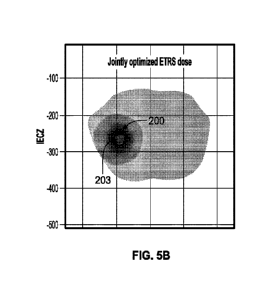
[0021] FIG. 5A depicts a simulation plot of ETRS radiation dose distribution
that has not been
jointly optimized with ITRS dose, and FIG. 5B depicts a simulation plot of
ETRS radiation dose
distribution that has been jointly optimized with ITRS dose accordingly to the
methods
described herein.
[0022] FIG. 5C depicts the IRT dose (Dirt) distribution and DVH curves, FIG.
5D depicts the
EBRT dose (Debrt) distribution and DVH curves, and FIG. 5E depicts the
combined IRT and
EBRT dose (kiDirt + keDebrt) distribution and DVH curves resulting from a
simulation of a
combined external and internal radiotherapy dose delivery with joint
optimization.
9
CA 03169406 2022-07-27
WO 2021/154746 PCT/US2021/015119
[0023] FIG. 6 depicts a flowchart representation of one variation of a method
for generating a
joint radiotherapy treatment plan that comprises jointly optimizing ITRS and
ETRS radiation
dose and adapting or adjusting the ITRS radiation dose and/or ETRS dose for a
future treatment
session.
[0024] FIG. 7A depicts a flowchart representation of one variation of a method
for generating
a joint radiotherapy treatment plan that comprises jointly optimizing RN
radiation dose and
BGRT radiation dose and optionally, adapting or adjusting the RN radiation
dose and/or BGRT
dose for a future treatment session.
[0025] FIG. 7B depicts a flowchart representation of one variation of a method
for joint
optimization of RN radiation dose and BGRT radiation dose. BGRT treatment
planning includes
the calculation of one or more firing filters p.
[0026] FIG. 8 depicts a table that summarizes several examples of
radiopharmaceutical
compounds that may be used for image data acquisition (e.g., functional image
data) and
radiotherapy.
[0027] FIGS. 9A-9B depict one variation of a radiotherapy system.
[0028] FIG. 10 depicts one variation of a radiotherapy system.
[0029] FIG. 11 depicts another variation of a radiotherapy system.
DETAILED DESCRIPTION
[0030] Disclosed herein are systems and methods for generating a joint
radiotherapy treatment
plan by jointly optimizing for both the radiation dose provided by an internal
therapeutic
radiation source (ITRS) and the radiation dose provided by an external
therapeutic radiation
source (ETRS). One variation of a method comprises jointly optimizing both the
radiation dose
or fluence deliverable by an external beam radiation therapy (EBRT) system and
the injected
radiation dose of a radionuclide (e.g., IRT). In some variations, the joint
optimization of
radiation deliverable by both internal therapeutic radiation source(s) and
external therapeutic
radiation sources may be done only once before start of a treatment period. A
treatment period
may comprise multiple treatment sessions, some of which may be ITRS treatment
sessions and
some of which may be ETRS treatment sessions. Optionally, after one or more
ETRS and/or
CA 03169406 2022-07-27
WO 2021/154746 PCT/US2021/015119
ITRS treatment sessions in a treatment period, the ITRS and ETRS radiation
dose may be jointly
re-optimized based on updated or newly-acquired image data, such as image data
(e.g.,
functional image data) acquired during or between a treatment session. Jointly
optimizing the
ITRS and ETRS radiation dose between treatment sessions and/or throughout the
course of a
treatment period may help to adapt the radiation therapy to account for
biological changes in the
patient.
[0031] Generating a radiotherapy treatment plan by jointly optimizing for ITRS
dose and
ETRS dose may result in a cumulative dose profile that has better dose
homogeneity in patient
target regions than generating a radiotherapy treatment plan by separately
optimizing ITRS and
ETRS doses. Radiotherapy treatment plans that separately optimize ITRS and
ETRS dose
usually involve calculating an EBRT treatment plan using traditional
radiotherapy treatment
planning methods, and separately calculating the IRT dose. While calculating
the EBRT
treatment plan, the IRT dose is either not considered at all or simply treated
as a fixed dose
quantity. Similarly, while calculating the IRT dose, the dose provided by an
external therapeutic
radiation source is not considered. Before treatment, the separately
calculated EBRT treatment
plan and IRT dose may be combined, and may each be multiplied by a scaling
factor in order to
obtain a cumulative dose that meets prescribed dose levels and constraints.
FIG. 1 depicts one
example of a method (100) for generating a combined radiotherapy treatment
plan that
separately optimizes ITRS dose and ETRS dose (i.e., does not jointly optimize
ITRS and ETRS
dose), and delivers a combined dose that is a sum of ITRS dose and ETRS dose,
each optionally
multiplied by a scaling factor. Method (100) may comprise acquiring (102)
patient anatomical
data (e.g., CT image data), determining (104) patient organ contour data,
acquiring (106)
diagnostic functional imaging scans and/or functional image data, determining
(108) prescription
dose and organ dose limits and constraints, and determining (110) the number
of fractions or
treatment sessions in a treatment period. After these treatment parameters
(e.g., prescribed dose
to patient target regions, dose limits to organs at risk or OARs, dose
constraints, number of
fractions, etc.) have been determined, method (100) may comprise calculating
(112) the ITRS or
radionuclide dose based on the functional image data and then calculating
(114) the radionuclide
dose for delivery (IRT dose Dirt). For example, IRT dose is typically
calculated based on patient
weight and evaluated for toxicity based on functional image data. Functional
image scans
comprise image data that represent the biological distribution of a molecule
(e.g., an imaging
tracer such as a compound having a radionuclide and/or radiopharmaceutical)
inside of a patient.
11
CA 03169406 2022-07-27
WO 2021/154746 PCT/US2021/015119
In some variations, functional image data may be used to generate an image or
map of the
biodistribution and/or pharmacokinetics of the molecule within a patient.
Functional image data
may be combined (e.g., overlaid) with an anatomical image. Examples of
functional image scans
may include PET scans or SPECT scans, where the functional PET or SPECT image
data
provides information about the distribution of the PET or SPECT tracer within
a patient. These
scans may be combined with a CT scan, e.g., PET/CT, SPECT/CT scans. While the
various
methods disclosed herein are described in the context of using functional
image data such as
PET image data or SPECT image data, it should be understood that the methods
may also use
any imaging modalities, such as CT image data, MR image data, ultrasound image
data,
molecular image data, nuclear image data, etc.
[0032] In some variations, the imaging tracers used to generate imaging scans
(e.g., functional
imaging scans) may comprise a targeting backbone or carrier molecule that
binds to specific
cellular markers and a radioactive isotope (e.g., a positron-emitting isotope
in the case of PET
imaging). The targeting backbone may selectively bind to specific tumors,
while the radioactive
isotope may act as a marker that indicates the location of the tracer.
Alternatively, or
additionally, the radioactive isotope may act as a therapeutic radiation
source that lethally
irradiates a tumor when a sufficient quantity of the tracer accumulates at the
tumor. A
"theranostic" may be a compound that acts as both an imaging agent and a
therapeutic agent;
that is, having both therapeutic and diagnostic functions. An example of a
theranostic compound
is lutetium Lu-177 DOTA-TATE (e.g., LUTATHERAg), a labeled somatostatin
analogue
peptide. As a diagnostic agent, Lu-177 DOTA-TATE emits low energy gamma rays.
These
gamma rays can be imaged using SPECT or gamma cameras. For example, long term
(i.e., over
multiple days) pharmacokinetic information of the Lu-177 low-energy gamma-
emitting image
may be used to estimate the absorbed dose of the theranostic over the
treatment period. In some
variations, an imaging tracer with a targeting backbone and a radioactive
isotope may be used
for image scanning, and a radiopharmaceutical compound having the same
targeting backbone
but a different radioactive isotope may be used for treatment. The same
molecule, Lu-177
DOTA-TATE, also has a PET emitting version, Ga-68 DOTA-TATE. The PET images
may
have much better contrast, quantification, and resolution than the SPECT or
planar gamma
camera images of Lu-177. In some variations, the Ga-68 DOTA-TATE may be used
for initial
diagnostic evaluation to determine whether the patient is a candidate for Lu-
177 DOTA-TATE
therapy. Over the course of treatment, SPECT or planar gamma camera images of
the Lu-177
12
CA 03169406 2022-07-27
WO 2021/154746 PCT/US2021/015119
may be used to monitor the pharmacokinetics during the treatment period. Both
the initial Ga-68
DOTA-TATE and the SPECT Lu-177 DOTA-TATE images are images that may be used to
determine the absorbed dose of the radiopharmaceutical.
[0033] Method (100) may comprise separately calculating (116) a dose
deliverable by an
external beam radiation therapy system and EBRT dose (Debrt), and then
adjusting (118) the
cumulative IRT and EBRT dose by calculating the scaling factors ki and ke for
the IRT dose
and EBRT dose, respectively. Calculating the scaling factors ki and ke may
comprise
determining the values of ki and ke such that the cumulative IRT and EBRT dose
(kiDirt +
keDebrt) meets the prescribed dose to patient target regions. While adjusting
the cumulative
dose (118) for delivery, the calculated EBRT dose Debrt and the calculated IRT
dose Dirt are not
modified. Method (100) may then comprise delivering (120) the EBRT radiation
dose (keDebrt)
to the patient and then delivering (122) the IRT radiation dose (ktDtrt) to
the patient. Linearly
scaling the EBRT radiation dose may comprise modifying the dose rate of the
ETRS, i.e.,
adjusting the number of therapeutic radiation beam pulses emitted per unit
time. Linearly scaling
the IRT radiation dose may comprise linearly scaling the volume of the
radionuclide and/or
radiopharmaceutical that is injected or implanted into the patient.
[0034] However, linearly scaling and summing the IRT and EBRT radiation doses
that have
been separately optimized retains the heterogeneous dose distribution that
results from IRT.
FIGS. 2A-2C depict the dose distribution (upper plots) and dose-volume
histogram DVH (lower
plots) for a simulation of a treatment planning method that separately
optimizes ITRS and ETRS
dose, and scales and sums the doses for delivery. The PTV is represented by
the outer black line
(200) in the top panels of FIGS. 2A-2C. FIG. 2A depicts the IRT dose (Dirt)
distribution and
DVH curves, FIG. 2B depicts the EBRT dose (Debrt) distribution and DVH curves,
and FIG. 2C
depicts the combined IRT and EBRT dose (iciDtrt keDebrt) distribution and DVH
curves. The
DVH curve (202) corresponds to the dose delivered per volume
fraction/proportion of the PTV.
FIG. 2A shows that a radionuclide is able to provide a high dose to a
relatively small proportion
of the PTV (e.g., per the lower plot, the DVH curve shows that less than 10%
of the PTV
receives a dose greater than about 25 Gy), and that the high-dose region is
located at the center
of the PTV (e.g., per the upper plot, the high-intensity region is in the
central portion of the PTV
(200)). FIG. 2B shows that EBRT is able to provide a more homogeneous dose to
the PTV (e.g.,
per the lower plot, the DVH curve shows that 100% of the PTV receives a dose
that is greater
13
CA 03169406 2022-07-27
WO 2021/154746 PCT/US2021/015119
than about 50 Gy with a steep fall-off), and that the high-dose region
encompasses nearly the
entirety of the PTV (e.g., per the upper plot, the high-intensity region spans
nearly all of the PTV
(200)). However, when the IRT and EBRT doses are combined, the cumulative dose
distribution
in the PTV is relatively heterogeneous. The upper plot of FIG. 2C shows that
the high-dose
region is still largely located at the center of the PTV, and the DVH curve
shows a slower dose
fall-off. For example, while 80% of the PTV receives a dose of about 58 Gy,
20% of the PTV
receives a dose of about 77 Gy, a dose spread (204) of about 19 Gy over 60% of
the PTV.
Another way to quantify the effect is called the homogeneity index (HI). HI
may be calculated
by dividing the maximum dose or intensity level of the PTV by the minimum dose
or intensity
of the PTV. The HI over the PTV (200) when combining IRT dose and EBRT dose
that have
been separately optimized is 95 Gy/50 Gy or approximately 1.9. While linearly
combining IRT
dose and EBRT dose irradiate the majority of a tumor with sufficient levels of
radiation, the
heterogeneity may miss cancer cells at the edges of the tumor, which may
increase the likelihood
of recurrence. Furthermore, adjusting the delivered dose by a scaling factor
may provide a lethal
dose of radiation to the tumor(s), however, may also increase toxicity to the
patient and expose
the patient to unnecessarily high levels of radiation.
[0035] In contrast, a treatment planning method that comprises jointly
optimizing the radiation
dose from internal therapeutic radiation sources and external therapeutic
radiation sources may
help provide a therapeutic and more homogeneous dose of radiation to tumor(s)
with potentially
less toxicity. Combining both ITRS and ETRS radiation therapy and jointly
optimizing for the
radiation dose provided by both modalities may also facilitate precise
treatment of metastatic
cancer while minimizing the significant toxicity that can result from either
modality. The joint
radiotherapy treatment planning methods described herein comprise adjusting
both the ITRS
radiation dose and the ETRS radiation dose during the optimization step of
treatment planning.
Adjusting both ITRS and ETRS radiation doses may comprise modifying the ITRS
dose
distribution in conjunction with the ETRS dose distribution (and/or vice
versa), and evaluating
the cumulative ITRS and ETRS dose distribution to determine whether dose
constraints are met.
Jointly optimizing ITRS and ETRS dose together may impose dose constraints on
the ITRS dose
(and therefore, the combined dose) that are not typically included when ITRS
dose is calculated
separately. This may provide more granular and precise adjustment of the
cumulative dose
distribution so that dose and toxicity constraints are met while providing
lethal doses of radiation
to cancer cells.
14
CA 03169406 2022-07-27
WO 2021/154746 PCT/US2021/015119
Synergy Between Internal Radionuclide Therapy (IRT) and Biologically-based
External
Beam Radiation Therapy (EBRT)
[0036] Furthermore, EBRT methods that use image data (e.g., functional image
data) for
radiation delivery may have additional synergies with treatment planning
methods that comprise
joint optimization of ITRS and ETRS radiation dose. As described above and
depicted in the
method flowchart in FIG. 1, imaging scans of a patient using a radionuclide
may be necessary
for calculating the dosimetry of radionuclides and to determine how much
(e.g., volume)
radionuclide is to be injected in order to deliver a prescribed dose. Most
EBRT methods do not
require imaging scans, so a joint IRT and EBRT treatment plan would involve an
"extra"
imaging session. However, the generation of an EBRT treatment plan that relies
on image data
for radiation delivery already includes an imaging session, so the same image
data used for
EBRT treatment planning may also be used for IRT treatment planning and joint
optimization.
In one variation, the imaging agent used in the imaging session may have the
same targeting
backbone as the radiopharmaceutical used to deliver IRT. One example of an
EBRT method that
uses imaging data (e.g., functional imaging data) to guide radiation delivery
is biologically-
guided radiotherapy (BGRT). BGRT guides radiation to a patient based on PET
image data
acquired during a treatment session. A PET tracer is injected into a patient
before the treatment
session (e.g., as part of treatment planning and/or at the start of a
treatment session), and the rate
of PET tracer uptake and/or the location(s) of PET tracer accumulation provide
biodistribution
and/or pharmacokinetics data that represents the biological state and/or
function of a patient's
physiology. This data may be used to guide external beam radiotherapy and/or
to calculate the
dosimetry of a radionuclide. An image scan using a positron-emitting isotope
attached to a
targeting backbone may be used in the dosimetry calculations of the
radiopharmaceutical
compound having the same targeting backbone. In this way, BGRT and IRT may use
the same
PET tracer for diagnostic analysis for dosimetry for IRT and biological
guidance for BGRT. For
example, a PET imaging tracer may comprise a PET emitting isotope (e.g., Ga-
68) attached to
the targeted peptide DOTA-TATE and a radiopharmaceutical compound for
treatment may have
a beta emitting isotope (e.g., Lu-177) attached to the targeted peptide DOTA-
TATE. This PET
imaging tracer and radiopharmaceutical compound may be paired together for the
diagnosis and
treatment of somatostatin positive neuroendocrine tumors. Similarly, a single-
photon emitting
isotope suitable for SPECT imaging may be attached to a targeting backbone for
imaging, while
CA 03169406 2022-07-27
WO 2021/154746 PCT/US2021/015119
a radiopharmaceutical with the same targeting backbone but different
radioactive isotope may be
used for treatment.
[0037] While the examples disclosed herein pertain to joint optimization of
radiation doses
deliverable using one or more radionuclides and one or more external high-
energy photon
sources, it should be understood that the methods described herein may be used
for joint
optimization of radiation doses deliverable using any internal therapeutic
radiation source
(ITRS) and any external therapeutic radiation source (ETRS). An ITRS may
comprise any
compound or device that is configured to emit therapeutic levels of radiation
from inside a
patient's body, for example, a radionuclide (RN), a radiopharmaceutical,
and/or a radioactive
seed or microsphere (e.g., brachytherapy devices). In some variations, an ITRS
may be
injectable into the bloodstream of a patient and/or implantable at a patient
target region. For
example, a radioactive seed or microsphere may be injectable into the patient
bloodstream
and/or may be implantable at a patient target region. Internal radionuclide
therapy (IRT) refers to
any radiotherapy method where the therapeutic radiation source comprises a RN
(including
radionuclides that operate alone or in conjunction with a targeting backbone
as part of a
radiopharmaceutical) that is injected or implanted or otherwise attached to
the patient's body.
An "ITRS dose" refers to a radiation dose provided by an internal therapeutic
radiation source.
[0038] An ETRS may comprise any compound or device that is configured to emit
therapeutic
levels of radiation from outside a patient's body and can be directed toward
patient target
regions. For example, an ETRS may comprise a source of high-energy photons
(e.g., X-rays or
gamma rays) or particles (e.g., protons, neutrons, electrons, etc.), and may
include linear
accelerators (linacs), a cobalt-60 source, proton beam source, neutron beam
source, betatron, and
the like. One or more ETRS may be included as part of an external beam
radiotherapy (EBRT)
system. EBRT involves generating high-energy photon or particle beam and
shaping the beam to
direct it to target regions while shielding non-target regions. EBRT systems
may comprise one
or more high-energy photon and/or particle sources and a beam-shaping assembly
that may
comprise one or more jaws and collimators. Examples of EBRT systems include
stereotactic
body radiotherapy (SBRT) systems, intensity-modulated radiotherapy (IMIRT)
systems, image-
guided radiotherapy (IGRT) systems, biologically-guided radiotherapy (BGRT)
systems, etc.
Additional details and examples of EBRT systems are provided below. An "ETRS
dose" refers
to a radiation dose provided by an external therapeutic radiation source.
16
CA 03169406 2022-07-27
WO 2021/154746 PCT/US2021/015119
Methods for Joint Radiotherapy Treatment Planning
[0039] Methods for generating a joint radiotherapy treatment plan that
irradiates one or more
patient target regions may comprise jointly optimizing the radiation dose
and/or fluence to be
delivered using one or more ITRS and one or more ETRS. The method may then
comprise
jointly optimizing an external beam radiotherapy plan in conjunction with the
radionuclide
dosimetry based on a set of clinician-determined dose constraints. More
generally, joint
optimization of ITRS and ETRS doses may comprise adjusting both the ITRS
radiation dose and
the ETRS radiation dose (e.g., dose distributions) iteratively to meet
prescribed dose constraints
for one or more patient target regions. Dose constraints may be defined (e.g.,
by a clinician
and/or medical physicist) for one or more patient target regions, and the
constraints may
comprise one or more cost functions. Cost functions may include penalty
functions and may
include constraints on the toxicity of the ITRS to non-target regions such as
healthy tissue and/or
organs-at-risk (OAR), as well constraints on the radiation dose delivered by
both the ITRS and
ETRS. One example of ITRS-specific constraints relates to limiting broad
hematological toxicity
(e.g., sparing toxicity to the white blood cells by limiting the mean ITRS
radiation dose). Other
examples of ITRS-specific constraints are on the minimum and maximum of the
injected ITRS
radiation dose to handle practical constraints on the preparation and
injection of the RN or to
help ensure that a minimum amount of RN is injected to treat non-visible micro-
metastases. For
example, injectable RN or radiopharmaceuticals may only be available in
certain volumes (e.g.,
absolute volume in mL, or radioactivity levels per unit volume kBq/mL) or
discrete or quantized
radiation dose levels (e.g., absolute dose levels in Gy, or dose levels per
unit radioactivity
Gy/kBq, or radioactivity levels tC). For example, an injectable RN may be
provided from about
100 mC to about 300 mC, in increments of about 100 mC. During the joint
optimization of the
ITRS dose and ETRS dose, the ITRS dose may be constrained to the pre-specified
injectable
volumes and/or radiation quanta. In joint optimization, both the ITRS dose and
the ETRS dose
are iteratively adjusted until dose requirements and/or constraints are met.
After joint
optimization, a joint radiotherapy treatment planning method may comprise
calculating a
quantity of the ITRS that is to be introduced into the patient to deliver the
ITRS dose, and the
calculated quantity of ITRS may be injected and/or implanted into the patient.
For example, the
treatment planning method may comprise calculating a volume of a RN and/or
radiopharmaceutical that is to be injected into the patient at each treatment
session. Alternatively
or additionally, the treatment planning method may comprise calculating a
quantity of
17
CA 03169406 2022-07-27
WO 2021/154746 PCT/US2021/015119
radioactive seeds and/or microbeads to be implanted at one or more patient
target regions. In
some variations, an implantable radiation source may comprise radioactive
tubes or wires, and
the radiotherapy treatment plan may further specify the implantation location
of the tubes or
wires, the number of tubes or wires, the implantation time, and/or the
radioactivity levels of the
tubes or wires. A joint radiotherapy treatment planning method also comprises
calculating a
delivery fluence map for an EBRT system and/or generating a set of EBRT system
machine
instructions for each treatment session. A delivery fluence map may comprise a
set of beamlets
and beamlet intensities for delivery using a high-energy beam during a
treatment session. In
some variations, an EBRT system may segment the delivery fluence map
calculated by the
treatment planning method into machine instructions during the treatment
session (i.e., real-time
segmentation where machine instructions are not calculated before the
treatment session).
Alternatively, an EBRT system may execute the machine instructions generated
by the
radiotherapy treatment planning system.
[0040] In some variations, generating a radiotherapy treatment plan may
comprise acquiring
planning images (e.g., CT images, functional image data such as PET image
data), defining the
contours of the patient target regions, calculating the dosimetry of a RN (or
any ITRS), and
determining the dose prescription for the patient target regions, OARs, and/or
any other region
of interest. A dose prescription may include the dose constraints that the
ITRS/ETRS combined
therapy needs to meet for a desired therapeutic effect. For example, the dose
prescription may
define the minimum necessary dose a patient target region must receive in
order to reduce or
block the proliferation of cancer cells. A dose prescription may also define
the maximum dose
that an organ system may receive to avoid unwanted side effects. In some
variations, the course
of treatment during a treatment period may be predefined by the clinician. For
example, the
clinician may determine the number and order of ITRS and ETRS treatment
sessions in a
treatment period. For example, a ETRS treatment session may be coupled with
one ITRS
treatment session. Alternatively, or additionally, several ETRS treatment
session may precede an
ITRS treatment session (or vice versa). The number, order, and type of
treatment sessions may
be used to calculate the ITRS dose and the ETRS dose so that they may be
summed into the
same equivalent dose space. The equivalent dose space may be scaled in units
relevant for ETRS
delivery (Gy), and/or in units relevant for ITRS delivery (absorbed Gy),
and/or in an
intermediate ETRS/ITRS dose space. In some variations, a mathematical method
called
biological-equivalent dose (BED) may be used to renormalize delivered ETRS
and/or absorbed
18
CA 03169406 2022-07-27
WO 2021/154746 PCT/US2021/015119
ITRS dose based on the fractionization and timing of the delivery of the dose.
Some methods
may comprise jointly optimizing for ITRS dose and ETRS dose in the BED space.
[0041] While the variations of joint radiotherapy treatment planning methods
provided herein
comprising jointly optimizing ITRS radiation dose and/or ETRS radiation dose,
it should be
understood that in other variations, joint radiotherapy treatment planning
methods may comprise
jointly optimizing ITRS radiation fluence and/or ETRS radiation fluence. In
some variations,
joint optimization methods may comprise optimizing for ITRS radiation dose and
ETRS
radiation fluence. For example, a joint radiotherapy treatment planning method
may comprise
jointly optimizing for IRT injection dose and EBRT fluence.
[0042] FIG. 3 is a flowchart depiction of one variation of a method (300) for
generating a joint
radiotherapy treatment plan that comprises jointly optimizing ITRS and ETRS
radiation dose.
Method (300) may comprise acquiring (302) patient anatomical data (e.g., CT
image data),
determining (304) patient organ contours, acquiring (306) functional imaging
scans, determining
(308) prescription and organ dose constraints, determining (310) the number of
fractions or
treatment sessions during a treatment period, and calculating (312) dosimetry
of a radionuclide
(or any desired ITRS) from the functional imaging scan(s). Optionally, the
functional image
data, anatomical image data, prescribed dose requirements, and RN dosimetry
data may be
provided (314) to a treatment planning system, which may comprise software
code that may be
executed by a treatment planning controller having one or more processors. In
some variations,
treatment planning analyses and calculations (302-316) of method (300) may be
performed
directly using the treatment planning system. Method (300) may further
comprise jointly
optimizing (316) RN dose and ETRS dose to generate a joint radiotherapy
treatment plan that
specifies a dose to be delivered by the RN and a dose to be delivered using
the ETRS (e.g., any
EBRT system, BGRT system). In some variations, the joint radiotherapy
treatment plan
comprises a delivery fluence map and/or machine instructions for an EBRT
system and injection
volume for a specified type of RN or radiopharmaceutical. The RN dosimetry may
be calculated
using one or more methods for determining the absorbed dose per unit of
injected dose. For
example, the RN dosimetry may use the treatment planning CT image for
anatomical tissue
density data, the functional image of the concentration and/or biodistribution
of the radionuclide,
a model of the pharmacokinetics over time for the radionuclide, and/or a Monte-
Carlo
simulation of the of the deposition of energy in the patient. Alternatively,
or additionally, the RN
19
CA 03169406 2022-07-27
WO 2021/154746 PCT/US2021/015119
dosimetry may be calculated using voxel-based methods based on S-value kernels
that compute
absorbed dose per unit of injection from an image. Alternatively, or
additionally, RN dosimetry
may be calculated using convolution of an image using a Dose-Volume-Kernel
(DVK).
Optionally, RN dosimetry may be further scaled by a biological equivalent dose
model, so that
the RN dosimetry is in the same scalar space as the ETRS dose.
[0043] Determining (310) the number of fractions or treatment sessions in a
treatment period
may comprise calculating the number of ETRS sessions based on a set number of
RN session(s),
and/or calculating the number of RN sessions based on a set number of ETRS
session(s). The
total number of treatment sessions, and/or the number of each type of
treatment session (i.e.,
ETRS session, RN session) may be set by a clinician or a clinic policy, and/or
may be calculated
by the treatment planning system. The clinician may use clinical trial data to
determine the
optimal fractionation scheme for a given indication. Additionally, the
clinician may use
histological or diagnostic blood test information to measure the
aggressiveness of the tumor. A
more aggressive tumor may have a higher dose per fraction for either ETRS or
RN or more
fractions to achieve a higher BED dose. Also, the clinician may adjust the
fractionization
scheme to reduce toxicity to a given OAR. For a treatment planning system to
automatically
calculate the number of fractions or treatment sessions, a tumor control
probability model (TCP)
and a normal tissue complication model (NTCP) may be generated for each of the
targets and/or
the tissues in the patient. The TCP and NTCP models can be used to derive a
recommended
fractionization scheme to the clinician. Alternatively, the patient may have
been treated
previously and this information may be used to determine the number of
fractions. In some
variations, a treatment planning system (i.e., which may also perform the
joint optimization
methods described herein) may calculate the number of fractions based on the
dose prescription
in terms of biological effective dose to each patient target region,
anatomical location of each
patient target region (e.g., "lung, left upper", location data that identifies
the relative tumor
location and nearby organs-at-risk), pathology data (e.g., tumor staging,
whether a patient target
region is a primary lesion or a metastatic lesion, genetic test data, and/or
histology data),
acceptable toxicity risk to organs-at-risk (e.g., normal tissue complication
probability NTCP),
and/or any prior treatment (e.g., radiation dose, CT/RTSS from prior
irradiation, chemotherapy,
and/or timing of any prior treatments). A proposed number of treatment
sessions or fractions for
ITRS and ETRS and a treatment session schedule may be determined and displayed
to the
CA 03169406 2022-07-27
WO 2021/154746 PCT/US2021/015119
clinician on a monitor for selection (e.g., approve and proceed, disapprove
and re-calculate)
and/or further modification (e.g., approve with clinician modifications).
[0044] Method (300) may optionally comprise treating the patient according to
the joint
radiotherapy plan. For example, method (300) may comprise delivering (318) one
or more
treatment sessions or fractions using an EBRT system and injecting (320) the
patient with the
calculated dose of RN in one or more treatment sessions. In some variations,
method (300) may
optionally comprise waiting (322) for the RN to decay before another EBRT
treatment session
and/or RN injection (i.e., a RN treatment session). Optionally, between the
treatment sessions,
additional image data (e.g., functional image data) may be acquired. The
additional image data
may be used to adapt the EBRT and/or RN dose for a future treatment session.
The imaging
tracer for the acquisition of image data may have the same targeting backbone
as the RN so that
the dosimetry of the RN may be updated to reflect any changes in the
biological and/or
physiological state of the patient as they are being treated (e.g., during the
treatment period,
between treatment sessions or fractions within the treatment period). In some
variations, imaging
data acquired during a treatment session and/or images acquired between
treatment sessions
(e.g., between ITRS treatment sessions, between ETRS treatment sessions, etc.)
may be used to
adapt the radiation dose for the next treatment session. Adapting a radiation
dose for a future
treatment session may comprise joint re-optimization with a different number
of fractions or
treatment sessions for that treatment period (e.g., changing the number of
ITRS sessions, the
number of ETRS sessions, or both, from the first joint optimization).
[0045] FIG. 4 depicts one variation of a method for joint optimization of ITRS
radiation dose
and ETRS radiation dose, which may be used with any of the joint radiotherapy
treatment
planning methods described herein. Method (400) may comprise calculating (408)
a radiation
dose that is deliverable by an ITRS (DO ITRS), calculating (410) a radiation
dose that is
deliverable by an ETRS (Do ETRS), adjusting (412) the ITRS and ETRS doses (Do
ITRS, DO ETRS)
to meet the dose prescription (as determined by the clinician), and evaluating
(414) one or more
prescribed dose requirements (e.g., constraints). If the prescribed dose
requirements are not met,
method (400) comprises iteratively adjusting the ITRS and ETRS dose
distributions (DO ITRS,
Do ETRS) until the requirements at met. After the dose requirements are met,
method (400) may
comprise outputting (416) the ITRS dose (D/TRs) and ETRS dose (DETRs) for
delivery during
one or more treatment sessions. In some variations, method (400) may comprise
outputting one
21
CA 03169406 2022-07-27
WO 2021/154746 PCT/US2021/015119
more of ITRS injection dosage (420), ETRS system machine instructions (422),
and/or ETRS
system fluence map (424).
[0046] In some variations, method (400) may optionally comprise determining
(402) the
prescribed dose distribution (y) and dose constraints to the patient,
calculating (404) an ITRS
dose-mapping matrix (R), and calculating (406) an ETRS dose-mapping matrix (A)
which may
be used to adjust or iterate (412) on the ITRS and ETRS doses (DO ITRS, DO
ETRS). The prescribed
dose distribution may be the cumulative radiation dose to the patient as
specified by a clinician
and may be represented by a vector of voxels (y) in the patient, each voxel
having a dose value.
Calculating (404) the ITRS dose-mapping matrix (R) may comprise determining
the relationship
between the volume of an injected or implanted ITRS and its delivered dose. In
some variations,
radionuclide dosimetry is performed for a fixed injection volume, and the
dosimetry of a
radionuclide treatment may be generally linearly related to the amount of
radionuclide that is
injected. Calculating (404) the ITRS dose-mapping matrix (R) may comprise
mapping one or
more images (I) (e.g., functional images) to the biologically-equivalent
absorbed dose Gy per
unit of an injected ITRS (e.g., RN and/or radiopharmaceutical). The images may
be acquired
using an imaging tracer that has a carrier molecule or targeting backbone that
is the same as the
carrier molecule or targeting backbone for the ITRS. This mapping (F) may be
given by:
F (I[¨kBql) = R[ GY
ml kBq
[0047] The ITRS radiation dose (Do ims) that is capable of being delivered to
the patient may
be represented by a similar linear relationship as the injected dose scalar
(q, which may, more
generally, be a quantity of the ITRS) multiplied by the ITRS dose-mapping
matrix (R), which
maps the injected dose (q) to the voxelized dosimetry Do ITRS= That is:
Do ITRS = Rq
[0048] Any of the RN dosimetry methods described above may be used to
calculate (404) the
ITRS dose-mapping matrix (R). Alternatively, or additionally, the ITRS
dosimetry may be non-
linearly related to the amount of injected ITRS, and may incorporate time-
variant
pharmacokinetics of the ITRS (e.g., where at high injection volumes, the ITRS
has a physiologic
effect on the patient that is independent of the ionization radiation).
22
CA 03169406 2022-07-27
WO 2021/154746 PCT/US2021/015119
[0049] Alternatively or additionally to delivering therapeutic doses of
radiation using a single
radiopharmaceutical in a single treatment session, internal therapeutic
radiation may be
delivered using multiple different radiopharmaceuticals over one or more
treatment sessions. In
some variations, an ITRS may comprise two different radiopharmaceuticals. For
example,
internal therapeutic radiation may be provided by two radiopharmaceuticals
that comprise Y-90
and Lu-177. Because the 0 energy of Y-90 and Lu-177 are different, they may
have different
dosimetry. By combining the two different radiopharmaceuticals, the ITRS dose
distribution
may be tuned and adjusted in a way that may not be attainable using a single
radiopharmaceutical. The total ITRS dose may be represented by a first
injection of a first
radionuclide (71) and a second injection of a second radionuclide (q2). The
first and second
radiopharmaceuticals may be injected simultaneously or sequentially into the
patient. Each
radiopharmaceutical may have a different dose mapping matrix (R1, R2), but the
doses may sum
linearly.
D o ITRS = RiCh R2q2
[0050] Joint optimization for two radiopharmaceuticals may generate the
optimal combination
of the two different injected radiopharmaceuticals q2). For example, one
variation of joint
radiotherapy treatment may use Lu-177 as a first radionuclide (e.g., Lu-177
conjugated with
DOTA-TATE), and Y-90, which has a much larger 0 range, as a second
radionuclide (e.g., Y-90
conjugated with DOTA-TATE). Joint optimization may comprise adjusting the
adjusting the
injected dose of the two RNs in conjunction with the ETRS dose such that the
cumulative dose
meets prescribed dose requirements. This method may be extended for any number
of N
radiopharmaceuticals, e.g., D o ITRS = R2q2 === RNqN.
[0051] The ETRS dose (D o ETRS) deliverable to the patient may be modeled as a
linear system
and calculated by multiplying the ETRS dose-mapping matrix (A) with the ETRS
fluence (x)
deliverable by a EBRT system (for example) to the patient:
DO ETRS = Ax
[0052] Iterating (412-414) on ITRS and ETRS doses may comprise scaling the
ITRS and
ETRS doses into a dose space that is equivalent to the prescription dose space
(418) and iterating
on RN quantity (q) and ETRS fluence (x). In some variations, the prescription
dose, ITRS dose
23
CA 03169406 2022-07-27
WO 2021/154746 PCT/US2021/015119
and ETRS dose may all be defined in the BED space. The sum of ITRS and ETRS
doses in the
BED space (D cumulative) aim to approximate or match the radiation dose
prescribed by the
clinician, i.e., the prescribed dose distribution (y):
Dcumulative = y, where
cumulative = DO ITRS DO ETRS = Rq + Ax
[0053] In addition to requiring that the ITRS and the ETRS radiation dose sum
to the
prescribed dose distribution, prescribed dose requirements may comprise a set
of constraints on
all the prescription objectives. In some variations, these constraints may be
convex constraints.
These convex constraints may imposed on the ETRS fluence (x), on the ITRS
quantity (q), on
the dose deliverable by the ITRS (DO ITRS), on the dose deliverable by the
ETRS (DO ETRS),
and/or on the cumulative dose (D cumulative = DO ITRS DO ETRS), and/or on any
combination
thereof An example of a convex constraint which may be unique to joint
optimization is the
minimum dose on the patient target region (e.g., PTV) where D cumulative = DO
ITRS DO ETRS,
does not exceed a predefined dose value (in Gy). The ITRS quantity (q) may be
constrained to
be within a range of acceptable quantities (i.e., q must be within a specified
range), and/or may
be constrained such that it is an integer multiple of quantized steps. For
example, for practical
reasons on dosage, the ITRS quantity may be only available in certain discrete
dosages. The
joint optimization may then have to optimize the injected dose (q) over a
limited set of fixed
dosages. In some variations, constraints may be derived based on a previously-
delivered ITRS
dose and/or ETRS dose (e.g., from a previous treatment period, from a previous
course of
therapy), and/or may optionally include constraints derived from toxicity
models of OARs
and/or healthy tissue. For example, if an OAR was subject to substantial
irradiation in a previous
treatment session or period, the dose constraint for the OAR may be more
stringent (i.e., to
guarantee a lower level of irradiation) for the next treatment session or
period. Such toxicity
constraints may be applied to the ITRS dose, the ETRS dose, and/or the
cumulative dose.
[0054] In some variations, these constraints may be weighted by a linear
factor that defines or
approximates their relative importance. For example, dose constraints may
comprise one or
more cost functions, and optionally, each cost function may be weighted by an
individual scaling
factor. Prescribed dose requirements or constraints (C) may comprise one or
more cost functions
and may include, for example, one or more of a cost function C(x) on radiation
fluence (x),
24
CA 03169406 2022-07-27
WO 2021/154746 PCT/US2021/015119
and/or a cost function C(q) on ITRS quantity (q), and/or a cost function C(Ax)
on Do ETRS,
and/or a cost function C (Rq) on DO ITRS, and/or a cost function
C(Dcumulative). These may each
optionally be weighted by an individual scaling factor (wi, vvp Wk, w1, Wm).
For example, a cost
function on the fluence can be used to optimize treatment time in the context
of j oint delivery.
Optionally, a cost function on the ETRS dose may be included to limit skin
dose and/or radiation
burn toxicity. For example, a cost function on the injected dose (q) can be
optimized ensuring
that the dose value is one that may be feasible to prepare and introduce into
the patient. For
example, a cost function of Do RN might optimize hematological toxicity (e.g.,
a cost function
that prioritizes the preservation of white blood cells) independent of ETRS
dose. Another
example is a cost function imposed on the cumulative ITRS and ETRS dose D
cumulative that
limits the mean combined dose to the heart.
C = Wt (x) + wiCi(q) +1wkCk(Ax) +IwiCi(Rq) +Iwn,Cm(Dcumulative)
[0055] In some variations, optimization constraints may be met based on a
priority ranking.
For example, each dose constraint may be ranked, and during optimization,
constraints may be
satisfied or met based on the corresponding priority ranking. For example, in
joint optimization,
RN constraints may be prioritized over ETRS constraints or vice versa.
Alternatively, for
example, the constraints may be prioritized based on organ system so that
different ETRS
constraints and ITRS constraints may have different priority rankings.
[0056] Methods of joint optimization may optionally comprise defining dose
constraints
where one or more cost functions are designated as high-priority (e.g.,
mandatory) cost
functions, and designating the other cost functions as low-priority (e.g.,
optional) cost functions.
The high-priority cost functions may be assigned the highest possible weight
and/or priority
ranking, and the low-priority cost functions may be assigned a lower weight
and/or priority
ranking. In some variations, the high-priority cost functions may have more
"stringent"
constraints, while the low-priority cost functions may have more "lax"
constraints. For example,
a high-priority cost function may tightly limit irradiation of the heart (or
any desired OAR) to a
range that is less than about 1 Gy, while a low-priority cost function may
limit irradiation of the
tissue around a patient target region to a broader range of no more than about
5 Gy. In some
variations, the clinician may prioritize bone marrow toxicity and/or liver
toxicity over potential
toxicity to the pancreas and/or bladder. That is, the constraints on the bone
marrow and/or liver
CA 03169406 2022-07-27
WO 2021/154746 PCT/US2021/015119
must be met before any constraints on the pancreas and/or bladder are
evaluated. In some
variations, the clinician may set the weight(s) and/or priority ranking(s) of
the high-priority cost
functions, and based on this clinician input, the treatment planning
system/optimizer may auto-
calculate the weight(s) and/or priority ranking(s) of the lower-priority cost
functions. During
joint optimization, the cumulative RN and/or ETRS dose must satisfy the high-
priority cost
functions at the specified weight and/or priority ranking (e.g., reduce the
value of any high-
priority penalty functions), while the low-priority cost functions may be
satisfied at varying
lower weights and/or priority rankings. For example, the range of acceptable
values of low-
priority cost functions may be wider than the range of acceptable values of
high-priority cost
functions. The weights and/or priority rankings of the low-priority penalty
functions may be
adjusted (e.g., automatically adjusted and/or calculated) relative to each
other in order to meet
the prescribed dose constraints or requirements. The acceptable ranges may be
specified by the
clinician and/or calculated by the treatment planning system (and may be
subject to clinician
review and/or approval).
[0057] Some methods of j oint optimization may optionally display a set of
clinical objectives
to a clinician, and the specific dose constraints and cost functions for
guiding joint optimization
may be defined based on the clinical objectives that are selected by the
clinician. This may
facilitate the definition of dose constraints and/or setting of specific cost
functions for clinicians,
and reduce the setup time for treatment planning and joint optimization. In
addition, defining
dose constraints based on clinical objectives may promote ease-of-use and
comprehensibility of
the treatment planning system for a broader range of clinicians, including but
not limited to,
radionuclide specialists and radiation oncologists. This may help ensure that
the patient's
treatment goals and needs are met, and help ensure that organs-at-risk are
correctly defined for
the treatment planning system. In one variation, each clinical objective may
be linked to one or
more cost functions. In some variations, an example of a clinical objective
could be control of all
tumors greater than about 1 cm by having a cost function on the minimum dose
on the PTV that
must be greater than 50 Gy. An example of a clinical objective could be to
limit the probability
of grade 2 kidney toxicity to less than 10% by linking that to a cost function
that limits the
maximum kidney dose to less than 40 Gy. Another example of a clinical
objective could be to
limit the probability of different grades of rectal bleeding as specified by a
clinician. For
example, a clinician could specify the acceptable risk of rectal bleeding
Grade 1 to a first level
(e.g., 1.25) or a second level (e.g., 1.10). If the risk of a Grade 1 rectal
bleed is set to the first
26
CA 03169406 2022-07-27
WO 2021/154746 PCT/US2021/015119
level, the treatment planning system may translate this clinical objective to
a dose constraint of
V7OGy < 5% rectal volume; if the risk of a Grade 1 rectal bleed is set to the
second level, the
treatment planning system may translate this clinical objective to a
constraint of V5OGy < 5%
rectal volume. While two levels of clinical risk are provided in this example,
it should be
understood that there may be any number of clinical risk levels that
correspond to a number of
cost functions or dose constraints. The cost functions from the one or more
clinical objectives
may be evaluated during joint optimization. Optionally, when multiple clinical
objectives are
selected, the clinician may be able to assign a priority or weight to each
clinical objective
relative to the other clinical objectives (e.g., set a priority ranking for
each clinical objective).
Joint optimization may then iterate on RN dose and ETRS dose to meet dose
constraints and/or
cost functions that have been prioritized and/or weighted according to their
corresponding
clinical objective(s).
[0058] In some variations of methods for joint optimization, a convex
optimizer may be used
to solve for the optimal ETRS fluence (x) and the optimal injected RN dose (q)
given dose
constraints that comprise one or more of the above cost functions. The
optimization may be
performed, evaluated, and analyzed in a treatment planning system, and may be
reviewed and
approved by a clinician.
[0059] In some variations, the prescribed dose distribution (y) may be
represented by a
bounded dose-volume histogram (bDVH) having a nominal prescribed dose curve
and any dose
delivery uncertainty is represented by an upper bound curve and a lower bound
curve. The upper
and lower bounds of the prescribed dose distribution bDVH may be calculated
based on
uncertainties and/or variabilities in radiation dose delivered by an ITRS
and/or an ETRS. For
example, ETRS dose delivery uncertainties and/or variabilities may arise from
patient motion
(e.g., respiratory motion, cardiac motion, physiologic motion that may alter
the position of the
patient target region during radiation delivery), high-energy radiation source
precision and/or
accuracy, and the like. Since the dose provided by an ITRS is calculated using
an imaging scan
(e.g., a functional imaging scan), any image base variance in the scan may
cause variance in the
dose estimate. Imaging scan uncertainties and/or variabilities may arise from
variations in blood
flow rate, perfusion distribution, pharmacokinetics, binding specificity of
the targeting
backbone, injection dose measurement, mis-calibration of the scanner,
limitations of the image
reconstruction algorithm, and the like. ITRS dose delivery uncertainties
and/or variabilities may
27
CA 03169406 2022-07-27
WO 2021/154746 PCT/US2021/015119
arise from variations in blood flow rate, perfusion distribution,
pharmacokinetics, binding
specificity of the targeting backbone, injection dose measurement uncertainty,
BED modelling
errors, and the like. Other ITRS dose variabilities may arise from changes in
the patient's
physiological or biological state. For example, there may be changes in the
patient's metabolism
and/or gastrointestinal state (e.g., constipation, gastritis, etc.) that may
alter the rate and manner
in which an injected or ingested RN or radiopharmaceutical is excreted. If a
patient is having
gastric issues, a greater percentage of a RN may be excreted through the
urinary tract instead of
the gastrointestinal tract. There may also be interactions between other
medications taken by the
patient and the RN or radiopharmaceutical that may affect the ITRS dose and
kinetics. ETRS
and ITRS dose uncertainties may be combined to derive the upper bound of
delivered dose and
the lower bound of delivered dose. In some examples, the upper and lower
bounds may be
derived using models of ETRS and/or ITRS dose uncertainties so that
interactions between
different uncertainties may be represented in the boundaries of bDVH.
Iterating (412-414, 418)
on ITRS and ETRS doses in the BED space may comprise adjusting one or both of
the D o ITRS
and DO ETRS such that the DVH of the cumulative dose (D cumulative) is within
the upper and
lower bounds of the prescribed dose distribution (y) bDVH. In some variations,
this may
comprise iterating on RN quantity (q) and/or ETRS fluence (x) until the DVH of
the cumulative
dose (Dcumulative) is within the upper and lower bounds of the prescribed dose
distribution (y)
bDVH. The output or result of jointly optimizing ITRS and ETRS doses may
comprise one or
more DVH curves that represent one or more cumulative doses (i.e., a range of
cumulative
doses) that are within the upper and lower bounds of the prescribed dose
distribution (y) bDVH.
The output of j ointly optimizing ITRS and ETRS doses may comprise one or more
solutions to
the same optimization problem set with different local minima for non-target
volumes. For
example, if there are three cost functions participating at low cost for
different OARs in the
optimization that would achieve the target prescription dose equally well, the
optimizer may
produce DVH curves and dose distributions for each of these results for the
clinician to choose
between. In all three cases, the prescription dose is met equally well, but
the individual doses for
OARs may differ substantially. In some variations, the joint optimization may
generate a set of
DVH curves for the optimized cumulative doses that fall within the upper and
lower bounds of
the prescribed dose distribution bDVH. Evaluating (414) the constraints may
optionally include
clinician evaluation and/or selection of a particular dose distribution based
on the DVH curves.
For example, this set of DVH curves may be displayed to the clinician, along
with the individual
28
CA 03169406 2022-07-27
WO 2021/154746 PCT/US2021/015119
ITRS dose and the ETRS dose for each of the DVH curves, and the clinician may
select one of
the DVH curves and its corresponding ITRS and ETRS doses for delivery. For
example, in a set
of DVH curves that meet the prescribed dose requirements and/or fall within
the bounds of the
prescribed dose bDVH for a patient target region, the dose to one or more OARs
may vary, and
as part of evaluating the jointly optimized dose distributions, a clinician
may select the dose
distribution that delivers less radiation dose to a particular OAR as compared
to other dose
distributions. After evaluation of the possible radiation doses for delivery
based on dose
constraints/requirements and/or clinician selection, the joint optimizer may
output (416) the
ITRS dose and the ETRS fluence/dose that is to be delivered during a treatment
period.
[0060] After clinician approval of the joint radiotherapy treatment plan, the
treatment can be
delivered and administered to the patient. In one variation, joint
radiotherapy treatment planning
only occurs once and the entire course of treatment (i.e., all of the ITRS
dose and ETRS dose
that were part of the same joint optimization) is delivered to the patient
over multiple treatment
sessions during a treatment period.
[0061] FIGS. 5A-5E depict the ITRS and/or ETRS depict dose distributions
(Dims, DETRs )
for a simulation of a treatment planning method that jointly optimizes ITRS
dose and ETRS dose
using the methods depicted in FIGS. 3-4 and described above. These plots
depict the differences
in dose distribution when ITRS and ETRS dose are separately optimized as
compared to when
they are jointly optimized. FIG. 5A depicts an ETRS dose distribution
resulting from separately
optimizing ETRS dose from ITRS dose (i.e., standard EBRT optimization
methods), and FIG.
5B depicts an ETRS dose distribution resulting from jointly optimizing ETRS
and ITRS doses
(i.e., using the joint optimization methods disclosed herein). As shown in
FIGS. 5A-5B, the
ETRS dose map has a significantly different dose distribution. The contour of
the PTV is
represented by the outer black line (200). When ITRS and ETRS doses are not
jointly optimized,
the ETRS dose distribution is homogeneous over the entire PTV region (FIG.
5A). However, the
ETRS dose distribution resulting from joint optimization has a central region
of lower dose
(203). This "donut" shaped ETRS dose map shown in FIG. 5B reflects the
incorporation of ITRS
dose to the cumulative dose distribution as part of joint optimization, since
ITRS dose tends to
aggregate strongly toward the central region of larger tumors. Without jointly
optimizing ETRS
dose with ITRS dose, the resulting cumulative dose map may create an intense
radiation "hot
spot" in the center of the PTV. By incorporating the ITRS dose with ETRS dose
optimization,
29
CA 03169406 2022-07-27
WO 2021/154746 PCT/US2021/015119
the ETRS dose may be reduced for areas in which the ITRS provides a
therapeutic level of
radiation. This may help reduce the overall radiation exposure and toxicity to
the patient, while
still providing therapeutic levels of radiation to patient target regions.
[0062] FIGS. 5C-5E depict the dose distribution (upper plot) and dose-volume
histogram
DVH (lower plot) for a simulation of a treatment planning method that jointly
optimizes ITRS
and ETRS dose. The PTV is represented by the outer black line (200) in the top
panels of FIGS.
5C-5E. FIG. 5C depicts the jointly optimized ITRS dose (Dims = Rq)
distribution and DVH
curves, FIG. 5D depicts the jointly optimized ETRS dose (DETRs = Ax)
distribution and DVH
curves, and FIG. 5E depicts the combined jointly optimized ITRS and ETRS dose
(Rq + Ax =
y) distribution and DVH curves. The DVH curve (212) corresponds to the dose
delivered per
volume fraction/proportion of the PTV (e.g., a patient target region). FIG. 5C
shows that an
ITRS such as a RN or radiopharmaceutical compound is able to provide a high
dose to a center
portion of the PTV (e.g., per the upper plot, the high-intensity region is in
the central portion of
the PTV (200)). FIG. 5D shows that with joint optimization, the ETRS dose
distribution
accounts for the contribution of the ITRS (i.e., in the center of the PTV) so
that the ETRS dose
toward the center portion of the PTV may be reduced, resulting in a "donut"
ETRS dose
distribution (203). FIG. 5E shows that when the jointly optimized ITRS and
ETRS dose
distributions are combined, the cumulative dose distribution in the PTV is
more homogeneous
than when the ITRS and ETRS dose distributions are separately optimized (i.e.,
in comparison to
the dose distributions depicted in FIG. 2C). In particular, the DVH curve for
the combined dose
distribution in FIG. 5E shows a rapid dose fall-off. For example, 80% of the
PTV receives a
dose of about 52 Gy and 20% of the PTV receives a dose of about 57 Gy, a dose
spread (214) of
about 5 Gy over 60% of the PTV (as compared to a dose spread of about 19 Gy in
FIG. 2C). The
dose distribution may also be quantified as the homogeneity index (HI). An HI
that approaches 1
is more homogenous and may be desired because it does not deliver extra dose
to the center of
the PTV, which may be unnecessary to kill the tumor. In FIG. 5E, the HI of the
dose delivered to
the PTV by jointly optimizing ITRS dose and ETRS dose is approximately 65 Gy
(max)/50 Gy
(min) or HI of 1.3. In contrast, as depicted in FIG. 2C, the HI of the dose
delivered to the PTV
by separately optimizing ITRS dose and ETRS dose is approximately 95 Gy/50 Gy
or 1.9. If
other higher weight or priority constraints have been met, a jointly optimized
plan may have a
lower HI value. Jointly optimizing ITRS dose and ETRS dose using the methods
disclosed
CA 03169406 2022-07-27
WO 2021/154746 PCT/US2021/015119
herein may provide a prescribed or desired dose level to patient target
regions while reducing
excess radiation exposure and/or toxicity to the patient.
Methods for Adapting Radionuclide Dose During a Treatment Period
[0063] In some variations, after the ITRS radiation dose and ETRS radiation
dose have been
jointly optimized, the same ITRS dose may be delivered during each ITRS
treatment session or
fraction in a treatment period. For example, after RN dose and EBRT dose joint
optimization,
the volume of RN injected during a RN treatment session may be constant for
all of the RN
treatment sessions in a treatment period (where a treatment period comprises
one or more RN
treatment sessions and one or more EBRT sessions). Alternatively, or
additionally, the ITRS
dose delivered at each treatment session may vary, and in some variations, may
be adapted based
on updated patient data. Since a radiotherapy treatment period can span over
multiple days and
weeks, a patient's disease condition and other biological functions may change
and be different
from the time when the treatment plan was first generated. For example, after
an IRT or EBRT
treatment session, a new set of images (e.g., functional images) may be
acquired, and additional
or updated contours of patient target regions and/or organs may be included in
the treatment
plan. Imaging scans acquired between treatment sessions may point to potential
toxicities that
were not visible or detectable on the pre-treatment images that were used to
generate the initial
treatment plan.
[0064] In some variations of treatment planning and delivery methods, the
radiation dose
delivered at each treatment session may be adapted to reflect any biological
changes to the
patient. One or more of the joint radiotherapy methods described herein may
optionally
comprise adapting or adjusting the ITRS radiation dose for a future treatment
session based on
additional image data (e.g., functional image data). Additional image data
used to adapt the
ITRS radiation dose may be acquired after the treatment planning images, and
may include, for
example, image data acquired after the treatment plan has been generated,
and/or image data
acquired during a treatment session. Additionally, or alternatively, the ETRS
radiation dose for a
future treatment session may be adapted based on additional image data. In
some variations,
adapting the ITRS dose for a future treatment session may comprise jointly
optimizing the ITRS
and ETRS radiation dose, as previously described. In this second joint
optimization (i.e., joint re-
optimization), the radiation dose that has already been delivered in past
treatment sessions (from
both ITRS and ETRS) may be incorporated such that it is not delivered again.
For example, the
31
CA 03169406 2022-07-27
WO 2021/154746 PCT/US2021/015119
previously-delivered radiation dose may be converted into the BED space, and
may be
subtracted from the overall prescribed dose, and the joint re-optimization may
proceed based on
the remaining (i.e., undelivered) prescribed dose. Alternatively, or
additionally, the previously-
delivered radiation dose may be used to re-scale the prescribed dose for joint
re-optimization.
The updated and/or additional image data may be analyzed to determine whether
toxicity levels
from the RN and/or EBRT are within prescribed tolerance ranges, and adjust the
future RN
and/or EBRT doses accordingly. For example, if image data analysis indicates
that toxicity from
RN injections is exceeding specified tolerance ranges for one or more patient
regions, toxicity
constraints (e.g., cost functions, penalty functions) may be adjusted for the
re-optimization, e.g.,
the constraints may be tightened. This may result in, for example, reducing
the RN dose for
future sessions and increasing the EBRT dose for future treatment sessions to
compensate for
patient target regions that would have been irradiated with the RN (i.e., EBRT
delivery fluence
map adapted to have increased fluence levels at areas previously irradiated by
a RN).
[0065] Image data (e.g., functional image data) may also indicate whether
certain sub-regions
of a lesion are responding to treatment while other regions of the lesion are
not. The ITRS and
ETRS dose for future treatment sessions may be jointly re-optimized to direct
more ETRS dose
to non-responsive patient regions. In some variations, future treatment
sessions may increase the
ITRS dose to non-responsive patient regions by selecting an ITRS with a
different radionuclide
(e.g., with higher photon energies) and/or targeting backbone (e.g., that may
be more specific
and/or have a higher affinity for the non-responsive target regions).
Alternatively, or
additionally, a clinician may decide to use other treatment methods (e.g.,
immunotherapy,
chemotherapy, etc.) to address the non-responsive patient regions, and as
such, the dose for
future treatment plans may be jointly re-optimized to reduce or eliminate dose
from one or both
of ETRS and ITRS to the non-responsive patient regions. In some variations,
the dose for future
treatment sessions may be adapted and re-optimized using a different ITRS from
the previous
treatment sessions. Alternatively, or additionally, the adaptation and re-
optimization may
comprise adjusting the number of ITRS treatment sessions and/or ETRS treatment
sessions.
[0066] FIG. 6 is a flowchart depiction of one variation of a method (600) for
generating a joint
radiotherapy treatment plan that comprises jointly optimizing ITRS and ETRS
radiation dose
and adapting or adjusting the ITRS radiation dose and/or ETRS dose for a
future treatment
session. Method (600) may comprise acquiring (602) patient anatomical data
(e.g., CT image
32
CA 03169406 2022-07-27
WO 2021/154746 PCT/US2021/015119
data), determining (604) patient organ contours, acquiring (606) imaging scans
(e.g., functional
imaging scans), determining (608) prescription and organ dose constraints,
determining (610)
the number of fractions or treatment sessions during a treatment period, and
calculating (612)
dosimetry of a radionuclide (or any desired ITRS) from the functional imaging
scan(s).
Optionally, the functional image data, anatomical image data, prescribed dose
requirements, and
RN dosimetry data may be provided (614) to a treatment planning system, which
may comprise
software code that is executable by a treatment planning controller having one
or more
processors. In some variations, treatment planning analyses and calculations
(602-616) of
method (600) may be performed directly using the treatment planning system.
Method (600)
may further comprise jointly optimizing (616) RN dose and ETRS dose using any
of the
methods described herein to generate a joint radiotherapy treatment plan that
specifies a dose to
be delivered by the RN and a dose to be delivered using the ETRS (e.g., any
EBRT system,
BGRT system). In some variations, the joint radiotherapy treatment plan
comprises a delivery
fluence map and/or machine instructions for an EBRT system and injection
volume for a
specified type of RN or radiopharmaceutical. Determining (610) the number of
fractions or
treatment sessions in a treatment period may comprise calculating the number
of ETRS sessions
based on a set number of RN session(s), and/or calculating the number of RN
sessions based on
a set number of ETRS session(s). The total number of sessions, and/or the
number of each type
of session (i.e., ETRS session, RN session) may be set by a clinician or a
clinic policy, and/or
may be calculated by the treatment planning system. The clinician may use
clinical trial data to
determine the optimal fractionation scheme for a given indication.
Additionally, the clinician
may be histological or diagnostic blood test information to measure the
aggressiveness of the
tumor. A more aggressive tumor may have a higher dose per fraction for either
ETRS or RN, or
more fractions to achieve a higher BED dose. Also, the clinician may adjust
the fractionization
scheme to reduce toxicity to a given OAR. For the treatment planning system to
automatically
calculate the number of fractions or treatment sessions, a tumor control
probably model (TCP)
and a normal tissue complication model (NTCP) may be used for each of the
patient target
regions and the tissues in the patient. The TCP and NTCP models can be used to
derive a
recommend fractionization scheme to the clinician. Alternatively, the patient
may have been
treated previously and this information may be used to determine the number of
fractions.
[0067] Method (600) may optionally comprise treating the patient according to
the joint
radiotherapy plan. For example, method (600) may comprise delivering (618) one
or more
33
CA 03169406 2022-07-27
WO 2021/154746 PCT/US2021/015119
treatment sessions or fractions using an EBRT system, and injecting (620) the
patient with the
calculated dose of RN in one or more treatment sessions. In some variations,
method (600) may
optionally comprise waiting (622) for the RN to decay and determining (624)
whether treatment
period is over (i.e., whether the prescribed dose has been delivered, whether
all treatment
sessions in a treatment period have been completed). Method (600) may comprise
acquiring
(628) additional or updated functional image data (e.g., functional imaging
scans), calculating
(630) updated dosimetry of the RN based on the additional or updated
functional image data,
and jointly optimizing (634) RN dose and/or ETRS/EBRT dose based on the
updated RN
dosimetry. Optionally, the previously delivered EBRT and RN dose may be
imported (632) into
the treatment planning system and incorporated into the joint optimization
(634). This may also
include toxicity data that may be used to generate OAR constraints. Method
(600) may then
comprise delivering the re-optimized RN and/or ETRS dose to the patient, i.e.,
delivering (618)
the updated EBRT dose in another treatment session, and injecting (620) the
updated RN dose in
a further treatment session). The acquisition of additional functional images,
updating RN
dosimetry, jointly re-optimizing RN and/or EBRT dose and delivering the
updated doses may be
repeated throughout a treatment period as many times as desired. For example,
updating/adapting the RN and/or EBRT dose may occur after each treatment
session, after every
second treatment session, after every RN treatment session (e.g., directly
after RN injection,
after the RN has decayed), halfway through the treatment period (e.g., after
half of the
prescribed treatment sessions have been completed), etc. In some variations,
updating/adapting
the RN and/or EBRT dose may comprise changing the number of RN and/or EBRT
treatment
sessions in the treatment period. For example, the overall number of treatment
sessions may be
decreased or increased, and/or the numbers of RN and/or EBRT treatment
sessions may be
decreased or increased.
[0068] As described above, some variations of joint optimization iterate on
RN/ITRS dose and
ETRS dose to attain a cumulative dose distribution that falls within the
bounds of the prescribed
dose distribution bDVH. Adapting the RN and/or ETRS radiation dose for a
future delivery
session may comprise re-optimizing (634) based on the prescribed dose
distribution bDVH, and
may generate a set of DVH curves that are within the bounds of the prescribed
dose bDVH, as
described above. In some variations, the upper and lower bounds of the
prescribed dose bDVH
may be updated before re-optimization (634). The upper and lower bounds may be
updated to
account for the dose that has already been delivered and/or the dose yet-to-be-
delivered, and the
34
CA 03169406 2022-07-27
WO 2021/154746 PCT/US2021/015119
updated upper and lower bounds of the prescribed dose bDVH may be different
from the upper
and lower bounds of the prescribed dose bDVH for the first joint optimization.
That is, the
nominal prescribed dose distribution may be the same across both joint
optimizations, but the
upper and lower bounds may be different. For example, the range of acceptable
deviations from
the nominal dose distribution may be wider for a first joint optimization than
for a second joint
optimization that occurs after one or more treatment sessions. A tighter
tolerance on later-
delivered dose may help ensure that the overall delivered dose for the entire
treatment period
(i.e., over multiple treatment sessions) converges to the prescribed dose
distribution. For
example, incorporating updated RN dosimetry (630) and/or doses from previous
external beam
and/or RN treatment sessions (632) into joint re-optimization (634) may
optionally comprise
updating the upper bounds and lower bounds of the prescribed dose distribution
bDVH based on
the updated RN dosimetry and/or previously delivered doses, and iterating on
RN dose and
ETRS dose to derive a cumulative dose that falls within the updated boundaries
of the bDVH.
Methods for Adapting Radionuclide Dose Between Treatment Periods
[0069] After a treatment period has ended (i.e., all of the jointly optimized
and planned ITRS
dose and ETRS dose has been delivered), imaging data (e.g., functional imaging
data) may
indicate that some tumors responded to the treatment (e.g., by reducing in
size or being
eliminated altogether) while other tumors did not respond (e.g., little, if
any, changes in size
and/or metabolic activity). Imaging data may reveal the presence of additional
tumors (i.e.,
tumors that were unknown when the first treatment period was planned). In some
variations, a
second treatment period may be added to address the non-responsive or new
tumors. The
treatment planning and joint optimization for the second treatment period may
be similar to the
methods described and depicted in FIGS. 3, 4, 6, 7A-7B (which will be
described below) and
may include updates to the dose prescription and optimization constraints. In
some variations,
the joint treatment planning and optimization for the second treatment period
may comprise
calculating the dosimetry of different radionuclides and compounds, different
dose structures,
adjusted OAR cost functions (or constraints) to reflect the effects of the
radiation delivered in
the previous treatment period. For example, a first treatment period may
deliver radiation to a
patient using a first RN compound (e.g., RN1-Targetl) in conjunction with
EBRT, which have
been jointly optimized. Imaging data taken at the end of the treatment period
may show that a
subset of tumors did not respond to the radiation delivered during the first
treatment period. This
CA 03169406 2022-07-27
WO 2021/154746 PCT/US2021/015119
imaging data, along with toxicity data, delivered dose data, and the like may
be incorporated into
the generation of a treatment plan for a second treatment period. For this
treatment plan, a
second RN compound may be used (e.g., RN2-Targetl, RN1-Target2), depending on
the
characteristics of the non-responsive tumors. For example, the targeting
backbone of the ITRS
may be changed from the first treatment period to another targeting backbone
that may be more
specific to the non-responsive tumors (e.g., to target cellular markers
specific to those tumors)
and/or to have higher affinity or uptake by the non-responsive tumors.
Alternatively, or
additionally, a higher-energy RN may be selected. In some variations, the ETRS
dose
distribution may be adjusted to address non-responsive tumors. Constraints for
one or more
OARs may be tightened for one or both ITRS and ETRS dose to help reduce
toxicity to the
OAR(s). This second ITRS dose may be jointly optimized with the ETRS dose, and
the
constraints on the ETRS dose may be adjusted to be complementary to the dose
distribution of
the second RN compound. The number and type of treatment sessions in the
second treatment
period may be adjusted from the number and type of treatment sessions in the
first treatment
period. The treatment plan may be delivered in the second treatment period in
a similar manner
as previously described for the first treatment period, and may optionally
include inter-fraction
adaptations or updates (i.e., adaptations or updates between treatment
sessions).
Methods for Joint Internal Radionuclide Therapy (IRT) and Biologically-Guided
Radiation
Therapy (BGRT)
[0070] Methods for generating a joint radiotherapy treatment plan for combined
internal
radionuclide therapy (IRT) and biologically-guided radiation therapy (BGRT)
may jointly
optimize the RN dose and BGRT dose using the same set of image data. For
variations of BGRT
that use PET data to guide real-time radiation delivery, the image data (e.g.,
functional image
data) acquired during treatment planning may use a PET tracer that has the
same targeting
backbone as the radiopharmaceutical that is injected or implanted into the
patient for
radiotherapy. During a BGRT treatment session or fraction, the patient may be
injected with the
same PET tracer that was used to generate the planning image and external-beam
radiation may
be directed to the patient target region(s) based on the PET data acquired
during the BGRT
treatment session. A RN treatment session may follow the BGRT treatment
session, where a
radiopharmaceutical that has the same targeting backbone as the PET tracer may
be injected into
the patient. While the injected radiopharmaceutical may have the same
targeting backbone as the
36
CA 03169406 2022-07-27
WO 2021/154746 PCT/US2021/015119
PET tracer used for imaging, the radionuclide may not be a positron-emitting
radionuclide, and
may instead be a radionuclide that emits different (e.g., higher) energy
levels and/or particles for
the therapeutic irradiation of a patient target region. In some variations,
the PET data acquired
during a BGRT treatment session may be used to adapt or modify the amount of
radiopharmaceutical that is injected into the patient during a RN treatment
session. The
adaptation may be scaling the volume of radiopharmaceutical dose based on any
changes in
patient toxicity, and/or may include re-optimizing the RN dose using any of
the optimization
methods described herein. Alternatively, or additionally, image data may be
acquired outside of
a BGRT treatment session, and this image data may be used to adapt or modify
the amount of
radiopharmaceutical that is injected into the patient during a RN treatment
session. Optionally,
RN and BGRT dose distributions for a future treatment session may be jointly
re-optimized
according to PET data acquired during or after a previous treatment session.
[0071] While the variations described herein relate to radionuclides that emit
positrons (i.e.,
PET tracers or radiopharmaceuticals), it should be understood that these
methods may be also be
used for any suitable radionuclides, for example, single-photon emitting
radionuclides (e.g.,
SPECT) tracers or radiopharmaceuticals.
[0072] FIG. 7A is a flowchart depiction of one variation of a method (700) for
generating a
joint radiotherapy treatment plan that comprises jointly optimizing RN
radiation dose and BGRT
radiation dose and optionally, adapting or adjusting the RN radiation dose
and/or BGRT dose for
a future treatment session. Method (700) may comprise acquiring (702) patient
anatomical data
(e.g., CT image data), determining (704) patient organ contours, acquiring
(706) PET imaging
scans, determining (708) prescription and organ dose constraints, determining
(710) the number
of fractions or treatment sessions during a treatment period, and calculating
(712) dosimetry of a
radionuclide or radiopharmaceutical from the PET imaging scan(s). Optionally,
the PET image
data, anatomical image data, prescribed dose requirements, and RN dosimetry
data may be
provided (714) to a treatment planning system, which may comprise software
code that is
executable by a treatment planning controller having one or more processors.
In some variations,
treatment planning analyses and calculations (702-716) of method (700) may be
performed
directly using the treatment planning system. Method (700) may further
comprise jointly
optimizing (716) RN or radiopharmaceutical dose and BGRT dose using any of the
methods
described herein to generate a joint radiotherapy treatment plan that
specifies a dose to be
37
CA 03169406 2022-07-27
WO 2021/154746 PCT/US2021/015119
delivered by the RN or radiopharmaceutical and a dose to be delivered using
the BGRT system.
Method (700) may also output one or more firing filters (e.g., shift-invariant
firing filters) that
may be used in conjunction with PET image data acquired during a BGRT
treatment session to
irradiate patient target regions. Determining (710) the number of fractions or
treatment sessions
in a treatment period may comprise calculating the number of BGRT sessions
based on a set
number of RN session(s), and/or calculating the number of RN sessions based on
a set number
of BGRT session(s). The total number of sessions, and/or the number of each
type of session
(i.e., BGRT session, RN session) may be set by a clinician or a clinic policy,
and/or may be
calculated by the treatment planning system, as previously described. For
example, the clinician
may use clinical trial data to determine the optimal fractionation scheme for
a given indication,
and/or may adjust the fractionization scheme to reduce toxicity to a
particular OAR.
Alternatively, or additionally, TCP and NTCP models may be used to derive a
recommended
fractionization scheme to the clinician.
[0073] Method (700) may optionally comprise treating the patient according to
the joint RN
and BGRT radiotherapy plan. For example, method (700) may comprise delivering
(718) one or
more treatment sessions or fractions using a BGRT system, and injecting (720)
the patient with
the calculated dose of radiopharmaceutical in one or more RN treatment
sessions. In some
variations, method (700) may optionally comprise waiting (722) for the
radiopharmaceutical to
decay. Method (700) may comprise adapting (724) the radiopharmaceutical dose
and/or BGRT
dose based on additional image data (e.g., functional image data). The
additional image data
may be acquired during an additional imaging session that occurs between
treatment sessions,
and/or during the BGRT treatment session. Adapting the radiopharmaceutical
and/or BGRT
dose may comprise joint re-optimization, as described above (e.g., steps (628-
634) of method
(600) depicted in FIG. 6). The acquisition of additional images, updating RN
dosimetry, jointly
re-optimizing RN and/or BGRT dose and delivering the updated doses may be
repeated
throughout a treatment period as many times as desired. For example,
updating/adapting the RN
and/or BGRT dose may occur after each treatment session, after every second
treatment session,
after every RN treatment session (e.g., directly after RN injection, after the
RN has decayed),
halfway through the treatment period (e.g., after half of the prescribed
treatment sessions have
been completed), etc.
38
CA 03169406 2022-07-27
WO 2021/154746 PCT/US2021/015119
[0074] One variation of j ointly adapting and re-optimizing (724) RN and BGRT
radiation
doses may comprise acquiring image data (e.g., functional image data) to
update
pharmacokinetic models of RN dose distribution, calculating an updated
dosimetry of the RN
based on the updated pharmacokinetic models, and jointly re-optimizing the RN
and BGRT
radiation doses using the updated RN dosimetry data. Image data may include
PET imaging data
acquired during a BGRT treatment session, and/or gamma camera imaging data
acquired during
a RN treatment session (e.g., after RN injection, but before RN decay).
Optionally, image data
may include imaging data acquired during an imaging session separate from a
treatment session,
such as a diagnostic imaging session. These newly-acquired image data may be
used to update
RN dosimetry so that the joint re-optimization may reflect the deliverable RN
dose more
accurately and/or account for any patient physiological changes that may
affect RN dose
distribution and pharmacokinetics. Alternatively, or additionally, jointly
adapting and re-
optimizing (724) RN and BGRT radiation doses may output one or more adapted
(e.g., updated)
firing filters (e.g., shift-invariant firing filters). When these updated
firing filters are used in
conjunction with PET image data acquired during the BGRT treatment session,
the therapeutic
radiation may deliver a different dose of radiation to the patient target
regions, thereby
responding to changes in the patient's physiological and/or disease state.
[0075] FIG. 7B depicts one variation of a method for joint optimization of RN
radiation dose
and BGRT radiation dose. This method may be used in the original joint
optimization and
optionally, for any successive joint re-optimizations. BGRT treatment planning
includes the
calculation of one or more firing filters p. A firing filter may be a matrix
or mapping operator
that designates the conversion from image data to a radiation fluence map,
where fluence map
may include radiation beamlet pattern and/or beamlet intensities to be applied
to the patient
during a treatment session. A firing filter p may be shift-invariant. During a
BGRT treatment
session, firing filters p may be applied (e.g., convolved, multiplied) to
image data acquired
during the treatment session to calculate the delivery fluence. The image data
may comprise
low-signal, partial, PET images that may be acquired during short time windows
(e.g., about 1
second, about 500 ms or less). In some variations, such image data may be
referred to as limited
time sampled (LTS) images. The calculated delivery fluence may be segmented
into BGRT
machine instructions, in real-time, to irradiate the patient target region
during the BGRT
treatment session. Conceptually, a firing filter (p) represents the
relationship between a fluence
map x for radiation delivery to a patient region and an image i of that
patient region; that is:
39
CA 03169406 2022-07-27
WO 2021/154746 PCT/US2021/015119
x = p = i
[0076] A firing filter p may be calculated as part of j oint optimization of
the
radiopharmaceutical dose and BGRT dose, as depicted in FIG. 7B and described
below. Method
(730) may comprise calculating (738) a radiation dose that is deliverable by a
RN or
radiopharmaceutical (Do RN), calculating (740) a radiation dose that is
deliverable by a BGRT
system (D0 BGRT), adjusting (742) the RN and BGRT doses (Do RN, D0 BGRT) to
meet the dose
prescription (as determined by the clinician), and evaluating (744) one or
more prescribed dose
requirements (e.g., constraints). If the prescribed dose requirements are not
met, method (730)
then comprises iteratively adjusting the RN and BGRT dose distributions (Do
RN, D o B G RT) until
the requirements at met. After the dose requirements are met, method (700) may
comprise
outputting (746) the RN dose (DRN) and BGRT dose (DBGRT) for delivery during
one or more
treatment sessions. In some variations, method (700) may comprise outputting
one more of RN
injection dosage (750), one or more firing filters for each patient target
region (752), and/or a
BGRT system fluence map (754). A BGRT system fluence map may contain the
fluence values
that are deliverable by a BGRT system.
[0077] In some variations, method (730) may optionally comprise determining
(732) the
prescribed dose distribution (y) and dose constraints to the patient,
calculating (734) RN dose-
mapping matrix (R), and calculating BGRT dose-mapping matrix (A), which may be
used to
adjust or iterate (742) on the RN and BGRT doses (Do RN, D0 BGRT). The
prescribed dose
distribution may be the cumulative radiation dose to the patient as specified
by a clinician and
may be represented by a vector of voxels (y) in the patient, each voxel having
a dose value.
Calculating (734) the RN dose-mapping matrix (R) may comprise determining the
relationship
between the volume of an injected or implanted RN and its delivered dose. In
some variations,
radionuclide dosimetry is performed for a fixed injection volume, and the
dosimetry of a
radionuclide treatment may be generally linearly related to the amount of
radionuclide that is
injected. Calculating (734) the RN dose-mapping matrix (R) may comprise
mapping one or more
images (I) (e.g., functional images) to the biologically-equivalent absorbed
dose Gy per unit of
an injected RN. The images may be acquired using an imaging tracer that has a
carrier molecule
or targeting backbone that is the same as the carrier molecule or targeting
backbone for the RN
or radiopharmaceutical. This mapping (F) may be given by:
CA 03169406 2022-07-27
WO 2021/154746 PCT/US2021/015119
F (i [kBqi) = R[ Gy [
ml! kBq
[0078] The RN radiation dose (Do RN) that is capable of being delivered to the
patient may be
represented by a similar linear relationship as the injected dose scalar (q,
which may, more
generally, be a quantity of RN) multiplied by the RN dose-mapping matrix (R),
which maps the
injected dose (q) to the voxelized dosimetry DO ITRS. That is:
DO RN = Rq
[0079] Any of the RN dosimetry methods described above may be used to
calculate (734) the
ITRS dose-mapping matrix (R). Alternatively, or additionally, the RN dosimetry
may be non-
linearly related to the amount of injected RN, and may incorporate time-
variant
pharmacokinetics of the RN (e.g., where at high injection volumes, the RN has
a physiologic
effect on the patient that is independent of the ionization radiation). The
time-variant
pharmacokinetics may be specific to the patient or derived from population
averages. Also, time-
variant pharmacokinetics may be updated from functional imaging scans acquired
during
treatment. Then for further adaptive treatments, the time-variant
pharmacokinetics can be
updated using the most recent function scan information.
[0080] The time-variant pharmacokinetics may be derived from other methods
besides
functional imaging. For example, time-variant pharmacokinetics may be derived
from a blood
sample or multiple blood samples before and after injection. Alternatively, or
additionally, time-
variant pharmacokinetics may be derived from measuring excreted radioactivity
in the urine.
[0081] Optionally, in some variations, an ITRS may comprise two different
radiopharmaceuticals. The total ITRS dose may be represented by a first
injection of a first
radionuclide (q1) and a second injection of a second radionuclide (q2). The
first and second
radiopharmaceuticals may be injected simultaneously or sequentially into the
patient. Each
radiopharmaceutical may have a different dose mapping matrix (R1, R2), but the
doses sum
linearly. The total RN dose may be calculated as described above with
reference to FIG. 4.
[0082] The BGRT dose (DO BGRT) deliverable to the patient may be modeled as a
linear system
and calculated by multiplying the BGRT dose-mapping matrix (A) with the
deliverable BGRT
41
CA 03169406 2022-07-27
WO 2021/154746 PCT/US2021/015119
fluence (x), where the deliverable BGRT fluence (x) is given by the firing
filter (p) multiplied (or
convolved) with the planning image (i) (which may be a functional image):
DO B GRT = Ax = Api
[0083] Iterating (742-744) RN and BGRT dose distributions may comprise scaling
the RN and
BGRT doses into a dose space that is equivalent to the prescription dose space
(748), and
iterating on RN quantity (q) and firing filter (p). In some variations, the
prescription dose, RN
dose and BGRT dose may all be defined in the BED space. The sum of RN and BGRT
doses in
the BED space (D cumulative) should be the radiation dose prescribed by the
clinician, i.e., the
prescribed dose distribution (y):
Dcumulative = y, where
cumulative = DO RN + DO BGRT = Rq + Api
[0084] In addition to requiring that the RN and the BGRT radiation dose sum to
the prescribed
dose distribution, prescribed dose requirements may comprise a set of
constraints on all the
prescription objectives. In some variations, these constraints may be convex
constraints. These
convex constraints may imposed on the BGRT fluence (x) and/or firing filters
(p), on the RN
quantity (q), on the dose deliverable by the RN (Do RN), on the dose
deliverable by the BGRT
(Do BGRT), and/or on the cumulative dose (D cumulative = DO RN + DO BGRT). An
example of a
convex constraint which may be unique to joint optimization is the minimum
dose on the patient
target region (e.g., PTV) where D cumulative = DO RN + DO BGRT, does not
exceed a predefined
dose value (in Gy). The ITRS quantity (q) may be constrained to be within a
range of acceptable
quantities (i.e., q must be within a specified range), and/or may be
constrained such that it is an
integer multiple of quantized steps. For example, for practical reasons on
dosage, the ITRS
quantity may be only available in certain discrete dosages. The joint
optimization may then have
to optimize the injected dose (q) over a limited set of fixed dosages.
[0085] In some variations, these constraints may be weighted by a linear
factor that defines or
approximates their relative importance. For example, dose constraints may
comprise one or
more cost functions, and optionally, each cost function may be weighted by an
individual scaling
factor. Prescribed dose requirements or constraints (C) may comprise one or
more cost functions
42
CA 03169406 2022-07-27
WO 2021/154746 PCT/US2021/015119
and may include, for example, one or more of a cost function C(x) on radiation
fluence (x)
and/or firing filter (p), and/or a cost function C(q) on RN quantity (q),
and/or a cost function
C(Api) on DO BGRT, and/or a cost function C (Rq) on Do RN, and/or a cost
function
C(Dcumulative)= These may each optionally be weighted by an individual scaling
factor
(wi, wi, Wk, w1, Wm). For example, a cost function on the fluence can be used
to optimize
treatment time in the context of j oint delivery. Optionally, a cost function
on the ETRS dose may
be included to limit skin dose and/or radiation burn toxicity. For example, a
cost function on the
injected dose (q) can be optimized ensuring that the dose value is one that
may be feasible to be
prepare and introduce into the patient. For example, a cost function of Do RN
might optimize
hematological toxicity (e.g., a cost function that prioritizes the
preservation of white blood cells)
independent of ETRS dose. Another example is a cost function imposed on the
cumulative ITRS
and ETRS dose D cumulative that limits the mean combined dose to the heart.
C = wiCi(x) + wiCi(q) +1wkCk(Api) +IwiCi(Rq) +Iwn,Cm(D cumulative)
[0086] In some variations, optimization constraints may be met based on a
priority ranking.
For example, each dose constraint may be ranked, and during optimization,
constraints may be
satisfied or met based on the corresponding priority ranking. For example, in
joint optimization,
RN constraints may be prioritized over BGRT constraints or vice versa.
Alternatively, for
example, the constraints may be prioritized based on organ system so that
different BGRT
constraints and RN constraints may have different priority rankings.
[0087] Methods of j oint optimization may optionally comprise defining dose
constraints
where one or more cost functions are designated as high-priority (e.g.,
mandatory) cost
functions, and designating the other cost functions as low-priority (e.g.,
optional) cost functions.
The high-priority cost functions may be assigned the highest possible weight
and/or priority
ranking, and the low-priority cost functions may be assigned a lower weight
and/or priority
ranking. In some variations, the high-priority cost functions may have more
"stringent"
constraints, while the low-priority cost functions may have more "lax"
constraints. For example,
a high-priority cost function may tightly limit irradiation of the heart (or
any desired OAR) to a
range that is less than 1 Gy, while a low-priority cost function may limit
irradiation of the tissue
around a patient target region to a broader range of no more than 5 Gy. In
some variations, the
clinician may prioritize bone marrow toxicity over potential toxicity to the
pancreas. For
43
CA 03169406 2022-07-27
WO 2021/154746 PCT/US2021/015119
example, the constraints on the bone marrow must be met before any constraints
on the pancreas
are evaluated. In some variations, the clinician may set the weight(s) and/or
priority ranking(s)
of the high-priority cost functions, and based on this clinician input, the
treatment planning
system/optimizer may auto-calculate the weight(s) and/or priority ranking(s)
of the lower-
priority cost functions. During joint optimization, the cumulative RN and/or
BGRT dose must
satisfy the high-priority cost functions at the specified weight and/or
priority ranking (e.g.,
reduce the value of any high-priority penalty functions), while the low-
priority cost functions
may be satisfied at varying lower weights and/or priority rankings. For
example, the range of
acceptable values of low-priority cost functions may be wider than the range
of acceptable
values of high-priority cost functions. The weights and/or priority rankings
of the low-priority
penalty functions may be adjusted (e.g., automatically adjusted and/or
calculated) relative to
each other in order to meet the prescribed dose constraints or requirements.
The acceptable
ranges may be specified by the clinician and/or calculated by the treatment
planning system (and
may be subject to clinician review and/or approval).
[0088] As described previously, some methods of joint optimization may
optionally display a
set of clinical objectives to a clinician, and the specific dose constraints
and cost functions for
guiding joint optimization may be defined based on the clinical objectives
that are selected by
the clinician. Specifying one or more clinical objectives may facilitate the
definition of dose
constraints and/or setting of specific cost functions for clinicians, and
reduce the setup time for
treatment planning and joint optimization. In addition, defining dose
constraints based on
clinical objectives may promote ease-of-use and comprehensibility of the
treatment planning
system for a broader range of clinicians, including but not limited to,
radionuclide specialists and
radiation oncologists. This may help ensure that the patient's treatment goals
and needs are met,
and help ensure that organs-at-risk are correctly defined for the treatment
planning system. In
one variation, each clinical objective may be linked to one or more cost
functions. Some clinical
objectives may be specific to an ETRS (e.g., radiation delivered by an EBRT
system) or an ITRS
(e.g., radiation delivered by a radionuclide or brachytherapy), while some
clinical objectives
may be applicable to any type of therapeutic radiation (e.g., the combined
effects of ETRS and
ITRS dose). For example, clinical objective(s) that involve skin dose and/or
radiation burn
toxicities may be used to define dose constraints for the ETRS (but not
necessarily the ITRS),
while clinical objective(s) that involve blood toxicities linked to perfusion
rates and/or
distribution kinetics may be used to define dose constraints for the ITRS (but
not necessarily the
44
CA 03169406 2022-07-27
WO 2021/154746 PCT/US2021/015119
ETRS). In some variations, an example of a clinical objective (that may be
applicable to both
ETRS and ITRS dose) could be control of all tumors greater than 1 cm by having
a cost function
on the minimum dose on the PTV that must be greater than 50 Gy. In some
variations, an
example of a clinical objective could be to limit the probability of grade 2
kidney toxicity to less
than 10% by linking that to a cost function that limits the maximum kidney
dose to less than 40
Gy. Optionally, when multiple clinical objectives are selected, the clinician
may be able to
assign a priority or weight to each clinical objective relative to the other
clinical objectives (e.g.,
set a priority ranking for each clinical objective). Joint optimization may
then iterate on RN dose
and BGRT dose to meet dose constraints and/or cost functions that have been
prioritized and/or
weighted according to their corresponding clinical objective.
[0089] In some variations of methods for joint optimization, a convex
optimizer may be used
to solve for the optimal BGRT fluence (x) and/or firing filter (p), and the
optimal injected RN
dose (q) given dose constraints that comprise one or more of the above cost
functions. The
optimization may be performed, evaluated, and analyzed in a treatment planning
system, and
approved by a clinician. Additional details regarding BGRT treatment planning
and delivery
methods may be found in U.S. Pat. Appin. No. 15/993,325, filed May 30, 2018,
which is hereby
incorporated by reference in its entirety.
[0090] In some variations, a BGRT firing filter (p) may be generated such that
BGRT
radiation delivery is reduced to areas in an image that have higher RN dose
values. For example,
BGRT dose to areas where PET data intensity (e.g., standard uptake value or
SUV) exceeds a
threshold may be scaled back while BGRT dose to areas where PET data intensity
or SUV is at
or below a threshold is increased (e.g., not scaled back). For example, an
area in a patient target
region that has relatively high RN dose values may be considered a "hot spot",
and BGRT dose
to that area may be lowered accordingly.
[0091] In some variations, the prescribed dose distribution (y) may be
represented by a
bounded dose-volume histogram (bDVH) having a nominal prescribed dose curve
and any dose
delivery uncertainty is represented by an upper bound curve and a lower bound
curve. The upper
and lower bounds of the prescribed dose distribution bDVH may be calculated
based on
uncertainties and/or variabilities in radiation dose delivered by a RN and/or
a BGRT. For
example, BGRT dose delivery uncertainties and/or variabilities may arise from
patient motion
(e.g., respiratory motion, cardiac motion, physiologic motion that may alter
the position of the
CA 03169406 2022-07-27
WO 2021/154746 PCT/US2021/015119
patient target region during radiation delivery), high-energy radiation source
precision and/or
accuracy, PET tracer injection variabilities, and the like. Since the dose
provided by a RN may
be calculated using a functional imaging scan, any variance in the scan may
cause variance in
the dose estimate. Imaging scan uncertainties and/or variabilities may arise
from variations in
blood flow rate, perfusion distribution, pharmacokinetics, binding specificity
of the targeting
backbone, injection dose measurement and the like. Also, the dose may vary due
to infusion
variabilities of the radiopharmaceutical in the patient. RN dose delivery
uncertainties and/or
variabilities may arise from variations in blood flow rate, perfusion
distribution,
pharmacokinetics, binding specificity of the targeting backbone, injection
dose measurement
uncertainty, BED modelling errors and the like. Other ITRS dose variabilities
may arise from
changes in the patient's physiological or biological state. For example, there
may be changes in
the patient's metabolism and/or gastrointestinal state (e.g., constipation,
gastritis, etc.) that may
alter the rate and manner in which an injected or ingested RN or
radiopharmaceutical is
excreted. If a patient is having gastric issues, a greater percentage of a RN
may be excreted
through the urinary tract instead of the gastrointestinal tract. There may
also be interactions
between other medications taken by the patient and the RN or
radiopharmaceutical that may
affect the ITRS dose and kinetics. BGRT and RN dose uncertainties may be
combined to derive
the upper bound of delivered dose and the lower bound of delivered dose. In
some examples, the
upper and lower bounds may be derived using models of BGRT and/or RN dose
uncertainties so
that interactions between different uncertainties may be represented in the
boundaries of bDVH.
Iterating (742-744, 748) on RN and BGRT doses in the BED space may comprise
adjusting one
or both of the Do RN and D0 ETRS such that the DVH of the cumulative dose (D
cumulative) is
within the upper and lower bounds of the prescribed dose distribution (y)
bDVH. In some
variations, this may comprise iterating on RN quantity (q) and/or BGRT fluence
(x) until the
DVH of the cumulative dose (D cumulative) is within the upper and lower bounds
of the
prescribed dose distribution (y) bDVH. The output or result of j ointly
optimizing RN and BGRT
doses may comprise one or more DVH curves that represent one or more
cumulative doses (i.e.,
a range of cumulative doses) that are within the upper and lower bounds of the
prescribed dose
distribution (y) bDVH. The output of j ointly optimizing ITRS and ETRS doses
may comprise
one or more solutions to the same optimization problem set with different
local minima for non-
target volumes. For example, if there are three cost functions participating
at low cost for
different OARs in the optimization that would achieve the target prescription
dose equally well,
46
CA 03169406 2022-07-27
WO 2021/154746 PCT/US2021/015119
the optimizer may produce DVH curves and dose distributions for each of these
results for the
clinician to choose between. In all three cases, the prescription dose is met
equally well, but the
individual doses for OARs may differ substantially. In some variations, the
joint optimization
may generate a set of DVH curves for the optimized cumulative doses that fall
within the upper
and lower bounds of the prescribed dose distribution bDVH. Evaluating (744)
the constraints
may optionally include clinician evaluation and/or selection of a particular
dose distribution
based on the DVH curves. For example, this set of DVH curves may be displayed
to the
clinician, along with the individual RN dose and the BGRT dose for each of the
DVH curves,
and the clinician may select one of the DVH curves and its corresponding RN
and BGRT doses
for delivery. For example, in a set of DVH curves that meet the prescribed
dose requirements
and/or fall within the bounds of the prescribed dose bDVH for a patient target
region, the dose to
one or more OARs may vary, and as part of evaluating the jointly optimized
dose distributions, a
clinician may select the dose distribution that delivers less radiation dose
to a particular OAR as
compared to other dose distributions. After evaluation of the possible
radiation doses for
delivery based on dose constraints/requirements and/or clinician selection,
the joint optimizer
may output (746) the RN dose and the BGRT fluence/dose that is to be delivered
during a
treatment period.
[0092] In some variations, a joint RN and BGRT radiation treatment period may
comprise a
BGRT treatment session, a RN treatment session, an inter-session gap, and then
another BGRT
treatment session and another RN treatment session. This may be repeated until
the prescribed
number of BGRT and RN treatment sessions in the treatment period is completed.
The inter-
session gap may be any desired duration of time, for example, a few hours
(e.g., 1 hour, 2, hours,
3 hours, 4 hours, 6 hours, 8 hours, etc.), a few days (e.g., 1 day, 2 days, 3
days, 5 days, 6 days,
etc.) or a few weeks (e.g., 1 week, 1.5 weeks, 2 weeks, 3 weeks, 4 weeks or
more, etc.).
Optionally, the BGRT dose and RN dose for a future treatment session may be
jointly optimized,
adapted, and/or otherwise modified based on additional functional image data
and/or patient
data. The adapted BGRT and/or RN dose may reflect changes in patient toxicity,
disease state,
patient well-being, mood, physiological function.
Radiopharmaceutical Compounds for Imaging and Radiotherapy
[0093] Various radionuclides and radiopharmaceutical compounds may be used for
imaging
(e.g., functional imaging, molecular imaging, nuclear imaging, etc.) of the
patient and/or as an
47
CA 03169406 2022-07-27
WO 2021/154746 PCT/US2021/015119
internal therapeutic radiation source. Methods may comprise acquiring image
data (e.g.,
functional image data) using a radiopharmaceutical compound having the same
targeting
backbone as the radiopharmaceutical compound used for therapy. The radioactive
isotope for
radiotherapy may be the same or different radioactive isotope that is used for
imaging. In some
variations, a first radiopharmaceutical compound may be used for a first
injection and a second
radiopharmaceutical compound could be used for a second injection. The second
radiopharmaceutical compound may be different or the same as the first
radiopharmaceutical
compound. In some variations, radiotherapy treatment may commence with a first
radiopharmaceutical compound, but after acquiring and analyzing updated or
more recently-
acquired image data, a different radiopharmaceutical compound might be
selected for later
treatment sessions. For example, if updated image data (e.g., biological
and/or functional image
data) indicates higher than expected levels of toxicity, or an abrupt disease
progression, or little
or no response to the therapy, a clinician may select a different
radiopharmaceutical compound
for future sessions, e.g., with less toxicity and/or more targeted or
increased radiation delivery.
FIG. 8 summarizes several examples of radiopharmaceutical compounds that may
be used for
image data acquisition and radiotherapy (e.g., targeted radionuclide therapy)
in any of the
methods described herein. It should be understood that the compounds in the
table of FIG. 8 are
merely examples, and that the radionuclides in the table may be paired with
targeting backbones
or carrier molecules other than the ones in the table. Examples of
radionuclides that may be used
for imaging and/or therapy (either alone or conjugated with a carrier molecule
or targeting
backbone) may include, but are not limited to PET radionuclides such as NaF-
18, F-18, Ga-68,
Cu-64, Zr-89, 1-124, Sc-44, Tb-152, and Y-86, SPECT radionuclides such as Tc-
99m, In-111,
Tb-155, and 1-123, beta-emitting radionuclides such as Cu-67, Sr-89, Y-90, 1-
131, Tb-161, and
Lu-177, and alpha-emitting radionuclides such as Bi-212, Bi-213, At-211, Ac-
225, Th-227, Ra-
223, Pb-212, and Tb-149.
[0094] Any of the listed radionuclides may be attached to carrier molecules
that target (i.e.,
bind specifically to) prostate-specific membrane antigen (PSMA), fibroblast
activation protein
(FAP), somatostatin receptors (SSR), somatostatin receptor type 2 (SSTR 2),
human epidermal
growth factor receptor 2 (HER2), gastrin-releasing peptide receptor (GRPR), C-
X-C chemokine
receptor type 4 (CXCR4), hydroxyapatite crystals in bone, CD20 antigen, CD22
antigen, CD45
antigen, CD33 antigen, CD37 antigen, CD38 antigen, CD276 antigen, mesothelin
(MSLN),
hypoxia markers, folate receptor, immune checkpoint proteins such as PD-1, PD-
L1, PD-L2,
48
CA 03169406 2022-07-27
WO 2021/154746 PCT/US2021/015119
Na/I symporter, calcium-sensing receptors (e.g., a calcimimetic),
norepinephrine transporter, and
neurotensin receptor 1 (NTSR1). Examples of carrier molecules or targeting
backbones that may
be used with one or more of the radionuclides above may include, but are not
limited to, DOTA-
TATE (NETSPOT), DOTA-TOC, PSMA-11, PSMA-617, NeoBOMB1, Pentixafor, iobenguane
(MIBG), TCMC trastuzumab, MDP, iodine, ibritumomab tiuxetan, SARTATE,
thymidine,
methionine, misonidazole (MISO), azomycin-arabinoside, erythronitroimidazole,
other
nitromidazole derivatives, folic acid, 5F7 antibody, choline, DCFPyL, DCFBC,
PD-1 antibody
or other PD-1 binding protein, PD-Li antibody or other PD-Li binding protein,
PD-L2 antibody
or other PD-L2 binding protein, satoreotide tetraxetan, lexidronam,
tositumomab, apamistamab,
lilotomab satetraxetan, omburtamab, 3BP-227 (FAP-2286), fibroblast Activation
Protein
Inhibitors (FAPI) or other molecules binding to fibroblast activation protein,
Girentuximab, and
pentixather.
[0095] Examples of radiopharmaceutical compounds that may be used in
conjunction with
EBRT and incorporated in a treatment plan that jointly optimizes both the
radiopharmaceutical
dose and ETRS dose may include, but are not limited to, radium-223 chloride, Y-
90 loaded glass
microspheres (which may be resin microspheres), I-131 radioiodine, SM-153
lexidronam, Lu-
177 DOTATATE, I-131 mlBG, I-131 aCD45, Lu-177 PSMA-617, Lu-177 NeoBOMB1, Ho-
166
microspheres, Lu-177 DTA-JR11, Lu-177 PSMA-R2, Ac-225 aCD38, Ac-225 aCD33, Th-
227
MSLN-TTC, Th-227 PSMA-TTC, Th-227 CD22-TTC,Lu-177 CTT-1403, I-131 CLR131, I-
131
CLR1404, Ac-225 FPX-01, Sm-153 CycloSam, Pb-212 DOTAMTATE, Lu-177 RM2, Th-225
HER2-TTC, Pb-212 PLE, Pb-212 aTEM1, Pb-212 aCD37, At-211 aLAT-1.
Brachytherapy
[0096] While the examples described herein pertain to the use of
radionuclides, it should be
understood that similar methods and workflows may use any agent or device
configured to be
injected or implanted into a patient that emits radiation internally (i.e.,
where the therapeutic
radiation is emitted from within the patient body). For example, the methods
and workflows
described herein may be used with brachytherapy devices and methods. One
variation of a
brachytherapy device may comprise radioactive tubes or wires, and the
radiotherapy treatment
plan may further specify the implantation location of the tubes or wires, the
number of tubes or
wires, the implantation time, and/or the radioactivity levels of the tubes or
wires. The radioactive
tubes or wires may be temporarily inserted into the patient using one or more
catheters, and in
49
CA 03169406 2022-07-27
WO 2021/154746 PCT/US2021/015119
some variations, under robotic control. Another variation of a brachytherapy
device may
comprise a radioactive portion (e.g., seeds or microspheres) and a housing
disposed over the
radioactive portion. The housing may be sized and shaped to accommodate the
anatomical
structures at the implantation location so that the radioactive portion is
positioned at the location
specified by the joint radiotherapy treatment plan. Some methods may comprise
using both
injectable RN and brachytherapy devices are used for treatment, and those
methods may
comprise acquiring functional images of both the brachytherapy device(s) and
the injected RN to
obtain dosimetry data. The dosimetric data from such functional images may be
used to adapt
and/or re-optimize the ITRS dose and ETRS dose for future treatment sessions.
It should be
understood that functional images may also be acquired of only the
brachytherapy devices for
dose calculations, treatment delivery adaptation and/or re-optimization.
External Beam Radiation Therapy Systems
[0097] An external therapeutic radiation source may be part of a radiotherapy
system that
comprises components for the control and use for the external therapeutic
radiation source, and
in some variations, may be an external beam radiation therapy system. FIGS. 9A-
9B depict one
variation of a radiotherapy system that may be used for joint
radiopharmaceutical and BGRT
radiotherapy, according to any of the methods described herein. The BGRT
system (900) may
comprise a gantry (902) rotatable about a patient area (904), one or more PET
detectors (906)
mounted on the gantry, a therapeutic radiation source (908) mounted on the
gantry, and a
dynamic multi-leaf collimator (910) disposed in the beam path of the
therapeutic radiation
source. In some variations, the BGRT system may comprise a first array of PET
detectors (906a)
and a second array of PET detectors (906b) disposed across from the first
array, a linear
accelerator (908) or linac, and a dynamic binary multi-leaf collimator (910).
The system may
further comprise a controller that is in communication with the gantry, PET
detectors, linac, and
MLC, where the controller has one or more memories that may store treatment
plans, firing
filters, fluence maps, system instructions/commands, and a processor
configured to execute the
calculations and methods described herein. A patient disposed within the
patient area may have
been injected with a PET tracer that emits positrons, and the PET tracer may
accumulate at
particular regions of the patient (e.g., irradiation-target regions such as
tumor regions). In some
variations, the PET tracer may have a targeting backbone that is the same as
the targeting
backbone of a radiopharmaceutical compound that will be injected in another
treatment session
CA 03169406 2022-07-27
WO 2021/154746 PCT/US2021/015119
as an internal therapeutic radiation source. The annihilation of a positron
with a nearby electron
may result in the emission of two photons traveling in opposite directions to
define a line. One
or more acquired PET partial images or detected PET partial image data may
comprise one or
more positron annihilation emission paths (i.e., lines of response or LORs,
emission paths). In
some variations, the PET detectors may be time-of-flight PET detectors, which
may help to
identify the location of the positron annihilation event. Optionally, BGRT
system (900) may
comprise a CT imaging system mounted on the same gantry as the therapeutic
radiation source
or mounted on a separate gantry. Additional details and examples of PET-based
radiotherapy
systems are described in U.S. Patent Appl. No. 15/814,222, filed November 15,
2017 which is
hereby incorporated by reference in its entirety.
[0098] FIG. 10 depicts another variation of a radiotherapy system that may be
used for
external beam radiation therapy implementing any of the methods described
herein. The
radiotherapy system (1020) may comprise a gantry (1022) rotatable about a
patient area (1024),
a kV imaging system (1026) having a kV X-ray source (1028) and a kV detector
(1030)
mounted on the gantry, and a therapeutic radiation source (1032) (e.g., MV X-
ray source) and a
MV detector (1034) mounted on the gantry (1022). The kV detector (1030) may be
located
across the kV X-ray source (1028) and the MV detector (1034) may be located
across the MV
X-ray source (1032). Optionally, the kV imaging system may comprise a dynamic
MLC (1027)
over the kV X-ray source (1028). The system may comprise a dynamic MLC (1031)
disposed
over the MV X-ray source (1032). Partial images or imaging data may comprise
image data
acquired by the kV detector after each kV X-ray source pulse. Examples of
partial kV X-ray
images may include X-ray projection image data, such as 2D projection data.
The kV imaging
system may have a field-of-view that is co-planar or non-coplanar with the
therapeutic radiation
source irradiation field. The kV imaging system may be on the same or
different gantry from the
therapeutic radiation source. Additional details and examples of external beam
radiotherapy
systems are described in PCT/U518/25252, filed March 29, 2018, which is hereby
incorporated
by reference in its entirety.
[0099] FIG. 11 depicts another variation of a radiotherapy system (1150) that
may be used for
external beam radiation therapy implementing any of the methods described
herein.
Radiotherapy system (1150) may comprise a gantry (1151) comprising a first
pair of arms
(1152) rotatable about a patient area and a second pair of arms (1154)
rotatable about the patient
51
CA 03169406 2022-07-27
WO 2021/154746 PCT/US2021/015119
area, an imaging system comprising a kV radiation source (1156) mounted on a
first arm
(1152a) of the first pair of arms (1152) and a kV detector (1158) mounted on a
second arm
(1152b) of the first pair of arms (1152), and a therapeutic radiation system
comprising an MV
radiation source (1160) mounted on a first arm (1154a) of the second pair of
arms (1154) and an
MV detector (1162) mounted on a second arm (1154b) of the second pair of arms
(1154). The
first and second arms of the first pair of arms (1152) may be located opposite
each other (e.g., on
opposite sides of the patient area, across from each other, and/or about 180
degrees from each
other), such that the kV radiation source (1156) and the kV detector (1158)
are located opposite
each other (e.g., the kV detector is located in the beam path of the kV
radiation source). The first
and second arms of the second pair of arms (1154) may be located opposite each
other (e.g., on
opposite sides of the patient area, across from each other, and/or about 180
degrees from each
other), such that the MV radiation source (1160) and the MV detector (1162)
are located
opposite each other (e.g., the MV detector is located in the beam path of the
MV radiation
source). Partial images or imaging data may comprise image data acquired by
the kV detector
after each kV X-ray source pulse. Examples of partial kV X-ray images may
include X-ray
projection image data, such as 2D projection data.
[0100] While various inventive variations and embodiments have been described
and
illustrated herein, those of ordinary skill in the art will readily envision a
variety of other means
and/or structures for performing the function and/or obtaining the results
and/or one or more of
the advantages described herein, and each of such variations and/or
modifications is deemed to
be within the scope of the inventive embodiments described herein. More
generally, those
skilled in the art will readily appreciate that all parameters, dimensions,
materials, and
configurations described herein are meant to be exemplary and that the actual
parameters,
dimensions, materials, and/or configurations will depend upon the specific
application or
applications for which the inventive teachings is/are used. Those skilled in
the art will
recognize, or be able to ascertain using no more than routine experimentation,
many equivalents
to the specific inventive embodiments described herein. It is, therefore, to
be understood that the
foregoing embodiments are presented by way of example only and that, within
the scope of the
appended claims and equivalents thereto, inventive embodiments may be
practiced otherwise
than as specifically described and claimed. Inventive embodiments of the
present disclosure are
directed to each individual feature, system, article, material, kit, and/or
method described herein.
In addition, any combination of two or more such features, systems, articles,
materials, kits,
52
CA 03169406 2022-07-27
WO 2021/154746 PCT/US2021/015119
and/or methods, if such features, systems, articles, materials, kits, and/or
methods are not
mutually inconsistent, is included within the inventive scope of the present
disclosure.
[0101] The above-described variations and embodiments can be implemented in
any of
numerous ways. For example, embodiments of designing and making the technology
disclosed
herein may be implemented using hardware, software or a combination thereof.
When
implemented in software, the software code can be executed on any suitable
processor or
collection of processors, whether provided in a single computer (e.g.,
controller) or distributed
among multiple computers (e.g., controllers).
[0102] Further, it should be appreciated that a computer or controller may be
embodied in any
of a number of forms, such as a rack-mounted computer, a desktop computer, a
laptop computer,
or a tablet computer. Additionally, a computer or controller may be embedded
in a device not
generally regarded as a computer but with suitable processing capabilities,
including a Personal
Digital Assistant (PDA), a smart phone or any other suitable portable or fixed
electronic device.
[0103] Also, a computer or controller may have one or more input and output
devices. These
devices can be used, among other things, to present a user interface. Examples
of output devices
that can be used to provide a user interface include printers or display
screens for visual
presentation of output and speakers or other sound generating devices for
audible presentation of
output. Examples of input devices that can be used for a user interface
include keyboards, and
pointing devices, such as mice, touch pads, and digitizing tablets. As another
example, a
computer may receive input information through speech recognition or in other
audible format.
[0104] Such computers or controllers may be interconnected by one or more
networks in any
suitable form, including a local area network or a wide area network, such as
an enterprise
network, and intelligent network (IN) or the Internet. Such networks may be
based on any
suitable technology and may operate according to any suitable protocol and may
include
wireless networks, wired networks or fiber optic networks.
[0105] The various methods or processes outlined herein (e.g., methods of
joint radiotherapy
treatment planning and joint optimization of ITRS radiation dose and ETRS
radiation dose
disclosed above) may be coded as software that is executable on one or more
processors that
employ any one of a variety of operating systems or platforms. Additionally,
such software may
53
CA 03169406 2022-07-27
WO 2021/154746 PCT/US2021/015119
be written using any of a number of suitable programming languages and/or
programming or
scripting tools, and also may be compiled as executable machine language code
or intermediate
code that is executed on a framework or virtual machine.
[0106] In this respect, various inventive concepts may be embodied as a
computer readable
storage medium (or multiple computer readable storage media) (e.g., a computer
memory, one or
more floppy discs, compact discs, optical discs, magnetic tapes, flash
memories, circuit
configurations in Field Programmable Gate Arrays or other semiconductor
devices, or other non-
transitory medium or tangible computer storage medium) encoded with one or
more programs
that, when executed on one or more computers or other processors, perform
methods that
implement the various embodiments of the invention discussed above. The
computer readable
medium or media can be transportable, such that the program or programs stored
thereon can be
loaded onto one or more different computers or other processors to implement
various aspects of
the present invention as discussed above.
[0107] The terms "program" or "software" are used herein in a generic sense to
refer to any
type of computer code or set of computer-executable instructions that can be
employed to
program a computer or other processor to implement various aspects of
embodiments as
discussed above. Additionally, it should be appreciated that according to one
aspect, one or
more computer programs that when executed perform methods of the present
invention need not
reside on a single computer or processor, but may be distributed in a modular
fashion amongst a
number of different computers or processors to implement various aspects of
the present
invention.
[0108] Computer-executable instructions may be in many forms, such as program
modules,
executed by one or more computers or other devices. Generally, program modules
include
routines, programs, objects, components, data structures, etc. that perform
particular tasks or
implement particular abstract data types. Typically, the functionality of the
program modules
may be combined or distributed as desired in various embodiments.
[0109] Also, data structures may be stored in computer-readable media in any
suitable form.
For simplicity of illustration, data structures may be shown to have fields
that are related through
location in the data structure. Such relationships may likewise be achieved by
assigning storage
for the fields with locations in a computer-readable medium that convey
relationship between
54
CA 03169406 2022-07-27
WO 2021/154746 PCT/US2021/015119
the fields. However, any suitable mechanism may be used to establish a
relationship between
information in fields of a data structure, including through the use of
pointers, tags or other
mechanisms that establish relationship between data elements.
[0110] Also, various inventive concepts may be embodied as one or more
methods, of which
an example has been provided. The acts performed as part of the method may be
ordered in any
suitable way. Accordingly, embodiments may be constructed in which acts are
performed in an
order different than illustrated, which may include performing some acts
simultaneously, even
though shown as sequential acts in illustrative embodiments.
[0111] All definitions, as defined and used herein, should be understood to
control over
dictionary definitions, definitions in documents incorporated by reference,
and/or ordinary
meanings of the defined terms.
[0112] The indefinite articles "a" and "an," as used herein in the
specification and in the
claims, unless clearly indicated to the contrary, should be understood to mean
"at least one."
[0113] The phrase "and/or," as used herein in the specification and in the
claims, should be
understood to mean "either or both" of the elements so conjoined, i.e.,
elements that are
conjunctively present in some cases and disjunctively present in other cases.
Multiple elements
listed with "and/or" should be construed in the same fashion, i.e., "one or
more" of the elements
so conjoined. Other elements may optionally be present other than the elements
specifically
identified by the "and/or" clause, whether related or unrelated to those
elements specifically
identified. Thus, as a non-limiting example, a reference to "A and/or B", when
used in
conjunction with open-ended language such as "comprising" can refer, in one
embodiment, to A
only (optionally including elements other than B); in another embodiment, to B
only (optionally
including elements other than A); in yet another embodiment, to both A and B
(optionally
including other elements); etc.
[0114] As used herein in the specification and in the claims, "or" should be
understood to have
the same meaning as "and/or" as defined above. For example, when separating
items in a list,
"or" or "and/or" shall be interpreted as being inclusive, i.e., the inclusion
of at least one, but also
including more than one, of a number or list of elements, and, optionally,
additional unlisted
items. Only terms clearly indicated to the contrary, such as "only one of' or
"exactly one of,"
CA 03169406 2022-07-27
WO 2021/154746 PCT/US2021/015119
or, when used in the claims, "consisting of" will refer to the inclusion of
exactly one element of
a number or list of elements. In general, the term "or" as used herein shall
only be interpreted as
indicating exclusive alternatives (i.e., "one or the other but not both") when
preceded by terms
of exclusivity, such as "either," "one of" "only one of" or "exactly one of"
"Consisting
essentially of," when used in the claims, shall have its ordinary meaning as
used in the field of
patent law.
[0115] As used herein in the specification and in the claims, the phrase "at
least one," in
reference to a list of one or more elements, should be understood to mean at
least one element
selected from any one or more of the elements in the list of elements, but not
necessarily
including at least one of each and every element specifically listed within
the list of elements
and not excluding any combinations of elements in the list of elements. This
definition also
allows that elements may optionally be present other than the elements
specifically identified
within the list of elements to which the phrase "at least one" refers, whether
related or unrelated
to those elements specifically identified. Thus, as a non-limiting example,
"at least one of A and
B" (or, equivalently, "at least one of A or B," or, equivalently "at least one
of A and/or B") can
refer, in one embodiment, to at least one, optionally including more than one,
A, with no B
present (and optionally including elements other than B); in another
embodiment, to at least one,
optionally including more than one, B, with no A present (and optionally
including elements
other than A); in yet another embodiment, to at least one, optionally
including more than one, A,
and at least one, optionally including more than one, B (and optionally
including other
elements); etc.
[0116] In the claims, as well as in the specification above, all transitional
phrases such as
"comprising," "including," "carrying," "having," "containing," "involving,"
"holding,"
"composed of," and the like are to be understood to be open-ended, i.e., to
mean including but
not limited to. Only the transitional phrases "consisting of' and "consisting
essentially of' shall
be closed or semi-closed transitional phrases, respectively, as set forth in
the United States
Patent Office Manual of Patent Examining Procedures, Section 2111.03.
56