Note: Descriptions are shown in the official language in which they were submitted.
WO 2021/173929
PCT/US2021/019813
1
COMPOSITIONS AND METHODS FOR TREATING MISFOLDED PROTEIN
OCULAR DISORDERS
RELATED APPLICATION
[0001] This application claims priority from U.S. Provisional
Application
No. 62/981,g19, Filed February 26, 2020, the subject matter of which is
incorporated herein
by reference in its entirety.
GOVERNMENT FUNDING
[0002] This invention was made with government support under
Grant
Nos. P30 EY08098 awarded by The National Institutes of Health. The United
States
government has certain rights to the invention.
BACKGROUND
[0003] Protein misfolding and unfolded protein response are
found to contribute to
inherited retinal diseases, such as retinitis pigmentosa (RP), a progressive
retinal
degeneration affecting more than one million people worldwide. The disease
progression of
RP varies widely but can last for decades. The gradual loss of rod
photoreceptor cells causes
night blindness followed by reduced visual field and finally tunnel vision.
The central vision
of many RP patients can last for many years until the secondary loss of cone
photoreceptors
when legal blindness occurs. More than 160 mutations in the RHO gene have been
associated with RP, and about 1/3 of these mutations are believed to cause
rhodopsin
misfolding leading to a dominant-negative effect that is toxic to the rod
photoreceptor cells.
The RHO P23H mutation alone accounts for about 10-12% of all autosomal
dominant (ad)
RP cases in North America, and thus this mutation has been most commonly
studied as a
model of adRP. Like other protein misfolding diseases, no effective treatment
is currently
available for RP.
[0004] The dim-light receptor rhodopsin is the most abundant
protein residing in the
outer segments of rod photoreceptor cells and supports high visual sensitivity
at night.
Rhodopsin homeostasis is essential to maintain the rod outer segments (OS)
morphology and
rod photoreceptor function. Due to its high abundance and a 10% daily renewal
rate of rod
OS, rhodopsin biosynthesis is maintained at an extremely high level to keep
rod OS length
being constant. Thus, even one allele of RHO gene mutation can substantially
disturb
rhodopsin protein homeostasis, leading to rod cell death in RP. The P23H
mutation affects
CA 03169482 2022- 8- 25
WO 2021/173929
PCT/US2021/019813
2
the structural stability of the anti-parallel I3-plug scaffold sitting on top
of the retinal-binding
site of rhodopsin, and this I3-plug scaffold is essential for secluding the
hydrophobic ligand-
binding pocket from the aqueous environment. The mutant rhodopsin protein
accumulates in
the endoplasmic reticulum (ER) in cultured cells. The ER-associated protein
degradation
pathway is activated in the RhoP231V+ knock-in mouse retina, and more than 90%
of the
mutant rhodopsin protein undergoes degradation, supporting the notion that the
protein
quality control system is working hard to maintain rhodopsin homeostasis.
Nonetheless, this
robust proteolytic system in the rods of the RhoP23/1'+ mouse retina is in the
long-term
overwhelmed by the constant and high load of rhodopsin degradation.
[0005] To prevent the misfolded rhodopsin-caused rod death in
early- or mid-stage
adRP, experimental efforts have been focused on supporting rhodopsin folding
or boosting
the ER associated protein degradation system. For example, pharmacological or
chemical
chaperones were reported to improve rhodopsin folding and its cellular
transport, including
the vitamin A derivatives and analogues, 4-phenylbutyate and curcumin. High-
dose vitamin
A supplementation has shown some level of visual protection among RP patients.
However,
due to the lack of genetic information of these patients, it is not clear
whether the efficacy of
vitamin A is due to an increased retinal supply of 11-cis-retinal as a
pharmacological
chaperone of rhodopsin.
[0006] Reducing the misfolded rhodopsin has been shown as an
effective strategy to
rescue rod photoreceptors. Long-term retinal protection has been shown in P23H
transgenic
rats that were treated by gene delivery of a small ribozyme that specifically
cut the mutant
allele of rhodopsin mRNA. Enhancing misfolded rhodopsin degradation by
transgenic
overexpression of a regulatory subunit of proteasome has also showed retinal
protection in
the rhodopsin P23H knock-in mice. These studies suggest clearing the misfolded
rhodopsin is
sufficient to preserve rod photoreceptors in RHO-associate adRP. However, no
effective
pharmacological tools are available to clear the misfolded rhodopsin and show
retinal
protection in vivo.
SUMMARY
[0007] Embodiments described herein relate to compounds and
methods of treating an
inherited ocular disorder associated with or caused by misfolded ocular
proteins in a subject
in need thereof. It was found that reducing misfolded ocular proteins, such as
misfolded
CA 03169482 2022- 8- 25
WO 2021/173929
PCT/US2021/019813
3
opsin proteins, can be an effective strategy to preserve or rescue rod
photoreceptors in
subjects with inherited ocular disorders associated with or caused by the
misfolded ocular
protein. Using a small molecule high-throughput screening assay, compounds
were
identified that selectively reduced misfolded mutant ocular proteins without
an effect on
corresponding wild type proteins. The compounds were found to promote
clearance or
accelerate degradation of misfolded ocular proteins, preserve visual function,
and prevent
photoreceptor death related to the inherited ocular disorder.
[0008] Accordingly, in some embodiments methods of promoting
clearance of
misfolded ocular proteins and/or treating an inherited ocular disorder
associated with or
caused by a misfolded ocular protein in a subject in need thereof include
administering to the
subject a therapeutically effective amount of a compound selected from:
N
\
N............õ.. / O N :_
H7 2+N......1j)
0 HO 0 _
u --N
1 ¨ 0
N
(¨\
L.,..õ)...._ ________________ \ N 10 I
N H e 0
0
\ 111pN F>ri3OH
S, N 01 N N
.--"OH F
---='--- ' N N
CI
CI F
0 N H N
\-4.; O 'N F
H N) ______________________________ NH c)õ,...1 0 il N --=( 0
H 2N...S"
0 HNki N H2 -- 0
CA 03169482 2022- 8- 25
WO 2021/173929
PCT/US2021/019813
4
)1\ii
0 0
"
0
NH2
NH2N ¨(1
N=N
N N
\ = 00
OH
HN
0 ,
a pharmaceutically
acceptable salt, tautomer, or solvate thereof, or combinations thereof.
[0009] In other embodiments, the compound can be selected from:
0 HO 0
HN C\
)Hi
NH \ 0
0
F>r,,ILOH F
NH N
N
rY 0 41NF
0
)-OH
H N'
NH2
0 2 0
CA 03169482 2022- 8- 25
WO 2021/173929 PCT/US2021/019813
NH 2
N¨
H 2 N¨( ¨N
N 00
OH
HN
OH
0 , a
pharmaceutically
acceptable salt, tautomer, or solvate thereof, or combinations thereof.
[0010] In some embodiments, the subject is predisposed to or has
an inherited ocular
disorder associated with or caused by the misfolded ocular protein. For
example, the subject
can he predisposed to or have a non syndromic retinal disorder associated with
or caused by
the misfolded ocular protein, such as non syndromic autosomal dominant
retinitis pigmentosa
associated with or caused by misfolded opsin protein.
[0011] In some embodiments, the misfolded ocular protein is a
misfolded opsin. The
misfolded opsin protein that can include a mutation in its amino acid
sequence. For example,
the misfolded mutant opsin protein can be misfolded mutant rhodopsin, wherein
the mutation
is at least one of P23H, C110Y, D190N, T17M, P347S, or P267L_
[0012] In some embodiments, the compound can be administered by
at least one of
topical administration, systemic administration, intravitreal injection, and
intraocular
delivery. Advantageous, the compound is administered to the subject at an
early or mid-stage
of the ocular disorder, such as early stage or mid stage of the non syndromic
autosomal
dominant retinitis pigmentosa, to arrest development or progression retinal
degeneration.
[0013] In some embodiments, a therapeutically effective amount
of the compound
administered to the subject is an amount effective to accelerate the
degradation of the
misfolded ocular protein, improve ocular protein homeostasis, improve or
preserve visual
function, inhibit photoreceptor cell death, and/or improve or preserve retinal
structure.
[0014] In some embodiments, the improvement or preservation in
visual function
includes an improvement or preservation of photopic electroretinogram (ERG)
response. In
other embodiments, the improvement or preservation in retinal structure is an
improvement
or preservation of outer nuclear layer (ONL) thickness.
CA 03169482 2022- 8- 25
WO 2021/173929
PCT/US2021/019813
6
BRIEF DESCRIPTION OF THE DRAWINGS
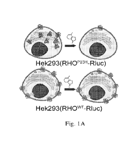
[0015] Figs. 1(A-H) illustrate schematic images, plots, and
charts showing high-
throughput screening (HTS) for small molecules that selectively reduce the
misfolded P23H
rhodopsin. A. Illustration of the cell-based luciferase reporter assay for HTS
and counter
screen. The mouse P23H or wild type (WT) rhodopsin was fused with Renilla
luciferase
(Rluc) and constitutively expressed in Hek293 cells, marked as the
Hek293(RHOP23H-Rluc),
or Hek293(RHOwT-Rluc), respectively. For the HTS, Hek293(RHOP2311-Rluc) cells
were
incubated with each compound for 24 h before assayed for luciferase activity,
and hits that
showed activity scores lower than Mean-2SD were selected. For the counter
screen, we tested
the hits by repeating the luciferase reporter assay in the Hek293(RHOwT-Rluc)
cells, and
selected those that showed preferred clearance activity in the Hek293(RHOP23H-
Rluc) versus
the Hek293(RHOwT-Rluc) cells. B. A pie chart showing the compound libraries
screened.
The number of compounds in each library is shown in the bracket. UC.
University of
Cincinnati Diversity Set; LOPAC, Library of Pharmacologically Active
Compounds; FDA,
U.S. Food and Drug Administration approved drugs; MIPE, NCATS Mechanism
Interrogation Plat E. C. An exemplary dose-response plot of a hit compound, CL-
009 in the
Hek293(RHOP23H-Rluc) and Hek293(RHOwT-Rluc) cells, as black squares and
magenta
circles, respectively. The luminescence was normalized by the mean
luminescence of cells
treated with 0.1% DMSO and 1 mM Evans Blue as the 0 and -100% controls,
respectively.
Data points and error bars are the means and SDs. N=3. Dose-response curves
were fitted by
the Origin Software using the Hills function. D. Rhodopsin dot blots of
untreated
NIH3T3(RhowT/GFP) and NIH3T3(RhoP2311/GFP) cells cell lysates loaded at 25,
50, 75 and
100%. E. Rhodopsin dot blot intensities in D were measured by ImageJ and
plotted in as a
function of loaded cell lysate amount. N=3. F. Rhodopsin dot blots of
NIH3T3(RhoP2311/GFP) and NIH3T3(RhowT/GFP) cells (bottom) each treated with
0.1%
DMSO or CL-001 to CL-009 at 101.tM for 24 h. Cells were loaded at the same
amount as the
100% loading control in D. G. Rhodopsin dot blot intensities in F were plotted
in as a box
chart, respectively. The middle lines and upper/bottom lines of boxes in G are
the means and
SDs. N=3. P23H and WT rhodopsin levels in each repeat are shown as black
squares and
magenta circles, respectively. H. The chemical structures of CL-001 to CL-009.
EC5os
CA 03169482 2022- 8- 25
WO 2021/173929
PCT/US2021/019813
7
shown in brackets were obtained from high-content image analyses quantifying
the
immunostaining of P23H rhodopsin in response to 8-10 doses of each hit
compound.
[0016] Figs. 2(A-J) illustrate images and plots showing the high-
content imaging assays
validated nine hits that selectively reduced the P23II rhodopsin in vitro. For
hit confirmation,
we used the NIH3T3 cells that stably co-expressed GFP and P23H or WT
rhodopsin, marked
as NIH3T3(RHOP2311/GFP) or NIH3T3(RHOwT/GFP), respectively. A. High-content
images
of cells treated with 0.1% DMSO or CL-001 to CL-009 at 10 l_tM for 24 h.
Immunostaining
of rhodopsin showed that CL-001 to CL-009 selectively reduced the P23H but not
the WT
rhodopsin level. Scale bar, 50 tim. B-J. Dose-response curves of nine hit
compounds by
image-based analysis. Relative immunostaining intensity of rhodopsin measured
from high-
content images of NIH3T3(RHOP23H/GFP) and NIH3T3(RHOwT/GFP) cells each treated
with
DMSO or CL-001 to CL-009 at 8-10 doses for 24 h. The immunostaining intensity
of
rhodopsin per cell was normalized by DMSO treated cells as 0% and cells
stained with
secondary antibody only as -100%, respectively. Data and error bars are means
and SDs.
N=3. Dose-response curves were fitted by a modified Hill function. Chemical
structure and
EC50 of each hit compound in the NIH3T3(RhoP2311/GFP) are shown in the inset
of each
graph.
[0017] Figs. 3(A-D) illustrate plots, schematics, and images
showing the effect of
active compounds on rhodopsin transcription, degradation, and clearance of
other adRP
causing mutants. A. Fold change of RHO transcripts in the NIH3T3(RHOP23H/GFP)
and
NIH3T3(RHOw'T/GFP) cells treated with 10 !.LM of each hit compound compared to
DMSO
control. Q-PCR result of RHO transcript was firstly normalized by 13-actin and
then by the
DMSO control. Middle lines and error bars are means and SDs of three
biological replicates
shown as data points. RHOP2311 and RHOwT. B. Illustration of the non-
radioactive pulse-
chase assay. Briefly, cells were starved in a Met-free medium for 1 h before
pulsed in the
azidohomoalanine (AHA) enriched medium that lacks Met for 4 h, so the nascent
protein
synthesized was labeled with AHA. Cells were then chased for 0-2411 in the
medium
containing 2 mM of Met, so the protein synthesized during the chase period was
not labeled
with AHA anymore. Next, via a "Click" reaction, the AHA incorporated proteins
in the cell
lys ate were linked with biotin (BTN). Total rhodopsin was immunoprecipitated
(IP) by the
1D4 anti-rhodopsin antibody, and the BTN labeled rhodopsin was finally dot
blotted (IB)
with IIRP-Streptavidin (SA). C. Percentage of nascent P23II rhodopsin from
CA 03169482 2022- 8- 25
WO 2021/173929
PCT/US2021/019813
8
NIH3T3(RHOP23II/GFP) cell lysates IP with 1D4 anti-rhodopsin antibody (RHO)
and IB with
SA at 24 h of chase time. Cells were treated with 10 04 of CL-001 to CL-009,
or DMSO at
0 h of chase time, respectively. The TB intensity of each dot was normalized
by the DMSO
control in the same membrane (Fig. 10). Three lines in each box represent the
75, 50 and
25% values of data in each group, and mean of each group was shown as filled
diamonds,
error bars were SDs. N=3. *, p<0.05; **, p<0.01; and ***, p<0.001 by an
unpaired two-tail
Student's t-test. D. Immunostaining images of rhodopsin in U2OS cells stably
expressing the
WT or six mutants of mouse rhodopsin (T4R, P23H, P53R, C110Y, D190N. P267L)
that
cause autosomal dominant retinitis pigmentosa, under treatment with 10 pM of
CL-001, CL-
002, CL-005 (11 p.M), CL-007, and CL-009. Scale bar, 100 pm.
[0018] Figs. 4(A-G) illustrate plots and images showing
Methotrexate (MTX/CL-009)
mediated P23H rhodopsin degradation via lysosomal activity in vitro. A.
Immunoblot of
rhodopsin in NIH3T3(RHOP23H/GFP) and NIH3T3(RHOwT/GFP) cells co-treated with
(+) or
without (-) 10 !AM MTX, 100 nM bafilomycin Al (BafAl) or 5 pM MG-132 for 24 h.
13-
Actin was the loading control. B. Immunoblots in A were repeated and
quantified as a
percentage of relative RHO level by ImageJ. The middle lines and error bars
are means and
SDs. *, p<0.05 by an unpaired two-tail Student's t-test. RHO", black squares;
and RHOwT,
magenta circles. C. High content images of rhodopsin immunostaining in
NIH3T3(RHOP2311/GFP) (top) and NIH3T3(RHOwT/GFP) (bottom) cells treated with
DMSO,
nM MTX, 10 nM MTX plus 100 nM BafAl, or 10 gM MTX plus 5 pM MG-132 Scale
bar, 50 pm. D. Relative immunostaining intensity (TNT) of P23H rhodopsin
measured from
high-content images of NIH3T3(RHOP2311/GFP) cells plotted as a function of MTX
concentration alone or in co-treatment with 100 nM BafAl or 5 luM MG-132.
Immunostaining intensity of P23H rhodopsin per cell was normalized by DMSO
treated cells
as 0% and cells stained with secondary antibody only as -100%, respectively.
Data points
and error bars are means and SDs. N=3. E-G. Effect of MTX on chymotrypsin-like
pruteasome activity in the NIH3T3(RHOP23H/GFP) and NIH3T3(RHOwT/GFP) cells. E.
The
luminescence readouts of proteasome activities as a function of cell number
showing the cell
number we used in B is within the sensitive range of the assay. F. The
luminescence readouts
of NIH3T3 (grey), NIH3T3(RHOwT/GFP) (magenta) and NIH3T3(RHOP2311/GFP) (dark
grey) cells treated with 0.1% DMSO or 5 pM MG-132 as the 100% and 0% controls,
respectively. Means and SDs from 8 biological replicates (shown as diamonds)
are shown as
CA 03169482 2022- 8- 25
WO 2021/173929
PCT/US2021/019813
9
middle bars and error bars. The Z' = 1 - 3 x (SDi00% control + SDo% control) /
(Mean 00% control -
MeanO% control) is in the inset demonstrating the assay being robust. G.
Normalized
chymotrypsin-like proteasome activity of cells in response to 10 doses of MTX.
Chymotrypsin-like proteasome activity was normalized by the 100% and 0%
controls,
respectively. Data points and error bars are the means and SDs of three
biological replicates.
[0019] Figs. 5(A-C) illustrate immunoblots and plots showing MTX
increased
autophagy flux in the retinae of RhoB231il+ mice. A. Immunoblots of SQSTM1/p62
and LC3
in 30 [ig of retina lysates from RhoP231il+ mice at 48 h after an intravitreal
injection (IVI) of 25
pmol/eye MTX or phosphate-buffered saline (PBS) at PND 15. I3-Actin was the
loading
control. Dashed boxes are samples selected for intensity analysis in B and C,
excluding lanes
6 and 7 in which samples may be mixed up when loaded. B and C. Ratios of band
intensities
of SQSTM1/p62 to I3-actin, and LC3-II to I3-actin measured from immunoblotting
images in
A, respectively. Left, retinae treated with PBS; right, retinae treated with
25 pmol MTX per
eye. Middle lines and error bars are means and SDs. N=5. *, p <0.05 between
MTX treated
and PBS groups calculated by an unpaired two-tail Student's t-test.
[0020] Figs. 6(A-R) illustrate plots and images showing one
intravitreal injection (IVI)
of MTX increased electroretinograrn (ERG) response and retinal rhodopsin level
in RhoP2311/+
mice. Eyes of mice were untreated or intravitreally injected with PBS, 25 or
100 pmol MTX
at PND 15, and ERG responses were recorded at PND 32. Mice were euthanized and
eyes
were enucleated at PND 33 for immunohistochemistry (IHC). Age-matched Rho" +
mice
were used as the normal control. A. Scotopic ERG recordings stimulated by a
flash of light
at 10 cd. s/m2. B and C. Eight-flash scotopic a- and b-wave amplitudes of
treated mice
plotted as a function of flash intensity (semi-log format), respectively. D.
Six-flash photopic
b-wave amplitudes plotted as a function of flash intensity (semi-log format).
Black squares,
red circles, blue triangles, and magenta reverse triangles were from
RHOP23/11+ mice that were
untreated, PBS, 25 and 100 pmol MTX treated, respectively. Data points and
error bars are
means and SEMs. respectively. N=5. ''', pi <0.05 between 25 pmol MTX and PBS
treated
groups calculated by a two-way ANOVA. Factor 1, treatment; and factor 2, flash
intensity.
Scale bar, 50 1.tm. Q. Spidergram of rhodopsin immunofluorescence E-P. IHC
images of
untreated Rho" + retina, and RhoP231il+ mouse retinae that are untreated, PBS-
treated, or 25
pmol MTX-treated, from top to bottom, respectively. RHO and nucleus (Hoechst
33342)
were stained in red and blue, respectively. E, II, K and N are retinal IIIC
images at low
CA 03169482 2022- 8- 25
WO 2021/173929
PCT/US2021/019813
magnification. Scale bar, 500 ttm. F, I, L and 0 are high magnification
retinal images taken at
sites marked in boxes shown in E, H, K and N on the retinal inferior side, and
G, J, M and P
are images on the superior side, respectively intensity in the OS measured by
ImageJ from
high magnification images taken at 0.6, 1 and 1.4 mm from the optic nerve head
(ONII).
Green squares, age-matched Rho / mouse retinae. R. Spidergram of outer
nuclear layer
(ONL) nucleus number per 200 nm length of retina cross-section images taken at
0.6, 1, and
1.4 mm distance from ONH. Data points and error bars are means and SEMs,
respectively.
N=3. *, p <0.05 between 25 pmol MTX and PBS groups by an unpaired two-tail
Student's t-
test.
[0021] Figs. 7(A-0) illustrate plots and images showing multiple
IVIs of MTX
improved ERG response, rhodopsin level and preserved photoreceptor cell
numbers of the
RhoP2sw+ mouse retinae. Eyes of RhoP23117' mice were untreated or administered
by four
weekly IVIs of PBS, 25 pmol MTX and 100 pmol MTX per treatment, starting at
PND 15
and ERGs were taken at PND 44. Eyes were enucleated at PND 46 for IHC. A.
Scotopic
ERG responses stimulated by a flash of light at 10 cd= s/m2. B and C. Eight-
flash scotopic a-
and b-wave amplitudes of treated mice plotted as a function of flash intensity
(semi-log
format), respectively. D. Six-flash photopic b-wave amplitudes plotted as a
function of flash
intensity. Black squares, red circles, blue triangles, and magenta reverse
triangles are from
RHOP23"/+ mice that were untreated, PBS, 25 and 100 pmol MTX treated,
respectively. Data
points and error bars are means and SEMs, respectively. N=5. *, pi <0.05
between 25 pmol
MTX and PBS treated groups calculated by a two-way ANOVA. Factor 1, treatment;
and
factor 2, flash intensity. E-M. IHC images of untreated, PBS-treated, and 25
pmol MTX
treated Rhol'231v+ mouse retinae, from top to bottom, respectively. RHO and
nucleus (Hoechst
33342) were stained in red and blue, respectively. E, H, and K are retinal
images at lower
magnification. Scale bar, 500 nm. F, I, and L are high magnification retinal
images taken at
sites marked as boxes shown in E, H, and K on the inferior side, and in G, J,
and M are
images on the superior side, respectively. Scale bar, 50 nm. N. Spidergram of
rhodopsin
immunofluorescence intensity in the OS measured by ImageJ from high
magnification
immunofluorescence images taken at 0.6, 1 and 1.4 mm from ONH. 0. Spidergram
of ONL
nucleus number per 200 ttm length of retina cross-section images taken at 0.6,
1, and 1.4 mm
from ONH. Data points and error bars are means and SEMs, respectively. N=3. *,
p <0.05
between 25 pmol MTX and PBS groups by an unpaired two-tail Student's t-test.
CA 03169482 2022- 8- 25
WO 2021/173929
PCT/US2021/019813
11
[0022] Figs. 8(A-E) illustrate Immunoblots confirmed rhodopsin
expression in stable
cells used for HTS and confirmation assays. A. The immunoblots of rhodopsin
showing the
Hek293(RHOP23"-Rlue) and Hek293(RHOwT-Rluc) cells expressed the equivalent
amount of
P23II or WT rhodopsin-Rluc fusion proteins. 13-Actin was used as loading
control. B-E.
Immunoblots of rhodopsin in U2OS stable cell single clones expressing WT, T4R,
P23H,
P53R, C110Y and P267L mouse rhodopsin each fused with Venus fluorescence
protein on
the C-terminus. Clones that were selected for the high-content imaging
analyses under the
treatment of hit compounds (shown in Fig. 3) were marked by red boxes.
Difference in
molecular masses between WT and mutant RHOs are due to immature glycosylation
of RHO
mutants that are accumulated in the endoplasmic reticulum.
[0023] P'31I
Figs. 9(A-H) illustrate Rhodopsin dot blots of NIH3T3(Rho /GFP) (A,
C, E
and G) and NIH3T3(RhowT/GFP) cells (B, D, F and H) each treated with different
compounds including CL-001 to CL-009 at 10 or 20 tiM for 24 h. A-F. Three
biological
repeats from cells treated with CL-002, CL-003, CL-004, CL-005 and CL-009. G-
H. Four
biological repeats of dot blots from cells treated with CL001, CL-006, CL-007
and CL-008
are shown in the bottom four scans. Dot blots shown in Fig. 1D&F were cropped
from these
original scans. Rhodopsin dot blot intensities in these repeats were measured
and plotted in
the curves and box chart shown in Fig. lE&G.
[0024] Figs. 10(A-F) illustrate immunoblots and plots showing
the effect of nine hits on
rhodopsin degradation in the NIH3T3(RHOP23H/GFP) cells using the non-
radioactive pulse-
chase assay. A. Dot blot of RHO at different steps of RHO immunoprecipitation
(IP) from
NIH3T3(RHOwT/GFP) cells suggesting a high yield of rhodopsin pulldown. Dot
blots from
5% of total cell lysate, 5% of flow-through, last wash through, first and
second elution, were
marked as T, FT, W, El, and E2, from left to right, respectively. B. Chase of
WT and P23H
rhodopsin at 0, 4, and 24 h after the pulse with AHA, IP by anti-rhodopsin
antibody (RHO)
and immunoblotted (IB) by streptavidin (SA). Met, lysates of cells always
incubated with
Met as the blank control. C. Percentage of nascent rhodopsin decayed as a
function of chase
time, quantified from B by ImageJ. Grey squares, P23H rhodopsin; magenta
circles, WT
rhodopsin. D. NIH3T3(RHOP23II/GFP) cells were treated with 10 1.1M of CL-001
to CL-009,
or DMSO at 0 h of chase time, respectively. Nascent P23H rhodopsin from cell
lysates IP
with RHO and IB with SA at 24 h of chase time. E. Total P23H rhodopsin from
the same
batch of cell lysate IP with RI JO and I13 with RHO confirming the activity of
each hit
CA 03169482 2022- 8- 25
WO 2021/173929
PCT/US2021/019813
12
compound indeed reduced the total P23H rhodopsin protein level. Dot blot
intensities in D
were quantified and shown in Figs. 3C. F. Dot intensities quantified from dot
blot scans in E
and normalized by the DMSO control. Squares and error bars are the means and
SDs of six
biological replicates, and boxes showed the 75, 50 and 25% of data values.
[0025] Figs. 11(A-E) illustrates plots and an immunoblot showing
rhodopsin
immunofluorescence intensities measured from high-content immunostaining
images of
U2OS cells stably expressing the WT or six mutants of rhodopsin that cause
autosomal
dominant retinitis pigmentosa, under treatment with CL-001 (A), CL-002 (B), CL-
005 (C),
CL-007 (D), and CL-009 (E). Normalized rhodopsin intensities were plotted as a
function of
seven doses of each compound in semi-log format. Dose-response curves were
fitted by the
Origin Software using the modified Hill function. Data points and error bars
were means and
SD from three biological replicates. The range of EC5os of each compound
towards different
rhodopsin mutants were shown at the bottom of each graph. Data from cells
expressing WT,
T4R, P23H, P53R, C110Y, D190N, and P267L rhodopsin are marked as black
squares, red
circles, green triangles, blue reverse triangles, cyan diamonds, magenta left-
pointed triangles
and olive right-pointed triangles, respectively.
[0026] Figs. 12(A-L) illustrate immunoblots showing the effect
of inhibiting
proteasomal or lysosomal activities on MTX's effect of rhodopsin degradation.
Left (A, B,
C) and right (G, H and I) are immunoblots of rhodopsin from lysates of
NIH3T3(RHOP23H/GFP) and NIH3T3(RHOwT/GFP) cells treated with DMSO, 10 pM
methotrexate (MTX), 35.5 !AM cycloheximide (CHX), 35.5 pM CHX and 10 RM MTX,
100 nM Bafilomycin Al (BafAl), 100 nm BafAl and 10 pM MTX, 5 pM MG-132, 5 pM
MG-132 and 10 pM MTX, 100 nM BafAl and 5 iuM MG-132, respectively. D, E and F
are
from NIH3T3(RHOP2311/GFP) cells treated with 0, 3, 10, 30 pM of MTX, with or
without co-
treatment of 100 nM BafAl. J, K and L are from NIH3T3(RHOP23II/GFP) cells
treated with
0, 3, 10, 30 pM of MTX, with or without co-treatment of 100 nM BafAl. Fig. 4A
was
cropped from the inununoblot scans of A and B here, and quantification shown
in Fig. 4B
was calculated from the three biological repeats shown here in A-L.
[0027] Figs. 13(A-F) illustrate plots showing MTX treatment
improved retinal function
and rhodopsin homeostasis in RhoP23-w+ knock-in mice. A single IVI of MTX did
not affect
the b-to-a-wave ratio in vivo. RhoP231-1/+ knock in mice were intravitreally
injected with PBS,
25 or 100 pmol of MTX at PND 15, and ERGs were recorded at PND32 and shown in
Fig.
CA 03169482 2022- 8- 25
WO 2021/173929
PCT/US2021/019813
13
6A-D. A and B are scotopic and photopic b-to a-wave ratios plotted as a
function of flash
intensity in a semi-log format, respectively. Data and error bars are means
and SEMs,
respectively. N=5. P1>0.05 by a two-way ANOVA suggests neither 25 nor 100 pmol
MTX
treatment affected the scotopic or photopic b-to-a wave ratio, compared to the
PBS control.
C-F. Effect of MTX on the rhodopsin level in RhoP23/1 mice. C&D are
quantifications of
retinal IHC fluorescence intensities from mice treated with single IVI, and
E&F are from
mice treated with four weekly IVIs. C&E, Spidergrams of the total rhodopsin
immunofluorescence intensity (INTST) in the retinae, measured by ImageJ from
high
magnification immunofluorescence images taken at 0.6, 1 and 1.4 mm from the
optic nerve
head (ONH). D&F, Spidergrams of rhodopsin immunofluorescence intensity in the
ONL
from high magnification images taken at 0.6, 1 and 1.4 mm from the ONH. Data
points and
error bars are means and SEMs, respectively. N=3; *, p <0.05 between 25 pmol
MTX and
PBS groups by an unpaired two-tail Student's t-test.
[0028] Fig. 14 illustrates immunohistochemistry (IHC) images of
Rho" + and RhoP23/14
mouse retinae treated with one intravitreal injection (IVI). Eyes of RhoP23Hi+
mice were
untreated, or treated with PBS, 25 pmol MTX via an IVI at PND 15, and
enucleated at PND
33. Untreated Rho-' eyes were used as the normal control. Genotype and
treatments were
labeled vertically on the left. N=3. RHO and nucleus (Hoechst 33342) were
stained in red and
blue, respectively. Images were taken at 0.6, 1 and 1.4 nun from the optic
nerve head (ONH)
on the retinal inferior side and the superior side, respectively. Scale bar,
50 um.
[0029] Fig. 15 illustrates IHC images of RhoP231'7+ mouse
retinae treated with four
weekly IVIs. Eyes of RhoP231/4 mice were untreated or treated by four weekly
IVIs of PBS or
25 pmol MTX each time, starting at PND 15. Eyes were enucleated at PND 46 for
1HC.
Genotype and treatment conditions were labeled vertically on the left. N=3.
RHO and
nucleus (Hoechst 33342) were stained in red and blue, respectively. Images
were taken at 0.6,
1 and 1.4 mm from the optic nerve head (ONH) on the retinal inferior side and
the superior
side, respectively. Scale bar, 50 um.
DETAILED DESCRIPTION
[0030] For convenience, certain terms employed in the
specification, examples, and
appended claims are collected here. Unless defined otherwise, all technical
and scientific
CA 03169482 2022- 8- 25
WO 2021/173929
PCT/US2021/019813
14
terms used herein have the same meaning as commonly understood by one of
ordinary skill
in the art to which this application belongs.
[0031] The articles "a" and "an" are used herein to refer to one
or to more than one
(i.e., to at least one) of the grammatical object of the article. By way of
example, "an
element" means one element or more than one element.
[0032] The terms "comprise," "comprising," "include,"
"including," ''have," and
"having' are used in the inclusive, open sense, meaning that additional
elements may be
included. The terms "such as", " e.g.," , as used herein are non-limiting and
are for illustrative
purposes only. "Including" and "including but not limited to" are used
interchangeably.
[0033] The term "or" as used herein should be understood to mean
"and/or", unless the
context clearly indicates otherwise.
[0034] As used herein, the term "about" or "approximately"
refers to a quantity, level,
value, number, frequency, percentage, dimension, size, amount, weight or
length that varies
by as much as 15%, 10%, 9%, 8%, 7%, 6%, 5%, 4%, 3%, 2% or 1% to a reference
quantity,
level, value, number, frequency, percentage, dimension, size, amount, weight
or length. In
one embodiment, the term "about" or "approximately" refers a range of
quantity, level, value,
number, frequency, percentage, dimension, size, amount, weight or length
15%, 10%,
9%, 8%, 7%, 6%, 5%, 4%, 3%, 2%, or 1% about a reference
quantity, level,
value, number, frequency, percentage, dimension, size, amount, weight or
length.
[0035] It will be noted that the structure of some of the
compounds of the application
include asymmetric (chiral) carbon or sulfur atoms. It is to be understood
accordingly that
the isomers arising from such asymmetry are included herein, unless indicated
otherwise.
Such isomers can be obtained in substantially pure form by classical
separation techniques
and by stereochemically controlled synthesis. The compounds of this
application may exist
in stereoisomeric form, therefore can be produced as individual stereoisomers
or as mixtures.
[0036] The term "derivative" refers to compounds that have a
common core structure,
and are substituted with various groups as described herein.
[0037] The term "bioisostere" refers to a compound resulting
from the exchange of an
atom or of a group of atoms with another, broadly similar, atom or group of
atoms. The
objective of a bioisosteric replacement is to create a new compound with
similar biological
properties to the parent compound. The bioisosteric replacement may be
physicochemically
or topologically based. Examples of carboxylic acid bioisosteres include acyl
sulfonimides,
CA 03169482 2022- 8- 25
WO 2021/173929
PCT/US2021/019813
tetrazoles, sulfonates, and phosphonates. See, e.g., Patani and LaVoie, Chem.
Rev. 96, 3147-
3176 (1996).
[0038] The phrases "parenteral administration" and "administered
parenterally" are
art-recognized terms, and include modes of administration other than enteral
and topical
administration, such as injections, and include, without limitation,
intravenous, intramuscular,
intrapleural, intravascular, intrapericardial, intraarterial, intrathecal,
intracapsular, intraorbital,
intracardiac, intradermal, intraperitoneal, transtracheal, subcutaneous,
subcuticular, intra-
articular, subcapsular, subarachnoid, intraspinal and intrastemal injection
and infusion.
[0039] The term "treating" is art-recognized and includes
inhibiting a disease, disorder
or condition in a subject, e.g., impeding its progress; and relieving the
disease, disorder or
condition, e.g., causing regression of the disease, disorder and/or condition.
Treating the
disease or condition includes ameliorating at least one symptom of the
particular disease or
condition, even if the underlying pathophysiology is not affected.
[0040] The term "preventing" is art-recognized and includes
stopping a disease,
disorder or condition from occurring in a subject, which may be predisposed to
the disease,
disorder and/or condition but has not yet been diagnosed as having it.
Preventing a condition
related to a disease includes stopping the condition from occurring after the
disease has been
diagnosed but before the condition has been diagnosed.
[0041] The term "pharmaceutical composition" refers to a
formulation containing the
disclosed compounds in a form suitable for administration to a subject. In a
preferred
embodiment, the pharmaceutical composition is in bulk or in unit dosage form.
The unit
dosage form is any of a variety of forms, including, for example, a capsule,
an IV bag, a
tablet, a single pump on an aerosol inhaler, or a vial. The quantity of active
ingredient (e.g., a
formulation of the disclosed compound or salts thereof) in a unit dose of
composition is an
effective amount and is varied according to the particular treatment involved.
One skilled in
the art will appreciate that it is sometimes necessary to make routine
variations to the dosage
depending on the age and condition of the patient. The dosage will also depend
on the route
of administration. A variety of routes are contemplated, including oral,
pulmonary, rectal,
parenteral, transdermal, subcutaneous, intravenous, intramuscular,
intraperitoneal, intranas al,
inhalational, and the like. Dosage forms for the topical or transdermal
administration of a
compound described herein includes powders, sprays, ointments, pastes, creams,
lotions,
CA 03169482 2022- 8- 25
WO 2021/173929
PCT/US2021/019813
16
gels, solutions, patches, nebulized compounds, and inhalants. In a preferred
embodiment, the
active compound is mixed under sterile conditions with a pharmaceutically
acceptable carrier,
and with any preservatives, buffers, or propellants that are required.
[0042] The term "flash dose" refers to compound formulations
that are rapidly
dispersing dosage forms.
[0043] The term "immediate release" is defined as a release of
compound from a
dosage form in a relatively brief period of time, generally up to about 60
minutes. The term
"modified release" is defined to include delayed release, extended release,
and pulsed release.
The term ''pulsed release" is defined as a series of releases of drug from a
dosage form. The
term "sustained release" or "extended release" is defined as continuous
release of a compound
from a dosage form over a prolonged period.
[0044] The phrase "pharmaceutically acceptable" is art-
recognized. In certain
embodiments, the term includes compositions, polymers and other materials
and/or dosage
forms which are, within the scope of sound medical judgment, suitable for use
in contact with
the tissues of human beings and animals without excessive toxicity,
irritation, allergic
response, or other problem or complication, commensurate with a reasonable
benefit/risk
ratio.
[0045] The phrase "pharmaceutically acceptable carrier" is art-
recognized, and
includes, for example, pharmaceutically acceptable materials, compositions or
vehicles, such
as a liquid or solid filler, diluent, excipient, solvent or encapsulating
material, involved in
carrying or transporting any subject composition from one organ, or portion of
the body, to
another organ, or portion of the body. Each carrier must be "acceptable" in
the sense of being
compatible with the other ingredients of a subject composition and not
injurious to the
patient. In certain embodiments, a pharmaceutically acceptable carrier is non-
pyrogenic.
Some examples of materials which may serve as pharmaceutically acceptable
carriers
include: (1) sugars, such as lactose, glucose and sucrose; (2) starches, such
as corn starch and
potato starch; (3) cellulose, and its derivatives, such as sodium carboxy
methyl cellulose, ethyl
cellulose and cellulose acetate; (4) powdered tragacanth; (5) malt; (6)
gelatin; (7) talc; (8)
excipients, such as cocoa butter and suppository waxes; (9) oils, such as
peanut oil,
cottonseed oil, sunflower oil, sesame oil, olive oil, corn oil and soybean
oil; (10) glycols,
such as propylene glycol; (11) polyols, such as glycerin, sorbitol, mannitol
and polyethylene
glycol; (12) esters, such as ethyl oleate and ethyl laurate; (13) agar; (14)
buffering agents,
CA 03169482 2022- 8- 25
WO 2021/173929
PCT/US2021/019813
17
such as magnesium hydroxide and aluminum hydroxide; (15) alginic acid; (16)
pyrogen-free
water; (17) isotonic saline; (18) Ringer's solution; (19) ethyl alcohol; (20)
phosphate buffer
solutions; and (21) other non-toxic compatible substances employed in
pharmaceutical
formulations.
[0046] The compounds of the application are capable of further
forming salts. All of
these forms are also contemplated herein.
[0047] "Pharmaceutically acceptable salt" of a compound means a
salt that is
pharmaceutically acceptable and that possesses the desired pharmacological
activity of the
parent compound. For example, the salt can be an acid addition salt. One
embodiment of an
acid addition salt is a hydrochloride salt. The pharmaceutically acceptable
salts can be
synthesized from a parent compound that contains a basic or acidic moiety by
conventional
chemical methods. Generally, such salts can be prepared by reacting the free
acid or base
forms of these compounds with a stoichiometric amount of the appropriate base
or acid in
water or in an organic solvent, or in a mixture of the two; generally, non-
aqueous media like
ether, ethyl acetate, ethanol, isopropanol, or acetonitrile being preferred.
Lists of salts are
found in Remington's Pharmaceutical Sciences, 18th ed. (Mack Publishing
Company. 1990).
[0048] The compounds described herein can also be prepared as
esters, for example
pharmaceutically acceptable esters. For example, a carboxylic acid function
group in a
compound can be converted to its corresponding ester, e.g., a methyl, ethyl,
or other ester.
Also, an alcohol group in a compound can be converted to its corresponding
ester, e.g., an
acetate, propionate, or other ester.
[0049] The compounds described herein can also be prepared as
prodrugs, for example
pharmaceutically acceptable prodrugs. The terms "pro-drug" and "prodrug" are
used
interchangeably herein and refer to any compound, which releases an active
parent drug in
vivo. Since prodrugs are known to enhance numerous desirable qualities of
pharmaceuticals
(e.g., solubility, bioavailability, manufacturing, etc.) the compounds can be
delivered in
prodrug form. Thus, the compounds described herein are intended to cover
prodrugs of the
presently claimed compounds, methods of delivering the same and compositions
containing
the same. "Prodrugs" are intended to include any covalently bonded carriers
that release an
active parent drug in vivo when such prodrug is administered to a subject.
Prodrugs are
prepared by modifying functional groups present in the compound in such a way
that the
modifications are cleaved, either in routine manipulation or in vivo, to the
parent compound.
CA 03169482 2022- 8- 25
WO 2021/173929
PCT/US2021/019813
18
Prodrugs include compounds wherein a hydroxy, amino, sulfhydryl, carboxy, or
carbonyl
group is bonded to any group that may be cleaved in vivo to form a free
hydroxyl, free amino,
free sulfhydryl, free carboxy or free carbonyl group, respectively.
[0050] Examples of prodrugs include, but are not limited to,
esters (e.g., acetate,
dialkylaminoacetates, formates, phosphates, sulfates, and benzoate
derivatives) and
carbamates (e.g., N,N-dimethylaminocarbonyl) of hydroxy functional groups,
ester groups
(e.g., ethyl esters, morpholinoethanol esters) of carboxyl functional groups,
N-acyl
derivatives (e.g., N-acetyl) N-Mannich bases, Schiff bases and enaminones of
amino
functional groups, oximes, acetals, ketals and enol esters of ketone and
aldehyde functional
groups in compounds of Formula I, and the like, See Bundegaard, H. "Design of
Prodrugs"
p1-92, Elesevier, New York-Oxford (1985).
[0051] Additionally, the salts of the compounds described
herein, can exist in either
hydrated or unhydrated (the anhydrous) form or as solvates with other solvent
molecules.
Nonlimiting examples of hydrates include monohydrates, dihydrates, etc.
Nonlimiting
examples of solvates include ethanol solvates, acetone solvates, etc.
[0052] The term "solvates" means solvent addition forms that
contain either
stoichiometric or non stoichiometric amounts of solvent. Some compounds have a
tendency
to trap a fixed molar ratio of solvent molecules in the crystalline solid
state, thus forming a
solvate. If the solvent is water the solvate formed is a hydrate, when the
solvent is alcohol,
the solvate formed is an alcoholate. Hydrates are formed by the combination of
one or more
molecules of water with one of the substances in which the water retains its
molecular state as
H20, such combination being able to form one or more hydrate.
[0053] The compounds, salts and prodrugs described herein can
exist in several
tautomeric forms, including the enol and imine form, and the keto and en amine
form and
geometric isomers and mixtures thereof. Tautomers exist as mixtures of a
tautomeric set in
solution. In solid form, usually one tautomer predominates. Even though one
tautomer may
be described, the present application includes all tautomers of the present
compounds. A
tautomer is one of two or more structural isomers that exist in equilibrium
and are readily
converted from one isomeric form to another. This reaction results in the
formal migration of
a hydrogen atom accompanied by a switch of adjacent conjugated double bonds.
In solutions
where tautomerization is possible, a chemical equilibrium of the tautomers
will be reached.
The exact ratio of the tautomers depends on several factors, including
temperature, solvent,
CA 03169482 2022- 8- 25
WO 2021/173929
PCT/US2021/019813
19
and pH. The concept of tautomers that are interconvertable by tautomerizations
is called
tautomerism.
[0054] The term "analog" refers to a chemical compound that is
structurally similar to
another but differs slightly in composition (as in the replacement of one atom
by an atom of a
different element or in the presence of a particular functional group, or the
replacement of
one functional group by another functional group). Thus, an analog is a
compound that is
similar or comparable in function and appearance, but not in structure or
origin to the
reference compound.
[0055] A "patient," "subject," or "host" to be treated by the
subject method may mean
either a human or non-human animal, such as a mammal, a fish, a bird, a
reptile, or an
amphibian. Thus, the subject of the herein disclosed methods can be a human,
non-human
primate, horse, pig, rabbit, dog, sheep, goat, cow, cat, guinea pig or rodent.
The term does
not denote a particular age or sex. Thus, adult and newborn subjects. as well
as fetuses,
whether male or female, are intended to be covered. In one aspect, the subject
is a mammal.
A patient refers to a subject afflicted with a disease or disorder.
[0056] The terms "prophylactic" or "therapeutic" treatment is
art-recognized and
includes administration to the host of one or more of the subject
compositions. If it is
administered prior to clinical manifestation of the unwanted condition then
the treatment is
prophylactic, i.e., it protects the host against developing the unwanted
condition, whereas if it
is administered after manifestation of the unwanted condition, the treatment
is therapeutic
(i.e., it is intended to diminish, ameliorate, or stabilize the existing
unwanted condition or
side effects thereof).
[0057] By "reduces" or "increases" is meant a negative or
positive alteration,
respectively, of at least 10%, 25%, 50%, 75%, or 100%
[0058] The terms "therapeutic agent", "drug", "medicament" and
"bioactive substance"
are art-recognized and include molecules and other agents that are
biologically,
physiologically, or pharmacologically active substances that act locally or
systemically in a
patient or subject to treat a disease or condition. The terms include without
limitation
pharmaceutically acceptable salts thereof and prodrugs. Such agents may be
acidic, basic, or
salts; they may be neutral molecules, polar molecules, or molecular complexes
capable of
hydrogen bonding; they may be prodrugs in the form of ethers, esters, amides
and the like
that are biologically activated when administered into a patient or subject.
CA 03169482 2022- 8- 25
WO 2021/173929
PCT/US2021/019813
[0059] The phrase "therapeutically effective amount" or
"pharmaceutically effective
amount" is an art-recognized term. In certain embodiments, the term refers to
an amount of a
therapeutic agent that produces some desired effect at a reasonable
benefit/risk ratio
applicable to any medical treatment. In certain embodiments, the term refers
to that amount
necessary or sufficient to eliminate, reduce or maintain a target of a
particular therapeutic
regimen. The effective amount may vary depending on such factors as the
disease or
condition being treated, the particular targeted constructs being
administered, the size of the
subject or the severity of the disease or condition. One of ordinary skill in
the art may
empirically determine the effective amount of a particular compound without
necessitating
undue experimentation. In certain embodiments, a therapeutically effective
amount of a
therapeutic agent for in vivo use will likely depend on a number of factors,
including: the rate
of release of an agent from a polymer matrix, which will depend in part on the
chemical and
physical characteristics of the polymer; the identity of the agent; the mode
and method of
administration; and any other materials incorporated in the polymer matrix in
addition to the
agent.
[0060] Throughout the description, where compositions are
described as having,
including, or comprising, specific components, it is contemplated that
compositions also
consist essentially of, or consist of, the recited components. Similarly,
where methods or
processes are described as having, including, or comprising specific process
steps, the
processes also consist essentially of, or consist of, the recited processing
steps. Further, it
should be understood that the order of steps or order for performing certain
actions is
immaterial so long as the compositions and methods described herein remains
operable.
Moreover, two or more steps or actions can be conducted simultaneously.
[0061] The term "small molecule" is an art-recognized term. In
certain embodiments,
this term refers to a molecule, which has a molecular weight of less than
about 2000 amu, or
less than about 1000 amu, and even less than about 500 amu.
[0062] The term "wild-type" or "wild-type conformation" refers
to the 3 dimensional
conformation or shape of a protein that is free of mutations present in its
amino acid sequence
that affect the conformation or shape of the protein, such that protein
function is altered
relative to wild-type protein function. For opsin, a wild-type conformation is
a conformation
that is free from mutations that cause mis-folding, such as the mutation
designated P23H
(P23II opsin) (see, for example, GenBank Accession Nos. NM000539 and NP000530)
CA 03169482 2022- 8- 25
WO 2021/173929
PCT/US2021/019813
21
(meaning that a proline is replaced by a histidine at residue 23 starting from
the N-terminus).
Opsin in a "wild-type conformation" is capable of opsin biological function,
including but not
limited to, retinoid binding, visual cycle function, and insertion into a
photoreceptor
membrane.
[0063] By "mis-folded opsin protein" is meant a protein whose
tertiary structure differs
from the conformation of a wild-type protein, such that the misfolded protein
lacks one or
more biological activities associated with the wild-type protein.
[0064] "P23H rhodopsin" means any nucleic acid or protein of
P23H rhodopsin.
[0065] All percentages and ratios used herein, unless otherwise
indicated, are by
weight.
[0066] Embodiments described herein relate to compounds and
methods of treating an
inherited ocular disorder associated with or caused by misfolded ocular
proteins in a subject
in need thereof. It was found that reducing misfolded ocular proteins, such as
misfolded
opsin proteins, can be an effective strategy to preserve or rescue rod
photoreceptors in
subjects with inherited ocular disorders associated with or caused by the
misfolded ocular
protein. Using a small molecule high-throughput screening assay, compounds
were
identified that selectively reduced misfolded mutant ocular proteins without
an effect on
corresponding wild type proteins. The compounds were found to promote
clearance or
accelerate degradation of misfolded ocular proteins, preserve visual function,
and prevent
photoreceptor death related to the inherited ocular disorder.
[0067] In some embodiments, a method of treating an inherited
ocular disorder
associated with or caused by a misfolded ocular protein in a subject in need
thereof includes
administering to the subject a therapeutically effective amount of a compound
that promotes
clearance of misfolded ocular proteins in a subject in need thereof. In some
embodiments, a
compound can be selected that promoted degradation of a misfolded mutant
ocular protein
(e.g., misfolded mutant opsin or rhodopsin) but not the corresponding wild
type ocular
protein in cells. Compounds identified using a high throughput screening assay
described
herein that that promoted degradation of a misfolded mutant ocular protein
(e.g., misfolded
mutant opsin or rhodopsin) but not the corresponding wild type ocular protein
in cells are
selected from:
CA 03169482 2022- 8- 25
WO 2021/173929
PCT/US2021/019813
22
H2 N
N 0 + / N\
N / \ 0-
0 HO 0 N ___-N
if
HN -1 /_,,, le
I ¨
rN
1\11i /7 0 ,
______________________________________________________________ , ,
0
\pN 0
F>r.1.,OH
N F
'-."-
F
S N
nN
N N N y...',..OH
0 ,
CI
CI F 0
0 N H N
i
H N 0 , 'NI F
H N )¨N H 0
\ ) h M
H N,S"
0 HN" 2 0
N H2 ,
N H2
N 1_
H 2 N ¨( ¨N
NJN\ \ le 00
N N
NH ..N
101 Tiõ),,,.L
..../- i H N OH
0
N /
,S
)i¨OH
0 0
,
,
a pharmaceutically acceptable salt, tautomer, or solvate thereof, or
combinations thereof.
[0068] In some embodiments, the inherited ocular disorder is a
non syndromic retinal
disorder associated with or by the misfolded ocular protein. For example, the
non syndromic
retinal disorder can be non syndromic autosomal dominant retinitis pigmentosa
(adRP)
associated with or caused by the misfolded ocular protein.
[0069] In other embodiment, the compound can be selected from:
CA 03169482 2022- 8- 25
WO 2021/173929
PCT/US2021/019813
23
0 HO 0
HN)Hi
NH _____________________________________ 0
0
F>r,õ1,0H F
q
NH N
411
N 0
H2N - NH2
0 0
N H2
N ¨
H2 N ¨N
____________________ 0 0 N ,N1 \ N
OH
HN
)i¨OH
0 , a pharmaceutically
acceptable salt,
tautomer, or solvate thereof, or combinations thereof.
[0070] Advantageously, the compound can be:
NH2
N
H2 N ¨(4\ ¨N
N \ (/) ____________________ \ = 0 0
N OH
HN
OH
0 or a pharmaceutically
acceptable
salt, tautomer, or solvate thereof. A compound having this formula is also
referred to as
Methotrexate. Methotrexate is a non-naturally occurring chemical also known as
1\144-[[(2,4-
diamino-6-pteridinyl)methyllmethylaminolbenzoyll-L-glutamic acid.
[0071] In certain embodiments, the compounds described herein
can be used in a
method of treating, preventing, ameliorating, or slowing progression of
retinitis pigmentosa
CA 03169482 2022- 8- 25
WO 2021/173929
PCT/US2021/019813
24
(RP) or autosomal dominant retinitis pigmentosa (AdRP) in a subject. The
method can
include administering to the subject a compound described herein, thereby
treating,
preventing, ameliorating, or slowing progression of retinitis pigmentosa (RP)
or autosomal
dominant retinitis pigmentosa (AdRP) in the subject.
[0072] In other embodiments, the compounds described herein can
be used in a method
of improving or preserving visual function, visual field, photoreceptor cell
function, ERG
response, or visual acuity in a subject having a P23H rhodopsin mutant allele
or having
retinitis pigmentosa (RP), such as autosomal dominant retinitis pigmentosa
(AdRP). The
method can include administering a compound described herein to the subject.
In certain
embodiments, a method of inhibiting, preventing, or delaying progression of
photoreceptor
cell loss and/or deterioration of the retina outer nuclear layer (ONL) in a
subject having a
P23H rhodopsin mutant allele or having retinitis pigmentosa (RP), such as
autosomal
dominant retinitis pigmentosa (AdRP), comprises administering a compound
described herein
to the subject.
[0073] In some embodiments, the misfolded ocular protein is a
misfolded opsin. The
methods may be carried out in vitro or in vivo and the opsin protein may be in
a medium,
such as a buffer, or may be contained within a cell. Such cell is commonly a
mammalian cell,
such as a human cell, and may also be a recombinant cell or part of a cell
line having selected
biochemical or physiological properties. In one embodiment, the cell is an
ocular cell, such
as a retinal cell. The cell can be a vertebrate or mammalian (e.g., a human)
photoreceptor cell
(e.g., a rod cell, a cone cell). In one embodiment, the rod cell is present in
a mammalian eye,
such as a human eye.
[0074] In specific embodiments, the misfolded opsin protein can
include a mutation in
its amino acid sequence. For example, the misfolded mutant opsin protein can
be misfolded
mutant rhodopsin, wherein the mutation is at least one of P23H, C1 10Y, DI9ON,
T17M,
P347S, or P267L.
[0075] Other embodiments described herein relate to a, a method
of ameliorating loss
of photoreceptor function in a mammalian eye by administering a
therapeutically effective
amount of a compound described herein to a mammal afflicted with a mutant
opsin protein
that has reduced affinity for 11-cis-retinal, whereby the compound promotes
clearance of the
mutant opsin protein, thereby ameliorating loss of photoreceptor function in
said mammalian
CA 03169482 2022- 8- 25
WO 2021/173929
PCT/US2021/019813
eye. In one embodiment, the contacting occurs by administering compound
described herein
to a mammal afflicted with the reduced photoreceptor function.
[0076] In some embodiments, such loss of photoreceptor function
may be a partial loss
or a complete loss, and where a partial loss it may be to any degree between
1% loss and 99%
loss. In addition, such loss may be due to the presence of a mutation that
causes mis-folding
of the opsin, such as where the mutation is the P23H mutation.
[0077] In another embodiment, the opsin binding agent can be
administered to
ameliorate an opthalmic condition related to the mislocalization of a
misfolded opsin protein.
In one embodiment, administration of compound described herein to a subject
having a
mislocalized opsin protein promotes clearance of the mislocalized opsin
protein.
Accordingly, the methods and compounds described herein are useful to prevent
or treat an
ophthalmic condition related to opsin mislocalization associate with a
misfolded opsin
protein.
[0078] Optionally, the compounds described herein can be
administered together with
another therapeutic agent. For example, the compounds described herein can be
used in
combination with a synthetic retinoid (e.g., as disclosed in U.S. Patent
Publication No. 2004-
0242704), and optionally with another active compound (e.g., as discussed
herein). In still
another embodiment, the compounds described herein can be administered in
combination
any other agent that can promote clearance of mutant P23H opsin protein.
[0079] The compounds used in methods described herein can be
administered to the
subject using standard delivery methods including, for example, topical and
systemic delivery
methods, such as ophthalmic, parenteral, subcutaneous, intravenous,
intraarticular,
intrathecal, intramuscular, intraperitoneal, and intradermal injections, or by
intravitreal
injection, intraocular injection or periocular injection. The particular
approach and dosage
used for a particular subject depends on several factors including, for
example, the general
health, weight, and age of the subject. Based on factors such as these, a
medical practitioner
call select an appropriate approach to treatment.
[0080] "Treating" or "treatment" as used herein, refers to the
reduction in severity
and/or frequency of symptoms, elimination of symptoms and/or underlying cause,
prevention
of the occurrence of symptoms and/or their underlying cause, and improvement
or
remediation of disease. Such treatment need not necessarily completely
ameliorate the
disease. For example, treatment of a subject with retinal degeneration by
administration of
CA 03169482 2022- 8- 25
WO 2021/173929
PCT/US2021/019813
26
the compounds described herein can encompass inhibiting or causing regression
of the
disease. Further, such treatment can be used in conjunction with other
traditional treatments
for retinal degeneration known to those of skill in the art.
[0081] Treatment according to the method described herein can be
altered, stopped, or
re-initiated in a subject depending on the status of ocular disorder.
Treatment can be carried
out as intervals determined to be appropriate by those skilled in the art. For
example, the
administration can be carried out 1, 2, 3, or 4 times a day. In some
embodiments, the
compounds can be administered after induction of retinal degeneration has
occurred.
[0082] The treatment methods can include administering to the
subject a therapeutically
effective amount of a compound described herein. For example, pharmaceutical
compositions for use in the methods described herein can have a
therapeutically effective
amount of the compound or salts thereof in a dosage in the range of .01 to
1,000 mg/kg of
body weight of the subject. and more preferably in the range of from about 10
to 100 mg/kg
of body weight of the patient.
[0083] Formulation of the pharmaceutical compounds for use in
the modes of
administration noted above (and others) are known in the art and are
described, for example,
in Remington's Pharmaceutical Sciences (18th edition), ed. A. Gennaro, 1990,
Mack
Publishing Company, Easton, Pa. (also see, e.g., M. J. Rathbone, ed., Oral
Mucosal Drug
Delivery, Drugs and the Pharmaceutical Sciences Series, Marcel Dekker, Inc.,
N.Y., U.S.A.,
1996; M. J. Rathbone et al., eds., Modified-Release Drug Delivery Technology,
Drugs and
the Pharmaceutical Sciences Series, Marcel Dekker, Inc., N.Y., U.S.A., 2003;
Ghosh et al.,
eds., Drug Delivery to the Oral Cavity, Drugs and the Pharmaceutical Sciences
Series,
Marcel Dekker, Inc., N.Y., U.S.A., 2005; and Mathiowitz et al., eds.,
Bioadhesive Drug
Delivery Systems, Drugs and the Pharmaceutical Sciences Series, Marcel Dekker,
Inc., N.Y.,
U.S.A., 1999. Compounds of the invention can be formulated into pharmaceutical
compositions containing pharmaceutically acceptable non-toxic excipients and
carriers. The
excipients are all components present in the pharmaceutical formulation other
than the active
ingredient or ingredients. Suitable excipients and carriers can be composed of
materials that
are considered safe and effective and may be administered to an individual
without causing
undesirable biological side effects, or unwanted interactions with other
medications. Suitable
excipients and carriers are those, which are composed of materials that will
not affect the
bioavailability and performance of the agent. As generally used herein
"excipient" includes,
CA 03169482 2022- 8- 25
WO 2021/173929
PCT/US2021/019813
27
but is not limited to surfactants, emulsifiers, emulsion stabilizers,
emollients, buffers,
solvents, dyes, flavors, binders, fillers, lubricants, and preservatives.
Suitable excipients
include those generally known in the art such as the "Handbook of
Pharmaceutical
Excipients", 4th Ed., Pharmaceutical Press, 2003.
[0084] Pharmaceutical compositions can optionally further
contain one or more
additional proteins as desired, including plasma proteins, proteases, and
other biological
material, so long as it does not cause adverse effects upon administration to
a subject.
Suitable proteins or biological material may be obtained from human or
mammalian plasma
by any of the purification methods known and available to those skilled in the
art; from
supernatants, extracts, or lysates of recombinant tissue culture, viruses,
yeast, bacteria, or the
like that contain a gene that expresses a human or mammalian plasma protein
which has been
introduced according to standard recombinant DNA techniques; or from the
fluids
(e.g., blood, milk, lymph, urine or the like) or transgenic animals that
contain a gene that
expresses a human plasma protein which has been introduced according to
standard
transgenic techniques.
[0085] Pharmaceutical compositions can comprise one or more pH
buffering
compounds to maintain the pH of the formulation at a predetermined level that
reflects
physiological pH, such as in the range of about 5.0 to about 8Ø The pH
buffering compound
used in the aqueous liquid formulation can be an amino acid or mixture of
amino acids, such
as histidine or a mixture of amino acids such as histidine and glycine.
Alternatively, the pH
buffering compound is preferably an agent which maintains the pH of the
formulation at a
predetermined level, such as in the range of about 5.0 to about 8.0, and which
does not
chelate calcium ions. Illustrative examples of such pH buffering compounds
include, but are
not limited to, imidazole and acetate ions. The pH buffering compound may be
present in
any amount suitable to maintain the pH of the formulation at a predetermined
level.
[0086] Pharmaceutical compositions can also contain one or more
osmotic modulating
agents, i.e., a compound that modulates the osmotic properties (e.g.,
tonicity, ostnolality
and/or osmotic pressure) of the formulation to a level that is acceptable to
the blood stream
and blood cells of recipient individuals. The osmotic modulating agent can be
an agent that
does not chelate calcium ions. The osmotic modulating agent can be any
compound known
or available to those skilled in the art that modulates the osmotic properties
of the
formulation. One skilled in the art may empirically determine the suitability
of a given
CA 03169482 2022- 8- 25
WO 2021/173929
PCT/US2021/019813
28
osmotic modulating agent for use in the inventive formulation. Illustrative
examples of
suitable types of osmotic modulating agents include, but are not limited to:
salts, such as
sodium chloride and sodium acetate; sugars, such as sucrose, dextrose, and
mannitol; amino
acids, such as glycine; and mixtures of one or more of these agents and/or
types of agents.
The osmotic modulating agent(s) maybe present in any concentration sufficient
to modulate
the osmotic properties of the formulation.
[0087] Compositions comprising the compounds described herein
can contain
multivalent metal ions, such as calcium ions, magnesium ions and/or manganese
ions. Any
multivalent metal ion that helps stabilizes the composition and that will not
adversely affect
recipient individuals may be used. The skilled artisan, based on these two
criteria, can
determine suitable metal ions empirically and suitable sources of such metal
ions are known,
and include inorganic and organic salts.
[0088] Other delivery systems can include time-release, delayed
release or sustained
release delivery systems. Such systems can avoid repeated administrations of
compositions,
increasing convenience to the subject and the physician. Many types of release
delivery
systems are available and known to those of ordinary skill in the art. They
include polymer
base systems such as polylactides (U.S. Pat. No. 3,773,919; European Patent
No. 58,481),
poly(lactide-glycolide), copolyoxalates polycaprolactones, polyesteramides,
polyorthoesters,
polyhydroxybutyric acids, such as poly-D-(-)-3-hydroxybutyric acid (European
Patent No.
133,988), copolymers of L-glutamic acid and gamma-ethyl-L-glutamate (Sidman, K
R. et at,
Biopolymers 22: 547-556). poly (2-hydroxyethyl methacrylate) or ethylene vinyl
acetate
(Langer, ft et at, J. Biomed. Mater. Res. 15:267-277; Langer, B. Chem. Tech.
12:98-105), and
polyanhydrides.
[0089] Other examples of sustained-release compositions include
semi-permeable
polymer matrices in the form of shaped articles, e.g., films, or
microcapsules. Delivery
systems also include non-polymer systems that are: lipids including sterols
such as
cholesterol, cholesterol esters and fatty acids or neutral fats such as mono-,
di- and tri-
glycerides; hydrogel release systems such as biologically-derived
bioresorbable hydrogel
(i.e., chitin hydrogels or chitosan hydrogels); sylastic systems; peptide
based systems; wax
coatings; compressed tablets using conventional binders and excipients;
partially fined
implants; and the like. Specific examples include, but are not limited to: (a)
erosional systems
in which the agent is contained in a form within a matrix, such as those
described in 13.5.
CA 03169482 2022- 8- 25
WO 2021/173929
PCT/US2021/019813
29
U.S. Pat. Nos. 4,452,775, 4,667,014, 4,748,034 and 5,239,660 and (b)
diffusional systems in
which an active component permeates at a controlled rate from a polymer such
as described
in U.S. Pat. Nos. 3,832,253, and 3,854,480.
[0090] Compositions including the compounds described herein are
particularly
suitable for treating ocular diseases or conditions, such as retinitis
pigmentosa.
[0091] In one approach, the compositions can be administered
through an ocular device
suitable for direct implantation into the vitreous of the eye. The
compositions may be
provided in sustained release compositions, such as those described in, for
example, U.S. Pat.
Nos. 5,672,659 and 5,595,760. Such devices are found to provide sustained
controlled release
of various compositions to treat the eye without risk of detrimental local and
systemic side
effects. An object of the ocular method of delivery is to maximize the amount
of drug
contained in an intraocular device or implant while minimizing its size in
order to prolong the
duration of the implant. See, e.g., U.S. Pat. Nos. 5,378,475; 6,375,972, and
6,756,058 and
U.S. Publications 20050096290 and 200501269448. Such implants may be
biodegradable
and/or biocompatible implants, or may be non-biodegradable implants.
[0092] Biodegradable ocular implants are described, for example,
in U.S. Patent
Publication No. 20050048099. The implants may be permeable or impermeable to
the active
agent, and may be inserted into a chamber of the eye, such as the anterior or
posterior
chambers or may be implanted in the sclera, transchoroidal space, or an
avascularized region
exterior to the vitreous. Alternatively, a contact lens that acts as a depot
for compositions of
the invention may also be used for drug delivery.
[0093] In some embodiments, the implant may be positioned over
an avascular region,
such as on the sclera, so as to allow for transcleral diffusion of the drug to
the desired site of
treatment, e.g., the intraocular space and macula of the eye. Furthermore, the
site of
transcleral diffusion is preferably in proximity to the macula. Examples of
implants for
delivery of a composition of the invention include, but are not limited to,
the devices
described in U.S. Pat. Nos. 3,416,530; 3,828,777; 4,014,335; 4,300,557;
4,327,725;
4,853,224; 4,946,450; 4,997,652; 5,147,647; 164,188; 5,178,635; 5,300,114;
5,322,691;
5,403,901; 5,443,505; 5,466,466; 5,476,511; 5,516,522; 5,632,984; 5,679,666;
5,710,165;
5,725,493; 5,743,274; 5,766,242; 5,766,619; 5,770,592; 5,773,019; 5,824,072;
5,824,073;
5,830,173; 5,836,935; 5,869,079, 5,902,598; 5,904,144; 5,916,584; 6,001,386;
6,074,661;
CA 03169482 2022- 8- 25
WO 2021/173929
PCT/US2021/019813
6,110,485; 6,126,687; 6,146.366; 6,251,090; and 6,299,895, and in WO 01/30323
and WO
01/28474, all of which are incorporated herein by reference.
[0094] Other approaches for ocular delivery include the use of
liposomes to target a
compound described herein to retinal pigment epithelial cells and/or Bruch's
membrane. For
example, the compound maybe complexed with liposomes in the manner described
above,
and this compouncl/liposome complex injected into patients with an ocular
disorder, such as
retinitis pigmentosa, using intravenous injection to direct the compound to
the desired ocular
tissue or cell. Directly injecting the liposome complex into the proximity of
the retinal
pigment epithelial cells or Bruch's membrane can also provide for targeting of
the complex
with some forms of ocular disorders, such as retinitis pigmentosa. In a
specific embodiment,
the compound is administered via intra-ocular sustained delivery (such as
VITRASERT or
ENVISION. In a specific embodiment, the compound is delivered by posterior
subtenons
injection. In another specific embodiment, microemulsion particles containing
the
compositions of the invention are delivered to ocular tissue to take up lipid
from Bruchs
membrane, retinal pigment epithelial cells, or both.
[0095] Compositions including the compounds described herein may
also be delivered
topically. For topical delivery, the compositions are provided in any
pharmaceutically
acceptable excipient that is approved for ocular delivery. Preferably, the
composition is
delivered in drop form to the surface of the eye. For some applications, the
delivery of the
composition relies on the diffusion of the compounds through the cornea to the
interior of the
eye.
[0096] In one example, a compound described herein can be
provided in an ophthalmic
preparation that can be administered to the subject's eye. The ophthalmic
preparation can
contain the compound in a pharmaceutically acceptable solution, suspension or
ointment.
Some variations in concentration will necessarily occur, depending on the
particular
compound employed, the condition of the subject to be treated and the like,
and the person
responsible for treatment will determine the most suitable concentration for
the individual
subject. The ophthalmic preparation can be in the form of a sterile aqueous
solution
containing, if desired, additional ingredients, for example, preservatives,
buffers, tonicity
agents, antioxidants, stabilizers, nonionic wetting or clarifying agents, and
viscosity
increasing agents.
CA 03169482 2022- 8- 25
WO 2021/173929
PCT/US2021/019813
31
[0097] In some embodiments, a composition used in the methods
described herein can
include methotrexate. A composition including methotrexate can be formulated
for repeated
injection.
[0098] In some embodiments, the methotrexate is formulated for
sustained release. A
number of sustained release formulations of methotrexate are known in the art,
including but
not limited to biodegradable implants such as lipid-encapsulated formulations,
e.g., Depo/Methotrexate, as described in Bonetti et al., Cancer Chemother
Pharmacol 33:303-
306 (1994) and Chatelut et al., J Pharm Sci. 1994 March; 83(3):429-32;
multivesicular
liposome (MVL) formulations of methotrexate (MTX), e.g., as described in
W02011143484;
nano- or microparnicules, e.g., alpha-lactalbumin microparticles, e.g., as
described in
Vijayaragavan et al., Int J Pharm Res 3(1):39-44 (2011) or nanoparticles of
conjugated
methotrexate-human serum albumin as described in Taheri et al., J
Nanomaterials 2011
(dx.doi.org/10.1155/2011/768201); polyion complex (PIC) micelles; bioadhesive
polymers
such as hydroxypropyl methylcellulose (HPMC), carboxymethylcellulose (CMC) and
polyacrylic acid (PAA) derivatives, as well as hyaluronic acid (HA), e.g.,
Lacrisert (Aton
Pharma), which is a soluble hydroxy propyl cellulose ocular insert.
[0099] Alternatively, or in addition, sustained release can be
achieved using a
sustained-release device such as intravitreal implants, e.g., as described in
Palakurthi et al.,
Current Eye Research, 35(12):1105-1115 (2010) or similar to the Retisert
(Bausch & Lomb),
Ozurdex (Allergan); or non-biodegradable implants, e.g., similar to Iluvien
(Alimera) or
Vitrasert (Bausch & Lomb) implants; the I-vation platform (SurModics Inc.).
See also Lee et
al., Pharm Res. 27(10):2043-53 (2010); Haghjou et al., J Ophthalmic Vis Res.
6(4):317-329
(2011); Kim et al., Invest. Ophthalmol. Vis. Sci. 45(8):2722-2731 (2004); and
Velez and
Whitcup, Br J Ophthalmol 83:1225-1229 (1999).
[00100] The compositions including the compounds described
herein, as described
above, can be administered in therapeutically effective or effective amounts.
The term
"therapeutically effective amount" refers to an amount (dose) effective in
treating a subject,
having, for example, retinal degeneration related disease or disorder (e.g.,
retinitis
pigmentosa). The therapeutically effective amount will depend upon the mode or
administration, the particular condition being treated and the desired
outcome. It may also
depend upon the stage of the condition, the age and physical condition of the
subject, the
nature of concurrent therapy, if any, and like factors well known to the
medical practitioner.
CA 03169482 2022- 8- 25
WO 2021/173929
PCT/US2021/019813
32
For therapeutic applications, it is that amount sufficient to achieve a
medically desirable
result.
[00101] With respect to a subject suffering from retinitis
pigmentosa, an effective
amount is sufficient to promote clearance of the misfolded opsin protein in a
cell. With
respect to a subject having a disease or disorder related to a misfolded
protein, an effective
amount is an amount sufficient to stabilize, slow, or reduce the symptom
associated with a
pathology such as retinitis pigmentosa.
[00102] In some embodiments, a therapeutically effective amount
of the compound
administered to the subject is an amount effective to accelerate the
degradation of the
misfolded ocular protein, improve ocular protein homeostasis, improve or
preserve visual
function, inhibit photoreceptor cell death, and/or improve or preserve retinal
structure.
[00103] In some embodiments, the improvement or preservation in
visual function
include an improvement or preservation of photopic electroretinogram (ERG)
response. In
other embodiments, the improvement or preservation in retinal structure is an
improvement
or preservation of outer nuclear layer (ONL) thickness.
[00104] Generally, doses of the compounds of the present
invention would be from
about 0.01 mg/kg per day to about 1000 mg/kg (e.g., 0.01, 0.05, 0.1, 0.25,
0.5, 1.0,5, 10, 15,
20, 25) per day. It is expected that doses ranging from about 50 to about 2000
mg/kg (e.g.,
50, 100, 200, 250, 500, 750, 1000, 1250, 1500, 1750, 2000) will be suitable.
Lower doses
will result from certain forms of administration, such as intravenous
administration. In the
event that a response in a subject is insufficient at the initial doses
applied, higher doses (or
effectively higher doses by a different, more localized delivery route) may be
employed to
the extent that patient tolerance permits. Multiple doses per day are
contemplated to achieve
appropriate systemic levels of a composition including the compounds described
herein.
[00105] Human dosage amounts can initially be determined by
extrapolating from the
amount of compound used in mice, as a skilled artisan recognizes it is routine
in the art to
modify the dosage for humans compared to animal models. In certain embodiments
it is
envisioned that the dosage may vary an amount ranging from about 10-1000 mg
(e.g., about
20 mg-1,000 mg, 30 mg-1,000 mg, 40 mg-1,000 mg, 50 mg-1,000 mg, 60 mg-1,000
mg, 70
mg-1,000 mg, 80 mg-1,000 mg, 90 mg-1,000 mg, about 10-900 mg, 10-800 mg, 10-
700 mg,
10-600 mg, 10-500 mg, 100-1000 mg, 100-900 mg, 100-800 mg, 100-700 mg, 100-600
mg,
100-500 mg, 100-400 mg, 100-300 mg, 200-1000 mg, 200-900 mg, 200-800 mg, 200-
700
CA 03169482 2022- 8- 25
WO 2021/173929
PCT/US2021/019813
33
mg, 200-600 mg, 200-500 mg, 200-400 mg, 300-1000 mg, 300-900 mg, 300-800 mg,
300-
700 mg, 300-600 mg, 300-500 mg, 400 mg-1,000 mg, 500 mg-1,000 mg, 100 mg-900
mg,
200 mg-800 mg, 300 mg-700 mg, 400 mg-700 mg, and 500 mg-600 mg). In some
embodiments, the compound is present in an amount of or greater than about 10
mg, 50 mg,
100 mg, 150 mg, 200 mg, 250 mg, 300 mg, 350 mg, 400 mg, 450 mg, 500 mg, 550
mg, 600
mg, 650 mg, 700 mg, 750 mg, 800 mg. In some embodiments, the 15-PGDH inhibitor
is
present in an amount of or less than about 1000 mg, 950 mg, 900 mg, 850 mg,
800 mg, 750
mg, 700 mg, 650 mg, 600 mg, 550 mg, 500 mg, 450 mg, 400 mg, 350 mg, 300 mg,
250 mg,
200 mg, 150 mg, or 100 mg.
[00106] In other embodiments, a therapeutically effective dosage
amount of the
compound may be, for example, about 0.001 mg/kg weight to 500 mg/kg weight,
e.g., from
about 0.001 mg/kg weight to 400 mg/kg weight, from about 0.001 mg/kg weight to
300
mg/kg weight, from about 0.001 mg/kg weight to 200 mg/kg weight, from about
0.001 mg/kg
weight to 100 mg/kg weight, from about 0.001 mg/kg weight to 90 mg/kg weight,
from about
0.001 mg/kg weight to 80 mg/kg weight, from about 0.001 mg/kg weight to 70
mg/kg weight,
from about 0.001 mg/kg weight to 60 mg/kg weight, from about 0.001 mg/kg
weight to
50 mg/kg weight, from about 0.001 mg/kg weight to 40 mg/kg weight, from about
0.001 mg/kg weight to 30 mg/kg weight, from about 0.001 mg/kg weight to 25
mg/kg weight,
from about 0.001 mg/kg weight to 20 mg/kg weight, from about 0.001 mg/kg
weight to
15 mg/kg weight, from about 0.001 mg/kg weight to 10 mg/kg weight.
[00107] In still other embodiments, a therapeutically effective
dosage amount may be,
for example, about 0.0001 mg/kg weight to 0.1 mg/kg weight, e.g. from about
0.0001 mg/kg
weight to 0.09 mg/kg weight, from about 0.0001 mg/kg weight to 0.08 mg/kg
weight, from
about 0.0001 mg/kg weight to 0.07 mg/kg weight, from about 0.0001 mg/kg weight
to
0.06 mg/kg weight, from about 0.0001 mg/kg weight to 0.05 mg/kg weight, from
about
0.0001 mg/kg weight to about 0.04 mg/kg weight, from about 0.0001 mg/kg weight
to
0.03 mg/kg weight, from about 0.0001 mg/kg weight to 0.02 mg/kg weight, from
about
0.0001 mg/kg weight to 0.019 mg/kg weight, from about 0.0001 mg/kg weight to
0.018 mg/kg weight, from about 0.0001 mg/kg weight to 0.017 mg/kg weight, from
about
0.0001 mg/kg weight to 0.016 mg/kg weight, from about 0.0001 mg/kg weight to
0.015 mg/kg weight, from about 0.0001 mg/kg weight to 0.014 mg/kg weight, from
about
0.0001 mg/kg weight to 0.013 mg/kg weight, from about 0.0001 mg/kg weight to
CA 03169482 2022- 8- 25
WO 2021/173929
PCT/US2021/019813
34
0.012 mg/kg weight, from about 0.0001 mg/kg weight to 0.011 mg/kg weight, from
about
0.0001 mg/kg weight to 0.01 mg/kg weight, from about 0.0001 mg/kg weight to
0.009 mg/kg
weight, from about 0.0001 mg/kg weight to 0.008 mg/kg weight, from about
0.0001 mg/kg
weight to 0.007 mg/kg weight, from about 0.0001 mg/kg weight to 0.006 mg/kg
weight, from
about 0.0001 mg/kg weight to 0.005 mg/kg weight, from about 0.0001 mg/kg
weight to
0.004 mg/kg weight, from about 0.0001 mg/kg weight to 0.003 mg/kg weight, from
about
0.0001 mg/kg weight to 0.002 mg/kg weight. In some embodiments, the
therapeutically
effective dose may be 0.0001 mg/kg weight, 0.0002 mg/kg weight, 0.0003 mg/kg
weight,
0.0004 mg/kg weight, 0.0005 mg/kg weight, 0.0006 mg/kg weight, 0.0007 mg/kg
weight,
0.0008 mg/kg weight, 0.0009 mg/kg weight, 0.001 mg/kg weight, 0.002 mg/kg
weight,
0.003 mg/kg weight, 0.004 mg/kg weight, 0.005 mg/kg weight, 0.006 mg/kg
weight,
0.007 mg/kg weight, 0.008 mg/kg weight, 0.009 mg/kg weight, 0.01 mg/kg weight,
0.02 mg/kg weight, 0.03 mg/kg weight, 0.04 mg/kg weight, 0.05 mg/kg weight,
0.06 mg/kg
weight, 0.07 mg/kg weight, 0.08 mg/kg weight, 0.09 mg/kg weight, or 0.1 mg/kg
weight.
The effective dose for a particular individual can be varied (e.g., increased
or decreased) over
time, depending on the needs of the individual.
[00108] In some embodiments, a therapeutically effective dosage
may be a dosage of
pg/kg/day, 50 ug/kg/day, 100 lug/kg/day, 250 jig/kg/day, 500 jig/kg/day, 1000
jig/kg/day
or more. In various embodiments, the amount of the 15-PGDH inhibitor or
pharmaceutical
salt thereof is sufficient to provide a dosage to a patient of between 0.01
jig/kg and 10 jig/kg;
0.1 ug/kg and 5 jig/kg; 0.1 jig/kg and 1000 jig/kg; 0.1 jig/kg and 900 jig/kg;
0.1 pg/kg and
900 jig/kg; 0.1 pg/kg and 800 jig/kg; 0.1 jig/kg and 700 pg/kg; 0.1 pg/kg and
600 pg/kg;
0.1 jig/kg and 500 lag/kg; or 0.1 jig/kg and 400 ug/kg.
[00109] In one aspect, a pharmaceutical composition comprising an
effective amount of
the compound is administered at least twice. In another aspect, a
pharmaceutical composition
is administered at least five times. In yet another aspect, a pharmaceutical
composition is
administered at least 10 times. One of ordinary skill in the art call
determine how often to
administer the composition based on the particular disease or disorder being
treated or how
the subject has responded to prior treatments.
[00110] In some embodiments, the compounds described herein can
be administered to
the subject at early stage or mid stage of the non syndromic autosomal
dominant retinitis
CA 03169482 2022- 8- 25
WO 2021/173929
PCT/US2021/019813
pigmentosa. The retinitis pigmentosa disease course can be conveniently
divided into three
stages, i.e., the early stage, mid stage, and end stage.
[00111] In the early stage, night blindness is the main symptom.
It may be present from
the first years of life or may appear during the second decade, or even later.
At this stage,
there may be peripheral visual field defects in dim light. However, these
defects do not exist
or are minimal in day light, thus patients have normal life habits and the
disease may appear
stable. Diagnosis is difficult to establish at this stage, particularly when
there is no familial
history (about half of the cases). Visual acuity is normal or subnormal.
Fundus examination
may seem normal, as bone spicule- shaped pigment deposits are not present or
rare.
Moreover, the attenuation of retinal arterioles is modest and the optic disc
is normal. The
electroretinogram (ERG) is the key test. In most cases, it shows a decreased
amplitude of the
b-wave that predominates in scotopic conditions. However. ERG may appear
normal when
the retina is only partially affected, though the decrease in maximum ERG
amplitude.
[00112] In the mid stage, the clinical picture is complete. Night
blindness is obvious,
with difficulties to drive during the night, and to walk at evening and in
dark staircases.
Patients become aware of the loss in the peripheral visual field in day light
conditions
through stereotypic situations: while driving, they do not see pedestrians or
side-coming cars,
they miss hands in handshaking and frequently step into various objects.
Consequently,
patients adapt themselves by avoiding night driving and circulation in
unfamiliar places.
Dyschromatopsia to pale colors (particularly blue and yellows hues) is often
present. In
addition, patients become photophobic, especially in presence of diffuse light
(white cloudy
weather). This leads to reading difficulties, with a narrow window between
insufficient and
too bright light. Difficulties with reading are due also to decreased visual
acuity, partly
because of macular involvement (macular edema or mild foveomacular atrophy)
and
subcortical posterior cataract. Fundus examination reveals the presence of
bone spicule-
shaped pigment deposits in the mid periphery, along with atrophy of the
retina. Narrowing of
the retinal vessels is evident and the optic disc is moderately pale. In
contrast, the extreme
periphery and the macular region appear relatively spared, although mild
macular
involvement is frequent. The ERG is usually unrecordable in scotopic
conditions (rods) and
the cone responses (30-Hz flickers, bright light) are markedly hypovolted.
[00113] In the end stage, patients can no longer move
autonomously, as a result of
peripheral vision loss (classical tunnel vision), with few degrees of
remaining visual field
CA 03169482 2022- 8- 25
WO 2021/173929
PCT/US2021/019813
36
around the fixation point. Reading is difficult and magnifying glasses are
necessary.
Photophobia is intense. Fundus examination reveals widespread pigment deposits
reaching
the macular area. Vessels are thin and the optic disc has a waxy pallor.
Fluorescein
angiography detects chorioretinal atrophy in the periphery and also in the
foveomacular area.
The ERG is unrecordable.
[00114] In one embodiment, a subject is diagnosed as having
symptoms of retinitis
pigmentosa (such as impaired vision, night blindness, light sensitivity,
tunnel vision, and loss
of peripheral vision to total loss of vision), and then a disclosed compound
is administered.
In another embodiment, a subject may be identified as being at risk for
developing retinitis
pigmentosa (risk factors may include family history or testing positive for a
rhodopsin
mutation), and then a disclosed compound is administered. In yet another
embodiment, a
subject may be diagnosed as having retinitis pigmentosa and then a disclosed
compound is
administered. In another embodiment, a subject is diagnosed as having symptoms
of other
forms of retinal degeneration whose etiology involves a rhodopsin mutation
(e.g., a P23H rod
opsin mutation) in photoreceptor cells of a subject, and then the compound is
administered.
In another embodiment, a subject may be identified as being at risk for
developing other
forms of retinal degeneration whose etiology is a rhodopsin mutation) in
photoreceptor cells,
and then the disclosed compound is administered. In some embodiments, a
compound is
administered prophylactically. In some embodiments, a subject has been
diagnosed as having
the disease before retinal damage is apparent. In some embodiments, a human
subject may
know that he or she is in need of the retinal generation treatment or
prevention.
[00115] In some embodiments, a subject may be monitored for the
extent of retinal
degeneration. A subject may be monitored in a variety of ways, such as by eye
examination,
dilated eye examination, fundoscopic examination, visual acuity test, and/or
biopsy.
Monitoring can be performed at a variety of times. For example, a subject may
be monitored
after a compound is administered. The monitoring can occur, for example, one
day, one
week, two weeks, one month, two months, six months, one year, two years, five
years, or any
other time period after the first administration of a compound. A subject can
be repeatedly
monitored. In some embodiments, the dose of a compound may be altered in
response to
monitoring.
[00116] Another strategy for treating a subject suffering from a
retinal degeneration is to
administer a therapeutically effective amount of a compound described herein
along with a
CA 03169482 2022- 8- 25
WO 2021/173929
PCT/US2021/019813
37
therapeutically effective amount of an additional compound that acts as a
chaperone of
rhodopsin and/or an anti-retinal degeneration agent or therapy. Examples of
anti-retinal
degeneration agents or therapies include but are not limited to supplements,
such as vitamin
A, DIIA, and lutien, as well as optic prosthetic devices, gene therapy
mechanisms and retinal
sheet transplantations.
[00117] Those of skill in the art will recognize that the best
treatment regimens for using
any of the compounds of the present invention to treat retinitis pigmentosa
can be
straightforwardly determined. This is not a question of experimentation, but
rather one of
optimization, which is routinely conducted in the medical arts. In vivo
studies in nude mice
often provide a starting point from which to begin to optimize the dosage and
delivery
regimes. The frequency of injection will initially be once a week, as has been
done in some
mice studies. However, this frequency might be optimally adjusted from one day
to every
two weeks to monthly, depending upon the results obtained front the initial
clinical trials and
the needs of a particular patient.
Example
[00118] In this example, we targeted rhodopsin homeostasis in the
rod cells expressing a
mutant rhodopsin by small molecules for a preventive treatment of RHO-
associated adRP
because this is the first cellular event being disrupted that causes rod
stress and death. Here
we have identified an approved drug methotrexate (MTX) by high-throughput
screening
(HTS) with a cell-based assay that selectively accelerated P23H rhodopsin
degradation via
lysosomal activity. Importantly, MTX improved rhodopsin homeostasis and
increased visual
function by a single intravitreal injection (IVI) in the Rhor231/' knock-in
mice. Further,
multiple weekly IVIs of MTX led to higher photoreceptor cell numbers on the
superior
retinae of the RhoP231il+ knock-in mice compared to vehicle control. The
activity of MTX in
inducing misfolded rhodopsin clearance implicates its potential in treating
inherited retinal
degenerations caused by protein misfolding.
Materials and methods
Stable cell lines
[00119] Two Hek293 stable cell lines, Hek293 (RHOwT-Rluc) and
Hek293 (RHOP2311-
Rluc) cells were generated which stably express the WT and P23H mutant mouse
rhodopsin
CA 03169482 2022- 8- 25
WO 2021/173929
PCT/US2021/019813
38
each fused with a Renilla luciferase (Rluc) for the luciferase reporter assay
as previously
described. Briefly, the Hek 293 cells (ATCC, Manassas, VA, USA) were
transfected with
the pcDNA3.1 Zeo containing the cDNA of mouse WT or P23H rhodopsin fused with
a Rluc
8 (a gift from Dr. Navine Lambert at Augusta University, Augusta, GA USA) and
transfected
cells were incubated in Dulbecco's modified Eagle's medium (DMEM, Genesee
Scientific,
El Cajon, CA USA) with 10% fetal bovine serum (FBS, Gibco Laboratories,
Gaithersburg,
MD USA) for 48 h before addition of 400 ng/mL of Zeocin (InvivoGen, San Diego,
CA
USA) for positive clone selection. Colonies of cells survived from 1 week of
Zeocin selection
were collected, and expression of WT and P23H rhodopsin were confirmed by a
luciferase
assay and immunoblotting of rhodopsin with positive bands of monomers at about
70 kD
(Fig. 8). The difference of molecular masses between WT and P23H rhodopsin
proteins was
due to differences in glycosylation, which was also seen in the NIH3T3 cells
expressing the
WT and P23H rhodopsin proteins.
[00120] Two NIH3T3 stable cell lines, NIH3T3 (RHOwT/GFP) and
NIH3T3
(RHOP2311/GFP) were shared by Dr. Krzysztof Palczew ski's lab who generated
them by
incorporating the pMiLRO 23 and pMiLRO DNA constructs to the NIH3T3 cells,
respectively, via viral transduction (24, 33). GFP was co-expressed with
rhodopsin for
positive-clone selection. The expression of the WT and P23H rhodopsin protein
was
confirmed by imrnunoblots and immunostaining.
[00121] Seven U2OS stable cell lines were generated which
separately express the WT
and six mutant mouse rhodopsin (T4R, P23H, P53R, C110Y, DI9ON, P267L) fused
with
Venus fluorescence protein. Briefly, the U2OS cells (ATCC, Manassas, VA, USA)
were
transfected with the pcDNA3.1 Zeo containing the cDNA of mouse WT or T4R,
P23H,
P53R, Cl 10Y, D190N, P267L rhodopsin fused with Venus and transfected cells
were
incubated in DMEM with 10% FBS for 48 h before addition of 400 ngtmL of Zeocin
for
positive clone selection. Colonies of cells survived from 1 week of Zeocin
selection were
collected, and expression of WT and six mutant rhodopsins were confirmed by
fluorescence
of Venus and immunoblotting of rhodopsin.
Cell culture and media
[00122] Cells were cultured in the complete medium containing
DMEM with 10% FBS
and 5 ttg/mL plasmocin (InvivoGen, San Diego, CA USA) at 37 C with 5% CO2 and
>95%
CA 03169482 2022- 8- 25
WO 2021/173929
PCT/US2021/019813
39
humidity, and subcultured as instructed in the ATCC Animal Cell Culture Guide
(www.atcc.org).
Chemicals and reagents
[00123] ViviRen was purchased from Promega (Madison, WI, USA),
dissolved in
dimethyl sulfoxide (DMSO) as 60 rriM stock aliquots and stored at -80 C in
amber tubes.
The UC 10k Diversity Set was provided by the University of Cincinnati Drug
Discovery
Center (UCDDC, Cincinnati, OH USA) at 10 mM per compound in 384-well format,
the
Spectrum Collection (MicroSource, Gaylordsville, CT USA) and the Life
Chemicals 50K
Diversity Set (Life Chemicals USA, Woodbridge, CT USA) were provided by Dr.
Krzysztof
Palczewski at 10 mM per compound in 384-well format, and the U.S. Food and
Drug
Administration (FDA) approved drugs, Library of Pharmacologically Active
Compounds
(LOPAC, Millipore Sigma, St.Louis, MO USA) and Mechanism Interrogation Plat E
(MIPE)
collections were provided by NCATS in 7 or 11 dose series for each compound in
1536-well
format. All the compounds in these compound libraries were dissolved in DMSO
and stored
at -80 C, and sealed with adhesive foil films. Hit compounds were cherry-
picked from the
compound stock in powder or ordered from chemical vendors for triplicate, dose-
response
tests, as well as confirmation and counter screens. CL-001 (Pubchem
CID:11715767), CL-
006 (CID:11338033), CL-007 (CID:5330790), and CL-008 (CID:16747683) were
purchased
from Selleckchem (Houston, TX USA); CL-002 (CID:6224422), CL-003
(CID:4438424),
and CL-004 (CID:6624030) were ordered from Life Chemicals, CL-005
(CID:10091681)
was provided by UCDDC, and CL-009/MTX (CID: 126941) were purchased from Cayman
Chemical (Ann Arbor, MI USA). DMSO and L-methionine were from MilliporeSigma
(St.Louis, MO USA). The anti-rhodopsin antibodies 1D4 and B630 were shared by
Dr.
Krzysztof Palczewski's lab. Anti- microtubule-associated proteins light chain
3 (LC3)
antibody was purchased from Cell Signaling Technology (4108S, Danvers, MA
USA). The
anti- sequestosome 1 (SQSTMl/p62) antibody was purchased from Novus
Biologicals
(NBP1-42821, Centennial, CO USA). Cy3-conjugated goat anti-mouse IgG (A10521),
horseradish peroxidase (HRP)-conjugated goat anti-mouse IgG (32230), HRP-
conjugated
goat anti-rabbit IgG (32260), HRP-Streptavidin (SA) (434323), L-
azidohomoalanine (AHA,
C10102), biotin (BTN)-sDIBO (C20023), and DynabeadsTM Protein G (10004D), BCA
protein assay (23225), paraformaldehyde (28908), Hoechst 33342 (H3570), and
CA 03169482 2022- 8- 25
WO 2021/173929
PCT/US2021/019813
SuperSignalTM West Pico PLUS Chemiluminescent Substrate (34580) were obtained
from
ThermoFisher (Waltham, MA USA).
HTS and counter screening procedures
[00124] The HTS was performed at three facilities, including
University of Cincinnati
(UC), Case Western Reserve University, and National Center for Advancing
Translational
Sciences (NCATS), testing a total of six compound collections. These compound
collections
include: 1) the UC 10K Diversity Set (10.011 compounds), 2) the Life Chemicals
50K
Collection (50,560), 3) the Spectrum Collection (2,400 compounds), 4) the
LOPAC
collection (1,280 compounds), 5) the FDA collection (2,816 compounds), and 6)
the NCATS
MIPE collection (1,912 compounds). The procedure of the HTS assay and
confirmation
assays are slightly different (Table 4) to adapt to equipment at different
facilities, nonetheless
the quality control parameters (signal to noise ratio and Z' factors)
demonstrate these assays
were robust (Table 1). For the compound collections 1), 2) and 3), we firstly
tested each
compound using the P2311 rhodopsin luciferase reporter assay at a single dose
for once (9.93
to 16.1311M) in 384-well format, and we identified 2072 compounds with
activity scores
lower than the cutoff at mean-2SD. Activity scores were normalized using DMSO
treated
cells as 0% control and the 1 rnM Evans Blue treated cells as -100% control.
From these hit
compounds, we excluded those reported with luciferase inhibitor activities and
cherry-picked
the rest for hit confirmation. Each hit was tested at 101AM in the
Hek293(RHOP231-R1uc)
cells again by the luciferase reporter assay in triplicates, and the confirmed
hits with activity
scores lower than -50% were then counter screened for their effects on Rluc
activity by the
recombinant Rluc activity assay, and on WT rhodopsin level by the luciferase
reporter assay
in the Hek293(RHOwT-Rluc) cells at 10 pM in triplicates, respectively. We
identified 52
compounds that do not significantly affect the Rluc activity or the WT
rhodopsin-Rluc
reporter activity (activity scores higher than -50%). Next, we tested the dose
responses of
these confirmed hits in both the Hek293(RHOP23H-Rluc) and the Hek293(RHOwT-
Rluc) cells
and identified compounds that selectively favored the clearance of P23H than
WT rhodopsin
in a dose-dependent manner (Table 4). For the compound collections 4)-6), we
tested each
compound directly at 7 or 11 doses using the luciferase reporter assay in the
Hek293(RHOP23H-Rluc) cells in 1536-well format, and identified 128 compounds
with
efficacy smaller than -50% and ECso<20 uM. We then cherry-picked these 128
compounds
CA 03169482 2022- 8- 25
WO 2021/173929
PCT/US2021/019813
41
and tested them by the luciferase reporter assay in both Hek293(RHOP23II-Rluc)
and
Hek293(RHOwT-Rluc) cells in 7 or 11 doses in triplicates and selected 34
compounds that
showed higher efficacy towards the clearance of P23H than WT rhodopsin.
Table 1 - The quality control parameters of the IITS of each compound
collection using the
luciferase reporter assay
Number 1 2 3 4 5
6
Life
Compound 10K UC Spectrum Chemicals FDA
LOPAC MIPE
Library Collection Collection 50K Collection
collection
Case Case
University
Western Western
Location of
NCATS NCATS NCATS
Reserve Reserve
Cincinnati
University University
Number of
10,011 2,400 50560 2816 1280 1912
compounds
Tested
concentration 9.93 12.99 16.13 7 doses 7 doses
11 doses
0.52-
Z' range 0.53-0.71 0.54-0.84 0.44-0.87
0.37-0.61 0.46-0.66
0.71
0.63 0.0
Z' mean SD 0.62 0.05 0.71 0.10 0.67 0.10
0.50 0.07 0.59 0.05
7
13.07- 137.24- 94.63- 145.97-
S/B range 8.27-51.33 70.1-280.5
18.56 158.20 184.52 192.00
S/B 16.00 1.7
124.77 159.67 1
19.81 19.36 140.9 26.3 148.21 7.94
rnean SD 3 28.97
3.66
DMSO DMSO DMSO
0% DMSO 0.1%
0.1% 0.1% 0.1%
Evans Evans Blue Evans
-100% Evans Blue 1 mM
Blue 200 1 mM Blue 1
CA 03169482 2022- 8- 25
WO 2021/173929 PCT/US2021/019813
42
mM
Cutoff
(Mean-2SD) -50 -32.25 -49.76 EC50<20 PM
(%)
Number of
82 89 1901 34
hits
hit rate (%) 0.82 3.71 3.76 0.57
Rluc reporter assay
[00125] The Rluc reporter assay has been described before. HIS
was undertaken in
384-well format for the UC 10K Diversity Set, Spectrum Collection and the Life
Chemicals
50K diversity set, and in 1536-well format for the FDA, LOPAC, and MIPE
collections.
Using the 384-well format as an example, Hek(RHOP23"-Rluc) cells were seeded
in 384-well
white-wall clear-bottom plates (assay plates) at 3 x 10⁵/mL for 40 [EL/well by
an 8-channel
Multiflo liquid dispenser (Bioteck, Winooski, VT USA). The assay plates were
centrifuged
at 300 x g for 30 s and cultured at 37 C with 5 % CO2 overnight. On the next
day,
compounds were transferred by a 50 nI. 384-pin tool from 384-well compound
plates to 384-
well assay plates that contained cultured cells, operated by a JANUS MDT
automatic
workstation (PerkinElmer, Waltham, MA USA). Compound plates contained 50
[EL/well of
compound solutions dissolved in DMSO in columns 3-22. Controls were loaded in
a control
plate in columns 1, 2, 23. and 24, containing 50 [EL of DMEM, DMSO, DMSO, and
Evans
Blue (606 mM in DMSO), respectively. Controls were transferred using the 50 nL
pin tool
from the compound plate to the assay plate. Before reuse, the pin tool was
washed
thoroughly by sequentially dipping into wash wells containing 100 ml. of DMSO,
flushing
water, and 100 mL of ethanol and air-dried for 30 s. Treated assay plates were
centrifuged at
300 x g for 30 s and incubated at 37 C with 5% CO2 for 24h. On the third day,
5 [tL/well 2%
n-Dodecy1-13-D-maltopyranoside (DDM) was added to the assay plates followed by
5 s of
shake. The assay plates were incubated at room temperature for 5 min and added
with 5
[EL/well of 50 [IM ViviREN solution diluted in phosphate buffered saline
(PBS), followed by
s of shake. The assay plates were incubated in dim light at room temperature
for lh. The
luminescence of each well was read by an Enspire plate reader (PerkinElmer)
with 0.1 s of
integration time. For the 1536-well format of the HTS assay, the procedures
are same as the
CA 03169482 2022- 8- 25
WO 2021/173929
PCT/US2021/019813
43
384-well assay, except for the following differences: cells were seeded at
3000 cells/well in 5
!IL/well of a white-wall clear-bottom 1536-well plate; compounds were
transferred using a 23
nL1536-pin tool; and 0.51AL/well of DDM and 1 !IL/well of 30 !AM ViviREN
solution were
sequentially added to the assay plates. For the counter screening assay, the
assay was
repeated as the HTS assay using the Hek293(RhowT-Rluc) cells. Activity score
(%)
=(RLUeoittpoitne - RLUDmso)/(RLUDmso ¨ RLUEvans mt.) x100. RLU, relative
luminescence
unit.
Recombinant Rluc activity assay
[00126] Recombinant Rluc (RayBiotech, Peachtree Corners, GA USA)
was dissolved in
PBS and diluted to 0.3 iLtg/mL. Each compound was diluted to 10 x final
concentration in
PBS as the compound working solution. In 384-well plates, 16 tiL/well of
diluted Rluc was
mixed with 4 pL/well of compound working solution. Using a MolecularDevices
SpectraMax
XL microplate reader, luminescence in each well was read 2.5 s after an
injection of
20 [iL/well of 5 [tM of coelenterazine h substrate (Nanolight Technologies,
Pinetop, AZ
USA). Each compound was tested at a final concentration of 10 !AM and repeated
three
times. Luciferase activity was normalized by the DMSO and 1 mM Evans Blue as
0% and -
100% controls, respectively.
Dot Blot
[00127] NIH3T3 (RHOP2314/GFP) or NIFI3T3 (RHOwT/GFP) cells were
seeded at 2.5 x
104 cells/well in 100 tuL/well complete medium in a 96-well plate and cultured
at 37 C with
5% CO2 for 4 h. Cells then were treated with 100 1.tL/well complete medium
containing 2 x
the final concentration of tested compounds. After 24 h of further incubation
at 37 C with
5% CO2, the medium was aspirated, and the cells were washed once with PBS. The
cell lysis
buffer containing radioimmunoprecipitation assay (RIPA) buffer and complete
protease
inhibitor cocktail (Roche Diagnostics, Basel, Switzerland) were added to the
cells at
200 ttL/well followed by sonication for 6 s. Because the cellular protein
level of RHOwT was
higher than RHOP2311. 5% of one well NIH3T3 (RHOwT/GFP) cells samples and 90%
of one
well NIH3T3 (RHOP23H/GFP) cells samples were loaded to a nitrocellulose
membrane and
air-dried. Opsin protein was immunostained with 0.1 [tg/mL HRP-conjugated 1D4
anti-
CA 03169482 2022- 8- 25
WO 2021/173929
PCT/US2021/019813
44
rhodopsin antibody. Densitometry of the bands was measured by the ImageJ
software and
normalized to DMSO-treating control.
Western blot
[00128] Cells were seeded in 6-well plates at a density of 5 x
105 cells/well and cultured
at 37 C with 5% CO2 for 17 h. The medium was replaced with a fresh medium
containing a
corresponding concentration of compounds. After treatment for 24 h, cells
collected and
lysed in 150 [IL PBS containing 0.1% SDS and complete protease inhibitor
cocktail with 12 s
of sonication on ice. Retina samples were lysed in 300 taL per retina of PBS
containing 0.1%
SDS and complete protease inhibitor cocktail under 24 s of sonication on ice.
Protein
concentrations were determined by measurements of OD75onm using a Nanodrop
spectrometer. To immunoblot rhodopsin, one hundred lig of total protein for
NIH3T3
(RHOP2311/GFP) cells, or twenty tag of total protein for NIH3T3 (RHOwT/GFP)
cells was
loaded per well onto an SDS-PAGE gel. To detect other proteins, fifty lag of
total protein was
loaded per well. After separation by electrophoresis in 10% and 16% SDS-PAGE
gels,
proteins were transferred to a nitrocellulose membrane using a wet membrane
electrotransfer
cassette followed by blocking with 5% bovine serum albumin in PBS containing
0.05%
Tween 20 for 1 h. The membranes were incubated with primary antibodies at 4 C
overnight
following with appropriate secondary antibodies for 1 h at room temperature.
Blots were
visualized using SuperSignalTM West Pico PLUS chemiluminescent substrate, and
scanned by
a BioRad gel imager.
High Content Imaging
[00129] To assess the effect of active compounds on the clearance
of P23H rhodopsin in
mammalian cells, an image-based assay was performed using NIH3T3 (RHOwT/GFP)
and
NH-13T3 (RHOP23H/GFP) cells, as described previously. Briefly, cells were
seeded at 5000
cells per well in 384-well plates and incubated at 37 C in 5% CO2 for 4 hours
until cells were
attached to the bottom of the plate. Attached cells were treated with
compounds for 24 hours.
The assay medium was aspirated, and cells were fixed with 4% paraformaldehyde
at 20 RL
per well for 20 min at room temperature_ Cell membranes were permeabilized
with PBS
containing 0.1% Triton X-100 (PBST) and then incubated with 50 ttg/mL 1D4 anti-
rhodopsin
antibody at 15 jut/well for 1 hour at room temperature. After three washes
with 50 jul/well of
CA 03169482 2022- 8- 25
WO 2021/173929
PCT/US2021/019813
PBST, cells were incubated with 15 pL/well Cy3-conjugated goat anti-mouse IgG
antibody
for 1 hour at room temperature. After three washes with 50 p1/well of PBST,
cells were
incubated in 50 p1/well of PBS containing 2 hg/mL Hoechst 33342 to stain the
nuclei.
Finally, the immunostained cells were imaged by an ImageExpress High Content
Imaging
System (MolecularDevices). Immunofluorescence of rhodopsin was measured by
MetaXpress software using the mean of total fluorescence of Cy3 channel per
cell per well
taken from five fields containing 600-1000 intact cells per well. After
normalizing the
fluorescence intensities of rhodopsin to the DMSO treated cells as 0% and
cells
immunostained with secondary antibody only as -100%, dose-response curves for
each
compound were plotted and fitted by a modified Hill function using the Origin
software. The
high-content imaging experiments for hit validation were performed at the CWRU
drug
discovery facility, and the high-content imaging analyses for the MTX co-
treatment with
BafAl and MG-132 were undertaken at the University of Pittsburgh Drug
Discovery
Institute. Thus, small differences were seen for CL-009/MTX's activity due to
experimental
variation.
qPCR
[00130] NIH3T3 (RHOwT/GFP) and NIH3T3 (RH0P23H/GFP) cells were
seeded in the
24-well plates at a density of 2.5x10⁵ cells/well and cultured at 37 C with 5
% CO? for
overnight. Cells were then incubated with the culture medium containing
compounds for 24
h. After aspirating the medium, cells were collected and lysed in 300 pL/well
of TRIzol
(ThermoFisher, 15596026, Waltham, MA USA) for 5 mm. Samples were vigorously
mixed
with 60 pL chloroform for 30 s, then centrifuged at 12,000xg for 15min at 4 C.
The upper
aqueous phase containing RNA was taken out and mixed with 150 pL isopropanol
by
vigorous shaking. After sedation for 10 mm, samples were centrifuged at
12,000xg for 10
min at 4 C. The pellets were washed with 300 pL 75% ethanol and centrifuged at
7,500xg at
4 C for 5 mm. The RNA pellets were air-dried for 15 mm and dissolved in 30 pL
nuclease-
free water (FisherScientific, BP2484-100, Houston, TX USA). The yield and
purity of RNA
were assessed by a NanoDrop spectrometer. The cDNAs was generated from 1 hg
RNA
using a High-Capacity RNA-to-cDNA Kit (ThermoFisher, 4387406, Waltham, MA USA)
qPCR amplifications were performed using the PowerUp SYBR Green Master Mix
(ThermoFisher, A25741, Waltham, MA USA) with the cDNA templates and primers,
and the
CA 03169482 2022- 8- 25
WO 2021/173929
PCT/US2021/019813
46
reaction was controlled by a QuantStudio 3 thermocycler (ThermoFisher,
Waltham, MA
USA). Primer sequences are as follows: RHO, forward 5'-CCC TTC TCC AAC GTC ACA
GG-3' (SEQ ID NO: 1), reverse 5'-TGA GGA AGT TGA TGG GGA AGC-3' (SEQ ID NO:
2); 13-actin, forward 5'-ACC TTC TAC AAT GAG CTG CG-3' (SEQ ID NO: 3), reverse
5'-
CTG GAT GGC TAC GTA CAT GG-3' (SEQ ID NO: 4).
Pulse-chase assay
[00131] To quantify rhodopsin degradation, a non-radioactive
pulse-chase assay was
used with a 'click' reaction. Briefly, NIH3T3(RhoP2311/GFP) or
NIH3T3(RhowT/GFP) cells
were cultured in a 24-well plate at 3 x 10⁵ cells/well in the complete medium
with 10% FBS.
The medium was aspirated after overnight culture, and cells were gently washed
once with
1 mL/well of PBS. Cell were incubated in the L-methionine-free DMEM (Gibco,
21013-024,
Gaithersburg, MD USA) for 1 h to exhaust the intracellular methionine. Then
the cells were
pulsed with L-AHA at final concentration 50 (..iM for 4 h. Meanwhile, the
cells treated with
50 hM L-methionine were used as negative control. After labeling, cells were
replaced with
the complete DMEM medium with 2 mM L-methionine in the presence or absence of
active
compounds for a varying time of chase. Then cells were lysed with PBS
containing 0.1%
SDS, 1% DDM and the complete protease inhibitor cocktail. The total protein
concentrations
were measured by a BCA assay. Cell lysate containing 200 'Lig total protein
was mixed with
hM BTN-sDIBO to make a 'click' reaction for 1 h at 37 C. Samples were
incubated with
DynabeadsTM Protein G binding with 1D4 anti-rhodopsin antibody for 15 mM at
room
temperature followed by three washes with PBS containing 0.02% Tween 20.
Proteins were
eluted with 50 mM Glycine at pH 2.8 for 10 mM at room temperature and then
loaded onto
nitrocellulose membranes. After air-dried, the membranes were blocked with 5%
milk and
immunoblotted with 0.25 pg/mL HRP-conjugated SA.
Proteasome activity assay
[00132] NIH3T3(RHOP2311/GFP), NIH3T3(RHOwT/GFP) and NIH3T3 cells
were seeded
in a white-wall clear-bottom 384-well plate at 2500 cells/well in 20 hL, of
complete mediu.
After 3 h of incubation, five jiT /well of complete medium containing MTX was
added to
cells to treat cells with MTX at final concentrations from 0.0195 to 10 FM for
24 h. The cells
treated with a complete medium containing 0.1% DMSO were used as the 100%
control, and
CA 03169482 2022- 8- 25
WO 2021/173929
PCT/US2021/019813
47
those treated with 5 1.iM MG-132 for 8 h were used as the 0% control. For
endpoint
proteasome activity measurement, each well was added with 25 jiL of
ProteasomeGloTM
Reagent containing the SucLLVYGloTM substrate (Promega, G8660, Madison, WI
USA).
The 384-well plate was shaken for 2 min to mix the solutions followed by a 24-
min
incubation at room temperature. Luminescence of each well was detected by a
SpectraMax
I3X microplate reader (Molecular Devices). The chymotrypsin-like proteasome
activity was
normalized by the 100% and 0% controls, respectively.
Animals
[00133] The C57BL/6J (Rho-") mice and RhOP23111P2311 mice were
purchased from
Jackson Laboratory (Stock No 017628) generated by Dr. Krzysztof Palczewski's
lab. The
RhOP231"2311 mice were crossed with wild-type C57BL/6J mice to produce P23H
heterozygotes mice. Genotyping for all strains was conducted as guided using
forward and
reverse primers: 1) GGT AGC ACT GTT GGG CAT CT (SEQ ID NO: 5); and 2) GAC CCC
ACA GAG ACA AGC TC (SEQ ID NO: 6), respectively. The PCR products at 573 and
399
bp indicated the P23H knock-in mutant and WT allele of the RHO gene,
respectively. Mice
were bred and housed under standard 12-h light/12-h dark conditions in the
University of
Pittsburgh animal facility. All animal experiments were approved by the
University of
Pittsburgh Institutional Animal Care and Use Committee (IACUC) following the
guide for
the Animal Welfare Act and Regulations.
Intravitreal injection (IVI)
[00134] To determine the effect of compounds on retina in vivo,
we administered
compounds directly into the vitreous space. Briefly, mice were treated with 1%
tropicamide
eye drops (Akom, Lake Forest, IL USA) to dilate the pupils and then they were
anesthetized
with an intraperitoneal injection of ketamine (Henry Schein, Dublin, OH USA)
at 80 mg/kg
body weight (bw) and xylazine (Bimeda, Le Sueur, MN USA) at 7 mg/kg bw. One
drop of
0.5% tetracaine hydrochloride (TCI, Tokyo, Japan) was applied to mouse eyes as
topical
anesthetics before injection. Eyes were kept lubricated during the injections
by a 0.3%
hypromellose eye gel (Alcon, Fort Worth, TX ITS A). A heating pad was used for
maintaining body temperature. Mice were positioned to expose the sclera of the
eyes. A 30-
gauge needle (Medline, Northfield, IL USA) was used to puncture a hole through
sclera
CA 03169482 2022- 8- 25
WO 2021/173929
PCT/US2021/019813
48
behind limbus at a 450 angle. A 33-gauge blunt-end needle (Hamilton, Reno, NV
USA) was
used to be inserted into the hole and a total of 0.5 1.iL of sterile PBS or
MTX in PBS was
slowly injected into the posterior chamber and the needle was kept in place
for about 30
seconds before a slow removal of the needle. A small amount of Triantibiotic
ointment
(Medline, Northfield, IL USA) was applied on the injection site to prevent
infection. A
single IVI with 25 or 100 prnol of MTX was administered to one eye of each
RhoP231Y+ mice
at postnatal day (PND) 15, and PBS was injected to the other eye as vehicle
control. We also
performed four weekly IVIs of MTX or PBS to the second group of RhoP2311"+
mouse eyes at
PND 15, 22. 29 and 36.
Electroretinogram (ERG)
1001351 ERG was performed using the Celeris system (Diagnosys,
Lowell, MA, USA),
as described previously. Before each test, mice were kept in the dark
overnight. Pupils were
dilated with 1% tropicamide (Akorn, Lake Forest, IL, USA). Mice were
anesthetized with an
intraperitoneal injection of ketamine at 80 mg/kg bw and xylazine at 7 mg/kg
bw. Eyes were
lubricated by a 0.3% hypromellose eye gel (Alcon. Fort Worth, TX, USA). A
heating pad
was used to maintain body temperature at 37 C. Scotopic ERG responses of dark-
adapted
eyes to ten flashes from 0.01 cd=s/m2 to 30 cd= s/m2 were recorded and
averaged from three
sweeps per flash intensity with inter-sweep intervals of 10 to 30 s. After
exposed to 10 cd/m2
illumination for 5 min, the photopic ERG responses were recorded from the
light-adapted
eyes in respond to flashes from 0.01 cd=s/m2 to 30 cd= s/m2 in addition to a
10 cd/m2
background light. P-values were calculated by a two-way ANOVA to determine the
statistical significance between the response amplitudes in the MTX-treated
and vehicle
(PBS) group. Factor 1, treatment condition; and factor 2, flash intensity.
Tissue collection and Immunohistochemistry (IHC)
[00136] Mice were euthanized and the superior side of each eye
was labeled by a burn
mark generated by a Cautery pen. Eyes were enucleated by a pair of curved-tip
forceps and
fixed in the freshly prepared 4% paraformaldehyde for 2 h. Fixed eyes were
dehydrated
sequenti ally in 5, 10, 20 and 40% sucrose solutions in PBS each for 30 min at
room
temperature. Finally, eyes were incubated in a mixture of 40% sucrose in PBS
and O.C.T.
compound (FisherScientific, Houston, TX USA) at 1:1 volume ratio for overnight
at 4 C
CA 03169482 2022- 8- 25
WO 2021/173929
PCT/US2021/019813
49
before they are embedded in the same mixed solution in an orientation-specific
manner and
frozen in liquid nitrogen-bathed isobutene. Twelve-micron retinal cross-
sections were made
by a microtome at -16 C and those containing the optic nerve head were applied
onto a
SuperFrost glass slide (FisherScientific, IIouston, TX USA). These slides were
then used for
IHC. After rehydration and permeabilization in PBST for 15 min, retinal
sections were
incubated in 5% goat serum for 30 min, and then they were incubated with PBS
containing
the mouse 1D4 anti-rhodopsin antibody (20 iug/mL) for 2 hours at room
temperature in a
humidified chamber. The retinal sections were washed with PB ST for four times
and
incubated with Cy3 conjugated goat anti-mouse antibody (5 mg/mL) for 1 hour at
room
temperature. Hoechst 33342 (1:10000 dilution) was applied for 5 min to stain
the nuclei.
Sections were mounted with the ProlongGold mounting solution (ThermoFisher,
Waltham.
MA USA). Immunofluorescence images were then taken by a fluorescence
microscope for
low-magnification images and a confocal microscope for high-magnification
images. A total
of six high magnification images were taken using a 60 x objective with oil
per retinal
cryosections at proximately 0.6, 1.0 and 1.4 mm to the optic nerve head (ONH).
Immunofluorescence intensity of rhodopsin in OS, and outer nuclear layers
(ONL) were
measured using ImageJ by selecting corresponding layers using a magic wand and
measuring
the fluorescent intensity within the selected area. Nucleus number in ONL was
calculated by
counting the Hoechst33343 positive objects in the ONL in each high
magnification image
that spans 200 pm along the retina.
Statistical Analyses
[00137] The HTS and high-content imaging assays were performed
with each assay plate
containing 16 repeats of 0% and -100% controls and Z' was calculated plate by
plate to make
sure Z' >0.5 for HTS and Z' >0 for high-content imaging assay indicating
results in each plate
were robust and activity scores calculated properly by the controls. Z'=1-
3x(SDo% control +SD_
t00% contrc4)/(Meano% control-Mean_ ton% control).
[00138] The ERG recordings were analyzed by a two-way analysis of
variance
(ANOVA), because the ERG responses can be affected by two factors, compound
treatment
(factor 1) and hash intensity (factor 2). pi and determined whether compound
treatment
and flash intensity significantly affect the ERG response, respectively;
whereas pi_?
determined whether the two factors interact with each other. The other assays
were analyzed
CA 03169482 2022- 8- 25
WO 2021/173929
PCT/US2021/019813
by the unpaired two-tailed Student's t-test. The criteria for significance
was: not significant,
P>005; *, P<0.05; ** P<0.01; ***, P<0.001; ****, P<0.0001. Sample size was
chosen based
on power analyses using the functions: 1) n = kr:fi _______ 2'"sr; and 2)
1 ¨fi = --------------------- z1 where n is SD, the standard normal
distribution function,
a is Type I error or P-value set at 0.05, T is the number of comparisons to be
made, 13 is the
Type II error, and 143 is the power set at 0.90 (39). Both males and females
were included
randomly in each sample group of the animal studies.
Results
HTS Identified 46 compounds that selectively reduced P23H rhodopsin
[00139] To identify small molecules that clear the misfolded P23H
rhodopsin mutant
protein, we developed a cell-based HTS assay using a Hek293 cell line stably
expressing the
bright Rluc as a reporter fused to P23H rhodopsin (Hek293(RHOP23H-Rluc), Fig.
lA and Fig.
8). Because a photoreceptor cell line is not available in culture, we selected
one of the most
commonly used Hek293 cells for HTS. Using this luciferase reporter assay to
quantify P23H
rhodopsin in response to 24 h of compound treatment, we conducted an HTS of
68,979 small
molecules in a total of 6 compound collections (Fig. 1B, Table 4). We
identified 2072
compounds with activity scores lower than the cutoff at mean-2SD (Table 1)
tested at one
dose (9.96 to 16.13 M) from three compound collections, and 128 hits with
EC50<20 IuM
tested at 7-11 doses from the other three compound collections. In the hit
selection process,
we excluded those compounds that inhibited the recombinant Rluc activity, and
we
performed a counter screen to select only those not reducing luminescence on
the wild type
(WT) rhodopsin-expressing Hel(293(RHOwT-Rluc) cells at 10 luM compound
concentrations,
suggesting these selected compounds selectively favored the clearance of the
mutant
rhodopsin. Then we tested the dose response effects of these compounds in the
Hek293(RHOP2314-Rluc) and Hek293(RHOwT-Rluc) cells by the luciferase reporter
assay, to
measure the potency and efficacy of these compounds that selectively reduced
the P23H
rhodopsin (Fig. 1C). Together, 46 compounds with the mutant rhodopsin
selectivity were
confirmed from this HTS of 68,979 compounds.
CA 03169482 2022- 8- 25
WO 2021/173929
PCT/US2021/019813
51
Hit validation confirmed 9 hits
[00140] To eliminate the false positives related to cell lines or
Rluc fusion and confirm
the selective activity of 46 hits on clearance of P23H over WT rhodopsin, we
tested these
compounds in two NIH3T3 cell lines stably expressing the P23H rhodopsin
(NIH3T3(RHOP23H/GFP)) and WT rhodopsin (NIH3T3(RHOwT/GFP)), respectively. We
used a different cell line from the Hek293 that was used for HTS is to select
compounds with
activities that are not cell type specific. We also determined the rhodopsin
levels using the
dot blots of cell lysates from NIH3T3(RHOP23H/GFP) and NIH3T3(RHOw'T/GFP)
cells
treated with 10 ii1V1 of each compound that showed a selective decrease of
P23H compared to
WT rhodopsin (Figs. 1D-G and Fig. 9). We used high-content imaging analysis of
rhodopsin
immunofluorescence to quantify P23H and WT rhodopsin in these cells after 24 h
treatment
with these compounds (Fig. 1H and 2). As a result, we validated 9 compounds
that
selectively cleared the misfolded P23H rhodopsin in mammalian cells (Fig. 1H
and Table 2).
CA 03169482 2022- 8- 25
9
a
..4'
2
.D,'
Table 2 - Pharmacological activities of confirmed compounds that selectively
clear P23H rhodopsin 0
N
=
PubChem Rluc assay Hek293 Rluc assay Hek293
Image-based assay Image-based assay N
..
Hit name Libraries
7.-1
CID (RHOP23H-Rluc) (RHOwT-Rluc) NIH3T3
(RHOP23H/GFP) NIH3T3 (RHOwr/GFP) w
N
,L0
EC50 Efficacy Efficacy
Efficacy Efficacy
ECso (PM) ECso
(PM) ECso (PM)
(11M) (%) (%)
(%) (%)
CL-001 11715767 MIRE 1.1 -69.4 1.0 -44.3 2.1 -83.6 NA
NA
Life
CL-002 6224422 2.0 -86.1 3.4 -62.0 1.6
-61.0 NA NA
Chemicals
Life
CL-003 4438424 0.89 -65.3 1.5 -37.7 0.60
-46.1 NA NA
Chemicals
vi
i..)
Life
CL-004 6624030 1.6 -68.6 0.92 -45.5 0.47
-70.7 NA NA
Chemicals
CL-005 10091681 UC 6.6 -78.2 4.2 -57.9 1.3 -72.7 NA
NA
CL-006 11338033 MIPE 1.7 -66.2 1.52 -42.9 2.2 -76.4 NA
NA
CL-007 5330790 MIPE 13 -72.5 11 -
51.7 2.9 -72.7 NA NA
CL-008 16747683 MIPE 0.93 -89.5 0.59 -52.7 1.6 -70.1 NA NA
CL-009 126941 Spectrum 0.43 -53.0 NA NA
3.3 -36.0 NA NA -d
n
-i
,---=
cp
N
e
N
--e
7:5
oo
(";;
WO 2021/173929
PCT/US2021/019813
53
Chemoinformatics of the 9 mutant rhodopsin selective hits
[00141] Among the nine hits, only two compounds (CL-002 and CL-
004) have no
previously known pharmacological activities. Four of them (CL-001, CL-006, CL-
007, and
CL-008) were pan-cyclin-dependent kinase inhibitors (Fig. 1H). CL-003 has been
documented with activities in numerous assays targeting different proteins
including
nucleotide binding oligomerization domain containing 1 (NOD1), NOD2,
huntingtin, tumor
necrosis factor a, glycogen synthase kinase 3 and so forth, and may be a pan-
assay
interference compound that non-selectively interacts with many targets. CL-005
is an
inhibitor of the prolyl hydroxylase, as well as a stabilizer of heat shock-
induced factor la,
suggesting its activity in inducing hypoxia responses. CL-009, MTX, is an
approved drug for
the treatment of cancer and rheumatoid arthritis. However, the molecular
mechanisms of
action by which these compounds mediate P23H rhodopsin clearance require
further
investigation.
Effect of nine confirmed compounds on rhodopsin transcription and
biodegradation
[00142] We performed qPCR and a non-radioactive pulse-chase assay
in
NIH3T3(RHOP23H/GFP) and NIH3T3(RHOwT/GFP) cells treated with or without each
hit
compound for 24 h to determine whether the effect of selective clearance of
P23H rhodopsin
is due to either reduced biosynthesis or increased degradation of rhodopsin.
The qPCR
showed that both WT and P23II rhodopsin transcripts were reduced non-
selectively by CL-
001, CL-002, CL-003, CL-004, CL-006, CL-007 and CL-008, in comparison to DMSO
control (Fig. 3A). Surprisingly, CL-005 increased rhodopsin transcripts in
both
NIEI3T3(RHOP231'/GFP), and NIH3T3(RHOwT/GFP) cells up to 2-fold compared to
DMSO
control, whereas CL-009/MTX did not affect the transcription of both the WT
and P23H
rhodopsin.
[00143] In the non-radioactive pulse-chase assay, we transiently
labeled nascent proteins
for 4h by replacing methionine (Met) with AHA, an analogue of Met with an
azide group in
the side chain, in the culture medium of NIH3T3(RHOwT/GFP) and
NIH3T3(RHOP23H/GFP)
cells, followed by addition of Met back to the medium and chased for 24 h. The
remaining
AHA labeled rhodopsin was measured by attaching BTN to AHA incorporated
proteins via a
"Click" reaction, immunoprecipitating the cell lysate with 1D4 anti-rhodopsin
antibody and
dot blotted with SA (Fig. 3B). We found that the BTN-AHA labeled P23H
rhodopsin was
significantly reduced at 24 h of chase by treatment with 10 uM of CL-001, CL-
002, CL-005,
CL-007 and CL-009 (MTX), in comparison to DMSO control (Fig. 3C and Fig. 10).
The
CA 03169482 2022- 8- 25
WO 2021/173929
PCT/U52021/019813
54
total P23H rhodopsin pull-down level was decreased by all nine hits tested,
confirming their
previously validated activity (Fig. 10F). This result suggests only these five
compounds (CL-
001, CL-002, CL-005, CL-007 and CL-009/MTX) accelerated the degradation of the
misfolded rhodopsin. Together, the results of rhodopsin dot blot, qPCR and
pulse-chase
assay showed that (Table 3) the hit compounds reduced the P23H rhodopsin by:
1) reducing
rhodopsin transcription only (CL-003, CL-004, CL-006, and CL-008); 2)
increasing its
degradation only (CL-005 and CL-009); 3) or both decreasing transcription and
increasing
degradation of P23H rhodopsin (CL-001, CL-002, and CL-007). Because the
transcription of
rhodopsin in the stable cells is driven by the cytomegalovirus (CMV) promotor
but not the
rhodopsin promotor, thus the group 1) compounds were not included for further
investigation
in this study because they may not affect the P23H rhodopsin level in vivo.
Only 5
compounds (CL-001, CL-002, CL-005, CL-007, and CL-009) remained for further
study.
Table 3 - Effect of nine confirmed compounds on transcription and degradation
of rhodopsin
Compounds name Rho protein Rho mRNA Rate of
RhoP23H
level level
degradation
(Dot blot) (qPCR)
(Pulse-chase)
CL-001 Down Down Up
CL-002 Down Down Up
CL-003 Down Down No
Change
CL-004 Down Down No
Change
CL-005 Down Up Up
CL-006 Down Down No
Change
CL-007 Down Down Up
CL-008 Down Down No
Change
CL-009 Down No Change Up
CA 03169482 2022- 8- 25
9
a
..4,
2
8
,..,
9'
Table 4 - The workflow of HTS and confirmation assays performed to each
compound library
0
N
Number of
o
N
Tested
Number of
,
1--,
Compound library Assay Cell line/enzyme
compounds --.1
Conc. (04)
hits w
=0
N
tested
..t:
Luciferase reporter assay, single dose Hek293(RHOP23H-
Rluc) 9.93 10,011 82
Luciferase reporter assay, single dose,
Hek293(RHOP23H-R1uc)
10 76 42
triplicate
Rluc activity assay, single dose,
Recombinant Rluc
10 76 37
triplicate
10K UC Collection,
Dot blot of rhodopsin, single dose, four NII-13T3(RHOP23H/GFP) and
10 37 4 ul
University of replicates NIH3T3(RHOwT/GFP)
Cincinnati Luciferase reporter assay, dose-
Hek293(RHOP23H-Rluc) and
doses
4 1
response, triplicate Hek293(RHOwT Rluc)
Image-based analysis, dose-response,
NII-13T3(RHOP23H/GFP) and
10 doses
5 1
triplicate NIH3T3(RHOwT/GFP)
Western Blot of rhodopsin, single dose, NIH3T3(RHOP23H/GFP) and
10 1 1
triplicate NIH3T3(RHOwT/GFP)
ro
n
Luciferase reporter assay, single dose Hek293(RHOP23H-
Rluc) 12.99 2400 89 .t.!
Spectrum Collection,
Luciferase reporter assay, single dose,
cp
N
Case Western Hek293(RHOP23H-
R1uc) 12.99 89 86
ts.)
triplicate
-c-=--,
Reserve University
Cytotoxicity assay, single dose, Hek293(RHOP23H-
Rluc) 86 19 ,o
oc
1-,
w
5
0
triplicate
,c
Rluc activity assay, single dose,
Purified Rluc
10 19 9
triplicate
Luciferase reporter assay, dose-
Hek293(RHO123H-Rluc) and
doses
19 3
response, triplicate Hek293(RHOwr-Rluc)
Image-based analysis, dose-response,
NII-13T3(RHOP2311/GFP) and
10 doses
5 2
triplicate NIH3T3(RHOwT/GFP)
Dot blot of rhodopsin, single dose, four NIII3T3(RHOP231'/GFP) and
10
2 2
replicates NIH3T3(RHOwT/GFP)
Western Blot of rhodopsin, single dose, NITH3T3(RHOP23H/GFP) and
10
2 1
triplicate NIH3T3(RHOwT/GFP)
Luciferase reporter assay, single dose Hek293(RHOP23H-
Rluc) 16.13 50,560 1,901
Luciferase reporter assay, single dose,
He1c293(RHOP2311-Rluc) and
16.13 842 38
Life Chemicals 50K, triplicate Hek293(RHOwr-Rluc)
collection, Case Luciferase reporter assay, dose-
Hek293(RH01'2311-Rluc) and
10 doses
38 12
Western Reserve response, triplicate Hek293(RHOwr-Rluc)
University Image-based analysis, dose-response,
NII-13T3(RHOP2311/GFP) and
10 doses
38 12
triplicate NIH3T3(RHOwT/GFP)
ts.)
Dot blot of rhodopsin, single dose, four NII-13T3(RHOP2311/GFP) and 10
12 3
,c
co
5
0
replicates NIH3T3(RHOwT/GFP)
Western Blot of rhodopsin, single dose, NIH3T3(RHOP2311/GFP) and
3 3
triplicate NIH3T3(RHOwT/GFP)
Luciferase reporter assay, dose- Hek293(RHO123H-Rluc)
and 7 or 11
6,008
34
response, triplicate Hek293(RHOwr-Rluc)
doses
Dot blot of rhodopsin, single dose, four NIH3T3(RHOP2311/GFP) and
10
34 4
FDA, LOPAC, and replicates NIH3T3(RHOwT/GFP)
MIPE, NCATS Image-based analysis, dose-response,
NIH3T3(RHOP231'/GFP) and
10 doses
4 4
triplicate NIH3T3(RHOwT/GFP)
Western Blot of rhodopsin, single dose, NIFI3T3(RHOP2311/GFP) and
10
4 4
triplicate NIH3T3(RHOwT/GFP)
Final number of hits
9
00
Co)
WO 2021/173929
PCT/US2021/019813
58
The activity of five compounds on the clearance of other misfolded rhodopsin
mutants
[00144] To determine whether these five confirmed compounds
affect the clearance of
other RP-causing rhodopsin mutants in vitro, we measured the protein levels of
six adRP-
causing rhodopsin mutants stably expressed in the U2OS cells (T4R, P23H, P53R,
C110Y,
D190N and P237L, Fig. 8) when they were treated with these compounds. These
Class II
mutants were previously reported to cause rhodopsin misfolding
(www.hgmd.cf.ac.uk). The
U2OS cells used here were previously used to quantify the effect of small
molecule
chaperones on rescuing rhodopsin transport. Using immunofluorescence and high-
content
imaging to quantify rhodopsin levels, we found that none of the five compounds
affected the
cellular localization of rhodopsin mutants, but the immunofluorescence
intensities of these
rhodopsin mutants were reduced by CL-001, CL-002, CL-005, and CL-007 (Fig. 3D
and
Fig. 11). CL-009 (MTX) treatment only showed dose-dependent reductions in
cells
expressing the P23H, C110Y, D190N and P267L mutants, but not the T4R and P53R
mutants. The slightly different pharmacological activity of these compounds on
P23H
rhodopsin clearance in NIH3T3 (Fig. 2A) and U2OS cells (Fig. 3D) is due to the
difference
of cell type and expression level of P23H rhodopsin in the two stable cell
lines. We then
focused on CL-009 (MTX) for its mechanism of action and in-vivo efficacy
studies because it
is the only compound that accelerates mutant rhodopsin degradation without an
effect on its
transcription.
MTX mediated P23H rhodopsin clearance via the lysosome but not proteasome
pathway
[00145] Rhodopsin is degraded via both the proteasome and the
lysosome pathways. To
determine which proteolytic pathway is involved in MTX mediated P23H rhodopsin
clearance, we treated the NIH3T3(RHOP23H/GFP) and NIH3T3(RHOwT/GFP) cells with
MTX plus either Bafilomycin Al (BafAl), an ATPase inhibitor preventing
lysosome
acidification and activity, or MG-132, a proteasome inhibitor, and quantified
the P23H or WT
rhodopsin levels in these cells by immunoblots. We found only BafAl but not MG-
132
treatment abolished MTX induced P23H rhodopsin clearance, suggesting the
lysosome rather
than the proteasome pathway was involved in the MTX mediated P23H rhodopsin
clearance
(Fig. 4A&B and Fig. 12)). BafAl and MTX co-treatment also led to an
accumulation of WT
rhodopsin, whereas MG-132 plus MTX did not (Fig. 4A&B), suggesting both WT and
P23H
rhodopsin are degraded mainly via the lysosome pathway in these NIH3T3 stable
cells.
[00146] We repeated these treatments and quantified rhodopsin
levels by
immunofluorescence and high-content imaging (Fig. 4C&D). We found that the
mean
CA 03169482 2022- 8- 25
WO 2021/173929
PCT/US2021/019813
59
intensity of P23H rhodopsin immunostaining per cell was reduced in an MTX dose-
dependent manner, which was not affected by the addition of MG-132 but was
entirely
abolished by co-treatment with BafAl. This result confirmed the above
immunoblots data
that MTX selectively improved the degradation of P23II rhodopsin via the
lysosome
pathway.
[00147] We further found that MTX treatment did not affect
chymotrypsin-like
proteasome activity in the NIH3T3(RHOP23H/GFP) and NIH3T3(RHOwT/GFP) cells
(Fig. 4E-
G), confirming the conclusion that MTX did not affect the proteasome pathway.
MTX increased autophagy flux in vivo
[00148] Because autophagy is known to regulate the clearance of
protein aggregates via
lysosomal activity, we then asked whether MTX treatment affects autophagy in
RhoP2311/+
knock-in mice, a commonly used animal model of RP. We administered 25 pmol of
MTX by
an IVI to one eye at PND 15 and an equal volume of PBS as vehicle control in
the other eye.
To determine whether autophagy flux was affected by MTX, we immunoblotted the
LC3 and
SQSTMI/p62 in the retinae after 48 h of treatments (Fig. 5). LC3-II is the
lipidated form of
LC3 that is incorporated in the autophagosomes, and SQSTMI/p62 is a known
cargo of
autophagy flux. Compared to PBS control, MTX treatment led to a decrease of
SQSTM1/p62 level (Fig. 5B) and an increase of LC3-II (Fig. 5C), suggesting MTX
increased
autophagy flux in vivo.
One IVI of MTX increased ERG response and retinal rhodopsin level in the
Rh0P231-1/+ mice
[00149] Photoreceptors of the RhoP231il+ mice undergo a period of
fast degeneration from
PND 15 to one month of age, and they die at a slower rate afterward. We tested
the effect of
MTX treatment on the retinal function and structure during the fast phase of
retinal
degeneration in the RhoP23H/+ mice. We administered 25 or 100 pmol of MTX to
one eye of
these mice at PND 15 by an 1V1 and an equal volume of PBS as vehicle control
in the other
eye, and recorded scotopic and photopic full-field ERGs of these mice at PND
32. These two
doses were estimated based on the volume of the mouse eyeball, and efficacious
concentration of MTX in vitro. Exemplary scotopic ERG responses at 10 cd= s/m2
showed
that the a- and b-waves of 25 pmol MTX-treated RhoP231Y+ mouse eyes were
higher than the
PBS and non-treated groups (Fig. 6A). Multi-flash scotopic ERG measurements
confirmed
CA 03169482 2022- 8- 25
WO 2021/173929
PCT/US2021/019813
that both a- and b-waves of 25 pmol MTX-treated Rhor23/1"+ eyes were
significantly higher
than the PBS or non-treated eyes, whereas 100 pmol MTX-treated eyes showed no
effect
(Figs. 6B&C). The 25 pmol MTX-treated Rho'w+ eyes also showed higher photopic
ERG
responses than PBS treated or non-treated eyes (Fig. 6D), but the 100 pmol of
MTX did not
show any effects. The ratio of scotopic and photopic b- to a-wave amplitudes
were not
affected by MTX treatment, suggesting the functional increase of b waves by 25
pmol MTX
was mainly due to the increased photoreceptor function, but not the
independently increased
bipolar cell responses (Fig. 13A-B). The PBS-treated group showed no
difference in scotopic
or photopic responses to the non-treated group, suggesting that a single IVI
was safe and did
not affect visual function.
[00150] To examine the retinal structure and rhodopsin
homeostasis, we immunostained
the retinal cryosection from these treated mice (euthanized at PND 33) with
the anti-
rhodopsin antibody labeling the OS, and Hoechst33342 for nucleus staining. As
previously
reported, the non-treated RhoP23/il+ retina at PND 33 showed significantly
shorter OS layer,
reduced rhodopsin level and about half of nucleus number in the ONL compared
to Rho'+
retina, supporting that rhodopsin homeostasis was disrupted and retinal
degeneration
occurred in the Rho'2311" mice at one month of age (Fig. 6E-J&Q-R and Figs.
13C-D & 14).
The 25 pmol MTX-treated RhoP23"/+ retinae showed a significant increase in the
total
rhodopsin level and rhodopsin in the OS, compared to PBS control on the
superior side, but
not the inferior side (Fig. 6K-Q, and Figs. 13C-D & 14). No change in ONL
nucleus number
was seen on either side of the RhoP23/il+ retina by treatment of MTX (Fig.
6R), suggesting one
IVI of 25 pmol MTX may not be sufficient to protect the RhoP231Y+ mice from
the fast period
of retinal degeneration. PBS-treated retinae showed no difference in total
rhodopsin level or
localization, nor the ONL nucleus number, compared to untreated RhoP231-17+
retinae,
confirming that one IVI itself is safe and does not change retinal morphology
(Fig. 6H-M&Q-
R and Fig. 13C-D). Combining the ERG and IHC results, we conclude that a
single IVI of 25
pmol MTX improved ERG responses in the Rhcrw' eyes by increasing the
functional
rhodopsin level on the superior side of the retina that was not due to
increased number of rod
photoreceptors.
CA 03169482 2022- 8- 25
WO 2021/173929
PCT/US2021/019813
61
Multiple IVIs of MTX increased ERG response and rhodopsin level as well as
photoreceptor
cell numbers in the RhoP2311/+ mice
[00151] We then asked whether multiple administrations of MTX
improved its efficacy
in restoring rhodopsin homeostasis and preserving photoreceptors in the
RhoP23/ mice
(Fig. 7). Thus, we performed four weekly IVIs of MTX to the RhoP231Y+ mouse
eyes starting
at PND 15, followed by ERG recordings at PND 44 and euthanasia of the animals
at PND 46
for IHC. The multi-flash scotopic and photopic b-waves of the MTX treated eyes
were
significantly increased compared to PBS control group (Fig. 7A-D). Photopic b-
waves of
100 pmol MTX-treated RhoP2314 eyes were also higher than the PBS group at
higher flash
intensities, even though they were not as high as the 25 pmol MTX group (Fig.
7D). The
ERG recordings showed that multiple IVIs of MTX improved visual function
compared to
vehicle control. However, weekly IVIs of the vehicle showed reduction of both
scotopic and
photopic responses compared to untreated control (Fig. 7A-D), suggesting the
weekly IVIs
compromised the visual function of RhoP2' mice_
[00152] The IHC of RhoP23/il+ retinae treated with four IVIs of
25 pmol MTX showed
significantly higher levels of rhodopsin in the OS, lower level of rhodopsin
in the ONL, and
higher number of nucleus number in the ONL, on the superior side, but not the
inferior side,
in comparison to the PBS group (Fig. 7E-0 and Figs. 13E-F & 15D). The results
suggested
that four IVIs of 25 pmol MTX increased folded rhodopsin in the OS and
decreased
mislocalized rhodopsin in the ONL on the superior side of the retina, even
though we cannot
distinguish the WT rhodopsin from the mutant. Compared to one injection of 25
pmol MTX,
we found four weekly IVIs of MTX showed higher efficacy in retina protection,
which
preserved more ONL nucleus number on the superior side compared to the PBS
group, but
one MTX injection did not. However, four weekly IVIs of PBS showed reduced
total
rhodopsin level and lower nucleus number in the ONL of the RhoP23111+ retinae,
compared to
untreated control, confirming an adverse side effect by the multiple weekly
IVIs that is also
seen in the ERG responses (Figs. 7E-J & N-0). Future optimization of IV1
intervals or
change in the treatment route is needed for long-term MTX treatment.
[00153] Therapeutic strategies for RP at different stages are
different, depending on the
surviving photoreceptor number at the time of intervention. To restore vision
for the late-
stage RP, many efforts have been devoted to varieties of techniques including
stem cell
therapies, optogenetics, and retina prosthetic developments to re-build visual
responses in the
CA 03169482 2022- 8- 25
WO 2021/173929
PCT/US2021/019813
62
retinae where most photoreceptors are gone. Gene therapy has made a
substantial
breakthrough for treating autosomal recessive blindness mainly by delivery of
the functional
gene locally that is lost due to genetic mutations when retinal structures are
still largely
maintained. Alternatively, gene delivery of neurotrophic factors by adeno-
associated virus
such as ciliary neurotrophic factor and cone-rod derived neurotrophic factor
showed
protective effects that delayed rod and cone death in animal models of RP,
respectively. In
complementation to gene therapy, we are looking for pharmacological
interventions targeting
the early events in the rods before they die, so that the retinal structure
and function can be
preserved, and vision can be maintained at the early- or mid-stages of RP.
Specifically, we
are targeting adRP, where rod death is not due to insufficiency of a gene's
function, but rather
the dominant-negative effect of the mutated gene, such as RHO. Thus, the goal
of this study
is to develop preventive therapies targeting the early- and mid- stages of
adRP caused by
misfolded rhodopsin. Importantly, we discovered a novel activity of MTX, an
FDA-
approved drug, that upregulated misfolded rhodopsin degradation and improved
visual
function, preserved retinal structure from the fast period of retinal
degeneration in the animal
model of RP. Potentially, this misfolded protein degradation pathway
upregulated by MTX
may not be restricted to the clearance of rhodopsin alone that could be
applied to other
misfolded protein associated blindness such as myocilin- associated primary
open-angle
glaucoma_
[00154] The molecular pathways regulating the protein homeostasis
of G protein
coupled receptors are not well understood. By screening both novel and
pharmacologically
active small molecule compounds, we were able to explore the chemical genetics
and find out
the most relevant molecular pathways that regulate rhodopsin homeostasis.
Here, we
identified 5 compounds that increased misfolded rhodopsin degradation on the
HTS
campaign of 68.979 small molecules: CL-001 and CL-007 are pan-cyclin-dependent
kinase
inhibitors; CL-005 is a stabilizer of HIFI a; CL-009 (MTX) is an inhibitor of
folic acid
metabolism; and only CL-002 is an unknown chemical without repotted
pharmacological
activities. Although this study only focused on MTX for the mechanisms of
action and in
vivo effect, exploring potential roles of CKDs or other kinases, as well as
modulators of
HIFI a in mediating misfolded rhodopsin degradation are exciting future
directions that may
lead to a better understanding of membrane protein degradation.
CA 03169482 2022- 8- 25
WO 2021/173929
PCT/US2021/019813
63
[00155] MTX is water-soluble and it has been intravitreally
administered as an off-tag
treatment for inflammatory ocular diseases such as retina uveitis. Thus, the
novel activity of
MTX in the selective clearance of the P23H rhodopsin and its retinal
protective effects in the
RhoP2311/+ mice make this drug with a therapeutic potential to be repurposed
for the treatment
of RHO-associated adRP.
[00156] MTX showed clear in vitro activity in the selective
clearance of the P23H
rhodopsin. Thus, it is counter-intuitive to observe an increase of rhodopsin
level on the
superior side of the RhoP231il+ mouse retinae after a single or multiple IVIs
of MTX. Most of
the rhodopsin immunostain is on the OS of MTX-treated RhoP2311/+ mouse retinae
(Figs. 6 &
7), suggesting that the increased rhodopsin by MTX is adequately folded and
transported to
the targeted site. Considering the heterozygous background of the knock-in
mice we tested
here and that anti-rhodopsin antibody cannot distinguish P23H from WT
rhodopsin, MTX's
activity in increasing the folded rhodopsin level in the OS can be due to its
selective
clearance of P23H rhodopsin in vivo. Indeed, we saw an increased autophagy
flux after an
IVI of MTX in the Rhol'231+ mice, suggesting MTX may enhanced the misfolded
rhodopsin
degradation via inducing autophagy. To test this postulation, we determined
the effect of
MTX treatment on P23H rhodopsin level in the RhoP2311/P2311 mice by _
an IVI of 25 pmol MTX
at PND 15 followed by retinal immunoblotting at 24, 48 and 72 h after
treatment. However,
due to the low protein level of P23H rhodopsin in the RhoP23H/P23H mice (about
1/200 of
rhodopsin compared to the Rho +/+ retina), and high variations between
individual animals, we
did not see consistently and statistically significant difference of P23H
rhodopsin by
treatment of MTX, compared to PBS control (data not shown).
[00157] Spatial difference in retinal degeneration is known in
the animal models of RP
as well as in RP patients, hut we are not clear why the inferior side of the
retina degenerate
faster than the superior side in RP. Interestingly, we observed repetitive
asymmetric efficacy
of MTX treatment to the RhoP23/il+ retinae only on the superior side. This
spatial difference
in response to drug treatment has also been seen in IVI of neurottophic
factors to a rodent
model of RP. Differential gene expression or light exposure between the
superior and
inferior retinae could contribute to the spatial susceptibility of MTX
treatment.
[00158] The reason for MTX showed better retinal protection at 25
pmol than 100 pmol
requires further investigation. One potential explanation could be the
cytotoxicity of MTX at
higher dose that may counter act with its protective effect by clearing out
misfolded
CA 03169482 2022- 8- 25
WO 2021/173929
PCT/US2021/019813
64
rhodopsin. Retinal damages were observed in rabbits with intravitreal
injection of L76 pmol
MTX (about 1 mM final concentration in vitreous, considering rabbit's vitreous
cavity is
about 1.5 mL). Mouse vitreous volume is about 5.3 pL, thus 100 pmol of MTX
will yield an
initial vitreous concentration of 18.9 p.M, whereas 25 pmol of MTX
administration gave
about 4.7 ILIM vitreous concentration (EC50 of MTX is about 3.3 p M in vitro).
Even though
we did not see obvious retinal degeneration caused by 100 pmol MTX, this
result indicates
that a thorough toxicity study of MTX is required for our future study. A
transcriptome
analysis by RNA-seq will also be an important future direction to understand
the molecular
pathways altered by MTX.
[00159]
Caution should be taken with multiple IVIs, because our results
demonstrate that
four weekly IVIs of sterile PBS accelerated retinal degeneration in the
Rhol'231/' mice
compared to the untreated control. However. the four-dose MTX treated retinae
showed
increased photoreceptor numbers compared to PBS control, whereas one injection
of MTX
did not have this effect, suggesting one injection is not sufficient for long-
term retinal
protection in RP. A future development of optimized IVI intervals or a slow-
release formula
is required for long-term treatment with MTX to avoid adverse effects by IVIs.
[00160]
From the above description of the invention, those skilled in the art will
perceive
improvements, changes and modifications. Such improvements, changes and
modifications
within the skill of the art are intended to be covered by the appended claims.
All references,
publications, and patents cited in the present application are herein
incorporated by reference
in their entirety.
CA 03169482 2022- 8- 25