Note: Descriptions are shown in the official language in which they were submitted.
CA 03170120 2022-08-04
WO 2021/158913
PCT/US2021/016817
-1-
IMPLANT FOR FUSING AT LEAST TWO BONE COMPONENTS AND
METHOD OF FUSING BONE COMPONENTS USING THE IMPLANT
BACKGROUND OF THE INVENTION
FIELD OF THE INVENTION
[0001] This invention relates to medical implants and, more
particularly, to an implant for fusing separate bones/bone components. The
invention is also directed to a method of using the implant to effect fusion
of
bone components.
BACKGROUND ART
[0002] Joint arthritis results in limited motion, pain, and dysfunction
and can affect any joint in the human body. One option for treating pain is
joint fusion, which involves roughening bone surfaces and applying some
type of fixation to hold separate bone components rigidly fixed in apposition
until they heal as a single block. Although successful fusion eliminates
relative movement between bone components/bones at a joint, this
procedure can be very effective in resolving most, if not all, of the
arthritic
pain.
[0003] The invention herein can be applied to any joint in the body in
which one bone has a tubular portion. For purposes of example, the wrist
will be used throughout this document, in terms of describing the prior art
and
CA 03170120 2022-08-04
WO 2021/158913
PCT/US2021/016817
-2-
the present invention, but is provided by way of example only and should not
imply any anatomic limitation.
[0004] The wrist contains multiple small bones, resulting in difficulties
in individually fixing these bones and providing adequate screw fixation. In
addition, the wrist has multiple tendons in close apposition to the bones in
this area. Implants can irritate or damage these structures resulting in
stiffness, pain, inflammation, and even rupture. The wrist is a highly mobile
joint and subject to strong forces which can produce significant bending
loads from the pull of tendons that cross the joint.
[0005] In general, there are four main types of contemporary implants
used for providing fixation when doing a joint arthrodesis or fusion: external
fixation; spanning plates; intramedullary nails; and circular cups.
[0006] External fixation immobilizes the joint with an external bar that
is fixed to a cluster of one or more pins placed in bones on either side of
the
joint. This method is usually not preferred for several reasons, including
possible infection, tendon irritation, inability to rigidly secure bones in
between the pin clusters, pain, non-unions, and others.
[0007] Spanning plates are internal implants that screw into the bones
on either side of the joint, and sometimes the intermediate bones in the
joint.
The most commonly accepted plate for wrist fusions spans from the radial
bone in the forearm to one of the metacarpals in the hand. Plates of this type
are offset from the central, neutral axis of bone, which places them at a
further mechanical disadvantage. They need to be fairly thick to resist the
normal torque and bending moments, resulting in a bulky surface implant that
CA 03170120 2022-08-04
WO 2021/158913
PCT/US2021/016817
-3-
can often result in soft tissue irritation, cosmetic prominence, and even
tendon rupture. Because of the curved shape of the bone surface as it
extends from the distal radius across the carpus, these plates are typically
formed with a complex curvilinear shape to maintain apposition of the
hardware to the bone over the length of the plate. Often these shapes do not
precisely fit a specific anatomy and may be prominent or necessitate
extensive modification of the bone surfaces. Since the plate is secured to
the metacarpal, which is a narrow bone, screw holes in the bone can lead to
secondary fracture and result in morbidity and secondary surgical
procedures. Furthermore, fixation to the metacarpals results in the plate
spanning the carpometacarpal joints, which are typically not damaged and do
not require fusion. These plates are incapable of, or ineffective in,
including
fixation to the intermediate carpal bones, as some are located beyond the
lateral border of the plate. With this type of implant it is nearly impossible
to
ensure that screw holes will be optimally located under each carpal bone
involved in the fusion. Heavy
surface plates cause stress shielding and
disuse osteoporosis which may lead to fracture at either end of the plate.
Finally, the plates can be prominent and cause a cosmetic issue.
[0008] An
alternative to spanning plate fixation is a non-spanning
plate. This type of device is similar to the spanning plate but does not cross
the carpometacarpal joints, thereby avoiding the problems caused by
metacarpal fixation. Instead, the plate widens at its distal end and screws
are placed into several of the carpal bones. This type
of plate design,
however, still has the other shortcomings associated with spanning plates,
including: the offset of fixation from the central, neutral axis of bone; need
for
CA 03170120 2022-08-04
WO 2021/158913
PCT/US2021/016817
-4-
enough plate bulk and thickness to overcome bending loads; surface
prominence and soft tissue irritation; and tendon problems. In addition,
however, it introduces yet other issues.
[0009] Because this design does not extend to the metacarpal, it has
only a limited lever arm at its distal purchase of the fusion mass and for
this
reason is subject to larger loads than a spanning plate. Most designs offer a
flared, widened distal end to allow screws in multiple planes to purchase
various carpal bones. However, screw holes may not align with the optimal
purchase sites on the carpal bones. This implant still requires the surgeon to
tediously decorticate both joint surfaces as well as the superficial bone
surfaces in order to provide a rich, raw bed of bone to promote fusion. These
plates are applied to the surface of the bone and have a certain degree of
surface prominence which may still cause soft tissue irritation. Finally, most
of these plates still require a complex curvature to match the surface of the
bone, or require the surgeon to effectively be a skilled carpenter and make a
precision cut out of a flat channel to match the plate contour so the plate
can
be recessed within the bone.
[0010] Yet another implant option for wrist fusion is an intramedullary
device that extends from the intramedullary canal of the distal radius, across
the carpus, and into the intramedullary canal of one of the metacarpals.
Although this concept has a theoretical advantage of removing hardware
from the surface of the bone and placing the implant close to the central,
neutral axis of bone, it introduces a significant number of other problems
that
have severely limited its acceptance into clinical use.
CA 03170120 2022-08-04
WO 2021/158913
PCT/US2021/016817
-5-
[0011] First, the canal of the metacarpals is quite narrow, limiting both
the size of the implant as well as the size of the interlocking screws used to
secure the implant ¨ both of which increase the risk of implant failure.
Adding screws to this narrow intramedullary nail further weakens it. Second,
because of the anatomy, it is impossible to place a one piece intramedullary
nail across the wrist. Since the wrist is typically fused in 00 to 300 of
extension, implanting a nail in this position requires two pieces that are
coupled together once the separate intramedullary components are
implanted on either side of the joint. The coupling mechanism is awkward,
adds small intermediate components that further weaken the device, is
difficult to apply, may fail due to insufficient strength, and adds additional
bulk
between bones that the surgeon is trying to fuse. Third, once the wrist is
fused, these implants are nearly impossible to remove without extensive
destruction of the bone. If the wrist gets an infection and removal is
required,
the surgeon is faced with cutting open the bone canals to remove the
implant. The surgical technique for this implant is difficult and technically
challenging.
[0012] Metal or Polyetheretherketone (PEEK) circular, or partially
circular, cups have been used successfully for partial intercarpal fusions
that
limit the number of carpal bones. Examples include four corner fusions that
fuse the capitate, hamate, lunate, and triquetral bones, scapho-trapezium-
trapezoid fusions, midcarpal fusions, or the radio-scapho-lunate fusion that
fuses the radius to the scaphoid and lunate. These are also done for bones
in the foot as well. In this procedure, the joint surfaces are first
decorticated
as with any fusion. A hemispherically curved power reamer is then used to
CA 03170120 2022-08-04
WO 2021/158913
PCT/US2021/016817
-6-
create a cup-shaped trough recessed within the surface of the bone. This
easily and quickly creates a raw, vascular bone bed conducive for producing
a fusion mass of new bone across the joint, and is matched to at least a
portion of the curvature of the cup to improve fixation stability. In
addition,
the PEEK cup has several other advantages. It is more isoelastic to cortical
bone in terms of stiffness, so has less stress shielding than metal plates.
The cup-shaped design has a natural rigidity, effectively improving the
resistance bending load due to the three dimensional structural form while
allowing the use of a thinner implant. Some designs provide polyaxial
locking screws for plate fixation. This allows variation in the direction over
a
range of angles and creates an angular lock to the plate which adds to
stiffness and stability. This type of design makes it easier for the surgeon
to
direct each screw in an optimal direction into the underlying carpal bone.
PEEK cups are also radiolucent, thereby allowing the surgeon to visualize
the accuracy of screw position, implant and bone position, and bone
apposition on x-ray.
[0013] Currently, PEEK cups are predominantly used for partial wrist
fusions. Because of the high bending loads at the wrist, they have had
limited use when applied for total wrist fusion or fusion of the proximal
carpal
row to the radius . Circular cups spans only a limited distance, providing a
short lever arm to resist the large bending loads that occur across the wrist.
Further, if considered for a total wrist fusion, the circular cup would need
to
be excessively large in diameter. In addition to creating an awkward, bulky
implant that would be difficult to apply, it would physically extend the span
of
the implant, causing interference with motion of the distal radioulnar joint.
CA 03170120 2022-08-04
WO 2021/158913
PCT/US2021/016817
-7-
[0014] The medical industry continues to seek out alternative implant
designs and fusion methods to address one or more of the problems or
shortcomings described above.
SUMMARY OF THE INVENTION
[0015] In one form, the invention is directed to an implant for fusing at
least two bone components. The implant has a body with first and second
anchoring portions. The first anchoring portion has a stem configured to be
directed to within a first bone component with the first anchoring portion in
an
operative position. The first anchoring portion is configured to cooperate
with
at least a first fastener usable to cause the stem to be fixed relative to the
first bone component with the first anchoring portion in its operative
position.
The second anchoring portion is configured to be fixed to at least a second
bone component. The second anchoring portion is configured such that
when in an operative position at least a portion of the second anchoring
portion lies within a cavity produced in the at least second bone. The second
anchoring portion is further configured to cooperate with at least a second
fastener usable to fix a part of the second anchoring portion relative to the
second bone component to thereby maintain the second anchoring portion in
its operative position.
[0016] In one form, the stem has an opening therein to cooperate with
the first fastener usable to fix the stem relative to the first bone component
and thereby maintain the first anchoring portion in its operative position.
CA 03170120 2022-08-04
WO 2021/158913
PCT/US2021/016817
-8-
[0017] In one form, the implant is provided in combination with the first
fastener configured to extend into the first bone component and the stem
opening to fix the stem relative to the first bone component.
[0018] In one form, the second anchoring portion has an opening
therein through which the second fastener can extend to be directed into the
second bone component to fix the part of the second anchoring portion
relative to the second bone component.
[0019] In one form, the implant is provided in further combination with
the second fastener configured to extend through the opening in the second
anchoring portion and into the second bone component to fix the part of the
second anchoring portion relative to the second bone component.
[0020] In one form, the body has a single rigid piece that defines the
first and second anchoring portions.
[0021] In one form, the body has an elongate shape with a length
between first and second ends and a width. The stem extends to the first
body end and the second anchoring portion is at the second body end.
[0022] In one form, a surface on the second anchoring portion that
extends into the cavity with the second anchoring portion in its operative
position has at least a portion with a convex shape.
[0023] In one form, the second anchoring portion has a cup-shaped
surface.
[0024] In one form, the cup-shaped surface has a central axis. The
body has an elongate portion defining the stem. The elongate portion
extends away from the part of the second anchoring portion and has a
CA 03170120 2022-08-04
WO 2021/158913
PCT/US2021/016817
-9-
lengthwise center line. The lengthwise center line is offset from the central
axis.
[0025] In one form, the cup-shaped surface extends to a rim. There is
a discrete cutout through the rim.
[0026] In one form, at least a part of the second anchoring portion has
a cup-shaped wall.
[0027] In one form, the cup-shaped wall has a central axis. A convex
outer surface defines the surface on the second anchoring part that extends
into the cavity. The convex outer surface is symmetrical around the central
axis and tapers in an axial direction.
[0028] In one form, the cup-shaped wall has a plurality of openings
through which fasteners can be directed at different angles.
[0029] In one form, the cup-shaped wall has a discrete receptacle
therein for a quantity of bone graft material.
[0030] In one form, the cup-shaped wall has at least a portion that is
substantially flat. The discrete receptacle is formed in the substantially
flat
wall portion.
[0031] In one form, the body is made from polyetheretherketone
(PEEK).
[0032] In one form, the body is made from a non-PEEK material that is
one of metal and non-metal.
[0033] In one form, the body has an elongate shape with a length
between first and second ends and a width. At least a part of the second
CA 03170120 2022-08-04
WO 2021/158913
PCT/US2021/016817
-10-
anchoring part has a cup shape. The stem extends away from the part of the
second anchoring portion with the cup shape to the first body end. Another
part of the body extends away from the part of the second anchoring portion
with the cup shape at a location spaced from a location where the stem
extends away from the part of the second anchoring portion with the cup
shape.
[0034] In one form, the another part of the body extends away from
the part of the second anchoring portion with the cup shape in a direction
away from the first body end.
[0035] In one form, the stem has an elongate shape with a central axis
and a flat profile approximated by a reference plane containing the central
axis. The second anchoring portion has a cup-shaped wall with a
substantially flat surface to bear against the second bone component with the
second anchoring portion in its operative position. The flat surface of the
cup-shaped wall is angled in two dimensions with respect to the reference
plane.
[0036] In one form, the second anchoring portion has a surface that
has at least a portion with a convex shape to be directed into the cavity in
the
at least second bone component so as to appose at least a part of a surface
on the at least second component bounding the cavity.
[0037] In one form, at least a part of the second anchoring portion has
a cup-shaped surface with an axis. The convex shape is an arcuate shape
extending at least partially around the axis.
CA 03170120 2022-08-04
WO 2021/158913
PCT/US2021/016817
-11-
[0038] In one form, the second anchoring portion has a cup-shaped
wall through which an opening is defined to accept the second fastener.
[0039] In one form, the second anchoring portion has a cup-shaped
wall with a bottom wall portion at which a first connector is provided. The
implant is provided in further combination with a first bracing component with
a second connector. The first and second connectors are configured to be
joinable to releasably maintain the first bracing component in an operative
position on the implant.
[0040] In one form, the cup-shaped wall has an axis. The first bracing
component is elongate with a length. With the first bracing component in its
operative position, the length of the first bracing component aligns with the
axis of the cup-shaped wall.
[0041] In one form, the implant is provided in combination with a
cutting tool to be operated to produce the predetermined cavity shape in the
at least second bone component. With the second anchoring portion in its
operative position, a surface on the at least portion of the second anchoring
portion that is extended into the cavity is apposed to at least a part of a
surface on the at least second bone component bounding the cavity.
[0042] In one form, the cutting tool has a reamer with a shaft that is
turned to cause a cutting surface on the reamer to produce the
predetermined cavity shape in the at least second component.
[0043] In one form, the predetermined cavity shape is a cup shape.
[0044] In one form, the stem has a plurality of openings therein each
to cooperate with a fastener usable to fix the stem relative to the first bone
CA 03170120 2022-08-04
WO 2021/158913
PCT/US2021/016817
-12-
component. The implant is provided in further combination with an outrigger
guide assembly that is releasably attachable to the implant. The outrigger
guide assembly has guide openings to facilitate controlled formation of a
plurality of openings in the first bone component, each alignable with one of
the openings in the stem.
[0045] In one form, one of the openings in the stem is elongate.
[0046] In one form, the combination described above is provided in
further combination with a bracing component. The outrigger guide
assembly is configured to facilitate formation of a first of the plurality of
openings such that the first bracing component can be directed into the first
opening and the one elongate stem opening at one end thereof to allow the
implant to shift relative to the first bracing component to thereby reside at
an
opposite end of the one elongate stem opening.
[0047] In one form, the stem has a plurality of openings therein, each
to cooperate with a fastener usable to fix the stem relative to the first bone
component. One of the openings in the stem is elongate.
[0048] In one form, the stem has a plurality of openings therein each
to cooperate with a fastener usable to fix the stem relative to the first bone
component. The implant is provided in further combination with an outrigger
guide assembly that is releasably attachable to the implant and has guide
openings to facilitate controlled formation of a plurality of openings in the
first
bone component, each alignable with one of the openings in the stem.
CA 03170120 2022-08-04
WO 2021/158913
PCT/US2021/016817
-13-
[0049] In one form, the combination described above is provided in
further combination with a second bracing component configured to be
connected to the first bone component.
[0050] In one form, the stem has an elongate opening therein through
which the second bracing component can be extended.
[0051] In one form, the combination described above is provided in
further combination with a tool for engaging the first and second bracing
components and urging the first and second bracing components towards
each other.
[0052] In one form, the invention is directed to a method of fusing
bone components. The method includes the steps of: obtaining the implant
described above; directing the stem to within the first bone component to
place the first anchoring component in its operative position; fixing the stem
in its operative position using at least a first fastener; strategically
removing
bone from at least a second bone component to define a cavity at a
placement location for the second anchoring portion; placing the second
anchoring portion in its operative position wherein at least a part of the
second anchoring portion overlies the at least one bone at the placement
location; and fixing the second anchoring portion in its operative position
using at least a second fastener.
[0053] In one form, the first bone component is a radius bone and the
second bone component is a carpal bone.
[0054] In one form, the at least second bone component is multiple
carpal bones.
CA 03170120 2022-08-04
WO 2021/158913
PCT/US2021/016817
-14-
[0055] In one form, the step of strategically removing bone involves
removing bone using a reamer with a rotary cutting surface.
[0056] In one form, the method further includes the step of placing
bone graft material between the second anchoring portion and bone at the
placement location.
[0057] In one form, the step of strategically removing bone involves
removing bone from the first bone component at another placement location.
With the second anchoring portion in its operative position, the second
anchoring portion overlies the another placement location.
[0058] In one form, the first bone component is a tibia and the at least
second bone component is a tarsal bone.
[0059] In one form, the first bone component is a metatarsal and the
second bone component is a tarsal bone.
[0060] In one form, the step of strategically removing bone involves
removing bone to define the cavity at the placement location with a shape
that is complementary to the part of the second anchoring portion that
overlies the placement location.
[0061] In one form, the step of strategically removing bone involves
removing bone in a manner to allow the part of the second anchoring portion
that overlies the placement location to be recessed in the cavity at the
placement location.
[0062] In one form, the step of using a reamer involves using the
reamer so that the rotary cutting surface removes bone simultaneously from
a plurality of bone components.
CA 03170120 2022-08-04
WO 2021/158913
PCT/US2021/016817
-15-
[0063] In one form, the cavity at the placement location has a tapered
shape. The step of placing the second anchoring portion involves directing a
part of the second anchoring portion into the cavity so that at least one bone
surface surrounding the cavity cooperates with a part of the second
anchoring portion to consistently guide the second anchoring portion into its
operative position.
[0064] In one form, the second anchoring portion has a cup-shaped
wall. The step of fixing the second anchoring portion involves directing a
plurality of fasteners through the cup-shaped wall into different bone
components.
[0065] In one form, at least two of the plurality of fasteners are
directed into different bone components at different angles.
[0066] In one form, the second anchoring portion has a discrete cutout
therein. The method further includes the step of fixing the second anchoring
portion in its operative position with the cutout located to avoid impingement
of a radial ulnar joint by the implant.
[0067] In one form, the method further includes the steps of:
connecting a first bracing component to the implant; directing a second
bracing component through an elongate opening in the first anchoring portion
and into the first bone component; and exerting a force tending to draw the
first and second anchoring portions towards each other, whereby the second
bracing component moves within the elongate opening and the first bone
component and at least second bone component are urged towards each
other into a desired relationship.
CA 03170120 2022-08-04
WO 2021/158913
PCT/US2021/016817
-16-
[0068] In one form, the step of fixing the stem in its operative position
involves directing the first fastener into the first bone component and stem
after the first bone component and at least second bone component are
placed in the desired relationship.
[0069] In one form, the method further includes the step of broaching
the first bone component with a broaching tool and after broaching the first
bone component, separating the broaching tool from the first bone
component and directing the stem to within the first bone component.
[0070] In one form, the rotary cutting surface is configured to produce
a cup-shaped cavity and has a pilot extension.
[0071] In one form, the method further includes the step of forming a
pilot bore in the at least second bone component.
[0072] In one form, the method further includes the step of releasably
connecting a guide to the second anchoring portion. The step of placing the
second anchoring portion in its operative position involves directing a part
of
the guide into the pilot bore to consistently guide the second anchoring
portion into its operative position.
[0073] In one form, the method further includes the step of
provisionally securing the second anchoring part in its operative position
with
temporary fasteners before using the at least first fastener.
[0074] In one form, the method further includes the step of stabilizing
the first bone component and the second bone component before
strategically removing bone from the at least second bone component.
CA 03170120 2022-08-04
WO 2021/158913
PCT/US2021/016817
-17-
[0075] In one form, the method further involves the step of using the
broaching tool as a guide to form a pilot bore in the at least second bone
component.
[0076] In one form, with the second anchoring portion in its operative
position, a surface on the second anchoring portion is situated so as to
appose a surface bounding the cavity.
BRIEF DESCRIPTION OF THE DRAWINGS
[0077] Fig. 1 is a schematic representation of an implant for fusing
separate bone components, according to the invention;
[0078] Fig. 2 is a schematic representation showing additional details
of a body on the implant in Fig. 1;
[0079] Fig. 3 is a schematic representation showing additional details
of the body in Figs. 1 and 2 and an optional additional implant body part;
[0080] Fig. 4 is a side elevation view of a specific, exemplary form of
implant, as shown schematically in Figs. 1-3;
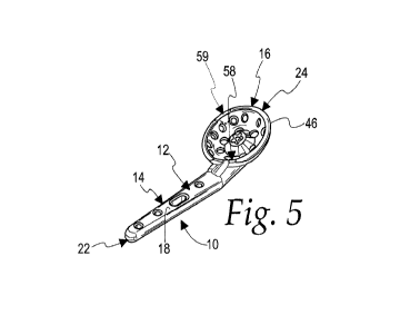
[0081] Fig. 5 is a perspective view of the implant in Fig. 4;
[0082] Fig. 6 is a plan view of the implant in Figs. 4 and 5;
[0083] Fig. 7 is an end elevation view of the implant in Figs. 4-6,
together with a schematic depiction of an associated bone component to
which one portion of the implant is fixed through one or more fasteners;
CA 03170120 2022-08-04
WO 2021/158913
PCT/US2021/016817
-18-
[0084] Fig. 8 is a view as in Fig. 6 with a schematic representation of a
bone component to which another portion of the implant is fixed using one or
more fasteners;
[0085] Fig. 9 is a cross-sectional view of the implant taken along line
9-9 of Fig. 8;
[0086] Fig. 10 is a schematic representation of polyaxial openings in a
body, as on the implant in Figs. 1-9;
[0087] Fig. 11 is a cross-sectional representation of an alternative form
of a cup-shaped part, as shown on the implant in Figs. 4-9;
[0088] Fig. 12 is a view as in Fig. 11 showing a further alternative
form;
[0089] Fig. 13 is a depiction of a human hand and forearm region at
which a wrist fusion is to be performed and with a broaching tool inserted
into
an intramedullary canal on the radius;
[0090] Fig. 14 corresponds to Fig. 13 from a different perspective;
[0091] Fig. 15 is a perspective view of the broaching tool depicted in
Figs. 13 and 14;
[0092] Fig. 16 is a schematic representation of a cutting tool usable to
form a cavity to receive a portion of the inventive implant;
[0093] Fig. 17 is a perspective view of an exemplary form of cutting
tool as shown schematically in Fig. 16;
[0094] Fig. 18 is a view as in Fig. 13 and showing K-wires inserted to
stabilize bones in the wrist region;
CA 03170120 2022-08-04
WO 2021/158913
PCT/US2021/016817
-19-
[0095] Fig. 19 corresponds to Fig. 18 from a different perspective;
[0096] Fig. 20 is a view as in Fig. 18 wherein the cutting tool is
operated to create a cavity for receiving a portion of the inventive implant;
[0097] Fig. 21 corresponds to Fig. 20 from a different perspective;
[0098] Fig. 22 is a view as in Fig. 20 wherein the cutting tool and K-
wires have been removed and the inventive implant has been placed in an
operative position and provisionally held in place through K-wires;
[0099] Fig. 23 corresponds to Fig. 22 but from a different perspective;
[00100] Fig. 24 is a view as in Fig. 22 wherein fasteners have been
directed through one portion of the implant;
[00101] Fig. 25 corresponds to Fig. 24 but from a different perspective;
[00102] Fig. 26 is an exploded view of an outrigger guide assembly in
relationship to the inventive implant;
[00103] Fig. 27 is a view as in Fig. 24 wherein the outrigger guide
assembly of Fig. 26 has been placed in an operative position;
[00104] Fig. 28 corresponds to Fig. 27 but from a different perspective;
[00105] Fig. 29 is a view as in Fig. 27 with the addition of an anchoring
part directed into one of the bone components;
[00106] Fig. 30 corresponds to Fig. 30 but from a different perspective;
[00107] Fig. 31 is a view corresponding to Fig. 29 wherein parts of the
outrigger guide assembly are urged towards each other to move bone
components to be fused into a desired relationship;
CA 03170120 2022-08-04
WO 2021/158913
PCT/US2021/016817
-20-
[00108] Fig. 32 corresponds to Fig. 31 but from a different perspective;
[00109] Fig. 33 is a view as in Fig. 31 wherein additional fasteners have
been used to maintain the desired relationship of the fused bone
components;
[00110] Fig. 34 corresponds to Fig. 33 but from a different perspective;
[00111] Fig. 35 is a view as in Fig. 33 with the outrigger guide assembly
removed;
[00112] Fig. 36 corresponds to Fig. 35 but from a different perspective;
[00113] Fig. 37 is a perspective view of the inventive implant, as in
Figs.
4-9, and with a bracing component associated with the outrigger guide
assembly releasably connected thereto;
[00114] Fig. 38 is a side elevation view of the inventive implant in Figs.
4-9 with the outrigger guide assembly in an operative relationship therewith;
[00115] Fig. 39 is a schematic representation of an alternative use of
the inventive implant for fusing a tibia and a tarsal bone; and
[00116] Fig. 40 is a schematic representation of the inventive implant as
used to fuse a metatarsal and tarsal bone.
DETAILED DESCRIPTION OF THE PREFERRED EMBODIMENTS
[00117] Referring initially to Fig. 1, a preferred form of implant for
fusing
at least two bones/bone components, according to the invention, is shown at
10. The implant 10 consists of a body 12 with first and second anchoring
CA 03170120 2022-08-04
WO 2021/158913
PCT/US2021/016817
-21-
portions 14, 16, respectively. The first anchoring portion 14 has a stem 18
configured to be directed to within a first bone component to place the first
anchoring portion 14 in an operative position. The first anchoring portion 14
is further configured to cooperate with at least a first fastener 20 usable to
cause the stem 18 to be fixed relative to the first bone component and
thereby maintain the first anchoring portion 14 in its operative position.
[00118] The second anchoring portion 16 is configured to overlie at
least a second bone component with the second anchoring portion 16 in an
operative position and is further configured to cooperate with at least a
second fastener 20 usable to fix a part of the second anchoring portion to the
second bone component to thereby maintain the second anchoring portion
16 in its operative position.
[00119] The fasteners 20 for the first and second anchoring portions 14,
16 may be the same or different and may take any known form.
[00120] The body 12 may be made in multiple pieces but in a preferred
form has a single rigid piece that defines the first and second anchoring
portions 14, 16.
[00121] As shown in Fig. 2, the body 12 has a length between first and
second ends 22, 24, respectively. The stem 18, in one preferred form,
extends fully to the first body end 22.
[00122] In Fig. 3, the body 12, consisting of the first and second
anchoring portions 14, 16, is shown in more detail and with an optional
modification. In this general form, the second anchoring portion 16 has a
cup-shaped part 26 from which a body part 28 extends. The body part 28
CA 03170120 2022-08-04
WO 2021/158913
PCT/US2021/016817
-22-
and first anchoring portion 14 typically project from different locations on
the
cup-shaped part 26. With the Fig. 3 construction, the second anchoring
portion 16 may by itself extend fully to the second body end 24.
Alternatively, the body part 28 may extend either up to an end of the cup-
shaped part 26 and to the second body end 24 or therebeyond as to by itself
define the second body end 24.
[00123] It should be understood that multiple cup-shaped parts 26 could
be incorporated into the body 12.
[00124] The schematic showing of the components in Figs. 1-3 is
intended to encompass the components as shown in specific forms
hereinbelow, as well as virtually an unlimited number of variations in those
components and their cooperation.
[00125] Referring now to Figs. 4-9, a specific form of the implant 10 will
be described. The implant 10 has the aforementioned body 12 with a length
L between the first and second ends 22, 24, respectively, and a predominant
width W.
[00126] The first anchoring portion 14 defines the stem 18 configured to
be directed into at least a first bone component 30. The stem 18 is elongate
in shape and has a plurality of lengthwise spaced openings 32a, 32b, 32c,
32d, each to accept a fastener 20 usable to fix the stem 18 in an operative
position relative to the bone component 30. The opening 32c is elongate to
allow the stem 18 to shift lengthwise relative to a fastener 20 directed
therethrough into the first bone component 30.
CA 03170120 2022-08-04
WO 2021/158913
PCT/US2021/016817
-23-
[00127] The second anchoring portion 16 is configured to overlie at
least a second bone component 34 and has a cup-shaped wall 36,
corresponding to the aforementioned cup-shaped part 26, with a plurality of
openings 38a-38t, each to receive a fastener 20 usable to fix a part of the
second anchoring portion 16 directly to at least one of the second bone
components 34 with the second anchoring portion 16 in an operative
position.
[00128] As noted above, the nature of the particular fasteners 20 is not
critical to the present invention. Typically, threaded fasteners 20 will be
directed through or into the openings 32, 38 and will purchase bone to effect
fixation.
[00129] As noted above, in reference to Fig. 3, the second anchoring
portion 16 has a generically depicted cup-shaped part 26, that may have a
wide range of different shapes. The corresponding cup-shaped wall 36 may
be symmetrical around a central axis 40, as depicted for the cup-shaped wall
in Figs. 4-9, or may have a non-symmetrical shape.
[00130] As depicted, the cup-shaped wall 36 has a cup-shaped
concave surface 42 and an oppositely facing cup-shaped convex surface 44.
As depicted, the surfaces 42, 44 are complementary in shape, with the wall
36 having a uniform thickness therebetween. This, however, is not a
requirement as the shapes of the surfaces 42, 44 could be considerably
different.
CA 03170120 2022-08-04
WO 2021/158913
PCT/US2021/016817
-24-
[00131] At least a part of the second anchoring portion 16 has this cup
shape. As depicted, the cup-shaped wall 36 makes up substantially the
entirety of the second anchoring portion 16.
[00132] In the depicted form, the convex surface 44 extends around the
axis 40 and has an axial width AW tapering between a top rim 46 and a flat
bottom wall portion 48. The bottom wall portion actually has a "W' shape, as
seen in cross-section in Fig. 9, but is effectively flat, and will be
considered
such since the downwardly facing surface 50 thereon will seat stably against
a flat bone surface.
[00133] The surface 44 is convex from two different perspectives ¨ in
cross-section as in Fig. 9 and as viewed from an axial perspective.
[00134] The "W' shape forms a discrete receptacle 52 in the bottom
wall portion 48 to receive bone graft material 54.
[00135] The convex surface 44 is configured to appose a surface on at
least one of the second bone components 34 with the second anchoring
portion 16 in its operative position. Bone graft material 54 is in contact
with
the cup-shaped wall 36 over the surface bounding the receptacle 52 and the
bone region which the bottom wall portion 48 overlies. The bottom wall
surface 50 may bear against at least one of the second bone components 34
but may be spaced above the same to accommodate an appropriate volume
of bone graft material 54. Bone graft material 54 is not required or may be
located other than at the bottom wall portion 48, which may allow the bottom
wall portion 48 to directly contact at least one of the second bone
components 34.
CA 03170120 2022-08-04
WO 2021/158913
PCT/US2021/016817
-25-
[00136] The receptacle 52 may take many different forms. Further,
multiple receptacles might be formed at different locations.
[00137] In the form depicted, the first anchoring portion 14 is elongate
and has a lengthwise center line 56. The center line 56 is offset from the
central axis 40 of the cup-shaped wall 36, as seen most clearly in Fig. 6.
[00138] In the event that the body part 28 is provided, it preferably
extends away from the cup-shaped wall 36 at a location spaced
circumferentially from a location at 58 where the first anchoring part 14
projects away from the cup-shaped wall 36.
[00139] In most constructions, the body part 28 projects away from the
cup-shaped wall 36 in a direction away from the first end 22 of the body 12.
As noted above, the body part 28 may extend fully to the second body end
24. The cup-shaped part 26 may likewise extend fully to the second body
end 24, or may be adjacent to or spaced therefrom.
[00140] In one modified form, an optional, discrete cutout 59 is formed
through the rim 46, as shown in dotted lines in Fig. 5, to avoid interference
at
certain joint sites, as explained hereinbelow.
[00141] The stem 18 has an obround shape as viewed in cross-section
orthogonally to the length of the stem 18. Other cross-sectional shapes are
contemplated, such as rectilinear, elliptical, etc. The width of the stem 18
along the major axis of the obround cross-sectional shape increases from the
first body end 22 up to the cup-shaped wall 36.
[00142] The body 12 may be made from any of a number of different
materials. In one form, it is made from metal.
CA 03170120 2022-08-04
WO 2021/158913
PCT/US2021/016817
-26-
[00143] In a more preferred form, the body 12 is made from a non-
metal material such as polyetheretherketone (PEEK), or other medical grade
plastic. The use of PEEK material facilitates the formation of polyaxial
openings, as identified generically at 60 in Fig. 10, at each location on the
body 12 where it is desirable to be able to direct the fasteners 20 at
different
angles threadably through each such opening 60.
[00144] As noted above, the particular "cup shape" for the cup-shaped
part 26 of the second anchoring portion 16 may vary considerably. As shown
in Fig. 11, the cup-shaped part 26' is formed as a hollowed segment of a
sphere, which has a central axis 40'.
[00145] In Fig. 12, a variation is shown wherein the cup-shaped part
26" has a convex surface 42" made up of portions with radii of different
lengths as viewed orthogonally to the axis 40".
[00146] These are just examples of the multitude of different forms that
the cup-shaped part 26, making up at least part of the second anchoring
portion 16, may take, keeping in mind that symmetry is not required around a
respective axis 40 and the cup-shaped wall 36 need not have a uniform
thickness.
[00147] A method of fusing bone components is described in Figs. 13-
36 on a human wrist. This is but one exemplary application of the implant,
described above.
[00148] The wrist is exposed and the arthritic joint surfaces are
decorticated and residual cartilage removed. The distal end of the radius 64
CA 03170120 2022-08-04
WO 2021/158913
PCT/US2021/016817
-27-
is exposed and a drill placed into the central intramedullary canal to
determine the longitudinal axis of the radius.
[00149] As seen in Figs. 13-15, a broaching tool 66 is used to prepare
the radius canal 68, and inserted from the articular end of the radius 64
centrally into the canal 68. The penetrating portion 70 of the broaching tool
66 preferably matches the contour of the stem 18. In the depicted form, a
handle 72 on the broaching tool is offset so that when the broaching tool 66
is fully seated as in Figs. 13 and 14, reduction of the joint surface is
permitted.
[00150] While not required, one preferred method of using the implant
is carried out with the assistance of a cutting tool, as shown generically at
74 in Fig. 16.
[00151] The cutting tool 74 has at least one cutting surface 76
configured so that as the cutting tool 74 is operated, it is capable of
producing a cavity with a predetermined shape. The cutting tool 74 is not
limited in terms of its configuration or manner of operation so long as it is
capable of consistently producing a cavity with a predetermined shape.
[00152] In an exemplary form, as shown in Fig. 17, the cutting tool 74 is
in the form of a reamer with a plurality of cutting surfaces 76. The reamer 74
is turned around an axis 78 through an appropriate drive 80, as an incident of
which the cutting surfaces 76 are capable of progressively removing bone in
a symmetrical pattern around the axis 78.
CA 03170120 2022-08-04
WO 2021/158913
PCT/US2021/016817
-28-
[00153] The depicted cutting surfaces 76 are configured to produce a
cup-shaped cavity. The reamer 74 has a pilot extension 82 projecting axially
beyond the cutting surfaces 76.
[00154] The cutting tool/reamer 74 has a footprint diameter D selected
based upon the particular implant configuration and the desired number of
bone components to be fused. In the particular wrist application depicted,
the implant 10 is configured and dimensioned to allow fusion of five carpal
bones ¨ the scaphoid 84, capitate 86, hamate 88, triquetrum 90, and lunate
92 ¨ to each other and the radius 64.
[00155] Preparatory to using the reamer 74, and with the broaching tool
in place, as in Figs. 13, 14, 18, and 19, the carpus at 94, consisting of,
inter
alia, the scaphoid 84, capitate 86, hamate 88, triquetrum 90, and lunate 92,
is stabilized relative to the radius 64 as by using conventional K-wires 96
directed through bones in the carpus 94 and into the radius 64. This assures
that the wrist is fused in the preferred angle of dorsiflexion.
[00156] Once the region is stabilized, a pilot bore 98 is formed in the
carpus 94. The broaching tool 66 has a guide opening 100 for a boring tool
102 that is directed towards a target location in the region whereat the
second anchoring portion 16 is placed in its operative position. The boring
tool 102 may be manually controlled or may be turned through an
appropriate drive 104.
[00157] As shown in Figs. 20 and 21, with a region of the carpus 94
stabilized by the K-wires 96, the broaching tool 66 is removed and the cutting
tool/reamer 74 placed strategically over the carpus region and the distal end
CA 03170120 2022-08-04
WO 2021/158913
PCT/US2021/016817
-29-
of the radius 64 by directing the pilot extension 82 thereon into the pilot
bore
98.
[00158] By operating the cutting tool/reamer 74, a desired quantity of
surface cortical bone can be removed to create a cavity 106 in at least the
carpus 94, and as depicted in the radius 64.
[00159] The overall cup-shaped cavity 106 is complementary in shape
to the second anchoring portion and, more particularly, the concave surface
42 as well as potentially the surface 50 on the bottom wall portion 48.
[00160] The cutting path for the cutting tool/reamer 74 is dictated by the
particular fusion that is desired. It is not necessary that the surface
cortical
bone be removed from the radius 64 to use the implant 10. The diameter D,
which represents the effective cutting diameter of the cutting surfaces 76,
also determines the number of carpal bones to be treated as well as the
particular area of the placement locations thereon. The reamer/cutting tool
74 can be simply and conveniently operated to strategically and precisely
remove cortical bone surface to provide a bed for effective fusion.
[00161] As seen in Figs. 22 and 23, once the bone treatment with the
cutting tool/reamer 74 is completed, bone graft material can be applied within
the receptacle 52 on the cup-shaped wall 36. The stem 18 is inserted into
the radius canal 68, whereupon the cup-shaped wall 36 is pressed into the
cavity 106.
[00162] A guide/inserter bar 108 can be releasably connected to the
bottom wall portion 48 and may have a leading part 110 that projects past the
bottom wall portion 48 to advance into the pilot bore 98. The guide 108 is
CA 03170120 2022-08-04
WO 2021/158913
PCT/US2021/016817
-30-
graspable to facilitate reorientation of the cup-shaped wall 36 with the
leading
portion 110 facilitating alignment of the convex surface 44 with the
complementary surface 114 bounding the cavity 106, as defined
cooperatively by the carpus 94 and radius 64.
[00163] In Figs. 22 and 23, the first and second anchoring portions 14,
16, respectively, are shown in their respective operative positions wherein
the convex surface 44 is apposed to the surface 114 bounding the cavity
106. The surface 50 on the bottom wall portion 48 may likewise appose the
bottom surface region 116 bounding the cavity 106.
[00164] With the first and second anchoring portions 14, 16 in their
respective operative positions, provisional fixation of the implant 10 can be
effected using small K-wires 118, directed in this case through openings 120
through the rim 46 of the cup-shaped wall 36 and into the carpus 94 and
radius 64.
[00165] As shown in Figs. 24 and 25, with the implant 10 provisionally
fixed as in Figs. 22 and 23, holes 122 can be formed strategically into bones
of the carpus 94 to accept fasteners 20 directed through openings 38 in the
cup-shaped wall 36.
[00166] Once the fasteners 20 are secured as in Figs. 24 and 25, the
guide/inserter bar 108 is separated from the cup-shaped wall 36.
[00167] While different arrangements of fasteners are contemplated,
the fasteners 20 of an appropriate size are directed into each of the scaphoid
84, capitate 86, hamate 88, triquetrum 90, and lunate 92, which collectively
define one placement location that the cup-shaped wall 36 overlies.
CA 03170120 2022-08-04
WO 2021/158913
PCT/US2021/016817
-31-
[00168] In this
embodiment, cortical bone from the dorsal rim 124 of the
radius 64 is also removed so that the cup-shaped wall 36 seats at a second
placement location 125 where the cup-shaped wall 36 overlies the radius 64
which, while not required, in this embodiment is reconfigured through the
cutting tool/reamer 74. As depicted in Fig. 25, one of the temporary K-wires
118 is directed into the radius 64.
[00169] As seen in
Figs. 26-30, an outrigger guide assembly 126 may
then be utilized, consisting of a first bracing component 128 releasably
connected to the cup-shaped wall 36, and a second bracing component 130.
Alternatively, the guide assembly 126 may be attached to any other site on
the implant body 12. The aforementioned guide 108 and first bracing
component 128, while shown to be different, may be one and the same. The
first bracing component 128 consists of an elongate sleeve 132 with a length
alignable with the axis 40 of the cup-shaped wall 36. An anchoring part 134
has a connector 136 that is releasably engageable with a connector 138 on
the cup-shaped wall 36. The connectors 136, 138 may make threaded
engagement or may be otherwise configured. With the
threaded
arrangement, utilizing an enlarged head 140 for manual gripping, the
anchoring part 134 may be turned to engage and release the connectors 136
138. With the connectors 136, 138 engaged, the sleeve 132 and anchoring
part 134 are fixed with their lengths aligned with the axis 40.
[00170] It should
be noted that the connector 138 is also usable to
releasably engage a connector 142 on the aforementioned guide/inserter bar
108, as shown schematically in Fig. 23, to make a releasable connection
therewith.
CA 03170120 2022-08-04
WO 2021/158913
PCT/US2021/016817
-32-
[00171] The sleeve 132 is fixed with respect to an elongate guide bar
144 which has openings 32a', 32b', 32c', 32d', corresponding to the stem
openings 32a, 32b, 32c, 32d in shape and location, whereby with the
elongate guide 144 overlying the stem 18 with these components in
lengthwise alignment, the openings 32a', 32b', 32c', 32d' register with the
openings 32a, 32b, 32c, 32d, as shown most clearly in Fig. 28.
[00172] The second bracing component 130 is then directed through
the opening 32c' into the radius 64 and through the stem opening 32c. The
second bracing component 130 is configured to be slidable within each of the
slots 32c, 32c' lengthwise of the elongate guide 144 and stem 18. The
second bracing component 130 is directed into the openings 32c', 32c to
reside at or adjacent an edge 146, 146' closest to the end 22 of the body 12
and the end 148 of the elongate guide 144.
[00173] As shown in Figs. 31 and 32, a clamping tool 150 is employed
with jaws 152, 154 that can be respectively borne against the first and
second bracing components 128, 130, respectively, to draw the bracing
components 128, 130 towards each other as indicated by the arrows 156.
This is permitted by the elongate configuration of the openings 32c, 32c'. As
this occurs, the carpus bone components fixed to the cup-shaped wall 36 are
drawn towards the radius 64 along the line of the double-headed arrow 158
into a desired relationship wherein at least one of the engaged carpus bones
is compressed against the distal end of the radius 64. While a scissors-type
clamping tool 150 is depicted, any type of device or devices might be utilized
to effect this compressive movement at the fusion site.
CA 03170120 2022-08-04
WO 2021/158913
PCT/US2021/016817
-33-
[00174] As shown in Figs. 33 and 34, once the desired relationship is
established between the carpal bones and the radius 64, a drill 159 can be
placed through the drill sleeve 160 and used to form openings 162a, 162b,
162d through the radius 64 that register with the openings 32a, 32b, 32d in
the stem 18, whereupon fasteners 20 can be utilized to fix the stem 18
relative to the radius 64.
[00175] In Figs. 35 and 36, the user's wrist region is shown with all of
the fasteners 20 secured and the outrigger guide assembly 126 removed.
[00176] In Figs. 37 and 38, additional details of the outrigger guide
assembly 126 are more clearly shown in relationship to the implant 10.
[00177] In Fig. 37, the first bracing component 128, which is an integral
part of the outrigger guide assembly 126, is shown releasably fixed in place
on the implant through the anchoring part 134 and without the elongate
guide 144 thereon.
[00178] As seen in Fig. 38, the elongate guide 144 has a guide
passage 164 which is aligned to create an opening in the radius 64 to accept
a fastener 20 directed through an opening 166 at a region adjacent to where
the stem 18 and cup-shaped wall 36 connect.
[00179] Fig. 38 also shows depth guides 168, 170 to facilitate controlled
drilling of bone to accept the fasteners 20.
[00180] As noted above, the body 12 may include the body part 28
which may be appropriately fixed to one or more metacarpal bones. The
body part 28 may overlie, and/or be inserted into, one or more of the
metacarpal bones.
CA 03170120 2022-08-04
WO 2021/158913
PCT/US2021/016817
-34-
[00181] As noted above, the inventive implant is not limited to use with
wrist fusion applications. The same concepts can be employed to effect
fusion at other anatomical locations. As just examples, as shown in Fig. 39,
the implant 10 may be used to effect fusion between a tibia 172 and tarsal
bone 174.
[00182] Alternatively, as shown in Fig. 40, the implant 10 may be used
to effect fusion between a metatarsal 176 and tarsal bone 178.
[00183] In these particular applications, the stem 18 may be inserted
into the tibia or metatarsal, with the cup-shaped wall 36 fixed to the tarsal
bone.
[00184] As seen in Figs. 4, 7, and 9, the flat surface 50 on the bottom
wall portion 48 is angled in two dimensions with respect to a reference plane
P that approximates a flat profile of the stem 18 extending through a central,
lengthwise axis 180 of the stem 18. In other words, the cup-shaped wall 36
is angled dorsally with respect to the plane of the patient's forearm, as well
as angled rotationally in supination.
[00185] As seen in Figs. 4, 7, and 9, the plane P is at an angle a with
respect to a reference plane P1 containing the bottom wall surface 50, which
in turn is substantially parallel to a reference plane P2 across the edge of
the
rim 46. In one preferred form, the angle a is on the order of at least 10 .
[00186] With fasteners securing the implant 10 to multiple bone
components on opposite sides of the fusion site, the implant 10 is able to
resist forces with large moment arms across the joint. At the same time, in
the wrist application, situation of the stem 18 within the radius canal
obviates
CA 03170120 2022-08-04
WO 2021/158913
PCT/US2021/016817
-35-
the need to fix a plate that requires a complex curved implant, cutting of a
channel for the plate, and the use of a bulky superficial plate. Since the
carpus is centered over the joint surface of the distal radius, the stem 18
directly aligns with the cup-shaped wall 36.
[00187] As seen in exemplary Fig. 36, the second anchoring portion 16
is effectively recessed over its entire footprint so as not to add protruding
mass that could cause discomfort, as from soft tissue irritation, and/or
potentially damage to a patient such as tendon rupture or cosmetic deformity.
[00188] The ability to direct fasteners through the cup-shaped wall 36 at
different angles reinforces the connection between the multiple bone
components.
[00189] By reason of having complementary tapered cup shapes on the
cup-shaped wall 36 and cavity 106, as the cup-shaped wall 36 is directed
into the cavity 106, the convex surface 44 and surface bounding the cavity
106 cooperate to consistently guide the cup-shaped wall 36 into the cavity
106 wherein the second anchoring portion 16 realizes its operative position.
[00190] By reason of providing an implant that is centrally positioned
close to the neutral axis of the exemplary radius, bending loads on the
implant may be reduced compared to other conventional implants.
[00191] By reason of using the intramedullary construction and
recessing to at least a certain extent the second anchoring portion 16, the
inventive implant makes possible a reduction in: prominence of the implant;
cosmetic deformity; soft tissue irritation; and tendon problems.
CA 03170120 2022-08-04
WO 2021/158913
PCT/US2021/016817
-36-
[00192] With the inventive structure, it is possible to provide a
relatively
short surgical incision to retain blood supply to the bone.
[00193] The inventive implant, as described above, can be made
without a complex shape to fit a wide range of applications without the need
for extensive bone carpentry or implant bending to avoid implant prominence
at the surgical site. At the same time, the implant can be made with a
significant vertical thickness providing bending strength and stiffness while
avoiding soft tissue prominence.
[00194] With the inventive structure, it is possible to provide reliable
fixation that does not necessarily require extending fixation and complication
risk to unrelated regions, such as crossing the carpal-metacarpal joint and
requiring screw placement into a metacarpal bone.
[00195] Further, the particular design as described herein, which is
exemplary in nature only, provides sufficient multiplicity of fastener
openings
and a variable range of fastener angular placement into small bones that
may be part of the fusion mass, such as into carpal bones.
[00196] The strategic use and placement of fasteners may also allow
the same to be removed without requiring extensive destruction of bone.
[00197] The foregoing disclosure of specific embodiments is intended to
be illustrative of the broad concepts comprehended by the invention.