Note: Descriptions are shown in the official language in which they were submitted.
CA 03170374 2022-08-08
WO 2021/156878
PCT/IL2021/050155
1
RAPID DETECTION TEST FOR SARS-COV-2
RELATED APPLICATION/S
This application claims the benefit of priority of US Provisional Patent
Application
No. 62/972,005 filed 9 February 2020, the contents of which are incorporated
herein by reference
in their entirety.
SEQUENCE LISTING STATEMENT
The ASCII file, entitled 82013 Sequence Listing.txt, created on 9 February
2021,
comprising 70,597,807 bytes, submitted concurrently with the filing of this
application is
incorporated herein by reference.
TECHNICAL FIELD
The present invention relates to the field of viral sensing and rapid
diagnostics, in general,
and to a method, reagents and a kit for the detection of SARS-CoV-2 in a test
sample, in particular.
The method involves the detection of the 3C-L protease.
BACKGROUND
Timely and accurate COVID-19 testing is an essential part of the management of
the
current pandemic. The etiologic agent of COVID-19 is the Severe Acute
Respiratory Syndrome
Coronavirus 2 (SARS-CoV-2), which is a newly emerged member of the family
Coronaviridae,
subfamily Coronavirinae, genus I coronaviridae that includes the SARS-CoV and
MERS-CoV
viruses. These viruses are involved in Severe Acute Respiratory Syndrome
(SARS) outbreaks in
the past two decades. US 10,130,701 B2 describes an attenuated coronavirus
SARS-CoV
comprising a variant replicase gene, which causes the virus to have reduced
pathogenicity.
The SARS-CoV-2 viral genome is a single-strand, positive-sense RNA with a size
of ¨30
kb, which contains numerous open-reading frames. Two-thirds of the viral
genome encodes 16
non-structural proteins (nsp 1-16), while the remaining genome encodes four
structural and nine
accessory proteins (0rf3a, 0rf3b, 0rf6, 0rf7a, 0rf7b, 0rf8, 0r19b, 0rf9c, and
Orf10). Several
non-structural proteins harbour enzymatic activities, such as protease
activity and RNA-directed
RNA polymerase activities.
SARS-CoV-2 replicates at much higher levels in the nose and mouth than SARS-
CoV and
MERS, and this leads to much higher levels of virus shedding in the
environment by people who
CA 03170374 2022-08-08
WO 2021/156878
PCT/IL2021/050155
2
are either pre-symptomatic or asymptomatic. Thus, a large percentage of
infected people can
transmit the virus without realizing that they are even infected. Due to these
reasons, rapid, low
cost and accurate methods of SARS-CoV-2 detection are critical for
significantly slowing the
spread of the virus and for population surveillance well into the future.
Currently, the molecular technique of quantitative real time polymerase chain
reaction
(qRT PCR) is the gold standard for SARS-CoV-2 detection using samples from
respiratory
secretions. Cycle threshold (CT) values of the PCR tests indicate the CT used
in the PCR for
exponential amplification of the target specimen and are inversely related to
the viral load in the
sample. CT40 is the accepted minimum viral load that can be detected by high-
end PCR
techniques. However, as noted above, the cost and organizational complexity of
performing a large
number of PCR reactions for downstream applications render this option
feasible but unattractive.
Furthermore, the SARS-CoV-2 virus is mutating over time, resulting in genetic
variation in the
population of circulating viral strains, which become indistinguishable by the
routine PCR tests.
Thus, false negative results frequently occur with any molecular test for the
detection of SARS-
CoV-2 if some particular mutation occurs in the part of the virus' genome
assessed by that
test. Lastly, PCR-based assays can produce false positive results because they
cannot distinguish
between nucleic acid fragments from live vs dead, decaying or inactive virus.
Several other molecular assays have been recently developed to detect the
present SARS-
CoV-2 virus, based on enzyme-linked immunosorbent assay (ELISA), and rapid
tests that aim to
detect either antibodies against the virus or the viral antigen themselves.
Nevertheless, most of
these immunochemical assays have recently failed due to the large number of
false negative or
false positive results. These assays also suffer from the same issue of not
being able to distinguish
between active or inactive viral products.
Moreover, antibodies against specific proteins of a new virus may not be
instantly and
constantly available (e.g., monoclonals). The production of antibodies uses
biological systems. To
produce antibodies, the induction of an immune response is necessary. However,
this procedure
might discriminate target proteins that has similar structure to endogenous
protein or toxic
compounds that would kill the animal. Another complication for in-vivo
production of antibodies
is that the antibodies can only work under physiological conditions. This
restricts the range of
application and function of antibodies. It is worthwhile to mention here that
the last, but not the
least problem of ELISA is that the same antibodies produced by different
animals may
significantly differ in their structure and functionality. As a result, any
ELISA-based bioassay or
CA 03170374 2022-08-08
WO 2021/156878
PCT/IL2021/050155
3
biosensing system for detection of a certain analyte would differ in
sensitivity and specificity, and
might be difficult to calibrate and universalize.
Thus, while antibody recognition has been the gold standard for decades,
numerous
problems mentioned above abound towards new designs, limited protein shelf
lives, time-
consuming washing procedures and manufacturing scale-up. These problems can be
surmounted,
but only through laborious research programs at great cost. Although the
aforementioned PCR
methods and immunoassays have recently been developed to detect the presence
of, or exposure
to SARS-CoV-2 virus, but they do not distinguish between active vs. degraded
virus remnants.
Because of the deficiencies of the presently available testing methods, there
is a need for an
improved test enabling the presence of viruses, such as the SARS-CoV-2 virus,
to be accurately
and rapidly detected at an early stage of infection. Such a test will benefit
those showing symptoms
of COVID-19 by allowing for the monitoring of the course of their infection
and subsequent
recovery. In addition, a rapid, sensitive and selective test will benefit
persons suspected of having
the disease by allowing uninfected persons to be released from quarantine.
There is also the need
for an automated test avoiding the need for manual intervention. Such a test
will prevent spread of
the disease due to infection during the testing process.
During the replication of many viruses, the viral genetic material is
transcribed and
translated to form a polyprotein, which is ultimately cleaved into
biologically active proteins by
an essential virally encoded cysteine protease. The 3C-like protease (3CLpro),
formally known as
C30 Endopeptidase, is the main protease found in coronaviruses. It cleaves the
coronavirus
polyprotein at 11 conserved sites, and it is the main protease found in SARS-
CoV-1 and SARS-
CoV-2, which is responsible for the viral replication. Both viruses have this
3C-Like cysteine
protease that exhibits similar, but not identical cleavage-site specificity to
that of picornavirus 3C
protease, and are therefore termed "3C-Like protease" (3CL protease). US
7,635,557 by the
present inventors describes methods, compositions and kits for testing for
SARS-CoV-1 virus in a
sample. The methods determine the presence of the 3CL protease by contacting
the sample with a
peptide compound capable of being cleaved by the 3CL protease to form peptide
compound
fragments. Detection of a peptide compound fragment confirms the presence of
the virus.
Quantitation of the 3CL protease activity presents a unique approach to viral
diagnosis.
The premise for a diagnostic test is that clinical samples, such as
nasopharyngeal swab, saliva,
buccal swab others, are collected and incubated in the presence of a peptide
substrate containing
the unique viral cleavage sequence linked to particular analytical probes, for
examples donor and
quencher fluorophores at the amino and carboxyl terminals. Presence of active
SARS CoV-2 3CL
CA 03170374 2022-08-08
WO 2021/156878
PCT/IL2021/050155
4
protease can then be detected by peptide cleavage, which results in a visual
signal that can be
easily quantified with any suitable analytical technical, for example
fluorescence or luminescence
spectroscopy, terahertz spectroscopy, lateral flow etc.
SUMMARY
Unless otherwise defined, all technical and/or scientific terms used herein
have the same
meaning as commonly understood by one of ordinary skill in the art to which
the invention
pertains. Although methods and materials similar or equivalent to those
described herein can be
used in the practice or testing of embodiments of the invention, exemplary
methods and/or
materials are described below. In case of conflict, the patent specification,
including definitions,
will control. In addition, the materials, methods, and examples are
illustrative only and are not
intended to be necessarily limiting.
Implementation of the method and/or apparatus of embodiments of the invention
can
involve performing or completing selected tasks manually, automatically, or a
combination
thereof Moreover, according to actual instrumentation and equipment of
embodiments of the
method and/or apparatus of the invention, several selected tasks could be
implemented by
hardware, by software or by firmware or by a combination thereof using an
operating system.
For example, hardware for performing selected tasks according to embodiments
of the
invention could be implemented as a chip or a circuit. As software, selected
tasks according to
embodiments of the invention could be implemented as a plurality of software
instructions being
executed by a computer using any suitable operating system. In an exemplary
embodiment of the
invention, one or more tasks according to exemplary embodiments of method
and/or apparatus as
described herein are performed by a data processor, such as a computing
platform for executing a
plurality of instructions. Optionally, the data processor includes a volatile
memory for storing
instructions and/or data and/or a non-volatile storage, for example, a
magnetic hard-disk and/or
removable media, for storing instructions and/or data. Optionally, a network
connection is
provided as well. A display and/or a user input device such as a keyboard or
mouse are optionally
provided as well.
The present invention describes embodiments of a method of diagnosing a Severe
Acute
Respiratory Syndrome Coronavirus 2 (SARS-CoV-2) infection in a sample of a
subject, the
method comprising contacting the sample with a composition comprising an agent
that detects
3CL-protease of the SARS-Co-V2 virus, wherein a presence of the 3CL-protease
in the sample is
indicative of a SARS-Co-V2 infection.
CA 03170374 2022-08-08
WO 2021/156878
PCT/IL2021/050155
In another embodiment, the method of detecting a SARS-CoV-2 virus in a sample
of a
subject suspected of having COVID-19 comprises comprising contacting the
sample with a
composition comprising an agent that monitors the activity of a 3CL protease
of the SARS-CoV-
2 virus, wherein the activity level of the 3CL protease in the sample is
indicative of the presence
5 __ of SARS-CoV-2 in the sample.
In particular embodiments, the sample is selected from the group consisting of
mucus,
saliva, throat wash, nasal wash, spinal fluid, sputum, urine, semen, sweat,
faeces, plasma, blood,
bronchioalveolar fluid, vaginal fluid, tear fluid, tissue biopsy, and
nasopharyngeal, oropharyngeal,
nasal mid turbinate, anterior nasal and buccal swabs.
In other embodiments, the detectable moiety is a fluorescence moiety. In a
particular
embodiment, the detectable moiety is a Forster Resonance Energy Transfer
(FRET) pair of donor
and acceptor moieties, and the cleavage of the substrate peptide generates or
modulates a signal
from the FRET pair. Specific donor moiety is a quantum dot.
In certain embodiments, the donor and acceptor moiety are attached to the
peptide in a
configuration that permits energy transfer from the donor to the acceptor to
result in quenching of
the fluorescence by FRET process. In some other embodiments, the donor and
acceptor moiety are
separated by no more than 3, 5, 10, 15 or 20 amino acid residues.
In a further embodiment, the acceptor moiety the acceptor moiety is radiative
or non-
radiative. Specific examples of the acceptors used in the present invention
are tetramethy1-6-
carboxyrhodamine (TAMRA) and Black Hole Quenchers (BHQs) including Black Hole
Quencher-1 (BHQ-1), Black Hole Quencher-2 (BHQ-2), Black Hole Quencher-3 (BHQ-
3).
Specific FRET pairs are Alexa Fluor 488 (AF488) (donor)/BHQ1 (acceptor),
AF488
(donor)/QSY9 (acceptor), EDANS (donor)/DABCYL (acceptor).
Donor Acceptor
Alexa Fluor 488 BHQ1
CA 03170374 2022-08-08
WO 2021/156878 PCT/IL2021/050155
6
Et3NH Et3NH+ ¨P .= 0
SO3- SO3-
0
0 NO,s.
CO2-
Black Hole Quencher I (BH01 Modified Olio
0 0 QSY9
ON
, N ,õ 0
11
so3H-
0,
QSY Quencher Modified Oligoriudeoble 6
EDANS DABCYL
H2 0
HN/ OH
N
0=S=0
OH
In other embodiments, the C-terminus of the peptide is attached to the
acceptor moiety and
the N-terminus of the peptide is attached to the donor moiety. In still other
embodiments, the C-
terminus of the peptide is attached to the acceptor moiety and the donor
moiety is attached to no
more than three amino acids from the N-terminus. In a particular embodiment,
the donor moiety
is attached to the separating moiety Z.
In a further embodiment, the detectable moiety is a chemiluminescent
signalling moiety
attached to one side of the cleavage region of the substrate peptide, and an
acceptor moiety is
CA 03170374 2022-08-08
WO 2021/156878
PCT/IL2021/050155
7
attached at the other side of the cleavage region of the substrate peptide.
Non-limiting example of
the chemiluminescent signalling moiety is a 1,2-dioxetane compound.
In yet further embodiment, the detectable moiety is a pre-enzyme, which upon
substrate
peptide cleavage is activated and detected via the detection of a catalytic
activity of same. Non-
limiting example of the pre-enzyme is pro-Thrombin (factor II) or other
enzymes in this cascade.
In some embodiment, the substrate peptide Y comprises a sequence of 8-12 amino
acids.
In a particular embodiment, this sequence is an amino acid sequence selected
from the group
consisting of SEQ ID Nos: 13-23, specifically SEQ ID NO: 13, more specifically
SEQ ID Nos.
24-33 and SEQ ID Nos: 1-10.
In a certain embodiment, the sample tested in the method of the present
invention is a saliva
sample, and the composition further comprises a protease inhibitor selected
from the group
consisting of Antipain, AC-DEVD-CHO, Aprotinin, Eglin C, GW, PMSF and 2,6 PDA.
The
sample can be a buccal sample, the composition comprises a protease inhibitor
selected from the
group consisting of PMSF, GW, aprotinin, eglinC and pepstatin.
In another embodiment, the method of the present invention further comprises
contacting
the sample with at least one substrate of a viral protease of a virus which is
not the SARS-CoV-2,
wherein absence of cleavage of the at least one substrate is indicative of the
absence of the virus
from the sample.
In a further embodiment, the device suitable for reading the signal in the
method of the
present invention is an optical or spectroscopic device. This optical or
spectroscopic device can be
modular. In yet further embodiment, the optical or spectroscopic device is
configured to operate
as a portable and highly sensitive fluorescence spectrophotometer
(fluorometer), luminometer,
fluorescence microscope or combinations thereof for measuring fluorescence,
luminescence or
phosphorescence. These optical or spectroscopic devices can be conveniently
placed at the
entrance to public areas, such as theatres, restaurants and places of work.
They can be also
miniaturised and used for rapid point-of-care diagnostics in public areas,
work places and at home.
In yet another embodiment, the assay can be adapted to a fully automated
robotic system.
The optical or spectroscopic device comprises a fluorescence reading module, a
sample handling
mechanism and a dispenser/pipette module. The samples are loaded on to the
sample handling
mechanism, where it is processed and all reagents are added by the
dispenser/pipette module. Then
the sample is monitored by fluorescence reading and analysed by designated
software. This
embodiment is intended for rapid laboratory, high-throughput diagnostics for
multiple patients.
CA 03170374 2022-08-08
WO 2021/156878
PCT/IL2021/050155
8
In yet further embodiment, the optical or spectroscopic device comprises an
excitation
module, a sample chamber and an acquisition and/or detector module. The sample
chamber can
be a fluorescence multiplate reader for laboratory high-throughput and rapid,
multiplexing analysis
of multiple samples for point-of-care diagnostics.
In a particular embodiment, the optical or spectroscopic device further
comprises a
computing unit. In another particular embodiment, the acquisition and/or
detector module and the
computing unit are combined in a single unit designed to perform acquisition
of the fluorescence
emission, to measure its intensity, to process the fluorescent emission data
and optionally display
it in a readable format and/or output it to an external memory or user's
interface.
In still another particular embodiment, the acquisition module is a part of a
smartphone or
any other mobile device or gadget suitable for performing the desired
measurements.
In some embodiments, the detector is an electron-multiplying charge-coupled
device
(EMCCD) imager, a charge-coupled device (CCD) imager, an avalanche photodiode
(APD), a
photomultiplier tube (PMT), scientific complementary metal-oxide-semiconductor
(sCMOS)
imager, or CMOS imager of a smartphone camera, a stand-alone camera, or a
camera of any mobile
device or gadget, the detector optionally having a focusing apparatus and a
computer link.
In a specific embodiment, the detector is a CMOS imager of a smartphone
camera.
In some embodiments, the device suitable for reading the signal is a lateral
flow device,
which can be in a format of a stick or a stack. In a particular embodiment,
the lateral flow device
is based on a nitrocellulose membrane or a cellulose (paper) membrane. The
lateral flow device is
suitable for home use or point of care detection of the virus.
The present invention also provides a microfluidic chip or lab-on-a-chip
suitable for
carrying out the method of any embodiment of the present invention.
Another aspect of the present invention is an isolated peptide comprising an
amino acid
sequence as set forth in SEQ ID NOs: 25-33, the peptide being no longer than
14 amino acids. The
isolated peptide further comprising a detectable moiety. In a particular
embodiment, the isolated
peptide consists of an amino acid sequence as set forth in any one of SEQ ID
Nos: 2-10.
A further aspect of the present invention is an article of manufacture
comprising the
isolated peptide of the present invention attached to a solid support. The
solid support can be a test
3()
tube, microtiter plate, microtiter well, bead, dipstick, polymer
microparticle, magnetic
microparticle, nitrocellulose, cellulose, or a chip array.
Yet further aspect of the present invention is a diagnostic kit for detection
of SARS-CoV-
2 in a sample. The kit comprises the isolated peptide or the article of
manufacture of the present
CA 03170374 2022-08-08
WO 2021/156878
PCT/IL2021/050155
9
invention and reagents for detecting cleavage of the peptide. The kit further
comprises at least one
agent which specifically detects the presence of a virus other than the SARS-
CoV-2.
According to another aspect of the present invention, there is provided a
method of
diagnosing a Severe acute respiratory syndrome coronavirus 2 (SARS-CoV-2)
infection in a
subject comprising contacting a sample of the subject with a composition
comprising an agent that
detects 3CL-protease of the SARS-Co-V2 virus, wherein a presence of the 3CL-
protease in the
sample is indicative of a SARS-Co-V2 infection.
According to another aspect of the present invention, there is provided a
method of
detecting a SARS-CoV-2 virus in a sample of a subject suspected of having
COVID, the method
comprising contacting the sample with a composition comprising an agent that
monitors the
activity of a 3CL protease of the SARS-CoV-2 virus, wherein the activity level
of the 3CL protease
in the sample is indicative of the presence of SARS-CoV-2 in the sample.
According to embodiments of the present invention, the sample is selected from
the group
consisting of mucus, saliva, throat wash, nasal wash, spinal fluid, sputum,
urine, semen, sweat,
feces, plasma, blood, broncheoalveolar fluid, vaginal fluid, tear fluid and
tissue biopsy.
According to embodiments of the present invention, the sample is a saliva or
buccal
sample.
According to embodiments of the present invention, the agent monitors the
activity of the
3CL protease.
According to embodiments of the present invention, the agent is a substrate
peptide for the
3CL protease, the peptide being attached to at least one moiety which
generates a detectable signal
on cleavage of the peptide by the 3C-L protease of the SARS-CoV-2.
According to embodiments of the present invention, the peptide is between 10-
12 amino
acids.
According to embodiments of the present invention, the at least one moiety is
a FRET pair,
and wherein cleavage of the peptide generates a signal from the FRET pair.
According to embodiments of the present invention, the FRET pair is AF488 and
BHQ 1 .
According to embodiments of the present invention, the FRET pair is EDANS and
dabcyl.
According to embodiments of the present invention, the substrate peptide
further comprises
a separating moiety.
According to embodiments of the present invention, the agent is represented by
the general
formula:
X-Y-Z
CA 03170374 2022-08-08
WO 2021/156878
PCT/IL2021/050155
wherein:
Y comprises a substrate peptide of a 3CL protease of the SARS-CoV-2, cleavage
of X-Y-Z by
the 3CL protease forming cleavage products X-Y' and Y"-Z wherein Y' is a first
cleavage product
of Y and Y" is a second cleavage product of Y;
5 X comprises a detectable moiety; and
Z comprises a separating moiety capable of binding to a separate phase of a
two phase separating
system;
wherein the X-Y-Z does not form a contiguous portion of a natural substrate of
the 3CL protease.
According to embodiments of the present invention, the detectable moiety X
comprises a labeling
10 agent selected from the group consisting of an enzyme, a fluorophore, a
chromophore, a protein,
a pre-enzyme, a chemiluminescent substance and a radioisotope.
According to embodiments of the present invention, the separating moiety Z is
selected from the
group consisting of an immunological binding agent, a magnetic binding moiety,
a peptide binding
moiety, an affinity binding moiety and a nucleic acid moiety.
According to embodiments of the present invention, the substrate peptide
comprises an amino acid
sequence selected from the group consisting of SEQ ID Nos: 13-23.
According to embodiments of the present invention, the substrate peptide
comprises an amino acid
sequence as set forth in SEQ ID NO: 13.
According to embodiments of the present invention, the amino acid sequence of
the substrate
peptide consists of a sequence selected from the group consisting of SEQ ID
Nos. 24-33.
According to embodiments of the present invention, the substrate peptide is as
set forth in the
sequence selected from the group consisting of SEQ ID Nos: 1-10.
According to embodiments of the present invention, the substrate peptide is as
set forth in SEQ ID
NO: 8.
According to embodiments of the present invention, the substrate peptide is
attached to a
fluorescent moiety.
According to embodiments of the present invention, when the sample is a saliva
sample, the
compostion comprises a protease inhibitor selected from the group consisting
of Antipain, AC-
DEVD-CHO, Aprotinin, Eglin C, GW, PMSF and 2,6 PDA.
According to embodiments of the present invention, the protease inhibitor
comprises PMSF and
GW.
CA 03170374 2022-08-08
WO 2021/156878
PCT/IL2021/050155
11
According to embodiments of the present invention, when the sample is a buccal
sample, the
compostion comprises a protease inhibitor selected from the group consisting
of PMSF, GW,
aprotinin, eglinC and pepstatin.
According to embodiments of the present invention, the protease inhibitor
comprises PMSF, GW,
aprotinin and eglinC.
According to embodiments of the present invention, the method further
comprises contacting the
sample with at least one substrate of a viral protease of a virus which is not
the S ARS-CoV-2,
wherein absence of cleavage of the at least one substrate is indicative of the
absence of the virus
from the sample.
According to another aspect of the present invention, there is provided an
isolated peptide
comprising an amino acid sequence as set forth in SEQ ID NOs: 25-34, the
peptide being no longer
than 14 amino acids.
According to embodiments of the present invention, the isolated peptide
further comprises a
detectable moiety.
According to embodiments of the present invention, the isolated peptide
consists of an amino acid
sequence as set forth in any one of SEQ ID Nos: 2-10.
According to another aspect of the present invention, there is provided an
article of manufacture
comprising the isolated peptide described herein attached to a solid support.
According to embodiments of the present invention, the solid support is a
bead.
According to another aspect of the present invention, there is provided a
diagnostic kit for detection
of SARS-CoV-2 in a sample, the kit comprising the peptide described herein, or
the article of
manufacture described herein and reagents for detecting cleavage of the
peptide.
According to embodiments of the present invention, the diagnostic kit further
comprises at least
one agent which specifically detects the presence of a virus other than the
SARS-CoV-2.
According to another aspect of the present invention, there is provided a
method of treating a
SARS-CoV-2 infection of a subject in need thereof comprising:
(a) diagnosing a SARS-CoV-2 infection in the subject according to the method
of claim 2; and
(b) treating the subject.
Various embodiments may allow various benefits and may be used in conjunction
with
various applications. The details of one or more embodiments are set forth in
the accompanying
figures and the description below. Other features, objects and advantages of
the described
techniques will be apparent from the description and drawings and from the
claims.
CA 03170374 2022-08-08
WO 2021/156878
PCT/IL2021/050155
12
BRIEF DESCRIPTION OF THE SEVERAL VIEWS OF THE DRAWINGS
Disclosed embodiments will be understood and appreciated more fully from the
following
detailed description taken in conjunction with the appended figures. The
drawings included and
described herein are schematic and are not limiting the scope of the
disclosure. It is also noted that
in the drawings, the size of some elements may be exaggerated and, therefore,
not drawn to scale
for illustrative purposes. The dimensions and the relative dimensions do not
necessarily
correspond to actual reductions to practice of the disclosure.
FIG. 1 shows the SDS PAGE gel with different fractions eluted from the Histrap
column.
Elution performed with a linear gradient starting with 20 mM and ending with
250 mM imidazole
.. in 19 steps. A final elution with 500 mM imidazole was used to ensure that
all protein was eluted.
(1+2) Total cell lysate from an induced bacterial culture at 18 C and 30 C
respectively. The
3CLpro is evidently present in the lysate and the 18 C induction shows a
higher abundance of the
3CLpro.
(3) Flowthrough of the 18 C-induced lysate after binding to the Histrap
column.
FIG. 2 shows the SDS-PAGE gel assessing 229e-3CLpro purity and TEV-cleavage of
the SARS-CoV-2 3CLpro.
(1) Blueye pre-stained protein ladder.
(2) Purified 229e-3CLpro in storage buffer 2.
(3) Uncleaved SARS-CoV-2 3CLpro.
(4+5) TEV-cleaved SARS-CoV-2 3CLpro at 30 C for 1 hour and at 4 C overnight
respectively.
Samples were loaded on a Histrap column and the non-binding flow through was
collected. The
cleaved version weighs ¨1.7 kDa less.
(6) Blank lane.
(7+8) Non-purified, TEV-cleaved SARS-CoV-2 3CLpro at 4 C and 30 C
respectively. TEV-
protease (28 kDa) is visible.
(9+10) Elution of proteins bound to Histrap column after TEV-cleavage at 4 C
and 30 C
respectively. TEV-protease is visible and a small amount of what is probably
uncleaved SARS-
CoV-2-3CLpro.
FIGs. 3A-3C show the spike effect in saliva sample Y:
FIG. 3A - commercial buffer.
FIG. 3B - starting buffer.
FIG. 3C - final buffer.
CA 03170374 2022-08-08
WO 2021/156878
PCT/IL2021/050155
13
FIG. 4 shows the graph comparing the 3CLPro activity of five different
substrates (SEQ
ID NOs: 2-6) and a commercially available substrate (SEQ ID NO: 1).
FIGs. 5A-5F show the graphs illustrating 3CLPro and Saliva activity using five
different test
substrates (SEQ ID NOs: 2-6) and a commercially available substrate (SEQ ID
NO: 1).
FIG. 6 shows the graph comparing the 3CLPro activity of four different
substrates (SEQ ID NOs:
7-10).
FIGs. 7A-7D show the graphs illustrating 3CLPro and Saliva activity using four
different
substrates (SEQ ID NOs: 7-10).
FIG. 8 schematically shows the detection mechanism behind the method of the
present
invention.
FIG. 9 demonstrates the enzyme activity test for the SARS-CoV-2 detection
using the
method of the present invention.
FIG. 10 shows buccal vs nasopharyngeal (NP) samples test results. Bars
represent mean
slope values for five healthy control (HC) samples vs six patient samples
comparing matched
nasopharyngeal and buccal samples from each subject.
FIG. 11 shows buccal vs nasopharyngeal (NP) samples test results.
FIG. 12 shows overlaying results with patient discharge. The boxes (P12-P16,
P18, P21
and P24) indicate patients who were discharged within 48 hours of the protease
assay result. At
least three patient samples still showed significant protease activity above
controls. These results
clearly show that patient discharge on the basis of PCR values or resolution
of clinical symptoms
should be reconsidered.
FIG. 13 shows the correlating results with the PCR CT (cycle threshold)
values. Bars
represent mean slope ratios for seven patient samples from whom matching PCR
CT values were
determined within 48 hours of sample collection for the 3CL protease assay.
The 3CL protease
results track with the Ct values, which may act as a surrogate measure of
viral load.
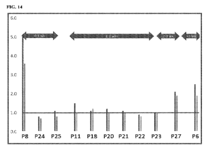
FIG. 14 shows the overlay of the assay results vs days since symptom onset.
Evidence of
active 3CL protease can still be seen in specimens from individuals whose
symptoms first arose
more than 3 weeks prior to testing.
FIG. 15 shows the sample stability profile. Samples held at 4 C retained ¨40-
50% of the
enzymatic activity above background through 24h. The most significant
reduction occurred
between 0-2 hrs. Samples held at -20 C from time 0 retained activity even at
48 hrs. Buccal and
nasal (mid turbinate) matrices yielded comparable results.
CA 03170374 2022-08-08
WO 2021/156878
PCT/IL2021/050155
14
FIG. 16 is a graph showing the ratio of patient sample slopes vs. mean control
protease
slope within the experimental set of 25 positive Covid-19 patients.
FIGs. 17A-C are graphs illustrating the sensitivity of the assay compared to
the
sensitivity of a PCR assay for various genes.
DETAILED DESCRIPTION
In the following description, various aspects of the present application will
be described.
For purposes of explanation, specific configurations and details are set forth
in order to provide a
thorough understanding of the present application. However, it will also be
apparent to one skilled
in the art that the present application may be practiced without the specific
details presented herein.
Furthermore, well-known features may be omitted or simplified in order not to
obscure the present
application.
The term "comprising", used in the claims, is "open ended" and means the
elements recited,
or their equivalent in structure or function, plus any other element or
elements which are not
recited. It should not be interpreted as being restricted to the means listed
thereafter; it does not
exclude other elements or steps. It needs to be interpreted as specifying the
presence of the stated
features, integers, steps or components as referred to, but does not preclude
the presence or
addition of one or more other features, integers, steps or components, or
groups thereof. Thus, the
scope of the expression "a composition comprising x and z" should not be
limited to compositions
consisting only of components x and z. Also, the scope of the expression "a
method comprising
the steps x and z" should not be limited to methods consisting only of these
steps. The term
"consisting of' means "including and limited to". The term "consisting
essentially of' means that
the composition, method or structure may include additional ingredients, steps
and/or parts, but
only if the additional ingredients, steps and/or parts do not materially alter
the basic and novel
characteristics of the claimed composition, method or structure.
Unless specifically stated, as used herein, the term "about" is understood as
within a range of
normal tolerance in the art, for example within two standard deviations of the
mean. In one
embodiment, the term "about" means within 10% of the reported numerical value
of the number
with which it is being used, preferably within 5% of the reported numerical
value. For example,
the term "about" can be immediately understood as within 10%, 9%, 8%, 7%, 6%,
5%, 4%, 3%,
2%, 1%, 0.5%, 0.1%, 0.05%, or 0.01% of the stated value. In other embodiments,
the term "about"
can mean a higher tolerance of variation depending on for instance the
experimental technique
used. Said variations of a specified value are understood by the skilled
person and are within the
CA 03170374 2022-08-08
WO 2021/156878
PCT/IL2021/050155
context of the present invention. As an illustration, a numerical range of
"about 1 to about 5"
should be interpreted to include not only the explicitly recited values of
about 1 to about 5, but
also include individual values and sub-ranges within the indicated range.
Thus, included in this
numerical range are individual values such as 2, 3, and 4 and sub-ranges, for
example from 1-3,
5 from 2-4, and from 3-5, as well as 1, 2, 3, 4, 5, or 6, individually.
This same principle applies to
ranges reciting only one numerical value as a minimum or a maximum. Unless
otherwise clear
from context, all numerical values provided herein are modified by the term
"about". Other similar
terms, such as "substantially", "generally", "up to" and the like are to be
construed as modifying a
term or value such that it is not an absolute. Such terms will be defined by
the circumstances and
10 the terms that they modify as those terms are understood by those of
skilled in the art. This
includes, at very least, the degree of expected experimental error, technical
error and instrumental
error for a given experiment, technique or an instrument used to measure a
value. Whenever a
numerical range is indicated herein, it is meant to include any cited numeral
(fractional or integral)
within the indicated range. The phrases "ranging/ranges between" a first
indicate number and a
15 second indicate number and "ranging/ranges from" a first indicate number
"to" a second indicate
number are used herein interchangeably and are meant to include the first and
second indicated
numbers and all the fractional and integral numerals therebetween.
As used herein the term "method" refers to manners, means, techniques and
procedures for
accomplishing a given task including, but not limited to, those manners,
means, techniques and
procedures either known to, or readily developed from known manners, means,
techniques and
procedures by practitioners of the chemical, pharmacological, biological,
biochemical and medical
arts. As used herein, the term "treating" includes abrogating, substantially
inhibiting, slowing or
reversing the progression of a condition, substantially ameliorating clinical
or aesthetical
symptoms of a condition or substantially preventing the appearance of clinical
or aesthetical
symptoms of a condition. As used herein, the term "diagnosing" refers to
determining presence or
absence of the virus in the subject, classifying the infection, determining a
severity of the infection,
monitoring virus progression, forecasting an outcome of a pathology and/or
prospects of recovery
and/or screening of a subject for the virus.
As used herein, the term "and/or" includes any and all combinations of one or
more of the
associated listed items. Unless otherwise defined, all terms (including
technical and scientific
terms) used herein have the same meaning as commonly understood by one of
ordinary skill in the
art to which this invention belongs. It will be further understood that terms,
such as those defined
in commonly used dictionaries, should be interpreted as having a meaning that
is consistent with
CA 03170374 2022-08-08
WO 2021/156878
PCT/IL2021/050155
16
their meaning in the context of the specification and relevant art and should
not be interpreted in
an idealized or overly formal sense unless expressly so defined herein. Well-
known functions or
constructions may not be described in detail for brevity and/or clarity. As
used herein, the singular
form "a", "an" and "the" include plural references unless the context clearly
dictates otherwise. For
example, the term "a compound" or "at least one compound" may include a
plurality of
compounds, including mixtures thereof
It will be understood that when an element is referred to as being "on",
"attached to",
"connected to", "coupled with", "contacting", etc., another element, it can be
directly on, attached
to, connected to, coupled with or contacting the other element or intervening
elements may also
be present. In contrast, when an element is referred to as being, for example,
"directly on", "directly
attached to", "directly connected to", "directly coupled" with or "directly
contacting" another
element, there are no intervening elements present. It will also be
appreciated by those of skill in
the art that references to a structure or feature that is disposed "adjacent"
another feature may have
portions that overlap or underlie the adjacent feature.
The present invention provides a method, reagents and a kit for the detection
of SARS-
CoV-2 in a test sample and involves the detection of the 3C-L protease. The
present invention
directs at diagnosing COVID-19 using the 3C-L protease assay that selectively
detects active
forms of a SARS-CoV-2-encoded enzyme that is required for viral replication
and transmission,
and which may also play a role in cellular apoptosis. Detection of live
viruses (as opposed to viral
fragments) further allows for the ability to diagnose infectivity status.
Whilst reducing the present invention to practice, the present inventors have
shown that
the SARS-CoV-2 virus can be identified in upper respiratory and oral samples
derived from both
asymptomatic and symptomatic patients. The activity assay provides results
within < 15 minutes
(see Figs. 3A-C, 4, 5A-F, 6 and 7A-D). Furthermore, the present inventors were
able to detect
persistent, active viral reservoirs in recovering individuals, including early-
stage symptomatic and
asymptomatic individuals.
Whilst further reducing the present invention to practice, the present
inventors
demonstrated they could detect 3CL protease activity from additional
coronaviruses (Tables 4-6).
The ability to deploy a mechanism-based assay that can detect different
coronaviruses ensures that
the assay performance will not decrease, even as new mutations arise. The
assay was shown to be
specific for identifying coronaviruses and not picornaviruses (see Tables 4-
6).
Both the speed in which the test can be carried out, together with its high
accuracy makes
the test particularly suitable for point-of-care (POC) rapid diagnostics in
highly populated
CA 03170374 2022-08-08
WO 2021/156878
PCT/IL2021/050155
17
locations (e.g., nursing homes and schools). Since the test reliably
identifies asymptomatic
individuals, the test is also suitable for use in the travel industry and for
healthcare workers.
Population monitoring via testing of waste water/pooled samples can also be
carried out using the
proposed diagnostic test. The assay format may be easily adapted for in-home
testing.
Thus, according to a first aspect of the present invention there is provided a
method of
diagnosing a Severe Acute Respiratory Syndrome Coronavirus 2 (SARS-CoV-2)
infection in a
sample of a subject, the method comprising contacting the sample with a
composition comprising
an agent that detects 3CL-protease of the SARS-Co-V2 virus, wherein a presence
of said 3CL-
protease in the sample is indicative of a SARS-Co-V2 infection.
In another embodiment, the method of detecting a SARS-CoV-2 virus in a sample
of a
subject suspected of having COVID-19 comprises contacting the sample with a
composition
comprising an agent that monitors the activity of a 3CL protease of the SARS-
CoV-2 virus,
wherein the activity level of said 3CL protease in the sample is indicative of
the presence of SARS-
CoV-2 in the sample. The activity level may also allow quantification of the
SARS-CoV-2 virus
in the sample.
In some embodiments, the detectable moiety is a fluorescence moiety. In a
particular
embodiment, the detectable moiety is a Forster Resonance Energy Transfer
(FRET) pair of donor
and acceptor moieties, and the cleavage of the substrate peptide generates or
modulates a signal
from said FRET pair. Specific donor moiety is a quantum dot.
In a further aspect, the present invention provides a method of a
heterogeneous assay, in
which the solid-phase is separated from another assay component during the
assay, for
biomolecular diagnostics of a SARS-CoV-2 virus in a sample of a subject
suspected of having
COVID-19, the method comprising:
(A) Contacting the sample with a composition comprising a cleavable agent
represented by the
general formula:
X-Y-Z,
wherein:
i) Y is a substrate peptide capable of being cleaved by the SARS-CoV-2 3CL
protease;
ii) cleavage of X-Y-Z by said 3CL protease forms products X-Y' and Y"-Z of the
cleavage,
wherein Y' and Y" are two cleavage fragments of said substrate peptide Y;
iii) Z is an optional separating moiety capable of binding to a separate phase
of a two-phase
separating system;
CA 03170374 2022-08-08
WO 2021/156878
PCT/IL2021/050155
18
iv) said cleavable agent X-Y-Z does not form a contiguous portion of a natural
substrate of said
3CL protease; and
v) X is a detectable moiety capable of generating a detectable signal on
cleavage of the substrate
peptide by said 3CL protease, thereby monitoring activity of the 3CL protease
in the sample,
wherein the activity level of the 3CL protease that correlates to said
detectable signal is
i) indicative of the presence of the SARS-CoV-2 virus in the sample, and ii)
allows quantification
of the SARS-CoV-2 virus in the sample; and
(B) Recording or reading said signal with a device suitable for reading
this signal.
In some embodiments, the detectable moiety X comprises a labelling agent
selected from
the group consisting of an enzyme, a fluorophore, a chromophore, a protein, a
pre-enzyme, a
chemiluminescent substance and a radioisotope. The separating moiety Z is
selected from the
group consisting of an immunological binding agent, magnetic binding moiety,
peptide binding
moiety, affinity binding moiety, nucleic acid moiety, biotin/streptavidin
moiety, quantum dots,
metamaterials, conductive polymeric moiety, dendrimer moiety, crown ether or
imprinting
polymer moiety, aptamer moiety, electrochemical binding moiety, and metallic
nanoparticle
moiety.
In particular embodiments, the sample is selected from the group consisting of
mucus, saliva,
throat wash, nasal wash, spinal fluid, sputum, urine, semen, sweat, faeces,
plasma, blood,
bronchioalveolar fluid, vaginal fluid, tear fluid, tissue biopsy, and
nasopharyngeal, oropharyngeal,
nasal mid turbinate, anterior nasal and buccal swabs.
In other embodiments, the detectable moiety X is a fluorescence moiety. In a
particular
embodiment, the detectable moiety X is a Forster Resonance Energy Transfer
(FRET) pair of
donor and acceptor moieties, and the cleavage of the substrate peptide
generates or modulates a
signal from said FRET pair. Specific donor moiety is a quantum dot. Fig. 8
schematically shows
.. the detection mechanism behind this method of the present invention. Fig. 9
demonstrates the
enzyme activity test for the SARS-CoV-2 detection using the method of the
present invention.
In certain embodiments, the donor and acceptor moiety are attached to the
peptide in a
configuration that permits energy transfer from the donor to the acceptor to
result in quenching of
the fluorescence by FRET process. In some other embodiments, the donor and
acceptor moiety are
.. separated by no more than 3, 5, 10, 15 or 20 amino acid residues.
Subjects which can be tested according to this aspect of the present invention
may be
symptomatic or asymptomatic of the infection. They may be contagious or non-
contagious with
the infection. It will be appreciated that the very high accuracy and
sensitivity of the assay allows
CA 03170374 2022-08-08
WO 2021/156878
PCT/IL2021/050155
19
for the detection of very low levels of the SARS-CoV-2 virus. Thus, the method
may be used to
detect the virus in samples of subjects very soon after initial infection or
even in samples tested to
be negative by other means (clinical assessments, molecular-, antigen-,
antibody-based tests), but
which still contain low levels of active virus. Furthermore, the method may be
used to detect the
virus in samples of asymptomatic subjects.
In a particular embodiment, the method of the present invention is carried on
a sample taken
from a subject who does not show symptoms of COVID-19, no more than one day
following
exposure to a subject known to have COVID.
In another particular embodiment, the method of the present invention is
carried on a sample
1() taken from a subject who does not show symptoms of COVID-19, no more than
two days
following exposure to a subject known to have COVID-19.
In still other particular embodiment, the method of the present invention is
carried on a
sample taken from a subject who does not show symptoms of COVID-19, no more
than three days
following exposure to a subject known to have COVID-19.
In a further particular embodiment, the method of the present invention is
carried on a sample
taken from a subject who does not show symptoms of COVID-19, no more than four
days
following exposure to a subject known to have COVID-19.
In yet further particular embodiment, the method of the present invention is
carried on a
sample taken from a subject who does not show symptoms of COVID-19, no more
than five days
following exposure to a subject known to have COVID-19.
In a particular embodiment, the method of the present invention is carried on
a sample taken
from a subject who does not show symptoms of COVID-19, no more than six days
following
exposure to a subject known to have COVID-19.
In another particular embodiment, the method of the present invention is
carried on a sample
.. taken from a subject who does not show symptoms of COVID-19, no more than
seven days
following exposure to a subject known to have COVID-19.
In still another particular embodiment, the method of the present invention is
carried on a
sample taken from a subject who does not show symptoms of COVID-19, more than
seven days
following exposure to a subject known to have COVID-19.
In a further particular embodiment, the method of the present invention is
carried on a sample
taken from a subject having a negative PCR test or a positive PCR test with a
cycle threshold CT40
and less.
CA 03170374 2022-08-08
WO 2021/156878 PCT/IL2021/050155
The 3CL protease of the SARS-CoV-2 virus is a 34 kD trypsin-like cysteine
protease.
According to a particular embodiment, the 3CL protease comprises an amino acid
sequence as set
forth in SEQ ID NO: 34.
Exemplary samples in which SARS-CoV-2 can be detected include, but are not
limited to
5 saliva, mucous, throat wash, nasal wash, spinal fluid, sputum, urine,
semen, sweat, feces, plasma,
blood, bronchioalveolar fluid, vaginal fluid, tear fluid and tissue biopsy.
In some embodiments, the sample is a sewage sample.
In other embodiments, the sample comprises saliva.
The sample may be taken from the mouth, back of the throat or from inside the
cheek (e.g.,
1() using a buccal swab).
Examples of swabs which can be used to obtain the sample include cheek swabs,
oropharyngeal and nasal pharyngeal swabs.
In order to determine the activity of the 3CL of the SARS-CoV-2 virus, a
peptide may be
used which serves as a substrate for the enzyme. The peptide is attached to at
least one moiety
15 which generates a detectable signal on cleavage by the 3CL protease.
The peptide is typically between 8-30 amino acids long, more preferably
between 10 and 20
amino acids long and even more preferably between 10 and 15 amino acids long.
According to a
particular embodiment, the peptide is no longer than 14 amino acids long.
According to a particular
embodiment, the peptide is between 8-12 amino acids long.
20 The peptides described herein may include natural or non-naturally
occurring amino acids.
The term "amino acid" or "amino acids" is understood to include the 20
naturally occurring
amino acids; those amino acids often modified post-translationally in vivo,
including, for example,
hydroxyproline, phosphoserine and phosphothreonine; and other unusual amino
acids including,
but not limited to, 2-aminoadipic acid, hydroxylysine, isodesmosine, nor-
valine, nor-leucine and
ornithine. Furthermore, the term "amino acid" includes both D- and L-amino
acids.
Tables A and B below list naturally occurring amino acids (Table A), and non-
conventional
or modified amino acids (e.g., synthetic, Table B) which can be used with some
embodiments of
the invention. It will be appreciated that non-conventional amino acids may be
used in order to
reduce background noise in the assay.
Table A
Amino Acid Three-Letter Abbreviation One-letter Symbol
Alanine Ala A
Arginine Arg
CA 03170374 2022-08-08
WO 2021/156878
PCT/IL2021/050155
21
Asparagine Asn N
Aspartic acid Asp D
Cysteine Cys C
Glutamine Gin Q
Glutamic Acid Glu E
Glycine Gly G
Histidine His H
Isoleucine Ile I
Leucine Leu L
Lysine Lys K
Methionine Met M
Phenylalanine Phe F
Proline Pro P
Serine Ser S
Threonine Thr T
Tryptophan Trp W
Tyrosine Tyr Y
Valine Val V
Any amino acid as above Xaa X
Table B
Non-conventional amino acid Code Non-conventional amino acid Code
ornithine Orn hydroxyproline Hyp
a-aminobutyric acid Abu aminonorbornyl-carboxylate Norb
D-alanine Dala aminocyclopropane-carboxylate Cpro
D-arginine Darg N-(3 -guanidinopropyl)glycine Narg
D-asparagine Dasn N-(carbamylmethyl)glycine Nasn
D-aspartic acid Dasp N-(carboxymethyl)glycine Nasp
D-cysteine Dcys N-(thiomethyl)glycine Ncys
D-glutamine Dgln N-(2-carbamylethyl)glycine Ngln
D-glutamic acid Dglu N-(2-carboxyethyl)glycine Nglu
CA 03170374 2022-08-08
WO 2021/156878 PCT/IL2021/050155
22
D-histidine Dhis N-(imidazolylethyl)glycine Nhis
D-isoleucine Dile N-(1-methylpropyl)glycine Nile
D-leucine Dleu N-(2-methylpropyl)glycine Nleu
D-lysine Dlys N-(4-aminobutyl)glycine Nlys
D-methionine Dmet N-(2-methylthioethyl)glycine Nmet
D-ornithine Dorn N-(3-aminopropyl)glycine Norn
D-phenylalanine Dphe N-benzylglycine Nphe
D-proline Dpro N-(hydroxymethyl)glycine Nser
D-serine Dser N-(1-hydroxyethyl)glycine Nthr
D-threonine Dthr N-(3-indolylethyl) glycine Nhtrp
D-tryptophan Dtrp N-(p-hydroxyphenyl)glycine Ntyr
D-tyrosine Dtyr N-(1-methylethyl)glycine Nval
D-valine Dval N-methylglycine
Nmgly
D-N-methylalanine Dnmala L-N-methylalanine
Nmala
D-N-methylarginine Dnmarg L-N-methylarginine
Nmarg
D-N-methylasparagine Dnmasn L-N-methylasparagine
Nmasn
D-N-methylasparatate Dnmasp L-N-methylaspartic acid
Nmasp
D-N-methylcysteine Dnmcys L-N-methylcysteine
Nmcys
D-N-methylglutamine Dnmgln L-N-methylglutamine
Nmgln
D-N-methylglutamate Dnmglu L-N-methylglutamic acid
Nmglu
D-N-methylhistidine Dnmhis L-N-methylhistidine
Nmhis
D-N-methylisoleucine Dnmile L-N-methylisolleucine
Nmile
D-N-methylleucine Dnmleu L-N-methylleucine
Nmleu
D-N-methyllysine Dnmlys L-N-methyllysine
Nmlys
D-N-methylmethionine Dnmmet L-N-methylmethionine
Nmmet
D-N-methylornithine Dnmorn L-N-methylornithine
Nmorn
D-N-methylphenylalanine Dnmphe L-N-methylphenylalanine
Nmphe
D-N-methylproline Dnmpro L-N-methylproline
Nmpro
D-N-methylserine Dnmser L-N-methylserine
Nmser
D-N-methylthreonine Dnmthr L-N-methylthreonine
Nmthr
D-N-methyltryptophan Dnmtrp L-N-methyltryptophan
Nmtrp
D-N-methyltyrosine Dnmtyr L-N-methyltyrosine
Nmtyr
CA 03170374 2022-08-08
WO 2021/156878 PCT/IL2021/050155
23
D-N-methylvaline Dnmval L-N-methylvaline Nmval
L-norleucine Nle L-N-methylnorleucine Nmnle
L-norvaline Nva L-N-methylnorvaline Nmnva
L-ethylglycine Etg L-N-methyl-ethylglycine Nmetg
L-t-butylglycine Tbug L-N-methyl-t-butylglycine Nmtbug
L-homophenylalanine Hphe L-N-methyl-homophenylalanine Nmhphe
a-naphthylalanine Anap N-methyl-a-naphthylalanine Nmanap
penicillamine Pen N-methylpenicillamine Nmpen
y-aminobutyric acid Gabu N-methyl-y-aminobutyrate Nmgabu
cyclohexylalanine Chexa N-methyl-cyclohexylalanine Nmchexa
cyclopentylalanine Cpen N-methyl-cyclopentylalanine Nmcpen
a-amino-a-methylbutyrate Aabu N-methyl-a-amino-a-methylbutyrate Nmaabu
a-aminoisobutyric acid Aib N-methyl-a-aminoisobutyrate Nmaib
D-a-methylarginine Dmarg L-a-methylarginine Marg
D-a-methylasparagine Dmasn L-a-methylasparagine Masn
D-a-methylaspartate Dmasp L-a-methylaspartate Masp
D-a-methylcysteine Dmcys L-a-methylcysteine Mcys
D-a-methylglutamine Dmgln L-a-methylglutamine Mgln
D-a-methyl glutamic acid Dmglu L-a-methylglutamate Mglu
D-a-methylhistidine Dmhis L-a-methylhistidine Mhis
D-a-methylisoleucine Dmile L-a-methylisoleucine Mile
D-a-methylleucine Dmleu L-a-methylleucine Mleu
D-a-methyllysine Dmlys L-a-methyllysine Mlys
D-a-methylmethionine Dmmet L-a-methylmethionine Mmet
D-a-methylornithine Dmorn L-a-methylornithine Morn
D-a-methylphenylalanine Dmphe L-a-methylphenylalanine Mphe
D-a-methylproline Dmpro L-a-methylproline Mpro
D-a-methylserine Dmser L-a-methylserine Mser
D-a-methylthreonine Dmthr L-a-methylthreonine Mthr
D-a-methyltryptophan Dmtrp L-a-methyltryptophan Mtrp
D-a-methyltyrosine Dmtyr L-a-methyltyrosine Mtyr
CA 03170374 2022-08-08
WO 2021/156878
PCT/IL2021/050155
24
D-a-methylvaline Dmval L-a-methylvaline Mval
N-cyclobutylglycine Ncbut L-a-methylnorvaline Mnva
N-cycloheptylglycine Nchep L-a-methylethylglycine Metg
N-cyclohexylglycine Nchex L-a-methyl-t-butylglycine Mtbug
N-cyclodecylglycine Ncdec L-a-methyl-homophenylalanine Mhphe
N-cyclododecylglycine Ncdod a-methyl-a-naphthylalanine Manap
N-cyclooctylglycine Ncoct a-methylpenicillamine Mpen
N-cyclopropylglycine Ncpro a-methyl-y-aminobutyrate Mgabu
N-cycloundecylglycine Ncund a-methyl-cyclohexylalanine Mchexa
N-(2-aminoethyl)glycine Naeg a-methyl-cyclopentylalanine Mcpen
N-(2,2-diphenylethyl)glycine Nbhm N-(N-(2,2-diphenylethyl)- Nnbhm
carbamylmethyl-glycine
N-(3,3-diphenylpropyl)glycine Nbhe N-(N-(3,3-diphenylpropy1)- Nnbhe
carbamylmethyl-glycine
1-carboxy-1-(2,2-diphenyl Nmbc 1,2,3,4-tetrahydroisoquinoline-3-
Tic
ethylamino)cyclopropane carboxylic acid
phosphoserine pSer phosphothreonine pThr
phosphotyro sine pTyr 0-methyl-tyrosine
2-aminoadipic acid hydroxylysine
The amino acid sequence of the peptide is selected such that it can be cleaved
by the 3CL
of SARS-CoV-2. Preferably, the peptide does not serve as a substrate for the
3C of Human
Rhinovirus (HRV) under the same assay conditions in human samples. In another
embodiment,
the peptide is selected such that it can distinguish between the 3CL activity
of SARS-CoV-2 and
other human pathogens in the Coronavirus family (e.g. CoV-229E) in human
samples under
identical assay conditions.
According to a particular embodiment, the peptide comprises at least one of
the amino acid
sequences set forth in SEQ ID NOs: 13-23.
In another embodiment, the peptide comprises an amino at least 90 % identical
to the
sequence as set forth in SEQ ID Nos: 13-23.
According to a particular embodiment, the peptide comprises the amino acid
sequence as
set forth in SEQ ID NO: 13.
CA 03170374 2022-08-08
WO 2021/156878
PCT/IL2021/050155
Exemplary peptide sequences which comprise the amino acid sequence as set
forth in SEQ
ID NO: 13 which have been shown to be effective substrates of the 3CL of SARS-
CoV-2 are set
forth in SEQ ID NOs: 24-33.
In a particular embodiment, the peptides used in the assay have an amino acid
sequence at
5 least 90 % identical to the sequences as set forth in SEQ ID Nos: 24-33.
In another embodiment, the peptides used in the assay have an amino acid
sequence at least
90 % identical to the sequences as set forth in SEQ ID Nos: 1-12.
The amino acids of the peptides of the present invention may be substituted
either
conservatively or non-conservatively.
10
According to a particular embodiment the amino acids of the peptides are
substituted conservatively.
The term "conservative substitution" as used herein, refers to the replacement
of
an amino acid present in the native sequence in the peptide with a naturally
or non-
naturally occurring amino or a peptidomimetics having similar steric
properties. Where
15
the side-chain of the native amino acid to be replaced is either polar or
hydrophobic,
the conservative substitution should be with a naturally occurring amino acid,
a non-
naturally occurring amino acid or with a peptidomimetic moiety which is also
polar or
hydrophobic (in addition to having the same steric properties as the side-
chain of the
replaced amino acid).
20
As naturally occurring amino acids are typically grouped according to their
properties, conservative substitutions by naturally occurring amino acids can
be easily
determined bearing in mind the fact that in accordance with the invention
replacement
of charged amino acids by sterically similar non-charged amino acids are
considered as
conservative substitutions.
25
For producing conservative substitutions by non-naturally occurring amino
acids
it is also possible to use amino acid analogs (synthetic amino acids) well
known in the
art. A peptidomimetic of the naturally occurring amino acid is well documented
in the
literature known to the skilled practitioner.
When affecting conservative substitutions the substituting amino acid should
have the same or a similar functional group in the side chain as the original
amino acid.
CA 03170374 2022-08-08
WO 2021/156878
PCT/IL2021/050155
26
The phrase "non-conservative substitutions" as used herein refers to
replacement
of the amino acid as present in the parent sequence by another naturally or
non-naturally
occurring amino acid, having different electrochemical and/or steric
properties. Thus,
the side chain of the substituting amino acid can be significantly larger (or
smaller) than
the side chain of the native amino acid being substituted and/or can have
functional
groups with significantly different electronic properties than the amino acid
being
substituted. Examples of non-conservative substitutions of this type include
the
substitution of phenylalanine or cycohexylmethyl glycine for alanine,
isoleucine for
glycine, or --NH¨CH[(--CH. sub.2). sub.5--0001-1]-00-- for aspartic acid.
According to a particular embodiment, the amino acid sequence of the substrate
peptide is
selected from the group consisting of SEQ ID NOs: 25-33.
According to a specific embodiment the substrate has the amino acid sequence
as set forth
in SEQ ID NO: 31, wherein X is cysteine, aspartic acid, glutamic acid,
arginine or lysine.
According to a particular embodiment, the X is cysteine.
According to a specific embodiment the substrate has the amino acid sequence
as set forth
in SEQ ID NO: 30, wherein X is cysteine, aspartic acid, glutamic acid,
arginine or lysine.
According to a particular embodiment, the X is cysteine.
Thus, an exemplary sequence contemplated by the present inventors is one set
forth in SEQ
ID NO: 38 or 39.
The peptides of the present invention may be synthesized by any techniques
that are known
to those skilled in the art of peptide synthesis. For solid phase peptide
synthesis, a summary of
the many techniques may be found in: Stewart, J. M. and Young, J. D. (1963),
"Solid Phase Peptide
Synthesis," W. H. Freeman Co. (San Francisco); and Meienhofer, J (1973).
"Hormonal Proteins
and Peptides," vol. 2, p. 46, Academic Press (New York). For a review of
classical solution
synthesis, see Schroder, G. and Lupke, K. (1965). The Peptides, vol. 1,
Academic Press (New
York).
In general, peptide synthesis methods comprise the sequential addition of one
or more
amino acids or suitably protected amino acids to a growing peptide chain,
either the amino or the
carboxyl group of the first amino acid is protected by a suitable protecting
group. The protected or
modified amino acid can then either be attached to an inert solid support or
utilized in solution by
adding the next amino acid in the sequence having the complimentary (amino or
carboxyl) group
suitably protected, under conditions suitable for forming the amide linkage.
The protecting group
CA 03170374 2022-08-08
WO 2021/156878
PCT/IL2021/050155
27
is then removed from this newly added amino acid residue and the next amino
acid (suitably
protected) is then added, and so forth; traditionally this process is
accompanied by wash steps as
well. After all of the desired amino acids have been linked in the proper
sequence, any remaining
protecting groups (and any solid support) are removed sequentially or
concurrently, to afford the
final peptide compound. By simple modification of this general procedure, it
is possible to add
more than one amino acid at a time to a growing chain, for example, by
coupling (under conditions
which do not racemize chiral centers) a protected tripeptide with a properly
protected dipeptide to
form, after deprotection, a pentapeptide, and so forth.
Further description of peptide synthesis is disclosed in U.S. Pat. No.
6,472,505. A preferred
method of preparing the peptide compounds of the present invention involves
solid-phase peptide
synthesis, utilizing a solid support. Large-scale peptide synthesis is
described by Andersson
Biopolymers 2000, 55(3), 227-50.
As mentioned above, the peptides of this aspect of the present invention are
attached to at
least one moiety which generates a detectable signal on cleavage of the
peptide by the 3CL
protease of the SARS-CoV-2.
In some embodiments, the detectable moiety can be chemically conjugated
(coupled) to
the peptide of the invention, using any conjugation method known to one
skilled in the art. For
example, a detectable moiety can be conjugated to the substrate peptides
disclosed herein, using a
3-(2-pyridyldithio)propionic acid N-hydroxysuccinimide ester (also called N-
succinimidyl 3-(2-
.. pyridyldithio) propionate) ("SDPD") (Sigma, Cat. No. P-3415; see e.g.,
Cumber et al. 1985,
Methods of Enzymology 112: 207-224), a glutaraldehyde conjugation procedure
(see e.g., G.T.
Hermanson 1996, "Antibody Modification and Conjugation, in Bioconjugate
Techniques,
Academic Press, San Diego) or a carbodiimide conjugation procedure [see e.g.,
J. March,
Advanced Organic Chemistry: Reaction's, Mechanism, and Structure, pp. 349-50 &
372-74 (3d
ed.), 1985; B. Neises et al. 1978, Angew Chem., Int. Ed. Engl. 17:522; A.
Hassner et al. 1978,
Tetrahedron Lett. 4475; E.P. Boden et al. 1986, J. Org. Chem. 50:2394 and L.J.
Mathias 1979,
Synthesis 561].
Additionally, or alternatively, the detectable moiety is conjugated to the
peptide by
translationally fusing the polynucleotide encoding the peptide of the
invention with the nucleic
acid sequence encoding the detectable moiety.
The detectable signal may be directly detectable such as for example a
fluorescent signal,
a phosphorescent signal, a radioactive signal or a colour signal (such as
emitted by a chromophore).
Alternatively, the detectable signal may be indirectly detectable, such as for
example a pre-
CA 03170374 2022-08-08
WO 2021/156878
PCT/IL2021/050155
28
enzyme, as further described herein below. Other examples of detectable
moieties are described at
length in U.S. Pat. Appl. No. 20050048473 which is fully incorporated herein
by reference.
Any assay known in the art for monitoring proteolytic substrate cleavage can
be used in
accordance with this aspect of the present invention.
In one embodiment the assay is a homogeneous assay. As used herein the phrase
"homogeneous assay" refers to an assay not requiring separation of signalling
moiety from other
assay components.
The composition is contacted with the sample being tested for the presence of
the SARS-
CoV-2 virus. If the virus is present in the sample, the viral 3CL protease is
also present. This
protease cleaves the substrate and a change in the signal from the signalling
moiety can be
observed. Such homogenous fluorescent and colorimetric assays are known to
those skilled in the
art. See, for example: Biochemistry, Allinger, Wang Q. M. et al., "A
continuous calorimetric assay
for rhinovirus-14 3C protease using peptide p-nitroanilides as substrates"
Anal. Biochem. Vol.
252, pp. 238-45 (1997), and Basak S. et al. "In vitro elucidation of substrate
specificity and
bioassay of proprotein convertase 4 using intramolecularly quenched
fluorogenic peptides"
Biochem. J. Vol. 380, pp. 505-14 (2004).
In one embodiment, the moiety to which the peptides, which generates a
detectable signal
on cleavage of the substrate peptide by said 3CL protease, is a Forster
Resonance Energy Transfer
(FRET) pair, whereby cleavage of the substrate peptide generates a signal from
the FRET pair.
The FRET pair comprises a donor moiety and an acceptor moiety as further
described herein
below.
The traditional method to detect interactions of molecules, for example in a
biochemical
system, is to pull down one of the molecules and look at what comes down in a
microscope. This
is a direct measurement of a molecular interaction that inferred the
interaction by coincidence in
space. The problem with a normal wide-field microscope is that the diffraction
limits are
observable elements. So, a typical volume element that can be resolved with a
wide-field
microscope is in the order of 1015 metres, which corresponds to approximately
a micron cubed.
However, the biomolecular entities interacting with each other that need to be
detected have a way
smaller volume in the order of 10-22 metres. This is several orders of
magnitude less than what the
conventional wide-filed microscope offers. Although co-localisation can be
used in this case in
order to infer interaction, the probability that the molecular interaction is
observed with the co-
localisation is very low. Here comes the FRET that actually allows to
significantly decrease the
detection volume.
CA 03170374 2022-08-08
WO 2021/156878
PCT/IL2021/050155
29
As mentioned above, the FRET is a non-radiative energy transfer, where the
term "non-
radiative" is of particular importance. The non-radiative energy transfer is
essentially based on a
dipole-dipole coupling mechanism between the donor and acceptor of the
interacting molecular
pair in their excited states. It is not a trivial emission of a photon, but a
re-absorption by the donor
and acceptor.
There are a number of approaches to FRET quantification which can be used in
the present
invention:
1) Sensitised emission is a direct two-channel imaging technique using an
algorithm that corrects
for excitation and emission crosstalk;
2) Acceptor photobleaching (sometimes called donor dequenching) is a technique
capable of
measuring increased donor emission when the acceptor is photobleached;
3) Fluorescence lifetime imaging microscopy FRET (FLIM FRET) is a technique
capable of
detecting fluorescence lifetime changes of donor; and
4) Fluorophore donor spectral imaging is a technique involving excitation at
one or two
wavelengths and measuring the spectral profiles of both donor and acceptor.
If the donor of the FRET pair is normally excited, for example with a blue
light, it is very
quickly relaxed from a high excited state by interconversion or preparative
relaxation to the first
electron excited state. From there it can go back to the ground state either
through the non-radiative
decay interconversion, or through the radiative pathway by emitting a photon.
In this case, the
molecular orbitals of the donor can energetically couple with the orbitals of
the acceptor, the
dipole-dipole coupling occurs, thereby creating an extra channel for the non-
radiative decay with
much shorter excited state lifetimes. This results in the donor actually
emitting less light, or in
other words, quenching the donor when the sensitised emission FRET occurs. At
the same time,
as the donor radiative emission is quenched, the acceptor gets excited by this
process as a result of
the same dipole-dipole coupling and starts emitting fluorescence. So, by
exciting the donor in this
process, the emission is received also partially from the acceptor.
The sensitised emission is perhaps the simplest FRET method, because there is
a single
excitation source, from which the donor fluorophore is excited, and the signal
is collected using
emission filters chosen for both the donor fluorescence and the acceptor
fluorescence. The acceptor
fluorescence increases in the presence of donor, whereas the donor
fluorescence decreases in the
presence of the acceptor. The ratiometric change of fluorescence intensity can
then be used to
measure the FRET. This is the most straight-forward approach to measuring the
process of the
FRET. It is inherently based on quenching of the donor molecules during the
process, thereby
CA 03170374 2022-08-08
WO 2021/156878
PCT/IL2021/050155
increasing fluorescence intensity of the acceptor. The spectral images
recorded in this approach
are actually the donor emission upon excitation of the donor (DD) and the
acceptor emission upon
excitation of the donor (DA). The ratio between their intensities can indicate
the FRET efficiency.
However, as mentioned above, the direct measurement of the FRET is not
practical because
5 of the donor bleed-through and acceptor direct excitation, as will be
explained below. This is
essentially a crosstalk between the two fluorophores (donor and acceptor).
Thus, it is very difficult
to obtain quantitatively accurate FRET data with this approach. Additional
control experiments
are required in order to establish the presence or absence of the FRET in a
sample.
Despite the above limitations, it is possible however to deduce the FRET from
the change in
10 the emission of the donor or the acceptor. The major parameter that is
used to quantify the FRET
is the FRET efficiency E, which is basically the number of excited donors that
transfer the energy
to the acceptor, divided by the number of photons absorbed by the donor. So,
this is basically a
fraction of donors that transfer their energy to the acceptor. The FRET
efficiency E can also be
expressed as the following ratio:
6
0
15 E= R
R6+r6
where R is the Forster radius (typically in the order of nanometres) that
represents the distance
between the donor and acceptor at which the FRET efficiency is 50% (when half
of the excited
donor molecules transfer their energy to the acceptor), and r is the distance
between the donor and
acceptor. Since it is r6, it makes the dependence very steep. So, measuring
the FRET efficiency E
20 allows to assess the distance r between the donor and acceptor.
There are several methods to measure the FRET efficiency. The important point
to consider
here is the shape of the spectrum. The typical excitation spectra always have
a tail toward the blue
part of the spectrum and are steep at the red part of the spectrum. The
typical emission spectrum
looks opposite and symmetrical to the excitation spectra and are very steep at
the blue part of the
25 spectrum, but tail towards the red. Therefore, the acceptor can be
specifically excited without
exciting the donor, but the donor cannot be typically excited without exciting
the acceptor.
Similarly, the donor emission can be detected without detecting the acceptor,
but the acceptor
cannot be specifically detected without detecting the donor. Thus, the
specific excitation of the
acceptor and the specific detection of the donor become very relevant to the
FRET measurements
30 with a microscope.
CA 03170374 2022-08-08
WO 2021/156878
PCT/IL2021/050155
31
Because of the aforementioned problems of the crosstalk between the donor and
acceptor,
this approach is only feasible when a donor and acceptor are in the same
molecule, for example in
the same polypeptide. In this case, the stoichiometry of the donor and
acceptor can be kept constant
in each pixel. This specifically works, for example, in bioassays and
biosensors that can measure
a change in a physiological parameter, such as calcium, or measure
phosphorylation. Thus, the
sensitised emission measurements can be useful for detecting rapid dynamic
changes and is
especially useful when examining fluorescent proteins where the FRET dynamic
range is large,
and the stoichiometry of the donor and acceptor is fixed at a 1:1 ratio.
The sensitised emission FRET can be implemented in various types of optical
and
spectroscopic device, for example, in wide-field imaging with proper filters,
and it is very fast,
because only two spectral images need to be acquired in this approach.
However, this approach is
not quantitative, because it allows to measure only differences in excited
states, and the
stoichiometry of donor and acceptor should be constant, which basically means
that in many cases,
they have to label different parts of the same molecule. In other words, and
particularly in the
present invention, the sensitised emission is used as a simple FRET method for
acquiring
preliminary information about the presence or absence of the object of
interest, such as a DNA
molecule of a certain pathogen. The answer in this case provides the initial
indication for the
presence of the molecule in the sample. Based on this initial indication, the
system can be
calibrated. Furthermore, the algorithm of the present invention will proceed
to a further,
quantitative sensitive measurement of the sample, which is based on another
approach of the FRET
measurement. This aspect will be described below.
In some ideal situation, if it is possible to excite the donor specifically
without exciting the
acceptor and to monitor fluorescence of the acceptor only, one would be able
to observe the
acceptor emission when there is energy transfer. So, it would be possible to
make an image where
the donor is excited and only the acceptor is detected. The problem discussed
above is that the
acceptor is excited directly and the bleed-through of the donor occurs in the
acceptor channel.
Thus, where the donor is excited, and the recorded acceptor emission does not
equate to the
sensitised emission, the image is essentially contaminated with the direct
excitation and bleed-
through. So, it is impossible to measure precisely how much acceptor goes
through. However, it
is possible to precisely measure the amount of the acceptor when excited
directly at its excitation
peak. This is possible simply because the donor is typically not excited at
the excitation maximum
of the acceptor, and the measurement will be very specific.
CA 03170374 2022-08-08
WO 2021/156878
PCT/IL2021/050155
32
Since for the same molecule, the molar extinction coefficient (6) is constant,
the ratio
between the two specific excitations in the same spectrum is constant.
Therefore, it is possible to
precisely determine the amount of the acceptor at the donor emission maximum
by exciting the
acceptor at its excitation maximum. Exactly the same situation is with the
donor bleed-through. It
is impossible to precisely measure the acceptor in a sample containing both
the donor and acceptor,
because of the overlapping emission spectrum of the donor, but it is possible
to measure the donor
at that wavelength specifically in the absence of the acceptor.
In practice, the correction factors for the FRET measurements are determined
as follows.
First, for the sample containing only the donor, the intensity ratio Si
between: (a) the donor
emission at the acceptor emission maximum (the wavelength of the donor bleed-
through into the
acceptor detection channel), and (b) at the donor emission maximum, is
determined. Second, for
the sample containing only the acceptor, the intensity ratio Sz between: (a)
the acceptor excitation
at the donor excitation maximum, and (b) the acceptor excitation maximum, is
determined. Then,
by measuring the sample containing both the donor and acceptor, by exciting
only the acceptor in
the presence of the donor, it is possible to determine how much acceptor is
directly excited.
Thus, in the FRET experiment, after expressing the donor, the bleed-through
correction is
initially determined. The donor is excited in the sample in the absence of the
acceptor, and
therefore, the recorded are two spectral images: the donor measured at its
emission maximum (FD)
and at the emission wavelength of the acceptor (FF). The correction (scalar)
factor Si for the bleed-
.. through is then Si = FF/FD. It is only dependent on the spectrometer
settings and the donor used,
and it is calibrated only once using a donor-only sample.
Similarly, the correction for the direct excitation is introduced. The
acceptor is measured in
the sample in the absence of the donor. The recorded are two images: the
acceptor excited at the
donor excitation maximum (FF) and at the excitation wavelength of the acceptor
(FA). The scalar
factor S2 for the direct excitation is then: S2 = FF/FA. It is only dependent
on the microscope settings
and the acceptor used, and it is calibrated only once using an acceptor-only
sample. So, for the
sensitised emission, and assuming that the donor is not excited by the
acceptor excitation, three
images are taken:
1) the donor excitation/acceptor emission (FF),
2) the donor excitation/donor emission (FD) for determining the bleed-through
factor Si, and
3) the acceptor excitation/acceptor emission (FA) for determining the direct
excitation scalar
factor Sz.
CA 03170374 2022-08-08
WO 2021/156878
PCT/IL2021/050155
33
The scaled image intensity FF is then divided by the scaled acceptor intensity
FA to obtain the
FRET apparent efficiency that is proportional to the real FRET efficiency and
to the fraction of
interacting molecules. This scaling is easy to implement in most of the
conventional optical
instruments, such as a wide-field spectrometer or camera with appropriate
filters. The overall
measurement is fast because one can switch the filters quickly on a filter
wheel or on a confocal
microscope. However, this method is still semi-quantitative, because the
resulted image obtained
after scaling and correction is proportional to the relative concentration of
interacting molecules,
and it depends on the external calibration described above. The signal-to-
noise ratio of this method
is also low because of the scaling that requires measuring three separate
samples. In addition, the
spectra of the molecules should be invariant to the environment, for example
it should be the same
in a lipid environment and in a cytoplasmic environment. That is the case for
many fluorescent
proteins, fortunately.
Another approach for the FRET measurements is acceptor photobleaching and it
is actually
quantitative. Acceptor photobleaching is a method based on the fact that donor
fluorescence is
quenched during FRET when some of the donor fluorescence energy is transferred
to the acceptor.
But photobleaching the acceptor fluorophore stops it from fluorescing and thus
from using some
of the donor's energy, resulting in a total increase of donor fluorescence.
This phenomenon can be
used in order to calculate the FRET efficiency by subtracting the donor
intensity in the presence
of the acceptor from its intensity after photobleaching of the acceptor and
dividing the result by
the donor intensity after bleaching.
The advantage of this approach is that it is very robust and is quantitative.
In this approach,
the donor is excited and then its emission is recorded. If the FRET occurs,
the donor is quenched,
and so the emission intensity decreases, because this is actual channel for
the non-radiative decay.
In this method, the donor intensity should be initially measured in the
absence of acceptor, and the
.. FRET efficiency is calculated accordingly. The problem is that this
approach assumes that a
separate experiment where the donor is measured in the absence of the acceptor
should be
performed. However, this would be a completely different configuration with
the different
concentration of molecules, and the separate measurement should therefore be
avoided, also
because of the quantitation problems discussed above. The solution is
specifically exciting the
acceptor until it is completely saturated and cannot absorb more photons, and
then the donor is re-
measured. Bringing said acceptor fluorophore into short-lived and reversible
dark states (e.g., a
triplet state) during acceptor excitation would also prevent it from absorbing
more photons. Such
a process is also referred here as "acceptor saturation".
CA 03170374 2022-08-08
WO 2021/156878
PCT/IL2021/050155
34
In general, fluorophores can undergo excitation through the absorbance of a
photon and a
following excitation of an electron from a low energy level (typically, the
ground state) into a
higher energy level (typically, the first excited state). The electron may
lose some energy via
phonons, and then spontaneously return to the ground state while emitting a
photon (a process
known as fluorescence or spontaneous emission). The lifetime for fluorescence
emission is
typically in nanoseconds. A second relaxation mechanism of the excited
electron of a fluorophore
(other than fluorescence) is through inter-system crossing into a triplet
state. Such a transition is
quantum-mechanically forbidden and therefore, it has a relatively low cross-
section and a typical
lifetime of microseconds, significantly longer than the fluorescence lifetime.
Moreover, the
electron may subsequently move to other metastable electronic levels, e.g.,
via encountering other
molecules. Notably, these transitions do not involve photon emissions (or are
very inefficient in
doing so). Thus, these states are effectively considered as dark states. In
the present invention,
electronic inter-system crossing of the acceptor electron into a triplet state
and additional dark
states contribute to the FRET frustration, since the acceptor can no longer
share the energy with
the excited donor. This frustration is useful in the invention provided that
the dark states are short-
lived (in micro to milliseconds) relative to the modulation time of acceptor
excitation, and that the
transitions are reversible. Thus, the process of acceptor fluorophore entering
a short-lived and
reversible dark state (e.g., a triplet state) during acceptor excitation is
also included here in the
definition of the 'acceptor saturation'.
In this approach, when the acceptor is saturated, the donor becomes unquenched
and its
emission intensity will go up, which is actually the same situation as in the
absence of the acceptor.
The FRET apparent efficiency in this method is as follows:
FD
Eapp = 1 ¨
where FD is the donor spectral image recorded when the donor quenched by the
active
acceptor, and is the donor spectral image recorded when the acceptor is
photobleached. It will
be more precisely defined in the Examples.
Although being semi-quantitative, this approach produces readings which are
proportional
to the fraction of the complex true efficiency. So, in this sense, it is
rather the most quantitative
method in terms of the intensity-based methods, and it does not require
external calibrations, which
is the case for the sensitised emission measurement. The control is actually
internal by the
bleaching in this method.
CA 03170374 2022-08-08
WO 2021/156878
PCT/IL2021/050155
Along with the FRET fluorescence technique described above, other advanced
spectroscopic techniques, such as proximity ligation (PLA), bimolecular
fluorescence
complementation (BiFC) and fluorescence cross-correlation spectroscopy (FCS),
can be used in
the present invention.
5
The term "donor fluorophore" as used herein refers to a light-sensitive,
fluorescence
emitting molecule, which initially in its electronic excited state, may
transfer energy to "acceptor
fluorophore" through non-radiative dipole-dipole coupling. The donor
fluorophore must be bright
(having high quantum yield and high absorption coefficient), stable (having
long-living fluorescent
excited state and low photo bleaching), and insensitive to the acceptor
fluorophore excitation light.
10
The term "acceptor fluorophore" as used herein refers to a light-sensitive
molecule, which
initially in its ground-level electronic state may accept energy from "donor
fluorophore" through
non-radiative dipole¨dipole coupling. The acceptor fluorophore must have a
large Forster distance
Ro from the donor fluorophore (high spectral overlap of the absorption
spectrum of an acceptor
fluorophore with the fluorescence emission spectrum of a donor fluorophore),
low photobleaching,
15
must be insensitive to the donor fluorophore excitation light, having no cross-
talk of its
fluorescence emission spectrum with the fluorescence emission spectrum of the
donor fluorophore,
and must be capable of undergoing reversible saturation of its fluorescence
emission under light
excitation.
Synthetic fluorophores used in the present invention may include, but are not
limited to
20 generic or proprietary fluorophores listed in Table C below:
Table C. Generic or proprietary exemplary fluorophores suitable for use in the
present invention
Type 1 Fluorescein and derivatives thereof, Rhodamine and derivatives
thereof, Alexa
Fluor dyes, DyLight Fluor dyes, Cyanine CyTM dyes, ATTO dyes, Abberior
STAR dyes, Dyomics dyes, DNA fluorescent stains (for example, DAPI or 4',6-
diamidino-2-phenylindole), membrane fluorescent stains (for example, Dil or
DilC18(3), Di0 or Di0C18(3), DiD and DiR, which constitute a family of
lipophilic
fluorescent stains for labelling membranes and other hydrophobic structures).
Type 2 A subset of Type 1 fluorophores, for example, Alexa Fluor 488,
Alexa Fluor 555,
Alexa Fluor 568, Alexa Fluor 647, Alexa Fluor 750, Alexa Fluor 790, ATTO
488, ATTO 520, ATTO 565, ATTO 647, ATTO 647N, ATTO 655, ATTO
680, ATTO 740, Cy2, Cy3, Cy3B, Cy3.5, Cy5, Cy5.5, Cy7, DyLight Fluor 750,
Fluorescein Isothiocyanate (FITC), Dyomics 654 and IRDye 800CW. These dyes
CA 03170374 2022-08-08
WO 2021/156878
PCT/IL2021/050155
36
may change their fluorescent properties upon changes in the polarity of their
environment.
Type 3 Fluorescent proteins may include, but are not limited to, CFP,
CyPET, GFP, YFP,
YPET, RFP, and their mutants.
Type 4 Photoactivatable or photoswitchable fluorescent proteins include,
but are not limited
to, PAGFP, Dronpa (and mutants such as Dronpa2, Dronpa3, Padron), rsFastLime,
PAmCherry (and mutants PAmCherry 1, PAmCherry2, or PAmCherry3, reCherry,
rsCerryRev), PS-CFP1, PS-CFP2, Dendral, Dendra2, Kaeda, KikGR, mKikGR,
EosFP, mEos2, and KFP1.
Type 5 Quantum dots, quantum rods.
Type 6 Quenchers, for example the DYQ series by Dyomics , Black Hole
Quencher Dyes
by BioSearch Technologies , and the QSY series by ThermoFisher Scientific .
Type 7 Caged fluorophores that can include, but not limited to,
fluorophores that become
fluorescent upon illumination with UV light.
Type 8 Bioluminescent fluorophores may include, but are not limited to,
Luciferase derived
chimeras.
Type 9 Chemiluminescent fluorophores.
Type 10 Phosphorescent fluorophores may include, but are not limited to,
lanthanides with or
without sensitizers.
Examples of donor fluorophores that can be used with the peptides of the
present invention,
including but not limited to: CAL Fluor Gold 540, CAL Fluor Orange 560,
Quasar 670,
Quasar 705, 5-FAM (also called 5-carboxyfluorescein; also called
Spiro(isobenzofuran-1(3H),
9'-(9H)xanthene)-5-carboxylic acid,3',6'-dihydroxy-3-oxo-6-
carboxyfluorescein); 5-Hexachloro-
Fluorescein ([4,7,2',4',5',7'-hexachloro-(3',6'-dipivaloyl-fluoresceiny1)-6-
carboxylic acid]); 6-
Hexachloro-Fluorescein
([4,7,2',4',5',7'-hexachloro-(3',6'-dipivaloylfluoresceiny1)-5-carboxylic
acid]); 5-Tetrachloro-Fluorescein
([4,7,2',7'-tetra-chloro-(3',6'-dipivaloylfluoresceiny1)-5-
carboxylic acid]); 6-Tetrachloro-Fluorescein ([4,7,2',7'-tetrachloro-(3',6'-
dipivaloylfluoresceiny1)-
6-carboxylic acid]); 5-TAMRA (5- carboxytetramethylrhodamine; Xanthylium, 9-
(2,4-
dicarboxypheny1)-3,6-bis(dimethyl-amino); 6-TAMRA (6-
carboxytetramethylrhodamine;
Xanthylium, 9-(2,5-dicarboxypheny1)-3, 6-bis(dimethylamino); EDANS (5-((2-
aminoethyl)
amino)naphthalene-l-sulfonic acid); 1,5-IAEDANS (5-((((2-
iodoacetyl)amino)ethyl)
amino)naphthalene-l-sulfonic acid); DABCYL (4-((4-(dimethylamino)phenyl)
azo)benzoic acid)
CA 03170374 2022-08-08
WO 2021/156878
PCT/IL2021/050155
37
Cy5 (Indodicarbocyanine-5) Cy3 (Indo-dicarbocyanine-3); and BODIPY FL (2,6-
dibromo-4,4-
difluoro-5,7-dimethy1-4-bora-3a,4a-diaza-s-indacene-3-proprionic acid), ROX,
as well as suitable
derivatives thereof Additional examples include, but are not limited to
fluorescein, fluorescein
chlorotriazinyl, rhodamine green, rhodamine red, tetramethylrhodamine, FITC,
Oregon green,
Alexa Fluor (e.g. AF488), FAM, JOE, HEX, Texas Red, TET, TRITC, cyanine-based
dye and
thiadicarbocyanine dye. According to a particular embodiment, the fluorophore
is AF488 or
ED AN S .
In one embodiment, the donor moiety is a quantum dot. Quantum dots are coated
nanocrystals fabricated from semiconductor materials in which the emission
spectrum is controlled
by the nanocrystal size. Quantum dots have a wide absorption spectrum,
allowing simultaneous
emission of fluorescence of various colours with a single excitation source.
Quantum dots can be
modified with large number of small molecules and linker groups such as
conjugation of amino
(PEG) or carboxyl quantum dots to streptavidin (Quantum Dot Corporation,
Hayward, CA, USA).
In some embodiments of the present invention, the donor and acceptor moieties
are
attached to the peptide in a configuration that permits energy transfer from
the donor to the
acceptor to result in quenching of the fluorescence by FRET.
In one embodiment, the donor moiety and acceptor moiety are separated by no
more than
three or five amino acid residues. In another embodiment, the donor and
acceptor are separated by
no more than 10 amino acid residues. In yet another embodiment, the donor and
acceptor are
separated by no more than 15 amino acid residues. In yet another embodiment,
the donor and
acceptor are separated by no more than 20 amino acid residues.
In some embodiments, the FRET efficiency E described above is not limited, per
se, except
that a quenching effect should minimally be detectable by whatever detection
instrumentation is
used. Fluorescence is considered "quenched" when fluorescence emitted by donor
in the presence
of acceptor is reduced by at least 10%, for example, 15%, 20%, 30%, 40%, 50%,
60%, 70%, 80%,
90%, 95%, 98%, 99%, 99.9% or more.
As mentioned above, the acceptor moiety used in the present invention may be
radioative
or non-radioative, dependent on the FRET mechanism and technique used. Some
acceptor
fluorophores, for example, tetramethy1-6-carboxyrhodamine (TAMRA), can re-emit
the energy
absorbed from the donor fluorophore at a wavelength or using a signal type
that is also detectable
but distinguishable from the donor fluorophore emission. Other acceptor
moieties, such as the
Black Hole Quenchers (BHQs), including Black Hole Quencher-1 (BHQ-1), Black
Hole
Quencher-2 (BHQ-2), Black Hole Quencher-3 (BHQ-3) have no native fluorescence,
thus can
CA 03170374 2022-08-08
WO 2021/156878
PCT/IL2021/050155
38
virtually eliminate background problems described above and seen with other
acceptors. The
Black Hole Quenchers, which can be used to quench almost all donor
fluorophores, are
commercially available, for example, from Biosearch Technologies, Inc.
(Novato, CA).
Examples of FRET pairs contemplated by the present invention include
fluorescein
isothiocyanate/tetramethy1-6-carboxyrhodamine (FITC/TAMRA), fluorescein
amidite/TAMRA
(FAM/TAMRA), FAM/black hole quencher-1 (FAM/BHQ1), AF488 and BHQ1 (AF488/BHQ1)
and EDANS and Dabcyl (EDANS/dabcyl).
In one embodiment, the C-terminus of the peptide is attached to the acceptor
moiety and
the N-terminus of the peptide is attached to the donor moiety.
In another embodiment, the C-terminus of the peptide is attached to the
acceptor moiety
and the donor moiety is attached to no more than three amino acids from the N-
terminus.
In still another embodiment, the donor moiety is attached to a separating
moiety which is
not part of the substrate sequence per se (for example to a cysteine).
Contemplated peptides which comprise FRET pairs include those set forth in SEQ
ID NOs:
1-10. In one embodiment, the peptides are those set forth in SEQ ID NOs: 2-10.
In a particular
embodiment, the peptide has the amino acid sequence as set forth in SEQ ID NO:
8.
In another embodiment of the present invention, the signalling moiety is a
chemiluminescent signalling moiety. The chemiluminescent signalling moiety is
attached to one
side of the cleavage region of the substrate and an acceptor moiety is
attached at the other side of
the cleavage region. U.S Pat. No. 6,243,980, the contents of which are
incorporated by reference,
describes such detection system, involving the use of a chemiluminescent 1,2-
dioxetane compound
as the signalling moiety. If the viral protease is not present in the sample,
cleavage of the substrate
does not occur. The energy from the 1,2-dioxetane decomposition is transferred
to the acceptor
moiety and released at a wavelength distinct from the emission spectrum of the
1,2-dioxetane. If
the substrate is cleaved, the acceptor moiety is separated from the 1,2-
dioxetane and a
chemiluminescent emission from the dioxetane compound is observed.
Enzymatic activity of the SARS-CoV-2 3CL protease can be detected using
chromogenic
substrates as described in: Wang, Q. M. et al. "A continuous colorimetric
assay for rhinovirus-14
3C protease using peptide p-nitroanilides as substrates" Anal. Biochem. Vol.
252, pp. 238-45
(1997), the contents of which are incorporated by reference. Tagged substrates
are used to
determine the ability of the protease to cleave. The first peptide substrate
used is tagged using p-
nitroaniline. When p-nitroaniline is cleaved from the peptide, a signal is
produced. The cleavage
causes an aromatic pi-electron system to form, the presence of which absorbs
in the 405 nm range
CA 03170374 2022-08-08
WO 2021/156878
PCT/IL2021/050155
39
of the electromagnetic spectrum. The nanomolar extinction coefficient of the
cleaved p-
nitroaniline is 104 mole-1 cm31
Alternatively, a substrate is constructed having a florescent tag attached to
one end and a
quencher attached to the other end. When the peptide is cleaved fluorescence
is detected. Other
tags use a similar principle using colour reactions.
In another embodiment the assay used for detecting 3CL protease activity is a
heterogeneous assay. A "heterogeneous assay" is an assay in which the solid-
phase is separated
from another assay component during the assay. In this case the substrate is
comprised in a
composition which may have the following general formula:
X-Y-Z
wherein:
Y comprises the substrate of the viral protease, cleavage of X-Y-Z by said
viral protease forming
cleavage products X-Y' and Y"-Z wherein Y' is a first cleavage product of Y
and Y' is a second
cleavage product of Y;
X comprises a detectable moiety; and
Z comprises a separating moiety capable of binding to a separate phase of a
two-phase separating
system;
wherein said X-Y-Z does not form a contiguous portion of a natural substrate
said viral protease.
The detectable moiety X may directly or indirectly detect and may comprise a
labelling
agent such as an enzyme, a fluorophore, a chromophore, a protein (e.g., an
epitope tag), a
chemiluminescent substance and a radioisotope.
Separating moiety Z is being capable of directly or indirectly bind to a
separate phase of a
two-phase separating system (e.g., solid phase and liquid phase). Examples for
separating moiety
Z include an immunological binding agent, a magnetic binding moiety, a peptide
binding moiety,
an affinity binding moiety, a nucleic acid moiety.
The composition of the present invention may be incubated with the separating
system
prior to, concomitantly with or following incubation with the sample.
Measures should be taken that the detectable moiety does not bind to the
separating moiety.
In one embodiment, a detectable moiety of the present invention is a pre-
enzyme.
Accordingly, upon substrate cleavage the enzyme can be activated and detected
(via the detection
of a catalytic activity of same). An example of such a pre-enzyme is pro-
Thrombin (factor II) or
other enzymes in this cascade.
CA 03170374 2022-08-08
WO 2021/156878
PCT/IL2021/050155
In any of the embodiments described herein, any of the moieties can be
directly linked to
the peptide by a covalent bond or indirectly via a spacer molecule having
coupling functional
groups at each end. Examples of such linkers include an alkyl, a glycol, an
ether, a polyether, a
polynucleotide and a polypeptide molecule.
5
Solid-phases suitable for use in the heterogeneous assay include, but are not
limited to test
tubes, microtiter plates, microtiter wells, beads, dipsticks, polymer
microparticles, magnetic
microparticles, nitrocellulose, chip arrays and other solid phases familiar to
those skilled in the art.
The signalling moiety used in the heterogeneous assay may be any label known
to those skilled in
the art. Such labels include radioactive, calorimetric, fluorescent and
luminescent labels.
10
A heterogeneous chemiluminescent assay for the detection of proteases is
described in U.S.
Pat. No. 56,243,980, the contents of which are incorporated by reference. In
one embodiment, the
homogeneous or heterogeneous assay method of the present invention is
automated so that a result
can be obtained without the need for medical staff to be exposed to a subject
thought to be infected
by the viral disease under test. For example, the subject can be tested in a
clean room (for example,
15
but not limited to P3 type room). The subject can pick up, or get before
entering the room, a
diagnostic kit, which can include a solid phase coated with a labelled peptide
of the type discussed
above. For example, the solid phase can be a tissue which was previously
immersed with peptide,
or a test stick that can be from the type used to test pregnancy. The subject
can supply a sample,
such as a saliva sample, at a pre-prepared spot on the solid phase.
20
The solid phase containing the sample is then incubated to allow the enzymatic
reaction to
occur. In one embodiment, the reaction temperature in controlled at 37 C to
provide optimal
conditions for the enzyme reaction. When the incubation is complete, the
sample to be tested can
be measured on a spectrophotometer, using a remote control, or a mechanical
system operated
manually from outside the room. The sample can be tested for a qualitative
colour or UV detection.
25
After the test the sample can be discarded by an automated system, or a remote
operated handle
that trashes the sample.
In one embodiment, the peptides described herein are attached on one ned to a
biotin
moiety and to an HCG hormone on the other end. The 3CL protease may be
detected using a
lateral flow device configured for a pregnancy test (or the like).
30
Methods of analysing or monitoring detectable signals are known in the art. In
one
embodiment, the assay is carried out on a multi-well plate (e.g., 96 or 384
well plate) and a plate
reader (e.g., TMG plate reader) is used for detection of the signal. This may
be particularly relevant
CA 03170374 2022-08-08
WO 2021/156878
PCT/IL2021/050155
41
for rapid detections in public places such as educational locations (e.g.,
schools and universities)
and travel related locations such as airports and hotels.
Many different configurations of optical and spectroscopic devices can be used
in the
present invention. For example, the optical device of the present invention
may be modular and
may be configured to operate as a portable and highly sensitive fluorescence
spectrophotometer
(fluorometer), luminometer, fluorescence microscope or combinations thereof
for measuring
fluorescence, luminescence or phosphorescence. These optical devices may be
conveniently
placed at the entrance to public places, such as theatres, restaurants and
places of work. Their
miniaturised versions can be used for rapid point-of-care diagnostics in
public areas, working
places and at home.
An excitation module, a sample chamber and an acquisition module of the
optical device
of the present invention can be configured according to a desired application
and adapted for the
particular application. For example, the sample chamber may be chosen as a
fluorescence
multiplate reader for laboratory high-throughput and rapid, multiplexing
analysis of multiple
samples for point-of-care diagnostics.
In some embodiments, a detector and a computing unit are combined in a single
unit
designed to perform acquisition of the fluorescence emission, to measure its
intensity, to process
the fluorescent emission data and optionally display it in a readable format
and/or output it to an
external memory or user's interface. In another specific embodiment, the
acquisition module may
be a part of a smartphone or any other mobile device or gadget suitable for
performing the desired
measurements. In a certain embodiment, the detector is an electron-multiplying
charge-coupled
device (EMCCD) imager, a charge-coupled device (CCD) imager, an avalanche
photodiode
(APD), a photomultiplier tube (PMT), scientific complementary metal-oxide-
semiconductor
(sCMOS) imager, or CMOS imager of a smartphone camera, a stand-alone camera,
or a camera
of any mobile device or gadget, said detector optionally having a focusing
apparatus and a
computer link. In a specific embodiment, the detector is a CMOS imager of a
smartphone camera.
In yet further specific embodiment, the sample chamber combined with the
acquisition
module constitutes a fluorescence microscope, or said optical device is a
combined fluorometer
and a fluorescence microscope installed in a single case, or said optical
device is incorporated
inside a fluorescence microscope. Said microscope is designed to generate raw
data from single-
molecule localisation as a video or as a series of static images and to
further process said raw data
generated by the microscope, to integrate said fluorescence emission intensity
data and said
microscope raw data and to provide information on the molecular interactions
and on the
CA 03170374 2022-08-08
WO 2021/156878
PCT/IL2021/050155
42
nanometre proximity of single molecules in a readable format or to output said
information to an
external memory or user's interface. In a particular embodiment, said sample
chamber is a
multiplexing spectrophotometric or imaging device, or part thereof, suitable
for multiplexing
multiple samples. An example of such multiplexing device is the aforementioned
microplate
reader.
In case of the fluorometer functionality, the excitation sources may be
selected from a
wide-spectrum halogen lamp, an arc-lamp or a mercury-vapour lamp, configured
to emit said
donor fluorophore excitation light and said acceptor fluorophore excitation
light in a
predetermined wavelength range or near peak wavelength of said donor
fluorophore or said
1() acceptor fluorophore, respectively. The excitation monochromators in
this case may be
photomultiplier tubes (PMTs), and the emission monochromator may be a
diffraction grating.
In case of a microscope functionality, the first and second excitation
monochromators are
first and second excitation filters, respectively, designed to select and
transmit a narrow-
wavelength beam of the excitation wavelength of light from the corresponding
excitation source,
while said emission monochromator is the emission filter designed to transmit
a narrow-
wavelength beam of said donor fluorophore emission.
Another device for analysing detectable signals in the present invention is a
lateral flow
device, which may be in a format of a stick or a stack. The lateral flow
device may be based on a
regular nitrocellulose membrane or a cellulose (paper) membrane. This type of
device may be
useful for home or point of care detection of the virus.
In a specific embodiment, the bioassay of the present invention is
incorporated into a
microfluidic chip or lab-on-a-chip. The method of the present invention can be
adapted to perform
on such chip.
The CELLSCAN (Medis Technologies Ltd., New York, N.Y.) is a cytometer that
can be
used to monitor protease activity. The heart of the CELLSCAN is a Cell carrier
that contains up
to 10,000 wells. A description of the CELLSCAN cytometer and its other uses
for diagnosis of
cancer and autoimmune diseases is available at: www(dot)medisel(dot)com.
A CELLSCAN probe is loaded with a peptide substrate such as those disclosed
herein
above. The peptide substrate is tagged with a donor group on one side of the
cleavage region and
an acceptor on the other. The sample under test is loaded on the probe. If
active protease exists,
the CELLSCAN will detect fluorescence caused by the cleaved peptide. The
presence of active
protease and its concentration in the sample is an indication of an active
virus and serves as an
indication for the contagious status of the patient.
CA 03170374 2022-08-08
WO 2021/156878
PCT/IL2021/050155
43
In another embodiment, a paper-tissue-based automated system may be used for
the
detection of SARS-CoV-2. A solution of a colour-tagged peptide substrate (as
disclosed herein
above) specific for the 3CL protease of SARS-CoV-2 is prepared. A tissue
(e.g., wet-wipes tissues)
is immersed in the substrate compound solution and is kept moist. The specimen
suspected of
containing the SARS-CoV-2 viruses is put in contact with the tissue. If the
SARS-CoV-2 virus is
present, the 3CL protease cleaves the tagged peptide sequence and the reaction
mixture develops
or changes colour.
Several possibilities exist for detection of the coloured reaction product.
For a qualitative
analysis, a colour reaction may be detected visually. For a qualitative
analysis, the tissue is
transferred to spectrophotometric analyser for either fluorescence or colour
detection. The process
can be automated so as to protect those performing the assay from infection.
According to a particular embodiment, the assay can detect SARS-CoV-2 in less
than 30
minutes, preferably in less than 20 minutes.
As mentioned, once SARS-CoV-2 is detected in a sample of the subject, the
subject can be
diagnosed as having a SARS-CoV-2 infection (i.e., as having COVID-19).
Thus, according to another aspect of the present invention there is provided a
method of
diagnosing a Severe acute respiratory syndrome coronavirus 2 (SARS-CoV-2)
infection in a
subject comprising contacting a sample of the subject with a composition
comprising an agent that
detects 3CL-protease of the SARS-Co-V2 virus, wherein a presence of said 3CL-
protease in the
sample is indicative of a SARS-Co-V2 infection.
As used herein, the term "diagnosing" refers to determining presence or
absence of the
virus in the subject, classifying the infection, determining a severity of the
infection, monitoring
virus progression, forecasting an outcome of a pathology and/or prospects of
recovery and/or
screening of a subject for the virus.
In one embodiment, the activity of the 3CL-protease is monitored, wherein an
activity
above a predetermined amount (e.g., the activity present in a negative control
sample) is indicative
of the subject having COVID-19. Monitoring the activity of 3CL-protease is
described herein
above.
In another embodiment, the amount of 3CL-protease in the sample is determined,
wherein
an amount above a predetermined amount (e.g., the amount present in a negative
control sample)
is indicative of the subject having COVID-19.
Quantifying the amount of 3CL-protease in samples may be approached on the
protein or
the polynucleotide level as summarized herein below.
CA 03170374 2022-08-08
WO 2021/156878
PCT/IL2021/050155
44
Methods of detecting expression and/or activity of 3CL-protease
The expression level of 3CL-protease can be determined using methods known in
the arts.
Typically, the methods rely on antibodies which are capable of binding
specifically to the 3CL
protease. Thus, the invention according to some embodiments thereof also
envisages the use of
serum immunoglobulins, polyclonal antibodies or fragments thereof, (i.e.,
immunoreactive
derivatives thereof), or monoclonal antibodies or fragments thereof.
Monoclonal antibodies or
purified fragments of the monoclonal antibodies having at least a portion of
an antigen-binding
region, including the fragments described hereinbelow, chimeric or humanized
antibodies and
complementarily determining regions (CDR). An exemplary antibody that can be
used to detect
3CL protease is commercially available (Novus Biologicals, Catalogue number
NBP1-78110).
Various types of detectable or reporter moieties may be conjugated to the 3CL
protease
antibody of the invention. These include, but not are limited to, a
radioactive isotope (such as
[125]iodine), a phosphorescent chemical, a chemiluminescent chemical, a
fluorescent chemical
(fluorophore), an enzyme, a fluorescent polypeptide, an affinity tag, and
molecules (contrast
agents) detectable by Positron Emission Tomography (PET) or Magnetic Resonance
Imaging
(MM).
Examples of suitable fluorophores are provided herein above. Fluorescence
detection
methods which can be used to detect the antibody when conjugated to a
fluorescent detectable
moiety include, for example, fluorescence activated flow cytometry (FACS),
immunofluorescence
confocal microscopy, fluorescence in-situ hybridization (FISH) and
fluorescence resonance
energy transfer (FRET).
Numerous types of enzymes may be attached to the antibody [e.g., horseradish
peroxidase
(HPR), beta-galactosidase, and alkaline phosphatase (AP)] and detection of
enzyme-conjugated
antibodies can be performed using ELISA (e.g., in solution), enzyme-linked
immunohistochemical
assay (e.g., in a fixed tissue), enzyme-linked chemiluminescence assay (e.g.,
in an
electrophoretically separated protein mixture) or other methods known in the
art [see e.g.,
Khatkhatay MI. and Desai M., 1999. J Immunoassay 20:151-83; Wisdom GB., 1994.
Methods
Mol Biol. 32:433-40; Ishikawa E. et al., 1983. J Immunoassay 4:209-327;
Oellerich M., 1980. J
Clin Chem Clin Biochem. 18:197-208; Schuurs AH. and van Weemen BK., 1980. J
Immunoassay
1:229-49).
The affinity tag (or a member of a binding pair) can be an antigen
identifiable by a
corresponding antibody [e.g., digoxigenin (DIG) which is identified by an anti-
DIG antibody) or
a molecule having a high affinity towards the tag [e.g., streptavidin and
biotin]. The antibody or
CA 03170374 2022-08-08
WO 2021/156878
PCT/IL2021/050155
the molecule which binds the affinity tag can be fluorescently labeled or
conjugated to enzyme as
described above.
Various methods, widely practiced in the art, may be employed to attach a
streptavidin or
biotin molecule to the antibody. For example, a biotin molecule may be
attached to the antibody
5
of the invention via the recognition sequence of a biotin protein ligase
(e.g., BirA) as described in
the Examples section which follows and in Denkberg, G. et al., 2000. Eur. J.
Immunol. 30:3522-
3532. Alternatively, a streptavidin molecule may be attached to an antibody
fragment, such as a
single chain Fv, essentially as described in Cloutier SM. et al., 2000.
Molecular Immunology
37:1067-1077; Dubel S. et al., 1995. J Immunol Methods 178:201; Huston JS. et
al., 1991.
10
Methods in Enzymology 203:46; Kipriyanov SM. et al., 1995. Hum Antibodies
Hybridomas 6:93;
Kipriyanov SM. et al., 1996. Protein Engineering 9:203; Pearce LA. et al.,
1997. Biochem Molec
Biol Intl 42:1179-1188).
Western blot: This method involves separation of a substrate from other
protein by means
of an acrylamide gel followed by transfer of the substrate to a membrane
(e.g., nylon or PVDF).
15
Presence of the substrate is then detected by antibodies specific to the
substrate, which are in turn
detected by antibody binding reagents. Antibody binding reagents may be, for
example, protein
A, or other antibodies. Antibody binding reagents may be radiolabeled or
enzyme linked as
described hereinabove. Detection may be by autoradiography, colorimetric
reaction or
chemiluminescence. This method allows both quantitation of an amount of
substrate and
20
determination of its identity by a relative position on the membrane which is
indicative of a
migration distance in the acrylamide gel during electrophoresis.
Radio-immunoassay (RIA): In one version, this method involves precipitation of
the
desired protein (i.e., the substrate) with a specific antibody and
radiolabelled antibody binding
protein (e.g., protein A labelled with 1125) immobilized on a precipitable
carrier such as agarose
25
beads. The number of counts in the precipitated pellet is proportional to the
amount of substrate.
In an alternate version of the MA, a labelled substrate and an unlabelled
antibody binding
protein are employed. A sample containing an unknown amount of substrate is
added in varying
amounts. The decrease in precipitated counts from the labelled substrate is
proportional to the
amount of substrate in the added sample.
30
Fluorescence activated cell sorting (FAGS): This method involves detection of
a substrate
in situ in cells by substrate specific antibodies. The substrate specific
antibodies are linked to
fluorophores. Detection is by means of a cell sorting machine which reads the
wavelength of light
CA 03170374 2022-08-08
WO 2021/156878
PCT/IL2021/050155
46
emitted from each cell as it passes through a light beam. This method may
employ two or more
antibodies simultaneously.
Immunohistochemical analysis: This method involves detection of a substrate in
situ in
fixed cells by substrate specific antibodies. The substrate specific
antibodies may be enzyme
linked or linked to fluorophores. Detection is by microscopy and subjective or
automatic
evaluation. If enzyme linked antibodies are employed, a colorimetric reaction
may be required. It
will be appreciated that immunohistochemistry is often followed by
counterstaining of the cell
nuclei using for example Hematoxyline or Giemsa stain.
Once a negative diagnosis of a SARS-CoV-2 infection is made, the present
inventors
further contemplate a message being generated or a notification being made
which notifies the
subject of the diagnosis.
Once a positive diagnosis of a SARS-CoV-2 infection is made, the present
inventors further
contemplate treating and/or managing the infection.
Examples of treatments for COVID include administration of an anti-viral
agent, anti-viral
regimen, hospital admittance, mechanical ventilation, invasive monitoring,
last-resort drug,
sedation, intensive care admission, surgical intervention, hospital admittance
and isolation.
According to further embodiments, once a positive infection is diagnosed, the
method
further contemplates providing a recommendation to isolate the subject.
Kits which comprise the peptides of the present invention are also provided
.The different
kit components may be packaged in separate containers and admixed immediately
before use. Such
packaging of the components separately may permit long-term storage without
losing the active
components' functions. Embodiments in which two or more of components are
found in the same
container are also contemplated. An exemplary kit may comprise one or more of
the following
reagents: a) wash buffer reagent for use with heterogeneous assays; b)
negative control reagent
free of a protease capable of cleaving substrate; c) a positive control
containing a protease capable
of cleaving the substrate; (d) a signal generation reagent for development of
a detectable signal
from the signalling moiety; and (e) a sample collection means such as a
syringe, throat swab, or
other sample collection device, with appropriate collection buffer (matched to
the specimen type).
In one embodiment, the protease used in the positive control comprises an
amino acid
sequence as set forth in SEQ ID NO: 34.
In another embodiment, the protease used in the positive control comprises a
histidine tag
(e.g. as set forth in SEQ ID NO: 35).
CA 03170374 2022-08-08
WO 2021/156878
PCT/IL2021/050155
47
In one embodiment, the protease used in the negative control has an amino acid
sequence
as set forth in SEQ ID NO: 36.
For a multiple virus detection kit, in which one or more viruses are being
detected as
described herein above, each multi panel detection kit will be preferably
designed according to a
common theme, such as different viruses that cause the same or similar
diseases, viruses that infect
the same tissue or organ, viruses of close phylogenetic relationship such as
viruses that are
classified to the same family, subfamily and the like, viruses that can be
detected in the same body
fluid such as saliva, nasal secretion, blood, urine, feces etc., viruses that
are common and
widespread, viruses that spread via the same body fluid and more.
The reagents included in the kits can be supplied in containers of any sort
such that the
shelf lives of the different components are preserved and are not adsorbed or
altered by the
materials of the container. For example, sealed glass ampules may contain
lyophilized reagents,
or buffers that have been packaged under a neutral, non-reacting gas, such as
nitrogen. Ampules
may consist of any suitable material, such as glass, organic polymers, such as
polycarbonate,
polystyrene, etc.; ceramic, metal or any other material typically employed to
hold similar reagents.
Other examples of suitable containers include simple bottles that may be
fabricated from similar
substances as ampules, and envelopes, that may comprise foil-lined interiors,
such as aluminum
or an alloy. Other containers include test tubes, vials, flasks, bottles,
syringes, or the like.
Containers may have a sterile access port, such as a bottle having a stopper
that can be pierced by
a hypodermic injection needle. Other containers may have two compartments that
are separated
by a readily removable membrane that upon removal permits the components to be
mixed.
Removable membranes may be glass, plastic, rubber, etc.
The kit may also comprise buffers which are suitable for analysing the
proteases.
An exemplary buffer for analysing saliva includes 5mM Bolt (stabilized DTT),
50mM Tris
pH 8.0, 0.75M Na2SO4. Optionally, the buffer may further comprise BSA (e.g.
0.2mg/mL). The
kit may also comprise protease inhibitors which are not active against SARS-
CoV2 3CL protease.
These include but are not limited to at least one of Antipain, AC-DEVD-CHO,
Aprotinin, Eglin
C, GW, PMSF and 2,6 PDA. According to a particular embodiment, the protease
inhibitors
include PMSF and GW.
Kits for buccal samples may also comprise protease inhibitors. Such protease
inhibitors
are preferably not active against SARS-COV-2 3CL protease. Examples of
contemplated protease
inhibitors include but are not limited to at least one of PMSF, GW, aprotinin,
eglinC and pepstatin.
CA 03170374 2022-08-08
WO 2021/156878
PCT/IL2021/050155
48
According to a particular embodiment, the protease inhibitors include PMSF,
GW, aprotinin and
eglinC.
Kits may also be supplied with instructional materials. Instructions may be
printed on paper
or other substrate, and/or may be supplied as an electronic-readable medium,
such as a floppy disc,
CD-ROM, DVD-ROM, Zip disc, videotape, audiotape, etc. Detailed instructions
may not be
physically associated with the kit; instead, a user may be directed to an
interne web site specified
by the manufacturer or distributor of the kit, or supplied as electronic mail.
Various embodiments and aspects of the present invention as delineated
hereinabove and
as claimed in the claims section below find experimental support in the
following examples.
EXAMPLES
Reference is now made to the following examples, which together with the above
descriptions illustrate some embodiments of the invention in a non limiting
fashion.
Generally, the nomenclature used herein and the laboratory procedures utilized
in the
present invention include molecular, biochemical, microbiological and
recombinant DNA
techniques. Such techniques are thoroughly explained in the literature. See,
for example,
"Molecular Cloning: A laboratory Manual" Sambrook et al., (1989); "Current
Protocols in
Molecular Biology" Volumes I-III Ausubel, R. M., ed. (1994); Ausubel et al.,
"Current Protocols
in Molecular Biology", John Wiley and Sons, Baltimore, Maryland (1989);
Perbal, "A Practical
Guide to Molecular Cloning", John Wiley & Sons, New York (1988); Watson et
al., "Recombinant
DNA", Scientific American Books, New York; Birren et al. (eds) "Genome
Analysis: A
Laboratory Manual Series", Vols. 1-4, Cold Spring Harbor Laboratory Press, New
York (1998);
methodologies as set forth in U.S. Pat. Nos. 4,666,828; 4,683,202; 4,801,531;
5,192,659 and
5,272,057; "Cell Biology: A Laboratory Handbook", Volumes I-III Cellis, J. E.,
ed. (1994);
"Culture of Animal Cells - A Manual of Basic Technique" by Freshney, Wiley-
Liss, N. Y. (1994),
Third Edition; "Current Protocols in Immunology" Volumes I-III Coligan J. E.,
ed. (1994); Stites
et al. (eds), "Basic and Clinical Immunology" (8th Edition), Appleton & Lange,
Norwalk, CT
(1994); Mishell and Shiigi (eds), "Selected Methods in Cellular Immunology",
W. H. Freeman and
Co., New York (1980); available immunoassays are extensively described in the
patent and
scientific literature, see, for example, U.S. Pat. Nos. 3,791,932; 3,839,153;
3,850,752; 3,850,578;
3,853,987; 3,867,517; 3,879,262; 3,901,654; 3,935,074; 3,984,533; 3,996,345;
4,034,074;
4,098,876; 4,879,219; 5,011,771 and 5,281,521; "Oligonucleotide Synthesis"
Gait, M. J., ed.
(1984); "Nucleic Acid Hybridization" Hames, B. D., and Higgins S. J., eds.
(1985); "Transcription
CA 03170374 2022-08-08
WO 2021/156878
PCT/IL2021/050155
49
and Translation" Hames, B. D., and Higgins S. J., eds. (1984); "Animal Cell
Culture" Freshney,
R. I., ed. (1986); "Immobilized Cells and Enzymes" IRL Press, (1986); "A
Practical Guide to
Molecular Cloning" Perbal, B., (1984) and "Methods in Enzymology" Vol. 1-317,
Academic
Press; "PCR Protocols: A Guide To Methods And Applications", Academic Press,
San Diego, CA
(1990); Marshak et al., "Strategies for Protein Purification and
Characterization - A Laboratory
Course Manual" CSHL Press (1996); all of which are incorporated by reference
as if fully set forth
herein. Other general references are provided throughout this document. The
procedures therein
are believed to be well known in the art and are provided for the convenience
of the reader. All
the information contained therein is incorporated herein by reference.
EXAMPLE 1
3CL protease cloning and production
Cloning: The bacterial expression vector pET14b was chosen as a backbone for
expression
of the various proteases in E. Coli. The insert was flanked by XbaI and BamHI
restriction sites
hence the protease of interest was cloned in using these restriction enzymes.
Two viral proteases were produced and purified: SARS-CoV-2 3CL protease
(3CLpro)
(reference genome accession number: NC 045512) and Human Coronavirus 229e
3CLpro
(reference genome accession number: NC 002645). Each protease was designed to
have a N-
terminal 6x HIS-tag directly followed by a TEV-protease cleavage site
(ENLYFQG) to enable
removal of the HIS-tag when desired. The required DNA sequences were ordered
as linear double
stranded DNA fragments (gBlocks from IDT) containing the XbaI and BamHI
restriction sites.
Expression: Rosetta BL21 E. Coli were transformed with the pET14b plasmid
harboring
the ORF for each protease and single colonies were grown in overnight
cultures. The next day,
500 mL of LB (with ampicillin) was inoculated and grown until 0D600 = 0.6.
IPTG was added to
a final concentration of 1mM and the culture was incubated at 18 C overnight
for protein
expression.
Protein purification: All steps in the protein purification were performed on
ice or at 4 C.
Bacterial cells were centrifuged and the pellet was re-suspended in 25 mL of
binding buffer. Next,
the sample was sonicated for 3 cycles of 30s with 60s in between each cycle.
Cell debris was
removed by centrifugation for 20 min at 12.000g followed by sterile filtration
using a 0.22 1.tm
syringe filter. FF Histrap column (Cytiva) was washed with 10 column volumes
(CVs) of water
followed by 10 CVs of binding buffer. Next, the sample was applied to the
column at a speed of 1
mL/min. The column was washed with at least 10 CVs of binding buffer until the
absorbance
CA 03170374 2022-08-08
WO 2021/156878
PCT/IL2021/050155
levels of 280 and 230 nm were back to baseline. Elution was performed stepwise
by mixing
binding buffer with elution buffer to reach the desired imidazole
concentration and fractions were
collected. Unspecific proteins were eluted at imidazole concentrations of
20mM, 50mM, 80mM
and 100mM imidazole (1 mL for each fraction). Next, the viral protease was
eluted with 4 mL
5
buffer containing 250mM imidazole. After elution, the sample was quantified
and the buffer was
exchanged to storage buffer using Zeba desalting columns (Thermo-Fisher).
Purified protein was
stored at -20 C.
The protease elution profile from the Histrap column was optimized once for
the SARS-
CoV-2 protease. During the elution optimization, a linear gradient of
imidazole concentration was
10
used and fractions were collected. The imidazole concentration was raised from
the initial 20mM
to a final concentration of 250mM in 19 steps of lmL. Next, 3 mL of 500mM
imidazole was used
to ensure that all proteins eluted from the column. Figure 1 shows the elution
profile. Based on
these results, imidazole concentrations up to 100mM were chosen to remove
impurities followed
by a 250mM imidazole concentration for final elution of the 3CL proteases.
15
Purity of the protein was analyzed with a coomassie-stained SDS-PAGE gel. As
shown in
Figure 2, both the 229e and SARS-CoV-2 3Cpro were obtained at high purity >93%
(by
densitometry; lane 2 and 3 respectively). Western blot was performed for SARS-
CoV-2 3Cpro
and the band was specifically detected with the antibody against SARS-CoV-2
3Cpro, see Figure
3.
20
A published study from 2007 demonstrated that addition of a C-terminal or N-
terminal
HIS-tag to 3CLpro severely affect the enzymatic activity (Grum-Tokars et al.,
Virus Research,
2007). Therefore, the HIS-tag was added as a cleavable tag (ENLYFQG - SEQ ID
NO: 37) that
can be cleaved after purification by TEV-protease. The purified SARS-CoV-2
3CLpro was
cleaved with TEV protease and its activity was compared to the uncleaved
protease. Cleavage was
25
performed at two different conditions: either at 30 C for 1 hour or at 4 C for
overnight. TEV-
protease was ordered from New England Biolabs and includes a HIS-tag. This
way, the TEV-
protease and potential uncleaved 3CLpro can subsequently be removed from the
cleaved 3CLpro
by running the reaction mixture on a Histrap column and to collect the
flowthrough. Cleavage of
the 3CLpro was successful as can be derived from Figure 2 (molecular weight of
the uncleaved =
30 35.6 kDa and of the cleaved = 33.9 kDa).
CA 03170374 2022-08-08
WO 2021/156878
PCT/IL2021/050155
51
EXAMPLE 2
Optimal reaction conditions for 3CL activity
In order to find the final and optimal reaction conditions, several paramters
were tested:
Reaction stabilizer (BSA): Enzyme that was incubated at RT for 5 hrs with
0.2mg/mL
BSA retained its activity.
Reducing agent: DTT amd TCEP were tested at various concentrations to see if
enzyme
activity was affected. 5 mM DTT was shown to be the most effective at
preserving enzyme
activity. DTT-Bolt (Invitrogen Cat. B0009) showed an improved effect.
Glycerol, NaCl and Na2SO4: These parameters were tested as potential enyme
mediators.
No positive effect on enzyme activity was noted for NaCl and glycerol. Na2SO4
enhanced enzyme
activity. A concentration of 0.75 M Na2SO4 was selected.
pH-enzyme activity: pH-enzyme activity at a wide pH range was tested (6.5, 7,
7.4, 8, 8.5).
The activity was increased with increasing pH up to 8.0 (maximum activity) and
decrease in pH
8.5.
The final assay conditions that are recommended: 96 well black non-binding
flat bottom
(REF 655900 Greiner Bio-one), reaction volume-100 tL (504, substrate solution
added to 50 tL
enzyme/saliva/spike solution), Reaction was monitored on BMG CLARIOstar plus
with
emission/excitation at 340/510 nm, gain-2000 (sensitivity of plate reader),
enzyme concentration-
150,3 0Ong/reacti on, substrate concentration-5uM; Reaction buffer-5mM Bolt
(stabilized DTT),
50mM Tris pH 8.0, 0.75M Na2SO4 and 0.2mg/mL BSA.
A summary experiment was performed to validate optimization efforts, final
assay
conditions and reaction rate improvement. Assay was performed by adding 5 1
saliva to 45 1
reaction buffer with and without 300ng 3CL spiking. Reaction was initiated by
addition of 50p1 of
10uM kit substrate (5 .M final concentration from commercial kit BPS cat.
79955-1). 3 different
saliva samples were tested, represented here by sample Y, Figures 3A-C.
As clearly demonstrated by Figures 3A-C and Table 1, the optimization
procedure resulted
with significant improvement in enzyme activity under the final buffer
conditions (2986 RFU/min)
compared with the commercial buffer (186 RFU/min) and the starting buffer (110
RFU/min). Over
an order of magnitude improvement was recorded. This improvement effect was
maintained under
saliva clinical sample condition and spiking of 3CLpro. Spike effect (Delta)
in the final buffer
CA 03170374 2022-08-08
WO 2021/156878
PCT/IL2021/050155
52
(1586) compared with the delta in the commercial buffer (303 RFU/min) and the
starting buffer
(82 RFU/min). This translates to ten to fivefold improvement in signal to
noise from 0.1 to 0.5-1.
Table 1
Commercial Starting buffer Final buffer
buffer
Enzyme 186+/-20 25+/-24 2986+/-90
Saliva 2996+/-32 2215+/-33 3024+/-126
Saliva + enzyme 3299+/-21 2297+/-38 4572+/-73
Blank -15+/-5 -84+/-2 92+/-32
Delta spike 303 82 1586
EXAMPLE 3
Effect of substrate sequence and FRET pair on 3CL specific and saliva
nonspecific
activities
5 substrates were designed and compared to a known kit substrate (Biosyn).
Kit substate-Dabcyl-KTSAVLQSGFRKME-EDANS (SEQ ID NO: 1)
Substrate#1-Dabcyl-TSAVLQSGFRK-EDANS (A4760-1 - SEQ ID NO: 2)
Substrate#2-Dabcyl-TSAVLQSGF-EDANS (A4760-2 - SEQ ID NO: 3)
Substrate#3-Dabcyl-AVLQSGF-EDANS (A4760-3 - SEQ ID NO: 4)
Substrate#4-Dabcyl-AVLQSGFRK-EDANS (A4760-4 - SEQ ID NO: 5)
Substrate#5-Dabcyl-TSAVLQSGFYK-EDANS (A4760-5 - SEQ ID NO: 6)
For these enzymatic reactions 15Ong/well enzyme was used under final and
optimized
assay conditions, i.e. 504 substrate. Experiment were tested with 3 different
saliva samples;
representative results are shown.
The results are illustrated in Figure 4 and Figures 5A-F and summarized in
Table 2.
Table 2
Commercial #1 #2 #3 #4 #5
substrate
Enzyme 2312+/-25 978+/-14 449+/-36 354+/-18 1077+/- 527+/-7
Saliva 2309+/-64 1123+/- 187+/-7 47+/-5 1515+/- 4688+/-
35 40 53
CA 03170374 2022-08-08
WO 2021/156878
PCT/IL2021/050155
53
blank 94+/-16 34+/-8 7+/-4 15+/-2 41+/-1 66+/-1
In order to improve assay sensitivity, 4 additional substrates were
synthesized and tested
with BHQ1/AF488 FRET pair (Cambridge research biochemicals):
Substrate#1-[BHQ-1]-TSAVLQSGFRK-[Cys(AF488)]- amide (4168-3) ¨ SEQ ID NO: 7
Substrate#2-[BHQ-1]-TSAVLQSGF -[Cys(AF488)]-RK-amide (41469-2) - SEQ ID
NO: 8
Substrate#3-[BHQ-1]- AVLQSGF -[Cys(AF488)]-RK-amide (41470-2) - SEQ ID NO: 9
Substrate#4-[BHQ-1]- AVLQSGFRK-[Cys(AF488)]- amide (A4771-1) - SEQ ID NO: 10.
The following assay conditions were used:
Method: emission/excitation at 488/535nm, gain-1400.
Buffer-5mM Bolt (DTT), 50mM Tris pH 8.0, 0.75M Na2SO4 no BSA (as it has an
effect
on substrate cleavage).
Substrates concentration-luM.
Experiment were tested with 3 other saliva samples; representative results are
shown in
Figure 6 and Figures 7A-D and summarized in Table 3.
CA 03170374 2022-08-08
WO 2021/156878
PCT/IL2021/050155
54
Table 3
CBR #1 CBR #2 CBR #3 CBR #4 Biosyn #1
Enzyme 5508+/-86 6425+/-41 461+/-18 657+/-6 30+/-1.3
Saliva 1356+/-37 512+/-16 154+/-6 726+/-25
blank 38+/-3 23+/-4 28+/-1 41+/-1 4+/-0.2
The new substrates resulted in higher assay sensitivity (as compared to the
bioSYNTHESIS
peptides). For substrate 2 (SEQ ID NO: 8), signal to noise was improved by
over two orders of
magnitude (from 0.1 to 12.5).
SARS-CoV-2 3CL Pro specific enzymatic activity evaluation
The specific enzymatic activity of 3CL Pro of SARS-CoV-2 was tested with 4
different
substrates (Biosyn#1, 6, 7 and BPS substrate) and compared to the enzymatic
activity of Human
Rhinovirus (HRV) and the common Human Coronavirus species CoV-229E. Running
condition:
Substrate concentration-5uM, enzyme concentration 15Ong/reaction.
BPS substrate (cov19) - Dabcyl-KTSAVLQSGFRKME-EDANS (SEQ ID NO: 1; 79955-
1, BPS Bioscience)
Biosyn#1 (cov19)-Dabcyl-TSAVLQSGFRK-EDANS (SEQ ID NO: 2; A4760-1,
bioSYNTHESIS)
Biosyn#6 (229e)-Dabcyl-YGSTLQAGLRK-EDANS (SEQ ID NO: 11; A4911-1,
bioSYNTHESIS)
Biosyn#7 (HRV)-Dabcyl-LEALFQGP[Asp-(EDANS) SQ[Amide] (SEQ ID NO: 12,
A4911-2, bioSYNTHESIS.
The results are summarized in Tables 4 and 5 herein below.
Table 4: Enzymatic activity (RFU/min) of SARS-CoV-2, HCoV-229E and HRV using 4
substrates
Blank SARS-CoV-2 HCoV-229E HRV
Substrate Blank SD Slope SD Slope SD Slope SD
BPS 79 8 1658 21 493 17 87 7
(commericial)
Biosyn#1 -1 10 913 15 856 -12 6
51
Biosyn#6 64 24 349 52 213 23 44 13
Biosyn#7 44 9 43 13 45 8 1041 32
CA 03170374 2022-08-08
WO 2021/156878
PCT/IL2021/050155
Table 5: Specific Enzymatic activity of HoCoV-19 compared to HCoV-229E and HRV
using 4 substrates
Sub strate/% Enz activity SARS-CoV-2 HCoV-229E HRV
BPS (commercial) 100 26 1
Biosyn#1 100 95 -1*
Biosyn#6 190 100 -7*
Biosyn#7 0 0 100
The specific enzymatic activity of HoCoV-19 can be derived from the results
that are
5 presented in Tables 4 and 5. Cross reactivity was detected for Biosyn#1
substrate. The enzymatic
activity of HoCoV-19 was similar to that of HCoV-229E for Biosyn#1 substrate.
This cross
reactivity was expected to some extent due to the high evolutionary
conservation of the 3CL
protease. However, both BPS and Biosyn#6
substrates had greater activity towards SARS-CoV-2 than HCoV-229E protease.
This difference
10 in activity can serve as a differentiating factor between these viruses.
Complete specificity (no cross reactivity) was observed for the most abundant
common
cold virus, HRV. The Biosyn#7 substrate was specific for HRV.
Enzymatic activity (RFU/min) of SARS-CoV-2, HCoV-229E and HRV 3CL and 3C
proteases using peptide (SEQ ID NO: 8) is summarized in Table 6. Substrate
concentration-luM,
15 enzyme concentration 150 ng/reaction.
Table 6: Enzymatic activity (RFU/min) of SARS-CoV-2, HCoV-229E and HRV using 4
substrates
Blank SARS-CoV-2 HCoV-229E HRV
Substrate Blank SD Slope SD Slope SD Slope SD
SEQ ID NO: 8 23 3 3536 119 1116 6 9 7
EXAMPLE 4
20 Optimizing inhibitor cocktail to maximize saliva non-specific protease
activity inhibition
The purpose of adding protease inhibitors to the buffer is to reduce
nonspecific saliva
activity and increase signal to noise ratio. 29 inhibitors were tested using a
competitive enzymatic
activity assay of 3CL Protease in saliva samples.
Antipain, AC-DEVD-CHO, Aprotinin, Eglin C, GW, PMSF and 2,6 PDA all decreased
25 non-specific protease activity in saliva, with a non significant
inhibition of 3CL specific activity.
CA 03170374 2022-08-08
WO 2021/156878
PCT/IL2021/050155
56
Various inhibitor cocktails were also tested:
Cocktail 1 (PMSF, GW, Aprotinin, Pepstatin and heparin);
Cocktail 2 (PMSF and GW)
Cocktail 3 (PMSF, GW and Aprotonin).
No significant difference was found between the different inhibitor cocktails.
EXAMPLE 5
Optimizing inhibitor cocktail to maximize buccal sample non-specific protease
activity
inhibition
29 inhibitors were tested using a competitive enzymatic activity assay of 3CL
protease in
buccal samples.
PMSF, GW, aprotinin, eglinC and pepstatin all decreased non-specific protease
activity in
saliva, with a non significant inhibition of 3CL specific activity.
The four best inhibitors were Eglin C, GW, PMSF and 2,6 PDA.
Various inhibitor cocktails were also tested:
Cocktail 1 (PMSF, GW);
Cocktail 2 (Eglin C, GW, PMSF and 2,6 PDA)
Cocktail 2 reduced non-specific proteases to a greater degree than cocktail 1.
EXAMPLE 6
Storage of samples
Storage of samples (buccal, NMT and spike) at 4 C resulted with preservation
of about ¨
50% of enzymatic activity after 24h. The most significant reduction occurred
between 0-2 hrs. The
enzymatic activity of all sample remained stable between 4-8 hrs.
Nevertheless, spiking of 3CL
recombinant protease into Buccal and NMT swabs can clearly be detected after
24h at 4 C and
after freeze/thaw cycle, storing samples at -20 C.
EXAMPLE 7
Comparison of PCR test with 3CL activity assay
The sensitivity of the assay was compared to the sensitivity of a PCR assay
for various
genes (RdR gene - Figure 17A, E gene - Figure 17B and N gene - Figure 17C). As
shown, higher
3CL protease activity ratios correlate with PCR Ct values indicative of higher
viral RNA copy
number.
CA 03170374 2022-08-08
WO 2021/156878 PCT/IL2021/050155
57
As summarized in Table 7, positive results were obtained using the 3CL
protease activity
assay from samples collected more than one month after symptom onset.
Table 7
Patient PCR Ct Ct Ct 3C1 3CL 3CL
result value E value value N sample: protease protease
RdR days Assay Ratios-
from Ratios- buccal
symptom NP
onset
1 positive No data 12 1.58 NT
2 Low 0 38 37 41 1.87 NT
positive
3 Low 0 39 0 ND 1.32 NT
positive
4 Low 39 0 0 12 1.44 NT
positive
positive 22 24 23 ND 2.73 2.49
6 positive 23 25 22 31 2.26 1.70
7 positive 24 25 23 ND 3.61 2.45
8 positive 26 27 26 17 4.52 3.23
5 ND = Not determined, no data
NT = Not tested
In addition, any priority document(s) of this application is/are hereby
incorporated herein
by reference in its/their entirety.