Note: Descriptions are shown in the official language in which they were submitted.
CA 03170538 2022-08-10
WO 2021/163222 PCT/US2021/017506
RAPID VACCINE PLATFORM
CROSS REFERENCE
[0001] This application claims the benefit of US Provisional Application
Serial Number
62/975,044, filed on February 11, 2020, and US Provisional Application Serial
Number
63/014,002, filed on April 22, 2020, each of which is hereby incorporated by
reference in its
entirety.
SEQUENCE LISTING
[0002] The instant application contains a Sequence Listing which has been
submitted electronically
in ASCII format and is hereby incorporated by reference in its entirety. Said
ASCII copy, created
on February 9, 2021, is named 53712-706 601 SL.txt and is 1,695,927 bytes in
size.
BACKGROUND
[0003] The severe acute respiratory syndrome coronavirus 2 (SARS-CoV-2)
pandemic and its
attendant morbidity and mortality underscores a need for safe and efficacious
vaccines that induce
protective and durable immune responses. The pandemic also revealed severe
shortcomings in the
conventional vaccine development pipelines around the world to address urgent
medical needs,
such as the widespread transmission of Coronavirus disease 2019 (COVID-19).
There exists an
urgent and unmet need for a new vaccine development platform that can improve
time-to-market
for safe and efficacious vaccines and therapeutic agents to treat diseases or
conditions caused by
rapidly evolving pathogens, such as SARS-CoV-2.
SUMMARY
[0004] Described herein, in some embodiments, is a cell without a nucleus, the
cell without the
nucleus comprising: one or more intracellular organelles for synthesis or
secretion of a vaccine
against a pathogen in absence of the nucleus. In some embodiments, the
pathogen is a virus. In
some embodiments, the virus is a coronavirus. In some embodiments, the
coronavirus is a severe
acute respiratory syndrome (SARS) coronavirus. In some embodiments, the SARS
coronavirus is
severe acute respiratory syndrome coronavirus 2 (SARS-CoV-2). In some
embodiments, the virus
is an oncolytic virus. In some embodiments, the pathogen is a bacterium. In
some embodiments, the
bacterium is Bacillus anthracis, Yersinia pestis, Francisella tularensis,
Brucella , salmonella,
Escherichia coli 0157:H7, Shigella, Burkholderia mallei, Burkholderia
pseudomallei, Chlamydia
psittaci, Coxiella burnetii, Rickettsia prowazekii, Vibrio cholerae, or
Cryptosporidium parvum, or
any combination thereof. In some embodiments, the pathogen is a toxin. In some
embodiments, the
toxin is Clostridium botulinum toxin, epsilon toxin of Clostridium
perfringens, Staphylococcal
1
CA 03170538 2022-08-10
WO 2021/163222 PCT/US2021/017506
enterotoxin B, or Ricin toxin from Ricinus communis, or any combination
thereof. In some
embodiments, the one or more intracellular organelles is an endoplasmic
reticulum or a Golgi
apparatus. In some embodiments, the vaccine is coupled to a surface of the
cell without the nucleus.
In some embodiments, the vaccine comprises a transmembrane domain that couples
the vaccine to
the surface of the cell without the nucleus. In some embodiments, the cell
without the nucleus
further comprises an immune-modulator comprising granulocyte-macrophage colony-
stimulating
factor. In some embodiments, the cell without the nucleus further comprises a
homing receptor
comprising: (a) Leukosialin; (b) L-selectin, lymphocyte function-associated
antigen 1; (c) very late
antigen-4; a portion of any one of (a) to (c); or any combination of (a) to
(d). In some embodiments,
the cell without the nucleus has a diameter that is between about 1
micrometers (.ull) to 100 p.m. In
some embodiments, the diameter is about 8 p.m. In some embodiments, the cell
without the nucleus
is viable following cryohibernation for at least 24 hours. In some
embodiments, the cell without the
nucleus is viable following cryohibernation for at least 48 hours. In some
embodiments, the cell
without the nucleus is viable following cryopreservation for at least 24
hours. In some
embodiments, the cell without the nucleus is viable following lyophilization
for at least 24 hours. In
some embodiments, the cell without the nucleus is cryopreserved,
cryohybernated, or lyophilized.
In some embodiments, the cell without a nucleus is isolated or purified. In
some embodiments,
viability is measured using Trypan blue dye exclusion as described herein. In
some embodiments,
the Trypan blue dye exclusion is performed by: (a) centrifuging an aliquot of
a plurality of the cell
without the nucleus in a suspension to create a cell pellet; (b) resuspending
the cell pellet in serum-
free medium to produce a serum-free cell suspension; (c) mixing 1 part Trypan
blue dye and 1 part
of the serum-free cell suspension; (d) counting the plurality of the cells
without the nucleus within
3-5 minutes of (c), wherein at least some of the plurality of cells without
the nucleus are unstained
with the Trypan blue dye, which is indicative of viability. In some
embodiments, viability is
measured using Annexin-5 cell surface staining as described herein. In some
embodiments, the cell
without the nucleus is not a red blood cell or a red blood cell precursor.
[0005] Described herein, in some embodiments, is a pharmaceutical formulation
comprising: the
cell without the nucleus or a plurality of the cell without the nucleus
described herein; and a
pharmaceutically acceptable: excipient, diluent, or carrier.
[0006] Described herein, in some embodiments, is a method of producing a
vaccine, the method
comprising: (a) removing a nucleus from a cell to produce an enucleated cell
comprising one or
more intracellular organelles for synthesis or secretion of a vaccine against
a pathogen; and (b)
introducing an exogenous mRNA encoding the vaccine to the enucleated cell,
wherein the
2
CA 03170538 2022-08-10
WO 2021/163222 PCT/US2021/017506
enucleated cell expresses the vaccine in absence of the nucleus. In some
embodiments, the
pathogen is a virus. In some embodiments, the virus is a coronavirus. In some
embodiments, the
coronavirus is a severe acute respiratory syndrome (SARS) coronavirus. In some
embodiments, the
SARS coronavirus is severe acute respiratory syndrome coronavirus 2 (SARS-CoV-
2). In some
embodiments, the virus is an oncolytic virus. In some embodiments, the
pathogen is a bacterium. In
some embodiments, the bacterium is Bacillus anthracis, Yersinia pestis,
Francisella tularensis,
Brucella , salmonella, Escherichia coli 0157:H7, Shigella, Burkholderia
mallei, Burkholderia
pseudomallei, Chlamydia psittaci, Coxiella burnetii, Rickettsia prowazekii,
Vibrio cholerae, or
Cryptosporidium parvum, or any combination thereof. In some embodiments, the
pathogen is a
toxin. In some embodiments, the toxin is Clostridium botulinum toxin, epsilon
toxin of Clostridium
perfringens, Staphylococcal enterotoxin B, or Ricin toxin from Ricinus
communis, or any
combination thereof In some embodiments, the enucleated cell was stored at or
below 4 C to
reversibly slow or stop biological activity of enucleated cell, and
subsequently thawed prior to
introducing in (b). In some embodiments, the cell without the nucleus was
lyophilized and
subsequently rehydrated prior to introducing in (b). In some embodiments, the
enucleated cell was
stored at or below -120 C to reversibly slow or stop biological activity of
enucleated cell, and
subsequently thawed prior to introducing in (b). In some embodiments, the
removing the nucleus
from the cell in (a) is performed without differentiation of the cell. In some
embodiments, the one
or more intracellular organelles is an endoplasmic reticulum or a Golgi
apparatus. In some
embodiments, the cell without the nucleus has a diameter that is between about
1 micrometers (.ull)
to 100 p.m. In some embodiments, the diameter is about 8 p.m. In some
embodiments, the method
further comprises introducing to the cell prior to removing the nucleus in (a)
an exogenous nucleic
acid molecule with a nucleic acid sequence encoding an immune-modulator
comprising
granulocyte-macrophage colony-stimulating factor. In some embodiments, the
method further
comprises introducing to the cell prior to removing the nucleus in (a) an
exogenous nucleic acid
molecule with a nucleic acid sequence encoding a homing receptor comprising:
Leukosialin; L-
selectin, lymphocyte function-associated antigen 1; very late antigen-4; C-X-C
chemokine receptor
type 3; CD44 antigen; C-C chemokine receptor type 7; a portion of any one of
the homing receptor
thereof; or any combination of any one of the homing receptor thereof In some
embodiments, the
method further comprises introducing to the cell without the nucleus an
exogenous mRNA
molecule comprising a sequence encoding an immune-modulator comprising
granulocyte-
macrophage colony-stimulating factor. In some embodiments, the method further
comprises
introducing to the cell without the nucleus an exogenous mRNA molecule
comprising a sequence
3
CA 03170538 2022-08-10
WO 2021/163222 PCT/US2021/017506
encoding a homing receptor comprising: Leukosialin; L-selectin, lymphocyte
function-associated
antigen 1; very late antigen-4; C-X-C chemokine receptor type 3; CD44 antigen;
C-C chemokine
receptor type 7; a portion of any one of the homing receptor thereof; or any
combination of any one
of the homing receptor thereof In some embodiments, the cell without the
nucleus is not a red
blood cell or a red blood cell precursor.
[0007] Described herein, in some embodiments, is a method of delivering a
vaccine against severe
acute respiratory syndrome coronavirus 2 (SARS-CoV-2) to a subject, the method
comprising:
administering to the subject a cell without a nucleus comprising one or more
intracellular
organelles for synthesis or secretion of the vaccine against SARS-CoV-2 in
absence of the nucleus.
In some embodiments, the one or more intracellular organelles is an
endoplasmic reticulum or a
Golgi apparatus. In some embodiments, the cell without the nucleus further
comprises an immune-
modulator comprising granulocyte-macrophage colony-stimulating factor. In some
embodiments,
the cell without the nucleus further comprises a homing receptor comprising:
Leukosialin; L-
selectin, lymphocyte function-associated antigen 1; very late antigen-4; C-X-C
chemokine receptor
type 3; CD44 antigen; C-C chemokine receptor type 7; a portion of any one of
the homing receptor
thereof; or any combination of any one of the homing receptor thereof In some
embodiments, the
cell without the nucleus has a diameter that is between about 1 micrometers
(.ull) to 100 p.m. In
some embodiments, the diameter is about 8 p.m. In some embodiments,
administrating comprises
systemic administration. In some embodiments, the cell without the nucleus is
administered in a
dosage amount of between about 103 cells/kg body weight to about 1012 cells/kg
body weight. In
some embodiments, the cell without the nucleus is administered to the subject
twice within at least
an hour, 2 hours, 4 hours, 6 hours, 8 hours, 12 hours, 1 day, 2 days, a week,
2 weeks, 3 weeks, a
month, 2 months, 3 months, 4 months, 5 months, 6 months, 7 months, 8 months, 9
months, 10
months, 11 months, a year, 2 years, 3 years, or 4 years. In some embodiments,
the subject is human.
In some embodiments, the method further comprises administering an adjuvant.
In some
embodiments, the cell without the nucleus is not a red blood cell or a red
blood cell precursor.
[0008] Described herein, in some embodiments, is a kit comprising: a plurality
of cells substantially
free of nuclei, wherein at least one cell without a nucleus of the plurality
comprises one or more
intracellular organelles for synthesis or secretion of a vaccine against a
pathogen in absence of the
nucleus; and instructions for administering the plurality of cells
substantially free of nuclei to a
subject. In some embodiments, the plurality of cells substantially free of
nuclei are cryopreserved,
cryo-hibernated, or lyophilized. In some embodiments, the kit further
comprises instructions for
restoring biological activity of the plurality of cells substantially free of
nuclei prior to
4
CA 03170538 2022-08-10
WO 2021/163222 PCT/US2021/017506
administering the plurality of cells substantially free of nuclei to the
subject. In some embodiments,
the kit further comprises instructions for introducing an exogenous mRNA
encoding the vaccine to
the enucleated cell.
[0009] Described herein, in some embodiments, is a cell without a nucleus, the
cell without the
nucleus comprising: one or more intracellular organelles for synthesis of a
receptor for a pathogen
antigen or a pathogen antigen-binding fragment thereof in absence of the
nucleus, wherein the
receptor or an expression level of the receptor is exogenous to the cell
without the nucleus. In some
embodiments, the one or more intracellular organelles is an endoplasmic
reticulum or a Golgi
apparatus. In some embodiments, the receptor for the pathogen antigen or the
pathogen antigen-
binding fragment thereof is coupled to a surface of the cell without the
nucleus. In some
embodiments, the receptor for the pathogen antigen or the pathogen antigen-
binding fragment
thereof comprises a transmembrane domain within a cell membrane of the cell
without the nucleus.
In some embodiments, the cell without the nucleus further comprises an
exogenous mRNA
molecule having a sequence encoding an immune-modulator comprising granulocyte-
macrophage
colony-stimulating factor, or a portion thereof. In some embodiments, the cell
without the nucleus
has a diameter that is between about 1 micrometers ( m) to 100 p.m. In some
embodiments, the
diameter is about 8 p.m. In some embodiments, the cell without the nucleus is
viable following
cryohibernation for at least 24 hours. In some embodiments, the cell without
the nucleus is viable
following cryohibernation for at least 48 hours. In some embodiments, the cell
without the nucleus
is viable following cryopreservation for at least 24 hours. In some
embodiments, the cell without
the nucleus is viable following lyophilization for at least 24 hours. In some
embodiments, the cell
without the nucleus is cryopreserved, cryohybernated, or lyophilized. In some
embodiments, the
cell without a nucleus is isolated or purified. In some embodiments, viability
is measured using
Trypan blue dye exclusion as described herein. In some embodiments, the Trypan
blue dye
exclusion is performed by: (a) centrifuging an aliquot of a plurality of the
cell without the nucleus
in a suspension to create a cell pellet; (b) resuspending the cell pellet in
serum-free medium to
produce a serum-free cell suspension; (c) mixing 1 part Trypan blue dye and 1
part of the serum-
free cell suspension; (d) counting the plurality of the cells without the
nucleus within 3-5 minutes
of (c), wherein at least some of the plurality of cells without the nucleus
are unstained with the
Trypan blue dye, which is indicative of viability. In some embodiments,
viability is measured
using Annexin-5 cell surface staining as described herein. In some
embodiments, the cell without a
nucleus is isolated or purified. In some embodiments, the cell further
comprises a neutralizing
antibody that blocks binding between the pathogen antigen and its natural
receptor produced by a
CA 03170538 2022-08-10
WO 2021/163222 PCT/US2021/017506
host cell. In some embodiments, the neutralizing antibody is synthesized by
the one or more
intracellular organelles of the cell without the nucleus. In some embodiments,
the cell further
comprises: a homing receptor comprising: Leukosialin; L-selectin, lymphocyte
function-associated
antigen 1; very late antigen-4; C-X-C chemokine receptor type 3; CD44 antigen;
C-C chemokine
receptor type 7; a portion of any one of the homing receptor thereof; or any
combination of any one
of the homing receptor thereof In some embodiments, the pathogen is a virus.
In some
embodiments, the virus is a coronavirus. In some embodiments, the coronavirus
is a severe acute
respiratory syndrome (SARS) coronavirus. In some embodiments, the SARS
coronavirus is severe
acute respiratory syndrome coronavirus 2 (SARS-CoV-2). In some embodiments,
the virus is an
oncolytic virus. In some embodiments, the pathogen is a bacterium. In some
embodiments, the
bacterium is Bacillus anthracis, Yersinia pestis, Francisella tularensis,
Brucella , salmonella,
Escherichia coli 0157:H7, Shigella, Burkholderia mallei, Burkholderia
pseudomallei, Chlamydia
psittaci, Coxiella burnetii, Rickettsia prowazekii, Vibrio cholerae, or
Cryptosporidium parvum, or
any combination thereof. In some embodiments, the pathogen is a toxin. In some
embodiments, the
toxin is Clostridium botulinum toxin, epsilon toxin of Clostridium
perfringens, Staphylococcal
enterotoxin B, or Ricin toxin from Ricinus communis, or any combination
thereof. In some
embodiments, the vaccine is a vaccine described herein. In some embodiments,
the cell without the
nucleus is not a red blood cell or a red blood cell precursor.
[00010] Described herein, in some embodiments, is a method of reducing an
infection by a
pathogen in a subject or a method of reducing a pathogen in the process of
infecting a subject, the
method comprising: administering to a subject the cell without the nucleus
described herein or the
pharmaceutical formulation described herein, thereby trapping a pathogen
having the pathogen
antigen in the cell and preventing the pathogen from propagating within the
cell. In some
embodiments, the pathogen is cleared from the subject in fewer than or equal
to about 14 days
following administration. In some embodiments, the cell without the nucleus
releases a neutralizing
antibody or nanobody, thereby blocking binding between the pathogen antigen of
the pathogen and
its natural receptor produced by a host cell. In some embodiments, the
administrating comprises
systemic administration. In some embodiments, the cell without the nucleus is
administered in a
dosage amount of between about 103 cells/kg body weight to about 1012 cells/kg
body weight. In
some embodiments, the cell without the nucleus is administered to the subject
twice within at least
an hour, 2 hours, 4 hours, 6 hours, 8 hours, 12 hours, 1 day, 2 days, a week,
2 weeks, 3 weeks, a
month, 2 months, 3 months, 4 months, 5 months, 6 months, 7 months, 8 months, 9
months, 10
months, 11 months, a year, 2 years, 3 years, or 4 years. In some embodiments,
the pathogen is a
6
CA 03170538 2022-08-10
WO 2021/163222 PCT/US2021/017506
virus. In some embodiments, the virus is a coronavirus. In some embodiments,
the coronavirus is a
severe acute respiratory syndrome (SARS) coronavirus. In some embodiments, the
SARS
coronavirus is severe acute respiratory syndrome coronavirus 2 (SARS-CoV-2).
In some
embodiments, the virus is an oncolytic virus. In some embodiments, the
pathogen is a bacterium. In
some embodiments, the bacterium is Bacillus anthracis, Yersinia pestis,
Francisella tularensis,
Brucella , salmonella, Escherichia coli 0157:H7, Shigella, Burkholderia
mallei, Burkholderia
pseudomallei, Chlamydia psittaci, Coxiella burnetii, Rickettsia prowazekii,
Vibrio cholerae, or
Cryptosporidium parvum, or any combination thereof. In some embodiments, the
pathogen is a
toxin. In some embodiments, the toxin is Clostridium botulinum toxin, epsilon
toxin of Clostridium
perfringens, Staphylococcal enterotoxin B, or Ricin toxin from Ricinus
communis, or any
combination thereof In some embodiments, the vaccine is a vaccine described
herein. In some
embodiments, the cell without the nucleus further comprises an immune-
modulator comprising
granulocyte-macrophage colony-stimulating factor. In some embodiments, the
cell without the
nucleus further comprises a homing receptor that is specific to a ligand
expressed on one or more
cells in lymph tissue. In some embodiments, the homing receptor comprises C-X-
C chemokine
receptor type 3, leukosialin, CD44 antigen, C-C chemokine receptor type 7, L-
selectin, lymphocyte
function-associated antigen 1, or very late antigen-4, or a combination
thereof In some
embodiments, the cell without the nucleus has a diameter that is between about
1 micrometers (.ull)
to 100 p.m. In some embodiments, the cell without the nucleus has a diameter
that is about 8 p.m. In
some embodiments, the cell without the nucleus is viable following
cryohibernation for at least 24
hours. In some embodiments, the cell without the nucleus is viable following
cryohibernation for at
least 48 hours. In some embodiments, the cell without the nucleus is viable
following
cryopreservation for at least 24 hours. In some embodiments, the cell without
the nucleus is viable
following lyophilization for at least 24 hours. In some embodiments, the cell
without the nucleus is
cryopreserved, cryohybernated, or lyophilized. In some embodiments, the cell
without a nucleus is
isolated or purified. In some embodiments, viability is measured using Trypan
blue dye exclusion
as described herein. In some embodiments, the Trypan blue dye exclusion is
performed by: (a)
centrifuging an aliquot of a plurality of the cell without the nucleus in a
suspension to create a cell
pellet; (b) resuspending the cell pellet in serum-free medium to produce a
serum-free cell
suspension; (c) mixing 1 part Trypan blue dye and 1 part of the serum-free
cell suspension; (d)
counting the plurality of the cells without the nucleus within 3-5 minutes of
(c), wherein at least
some of the plurality of cells without the nucleus are unstained with the
Trypan blue dye, which is
indicative of viability. In some embodiments, viability is measured using
Annexin-5 cell surface
7
CA 03170538 2022-08-10
WO 2021/163222 PCT/US2021/017506
staining as described herein. In some embodiments, the cell without the
nucleus is not a red blood
cell or a red blood cell precursor.
[00011] Aspects disclosed herein provide a cell without a nucleus, the cell
comprising: one or more
intracellular organelles for synthesis or secretion, in absence of the
nucleus, of a vaccine against a
virus encoded by a sequence with a sequence identity that is greater than or
equal to about 80%,
81%, 82%, 83%, 84%, 85%, 86%, 87% 88%, 89% 90%, 91%, 92%, 93%, 94%, 95%, 96%,
97%,
98%, 99% to one or more of SEQ ID NOs: 1, 301-347, or 501-512. In some
embodiments, the cell
without the nucleus is not a red blood cell or a red blood cell precursor. In
some embodiments, the
cell without the nucleus is derived from a nucleated parent cell to which the
one or more
intracellular organelles is endogenous. In some embodiments, the virus is a
coronavirus. In some
embodiments, the vaccine composition is a DNA, a RNA, an antigenic peptide, an
attenuated live
virus, or an inactivated virus, or a combination thereof In some embodiments,
the antigenic peptide
comprises an amino acid sequence having a sequence identity that is greater
than or equal to about
80%, 81%, 82%, 83%, 84%, 85%, 86%, 87% 88%, 89% 90%, 91%, 92%, 93%, 94%, 95%,
96%,
97%, 98%, 99% to one or more of SEQ ID NOs: 2, 3-7, 151-154, 251-260, 401-447,
551-562,
651-660, 751-761, 851-859, 951-984, 1051-1057, or 1151-1153. In some
embodiments, the
antigenic peptide comprises an amino acid sequence having a sequence identity
that is greater than
or equal to about 80%, 81%, 82%, 83%, 84%, 85%, 86%, 87% 88%, 89% 90%, 91%,
92%, 93%,
94%, 95%, 96%, 97%, 98%, 99% to one or more of SEQ ID NOs: 2, 8, 401-447 or
551-562. In
some embodiments, the antigenic peptide is encoded from a nucleic acid
sequence having a
sequence identity that is greater than or equal to about 80%,81%, 82%, 83%,
84%, 85%, 86%, 87%
88%, 89% 90%, 91%, 92%, 93%, 94%, 95%, 96%, 97%, 98%, 99% to one or more of
SEQ ID
NOs: 101-104, 201-209, 301-347, 501-512, 601-610, 701-711, 801-809, 901-934,
1001-1007, or
1101-1103. In some embodiments, the antigenic peptide further comprises an
amino acid sequence
encoding albumin, or a portion thereof. In some embodiments, the vaccine is
coupled to a surface
of the cell. In some embodiments, the vaccine is secretory. In some
embodiments, the cell without
the nucleus further comprises an immune-modulator comprising granulocyte-
macrophage colony-
stimulating factor. In some embodiments, the cell without the nucleus further
comprises a homing
receptor that is specific to a ligand expressed on one or more cells in lymph
tissue. In some
embodiments, the homing receptor comprises C-X-C chemokine receptor type 3,
leukosialin, CD44
antigen, C-C chemokine receptor type 7, L-selectin, lymphocyte function-
associated antigen 1, or
very late antigen-4, or a combination thereof. In some embodiments, the cell
without the nucleus
has a diameter that is between about 1 micrometers ( m) to 100 p.m. In some
embodiments, the cell
8
CA 03170538 2022-08-10
WO 2021/163222 PCT/US2021/017506
without the nucleus has a diameter that is about 8 p.m. In some embodiments,
the cell without the
nucleus is viable following cryohibernation for at least 24 hours. In some
embodiments, the cell
without the nucleus is viable following cryopreservation for at least 24
hours. In some
embodiments, the cell without the nucleus is viable following cryohibernation
for at least 48 hours.
In some embodiments, the cell without the nucleus is viable following
cryopreservation for at least
48 hours. In some embodiments, the cell without the nucleus is viable
following lyophilization for
at least 24 hours. In some embodiments, viability is measured using Trypan
blue dye exclusion as
described herein. In some embodiments, the Trypan blue dye exclusion is
performed by: (a)
centrifuging an aliquot of a plurality of the cell without the nucleus in a
suspension to create a cell
pellet; (b) resuspending the cell pellet in serum-free medium to produce a
serum-free cell
suspension; (c) mixing 1 part Trypan blue dye and 1 part of the serum-free
cell suspension; (d)
counting the plurality of the cells without the nucleus within 3-5 minutes of
(c), wherein at least
some of the plurality of cells without the nucleus are unstained with the
Trypan blue dye, which is
indicative of viability. In some embodiments, viability is measured using
Annexin-5 cell surface
staining as described herein. In some embodiments, the cell without the
nucleus is cryopreserved,
cryohybernated, or lyophilized. In some embodiments, synthesis or secretion of
the vaccine in the
absence of the nucleus is performed by the cell without the nucleus for
greater than or equal to
about 3 days. In some embodiments, the cell without the nucleus is in a
pharmaceutically
acceptable carrier. In some embodiments, the cell without the nucleus is in a
dosage of between
about 103 cells/kg body weight to about 1012 cells/kg body weight. In some
embodiments, the cell
without the nucleus is in a dosage of between at least or about 103' 104
105'106' 107'108, 109, 1010,
1-11,
u 1012 cells/kg body. In some embodiments, the cell without the nucleus is
in a dosage of
between at most or about 103' 104' 105' 106' 107' 108, 109, 1010, 10", 1012
cells/kg body. In some
embodiments, the cell without the nucleus is isolated and purified.
[00012] Aspects disclosed herein provide a cell without a nucleus, the cell
comprising: one or more
intracellular organelles for synthesis or secretion of a vaccine against a
bacteria or a toxin in
absence of the nucleus. In some embodiments, the cell without the nucleus is
not a red blood cell or
a red blood cell precursor. In some embodiments, the cell without the nucleus
is derived from a
nucleated parent cell to which the one or more intracellular organelles is
endogenous. In some
embodiments, the toxin is Clostridium botulinum toxin, epsilon toxin of
Clostridium perfringens,
Staphylococcal enterotoxin B, or Ricin toxin from Ricinus communis, or any
combination thereof.
In some embodiments, the bacterium is Bacillus anthracis, Yersinia pestis,
Francisella tularensis,
Brucella , salmonella, Escherichia coli 0157:H7, Shigella, Burkholderia
mallei, Burkholderia
9
CA 03170538 2022-08-10
WO 2021/163222 PCT/US2021/017506
pseudomallei, Chlamydia psittaci, Coxiella bumetii, Rickettsia prowazekii,
Vibrio cholerae, or
Cryptosporidium parvum, or any combination thereof. In some embodiments, the
vaccine is
coupled to a surface of the cell. In some embodiments, the vaccine is
secretory. In some
embodiments, the cell without the nucleus further comprises an immune-
modulator comprising
granulocyte-macrophage colony-stimulating factor. In some embodiments, the
cell without the
nucleus further comprises a homing receptor that is specific to a ligand
expressed on one or more
cells in lymph tissue. In some embodiments, the homing receptor comprises C-X-
C chemokine
receptor type 3, leukosialin, CD44 antigen, C-C chemokine receptor type 7, L-
selectin, lymphocyte
function-associated antigen 1, or very late antigen-4, or a combination
thereof In some
embodiments, the cell without the nucleus has a diameter that is between about
1 micrometers ( m)
to 100 p.m. In some embodiments, the cell without the nucleus has a diameter
that is about 8 p.m. In
some embodiments, the cell without the nucleus is viable following
cryohibemation for at least 24
hours. In some embodiments, the cell without the nucleus is viable following
cryopreservation for
at least 24 hours. In some embodiments, the cell without the nucleus is viable
following
cryohibemation for at least 48 hours. In some embodiments, the cell without
the nucleus is viable
following cryopreservation for at least 48 hours. In some embodiments, the
cell without the nucleus
is viable following lyophilization for at least 24 hours. In some embodiments,
the cell without the
nucleus is cryopreserved, cryohybernated, or lyophilized. In some embodiments,
the cell without a
nucleus is isolated or purified. In some embodiments, viability is measured
using Trypan blue dye
exclusion as described herein. In some embodiments, the Trypan blue dye
exclusion is performed
by: (a) centrifuging an aliquot of a plurality of the cell without the nucleus
in a suspension to create
a cell pellet; (b) resuspending the cell pellet in serum-free medium to
produce a serum-free cell
suspension; (c) mixing 1 part Trypan blue dye and 1 part of the serum-free
cell suspension; (d)
counting the plurality of the cells without the nucleus within 3-5 minutes of
(c), wherein at least
some of the plurality of cells without the nucleus are unstained with the
Trypan blue dye, which is
indicative of viability. In some embodiments, viability is measured using
Annexin-5 cell surface
staining as described herein. In some embodiments, the cell without the
nucleus is cryopreserved,
cryohybemated, or lyophilized. In some embodiments, synthesis or secretion of
the vaccine in the
absence of the nucleus is performed by the cell without the nucleus for
greater than or equal to
about 3 days. In some embodiments, the cell without the nucleus is in a
pharmaceutically
acceptable carrier. In some embodiments, the cell without the nucleus is in a
dosage of between
about 103 cells/kg body weight to about 1012 cells/kg body weight. In some
embodiments, the cell
without the nucleus is in a dosage of between at least or about 103 104' 105'
106' 107' 108, 109, 1010,
CA 03170538 2022-08-10
WO 2021/163222 PCT/US2021/017506
1011, 1012 cells/kg body. In some embodiments, the cell without the nucleus is
in a dosage of
between at most or about 103' 104 105' 106' 107' 108, 109, 1010, 10", 1012
cells/kg body. In some
embodiments, the cell without the nucleus is isolated and purified.
[00013] Aspects disclosed here provide a population of cells comprising a
plurality of the cell
without the nucleus described herein.
[00014] Aspects disclosed herein provide methods of delivering to a subject a
vaccine, the method
comprising administering to the subject a first dose of a cell of the
plurality of cells described
herein. In some embodiments, the subject becomes vaccinated following
administration. In some
embodiments, administering is performed at least 24 hours following removing
the cell from
cryohibernation or cryopreservation. In some embodiments, administering is
performed at least 48
hours following removing the cell of from cryohibernation or cryopreservation.
In some
embodiments, the cell without the nucleus is viable following lyophilization
for at least 24 hours. In
some embodiments, viability is measured using Trypan blue dye exclusion as
described herein. In
some embodiments, the Trypan blue dye exclusion is performed by: (a)
centrifuging an aliquot of a
plurality of the cell without the nucleus in a suspension to create a cell
pellet; (b) resuspending the
cell pellet in serum-free medium to produce a serum-free cell suspension; (c)
mixing 1 part Trypan
blue dye and 1 part of the serum-free cell suspension; (d) counting the
plurality of the cells without
the nucleus within 3-5 minutes of (c), wherein at least some of the plurality
of cells without the
nucleus are unstained with the Trypan blue dye, which is indicative of
viability. In some
embodiments, viability is measured using Annexin-5 cell surface staining as
described herein. In
some embodiments, the cell synthesizes or secretes the vaccine in the subject
in the absence of the
nucleus for greater than or equal to about 3 days. In some embodiments, the
cell synthesizes or
secretes the vaccine in the subject in the absence of the nucleus for between
about 3 to 5 days. In
some embodiments, methods further comprise administering a second dose of a
second cell of the
population of cells to the subject at least 1 month following administering
the first dose of the cell.
In some embodiments, methods further comprise administering a third dose of a
second cell of the
population of cells to the subject at least 2 months following administering
the first dose of the cell.
[00015] Aspects disclosed herein provide methods comprising administering to a
subject in need
thereof a cell without a nucleus that synthesizes or secretes a therapeutic
agent in an absence of the
nucleus, wherein the therapeutic agent is therapeutically effective to treat a
disease or condition
associated with an infection by a virus encoded by a sequence with a sequence
identity of greater
than or equal to about 80%, 81%, 82%, 83%, 84%, 85%, 86%, 87% 88%, 89% 90%,
91%, 92%,
93%, 94%, 95%, 96%, 97%, 98%, 99% of SEQ ID NO: 1. In some embodiments,
methods further
11
CA 03170538 2022-08-10
WO 2021/163222 PCT/US2021/017506
comprise treating the disease or condition in the subject. In some
embodiments, the therapeutic
agent is: (a) an agonist of interleukin 10; (b) an antagonist of interleukin
10; (c) interleukin 6; (d)
tumor necrosis factor (TNF); (e) a portion of any one of (a) to (d); or (e) a
combination of any of (a)
to (d). In some embodiments, the agonist of interleukin 10 is interleukin 10,
or portion thereof,
comprises an amino acid sequence with a sequence identity of greater than or
equal to about 80%,
81%, 82%, 83%, 84%, 85%, 86%, 87% 88%, 89% 90%, 91%, 92%, 93%, 94%, 95%, 96%,
97%,
98%, 99% to SEQ ID NO: 13. In some embodiments, the agonist of interleukin 10,
or portion
thereof, further comprises an amino acid sequence encoding albumin or a
portion thereof In some
embodiments, the therapeutic agent is secreted by the cell. In some
embodiments, the agonist of
interleukin 6, or portion thereof, comprises an amino acid sequence with a
sequence identity of
greater than or equal to about 80%, 81%, 82%, 83%, 84%, 85%, 86%, 87% 88%, 89%
90%, 91%,
92%, 93%, 94%, 95%, 96%, 97%, 98%, 99% to SEQ ID NO: 14. In some embodiments,
the
agonist of TNF comprises an amino acid sequence with a sequence identity of
greater than or equal
to about 80%, 81%, 82%, 83%, 84%, 85%, 86%, 87% 88%, 89% 90%, 91%, 92%, 93%,
94%,
95%, 96%, 97%, 98%, 99% to SEQ ID NO: 15. In some embodiments, the cell
without the nucleus
further comprises a homing receptor that is specific to a ligand expressed on
one or more cells in
lung tissue of the subject. In some embodiments, the homing receptor comprises
P-selectin
glycoprotein ligand-1, C-C Motif Chemokine Receptor 2, or C-X-C Motif
Chemokine Receptor 4,
or a combination thereof In some embodiments, the cell further comprises a
homing receptor that
is specific to a ligand expressed on one or more cells in lymph tissue of the
subject. In some
embodiments, the homing receptor comprises C-X-C chemokine receptor type 3,
leukosialin, CD44
antigen, C-C chemokine receptor type 7, L-selectin, lymphocyte function-
associated antigen 1, or
very late antigen-4, or a combination thereof. In some embodiments, the cell
without the nucleus
further comprises an immune-modulator comprising granulocyte-macrophage colony-
stimulating
factor (GM-CSF). In some embodiments, the disease or condition is a
respiratory disease or
condition. In some embodiments, the disease or condition comprises symptoms of
coronavirus
disease (COVID). In some embodiments, the COVID is COVID-19.
[00016] Aspects disclosed herein provide methods comprising administering to a
subject in need
thereof a cell without a nucleus that synthesizes or secretes a therapeutic
agent in an absence of the
nucleus, wherein the therapeutic agent is therapeutically effective to treat a
disease or condition
caused, at least in part, by an infection by a pathogen. In some embodiments,
the pathogen is a
virus, a bacterium, a fungus, or a toxin. In some embodiments, the virus is an
oncolytic virus. In
some embodiments, the toxin is Clostridium botulinum toxin, epsilon toxin of
Clostridium
12
CA 03170538 2022-08-10
WO 2021/163222 PCT/US2021/017506
perfringens, Staphylococcal enterotoxin B, or Ricin toxin from Ricinus
communis, or any
combination thereof In some embodiments, the bacterium is Bacillus anthracis,
Yersinia pestis,
Francisella tularensis, Brucella , salmonella, Escherichia coli 0157:H7,
Shigella, Burkholderia
mallei, Burkholderia pseudomallei, Chlamydia psittaci, Coxiella burnetii,
Rickettsia prowazekii,
Vibrio cholerae, or Cryptosporidium parvum, or any combination thereof In some
embodiments,
the therapeutic agent is: (a) an agonist of interleukin 10; (b) an antagonist
of interleukin 10 (e.g.,
GIT27, AS101, mesopram, or rituximab); (c) interleukin 6; (d) tumor necrosis
factor (TNF); (e) a
portion of any one of (a) to (d); or (e) a combination of any of (a) to (d).
In some embodiments, the
agonist of interleukin 10 is interleukin 10, or portion thereof, comprises an
amino acid sequence
with a sequence identity of greater than or equal to about 80%, 81%, 82%, 83%,
84%, 85%, 86%,
87% 88%, 89% 90%, 91%, 92%, 93%, 94%, 95%, 96%, 97%, 98%, 99% to SEQ ID NO:
13. In
some embodiments, the agonist of interleukin 10, or portion thereof, further
comprises an amino
acid sequence encoding albumin or a portion thereof In some embodiments, the
therapeutic agent
is secreted by the cell. In some embodiments, the agonist of interleukin 6, or
portion thereof,
comprises an amino acid sequence with a sequence identity of greater than or
equal to about 80%,
81%, 82%, 83%, 84%, 85%, 86%, 87% 88%, 89% 90%, 91%, 92%, 93%, 94%, 95%, 96%,
97%,
98%, 99% to SEQ ID NO: 14. In some embodiments, the agonist of TNF comprises
an amino acid
sequence with a sequence identity of greater than or equal to about 80%, 81%,
82%, 83%, 84%,
85%, 86%, 87% 88%, 89% 90%, 91%, 92%, 93%, 94%, 95%, 96%, 97%, 98%, 99% to SEQ
ID
NO: 15. In some embodiments, the cell without the nucleus further comprises a
homing receptor
that is specific to a ligand expressed on one or more cells in lung tissue of
the subject. In some
embodiments, the homing receptor comprises P-selectin glycoprotein ligand-1, C-
C Motif
Chemokine Receptor 2, or C-X-C Motif Chemokine Receptor 4, or a combination
thereof. In some
embodiments, the cell further comprises a homing receptor that is specific to
a ligand expressed on
one or more cells in lymph tissue of the subject. In some embodiments, the
homing receptor
comprises C-X-C chemokine receptor type 3, leukosialin, CD44 antigen, C-C
chemokine receptor
type 7, L-selectin, lymphocyte function-associated antigen 1, or very late
antigen-4, or a
combination thereof In some embodiments, the cell without the nucleus further
comprises an
immune-modulator comprising granulocyte-macrophage colony-stimulating factor
(GM-CSF). In
some embodiments, the disease or condition is provided in Tables 3-6.
[00017] Aspects disclosed herein provide methods of treating a pathogen-
associated disease or
condition, the method comprising: (a) administering to a subject with an
infection by a pathogen a
plurality of cells substantially free of nuclei, thereby sequestering the
pathogen from the subject in
13
CA 03170538 2022-08-10
WO 2021/163222 PCT/US2021/017506
vivo by (i) permitting infection of at least one cell without a nucleus of the
plurality of cells
administered to the subject in (a) by the pathogen; and (ii) following (i),
preventing propagation of
the pathogen within the at least one cell without the nucleus; and (b)
treating the pathogen-
associated disease or condition by at least one of: (i) removing or reducing
the pathogen from the at
least one cell of the plurality of cells in vivo; and (ii) substantially
removing the at least one cell
without the nucleus from the subject. In some embodiments, the at least one
cell without the
nucleus comprises a homing receptor that is specific to a ligand expressed on
one or more cells in
lymph tissue of the subject. In some embodiments, the homing receptor
comprises C-X-C
chemokine receptor type 3, leukosialin, CD44 antigen, C-C chemokine receptor
type 7, L-selectin,
lymphocyte function-associated antigen 1, or very late antigen-4, or a
combination thereof In some
embodiments, the pathogen is a coronavirus. In some embodiments, the
coronavirus is encoded by
a nucleic acid sequence with a sequence identity that is greater than or equal
to about 80%, 81%,
82%, 83%, 84%, 85%, 86%, 87% 88%, 89% 90%, 91%, 92%, 93%, 94%, 95%, 96%, 97%,
98%,
99% to SEQ ID NO: 1. In some embodiments, the at least one cell without the
nucleus comprises
an immune-modulator comprising: (a) granulocyte-macrophage colony-stimulating
factor; (b) a
cytokine; (c) a portion of (a) or (b); or (d) any combination of (a) to (c).
In some embodiments, the
at least one cell without the nucleus comprises one or more intracellular
organelles sufficient to
synthesize or secrete one or more of (a) to (d). In some embodiments, the
cytokine comprises an
amino acid sequence with a sequence identity of greater than or equal to about
80%, 81%, 82%,
83%, 84%, 85%, 86%, 87% 88%, 89% 90%, 91%, 92%, 93%, 94%, 95%, 96%, 97%, 98%,
99% to
SEQ ID NOs: 13, 14, or 15, or a combination thereof In some embodiments, the
cytokine is
secretory. In some embodiments, the at least one cell without the nucleus has
a diameter that is
between bout 1 micrometers (.ull) to 100 p.m. In some embodiments, the at
least one cell without
the nucleus has a diameter is about 8 p.m. In some embodiments, methods
further comprise
removing the plurality of cells substantially free of nuclei from
cryohybernation or
cryopreservation prior to administering in (a). In some embodiments, the
plurality of cells
substantially free of nucleic is viable for at least 24 hours following
removing the plurality of cells
substantially free of nuclei from cryohybernation, cryopreservation, or
lyophilization. In some
embodiments, the cell without the nucleus is viable following lyophilization
for at least 24 hours. In
some embodiments, the cell without the nucleus is cryopreserved,
cryohybernated, or lyophilized.
In some embodiments, the cell without a nucleus is isolated or purified. In
some embodiments,
viability is measured using Trypan blue dye exclusion as described herein. In
some embodiments,
the Trypan blue dye exclusion is performed by: (a) centrifuging an aliquot of
a plurality of the cell
14
CA 03170538 2022-08-10
WO 2021/163222 PCT/US2021/017506
without the nucleus in a suspension to create a cell pellet; (b) resuspending
the cell pellet in serum-
free medium to produce a serum-free cell suspension; (c) mixing 1 part Trypan
blue dye and 1 part
of the serum-free cell suspension; (d) counting the plurality of the cells
without the nucleus within
3-5 minutes of (c), wherein at least some of the plurality of cells without
the nucleus are unstained
with the Trypan blue dye, which is indicative of viability. In some
embodiments, viability is
measured using Annexin-5 cell surface staining as described herein. In some
embodiments, treating
the pathogen-associated disease or condition in (b) is by removing or reducing
the pathogen from
the at least one cell of the plurality of cells. In some embodiments, the at
least one cell comprises an
anti-viral agent effective to reduce or removing the pathogen from the at
least one cell. In some
embodiments, treating the pathogen-associated disease or condition in (b) is
by substantially
removing the at least one cell without the nucleus from the subject. In some
embodiments, the
plurality of cells are not red blood cells or red blood cell precursors. In
some embodiments, the at
least one cell without the nucleus comprises an heterologous polynucleotide
encoding a
neutralizing antibody that blocks binding between the pathogen and a pathogen-
recognized receptor
expressed by a cell of the subject.
In some embodiments, methods further comprise secreting the neutralizing
antibody, by the at least
one cell without the nucleus, in the absence of the nucleus, thereby reducing
or ameliorating
binding between the pathogen and a pathogen-recognized moiety of a cell of the
subject. In some
embodiments, the pathogen is a virus, bacterium, toxin, or fungus. In some
embodiments, the virus
is an oncolytic virus. In some embodiments, the virus is a coronavirus. In
some embodiments, the
coronavirus is SARS-CoV-2, or a variant thereof. In some embodiments, the
toxin is Clostridium
botulinum toxin, epsilon toxin of Clostridium perfringens, Staphylococcal
enterotoxin B, or Ricin
toxin from Ricinus communis, or any combination thereof In some embodiments,
the bacterium is
Bacillus anthracis, Yersinia pestis, Francisella tularensis, Brucella,
salmonella, Escherichia coli
0157:H7, Shigella, Burkholderia mallei, Burkholderia pseudomallei, Chlamydia
psittaci, Coxiella
burnetii, Rickettsia prowazekii, Vibrio cholerae, or Cryptosporidium parvum,
or any combination
thereof
INCORPORATION BY REFERENCE
[00018] All publications, patents, and patent applications mentioned in this
specification are herein
incorporated by reference to the same extent as if each individual
publication, patent, or patent
application was specifically and individually indicated to be incorporated by
reference. To the
extent publications and patents or patent applications incorporated by
reference contradict the
CA 03170538 2022-08-10
WO 2021/163222 PCT/US2021/017506
disclosure contained in the specification, the specification is intended to
supersede and/or take
precedence over any such contradictory material.
BRIEF DESCRIPTION OF THE DRAWINGS
[00019] Some novel features of the methods and compositions disclosed herein
are set forth in the
present disclosure. A better understanding of the features and advantages of
the methods and
compositions disclosed herein will be obtained by reference to the following
detailed description
that sets forth illustrative embodiments, in which the principles of the
disclosed compositions and
methods are utilized, and the accompanying drawings of which:
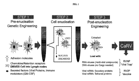
[00020] FIG. 1 shows a process for engineering cells for the rapid virus
vaccine platform according
to an embodiment of the present disclosure.
[00021] FIG. 2 shows a timeline for production of a vaccine using the rapid
virus vaccine platform
according to an embodiment of the present disclosure, as compared to a
traditional vaccine
development timeline.
[00022] FIG. 3 shows a process for deploying the rapid virus vaccine platform
to address a newly
identified virus according to an embodiment of the present disclosure.
[00023] FIG. 4 shows a process by which cytoplasts described herein trap and
clear live virus (e.g.,
coronavirus) according to an embodiment of the present disclosure.
[00024] FIG. 5 shows non-limiting examples of the benefits of the rapid virus
vaccine platform
described herein.
[00025] FIG. 6A is a representative line graph showing the viability of MSC
and MSC-derived
cytoplasts immediately after recovery from cryohibernation at 4 degrees
Celsius for the indicated
amounts of time. Viability was assessed in an automated cell count (Cell
Countess) using Trypan
blue dye exclusion and displayed as a ratio to the number of input cells.
[00026] FIG. 6B is a representative bar graph comparing the migrated MSC and
MSC-derived
cytoplasts in a Boyden chamber assay immediately after recovery from
cryohibernation at 4
degrees Celsius for the indicated amounts of time. Cells and cytoplasts were
allowed to migrate for
3 hours with either no serum (negative control) or 10% premium FBS (P-FBS) as
a chemoattractant
in the bottom chamber, and counts were normalized to loading controls.
[00027] FIG. 7A is a schematic representation of an interleukin 10 (IL-10)
mRNA transfected into
MSC and cytoplasts. Kozak sequence was added in front of the start codon of
the IL-10 mRNA
coding region (CDS). 5'UTR and 3 'UTR of human beta globin (HBB) mRNA were
added
16
CA 03170538 2022-08-10
WO 2021/163222 PCT/US2021/017506
respectively to the 5' and 3' end of IL-10 CDS. An artificial 5'Cap was added
to the 5' end of the
IL-10 mRNA and the pseudouridine modification was engineered to increase mRNA
stability.
[00028] FIG. 7B is a bar graph showing IL-10 concentration in the culture
medium of transfected
(++) or non-transfected (--) MSC or MSC-derived cytoplasts. MSC-derived
cytoplasts were
transfected with IL-10 mRNA, then seeded in a 24 well plate at 2.5 x 10⁴
cells/well. Conditioned
medium (CM) was collected 24 hours after transfection and the IL-10
concentration determined by
ELISA.
[00029] FIG. 7C is an immunoblot showing protein expression of Stat3 and
phosphorylated Stat3
(P-Stat3, a marker of IL-10 activation) in serum-starved RAW macrophage cells
treated with the
indicated conditioned media (CM) from MSCs or cytoplasts treated as in FIG. 7B
for 1 hour.
Untreated = no CM treated control. Complete medium = RAW cells treated with
MSC complete
culture medium. MSC Ctrl = RAW cells treated with CM from non-transfected
MSCs. MSC IL-10
= RAW cells treated with CM from IL-10 mRNA transfected MSCs. Cytoplast Ctrl
=RAW cells
treated with CM from non-transfected cytoplasts. Cytoplasts IL-10 = RAW cells
treated with CM
from IL-10 mRNA transfected cytoplasts.
[00030] FIG. 7D is a bar graph showing the concentration of secreted IL-10
cytokine in the mouse
blood as determined by ELISA. MSC or MSC-derived cytoplasts were treated as in
FIG. 7B and
retro-orbitally injected into the vasculature of C57BL/6 mice. Two hours after
injection, animals
were euthanized, and blood samples were collected by cardiac puncture. Mean
SEM; n=3.
[00031] FIG. 8A are representative bright field microscopy images of Crystal
Violet-stained MSCs
or MSC-derived cytoplasts in a Boyden chamber assay that invaded to the
undersurface of 8.01.tm
porous filters coated with Basement Membrane Extract (BME) towards 10% FBS as
a
chemoattractant for 24 hours. Negative= no FBS (negative control). Scale Bar =
5011m.
[00032] FIG 8B is a representative bar graph showing the ratio of MSC or MSC-
derived cytoplasts
treated as in FIG. 8A that invaded to the undersurface of the membrane
compared to the loading
control. Mean SEM; n=18.
[00033] FIG. 9A is representative epifluorescence microscopy images (upper
panel) and phase
contrast microscopy images (lower panel) of MSCs and cytoplasts in suspension
media. Actin
cortex was stained with Lifeact RFP, while the cell nucleus was stained with
Vybrant DyecycleTm
Green. Arrows point to cytoplasts and arrowhead points to MSC nucleus. Scale
bar = 201.tm.
[00034] FIG. 9B is a representative scatter plot showing the size distribution
of MSCs and
cytoplasts as measured with Nikon Element software. Mean SEM; n=80.
17
CA 03170538 2022-08-10
WO 2021/163222 PCT/US2021/017506
[00035] FIG. 9C is a representative bar graph showing the detected Vybrant
DiD-labeled MSCs
or cytoplasts present in lung. MSCs or cytoplasts were labeled with DiD dye
and retro-orbitally
injected into the vasculature of C57BL/6 mice. Tissues were harvested after 24
hours and cell
suspensions analyzed by flow cytometry. Mean SEM; n=3.
[00036] FIG. 9D is a representative bar graph showing the detected Vybrant
DiD labeled MSCs
or cytoplasts present in liver. Mean SEM; n=3. MSCs or cytoplasts were
labeled with DiD dye
and retro-orbitally injected into the vasculature of C57BL/6 mice. Tissues
were harvested after 24
hours and cell suspensions analyzed by flow cytometry.
[00037] FIG. 10A is a representative scatter plot showing the number of DiD-
labeled MSCs or
cytoplasts detected in the lung. MSCs were cultured under standard adherent
conditions (2D) or in
suspension by the handing drop method (3D) to generate 3D cytoplasts. MSCs and
cytoplasts were
labeled with Vybrant DiD dye and retro-orbitally injected into the
vasculature of C57BL/6 mice.
Tissues were harvested after 24 hours and cell suspensions analyzed by flow
cytometry. Mean
SEM; n=2.
[00038] FIG. 10B is a representative scatter plot showing the number of DiD-
labeled MSCs or
cytoplasts detected in the liver. MSCs were cultured under standard adherent
conditions (2D) or in
suspension by the handing drop method (3D) to generate 3D cytoplasts. MSCs and
cytoplasts were
labeled with Vybrant DiD dye and retro-orbitally injected into the
vasculature of C57BL/6 mice.
Tissues were harvested after 24 hours and cell suspensions analyzed by flow
cytometry. Mean
SEM; n=2.
[00039] FIG. 10C is a representative scatter plot showing the number of
Vybrant DiD-labeled
MSCs or cytoplasts detected in the spleen. MSCs were cultured under standard
adherent conditions
(2D) or in suspension by the handing drop method (3D) to generate 3D
cytoplasts. MSCs and
cytoplasts were labeled with DiD dye and retro-orbitally injected into the
vasculature of C57BL/6
mice. Tissues were harvested after 24 hours and cell suspensions analyzed by
flow cytometry.
Mean SEM; n=2.
[00040] FIG. 11A-11B illustrate Epifluorescent microscopy images of nucleated
parental MSCs
(top) and MSC-derived cytoplast (bottom) infected with VSV-GFP (arrows) at MOI
0.05 at 12 hrs
after infection. The GFP antigen was clearly and robustly expressed by MSCs
without nuclei
indicating viral replication and antigen production in enucleated cells. Scale
bar = 50 [tm. FIG.
11B. High magnification epifluorescent image of an MSC-derived cell without
nucleus infected
with VSV-GFP (arrowheads) at MOI 0.1 at 12 hours after infection. The
cytoplast was also stained
18
CA 03170538 2022-08-10
WO 2021/163222 PCT/US2021/017506
for F-actin filaments using rhodamine phalloidin (arrows) and the nuclear
stain DAPI to illustrate
the lack of the nucleus.
[00041] FIG. 12A-12D illustrate Epifluorescent microscopy images (FIG. 12A) of
MSC and MSC
without nucleus infected with oHSV encoding GFP antigen at MOI 0.05 at 48 hrs
after infection.
MSCs without nuclei (cytoplast) were generated from MSCs 18 hrs after
inoculation with oHSV-
GFP. Scale bar = 50 pm. FIG. 12B illustrates that MSCs or MSCs without nuclei
expressing
lifeact-RFP were infected with 0.05 MOI of the oncolytic herpes simplex virus
encoding GFP
(oHSV-GFP) then injected into established U87 glioblastoma tumors growing in
Nude mice.
Images were taken 7 days after the injection. Both MSCs and MSCs without
nuclei delivered oHSV
to tumor cells as indicated by the strong GFP signal. It was notable that very
few MSCs without
nuclei were detected in the tumor after 7 days, whereas a large number of MSCs
were present in the
center (injection site) and at the outer edge of the growing tumor. FIG. 12C
is a bar graph showing
percentage of GFP-covered tumor area, which represents the portion of tumor
cells infected by
MSCs or MSCs without nuclei carrying the oHSV-GFP virus. FIG. 12D is a graph
showing the
increased ratio of CD8+ effector T cells present in established glioblastoma
tumors treated with
combination of IL-12 (adjuvant) engineered MSCs without nuclei and oHSV
engineered MSCs
without nuclei compared to PBS injected controls.
[00042] FIG. 13A-13B illustrate enucleated mesenchymal stromal cells (MSCs)
(cytoplasts)
readily uptake cell permeable antigen peptides. FIG. 13A shows MSCs (left) and
enucleated MSCs
(cytoplast) (right) incubated with 100 pA/1 of the cell-permeable antigen
peptide (Arg)9-FAM (6-
Carboxyfluorescein, FAM-Arg-Arg-Arg-Arg-Arg-Arg-Arg-Arg-Arg-OH). Scale bar =
50 pm.
Arrows indicate Hoechst stained nuclei, arrowheads indicate positive (Arg)9-
FAM. FIG. 13B
illustrates bar graphs represents relative fluorescence intensity measured in
ImageJ. Corrected Total
Cell Fluorescence = Integrated Density ¨ (Area of selected cell X Mean
fluorescence of
background readings). Mean SEM; n=10.
DETAILED DESCRIPTION
[00043] Disclosed herein are compositions and kits, and methods of their use
to treat or prevent
pathogenic infections (e.g., viral, fungal, parasite, bacterial) or a disease
or condition associated
with such pathogenic infections. The compositions of the present disclosure
comprise cytoplasts,
which are enucleated cells engineered to contain, and in some cases, produce a
therapeutic agent
that is effective to treat the disease or the condition associated with a
pathogenic infection, and/or
prevent the pathogenic infection. In some embodiments, the therapeutic agent
described herein may
be a vaccine (e.g., attenuated viral antigen), a virus-targeting agent
effective to treat acute viral
19
CA 03170538 2022-08-10
WO 2021/163222 PCT/US2021/017506
infections, or combinations of the two. In some embodiments, the cytoplast may
also be engineered
to trap pathogens (e.g., in vivo) and inactivate them to treat acute
infections and prevent further
infection. In some embodiments, the pathogens are one or more viruses, such as
coronavirus.
[00044] Existing cell-based therapies have many shortcomings. Development of
effective cell-
based therapeutics often requires genetic engineering and the introduction of
new genetic material
into the genome of cells ex vivo. However, this process can introduce
dangerous mutations into the
genome that produce cancer and other life-threatening diseases, especially if
the engineered cells
permanently engraft into the body or fuse with host cells. Another significant
problem with many
existing cell-based therapeutics is that after delivery to the body, the cells
proliferate uncontrollably
and can permanently engraft into the body, which can be life-threatening.
Also, the lack of cell
control after administration to the subject can make the delivery of precise
doses of therapeutic
cells and their bioactive products difficult (e.g., poor pharmacokinetics).
Thus, there exists a need
for a safe and controllable cell-based therapy to deliver therapeutic agents
or other biomolecules.
[00045] Prior to patient or subject delivery, traditional cell-based
therapeutics are commonly
modified or genetically altered ex vivo to generate desirable cellular and
therapeutic functions.
However, when these cells are introduced into the subject, the new host
environment can
significantly reprogram and negatively alter, or otherwise render them
ineffective. Thus, there is a
need for a more predictable cell-based therapy that cannot respond to
reprogramming and
detrimental external signals.
[00046] Cell-based therapies that exist today are limited by the amount of DNA-
damaging/gene
targeting agents can be loaded into them for delivery to subjects as a
therapeutic against cancer or
other diseases. This includes, but is not limited to, DNA-damaging
chemotherapeutic drugs, DNA-
integrating viruses, oncolytic viruses, and gene therapy applications/delivery
including, but not
limited to, cluster regularly interspaced short palindromic repeats (CRISPR),
small clusters of Cas
(CRISPR/Cas system), and plasmids. Thus, there is a need for a cell-based
therapy without such
limitations, which may be an ideal platform delivering high doses of cytotoxic
therapeutic agents.
[00047] There are several advantages to delivering a therapeutic agent to a
subject using the
cytoplasts of the instant disclosure. Unlike conventional cell-based therapies
that transfer DNA
from their nuclei (e.g., nuclear-encoded genes or foreign or mutant DNA) to
host cells
unintentionally, the cytoplast of the present disclosure are unable to do so
without a nucleus.
Additionally, delivery of the therapeutic agent to the subject using the
cytoplasts described herein is
controllable and finite (e.g., 14 days or fewer), at least because, without a
nucleus, the cytoplasts
cannot proliferate or differentiate into other cell types. The cytoplasts of
the present disclosure may,
CA 03170538 2022-08-10
WO 2021/163222 PCT/US2021/017506
in the absence of a nucleus, express and/or secrete the therapeutic agent or
other biomolecules
described herein, as well as migrate or home to a target cell or target tissue
or environment in vivo.
This is achieved, at least in part, by enucleating a parent cell using the
methods described herein
such that the resulting cytoplast retains the organelles from the parent cell
that are sufficient for
normal biological function (e.g., protein production/secretion, cell motility,
chemokine sensing, and
like). Even when delivered to a subject systemically, the cytoplasts described
herein deliver the
therapeutic agent to a target tissue or a target cell in the subject (e.g.,
lymph tissue, lung tissue)
efficiently and effectively in a manner that is safe and controllable.
Moreover, manufacturing large
quantities of conventional cell-based therapies is time intensive and
expensive, which limits their
clinical applications. Although, it is thought that using immortalized cells
containing nuclei (e.g.,
hTERT) to increase manufacturing capabilities could increase manufacturing
scale and lower
manufacturing costs, there are concerns that immortalized cells are prone to
chromosomal
abnormalities and promote tumor or ectopic tissue formation, rendering them
unsafe for clinical
applications. By enucleating such cells, or any cell type, according to the
embodiments of the
instant disclosure, increased scale and lower costs associated with
manufacturing the cytoplasts
may be achieved, while mitigating the risks to human health posed by
conventional cell-based
therapies.
[00048] The improved manufacturing scale and cost, safety profile, and
efficiency of the
compositions described herein have important benefits for vaccine development.
The methods for
producing the compositions described herein are faster than conventional
vaccine development
timelines, which usually require the isolation and purification of the vaccine
(e.g., antigen, mRNA)
from the producer cell line. By contrast, cytoplasts of the present disclosure
are engineered to
continuously produce the anti-viral composition, obviating the need for
isolation and purification of
the vaccine. At the point of need, the compositions described herein may be
administered
systemically (e.g., inhalation), rather than by intramuscular injection,
avoiding a need for a medical
facility to administer the vaccine and improving patient experience. Due to
the ability of the
cytoplast to rapidly home to the lymph tissue (or other target tissue), the
vaccine may be deployed
to the lymphatic system of a subject in a fraction of the time it would take
certain conventional cell-
based therapies (e.g., exosomes) administered systemically. In addition, the
small size of the
cytoplast (e.g., about 8 micrometers) ensures that the cytoplasts are not
trapped small openings in
the vasculature and tissue parenchyma, thereby improving biodistribution as
compared with
conventional cell-based therapies. Cytoplasts disclosed herein, may be
engineered to express
virtually any type of vaccine or anti-viral agent (e.g., anti-viral and/or
neutralizing antibody) to
21
CA 03170538 2022-08-10
WO 2021/163222 PCT/US2021/017506
fight an active infection as well as prevent future infections. In addition,
the cytoplasts described
herein may be engineered to express more than one type of vaccine (e.g.,
against more than one
type of pathogen), enabling a panel of vaccines to be administered to a
subject in a single dosage
form. This is particularly beneficial for rapidly evolving pathogens (e.g.,
SARS-CoV-2), which
may require multiple vaccines in the future for an effective immunization
strategy.
[00049] The cytoplasts disclosed here are an off-the-shelf solution to an
urgent medical need. The
cytoplasts may be engineered before or after enucleation to express targeting
moieties (e.g., homing
receptors), immune-evading moieties (e.g., "don't eat me" signaling peptides),
among other
biomolecules sufficient to target the cytoplast to the lymph tissue without
risk of clearance by the
immune system before they get there. The cytoplasts may be cryopreserved, cryo-
hibernated, or
cryodesiccated, and stored for long periods of time with their biological
activity slowed or stopped.
When there is an urgent medical need, the biological function of the
cytoplasts may be restored
(e.g., thawing, rehydrating), and remain viable for up to 5 days for further
engineering (e.g., to
express a vaccine or anti-viral agent) as needed before delivery. Such
biological functions include,
but are not limited to expression of therapeutic surface proteins, immune
stimulating antigens, or
receptors, secrete cytokines, hormones, or proteins, release of exosomes,
shedding membrane
particles, stimulate the immune system through death processes, or create
tunneling nanotubes. The
cytoplasts of the instant disclosure may be frozen and thawed multiple times
during the
manufacturing and distribution process, without negatively impacting the
cytoplast intended
function, making them an ideal platform for a rapid vaccine deployment.
[00050] In some embodiments, the cytoplasts of the instant disclosure can be
therapeutic without
being engineered to produce or deliver an exogenous vaccine or other
biomolecule described
herein. For example, an unmanipulated cytoplast itself can have therapeutic
properties when
delivered into a patient or subject, such as for example a cytoplast derived
from a cell obtained
from a subject immune to a pathogen of interest, similar to a convalescent
plasma therapy
approach. Such cell may naturally produce neutralizing antibodies that block
pathogen-host
receptor engagement. In some embodiments, an unmanipulated cytoplast can
produce any one of
the therapeutic agents or biomolecules described herein naturally, which may
be used to achieve a
therapeutic effect in a subject in need thereof
[00051] Non-limiting examples of the many benefits of the rapid vaccine
platform described herein
are provided in FIG. 5. The production of cytoplasts may be scaled up rapidly,
where hundreds of
millions cytoplasts engineered to express viral antigen may be manufactured
with ease and may be
stored until needed. The cytoplasts described herein, in addition to being
engineered to express
22
CA 03170538 2022-08-10
WO 2021/163222 PCT/US2021/017506
viral antigen, may act as a trap. Such technical feature allows the engineered
cytoplast to be
infected by a pathogen, thus sequestering the pathogen and preventing the
pathogen from infecting
other cells. For example, the cytoplast described herein can be engineered to
express ACE2
receptor to be infected by a SARS-CoV-2 virus expressing the Spike protein.
Upon infection, the
SARS-CoV-2 virus is trapped in the cytoplast may no longer replicate. The
infected cytoplast may
be targeted by the immune system for degradation. The cytoplast may be
engineered to express
chemokine receptor to home the cytoplast to target tissue or microenvironment
such as lymph node.
[00052] Provided here are compositions, methods, and kits for the prevention
or treatment of
pathogenic infections in a subject. In some embodiments, the pathogenic
infection is a viral
infection, such as infection of coronavirus or influenza virus. In some
embodiments, the pathogenic
infection is a bacterial infection. Disclosed herein are cytoplasts that are
engineered to express an
anti-viral composition that are suitable to prevent viral infection or
outbreak, or treat acute
infections. When delivered to a subject, the cytoplast delivers the anti-viral
composition to a target
tissue either by presenting the anti-viral composition on the surface of the
cytoplast or by secreting
the anti-viral composition into extracellular space surrounding the target
tissue.
[00053] In some embodiments, the cytoplasts of the present disclosure are also
suitable for trapping
pathogens in a subject by permitting infection of the cytoplast by the
pathogen and preventing
propagation of the pathogen in vivo. As shown in FIG. 4, the cytoplast
described herein can express
a viral receptor that can be recognized by the pathogen, promoting infection
of the cytoplast. The
pathogen, upon infecting the cytoplast, is sequestered in the cytoplast unable
to replicate or
propagate in the absence of a nuclear genome. After 5 days or fewer, the
cytoplast is cleared from
the subject using natural processes of phagocytosis. In some embodiments, the
cytoplast activates
the immune system to accelerate clearance of the virus in the subject. At
least one advantage to the
cytoplasts disclosed herein for preventing the propagation of a pathogen in
vivo is that they lack a
nucleus containing genetic information necessary for many pathogens to
replicate.
[00054] Referring to FIG. 1, in some embodiments, cells (e.g., stem cells)
that are genetically
engineered prior to enucleation to express adhesion molecules, chemokine or
retention receptors or
both, that target a target cell or tissue, such as the lymph tissue (e.g.,
lymph nodes) or the lung
tissue in a subject (STEP 1). Next, the engineered cells are enucleated using
the methods described
herein to produce the cytoplasts (STEP 2). The cytoplasts may then be
engineered to express and,
in some embodiments, secrete a vaccine or other biomolecule (e.g., therapeutic
agent, neutralizing
antibody), and/or immune modulators (e.g., immune activators) to enhance the
adaptive immune
response in the subject (STEP 3). Cytoplasts are further engineered as needed
depending on the
23
CA 03170538 2022-08-10
WO 2021/163222 PCT/US2021/017506
intended function. The resulting cytoplasts may be used as a trap for viral
trap or to deploy a
vaccine. In the non-limiting example of the viral trap, the cytoplast may not
be engineered with a
therapeutic (e.g., vaccine) payload. Although, in some cases, it may be
advantageous to express
and/or secrete neutralizing antibodies against the pathogen of interest to
prevent future infections
by the virus. In some embodiments, the virus in this example is a coronavirus,
such as SARS-CoV-
2. However, the workflow in FIG. 1 may be applicable to any pathogen described
herein, including
bacterial pathogens (e.g., Bacillus anthracis) or toxins posing a significant
risk to human health.
[00055] The process of manufacturing the cytoplasts of the present disclosure
from identification of
a new pathogen (e.g., a virus) to distribution is roughly 2 months, as
compared with traditional
vaccine development, which is 12 months or longer. As shown in FIG. 2, the
cytoplasts of the
present disclosure may be prepared in advance of the viral outbreak and
cryopreserved for a length
of time. This means, the cytoplasts of the present disclosure (e.g.,
engineered to express the homing
receptors, immune activators) may be rapidly deployed to address the next
viral outbreak. Referring
to FIG. 3, the cytoplasts that were prepared in advance and cryopreserved are
engineered to secrete
attenuated viral proteins. When administered to a subject in need thereof, the
cytoplasts drive
immune activation and production of neutralizing antibodies against the virus
in the subject.
[00056] Methods of producing cytoplasts of the present disclosure are
provided. In some
embodiments, cells can be treated with cytochalasin B to soften the cortical
actin cytoskeleton. The
nucleus can then be physically extracted from the cell body by highspeed
centrifugation in
gradients of Ficoll to generate a nucleus-free (enucleated) cytoplast. Because
cytoplast and intact
nucleated cells sediment to different layers in the Ficoll gradient,
cytoplasts can, in some
embodiments, be easily isolated and prepared for therapeutic purposes or
fusion to other cells
(nucleated or enucleated). The enucleation process can be clinically scalable
to process tens of
millions of cells.
[00057] Disclosed herein are methods of using or delivering the cytoplasts of
the present
disclosure. The cytoplasts can be used as a homing vehicle to deliver
clinically relevant
cargos/payloads to treat healthy individuals (e.g., to improve energy,
recovery from exercise, or to
deliver natural products) or various diseases (e.g., any of the diseases
described herein). For
example, cytoplasts may be used to deliver supplements, anti-aging factors,
preventative
treatments, and the like to healthy individuals, e.g., individuals who have
not been diagnosed with a
specific disorder for which the delivered therapeutic is effective.
[00058] Also, provided herein are kits that include any composition described
herein. For example,
a kit can include instructions for using any of the compositions or methods
described herein. In
24
CA 03170538 2022-08-10
WO 2021/163222 PCT/US2021/017506
some embodiments, the kits can include at least one dose of any of the
compositions described
herein.
I. COMPOSITIONS
[00059] Provided herein are compositions useful for treating or preventing a
pathogen-associated
disease or condition in a subject. In some embodiments, the compositions
disclosed herein
comprise a cytoplast (e.g., an enucleated cell) engineered to express an
active agent suitable for the
treatment or the prevention of a pathogen-associated disease or condition. In
some embodiments,
the pathogen-associated disease or condition is a viral infection, such as a
coronavirus infection. In
some embodiments, cytoplast is engineered to express an anti-viral
composition, such as an
attenuated viral antigen or anti-viral antibody, or a combination thereof. In
some embodiments, the
cytoplast comprises the anti-viral composition at the surface of the cytoplast
(e.g., antigen
presentation). In some embodiments, the anti-viral composition is secreted by
the cytoplast into
extracellular space at a target tissue. In some embodiments, the cytoplast is
engineered to capture or
trap a pathogen in vivo by permitting infection of the cytoplast and
preventing propagation of the
pathogen in vivo, thereby treating an acute pathogenic infection, or pathogen-
associate disease or
condition.
[00060] The cytoplasts described herein are engineered to have a limited or
defined (e.g., known,
or programmable) life span. The cytoplasts described herein have a reduced
size compared to cells
in some other cell-based therapies (e.g., exosomes, red blood cells, adoptive
cell therapies), which
In some embodiments, improves biodistribution.
[00061] The cytoplasts described herein maintain viability following
cryohibernation or
cryopreservation, making them uniquely suitable for widespread adoption as a
platform for drug
delivery. Cryopreservation includes cooling or freezing, and storing, in the
short-term or long-term,
biological material (e.g., cells, cytoplasts) at very low temperatures (e.g., -
80 C in solid CO2, -196
C in liquid nitrogen, etc.). Cryohibernation includes short-term cooling and
storing of biological
material (e.g., cells, cytoplasts) in suspended animation, at non-freezing
temperatures, such as, e.g.,
at 4 C. Cryohibernation of cytoplasts can be advantageous for one or more of
the following
reasons: cryohibernation is less labor-intensive than cryopreservation, and
cytoplasts that have
undergone cryohibernation can be transported (e.g., shipped). In some
embodiments, the cytoplast
is cryopreserved. In some embodiments, the cytoplast is cryohybernated.
Following removal of the
cytoplast from cryohybernation or cryopreservation, the cytoplasts may be used
in accordance with
the methods described herein. In some embodiments, the cytoplasts are viable
for at least or about
CA 03170538 2022-08-10
WO 2021/163222 PCT/US2021/017506
24 hours, 48 hours, 72, or any increment of time between 24 and 72 hours
following removal from
cryohybernation or cryopreservation. In some embodiments, the cytoplasts are
viable for between
about 24 and about 48 hours. In some embodiments, the cytoplasts are viable
for between about 48
and about 72 hours. In some embodiments, viability is measured using trypan
blue dye exclusion as
described herein. In some embodiments, viability is measured using Annexin-5
cell surface staining
as described herein.
[00062] The cytoplasts described herein are extensively engineered, to best
suit a given therapeutic
application. For example, the cytoplasts are engineered (e.g., with cell-
surface receptors) that
increase infection of the cytoplast by a target pathogen. In some embodiments,
cytoplasts are
engineered to express an attenuated viral antigen for use as a vaccine or an
anti-viral antibody for
use in treating acute viral infections. In another example the cytoplasts are
engineered to produce or
express a protein that specifically targets difficult tissues (e.g., muscle)
and an active agent such as
an attenuated viral antigen or anti-viral antibody. In addition, in some
embodiments, the cytoplasts
are engineered with immune evading moieties (e.g., CD34+) to avoid an
antigenic response in the
host. Cytoplasts are also engineered to express cell-surface receptors (e.g.,
adhesion molecules,
chemokine receptors) used for cellular homing, chemokine sensing, and other
biological functions
that are essential to targeting damaged tissue in a predominately affected
area.
[00063] In some embodiments, a cytoplast has a defined life span of less than
1 hour to 14 days
(e.g., less than 1 hour to 1 hour, less than 1 hour to 6 hours, 6 hours to 12
hours, 12 hours to 1 day,
1 day, 2 days, 3 days, 4 days, 5, days, 6 days, 7 days, 8 days, 9 days, 10
days, 11 days, 13 days, 14
days, 1 to 14 days, 1 to 12 days, 1 to 10 days, 1 to 9 days, 1 to 8 days, 1 to
7 days, 1 to 6 days, 1 to
days, 1 to 4 days, 1 to 3 days, 1 to 2 days, 2 to 14 days, 2 to 12 days, 2 to
10 days, 2 to 8 days, 2
to 7 days, 2 to 6 days, 2 to 5 days, 2 to 4 days, 2 to 3 days, 3 to 14 days, 3
to 12 days, 3 to 10 days,
3 to 8 days, 3 to 7 days, 3 to 6 days, 3 to 5 days, 3 to 4 days, 4 to 14 days,
4 to 12 days, 4 to 10
days, 4 to 8 days, 4 to 7 days, 4 to 6 days, 4 to 5 days, 4 to 7 days, 5 to 14
days, 5 to 12 days, 5 to
days, 5 to 8 days, 5 to 7 days, 5 to 6 days, 6 to 14 days, 6 to 12 days, 6 to
10 days, 6 to 8 days, 6
to 7 days, 7 to 14 days, 7 to 12 days, 7 to 10 days, 7 to 8 days, 8 to 14
days, 8 to 12 days, 8 to 10
days, 10 to 14 days, 10 to 12 days, 12 to 14 days, less than 14 days, less
than 12 days, less than 10
days, less than 8 days, less than 7 days, less than 6 days, less than 5 days,
less than 4 days, less than
3 days, less than 2 days, less than 1 day, less than 12 hours, or less than 6
hours). In some
embodiments, the lifespan of a population of cytoplasts can be evaluated by
determining the
average time at which a portion of the cytoplast population (e.g., at least
50%, at least 60% at least
70%, at least 75%, at least 80%, at least 85%, at least 90%, at least 95%, or
at least 98% of the
26
CA 03170538 2022-08-10
WO 2021/163222 PCT/US2021/017506
population) is determined to be dead. Cell death can be determined by any
method known in the art.
In some embodiments, the viability of cytoplasts, e.g., at one or more time
points, can be evaluated
by determining whether morphometric or functional parameters are intact (e.g.
by trypan-blue dye
exclusion, evaluating for intact cell membranes, evaluating adhesion to
plastics (e.g., in adherent
cytoplasts), evaluating cytoplast migration, negative staining with apoptotic
markers, and the like).
In some embodiments, the life span of a cytoplast may be related to the life
span of the cell from
which it was obtained. For example, in some embodiments, a cytoplast obtained
from a
macrophage may live 12 to 24 hours.
[00064] In some embodiments, a cytoplast is at least or equal to 1 [tm in
diameter. In some
embodiments, a cytoplast is greater than 1 [tm in diameter. In some
embodiments, a cytoplast is 1-
100 [tm in diameter (e.g., 1- 90 [tm, 1-80 [tm, 1-70 [tm, 1-60 [tm, 1-50 [tm,
1-40 [tm, 1-30 [tm, 1-20
[tm, 1-10 [tm, 1-5 [tm, 5- 90 [tm, 5-80 [tm, 5-70 [tm, 5-60 [tm, 5-50 [tm, 5-
40 [tm, 5-30 [tm, 5-20
[tm, 5-10 [tm, 10-90 [tm, 10-80 [tm, 10-70 [tm, 10-60 [tm, 10-50 [tm, 10-40
[tm, 10-30 [tm, 10-20
[tm, 10-15 [tm 15-90 [tm, 15-80 [tm, 15-70 [tm, 15-60 [tm, 15-50 [tm, 15-40
[tm, 15-30 [tm, 15-20
[tm). In some embodiments, a cytoplast is 10-30 [tm in diameter. In some
embodiments, the
diameter of a cytoplast is between 5-25 [tm (e.g., 5-20 [tm, 5-15 [tm. 5-10
[tm, 10-25 [tm, 10-20
[tm, 10-15 [tm, 15-25 [tm, 15-20 [tm, or 20-25 [tm. In some embodiments, a
cytoplast is not an
exosome. Without being bound by any particular theory, it is believed that, In
some embodiments,
some cytoplasts can advantageously be small enough to allow for better
biodistribution or to be less
likely to be trapped in the lungs of a subject.
[00065] In some embodiments, cytoplasts can be applied to or cultured with
cells (e.g.,
xenocultured cells) to alter their properties. For example, in some
embodiments, cytoplasts (e.g.,
unmanipulated cytoplasts or engineered cytoplasts) can upregulate health-
promoting factors in
xenocultured cells, and in some embodiments, the xenocultured cells can be
returned to the subject
from which they were taken.
A. Cells
[00066] Provided herein are cells and cell lines that are engineered to
produce the cytoplasts of the
present disclosure. The cytoplast may be derived from a corresponding parent
cell, such as a
nucleated parent cell. Non-limiting examples of parent cells include an
immortalized cell, a cancer
cell (e.g., any cancer cell), a primary (e.g., host-derived) cell, or a cell
line. In some embodiments,
the parent cell is derived from a cell is immortalized using suitable methods,
such as those described
in Huang et at., J. Exp. Clin. Med. 2010 Oct. 221 2(5):202-217. In some
embodiments, the cytoplast
27
CA 03170538 2022-08-10
WO 2021/163222 PCT/US2021/017506
is derived from a parent cell using suitable methods provided in US Patent
Application No.
16/715,859, which is hereby incorporated by reference in its entirety.
[00067] In some embodiments, the cell can originate from any organism having
one or more cells.
Some non-limiting examples include: a prokaryotic cell, eukaryotic cell, a
bacterial cell, an
archaeal cell, a cell of a single-cell eukaryotic organism, a protozoa cell, a
cell from a plant, an
algal cell, a fungal cell, an animal cell, a cell from an invertebrate animal,
a cell from a vertebrate
animal, a cell from a mammal (e.g., a pig, a cow, a goat, a sheep, a rodent, a
rat, a mouse, a non-
human primate, a human, etc.), and etcetera. In some embodiments, the cell is
a somatic cell. In
some embodiments, the cell is a stem cell or a progenitor cell. In some
embodiments, the cell is a
mesenchymal stem or progenitor cell. In some embodiments, the cell is a
mesenchymal stromal
cell. A cell can originate from any organism having one or more cells.
[00068] Some non-limiting examples of cells include: a prokaryotic cell,
eukaryotic cell, a bacterial
cell, an archaeal cell, a cell of a single-cell eukaryotic organism, a
protozoa cell, a cell from a plant
(e.g. cells from plant crops, fruits, vegetables, grains, soy bean, corn,
maize, wheat, seeds,
tomatoes, rice, cassava, sugarcane, pumpkin, hay, potatoes, cotton, cannabis,
tobacco, flowering
plants, conifers, gymnosperms, ferns, clubmosses, hornworts, liverworts,
mosses), an algal cell,
(e.g., B otryococcus braunii, Chlamydomonas reinhardtii, Nannochloropsis
gaditana, Chlorella
pyrenoidosa, Sargassum patens C. Agardh, and the like), seaweeds (e.g. kelp),
a fungal cell (e.g., a
yeast cell, a cell from a mushroom), an animal cell, a cell from an
invertebrate animal (e.g. fruit fly,
cnidarian, echinoderm, nematode, etc.), a cell from a vertebrate animal (e.g.,
fish, amphibian,
reptile, bird, mammal), a cell from a mammal (e.g., a pig, a cow, a goat, a
sheep, a rodent, a rat, a
mouse, a non-human primate, a human, etc.), and etcetera. Sometimes a cell is
not originating from
a natural organism (e.g. a cell can be a synthetically made, sometimes termed
an artificial cell). In
some embodiments, the cell is a somatic cell. In some embodiments, the cell is
a stem cell or a
progenitor cell. In some embodiments, the cell is a mesenchymal stem or
progenitor cell. In some
embodiments, the cell is a hematopoietic stem or progenitor cell. In some
embodiments, the cell is a
muscle cell, a skin cell, a blood cell, or an immune cell. Other exemplary
cells can include
lymphoid cells, such as B cell, T cell (Cytotoxic T cell, Natural Killer T
cell, Regulatory T cell, T
helper cell), Natural killer cell, cytokine induced killer (CIK) cells;
myeloid cells, such as
granulocytes (B a s op hi 1 granulocyte, Eosinophil granulocyte, Neutrophil
granulocyte/Hypersegmented neutrophil), Monocyte/Macrophage, Red blood cell
(Reticulocyte),
Mast cell, Thrombocyte/Megakaryocyte, Dendritic cell; cells from the endocrine
system, including
thyroid (Thyroid epithelial cell, Parafollicular cell), parathyroid
(Parathyroid chief cell, Oxyphil
28
CA 03170538 2022-08-10
WO 2021/163222 PCT/US2021/017506
cell), adrenal (Chromaffin cell), pineal (Pinealocyte) cells; cells of the
nervous system, including
glial cells (Astrocyte, Microglia), Magnocellular neurosecretory cell,
Stellate cell, Boettcher cell,
and pituitary (Gonadotrope, Corticotrope, Thyrotrope, Somatotrope, Lactotroph
); cells of the
Respiratory system, including Pneumocyte (Type I pneumocyte, Type II
pneumocyte), Clara cell,
Goblet cell, Dust cell; cells of the circulatory system, including
Myocardiocyte, Pericyte; cells of
the digestive system, including stomach (Gastric chief cell, Parietal cell),
Goblet cell, Paneth cell, G
cells, D cells, ECL cells, I cells, K cells, S cells; enteroendocrine cells,
including enterochromaffm
cell, APUD cell, liver (Hepatocyte, Kupffer cell), Cartilage/bone/muscle; bone
cells, including
Osteoblast, Osteocyte, Osteoclast, teeth (Cementoblast, Ameloblast); cartilage
cells, including
Chondroblast, Chondrocyte; skin cells, including Trichocyte, Keratinocyte,
Melanocyte (Nevus
cell); muscle cells, including Myocyte; urinary system cells, including
Podocyte, Juxtaglomerular
cell, Intraglomerular mesangial cell/Extraglomerular mesangial cell, Kidney
proximal tubule brush
border cell, Macula densa cell; reproductive system cells, including
Spermatozoon, Sertoli cell,
Leydig cell, Ovum; and other cells, including Adipocyte, Fibroblast, Tendon
cell, Epidermal
keratinocyte (differentiating epidermal cell), Epidermal basal cell (stem
cell), Keratinocyte of
fingernails and toenails, Nail bed basal cell (stem cell), Medullary hair
shaft cell, Cortical hair shaft
cell, Cuticular hair shaft cell, Cuticular hair root sheath cell, Hair root
sheath cell of Huxley's layer,
Hair root sheath cell of Henle's layer, External hair root sheath cell, Hair
matrix cell (stem cell),
Wet stratified barrier epithelial cells, Surface epithelial cell of stratified
squamous epithelium of
cornea, tongue, oral cavity, esophagus, anal canal, distal urethra and vagina,
basal cell (stem cell) of
epithelia of cornea, tongue, oral cavity, esophagus, anal canal, distal
urethra and vagina, Urinary
epithelium cell (lining urinary bladder and urinary ducts), Exocrine secretory
epithelial cells,
Salivary gland mucous cell (polysaccharide-rich secretion), Salivary gland
serous cell (glycoprotein
enzyme -rich secretion), Von Ebner's gland cell in tongue (washes taste buds),
Mammary gland cell
(milk secretion), Lacrimal gland cell (tear secretion), Ceruminous gland cell
in ear (wax secretion),
Eccrine sweat gland dark cell (glycoprotein secretion), Eccrine sweat gland
clear cell (small
molecule secretion). Apocrine sweat gland cell (odoriferous secretion, sex -
hormone sensitive),
Gland of Moll cell in eyelid (specialized sweat gland), Sebaceous gland cell
(lipid-rich sebum
secretion), Bowman's gland cell in nose (washes olfactory epithelium),
Brunner's gland cell in
duodenum (enzymes and alkaline mucus), Seminal vesicle cell (secretes seminal
fluid components,
including fructose for swimming sperm), Prostate gland cell (secretes seminal
fluid components),
Bulbourethral gland cell (mucus secretion), Bartholin's gland cell (vaginal
lubricant secretion),
Gland of Littre cell (mucus secretion), Uterus endometrium cell (carbohydrate
secretion), Isolated
29
CA 03170538 2022-08-10
WO 2021/163222 PCT/US2021/017506
goblet cell of respiratory and digestive tracts (mucus secretion), Stomach
lining mucous cell
(mucus secretion), Gastric gland zymogenic cell (pepsinogen secretion),
Gastric gland oxyntic cell
(hydrochloric acid secretion), Pancreatic acinar cell (bicarbonate and
digestive enzyme secretion),
Paneth cell of small intestine (lysozyme secretion), Type II pneumocyte of
lung (surfactant
secretion), Clara cell of lung, Hormone secreting cells, Anterior pituitary
cells, Somatotropes,
Lactotropes, Thyrotropes, Gonadotropes, Corticotropes, Intermediate pituitary
cell, Magnocellular
neurosecretory cells, Gut and respiratory tract cells, Thyroid gland cells,
thyroid epithelial cell,
parafollicular cell, Parathyroid gland cells, Parathyroid chief cell, Oxyphil
cell, Adrenal gland cells,
chromaffin cells, Ley dig cell of testes, Theca interna cell of ovarian
follicle, Corpus luteum cell of
ruptured ovarian follicle, Granulosa lutein cells, Theca lutein cells,
Juxtaglomerular cell (renin
secretion), Macula densa cell of kidney, Metabolism and storage cells, Barrier
function cells (Lung,
Gut, Exocrine Glands and Urogenital Tract), Kidney, Type I pneumocyte (lining
air space of lung),
Pancreatic duct cell (centroacinar cell), Nonstriated duct cell (of sweat
gland, salivary gland,
mammary gland, etc.), Duct cell (of seminal vesicle, prostate gland, etc.),
Epithelial cells lining
closed internal body cavities, Ciliated cells with propulsive function,
Extracellular matrix secretion
cells, Contractile cells; Skeletal muscle cells, stem cell, Heart muscle
cells, Blood and immune
system cells, Erythrocyte (red blood cell), Megakaryocyte (platelet
precursor), Monocyte,
Connective tissue macrophage (various types), Epidermal Langerhans cell,
Osteoclast (in bone),
Dendritic cell (in lymphoid tissues), Microglial cell (in central nervous
system), Neutrophil
granulocyte, Eosinophil granulocyte, Basophil granulocyte, Mast cell, Helper T
cell, Suppressor T
cell, Cytotoxic T cell, Natural Killer T cell, B cell, Natural killer cell,
Reticulocyte, Stem cells and
committed progenitors for the blood and immune system (various types),
Pluripotent stem cells,
Totipotent stem cells, Induced pluripotent stem cells, adult stem cells,
Sensory transducer cells,
Autonomic neuron cells, Sense organ and peripheral neuron supporting cells,
Central nervous
system neurons and glial cells, Lens cells, Pigment cells, Melanocyte, Retinal
pigmented epithelial
cell, Germ cells, Oogonium/Oocyte, Spermatid, Spermatocyte, Spermatogonium
cell (stem cell for
spermatocyte), Spermatozoon, Nurse cells, Ovarian follicle cell, Sertoli cell
(in testis), Thymus
epithelial cell, Interstitial cells, and Interstitial kidney cells.
[00069] Non-limiting examples of eukaryotic cells include mammalian (e.g.,
rodent, non-human
primate, or human), non-mammalian animal (e.g., fish, bird, reptile, or
amphibian), invertebrate,
insect, fungal, or plant cells. In some embodiments, the eukaryotic cell is a
yeast cell, such as
Saccharomyces cerevisiae. In some embodiments, the eukaryotic cell is a higher
eukaryote, such as
mammalian, avian, plant, or insect cells. In some embodiments, the nucleated
cell is a primary cell.
CA 03170538 2022-08-10
WO 2021/163222 PCT/US2021/017506
In some embodiments, the nucleated cell is an immune cell (e.g., a lymphocyte
(e.g., a T cell, a B
cell), a macrophage, a natural killer cell, a neutrophil, a mast cell, a
basophil, a dendritic cell, a
monocyte, a myeloid-derived suppressor cell, an eosinophil). In some
embodiments, the nucleated
cell is a phagocyte or a leukocyte. In some embodiments, the nucleated cell is
a stem cell (e.g., an
adult stem cell (e.g., a hematopoietic stem cell, a mammary stem cell, an
intestinal stem cell,
mesenchymal stem cell, an endothelial stem cell, a neural stem cell, an
olfactory adult stem cell, a
neural crest stem cell, a testicular cell), an embryonic stem cell, an
inducible pluripotent stem cell
(iPS)). In some embodiments, the nucleated cell is a progenitor cell. In some
embodiments, the
nucleated cell is from a cell line. In some embodiments, the nucleated cell is
a suspension cell. In
some embodiments, the nucleated cell is an adherent cell. In some embodiments,
the nucleated cell
is a cell that has been immortalized by expression of an oncogene. In some
embodiments, the
nucleated cell is immortalized by the expression of human telomerase reverse
transcriptase
(hTERT) or any oncogene. In some embodiments, the nucleated cell is a patient
or subject derived
cell (e.g., an autologous patient-derived cell, or an allogenic patient-
derived cell). In some
embodiments, the nucleated cell is transfected with a vector (e.g., a viral
vector (e.g., a retrovirus
vector (e.g., a lentivirus vector), an adeno-associated virus (AAV) vector, a
vesicular virus vector
(e.g., vesicular stomatitis virus (VSV) vector), or a hybrid virus vector), a
plasmid) before the
nucleated cell is enucleated using any of the enucleation techniques described
herein and known in
the art.
[00070] In some embodiments, the cytoplast can be derived from a cell
autologous to the subject. In
some embodiments, the cytoplast can be derived from a cell allogenic to the
subject.
[00071] In some embodiments, the cytoplast is derived from an immune cell. In
some embodiments,
the cytoplast is derived from a natural killer (NK) cell, a neutrophil, a
macrophage, a lymphocyte, a
fibroblast, an adult stem cell (e.g., hematopoietic stem cell, a mammary stem
cell, an intestinal stem
cell, a mesenchymal stem cell, a mesenchymal stromal cell, an endothelial stem
cell, a neural stem
cell, an olfactory adult stem cell, a neural crest stem cell, a skin stem
cell, or a testicular cell), a
mast cell, a basophil, an eosinophil, or an inducible pluripotent stem cell.
[00072] In some embodiments, prior to enucleation, two or more cells (e.g.,
any of the cells
disclosed herein) are fused by any method disclosed herein or known in the
art. Enucleation of the
fusion product can result in a cytoplast.
[00073] In some embodiments, a first cytoplast is fused to a cell or second
cytoplast. In some
embodiments, the cell is any nucleated (e.g., a mammalian cell (e.g., a human
cell, or any
mammalian cell described herein), a protozoal cell (e.g., an amoeba cell), an
algal cell, a plant
31
CA 03170538 2022-08-10
WO 2021/163222 PCT/US2021/017506
cell, a fungal cell, an invertebrate cell, a fish cell, an amphibian cell, a
reptile cell, or a bird cell).
In some embodiments, the second cell is a synthetic cell. Accordingly,
provided are methods of
altering the behavior of a cell comprising fusing the cell with any of the
cytoplasts described
herein. Also provided herein are methods comprising administering to a subject
a therapeutically
effective amount of a cell to which a cytoplast has been fused.
[00074] In some embodiments, the second cytoplast is derived from the same
type of cell as the first
cytoplast. In some embodiments, the second cytoplast is derived from a
different type of cell as the
first cytoplast. In some embodiments, the second cytoplast contains or
expresses at least one
therapeutic DNA molecule, therapeutic RNA molecule, therapeutic protein,
therapeutic peptide,
small molecule therapeutic, therapeutic gene editing factor, a therapeutic
nanoparticle, or another
active agent that is the same as a therapeutic DNA molecule, therapeutic RNA
molecule, therapeutic
protein, therapeutic peptide, small molecule therapeutic, therapeutic gene
editing factor, a
therapeutic nanoparticle contained in or expressed by the first cytoplast. In
some embodiments, the
second cytoplast contains or expresses at least one therapeutic DNA molecule,
therapeutic RNA
molecule, therapeutic protein, therapeutic peptide, small molecule
therapeutic, therapeutic gene
editing factor, a therapeutic nanoparticle, or another active agent that is
different from a therapeutic
DNA molecule, therapeutic RNA molecule, therapeutic protein, therapeutic
peptide, small molecule
therapeutic, therapeutic gene editing factor, a therapeutic nanoparticle
contained in or expressed by
the first cytoplast. In some embodiments, a first cytoplast can be fused to a
cell or to a second
cytoplast using any method known in the art, for example, electrofusion or
viral fusion using viral-
based cell surface peptides.
[00075] In some embodiments, a cytoplast is not a naturally occurring
enucleated cell. In some
embodiments, a cytoplast is not obtained from a cell that naturally undergoes
enucleation. In some
embodiments, a cytoplast is not a cell that has been enucleated by in the body
of a subject. In some
embodiments, a cytoplast is not obtained from a cell that would be enucleated
by in the body of a
subject. In some embodiments, a cytoplast is not obtained from an
erythroblast. In some
embodiments, a cytoplast is obtained from a cell that maintains a nucleus over
its lifespan (e.g., in
the absence of manipulations such as enucleation as described herein). In some
embodiments, a
cytoplast is not a cell that is found in a subject as an anucleate cell (e.g.,
a red blood cell
(erythrocyte), a platelet, a lens cell, or an immediate nucleated precursor
thereof). In some
embodiments, a cytoplast includes one or more components selected from the
group consisting of
an endoplasmic reticulum, a Golgi apparatus, mitochondria, ribosomes,
proteasomes, or
spliceosomes. In some embodiments, a cytoplast is characterized by one or more
of the following
32
CA 03170538 2022-08-10
WO 2021/163222 PCT/US2021/017506
features: adhesion, tunneling nanotube formation, actin-mediated spreading (2D
and/or 3D),
migration, chemoattractant gradient sensing, mitochondrial transfer, mRNA
translation, protein
synthesis, and secretion of exosomes and/or other bioactive molecules. In some
embodiments, a
cytoplast is characterized by an ability to secrete proteins (e.g., using
exosomes). In some
embodiments, a cytoplast has been enucleated ex vivo. In some embodiments, a
cytoplast has been
enucleated in vitro. In some embodiments, a cytoplast has been physically
enucleated (e.g., by
centrifugation). In some embodiments, a cytoplast is an engineered enucleated
cell. In some
embodiments, a cytoplast is not a red blood cell. In some embodiments, a
cytoplast does not contain
hemoglobin. In some embodiments, a cytoplast does not have a bi-concave shape.
[00076] In some embodiments, a cytoplast is not obtained from an erythroblast.
In some
embodiments, a cytoplast is obtained from a cell that would not become a red
blood cell (RBC).
Unlike RBCs cytoplasts can be viable cell-like entities that can retain many
active biological
processes and all cellular organelles (e.g., ER/Golgi, mitochondrial,
endosome, lysosome,
cytoskeleton, etc.). Thus, cytoplasts can function like nucleated cells and
exhibit critical biological
functions such as adhesion, tunneling nanotube formation, actin-mediated
spreading (2D and 3D),
migration, chemoattractant gradient sensing, mitochondrial transfer, mRNA
translation, protein
synthesis, and secretion of exosomes and other bioactive molecules. One or
more of these functions
may not be exhibited by RBCs. Compared to RBCs, which are derived from
erythroblasts, a
cytoplast can be derived from any type of nucleated cell, including, but not
limited to iPSC
(induced pluripotent stem cells), any immortalized cell, stem cells, primary
cells (e.g., host-derived
cells), cell lines, any immune cell, cancerous cells, or from any eukaryotic
cell. In some
embodiments, a cytoplast is obtained from a lymphoid progenitor cell. In some
embodiments, a
cytoplast is obtained from a lymphocyte. In some embodiments, a cytoplast is
obtained from a
mesenchymal stem cell (e.g., from bone marrow). In some embodiments, a
cytoplast is obtained
from an endothelial stem cell. In some embodiments, a cytoplast is obtained
from a neural stem
cell. In some embodiments, a cytoplast is obtained from a skin stem cell.
B. Pathogens
[00077] The cytoplasts described herein and compositions containing the
cytoplasts, in some
embodiments, comprise biomolecules (e.g., vaccine, therapeutic agent,
targeting moieties) that
target and/or kill, or otherwise render inoperable, a pathogen. In some
embodiments, the pathogen
is a bacteria, a virus, a fungus, or a toxin. In some embodiments, the
pathogen is naturally
occurring. In some embodiments, the pathogen is synthetic.
33
CA 03170538 2022-08-10
WO 2021/163222 PCT/US2021/017506
[00078] In some embodiments, the pathogen is a virus. In some embodiments, the
virus is an
animal virus, a plant virus, a bacterial virus, or an archaeal virus. In some
embodiments, the animal
virus causes a disease or condition in the same or a different animal. In some
embodiments, the
virus is an RNA virus or a DNA virus. In some embodiments, the RNA or DNA
virus is single-
stranded or double-stranded. In some embodiments, the DNA or RNA virus is a
positive-sense or a
negative-sense virus.
[00079] In some embodiments, the double-stranded virus (dsDNA) virus is from
the family:
Myoviridae, Podoviridae, Siphoviridae, Alloherpesviridae, Herpesviridae,
Malacoherpesviridae,
Lipothrixviridae, Rudiviridae, Adenoviridae, Ampullaviridae, Ascoviridae,
Asfaviridae,
Baculoviridae, Bicaudaviridae, Clavaviridae, Corticoviridae, Fuselloviridae,
Globuloviridae,
Guttaviridae, Hytrosaviridae, Iridoviridae, Marseilleviridae, Mimiviridae,
Nimaviridae,
Pandoraviridae, Papillomaviridae, Phycodnaviridae, Plasmaviridae,
Polydnaviruses,
Polyomaviridae, Poxviridae, Sphaerolipoviridae, and Tectiviridae.
[00080] In some embodiments, the single-stranded (ssDNA) virus is from the
family:
Anelloviridae, Bacillariodnaviridae, Bidnaviridae, Circoviridae,
Geminiviridae, Inoviridae,
Microviridae, Nanoviridae, Parvoviridae, and Spiraviridae.
[00081] A DNA virus that contains both ss and ds DNA regions can be from the
group of
pleolipoviruses. In some embodiments, the pleolipoviruses include Haloarcula
hispanica
pleomorphic virus 1, Halogeometricum pleomorphic virus 1, Halorubrum
pleomorphic virus 1,
Halorubrum pleomorphic virus 2, Halorubrum pleomorphic virus 3, and Halorubrum
pleomorphic
virus 6.
[00082] In some embodiments, the dsRNA virus is from the family: Birnaviridae,
Chrysoviridae,
Cystoviridae, Endornaviridae, Hypoviridae, Megavirnaviridae, Partitiviridae,
Picobirnaviridae,
Reoviridae, Rotavirus and Totiviridae.
[00083] In some embodiments, the positive-sense ssRNA virus can be from the
family:
Alphaflexiviridae, Alphatetraviridae, Alvernaviridae, Arteriviridae,
Astroviridae, Barnaviridae,
Betaflexiviridae, Bromoviridae, Caliciviridae, Carmotetraviridae,
Closteroviridae, Coronaviridae,
Dicistroviridae, Flaviviridae, Gammaflexiviridae, Iflaviridae, Leviviridae,
Luteoviridae,
Marnaviridae, Mesoniviridae, Narnaviridae, Nodaviridae, Permutotetraviridae,
Picornaviridae,
Potyviridae, Roniviridae, Secoviridae, Togaviridae, Tombusviridae,
Tymoviridae, and
Virgaviridae.
34
CA 03170538 2022-08-10
WO 2021/163222 PCT/US2021/017506
[00084] In some embodiments, the negative-sense ssRNA virus can be from the
family:
Bornaviridae, Filoviridae, Paramyxoviridae, Rhabdoviridae, Nyamiviridae,
Arenaviridae,
Bunyaviridae, Ophioviridae, and Orthomyxoviridae.
[00085] Non-limiting examples of viruses include: Abelson leukemia virus,
Abelson murine
leukemia virus, Abelson's virus, Acute laryngotracheobronchitis virus,
Adelaide River virus, Adeno
associated virus group, Adenovirus, African horse sickness virus, African
swine fever virus, AIDS
virus, Aleutian mink disease parvovirus, Alpharetrovirus, Alphavirus, ALV
related virus, Amapari
virus, Aphthovirus, Aquareovirus, Arbovirus, Arbovirus C, arbovirus group A,
arbovirus group B,
Arenavirus group, Argentine hemorrhagic fever virus, Argentine hemorrhagic
fever virus,
Arterivirus, Astrovirus, Ateline herpesvirus group, Aujezky's disease virus,
Aura virus, Ausduk
disease virus, Australian bat lyssavirus, Aviadenovirus, avian
erythroblastosis virus, avian
infectious bronchitis virus, avian leukemia virus, avian leukosis virus, avian
lymphomatosis virus,
avian myeloblastosis virus, avian paramyxovirus, avian pneumoencephalitis
virus, avian
reticuloendotheliosis virus, avian sarcoma virus, avian type C retrovirus
group, Avihepadnavirus,
Avipoxvirus, B virus, B19 virus, Babanki virus, baboon herpesvirus,
baculovirus, Barmah Forest
virus, Bebaru virus, Berrimah virus, Betaretrovirus, Birnavirus, Bittner
virus, BK virus, Black
Creek Canal virus, bluetongue virus, Bolivian hemorrhagic fever virus, Boma
disease virus, border
disease of sheep virus, borna virus, bovine alphaherpesvirus 1, bovine
alphaherpesvirus 2, bovine
coronavirus, bovine ephemeral fever virus, bovine immunodeficiency virus,
bovine leukemia virus,
bovine leukosis virus, bovine mammillitis virus, bovine papillomavirus, bovine
papular stomatitis
virus, bovine parvovirus, bovine syncytial virus, bovine type C oncovirus,
bovine viral diarrhea
virus, Buggy Creek virus, bullet shaped virus group, Bunyamwera virus
supergroup, Bunyavirus,
Burkitt's lymphoma virus, Bwamba Fever, CA virus, Calicivirus, California
encephalitis virus,
camelpox virus, canarypox virus, canid herpesvirus, canine coronavirus, canine
distemper virus,
canine herpesvirus, canine minute virus, canine parvovirus, Cano Delgadito
virus, caprine arthritis
virus, caprine encephalitis virus, Caprine Herpes Virus, Capripox virus,
Cardiovirus, caviid
herpesvirus 1, Cercopithecid herpesvirus 1, cercopithecine herpesvirus 1,
Cercopithecine
herpesvirus 2, Chandipura virus, Changuinola virus, channel catfish virus,
Charleville virus,
chickenpox virus, Chikungunya virus, chimpanzee herpesvirus, chub reovirus,
chum salmon virus,
Cocal virus, Coho salmon reovirus, coital exanthema virus, Colorado tick fever
virus, Coltivirus,
Columbia SK virus, common cold virus, contagious eethyma virus, contagious
pustular dermatitis
virus, Coronavirus, Corriparta virus, coryza virus, cowpox virus, coxsackie
virus, CPV
(cytoplasmic polyhedrosis virus), cricket paralysis virus, Crimean-Congo
hemorrhagic fever virus,
CA 03170538 2022-08-10
WO 2021/163222 PCT/US2021/017506
croup associated virus, Cryptovirus, Cypovirus, Cytomegalovirus,
cytomegalovirus group,
cytoplasmic polyhedrosis virus, deer papillomavirus, deltaretrovirus, dengue
virus, Densovirus,
Dependovirus, Dhori virus, diploma virus, Drosophila C virus, duck hepatitis B
virus, duck
hepatitis virus 1, duck hepatitis virus 2, duovirus, Duvenhage virus, Deformed
wing virus DWV,
eastern equine encephalitis virus, eastern equine encephalomyelitis virus, EB
virus, Ebola virus,
Ebola-like virus, echo virus, echovirus, echovirus 10, echovirus 28, echovirus
9, ectromelia virus,
EEE virus, ETA virus, ETA virus, encephalitis virus, encephalomyocarditis
group virus,
encephalomyocarditis virus, Enterovirus, enzyme elevating virus, enzyme
elevating virus (LDH),
epidemic hemorrhagic fever virus, epizootic hemorrhagic disease virus, Epstein-
Barr virus, equid
alphaherpesvirus 1, equid alphaherpesvirus 4, equid herpesvirus 2, equine
abortion virus, equine
arteritis virus, equine encephalosis virus, equine infectious anemia virus,
equine morbillivirus,
equine rhinopneumonitis virus, equine rhinovirus, Eubenangu virus, European
elk papillomavirus,
European swine fever virus, Everglades virus, Eyach virus, felid herpesvirus
1, feline calicivirus,
feline fibrosarcoma virus, feline herpesvirus, feline immunodeficiency virus,
feline infectious
peritonitis virus, feline leukemia/sarcoma virus, feline leukemia virus,
feline panleukopenia virus,
feline parvovirus, feline sarcoma virus, feline syncytial virus, Filovirus,
Flanders virus, Flavivirus,
foot and mouth disease virus, Fort Morgan virus, Four Corners hantavirus, fowl
adenovirus 1,
fowlpox virus, Friend virus, Gammaretrovirus, GB hepatitis virus, GB virus,
German measles
virus, Getah virus, gibbon ape leukemia virus, glandular fever virus, goatpox
virus, golden shinner
virus, Gonometa virus, goose parvovirus, granulosis virus, Gross' virus,
ground squirrel hepatitis B
virus, group A arbovirus, Guanarito virus, guinea pig cytomegalovirus, guinea
pig type C virus,
Hantaan virus, Hantavirus, hard clam reovirus, hare fibroma virus, HCMV (human
cytomegalovirus), hemadsorption virus 2, hemagglutinating virus of Japan,
hemorrhagic fever
virus, hendra virus, Henipaviruses, Hepadnavirus, hepatitis A virus, hepatitis
B virus group,
hepatitis C virus, hepatitis D virus, hepatitis delta virus, hepatitis E
virus, hepatitis F virus, hepatitis
G virus, hepatitis nonA nonB virus, hepatitis virus, hepatitis virus
(nonhuman),
hepatoencephalomyelitis reovirus 3, Hepatovirus, heron hepatitis B virus,
herpes B virus, herpes
simplex virus, herpes simplex virus 1, herpes simplex virus 2, herpesvirus,
herpesvirus 7,
Herpesvirus ateles, Herpesvirus hominis, Herpesvirus infection, Herpesvirus
saimiri, Herpesvirus
suis, Herpesvirus varicellae, Highlands J virus, Hirame rhabdovirus, hog
cholera virus, human
adenovirus 2, human alphaherpesvirus 1, human alphaherpesvirus 2, human
alphaherpesvirus 3,
human B lymphotropic virus, human betaherpesvirus 5, human coronavirus, human
cytomegalovirus group, human foamy virus, human gammaherpesvirus 4, human
36
CA 03170538 2022-08-10
WO 2021/163222 PCT/US2021/017506
gammaherpesvirus 6, human hepatitis A virus, human herpesvirus 1 group, human
herpesvirus 2
group, human herpesvirus 3 group, human herpesvirus 4 group, human herpesvirus
6, human
herpesvirus 8, human immodeficiency virus, human immodeficiency virus 1, human
immunodeficiency virus 2, human papillomavirus, human T cell leukemia virus,
human T cell
leukemia virus I, human T cell leukemia virus II, human T cell leukemia virus
III, human T cell
lymphoma virus I, human T cell lymphoma virus II, human T cell lymphotropic
virus type 1,
human T cell lymphotropic virus type 2, human T lymphotropic virus I, human T
lymphotropic
virus II, human T lymphotropic virus III, Ichnovirus, infantile
gastroenteritis virus, infectious
bovine rhinotracheitis virus, infectious haematopoietic necrosis virus,
infectious pancreatic necrosis
virus, influenza virus A, influenza virus B, influenza virus C, influenza
virus D, influenza virus pr8,
insect iridescent virus, insect virus, iridovirus, Japanese B virus, Japanese
encephalitis virus, JC
virus, Junin virus, Kaposi's sarcoma-associated herpesvirus, Kemerovo virus,
Kilham's rat virus,
Klamath virus, Kolongo virus, Korean hemorrhagic fever virus, kumba virus,
Kysanur forest
disease virus, Kyzylagach virus, La Crosse virus, lactic dehydrogenase
elevating virus, lactic
dehydrogenase virus, Lagos bat virus, Langur virus, lapine parvovirus, Lassa
fever virus, Lassa
virus, latent rat virus, LCM virus, Leaky virus, Lentivirus, Leporipoxvirus,
leukemia virus,
leukovirus, lumpy skin disease virus, lymphadenopathy associated virus,
Lymphocryptovirus,
lymphocytic choriomeningitis virus, lymphoproliferative virus group, Machupo
virus, mad itch
virus, mammalian type B oncovirus group, mammalian type B retroviruses,
mammalian type C
retrovirus group, mammalian type D retroviruses, mammary tumor virus, Mapuera
virus, Marburg
virus, Marburg-like virus, Mason Pfizer monkey virus, Mastadenovirus, Mayaro
virus, ME virus,
measles virus, Menangle virus, Mengo virus, Mengovirus, Middelburg virus,
milkers nodule virus,
mink enteritis virus, minute virus of mice, MLV related virus, MM virus,
Mokola virus,
Molluscipoxvirus, Molluscum contagiosum virus, monkey B virus, monkeypox
virus,
Mononegavirales, Morbillivirus, Mount Elgon bat virus, mouse cytomegalovirus,
mouse
encephalomyelitis virus, mouse hepatitis virus, mouse K virus, mouse leukemia
virus, mouse
mammary tumor virus, mouse minute virus, mouse pneumonia virus, mouse
poliomyelitis virus,
mouse polyomavirus, mouse sarcoma virus, mousepox virus, Mozambique virus,
Mucambo virus,
mucosal disease virus, mumps virus, murid betaherpesvirus 1, murid
cytomegalovirus 2, murine
cytomegalovirus group, murine encephalomyelitis virus, murine hepatitis virus,
murine leukemia
virus, murine nodule inducing virus, murine polyomavirus, murine sarcoma
virus,
Muromegalovirus, Murray Valley encephalitis virus, myxoma virus, Myxovirus,
Myxovirus
multiforme, Myxovirus parotitidis, Nairobi sheep disease virus, Nairovirus,
Nanirnavirus, Nariva
37
CA 03170538 2022-08-10
WO 2021/163222 PCT/US2021/017506
virus, Ndumo virus, Neethling virus, Nelson Bay virus, neurotropic virus, New
World Arenavirus,
newborn pneumonitis virus, Newcastle disease virus, Nipah virus,
noncytopathogenic virus,
Norwalk virus, nuclear polyhedrosis virus (NPV), nipple neck virus,
O'nyong'nyong virus, Ockelbo
virus, oncogenic virus, oncogenic viruslike particle, oncornavirus, Orbivirus,
Orf virus, Oropouche
virus, Orthohepadnavirus, Orthomyxovirus, Orthopoxvirus, Orthoreovirus,
Orungo, ovine
papillomavirus, ovine catarrhal fever virus, owl monkey herpesvirus, Palyam
virus, Papillomavirus,
Papillomavirus sylvilagi, Papovavirus, parainfluenza virus, parainfluenza
virus type 1,
parainfluenza virus type 2, parainfluenza virus type 3, parainfluenza virus
type 4, Paramyxovirus,
Parapoxvirus, paravaccinia virus, Parvovirus, Parvovirus B19, parvovirus
group, Pestivirus,
Phlebovirus, phocine distemper virus, Picodnavirus, Picornavirus, pig
cytomegalovirus-pigeonpox
virus, Piry virus, Pixuna virus, pneumonia virus of mice, Pneumovirus,
poliomyelitis virus,
poliovirus, Polydnavirus, polyhedral virus, polyoma virus, Polyomavirus,
Polyomavirus bovis,
Polyomavirus cercopitheci, Polyomavirus hominis 2, Polyomavirus maccacae 1,
Polyomavirus
muris 1, Polyomavirus muris 2, Polyomavirus papionis 1, Polyomavirus papionis
2, Polyomavirus
sylvilagi, Pongine herpesvirus 1, porcine epidemic diarrhea virus, porcine
hemagglutinating
encephalomyelitis virus, porcine parvovirus, porcine transmissible
gastroenteritis virus, porcine
type C virus, pox virus, poxvirus, poxvirus variolae, Prospect Hill virus,
Provirus, pseudocowpox
virus, pseudorabies virus, psittacinepox virus, quailpox virus, rabbit fibroma
virus, rabbit kidney
vaculolating virus, rabbit papillomavirus, rabies virus, raccoon parvovirus,
raccoonpox virus,
Ranikhet virus, rat cytomegalovirus, rat parvovirus, rat virus, Rauscher's
virus, recombinant
vaccinia virus, recombinant virus, reovirus, reovirus 1, reovirus 2, reovirus
3, reptilian type C virus,
respiratory infection virus, respiratory syncytial virus, respiratory virus,
reticuloendotheliosis virus,
Rhabdovirus, Rhabdovirus carpia, Rhadinovirus, Rhinovirus, Rhizidiovirus, Rift
Valley fever virus,
Riley's virus, rinderpest virus, RNA tumor virus, Ross River virus, Rotavirus,
rougeole virus, Rous
sarcoma virus, rubella virus, rubeola virus, Rubivirus, Russian autumn
encephalitis virus, SA 11
simian virus, SA2 virus, Sabia virus, Sagiyama virus, Saimirine herpesvirus 1,
salivary gland virus,
sandfly fever virus group, Sandjimba virus, SARS virus, SDAV
(sialodacryoadenitis virus),
sealpox virus, Semliki Forest Virus, Seoul virus, sheeppox virus, Shope
fibroma virus, Shope
papilloma virus, simian foamy virus, simian hepatitis A virus, simian human
immunodeficiency
virus, simian immunodeficiency virus, simian parainfluenza virus, simian T
cell lymphotrophic
virus, simian virus, simian virus 40, Simplexvirus, Sin Nombre virus, Sindbis
virus, smallpox virus,
South American hemorrhagic fever viruses, sparrowpox virus, Spumavirus,
squirrel fibroma virus,
squirrel monkey retrovirus, SSV 1 virus group, STLV (simian T lymphotropic
virus) type I, STLV
38
CA 03170538 2022-08-10
WO 2021/163222 PCT/US2021/017506
(simian T lymphotropic virus) type II, STLV (simian T lymphotropic virus) type
III, stomatitis
papulosa virus, submaxillary virus, suid alphaherpesvirus 1, suid herpesvirus
2, Suipoxvirus,
swamp fever virus, swinepox virus, Swiss mouse leukemia virus, TAC virus,
Tacaribe complex
virus, Tacaribe virus, Tanapox virus, Taterapox virus, Tench reovirus,
Theiler's encephalomyelitis
virus, Theiler's virus, Thogoto virus, Thottapalayam virus, Tick borne
encephalitis virus, Tioman
virus, Togavirus, Torovirus, tumor virus, Tupaia virus, turkey rhinotracheitis
virus, turkeypox
virus, type C retroviruses, type D oncovirus, type D retrovirus group,
ulcerative disease
rhabdovirus, Una virus, Uukuniemi virus group, vaccinia virus, vacuolating
virus, varicella zoster
virus, Varicellovirus, Varicola virus, variola major virus, variola virus,
Vasin Gishu disease virus,
VEE virus, Venezuelan equine encephalitis virus, Venezuelan equine
encephalomyelitis virus,
Venezuelan hemorrhagic fever virus, vesicular stomatitis virus, Vesiculovirus,
Vilyuisk virus, viper
retrovirus, viral haemorrhagic septicemia virus, Visna Maedi virus, Visna
virus, volepox virus,
VSV (vesicular stomatitis virus), Wallal virus, Warrego virus, wart virus, WEE
virus, West Nile
virus, western equine encephalitis virus, western equine encephalomyelitis
virus, Whataroa virus,
Winter Vomiting Virus, woodchuck hepatitis B virus, woolly monkey sarcoma
virus, wound tumor
virus, WRSV virus, Yaba monkey tumor virus, Yaba virus, Yatapoxvirus, yellow
fever virus, and
the Yug Bogdanovac virus.
[00086] In some embodiments, the virus is a coronavirus. In some embodiments,
the coronavirus
can be selected from the group consisting of: alphacoronavirus,
betacoronavirus, deltacoronavirus,
and gammacoronavirus. Examples of alphacoronavirus can include, but are not
limited to, Bat
coronavirus CDPHE15, Bat coronavirus HKU10, Human coronavirus 229E, Human
coronavirus
NL63, Miniopterus bat coronavirus 1, Miniopterus bat coronavirus HKU8, Mink
coronavirus 1,
Porcine epidemic diarrhea virus, Rhinolophus bat coronavirus HKU2, and
Scotophilus bat
coronavirus 512. Examples of betacoronavirus can include, but are not limited
to, Betacoronavirus
1, Hedgehog coronavirus 1, Human coronavirus HKU1, Middle East respiratory
syndrome-related
coronavirus, Murine coronavirus, Pipistrellus bat coronavirus HKU5, Rousettus
bat coronavirus
HKU9, Severe acute respiratory syndrome-related coronavirus, Tylonycteris bat
coronavirus
HKU4. Examples of deltacoronavirus can include, but are not limited to, Bulbul
coronavirus
HKUll, Common moorhen coronavirus HKU21, Coronavirus HKU15, Munia coronavirus
HKU13, Night heron coronavirus HKU19, Thrush coronavirus HKU12, White-eye
coronavirus
HKU16, Wigeon coronavirus HKU20. Examples of gammacoronavirus can include, but
are not
limited to, Avian coronavirus, Beluga whale coronavirus SW1. Additional
examples of coronavirus
39
CA 03170538 2022-08-10
WO 2021/163222 PCT/US2021/017506
can include MERS-CoV, SARS-CoV, and SARS-CoV-2. In some embodiments, the
coronavirus
can be SARS-CoV-2.
[00087] In some embodiments, the pathogen can: be easily disseminated or
transmitted from person
to person; result in high mortality rates and have the potential for major
public health impact; and
cause public panic and social disruption; and require special action for
public health preparedness.
Example of these pathogens can include Anthrax (Bacillus anthracis), Botulism
(Clostridium
botulinum toxin), Plague (Yersinia pestis), Smallpox (variola major),
Tularemia (Francisella
tularensis), or Viral hemorrhagic fevers, including Filoviruses (Ebola,
Marburg) and Arenaviruses
(Lassa, Machupo).
[00088] In some embodiments, the pathogen can: be moderately easy to
disseminate; result in
moderate morbidity rates and low mortality rates; and require specific
enhancements of diagnostic
capacity and enhanced disease surveillance. Example of these pathogens can
include Brucellosis
(Brucella species), Epsilon toxin of Clostridium perfringens, Food safety
threats (e.g., Salmonella
species, Escherichia coli 0157:H7, or Shigella), Glanders (Burkholderia
mallei), Melioidosis
(Burkholderia pseudomallei), Psittacosis (Chlamydia psittaci), Q fever
(Coxiella burnetii), Ricin
toxin from Ricinus communis (castor beans), Staphylococcal enterotoxin B,
Typhus fever
(Rickettsia prowazekii), Viral encephalitis (alphaviruses, such as eastern
equine encephalitis,
Venezuelan equine encephalitis, and western equine encephalitis), or Water
safety threats (e.g.,
Vibrio cholerae and Cryptosporidium parvum).
[00089] In some embodiments, the pathogen is an emerging pathogen with a
sequence that is not
yet identified. In some embodiments, the emerging pathogen has a potential for
high morbidity and
mortality rates and major health impact. Example of these pathogens can
include Nipah virus and
hantavirus.
[00090] In some embodiments, the pathogen can comprise a toxin. In some
embodiments, the toxin
can be secreted by any one of the pathogen described herein.
[00091] In some embodiments, the pathogen comprise a bacterium. In some
embodiments, the
bacterium may be a Gram-positive bacterium. In some embodiments, the bacterium
is a Gram-
negative bacterium. In some embodiments, the bacterium is a strain that is
resistant to B-lactamase
In some embodiments, the antigen is derived from Enterotoxigenic Escherichia
coli (ETEC), Shiga
toxin-producing Escherichia coli (STEC), Campylobacter jejuni, Pseudomonas
aeruginosa,
Acinetobacter baumannii, Streptococcus mutans, Helicobacter pylori, or
Bacillus anthracis.
CA 03170538 2022-08-10
WO 2021/163222 PCT/US2021/017506
[00092] Exemplary list of pathogens and the diseases or conditions associated
with these pathogens
that can be treated with the enucleated cell, the composition, or the
pharmaceutical composition
described herein can be found in Tables 3-6.
Table 3. Exemplary virus and viral disease that may be treated or vaccinated
by the cytoplast
Target Disease
Respiratory syncytial virus (RSV) RSV Infection
Rhesus monkey rotavirus (RV) RV-induced diarrhea
Rhesus monkey RV serotype G3, strain RRV RV-induced diarrhea
P domain VP1 capsid protein Norovirus
H5 hemagglutinin H5N1 influenza
HAI hemagglutinin H5N2 influenza
Nucleoprotein Influenza A
Nsp9 Porcine reproductive and respiratory
syndrome virus (PRRSV)
Hepatitis C (HCV) E2 glycoprotein HCV
NS3/4A HCV genotype 3a
Retrovirus (Rev) HIV-1
CXCR4 HIV-1
Human glycophorin A HIV (diagnostics)
HIV-1 Nef HIV-1
Nucleoprotein Ebolavirus (diagnostics biothreat
assays:
MARSA)
Nucleoprotein prNA85 Hantavirus (diagnostics)
H5N1 Influenza H5N1 Influenza (diagnostics)
H3N2 H3N2 Influenza (diagnostics)
HIV-1 virion infectivity factor (Vif) HIV monitoring (diagnostics)
Table 4. Exemplary bacterium and bacterial disease that may be treated or
vaccinated by the
cytoplast
Target Disease
Lectin domain F18 fimbriae Enterotoxigenic Escherichia coli (ETEC)
and
Shiga toxin-producing Escherichia coli
(STEC)
F4 fimbriae ETEC
FeaGac Adhesin of F4 fimbriae ETEC
Flagella Campylobacter j ejuni
Flagella Pseudomonas aeruginosa
Biofilm-associated protein Acinetobacter baumannii
Streptococcus mutans strain HG982 Streptococcus. mutans
TssM protein of type VI secretion system Gram-negative bacteria
TEM-1 and Bell B-lactamase B-lactamase-resistant bacterial strains
UreC subunit of urease Helicobacter pylori
41
CA 03170538 2022-08-10
WO 2021/163222 PCT/US2021/017506
Table 5. Exemplary parasite and fungus and parasite and fungal disease that
may be treated
or vaccinated by the cytoplast
Target Disease
VSG Trypanosoma brucei
VSG Human African Trypanosoma
Paraflagellar rod protein Detection of all trypanosoma species
(diagnostics)
Cell wall protein Malfl Malassezia furfur
Myosin tail interaction protein Plasmodium falciparum
Table 6. Exemplary toxin and toxin disease that may be treated or vaccinated
by the cytoplast
Target Disease
Toxic venom fractions: Aahl' and Aahll Androctonus australis hector (Aah)
scorpion
venom
HNc Hemiscorpius lepturus scorpion venom
a-Cobratoxin Naja kaouthia venom
RTA/RTB subunits Ricin
CDTa toxin Clostridium difficile
CDTa/CDTb toxin C. difficile
LPS derived from Neisseria meningitidis N. meningitidis
Staphylococcal enterotoxin B Toxin of Cholera
Anthrax Bacillus anthracis
Shiga toxin 1 and 2 Shiga toxin-producing Escherichia coli
Botulinum neurotoxin A and E Clostridium botulinum
ADP-rib osylating toxin Salmonella typhimurium
Tetanus toxin and CD11b/CD18 (mac-1) Clostridium tetani
C. Active Agents
[00093] The cytoplasts of the present disclosure express or contain an active
agent, such an anti-
viral composition (e.g., vaccine, neutralizing antibodies against a pathogen).
A active agent can
comprise at least one of a therapeutic DNA molecule, a therapeutic RNA
molecule, a therapeutic
protein (e.g., an enzyme, an antibody, an antigen, a toxin, cytokine, a
protein hormone, a growth
factor, a cell surface receptor, or a vaccine), a therapeutic peptide (e.g., a
peptide hormone or an
antigen), a small molecule active agent (e.g., a steroid, a polyketide, an
alkaloid, a toxin, an
antibiotic, an antiviral, a colchicine, a taxol, a mitomycin, or emtansine),
and a therapeutic gene
editing factor. In some embodiments, a cytoplast can be engineered to produce
(e.g., express, and in
some embodiments, secrete) at least one of a therapeutic DNA molecule, a
therapeutic RNA
molecule, a therapeutic protein, a therapeutic peptide, a therapeutic small
molecule, or a therapeutic
gene editing component. Alternatively, or in addition, the nucleated cell (the
"parent" cell as used
herein) may be engineered to produce at least one of a therapeutic DNA
molecule, a therapeutic
42
CA 03170538 2022-08-10
WO 2021/163222 PCT/US2021/017506
RNA molecule, a therapeutic protein, a therapeutic peptide, a small molecule
active agent, and a
gene editing factor, prior to enucleation into a cytoplast.
[00094] The therapeutic DNA molecule, a therapeutic RNA molecule, a
therapeutic protein, a
therapeutic peptide, a small molecule active agent, or a therapeutic gene
editing factor can include
a targeting moiety. Non-limiting exemplary targeting moieties that can be
produced by or contained
in a cytoplast include chemokine receptors, adhesion molecules, and antigens.
[00095] A cytoplast of the present disclosure may be administered to a
subject, and may contain a
therapeutic DNA molecule, a therapeutic RNA molecule, a therapeutic protein
(e.g., an enzyme, an
antibody, an antigen, a toxin, cytokine, a protein hormone, a growth factor, a
cell surface receptor,
or a vaccine, or any therapeutic protein that is currently available or in
development), a therapeutic
peptide (e.g., a peptide hormone or an antigen, or any therapeutic peptide
that is currently available
or in development), a small molecule active agent (e.g., a steroid, a
polyketide, an alkaloid, a toxin,
an antibiotic, an antiviral, an analgesic, an anticoagulant, an
antidepressant, an anticancer drug, an
antiepileptic, an antipsychotic, a sedative, a colchicine, a taxol, a
mitomycin, emtansine, or any
small molecule active agent that is currently available or in development), a
therapeutic gene
editing factor, a therapeutic nanoparticle, or another active agent (e.g.,
bacteria, bacterial spores,
bacteriophages, bacterial components, viruses (e.g., oncolytic viruses),
exosomes, lipids, or ions).
Non-limiting examples of oncolytic viruses include Talimogene laherparepvec,
Onyx-015, GL-
ONC1, CV706, Voyager-V1, and HSV-1716. Some wild-type viruses also show
oncolytic
behavior, such as Vaccinia virus, Vesicular stomatitis virus, Poliovirus,
Reovirus, Senecavirus,
ECHO-7, and Semliki Forest virus.
[00096] In some embodiments, the DNA molecule, the RNA molecule, the protein,
the peptide, the
small molecule active agent, and/or the gene-editing factor are recombinantly
expressed. In some
embodiments, the cell from which the cytoplast is derived or obtained is
engineered to produce one
or more of the DNA molecule, the RNA molecule, the protein, the peptide, the
small molecule
active agent, and/or the gene-editing factor. In some embodiments, the cell
from which the cytoplast
is derived or obtained is engineered to stably (e.g., permanently) express one
or more of the DNA
molecule, the RNA molecule, the protein, the peptide, the small molecule
active agent, and/or the
gene-editing factor. In some embodiments, the cell from which the cytoplast is
derived or obtained is
engineered to transiently express one or more of the DNA molecule, the RNA
molecule, the protein,
the peptide, the small molecule active agent, and/or the gene-editing factor.
In some embodiments,
the cell from which the cytoplast is derived or obtained is engineered prior
to enucleation. In some
embodiments, the cytoplast is engineered to transiently express one or more of
the DNA molecule,
43
CA 03170538 2022-08-10
WO 2021/163222 PCT/US2021/017506
the RNA molecule, the protein, the peptide, the small molecule active agent,
and/or the gene-
editing factor (e.g., engineered following enucleation).
[00097] In some embodiments, DNA molecule, the RNA molecule, the protein, the
peptide, the
small molecule active agent, and/or the gene-editing factor are not naturally
expressed (e.g., in the
absence of engineering) in the cell from which the cytoplast was derived or
obtained (e.g., the DNA
molecule, the RNA molecule, the protein, the peptide, the small molecule
active agent, and/or the
gene-editing factor are exogenous to the cytoplast). In some embodiments, the
DNA molecule, the
RNA molecule, the protein, the peptide, the small molecule active agent,
and/or the gene-editing
factor are not naturally expressed in the subject (e.g., the DNA molecule, the
RNA molecule, the
protein, the peptide, the small molecule active agent, and/or the gene-editing
factor are exogenous
to the subject). In some embodiments, the DNA molecule, the RNA molecule, the
protein, the
peptide, the small molecule active agent, and/or the gene-editing factor are
not naturally expressed
in the subject at the intended site of therapy (e.g., a tumor, or a particular
tissue, such as the brain,
the intestine, the lungs, the heart, the liver, the spleen, the pancreas,
muscles, eyes, and the like)
(e.g., the DNA molecule, the RNA molecule, the protein, the peptide, the small
molecule active
agent, and/or the gene-editing factor are exogenous to the intended site of
therapy).
[00098] In some embodiments, the DNA molecule, the RNA molecule, the protein,
the peptide, the
small molecule active agent, and/or the gene-editing factor are naturally
expressed (e.g., in the
absence of engineering) in the cell from which the cytoplast was derived or
obtained (e.g., the DNA
molecule, the RNA molecule, the protein, the peptide, the small molecule
active agent, and/or the
gene-editing factor are innately endogenous to the cytoplast) (e.g., in the
absence of engineering of
the cell from which the cytoplast was derived or obtained). In some
embodiments, the DNA
molecule, the RNA molecule, the protein, the peptide, the small molecule
active agent, and/or the
gene-editing factor are naturally expressed in the subject (e.g., the DNA
molecule, the RNA
molecule, the protein, the peptide, the small molecule active agent, and/or
the gene-editing factor
are endogenous to the subject). In some embodiments, the DNA molecule, the RNA
molecule, the
protein, the peptide, the small molecule active agent, and/or the gene-editing
factor are naturally
expressed in the subject at the intended site of therapy (e.g., a tumor, or a
particular tissue, such as
the brain, the intestine, the lungs, the heart, the liver, the spleen, the
pancreas, muscles, eyes, and
the like) (e.g., the DNA molecule, the RNA molecule, the protein, the peptide,
the small molecule
active agent, and/or the gene-editing factor are endogenous to the intended
site of therapy).
44
CA 03170538 2022-08-10
WO 2021/163222 PCT/US2021/017506
[00099] In some embodiments, therapeutic, e.g., the DNA molecule, the RNA
molecule, the
protein, the peptide, the small molecule active agent, and/or the gene-editing
factor, is derived from
a synthetic cell and loaded into the cytoplast.
[000100] In some embodiments, the cytoplast expresses a corrected, a
truncated, or a non-mutated
version and/or copy of the DNA molecule, the RNA molecule, the protein, the
peptide, the small
molecule active agent, and/or the gene-editing factor as compared to the cell
from which the
cytoplast was derived or obtained. In some embodiments, the cytoplast is
obtained from any
nucleated cell (e.g., a eukaryotic cell, a mammalian cell (e.g., a human cell,
or any mammalian cell
described herein), a protozoal cell (e.g., an amoeba cell), an algal cell, a
plant cell, a fungal cell, an
invertebrate cell, a fish cell, an amphibian cell, a reptile cell, or a bird
cell).
[000101] In some embodiments, a cytoplast produces or contains at least 2
(e.g., at least 2, 3, 4, 5, or
more) different therapeutic DNA molecules, therapeutic RNA molecules,
therapeutic proteins,
therapeutic peptides, small molecule active agent s, or therapeutic gene-
editing factors, in any
combination. For example, in some embodiments, a cytoplast can produce or
contain a therapeutic
DNA molecule and a small molecule active agent. For example, in some
embodiments, a cytoplast
can produce or contain two different small molecule active agent s. For
example, in some
embodiments, a cytoplast can produce or contain a chemokine receptor (e.g.,
for targeting) and a
small molecule active agent.
[000102] In some embodiments, the therapeutic RNA molecule is messenger RNA
(mRNA), short
hairpin RNA (shRNA), small interfering RNA (siRNA), microRNA, long non-coding
RNA
(lncRNA) or a RNA virus. In some embodiments, the therapeutic DNA molecule is
single-stranded
DNA, double-stranded DNA, an oligonucleotide, a plasmid, a bacterial DNA
molecule or a DNA
virus. In some embodiments, the therapeutic protein is a cytokine, a growth
factor, a hormone, an
antibody, a small-peptide based drug, or an enzyme. In some embodiments, the
cytoplast transiently
expresses the therapeutic DNA molecule, the therapeutic RNA molecule, the
therapeutic protein,
the therapeutic peptide, the small molecule therapeutic, and/or the
therapeutic gene editing factor.
In some embodiments, the expression of the therapeutic DNA molecule, the
therapeutic RNA
molecule, the therapeutic protein, the therapeutic peptide, the small molecule
therapeutic, and/or
the therapeutic gene editing factor is inducible. In some embodiments, a
nucleated cell is
permanently engineered to express the therapeutic DNA molecule, the
therapeutic RNA molecule,
the therapeutic protein, the therapeutic peptide, the small molecule
therapeutic, and/or the
therapeutic gene editing factor. In some embodiments, the expression of the
therapeutic DNA
molecule, the therapeutic RNA molecule, the therapeutic protein, the
therapeutic peptide, the small
CA 03170538 2022-08-10
WO 2021/163222 PCT/US2021/017506
molecule therapeutic, and/or the therapeutic gene editing factor. In some
embodiments of any of the
methods described herein, the cytoplast comprises a active agent or a
nanoparticle. In some
embodiments, the active agent is a small molecule or a bacteria or an exosome.
[000103] For the systemic administration of therapeutic cells, there are two
major problems for
their successful homing to the diseased tissues. First, most of the cells may
be trapped in the small
capillaries in the lung or other tissues, which may also cause serious side
effects such as pulmonary
embolism. Cytoplasts are, in some embodiments, much smaller than their
parental cells (e.g., about
60% of the diameter of parental cells and 1/8 the volume) and do not have the
rigid nucleus,
therefore, cytoplasts can pass better through small capillaries and vessels
than their parental cells.
Second, the specific homing of cells to the diseased tissues can depend on the
chemokine receptor
signaling such as SDF-1a/CXCR4, CCL2/CCR2, and the adhesion molecules such as
PSGL-1. As
shown herein, cytoplasts can be engineered to specifically express functional
CXCR4, CCR2 as
well as glycosylated PSGL-1, which can greatly promote the specific homing of
the engineered
cytoplasts.
[000104] In some embodiments, the cytoplasts can further include (e.g. by
engineering or from the
cell from which they were obtained) a targeting moiety that is expressed on
the cell surface of the
cytoplast, e.g., CXCR4, CCR2 or PSGL-1. Non-limiting examples of cell surface
proteins that may
be expressed on the cell surface of the cytoplast include chemokines such as
CXCR4, CCR2,
CCR1, CCR5, CXCR7, CXCR2, and CXCR1. Other example of cell surface proteins
that can be
expressed on the cell surface of the cytoplast as homing receptor can include
C-X-C chemokine
receptor type 3, leukosialin, CD44 antigen, C-C chemokine receptor type 7, L-
selectin, lymphocyte
function-associated antigen 1, or very late antigen-4, or a combination
thereof In some
embodiments, the cytoplasts can further include (e.g. by engineering or from
the cell from which
they were obtained) a cell targeting moiety that is secreted by the
cytoplasts, or is tethered to the
extracellular matrix, e.g., SDFla or CCL2. Non-limiting examples of proteins
that may be secreted
by the cytoplast for cell homing include: SDFla, CCL2, CCL3, CCL5, CCL8, CCL1,
CXCL9,
CXCL10, CCL11, and CXCL12. The targeting moiety may direct the cytoplast to a
target cell,
target tissue, or target environment. In some embodiments, the targeting
moiety directs the
cytoplast based on chemokine/chemokine receptor sensing. In some embodiments,
the targeting
moiety directs the cytoplast based on direct binding. For example, the
targeting moiety may
comprise an antibody that may bind to an antigen expressed by the target cell.
[000105] In some embodiments, the cytoplasts can express and/or secret at
least one of cytokines
selected from the group consisting of: 4-1BBL, acylation stimulating protein,
adipokine,
46
CA 03170538 2022-08-10
WO 2021/163222 PCT/US2021/017506
albinterferon, APRIL, Arh, BAFF, Bc1-6, CCL1, CCL1/TCA3, CCL11, CCL12/MCP-5,
CCL13/MCP-4, CCL14, CCL15, CCL16, CCL17/TARC, CCL18, CCL19, CCL2, CCL2/MCP-1,
CCL20, CCL21, CCL22/MDC, CCL23, CCL24, CCL25, CCL26, CCL27, CCL28, CCL3,
CCL3L3, CCL4, CCL4L1/LAG-1, CCL5, CCL6, CCL7, CCL8, CCL9, CCR10, CCR3, CCR4,
CCR5, CCR6, CCR7, CCR8, CD153, CD154, CD178, CD4OLG, CD70, CD95L/CD178,
Cerberus
(protein), chemokines, CLCF1, CNTF, colony-stimulating factor, common b chain
(CD131),
common g chain (CD132), CX3CL1, CX3CR1, CXCL1, CXCL10, CXCL11, CXCL12, CXCL13,
CXCL14, CXCL15, CXCL16, CXCL17, CXCL2, CXCL2/MIP-2, CXCL3, CXCL4, CXCL5,
CXCL6, CXCL7, CXCL9, CXCR3, CXCR4, CXCR5, EDA-Al, Epo, erythropoietin,
FAM19A1,
FAM19A2, FAM19A3, FAM19A4, FAM19A5, Flt-3L, FMS-like tyrosine kinase 3 ligand,
Foxp3,
GATA-3, GcMAF, G-CSF, GITRL, GM-CSF, granulocyte colony-stimulating factor,
granulocyte-
macrophage colony-stimulating factor, hepatocyte growth factor, IFNA1, IFNA10,
IFNA13,
IFNA14, IFNA2, IFNA4, IFNA5/IFNaG, IFNA7, IFNA8, IFNB1, IFNE, IFNG, IFNZ, IFN-
a,
IFN-f3, IFN-y, IFN(D/IFNW1, IL-1, IL-10, IL-10 family, IL-10-like, IL-11, IL-
12, IL-13, IL-14, IL-
15, IL-16, IL-17, IL-17 family, IL-17A-F, IL-18, IL-18BP, IL-19, IL-1A, IL-1B,
IL-1F10, IL-
1F3/IL-1RA, IL-1F5, IL-1F6, IL-1F7, IL-1F8, IL-1F9, IL-1-like, IL-1RA, IL-
1RL2, IL-la, IL-10,
IL-2, IL-20, IL-21, IL-22, IL-23, IL-24, IL-28A, IL-28B, IL-29, IL-3, IL-31,
IL-33, IL-35, IL-4,
IL-5, IL-6, IL-6-like, IL-7, IL-8/CXCL8, IL-9, inflammasome, interferome,
interferon, interferon
beta-1a, interferon beta-lb, interferon gamma, interferon type I, interferon
type II, interferon type
III, interferons, interleukin, interleukin 1 receptor antagonist, Interleukin
8, IRF4, Leptin, leukemia
inhibitory factor (LIF), leukocyte-promoting factor, LIGHT, LTA/TNFB, LT-f3,
lymphokine,
lymphotoxin, lymphotoxin alpha, lymphotoxin beta, macrophage colony-
stimulating factor,
macrophage inflammatory protein, macrophage-activating factor, M-CSF, MHC
class III,
miscellaneous hematopoietins, monokine, MSP, myokine, myonectin, nicotinamide
phosphoribosyltransferase, oncostatin M (OSM), oprelvekin, OX4OL, platelet
factor 4,
promegapoietin, RANKL, SCF, STAT3, STAT4, STAT6, stromal cell-derived factor
1, TALL-1,
TBX21, TGF-a, TGF-f3, TGF-01, TGF-02, TGF-03, TNF, TNFSF10, TNF SF11, TNFSF12,
TNFSF13, TNFSF14, TNFSF15, TNFSF4, TNFSF8, TNF-a, TNF-f3, Tpo, TRAIL, TRANCE,
TWEAK, vascular endothelial growth inhibitor, XCL1, or XCL2.
[000106] In some embodiments, the cytoplasts can express and/or secrete at
least one cytokine to
modulate biological activities of any one of myeloid cell, a T cell such as
alpha beta cytotoxic T cell,
a gamma delta T cell, a regulatory T cell, a natural killer T cell, a B cell,
a natural killer cell,
macrophages, mast cells, endothelial cells, fibroblasts, or various stromal
cells.
47
CA 03170538 2022-08-10
WO 2021/163222 PCT/US2021/017506
[000107] In some embodiments, the cytoplasts can further include (e.g. by
engineering or from the
cell from which they were obtained) a surface marker that aids in their
evasion of the subject
immune system. For example, in some embodiments, the cytoplasts can include a
CD47 marker.
Without being bound by any particular theory, it is believed that a CD47
marker helps to prevent
the cytoplasts from being phagocytosed by macrophages. Non-limiting examples
of cell-matrix
receptors and cell-cell adhesion molecules include integrins, cadherins,
glycoproteins, and heparin
sulfate proteoglycans. Non-limiting examples of therapeutic molecules include
tumor antigens and
immunomodulatory peptides, polyamines, and ATP.
1. Vaccine Compositions
[000108] Described herein, in some embodiments, are cytoplasts engineered to
express or deliver
an active agent that is a vaccine composition. In some embodiments, a nucleic
acid molecule
encoding the vaccine composition is introduced into the cytoplast, or parent
cell thereof, using the
methods described herein. In some embodiments, the vaccine composition is
expressed in the
cytoplast using cell machinery endogenous to the corresponding parent cell
(e.g., mRNA
translational machinery, protein synthesis). Once administered to the subject,
in some
embodiments, the cytoplast utilizes endogenous protein secretion machinery of
the corresponding
parent cell to secrete the vaccine composition into extracellular space. The
cytoplasts may also be
engineered with homing receptors specific to target tissues in the subject
(e.g., lung, lymph) in
which the vaccine composition is secreted. The cytoplasts may also be
engineered to express
immune system activators, such as granulocyte-macrophage colony-stimulating
factor (GM-CSF)
or any one of the cytokines or receptors for the cytokines described herein.
[000109] In some embodiments, the vaccine composition is against an antigen of
a pathogen. Non-
limiting examples of antigens include proteins comprising native sequences,
polypeptides
comprising natural or unnatural amino acids and/or with modifications such as
glycosylation,
palmitoylation, myristoylation, and the like, and nucleic acids comprising
natural or unnatural
bases. A pathogen can be any bacteria, virus, or fungus that causes infection
in a mammal. In some
embodiments, a pathogen can be a virus. In some embodiments, the viral antigen
can be prepared
from a viral protein, a fragment of a viral protein, or nucleic acid encoding
the viral protein or the
fragment of the viral protein. In some embodiments, the vaccine comprises an
inactivated version
of a virus described herein. In some embodiments, the vaccine comprises a live-
attenuated version
of a virus described herein. A live-attenuated virus, in some embodiments, is
a virus that is alive
but is replication deficient. A live-attenuated virus, in other cases, is a
virus that is alive but is not
infectious.
48
CA 03170538 2022-08-10
WO 2021/163222 PCT/US2021/017506
[000110] In some embodiments, the vaccine comprising the cytoplast described
herein induces an
adaptive immune response in the subject following administered of the
cytoplast comprising the
vaccine composition to a subject. In some embodiments, the vaccine described
herein induce an
adaptive immune response that is sufficient to immunize the subject against an
infection by the
virus, or lessen the severity of a disease or condition caused by an infection
by the virus.
[000111] Provided herein, in some embodiments, are cytoplasts engineered to
express a vaccine
compositions against a viral antigen of a pathogen disclosed herein. The virus
can be a DNA virus
or an RNA virus. A DNA virus can be a single-stranded (ss) DNA virus, a double-
stranded (ds)
DNA virus, or a DNA virus that contains both ss and ds DNA regions. An RNA
virus can be a
single-stranded (ss) RNA virus or a double-stranded (ds) RNA virus. A ssRNA
virus can further be
classified into a positive-sense RNA virus or a negative-sense RNA virus.
[000112] In some embodiments, the viral antigen is at least or equal to 50%,
60%, 70%, 75%, 80%,
85%, 90%, 95%, or 99% identical to an influenza protein encoded by any,
genera, strain, or subtype
of influenza. Exemplary influenza genus can include Influenza virus A,
Influenza virus B,
Influenza virus C, and Influenza virus D. In some embodiments, the cytoplast
described herein can
be engineered to express a combination of influenza viral proteins of
hemagglutinin (HA) and
neuraminidase (NA). Influenza hemagglutinin (HA) that can be expressed by the
cytoplast
described herein can include HA subtype H1, H2, H3, H4, H5, H6, H7, H8, H9,
H10, H11, H12,
H13, H14, H15, H16, H17, or H18. Influenza neuraminidase (NA) that can be
expressed by the
cytoplast described herein can include NA subtype Ni, N2, N3, N4, N5, N6, N7,
N8, N9, N10, or
N11. In some embodiments, the cytoplast described herein can express a
combination of any one of
the HA and NA subtype described herein. Exemplary combination that can be
expressed by one a
single cytoplast can include H1N1, H2N2, H3N2, H5N1, H7N7, H1N2, H9N2, H7N2,
H7N3,
H10N7, H7N9, or H6N1. Other additional exemplary combination can include H1N1,
H1N2,
H1N3, H1N4, H1N5, H1NG, H1N7, H1N8, H1N9, H1N10, H1N11, H2N1, H2N2, H2N3,
H2N4,
H2N5, H2NG, H2N7, H2N8, H2NB, H2N1D, H2N11, H3N1, H3N2, H3N3, H3N4, H3N5,
H3NB,
H3N7, H3N8, H3NB, H3N1D, H3N11, H4N1, H4N2, H4N3, H4N4, H4N5, H4NB, H4N7,
H4N8,
H4N9, H4N10, H4N11, H5N1, H5N2, H5N3, H5N4, H5N5, H5NB, H5N7, H5N8, H5N3,
H5N1D,
H5N11, HBN1, HBN2, HBN3, HBN4, HBN5, HBNB, HBN7, HBN8, HBN9, HBN10, HBN11,
H7N1, H7N2, H7N3, H7N4, H7N5, H7NB, H7N7, H7N8, H7N9, H7N10, H7N11, H8N1,
H8N2,
H8N3, H8N4, H8N5, H8NG, H8N7, H8N8,5 H8N9, H8N10, HBN11, HBN1, H9N2, HBN3,
H9N4, H3N5, H3N7, H3N8, H3N3, H9N1D, HBN11, H1DN1, H10N2, H1DN3, H1DN4, H1DN5,
H1DNG, H1DN7, H1DN8, H1DN3, H10N10, H1DN11, H11N1, H11N2, H11N3, H11N4, H11N5,
49
CA 03170538 2022-08-10
WO 2021/163222 PCT/US2021/017506
HUNG, H11N7, HUNS, H11NS, H11N10, H11N11, H12N1, H12N2, H12N3, H12N4, H12N5,
H12NB, H12N7, H12N8, H12N3, H12N1D, H12N11, H13N1, H13N2, H13N3, H13N4, H13N5,
H13NB, H13N7, H13N8, H13N3, H13N1D, H13N11, H14N1, H14N2, H14N3, H14N4, H14N5,
H14NB, H14N7, H14N8, H14N9, H14N10, H14N11, H15N1, H15N2, H15N3, H15N4, H15N5,
H15NB, H15N7, H15N8, H15N3, H15N1D, H15N11, H1BN1, H1BN2, H1BN3, H1BN4, H1BN5,
H1BNB, H1BN7, H1BN8, H1GN3, H1BN10, H1BN11, H17N1, H17N2, H17N3, H17N4, H17N5,
H17NB, H17N7, H17N8, H17N3, H17N10, H17N11, H1BN1, H18N2, H18N3, H18N4, H18N5,
H1BNB, H18N7, H18N8, H18N3, H1BN10, or H1BN11.
[000113] Provided herein, in some embodiments, are cytoplasts engineered to
express a vaccine
composition against a bacterial antigen. In some embodiments, the bacterial
antigen is derived from
anthrax (Bacillus anthracis), Botulism (Clostridium botulinum toxin), plague
(Yersinia pestis),
tularemia (Francisella tularensis), Brucellosis (Brucella species), epsilon
toxin of Clostridium
perfringens, salmonella species, Escherichia coli 0157:H7, Shigella, Glanders
(Burkholderia
mallei), Melioidosis (Burkholderia pseudomallei), Psittacosis (Chlamydia
psittaci), Q fever
(Coxiella burnetii), Staphylococcal enterotoxin B, Typhus fever (Rickettsia
prowazekii), Vibrio
cholerae, Cryptosporidium parvum. In some embodiments, the cytoplasts are
engineered to express
a vaccine composition against Ricin toxin from Ricinus communis (castor
beans).
[000114] Provided herein, in some embodiments, are cytoplasts engineered to
express a vaccine
compositions against a tumor antigen. A "tumor antigen" as used herein refers
to an antigen
produced by a cancer cell. Non-limiting examples of cancer cell or tumor cell,
as used in the
present disclosure, can include cell of cancer including Acanthoma, Acinic
cell carcinoma,
Acoustic neuroma, Acral lentiginous melanoma, Acrospiroma, Acute eosinophilic
leukemia, Acute
lymphoblastic leukemia, Acute megakaryoblastic leukemia, Acute monocytic
leukemia, Acute
myeloblastic leukemia with maturation, Acute myeloid dendritic cell leukemia,
Acute myeloid
leukemia, Acute promyelocytic leukemia, Adamantinoma, Adenocarcinoma, Adenoid
cystic
carcinoma, Adenoma, Adenomatoid odontogenic tumor, Adrenocortical carcinoma,
Adult T-cell
leukemia, Aggressive NK-cell leukemia, AIDS-Related Cancers, AIDS-related
lymphoma,
Alveolar soft part sarcoma, Ameloblastic fibroma, Anal cancer, Anaplastic
large cell lymphoma,
Anaplastic thyroid cancer, Angioimmunoblastic T-cell lymphoma, Angiomyolipoma,
Angiosarcoma, Appendix cancer, Astrocytoma, Atypical teratoid rhabdoid tumor,
Basal cell
carcinoma, Basal-like carcinoma, B-cell leukemia, B-cell lymphoma, Bellini
duct carcinoma,
Biliary tract cancer, Bladder cancer, Blastoma, Bone Cancer, Bone tumor, Brain
Stem Glioma,
Brain Tumor, Breast Cancer, Brenner tumor, Bronchial Tumor, Bronchioloalveolar
carcinoma,
CA 03170538 2022-08-10
WO 2021/163222 PCT/US2021/017506
Brown tumor, Burkitt's lymphoma, Cancer of Unknown Primary Site, Carcinoid
Tumor,
Carcinoma, Carcinoma in situ, Carcinoma of the penis, Carcinoma of Unknown
Primary Site,
Carcinosarcoma, Castleman's Disease, Central Nervous System Embryonal Tumor,
Cerebellar
Astrocytoma, Cerebral Astrocytoma, Cervical Cancer, Cholangiocarcinoma,
Chondroma,
Chondrosarcoma, Chordoma, Choriocarcinoma, Choroid plexus papilloma, Chronic
Lymphocytic
Leukemia, Chronic monocytic leukemia, Chronic myelogenous leukemia, Chronic
Myeloproliferative Disorder, Chronic neutrophilic leukemia, Clear-cell tumor,
Colon Cancer,
Colorectal cancer, Craniopharyngioma, Cutaneous T-cell lymphoma, Degos
disease,
Dermatofibrosarcoma protuberans, Dermoid cyst, Desmoplastic small round cell
tumor, Diffuse
large B cell lymphoma, Dysembryoplastic neuroepithelial tumor, Embryonal
carcinoma,
Endodermal sinus tumor, Endometrial cancer, Endometrial Uterine Cancer,
Endometrioid tumor,
Enteropathy-associated T-cell lymphoma, Ependymoblastoma, Ependymoma,
Epithelioid sarcoma,
Erythroleukemia,Esophageal cancer, Esthesioneuroblastoma, Ewing Family of
Tumor, Ewing
Family Sarcoma, Ewing' s sarcoma, Extracranial Germ Cell Tumor, Extragonadal
Germ Cell
Tumor, Extrahepatic Bile Duct Cancer, Extramammary Paget's disease, Fallopian
tube cancer,
Fetus in fetu, Fibroma, Fibrosarcoma, Follicular lymphoma, Follicular thyroid
cancer, Gallbladder
Cancer, Gallbladder cancer, Ganglioglioma, Ganglioneuroma, Gastric Cancer,
Gastric lymphoma,
Gastrointestinal cancer, Gastrointestinal Carcinoid Tumor, Gastrointestinal
Stromal Tumor,
Gastrointestinal stromal tumor, Germ cell tumor, Germinoma, Gestational
choriocarcinoma,
Gestational Trophoblastic Tumor, Giant cell tumor of bone, Glioblastoma
multiforme, Glioma,
Gliomatosis cerebri, Glomus tumor, Glucagonoma, Gonadoblastoma, Granulosa cell
tumor, Hairy
Cell Leukemia, Hairy cell leukemia, Head and Neck Cancer, Head and neck
cancer, Heart cancer,
Hemangioblastoma, Hemangiopericytoma, Hemangiosarcoma, Hematological
malignancy,
Hepatocellular carcinoma, Hepatosplenic T-cell lymphoma, Hereditary breast-
ovarian cancer
syndrome, Hodgkin Lymphoma, Hodgkin's lymphoma, Hypopharyngeal Cancer,
Hypothalamic
Glioma, Inflammatory breast cancer, Intraocular Melanoma, Islet cell
carcinoma, Islet Cell Tumor,
Juvenile myelomonocytic leukemia, Kaposi Sarcoma, Kaposi's sarcoma, Kidney
Cancer, Klatskin
tumor, Krukenberg tumor, Laryngeal Cancer, Laryngeal cancer, Lentigo maligna
melanoma,
Leukemia, Leukemia, Lip and Oral Cavity Cancer, Liposarcoma, Lung cancer,
Luteoma,
Lymphangioma, Lymphangiosarcoma, Lymphoepithelioma, Lymphoid leukemia,
Lymphoma,
Macroglobulinemia, Malignant Fibrous Histiocytoma, Malignant fibrous
histiocytoma, Malignant
Fibrous Histiocytoma of Bone, Malignant Glioma, Malignant Mesothelioma,
Malignant peripheral
nerve sheath tumor, Malignant rhabdoid tumor, Malignant triton tumor, MALT
lymphoma, Mantle
51
CA 03170538 2022-08-10
WO 2021/163222 PCT/US2021/017506
cell lymphoma, Mast cell leukemia, Mediastinal germ cell tumor, Mediastinal
tumor, Medullary
thyroid cancer, Medulloblastoma, Medulloblastoma, Medulloepithelioma,
Melanoma, Melanoma,
Meningioma, Merkel Cell Carcinoma, Mesothelioma, Mesothelioma, Metastatic
Squamous Neck
Cancer with Occult Primary, Metastatic urothelial carcinoma, Mixed Mullerian
tumor, Monocytic
leukemia, Mouth Cancer, Mucinous tumor, Multiple Endocrine Neoplasia Syndrome,
Multiple
Myeloma, Multiple myeloma, Mycosis Fungoides, Mycosis fungoides,
Myelodysplastic Disease,
Myelodysplastic Syndromes, Myeloid leukemia, Myeloid sarcoma,
Myeloproliferative Disease,
Myxoma, Nasal Cavity Cancer, Nasopharyngeal Cancer, Nasopharyngeal carcinoma,
Neoplasm,
Neurinoma, Neuroblastoma, Neuroblastoma, Neurofibroma, Neuroma, Nodular
melanoma, Non-
Hodgkin Lymphoma, Non-Hodgkin lymphoma, Nonmelanoma Skin Cancer, Non-Small
Cell Lung
Cancer, Ocular oncology, Oligoastrocytoma, Oligodendroglioma, Oncocytoma,
Optic nerve sheath
meningioma, Oral Cancer, Oral cancer, Oropharyngeal Cancer, Osteosarcoma,
Osteosarcoma,
Ovarian Cancer, Ovarian cancer, Ovarian Epithelial Cancer, Ovarian Germ Cell
Tumor, Ovarian
Low Malignant Potential Tumor, Paget's disease of the breast, Pancoast tumor,
Pancreatic Cancer,
Pancreatic cancer, Papillary thyroid cancer, Papillomatosis, Paraganglioma,
Paranasal Sinus
Cancer, Parathyroid Cancer, Penile Cancer, Perivascular epithelioid cell
tumor, Pharyngeal Cancer,
Pheochromocytoma, Pineal Parenchymal Tumor of Intermediate Differentiation,
Pineoblastoma,
Pituicytoma, Pituitary adenoma, Pituitary tumor, Plasma Cell Neoplasm,
Pleuropulmonary
blastoma, Polyembryoma, Precursor T-lymphoblastic lymphoma, Primary central
nervous system
lymphoma, Primary effusion lymphoma, Primary Hepatocellular Cancer, Primary
Liver Cancer,
Primary peritoneal cancer, Primitive neuroectodermal tumor, Prostate cancer,
Pseudomyxoma
peritonei, Rectal Cancer, Renal cell carcinoma, Respiratory Tract Carcinoma
Involving the NUT
Gene on Chromosome 15, Retinoblastoma, Rhabdomyoma, Rhabdomyosarcoma,
Richter's
transformation, Sacrococcygeal teratoma, Salivary Gland Cancer, Sarcoma,
Schwannomatosis,
Sebaceous gland carcinoma, Secondary neoplasm, Seminoma, Serous tumor, Sertoli-
Leydig cell
tumor, Sex cord-stromal tumor, Sezary Syndrome, Signet ring cell carcinoma,
Skin Cancer, Small
blue round cell tumor, Small cell carcinoma, Small Cell Lung Cancer, Small
cell lymphoma, Small
intestine cancer, Soft tissue sarcoma, Somatostatinoma, Soot wart, Spinal Cord
Tumor, Spinal
tumor, Splenic marginal zone lymphoma, Squamous cell carcinoma, Stomach
cancer, Superficial
spreading melanoma, Supratentorial Primitive Neuroectodermal Tumor, Surface
epithelial-stromal
tumor, Synovial sarcoma, T-cell acute lymphoblastic leukemia, T-cell large
granular lymphocyte
leukemia, T-cell leukemia, T-cell lymphoma, T-cell prolymphocytic leukemia,
Teratoma, Terminal
lymphatic cancer, Testicular cancer, Thecoma, Throat Cancer, Thymic Carcinoma,
Thymoma,
52
CA 03170538 2022-08-10
WO 2021/163222 PCT/US2021/017506
Thyroid cancer, Transitional Cell Cancer of Renal Pelvis and Ureter,
Transitional cell carcinoma,
Urachal cancer, Urethral cancer, Urogenital neoplasm, Uterine sarcoma, Uveal
melanoma, Vaginal
Cancer, Verner Morrison syndrome, Verrucous carcinoma, Visual Pathway Glioma,
Vulvar Cancer,
Waldenstrom's macroglobulinemia, Warthin's tumor, Wilms' tumor, and
combinations thereof. In
some embodiments, the targeted cancer cell represents a subpopulation within a
cancer cell
population, such as a cancer stem cell. In some embodiments, the cancer is of
a hematopoietic
lineage, such as a lymphoma. In some embodiments, the cancer can be lung
cancer, including non-
small cell lung cancer (NSCLC), small cell lung cancer (SCLC), or any other
lung cancer type. For
example, the lung cancer can include adenocarcinoma, squamous carcinoma, large
cell
(undifferentiated) carcinoma, large cell neuroendocrine carcinoma,
adenosquamous carcinoma,
sarcomatoid carcinoma, lung carcinoid tumor, or adenoid cystic carcinoma.
Other exemplary lung
cancer can include lymphoma, sarcoma, benign lung tumor, or hamartoma.
a. Antigens
[000115] Described herein, in some embodiments, is a cytoplast comprising at
least one antigen, or
portion thereof, expressed by the cytoplast. In some embodiments, the at least
one antigen may be
an antigen expressed or released by a cancer cell. In some embodiments, the at
least one antigen
may be an antigen expressed or released by a pathogen. In some embodiments,
the at least one
antigen may be an antigen expressed or released by a virus. In some
embodiments, the at least one
antigen may be an antigen expressed or released by a bacterium. In some
embodiments, the at least
one antigen may be an antigen expressed or released by a fungus. In some
embodiments, the at least
one antigen can be encoded by at least one heterologous polynucleotide, where
the at least one
heterologous polynucleotide can be a cargo of the cytoplast. In some
embodiments, the
heterologous polynucleotide can comprise a viral vector or a plasmid. In some
embodiments, the
cytoplast delivers the heterologous polynucleotide to the target tissue. In
some embodiments, the
cytoplast comprising the at least one antigen or comprising the heterologous
polynucleotide
encoding the at least one antigen can be part of the vaccine described in the
instant specification.
[000116] In some embodiments, the at least one antigen, or portion thereof,
may be a cancer
antigen expressed or associated with a cancer cell. In some embodiments, the
cytoplast expresses at
least one cancer antigen on the surface of the cytoplast. In some embodiments,
the cytoplast
releases or secretes at least one cancer antigen. In some embodiments, the at
least one cancer
antigen may be a cargo of the cytoplast. In some embodiments, the cytoplast
delivers the at least
one cancer antigen to target cell or tissue. The cancer antigen may be
expressed by any one of the
cancer cell described herein. In some embodiments, the cancer antigen
expressed or released by the
53
CA 03170538 2022-08-10
WO 2021/163222 PCT/US2021/017506
cytoplast described herein may be sufficient to trigger immune response (e.g.
B cell activation),
when the cytoplast is administered to a subject.
[000117] In some embodiments, the cytoplast comprises at least one cancer
antigen, or a portion
thereof In some embodiments, the cytoplast comprise one, two, three, four,
five, six, seven, eight,
nine, ten, 20, 50, 100, 200, 500, 1000, 2000, 5000, 10000, or more cancer
antigens. In some
embodiments, the cancer antigen is at least or equal to 50%, 60%, 70%, 80%,
90%, 95%, or 99%
identical to a peptidyl sequence of an antigen expressed or associated with a
cancer cell.
[000118] In some embodiments, the cytoplast comprise one, two, three, four,
five, six, seven, eight,
nine, ten, 20, 50, 100, 200, 500, 1000, 2000, 5000, 10000, or more antigens.
In some embodiments,
the antigen is greater than or equal to about 50%, 60%, 70%, 80%, 90%, 95%, or
99% identical to a
peptidyl sequence of an antigen described herein. In some embodiments, the
antigen or portion
thereof, comprises an amino acid length between about 5 amino acids to about
5,000 amino acids.
In some embodiments, the antigen or portion thereof comprises an amino acid
length between about
amino acids to about 10 amino acids, about 5 amino acids to about 15 amino
acids, about 5 amino
acids to about 20 amino acids, about 5 amino acids to about 25 amino acids,
about 5 amino acids to
about 50 amino acids, about 5 amino acids to about 100 amino acids, about 5
amino acids to about
200 amino acids, about 5 amino acids to about 500 amino acids, about 5 amino
acids to about 1,000
amino acids, about 5 amino acids to about 2,000 amino acids, about 5 amino
acids to about 5,000
amino acids, about 10 amino acids to about 15 amino acids, about 10 amino
acids to about 20
amino acids, about 10 amino acids to about 25 amino acids, about 10 amino
acids to about 50
amino acids, about 10 amino acids to about 100 amino acids, about 10 amino
acids to about 200
amino acids, about 10 amino acids to about 500 amino acids, about 10 amino
acids to about 1,000
amino acids, about 10 amino acids to about 2,000 amino acids, about 10 amino
acids to about 5,000
amino acids, about 15 amino acids to about 20 amino acids, about 15 amino
acids to about 25
amino acids, about 15 amino acids to about 50 amino acids, about 15 amino
acids to about 100
amino acids, about 15 amino acids to about 200 amino acids, about 15 amino
acids to about 500
amino acids, about 15 amino acids to about 1,000 amino acids, about 15 amino
acids to about 2,000
amino acids, about 15 amino acids to about 5,000 amino acids, about 20 amino
acids to about 25
amino acids, about 20 amino acids to about 50 amino acids, about 20 amino
acids to about 100
amino acids, about 20 amino acids to about 200 amino acids, about 20 amino
acids to about 500
amino acids, about 20 amino acids to about 1,000 amino acids, about 20 amino
acids to about 2,000
amino acids, about 20 amino acids to about 5,000 amino acids, about 25 amino
acids to about 50
amino acids, about 25 amino acids to about 100 amino acids, about 25 amino
acids to about 200
54
CA 03170538 2022-08-10
WO 2021/163222 PCT/US2021/017506
amino acids, about 25 amino acids to about 500 amino acids, about 25 amino
acids to about 1,000
amino acids, about 25 amino acids to about 2,000 amino acids, about 25 amino
acids to about 5,000
amino acids, about 50 amino acids to about 100 amino acids, about 50 amino
acids to about 200
amino acids, about 50 amino acids to about 500 amino acids, about 50 amino
acids to about 1,000
amino acids, about 50 amino acids to about 2,000 amino acids, about 50 amino
acids to about 5,000
amino acids, about 100 amino acids to about 200 amino acids, about 100 amino
acids to about 500
amino acids, about 100 amino acids to about 1,000 amino acids, about 100 amino
acids to about
2,000 amino acids, about 100 amino acids to about 5,000 amino acids, about 200
amino acids to
about 500 amino acids, about 200 amino acids to about 1,000 amino acids, about
200 amino acids
to about 2,000 amino acids, about 200 amino acids to about 5,000 amino acids,
about 500 amino
acids to about 1,000 amino acids, about 500 amino acids to about 2,000 amino
acids, about 500
amino acids to about 5,000 amino acids, about 1,000 amino acids to about 2,000
amino acids, about
1,000 amino acids to about 5,000 amino acids, or about 2,000 amino acids to
about 5,000 amino
acids. In some embodiments, the cancer antigen comprises an amino acid length
between about 5
amino acids, about 10 amino acids, about 15 amino acids, about 20 amino acids,
about 25 amino
acids, about 50 amino acids, about 100 amino acids, about 200 amino acids,
about 500 amino acids,
about 1,000 amino acids, about 2,000 amino acids, or about 5,000 amino acids.
In some
embodiments, the cancer antigen comprises an amino acid length between at
least about 5 amino
acids, about 10 amino acids, about 15 amino acids, about 20 amino acids, about
25 amino acids,
about 50 amino acids, about 100 amino acids, about 200 amino acids, about 500
amino acids, about
1,000 amino acids, or about 2,000 amino acids. In some embodiments, the cancer
antigen
comprises an amino acid length between at most about 10 amino acids, about 15
amino acids, about
20 amino acids, about 25 amino acids, about 50 amino acids, about 100 amino
acids, about 200
amino acids, about 500 amino acids, about 1,000 amino acids, about 2,000 amino
acids, or about
5,000 amino acids. In some embodiments, the cancer antigen comprises an amino
acid length at
least about 5 amino acids to about 5,000 amino acids. In some embodiments, the
cancer antigen
comprises an amino acid length at least about 5 amino acids to about 10 amino
acids, about 5 amino
acids to about 15 amino acids, about 5 amino acids to about 20 amino acids,
about 5 amino acids to
about 25 amino acids, about 5 amino acids to about 50 amino acids, about 5
amino acids to about
100 amino acids, about 5 amino acids to about 200 amino acids, about 5 amino
acids to about 500
amino acids, about 5 amino acids to about 1,000 amino acids, about 5 amino
acids to about 2,000
amino acids, about 5 amino acids to about 5,000 amino acids, about 10 amino
acids to about 15
amino acids, about 10 amino acids to about 20 amino acids, about 10 amino
acids to about 25
CA 03170538 2022-08-10
WO 2021/163222 PCT/US2021/017506
amino acids, about 10 amino acids to about 50 amino acids, about 10 amino
acids to about 100
amino acids, about 10 amino acids to about 200 amino acids, about 10 amino
acids to about 500
amino acids, about 10 amino acids to about 1,000 amino acids, about 10 amino
acids to about 2,000
amino acids, about 10 amino acids to about 5,000 amino acids, about 15 amino
acids to about 20
amino acids, about 15 amino acids to about 25 amino acids, about 15 amino
acids to about 50
amino acids, about 15 amino acids to about 100 amino acids, about 15 amino
acids to about 200
amino acids, about 15 amino acids to about 500 amino acids, about 15 amino
acids to about 1,000
amino acids, about 15 amino acids to about 2,000 amino acids, about 15 amino
acids to about 5,000
amino acids, about 20 amino acids to about 25 amino acids, about 20 amino
acids to about 50
amino acids, about 20 amino acids to about 100 amino acids, about 20 amino
acids to about 200
amino acids, about 20 amino acids to about 500 amino acids, about 20 amino
acids to about 1,000
amino acids, about 20 amino acids to about 2,000 amino acids, about 20 amino
acids to about 5,000
amino acids, about 25 amino acids to about 50 amino acids, about 25 amino
acids to about 100
amino acids, about 25 amino acids to about 200 amino acids, about 25 amino
acids to about 500
amino acids, about 25 amino acids to about 1,000 amino acids, about 25 amino
acids to about 2,000
amino acids, about 25 amino acids to about 5,000 amino acids, about 50 amino
acids to about 100
amino acids, about 50 amino acids to about 200 amino acids, about 50 amino
acids to about 500
amino acids, about 50 amino acids to about 1,000 amino acids, about 50 amino
acids to about 2,000
amino acids, about 50 amino acids to about 5,000 amino acids, about 100 amino
acids to about 200
amino acids, about 100 amino acids to about 500 amino acids, about 100 amino
acids to about
1,000 amino acids, about 100 amino acids to about 2,000 amino acids, about 100
amino acids to
about 5,000 amino acids, about 200 amino acids to about 500 amino acids, about
200 amino acids
to about 1,000 amino acids, about 200 amino acids to about 2,000 amino acids,
about 200 amino
acids to about 5,000 amino acids, about 500 amino acids to about 1,000 amino
acids, about 500
amino acids to about 2,000 amino acids, about 500 amino acids to about 5,000
amino acids, about
1,000 amino acids to about 2,000 amino acids, about 1,000 amino acids to about
5,000 amino acids,
or about 2,000 amino acids to about 5,000 amino acids. In some embodiments,
the antigen or
portion thereof comprises an amino acid length at least about 5 amino acids,
about 10 amino acids,
about 15 amino acids, about 20 amino acids, about 25 amino acids, about 50
amino acids, about
100 amino acids, about 200 amino acids, about 500 amino acids, about 1,000
amino acids, about
2,000 amino acids, or about 5,000 amino acids. In some embodiments, the cancer
antigen
comprises an amino acid length at least at least about 5 amino acids, about 10
amino acids, about
15 amino acids, about 20 amino acids, about 25 amino acids, about 50 amino
acids, about 100
56
CA 03170538 2022-08-10
WO 2021/163222 PCT/US2021/017506
amino acids, about 200 amino acids, about 500 amino acids, about 1,000 amino
acids, or about
2,000 amino acids. In some embodiments, the cancer antigen comprises an amino
acid length at
least at most about 10 amino acids, about 15 amino acids, about 20 amino
acids, about 25 amino
acids, about 50 amino acids, about 100 amino acids, about 200 amino acids,
about 500 amino acids,
about 1,000 amino acids, about 2,000 amino acids, or about 5,000 amino acids.
In some
embodiments, the antigen or portion thereof comprises an amino acid length at
most about 5 amino
acids to about 5,000 amino acids. In some embodiments, the cancer antigen
comprises an amino
acid length at most about 5 amino acids to about 10 amino acids, about 5 amino
acids to about 15
amino acids, about 5 amino acids to about 20 amino acids, about 5 amino acids
to about 25 amino
acids, about 5 amino acids to about 50 amino acids, about 5 amino acids to
about 100 amino acids,
about 5 amino acids to about 200 amino acids, about 5 amino acids to about 500
amino acids, about
amino acids to about 1,000 amino acids, about 5 amino acids to about 2,000
amino acids, about 5
amino acids to about 5,000 amino acids, about 10 amino acids to about 15 amino
acids, about 10
amino acids to about 20 amino acids, about 10 amino acids to about 25 amino
acids, about 10
amino acids to about 50 amino acids, about 10 amino acids to about 100 amino
acids, about 10
amino acids to about 200 amino acids, about 10 amino acids to about 500 amino
acids, about 10
amino acids to about 1,000 amino acids, about 10 amino acids to about 2,000
amino acids, about 10
amino acids to about 5,000 amino acids, about 15 amino acids to about 20 amino
acids, about 15
amino acids to about 25 amino acids, about 15 amino acids to about 50 amino
acids, about 15
amino acids to about 100 amino acids, about 15 amino acids to about 200 amino
acids, about 15
amino acids to about 500 amino acids, about 15 amino acids to about 1,000
amino acids, about 15
amino acids to about 2,000 amino acids, about 15 amino acids to about 5,000
amino acids, about 20
amino acids to about 25 amino acids, about 20 amino acids to about 50 amino
acids, about 20
amino acids to about 100 amino acids, about 20 amino acids to about 200 amino
acids, about 20
amino acids to about 500 amino acids, about 20 amino acids to about 1,000
amino acids, about 20
amino acids to about 2,000 amino acids, about 20 amino acids to about 5,000
amino acids, about 25
amino acids to about 50 amino acids, about 25 amino acids to about 100 amino
acids, about 25
amino acids to about 200 amino acids, about 25 amino acids to about 500 amino
acids, about 25
amino acids to about 1,000 amino acids, about 25 amino acids to about 2,000
amino acids, about 25
amino acids to about 5,000 amino acids, about 50 amino acids to about 100
amino acids, about 50
amino acids to about 200 amino acids, about 50 amino acids to about 500 amino
acids, about 50
amino acids to about 1,000 amino acids, about 50 amino acids to about 2,000
amino acids, about 50
amino acids to about 5,000 amino acids, about 100 amino acids to about 200
amino acids, about
57
CA 03170538 2022-08-10
WO 2021/163222 PCT/US2021/017506
100 amino acids to about 500 amino acids, about 100 amino acids to about 1,000
amino acids,
about 100 amino acids to about 2,000 amino acids, about 100 amino acids to
about 5,000 amino
acids, about 200 amino acids to about 500 amino acids, about 200 amino acids
to about 1,000
amino acids, about 200 amino acids to about 2,000 amino acids, about 200 amino
acids to about
5,000 amino acids, about 500 amino acids to about 1,000 amino acids, about 500
amino acids to
about 2,000 amino acids, about 500 amino acids to about 5,000 amino acids,
about 1,000 amino
acids to about 2,000 amino acids, about 1,000 amino acids to about 5,000 amino
acids, or about
2,000 amino acids to about 5,000 amino acids. In some embodiments, the cancer
antigen comprises
an amino acid length at most about 5 amino acids, about 10 amino acids, about
15 amino acids,
about 20 amino acids, about 25 amino acids, about 50 amino acids, about 100
amino acids, about
200 amino acids, about 500 amino acids, about 1,000 amino acids, about 2,000
amino acids, or
about 5,000 amino acids. In some embodiments, the cancer antigen comprises an
amino acid length
at most at least about 5 amino acids, about 10 amino acids, about 15 amino
acids, about 20 amino
acids, about 25 amino acids, about 50 amino acids, about 100 amino acids,
about 200 amino acids,
about 500 amino acids, about 1,000 amino acids, or about 2,000 amino acids. In
some
embodiments, the cancer antigen comprises an amino acid length at most at most
about 10 amino
acids, about 15 amino acids, about 20 amino acids, about 25 amino acids, about
50 amino acids,
about 100 amino acids, about 200 amino acids, about 500 amino acids, about
1,000 amino acids,
about 2,000 amino acids, or about 5,000 amino acids.
[000119] In some embodiments, the cytoplast expresses the antigen on the
surface of the cytoplast.
In some embodiments, the cytoplast releases or secretes the antigen. In some
embodiments, the
antigen may be a cargo of the cytoplast. In some embodiments, the cytoplast
delivers the antigen to
target cell or tissue. In some embodiments, the antigen expressed or released
by the cytoplast
described herein may be sufficient to trigger immune response (e.g. B cell
activation), when the
cytoplast is administered to a subject.
[000120] In some embodiments, the antigen or portion thereof, is a cancer
antigen. In some
embodiments, the cancer antigen is a pathogen antigen that is introduced into
a cancer cell. For
example, the cytoplast can be engineered to introduce a Spike protein of the
SARS-CoV-2 virus
into the cancer cell. In such scenario, a subject who has been vaccinated
against SARS-CoV-2
would have acquired adaptive immune system that can target and kill the cancer
cell. In some
embodiments, the cancer antigen can be introduced into cancer cell by
utilizing oncolytic virus as a
vector (loaded into the cytoplast) to introduce the mRNA into the cancer cell.
58
CA 03170538 2022-08-10
WO 2021/163222 PCT/US2021/017506
[000121] In some embodiments, the at least one antigen may be a pathogen
antigen. In some
embodiments, the pathogen antigen is a viral antigen, a bacterial antigen, a
fungal antigen, or a
toxin antigen. The antigen may be expressed by any one of the described herein
(e.g., any one of
the pathogens in Table 3-6). In some embodiments, the at least one antigen may
be a viral antigen.
The viral antigen may be an antigen of a virus described herein (e.g., SARS-
CoV-2). In some
embodiments, the antigen is derived from a coronavirus. In some embodiments,
the cytoplast
comprises at least one viral antigen that is Spike protein (S protein) or a
fragment of the Spike
protein of the coronavirus. In some embodiments, the Spike protein or a
fragment thereof can be a
monomer or a trimer. In some embodiments, the Spike protein is a prefusion
stabilized Spike
protein. In some embodiments, the coronavirus is SARS-CoV-2.
[000122] In some embodiments, the viral antigen of the Spike protein or a
fragment thereof is at
least or equal to 50%, 60%, 70%, 80%, 90%, 95%, or 99% identical to SEQ ID
NOs: 2 or 8. In
some embodiments, the viral antigen comprising the Spike protein or a fragment
thereof comprises
at least one mutation or variant as described in da Silva Filipe, A.,
Shepherd, J.G., Williams, T. et
al. Genomic epidemiology reveals multiple introductions of SARS-CoV-2 from
mainland Europe
into Scotland. Nat Microbiol 6, 112-122 (2021), the entirety of which is
incorporated herein. In
some embodiments, the viral antigen comprising the Spike protein or a fragment
thereof comprises
at least one mutation comprising Asp614Gly, with reference to SEQ ID NO: 2.
[000123] In some embodiments, the viral antigen of the Spike protein or a
fragment thereof
comprise an amino acid length at least or equal to 5 amino acids, 10 amino
acids, 20 amino acids,
25 amino acids, 50 amino acids, 100 amino acids, 200 amino acids, or more. In
some embodiments,
the Spike protein or a fragment thereof is expressed on the surface of the
cytoplast. In some
embodiments, the Spike protein or a fragment thereof is secreted by the
cytoplast. In some
embodiments, the Spike protein or a fragment thereof is a cargo of the
cytoplast. In some
embodiments, the Spike protein or a fragment thereof is delivered by the
cytoplast to target tissue.
In some embodiments, the cytoplast comprising the Spike protein of a fragment
thereof can induce
an immune response in the subject. In some embodiments, the cytoplast
comprising the Spike
protein of a fragment thereof can induce and confer an adaptive immunity to
SARS-CoV-2
infection. In some embodiments, the cytoplast comprising the Spike protein of
a fragment thereof
can treat or prevent SARS-CoV-2 infection. In some embodiments, the cytoplast
expresses the
Spike protein on the surface of the cytoplast. In some embodiments, the
cytoplast secretes the Spike
protein. In some embodiments, the cytoplast delivers the Spike protein to
target tissue. In some
59
CA 03170538 2022-08-10
WO 2021/163222 PCT/US2021/017506
embodiments, the cytoplast expresses the Spike protein on the surface of the
cytoplast, secretes the
Spike protein and/or delivers the Spike protein to target tissue.
[000124] In some embodiments, the cytoplast comprises at least one viral
antigen that is
Nucleocapsid protein (N protein) or a fragment of the n protein. In some
embodiments, the viral
antigen of the Nucleocapsid protein or a fragment thereof is at least or equal
to 50%, 60%, 70%,
80%, 90%, 95%, or 99% identical to SEQ ID NO: 9. In some embodiments, the
viral antigen of the
Nucleocapsid protein or a fragment thereof comprise an amino acid length at
least or equal to 5
amino acids, 10 amino acids, 20 amino acids, 25 amino acids, 50 amino acids,
100 amino acids,
200 amino acids, or more. In some embodiments, the Nucleocapsid protein or a
fragment thereof is
expressed on the surface of the cytoplast. In some embodiments, the
Nucleocapsid protein or a
fragment thereof is secreted by the cytoplast. In some embodiments, the
Nucleocapsid protein or a
fragment thereof is a cargo of the cytoplast. In some embodiments, the
Nucleocapsid protein or a
fragment thereof is delivered by the cytoplast to target tissue. In some
embodiments, the cytoplast
comprising the Nucleocapsid protein of a fragment thereof can induce an immune
response in the
subject. In some embodiments, the cytoplast comprising the Nucleocapsid
protein of a fragment
thereof can induce and confer an adaptive immunity to SARS-CoV-2 infection. In
some
embodiments, the cytoplast comprising the Nucleocapsid protein of a fragment
thereof can treat or
prevent SARS-CoV-2 infection. In some embodiments, the cytoplast expresses the
Nucleocapsid
protein on the surface of the cytoplast. In some embodiments, the cytoplast
secretes the
Nucleocapsid protein. In some embodiments, the cytoplast delivers the
Nucleocapsid protein to
target tissue. In some embodiments, the cytoplast expresses the Nucleocapsid
protein on the surface
of the cytoplast, secretes the Nucleocapsid protein and/or delivers the
Nucleocapsid protein to
target tissue.
[000125] In some embodiments, the cytoplast comprises at least one viral
antigen that is Membrane
protein (M protein) or a fragment of the n protein. In some embodiments, the
viral antigen of the
Membrane protein or a fragment thereof is at least or equal to 50%, 60%, 70%,
80%, 90%, 95%, or
99% identical to SEQ ID NO: 10. In some embodiments, the viral antigen of the
Membrane
protein or a fragment thereof comprise an amino acid length at least or equal
to 5 amino acids, 10
amino acids, 20 amino acids, 25 amino acids, 50 amino acids, 100 amino acids,
200 amino acids, or
more. In some embodiments, the Membrane protein or a fragment thereof is
expressed on the
surface of the cytoplast. In some embodiments, the Membrane protein or a
fragment thereof is
secreted by the cytoplast. In some embodiments, the Membrane protein or a
fragment thereof is a
cargo of the cytoplast. In some embodiments, the Membrane protein or a
fragment thereof is
CA 03170538 2022-08-10
WO 2021/163222 PCT/US2021/017506
delivered by the cytoplast to target tissue. In some embodiments, the
cytoplast comprising the
Membrane protein of a fragment thereof can induce an immune response in the
subject. In some
embodiments, the cytoplast comprising the Membrane protein of a fragment
thereof can induce and
confer an adaptive immunity to SARS-CoV-2 infection. In some embodiments, the
cytoplast
comprising the Membrane protein of a fragment thereof can treat or prevent
SARS-CoV-2
infection. In some embodiments, the cytoplast expresses the Membrane protein
on the surface of
the cytoplast. In some embodiments, the cytoplast secretes the Membrane
protein. In some
embodiments, the cytoplast delivers the Membrane protein to target tissue. In
some embodiments,
the cytoplast expresses the Membrane protein on the surface of the cytoplast,
secretes the
Membrane protein and/or delivers the Membrane protein to target tissue.
[000126] In some embodiments, the cytoplast comprises at least one viral
antigen that is Envelope
protein (E protein) or a fragment of the n protein. In some embodiments, the
viral antigen of the
Envelope protein or a fragment thereof is at least or equal to 50%, 60%, 70%,
80%, 90%, 95%, or
99% identical to SEQ ID NO: 11. In some embodiments, the viral antigen of the
Envelope protein
or a fragment thereof comprise an amino acid length at least or equal to 5
amino acids, 10 amino
acids, 20 amino acids, 25 amino acids, 50 amino acids, 100 amino acids, 200
amino acids, or more.
In some embodiments, the Envelope protein or a fragment thereof is expressed
on the surface of the
cytoplast. In some embodiments, the Envelope protein or a fragment thereof is
secreted by the
cytoplast. In some embodiments, the Envelope protein or a fragment thereof is
a cargo of the
cytoplast. In some embodiments, the Envelope protein or a fragment thereof is
delivered by the
cytoplast to target tissue. In some embodiments, the cytoplast comprising the
Envelope protein of a
fragment thereof can induce an immune response in the subject. In some
embodiments, the
cytoplast comprising the Envelope protein of a fragment thereof can induce and
confer an adaptive
immunity to SARS-CoV-2 infection. In some embodiments, the cytoplast
comprising the Envelope
protein of a fragment thereof can treat or prevent SARS-CoV-2 infection. In
some embodiments,
the cytoplast expresses the Envelope protein on the surface of the cytoplast.
In some embodiments,
the cytoplast secretes the Envelope protein. In some embodiments, the
cytoplast delivers the
Envelope protein to target tissue. In some embodiments, the cytoplast
expresses the Envelope
protein on the surface of the cytoplast, secretes the Envelope protein and/or
delivers the Envelope
protein to target tissue.
[000127] In some embodiments, the viral antigen is encoded by a nucleic acid
sequence that is at
least or equal to 50%, 60%, 70%, 80%, 90%, 95%, or 99% identical to a fragment
of any one of
SEQ ID NOs: 4-7. In some embodiments, the cytoplast comprises at least one
viral antigen
61
CA 03170538 2022-08-10
WO 2021/163222 PCT/US2021/017506
encoded by a nucleic acid sequence that is 100% identical to a fragment of any
one of SEQ ID
NOs: 4-7.
[000128] In some embodiments, the viral antigen is derived from a coronavirus
variant. In some
embodiments, In some embodiments, the coronavirus variant antigen comprises an
amino acid
sequence that is at least or equal to about 50%, 60%, 70%, 75%, 80%, 85%, 90%,
95%, or 99%
identical to one or more of SEQ ID NOs: 401-447 or 551-562. In some
embodiments, the
coronavirus variant antigen is encoded from a nucleic acid sequence that is at
least or equal to about
50%, 60%, 70%, 75%, 80%, 85%, 90%, 95%, or 99% identical to one or more of SEQ
ID NOs:
301-347 or 501-512. In some embodiments, administration of the cytoplast
expressing antigen
derived from the coronavirus variant to a subject is therapeutically effective
to confer immunity
against an infection by the coronavirus variant, or reduce disease severity
caused by the coronavirus
variant, in the subject.
[000129] In some embodiments, the viral antigen is derived from an avian
coronavirus. In some
embodiments, In some embodiments, the avian coronavirus antigen comprises an
amino acid
sequence that is at least or equal to about 50%, 60%, 70%, 75%, 80%, 85%, 90%,
95%, or 99%
identical to one or more of SEQ ID NOs: 251-260. In some embodiments, the
avian coronavirus
antigen is encoded from a nucleic acid sequence that is at least or equal to
about 50%, 60%, 70%,
75%, 80%, 85%, 90%, 95%, or 99% identical to one or more of SEQ ID NOs: 201-
209. In some
embodiments, administration of the cytoplast expressing antigen derived from
the avian
coronavirus to a subject is therapeutically effective to confer immunity
against an infection by the
avian coronavirus, or reduce disease severity caused by the avian coronavirus,
in the subject.
[000130] In some embodiments, the antigen is derived from an ebolavirus. In
some embodiments,
the antigen is at least or equal to about 50%, 60%, 70%, 75%, 80%, 85%, 90%,
95%, or 99%
identical to ebolavirus glycoprotein, matrix protein, nucleoprotein,
nucleocapsid protein (e.g.,
VP30, VP35, or VP24), or polymerase (L) protein. In some embodiments, the
antigen comprises an
amino acid sequence that is at least or equal to about 50%, 60%, 70%, 75%,
80%, 85%, 90%, 95%,
or 99% identical to one or more of SEQ ID NOs: 851-859. In some embodiments,
the antigen is
encoded from a nucleic acid sequence that is at least or equal to about 50%,
60%, 70%, 75%, 80%,
85%, 90%, 95%, or 99% identical to one or more of SEQ ID NOs: 801-809. In some
embodiments, administration of the cytoplast expressing antigen derived from
the ebolavirus to a
subject is therapeutically effective to confer immunity against an infection
by the ebolavirus, or
reduce disease severity caused by the ebolavirus, in the subject.
62
CA 03170538 2022-08-10
WO 2021/163222 PCT/US2021/017506
[000131] In some embodiments, the viral antigen is derived from a hantavirus.
In some
embodiments, In some embodiments, the antigen is at least or equal to about
50%, 60%, 70%, 75%,
80%, 85%, 90%, 95%, or 99% identical to hantaviral polymerase, the M segment
encodes the
precursor (GPC) for two viral surface glycoproteins (Gn and Gc), and the S
segment encodes the
nucleocapsid (N) protein. In some embodiments, the antigen comprises an amino
acid sequence that
is at least or equal to about 50%, 60%, 70%, 75%, 80%, 85%, 90%, 95%, or 99%
identical to one
or more of SEQ ID NOs: 151-154. In some embodiments, the antigen is encoded
from a nucleic
acid sequence that is at least or equal to about 50%, 60%, 70%, 75%, 80%, 85%,
90%, 95%, or
99% identical to one or more of SEQ ID NOs: 101-104. In some embodiments,
administration of
the cytoplast expressing antigen derived from the hantavirus to a subject is
therapeutically effective
to confer immunity against an infection by the hantavirus, or reduce disease
severity caused by the
hantavirus, in the subject.
[000132] In some embodiments, the viral antigen is derived from a human
immunodeficiency virus
(HIV). In some embodiments, In some embodiments, the HIV antigen comprises an
amino acid
sequence that is at least or equal to about 50%, 60%, 70%, 75%, 80%, 85%, 90%,
95%, or 99%
identical to one or more of SEQ ID NOs: 651-660. In some embodiments, the HIV
antigen is
encoded from a nucleic acid sequence that is at least or equal to about 50%,
60%, 70%, 75%, 80%,
85%, 90%, 95%, or 99% identical to one or more of SEQ ID NOs: 601-610. In some
embodiments, administration of the cytoplast expressing antigen derived from
the HIV to a subject
is therapeutically effective to confer immunity against an infection by the
HIV, or reduce disease
severity caused by the HIV, in the subject.
[000133] In some embodiments, the viral antigen is derived from a respiratory
syncytial virus
(RSV) such as RSV Memphis 37. In some embodiments, In some embodiments, the
RSV antigen
comprises an amino acid sequence that is at least or equal to about 50%, 60%,
70%, 75%, 80%,
85%, 90%, 95%, or 99% identical to one or more of SEQ ID NOs: 751-761. In some
embodiments, the RSV antigen is encoded from a nucleic acid sequence that is
at least or equal to
about 50%, 60%, 70%, 75%, 80%, 85%, 90%, 95%, or 99% identical to one or more
of SEQ ID
NOs: 701-711. In some embodiments, administration of the cytoplast expressing
antigen derived
from the RSV to a subject is therapeutically effective to confer immunity
against an infection by
the RSV, or reduce disease severity caused by the RSV, in the subject.
[000134] In some embodiments, the cytoplast can comprise a plurality of viral
antigens, where the
viral antigens are same (e. g. the cytoplast comprising only Spike protein as
the viral antigen). In
some embodiments, the cytoplast can comprise a plurality of viral antigens,
where the viral
63
CA 03170538 2022-08-10
WO 2021/163222 PCT/US2021/017506
antigens are different. For example, a cytoplast can comprise viral antigens
comprising a
combination of Spike protein, Nucleocapsid protein, Membrane protein, or
Envelop protein. In
some embodiments, the cytoplast can comprise a combination of viral antigens
that can be
expressed on the surface of the cytoplast, encapsulated by the cytoplast,
and/or secreted by the
cytoplast.
[000135] In some embodiments, the antigen is derived from a bacterium. The
bacterium may be a
Gram-positive bacterium. In some embodiments, the bacterium is a Gram-negative
bacterium. In
some embodiments, the bacterium is a strain that is resistant to B-lactamase
In some embodiments,
the antigen is derived from Enterotoxigenic Escherichia coli (ETEC), Shiga
toxin-producing
Escherichia coli (STEC), Campylobacter jejuni, Pseudomonas aeruginosa,
Acinetobacter
baumannii, Streptococcus mutans, Helicobacter pylori, or Bacillus anthracis.
[000136] In some embodiments, the bacterial antigen is derived from Bacillus
anthracis (e.g.,
Anthrax). In some embodiments, the bacterial antigen is more than or equal to
about 50%, 60%,
70%, 75%, 80%, 85%, 90%, 95%, or 99% identical to protective antigen (PA), and
two enzyme
components, edema factor (EF) and lethal factor (LF). In some embodiments, the
bacterial antigen
comprises an amino acid sequence that is at least or equal to about 50%, 60%,
70%, 75%, 80%,
85%, 90%, 95%, or 99% identical to one or more of SEQ ID NOs: 1151-1153. In
some
embodiments, the bacterial antigen is encoded from a nucleic acid sequence
that is at least or equal
to about 50%, 60%, 70%, 75%, 80%, 85%, 90%, 95%, or 99% identical to one or
more of SEQ ID
NOs: 1101-1103. In some embodiments, administering the cytoplast expressing
the bacterial
antigen derived from Bacillus anthracis to a subject is therapeutically
effective to immunize the
subject from an infection by the Bacillus anthracis, or reduce severity of a
disease or condition
caused by an infection by the Bacillus anthracis.
[000137] In some embodiments, the bacterial antigen is derived from
Clostridium. In some
embodiments, In some embodiments, the clostridium antigen comprises an amino
acid sequence
that is at least or equal to about 50%, 60%, 70%, 75%, 80%, 85%, 90%, 95%, or
99% identical to
one or more of SEQ ID NOs: 951-984. In some embodiments, the Clostridium
antigen is encoded
from a nucleic acid sequence that is at least or equal to about 50%, 60%, 70%,
75%, 80%, 85%,
90%, 95%, or 99% identical to one or more of SEQ ID NOs: 901-934. In some
embodiments,
administration of the cytoplast expressing antigen derived from the
Clostridium to a subject is
therapeutically effective to confer immunity against an infection by the
Clostridium, or reduce
disease severity caused by the Clostridium, in the subject.
64
CA 03170538 2022-08-10
WO 2021/163222 PCT/US2021/017506
[000138] In some embodiments, the vaccine antigen is derived from Ricin. In
some embodiments,
In some embodiments, the Ricin antigen comprises an amino acid sequence that
is at least or equal
to about 50%, 60%, 70%, 75%, 80%, 85%, 90%, 95%, or 99% identical to one or
more of SEQ ID
NOs: 1051-1057. In some embodiments, the Ricin antigen is encoded from a
nucleic acid sequence
that is at least or equal to about 50%, 60%, 70%, 75%, 80%, 85%, 90%, 95%, or
99% identical to
one or more of SEQ ID NOs: 1001-1007. In some embodiments, administration of
the cytoplast
expressing antigen derived from the Ricin to a subject is therapeutically
effective to confer
immunity against or reduce toxic effect caused by the Ricin in the subject.
[000139] In some embodiments, the antigen can be a fusion protein, where any
one of the protein
described herein or a fragment thereof can be fused with another peptide. In
some embodiments,
the antigen described herein can be fused with a cell membrane protein or a
transmembrane protein.
Exemplary cell membrane protein or transmembrane protein can include CD63,
CD81, CD82,
CD47, heterotrimeric G proteins, MHC class I, integrins, transferrin receptor
(TFR2), LAMP1/2,
heparan sulfate proteoglycans, EMMPRIN, ADAM10, GPI-anchored 5'nucleotidase,
CD73,
complement-binding proteins CD55 and CD59, sonic hedgehog (SHE), TSPAN8, CD37,
CD53,
CD9, PECAM1, ERBB2, EPCAM, CD90, CD45, CD41, CD42a, Glycophorin A, CD14, MHC
class II, CD3, Acetylcholinesterase/AChE-S, AChE-E, amyloid beta A4/APP, and
multidrug
resistance-associated protein.
[000140] In some embodiments, the antigen can be fused with glycosyl-
phosphatidylinositol (GPI)
or a B7-1 antigen (B7-1) cytoplasmic tail. In some embodiments, the antigen
can be fused with
albumin. In some embodiments, the antigen can be expressed along with a
polypeptide comprising
a molecular clamp. In some embodiments, the molecular clamp, when expressed
along with antigen
in the same cytoplast, keeps the antigen in a pre-fusion form. In some
embodiments, the molecular
clamp comprises the polypeptide encoding a pattern that repeats after every
two, three, four, five,
six, seven, eight, nine, 10, 11, 12, 13, 14, 15, or more amino acid residues.
In some embodiments,
the polypeptide encoding the molecular clam is at least seven, eight, nine,
10, 11, 12, 13, 14, 15, 16,
17, 18, 19, 20 or more amino acid residues in length. In some embodiments, the
molecular clamp
self-assembles into a twin helix with one strand going forward and the other
in reverse. In some
embodiments, the pairing of the amino acids in the strands is ensured by a
pattern of hydrophobic
and hydrophilic amino acids. In some embodiments, the pattern is arranged so
that none of the
clamp binds to the viral antigen. In some embodiments, the molecular clamp
self-assembles into a
stiff rod. In some embodiments, the molecular clamp is linked to the desired
part of the viral
CA 03170538 2022-08-10
WO 2021/163222 PCT/US2021/017506
antigen by a linker, which can serve other functions such as allowing the
cytoplast expressing the
molecular clamp to be purified from a mixture.
[000141] In some embodiments, the antigen is a tumor antigen, or portion
thereof, such as
alphafetoprotein (AFP), carcinoembryonic antigen (CEA), CA0125, MUC-1,
epithelium tumor
antigen (ETA). In some embodiments, the antigen comprises an amino acid
sequence that is at least
or equal to about 50%, 60%, 70%, 75%, 80%, 85%, 90%, 95%, or 99% identical to
any cancer
epitope that is commonly known. In some embodiments, administration of the
cytoplast expressing
the tumor antigen, or portion thereof, to a subject is therapeutically
effective to immunize the
subject against an infection by an oncovirus, or reduce the severity of the
cancer caused by the
oncovirus.
b. Heterologous Nucleic Acid
[000142] Described herein, in some embodiments, is a vaccine comprising at
least one
heterologous polynucleotide. Non-limiting examples of polynucleotides that may
be heterologous
include coding or non-coding regions of a gene or gene fragment, loci (locus)
defined from linkage
analysis, exons, introns, messenger RNA (mRNA), self-amplifying RNA, uridine
containing RNA
(uRNA), self-amplifying mRNA, transfer RNA (tRNA), ribosomal RNA (rRNA), short
interfering
RNA (siRNA), short-hairpin RNA (shRNA), micro-RNA (miRNA), ribozymes, cDNA,
recombinant polynucleotides, branched polynucleotides, plasmids, vectors,
isolated DNA of any
sequence, isolated RNA of any sequence, cell-free polynucleotides including
cell-free DNA
(cfDNA) and cell-free RNA (cfRNA), nucleic acid probes, and primers. The
sequence of
nucleotides can be interrupted by non-nucleotide components. In some
embodiments, the antigen
translated from the heterologous polynucleotide can induce immune response in
the subject. In
some embodiments, the antigen translated from the heterologous polynucleotide
can confer
adaptive immunity to infection caused by any one of the pathogen described
herein in the subject.
In some embodiments, the antigen translated from the heterologous
polynucleotide can treat or
prevent a pathogenic infection caused by any one of the pathogens described
herein in the subject.
[000143] In some embodiments, the heterologous polynucleotide can encode one
or more of the
immune-modulators described herein. In some embodiments, the immune-modulators
augment the
immune response induced by any one of the antigens described herein. In some
embodiments, the
immune-modulator is Ii-key/MHC class II epitope peptide. In some embodiments,
the immune-
modulator is any one of the cytokines described herein. In some embodiments,
the heterologous
polynucleotide can encode one or more of the homing proteins or one or more of
the homing
receptors described herein. In some embodiments, the homing protein can be
secreted by the
66
CA 03170538 2022-08-10
WO 2021/163222 PCT/US2021/017506
cytoplast. In some embodiments, the homing receptor can be expressed on the
surface of the
cytoplast. In some embodiments, the one or more homing receptors can be
specific to one or more
ligands expressed on one or more cells in lymph tissue, cells in the lymph
tissue can comprise
endothelial cells, lymphocytes, macrophages, or reticular cells, or a
combination thereof
[000144] In some embodiments, the heterologous polynucleotide can encode one
or more of the
targeting moieties described herein. In some embodiments, the heterologous
polynucleotide can
encode one or more of the immune-modulators described herein. In some
embodiments, the
heterologous polynucleotide can encode one or more of homing receptors
described herein. In some
embodiments, the heterologous polynucleotide can encode one or more of the
homing proteins
described herein. In some embodiments, the heterologous polynucleotide can
encode one or more
of the anti-viral compositions described herein.
[000145] In some embodiments, the heterologous polynucleotide comprises a
heterologous DNA
sequence encoding the viral antigen. In some embodiments, the heterologous DNA
sequence
encodes any one of orfla, orflab, Spike protein (S protein), 3a, 3b, Envelope
protein (E protein),
Membrane protein ( M protein), p6, 7a, 7b, 8b, 9b, Nucleocapsid protein (N
protein), orf14, nspl
(leader protein), n5p2, nsp3, nsp4, nsp5 (3C-like proteinase), n5p6, nsp7,
n5p8, nsp9, nsp10
(growth-factor-like protein), nsp12 (RNA-dependent RNA polymerase, or RdRp),
nsp13 (RNA 5'-
triphosphatase), nsp14 (3'-to-5' exonuclease), nsp15 (endoRNAse), and nsp16
(2'-0-ribose
methyltransferase). In some embodiments, the cytoplast comprises the
heterologous DNA sequence
encoding Spike protein or a fragment thereof. In some embodiments, the
cytoplast comprises the
heterologous DNA sequence encoding Nucleocapsid protein or a fragment thereof
In some
embodiments, the cytoplast comprises the heterologous DNA sequence encoding
Membrane
protein or a fragment thereof. In some embodiments, the cytoplast comprises
the heterologous
DNA sequence encoding Envelope protein or a fragment thereof. In some
embodiments, the
heterologous polynucleotide can comprise one or more heterologous DNA
sequences encoding one
or more antigens. For example, the heterologous polynucleotide can encode an S
protein antigen
and a N protein antigen. In some embodiments, the heterologous DNA sequences
can encode any
one of the different viral antigens described herein. In some embodiments, the
cytoplast transcribes
and translates the heterologous DNA sequence into the viral antigen. In some
embodiments, the
cytoplast delivers the heterologous DNA sequence to target tissue, where the
heterologous DNA
sequence is transcribed and then translated into the viral antigen by the
target tissue. In some
embodiments, the heterologous polynucleotide comprises a plasmid comprising
the heterologous
DNA sequence encoding any one of the antigens described herein. In some
embodiments, the
67
CA 03170538 2022-08-10
WO 2021/163222 PCT/US2021/017506
cytoplast comprises a SARS-CoV-2 vaccine comprising a DNA vaccine (GX-19)
comprising a
nucleic acid encoding an antigen derived from Spike protein of SARS-CoV-2.
[000146] In some embodiments, the at least one heterologous polynucleotide is
at least or equal to
about 50%, 60%, 70%, 80%, 90%, 95%, or 99% identical to a fragment of any one
of SEQ ID
NOs: 4-7. In some embodiments, the at least one heterologous polynucleotide is
about 100%
identical to a fragment of any one of SEQ ID NOs: 4-7. In some embodiments,
the at least one
heterologous polynucleotide encodes a viral antigen that is at least or equal
to about 50%, 60%,
70%, 80%, 90%, 95%, or 99% identical to a fragment of SEQ ID NO: 8. In some
embodiments,
the at least one heterologous polynucleotide encodes a viral antigen that is
100% identical to a
fragment of SEQ ID NO: 8. In some embodiments, the at least one heterologous
polynucleotide
encodes a viral antigen that is at least or equal to about 50%, 60%, 70%, 80%,
90%, 95%, or 99%
identical to a fragment of SEQ ID NO: 9. In some embodiments, the at least one
heterologous
polynucleotide encodes a viral antigen that is about 100% identical to a
fragment of SEQ ID NO:
9. In some embodiments, the at least one heterologous polynucleotide encodes a
viral antigen that is
at least or equal to about 50%, 60%, 70%, 80%, 90%, 95%, or 99% identical to a
fragment of SEQ
ID NO: 10. In some embodiments, the at least one heterologous polynucleotide
encodes a viral
antigen that is about 100% identical to a fragment of SEQ ID NO: 10. In some
embodiments, the at
least one heterologous polynucleotide encodes a viral antigen that is at least
or equal to about 50%,
60%, 70%, 80%, 90%, 95%, or 99% identical to a fragment of SEQ ID NO: 11. In
some
embodiments, the at least one heterologous polynucleotide encodes a viral
antigen that is about
100% identical to a fragment of SEQ ID NO: 11.
[000147] In some embodiments, the heterologous polynucleotide comprises a
heterologous RNA
sequence encoding the viral antigen. In some embodiments, the heterologous RNA
sequence
comprises an mRNA sequence encoding the viral antigen In some embodiments, the
mRNA
encodes any one of orfla, orflab, Spike protein (S protein), 3a, 3b, Envelope
protein (E protein),
Membrane protein ( M protein), p6, 7a, 7b, 8b, 9b, Nucleocapsid protein (N
protein), orf14, nspl
(leader protein), n5p2, nsp3, nsp4, nsp5 (3C-like proteinase), n5p6, nsp7,
n5p8, nsp9, nsp10
(growth-factor-like protein), nsp12 (RNA-dependent RNA polymerase, or RdRp),
nsp13 (RNA 5'-
triphosphatase), nsp14 (3'-to-5' exonuclease), nsp15 (endoRNAse), and nsp16
(2'-0-ribose
methyltransferase). In some embodiments, the cytoplast comprises mRNA encoding
Spike protein
or a fragment thereof In some embodiments, the cytoplast comprises mRNA
encoding
Nucleocapsid protein or a fragment thereof. In some embodiments, the cytoplast
comprises mRNA
encoding Membrane protein or a fragment thereof In some embodiments, the
cytoplast comprises
68
CA 03170538 2022-08-10
WO 2021/163222 PCT/US2021/017506
mRNA encoding Envelope protein or a fragment thereof. In some embodiments, the
heterologous
polynucleotide can comprise one or more mRNA sequences. In some embodiments,
the mRNA
sequences can encode any one of the different viral antigens described herein.
In some
embodiments, the cytoplast translates the mRNA into the viral antigen. In some
embodiments, the
cytoplast delivers the mRNA to target tissue, where the mRNA is translated
into the viral antigen
by the target tissue. In some embodiments, the mRNA is a self-amplifying mRNA
(saRNA). In
some embodiments, the mRNA comprises uridine (uRNA). In some embodiments, the
cytoplast
comprises a SARS-CoV-2 vaccine comprising an mRNA encoding the full-length,
prefusion
stabilized Spike (S) protein (mRNA-1273). In some embodiments, the
heterologous polynucleotide
comprises one or more heterologous RNA sequences encoding one or more of the
antigens
described herein. In some embodiments, the cytoplast comprises a SARS-CoV-2
vaccine (mRNA-
LNP vaccine) comprising an mRNA encoding an antigen derived from a protein of
SARS-CoV-2.
The mRNA is encapsulated and delivered via the use of lipid nanoparticle.
[000148] In some embodiments, the cytoplast comprises DNA or RNA vectors
comprising the at
least one heterologous polynucleotide encoding the viral antigens. In some
embodiments, the DNA
or RNA vectors can be plasmids. In some embodiments, the DNA or RNA vectors
can be viral
vector. Viral vectors, and especially retroviral vectors, can be engineered to
comprise nucleic acid
sequence encoding any one of the viral antigen described herein and be
delivered to the target
tissue by the cytoplast. In some embodiments, the viral vectors can be derived
from lentivirus,
poxviruses, herpes simplex virus I, adenoviruses and adeno-associated viruses,
and the like.
Exemplary viral vectors include retroviral vectors, adenoviral vectors, adeno-
associated viral
vectors (AAVs), replication-deficient chimpanzee adenovirus, ChAdOxl,
Newcastle disease virus
vector, M2-deficient single replication (M2SR) influenza vector, pox vectors,
parvoviral vectors,
baculovirus vectors, measles viral vectors, vesicular stomatitis virus (VSV)
vector, or herpes
simplex virus vectors (HSVs). In some embodiments, the retroviral vectors
include gamma-
retroviral vectors such as vectors derived from the Moloney Murine Leukemia
Virus (MoMLV,
MMLV, MuLV, or MLV) or the Murine Steam cell Virus (MSCV) genome. In some
embodiments,
the retroviral vectors also include lentiviral vectors such as those derived
from the human
immunodeficiency virus (HIV) genome. In some embodiments, AAV vectors include
AAV1,
AAV2, AAV4, AAV5, AAV6, AAV7, AAV8, or AAV9 serotype. In some embodiments, the
viral
vector is a chimeric viral vector, comprising viral portions from two or more
viruses. In additional
instances, the viral vector is a recombinant viral vector. In some
embodiments, the cytoplast
comprises a SARS-CoV-2 vaccine (Gam-COVID-Vac or Gam-COVID-Vac lyo) non-
replicating
69
CA 03170538 2022-08-10
WO 2021/163222 PCT/US2021/017506
viral vector comprising nucleic acid encoding S protein or a fragment thereof
of the SARS-CoV-2.
In some embodiments, the cytoplast comprises a SARS-CoV-2 vaccine comprising
an adenoviral
vector comprising nucleic acid sequence the Spike (S) protein of SARS-CoV-2
(Ad5-nCoV). In
some embodiments, the cytoplast comprises a SARS-CoV-2 vaccine comprising a
replication-
deficient chimpanzee adenovirus, ChAdOxl, which is engineered to express the
Spike (S) protein
of SARS-CoV-2. In some embodiments, the cytoplast comprises a SARS-CoV-2
vaccine
comprising a non-replicative adenoviral vector (AdVac) comprising nucleic acid
encoding an
antigen derived from a protein of SARS-CoV-2. In some embodiments, the AdVac
vaccine is
prepared with PER.C6 cells. In some embodiments, the cytoplast comprises a
SARS-CoV-2
vaccine comprising INO-4800 pGX DNA plasmid with nucleic acid encoding the
Spike (S) protein
of SARS-CoV-2 as the insert. In some embodiments, the cytoplast comprises a
SARS-CoV-2
vaccine comprising mRNA or modified mRNA to express the Spike (S) protein or a
fragment
thereof of SARS-CoV-2 (BNT162). In some embodiments, the cytoplast comprises a
SARS-CoV-2
vaccine comprising a measles vector comprising nucleic acid encoding the Spike
protein or a
fragment thereof of SARS-CoV-2. In some embodiments, the cytoplast comprises a
SARS-CoV-2
vaccine comprising DNA encoding the Spike protein delivered to the muscle of
the subject via
injection followed by electroporation.
c. Inactivated Pathogen and Portions Thereof
[000149] In some embodiments, the cytoplast comprises an inactivated pathogen
(e.g., virus,
bacterium, parasite, or fungus), or portion thereof. In some embodiments, the
inactivated pathogen
is an inactivated virus or a portion thereof In some embodiments, the
inactivated virus is any one
of the viruses described herein. In some embodiments, the inactivated virus is
derived from a
coronavirus, a hantavirus, an ebolavirus, an influenza virus, a respiratory
syncytial virus, a
rotavirus, a norovirus, a hepatitis virus, or porcine reproductive and
respiratory syndrome virus. In
some embodiments, the inactivated virus is derived from a coronavirus. In some
embodiments, the
inactivated virus is a betacoronavirus such as a SARS-CoV-2. In some
embodiments, the
inactivated virus is inactivated SARS-CoV-2.
[000150] In some embodiments, the cytoplast comprises inactivated SARS-CoV-2.
In some
embodiments, the SARS-CoV-2 comprises a mutation comprising Asp614Gly,
Pro323Leu,
Ile599Val, pr0585Ser,Phe308Tyr, Thr141Ile, Asp248G1u, Thr85Ile, Ala18Val,
Asn439Lys,
Glu251Val, Pro lOSer, Ser194Leu, Ser197Leu, Gly196Val, Leu108Phe, Gln213Lys,
Leu84Ser,
Thr175Met, Ser563Leu, Val 13Leu, Gln57His, or Thrl4Ile, as compared with the
full-length amino
acid sequence for the Wuhan strain.
CA 03170538 2022-08-10
WO 2021/163222 PCT/US2021/017506
[000151] In some embodiments, the cytoplast comprising inactivated SARS-CoV-2
induces
immune response and adaptive immunity towards SARS-CoV-2 in a subject when the
cytoplast
comprising the inactivated SARS-CoV-2 is engulfed by immune cell of the
subject. Upon engulfing
the cytoplast, the immune cell contacts the inactivated SARS-CoV-2 and
subsequently develops
adaptive immune response towards SARS-CoV-2. In some embodiments, the
inactivated SARS-
CoV-2 virus is formalin-inactivated SARS-CoV-2 virus. In some embodiments, the
cytoplast
comprises a SARS-CoV-2 vaccine (PiCoVacc) comprising a formalin-inactivated
SARS-CoV-2
virus, obtained from vero cell culture. In some embodiments, the cytoplast
comprises a SARS-
CoV-2 vaccine comprising Bacille Calmette-Guerin (BCG). In some embodiments,
the cytoplast
comprises a SARS-CoV-2 vaccine (bacTRL-Spike) comprising bifidobacterial
engineered to
express the Spike protein of SARSO-CoV-2. In some embodiments, the cytoplast
comprises a
SARS-CoV-2 vaccine (PittCoVacc) comprising delivering the Spike (S) protein or
a fragment
thereof of SARS-CoV-2 via the use of microneedle array. In some embodiments,
the cytoplast
comprises a SARS-CoV-2 vaccine (NVX-CoV2373) comprising a multiple recombinant
nanoparticle vaccine comprising a prefusion form of the Spike protein of SARS-
CoV-2. In some
embodiments, the cytoplast comprising the NVX-CoV2373 comprises an adjuvant or
an immune-
modulator. In some embodiments, the cytoplast comprises a SARS-CoV-2 vaccine
comprising
virus-like particle (VLP) mimicking the viral structure of SARS-CoV-2, where
the VLP is
manufactured from plant-based production methods. In some embodiments, the
cytoplast comprises
a SARS-CoV-2 vaccine (LUNAR-COV19) comprising an mRNA encoding the Spike
protein of
SARS-CoV-2. The mRNA is encapsulated and delivered via the use of lipid-
mediated delivery
system. In some embodiments, the cytoplast comprises a SARS-CoV-2 vaccine
comprising an
antigen derived from a Spike protein, said vaccine further comprising gp96 and
OX4OL, co-
stimulators of T cell. In some embodiments, the cytoplast comprises a SARS-CoV-
2 vaccine (T-
COVIDTM)) comprising replication-deficient adenovirus 5 (RD-Ad5) vector
comprising nucleic
acid encoding Spike protein or a fragment thereof of SARS-CoV-2, where the T-
COVIDTM
vaccine is formulated for intranasal delivery. In some embodiments the
cytoplast comprising a
SARS-CoV-2 vaccine is formulated for administration via any suitable route,
e.g., subcutaneous,
intravenous, arterial, ocular, oral, intramuscular, intranasal (e.g.,
inhalation), intraperitoneal,
topical, mucosal, epidural, sublingual, epicutaneous, extra-amniotic, inter-
articular, intradermal,
intraosseous, intrathecal, intrauterine, intravaginal, intravesical,
intravitreal, perivascular, and/or
rectal administration, or any combination of known administration methods.
71
CA 03170538 2022-08-10
WO 2021/163222 PCT/US2021/017506
[000152] In some embodiment the inactivated virus is derived from a virus that
causes viral
hemorrhagic fevers, including Filoviruses (Ebola, Marburg) and Arenaviruses
(Lassa, Machupo). In
some embodiments, the inactivated virus is derived from a virus that causes
viral encephalitis (alphaviruses, such as eastern equine encephalitis,
Venezuelan equine
encephalitis, and western equine encephalitis). In some embodiments, the
inactivated virus is
derived from a hantavirus, an ebolavirus, an influenza virus, a respiratory
syncytial virus, a
rotavirus, a norovirus, a hepatitis virus, or porcine reproductive and
respiratory syndrome virus.
[000153] In some embodiments, the inactivated pathogen is an inactivated
bacterium, or portion
thereof In some embodiments, the antigen is derived from an inactivated
bacterium. The
inactivated bacterium may be derived from a Gram-positive bacterium. In some
embodiments, the
inactivated bacterium is derived from a Gram-negative bacterium. In some
embodiments,
inactivated bacterium is derived from a strain that is resistant to B-
lactamase In some embodiments,
the inactivated bacterium is derived from Enterotoxigenic Escherichia coli
(ETEC), Shiga toxin-
producing Escherichia coli (STEC), Campylobacter jejuni, Pseudomonas
aeruginosa, Acinetobacter
baumannii, Streptococcus mutans, Helicobacter pylori, or Bacillus anthracis.
In some embodiments,
the inactivated bacterium is derived from a bacterium of Brucellosis (Brucella
species), Epsilon
toxin of Clostridium perfringens. Food safety threats (Salmonella species,
Escherichia coli
0157:H7, Shigella), Glanders (Burkholderia mallei), Melioidosis (Burkholderia
pseudomallei),
Psittacosis (Chlamydia psittaci), Q fever (Coxiella burnetii), Ricin toxin
from Ricinus communis
(castor beans), Staphylococcal enterotoxin B, Typhus fever (Rickettsia
prowazekii), Water safety
threats (Vibrio cholerae, Cryptosporidium parvum), Anthrax (Bacillus
anthracis), Botulism
(Clostridium botulinum toxin), Plague (Yersinia pestis), Smallpox (variola
major), or Tularemia
(Francisella tularensis)
2. Additional Exogenous Agents
[000154] Cytoplasts of the present disclosure may be engineered to express an
additional
exogenous agent such as an immune modulator. In some embodiments, the
cytoplast comprises one
or more immune-modulators described herein. An immune-modulator may be a
molecule that
directly or indirectly stimulates an immune response in a subject. In some
embodiments, the
immune-modulator may be an immune activator to elicit an adaptive immune
response in the
subject. In some embodiments, the immune activator may be an immune suppressor
to suppress an
overactive immune system in a subject, for example, a subject with a
proliferative disease or
disorder. In some embodiments, the immune-modulator may be expressed on the
surface of the
cytoplast. In some embodiments, the immune-modulator may be released by the
cytoplast. In some
72
CA 03170538 2022-08-10
WO 2021/163222 PCT/US2021/017506
embodiments, the immune-modulator may be secreted by the cytoplast. In some
embodiments, the
immune-modulator may be a cargo of the cytoplast. In some embodiments, the
immune-modulator
maybe a peptide or protein that is fused with the antigen described herein. In
some embodiments,
the immune-modulator may be an adjuvant.
[000155] In a non-limiting example, the immune-modulator may directly
stimulates an immune
response by binding to a cognate receptor on the surface of immune cells,
which causes the immune
cells to release cytokines, thereby activating the immune cells. Activation of
immune cells, in some
embodiments, facilitates the development of adaptive immunity against the
virus. As another
example, an immune-modulator indirectly stimulates an immune response by
suppressing IL-10
production and secretion by the target cell and/or by suppressing the activity
of regulatory T cells,
resulting in, for example, an increased anti-tumor response by immune cells.
By contrast, an
immune-modulator acting as an immune suppressor can directly or indirectly
inhibit an immune
response in the subject.
[000156] In certain embodiments, an immune-modulator targets a pattern
recognition receptor
(PRR). These receptors can be transmembrane or intra-endosomal proteins which
can prime
activation of the immune system in response to infectious agents such as
pathogens. PRRs can
recognize pathogen-associated molecular patterns (PAMPs) molecules and damage-
associated
molecular patterns (DAMPs) molecules. A PRR can be membrane bound. A PRR can
be cytosolic.
Membrane-bound PRRs include toll-like receptors and C-type lectin receptors,
such as mannose
receptors and asialoglycoprotein receptors. Cytoplastic PRRs include NOD-like
receptors, and
RIG-I-like receptors.
[000157] In certain embodiments, an immune-modulator is a Damage-Associated
Molecular
Pattern (DAMP) molecule or a Pathogen-Associated Molecular Pattern (PAMP)
molecule, such as
a DAMP agonist or a PAMP agonist. DAMP molecules and PAMP molecules can be
recognized by
receptors of the innate immune system, such as Toll-like receptors (TLRs), Nod-
like receptors, C-
type lectins, and RIG-I-like receptors. In certain embodiments, an immune-
modulatory agent is a
Toll-like receptor agonist, a STING agonist, or a RIG-I agonist. Examples of
DAMP molecules can
include proteins such as chromatin-associated protein high-mobility group box
1 (HMGB1), S100
molecules of the calcium modulated family of proteins and glycans, such as
hyaluronan fragments,
and glycan conjugates. DAMP molecules can also be nucleic acids, such as DNA,
when released
from tumor cells following apoptosis or necrosis. Examples of additional DAMP
nucleic acids can
include RNA and purine metabolites, such as ATP, adenosine and uric acid,
present outside of the
nucleus or mitochondria.
73
CA 03170538 2022-08-10
WO 2021/163222 PCT/US2021/017506
[000158] In some embodiments, an immune-modulator is a cytosolic DNA and
bacterial nucleic
acids called cyclic dinucleotides, that are recognized by Interferon
Regulatory Factor (IRF) or
stimulator of interferon genes (STING), which can act a cytosolic DNA sensor.
Compounds
recognized by Interferon Regulatory Factor (IRF) can play a role in
immunoregulation by TLRs
and other pattern recognition receptors.
[000159] An immune-modulator can be a toll-like receptor (TLR) agonist. An
immune-modulatory
agent can be RIG-I-like receptor ligand. An immune-modulatory agent can be a C-
type lectin
receptor ligand. An immune-modulatory agent can be a NOD-like receptor ligand.
[000160] In some embodiments, an immune-modulator is a TLR agonist. In some
embodiments, an
immune-modulator is selected the group consisting of a TLR1, TLR2, TLR3, TLR4,
TLR5, TLR6,
TLR7, TLR8, TLR9, TLR10, TLR11, TLR12 or TLR13 agonist, according the animal
species.
[000161] In some embodiments, an immune-modulator activator is a ligand of
TLR2 comprising:
(a) a heat killed bacteria product, preferably HKAL, HKEB, HKHP, HKLM, HKLP,
HKLR,
HKMF, HKPA, HKPG, or HKSA, HKSP, and (b) a cell-wall components product,
preferably
LAM, LM, LPS, LIA, LIA, PGN, FSL, Pam2CSK4, Pam3CSK4, or Zymosan.
[000162] In some embodiments, an immune-modulator is a ligand of TLR3 selected
from the group
consisting of: rintatolimod, poly-ICLC, RIBOXXON , Apoxxim, IPH-33, MCT-
465, MCT-475, and ND-1.1.
[000163] In some embodiments, an immune-modulator is a ligand of TLR4 selected
from the group
consisting of LPS, MPLA or a pyrimido[5,4-b]indole such as those described in
WO 2014/052828
(U of Cal), AZ126 (N-(2-(cyclopentylamino)-2-oxo-1-(pyridin-4-ypethyl)-N-(4-
methoxypheny1)-3-
methyl-5-phenyl-1H-pyrrole-2-carboxamide) or AZ368 ((E)-3-(4-(2-
(cyclopentylamino)-1-(N-(4-
isopropylpheny1)-1,5-dipheny1-1H-pyrazole-3-carboxamido)-2-
oxoethyl)phenyl)acrylic acid).
[000164] In some embodiments, an immune-modulator is a ligand of TLR5 selected
from the group
consisting of: FLA and Flagellin. In some embodiments, an immune-modulator is
a ligand of
TLR6. In certain embodiments, an immune-modulator is a TLR7 agonist and/or a
TLR8 agonist. In
certain embodiments, an immune-modulator is a TLR7 agonist. In certain
embodiments, an
immune-modulator is a TLR8 agonist. In some embodiments, an immune-modulator
selectively
agonizes TLR7 and not TLR8. In other embodiments, an immune-stimulator
agonizes TLR8 and
not TLR7.
[000165] In certain embodiments, an immune-modulator is a TLR7 agonist. In
certain
embodiments, the TLR7 agonist is selected from an imidazoquinoline, an
imidazoquinoline amine,
a thiazoquinoline, an aminoquinoline, an aminoquinazoline, a pyrido [3,2-
d]pyrimidine-2,4-
74
CA 03170538 2022-08-10
WO 2021/163222 PCT/US2021/017506
diamine, pyrimidine-2,4-diamine, 2-aminoimidazole, 1-alky1-1H-benzimidazol-2-
amine,
tetrahydropyridopyrimidine, heteroarothiadiazide-2,2-dioxide, a benzonaphthyri
dine, a guanosine
analog, an adenosine analog, a thymidine homopolymer, ssRNA, CpG-A, PolyG10,
and PolyG3. In
certain embodiments, the TLR7 agonist is selected from an imidazoquinoline, an
imidazoquinoline
amine, a thiazoquinoline, an aminoquinoline, an aminoquinazoline, a pyrido
[3,2-d]pyrimidine-2,4-
diamine, pyrimidine-2,4-diamine, 2-aminoimidazole, 1-alky1-1H-benzimidazol-2-
amine,
tetrahydropyridopyrimidine, heteroarothiadiazide-2,2-dioxide or a
benzonaphthyridine, but is other
than a guanosine analog, an adenosine analog, a thymidine homopolymer, ssRNA,
CpG-A,
PolyG10, and PolyG3. In some embodiments, a TLR7 agonist is a non-naturally
occurring
compound. Examples of TLR7 modulators include GS-9620, GSK-2245035, imiquimod,
resiquimod, DSR-6434, DSP-3025, IM0-4200, MCT-465, 1VIEDI-9197, 3M-051, SB-
9922, 3M-
052, Limtop, TMX-30X, TMX-202, RG-7863, RG-7795, and the compounds disclosed
in
US20160168164 (Janssen), US 20150299194 (Roche), US20110098248 (Gilead
Sciences),
U520100143301 (Gilead Sciences), and U520090047249 (Gilead Sciences). In some
embodiments,
a TLR7 agonist has an EC50 value of 500 nM or less by PBMC assay measuring
TNFalpha or
IFNalpha production. In some embodiments, a TLR7 agonist has an EC50 value of
100 nM or less
by PBMC assay measuring TNFalpha or IFNalpha production. In some embodiments,
a TLR7
agonist has an EC50 value of 50 nM or less by PBMC assay measuring TNFalpha or
IFNalpha
production. In some embodiments, a TLR7 agonist has an EC50 value of 10 nM or
less by PBMC
assay measuring TNFalpha or IFNalpha production.
[000166] In certain embodiments, an immune-modulator is a TLR8 agonist. In
certain
embodiments, the TLR8 agonist is selected from the group consisting of a
benzazepine, an
imidazoquinoline, a thiazoloquinoline, an aminoquinoline, an aminoquinazoline,
a pyrido [3,2-
d]pyrimidine-2,4-diamine, pyrimidine-2,4-diamine, 2-aminoimidazole, 1-alky1-1H-
benzimidazol-2-
amine, tetrahydropyridopyrimidine or a ssRNA. In certain embodiments, a TLR8
agonist is selected
from the group consisting of a benzazepine, an imidazoquinoline, a
thiazoloquinoline, an
aminoquinoline, an aminoquinazoline, a pyrido [3,2-d]pyrimidine-2,4-diamine,
pyrimidine-2,4-
diamine, 2-aminoimidazole, 1-alkyl-1H-benzimidazol-2-amine,
tetrahydropyridopyrimidine and is
other a ssRNA. In some embodiments, an immune-modulator is a TLR8 agonist,
other than a
naturally occurring TLR8 agonist or a benzazepine agonist of TLR8.
[000167] In one embodiment, the cytoplast described herein can express and/or
secret at least one
immune-modulator comprising a co-stimulatory ligand which is a non-antigen
specific signal
important for full activation of an immune cell. Co-stimulatory ligands
include, without limitation,
CA 03170538 2022-08-10
WO 2021/163222 PCT/US2021/017506
tumor necrosis factor (TNF) ligands, cytokines (such as IL-2, IL-12, 1L-15 or
IL21), and
immunoglobulin (Ig) superfamily ligands. Tumor necrosis factor (TNF) is a
cytokine involved in
systemic inflammation and stimulates the acute phase reaction. Its primary
role is in the regulation
of immune cells. Tumor necrosis factor (TNF) ligands share a number of common
features. The
majority of the ligands are synthesized as type II transmembrane proteins
containing a short
cytoplasmic segment and a relatively long extracellular region. TNF ligands
include, without
limitation, nerve growth factor (NGF), CD4OL (CD4OL)/CD154, CD137L/4-1BBL,
tumor
necrosis factor alpha (TNFa), CD134L/OX4OL/CD252, CD27L/CD70, Fas ligand
(FasL),
CD3OL/CD153, tumor necrosis factor f3 (TNF(3)/lymphotoxin-alpha (LTa),
lymphotoxin-beta
(ur(3), CD257/B cell-activating factor (BAFF)/Blys/THANK/Ta11-1,
glucocorticoid-induced TNF
Receptor ligand (GITRL), and TNF-related apoptosis-inducing ligand (TRAIL),
LIGHT
(TNFSF14). The immunoglobulin (Ig) superfamily is a large group of cell
surface and soluble
proteins that are involved in the recognition, binding, or adhesion processes
of cells. These proteins
share structural features with immunoglobulins, they possess an immunoglobulin
domain (fold).
Immunoglobulin superfamily ligands include, without limitation, CD80 and CD86,
both ligands for
CD28.
[000168] In some embodiments, the immune-modulator can be an adjuvant. In some
embodiments,
the adjuvant can comprise analgesic adjuvants. In some embodiments, the
adjuvant can comprise
inorganic compounds such as alum, aluminum hydroxide, aluminum phosphate, or
calcium
phosphate hydroxide. In some embodiments, the adjuvant can comprise mineral
oil or paraffin oil.
In some embodiments, the adjuvant can comprise bacterial products such as
inactivated Bordetella
pertussis, Mycobacterium bovis, tor oxoids. In some embodiments, the adjuvant
can comprise
nonbacterial organics like squalene. In some embodiments, the adjuvant can
comprise the use of
delivery systems such as detergents (Quil A). In some embodiments, the
adjuvant can comprise
plant saponins such as saponin derived from Quillaj a, soybean, or Polygala
senega. In some
embodiments, the adjuvant can comprise Freund's complete adjuvant or Freund's
incomplete
adjuvant. In some embodiments, the adjuvant can comprise food-based oil like
peanut oil.
[000169] In some embodiments, the cytoplast comprises one or more additional
therapeutic agents
such as an anti-viral composition described herein. In some embodiments, the
one or more
additional therapeutic agents may be any one of or any combination of a
therapeutic DNA
molecule, a therapeutic RNA molecule, a therapeutic protein (e.g., an enzyme,
an antibody, an
antigen, a toxin, cytokine, a protein hormone, a growth factor, a cell surface
receptor, or a vaccine),
a therapeutic peptide (e.g., a peptide hormone or an antigen), a small
molecule active agent (e.g., a
76
CA 03170538 2022-08-10
WO 2021/163222 PCT/US2021/017506
steroid, a polyketide, an alkaloid, a toxin, an antibiotic, an antiviral, a
colchicine, a taxol, a
mitomycin, or emtansine), and a therapeutic gene editing factor.
D. Pharmaceutic Compositions, Formulations, Dosages, and Routes of
Administration
[000170] Provided herein are pharmaceutical compositions that include a
cytoplast (e.g., a
cytoplast obtained from any cell described herein). In some embodiments, the
compositions are
formulated for different routes of administration (e.g., intravenous,
subcutaneous, intramuscular,
retro-orbital, intraperitoneal, intra-lymph node). In some embodiments, the
compositions can
include a pharmaceutically acceptable carrier (e.g., phosphate buffered
saline). The term
"pharmaceutical composition" refers to a mixture of a cytoplast disclosed
herein with other
chemical components, such as diluents or carriers. The pharmaceutical
composition can facilitate
administration of the compound to an organism.
[000171] In general, methods disclosed herein comprise administering a
cytoplast composition by
systemic administration. In some embodiments, methods comprise administering a
cytoplast
composition by oral administration. In some embodiments, methods comprise
administering a
cytoplast composition by intraperitoneal injection. In some embodiments,
methods comprise
administering a cytoplast composition in the form of an anal suppository. In
some embodiments,
methods comprise administering a cytoplast composition by intravenous ("iv.")
administration. It
is conceivable that one may also administer cytoplast compositions disclosed
herein by other
routes, such as subcutaneous injection, intramuscular injection, intradermal
injection, transdermal
injection percutaneous administration, intranasal administration,
intralymphatic injection, rectal
administration intragastric administration, intraocular administration,
intracerebro-ventricular
administration, intrathecally, or any other suitable parenteral
administration. In some embodiments,
routes for local delivery closer to site of injury or inflammation are
preferred over systemic routes.
Routes, dosage, time points, and duration of administrating therapeutics may
be adjusted. In some
embodiments, administration of therapeutics is prior to, or after, onset of
either, or both, acute and
chronic symptoms of the pathogen-associated disease or condition.
[000172] An effective dose and dosage of the cytoplasts disclosed herein to
prevent or treat the
disease or condition disclosed herein is defined by an observed beneficial
response related to the
disease or condition, or symptom of the disease or condition. Beneficial
response comprises
preventing, alleviating, arresting, or curing the disease or condition, or
symptom of the disease or
condition. In some embodiments, the beneficial response may be measured by
detecting a
measurable improvement in the presence, level, or activity, of biomarkers,
transcriptomic risk
profile, or intestinal microbiome in the subject. An "improvement," as used
herein refers to shift in
77
CA 03170538 2022-08-10
WO 2021/163222 PCT/US2021/017506
the presence, level, or activity towards a presence, level, or activity,
observed in normal individuals
(e.g. individuals who do not suffer from the disease or condition). In
instances wherein the
cytoplast composition is not therapeutically effective or is not providing a
sufficient alleviation of
the disease or condition, or symptom of the disease or condition, then the
dosage amount and/or
route of administration may be changed, or an additional agent may be
administered to the subject,
along with the cytoplast composition. In some embodiments, as a patient is
started on a regimen of
a cytoplast composition, the patient is also weaned off (e.g., step-wise
decrease in dose) a second
treatment regimen.
[000173] Disclosed herein, in some embodiments are formulations of
pharmaceutically-acceptable
excipients and carrier solutions suitable for delivery of the cytoplast
composition described herein,
as well as suitable dosing and treatment regimens for using the particular
compositions described
herein in a variety of treatment regimens. In some embodiments, the amount of
therapeutic gene
expression product in each therapeutically-useful composition may be prepared
is such a way that a
suitable dosage will be obtained in any given unit dose of the compound.
Factors such as solubility,
bioavailability, biological half-life, route of administration, product shelf
life, as well as other
pharmacological considerations will be contemplated by one skilled in the art
of preparing such
pharmaceutical formulations, and as such, a variety of dosages and treatment
regimens may be
desirable. In some embodiments, the cytoplast composition are suitably
formulated pharmaceutical
compositions disclosed herein, to be delivered either intraocularly,
intravitreally, parenterally,
subcutaneously, intravenously, intracerebro-ventricularly, intramuscularly,
intrathecally, orally,
intraperitoneally, by oral or nasal inhalation, or by direct injection to one
or more cells, tissues, or
organs by direct injection.
[000174] In some embodiments, the pharmaceutical forms of the cytoplast
compositions suitable
for injectable use include sterile aqueous solutions or dispersions and
sterile powders for the
extemporaneous preparation of sterile injectable solutions or dispersions. The
carrier can be a
solvent or dispersion medium containing, for example, water, ethanol, polyol
(e.g., glycerol,
propylene glycol, and liquid polyethylene glycol, and the like), suitable
mixtures thereof, and/or
vegetable oils. Proper fluidity may be maintained, for example, by the use of
a coating, such as
lecithin, by the maintenance of the required particle size in the case of
dispersion and by the use of
surfactants. The prevention of the action of microorganisms can be brought
about by various
antibacterial ad antifungal agents, for example, parabens, chlorobutanol,
phenol, sorbic acid,
thimerosal, and the like. In many cases, it will be preferable to include
isotonic agents, for example,
sugars or sodium chloride. Prolonged absorption of the injectable compositions
can be brought
78
CA 03170538 2022-08-10
WO 2021/163222 PCT/US2021/017506
about by the use in the compositions of agents delaying absorption, for
example, aluminum
monostearate and gelatin.
[000175] In some embodiments, for administration of an injectable aqueous
solution, for example,
the solution may be suitably buffered, if necessary, and the liquid diluent
first rendered isotonic
with sufficient saline or glucose. These particular aqueous solutions are
especially suitable for
intravenous, intramuscular, subcutaneous and intraperitoneal administration.
Some variation in
dosage will necessarily occur depending on the condition of the subject being
treated. The person
responsible for administration will, in any event, determine the appropriate
dose for the individual
subject. Moreover, for human administration, preparations should meet
sterility, pyrogenicity, and
the general safety and purity standards as required by FDA Office of Biologics
standards.
[000176] Other pharmaceutical compositions optionally include one or more
preservatives to
inhibit microbial activity. Suitable preservatives include mercury-containing
substances such as
merfen and thiomersal; stabilized chlorine dioxide; and quaternary ammonium
compounds such as
benzalkonium chloride, cetyltrimethylammonium bromide and cetylpyridinium
chloride.
[000177] In one embodiment, the aqueous suspensions and dispersions described
herein remain in a
homogenous state for at least 4 hours. In one embodiment, an aqueous
suspension is re-suspended
into a homogenous suspension by physical agitation lasting less than 1 minute.
In still another
embodiment, no agitation is necessary to maintain a homogeneous aqueous
dispersion.
[000178] An aerosol formulation for nasal administration is generally an
aqueous solution designed
to be administered to the nasal passages in drops or sprays. Nasal solutions
can be similar to nasal
secretions in that they are generally isotonic and slightly buffered to
maintain a pH of about 5.5 to
about 6.5, although pH values outside of this range can additionally be used.
Antimicrobial agents
or preservatives can also be included in the formulation.
[000179] An aerosol formulation for inhalations and inhalants can be designed
so that the agent or
combination of agents is carried into the respiratory tree of the subject when
administered by the
nasal or oral respiratory route. Inhalation solutions can be administered, for
example, by a
nebulizer. Inhalations or insufflations, comprising finely powdered or liquid
drugs, can be delivered
to the respiratory system as a pharmaceutical aerosol of a solution or
suspension of the agent or
combination of agents in a propellant, e.g., to aid in disbursement.
Propellants can be liquefied
gases, including halocarbons, for example, fluorocarbons such as fluorinated
chlorinated
hydrocarbons, hydrochlorofluorocarbons, and hydrochlorocarbons, as well as
hydrocarbons and
hydrocarbon ethers.
79
CA 03170538 2022-08-10
WO 2021/163222 PCT/US2021/017506
[000180] Halocarbon propellants can include fluorocarbon propellants in which
all hydrogens are
replaced with fluorine, chlorofluorocarbon propellants in which all hydrogens
are replaced with
chlorine and at least one fluorine, hydrogen-containing fluorocarbon
propellants, and hydrogen-
containing chlorofluorocarbon propellants. Hydrocarbon propellants useful
include, for example,
propane, isobutane, n-butane, pentane, isopentane and neopentane. A blend of
hydrocarbons can
also be used as a propellant. Ether propellants include, for example, dimethyl
ether as well as the
ethers. An aerosol formulation can also comprise more than one propellant. For
example, the
aerosol formulation can comprise more than one propellant from the same class,
such as two or
more fluorocarbons; or more than one, more than two, more than three
propellants from different
classes, such as a fluorohydrocarbon and a hydrocarbon. Pharmaceutical
compositions of the
present disclosure can also be dispensed with a compressed gas, e.g., an inert
gas such as carbon
dioxide, nitrous oxide or nitrogen.
[000181] Aerosol formulations can also include other components, for example,
ethanol,
isopropanol, propylene glycol, as well as surfactants or other components such
as oils and
detergents. These components can serve to stabilize the formulation and/or
lubricate valve
components.
[000182] The aerosol formulation can be packaged under pressure and can be
formulated as an
aerosol using solutions, suspensions, emulsions, powders and semisolid
preparations. For example,
a solution aerosol formulation can comprise a solution of an agent such as a
transporter, carrier, or
ion channel inhibitor in (substantially) pure propellant or as a mixture of
propellant and solvent.
The solvent can be used to dissolve the agent and/or retard the evaporation of
the propellant.
Solvents can include, for example, water, ethanol and glycols. Any combination
of suitable solvents
can be use, optionally combined with preservatives, antioxidants, and/or other
aerosol components.
[000183] An aerosol formulation can be a dispersion or suspension. A
suspension aerosol
formulation can comprise a suspension of an agent or combination of agents,
e.g., a transporter,
carrier, or ion channel inhibitor, and a dispersing agent. Dispersing agents
can include, for example,
sorbitan trioleate, oleyl alcohol, oleic acid, lecithin and corn oil. A
suspension aerosol formulation
can also include lubricants, preservatives, antioxidant, and/or other aerosol
components.
[000184] An aerosol formulation can similarly be formulated as an emulsion. An
emulsion aerosol
formulation can include, for example, an alcohol such as ethanol, a
surfactant, water and a
propellant, as well as an agent or combination of agents, e.g., a transporter,
carrier, or ion channel.
The surfactant used can be nonionic, anionic or cationic. One example of an
emulsion aerosol
formulation comprises, for example, ethanol, surfactant, water and propellant.
Another example of
CA 03170538 2022-08-10
WO 2021/163222 PCT/US2021/017506
an emulsion aerosol formulation comprises, for example, vegetable oil,
glyceryl monostearate and
propane.
[000185] Disclosed herein are sterile injectable solutions comprising the
cytoplast composition
disclosed herein, which are prepared by incorporating the cytoplast
composition disclosed herein in
the required amount in the appropriate solvent with several of the other
ingredients enumerated
above, as required, followed by filtered sterilization. Generally, dispersions
are prepared by
incorporating the various sterilized active ingredients into a sterile vehicle
which contains the basic
dispersion medium and the required other ingredients from those enumerated
above. In the case of
sterile powders for the preparation of sterile injectable solutions, the
preferred methods of
preparation are vacuum-drying and freeze-drying techniques which yield a
powder of the active
ingredient plus any additional desired ingredient from a previously sterile-
filtered solution thereof.
[000186] In some embodiments, the compositions disclosed herein may also be
formulated in a
neutral or salt form. Pharmaceutically-acceptable salts include the acid
addition salts (formed with
the free amino groups of the protein) and which are formed with inorganic
acids such as, for
example, hydrochloric or phosphoric acids, or such organic acids as acetic,
oxalic, tartaric,
mandelic, and the like. Salts formed with the free carboxyl groups can also be
derived from
inorganic bases such as, for example, sodium, potassium, ammonium, calcium, or
ferric
hydroxides, and such organic bases as isopropylamine, trimethylamine,
histidine, procaine and the
like. Upon formulation, solutions will be administered in a manner compatible
with the dosage
formulation and in such amount as is therapeutically effective. The
formulations are easily
administered in a variety of dosage forms such as injectable solutions, drug-
release capsules, and
the like.
[000187] Suitable dose and dosage administrated to a subject is determined by
factors including,
but not limited to, the particular cytoplast composition, disease condition
and its severity, the
identity (e.g., weight, sex, age) of the subject in need of treatment, and can
be determined according
to the particular circumstances surrounding the case, including, e.g., the
specific agent being
administered, the route of administration, the condition being treated, and
the subject or host being
treated.
[000188] The amount cytoplast compositions and time of administration of such
compositions will
be within the purview of the skilled artisan having benefit of the present
teachings. It is likely,
however, possible that administration of therapeutically-effective amounts of
the disclosed
compositions may be achieved by a single administration, such as for example,
a single injection of
81
CA 03170538 2022-08-10
WO 2021/163222 PCT/US2021/017506
sufficient numbers of cytoplasts to provide therapeutic benefit to the patient
undergoing such
treatment.
[000189] Alternatively, in some circumstances, it may be desirable to provide
multiple, or
successive administrations of the cytoplast compositions, either over a
relatively short, or a
relatively prolonged period of time, as may be determined by the medical
practitioner overseeing
the administration of such compositions. For example, the number of cytoplasts
administered to a
mammal may be on the order of about 107, 108, 109, 1010, 1011, 1012, 1-13,
u
or even higher, cytoplasts
given either as a single dose, or divided into two or more administrations as
may be required to
achieve therapy of the particular disease or disorder being treated. In fact,
in certain embodiments,
it may be desirable to administer two or more different cytoplast
compositions, either alone, or in
combination with one or more other therapeutic drugs to achieve the desired
effects of a particular
therapy regimen. In various embodiments, the daily and unit dosages are
altered depending on a
number of variables including, but not limited to, the activity of the
cytoplast composition used, the
disease or condition to be treated, the mode of administration, the
requirements of the individual
subject, the severity of the disease or condition being treated, and the
judgment of the practitioner.
[000190] In some embodiments, the administration of the cytoplast composition
is hourly, once
every 2 hours, 3 hours, 4 hours, 5 hours, 6 hours,7 hours, 8 hours, 9 hours,
10 hours, 11 hours, 12
hours, 13 hours, 14 hours, 15 hours, 16 hours, 17 hours, 18 hours, 19 hours,
20 hours, 21 hours 22
hours, 23 hours, 1 day, 2 days, 3 days, 4 days, 5 days, 6 days, 7 days, 8
days, 9 days, 10 days, 11
days, 12 days, 13 days, 14 days, 15 days, 1 month, 2 months, 3 months, 4
months, 5 months, 6
months, 7 months, 8 months, 9 months, 10 months, 11 months, 1 year, 2 years, 3
years, 4 years, or
years, or 10 years. The effective dosage ranges may be adjusted based on
subject's response to
the treatment. Some routes of administration will require higher
concentrations of effective amount
of therapeutics than other routes.
[000191] Although not anticipated given the advantages of the present
disclosure, in certain
embodiments wherein the patient's condition does not improve, upon the
doctor's discretion the
administration of cytoplast composition is administered chronically, that is,
for an extended period
of time, including throughout the duration of the patient's life in order to
ameliorate or otherwise
control or limit the symptoms of the patient's disease or condition. In
certain embodiments wherein
a patient's status does improve, the dose of cytoplast composition being
administered may be
temporarily reduced or temporarily suspended for a certain length of time
(i.e., a "drug holiday").
In specific embodiments, the length of the drug holiday is between 2 days and
1 year, including by
way of example only, 2 days, 3 days, 4 days, 5 days, 6 days, 7 days, 10 days,
12 days, 15 days, 20
82
CA 03170538 2022-08-10
WO 2021/163222 PCT/US2021/017506
days, 28 days, or more than 28 days. The dose reduction during a drug holiday
is, by way of
example only, by 10%400%, including by way of example only 10%, 15%, 20%, 25%,
30%, 35%,
40%, 45%, 50%, 55%, 60%, 65%, 70%, 75%, 80%, 85%, 90%, 95%, and 100%. In
certain
embodiments, the dose of drug being administered may be temporarily reduced or
temporarily
suspended for a certain length of time (i.e., a "drug diversion"). In specific
embodiments, the length
of the drug diversion is between 2 days and 1 year, including by way of
example only, 2 days, 3
days, 4 days, 5 days, 6 days, 7 days, 10 days, 12 days, 15 days, 20 days, 28
days, or more than 28
days. The dose reduction during a drug diversion is, by way of example only,
by 10%-100%,
including by way of example only 10%, 15%, 20%, 25%, 30%, 35%, 40%, 45%, 50%,
55%, 60%,
65%, 70%, 75%, 80%, 85%, 90%, 95%, and 100%. After a suitable length of time,
the normal
dosing schedule is optionally reinstated.
[000192] In some embodiments, once improvement of the patient's conditions has
occurred, a
maintenance dose is administered if necessary. Subsequently, in specific
embodiments, the dosage
or the frequency of administration, or both, is reduced, as a function of the
symptoms, to a level at
which the improved disease, disorder or condition is retained. In certain
embodiments, however, the
patient requires intermittent treatment on a long-term basis upon any
recurrence of symptoms.
[000193] Toxicity and therapeutic efficacy of such therapeutic regimens are
determined by
standard pharmaceutical procedures in cell cultures or experimental animals,
including, but not
limited to, the determination of the LD50 and the EDS . The dose ratio between
the toxic and
therapeutic effects is the therapeutic index and it is expressed as the ratio
between LD50 and EDS .
In certain embodiments, the data obtained from cell culture assays and animal
studies are used in
formulating the therapeutically effective daily dosage range and/or the
therapeutically effective unit
dosage amount for use in mammals, including humans. In some embodiments, the
dosage amount
of the cytoplast composition described herein lies within a range of
circulating concentrations that
include the ED50 with minimal toxicity. In certain embodiments, the daily
dosage range and/or the
unit dosage amount varies within this range depending upon the dosage form
employed and the
route of administration utilized.
E. Pathogen Trapping Cytoplasts
[000194] Disclosed herein, in some embodiments, is a cytoplast engineered to
trap a pathogen by
permitting the pathogen to infect the cytoplast and preventing the pathogen
from propagating or
replicating within the cytoplast. The controllable and finite lifespan of the
cytoplast enables the
cytoplast to kill the pathogen when the cytoplast dies having the pathogen
trapped in the cytoplast
at death. Death of the cytoplast can be a natural process, such through
apoptosis or autophagy. The
83
CA 03170538 2022-08-10
WO 2021/163222 PCT/US2021/017506
cytoplasts engineered to trap a pathogen can be engineered to express pathogen-
recognized
moieties, such as a host receptor, that encourages infection of the cytoplast
by the pathogen. In
addition, or alternatively, the cytoplast can be engineered to express or
contain an active agent
described herein that is therapeutically effective to treat or prevent an
infection by the pathogen in
cell of a subject. Such active agents, for example, can be neutralizing
antibodies that, when secreted
from the cytoplast, functionally block binding between the pathogen in
extracellular space and host
cells. In the case of preventing an infection by a SARS-CoV-2, the
neutralizing antibodies block
binding between the SARS-CoV-2 spike protein and the human angiotensin-
converting enzyme 2
(ACE2) expressed on the host cell to prevent infection.
[000195] A pathogen can be any bacteria, virus, or fungus that can infect a
cell described herein
that, at least partially, requires nuclear genetic information to replicate or
propagate, such as those
disclosed herein. The infected cytoplast lacks nuclear components needed for
replication or
propagation of pathogens that have replicative stages in the nuclei of a host
cell, thus decreasing
preventing or treating the infection by the pathogen in a subject.
[000196] In the case for reducing or preventing an infection by SARS-CoV-2,
the cytoplast is
engineered to express a pathogen-recognized moiety for SARS-CoV-2 (e.g.,
ACE2), and when the
cytoplast is infected by SARS-CoV-2 via spike protein and ACE2 binding, the
cytoplast can
naturally, or be engineered to, recruit macrophages for macrophage
phagocytosis. As seen in FIG.
4, as a non-limiting example, the phagocytosis of the infected cytoplast can
activate immune cells
such as helper T cells and B cells to generate antibodies against SARS-CoV-2.
In some
embodiments, the phagocytosis of the infected cytoplast can activate T cells
for treating the viral
infection.
[000197] The cytoplasts described herein, in some embodiments, are engineered
to express, and in
some cases, display a pathogen-recognized moiety. In some embodiments, the
pathogen-recognized
moiety is a host receptor (a cognate receptor for the pathogen of interest),
or a portion thereof
sufficient to facilitate binding between the pathogen and the host cell. The
pathogen-recognized
moiety may be expressed by the cytoplast on the surface of the cytoplast. In
some embodiments,
the pathogen-recognized moiety is derived from a protein that is at least
partially exposed to an
extracellular environment. In some embodiments, the pathogen-recognized moiety
is derived from
a polypeptide encoding a cell surface receptor or a transmembrane protein. In
some embodiments,
pathogen-recognized moiety is derived from a protein that is bound by a viral
protein during viral
infection. For example, the pathogen-recognized moiety may be derived from the
Angiotensin I
Converting Enzyme 2 (ACE2), which is bound by the Spike protein of the SARS-
CoV-2 during
84
CA 03170538 2022-08-10
WO 2021/163222 PCT/US2021/017506
viral infection. In some embodiments, the pathogen-recognized moiety is
derived from a cell
surface receptor or a transmembrane protein that can be recognized and bound
by any one of the
virus described herein. In some embodiments, the pathogen-recognized moiety is
derived from a
cell surface receptor or a transmembrane protein that can be recognized and
bound by any one of
the coronavirus described herein. In some embodiments, the pathogen-recognized
moiety is a sugar.
In some embodiments, the pathogen-recognized moiety is a polypeptide. Non-
limiting receptors
that are recognized by a coronavirus include ACE2, Alanine aminopeptidase
(ANPEP),
Carcinoembryonic antigen-related cell adhesion molecule (CEACAM1), Dipeptidyl
peptidase-4
(DPP4), or a sugar.
[000198] In some embodiments, the cytoplast is engineered to express human
angiotensin-
converting enzyme 2 (ACE2), or a portion thereof, which can be recognized and
bound by a
coronavirus specific to ACE2, such as for example, SARS-CoV, SARS-CoV-2, and
NL63. In some
embodiments, the cytoplast is engineered to express the ACE2, or portion
thereof, on the surface of
the cytoplast. In some embodiments, the cytoplast is engineered to express
full length of ACE2. In
some embodiments, the cytoplast is engineered to express a fragment of ACE2.
In some
embodiments, the portion of the ACE2 comprises between about 5 amino acids to
about 805 amino
acids of an amino acid sequence of the ACE2 polypeptide. In some embodiments,
the pathogen-
recognized moiety comprising the portion of the ACE2 is derived from the
extracellular domain or
the portion of the ACE2 that is expressed on the outside of the cell. In some
embodiments, the
portion of the ACE2 comprises a N-terminus portion of the amino acid sequence
of ACE2. In some
embodiments, the portion of the ACE2 comprises a C-terminus portion of the
amino acid sequence
of ACE2. In some embodiments, the portion of the ACE2 comprises an amino acid
sequence of the
ACE2 polypeptide comprising between about 5 amino acids to about 10 amino
acids, about 5
amino acids to about 15 amino acids, about 5 amino acids to about 20 amino
acids, about 5 amino
acids to about 25 amino acids, about 5 amino acids to about 50 amino acids,
about 5 amino acids to
about 100 amino acids, about 5 amino acids to about 200 amino acids, about 5
amino acids to about
400 amino acids, about 5 amino acids to about 500 amino acids, about 5 amino
acids to about 600
amino acids, about 5 amino acids to about 805 amino acids, about 10 amino
acids to about 15
amino acids, about 10 amino acids to about 20 amino acids, about 10 amino
acids to about 25
amino acids, about 10 amino acids to about 50 amino acids, about 10 amino
acids to about 100
amino acids, about 10 amino acids to about 200 amino acids, about 10 amino
acids to about 400
amino acids, about 10 amino acids to about 500 amino acids, about 10 amino
acids to about 600
amino acids, about 10 amino acids to about 805 amino acids, about 15 amino
acids to about 20
CA 03170538 2022-08-10
WO 2021/163222 PCT/US2021/017506
amino acids, about 15 amino acids to about 25 amino acids, about 15 amino
acids to about 50
amino acids, about 15 amino acids to about 100 amino acids, about 15 amino
acids to about 200
amino acids, about 15 amino acids to about 400 amino acids, about 15 amino
acids to about 500
amino acids, about 15 amino acids to about 600 amino acids, about 15 amino
acids to about 805
amino acids, about 20 amino acids to about 25 amino acids, about 20 amino
acids to about 50
amino acids, about 20 amino acids to about 100 amino acids, about 20 amino
acids to about 200
amino acids, about 20 amino acids to about 400 amino acids, about 20 amino
acids to about 500
amino acids, about 20 amino acids to about 600 amino acids, about 20 amino
acids to about 805
amino acids, about 25 amino acids to about 50 amino acids, about 25 amino
acids to about 100
amino acids, about 25 amino acids to about 200 amino acids, about 25 amino
acids to about 400
amino acids, about 25 amino acids to about 500 amino acids, about 25 amino
acids to about 600
amino acids, about 25 amino acids to about 805 amino acids, about 50 amino
acids to about 100
amino acids, about 50 amino acids to about 200 amino acids, about 50 amino
acids to about 400
amino acids, about 50 amino acids to about 500 amino acids, about 50 amino
acids to about 600
amino acids, about 50 amino acids to about 805 amino acids, about 100 amino
acids to about 200
amino acids, about 100 amino acids to about 400 amino acids, about 100 amino
acids to about 500
amino acids, about 100 amino acids to about 600 amino acids, about 100 amino
acids to about 805
amino acids, about 200 amino acids to about 400 amino acids, about 200 amino
acids to about 500
amino acids, about 200 amino acids to about 600 amino acids, about 200 amino
acids to about 805
amino acids, about 400 amino acids to about 500 amino acids, about 400 amino
acids to about 600
amino acids, about 400 amino acids to about 805 amino acids, about 500 amino
acids to about 600
amino acids, about 500 amino acids to about 805 amino acids, or about 600
amino acids to about
805 amino acids. In some embodiments, the portion of the ACE2 comprises
between about 5 amino
acids, about 10 amino acids, about 15 amino acids, about 20 amino acids, about
25 amino acids,
about 50 amino acids, about 100 amino acids, about 200 amino acids, about 400
amino acids, about
500 amino acids, about 600 amino acids, or about 805 amino acids, of the amino
acid sequence of
the ACE2 polypeptide. In some embodiments, the portion of the ACE2 comprises
at least or equal
to about 5 amino acids, about 10 amino acids, about 15 amino acids, about 20
amino acids, about
25 amino acids, about 50 amino acids, about 100 amino acids, about 200 amino
acids, about 400
amino acids, about 500 amino acids, or about 600 amino acids, of the amino
acid sequence of the
ACE2 polypeptide. In some embodiments, the portion of the ACE2 comprises at
most about 10
amino acids, about 15 amino acids, about 20 amino acids, about 25 amino acids,
about 50 amino
acids, about 100 amino acids, about 200 amino acids, about 400 amino acids,
about 500 amino
86
CA 03170538 2022-08-10
WO 2021/163222 PCT/US2021/017506
acids, about 600 amino acids, or about 805 amino acids, of the amino acid
sequence of the ACE2
polypeptide. In some embodiments, the ACE2 is human ACE2 (huACE2). In some
embodiments,
the amino acid sequence for huACE2 is provided in SEQ ID NO: 12.
[000199] In some embodiments, the cytoplast is engineered to express a
heterologous polypeptide
that is at least or equal to 50%, 60%, 70%, 80%, 90%, 95%, or 99% identical to
SEQ ID NO: 12.
In some embodiments, the cytoplast is engineered to express a heterologous
polypeptide that is at
least or equal to 50%, 60%, 70%, 80%, 90%, 95%, or 99% identical to a fragment
of SEQ ID NO:
12. In some embodiments, the cytoplast is engineered to express a heterologous
polypeptide that is
100% identical to SEQ ID NO: 12. In some embodiments, the cytoplast is
engineered to express a
heterologous polypeptide that is 100% identical to a fragment of SEQ ID NO:
12.
[000200] In some embodiments, the cytoplast can be engineered to express more
ACE2 compared
to a cell that expresses ACE2 at an endogenous level and can be infected by
SARS-CoV-2. In some
embodiments, the cytoplast can express at least or equal to 10%, 20%, 30% 40%,
50%, 60%, 70%,
80%, 90%, 95%, 99%, or more ACE2 compared to the cell expressing the ACE2 at
the endogenous
level. In some embodiments, the cytoplast can express at least or equal to 2
folds, 5 folds, 10 folds,
50 folds, 100 folds, 500 folds, 1000 folds, 5000 folds, 10000 folds, or more
folds ACE2 compared
to the cell that expresses ACE at the endogenous level and can be infected by
SARS-CoV-2. In
some embodiments, the cytoplast can be engineered to express more ACE2 on the
surface of the
cytoplast compared to a cell expressing ACE2 at the endogenous level on the
surface of the cell. In
some embodiments, the cytoplast can express at least or equal to 10%, 20%, 30%
40%, 50%, 60%,
70%, 80%, 90%, 95%, 99%, or more ACE2 on the surface or the cytoplast compared
to the cell
expressing ACE2 at the endogenous level on the surface of the cell. In some
embodiments, the
cytoplast can express at least or equal to 2 folds, 5 folds, 10 folds, 50
folds, 100 folds, 500 folds,
1000 folds, 5000 folds, 10000 folds, or more folds of ACE2 on the surface of
the cytoplast
compared to the cell expressing ACE2 at the endogenous level on the surface of
the cell.
[000201] In some embodiments, the cytoplast expressing ACE2 can have higher
viral infectivity as
compared to a reference cell. A "reference cell" in this context can be a
naturally occurring cell
capable of being infected by SARS-CoV-2 (e.g., naturally expresses ACE2). In
some embodiments,
the reference cell is the same cell type as the cytoplast. In some
embodiments, the reference cell is
an otherwise identical to the cytoplast, except that it does not express ACE2.
Viral infectivity can
be measured and determined by assays commonly known. Exemplary measurements of
viral
infectivity can include viral plaque assay, fluorescent focus assay (FFA) and
endpoint dilution
assay (TCID50). Each of these assays can rely on serial viral dilutions added
to cytoplast and/or
87
CA 03170538 2022-08-10
WO 2021/163222 PCT/US2021/017506
cell to measure viral infectivity. Other exemplary measurements for
determining viral infectivity
can include qPCR or ELISA for quantifying the amount of viral genome or
particle necessary to
infect a set number of cytoplasts and/or cells. In some embodiments, the
cytoplast expressing
ACE2 can have viral infectivity at least or equal to about 10%, 20%, 30% 40%,
50%, 60%, 70%,
80%, 90%. In some embodiments, the cytoplast expressing ACE2 can have a viral
infectivity that at
least or equal to about 2 fold, 5 fold, 10 fold, 50 fold, 100 fold, 500 fold,
1000 fold, 5000 fold, or
10000 fold higher than the reference cell.
[000202] Described herein, in some embodiments, are cytoplasts engineered to
express at least one
targeting moiety, such as a homing protein or receptor. In some embodiments,
the targeting moiety
is secreted by the cytoplast. In some embodiments, the targeting moiety is a
ligand for the
chemokine receptor described herein. In some embodiments, the targeting moiety
is a cytokine
described herein. In some embodiments, the targeting moiety is a homing
receptor. In some
embodiments, the targeting moiety is expressed on the surface of the
cytoplast. In some
embodiments, the targeting moiety is a chemokine receptor described herein. In
some
embodiments, the targeting moiety is a receptor for any one of the cytokine
described herein.
[000203] In some embodiments, the targeting moiety can be specific to one or
more ligands
expressed on one or more cells in lymph tissue, cells in the lymph tissue can
comprise endothelial
cells, lymphocytes, macrophages, or reticular cells, or a combination thereof
Non-limiting
examples of the secreted targeting moiety include SDFla, CCL2, CCL3, CCL5,
CCL8, CCL1,
CXCL9, CXCL10, CCL11, CXCL12, or a combination thereof In some embodiments,
the
targeting moiety is expressed on the surface of the cytoplast. Non-limiting
examples of the
targeting moiety expressed on the surface of the cytoplast include CXCR4, CCR2
or PSGL-1. Non-
limiting examples of cell surface proteins that may be expressed on the cell
surface include
CXCR4, CCR2, CCR1, CCR5, CXCR7, CXCR2, CXCR1, C-X-C chemokine receptor type 3,
leukosialin, CD44 antigen, C-C chemokine receptor type 7, L-selectin,
lymphocyte function-
associated antigen 1, or very late antigen-4, or a combination thereof.
[000204] In some embodiments, the cytoplast expressing the targeting moiety
(e.g., homing protein
or homing receptor) also expresses an active agent disclosed herein. In some
embodiments, the
active agent is an additional exogenous agent described herein. In some
embodiments, the active
agent is pathogen-recognized moiety described herein. In some embodiments, the
active agent
comprises an antibody or single-domain antibody that binds to: an epitope
expressed by the
pathogen; an epitope associated with a microenvironment associated with the
pathogen; or an
epitope associated with a biomolecule released by the pathogen. In some
embodiments, the binding
88
CA 03170538 2022-08-10
WO 2021/163222 PCT/US2021/017506
of the antibody or single-domain antibody to the epitope confers therapeutic
or vaccination
properties against the pathogen. In some embodiments, the binding of the
antibody or single-
domain antibody to the epitope recruits immune cells to activate immune
response to confer
therapeutic properties against the pathogen.
METHODS OF TREATMENT AND PREVENTION
[000205] Provided herein are methods of treating or preventing a pathogen-
associated disease or a
condition by administering a cytoplast or pharmaceutical composition
containing the cytoplast, of
the present disclosure to a subject in need thereof In some embodiments, the
cytoplasts and
pharmaceutical compositions thereof are suitable for treatment of a disease or
a condition described
herein. Such disease or condition may, in some cases, be caused (at least in
part) by an infection by
a pathogen described herein. In some embodiments, the disease or the condition
is cancer, such as
for example, caused by an infection by an oncolytic virus.
[000206] In some embodiments, methods comprise administering the cytoplast or
pharmaceutical
composition containing the cytoplast to a subject systemically.
[000207] Disclosed herein, in some embodiments, are methods of treating cancer
by administering
to a subject in need thereof a cytoplast or a pharmaceutical composition
containing the cytoplast to
the subject. In some embodiments, the cytoplast comprises an exogenous nucleic
acid encoding an
anti-cancer active agent. In some embodiments, the anti-cancer active agent is
a vaccine against an
oncolytic virus. In some embodiments, the cytoplast is engineered to express
an antibody or small
molecule specific to a cancer cell. In some embodiments, the antibody may be a
neutralizing
antibody may target the cancer cell and subsequently activate the adaptive
immune system to
neutralize the cancer cell. In some embodiments, the antibody may be a single-
domain antibody
(e.g., a nanobody). In some embodiments, the antibody may be conjugated to a
drug such as a
cytotoxic drug to form an antibody drug conjugate (ADC). In some embodiments,
the cytoplast
confers therapeutic properties by directly contacting the cancer cell. In some
embodiments, the
cytoplast confers therapeutic properties by recruiting and activating immune
response (e.g.,
immune cells) to the cancer cell.
[000208] Also disclosed are methods of vaccinating a subject against a
pathogen described herein.
In some embodiments, the cytoplast is engineered to express a pathogen antigen
for uses as a
pathogen vaccine. In some embodiments, the pathogen may be any one of the
pathogens selected
from Tables 3-6. In some embodiments, the cytoplast is engineered to express
an antigen of any
one of the pathogens selected from Tables 3-6. In some embodiments, the
antigen comprises an
89
CA 03170538 2022-08-10
WO 2021/163222 PCT/US2021/017506
amino acid sequence that is at least or equal to about 50%, 60%, 70%, 75%,
80%, 85%, 90%, 95%,
or 99% identical to one or more of SEQ ID NOs: 1, 3-7, 151-154, 251-260, 401-
447, 551-562,
651-660, 751-761, 851-859, 951-984, 1051-1057, or 1151-1153. In some
embodiments, the antigen
is encoded from a nucleic acid sequence that is at least or equal to about
50%, 60%, 70%, 75%,
80%, 85%, 90%, 95%, or 99% identical to one or more of SEQ ID NOs: 2, 8, 101-
104, 201-209,
301-347, 501-512, 601-610, 701-711, 801-809, 901-934, 1001-1007, or 1101-1103.
In some
embodiments, the cytoplast is engineered to express a viral antigen for use as
a viral vaccine. In
some embodiments, the cytoplast is engineered to express a bacterial antigen
for use as a bacterial
vaccine.
[000209] Also described herein are method for treating a subject against a
pathogen infection. In
some embodiments, the cytoplast is engineered to express an antibody or small
molecule specific to
a pathogen, that is effective to reduce the pathogen in a subject in need
thereof. In some
embodiments, the antibody may be a neutralizing antibody that may target the
pathogen and
subsequently activate the adaptive immune system to neutralize the pathogen.
In some
embodiments, the antibody can be a single-domain antibody (e.g., a nanobody).
In some
embodiments, the antibody can be conjugated to a drug such as a cytotoxic drug
to form an
antibody drug conjugate (ADC). In some embodiments, the cytoplast confers
therapeutic properties
by directly contacting the pathogen. In some embodiments, the cytoplast
confers therapeutic
properties by recruiting and activating immune response (e.g., immune cells)
to the pathogen.
[000210] Disclosed herein, in some embodiments, are methods of treating an
infection by a
pathogen in a subject, by administering the cytoplast or the pharmaceutical
composition containing
the cytoplast to the subject, wherein the cytoplast is engineered to trap
pathogen in any tissue (e.g.,
blood, muscle, or lymph) of a subject, prevent propagation of the pathogen in
the subject, and
optionally, clear the pathogen from the subject, such as for example, by
phagocytosis. In some
embodiments, the cytoplast is engineered to express a therapeutic agent that
is effective to treat the
pathogen-associated disease or condition. In some embodiments, the cytoplast
is engineered to
express a therapeutic agent that is effective to treat cancer. In some
embodiments, the method
further includes administering to the subject one or more additional
therapeutic agents. In some
embodiments, the one or more additional therapeutic agents is selected from
the group consisting of:
cell-based therapy, a small molecule, immuno-therapy, chemotherapy, radiation
therapy, gene
therapy, and surgery. The additional therapy may be administered to the
subject simultaneously with
the cytoplasts of the present disclosure. The additional therapy may be
administered before or after
the cytoplasts of the present disclosure.
CA 03170538 2022-08-10
WO 2021/163222 PCT/US2021/017506
A. Disease or Condition
[000211] The pathogen-associated disease or condition disclosed herein
includes viral infections,
bacterial infections, fungal infections, parasitic infections, and protozoal
infections, and diseased or
condition associated with an infection disclosed herein. In some embodiments,
the pathogen may
selected from any one of the pathogens listed in Tables 3-6. Non-limiting
examples of infections
that may be treated or prevented by the compositions and methods utilizing the
compositions
described herein may include Acinetobacter infections, Actinomycosis, African
sleeping sickness
(African trypanosomiasis), AIDS (Acquired immunodeficiency syndrome),
Amebiasis,
Anaplasmosis, Angiostrongyliasis, Anisakiasis, Anthrax, Arcanobacterium
haemolyticum infection,
Argentine hemorrhagic fever, Ascariasis, Aspergillosis, Astrovirus infection,
Babesiosis, Bacillus
cereus infection, Bacterial pneumonia, Bacterial vaginosis, Bacteroides
infection, Balantidiasis,
Bartonellosis, Baylisascaris infection, BK virus infection, Black piedra,
Blastocystosis,
Blastomycosis, Bolivian hemorrhagic fever, Botulism (and Infant botulism),
Brazilian hemorrhagic
fever, Brucellosis, Bubonic plague, Burkholderia infection, Buruli ulcer,
Calicivirus infection
(Norovirus and Sapovirus), Campylobacteriosis, Candidiasis (Moniliasis;
Thrush), Capillariasis,
Carrion's disease, Cat-scratch disease, Cellulitis, Chagas Disease (American
trypanosomiasis),
Chancroid, Chickenpox, Chikungunya, Chlamydia, Chlamydophila pneumoniae c
infection
(Taiwan acute respiratory agent or TWAR), Cholera, Chromoblastomycosis,
Chytridiomycosis,
Clonorchiasis, Clostridium difficile colitis, Coccidioidomycosis, Colorado
tick fever (CTF),
Common cold (Acute viral rhinopharyngitis; Acute coryza), Coronavirus
infection, Creutzfeldt¨
Jakob disease (CJD), Crimean-Congo hemorrhagic fever (CCHF), Cryptococcosis,
Cryptosporidiosis, Cutaneous larva migrans (CLM), Cyclosporiasis,
Cysticercosis,
Cytomegalovirus infection, Dengue fever, Desmodesmus infection,
Dientamoebiasis, Diphtheria,
Diphyllobothriasis, Dracunculiasis, Ebola hemorrhagic fever, Echinococcosis,
Ehrlichiosis,
Enterobiasis (Pinworm infection), Enterococcus infection, Enterovirus
infection, Epidemic typhus,
Erythema infectiosum (Fifth disease), Exanthem subitum (Sixth disease),
Fasciolasis,
Fasciolopsiasis, Fatal familial insomnia (FFI), Filariasis, Food poisoning by
Clostridium
perfringens, Free-living amebic infection, Fusobacterium infection, Gas
gangrene (Clostridial
myonecrosis), Geotrichosis, Gerstmann-Straussler-Scheinker syndrome (GS 5),
Giardiasis,
Glanders, Gnathostomiasis, Gonorrhea, Granuloma inguinale (Donovanosis), Group
A
streptococcal infection, Group B streptococcal infection, Haemophilus
infection, Hand, foot and
mouth disease (HFMD), Hantavirus Pulmonary Syndrome (HPS), Heartland virus
disease,
Helicobacter pylori infection, Hemolytic-uremic syndrome (HUS), Hemorrhagic
fever with renal
91
CA 03170538 2022-08-10
WO 2021/163222 PCT/US2021/017506
syndrome (HFRS), Hepatitis A, Hepatitis B, Hepatitis C, Hepatitis D, Hepatitis
E, Herpes simplex,
Histoplasmosis, Hookworm infection, Human bocavirus infection, Human ewingii
ehrlichiosis,
Human granulocytic anaplasmosis (HGA), Human immnunodeficiency virus (HIV)
infection,
Human metapneumovirus infection, Human monocytic ehrlichiosis, Human
papillomavirus (HPV)
infection, Human parainfluenza virus infection, Hymenolepiasis, Epstein¨Barr
virus infectious
mononucleosis (Mono), Influenza (flu), influenza virus A, influenza virus B,
influenza virus C,
influenza virus D, influenza virus pr8, Isosporiasis, Kawasaki disease,
Keratitis, Kingella kingae
infection, Kuru, Lassa fever, Legionellosis (Legionnaires' disease),
Legionellosis (Pontiac fever),
Leishmaniasis, Leprosy, Leptospirosis, Listeriosis, Lyme disease (Lyme
borreliosis), Lymphatic
filariasis (Elephantiasis), Lymphocytic choriomeningitis, Malaria, Marburg
hemorrhagic fever
(MHF), Measles, Middle East respiratory syndrome (MERS), Melioidosis
(Whitmore's disease),
Meningitis, Meningococcal disease, Metagonimiasis, Microsporidiosis, Molluscum
contagiosum
(MC), Monkeypox, Mumps, Murine typhus (Endemic typhus), Mycoplasma pneumonia,
Mycoplasma genitalium infection, Mycetoma (disambiguation), Myiasis, Neonatal
conjunctivitis
(Ophthalmia neonatorum), Norovirus (children and babies), (New) Variant
Creutzfeldt¨Jakob
disease (vCJD, nvCJD), Nocardiosis, Onchocerciasis (River blindness),
Opisthorchiasis,
Paracoccidioidomycosis (South American blastomycosis), Paragonimiasis,
Pasteurellosis,
Pediculosis capitis (Head lice), Pediculosis corporis (Body lice), Pediculosis
pubis (Pubic lice, Crab
lice), Pelvic inflammatory disease (PD), Pertussis (Whooping cough), Plague,
Pneumococcal
infection, Pneumocystis pneumonia (PCP), Pneumonia, Poliomyelitis, Prevotella
infection,
Primary amoebic meningoencephalitis (PAM), Progressive multifocal
leukoencephalopathy,
Psittacosis, Q fever, Rabies, Relapsing fever, Respiratory syncytial virus
infection,
Rhinosporidiosis, Rhinovirus infection, Rickettsial infection, Rickettsialpox,
Rift Valley fever
(RVF), Rocky Mountain spotted fever (RMSF), Rotavirus infection, Respiratory
Syncytial virus
(RSV), Rubella, Salmonellosis, SARS (Severe Acute Respiratory Syndrome),
Scabies, Scarlet
fever, Schistosomiasis, Sepsis, Shigellosis (Bacillary dysentery), Shingles
(Herpes zoster),
Smallpox (Variola), Sporotrichosis, Staphylococcal food poisoning,
Staphylococcal infection,
Strongyloidiasis, Subacute sclerosing panencephalitis, Syphilis, Taeniasis,
Tetanus (Lockjaw),
Tinea barbae (Barber's itch), Tinea capitis (Ringworm of the Scalp), Tinea
corporis (Ringworm of
the Body), Tinea cruris (Jock itch), Tinea manum (Ringworm of the Hand), Tinea
nigra, Tinea
pedis (Athlete's foot), Tinea unguium (Onychomycosis), Tinea versicolor
(Pityriasis versicolor),
Toxocariasis (Ocular Larva Migrans (OLM)), Toxocariasis (Visceral Larva
Migrans (VLM)),
Toxoplasmosis, Trachoma, Trichinosis, Trichomoniasis, Trichuriasis (Whipworm
infection),
92
CA 03170538 2022-08-10
WO 2021/163222 PCT/US2021/017506
Tuberculosis, Tularemia, Typhoid fever, Typhus fever, Ureaplasma urealyticum
infection, Valley
fever, Venezuelan equine encephalitis, Venezuelan hemorrhagic fever, Vibrio
vulnificus infection,
Vibrio parahaemolyticus enteritis, Viral pneumonia, West Nile Fever, White
piedra (Tinea blanca),
Yersinia pseudotuberculosis infection, Yersiniosis, Yellow fever, Zika fever,
and Zygomycosis.
[000212] The coronavirus infection may be an infection by an alpha coronavirus
or a beta
coronavirus. Non-limiting examples of alpha coronavirus include 229E and NL63.
Non-limiting
examples of beta coronavirus include 0C43, HKU1, severe acute respiratory
syndrome (SARS)
coronavirus, or Middle East Respiratory Syndrome (MERS) coronavirus. In some
embodiments, the
SARS coronavirus is SARS-CoV, SARS-CoV-2, or a variant thereof In some
embodiments, the
MERS coronavirus is MERS-CoV or a variant thereof In some embodiments, the
SARS
coronavirus causes a disease or a condition, such as coronavirus disease 2019
(COVID-19).
[000213] The coronavirus described herein, in some embodiments, is encoded by
a nucleic acid
sequence provided in any one of SEQ ID NOs: 1 and 3-7. In some embodiments,
the coronavirus
(or variant thereof) is encoded by a nucleic acid sequence that is at least
about 70%, 75%, 80%,
81%, 82%, 83%, 84%, 85%, 86%, 87%, 88%, 89%, 90%, 91%, 92%, 93%, 94%, 95%,
96%, 97%,
98%, or 99% identical to any one of SEQ ID NOs: 1 and 3-7.
[000214] In some embodiments, the coronavirus comprises a spike protein
encoded an amino acid
sequence provided in SEQ ID NO: 2 or 8. In some embodiments, the S protein is
encoded by an
amino acid sequence that is at least about 70%, 75%, 80%, 81%, 82%, 83%, 84%,
85%, 86%, 87%,
88%, 89%, 90%, 91%, 92%, 93%, 94%, 95%, 96%, 97%, 98%, or 99% identical to SEQ
ID NO: 2
or 8.
[000215] In some embodiments, the coronavirus comprises a nucleocapsid (N)
protein encoded by
an amino acid sequence provided in SEQ ID NO: 9. In some embodiments, the N
protein is encoded
by an amino acid sequence that is at least about 70%, 75%, 80%, 81%, 82%, 83%,
84%, 85%, 86%,
87%, 88%, 89%, 90%, 91%, 92%, 93%, 94%, 95%, 96%, 97%, 98%, or 99% identical
to SEQ ID
NO: 9.
[000216] In some embodiments, the coronavirus comprises a membrane (M) protein
encoded by an
amino acid sequence provided in SEQ ID NO: 10. In some embodiments, the M
protein is encoded
by an amino acid sequence that is at least about 70%, 75%, 80%, 81%, 82%, 83%,
84%, 85%, 86%,
87%, 88%, 89%, 90%, 91%, 92%, 93%, 94%, 95%, 96%, 97%, 98%, or 99% identical
to SEQ ID
NO: 10.
[000217] In some embodiments, the coronavirus comprises an envelope (E)
protein encoded by an
amino acid sequence provided in SEQ ID NO: 11. In some embodiments, the E
protein is encoded
93
CA 03170538 2022-08-10
WO 2021/163222 PCT/US2021/017506
by an amino acid sequence that is at least about 70%, 75%, 80%, 81%, 82%, 83%,
84%, 85%, 86%,
87%, 88%, 89%, 90%, 91%, 92%, 93%, 94%, 95%, 96%, 97%, 98%, or 99% identical
to SEQ ID
NO: 11.
B. Subject
[000218] In some embodiments, the subject is in need of, has been determined
to be in need of, or
is suspected to be in need of a treatment. As used herein, the term "subject"
refers to any organism.
For example, a subject can be a mammal, amphibian, fish, reptile,
invertebrate, bird, plant, archaea,
fungus, or bacteria. In some embodiments, the subject is a mammal. In some
embodiments, the
subject may be a rodent (e.g., a mouse, a rat, a hamster, a guinea pig), a
canine (e.g., a dog), a feline
(e.g., a cat), an equine (e.g., a horse), an ovine, a bovine, a porcine, a
nonhuman primate, e.g., a
simian (e.g., a monkey), an ape (e.g., a gorilla, a chimpanzee, an orangutan,
a gibbon), or a human.
In some embodiments of any of the methods described herein, the subject is
between 0 and 120
years old (e.g., between birth and one month (e.g., a neonate), between one
month and two years
(e.g., an infant), between 2 years and 12 years (e.g., a child), between
twelve years and sixteen
years (e.g., an adolescent), between 1 and 120 years old, between 1 and 115
years old, between 1
and 110 years old, between 1 and 105 years old, between 1 and 100 years old,
between 1 and 95
years old, between 1 and 90 years old between 1 and 85 years old, between 1
and 80 years old,
between 1 and 75 years old, between 1 and 70 years old, between 1 and 65 years
old, between 1 and
60 years old, between 1 and 50 years old, between 1 and 40 years old, between
1 and 30 years old,
between 1 and 25 years old, between 1 and 20 years old, between 1 and 15 years
old, between 1 and
years old, between 5 and 120 years old, between 5 and 110 years old, between 5
and 100 years
old, between 5 and 90 years old, between 5 and 60 years old, between 5 and 50
years old, between 5
and 40 years old, between 5 and 30 years old, between 5 and 20 years old,
between 5 and 10 years
old, between 10 and 120 years old, between 10 and 110 years old, between 10
and 100 years old,
between 10 and 90 years old, between 10 and 80 years old between 10 and 60
years old, between 10
and 50 years old, between 10 and 40 years old, between 10 and 30 years old,
between 10 and 20
years, between 20 and 120 years old, between 20 and 110 years old, between 20
and 100 years old,
between 20 and 90 years old, between 20 and 70 years old, between 20 and 60
years old, between 20
and 50 years old, between 20 and 40 years old, between 20 and 30 years old,
between 30 and 120
years old, between 30 and 110 years old, between 30 and 100 years old, between
30 and 90 years
old, between 30 and 70 years old, between 30 and 60 years, between 30 and 50
years old, between
40 and 120 years old, between 40 and 110 years old, between 40 and 100 years
old, between 40 and
90 years old, between 40 and 80 years old, between 40 and 60 years old,
between 40 and 50 years
94
CA 03170538 2022-08-10
WO 2021/163222 PCT/US2021/017506
old, between 50 and 120 years old, between 50 and 110 years old, between 50
and 100 years old,
between 50 and 90 years old, between 50 and 80 years old, between 50 and 70
years old, between 50
and 60 years old, between 60 and 120 years old, between 60 and 110 years old,
between 60 and 100
years old, between 60 and 90 years old, between 60 and 80 years old, between
60 and 70 years old,
between 70 and 120 years old, between 70 and 110 years old, between 70 and 100
years old,
between 70 and 90 years old, between 70 and 80 years old, between 80 and 120
years old, between
80 and 110 years old, between 80 and 100 years old, between 80 and 90 years
old, between 90 and
120 years old, between 90 and 110 years old, between 90 and 100 years old,
between 100 and 120
years old, or between 110 and 120 years old). In some embodiments of any of
the methods described
herein, the subject is not yet born, e.g., in utero. In some embodiments of
any of the methods
described herein, the subject is at least 1 month old (e.g., at least 2 years
old, at least 12 years old, at
least 16 years old, or at least 18 years old). Any of the methods described
herein can be used to treat
a subject, e.g., a diseased subject (i.e., a subject with a disease, e.g., who
has been diagnosed with a
disease), or an asymptomatic subject (i.e., a subject who clinically presents
as healthy, or who has
not been diagnosed with a disease). As used herein, treating includes
"prophylactic treatment"
which means reducing the incidence of or preventing (or reducing risk of) a
sign or symptom of a
disease in a subject at risk for the disease, and "therapeutic treatment",
which means reducing signs
or symptoms of a disease, reducing progression of a disease, reducing severity
of a disease, re-
occurrence in a subject diagnosed with the disease. As used herein, the term
"treat" means to
ameliorate at least one clinical parameter of the disease, and/or to provide
benefits (e.g., anti-aging,
anti-scarring, wound healing, anti-depressant, anti-inflammatory, weight
loss).
C. Dosing Frequency and Administration
[000219] In some embodiments of any of the methods provided herein, the
composition is
administered at least once (e.g., 2, 3, 4, 5, 6,7, 8, 9, 10, 11, 12, 13, 14,
15, 16, 17, 18, 19, 20, 30, 40,
50, 60 ,70, 80, 90, 100 times) during a period of time (e.g., every day, every
2 days, twice a week,
once a week, every week, three times per month, two times per month, one time
per month, every 2
months, every 3 months, every 4 months, every 5 months, every 6 months, every
7 months, every 8
months, every 9 months, every 10 months, every 11 months, once a year). Also
contemplated are
monthly treatments, e.g., administering at least once per month for at least 1
month (e.g., at least
two, at least three, at least four, at least five, at least six or more
months, e.g., 12 or more months),
and yearly treatments (e.g., administration once a year for one or more
years). The frequency of the
administration may be relative to a particular event, such as for example, a
first symptom of a
CA 03170538 2022-08-10
WO 2021/163222 PCT/US2021/017506
pathogen-associated disease or disorder, a first dose of a vaccine
composition, travel to a another
state, county, country, or continent, and so forth.
[000220] Administration can be via any suitable route, e.g., subcutaneous,
intravenous, arterial,
ocular, oral, intramuscular, intranasal (e.g., inhalation), intraperitoneal,
topical, mucosal, epidural,
sublingual, epicutaneous, extra-amniotic, inter-articular, intradermal,
intraosseous, intrathecal,
intrauterine, intravaginal, intravesical, intravitreal, perivascular, and/or
rectal administration, or any
combination of known administration methods.
[000221] In some embodiments, the death process of cytoplasts can have a
therapeutic effect on a
subject. For example, in some embodiments, the death process of cytoplasts can
be
immunostimulatory. Accordingly, provided herein are methods of administering
cytoplasts to a
subject, wherein the death of the cytoplasts has a therapeutic effect on the
subject. In some
embodiments, the cytoplasts administered to the subject are dead. In some
embodiments, the
cytoplasts administered to the subject, when administered, have a remaining
life span of less than 5
days (e.g., less than 4 days, less than 3 days, less than 2 days, less than 36
hours, less than 1 day,
less than 18 hours, less than 12 hours, less than 6 hours, less than 2 hours,
or less than 1 hour).
[000222] In some embodiments, cells can be removed from a subject and
enucleated. In some
embodiments, the cells are engineered (e.g., to produce or contain a
therapeutic DNA molecule, a
therapeutic RNA molecule, a therapeutic protein, a therapeutic peptide, a
small molecule
therapeutic, a therapeutic gene-editing factor a therapeutic nanoparticle
and/or another therapeutic
agent) before being enucleated. In some embodiments, cells from a subject are
enucleated, and then
engineered (e.g., to produce or contain a therapeutic DNA molecule, a
therapeutic RNA molecule, a
therapeutic protein, a therapeutic peptide, a small molecule therapeutic, a
therapeutic gene-editing
factor a therapeutic nanoparticle and/or another therapeutic agent). In some
embodiments, the
cytoplasts (whether or not they have been engineered) are administered to the
subject from which
the cells were removed.
[000223] In some embodiments, the media in which the cytoplasts were cultured
and/or stored (a
"conditioned media") can have a therapeutic benefit. In some embodiments, the
media in which
cytoplasts were co-cultured and/or stored (e.g., after enucleation) with cells
(a "conditioned
media") can have a therapeutic benefit. In some embodiments, the media in
which cytoplasts fused
with cells were cultured and/or stored with cells (a "conditioned media") can
have a therapeutic
benefit.
[000224] Accordingly, provided herein are methods of treating, preventing, or
prophylactically
treating, or promoting health in a subject comprising administering to the
subject conditioned
96
CA 03170538 2022-08-10
WO 2021/163222 PCT/US2021/017506
media. Without being bound by any particular theory, it is believed that, in
some embodiments, the
therapeutic benefit of cultured media can be due to the presence in the media
of exosomes (e.g.,
containing therapeutic protein) secreted by the cytoplasts.
[000225] In some embodiments of any of the methods provided herein, the
composition is
administered with one or more additional therapies (e.g., any drug (e.g.,
antibiotics, antivirals, anti-
inflammatory medications) or chemotherapy (e.g., a chemotherapeutic agent
(e.g., doxorubicin,
paclitaxel, cyclophosphamide), or any of the small molecule therapeutics
described herein), cell-
based therapy, radiation therapy, immunotherapy, a small molecule, an
inhibitory nucleic acid (e.g.,
antisense RNA, antisense DNA, miRNA, siRNA, lncRNA), an exosome-based therapy,
gene
therapy or surgery). In some embodiments, the one or more additional therapies
comprise
combination therapy inhibiting an immune checkpoint protein such as PD-
1/PDCD1/CD279,
CTLA-4/CD152, TIM-3/HAVCR2, TIGIT, LAG3, VISTA/C1Oorf54, BTLA/CD272, A2AR,
KIR,
CD28, ICOS/CD278, CD4OL/CD154, CD137/4-1BB, CD27, 0X40/CD134/TNFRSF4, GITR, or
SIRPa.
[000226] In some embodiments provided herein, the composition further includes
one or more
additional therapies (e.g., any drug (e.g., antibiotics, antivirals) or
chemotherapy (e.g., a
chemotherapeutic agent (e.g., doxorubicin, paclitaxel, cyclophosphamide)),
cell-based therapy,
radiation therapy, immunotherapy, a small molecule, an inhibitory nucleic acid
(e.g., antisense
RNA, antisense DNA, miRNA, siRNA, lncRNA) or surgery).
METHODS OF MANUFACTURING
[000227] The present disclosure provides methods of manufacturing the anti-
viral compositions
and cytoplasts disclosed herein. In some embodiments, the disclosure provides
methods for the
removal of the cell nucleus (also called enucleation) from any nucleated cell
derived (e.g.,
obtained) from either normal or cancer cell lines or any primary cell removed
from the body
including, but not limited to, commonly used therapeutic cells derived (e.g.,
obtained) from the
immune system (e.g., natural killer (NK) cells, neutrophils, macrophages,
lymphocytes, mast cells,
basophils, eosinophils), stem cells (including, for example, iPSC (induced
pluripotent stem cells),
adult stem cells (e.g., mesenchymal stem cells), and embryonic stem cells),
and fibroblasts. Cell
enucleation can create a therapeutic cytoplast which is viable for a limited
period of time, for
example, up to 5 days. Therefore, the present disclosure, in some aspects,
provides a new use for
cytoplasts as a safe therapeutic vehicle that cannot perform one or more of
the following actions:
proliferate, differentiate, permanently engraft into the subject, become
cancerous, or transfer
97
CA 03170538 2022-08-10
WO 2021/163222 PCT/US2021/017506
nuclear-encoded DNA/genes to the subject (e.g., transfer of dangerous nuclear-
encoded DNA/genes
to the subject).
[000228] For cell-based therapies, FDA approval has, in some cases, rested on
the evidence that
cells are stable, meaning that they do not change or become dangerous once
inside a subject.
However, current cell products, including primary cells, irradiated cells, or
"death-switch"
controlled cells, still have the potential to respond to or change in the in
vivo microenvironment.
Importantly, current therapies can still retain the potential to transcribe
new genes, which is not a
controllable response in vivo. This gene transcription hampers the ability to
satisfy regulatory
requirements. In contrast, cytoplasts, which lack a nucleus, generally do not
have the potential for
new gene transcription even in very different in vivo microenvironments, and
therefore are a more
controlled and safer cell-based therapy.
[000229] To date, cell-based therapeutics generally use normal or engineered
nucleated cells. Some
cell-based therapies irradiate cells prior to subject administration in order
to prevent cell
proliferation and induced lethal DNA-damage. However, this approach induces
mutations and
produces significant amounts of reactive oxygen species that can irreversibly
damage cellular
proteins and DNA, which can release large amounts of damaged/mutated DNA into
the body of a
subject. Such products can be dangerous if they integrate into other cells
and/or induce an
unwanted anti-DNA immune response. Irradiated cells can also be dangerous
because they can
transfer their mutated DNA and genes to host cells by cell-cell fusion.
Removing the entire nucleus
from a cell is a less damaging and significantly safer method for limiting
cellular lifespan that can
preclude any introduction of nuclear DNA into a subject. Furthermore, many
stem cells, such as
mesenchymal stem cells (MSCs), are highly resistant to radiation-induced
death, and therefore
cannot be rendered safe using this method. In other cases, therapeutic cells
have been engineered
with a drug-inducible suicide switch to limit cellular lifespan. However,
activation of the switch in
vivo can require administering a subject with potent and potentially harmful
drugs with unwanted
side effects. While this method can induce suicide in culture cells (e.g.,
greater than 95%), it is
expected to be inefficient when translated into the clinic. Without being
bound by any particular
theory, it is believed that a drug-inducible suicide switch could be an
insufficient safety measure for
clinical practice, since not all cells in the subject may undergo drug-induced
death. Therefore, in
the case of extensively engineered cells or stem cells or cancer cells, a drug-
induced suicide switch
could be considered dangerous or insufficient for clinical practice. Moreover,
the death of a
therapeutic cell can release large amounts of DNA (normal or genetically
altered), which can
integrate into host cells or induce a dangerous systemic anti-DNA immune
response. If the cell
98
CA 03170538 2022-08-10
WO 2021/163222 PCT/US2021/017506
mutates and/or loses or inactivates the suicide switch, it can become an
uncontrollable mutant cell.
In addition, these cells can fuse with host cells in the subject, and
therefore transfer DNA (e.g.,
mutant DNA). Such fused cells can be dangerous because not all host cells
inherit the suicide gene,
but can inherit some of the therapeutic cell's genes/DNA during chromosomal
reorganization and
cell hybridization. In addition, for the same reason, therapeutic cells with
suicide switches may not
be ideal for use as cell fusion partners in vitro. Another method to limit
therapeutic cell lifespan is
heat-induced death that causes severe damage that terminates biological
functions beneficial in
therapeutic use (e.g., protein translation). Unlike cytoplasts, nucleated cell
therapies and even some
cells inactivated by the methods described above can still transfer DNA to the
subject since they
retain their nucleus and genetic material. Numerous chemicals inhibit cell
proliferation and/or cause
cell death prior to therapeutic use, including chemotherapeutic drugs and
mitomycin C, etc.
However, such drugs can have significant off-target effects that significantly
damage the cell,
which are unwanted for clinical applications due to high toxicities. Many anti-
proliferative and
death-inducing drugs do not effectively inhibit 100% of the cells due to
resistance, and unlike
cytoplasts, many drug effects are reversible. Thus, this approach is not
suitable to prevent cell
growth of immortalized or cancer cells in vivo.
[000230] Provided herein are methods of manufacturing a cytoplast of the
present disclosure. In
some embodiments, the nucleate cell (e.g., referred to herein as a "parent
cell") is treated with
cytochalasin B to soften the cortical actin cytoskeleton. In some embodiments,
methods comprise
introducing an active agent such as viral peptide or protein, to a nucleated
cell; and mechanically
removing the nucleus from the parent cell to produce a cytoplast
(enucleation). In some
embodiments, the parent cell is also introduced to a second active agent prior
to enucleation. In
some embodiments the parent cell is introduced to the second active agent
after enucleation. The
second active agent may be a therapeutic agent that is delivered by the
cytoplast to the target cell.
An exemplary target cell is a muscle cell, such as a myoblast or a mature
muscle cell.
[000231] The active agent is introduced to the parent cell using a suitable
transient transfection
methods (e.g., electroporation) or transduction (e.g., viral-mediated). In
some embodiments, a
plasmid comprising a transgene encoding the active agent is transfected into
the parent cell. In
some embodiments, a viral vector comprising a transgene encoding the active
agent is transduced
into the parent cell. The plasmid can be a bacterial plasmid (e.g., E.coli).
In some embodiments, the
parent cell is also introduced to a second active agent by a similar method.
In some embodiments,
the second active agent is a therapeutic agent.
99
CA 03170538 2022-08-10
WO 2021/163222 PCT/US2021/017506
[000232] The nucleus of the parent cell expressing the active agent, and
optionally, the second
active agent, is removed using mechanical enucleation. In some embodiments,
the parent cell wall
is permeabilized using a cell-permeable mycotoxin. Mechanical enucleation may
include
performing a density gradient centrifugation using discontinuous Ficoll
gradients, high-speed
centrifugation, to form a cytoplast. The cytoplast is isolated and purified
using standard purification
protocols. The cytoplast may be further engineered with an exogenous nucleic
acid (e.g., mRNA,
DNA, antisense oligonucleotide).
[000233] The present disclosure provides methods for manufacturing cytoplasts
with either natural
or inducible expression and/or uptake of biomolecules with therapeutic
functions including, but not
limited to, DNA/genes (e.g., plasmids) RNA (e.g., mRNA, shRNA, siRNA, miRNA),
proteins,
peptides, small molecule therapeutics (e.g., small molecule drugs), gene
editing components,
nanoparticles, and other therapeutic agents (e.g., bacteria, bacterial spores,
bacteriophages, bacterial
components, viruses (e.g., oncolytic viruses), exosomes, lipids, or ions).
[000234] Various methods are known in the art that can be used to introduce a
biomolecule (e.g., a
RNA molecule (e.g., mRNA, miRNA, siRNA, shRNA, lncRNA), a DNA molecule (e.g.,
a
plasmid), a protein, a gene-editing factor (e.g., a CRISPR/Cas9 gene-editing
factor), a peptide, a
plasmid) into a cytoplast (e.g., a cytoplast derived from any cell described
herein). Non-limiting
examples of methods that can be used to introduce a biomolecule into a
cytoplast include:
electroporation, microinjection, lipofection, transfection, calcium phosphate
transfection,
dendrimer-based transfection, cationic polymer transfection, cell squeezing,
sonoporation, optical
transfection, impalection, hydrodynamic delivery, magnetofection, and
nanoparticle transfection.
Non-limiting examples of gene-editing factors include: CRISPR/Cas9 gene-
editing, transcription
activator-like effector nuclease (TALEN), and zinc finger nucleases.
[000235] Methods of culturing a cell (e.g., any of the cells described herein)
are well known in the
art. Cells can be maintained in vitro under conditions that favor growth,
proliferation, viability,
differentiation and/or induction of specific biological functions with
therapeutic
capabilities/benefits including, but not limited to, 3-dimensional culturing,
hypoxic environments,
culturing on defined extracellular matrix components, treatment with chemical
agents, cytokines,
growth factors or exposure to any exogenous agent natural or synthetic that
induces a specific
desirable cell response.
[000236] Methods encompass the largescale in vitro production of cytoplasts
derived (e.g.,
obtained) from any nucleated cell type (e.g., a mammalian cell, a human cell),
a protozoal cell (e.g.,
an amoeba cell), an algal cell, a plant cell, a fungal cell, an invertebrate
cell, a fish cell, an
100
CA 03170538 2022-08-10
WO 2021/163222 PCT/US2021/017506
amphibian cell, a reptile cell, or a bird cell). For example, the cell can
have been immortalized
and/or oncogenically transformed naturally or by genetic engineering.
[000237] Provided herein methods of storing the purified and isolated
cytoplasts of the present
disclosure such that the biological activity of the cytoplast is slowed or
stopped completely. In
some embodiments, the cytoplast is stored in a suspension animation at a
temperature that is at
most 10 C. In some embodiments, the temperature is about 4 C. In some
embodiments, the
temperature is 4 C. In some embodiments, the temperature is at most 4 C. In
some embodiments,
the cytoplast is stored for at most or about 96 hours. After a period of time,
the cytoplast is removed
from the suspension animation to revive the biological activity of the
cytoplast. The resulting
cytoplast is viable, and suitable for delivery to a subject in need thereof.
In some embodiments, the
cytoplasts stored at between 4 C to 10 C exhibit at least or equal to 10%,
20%, 30%, 40%, 50%,
60%, 70%, 75%, 80%, 85%, 90%, 95%, or 99% viability as compared with a
cytoplast prior to
being stored at between 4 C to 10 C.
[000238] In some embodiments of any of the compositions and methods provided
herein, the
cytoplast is cooled or frozen for later use. Various methods of preserving
cells are known in the art,
including, but not limited to, the use of a serum (e.g., Fetal Bovine Serum)
and dimethyl sulfoxide
(DMSO) at ultralow temperatures (frozen cryopreservation) or hibernation media
for storage at 4
C (cryohibernation). In some embodiments of any of the compositions and
methods provided
herein, the cytoplast is thawed prior to use.
[000239] In some embodiments, the cytoplasts can be stored at a temperature
between
about -80 C and about 16 C (e.g., about -80 C and about 12 C, -80 C and
about 10 C, about -
80 C and about 8 C, about -80 C and about 6 C, about -80 C and about 4
C, about -80 C and
about 2 C, about -80 C and about 0 C, about -80 C and about -4 C, about -
80 C and about -10
C, about -80 C and about -16 C, about -80 C and about -20 C, about -80 C
and about -25 C,
about -80 C and about -30 C, about -80 C and about -35 C, about -80 C and
about -40 C,
about -80 C and about -45 C, about -80 C and about -50 C, about -80 C and
about -55 C,
about -80 C and about -60 C, about -80 C and about -65 C, about -80 C and
about -70 C,
about -60 C and about 16 C, about -60 C and about 12 C, about -60 C and
about 10 C, about -60
C and about 8 C, about -60 C and about 6 C, about -60 C and about 4 C,
about -60 C and about 2
C, about -60 C and about 0 C, about -60 C and about -4 C, about -60 C and
about -10 C, about -
60 C and about -10 C, about -60 C and about -16 C, about -60 C and about -
20 C, about -60 C
and about -25 C, about -60 C and about -30 C, about -60 C and about -35
C, about -60 C and
about -40 C, about -60 C and about -50 C, about -50 C and about 16 C,
about -50 C and about 12
101
CA 03170538 2022-08-10
WO 2021/163222 PCT/US2021/017506
C, about -50 C and about 10 C, about -50 C and about 8 C, about -50 C and
about 6 C, about -50
C and about 4 C, about -50 C and about 2 C, about -50 C and about 0 C,
about -50 C and about -
4 C, about -50 C and about -10 C, about -50 C and about -16 C, about -50
C and about -20 C,
about -50 C and about -30 C, about -50 C and about -40 C, about -20 C and
about 16 C, about -20
C and about 12 C, about -20 C and about 10 C, about -20 C and about 8 C,
about -20 C and
about 6 C, about -20 C and about 4 C, about -20 C and about 2 C, -about
20 C and about 0 C,
about -20 C and about -4 C, about -20 C and about -10 C, about - 20 C and
about -15 C, about -10
C and about 16 C, about -10 C and about 12 C, about -10 C and about 10 C,
about -10 C and
about 8 C, about -10 C and about 6 C, about -10 C and about 4 C, about -
10 C and about 2 C,
about -10 C and about 0 C, about -10 C and about -4 C, about -10 C and
about -6 C, about -4 C
and about 16 C, about -4 C and about 10 C, about -4 C and about 6 C,
about -4 C and about 4 C,
about -4 C and about 2 C, about -4 C and about 0 C, about -2 C and about
16 C, about -2 C and
about 12 C, about -2 C and about 10 C, about -2 C and about 6 C, about -2
C and about 4 C,
about -2 C and about 2 C, about -2 C and about 0 C, about 0 C and about
16 C, about 0 C and
about 14 C, about 0 C and about 12 C, about 0 C and about 10 C, about 0
C and about 8 C, about
0 C and about 6 C, about 0 C and about 4 C, about 2 C and about 16 C,
about 2 C and about 12
C, about 2 C and about 10 C, about 2 C and about 8 C, about 2 C and about
6 C, about 2 C and
about 4 C, about 4 C and about 16 C, about 4 C and about 12 C, about 4 C
and about 10 C, about
4 C and about 8 C, about 4 C and about 6 C, about 6 C and about 16 C,
about 6 C and about 12
C, about 6 C and about 10 C, about 6 C and about 8 C, about 8 C and about
16 C, about 8 C and
about 12 C, about 8 C and about 10 C, about 10 C and about 16 C, about 10
C and about 12 C, or
about 12 C and about 16 C) for about 1 day to about 7 days (e.g., about 1
day to about 6 days,
about 1 day to about 5 days, about 1 day to about 4 days, about 1 day to about
3 days, about 1 day
to about 2 days, about 2 days to about 7 days, about 2 days to about 6 days,
about 2 days to about 5
days, about 2 days to about 4 days, about 2 days to about 3 days, about 3 days
to about 7 days,
about 3 days to about 6 days, about 3 days to about 5 days, about 3 days to
about 4 days, about 4
days to about 7 days, about 4 days to about 6 days, about 4 days to about 5
days, about 5 days to
about 7 days, about 5 days to about 6 days, or about 6 days to about 7 days).
In some embodiments,
the cytoplasts stored at the temperature ranges described herein exhibit at
least or equal to 10%,
20%, 30%, 40%, 50%, 60%, 70%, 75%, 80%, 85%, 90%, 95%, or 99% viability as
compared with
a cytoplast prior to being stored at the same temperature ranges.
[000240] In some embodiments, the cytoplasts are lyophilized. In some
embodiments, the
cytoplasts are lyophilized for storage. In some embodiments, the cytoplasts
are lyophilized for at
102
CA 03170538 2022-08-10
WO 2021/163222 PCT/US2021/017506
least or equal to 1 hour, 2 hours, 4 hours, 6 hours, 8 hours, 12 hours, 24
hours, 2 days, 3 days, 4
days, 5 days, 6 days, 7 days, 8 days, 9 days, 10 days, 12 days, 14 days, 16
days, 18 days, 20 days,
22 days, 24 days, 26 days, 28 days, 30 days, 2 months, 3 months, 4 months, 5
months, 6 months, 2
months, 3 months, 4 months, 5 months, 6 months, 7 months, 8 months, 9 months,
10 months, 11
months, 12 months, 18 months, 24 months, 30 months, 3 years, 4 years, 5 years,
or 10 years. In
some embodiments, the cytoplasts exhibit at least or equal to 10%, 20%, 30%,
40%, 50%, 60%,
70%, 75%, 80%, 85%, 90%, 95%, or 99% viability as compared with a cytoplast
prior to being
lyophilized.
IV. KITS
[000241] Disclosed herein, in some embodiments, are kits for using the
compositions, the
pharmaceutical compositions, or the cytoplasts described herein. In some
embodiments, the kits
disclosed herein may be used to prevent or treat a disease or condition in a
subject; or select a
subject for prevention or treatment for the disease or condition disclosed
herein. In some
embodiments, the kit comprises the pharmaceutical compositions, the
compositions, or the
cytoplasts described herein, which may be used to perform the methods
described herein. Kits
comprise an assemblage of materials or components. Thus, in some embodiments
the kit contains a
composition including of the pharmaceutical composition or the cytoplast, for
the treatment of the
disease or disorder described herein.
[000242] In some embodiments, the kit described herein comprises components
for selecting for a
homogenous population of the cytoplasts. In some embodiments, the kit
described herein comprises
components for selecting for a heterogenous population of the cytoplasts. In
some embodiments,
the kit comprises the components for assaying the number of units of the
exogenous therapeutic
synthesized or released by the cytoplast. In some embodiments, the kit
comprises the components
for assaying the number of units of the exogenous therapeutic expressed on the
surface of the
cytoplast. In some embodiments, the kit comprises components for performing
assays such as
enzyme-linked immunosorbent assay (ELISA), single-molecular array (Simoa),
PCR, and qPCR.
The exact nature of the components configured in the kit depends on its
intended purpose. For
example, some embodiments are configured for the purpose of vaccinating or
treating a disease or
condition disclosed herein (e.g., respiratory disease) in a subject. In some
embodiments, the kit is
configured particularly for the purpose of vaccinating or treating mammalian
subjects. In some
embodiments, the kit is configured particularly for the purpose of vaccinating
or treating human
subjects.
103
CA 03170538 2022-08-10
WO 2021/163222 PCT/US2021/017506
[000243] Instructions for use may be included in the kit. For the example, the
instruction may
direct healthcare providers how to vaccinate the subject with the components
of the kit in a medical
facility or in a point of care capacity. Optionally, the kit also contains
other useful components,
such as, diluents, buffers, pharmaceutically acceptable carriers, syringes,
catheters, applicators,
pipetting or measuring tools, bandaging materials or other useful
paraphernalia. The materials or
components assembled in the kit may be provided to the practitioner stored in
any convenient and
suitable ways that preserve their operability and utility. For example the
components may be in
dissolved, dehydrated, or lyophilized form; they may be provided at room,
refrigerated or frozen
temperatures. The components are typically contained in suitable packaging
material(s). As
employed herein, the phrase "packaging material" refers to one or more
physical structures used to
house the contents of the kit, such as compositions and the like. The
packaging material is
constructed by well-known methods, preferably to provide a sterile,
contaminant-free environment.
The packaging materials employed in the kit are those customarily utilized in
gene expression
assays and in the administration of treatments. As used herein, the term
"package" refers to a
suitable solid matrix or material such as glass, plastic, paper, foil, and the
like, capable of holding
the individual kit components. Thus, for example, a package may be a glass
vial or prefilled
syringes used to contain suitable quantities of the pharmaceutical
composition. The packaging
material has an external label which indicates the contents and/or purpose of
the kit and its
components.
V. DEFINITIONS
[000244] Unless defined otherwise, all terms of art, notations and other
technical and scientific
terms or terminology used herein are intended to have the same meaning as is
commonly
understood by one of ordinary skill in the art to which the claimed subject
matter pertains. In some
embodiments, terms with commonly understood meanings are defined herein for
clarity and/or for
ready reference, and the inclusion of such definitions herein should not
necessarily be construed to
represent a substantial difference over what is generally understood in the
art.
[000245] Throughout this application, various embodiments may be presented in
a range format. It
should be understood that the description in range format is merely for
convenience and brevity and
should not be construed as an inflexible limitation on the scope of the
disclosure. Accordingly, the
description of a range should be considered to have specifically disclosed all
the possible subranges
as well as individual numerical values within that range. For example,
description of a range such
as from 1 to 6 should be considered to have specifically disclosed subranges
such as from 1 to 3,
from 1 to 4, from 1 to 5, from 2 to 4, from 2 to 6, from 3 to 6 etc., as well
as individual numbers
104
CA 03170538 2022-08-10
WO 2021/163222 PCT/US2021/017506
within that range, for example, 1, 2, 3, 4, 5, and 6. This applies regardless
of the breadth of the
range.
[000246] As used in the specification and claims, the singular forms "a", "an"
and "the" include
plural references unless the context clearly dictates otherwise. For example,
the term "a sample"
includes a plurality of samples, including mixtures thereof
[000247] Use of absolute or sequential terms, for example, "will," "will not,"
"shall," "shall not,"
"must," "must not," "first," "initially," "next," "subsequently," "before,"
"after," "lastly," and
"finally," are not meant to limit scope of the present embodiments disclosed
herein but as
exemplary.
[000248] As used herein, the phrases "at least one", "one or more", and
"and/or" are open-ended
expressions that are both conjunctive and disjunctive in operation. For
example, each of the
expressions "at least one of A, B and C", "at least one of A, B, or C", "one
or more of A, B, and
C", "one or more of A, B, or C" and "A, B, and/or C" means A alone, B alone, C
alone, A and B
together, A and C together, B and C together, or A, B and C together.
[000249] Whenever the term "at least," "greater than," or "greater than or
equal to" precedes the
first numerical value in a series of two or more numerical values, the term
"at least," "greater than"
or "greater than or equal to" applies to each of the numerical values in that
series of numerical
values. For example, greater than or equal to 1, 2, or 3 is equivalent to
greater than or equal to 1,
greater than or equal to 2, or greater than or equal to 3.
[000250] Whenever the term "no more than," "less than," or "less than or equal
to" precedes the
first numerical value in a series of two or more numerical values, the term
"no more than," "less
than," or "less than or equal to" applies to each of the numerical values in
that series of numerical
values. For example, less than or equal to 3, 2, or 1 is equivalent to less
than or equal to 3, less than
or equal to 2, or less than or equal to 1.
[000251] Any systems, methods, software, compositions, and platforms described
herein are
modular and not limited to sequential steps. Accordingly, terms such as
"first" and "second" do not
necessarily imply priority, order of importance, or order of acts.
[000252] The terms "increased," or "increase" are used herein to generally
mean an increase by a
statically significant amount. In some embodiments, the terms "increased," or
"increase," mean an
increase of at least 10% as compared to a reference level, for example an
increase of at least about
10%, at least about 20%, or at least about 30%, or at least about 40%, or at
least about 50%, or at
least about 60%, or at least about 70%, or at least about 80%, or at least
about 90% or up to and
including a 100% increase or any increase between 10-100% as compared to a
reference level,
105
CA 03170538 2022-08-10
WO 2021/163222 PCT/US2021/017506
standard, or control. Other examples of "increase" include an increase of at
least 2-fold, at least 5-
fold, at least 10-fold, at least 20-fold, at least 50-fold, at least 100-fold,
at least 1000-fold or more
as compared to a reference level.
[000253] The terms, "decreased" or "decrease" are used herein generally to
mean a decrease by a
statistically significant amount. In some embodiments, "decreased" or
"decrease" means a
reduction by at least 10% as compared to a reference level, for example a
decrease by at least about
20%, or at least about 30%, or at least about 40%, or at least about 50%, or
at least about 60%, or at
least about 70%, or at least about 80%, or at least about 90% or up to and
including a 100%
decrease (e.g., absent level or non-detectable level as compared to a
reference level), or any
decrease between 10-100% as compared to a reference level. In the context of a
marker or
symptom, by these terms is meant a statistically significant decrease in such
level. The decrease can
be, for example, at least 10%, at least 20%, at least 30%, at least 40% or
more, and is preferably
down to a level accepted as within the range of normal for an individual
without a given disease.
Other examples of "decrease" include a decrease of at least 2-fold, at least 5-
fold, at least 10-fold,
at least 20-fold, at least 50-fold, at least 100-fold, at least 1000-fold or
more as compared to a
reference level.
[000254] As used herein, a "cell" generally refers to a biological unit of a
living organism.
[000255] As used herein, the term "eukaryotic cell" refers to a cell having a
distinct, membrane-
bound nucleus. Such cells may include, for example, mammalian (e.g., rodent,
non-human primate,
or human), non-mammalian animal (e.g., fish, bird, reptile, or amphibian),
invertebrate, insect,
fungal, or plant cells. In some embodiments, the eukaryotic cell is a yeast
cell, such as
Saccharomyces cerevisiae. In some embodiments, the eukaryotic cell is a higher
eukaryote, such as
mammalian, avian, plant, or insect cells.
[000256] As used herein, the term "cytoplast," "cell without a nucleus," or
"enucleated cell" are
used interchangeably to refer to a nucleus-free cell that was obtained from a
previously nucleated
cell (e.g., any cell described herein). In some embodiments, the nucleated
cell comprises cell
organelles and the cytoplast derived from the nucleated cell retains such
organelles, which in some
cases, enables cellular functions such as cell motility, protein synthesis,
protein secretion, and the
like. In some embodiments "obtaining" does not involve differentiating the
nucleated cell into an
enucleated cell using natural processes or otherwise.
[000257] The term "nucleotide," as used herein, generally refers to a base-
sugar-phosphate
combination. A nucleotide can comprise a synthetic nucleotide. A nucleotide
can comprise a
synthetic nucleotide analog. Nucleotides can be monomeric units of a nucleic
acid sequence (e.g.
106
CA 03170538 2022-08-10
WO 2021/163222 PCT/US2021/017506
deoxyribonucleic acid (DNA) and ribonucleic acid (RNA)). The term nucleotide
can include
ribonucleoside triphosphates adenosine triphosphate (ATP), uridine
triphosphate (UTP), cytosine
triphosphate (CTP), guanosine triphosphate (GTP) and deoxyribonucleoside
triphosphates such as
dATP, dCTP, dITP, dUTP, dGTP, dTTP, or derivatives thereof. Such derivatives
can include, for
example, [aS]dATP, 7-deaza-dGTP and 7-deaza-dATP, and nucleotide derivatives
that confer
nuclease resistance on the nucleic acid molecule containing them. The term
nucleotide as used
herein can refer to dideoxyribonucleoside triphosphates (ddNTPs) and their
derivatives. Illustrative
examples of dideoxyribonucleoside triphosphates can include, but are not
limited to, ddATP,
ddCTP, ddGTP, ddITP, and ddTTP. A nucleotide can be unlabeled or detectably
labeled by well-
known techniques. Labeling can also be carried out with quantum dots.
Detectable labels can
include, for example, radioactive isotopes, fluorescent labels,
chemiluminescent labels,
bioluminescent labels and enzyme labels. Fluorescent labels of nucleotides can
include but are not
limited fluorescein, 5-carboxyfluorescein (FAM), 2'7'-dimethoxy-4'5-dichloro-6-
carboxyfluorescein (JOE), rhodamine, 6-carboxyrhodamine (R6G), N,N,N',N'-
tetramethy1-6-
carboxyrhodamine (TAMRA), 6-carboxy-X-rhodamine (ROX), 4-
(4'dimethylaminophenylazo)
benzoic acid (DABCYL), Cascade Blue, Oregon Green, Texas Red, Cyanine and 5-
(2'-
aminoethyl)aminonaphthalene-1-sulfonic acid (EDANS). Specific examples of
fluorescently
labeled nucleotides can include [R6G]dUTP, [TAMRA]dUTP, [R110]dCTP, [R6G]dCTP,
[TAMRA]dCTP, [JOE]ddATP, [R6G]ddATP, [FAM]ddCTP, [R110]ddCTP, [TAMRA]ddGTP,
[ROX]ddTTP, [dR6G]ddATP, [dR110]ddCTP, [dTAMRA]ddGTP, and [dROX]ddTTP
available
from Perkin Elmer, Foster City, Calif; FluoroLink DeoxyNucleotides, FluoroLink
Cy3-dCTP,
FluoroLink Cy5-dCTP, FluoroLink Fluor X-dCTP, FluoroLink Cy3-dUTP, and
FluoroLink Cy5-
dUTP available from Amersham, Arlington Heights, Ill.; Fluorescein-15-dATP,
Fluorescein-12-
dUTP, Tetramethyl-rodamine-6-dUTP, IR770-9-dATP, Fluorescein-12-ddUTP,
Fluorescein-12-
UTP, and Fluorescein-15-2'-dATP available from Boehringer Mannheim,
Indianapolis, Ind.; and
Chromosome Labeled Nucleotides, BODIPY-FL-14-UTP, BODIPY-FL-4-UTP, BODIPY-TMR-
14-UTP, BODIPY-TMR-14-dUTP, BODIPY-TR-14-UTP, BODIPY-TR-14-dUTP, Cascade Blue-
7-UTP, Cascade Blue-7-dUTP, fluorescein-12-UTP, fluorescein-12-dUTP, Oregon
Green 488-5-
dUTP, Rhodamine Green-5-UTP, Rhodamine Green-5-dUTP, tetramethylrhodamine-6-
UTP,
tetramethylrhodamine-6-dUTP, Texas Red-5-UTP, Texas Red-5-dUTP, and Texas Red-
12-dUTP
available from Molecular Probes, Eugene, Oreg. Nucleotides can also be labeled
or marked by
chemical modification. A chemically-modified single nucleotide can be biotin-
dNTP. Some non-
limiting examples of biotinylated dNTPs can include, biotin-dATP (e.g., bio-N6-
ddATP, biotin-14-
107
CA 03170538 2022-08-10
WO 2021/163222 PCT/US2021/017506
dATP), biotin-dCTP (e.g., biotin-11-dCTP, biotin-14-dCTP), and biotin-dUTP
(e.g. biotin-11-
dUTP, biotin-16-dUTP, biotin-20-dUTP).
[000258] The terms "polynucleotide," "oligonucleotide," and "nucleic acid" are
used
interchangeably to refer to a polymeric form of nucleotides of any length,
either
deoxyribonucleotides or ribonucleotides, or analogs thereof, either in single-
, double-, or multi-
stranded form. A polynucleotide can be exogenous or endogenous to a cell. A
polynucleotide can
exist in a cell-free environment. A polynucleotide can be a gene or fragment
thereof. A
polynucleotide can be DNA. A polynucleotide can be RNA. A polynucleotide can
have any three
dimensional structure, and can perform any function, known or unknown. A
polynucleotide can
comprise one or more analogs (e.g. altered backbone, sugar, or nucleobase). If
present,
modifications to the nucleotide structure can be imparted before or after
assembly of the polymer.
Some non-limiting examples of analogs include: 5-bromouracil, peptide nucleic
acid, xeno nucleic
acid, morpholinos, locked nucleic acids, glycol nucleic acids, threose nucleic
acids,
dideoxynucleotides, cordycepin, 7-deaza-GTP, fluorophores (e.g. rhodamine or
fluorescein linked
to the sugar), thiol containing nucleotides, biotin linked nucleotides,
fluorescent base analogs, CpG
islands, methyl-7-guanosine, methylated nucleotides, inosine, thiouridine,
pseudourdine,
dihydrouridine, queuosine, and wyosine. Non-limiting examples of
polynucleotides include coding
or non-coding regions of a gene or gene fragment, loci (locus) defined from
linkage analysis,
exons, introns, messenger RNA (mRNA), transfer RNA (tRNA), ribosomal RNA
(rRNA), short
interfering RNA (siRNA), short-hairpin RNA (shRNA), micro-RNA (miRNA),
ribozymes, cDNA,
recombinant polynucleotides, branched polynucleotides, plasmids, vectors,
isolated DNA of any
sequence, isolated RNA of any sequence, cell-free polynucleotides including
cell-free DNA
(cfDNA) and cell-free RNA (cfRNA), nucleic acid probes, and primers. The
sequence of
nucleotides can be interrupted by non-nucleotide components.
[000259] The terms "transfection" or "transfected" generally refers to
introduction of a nucleic acid
into a cell by non-viral or viral-based methods. The nucleic acid molecules
can be gene sequences
encoding complete proteins or functional portions thereof. See, e.g., Sambrook
et al., 1989,
Molecular Cloning: A Laboratory Manual, 18.1-18.88.
[000260] The term "gene," as used herein, refers to a segment of nucleic acid
that encodes an
individual protein or RNA (also referred to as a "coding sequence" or "coding
region"), optionally
together with associated regulatory region such as promoter, operator,
terminator and the like,
which can be located upstream or downstream of the coding sequence. The term
"gene" is to be
interpreted broadly, and can encompass mRNA, cDNA, cRNA and genomic DNA forms
of a gene.
108
CA 03170538 2022-08-10
WO 2021/163222 PCT/US2021/017506
In some uses, the term "gene" encompasses the transcribed sequences, including
5' and 3'
untranslated regions (5'-UTR and 3'-UTR), exons and introns. In some genes,
the transcribed
region will contain "open reading frames" that encode polypeptides. In some
uses of the term, a
"gene" comprises only the coding sequences (e.g., an "open reading frame" or
"coding region")
necessary for encoding a polypeptide. In some aspects, genes do not encode a
polypeptide, for
example, ribosomal RNA genes (rRNA) and transfer RNA (tRNA) genes. In some
aspects, the term
"gene" includes not only the transcribed sequences, but in addition, also
includes non-transcribed
regions including upstream and downstream regulatory regions, enhancers and
promoters. The term
"gene" can encompass mRNA, cDNA and genomic forms of a gene.
[000261] The term "mutation," as used herein, can refer to a substitution of a
residue within a
sequence, e.g., a nucleic acid or amino acid sequence, with another residue,
or a deletion or
insertion of one or more residues within a sequence. One or more mutations can
be described by
identifying the original residue followed by the position of the residue
within the sequence and by
the identity of the newly substituted residue. Mutation can be a change or
alteration in a sequence
(e.g., nucleic acid sequence, genomic sequence, genetic sequence such as DNA,
RNA, or protein
sequence) relative to a reference sequence. The reference sequence can be a
wild-type sequence, a
sequence of a healthy or normal cell, or a sequence that is not associated
with a disease or a
disorder. A reference sequence can be a sequence not associated with a cancer.
Non-limiting
examples of mutations include point mutations, substitution of one or more
nucleotides, deletion of
one or more nucleotides, insertion of one or more nucleotides, fusion of one
or more nucleotides,
frame shift mutation, aberration, alternative splicing, abnormal methylation,
missense mutation,
conservative mutation, non-conservative mutation, nonsense mutation, splice
variant, alternative
splice variant, transition, transversion, de novo mutation, deleterious
mutation, disease-causing
mutation, epimutation, founder mutation, germline mutation, somatic mutation,
predisposing
mutation, splice-site mutation, or susceptibility gene mutation. The mutation
can be a pathogenic
variant or mutation that increases an individual's susceptibility or
predisposition to a certain disease
or disorder. The mutation can be a driver mutation (e.g., a mutation that can
confer a fitness
advantage to cells in their microenvironment, thereby driving the cell lineage
to cancer). The driver
mutation can be a lost function mutation. The mutation can be a lost function
mutation. The
mutation can be a passenger mutation (e.g., a mutation that occurs in a genome
with the driver
mutation and can be associated with clonal expansion). As used herein, the
term "gene" can refer to
a combination of polynucleotide elements, that when operatively linked in
either a native or
recombinant manner, provide some product or function.
109
CA 03170538 2022-08-10
WO 2021/163222 PCT/US2021/017506
[000262] As used herein, the terms "polypeptide," "peptide" and "protein" can
be used
interchangeably herein in reference to a polymer of amino acid residues. A
protein can refer to a
full-length polypeptide as translated from a coding open reading frame, or as
processed to its
mature form, while a polypeptide or peptide can refer to a degradation
fragment or a processing
fragment of a protein that nonetheless uniquely or identifiably maps to a
particular protein. A
polypeptide can be a single linear polymer chain of amino acids bonded
together by peptide bonds
between the carboxyl and amino groups of adjacent amino acid residues.
Polypeptides can be
modified, for example, by the addition of carbohydrate, phosphorylation, etc.
Proteins can comprise
one or more polypeptides.
[000263] As used herein, the terms "portion," or "fragment," or equivalent
terms can refer to a
portion of an entity (e.g., a protein). In the case of proteins or
polypeptides, a portion or fragment is
less than the full-length of the protein or polypeptide. In some embodiments,
the portion or
fragment maintains an intended function of the full-length protein.
[000264] The terms "complement," "complements," "complementary," and
"complementarity," as
used herein, generally refer to a sequence that is fully complementary to and
hybridizable to the
given sequence. In some embodiments, a sequence hybridized with a given
nucleic acid is referred
to as the "complement" or "reverse-complement" of the given molecule if its
sequence of bases
over a given region is capable of complementarily binding those of its binding
partner, such that,
for example, A-T, A-U, G-C, and G-U base pairs are formed. In general, a first
sequence that is
hybridizable to a second sequence is specifically or selectively hybridizable
to the second sequence,
such that hybridization to the second sequence or set of second sequences is
preferred (e.g.
thermodynamically more stable under a given set of conditions, such as
stringent conditions
commonly used in the art) to hybridization with non-target sequences during a
hybridization
reaction. Typically, hybridizable sequences share a degree of sequence
complementarity over all or
a portion of their respective lengths, such as between 25%-100%
complementarity, including at
least 25%, 30%, 35%, 40%, 45%, 50%, 55%, 60%, 65%, 70%, 75%, 80%, 85%, 90%,
91%, 92%,
93%, 94%, 95%, 96%, 97%, 98%, 99%, and 100% sequence complementarity. Sequence
identity,
such as for the purpose of assessing percent complementarity, can be measured
by any suitable
alignment algorithm, including but not limited to the Needleman-Wunsch
algorithm (see e.g. the
EMBOSS Needle aligner available at www.ebi.ac.uk/Tools/psa/emboss
needle/nucleotide.html,
optionally with default settings), the BLAST algorithm (see e.g. the BLAST
alignment tool
available at blast.ncbi.nlm.nih.gov/Blast.cgi, optionally with default
settings), or the Smith-
Waterman algorithm (see e.g. the EMBOSS Water aligner available at
110
CA 03170538 2022-08-10
WO 2021/163222 PCT/US2021/017506
www.ebi.ac.uk/Tools/psa/emboss water/nucleotide.html, optionally with default
settings). Optimal
alignment can be assessed using any suitable parameters of a chosen algorithm,
including default
parameters.
[000265] The term "percent (%) identity," as used herein, generally refers to
the percentage of
amino acid (or nucleic acid) residues of a candidate sequence that are
identical to the amino acid (or
nucleic acid) residues of a reference sequence after aligning the sequences
and introducing gaps, if
necessary, to achieve the maximum percent identity (i.e., gaps can be
introduced in one or both of
the candidate and reference sequences for optimal alignment and non-homologous
sequences can
be disregarded for comparison purposes). Alignment, for purposes of
determining percent identity,
can be achieved in various ways that are within the skill in the art, for
instance, using publicly
available computer software such as BLAST, ALIGN, or Megalign (DNASTAR)
software. Percent
identity of two sequences can be calculated by aligning a test sequence with a
comparison sequence
using BLAST, determining the number of amino acids or nucleotides in the
aligned test sequence
that are identical to amino acids or nucleotides in the same position of the
comparison sequence,
and dividing the number of identical amino acids or nucleotides by the number
of amino acids or
nucleotides in the comparison sequence.
[000266] The terms "determining," "measuring," "evaluating," "assessing,"
"assaying," and
"analyzing" are often used interchangeably herein to refer to forms of
measurement. The terms
include determining if an element is present or not (for example, detection).
These terms can
include quantitative, qualitative or quantitative and qualitative
determinations. Assessing can be
relative or absolute. "Detecting the presence of' can include determining the
amount of something
present in addition to determining whether it is present or absent depending
on the context.
[000267] The terms "subject" and "individual," are often used interchangeably
herein, to refer to a
biological entity containing expressed genetic materials. As used herein, the
term "subject" refers to
any organism. For example, a subject can be a mammal, amphibian, fish,
reptile, invertebrate, bird,
plant, archaea, fungus, or bacteria. In some embodiments, the subject is a
mammal. In some
embodiments, the subject may be a rodent (e.g., a mouse, a rat, a hamster, a
guinea pig), a canine
(e.g., a dog), a feline (e.g., a cat), an equine (e.g., a horse), an ovine, a
bovine, a porcine, a non-
human primate, e.g., a simian (e.g., a monkey), an ape (e.g., a gorilla, a
chimpanzee, an orangutan,
a gibbon), or a human. The subject can be tissues, cells and their progeny of
a biological entity
obtained in vivo or cultured in vitro. The subject can be a mammal. The mammal
can be a human.
The subject may be a "patient," which in some embodiments, refers to a subject
that has been
diagnosed or has a disease or condition described herein. In some embodiments,
the subject has not
111
CA 03170538 2022-08-10
WO 2021/163222 PCT/US2021/017506
been diagnosed, but is predicted to beat high risk for developing or having
the disease or the
condition.
[000268] The term "in vivo" is used to describe an event that takes place in a
subject's body.
[000269] The term "ex vivo" is used to describe an event that takes place
outside of a subject's
body. An ex vivo assay is not performed on a subject. Rather, it is performed
upon a sample
separate from a subject. An example of an ex vivo assay performed on a sample
is an "in vitro"
assay.
[000270] The term "in vitro" is used to describe an event that takes places
contained in a container
for holding laboratory reagent such that it is separated from the biological
source from which the
material is obtained. In vitro assays can encompass cell-based assays in which
living or dead cells
are employed. In vitro assays can also encompass a cell-free assay in which no
intact cells are
employed.
[000271] As used herein, the term "about" a number refers to that number plus
or minus 10% of
that number. The term "about" a range refers to that range minus 10% of its
lowest value and plus
10% of its greatest value.
[000272] As used herein, the terms "treatment" or "treating" are used in
reference to a
pharmaceutical or other intervention regimen for obtaining beneficial or
desired results in the
recipient. Beneficial or desired results include but are not limited to a
therapeutic benefit and/or a
prophylactic benefit. A therapeutic benefit may refer to eradication or
amelioration of symptoms or
of an underlying disorder being treated. Also, a therapeutic benefit can be
achieved with the
eradication or amelioration of one or more of the physiological symptoms
associated with the
underlying disorder such that an improvement is observed in the subject,
notwithstanding that the
subject may still be afflicted with the underlying disorder. A prophylactic
effect includes delaying,
preventing, or eliminating the appearance of a disease or condition, delaying
or eliminating the
onset of symptoms of a disease or condition, slowing, halting, or reversing
the progression of a
disease or condition, or any combination thereof. For prophylactic benefit, a
subject at risk of
developing a particular disease, or to a subject reporting one or more of the
physiological
symptoms of a disease may undergo treatment, even though a diagnosis of this
disease may not
have been made.
[000273] The term "adaptive immune response" as used herein refers to the
components of the
immune response that respond in an antigen-restricted way and encompasses
cellular immune
responses attributable to T lymphocytes and humoral or antibody response
attributable to B cells
and plasma cells. A "cellular immune response" is indicated by any one or more
of the following:
112
CA 03170538 2022-08-10
WO 2021/163222 PCT/US2021/017506
cytokine/chemokine release by T cells; T-cell homing to secondary lymphoid
organs; T-cell
proliferation; and cytotoxic T-cell responses. Several methods can be used to
verify an antigen-
specific cellular immune response, including ex vivo antigen stimulation
assays of T lymphocytes
and in vivo assays, such as tetramer staining of T lymphocytes. An "antibody
response" is indicated
by any one or more of the following: B cell proliferation, B-cell
cytokine/chemokine release, B-cell
homing to secondary lymphoid organs, antibody secretion, isotype switching to
IgG type
antibodies, or plasma cell differentiation. An antibody response can be
verified by several methods,
but a predominant method is the detection of antigen-specific antibodies in
the serum or plasma of
a vaccinated individual.
[000274] An "adjuvant" as described herein refers to a substance that in
combination with an
antigen promotes an adaptive immune response to the antigen. An "immune
stimulatory
compound" refers to a substance that specifically interacts with the innate
immune system to
initiate a "danger signal" that ultimately leads to the development of the
adaptive components of
the immune response (e.g., B cell, T cells). Immune stimulatory compounds
include pathogen-
associated molecular patterns (PAMPs) such as dsRNA, lipopolysaccharide, and
CpG DNA, either
naturally occurring or synthetic. Immune stimulatory compounds are agonists of
various innate
immune receptors including Toll-like receptors (TLRs), NOD-like receptors, RIG-
1 or MDA-5
receptors, C-type lectin receptors, or the STING pathway.
[000275] The term "pharmaceutically acceptable carrier," "pharmaceutically
acceptable excipient,"
"physiologically acceptable carrier," or "physiologically acceptable
excipient" refers to a
pharmaceutically-acceptable material, composition, or vehicle, such as a
liquid or solid filler,
diluent, excipient, solvent, or encapsulating material. A component can be
"pharmaceutically
acceptable" in the sense of being compatible with the other ingredients of a
pharmaceutical
formulation. It can also be suitable for use in contact with the tissue or
organ of humans and
animals without excessive toxicity, irritation, allergic response,
immunogenicity, or other problems
or complications, commensurate with a reasonable benefit/risk ratio. See,
Remington: The Science
and Practice of Pharmacy, 21st Edition; Lippincott Williams & Wilkins:
Philadelphia, PA, 2005;
Handbook of Pharmaceutical Excipients, 5th Edition"; Rowe et al., Eds., The
Pharmaceutical Press
and the American Pharmaceutical Association: 2005; and Handbook of
Pharmaceutical Additives,
3rd Edition; Ash and Ash Eds., Gower Publishing Company: 2007; Pharmaceutical
Preformulation
and Formulation, Gibson Ed., CRC Press LLC: Boca Raton, FL, 2004).
[000276] The term "pharmaceutical composition" refers to a mixture of a
compound disclosed
herein with other chemical components, such as diluents or carriers. The
pharmaceutical
113
CA 03170538 2022-08-10
WO 2021/163222 PCT/US2021/017506
composition can facilitate administration of the compound to an organism.
Multiple techniques of
administering a compound exist in the art including, but not limited to, oral,
injection, aerosol,
parenteral, and topical administration. The section headings used herein are
for organizational
purposes only and are not to be construed as limiting the subject matter
described.
VI. EMBODIMENTS
COMPOSITIONS
[000277] Disclosed herein are compositions in accordance with the embodiments
below:
[000278] Embodiment 1. A composition comprising a cell that is enucleated and
comprises an anti-
viral agent.
[000279] Embodiment 2. The composition of embodiment 1, wherein the anti-viral
agent is an
attenuated version of a viral antigen, a virus, or an antibody specific to the
viral antigen.
[000280] Embodiment 3. The composition of embodiment 2, wherein the viral
antigen is a viral
protein, peptide fragment, nucleic acid, or sugar moiety, and wherein the
antibody specific to the
viral antigen is specific to the viral protein, peptide fragment, nucleic
acid, or sugar moiety.
[000281] Embodiment 4. The composition of embodiment 2, wherein the cell
comprises one or
more intracellular organelles for in vivo protein synthesis or protein
secretion of the anti-viral agent.
[000282] Embodiment 5. The composition of embodiment 4, wherein the one or
more intracellular
organelles is selected from Golgi apparatus, ribosome, endoplasmic reticulum.
[000283] Embodiment 6. The composition of any previous embodiment, wherein the
cell has a
diameter of about 1 micrometers to 100 micrometers in length.
[000284] Embodiment 7. The composition of any previous embodiment, wherein the
cell is a stem
cell.
[000285] Embodiment 8. The composition of embodiment 7, wherein the stem cell
is a
mesenchymal stem cell or an induced pluripotent stem cell.
[000286] Embodiment 9. The composition of embodiment 8, wherein the
mesenchymal stem cell is
from adipose tissue or bone.
[000287] Embodiment 10. The composition of embodiment 8, wherein the induced
pluripotent
stem cell is from urine, saliva, hair, skin, or feces.
[000288] Embodiment 11. The composition of embodiments 2-10, wherein the viral
antigen or the
antibody specific to the viral antigen is expressed at a surface of the cell
or is secretory.
[000289] Embodiment 12. The composition of any previous embodiment, wherein
the viral antigen
of the virus is tethered to a surface of the cell by a linker selected from a
chemical linker, peptide
linker, or a polymer.
114
CA 03170538 2022-08-10
WO 2021/163222 PCT/US2021/017506
[000290] Embodiment 13. The composition of any previous embodiment, wherein
the anti-viral
agent is specific to, or derived from, a virus is selected from:
b) a double stranded (ds) DNA viruses (e.g. Adenoviruses, Herpesviruses,
Poxviruses);
c) a single stranded (ss) DNA viruses (+ strand or "sense") DNA (e.g.
Parvoviruses);
d) a dsRNA viruses (e.g. Reoviruses);
e) a (+)ssRNA viruses (+ strand or sense) RNA (e.g. Picornaviruses,
Togaviruses);
f) a (¨)ssRNA viruses (¨ strand or antisense) RNA (e.g. Orthomyxoviruses,
Rhabdoviruses);
g) a ssRNA-RT viruses (+ strand or sense) RNA with DNA intermediate in life-
cycle (e.g.
Retroviruses); or
h) a dsDNA-RT viruses DNA with RNA intermediate in life-cycle (e.g.
Hepadnaviruses)
[000291] Embodiment 14. The composition of any previous embodiment, wherein
the anti-viral
agent is derived from a respiratory virus, a skin virus, a foodborne virus, a
sexually transmitted
virus, or an oncolytic virus, or a combination thereof
[000292] Embodiment 15. The composition of embodiment 14, wherein the
respiratory virus is
selected from Rhinovirus, influenza virus, respiratory syncytial virus, and
coronavirus.
[000293] Embodiment 16. The composition of embodiment 14, wherein the skin
virus is selected
from molluscum contagiosum, herpes simplex vius-1, and varicella-zoster virus.
[000294] Embodiment 17. The composition of embodiment 14, wherein the
foodborne virus is
selected from hepatitis A, norovirus, and rotavirus.
[000295] Embodiment 18. The composition of embodiment 14, wherein the sexually
transmitted
virus is selected from human papillomavirus, hepatitis B, genital herpes, and
human
immunodeficiency virus.
[000296] Embodiment 19. The composition of embodiment 14, wherein the
oncolytic virus is
human papilloma virus or hepatitis B.
[000297] Embodiment 20. The composition of embodiment 12, wherein the linker
comprises
glycosyl-phosphatidylinositol (GPI) or a B7-1 antigen (B7-1) cytoplasmic tail.
[000298] Embodiment 21. The composition of embodiment 3, wherein the viral
antigen is a
transmembrane peptide expressed in the cell.
[000299] Embodiment 22. The composition of embodiments 3-21, wherein the viral
antigen is
immunogenic to a human.
[000300] Embodiment 23. The composition of embodiment 3-22, wherein the viral
antigen is a
peptide derived from a coronavirus.
115
CA 03170538 2022-08-10
WO 2021/163222 PCT/US2021/017506
[000301] Embodiment 24. The composition of embodiment 23, wherein the
coronavirus is severe
acute respiratory syndrome coronavirus 2 (SARS-CoV-2), or a variant thereof
[000302] Embodiment 25. The composition of embodiment 23 or 24, wherein the
peptide is
selected from a spike protein, a membrane protein, or a nucleoprotein derived
from the coronavirus.
[000303] Embodiment 26. The composition of embodiments 25, wherein the cell
comprises mRNA
encoding the peptide.
[000304] Embodiment 27. The composition of embodiment 26, wherein the mRNA
comprises an
mRNA sequence that is at least 80% identical to SEQ ID NO: 1.
[000305] Embodiment 28. The composition of embodiment 26, wherein the mRNA
comprises an
mRNA sequence that is at least 85% identical to SEQ ID NO: 1.
[000306] Embodiment 29. The composition of embodiment 26, wherein the mRNA
comprises an
mRNA sequence that is at least 90% identical to SEQ ID NO: 1.
[000307] Embodiment 30. The composition of embodiment 26, wherein the mRNA
comprises an
mRNA sequence that is at least 91%, 92%, 93%, 94%, 95%, 96%, 97%, 98%, or 99%
identical to
SEQ ID NO:!.
[000308] Embodiment 31. The composition of embodiment 26, wherein the mRNA
comprises an
mRNA sequence that is at least 100% identical to SEQ ID NO: 1.
[000309] Embodiment 32. The composition of embodiment 23-26, wherein the
peptide comprises
an amino acid sequence that is at least 70%, 75%, 80%, 85%, 90%, 91%, 92%,
93%, 94%, 95%,
96%, 97%, 98%, 99%, or 100% identical to SEQ ID NO: 2 or 8.
[000310] Embodiment 33. The composition of embodiments 26-32, wherein the mRNA
has a half-
life of 3-5 days.
[000311] Embodiment 34. The composition of embodiments 26-32, wherein the mRNA
encodes a
fusion protein comprising an albumin peptide.
[000312] Embodiment 35. The composition of embodiments 26-32, wherein the mRNA
encodes a
fusion protein comprising an immune-modulator.
[000313] Embodiment 36. The composition of embodiment 35, wherein the immune-
modulator is
an activator of an immune response in a subject.
[000314] Embodiment 37. The composition of embodiment 36, wherein the immune-
modulator is
granulocyte-macrophage colony-stimulating factor (GM-CSF) or a cytokine, or a
combination
thereof
[000315] Embodiment 38. The composition of any previous embodiment, wherein
the cell further
comprises one or more homing receptors.
116
CA 03170538 2022-08-10
WO 2021/163222 PCT/US2021/017506
[000316] Embodiment 39. The composition of embodiment 38, wherein the one or
more homing
receptors is tethered to a surface of the cell by a linker selected from a
chemical linker, a peptide
linker, or a polymer.
[000317] Embodiment 40. The composition of embodiment 39, wherein the linker
comprises
glycosyl-phosphatidylinositol (GPI) or a B7-1 antigen (B7-1) cytoplasmic tail.
[000318] Embodiment 41. The composition of embodiment 38, wherein the one or
more homing
receptors is expressed on a surface of the cell.
[000319] Embodiment 42. The composition of embodiment 41, wherein the one or
more homing
receptors is genetically modified to increase expression of the one or more
homing receptors on a
surface of the cell.
[000320] Embodiment 43. The composition of embodiments 38-42, wherein the one
or more
homing receptors is specific to one or more ligands expressed on one or more
cells in lymph tissue.
[000321] Embodiment 44. The composition of embodiment 43, wherein the one or
more cells in
the lymph tissue comprises endothelial cells, lymphocytes, macrophages, or
reticular cells, or a
combination thereof
[000322] Embodiment 45. The composition of embodiments 38-44, wherein the one
or more
homing receptors comprise two or more homing receptors specific to two or more
ligands that are
not the same.
[000323] Embodiment 46. The composition of embodiments 38-45, wherein the one
or more
homing receptors is selected from C-X-C chemokine receptor type 3 (CXCR3),
leukosialin (CD43),
CD44 antigen (CD44), C-C chemokine receptor type 7 (CCR7), L-selectin (CD62L),
lymphocyte
function-associated antigen 1 (LFA-1), or very late antigen-4 (VLA4).
[000324] Embodiment 47. The composition of embodiments 38-46, wherein the one
or more
homing receptors comprise L-Selectin (CD62L) and C-C chemokine receptor type 7
(CCR7).
[000325] Embodiment 48. The composition of embodiments 38-46, wherein the one
or more
homing receptors is specific to a ligand expressed in endothelial cells of the
lymph tissue, and the
viral antigen is effective to activate an immune response in a subject against
a coronavirus, when
composition is administered to the subject.
[000326] Embodiment 49. The composition of any previous embodiment, wherein
the cell further
comprises one or more immune-modulators.
[000327] Embodiment 50. The composition of embodiment 49, wherein the one or
more immune-
modulators is tethered to a surface of the cell.
117
CA 03170538 2022-08-10
WO 2021/163222 PCT/US2021/017506
[000328] Embodiment 51. The composition of embodiment 50, wherein the one or
more immune-
modulators is tethered to a surface of the cell using a linker comprising
glycosyl-
phosphatidylinositol (GPI) or a B7-1 antigen (B7-1) cytoplasmic tail.
[000329] Embodiment 52. The composition of embodiments 49-51, wherein the one
or more
immune-modulators is expressed on a surface of the cell.
[000330] Embodiment 53. The composition of embodiments 49-52, wherein the one
or more
immune-modulators is selected from the group consisting of granulocyte-
macrophage colony-
stimulating factor (GM-CSF), tumor necrosis factor alpha (TNF-alpha),
lymphotoxin alpha (LTA),
lymphotoxin beta (LTB), TNF superfamily member 4 (TNFSF4), CD40 ligand
(CD4OLG), fas
ligand (FASLG), CD70 molecule (CD70), TNF superfamily member 8 (TNFSF8), TNF
superfamily member 9 (TNFSF9), TNF superfamily member 10 (TNFSF10), TNF
superfamily
member 11 (TNFSF11), TNF superfamily member 12 (TNFSF12), TNF superfamily
member 13
(TNFSF13), TNF superfamily member 13b (TNFSF13B), TNF superfamily member 14
(TNFSF14), TNF superfamily member 15 (TNFSF15), TNF superfamily 18 (TNFSF18),
ectodysplasin A (EDA), cytokines, and viral antigen proteins.
[000331] Embodiment 54. The composition of embodiment 49-53, wherein the one
or more
immune-modulators is a fusion protein comprising an albumin peptide.
[000332] Embodiment 55. The composition of embodiments 1-54, wherein the
composition is
isolated.
[000333] Embodiment 56. The composition of embodiments 1-54, wherein the
composition is
purified.
[000334] Embodiment 57. The composition of embodiments 1-54, comprising a
plurality of the
cells in a suspension or in a cell culture, or both.
[000335] Embodiment 58. The composition of embodiments 1-57, wherein the
composition is
cryopreserved or was previously cryopreserved for at least 48 hours.
[000336] Embodiment 59. A method of delivering the composition of any previous
embodiment,
the method comprising administering to a subject in need thereof the
composition by systemic
delivery or direct delivery.
[000337] Embodiment 60. The method of embodiment 59, wherein systemic delivery
comprises
intravenous delivery or inhalation, and wherein direct delivery comprises
intramuscular,
intraperitoneal, and intra-lymph node, delivery.
118
CA 03170538 2022-08-10
WO 2021/163222 PCT/US2021/017506
[000338] Embodiment 61. The method of embodiments 59-60, further comprising
substantially
immunizing the subject from an infection by a live virus comprising the
composition subsequent to
the delivery.
[000339] Embodiment 62. A method of preventing viral infection in a subject,
the method
comprising administering the composition of embodiments 1-58 to the subject,
thereby
substantially immunizing the subject from an infection by a live virus
comprising the composition.
[000340] Embodiment 63. A method of treating an acute viral infection in a
subject, the method
comprising administering the composition of embodiments 1-58 to the subject,
thereby reducing the
viral load in the subject.
[000341] Embodiment 64. A method of preventing a disease caused by a
coronavirus in a subject,
the method comprising administering the composition of embodiments 1-58 to the
subject, thereby
preventing the disease caused by the coronavirus.
[000342] Embodiment 65. A method of treating a disease caused by a coronavirus
in a subject, the
method comprising administering the composition of embodiments 1-58 to the
subject, thereby
treating the disease caused by the coronavirus.
[000343] Embodiment 66. The method of embodiments 64 and 65, wherein the
disease is
coronavirus disease of 2019 (COVID-19).
[000344] Embodiment 67. The method of embodiments 59-66, further comprising:
(a) receiving the
composition stored in a suspension at 4 degrees Celsius for at least 48 hours,
wherein the
composition has a slowed or stopped biological activity; and (b) removing the
composition from
the suspension, thereby reviving the biological activity of the composition.
METHODS OF MANUFACTURING
[000345] Disclosed herein are methods of utilizing a cytoplast to produce
compositions in
accordance with the embodiments below:
[000346] Embodiment 1. A method of manufacturing a composition, the method
comprising:
(a) introducing a first nucleic acid encoding a first viral antigen or an anti-
viral antibody to a
parent cell, the parent cell comprising:
i) a nucleus; and
ii) one or more intracellular organelles for protein synthesis or protein
secretion; and
(b) mechanically removing the nucleus from the parent stem cell to produce an
enucleated
stem cell, wherein the enucleated stem cell comprises the one or more
intracellular organelles.
[000347] Embodiment 2. A method of manufacturing a composition, the method
comprising:
119
CA 03170538 2022-08-10
WO 2021/163222 PCT/US2021/017506
(a) introducing a first nucleic acid encoding a first viral antigen or an
anti-viral antibody to an
enucleated stem cell, the enucleated stem cell comprising one or more
intracellular organelles for
protein synthesis or protein secretion of the first viral antigen or the anti-
viral antibody; and
(b) expressing the first viral antigen or the anti-viral antibody in the
enucleated stem cell.
[000348] Embodiment 3. The method of embodiments 1 and 2, wherein the first
viral antigen is
expressed at a surface of the enucleated stem cell.
[000349] Embodiment 4. The method of any previous embodiments, wherein the
first viral antigen
or the anti-viral antibody is secretory.
[000350] Embodiment 5. The method of any previous embodiments, further
comprising storing the
enucleated stem cell in a suspension at a temperature below the freezing
temperature of the
suspension for at least 24 hours, 48 hours, or 96 hours.
[000351] Embodiment 6. The method of any previous embodiments, further
comprising
introducing a second nucleic acid encoding a second viral antigen, wherein the
first and second
nucleic acids are not identical and the first and second viral antigens are
not identical.
[000352] Embodiment 7. The method of any previous embodiments, further
comprising
introducing a plurality of nucleic acids encoding a plurality of viral
antigens that differ from the
first viral antigen.
[000353] Embodiment 8. The method of any previous embodiments, wherein the
nucleic acid is a
messenger RNA (mRNA).
[000354] Embodiment 9. The method of any previous embodiments, wherein the
nucleic acid is
DNA.
[000355] Embodiment 10. The method of any previous embodiments, wherein the
first viral
antigen is derived from a mammal.
[000356] Embodiment 11. The method of any previous embodiment, wherein the
antiviral antibody
is specific to a coronavirus.
[000357] Embodiment 12. The method of any previous embodiment, wherein the
first viral antigen
is an attenuated viral particle derived from a coronavirus.
[000358] Embodiment 13. The method of any previous embodiment, wherein the
first viral antigen
is tethered to a surface of the enucleated stem cell by a linker selected from
a chemical linker, a
peptide linker, or a polymer.
[000359] Embodiment 14. The method of embodiment 13, wherein the linker
comprises glycosyl-
phosphatidylinositol (GPI) or a B7-1 antigen (B7-1) cytoplasmic tail.
120
CA 03170538 2022-08-10
WO 2021/163222 PCT/US2021/017506
[000360] Embodiment 15. The method of any previous embodiment, wherein the
first viral antigen
is a transmembrane peptide expressed in the enucleated stem cell.
[000361] Embodiment 16. The method of any previous embodiment, wherein the
first viral antigen
is immunogenic to a human.
[000362] Embodiment 17. The method of any previous embodiment, wherein the
first viral antigen
is a peptide derived from a coronavirus.
[000363] Embodiment 18. The method of embodiment 17, wherein the peptide is
selected from a
spike protein, a membrane protein, or a nucleoprotein derived from the
coronavirus.
[000364] Embodiment 19. The method of embodiment 18, wherein the coronavirus
is severe acute
respiratory syndrome coronavirus 2 (SARS-CoV-2), or a variant thereof
[000365] Embodiment 20. The method of embodiments 17-19, wherein the
enucleated stem cell
comprises mRNA encoding the peptide.
[000366] Embodiment 21. The method of embodiment 20, wherein the mRNA
comprises an
mRNA sequence that is at least 80% identical to SEQ ID NO: 1.
[000367] Embodiment 22. The method of embodiment 20, wherein the mRNA
comprises an
mRNA sequence that is at least 85% identical to SEQ ID NO: 1.
[000368] Embodiment 23. The method of embodiment 20, wherein the mRNA
comprises an
mRNA sequence that is at least 90% identical to SEQ ID NO: 1.
[000369] Embodiment 24. The method of embodiment 20, wherein the mRNA
comprises an
mRNA sequence that is at least 91%, 92%, 93%, 94%, 95%, 96%, 97%, 98%, or 99%
identical to
SEQ ID NO:!.
[000370] Embodiment 25. The method of embodiment 20, wherein the mRNA
comprises an
mRNA sequence that is at least 100% identical to SEQ ID NO: 1.
[000371] Embodiment 26. The method of embodiments 17-20, wherein the peptide
comprises an
amino acid sequence that is at least 70%, 75%, 80%, 85%, 90%, 91%, 92%, 93%,
94%, 95%, 96%,
97%, 98%, 99%, or 100% identical to SEQ ID NO: 2.
[000372] Embodiment 27. The method of embodiments 20-26, wherein the mRNA has
a half-life
of 3-5 days.
[000373] Embodiment 28. The method of embodiments 20-26, wherein the mRNA
encodes a
fusion protein comprising an albumin peptide.
[000374] Embodiment 29. The method of embodiments 20-26, wherein the mRNA
encodes a
fusion protein comprising an immune-modulator.
121
CA 03170538 2022-08-10
WO 2021/163222 PCT/US2021/017506
[000375] Embodiment 30. The method of embodiment 29, wherein the immune-
modulator is an
activator of an immune response in a subject.
[000376] Embodiment 31. The method of embodiment 30, wherein the immune-
modulator is
granulocyte-macrophage colony-stimulating factor (GM-CSF) or a cytokine, or a
combination
thereof
[000377] Embodiment 32. The method of any previous embodiment, wherein the
enucleated stem
cell further comprises one or more homing receptors.
[000378] Embodiment 33. The method of embodiment 32, wherein the one or more
homing
receptors is tethered to a surface of the enucleated stem cell by a linker
selected from a chemical
linker, a peptide linker, or a polymer.
[000379] Embodiment 34. The method of embodiment 33, wherein the linker
comprises glycosyl-
phosphatidylinositol (GPI) or a B7-1 antigen (B7-1) cytoplasmic tail.
[000380] Embodiment 35. The method of embodiment 32, wherein the one or more
homing
receptors is expressed on a surface of the enucleated stem cell.
[000381] Embodiment 36. The method of embodiment 32-35, wherein the one or
more homing
receptors is genetically modified to increase expression of the one or more
homing receptors on a
surface of the enucleated stem cell.
[000382] Embodiment 37. The method of embodiments 32-36, wherein the one or
more homing
receptors is specific to one or more ligands expressed on one or more cells in
lymph tissue.
[000383] Embodiment 38. The method of embodiment 37, wherein the one or more
cells in the
lymph tissue is selected from endothelial cells, lymphocytes, macrophages, or
reticular cells, or a
combination thereof
[000384] Embodiment 39. The method of embodiments 32-38, wherein the one or
more homing
receptors comprise two or more homing receptors specific to two or more
ligands that are not the
same.
[000385] Embodiment 40. The method of embodiments 32-39, wherein the one or
more homing
receptors is selected from C-X-C chemokine receptor type 3 (CXCR3),
leukosialin (CD43), CD44
antigen (CD44), C-C chemokine receptor type 7 (CCR7), L-selectin (CD62L),
lymphocyte
function-associated antigen 1 (LFA-1), or very late antigen-4 (VLA4).
[000386] Embodiment 41. The method of embodiments 32-40, wherein the one or
more homing
receptors comprise L-Selectin (CD62L) and C-C chemokine receptor type 7
(CCR7).
[000387] Embodiment 42. The method of embodiment 32-41, wherein the one or
more homing
receptors is specific to a ligand expressed in endothelial cells of the lymph
tissue, and the viral
122
CA 03170538 2022-08-10
WO 2021/163222 PCT/US2021/017506
antigen is effective to activate an immune response in a subject against a
coronavirus, when the
viral antigen is administered to the subject.
[000388] Embodiment 43. The method of any previous embodiment, wherein the
enucleated stem
cell further comprises one or more immune-modulators.
[000389] Embodiment 44. The method of embodiment 43, wherein the one or more
immune-
modulators is tethered to a surface of the enucleated stem cell.
[000390] Embodiment 45. The method of embodiment 44, wherein the one or more
immune-
modulators is tethered to a surface of the enucleated stem cell using a linker
comprising glycosyl-
phosphatidylinositol (GPI) or a B7-1 antigen (B7-1) cytoplasmic tail.
[000391] Embodiment 46. The method of embodiments 43-45, wherein the one or
more immune-
modulators is expressed on a surface of the enucleated stem cell.
[000392] Embodiment 47. The method of embodiment 43-46, wherein the one or
more immune-
modulators is selected from the group consisting of granulocyte-macrophage
colony-stimulating
factor (GM-CSF), tumor necrosis factor alpha (TNF-alpha), lymphotoxin alpha
(LTA),
lymphotoxin beta (LTB), TNF superfamily member 4 (TNFSF4), CD40 ligand
(CD4OLG), fas
ligand (FASLG), CD70 molecule (CD70), TNF superfamily member 8 (TNFSF8), TNF
superfamily member 9 (TNFSF9), TNF superfamily member 10 (TNFSF10), TNF
superfamily
member 11 (TNFSF11), TNF superfamily member 12 (TNFSF12), TNF superfamily
member 13
(TNFSF13), TNF superfamily member 13b (TNFSF13B), TNF superfamily member 14
(TNFSF14), TNF superfamily member 15 (TNFSF15), TNF superfamily 18 (TNFSF18),
ectodysplasin A (EDA), cytokines, and viral antigen proteins.
[000393] Embodiment 48. The method of embodiments 43-47, wherein the one or
more immune-
modulators is a fusion protein comprising an albumin peptide.
[000394] Embodiment 49. The method of embodiments 1-48, wherein the method
further
comprises isolating the enucleated stem cell.
[000395] Embodiment 50. The method of embodiments 1-48, wherein the method
further
comprises purifying the enucleated stem cell.
[000396] Embodiment 51. The method of embodiments 1-48, wherein the enucleated
stem cell is a
plurality of enucleated stem cells in a suspension or in a cell culture, or
both.
[000397] Embodiment 52. The method of embodiments 1-48, wherein the method
further
comprises cryopreserving the enucleated stem cell for at least 48 hours.
123
CA 03170538 2022-08-10
WO 2021/163222 PCT/US2021/017506
[000398] Embodiment 53. A method of preventing a disease caused by a
coronavirus in a subject,
the method comprising administering the composition of embodiments 1-48 to the
subject, thereby
preventing the disease caused by the coronavirus.
[000399] Embodiment 54. A method of treating a disease caused by a coronavirus
in a subject, the
method comprising administering the composition of embodiments 1-48 to the
subject, thereby
treating the disease caused by the coronavirus.
[000400] Embodiment 55. The method of embodiments 53 and 54, wherein the
disease is
coronavirus disease of 2019 (COVID-19).
[000401] Embodiment 56. The method of embodiments 1-55, further comprising:
(a) receiving the
enucleated stem cell stored in a suspension at 4 degrees Celsius for at least
48 hours, wherein the
enucleated stem cell has a slowed or stopped biological activity; and (b)
removing the enucleated
stem cell from the suspension, thereby reviving the biological activity of the
enucleated stem cell.
METHODS OF VIRUS TRAPPING
[000402] Embodiment 1. A method of clearing a pathogen in a subject, the
method comprising:
(a) administering to a subject in need thereof a plurality of cells
substantially free of nuclei;
(b) sequestering a pathogen in a tissue of the subject by:
i. permitting infection in vivo of the plurality of cells administered to the
subject in (a)
by the pathogen; and
ii. once the plurality of cells are infected, preventing propagation of the
pathogen;
iii. removing the plurality of cells from the subject by phagocytosis, thereby
eliminating the pathogen from the subject.
[000403] Embodiment 2. The method of embodiment 1, wherein a number of
pathogens is reduced
in a dose-dependent manner to administration in (a) of the plurality of cells.
[000404] Embodiment 3. The method of any previous embodiment, wherein the
plurality of cells
expresses one or more immune-modulators, and wherein the one or more immune-
modulators is
expressed at a surface of a cell in the plurality of cells, or secreted by a
cell in the plurality of cells.
[000405] Embodiment 4. The method of embodiment 3, wherein the one or more
immune-
modulators is tethered to a surface of a cell in the plurality of cells.
[000406] Embodiment 5. The method of embodiment 4, wherein the one or more
immune-
modulators is tethered to a surface of a cell using a linker comprising
glycosyl-phosphatidylinositol
(GPI) or a B7-1 antigen (B7-1) cytoplasmic tail.
[000407] Embodiment 6. wherein the one or more immune-modulators is selected
from the group
consisting of granulocyte-macrophage colony-stimulating factor (GM-CSF), tumor
necrosis factor
124
CA 03170538 2022-08-10
WO 2021/163222 PCT/US2021/017506
alpha (TNF-alpha), lymphotoxin alpha (LTA), lymphotoxin beta (LTB), TNF
superfamily member
4 (TNFSF4), CD40 ligand (CD4OLG), fas ligand (FASLG), CD70 molecule (CD70),
TNF
superfamily member 8 (TNFSF8), TNF superfamily member 9 (TNFSF9), TNF
superfamily
member 10 (TNFSF10), TNF superfamily member 11 (TNFSF11), TNF superfamily
member 12
(TNFSF12), TNF superfamily member 13 (TNFSF13), TNF superfamily member 13b
(TNFSF13B), TNF superfamily member 14 (TNFSF14), TNF superfamily member 15
(TNFSF15),
TNF superfamily 18 (TNFSF18), ectodysplasin A (EDA), one or more cytokines,
and viral antigen
proteins.
[000408] Embodiment 7. The method of embodiment 6, wherein the one or more
cytokines is
selected from interleukin 10 and interleukin 12.
[000409] Embodiment 8. The method of any previous embodiment, wherein the
plurality of cells
are engineered to express one or more homing receptors specific to a target
tissue, and wherein the
one or more homing receptors is expressed at a surface of a cell in the
plurality of cells, or secreted
by a cell in the plurality of cells.
[000410] Embodiment 9. The method of embodiment 8, wherein the target tissue
is the lung or
lymph tissue.
[000411] Embodiment 10. The method of embodiment 9, wherein the one or more
homing
receptors targets endothelial cells, lymphocytes, macrophages, or reticular
cells, or a combination
thereof, in the lymph tissue.
[000412] Embodiment 11. The method of embodiments 8-10, wherein the one or
more homing
receptors is tethered to a surface of a cell in the plurality of cells by a
linker selected from a
chemical linker, a peptide linker, or a polymer.
[000413] Embodiment 12. The method of embodiment 11, wherein the linker
comprises glycosyl-
phosphatidylinositol (GPI) or a B7-1 antigen (B7-1) cytoplasmic tail.
[000414] Embodiment 13. The method of embodiments 8-12, wherein the one or
more homing
receptors is genetically modified to increase expression of the one or more
homing receptors on a
surface of a cell in the plurality of cells.
[000415] Embodiment 14. The method of embodiments 8-13, wherein the one or
more homing
receptors comprise two or more homing receptors specific to two or more target
tissues that are not
the same.
[000416] Embodiment 15. The method of embodiments 8-14, wherein the one or
more homing
receptors is selected from C-X-C chemokine receptor type 3 (CXCR3),
leukosialin (CD43), CD44
125
CA 03170538 2022-08-10
WO 2021/163222 PCT/US2021/017506
antigen (CD44), C-C chemokine receptor type 7 (CCR7), L-selectin (CD62L),
lymphocyte
function-associated antigen 1 (LFA-1), or very late antigen-4 (VLA4).
[000417] Embodiment 16. The method of embodiment 1, wherein a cell of the
plurality of cells
comprises a viral antigen.
[000418] Embodiment 17. The method of embodiment 16, wherein the viral antigen
is expressed
on a surface of the cell in the plurality of cells.
[000419] Embodiment 18. The method of embodiment 16, wherein the viral antigen
is tethered to a
surface of a cell in the plurality of cells by a linker selected from a
chemical linker, a peptide linker,
or a polymer.
[000420] Embodiment 19. The method of embodiment 18, wherein the linker
comprises glycosyl-
phosphatidylinositol (GPI) or a B7-1 antigen (B7-1) cytoplasmic tail.
[000421] Embodiment 20. The method of embodiments 16-19, wherein the viral
antigen is a
transmembrane peptide expressed in a cell in the plurality of cells.
[000422] Embodiment 21. The method of embodiments 16-20, wherein the viral
antigen is
immunogenic to a human.
[000423] Embodiment 22. The method of embodiments 16-21, wherein the viral
antigen is a
peptide derived from a coronavirus.
[000424] Embodiment 23. The method of embodiment 22, wherein the peptide is
selected from a
spike protein, a membrane protein, or a nucleoprotein derived from the
coronavirus.
[000425] Embodiment 24. The method of embodiment 23, wherein the coronavirus
is severe acute
respiratory syndrome coronavirus 2 (SARS-CoV-2), or a variant thereof
[000426] Embodiment 25. The method of embodiments 22-24 , wherein a cell in
the plurality of
cells comprises mRNA encoding the peptide.
[000427] Embodiment 26. The method of embodiment 25, wherein the mRNA
comprises an
mRNA sequence that is at least 80% identical to SEQ ID NO: 1.
[000428] Embodiment 27. The method of embodiment 25, wherein the mRNA
comprises an
mRNA sequence that is at least that is at least 85% identical to SEQ ID NO: 1.
[000429] Embodiment 28. The method of embodiment 25, wherein the mRNA
comprises an
mRNA sequence that is at least 90% identical to SEQ ID NO: 1.
[000430] Embodiment 29. The method of embodiment 25, wherein the mRNA
comprises an
mRNA sequence that is at least 91%, 92%, 93%, 94%, 95%, 96%, 97%, 98%, or 99%
identical to
SEQ ID NO:!.
126
CA 03170538 2022-08-10
WO 2021/163222 PCT/US2021/017506
[000431] Embodiment 30. The method of embodiment 25, wherein the mRNA
comprises an
mRNA sequence that is at least 100% identical to SEQ ID NO: 1.
[000432] Embodiment 31. The method of embodiment 22-25, wherein the peptide
comprises an
amino acid sequence that is at least 70%, 75%, 80%, 85%, 90%, 91%, 92%, 93%,
94%, 95%, 96%,
97%, 98%, 99%, or 100% identical to SEQ ID NO: 2.
[000433] Embodiment 32. The method of embodiments 25-31, wherein the mRNA has
a half-life
of 3-5 days.
[000434] Embodiment 33. The method of embodiments 25-31, wherein the mRNA
encodes a
fusion protein comprising an albumin peptide.
[000435] Embodiment 34. The method of embodiments 25-31, wherein the mRNA
encodes a
fusion protein comprising an immune-modulator.
[000436] Embodiment 35. The method of embodiment 34, wherein the immune-
modulator is an
activator of an immune response in a subject.
[000437] Embodiment 36. The method of embodiment 34, wherein the immune-
modulator is
granulocyte-macrophage colony-stimulating factor (GM-CSF) or a cytokine, or a
combination
thereof
[000438] Embodiment 37. The method of any previous embodiment, wherein the
pathogen is a live
virus selected from a respiratory virus, a skin virus, a foodborne virus, a
sexually transmitted virus,
or an oncolytic virus, or a combination thereof.
[000439] Embodiment 38. The method of embodiment 37, wherein the respiratory
virus is selected
from Rhinovirus, influenza virus, respiratory syncytial virus, and
coronavirus.
[000440] Embodiment 39. The method of embodiment 38, wherein the coronavirus
is severe acute
respiratory syndrome coronavirus 2 (SARS-CoV-2), or a variant thereof
[000441] Embodiment 40. The method of embodiment 37, wherein the skin virus is
selected from
molluscum contagiosum, herpes simplex vius-1, and varicella-zoster virus.
[000442] Embodiment 41. The method of embodiment 37, wherein the foodborne
virus is selected
from hepatitis A, norovirus, and rotavirus.
[000443] Embodiment 42. The method of embodiment 37, wherein the sexually
transmitted virus is
selected from human papillomavirus, hepatitis B, genital herpes, and human
immunodeficiency
virus.
[000444] Embodiment 43. The method of embodiment 37, wherein the oncolytic
virus is human
papilloma virus or hepatitis B.
127
CA 03170538 2022-08-10
WO 2021/163222 PCT/US2021/017506
[000445] Embodiment 44. The method of any previous embodiment, wherein
administering in (a)
is intra-peritoneal, intra-tumoral, intra-venous, intra-lymphatic, intra-
muscular, or inhalation.
[000446] Embodiment 45. The method of embodiment 1, wherein the pathogen is a
live virus is
selected from:
a) a double stranded (ds) DNA viruses (e.g. Adenoviruses, Herpesviruses,
Poxviruses);
b) a single stranded (ss) DNA viruses (+ strand or "sense") DNA (e.g.
Parvoviruses);
c) a dsRNA viruses (e.g. Reoviruses);
d) a (+)ssRNA viruses (+ strand or sense) RNA (e.g. Picornaviruses,
Togaviruses);
e) a (¨)ssRNA viruses (¨ strand or antisense) RNA (e.g. Orthomyxoviruses,
Rhabdoviruses);
f) a ssRNA-RT viruses (+ strand or sense) RNA with DNA intermediate in life-
cycle (e.g.
Retroviruses); or
g) a dsDNA-RT viruses DNA with RNA intermediate in life-cycle (e.g.
Hepadnaviruses)
[000447] Embodiment 46. The method of embodiments 1-36, wherein the pathogen
is a bacteria,
virus, parasite, fugus, autoantibody, antibody, poisonous substance, toxic
substance, or a
combination thereof
[000448] Embodiment 47. The method of embodiments 1-46, further comprising:
(a) receiving the
plurality of cells stored in a suspension at 4 degrees Celsius for at least 48
hours, wherein the
plurality of cells has a slowed or stopped biological activity; and (b)
removing the plurality of cells
from the suspension, thereby reviving the biological activity of the plurality
of cells.
COMPOSITIONS FOR PATHOGEN TRAPPING
[000449] Embodiment 1. A cell without a nucleus, the cell comprising: one or
more intracellular
organelles for synthesis of a receptor for a pathogenic antigen or a pathogen
antigen-binding
fragment thereof in absence of the nucleus.
[000450] Embodiment 2. The cell without the nucleus of embodiment 1, wherein
the one or more
intracellular organelles is an endoplasmic reticulum or a Golgi apparatus.
[000451] Embodiment 3. The cell without the nucleus of any one of embodiments
1-2, wherein the
receptor for the pathogenic antigen or the pathogen antigen-binding fragment
thereof is coupled to
a surface of the cell without the nucleus.
[000452] Embodiment 4. The cell without the nucleus of any one of embodiments
1-3, wherein the
receptor for the pathogenic antigen or the pathogen antigen-binding fragment
thereof comprises a
transmembrane domain that couples the receptor for the pathogenic antigen or
the pathogen
antigen-binding fragment thereof to the surface of the cell without the
nucleus.
128
CA 03170538 2022-08-10
WO 2021/163222 PCT/US2021/017506
[000453] Embodiment 5. The cell without the nucleus of any one of embodiments
1-4, wherein the
cell without the nucleus further comprises an immune-modulator comprising
granulocyte-
macrophage colony-stimulating factor.
[000454] Embodiment 6.The cell without the nucleus of any one of embodiments 1-
5, wherein the
cell without the nucleus has a diameter that is between about 1 micrometers
(.ull) to 100 p.m.
[000455] Embodiment 7. The cell without the nucleus of embodiment 6, wherein
the diameter is
about 8 p.m.
[000456] Embodiment 8.The cell without the nucleus of any one of embodiments 1-
7, wherein the
cell without the nucleus is viable following cryohibernation for at least 24
hours.
[000457] Embodiment 9. The cell without the nucleus of any one of embodiments
1-7, wherein the
cell without the nucleus is viable following cryopreservation for at least 24
hours
[000458] Embodiment 10. The cell without the nucleus of any one of embodiments
1-9, wherein
the cell without the nucleus is cryopreserved, cryohybernated, or lyophilized.
[000459] Embodiment 11. The cell without the nucleus of any one of embodiments
1-10, wherein
the cell without a nucleus is isolated or purified.
[000460] Embodiment 12. The cell without the nucleus of any one of embodiments
1-11, wherein
the pathogenic antigen is an antigen of a coronavirus.
[000461] Embodiment 13. The cell without the nucleus of embodiment 12, wherein
the coronavirus
is SARS-CoV-2.
[000462] Embodiment 14. The cell without the nucleus of any one of embodiments
1-13, further
comprising a neutralizing antibody that blocks binding between the pathogen
antigen and its natural
receptor produced by a host cell.
[000463] Embodiment 15. The cell without the nucleus of any one of embodiments
1-14, further
comprising one or more immune-modulators.
[000464] Embodiment 16. The cell without the nucleus of embodiment 15, wherein
the one or
more immune-modulators is tethered to a surface of a cell without the nucleus
using a linker
comprising glycosyl-phosphatidylinositol (GPI) or a B7-1 antigen (B7-1)
cytoplasmic tail.
[000465] Embodiment 17. The cell without the nucleus of embodiment 15, wherein
the one or
more immune-modulators is selected from the group consisting of granulocyte-
macrophage colony-
stimulating factor (GM-CSF), tumor necrosis factor alpha (TNF-alpha),
lymphotoxin alpha (LTA),
lymphotoxin beta (LTB), TNF superfamily member 4 (TNFSF4), CD40 ligand
(CD4OLG), fas
ligand (FASLG), CD70 molecule (CD70), TNF superfamily member 8 (TNFSF8), TNF
superfamily member 9 (TNFSF9), TNF superfamily member 10 (TNFSF10), TNF
superfamily
129
CA 03170538 2022-08-10
WO 2021/163222 PCT/US2021/017506
member 11 (TNFSF11), TNF superfamily member 12 (TNFSF12), TNF superfamily
member 13
(TNFSF13), TNF superfamily member 13b (TNFSF13B), TNF superfamily member 14
(TNFSF14), TNF superfamily member 15 (TNFSF15), TNF superfamily 18 (TNFSF18),
ectodysplasin A (EDA), one or more cytokines, and viral antigen proteins.
[000466] Embodiment 18. The cell without the nucleus of any one of embodiments
1-17, further
comprising one or more homing receptors specific to a target tissue.
[000467] Embodiment 19. The cell without the nucleus of embodiment 18, wherein
the one or
more homing receptors targets endothelial cells, lymphocytes, macrophages, or
reticular cells, or a
combination thereof, in the lymph tissue.
[000468] Embodiment 20. The cell without the nucleus of embodiment 18, wherein
the one or
more homing receptors is tethered to a surface of a cell in the plurality of
cells by a linker selected
from a chemical linker, a peptide linker, or a polymer.
[000469] Embodiment 21. The cell without the nucleus of embodiment 20, wherein
the linker
comprises glycosyl-phosphatidylinositol (GPI) or a B7-1 antigen (B7-1)
cytoplasmic tail.
[000470] Embodiment 22. The cell without the nucleus of any one of embodiments
18-21, wherein
the one or more homing receptors is selected from C-X-C chemokine receptor
type 3 (CXCR3),
leukosialin (CD43), CD44 antigen (CD44), C-C chemokine receptor type 7 (CCR7),
L-selectin
(CD62L), lymphocyte function-associated antigen 1 (LFA-1), or very late
antigen-4 (VLA4).
[000471] Embodiment 23. The cell without the nucleus of any one of embodiments
1-23, further
comprising a viral antigen.
[000472] Embodiment 24. A pharmaceutical formulation comprising:
[000473] the cell without the nucleus of any one of embodiments 1-23 or a
plurality of the cell
without the nucleus of any one of embodiments 1-23; and
[000474] a pharmaceutically acceptable: excipient, diluent, or carrier.
[000475] Embodiment 25. A method of reducing an infection by a pathogen in a
subject, the
method comprising: administering to a subject the cell without the nucleus of
any one of
embodiments 1-23 or the pharmaceutical formulation of embodiment 24, thereby
trapping a
pathogen having the pathogen antigen in the cell and preventing the pathogen
from propagating
within the cell.
[000476] Embodiment 26. The method of embodiment 25, wherein the pathogen is
cleared from
the subject in 14 days or fewer following administration.
130
CA 03170538 2022-08-10
WO 2021/163222 PCT/US2021/017506
[000477] Embodiment 27. The method of any one of embodiments 26-27, wherein
the cell without
the nucleus releases the neutralizing antibody, thereby blocking binding
between the pathogen
antigen of the pathogen and its natural receptor produced by a host cell.
[000478] Embodiment 28. The method of any one of embodiments 26-28, wherein
the cell without
the nucleus presents the viral antigen, thereby immunizing the subject from an
infection by the
pathogen.
VII. EXAMPLES
[000479] The following examples are included for illustrative purposes only
and are not intended to
limit the scope of the invention.
Example 1. Method of Producing an Anti-Viral Composition for Coronavirus
[000480] Lentiviral-mediated transfection of stem cell (e.g., mesenchymal stem
cell) with a
heterologous nucleic acid encoding an attenuated coronavirus antigen is
performed. Next,
enucleation of the stem cell by methods described in Example 7 is performed.
Enucleated stem
cells expressing the attenuated coronavirus antigen at the surface of the cell
are verified using flow
cytometry. Successfully enucleated stem cells expressing the attenuated corona
virus antigen
(referred to as "cytoplast" in this example) are isolated and purified
according to known methods.
Optionally, the cytoplasts are cryopreserved using the methods provided in
Example 4. The
cytoplasts described above are useful as a vaccine for the preventing of
coronavirus infection.
[000481] A second anti-viral composition for coronavirus is produced using
similar methodology
as above, but instead of an attenuated coronavirus antigen, an antibody
against coronavirus is
expressed in the stem cell. Alternatively, or in addition, a small molecule
against coronavirus is
loaded into the enucleated stem cell using electroporation (or comparable
methods known in the
art). The successfully enucleated stem cells expressing the anti-viral
antibody against coronavirus
and/or the small molecule against coronavirus (referred to as "cytoplast" in
this example) are
isolated and purified according to known methods. Optionally, the cytoplasts
are cryopreserved
using the methods provided in Example 4. The cytoplasts described above are
useful to treat acute
coronavirus infection.
Example 2. Preventing Coronavirus Infection in a Subject
[000482] The anti-viral composition described in Example 1 expressing the
attenuated coronavirus
or a peptide fragment of a coronaviral protein is formulated for intravenous
administration. The
attenuated coronavirus or the peptide fragment of the coronaviral protein may
be encoded from
mRNA encapsulated in the cytoplasts described herein. In some embodiments, the
anti-viral
composition is formulated for intramuscular administration. In some
embodiments, the subject
131
CA 03170538 2022-08-10
WO 2021/163222 PCT/US2021/017506
receives a first and a second dose of the anti-viral compositions. In some
embodiments, the second
dose of the anti-viral composition is administered at least 1 day, 2 day, 3
day, 4 day, 1 week, 2
weeks, 3 weeks, 4 weeks, 1 month, 2 months, 3 months, or 4 months after the
administration of the
first does. The formulation is administered intravenously to a subject. For
example, administration
to a human subject would be performed at least 5 times when the subject is a
child. In some
embodiments, the formulation is administered to the subject when the subject
is age 2 months old, 4
months old, 6 months old, between 15-18 months old, and between 4-6 years old.
In this example,
the subject becomes immunized from a coronavirus infection.
Example 3. Treating an Acute Coronavirus Infection in a Subject
[000483] The anti-viral composition described in Example 1 expressing the anti-
coronavirus
antibody (e.g., neutralizing antibody) or small molecule against coronavirus
is formulated for
intravenous administration. The formulation is administered intravenously to a
subject infected, or
suspected of being infected with, coronavirus. In some embodiments,
administration is performed
more than once. For example administration may be performed every day, every
two days, every
week, every two weeks, every month, every two months, for a period of time
(for e.g., 1 year). In
this example, the coronavirus infection is reduced in the subject.
[000484] Alternatively, or in addition, enucleated stem cells (e.g.,
mesenchymal stem cell) without
a payload are formulated for intravenous administration. The formulation is
administered
intravenously to a subject infected, or suspected of being infected with,
coronavirus. In some
embodiments, administration is performed more than one. For example
administration may be
performed every day, every two days, every week, every two weeks, every month,
every two
months, for a period of time (for e.g., 1 year). In this example, the
cytoplasts are infected with the
coronavirus in vivo and become trapped in the cytoplast. Cytoplasts lacking a
nucleus lack the
genetic material required for coronavirus replication and propagation, thereby
preventing the
coronavirus from further infection. In this example the coronavirus infection
is reduced.
Example 4. Producing Cytoplasts from Mammalian Cells
[000485] Cytoplasts can be generated from allogenic or autologous donor-
derived cells, and can be
used for disease treatment as well as for diagnostics. As a proof of concept,
the enucleation
efficiency and recovery rate of various types of mammalian cells (e.g.,
mesenchymal stem cells,
neutrophils, fibroblast, and natural killer cells) was determined. After
removal of the mammalian
cells from the cell culture plates, the mammalian cells were enucleated by
density gradient
centrifugation using discontinuous Ficoll gradients, high-speed
centrifugation. Table 1 summarizes
the results of enucleation using a suspension protocol. Enucleation efficiency
and cell viability was
132
CA 03170538 2022-08-10
WO 2021/163222 PCT/US2021/017506
the highest in both hTERT transformed and primary mesenchymal stem cells
(MSCs), as well as in
fibroblasts and neutrophils. Table 2 summarizes the results of enucleation
using an adherent
protocol. Enucleation efficiency was greater than 70% in both mesenchymal stem
cells and
macrophages. This experiment showed that various types of mammalian cells
could undergo
enucleation using any of the methods described herein.
TABLE 1. Enucleation efficiency and viability determinations of mammalian
cells using the
suspension protocol.
Cell type Enucleation Recovery Viability after Yield per
Efficiency Rate 24 hours run
MSC cells AD-MSC 90%-95% 60%-90% 80%-95% 12-15M
(hTERT)
UC-MSC 85%-90% 60%-80% 80%-95% 10-15M
(primary)
BM-MSC 80%-90% 40%-50% 80%-90% ¨8M
(primary)
NK cells NKL 50%-85% 20%-50% 50%-75% ¨8M
NK-92 70%-90% 20%-40% 20%-40% __ ¨5M
Macrophages RAW 85%-95% 40%-70% 20%-40% ¨15M
264.7
Neutrophils HL-60 60%-98% 20%-40% 60%-80% ¨15M
Fibroblasts L929 70%-90% 50%-70% 70%-90% ¨15M
NIH3T3 70%-80% 40%-50% 70%-80% ¨9M
Enucleation efficiency = enucleated cells versus total recovered cells;
Recovery rate = recovered cells versus total input cells used for enucleation.
Viability after 24 hours = live cells measured by Trypan blue staining versus
total cells;
Yield per run = the number of cytoplasts harvested for each run; M = million
cells
AD-MSC (hTERT) = human hTERT immortalized adipose-derived mesenchymal stem
cells;
BM-MSC (primary) = human primary bone marrow-derived mesenchymal stem cells;
NK = natural killer cells.
133
CA 03170538 2022-08-10
WO 2021/163222 PCT/US2021/017506
TABLE 2. Enucleation efficiencies and viability determinations of mammalian
cells using the
adherent protocol
Cell type Enucleation Recovery Viability after Yield
Efficiency Rate 24 hours per run
MSC cells AD-MSC 70%-95% 40%-60% 80%-95%
1M
(hTERT)
Macrophages RAW 264.7 85%-95% 40%-70% 10%-30% ¨1M
Enucleation efficiency = enucleated cells versus total recovered cells;
Recovery rate = recovered cells versus total input cells used for enucleation.
Viability after 24 hours = live cells measured by Trypan blue staining versus
total cells;
Yield per run = the number of cytoplasts harvested for each run; M = million
cells
[000486] Next, the survival of cytoplasts was determined across 96 hours.
Whereas MSC
proliferated over-time, cytoplasts did not. Instead, the relative fold change
in viable cytoplasts
remained fairly constant for 72 hours before declining at 96 hours. Thus,
cytoplast survival spanned
3-4 days. As most cell-based therapies are not used immediately, the viability
of cytoplasts after
cryopreservation was determined. Surprisingly, the viability of cytoplast
after cryopreservation was
greater than the viability of MSC following cryopreservation. Cytoplasts
plated immediately after
enucleation and cytoplasts recovered from cryopreservation displayed similar
relative cell viability
after 24 hours. This experiment showed that cytoplasts survival was not
affected by
cryopreservation. Additionally, the viability of cytoplasts after
cryohibernation was similar to the
viability of MSC following cryohibernation (FIG. 6A). Cytoplasts recovered
after cryohibernation
for various lengths of time were able to undergo induced migration in a Boyden
chamber assay
similar to MSCs recovered after cryohibernation, (FIG. 6B).
[000487] Next, a large-scale production of cells was set up ex vivo, followed
by large-capacity
density gradient centrifugation and enucleation, which lead to the generation
of a therapeutic
cytoplast. In one embodiment, the therapeutic cytoplast is loaded with
therapeutic cargo (e.g.,
mRNA, drugs, peptides, etc...) for disease treatment. In another embodiment,
the therapeutic
cytoplast is prepared for immediate use (e.g., for intravenous injection (IV),
intraperitoneal
injection (IP), tissue, or in vitro applications) for diagnostic use.
134
CA 03170538 2022-08-10
WO 2021/163222 PCT/US2021/017506
Example 5. Cytoplasts Possess Organelles, Interact with the Extracellular
Matrix, Perform
Cell-Biological Functions, and Deliver Cargo
[000488] After determining whether cytoplasts could retain viability after
cryopreservation, flow
cytometry analysis were performed in order to determine whether the cell
surface marker profile of
MSC-derived cytoplasts differed from bone-marrow derived MSC. Both MSC-derived
cytoplasts
and bone-marrow derived MSCs maintained cell surface expression of CD45, CD90,
CD44,
CD146, and CD166. Cytoplasts attached, reorganized the cytoskeleton, spread on
matrix proteins in
2D and 3D culture systems, and formed tunneling nanotubes, which can transfer
bioproducts
between cells of the same or different origin. Organelle-staining indicated
that Golgi, ER, F-actin
cytoskeleton, lysosomes, endosomes, microtubules, and mitochondria remain
intact in cytoplasts.
Furthermore, cytoplasts exhibited homing potential in vitro. Cytoplasts
readily migrated on
extracellular matrix proteins and migrated directionally towards soluble
chemokine gradients (via
chemosensing). Notably, cytoplasts transfected exogenously with purified mRNAs
produced
functional intracellular proteins, which could mimic therapeutic mRNA
applications being
developed for a variety of clinical uses and disease-states. This also
demonstrates that the
machineries for mRNA translation and protein synthesis operate normally in
cytoplasts in the
absence of a nucleus, and thus can be used to produce bioactive molecules with
therapeutic value.
[000489] Cytoplasts transfected exogenously with purified mRNA encoding known
secreted
proteins produce functional extracellular proteins in conditioned culture
media, indicating that the
ER/Golgi and secretory pathways operate normally in cytoplasts in the absence
of a nucleus. In
addition, treatment of macrophages and endothelial cells with cytoplast-
conditioned media
containing secreted proteins activated key signal transduction responses in
these cells. This
provided a proof of concept that cytoplasts could be used as novel vehicles to
produce and deliver
secreted proteins and biomolecules with therapeutic value. Cytoplasts can be
loaded with various
cargo including, but not limited to, siRNA, shRNA, mRNA, DNA plasmids,
peptides, and
chemotherapeutic agents.
Example 6. Engineered Cytoplasts Can Express Functional Cell Surface Proteins
[000490] Engineered MSCs expressing CXCR4 and engineered MSC-derived
cytoplasts
expressing CXCR4 express comparable levels of CXCR4, as determined by flow
cytometry. To
determine whether engineered cytoplasts can express functional cell surface
proteins, MSCs and
MSC-derived cytoplasts expressing CXCR4 receptors were allowed to migrate
towards various
concentrations of SDF-la. MSC-derived cytoplasts engineered to express
functional CXCR4 can
migrate towards SDF-la, and cell migration increases with increasing
concentrations of SDF-la.
135
CA 03170538 2022-08-10
WO 2021/163222 PCT/US2021/017506
Furthermore, the number of migrating MSC-derived cytoplasts was greater than
the number of
migrating MSCs expressing CXCR4.
[000491] MSC-derived cytoplasts can be engineered to express functional cell
adhesion proteins
known to mediate cell adhesion to the inflamed vasculature. MSC-derived
cytoplasts can be
engineered to express cell proteins known to modulate macrophage interactions
and phagocytosis
of therapeutic cells.
Example 7 ¨ Engineered cytoplasts can function both in vitro and in vivo.
[000492] Without wishing to be bound by theory, the examples show that
cytoplasts that have been
engineered to express a "cargo", e.g., an exogenous mRNA molecule, can be
produced. FIG. 7B and
FIG. 7C show that MSC-derived cytoplasts can be engineered to produce and
secrete therapeutic
levels of a functional anti-inflammatory cytokine interleukin 10 (IL-10) in
vitro and in a preclinical
mouse model following intravenous injection. FIG. 7B shows that cytoplasts
transfected with IL-10
mRNA can secrete high levels of IL-10. To determine whether the secreted IL-10
is active, serum-
starved macrophages were incubated with conditioned medium (CM) from untreated
MSCs, MSCs
expressing IL-10, untreated cytoplasts, and cytoplasts expressing IL-10.
Phosphorylated STAT3 was
detected in macrophages following incubation with CM from MSCs expressing IL-
10 and following
incubation with CM from cytoplasts expressing IL-10, whereas no STAT3 activity
was detected in
macrophages following incubation with CM from untreated MSCs and untreated
cytoplasts (FIG.
7C). To determine whether cytoplast-secreted IL-10 can be detected in vivo,
C57B1/6 mice were
injected retro-orbitally with MSC or MSC-derived cytoplasts expressing IL-10.
Two hours post-
injection, blood was collected and the levels of IL-10 were determined. Little
to no IL-10 was
detected in the blood of mice that were injected with untreated MSC (FIG. 7D).
As shown in FIG.
7D, higher levels of IL-10 were detected in mice injected with MSC-derived
cytoplasts expressing
IL-10 as compared to the level in mice injected with untreated MSC.
[000493] These data illustrate the potential of genetically engineered
cytoplast-based cell therapies
to produce and secrete clinically-relevant therapeutic cytokines to treat
normal and diseased tissues.
[000494] To determine whether MSC-derived cytoplasts can invade through the
basement
membrane, MSC or MSC-derived cytoplasts were allowed to invade through the
basement
membrane towards 10% FBS for 24 hours. As shown in FIG. 8A and FIG. 8B, MSC-
derived
cytoplasts were just was efficient at invading the basement membrane as
untreated MSCs in the
presence of 10% FB S. Noteworthy, while untreated MSCs were able to invade the
basement
membrane in the absence of a chemoattractant, MSC-treated cytoplasts were far
less able to invade
the basement membrane in the absence of a chemoattractant. These data
illustrate that MSC-
136
CA 03170538 2022-08-10
WO 2021/163222 PCT/US2021/017506
derived cytoplasts can digest and invade through the basement membrane. These
data illustrate the
innate potential of cytoplast-based cell therapies to penetrate and migrate
through complex
extracellular matrix barriers to deliver their cargo(s) within tissues.
[000495] As shown in FIG. 9A and FIG. 9B, MSC-derived cytoplasts have an
average diameter of
12 tm, while MSC have an average diameter of 20 jim. To determine the
biodistribution of MSC-
derived cytoplasts, mice were retro-orbitally injected with MSC or MSC-derived
cytoplasts. As
shown in FIG. 9C and FIG. 9D, more MSC-derived cytoplasts were detected in the
liver than the
number of MSC detected in the liver. These data illustrate the potential of
cytoplast-based cell
therapies to be delivered directly to the circulation to treat a wide range of
diseases.
Example 8. Exemplary Methods for Generating Cytoplasts
[000496] Enucleation of Mesenchymal Stem Cells (MSC)
[000497] This protocol was modified from Methods in Cell Biology Volume 14,
1976, Pages 87-93
Chapter 7 Enucleation of Mammalian Cells in Suspension (Michael H. Wigler,
Alfred I. Neugut, I.
Bernard Weinstein).
[000498] Preparation of 50% Ficoll solution: In a glass beaker shielded from
light, grams of Ficoll
(PM400, GE Healthcare 17-0300-500) were dissolved in an equivalent number of
milliliters
ultrapure water (Invitrogen 10977-015) by continual magnetic stirring for 24
hours at room
temperature. The mixture was then autoclaved for 30 minutes. Once the mixture
was cooled, it was
stirred again to ensure uniform consistency. The refractive index was measured
on a refractometer
(Reichert 13940000), and was in the range of 1.4230-1.4290. Aliquots were
stored at -20 degrees
Celsius.
[000499] Preparation of 2X MEM: For each 50m1 quantity, 10mL 10X MEM (Gibco,
11430-030),
2.94mL exactly Sodium Bicarbonate (7.5%, Gibco, 25080-094), lmL 100X Pen-Strep
(Gibco
15140-122) and 36mL ultrapure water (Invitrogen 10977-015) was used. The
solution was then
filtered through 0.22um membrane flask (Olympus 25-227) and stored at 4
degrees Celsius.
[000500] On the day before enucleation, MSCs were seeded at 2.5 M per 15 cm
plate (Olympus
25-203) in 20mL MSC medium [MEM 1X (Gibco 12561-056); 16.5% premium FBS
(Atlanta
Biologics S1150); 1% HEPES 1M (Gibco 15630-80); 1% Anti-Anti 100X (Gibco 15240-
062); 1%
Glutamax 100X (Gibco 35050-061)]. Next, Cytochalasin B (Sigma Aldrich C6762)
was added to
the 2X MEM (2 1.1M/mL final concentration).
[000501] Preparation of Ficoll gradients: 2X CytoB was added to 50% Ficoll
aliquots at 1:1 dilution
to make 25% Ficoll stock concentration. Next, 17%, 16%, 15% and 12.5% Ficoll
were made by
diluting 25% Ficoll with the appropriate volume of lx MEM buffer (2X MEM
containing
137
CA 03170538 2022-08-10
WO 2021/163222 PCT/US2021/017506
Cytochalasin B added to ultrapure water at 1:1 dilution). The dilutions were
equilibrated in a CO2
incubator for at least 1 hour covered with loose cap. The Ficoll gradients
were then poured into
13.2mL ultra-clear tubes (Beckman, 344059), and incubated overnight (6-18
hours) in the CO2
incubator.
[000502] On the day of enucleation, 12-25M MSC (ideally 20M) were collected
into each tube for
enucleation. Media was aspirated, and the cells washed once with phosphate
buffered saline (PBS)
(GIBCO 14190-144). Five mL of TrypLE-Select (Gibco, 12563011) was added to
each plate, and
incubated up to 5 minutes. When 90% of the cells were detached, 5mL full MSC
media was added,
and the cells were collected into 50m1 tubes (3-4 plates/tube). The tubes were
then centrifuged at 1,
200 rpm for 5 minutes. The pellet was resuspended in 10 mL PBS. Cells were
counted, pelleted,
and re-suspended with 12.5% Ficoll. Next, the cell-Ficoll mixture was dropwise
passed through a
40 um cell strainer (Falcon 352340) into a new 50 mL tube. Using a syringe,
3.2mL of cell
suspension was slowly loaded onto the pre-made gradients. One mL of lx MEM
buffer was added
at the final (top) layer with syringe. The tubes were then loaded into rotor
buckets, balanced, and
run in the ultracentrifuge (Beckman, L8M) for 60 minutes, 26,000 rpm, 31 C,
Accel 7, Deccel 7. At
the end of the centrifugation, there were three layers: one near the top of
the 12.5% (cytoplasts and
debris), one near the 12.5/15% interface (cytoplasts), and a pellet at the
bottom of the 25%
(karyoplasts). The layers above 15% Ficoll solution were collected into 15 ml
conical tubes. The
collected layers are then diluted with more than 4 volumes warm serum-free MSC
medium (i.e. 3
mL of Ficoll and filled with up to 15mL media). After gently mixing, the
mixture was pelleted for
minutes at 1,200 rpm. Following three washes with warm serum-free MSC medium,
the cells
were resuspended in media according to the experimental protocol, e.g.,
transfection media vs.
migration media vs. serum free media vs. full media. Efficiency of enucleation
was determined in a
12-well plate by adding full MSC media with 1:2000 dilution Vybrant
DyecycleTm Green
(Molecular Probes V35004) or 1:5000 dilution Hoechst 33342. A small volume of
each layer was
added to each well and allowed to attach/stain for 10 minutes in the
incubator. The percentage of
negative cytoplasts per population was determined by epifluorescent
microscopy.
Cytoplast mRNA transfection
[000503] 1 M cytoplasts were suspended with warm 1 ml amino acid-free a-MEM
full medium
(ThermoFisher 12561056; 16.5% Premium fetal bovine serum (FBS), 1% Glutamax
(Gibco
35050061), 1% HEPES (Gibco 15630080)). 11.ig mRNA was diluted with warm opti-
MEM and
mixed with pipet at least 20 times. 4 pi lipofectamine-3000 (ThermoFisher
L300015) was added to
46 pi warm opti-MEM (ThermoFisher 31985062) and mixed with pipet for at least
20 times. The
138
CA 03170538 2022-08-10
WO 2021/163222 PCT/US2021/017506
ratio of mRNA and lipofectamine-3000 was 1:4 (w/v). The mRNA and lipofectamine-
3000
dilutions were mixed with pipet for at least 20 times and incubated at room
temperature for 15
minutes. The mRNA and lipofectamine-3000 mixture was added to the cytoplast
suspension, mixed
well and incubated at 37 C for 30 minutes. The suspension was shaken every 5
minutes to prevent
cell clumping. After incubation, the cells were centrifuged, and re-suspended
in normal a-MEM
full medium (16.5% Premium FBS, 1% Antibiotic-Antimycotic, 1% Glutamax, 1%
HEPES) or
PBS.
Cytoplast siRNA transfection
[000504] 1 M cytoplasts were suspended with warm 1 ml A/A free a-MEM full
medium (16.5%
Premium FBS, 1% Glutamax, 1% HEPES). Two Ill siRNA was diluted with warm opti-
MEM and
mixed with pipet at least 20 times. Eight pi lipofectamine-3000 was diluted
with 92 pi warm opti-
MEM and mixed with pipet at least 20 times. The ratio of siRNA and
lipofectamine-3000 was 1:4
(v/v). The siRNA and lipofectamine-3000 dilutions were mixed with pipet at
least 20 times and
incubated at room temperature for 15 minutes. The siRNA and lipofectamine-3000
mixture was
added to the cytoplast suspension, mixed well and incubated at 37 C for 20
minutes. The
suspension was shaken every 5 minutes to prevent cell clumping. After a 20
minute incubation, the
cells were centrifuged, and re-suspended with normal a-MEM full medium (16.5%
Premium FBS,
1% Antibiotic-Antimycotic, 1% Glutamax, 1% HEPES).
Generation of oncolytic virus infected cytoplasts
[000505] One day before enucleation (usually 18 hrs before enucleation),
2.5*10^6 hTERT-MSCs
were seeded on a 15-cm dish. Roughly two hours after seeding, the cells were
washed once with
PBS. Cells were then infected with oHSV-GFP (Imanis 0V3001) at different MOIs
(0.05 or 0.5 for
example) with 8 mL serum free opti-MEM. Next, cells were incubated at 37 C
for 2 hours with
occasionally shaking. The virus inoculum was then discarded. 20 mL pre-warmed
full culture
medium (a-MEM, 16.5% Premium FBS, 1% Antibiotic-Antimycotic, 1% Glutamax, 1%
HEPES)
was added to each well. The cells were incubated at 37 C until enucleation.
FIG. 11 illustrates
fluorescent images of introducing polypeptide (VSV-GFP) directly into the
parent or reference cell
(cell without a nucleus) and into the enucleated cell described herein. FIG.
12 illustrates infecting
MSCs with oncolytic Herpes Simplex Virus (oHSV) encoding GFP antigen. FIG. 12C
illustrates
increased delivery of the cargo (e.g. GFP reporter) to the target cancer cell
by the enucleated MSCs.
FIG. 12D illustrates increased recruitment of immune cells (e.g., CD8+
effector T cells) to the
target cancer cell contacted by the enucleated MSCs described herein.
Lentivirus overexpressing functional proteins in cytoplasts
139
CA 03170538 2022-08-10
WO 2021/163222 PCT/US2021/017506
[000506] Target cells were plated in one well of 6-well plate at density of 1-
2 x 10⁵ cells/well, or
cm plate with 0.5-1 M MSCs. The next day, the concentrated recombinant
lentivirus was thawed
in a 37 C water bath and removed from the bath immediately once thawed. The
cells were then
washed with PBS 3 times. 200pL serum free medium or 2mL serum free medium
(1:1250
SureENTRY) was added. The target cells were infected in a 6-well plate with
MOI 10:1. The next
day, the viral supernatant was removed and the appropriate complete growth
medium was added to
the cells. After 72 hours incubation, the cells were subcultured into 2 x 100
mm dishes. The
appropriate amount of selection drug (i.e. puromycin) was added for stable
cell-line generation. 10-
days after selection, clones were picked for expansion and were screened for
positive ones. The
selected positive clones were expanded for enucleation. Engineered cytoplasts
were prepared as
outlined above. The target protein expression on cytoplasts was determined by
ordinary
biochemical methods or functional assays, e.g., fluorescent activated cell
sorting (FACS), western
blot, or Boyden chamber assay.
Peptide loading into cytoplasts
[000507] 1 x 10⁵/m1 per well were plated onto a 4-chamber glass slide (LabTek
II 4-chamber glass
slide, 155383) in full MSC media [MEM lx (Gibco 12561-056); 16.5% premium FBS
(Atlanta
Biologics S1150); 1% HEPES 1M (Gibco 15630-80); 1% Anti-Anti 100X (Gibco 15240-
062); 1%
Glutamax 100X (Gibco 35050-061)]. Cells were allowed to attach for at least 1
hour or overnight.
Cells were then rinsed with PBS (Gibco 14190-144). Arg9(FAM) (SEQ ID NO: 1154)
(10mM,
Anaspec, AS-61207) was diluted in full media to a total concentration of 1:100
(100uM).
Cytoplasts were then incubated for 1 to 2 hours, and rinsed 3 times with PBS.
Hoechst 33342
(Invitrogen) was added at a 1:5000 dilution in full media for at least 10
minutes. Cells were then
washed with PBS and imaged by epifluorescent microscopy. FIG. 13 illustrates
the increased
peptide uptake or loading of a polypeptide of interest when co-incubated with
the Arg9.
Example 9. Cytoplasts Show Better Biodistribution In Vivo
[000508] MSCs were cultured in 3D-hanging drops (3D MSCs) then enucleated to
generate 3D
cytoplasts. The 3D culture protocol of MSC by hanging drops is modified from
Curr Protoc Stem
Cell Biol. 2014 Feb 6; 28: Unit-2B.6.( Thomas J. Bartoshl and Joni H.
Ylostalo).
[000509] Healthy MSCs were harvested from 2D-cultured plates by Trypsin and
resuspended in
fresh a-MEM (ThermoFisher 12561056) full medium (16.5% Premium FBS, 1%
Antibiotic-
Antimycotic, 1% Glutamax, 1% HEPES) at 1.43 million cells/ml. The lid of a 15
cm plate was
opened completely and 20m1 PBS was added to the plate. A multichannel pipette
was used to make
droplets on the lid of the plate at 3511.1 per droplet (approx. 50,000
cells/droplet). About 100- 120
140
CA 03170538 2022-08-10
WO 2021/163222 PCT/US2021/017506
droplets were placed on each lid. The lid was closed and the plate was placed
back into the
incubator. Droplets were cultured for 2 days, then harvested by cell lifter
and collected into 15 ml
tubes (approx. 300 droplets per tube). The tubes were centrifuged for 5
minutes at 1,200 rpm. The
supernatant was removed and the tubes were washed twice with PBS. All P BS was
then removed
and 7.5 ml of freshly thawed 0.25% Trypsin-EDTA (ThermoFisher 25200114) was
added to each
tube. The tubes were incubated in a water bath for 4 minutes. The droplets
were gently pipetted
with 1 ml pipettes with low-retention tips about 10-20 times and incubated in
the water bath for
another 4 minutes. The droplets were again gently pipetted with 1 ml pipettes
with low-retention
tips about 10-20 times until most of the droplets were dissociated. 7.5 ml of
full serum medium
(GlutaMAX Supplement (Gibco 35050061); Fetal Bovine Serum ¨ Premium Select
(Atlanta
Biologicals S11550); HEPES (1 M) (Gibco 15630080); antibiotic-Antimycotic
(100X) (Gibco
15240062)) was added to each tube and the tubes were centrifuged for 10
minutes at 1,200 rpm.
The dissociated cells were washed with 10 ml of full serum medium and the
cells were resuspended
with 5m1 full serum medium. The cells were passed through a 70 p.m cell filter
and then the filter
was washed with 5 ml full serum medium. The cells were counted and resuspended
with pre-treated
12.5% Ficoll at more than 10M/ml. 30-40M cells were used for each enucleation
tube.
Subsequently, the protocol for enucleation described above was followed.
[000510] DiD labeled normal 2D-cultured MSCs (2D MSC), 3D MSCs or 3D
cytoplasts were retro-
orbitally injected into BalB/C mice respectively. Indicated tissues were
harvested 24 hours after
injection and DiD labeled cells analyzed by FACS. FIG. 10A-10C show the
successful generation
of 3D-derived cytoplasts from 3D-cultured MSCs and also shows the 3D-derived
cytoplasts have
less lung trapping and better biodistribution to peripheral organs than 2D-
cultured cells after
injection into the circulation. This is expected to greatly improve their
therapeutic ability to locate
and deliver cargo to tissues.
Example 10. Methods of Treating a Disease Caused by An Infection
[000511] A patient infected with SARS-CoV-2 begins experiencing symptoms of
Coronavirus
disease 2019 (COVID-19). Respiratory symptoms of COVID-19 include shortness of
breath and/or
difficulty breathing.
[000512] The patient is administered a pharmaceutical formulation containing
the cytoplasts
described herein expressing an agonist of interleukin 10 (IL-10), or a portion
thereof that is
sufficient to treat the respiratory symptoms of the COVID-19 in the subject.
In this example, the
cytoplasts also expresses homing receptors that target the lymph tissue to
enable efficient homing
of the cytoplasts to the lymphatic system. The cytoplasts also expresses
immune-evading moieties,
141
CA 03170538 2022-08-10
WO 2021/163222 PCT/US2021/017506
such as a "don't eat me" signally peptide to ensure the cytoplasts are not
cleared from the subject
before reaching the lymphatic system. Following administration, the
respiratory symptoms of the
subject are reduced following administration.
Example 11. Generation of oncolytic virus infected cytoplasts
[000513] One day before enucleation (usually 18 hrs before enucleation),
2.5*10^6 hTERT-MSCs
were seeded on a 15-cm dish. Roughly two hours after seeding, the cells were
washed once with
PBS. Cells were then infected with oHSV-GFP (Imanis 0V3001) at different MOIs
(0.05 or 0.5 for
example) with 8 mL serum free opti-MEM. Next, cells were incubated at 37 C
for 2 hours with
occasionally shaking. The virus inoculum was then discarded. 20 mL pre-warmed
full culture
medium (a-MEM, 16.5% Premium FBS, 1% Antibiotic-Antimycotic, 1% Glutamax, 1%
HEPES)
was added to each well. The cells were incubated at 37 C until enucleation.
FIG. 11A-11B
illustrates fluorescent images of introducing polypeptide (VSV-GFP) directly
into the parent or
reference cell (cell without a nucleus) and into the enucleated cell described
herein. Epifluorescent
microscopy images of nucleated parental MSCs (top) and MSC-derived cell
without nucleus
(bottom) infected with VSV-GFP (arrow) at MOI 0.05 at 12 hrs after infection
illustrated the
introduction of a reporter peptide, GFP, into the MSC. The GFP antigen was
clearly and robustly
expressed by MSCs without nuclei indicating viral replication and antigen
production in enucleated
cells. Scale bar = 50 jim. FIG. 11B illustrates high magnification
epifluorescent image of an MSC-
derived cell without nucleus infected with VSV-GFP (arrowheads) at MOI 0.1 at
12 hours after
infection. The enucleated cell was also stained for F-actin filaments using
rhodamine phalloidin
(arrows) and the nuclear stain DAPI to illustrate the lack of the nucleus.
FIG. 11 illustrates that
cytoplasts can be engineered and transfected with oncolytic virus to express
exogenous peptide
such as antigenic peptide. FIG. 11 also illustrates that cytoplasts can be
infected by virus for viral-
trapping purpose.
Example 12. Recruitment and activation of immune response to target cells
contacted by
cytoplasts
[000514] One day before enucleation (usually 18 hrs before enucleation),
2.5*10^6 hTERT-MSCs
were seeded on a 15-cm dish. Roughly two hours after seeding, the cells were
washed once with
PBS. Cells were then infected with oHSV-GFP (Imanis 0V3001) at different MOIs
(0.05 or 0.5 for
example) with 8 mL serum free opti-MEM. Next, cells were incubated at 37 C
for 2 hours with
occasionally shaking. The virus inoculum was then discarded. 20 mL pre-warmed
full culture
medium (a-MEM, 16.5% Premium FBS, 1% Antibiotic-Antimycotic, 1% Glutamax, 1%
HEPES)
was added to each well. The cells were incubated at 37 C until enucleation.
FIG. 12A-12BD
142
CA 03170538 2022-08-10
WO 2021/163222 PCT/US2021/017506
illustrates infecting MSCs with oncolytic Herpes Simplex Virus (oHSV) encoding
GFP antigen.
Epifluorescent microscopy images of MSC and MSC without nucleus illustrate
infection with
oHSV encoding GFP antigen at MOI 0.05 at 48 hrs after infection. MSCs without
nuclei were
generated from MSCs 18 hrs after inoculation with oHSV-GFP. Scale bar = 50
[tm. FIG. 12B
illustrates MSCs or MSCs without nuclei expressing lifeact-RFP after infected
with 0.05 MOI of
the oncolytic herpes simplex virus encoding GFP (oHSV-GFP) then injected into
established U87
glioblastoma tumors growing in Nude mice. Images were taken 7 days after the
injection. Both
MSCs and MSCs without nuclei delivered oHSV to tumor cells as indicated by the
strong GFP
signal. It was noted that very few MSCs without nuclei could be detected in
the tumor after 7 days,
whereas a large number of MSCs were present in the center (injection site) and
at the outer edge of
the growing tumor. FIG. 12C is a bar graph showing percentage of GFP-covered
tumor area, which
represents the portion of tumor cells infected by MSCs or MSCs without nuclei
carrying the oHSV-
GFP virus. FIG. 12D is a graph showing the increased ratio of CD8+ effector T
cells present in
established glioblastoma tumors treated with combination of IL-12 (adjuvant)
engineered MSCs
without nuclei and oHSV engineered MSCs without nuclei compared to PBS
injected controls.
FIG. 12 illustrates that the cytoplasts described herein can induce sufficient
immune response by
recruiting immune cells to the site of the engineered cytoplasts. In such
scenario, the cytoplasts and
any cargo encapsulated by the cytoplasts (e.g., the virus that is trapped
inside the cytoplasts) would
be subjected destruction by the recruited immune response.
Example 13. Peptide loading into cytoplasts
[000515] 1 x 10⁵/m1 per well were plated onto a 4-chamber glass slide (LabTek
II 4-chamber glass
slide, 155383) in full MSC media [MEM lx (Gibco 12561-056); 16.5% premium FBS
(Atlanta
Biologics S1150); 1% HEPES 1M (Gibco 15630-80); 1% Anti-Anti 100X (Gibco 15240-
062); 1%
Glutamax 100X (Gibco 35050-061)]. Cells were allowed to attach for at least 1
hour or overnight.
Cells were then rinsed with PBS (Gibco 14190-144). Arg9(FAM) (SEQ ID NO: 1154)
(10mM,
Anaspec, AS-61207) was diluted in full media to a total concentration of 1:100
(100uM).
Cytoplasts were then incubated for 1 to 2 hours, and rinsed 3 times with PBS.
Hoechst 33342
(Invitrogen) was added at a 1:5000 dilution in full media for at least 10
minutes. Cells were then
washed with PBS and imaged by epifluorescent microscopy. FIG. 13A-13B
illustrates increased
peptide uptake or loading of a polypeptide of interest when co-incubated with
the Arg9. As shown
in FIG. 13A, MSCs (left) and enucleated MSCs (right) illustrate MSCs incubated
with 100 [tM of
the cell-permeable antigen peptide (Arg)9-FAM (6-Carboxyfluorescein, FAM-Arg-
Arg-Arg-Arg-
Arg-Arg-Arg-Arg-Arg-OH). Scale bar = 50 [tm. Arrows indicate Hoechst stained
nuclei,
143
CA 03170538 2022-08-10
WO 2021/163222 PCT/US2021/017506
arrowheads indicate positive (Arg)9-FAM. FIG. 13B illustrates bar graphs
represents relative
fluorescence intensity measured in Imagek Corrected Total Cell Fluorescence =
Integrated Density
¨ (Area of selected cell X Mean fluorescence of background readings). Mean
SEM; n=10.
Overall, FIG. 13 illustrates that the cytoplasts described herein (e.g., the
MSCs without nuclei) can
be directly loaded with a polypeptide of interest. For example, an antigen can
be introduced into the
cytoplasts by co-incubation of the antigen and the Arg9(FAM) with the
cytoplasts. These cytoplasts
can then function as the vaccine described herein.
[000516] While preferred embodiments of the present invention have been shown
and described
herein, it will be obvious to those skilled in the art that such embodiments
are provided by way of
example only. Numerous variations, changes, and substitutions will now occur
to those skilled in
the art without departing from the invention. It should be understood that
various alternatives to the
embodiments of the invention described herein may be employed in practicing
the invention. It is
intended that the following claims define the scope of the invention and that
methods and structures
within the scope of these claims and their equivalents be covered thereby.
144