Note: Descriptions are shown in the official language in which they were submitted.
Description
DEUTERATED OXOPHENYLARSINE COMPOUND AND USE THEREOF
Technical Field
The present invention belongs to the field of chemical synthesis, and in
particular relates to a novel deuterated oxophenylarsine compound and a
preparation method therefor and use thereof.
Background Art
Oxophenylarsine (Phenylarsine oxide, PAO) is a known biology inhibitor.
Arsenic atoms in oxophenylarsine have high affinity to sulfur atoms of
sulfhydryl
in a biomolecule. Recent studies have found that oxophenylarsine is a PI4KIIIa
inhibitor that can be used to treat Alzheimer's disease.
Deuterium is a stable isotope of hydrogen. Compared with hydrogen,
deuterium can form a more stable chemical bond, which makes a drug molecule
more stable. Human subject research has found that the substitution with
deuterium can alter the half-life period of a drug and reduce the frequency of
administration while maintaining the original activity and selectivity.
Deuterated
drugs have become a new direction and mode for new drug research and
development. In 2017, the United States Food and Drug Administration approved
the world's first deuterated drug, namely deuterated tetrabenazine (AUSTEDOTM,
for treating Huntington's disease and its related dyskinesia). At present, a
plurality
of deuterated drugs have entered clinical research.
Summary of the Invention
In one aspect, the present invention provides a compound of formula I or a
pharmaceutically acceptable salt thereof,
o.,..As
R5 R1
R4 R2
R3
CA 03171783 2022- 9- 14
1
formula (I)
wherein R', R2, R3, R4 and R5 are independently selected from hydrogen,
deuterium, halogen, methyl, mono-deuterated methyl, di-deuterated methyl or
tri-deuterated methyl, and at least one of R', R2, R3, R4 and R5 is deuterium
or
deuterated.
In one embodiment, R', R2, R3, R4 and R5 are independently selected from
hydrogen or deuterium, and at least one, at least two, and preferably at least
three,
four or five of R', R2, R3, R4 and R5 are deuterium.
In a specific embodiment, the compound is selected from the group
consisting of:
CL=As
0,
*As D 0-"As
io D io
D IP
D
D, D 40 , 40 D , D 0 , D , D
D ,
CD*As Q*..-As
N."As As 40 D Q=As Q'As D
D D
D
40
õ io
DDD io D D 40 D D
D
, ID D
,
,
"*As As As As
D DO! D D D CL=As D D
11111D D
D D D D
D , , D A11E:land D .
In another aspect, the present invention discloses the use of the
15 above-mentioned compound or the pharmaceutically acceptable salt
thereof in the
preparation of a drug for preventing or treating a disease or pathological
reaction
in a subject.
In one embodiment, the disease is selected from a tumor, cachexia such as a
malignancy or cachexia caused by a chemotherapeutic drug for treating tumor,
20 Alzheimer's disease, a disease related to intracellular protein misfolding,
a
lysosomal storage disease, an inflammatory reaction, tissue and organ
fibrosis, an
infectious disease caused by a virus and neurosis.
In one embodiment, the subject is a human or a non-human mammal.
In a specific embodiment, the tumor is selected from lymphoma, cervical
CA 03171783 2022- 9- 14
2
cancer, liver cancer, breast cancer such as triple negative breast cancer,
lung
cancer such as non-small cell lung cancer or small cell lung cancer,
colorectal
cancer, gastric cancer, skin cancer such as melanoma, osteocarcinoma,
osteosarcoma, myeloma, leukemia or ovarian cancer.
In a specific embodiment, the disease related to intracellular protein
misfolding is Parkinson's disease, Lewy body dementia, multiple system
atrophy,
inclusion body myositis, frontotemporal dementia, Huntington's disease, a
polyglutamine disease, amyotrophic lateral sclerosis or a prion disease.
In a specific embodiment, the lysosomal storage disease is a sphingolipid
metabolism disorder such as Gaucher disease, Niemann-Pick disease type C,
mucopolysaccharidosis, a glycogen storage disease, a glycoprotein storage
disease,
a lipid storage disease, post-translational modification deficiency, an
integral
membrane protein deficiency disorder, neuronal ceroid lipofuscinosis or a
disorder
of lysosome-related organelles.
In a specific embodiment, the inflammatory reaction is manifested by an
increase in inflammatory factors such as TNF-a or IL-6 in local tissue or
systemic
blood.
In a specific embodiment, the tissue and organ fibrosis is selected from
pulmonary fibrosis or hepatic fibrosis.
In a specific embodiment, the virus comprises a coronavirus and a
non-coronavirus, and preferably, the coronavirus is selected from avian
infectious
bronchitis virus, porcine epidemic diarrhea virus, porcine transmissible
gastroenteritis virus, porcine hemagglutinating encephalomyelitis virus,
porcine
delta coronavirus, canine respiratory coronavirus, mouse hepatitis virus,
feline
coronavirus, human coronavirus, severe acute respiratory syndrome virus,
middle
East respiratory syndrome virus or novel coronavirus, and the non-coronavirus
is
selected from a hepatitis C virus or an HIV.
In a specific embodiment, the neurosis is selected from neurasthenia, anxiety,
depression or mania.
In another aspect, the present invention discloses use of the above-mentioned
compound or the pharmaceutically acceptable salt thereof in the preparation of
a
CA 03171783 2022- 9- 14
3
drug for preventing or treating a disease in a subject, the use further
comprising
administering a second agent to a subject in need thereof. The present
invention
discloses use of the above-mentioned compound or the pharmaceutically
acceptable salt thereof and the second agent in the preparation of a drug in a
combined administration for preventing or treating a disease in a subject.
In one embodiment, the disease is selected from a tumor, and the second
agent is an agent for treating tumor.
In a specific embodiment, the disease is selected from pulmonary fibrosis,
and the second agent is an agent for treating pulmonary fibrosis, such as a
vascular
endothelial growth factor receptor tyrosine kinase inhibitor, preferably
nintedanib.
In a specific embodiment, the second agent is an agent for treating tumor,
and the agent for treating tumor is selected from at least one of paclitaxel,
gemcitabine, cyclophosphamide and temozolomide.
In one embodiment, the above-mentioned compound or the pharmaceutically
acceptable salt thereof is administered prior to, subsequent to or
concurrently with
administration of the second agent.
In another aspect, the present invention discloses a pharmaceutical
composition comprising the above-mentioned compound or the pharmaceutically
acceptable salt thereof and a pharmaceutically acceptable carrier.
In one embodiment, the pharmaceutical composition further comprises a
drug for treating tumor.
In a specific embodiment, the drug for treating tumor is selected from at
least
one of paclitaxel, gemcitabine, cyclophosphamide and temozolomide.
In another aspect, the present invention discloses a method for preparing the
above-mentioned compound or the pharmaceutically acceptable salt thereof, the
method comprising the following steps:
1) concentrated
0..,. /OH NaHso3ihydrochloric O.,õAs
NH2 hydrochloric ''As-oH acid and so2
R5 R1 R5
R1
acid NaNO2 R5 R1 KI, MeOH
1 _1.
11 , ..
R4 R2 R2 2) Na2CO3, A$203, R4 R2 R4
40 R2
R3 CuSO4, H20
R3
R3
1) adding concentrated hydrochloric acid and an aqueous solution of sodium
CA 03171783 2022- 9- 14
4
nitrite to an aqueous solution of aniline or a salt thereof that has a
structure
corresponding to formula (I) at 0-10 C, and maintaining the temperature below
C;
2) heating an aqueous solution of sodium carbonate, arsenic trioxide and
5 copper sulfate to 90 C-100 C and then cooling the aqueous solution, adding
the
solution prepared in step 1) to the cooled aqueous solution, stirring and
filtering
the resulting mixture, adjusting a pH value of a filtrate by adding an acid,
and
separating the precipitated solid; and
3) stirring the precipitated solid, potassium iodide, sodium hydrogen sulfite
or hydrochloric acid and sulfur dioxide in methanol until the reaction is
complete,
and then performing post-treatment to obtain the compound.
In one embodiment, the post-treatment in step 3) comprises adjusting a pH
value to an appropriate value with an acid or a base, extracting with ethyl
acetate,
combining organic phases, and then evaporating to dryness.
In yet another aspect, the present invention discloses use of oxophenylarsine
and a derivative thereof in the preparation of a drug for preventing or
treating
tissue and organ fibrosis such as pulmonary fibrosis or hepatic fibrosis.
The present invention discloses use of oxophenylarsine and a derivative
thereof in the preparation of a drug for preventing or treating an
inflammatory
reaction, wherein the inflammatory reaction is manifested by an increase in
inflammatory factors such as TNF-a or IL-6 in local tissue or systemic blood.
The present invention discloses use of oxophenylarsine and a derivative
thereof in the preparation of a drug for preventing or treating cachexia such
as
malignancy or cachexia caused by a chemotherapeutic drug for treating tumor.
The present invention discloses use of oxophenylarsine and a derivative
thereof in the preparation of a drug for preventing or treating tumor.
In one embodiment, the oxophenylarsine and the derivative thereof have a
structure of formula (II) or a pharmaceutically acceptable salt thereof,
õ.:.)::1
As
------. -
I
(R6)n
CA 03171783 2022- 9- 14
5
formula (II)
wherein (a) R6 is each independently selected from H, halogen, nitro, cyano,
hydroxyl, amino, carbamoyl, C1-6 alkylsulfuryl, C1-6 alkyl, C1-6 cycloalkyl,
C2-6 alkynyl, C2-6 alkenyl, C1-6 alkoxy, C1-6 haloalkyl, C1-6 alkylene-NH2,
C1-6 alkylene-NH-C(0)H, -As(0), -N=NH, N-(C1-6 alkyl)amino, N,N-(C1-6
alky1)2amino, -NH-C(0)H, -NH-S(0)2H, -C(0)0H, -0C(0)H, -SH, -S(0)2H,
-S(0)2-NH2 or heterocyclyl and is optionally substituted with R7 or R8,
wherein
the R7 and R8 are each independently selected from amino, C1-6 alkyl, C1-6
alkoxy, C1-6 haloalkyl, N-(C1-6 alkyl)amino, N-(6 to 12-membered aryl)amino,
N,N-(C1-6 alky1)2amino, C3-6 cycloalkyl, 6-12 membered aryl or 3-12 membered
heterocyclyl and are optionally substituted with one or more of halogen,
nitro,
cyano, hydroxyl, amino, carbamoyl, -NH-C(0)-R' , -C(0)0R9, 6-12 membered
aryl, C1-6 alkyl, C2-6 alkynyl, C2-6 alkenyl, C1-6 alkoxy, C1-6 haloalkyl, 3-6
membered heterocyclyl, C3-6 cycloalkyl or Bn-O-, wherein the R9 is C1-6 alkyl
and is optionally substituted with one or more of halogen, nitro, cyano,
hydroxyl,
amino, carbamoyl, 6-12 membered aryl, C1-6 alkyl, C2-6 alkynyl, C2-6 alkenyl,
C1-6 alkoxy, C1-6 haloalkyl, 3-6 membered heterocyclyl, C3-6 cycloalkyl or
Bn-O-, wherein the RH' is selected from H, C1-6 alkyl, C2-6 alkynyl, C2-6
alkenyl,
C1-6 alkoxy or C1-6 haloalkyl; and/or
(b) R6 on two adjacent carbon atoms forms a 5-12 membered cycloalkyl, aryl
or heterocyclyl, which is optionally substituted with one or more of halogen,
nitro,
cyano, hydroxyl, amino, carbamoyl, 6-12 membered aryl, C1-6 alkyl, C2-6
alkynyl, C2-6 alkenyl, C1-6 alkoxy, C1-6 haloalkyl, 3-6 membered heterocyclyl,
C3-6 cycloalkyl or Bn-O-,
wherein n is an integer from 0 to 5.
In one embodiment, n is an integer from 0 to 2, and the R6 is each
independently selected from H, halogen, nitro, cyano, hydroxyl, amino,
carbamoyl,
C1-6 alkylsulfuryl, C1-6 alkyl, C1-6 cycloalkyl, C1-6 alkoxy, C1-6 haloalkyl,
-As(0), N-(C1-6 alkyl)amino, N,N-(C1-6 alky1)2amino, -NH-C(0)H or
-NH-S(0)2H and is optionally substituted with the R7 or R8.
In one embodiment, n is an integer from 0 to 2, and the R6 is each
CA 03171783 2022- 9- 14
6
independently selected from H, halogen, nitro, cyano, hydroxyl, amino, C1-6
alkylsulfuryl, C1-6 alkyl, C1-6 cycloalkyl, C1-6 alkoxy, C1-6 haloalkyl, -
As(0),
-NH-C(0)H or -NH-S(0)2H and is optionally substituted with the R7 or R8.
In one embodiment, n is 1 or 2, and the R6 is each independently selected
from H, halogen, amino, C1-6 alkylsulfuryl, C1-6 cycloalkyl, C1-6 alkoxy, C1-6
haloalkyl, -NH-C(0)R7 or -NH-S(0)2R8, wherein the R7 is C1-6 alkyl which is
optionally substituted with 6-12 membered aryl, and the R8 is 6-12 membered
aryl
which is optionally substituted with one of halogen, C1-6 alkoxy or C1-6
haloalkyl.
In one embodiment, the R6 is located at an ortho position and/or a para
position to a -As(0) group.
In one embodiment, n is 0.
In one embodiment, the compound is selected from the group consisting of:
--0
F
40 Asõ0 F As-,0 F
As
As
--0
F
As
Asõ0
As0
C
As--0 F Asõ0
F F 0 NH
N
, F 0 O, F
As Asõ
Asõ0 As õO , Me O
Asõ0
õ0
0 CH3 F3C F
,
Asõ0 0
As
As õ0 0
N
WN H
H2N H
Asõ0
Asõ0
AsõO
0\ 0 V
\i,N 0
N
, F H
Asõ0
Asõ0
AsõO
0 0
0
/ N
CA 03171783 2022- 9- 14
7
As-,0
As --0
As-,O
1\1 0 0 a 0 *
1
N N
H H )) N N
H H
, ,
,
As
As-,0
? * As
0 0
)LN \AN ON
H , H H
,
As-'0 As -'0
0
0
0
Me00C 0 N eN =
H ON
H H
, ,
,
-,0
P4-0
As-_0
0 40 As
Bn,OLN Bn,O)LN S N
H H H
, ,
,
110 -'0
-'
0
As As
As-'0
N
1101
---- N 110A5 0 As
0
H H \ o 1101
, Me00C
, , ,
le 0 As As
N
As--0 1 As
r101 0 Ac0
As=0
,
As=0 As=0
As=0
As=0
0 0 S HS
, , ,
,
As=0 As=0
0 As 0
As 0
411 (1 AcHN
N \
\
NI' \\ H
HO 0
S
, , ,
,
, 0
As'
0 ,0
HN As
NH2
H
r_
0:As N S N. .N
, ,
,
Et
N
0
II H 0 H
Ps \ NC 41 S¨N As=0 Br S¨N 41 As=0
\ 0 8 8
, ,
,
0 co
0 0 H
II H II H
8
F3C = S¨N As=0 N S¨N = As=0 S-N
As=0
8 / --- __ II
0
,
,
CA 03171783 2022- 9- 14
8
9 0 H \ g-ki 40 As= 0H0
/
g-N II -
___________________________________________ 0 As-0 0
/ ii 02N 441 plIA 1, As=0
, ,
,
CN
0
0 H
Me g-N 41 As=0 HN A
N 1
As=0
8 H
, ,
F
0 Me0
HN AN ID As=0 0 1
HN N II As=0
H H
, ,
0 0 0
A A A
/ __ HN AN . As=0 / ______ HN N
______________ , = As=0 / HN AN ii As=0
Br / ,
_____________________ ,
0
HN AN 0
. As=0 0
H HN AN . As=0
H y HN AN =
As=0
, H
,
,0 0
As=0
0-=As
el As'
0
-----i \Sµ
N ' \\
N H H 0
NI
, , ,
As=0
0
As-;---C)
0 N)- NH2
H
=As NHAc AcHN
, ,
,
H
N
As=0
As
/
/
As Et2N
, ,
,
NO2
HN so
As=0
IIIIX0
As=0
0=As . As=0
As
Ph
6
, , ,
,
CN
CI
Si N 40 HN HN 0 HN INI CI
02N
Si NO2
0 NO2
1111 0
As As As
6 6 6 6
,
,
CA 03171783 2022- 9- 14
9
CI CI
CI 0 N 0
CI HN 40 c '
As As
8 and 6 .
In one embodiment, the object is a human or a mammal.
In one embodiment, the tumor is selected from lymphoma, cervical cancer,
liver cancer, breast cancer such as triple negative breast cancer, lung cancer
such
as non-small cell lung cancer or small cell lung cancer, colorectal cancer,
gastric
cancer, skin cancer such as melanoma, osteocarcinoma, osteosarcoma, myeloma,
leukemia or ovarian cancer.
In one embodiment, the use further comprises administering a second agent
to an object in need thereof, wherein the second agent is preferably an agent
for
treating tumor.
In one embodiment, the second agent is an agent for treating tumor.
In one embodiment, the compound is administered prior to, subsequent to or
concurrently with administration of the second agent.
In one embodiment, the agent for treating tumor is selected from at least one
of paclitaxel, gemcitabine, cyclophosphamide and temozolomide.
The present invention further discloses a method for screening a drug for
preventing or treating a disease, the method comprising contacting a candidate
drug with PI4KIIIa proteins or nucleic acids or PI4KIIIa and detecting whether
the candidate drug can inhibit the formation or activity of PI4KIIIa, wherein
the
disease is selected from tissue or organ fibrosis, an inflammatory reaction,
cachexia and a tumor.
In one embodiment, the tissue and organ fibrosis is selected from pulmonary
fibrosis or hepatic fibrosis.
In one embodiment, the inflammatory reaction is manifested by an increase
in inflammatory factors such as TNF-a or IL-6 in local tissue or systemic
blood.
In one embodiment, the tumor is selected from lymphoma, cervical cancer,
liver cancer, breast cancer such as triple negative breast cancer, lung cancer
such
as non-small cell lung cancer or small cell lung cancer, colorectal cancer,
gastric
CA 03171783 2022- 9- 14
cancer, skin cancer such as melanoma, osteocarcinoma, osteosarcoma, myeloma,
leukemia or ovarian cancer.
Brief Description of the Drawings
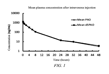
FIG. 1 shows a plasma drug concentration-time curve for pharmacokinetics
after single-dose intravenous injection of 0.1 mg/kg PAO or d5PAO in male SD
rats.
FIG. 2 shows a plasma drug concentration-time curve for pharmacokinetics
after single-dose oral lavage of 0.2 mg/kg PAO or d5PAO in male SD rats.
FIG. 3 shows a vector map of an a-synuclein overexpression plasmid.
FIG. 4 shows a standard preparation diagram of a-synuclein ELISA.
FIG. 5 shows inhibitory effects of d5PAO and PAO on apoptosis of SH-sy5y
cells. FIG. 5A shows effects of certain concentrations of d5PAO and PAO on
viability of SH-sy5y cells as detected by MTT (n = 5; mean SEM; One-way
ANOVA; ***p < 0.0001 vs. ctrl; and ### p < 0.0001 vs. ctrl). FIG. 5B shows
immunofluorescent staining images, wherein propidium iodide (PI) is added in
culture solution, and the mixture was co-incubated for 15 min, followed by
immunofluorescent staining of Ki67.
FIG. 6 shows effects of d5PAO and PAO on improving viability of
stably-transformed APP (SW) HEK293 cells and promoting AP release. FIG. 6A
shows effects of certain concentrations of d5PAO and PAO on viability of
stably-transformed APP (SW) HEK293 cells as detected by MTT (n = 5; mean
SEM; One-way ANOVA; and **p < 0.001, ***p <0.0001 vs. ctrl). FIG. 6B shows
AP contents in supernatants as detected by an ELISA kit, wherein AP values of
each group are calculated according to a standard curve. FIG. 6C shows fold
changes of AP contents in each group (data is normalized with ctrl as 1; n =
3;
mean SEM; One-way ANOVA; and *p <0.03, **p <0.001, ***p <0.0001 vs.
ctrl).
FIG. 7 shows comparisons of effects of deuterated compound PAO with a
structural formula on promoting AP release. FIG. 7A shows AP contents in
supernatants as detected by an ELISA kit, wherein AP values of each group are
CA 03171783 2022- 9- 14
11
calculated according to a standard curve. FIG. 7B shows fold changes of AP
contents in each group (data is normalized with ctrl as 1; n = 3; mean SEM;
One-way ANOVA; *p < 0.03, **p < 0.001, ***p < 0.0001 vs. ctrl; and ##p <
0.001, ###p <0.0001 vs. 50 nM d5PAO).
FIG. 8 shows effects of d5PAO and PAO on reducing damage of SH-sy5y
cells caused by a-synuclein overexpression and on promoting a-synuclein
release.
FIG. 8A shows effects of certain concentrations of d5PAO and PAO on viability
of
transiently-transformed a-synuclein cells as detected by MTT (n = 5; mean
SEM;
One-way ANOVA; and *p <0.03, **p <0.001, ***p < 0.0001 vs. ctrl). FIG. 8B
shows a-synuclein contents in supernatants as detected by an ELISA kit,
wherein
a-synuclein values of each group are calculated according to a standard curve
(n =
3; mean SEM; One-way ANOVA; and *p < 0.03 vs. Ctrl). FIG. 8C shows fold
changes of a-synuclein contents in each group (data is normalized with a-syn
OE
group as 1; n =3; mean SEM; One-way ANOVA; and *p < 0.03 vs. ctrl).
FIG. 9 shows protective effects of d5PAO and PAO in an SH-SY5Y cell
model constructed by CBE. FIG. 9A shows viability of SH-SY5Y cells treated
with CBE for 48 h, as detected by MTT. FIG. 9B shows viability of SH-SY5Y
cells that are firstly treated with 100 M of CBE for 24 h, then starved
(high-glucose DMEM free of FBS) while being treated with 100 M of CBE for
another 24 h, and finally treated with different concentrations of PAO for 24
h, as
detected by MTT. FIG. 9C shows viability of SH-SY5Y cells that are firstly
treated with 100 M of CBE for 24 h, then starved (high-glucose DMEM free of
FBS) while being treated with 100 M of CBE for another 24 h, and finally
treated
with different concentrations of d5PAO and PAO for 24 h, as detected by MTT (n
= 5; data is expressed as mean SEM; ###p <0.0001 vs. ctrl; and **p <0.001
vs.
100 M CBE, ***p <0.0001 vs. 100 M CBE).
FIG. 10 shows effects of PAO on inhibiting CBE-induced lysosome and
GlcCer storage and promoting GlcCer efflux. FIG. 10A shows immunofluorescent
staining images of Lyso-tracker, wherein SH-SY5Y cells are co-incubated with
lysosome trackers for 30 min, then the supernatant is removed, different
concentrations of PAO are added, the mixture was incubated for 10 min, and
CA 03171783 2022- 9- 14
12
Lyso-tracker is observed by immunofluorescent staining. FIG. 10B shows
statistical analysis of fluorescent intensity of Lyso-tracker in each group
(scale bar:
50 gm; n = 5; One-way ANOVA; ##p <0.001 vs. ctrl; and **p < 0.001, ***p <
0.0001 vs. 100 gM CBE). FIG. 10C shows statistical analysis of GlcCer
concentrations in cell lysates of each group according to LC/MS measurement
values. FIG. 10D shows statistical analysis of GlcCer concentrations in
supernatants of cell culture mediums of each group according to LC/MS
measurement values.
FIG. 11 shows effects of PI4Ka knockdown on promoting a reduction in
lysosomal storage. FIG. 11A shows PI4KIIIa protein levels as detected by
Western
blot, wherein SH-SY5Y cells are treated with different shRNA interfering
lentiviral vectors (sh-ctrl, shl-PI4Ka, sh2-PI4Ka and sh3-PI4Ka) for 48 h.
FIG.
11B shows statistical analysis results of Western blot. FIG. 11C shows
fluorescent
intensity as detected by immunofluorescent staining of Lyso-tracker after
treatment with shRNA interfering lentiviral vectors. FIG. 11D shows the
statistical
analysis (scale bar: 50 gm; n = 5; data is expressed as mean SEM; One-way
ANOVA; and ***p <0.0001).
FIG. 12 shows a pGMLV-SC5RNAi vector map.
FIG. 13 shows bright-field images of MRC-5 cells treated for 24 h, wherein
the MRC-5 cells are cultured in MEM (FBS-free, containing 5 ng/mL TGF-01 and
different concentrations of PAO or d5PAO according to groups) for 24 h (scale
bar:
50 gm).
FIG. 14 shows inhibitory effects of d5PAO and PAO on expression of
a-SMA and Calponinl in MRC-5 cell models. FIG. 14A shows expression levels
of a-SMA and Calponinl as detected by Western blot. FIG. 14B shows statistical
analysis results of expression levels of a-SMA in each group. FIG. 14C shows
statistical analysis results of expression levels of Calponinl in each group
(the
protein signal intensity is analyzed by Image J software, with the signal
intensity
of ctrl as 1; n = 3; One-way ANOVA; data is expressed as mean SEM; *p <0.03,
**p < 0.001, ***p < 0.0001 vs. 5 ng/mL TGF-01; and #p < 0.03 vs. ctrl).
FIG. 15 shows inhibitory effects of d5PAO and PAO on expression of
CA 03171783 2022- 9- 14
13
a-SMA and Calponinl in MRC-5 cell models. FIG. 15A shows
immunofluorescent staining images of a-SMA in each group (red: a-SMA, blue:
DAPI (nuclei), and scale bar: 50 gm). FIG. 15B shows immunofluorescent
staining images of Calponinl in each group (red: Calponinl, blue: DAPI
(nuclei),
and scale bar: 50 gm). FIGs. 15C and 15D show statistical analysis results of
immunofluorescent intensity of a-SMA and Calponinl, respectively (with the
mean value of ctrl as 1; n = 5; One-way ANOVA; data is expressed as mean
SEM; *p < 0.03, **p < 0.001, ***p < 0.0001 vs. 5 ng/mL TGF-01; and ###p <
0.0001 vs. ctrl).
FIG. 16 shows regulatory effects of d5PAO and PAO on expression of
Calponinl in MSC. FIG. 16A shows immunofluorescent staining images of
Calponinl in each group (red: Calponinl, blue: DAPI (cell nuclei), and scale
bar:
50 gm). FIG. 16B shows statistical analysis results of immunofluorescent
intensity
of Calponinl (n =4; One-way ANOVA; and data is expressed as mean SEM).
FIG. 17 shows inhibitory effects of d5PAO and PAO on secretion of COL1 in
MRC-5 cells during fibrosis. FIG. 17A shows COL1 concentrations in
supernatants of each group as detected by ELISA (n = 6, and data is expressed
as
mean SEM). FIG. 17B shows statistical analysis of COL1 concentrations in
supernatants of each group (with the mean value of ctrl as 1; data is
expressed as
mean SEM; *p <0.03, **p < 0.001, ***p < 0.0001 vs. 5 ng/mL TGF-01; and #p
<0.0001 vs. ctrl).
FIG. 18 shows effects of shRNA interfering lentiviral vectors on reducing
expression of PI4KIIIa, wherein the shRNA interfering lentiviral vectors are
co-incubated with MRC-5 cells for 48 h, and proteins are collected for Western
blot. FIG. 18A shows detection of PI4KIIIa proteins. FIG. 18B shows data
analysis results of Image J software (data is normalized with sh-ctrl as 1 and
expressed as mean SEM; n =3; and ***p <0.0001 vs. sh-ctrl).
FIG. 19 shows inhibitory effects of PI4Ka knockdown on expression of
Calponinl and a-SMA in MRC-5 cells treated with TGF-01, wherein after the
MRC-5 cells are adhered to the wall, lentiviral vectors with different
sequences
are added, and the mixture is cultured for 24 h and treated with or without 5
ng/mL
CA 03171783 2022- 9- 14
14
TGF- (31 according to groups for 24 h, and immunofluorescent staining is
performed prior to observation. FIG. 19A shows immunofluorescent staining
images of a-SMA in each group (red: a-SMA, blue: DAPI (nuclei), green: green
fluorescent protein (GFP), and scale bar: 50 gm). FIG. 19B shows
immunofluorescent staining images of calponinl in each group (red: Calponinl,
blue: DAPI (cell nuclei), green: green fluorescent protein (GFP), and scale
bar: 50
gm). Statistical analysis results of immunofluorescent intensity of a-SMA
(FIG.
19C) and Calponinl are showed (data is normalized with the mean value of sh-
ctrl
as 1 and expressed as mean SEM; n = 5; *p <0.03, ***p <0.0001 vs. sh-ctr1+5
ng/mL TGF-(31; and ##p <0.001, ###p < 0.0001 vs. sh-ctrl).
FIG. 20 shows inhibitory effects of d5PAO and PAO on secretion of IL-6 and
TNF-a in BV2 cell inflammatory models. FIG. 20A shows TNF-a concentrations
in supernatants of BV2 cells as detected by ELISA, wherein TNF-a contents
(pg/mL) are calculated according to a standard curve. FIG. 20B shows relative
concentration changes of TNF-a in each group (with the mean concentration of
ctrl as 1). FIG. 20C shows IL-6 concentrations in supernatants of BV2 cells as
detected by ELISA, wherein IL-6 contents (pg/mL) are calculated according to a
standard curve. FIG. 20D shows relative concentration changes of IL-6 in each
group (with the mean concentration of ctrl as 1; n = 3; One-way ANOVA; data is
expressed as mean SEM; *p < 0.03, **p < 0.001, ***p < 0.0001 vs. 1 gg/mL
LPS; and #p <0.03, ##p <0.001 vs. ctrl).
FIG. 21 shows inhibitory effects of PAO on breast cancer.
FIG. 22 shows inhibitory effects of PAO on lymphoma.
FIG. 23 shows inhibitory effects of d5PAO on melanoma.
FIG. 24 shows inhibitory effects of d5PAO on melanoma on day 28 of
administration.
FIG. 25 shows comparison of effects of high-dose PAO and d5PAO via
lavage on weights and survival rates of mice.
FIG. 26 shows effects of PAO on weights of breast cancer mouse models.
FIG. 27 shows effects of PAO on weights of pancreatic cancer models.
FIG. 28 shows effects of PAO on weights of lymphoma animal models.
CA 03171783 2022- 9- 14
FIG. 29 shows effects of d5PAO in a combined administration on weights of
mice with melanoma.
FIG. 30 shows inhibitory effects of PAO and d5PAO on HCoV229E
(influenza coronavirus).
FIG. 31 shows anti-anxiety effects of PAO and d5PAO, wherein d5PAO has a
more significant anti-anxiety effect than PAO.
FIG. 32 shows anti-depression effects of PAO and d5PAO, wherein d5PAO
has a more significant and stable anti-depression effect than PAO.
FIG. 33 shows inhibitory effects of d5PAO and PAO on cholesterol storage
caused by U18666A (scale bar: 50 gm).
FIG. 34 shows effects of PAO on promoting expression of LC3B and p62
and effects of Baf-Al on blocking protection of PAO in a cell model. FIG. 34A
shows LC3B and p62 proteins as detected by Western blot. FIGs. 34B and 34C
show statistical analysis of signal intensity of LC3B and p62 proteins as
analyzed
by Image J software. FIG. 34D shows immunofluorescent staining images of
LC3B and p62 (red: p62, green: LC3B, and scale bar: 50 gm). FIG. 34E shows
cell viability in each group as detected by MTT and the statistical analysis
(n = 5;
data is expressed as mean SEM; One-way ANOVA; ###p < 0.0001 vs. ctrl; and
**p <0.001 vs. 100 gM CBE).
FIG. 35 shows effects of PI4Ka knockdown on activation of ALP. FIGs. 35A
and 35B show LC3B protein levels as detected by Western blot, wherein
SH-SY5Y cells are treated with different shRNA interfering lentiviral vectors
(sh-ctrl, hl-PI4Ka, sh2-PI4Ka and sh3-PI4Ka) for 48 h, and the statistical
analysis.
FIGs. 35C and 35D show LC3B protein levels, wherein the cells are transfected
by
shRNA interfering lentiviral vectors while receiving CBE treatment for 48 h of
co-incubation, and the statistical analysis (n = 3; data is expressed as mean
SEM;
One-way ANOVA; and *p <0.03 vs. sh-ctrl).
FIG. 36 shows percentages of acetylcholine-induced enhanced pause (Penh)
values relative to a baseline.
FIG. 37 shows total counts of eosinophils, macrophages, neutrophils and
lymphocytes in bronchoalveolar lavage fluid (BALF) (T.Test; one-tailed; *
<0.5,
CA 03171783 2022- 9- 14
16
** <0.1 vs. the total cell content of the model group).
FIG. 38 shows respective counts of eosinophils, macrophages, neutrophils
and lymphocytes in bronchoalveolar lavage fluid (BALF) (T.Test; one-tailed; *
<
0.5, ** <0.1 vs. the BALF of the model group).
FIG. 39 shows collagen type I contents in plasma (taking a normal group as
ctrl).
FIG. 40 shows comparison of effects of PAO and dPAO with nintedanib (a
positive control drug) on the down-regulation of hyaluronic acid in plasma of
mice
with pulmonary fibrosis (taking a normal group as ctrl).
Detailed Description of The Invention
Hereinafter, the present invention will be described in detail according to
the
embodiments and in conjunction with the accompanying drawings. The above
aspects of the present invention and other aspects of the present invention
will be
apparent from the following detailed description. The scope of the present
invention is not limited to the following embodiments.
The term "compound" as used herein is intended to include all stereoisomers
(e.g., enantiomers and diastereomers), geometric isomers, tautomers and
isotopes
of the indicated structure.
In another aspect, the present invention relates to deuterated
oxophenylarsine,
preferably wherein all hydrogen atoms on a benzene ring are substituted with
deuterated isotopes.
The compound described herein can be asymmetric (e.g., having one or more
stereocenters). Unless otherwise indicated, all stereoisomers, such as
enantiomers
and diastereomers, are intended to be included. The compound described herein
may have various geometric isomers involving, for example, olefins and
carbon-carbon double bonds, and all the stable isomers have been considered
herein. Cis- and trans-geometric isomers of the compound are described herein
and may be isolated in the form of an isomer mixture or an individual isomer.
The compound described herein also includes a tautomeric form. The
tautomeric form results from the exchange of a single bond with an adjacent
CA 03171783 2022- 9- 14
17
double bond accompanied by the migration of protons. The tautomeric form
includes proton tautomers in isomeric protonation states that have the same
chemical formula and total charge. Examples of the proton tautomers include
keto-enol, amide-imidic acid, lactam-lactim, enamine-imine and cyclic forms,
wherein protons may occupy two or more positions (such as 1H- and
3H-imidazole, 1H-, 2H- and 4H-1,2,4-triazole, 1H- and 2H-isoindole, and 1H-
and
2H-pyrazole) of a heterocyclic system. The tautomeric form can be equilibrated
or
sterically locked into one form by means of suitable substitution.
In certain embodiments, the small-molecule compound described herein can
be obtained by means of organic synthesis. The compound described herein,
including a salt, ester, hydrate or solvate thereof, can be prepared by means
of any
well-known organic synthesis technique and can be synthesized according to a
variety of possible synthetic routes.
The term "oxophenylarsine" (PAO) as used herein refers to a small-molecule
compound with the following specific chemical structure:
-,0
As
Disease related to intracellular protein misfolding
The term "disease related to intracellular protein misfolding" as used herein
refers to a disease characterized by aggregation of abnormally folded proteins
in
the cytoplasm and is also diagnosed as protein aggregation (accumulation) or
protein misfolding disease. In addition, the term "disease related to
intracellular
protein misfolding" also includes some intracellular inclusion body diseases,
such
as a disease with protein aggregation and inclusion body formation, wherein
such
inclusion body is mainly formed by aggregation of a core protein due to
misfolding, with attachment of various stress proteins involved in responding
to
unfolded proteins.
The disease related to intracellular protein misfolding includes, but is not
limited to, Parkinson's disease (PD), Lewy body dementia (LBD), multiple
system
atrophy (MSA), inclusion body myositis (IBM), frontotemporal dementia (FTD),
CA 03171783 2022- 9- 14
18
Huntington's disease (HD), a polyamine disease (PolyQ), amyotrophic lateral
sclerosis (ALS) and a prion disease.
Lysosomal storage disease
The term "lysosomal storage disease" as used herein refers to a disease
caused by the accumulation of some endogenous or exogenous substances in
lysosomes for various reasons, including but not limited to, lysosomal
function
deficiency caused by insufficient enzyme activity in lysosomes and the lack of
processing and correction enzymes for activator proteins, transport proteins
or
lysosomal proteins, and due to the lysosomal function deficiency, the
corresponding substrate is unable to be digested in secondary lysosomes and
thus
is accumulated, and a metabolic disorder occurs to lead to a storage disease,
etc.
The lysosomal storage disease includes, but is not limited to, a sphingolipid
metabolism disorder, mucopolysaccharidosis, a glycogen storage disease, a
glycoprotein storage disease, a lipid storage disease, post-translational
modification deficiency, an integral membrane protein deficiency disorder,
neuronal ceroid lipofuscinosis or a disorder of lysosome-related organelles.
Among them, the sphingolipid metabolism disorder includes, but is not limited
to,
Fabry disease, dermotosis of metabolism disturbance (Farbe disease), Gaucher
disease types I, II and III and perinatal lethal Gaucher disease, GM1
gangliosidosis types I, II and III, GM2 gangliosidosis (amaurotic familial
idiocy),
GM2 gangliosidosis, globoid cell leukodystrophy (Krabbe disease),
metachromatic leukodystrophy and Niemann-Pick types A and B; the
mucopolysaccharidosis includes, but is not limited to, Hurler-Scheie syndrome
and Scheie syndrome (ML I), Hunter syndrome (MPS II), Sanfilippo syndrome A
(MPS IIIA), Sanfilippo syndrome B (MPS IIIB), Sanfilippo syndrome C (MPS
IIIC), Sanfilippo syndrome D (MPS IIID), eccentro-osteochondrodysplasia
syndrome (MPS IVA), eccentro-osteochondrodysplasia syndrome (MPS IVB),
mucopolysaccharidosis type VI (Maroteaux-lamy syndrome, MPS VI), Sly disease
(MPS VII) and MPS IX; the glycogen storage disease includes, but is not
limited
to, rare Pompe disease (GSD II); the glycoprotein storage disease includes,
but is
not limited to, alpha-mannosidosis, beta-mannosidosis, fucosidosis,
CA 03171783 2022- 9- 14
19
aspartylglucosaminuria, Schindler disease type I (infantile neuroaxonal
dystrophy),
Schindler disease type II (Kanzaki disease), Schindler disease type III
(intermediate severe), sialidosis type I (cherry-red spot-myoclonus syndrome),
sialidosis type II (mucopolysaccharidosis I) and galactosialidosis; the lipid
storage
disease includes, but is not limited to, acid lipase deficiency such as
Wolman's
disease and cholesteryl ester storage disease; the post-translational
modification
deficiency includes, but is not limited to, multiple sulfatase deficiency,
mucolipidosis type IIa/13 (I-cell disease), mucolipidosis type IIa/r3 (pseudo-
Hurler
syndrome), and mucolipidosis type Illy (pseudo-Hurler syndrome variant); the
integral membrane protein deletion disorder includes, but is not limited to,
homocystinuria, Danon disease, a myoclonus-renal failure syndrome, sialidosis
(such as ISSD, Salla disease and intermediate severe Salla disease), Niemann-
Pick
disease types Cl and C2 and mucolipidosis type IV; the neuronal ceroid
lipofuscinosis includes, but is not limited to, ceroid lipofuscinosis type 1
(Haltia-Santavuori disease and INCL), neuronal ceroid lipofuscinosis type 2
(Jansky-Bielschowsky disease), ceroid lipofuscinosis
type 3
(Batten-Spielmeyer-Sjogren disease), ceroid lipofuscinosis type 4 (Parry
disease
and Kufs disease types A and B), ceroid lipofuscinosis type 5 (finnish variant
late
infantile), ceroid lipofuscinosis type 6 (Lake-Cavanagh or Indian variant),
ceroid
lipofuscinosis type 7 (Turkish variant), ceroid lipofuscinosis type 8
(Northern
epilepsy and epilepsy-intellectual disability), ceroid lipofuscinosis type 9,
ceroid
lipofuscinosis type 10, ceroid lipofuscinosis type 11, ceroid lipofuscinosis
type 12,
ceroid lipofuscinosis type 13 and ceroid lipofuscinosis type 14; and the
disorder of
lysosome-related organelles includes, but is not limited to, Hermansky-Pudlak
syndrome type 1, Hermansky-Pudlak syndrome type 2, Hermansky-Pudlak
syndrome type 3, Hermansky-Pudlak syndrome type 4, Hermansky-Pudlak
syndrome type 5, Hermansky-Pudlak syndrome type 6, Hermansky-Pudlak
syndrome type 7, Hermansky-Pudlak syndrome type 8, Hermansky-Pudlak
syndrome type 9, Griscelli syndrome type 1 (Elejalde syndrome), Griscelli
syndrome type 2 and Chediak-Higashi disease.
Drug administration and medical use
CA 03171783 2022- 9- 14
The term "pharmaceutically acceptable" as used herein refers to those
compounds, materials, compositions and/or dosage forms that are, within the
scope of reasonable medical judgment, suitable for use in contact with human
and
animal tissues without undue toxicity, irritation, allergic response or other
problems or complications, and are commensurate with a reasonable benefit/risk
ratio. In certain embodiments, pharmaceutically acceptable compounds,
materials,
compositions and/or dosage forms are those approved by a regulatory agency
(e.g.,
the United States Food and Drug Administration, the China Food and Drug
Administration, or the European Medicines Agency) or listed on the generally
accepted pharmacopoeia (such as the United States Pharmacopoeia, the Chinese
Pharmacopoeia, or the European Pharmacopoeia) and used for animals (more
particularly, humans).
The term "object" as used herein may include a human and a non-human
animal. The non-human animal includes all vertebrates, such as a mammal and a
non-mammal. The "object" also may be livestock (e.g., a cow, a pig, a sheep, a
chicken, a rabbit or a horse), a rodent (e.g., rat or mouse), a primate (e.g.,
a gorilla
or a monkey) or a domestic animal (e.g., a dog or a cat). The "object" may be
a
male or female object, and also may be of different ages. The human "object"
may
be Caucasian, African, Asian, Sumerian or other races, or a hybrid of
different
races. The human "object" may be an old person, an adult, an adolescent, a
child
or an infant.
In some embodiments, the object described herein is a human or non-human
primate.
The deuterated oxophenylarsine disclosed herein can be administered by
means of administration routes known in the art, such as an injection (e.g.,
subcutaneous injection, intraperitoneal injection, intravenous injection
(including
intravenous drip or infusion), intramuscular injection or intradermal
injection)
administration or a non-injection administration (e.g., oral administration,
nasal
administration, sublingual administration, rectal administration or topical
administration). In some embodiments, the deuterated oxophenylarsine described
herein is administered orally, subcutaneously, intramuscularly or
intravenously. In
CA 03171783 2022- 9- 14
21
some embodiments, the deuterated oxophenylarsine described herein is
administered orally.
As used herein, the term "therapeutically effective amount" refers to an
amount of a drug that can alleviate or eliminate a disease or symptom in an
object
or prophylactically inhibit or prevent the occurrence of a disease or symptom.
The
therapeutically effective amount may be an amount of a drug that alleviates
one or
more diseases or symptoms in an object to a certain extent; an amount of a
drug
that can partially or fully restore one or more physiological or biochemical
parameters associated with the cause of the disease or symptom to normal;
and/or
an amount of a drug that can reduce the possibility of the onset of a disease
or
symptom. In some embodiments, the term "therapeutically effective amount" as
used herein refers to an amount of a drug that can alleviate or eliminate a
disease
related to intracellular protein misfolding or a lysosomal storage disease in
an
object.
The therapeutically effective amount of deuterated oxophenylarsine provided
herein depends on a variety of factors known in the art, such as weight, age,
medical history, current treatment received, object's health condition, the
strength
of drug-drug interaction, allergies, hypersensitivity and side effects, as
well as
administration routes and the extent of disease progression. A person skilled
in the
art (such as a doctor or a veterinarian) may reduce or increase the dose
according
to these or other conditions or requirements.
In some embodiments, the treatment further comprises administering a
second agent to an object in need thereof.
In some embodiments, the second agent is an agent for treating a disease
related to intracellular protein misfolding, and includes, but is not limited
to,
levodopa and riluzole.
In some embodiments, the deuterated oxophenylarsine is administered prior
to, subsequent to, or concurrently with the second agent.
The present application also relates to a method for preventing or treating a
disease related to intracellular protein misfolding, the method comprising
administering an effective amount of deuterated oxophenylarsine to an object
in
CA 03171783 2022- 9- 14
22
need thereof.
The present application also relates to a method for preventing or treating a
lysosomal storage disease, the method comprising administering an effective
amount of deuterated oxophenylarsine to a subject in need thereof.
Example 1. Synthesis and physical properties of compounds
1. Synthesis and physical properties of d5PAO (penta-deuterated
oxophenylarsine)
1.1 Synthesis of d5PAO
The synthetic route of d5PAO is as follows:
NH2 0 OH 0
As
As
01-6
D A D
H
D 60% 470/ ,
d5-Aniline d5-PA d5PAO
Step 1: synthesis of d5-PA:
DD NH2 HOAs0
0113
60%
d5-Aniline d5-PA
91.35 mL of water and 18.27 g of penta-deuterated aniline (d5-Aniline) were
added to a three-necked flask, stirred and cooled to 0-10 C. 37.45 mL of
concentrated hydrochloric acid was added dropwise. After that, an aqueous
solution of sodium nitrite (13.43 g of solid sodium nitrite dissolved in 36.5
mL of
water) was added dropwise, and the temperature was maintained below or at 5 C
for about 2-3 h to complete the preparation of a diazonium salt.
274 mL of purified water, 69.06 g of sodium carbonate, 36.82 g of arsenic
trioxide and 2.83 g of copper sulfate pentahydrate (CuSO4=5H20) were added to
another container, heated to 90 C-100 C, stirred thermostatically for 30 mm,
and
then cooled to 5 C-15 C. The diazonium salt prepared above was slowly added in
batches, and the temperature was controlled to be lower than 15 C. The
resulting
mixture was stirred for 2-3 h, then naturally heated to room temperature,
stirred
CA 03171783 2022- 9- 14
23
overnight and filtered. The filter cake was rinsed with water. The filtrate
was
combined. The pH value of the system was adjusted to 3.0 by slowly adding
concentrated hydrochloric acid, with a small amount of brown floccules
precipitated. Suction filtration was performed. The filtrate was washed three
times
with 100 mL of ethyl acetate, and the aqueous phase was concentrated under
reduced pressure at 50 C-60 C to about 170 mL, with a large amount of white
solids precipitated. Suction filtration was performed. The filter cake was
rinsed
with cold water and dehumidified, and the solid was directly dried at 50 C in
a
blast oven for 18 h to obtain 35 g of d5-PA as an off-white solid (yield:
90.83%,
MS ES+(m/z): 208.0 [(M+H)+]).
Step 2: Synthesis of d5-PAO:
0 As 0 As
1 0 I-6
D D D
__________________________________ ..-
-L 47%
D 'r D D D
D D
d5-PA d5PAO
400 mL of purified water, 25 g of d5-PA, 75.4 g of sodium bisulfite, 0.32 g of
potassium iodide, and 125 mL of methanol were added to a three-necked flask
and
stirred overnight at 30 C-40 C. The reaction was terminated until most of the
raw
materials were reacted completely. The temperature of the resulting solution
was
controlled below 30 C, and the pH value was adjusted to 7.0 by adding
concentrated hydrochloric acid. The mixture was extracted with ethyl acetate
for 4
times. Then, the organic phases were combined and concentrated at 35 C-45 C to
dryness to obtain 20 g of wet white solid. The solid was heated to reflux with
240
mL of tert-butyl methyl ether for 1 h and then cooled. Suction filtration was
performed. The filter cake was washed with cold MTBE and dried at 50 C in a
blast oven overnight to obtain 8.7 g of d5PAO as a white solid CC-NMR (6,
DMSO-d6): 149.9, 129.6, 129.4, 128.2, MS ES+(m/z): 173.99 [(M+H)+]).
1.2 Physicochemical properties of d5PAO
1.2.1 Instruments
Agilent1260Prime high performance liquid chromatograph, Mettler Toledo
CA 03171783 2022- 9- 14
24
XS105 balance (0.01 mg), KQ5200B ultrasonic instrument (Kunshan Ultrasonic
Instruments Co., Ltd.), BR2000-GM variable speed oscillator (VWR
International)
and 0.45-gm filter membrane (Shanghai Qingyang Biotechnology Co., Ltd.).
1.2.2 Experimental drugs
d5PAO (purity: 97.9%), acetonitrile (chromatographically pure, Sinopharm
Chemical Reagent Co., Ltd.), and hydrochloric acid and DMSO (analytically
pure,
Sinopharm Chemical Reagent Co., Ltd.).
1.2.3 Solution preparation
2 mL of 0.1 M hydrochloric acid solution was taken and diluted with
deionized water to 20 mL to obtain a 0.01 M hydrochloric acid solution, and
the
pH was determined as 2 with a precision pH test paper.
2 mL of 0.01 M hydrochloric acid solution was taken and diluted with
deionized water to 20 mL to obtain a 0.001 M hydrochloric acid solution.
2 mL of 0.001 M hydrochloric acid solution was taken and diluted with
deionized water to 20 mL to obtain a 0.0001 M hydrochloric acid solution, and
the
pH of the solution was determined as 4 by a precision pH test paper.
mL of deionized water was taken, and the pH was determined as 6.
7.5 mg of d5PAO was taken into a centrifuge tube, followed by adding 1 mL
of DMSO. The resulting mixture was ultrasonically oscillated for dissolution
and
20 diluted to 10 mL with an acetonitrile/water (1/1) mixed liquid to obtain a
0.75
mg/mL d5PAO stock solution. The d5PAO stock solution was diluted into
different d5PAO working solutions (0.3 mg/mL, 0.15 mg/mL, 0.075 mg/mL, 0.03
mg/mL and 0.015 mg/mL).
1.2.4 Chromatographic conditions
Liquid chromatograph: Agilent 1260 Infinity II Prime ultra-high pressure
liquid chromatography system.
Chromatographic column: ACQUITY UPLCS Peptide C18130A 2.1 * 100
mm ID., 1.7 gm (Waters).
Mobile phase A: water: ACN (v : v, 95 : 5) solution containing 0.01% AA
and 2 mmol/L NH40Ac.
Mobile phase B: water: ACN (v : v, 5 : 95) solution containing 0.01% AA
CA 03171783 2022- 9- 14
and 2 mmol/L NH40Ac.
Elution gradient:
Time (min) flow rate (mL/min) A (%) B (%)
Initial 0.400 95 5
2.00 0.400 2 98
3.00 0.400 2 98
3.20 0.400 95 5
4.00 0.400 95 5
Column temperature: 40 C
Feeding volume: 4 L
Detection wavelength: 254 nm
1.2.5 Methodological investigation
Investigation of linear relationship
Different d5PAO solutions (0.75 mg/mL, 0.3 mg/mL, 0.15 mg/mL, 0.075
mg/mL, 0.03 mg/mL and 0.015 mg/mL) were taken and measured according to the
above chromatographic conditions, and the chromatograms were recorded. A
linear regression of peak area against sample concentration was performed, and
a
regression equation was obtained:
y = 1647.7x + 0.9623, R2 = 0.9999
The results showed that the d5PAO sample concentration was in the range of
0.015-0.75 mg=mL-1 and had a good linear relationship with the peak area.
1.2.6 Determination of equilibrium solubility
Different pH buffer solutions were pipetted (2 mL each) into 3-mL centrifuge
tubes. Excess d5PAO powder was added. Until a large amount of white insoluble
precipitates appeared in the solution, the resulting mixture was
ultrasonically
treated for 30 min, put in a thermostatic oscillator, shaken at 25 C for 24 h,
then
ultrasonically treated for another 30 min and filtered with a 0.45- m filter
membrane. The subsequent filtrate was pipetted, 10-fold diluted with water and
measured according to the above chromatographic conditions, and the
chromatograms were recorded. The solubility of d5PAO in different pH buffers
was calculated as 5.36 mg/mL, 5.39 mg/mL and 5.98 mg/mL, respectively.
CA 03171783 2022- 9- 14
26
Table 1. Solubility of d5PAO
Drug pH Peak area Corresponding
solubility (mg/mL)
d5PAO 6 884.20 5.36
d5PAO 4 889.51 5.39
d5PAO 2 986.70 5.98
2. Synthesis and physical properties of PAO
2.1 Synthesis of PAO
Synthesis route:
0, ,OH
NH2 N2BF4 'As,OH (:)As
48% HBF4, acetone Na2CO3 CuSO4, As203 i) HCI, KI,
S02, Me0H.-
ft NaNO2, H20 ii) NaOH
H20, Et0H iii) HCI
85% 82%
Aniline PA 60% PAO
Step 1:
NH2 N2BF4
0 48% HBF4, acetone
NaNO2, H20
85%
Aniline (10.0 g, 107 mmol, 1.0 eq.) and acetone (20 mL) were added to a
250-mL single-neck flask and cooled to about -15 C (high temperature causes a
product to be dark), followed by slowly adding 48% HBF4 (30 mL, 29.5 g, 161
mmol, 1.5 eq.). NaNO2 (11.0 g, 161 mmol, 1.5 eq.) was dissolved in H20 (20
mL),
and the mixture was slowly added dropwise to the above solution. After the
addition, the temperature was maintained at -15 C for 2 h, followed by
stirring the
reaction at 0 C for about 1 h. The solid (white) was filtered and washed with
isopropyl ether (50 mL x 2). The product was dried under vacuum at 30 C
(weight:
17.5 g, yield: 85%, pink solid) and directly used in the next step.
Step 2:
0õOH
N2BF4 OH
1 Na2CO3 CuSO4, As203
H20, Et0H
82%
PA
Na2CO3 (21.2 g, 200.6 mmol, 3.85 eq.), As203 (11.3 g, 57.3 mmol, 1.1 eq.),
CA 03171783 2022- 9- 14
27
CuSO4-5H20 (800 mg, 3.13 mol, 0.06 eq.) and H20 (60 mL) were added to a
250-mL flask. The suspension was heated to about 90 C for about 20 min so that
most of the solids can be dissolved. The solution was cooled to about 0-15 C.
A
suspension of azobenzene fluoroborate (10.0 g, 52.1 mmol, 1.0 eq.) and H20 (60
mL) was slowly added in batches, and a small amount of acetone was added to
reduce the foam aggregation. After the addition, the mixture was stirred at
room
temperature for about 12 h, filtered with diatomite and washed with H20 (20
ini, x
3), wherein if the filtrate is too dark, activated carbon can be added for
decolorization prior to filtering. Concentrated hydrochloric acid (12 N, 40
mL)
was added to the filtrate for neutralization, and the reaction liquid was
further
adjusted to be acidic. The filtrate was concentrated to about 50 mL. The solid
was
filtered and washed once with cold water. The filtrate was concentrated to
produce
solids. The solids were filtered and washed once with cold water. All the
solids
were combined and dried (white solid, weight: 8.6 g, yield: 82%, MS ES+(m/z):
202.9 [(M+H)+]).
Step 3:
HO,As0 Cl'As
OH
1) HCI, KI, S02, Me0H.
li ii) NaOH
In) HCI
PA 60% PAO
SO2 was passed to a mixture of phenylarsenic acid (8.0 g, 40 mmol, 1.0 eq.),
methanol (40 mL), concentrated hydrochloric acid (15 mL) and KI (catalyst
amount, 50 mg) until saturation, and the resulting mixture was stirred at room
temperature for 2 h. A NaOH solution (2N) was added to the mixture until the
solution was transparent. Then, concentrated hydrochloric acid was added for
neutralization. A white solid was precipitated out, filtered, recrystallized
with
water/ethanol (1/5) and dried (weight: 4.0 g, yield: 60%, white powdery solid,
'11
NMR (6, CDC13): 7.41-7.62 (m, 3H), 7.75-7.78 (m, 2H). MS ES + (m/z): 168.8
[(M+H)+]).
2.2 Physicochemical properties of PAO
PAO has a molecular formula of C6H5As0 and a molecular weight of 168.03.
2.2.1 Instruments
CA 03171783 2022- 9- 14
28
Agilent1260Prime high performance liquid chromatograph, Mettler Toledo
XS105 balance (0.01 mg), KQ5200B ultrasonic instrument (Kunshan Ultrasonic
Instruments Co., Ltd.), BR2000-GM variable speed oscillator (VWR
International)
and 0.45-gm filter membrane (Shanghai Qingyang Biotechnology Co., Ltd.).
2.2.2 Experimental drugs
PAO (purity: 98%), acetonitrile (chromatographically pure, Sinopharm
Chemical Reagent Co., Ltd.), and hydrochloric acid and DMSO (analytically
pure,
Sinopharm Chemical Reagent Co., Ltd.).
2.2.3 Solution preparation
2 mL of 0.1 M hydrochloric acid solution was taken and diluted with
deionized water to 20 mL to obtain a 0.01 M hydrochloric acid solution, and
the
pH was determined as 2 with a precision pH test paper.
2 mL of 0.01 M hydrochloric acid solution was taken and diluted with
deionized water to 20 mL to obtain a 0.001 M hydrochloric acid solution.
2 mL of 0.001 M hydrochloric acid solution was taken and diluted with
deionized water to 20 mL to obtain a 0.0001 M hydrochloric acid solution, and
the
pH of the solution was determined as 4 by a precision pH test paper.
mL of deionized water was taken, and the pH was determined as 6.
7.5 mg of PAO was taken into a centrifuge tube, followed by adding 1 mL of
20 DMSO. The resulting mixture was ultrasonically oscillated for dissolution
and
diluted to 10 mL with an acetonitrile/water (1/1) mixed liquid to obtain a
0.75
mg/mL PAO stock solution. The PAO stock solution was diluted into different
PAO working solutions (0.3 mg/mL, 0.15 mg/mL, 0.075 mg/mL, 0.03 mg/mL and
0.015 mg/mL).
2.2.4 Chromatographic condition
Liquid chromatograph: Agilent 1260 Infinity II Prime ultra-high pressure
liquid chromatography system.
Chromatographic column: ACQUITY UPLCS Peptide C18130A 2.1 * 100
mm ID., 1.7 gm (Waters).
Mobile phase A: water: ACN (v : v, 95 : 5) solution containing 0.01% AA
and 2 mmol/L NH40Ac.
CA 03171783 2022- 9- 14
29
Mobile phase B: water: ACN (v : v, 5 : 95) solution containing 0.01% AA
and 2 mmol/L NH40Ac.
Elution gradient:
Time (min) flow rate (mL/min) A (%) B (%)
Initial 0.400 95 5
2.00 0.400 2 98
3.00 0.400 2 98
3.20 0.400 95 5
4.00 0.400 95 5
Column temperature: 40 C
Feeding volume: 4 !IL
Detection wavelength: 254 nm
2.2.5 Methodological investigation
Investigation of linear relationship
Different PAO solutions (0.75 mg/mL, 0.3 mg/mL, 0.15 mg/mL, 0.075
mg/mL, 0.03 mg/mL and 0.015 mg/mL) were taken and measured according to the
above chromatographic conditions, and the chromatograms were recorded. A
linear regression of peak area against sample concentration was performed, and
a
regression equation was obtained:
y = 1834.8x - 4.2355 R2 = 1
The results showed that the PAO sample concentration was in the range of
0.015-0.75 mg=mL-1 and had a good linear relationship with the peak area.
2.2.6 Determination of equilibrium solubility
Different pH buffer solutions were pipetted (2 mL each) into 3-mL centrifuge
tubes. Excess PAO powder was added. Until a large amount of white insoluble
precipitates appeared in the solution, the resulting mixture was
ultrasonically
treated for 30 min, put in a thermostatic oscillator, shaken at 25 C for 24 h,
then
ultrasonically treated for another 30 min and filtered with a 0.45-gm filter
membrane. The subsequent filtrate was pipetted, 10-fold diluted with water and
measured according to the above chromatographic conditions, and the
chromatograms were recorded. The solubility of PAO in different pH buffers was
CA 03171783 2022- 9- 14
calculated as 1.15 mg/mL, 3.38 mg/mL and 4.52 mg/mL, respectively.
Table 2. Solubility of PAO
Drug pH Peak area Corresponding
solubility (mg/mL)
PAO 6 207.25 1.15
PAO 4 616.40 3.38
PAO 2 824.35 4.52
3. Synthesis and physicochemical properties of deuterated compounds
d1PAO, d2PAO and d3PAO
3.1 Preparation of d1PAO
Na
Br ,/¨N H2 __________ D NH2
Me0D,D20
Under nitrogen, parabromoaniline (3.44 g) was dissolved in deuterated
methanol (5 mL), and the reaction mixture was refluxed for 30 min, spin-dried
3
times and then dehumidified for use. 3 g of metallic sodium was added to
deuterated methanol (10 mL) in batches, and after the reaction was completed,
heavy water (30 mL) was slowly added to obtain a 10% solution of sodium
deuteroxide in heavy water. Under nitrogen, the product in step 1 was
dissolved in
deuterated methanol (5 mL). Zinc powder (6.5 g) and the prepared 10% solution
of sodium deuteroxide in heavy water were added. The resulting mixture was
heated to reflux for 3 h, cooled to room temperature after parabromoaniline
disappeared as detected by TLC, extracted with ether and spin-dried at low
temperature to obtain a product, p-deuterated aniline.
37% HCI, H20 NH2 DN2 = HCI Na2CO3, CuSO4, As
D,?03 9
Ais-OH
NaNO2, H20 H20
OH
The p-deuterated aniline (0.94 g) obtained in the previous step and water (5
mL) were added to a round-bottomed flask with a magnetic stirring bar and
cooled
to 0-5 C. At this temperature, a 37% aqueous solution of hydrochloric acid (2
mL)
was slowly added, and the mixture was diluted with 5 mL of water. The
resulting
solution was stirred and reacted for another 30 min. Then, an aqueous solution
CA 03171783 2022- 9- 14
31
(724.5 mg, 10.5 mmol, 3 mL of H20) of NaNO2 was slowly added to the reaction
liquid within 30-40 min at 0-5 C to obtain a brownish yellow solution. The
brownish yellow solution was stirred and reacted at low temperature for
another 2
h. Na2CO3 (4.0 g, 38.0 mmol, 3.8 eq.), As203 (1.0 g, 5.0 mmol, 0.5 eq.),
CuSO4.5H20 (150 mg, 0.6 mmol, 0.06 eq.) and H20 (13 mL) were added
dropwise to another round-bottomed reaction flask. The mixture was reacted at
95 C for 45 min to obtain a green solution. The green solution was cooled to
0-5 C. The azo hydrochloride prepared in step 1 was slowly added dropwise to
the
reaction liquid in step 2, and the temperature of the reaction system was
controlled
to be lower than 5 C. When foam was generated during the dropwise addition, a
small amount of acetone was added to remove the foam. When the foam
disappeared, the dropwise addition was continued. The dropwise addition was
completed within 1 h, and then the temperature was naturally raised. The
resulting
product was stirred overnight and filtered with diatomite, and the filter cake
was
rinsed with ice water (2 mL x 2). The aqueous phase was concentrated to 10 mL
under reduced pressure at 50 C. The pH was adjusted to 7-8 by dropwise adding
4
mL of 6N HC1 in an ice-water bath, with a small amount of yellowish-brown
solid
appeared. Suction filtration was performed, and then the solid was washed with
2
mL of ice water and discarded. The pH was adjusted to 2-3 by dropwise adding
2.5 mL of 6N HC1 to the filtrate, with a lot of bubbles appeared. The filtrate
was
concentrated under reduced pressure. When a large amount of solids appeared in
the system, the temperature was lowered, suction filtration was performed, and
then the solids were collected. The pH was adjusted to 1 by adding 6N HC1 to
the
resulting filtrate, and then rotary evaporation was performed. When a large
amount
of solids appeared in the system, the temperature was lowered, and suction
filtration were performed. The mother liquor was washed twice, and the solid
was
collected. 900 mg of pure solid p-deuterated phenylarsonic acid was obtained
(yield: 44%, 1H NMR (6, DMSO-d6): 7.60 (d, 2H), 7.53 (d, 2H)).
0 KI, HCI(37/0), SO2 0
As-OH _______________________________________________________ D iAs
OH Me0H, r.t., 3h
di PAO
CA 03171783 2022- 9- 14
32
P-deuterated phenylarsonic acid (880 mg, 4.3 mmol, 1.0 eq.), KI (16.6 mg,
0.1 mmol, 0.023 eq.), 37% HC1 (1.7 mL, 20.0 mmol, 4.6 eq.) and methanol (5.8
mL) were sequentially added to a 25-mL three-necked flask and stirred at room
temperature for 5-10 min. SO2 was continuously passed, and the reaction was
carried out for 3 h. TLC showed the reaction was completed. The reaction
liquid
was subjected to suction filtration and washed with methanol (1.5 mL x 2). The
pH was adjusted to 14 by dropwise adding 15% NaOH solution to the resulting
liquid in an ice-water bath, and the system appeared as an orange cloudy
liquid.
The mixture was extracted with ethyl acetate (50 mL x 2). The organic phases
were combined, dried over anhydrous Na2SO4, filtered and spin-dried at low
temperature (20 C-28 C) to obtain a crude product. About 4 mL of diethyl ether
was added to the crude product, and the mixture was pulped for about 30 min,
filtered and dehumidified to obtain about 430 mg of d1PAO as an off-white
solid
(41 NMR (6, DMSO-d6): 7.70 (d, 2H), 7.46 (d, 2H), MS ES + (m/z): 169.9
[(M+H)+], yield: about 59%).
3.2 Preparation of d2PAO
Br
Br 1) conc HCI 10%Pd/C D
NH2=HBr
_____________________________________ ).- D NH2 ).-
. NH2 2) D20,120 C Me0H
D
D
Concentrated hydrochloric acid (5 mL) was added dropwise to a solution of
o-bromoaniline (3.44 g) in diethyl ether to obtain a solid. Suction filtration
was
performed with a sand core funnel, and the mixture was washed with diethyl
ether
and dehumidified to obtain aniline hydrochloride. Under nitrogen, the solid
was
dissolved in heavy water (7 mL) in a sealed tube and heated to 120 C, and the
reaction was carried out for 24 h. The heavy water was spin-dried, and the
nitrogen was replaced. Heavy water (7 mL) was added again, and the reaction
was
carried out for 48 h. The reactant was cooled to room temperature, and the pH
was
adjusted to 7 by using 30% sodium hydroxide. The mixture was extracted with
diethyl ether, dried over anhydrous sodium sulfate and spin-dried to obtain
2-bromo-4,6-dideuterium-aniline. Under nitrogen, 22 mL of methanol was added
to a mixture of 2-bromo-4,6-dideuterium-aniline and 10% Pd/C (300 mg). The
CA 03171783 2022- 9- 14
33
nitrogen was replaced by hydrogen, and the reaction was carried out for 3 h.
When
TLC showed 2-bromo-4,6-dideuterium-aniline disappeared, the resulting reaction
liquid was filtered with diatomite, washed with a small amount of methanol and
directly spin-dried to obtain 2,4-dideuterium-aniline hydrobromide.
0
37% HCI, H20 N2 = HCI
Na2CO3, CuSO4,As203
NH2=HBr D )--D
As OH
NaNO2, H20 H2 0
OH
The 2,4-dideuterium-aniline hydrobromide obtained in the previous step and
water (5 mL) were added to a 25-mL round-bottomed flask with a magnetic
stirring bar and cooled to 0-5 C. At this temperature, a 37% aqueous solution
(1.1
mL) of hydrochloric acid was slowly added, and the mixture was diluted with 5
mL of water. The resulting solution was stirred and reacted for another 30
min.
Then, an aqueous solution (724.5 mg, 10.5 mmol, 3 mL of H20) of NaNO2 was
slowly added to the reaction liquid within 30-40 min at 0-5 C to obtain a
brownish
yellow solution. The brownish yellow solution was stirred and reacted at low
temperature for another 2 h. Na2CO3 (4.0 g, 38.0 mmol, 3.8 eq.), As203 (1.0 g,
5.0
mmol, 0.5 eq.), CuSO4.5H20 (150 mg, 0.6 mmol, 0.06 eq.) and H20 (13 mL) were
added dropwise to a 50-mL round-bottomed reaction flask. The mixture was
reacted at 95 C for 45 min to obtain a green solution. The green solution was
cooled to 0-5 C. The azo hydrochloride prepared in step 1 was slowly added
dropwise to the reaction liquid in step 2, and the temperature of the reaction
system was controlled to be lower than 5 C. When foam was generated during the
dropwise addition, a small amount of acetone was added to remove the foam.
When the foam disappeared, the dropwise addition was continued. The dropwise
addition was completed within 1 h, and then the temperature was naturally
raised.
The resulting product was stirred overnight and filtered with diatomite, and
the
filter cake was rinsed with ice water (2 mL x 2). The aqueous phase was
concentrated to 10 mL under reduced pressure at 50 C. The pH was adjusted to
7-8 by dropwise adding 4 mL of 6N HC1 in an ice-water bath, with a small
amount
of yellowish-brown solid appeared. Suction filtration was performed, and then
the
solid was washed with 2 mL of ice water and discarded. The pH was adjusted to
3-4 by dropwise adding 2 mL of 6N HC1 to the filtrate, with a viscous solid
CA 03171783 2022- 9- 14
34
appeared. Suction filtration was performed, and the solid (NMR showed that
this
solid did not contain the product) was discarded. The resulting filtrate was
concentrated to 8 mL, and the pH was adjusted to 2-3 by adding 6N HC1 (0.5
mL),
with a large amount of solids appeared. Suction filtration was performed to
obtain
1.15 g of 2,4-dideuterium-phenylarsonic acid as a solid (yield: 56.4%,
NMR (6,
DMSO-d6): 7.72 (d, 1H), 7.56-7.59 (m, 2H)).
0 KI, HCI(37%), SO2 0
As-OH _______________________________________________________ D iAs
OH Me0H, r.t., 3h
d2PAO
2,4-dideuterium-phenylarsonic acid (1.1 g, 5.4 mmol, 1.0 eq.), KI (20.6 mg,
0.124 mmol, 0.023 eq.), 37% HC1 (2.1 mL, 25.0 mmol, 4.6 eq.) and Me0H (7.3
mL) were sequentially added to a 25-mL three-necked flask and stirred at room
temperature for 5-10 min. SO2 was continuously passed, and the reaction was
carried out for 3 h. TLC showed the reaction was completed. The pH was
adjusted
to 7 by dropwise adding 15% NaOH solution in an ice-water bath, with a large
amount of insoluble matter appeared in the system. The mixture was extracted
with ethyl acetate (50 mL x 2). The organic phases were combined, dried over
anhydrous Na2SO4, filtered and spin-dried at low temperature (20 C-28 C) to
obtain a crude product. About 3 mL of ethyl acetate was added to the crude
product, and the mixture was pulped for about 30 min, filtered and
dehumidified
to obtain about 400 mg of d2PAO as an off-white solid CH NMR (6, DMSO-d6):
7.58 (d, 1H), 7.36-7.39 (m, 2H). MS ES+ (m/z): 170.7 [(M+H)+], yield: about
48%).
3.3 Preparation of d3PAO
1) conc HCI
44I NH2 ______________________________________________ D NH2=HCI
2) D20,120 C
Concentrated hydrochloric acid (5 mL) was added dropwise to an aniline
solution (2.65 g of aniline, 7 mL of heavy water) to obtain a solid. Suction
filtration was performed with a sand core funnel, and the mixture was washed
with
diethyl ether and dehumidified to obtain aniline hydrochloride, which was
directly
CA 03171783 2022- 9- 14
used in the next step. Under nitrogen, the solid was dissolved in heavy water
(7
mL) in a sealed tube and heated to 120 C, and the reaction was carried out for
24
h. The heavy water was spin-dried, and the nitrogen was replaced. Heavy water
(7
mL) was added again, and the reaction was carried out for 48 h to obtain a
solution
of 2,4,6-trideuterium-aniline in heavy water, which was directly used in the
next
step.
_______________________________________________________________________________
0
37% HCI, H20 / Na2CO3, CuSO4 AS203
-As-OH
NH2=HCI NaNO2, H20 _L(')--N2 = HCI
H20
OH
OH
The solution (4.33 mL) of 2,4,6-trideuterium-aniline in heavy water and
water (2.5 mL) were added to a 25-mL round-bottomed flask with a magnetic
stirring bar and cooled to 0-5 C. At this temperature, a 37% aqueous solution
(1.1
mL) of hydrochloric acid was slowly added, and the mixture was diluted with 5
mL of water. The resulting solution was stirred and reacted for another 30
min.
Then, an aqueous solution (724.5 mg, 10.5 mmol, 3 mL of H20) of NaNO2 was
slowly added to the reaction liquid within 30-40 min at 0-5 C to obtain a
solution
turned from purple to yellow. The yellow solution was then stirred and reacted
at
low temperature for another 2 h. Na2CO3 (4.0 g, 38.0 mmol, 3.8 eq.), As203
(1.0 g,
5.0 mmol, 0.5 eq.), CuSO4.5H20 (150 mg, 0.6 mmol, 0.06 eq.) and H20 (13 mL)
were added dropwise to a 50-mL round-bottomed reaction flask. The mixture was
reacted at 95 C for 45 min to obtain a green solution. The green solution was
cooled to 0-5 C. The azo hydrochloride prepared in step 1 was slowly added
dropwise to the reaction liquid in step 2, and the temperature of the reaction
system was controlled to be lower than 5 C. When foam was generated during the
dropwise addition, a small amount of acetone was added to remove the foam.
When the foam disappeared, the dropwise addition was continued. The dropwise
addition was completed within 1 h, and then the temperature was naturally
raised.
The resulting product was stirred overnight and filtered with diatomite, and
the
filter cake was rinsed with ice water (2 mL x 2). The aqueous phase was
concentrated to 10 mL under reduced pressure at 50 C. The pH was adjusted to
7-8 by dropwise adding 1.8 mL of 6N HC1 in an ice-water bath, with a small
CA 03171783 2022- 9- 14
36
amount of yellowish-brown solid appeared. Suction filtration was performed,
and
then the solid was washed with 2 mL of ice water and discarded. The pH was
adjusted to 3 by dropwise adding 1.8 mL of 6N HC1 to the filtrate, with a
faint
yellow solid appeared. Suction filtration was performed, and then the solid
was
washed with 2 mL of ice water and collected. The resulting filtrate was
concentrated to 8 mL, and the pH was adjusted to 1 by adding 0.8 mL of 6N HC1,
with a large amount of solids appeared. Suction filtration was performed, and
the
solids were collected to obtain 860 mg of 2,4,6-trideuterium-phenylarsonic
acid as
a solid in total (yield: 39%, 41 NMR (6, DMSO-d6): 7.56 (s, 2H)).
0 KI, HCI(37%), SO2 0
As-OH _______________________________________________________ D As
0H Me0H, r.t., 3h
d3PAO
2,4,6-trideuterium-phenylarsonic acid (3.9 mmol, 1.0 eq.), KI (15 mg, 0.09
mmol, 0.023 eq), 37% HC1 (1.3 mL, 18.0 mmol, 4.6 eq.) and methanol (5.3 mL)
were sequentially added to a 25-mL three-necked flask and stirred at room
temperature for 5-10 min. SO2 was continuously passed, and the reaction was
carried out for 3 h. TLC showed the reaction was completed. The reaction
liquid
was subjected to suction filtration and washed with methanol (1.5 mL x 2). The
pH was adjusted to 7 by dropwise adding 4.3 mL of 17% NaOH solution in an
ice-water bath, with yellow oily substances appeared on the flask wall. The
mixture was extracted with ethyl acetate (25 mL x 2). The organic phases were
combined, dried over anhydrous NaSO4, filtered and spin-dried at low
temperature (20 C-28 C). About 3 mL of ethyl acetate was added to the obtained
solid, and the mixture was pulped for about 30 min, filtered and dehumidified
to
obtain about 200 mg of d3PAO as an off-white solid. The mother liquor obtained
in the previous step was spin-dried, and about 1.5 mL of Et20 was added to the
obtained solid, and the mixture was pulped for about 30 min, filtered and
dehumidified to obtain about 170 mg of d3PAO as an off-white solid. The d3PAO
was combined (yield: about 55%, NMR (6, DMSO-d6): 7.46 (s, 2H). MS ES+
(m/z): 171.9 [(M+H)+]).
Example 2. Pharmacokinetic study of d5PAO and PAO
CA 03171783 2022- 9- 14
37
d5PAO (d5-PAO) was prepared by the method described in example 1, and
PAO was prepared by the company. After adult male rat subjects received
single-dose intravenous injection (the dose is 0.1 mg/kg, and the compound is
dissolved in 0.1% DMSO) or single-dose oral gavage (the dose is 0.2 mg/kg) of
PAO and d5PAO, venous blood was drawn at 0, 0.083, 0.25, 0.5, 1, 2, 4, 6, 8,
24,
32 and 48 h post-administration for pharmacokinetic tests. The PAO and d5PAO
in
test samples were extracted by means of protein precipitation. The treated
samples
were loaded to a liquid chromatography-mass spectrometer/mass spectrometer
(LC-MS/MS) and detected in an ESI negative ion mode after liquid-phase
separation.
Sample treatment (blood samples):
1)40 L of unknown samples, a calibration standard , a quality control
sample, a single blank sample and a double blank sample were added into a
96-well plate;
2)120 L of 0.1 mg/mL sodium dimercaptosulphonate in water was added to
each sample;
3)40 L of 0.2% formic acid in water was added to each sample; the
resulting mixture was mixed uniformly, then incubated with shaking at 45 C for
15 min and centrifuged at 4 C for 5 min (x3220 g);
4)each sample (except from the double blank sample) was quenched with
200 L of IS1 (the double blank sample was quenched with 240 L of Me0H),
shaken and mixed for 15 min and then centrifuged at 4 C for 15 min (x3220 g);
and
5)50 L of supernatants were transferred to the 96-well plate, centrifuged at
4 C for 5 min (x3220 g) and then analyzed by LC-MS/MS via the Triple
Quad6500-1C-MS/MS (SCIEX) system.
Experimental results:
no significant difference was observed in the pharmacokinetic indexes of
PAO and d5PAO at the same dose administered to male SD rats (n = 3) via
single-dose intravenous injection (0.1 mg/kg) or single-dose oral gavage (0.2
mg/kg)(see table 3, table 4 and FIGs. 1 and 2). The bioavailability of the 0.1
CA 03171783 2022- 9- 14
38
mg/kg gavage group was 15.7%.
Table 3. Pharmacokinetic indexes of PAO and d5PAO at the same dose
administered to male SD rats via single-dose intravenous injection (0.1 mg/kg)
PAO (IV, 0.1 mg/kg) d5PA0 (IV, 0.1 mg/kg)
PK Parameters Mean SD Mean SD
No. points used for T1/2 3.00 3.00
Co (ng/mL) 1344.67 210.97 1487.67 141.76
T1/2 (h) 11.4 0.03 12.43
0.65
Vdss (L/kg) 0.16 0.02 0.155 0.02
Cl (mL/min/kg) 0.41 0.03 0.38
0.02
Tinst (h) 48.0
48.000
AUC0-inst (ng.h/mL) 4010 252.82 4337
280.63
AUCo-inf (ng.h/mL) 4061.67 258.10
4404.33 275.54
MRT0-inst (h) 5.84 0.44 5.87 0.049
MRTo_inf(h) 6.58 0.45 6.79 0.65
AUCExtra(%) 1.267 0.05 1.54 0.27
AUMCExna(%) 12.47 0.67 14.87
1.46
Table 4. Pharmacokinetic indexes of PAO and d5PAO administered to male SD
rats via single-dose oral gavage (0.2 mg/kg)
PAO (P0,0.2 mg/kg) d5PA0 (P0,0.2 mg/kg)
PK Parameters Mean SD Mean SD
No. points used for T1/2 3.00 3.00
C. (ng/mL) 156 12.53 161 12.5
Tmax (h) 8.00 0.00 7.33
1.15
T1/2 (h) 6.87 0.94 7.19
0.78
Tinst (h) 48.00 48.00
AUC0-inst (ng.h/mL) 2313.67 419.51
244.5.00 474.53
AUCo-inf (ng.h/mL) 2341 425.87
2478.67 483.09
MRT0-inst (h) 12.53 0.61 12.63 0.78
CA 03171783 2022- 9- 14
39
MRTo_inf(h) 13.03 0.75 13.27
0.9
AUCExtra(%) 1.16 0.47 1.35
0.44
AUMCExtra(%) 5.09 1.99 5.92
1.8
Bioavailability (%)a 28.82 28.14
Example 3. Comparison of effects on cell viability and
pharmacodynamics of d5PAO and PAO
Cell culture and drug administration: the complete culture system of
SH-SY5Y was high-glucose DMEM (Gibco) supplemented with 15% FBS
(Gibco). A plasmid was transfected by the Fugene HD transfection reagent
(Promega, Beijing Biotech Co., Ltd. Catalog No. E2311). The plasmid was
purchased from Obio Technology (Shanghai Corp., Ltd), and the vector map was
shown in FIG. 3. The a-synuclein overexpression plasmid has a sequence of
AIGGATGIATTCATGAAAGGACTITCAAAGGCCAAGGAGGGAGTIGTGOC TGCTGCTGAGAAAAC
CAAACAGGGTGTGGCAGAAGCAGCAGGAAAGACAAAAGAGGGTGTTCTCTATGTAGGCTCCAAAACCA
AGGAGGGAGTGGTGCATGGTGTGGCAACAGTGGCTGAGAAGACCAAAGAGCAAGTGACAAATGTTGG
AGGAGCA GTGG TGACGGG TGTGACAGCAGTAGCCCAGAAGACAGTGGAGGGAGCAGGGAGCATTGCA
GCAGCCACTGOCITTGICAAAAAGGACCAGITOGGCAAGAATGAAGAAGGAGCCCCACAGGAAGGAAT
TCTGGAAGATA TGCCTGTGGATCCTGACAA TGAGGCTTATGAAATGCC TTCTGAGGAAGGG TATCAAGA
CTACGAACCTGAAGCCTAA ( SEQ ID NO:1 )
The stably-transformed APP (SW) HEK293 cell line was a human embryonic
kidney cell line transfected with Swedish double-mutation APP695 cDNA. Before
the cells were seeded, the plate was treated with 20 lig/mL poly-D-lysine
(PDL)
for 24 h. The culture solution was high-glucose DMEM supplemented with 10%
FBS, and 200 gg/mL G418 was added for selection. After the cells were seeded
into the plate for 48 h, starvation was performed, that is, the serum was
removed
and only the high-glucose DMEM medium was used. After 24 h of culturing, a
complete culture system was used for replacement, and drugs were administered.
Cell viability was detected by Thiazolyl Blue (MTT) assay.
At 12 h post-administration, MTT with a final concentration of 0.5 mg/mL
was added, and the mixture was incubated for 4 h, followed by pipetting the
culture solution. 100 L of DMSO was added to dissolve the adsorbed MTT, the
residue was shaken for 15 min, and then the absorbance value was read.
ELI SA
CA 03171783 2022- 9- 14
a) The a-synuclein monoclonal antibody (Mouse monoclonal) for
a-synuclein ELISA was purchased from Sigma-Aldrich (Shanghai, Catalog No.
S5566); and the a-synuclein ELISA Kit was purchased from Thermo Fisher
Scientific (Catalog No. KHB0061). 50 !IL of Hua-synuclein Detection Antibody
solution was added to each well (except from chromogen blanks empty, namely
the chromogen blank wells), and 50 !IL of samples and standard curve samples
(see FIG. 4 for preparation of the standard curve samples) were added to each
well
(except from chromogen blanks empty). The resulting mixture was gently shaken
and mixed uniformly. The plate was covered with a film and incubated at 4 C
overnight. The wells were fully washed with 100 !IL of lx Wash buffer 4 times,
and 100 !IL of Anti-Rabbit IgG HRP to each well, except from the chromogen
blank wells. The plate was covered with a film, incubated at room temperature
for
30 min and then fully washed with lx Wash buffer four times. 100 L of
Stabilized Chromogen was added to each well. The solution turned blue and then
was incubated at room temperature in the dark for 30 min. 100 L of Stop
Solution was added to each well. The resulting mixture was gently shaken and
mixed uniformly. The solution turned yellow, and the Novo Star microplate
reader
(BMG company, Germany) was used to read at an absorption wavelength of 450
nm.
b) AP ELISA
The Amyloid beta42Human ELISA Kit was purchased from Thermo Fisher
Scientific (Catalog No. KHB3544). 100 !IL of diluted standard curve samples,
100
!IL of blank controls and 100 !IL of samples were added into the corresponding
wells of an assay plate. The plate was covered with a film and incubated at 37
C
for 2 h. The liquid in each well was discarded, and then 100 !IL of Detection
Reagent A Working Solution was added to each well. The plate was covered with
a
film and incubated at 37 C for 1 h. The supernatant was discarded. Then, each
well was washed with lx Wash buffer 3 times, 2 min each time, keeping the
liquid
residue as little as possible. 100 !IL of Detection Reagent B Working Solution
was
added to each well, and the plate was covered with a film and incubated at 37
C
for 1 h. Washing was repeated 5 times. 90 L of Substrate Solution was added
to
CA 03171783 2022- 9- 14
41
each well, and the plate was covered with a film and incubated at 37 C in the
dark
for 20 min. The solution turned blue. 50 L of Stop Solution was added to each
well. The resulting mixture was shaken gently and mixed uniformly. The
solution
turned yellow, and a microplate reader was used to read at an absorption
wavelength of 450 nm as soon as possible.
Propidium Iodide (PI) staining
PI (Cell Signaling Technology, Catalog No. 4087) was added 15 min before
immunofluorescent staining. The mixture was co-incubated in a cell incubator
for
min, followed by immunofluorescent staining. The longest excitation and
10 emission wavelengths of the PI/RNase staining solution were 535 nm and
617 nm,
respectively.
Immunofluorescent staining
The supernatant was aspirated from the cells treated with a drug and the like.
Next, the residue was washed with pre-cooled PBS three times, treated with 4%
15 PFA, left to stand at room temperature for 30 min and then washed with PB-S
three times (10 min each time). Triton-X was dissolved in PBS to prepare a
0.1%
Triton-X solution, and treatment was performed for 15 min. The solution was
blocked with 10% donkey serum for 1 h. Primary antibody: Mouse anti-Ki67 (Cell
Signaling Technology, Catalog No. 9129); Secondary antibody: anti-Mouse
Alex488.
Statistical analysis
Data were processed and analyzed by GraphPad Prism5 software. One-way
ANOVA and mean SEM were involved, and p < 0.03 indicated a statistical
difference.
Experimental results:
effects of d5PAO and PAO on the viability of SH-sy5y cells
The complete culture system for SH-sy5y cell lines is high-glucose DMEM
supplemented with 15% FBS. In the cytotoxicity experiment, SH-sy5y cells were
cultured for 48 h and treated with different concentrations of d5PAO and PAO
for
24 h. Thiazolyl Blue (MTT) with a final concentration of 0.5 mg/mL was added,
and the mixture was incubated for 4 h, followed by pipetting the culture
solution.
CA 03171783 2022- 9- 14
42
100 L of DMSO was added to dissolve the adsorbed MTT, the residue was
shaken for 15 min, and then the absorbance value was read. The results showed
that d5PAO at 6.25 nM, 25 nM, 50 nM, 100 nM and 200 nM significantly
promoted the proliferation of SH-sy5y cells, while PAO at 200 nM showed
toxicity and caused cell death after 24 h of treatment. In order to further
detect the
toxic effects of different concentrations of d5PAO and PAO on SH-sy5y cells,
the
effects of d5PAO and PAO on the apoptosis/death and proliferation of SH-sy5y
cells were investigated by PI staining and immunofluorescent staining of Ki67.
PI
(propidium iodide), as a fluorescent dye that can be inserted between DNA and
RNA bases and dyes, cannot pass through living cell membranes, but can pass
through damaged cell membranes to stain the nuclei of apoptotic/dead cells.
However, Ki67 is an indispensable protein in cell proliferation, and its
function is
closely related to mitosis. Therefore, Ki67 is often used to mark cells in the
proliferation cycle, and in clinical applications, cell tumors with a high
Ki67-positive rate are generally considered to grow faster. PI staining and
Ki67
staining were performed after 24 h of treatment with different concentrations
of
d5PAO and PAO. The results showed that PAO at 50 nM and 100 nM and d5PAO
at 50 nM and 100 nM all did not significantly increase Ki67-positive rates,
and
PI-positive cells were reduced in comparison with the control group (ctrl),
indicating that d5PAO and PAO reduced cell apoptosis or death, but did not
significantly promote cell proliferation (FIG. 5).
Example 4. Effects of d5PAO and PAO on viability and All release of
stably-transformed APP (SW) HEK293 cells
The stably-transformed APP (SW) HEK293 cell line was a human embryonic
kidney cell line that was transfected with Swedish double-mutation amyloid
precursor protein (APP) 695 cDNA and carried a G418 selection marker. Before
the cells were seeded, the plate was treated with 20 g/mL poly-D-lysine (PDL)
for 24 h. The culture solution was high-glucose DMEM supplemented with 10%
FBS, and 200 g/mL G418 was added for selection. After 48 h of culturing,
different concentrations of d5PAO and PAO were added, and treatment was
performed for 24 h. Thiazolyl Blue (MTT) with a final concentration of 0.5
CA 03171783 2022- 9- 14
43
mg/mL was added, and the mixture was incubated for 4 h, followed by pipetting
the culture solution. 100 (..tL of DMSO was added to dissolve the adsorbed
MTT,
the residue was shaken for 15 min, and then the absorbance value was read.
In the experiments of detecting the effects of d5PAO and PAO on the AP
release of APP(SW)HEK293 cells. After 48 h of culturing, starvation was
performed for 24 h, and then a complete culture system was used for
replacement.
The cells were treated with compounds d5PAO and PAO for 4 h, and the AP level
in the supernatant of the cell culture solution was detected by ELISA.
Experimental results:
The stably-transformed APP (SW) HEK293 cells were treated with d5PAO at
25 nM, 50 nM, 100 nM and 200 nM for 24 h, leading to a significantly increased
cell viability in comparison with the control group. Previous studies have
shown
that PAO can promote the release of amyloid (3-protein (AP) and other
proteins.
The AP in the supernatant of the stably-transformed APP (SW) HEK293 cells was
detected with an ELISA kit, and the results showed that: d5PAO at 25 nM, 50 nM
and 100 nM and PAO at 25 nM, 50 nM and 100 nM significantly increased
extracellular AP contents (FIG. 6).
Example 5. Comparison of effects of other deuterated compounds on
Ail release of stably-transformed APP (SW) HEK293 cells
Three deuterated compounds, d1PAO, d2PAO and d3PAO, of PAO were
selected. The culture method and administration method for APP(SW) HEK293
cells are the same as those in example 4. The groups involved: a control group
(ctrl), a d5PA050nM treatment group, a d5PA0100nM treatment group, a PA050
nM treatment group, a PA075nM treatment group, a d1PA025nM treatment group,
a d1PA050 nM treatment group, a d1PA075nM treatment group, a d2PA025nM
treatment group, a d2PA050 nM treatment group, a d2PA075nM treatment group,
a d3PA025nM treatment group, a d3PI0350 nM treatment group and a
d3PA075nM treatment group. The results showed that: in comparison with the
control group, the extracellular AP contents in the 50 nM and 75 nM d5PAO
treatment groups, the 50 nM and 75 nM PAO treatment groups, the 25 nM, 50 nM
and 75 nM d1PAO treatment groups, the 25 nM, 50 nM and 75 nM d2PAO
CA 03171783 2022- 9- 14
44
treatment groups and the 25 nM, 50 nM and 75 nM d3PAO treatment groups were
all significantly increased. By comparing the d5PA050 nM treatment group with
other groups (except the control group), the 50 nM and 75 nM PAO treatment
groups, the 25 nM, 50 nM and 75 nM d1PAO treatment groups, the 25 nM and 75
nM d2PAO treatment groups and the 25 nM and 75 nM d3PAO treatment groups
had significant differences in terms of the AP contents in cell culture
supernatants
(FIG. 7A and FIG. 7B).
Example 6. Effects of d5PAO and PAO on a-synuclein secretion
An SH-SY5Y cell line was transfected with an a-synuclein overexpression
(a-syn OE) plasmid by using the Fugene HD transfection reagent. Starvation was
performed for 24 h, and then a complete culture system was used for
replacement.
The cells were treated with compounds d5PAO and PAO for 24 h, and the effects
of d5PAO and PAO on the viability of SH-sy5y cells transfected with the
a-synuclein plasmid were detected by MTT. The results showed that the
a-synuclein overexpression significantly decreased the viability of SH-sy5y
cells,
and d5PAO at 25 nM, 50 nM, 75 nM, 100 nM and 200 nM and PAO at 50 nM, 75
nM and 100 nM significantly increased the viability of SH-sy5y cells
overexpressing a-synuclein in comparison with the a-synuclein overexpression
group. The a-synuclein in cell supernatants was detected by an ELISA kit. The
results showed that: 50 nM d5PAO significantly increased the a-synuclein
content
in the supernatant, and PAO and d5PAO showed a similar tendency to increasing
a-synuclein (FIG. 8).
Example 7. Therapeutic effects of d5PAO and PAO on Gaucher disease
1. Inhibitory effects of d5PAO and PAO on CBE-induced apoptosis or death
of SH-SY5Y cells
Conduritol Bepoxide (CBE) is an inhibitor of the GBA1 enzyme encoded by
lysosomal glucocerebrosidase GBA genes, and is commonly used to construct cell
and animal models of Gaucher disease (GD). SH-SY5Y cells were treated with
CBE for 48 h, leading to a concentration-dependent decrease of cell viability
of
the SH-SY5Y cells (FIG. 9A). 100 M of CBE was selected for subsequent
experiments. SH-SY5Y cells were treated with 100 M of CBE for 24 h and
CA 03171783 2022- 9- 14
co-incubated with a certain concentration of d5PAO or PAO according to
different
groups for 24 h. The cell viability was detected by MTT. The experimental
results
showed that in comparison with the 100 M CBE treatment group, the cell
viability in the 100 M CBE+25 nM, 50 nM and 100 nM d5PAO co-treatment
groups and the 100 M CBE+25 nM, 50 nM and 100 nM PAO co-treatment
groups was significantly increased (FIGs. 9B and 9C), indicating that d5PAO or
PAO have protective effects against CBE-induced apoptosis or death of SH-SY5Y
cells.
2. Effect of PAO on reducing lysosomal storage and promoting
glucosylceramide (GlcCer) efflux
GD is a common type of lysosomal storage disease. Whether d5PAO and
PAO alleviate the CBE-induced lysosomal storage was investigated by
Lyso-tracker red DND99, a lysosomal marker. The experimental results showed
that in comparison with the control group (ctrl), the fluorescent intensity in
the
100 M CBE treatment group and 100 M CBE Lyso-tracker was increased (FIGs.
10A and 10B), indicating that CBE treatment leads to lysosome storage in
SH-SY5Y cells. In comparison with the 100 M CBE treatment group, the
fluorescent intensity of Lyso-tracker in the 100 M CBE+50 nM and 100 nM PAO
co-treatment groups was significantly decreased (FIGs. 10A and 10B). The
fundamental defect of GD lies in lack of the activity of glucocerebrosidase.
However, this enzyme mainly mediates the process of decomposing
glucocerebroside into glucose and GlcCer. Therefore, the storage of substrates
such as GlcCer occurs in GD patients or CBE-treated cells. To further
investigate
whether PAO has an effect on GlcCer storage in an SH-SY5Y cell model, the
GlcCer contents of different side chains in cells and in the supernatants of
cell
culture mediums were detected by LC-MS. The detection results showed that the
GlcCer concentrations of various side chains in the cells in the 100 M CBE
treatment group was much higher than that in the control group (ctrl), and the
GlcCer concentration in the 100 M CBE+50 nM PAO co-treatment group was
lower than that in the 100 M CBE treatment group (FIG. 10C). The 100 M CBE
treatment alone reduced the GlcCer in the supernatants of cell culture
mediums,
CA 03171783 2022- 9- 14
46
while 100 M CBE+50 nM PAO co-treatment group increased the GlcCer
concentration (FIG. 10D).
3. Effect of PI4Ka knockdown on promoting a reduction in lysosomal
storage
Previous studies have shown that PAO at a low concentration (< 5 M)
mainly acts on phosphatidylinositol 4 kinase (PI4KIIIa). In order to further
investigate the effect of PAO on the target spot PI4KIIIa during fibrosis,
shRNA
interference lentivirus vectors (with green fluorescent protein GFP expression
sequence) were designed for a gene sequence PI4Ka encoding PI4KIIIa protein.
The Western blot results showed that after SH-SY5Y cells were transfected with
three shRNA interfering lentiviral vectors targeting PI4Ka for 48 h,
interfering
sequences shl, sh2 and sh3 all significantly decreased the expression level of
PI4KIIIa in the treatment groups (FIGs. 11A and 11B). After the cells were
treated
with shRNA interfering lentiviral vectors for 48 h, the fluorescent intensity
of
Lyso-tracker was observed by immunofluorescent staining. In comparison with
the
sh-ctrl group, the immunofluorescent intensity of Lyso-tracker in the sh-
ctr1+100
M CBE co-treatment group was significantly increased. In comparison with the
sh-ctr1+100 M CBE co-treatment group, the immunofluorescent intensity of
Lyso-tracker in the shl-PI4Ka+100 M CBE co-treatment group was significantly
decreased (FIG. 11C). The above results indicated that the PI4Ka knockdown
promoted a reduction in lysosomal storage.
Example 8. Inhibitory effects of d5PAO and PAO on pulmonary fibrosis
Pulmonary fibrosis (PF), as a chronic fibrotic lung disease that may be
caused by various factors, is mainly manifested by dry cough and progressive
dyspnea. It has poor prognosis and is still difficult to cure. The main
pathological
feature of PF is excessive scar repair after the destruction of a normal lung
tissue
structure, which eventually leads to respiratory insufficiency. Although
research
on the pathophysiological mechanism of pulmonary fibrosis has made great
progress, its pathogenesis has not been fully elucidated, and effective
therapeutic
drugs are lacked clinically.
Human fetal lung fibroblasts, namely MRC-5 cells, are important cell tools
CA 03171783 2022- 9- 14
47
for studying the pathological changes of PF and the drug development. During
the
development of PF, related transcription factors such as transforming growth
factor-1 (TGF-01) can regulate the abnormal activation, proliferation and
migration of fibroblasts, resulting in abnormal deposition of an extracellular
matrix (ECM) and destruction of an alveolar structure, which eventually leads
to
the formation of PF. TGF-01, as one of the key factors in PF induction, can
regulate the transformation of fibroblasts into myofibroblasts by binding to
the
corresponding receptors. Therefore, MRC-5 cells are usually treated with a
certain
concentration of TGF-01 in an experimental process to promote the development
of fibrosis. d5PAO and PAO are oxophenylarsine and a variant thereof,
respectively. Preliminary studies showed that PAO has the potential to
inhibiting
PF.
Experimental methods:
Culture and treatment of MRC-5 cells
MRC-5 cells were purchased from the Center for Excellence in Molecular
Cell Science, the Chinese Academy of Sciences. According to the cell culture
instructions, the cells were incubated in MEM (Gibco) containing 10% fetal
bovine serum (FBS) in a 37 C, 5% CO2 constant-temperature incubator with
saturated humidity for 24 h to adhere to the wall. The next day, 5 ng/mL TGF-
01
(Proteintech Group, HZ-1011) was added, and MEM (Gibco) containing different
concentrations of PAO or d5PAO was added according to groups. The cells were
cultured for further 24 h according to experimental needs. The groups are as
follows: a control group (ctrl), a 5 ng/mL TGF-01 group, a 5 ng/mL TGF-01+50
nM d5PAO co-treatment group (5 ng/mL TGF-01+50 nM d5PAO group), a 5
ng/mL TGF-01+25 nM d5PAO co-treatment group (5 ng/mL TGF-01+25 nM
d5PAO group), a 5 ng/mL TGF-01+50 nM PAO co-treatment group (5 ng/mL
TGF-01+50 nM PAO group) and a 5 ng/mL TGF-01+25 nM PAO co-treatment
group (5 ng/mL TGF-01+25 nM PAO group).
Primary culture and treatment of rat mesenchymal stem cells
6-week-old Sprague-Dawley (SD) rats (Shanghai Slack Experimental Animal
Co., Ltd.) were anesthetized with chloral hydrate, sterilized with 75%
ethanol, and
CA 03171783 2022- 9- 14
48
dissected on a super clean bench to take the tibia and femur out. Two ends of
the
tibia and femur were removed with a sterilized scissor to expose the marrow
cavity.
The marrow cavity was flushed with 5 mL of MEM containing 10% FBS. The
flushing fluid containing the marrow was added to a culture dish, filtered
through
a 70-1.im cell strainer and centrifuged at 2000 rpm for 3 min. The supernatant
was
removed, and the cells were resuspended in MEM containing 10% FBS. After the
cells were seeded to a plate, the plate was incubated in a 37 C, 5% CO2
constant-temperature incubator with saturated humidity for 6 h. Then,
non-adherent cells were removed by means of medium change. When growing to
80% density, the cells were digested with 2.5% trypsin for 1 min and passaged
at a
ratio of 1 : 2. The mesenchymal stem cells for immunofluorescence experiments
are seeded on a coverslip of a 24-well plate, 3000-5000 cells per well. When
the
cell density reached 70% as observed, MEM (free of FBS) containing different
concentrations of PAO or d5PAO was added according to groups. Next, the plate
was incubated in a 37 C, 5% CO2 constant-temperature incubator with saturated
humidity for 24 h. The groups are as follows: a control group (ctrl), a 50 nM
d5PAO treatment group, a 25 nM d5PAO treatment group, a 50 nM PAO treatment
group and a 25 nM PAO treatment group.
Immunofluorescent staining
After MRC-5 cells were treated for 24 h, the supernatant was aspirated.
Phosphate buffer saline (PBS) was added for washing 3 times, and 4%
paraformaldehyde (PFA) was added. The resulting mixture was left to stand at
room temperature for 30 min. 4% PFA was discarded. Then, PBS was added for
washing 3 times, 10 min each time, and 0.3% non-ionic detergent Triton-X (in
PBS) was added. The resulting mixture was left to stand at room temperature
for
min. 0.3% Triton-X was aspirated. PBS blocking buffer containing 10% goat
serum (GS) was added. The mixture was left to stand at room temperature for 1
h.
A primary antibody was incubated overnight at 4 C (the primary antibody was
diluted with 10% GS blocking buffer at 1 : 200, a-Smooth Muscle Actin (rabbit
30 anti-a-Smooth Muscle Actin, Cell Signaling Technology #19245), and the
Calponinl dilution ratio was 1: 100).
CA 03171783 2022- 9- 14
49
The next day, PBS was added for washing 3 times, 10 min each time. A
secondary antibody Alexa Flour555 goat-anti-rabbit IgG (Molecular Probes) was
incubated, diluted with blocking buffer at 1 : 500, and 1 1..tg/mL
4',6-diamidino-2-phenylindole (DAPI) was added). The resulting mixture was
left
to stand at room temperature for 2 h. PBS was added for washing 3 times, 10
min
each time. A mounting medium was used to adhere the coverslip to the slide,
followed by observation under a microscope. The immunofluorescent staining
process of bone marrow mesenchymal stem cells was the same as above.
Western blot experiment:
1) a cell culture dish was placed on ice, the supernatant was aspirated, and
the cells were washed with pre-cooled PBS 3 times;
2) a cell lysate was added at 120 1..tL/well, and the cell culture dish was
horizontally placed on ice for 30 min;
3) the lysate was scraped away by using a cell scraper and collected into a
1.5-mL EP tube;
4) centrifugation was performed at 15000 g for 15 min, the supernatant was
collected into a new EP tube, 5 !IL of samples was taken for protein content
detection (BCA method), and 1/4 (volume) of loading buffer was added to the
remaining lysate;
5) the resulting mixture was boiled in water for 5 min;
6) 10% SDS separation gel was prepared, and stacking gel was prepared;
7) proteins of different molecular weights were separated by SDS-PAGE gel
electrophoresis;
8) wet transfer buffer was prepared, and a 0.45-gm PVDF membrane was
used for a membrane transfer experiment;
9) 5% skim milk was added for blocking at room temperature for 1 h;
10) after washing, a primary antibody was incubated at 4 C overnight;
11) the next day, a TBST solution was added for washing 3 times, a
secondary antibody was incubated and then placed at room temperature for 2 h;
and
12) an ECL developer was added for color developing, and GE was added.
CA 03171783 2022- 9- 14
Enzyme-linked immunosorbent assay: Human Collagen Type IELISA Kit
8 samples in total, include: a control group (ctrl), a 5 ng/mL TGF-01 group, a
ng/mL TGF-01+50 nM d5PAO co-treatment group (5 ng/mL TGF-01+50 nM
d5PAO group), a 5 ng/mL TGF-01+25 nM d5PAO co-treatment group (5 ng/mL
5 TGF-01+25 nM d5PAO group), a 5 ng/mL TGF-01+50 nM PAO co-treatment
group (5 ng/mL TGF-01+50 nM PAO group) and a 5 ng/mL TGF-01+25 nM PAO
co-treatment group (5 ng/mL TGF-01+25 nM PAO group).
The Human Collagen Type IELISA Kit was purchased from Novus
Biologicals (Catalog No. NBP2-30102), and the following experimental steps
were performed according to the instructions:
1) standard curve samples (4000 pg/mL, 2000 pg/mL, 1000 pg/mL, 500
pg/mL, 250 pg/mL, 125 pg/mL, 62.5 pg/mL and 0 pg/mL) were prepared by using
Standard Diluent;
2) 100 !IL of samples to be tested (3 replicate wells) or standard curve
samples were added to each well, and the plate was sealed with a sealing film
and
incubated at 37 C for 2 h;
3) the supernatant was aspirated, and no washing was required;
4) 100 !IL of Detection Reagent A solution was added to each well, and the
plate was sealed with a sealing film and incubated at 37 C for 1 h;
5) the supernatant was discarded, 350 !IL of lxWash solution was added to
each well for washing in an oscillator for 2 min, and the plate was reversely
placed
on dust-free paper to remove the supernatant by tapping, and then
consecutively
washed 3 times;
6) 100 !IL of Detection Reagent B solution was added to each well, and the
plate was sealed with a sealing film and incubated at 37 C for 30 min;
7) the supernatant was discarded, 350 !IL of lxWash solution was added to
each well for washing in an oscillator for 2 min, and the plate was reversely
placed
on dust-free paper to remove the supernatant by tapping, and then
consecutively
washed 5 times;
8) after that, 90 !IL of TMB substrate was added to each well, the liquid in
the plate gradually turned blue, and the plate was sealed with a sealing film
and
CA 03171783 2022- 9- 14
51
left to stand at room temperature for 10 min;
9) 50 L of Stop solution was added to each well, and the liquid in the plate
turned yellow; and
10) a microplate reader was used to read at an absorption wavelength of 450
nM, and data was analyzed.
Construction and treatment of shRNA lentivirus vector
An shRNA lentiviral vector for PI4Ka interference was designed and
produced by Genomeditech (Shanghai) Co., Ltd. Vector information: pGMLV-5C5
RNAi vector (FIG. 12).
The following target spots were designed:
TargetSeq
sh-ctrl TTCTCCGAACGTGTCACGT
shl GCTGATCTCTACTACACTTCC
sh2 GGTTATCACCGGAAATCAATA
sh3 GCAACATTATGCTGGACAAGA
The MRC-5 cells were incubated in MEM containing 10% fetal bovine
serum (FBS) in a 37 C, 5% CO2 constant-temperature incubator with saturated
humidity for 24 h to adhere to the wall. The next day, the lentiviral vector
was
added at 1 L/well according to groups. 24 h later, 5 ng/mL TGF-01 was added
according to groups, and the mixture was co-incubated for 24 h. The proteins
were
collected for Western blot experiments or immunofluorescent staining.
Statistics
Fluorescent intensity processing was performed by using Image J software,
data processing and statistics were performed by using GraphPad prism 5
software,
and the data were expressed as mean standard error of the mean (mean SEM).
The differences between the groups were compared and analyzed by one-way
ANOVA, and p <0.03 indicated a statistical difference.
Experimental results:
MRC-5 cells were treated with MEM (free of FBS) containing 5 ng/mL
TGF-01 for 24 h and treated with different concentrations of d5PAO or PAO
according to groups. In comparison with the control group, TGF-01 used alone
or
CA 03171783 2022- 9- 14
52
in combination with a certain concentration of d5PAO or PAO did not cause
significant cell death or apoptosis (FIG. 13). a-smooth muscle actin (a-SMA)
and
actin-binding protein (Calponinl) are markers of myofibroblasts. To
investigate
whether d5PAO and PAO have regulatory effects on pulmonary fibrosis, a- SMA
and calponinl were separately detected. The Western blot experiment results
showed that: in comparison with the control group (ctrl), the expression level
of
a-SMA in the 5 ng/mL TGF- (31 treatment group was significantly increased,
indicating that MRC-5 cells treated with 5 ng/mL TGF- (31 for 24 h lead to
cellular
fibrosis, while d5PAO at 25 nM, 50 nM and 100 nM and PAO at 50 nM and 100
nM significantly inhibited the a-SMA overexpression induced by 5 ng/mL TGF-(31
in a dose-dependent relationship (FIGs. 14A and 14B). The Western blot
experiment results of calponinl were similar to the above results of a-SMA. In
comparison with the ctrl group, the expression level of Calponinl in the 5
ng/mL
TGF- (31 treatment group was significantly increased, and the expression level
of
Calponinl in a certain concentration of d5PAO or PA0+5 ng/mL TGF- (31
co-treatment groups was significantly higher than that in the 5 ng/mL TGF- (31
treatment group (FIGs. 14A and 14C). In addition, the immunofluorescence assay
results were also consistent with the Western blot results, and the expression
level
of a-SMA in the 5 ng/mL TGF-(31 treatment group was significantly higher than
that in the control group (FIGs. 15A and 15C). The expression level of a-SMA
in
the 5 ng/mL TGF-(31+25 nM d5PAO, 5 ng/mL TGF-(31+50 nM d5PAO, 5 ng/mL
TGF-(31+25 nM PAO and 5 ng/mL TGF-(31+50 nM PAO co-treatment groups did
not differ significantly from that in the control group. However, the
expression
level of a-SMA in the 5 ng/mL TGF-(31+25 nM d5PAO, 5 ng/mL TGF-(31+50 nM
d5PAO, 5 ng/mL TGF-(31+25 nM PAO and 5 ng/mL TGF-(31+50 nM PAO
co-treatment groups was significantly decreased in comparison with the 5 ng/mL
TGF-(31 treatment group (FIGs. 15A and 15C). The immunofluorescence assay
results of the actin-binding protein (Calponinl) were consistent with the
above
results of a-SMA (FIGs. 15B and 15D). These results indicated that d5PAO and
PAO can significantly inhibit the fibrosis process of MRC-S cells that is
induced
by TGF-(31.
CA 03171783 2022- 9- 14
53
Inhibitory effects of d5PAO and PAO on expression of Calponinl in bone
mesenchymal stem cells (bMSC)
Pulmonary fibrosis lacks effective therapeutic drugs and methods, and stem
cells have become a hot research field in the treatment of pulmonary fibrosis
in
recent years due to their unique biological properties and potential
biomedical
application values. Mesenchymal stem cells (MSCs) have become ideal
engineering cells for wound tissue repair, organ function reconstruction and
cell
therapy own to their advantages such as low immunogenicity, diverse
differentiation potential, immune regulation, anti-inflammatory abilities, a
wide
source, easiness to culture in an isolated manner and less ethical disputes.
Therefore, MSCs derived from rat bones were cultured in an isolated manner,
and
whether d5PAO and PAO regulate the expression of Calponinl in MSCs was
detected. The isolated MSCs were cultured in MEM containing 10% FBS and then
were seeded on a coverslip of a 24-well plate in the immunofluorescence
experiment, 3000-5000 cells per well. When the cell density reached 70% as
observed, MEM (free of FBS) containing different concentrations of PAO or
d5PAO was added according to groups. Next, the plate was incubated in a 37 C,
5% CO2 constant-temperature incubator with saturated humidity for 24 h.
Immunofluorescence assay results showed that d5PAO at 25 nM and 50 nM and
PAO at 25 nM and 50 nM treating MSCs for 24 h had a tendency to inhibiting the
expression of Calponinl in the MSCs (FIG. 16).
Inhibitory effects of d5PAO and PAO on secretion of collagen type I (COL1)
from MR C-5 cells treated with TGF-fil
COL1 is one of the important components of the extracellular matrix. Studies
have shown that TGF-01 significantly increases the secretion of COL1 in the
fibrosis process of MRC-5 cells. In order to further investigate whether d5PAO
and PAO can regulate the secretion of COL1 in the process of inhibiting
pulmonary fibrosis, the COL1 level in the cellular supernatant was detected by
ELISA. MRC-5 cells were treated with MEM (free of FBS) containing 5 ng/mL
TGF-01 for 24 h, and treated with different concentrations of d5PAO or PAO
according to groups for 24 h. The supernatant was collected for ELISA. The
CA 03171783 2022- 9- 14
54
experiment results showed that: in comparison with the ctrl group, the COL1
concentration in the cellular supernatant in the 5 ng/mL TGF-01 treatment
group
was significantly increased, and the COL1 level in the cellular supernatant in
the 5
ng/mL TGF-01+25 nM d5PAO, 5 ng/mL TGF-01+50 nM d5PAO, 5 ng/mL
TGF-01+100 nM d5PAO, 5 ng/mL TGF-01+25 nM PAO, 5 ng/mL TGF-01+50
nM PAO and 5 ng/mL TGF-01+100 nM PAO co-treatment groups was
significantly decreased in comparison with the 5 ng/mL TGF-01 treatment group
(FIGs. 17A and 17B), indicating that d5PAO and PAO can inhibit the secretion
of
COL1 in an MRC-5 cell model.
Inhibitory effect of PI4Ka knockdown on fibrosis of MR C-5 cells
Previous studies have shown that PAO at a low concentration (< 5 M)
mainly acts on phosphatidylinositol 4 kinase (PI4KIIIa). In order to further
investigate the effect of PAO on the target spot PI4KIIIa during fibrosis,
shRNA
interference lentivirus vectors (with green fluorescent protein GFP expression
sequence) were designed for a gene sequence PI4Ka encoding PI4KIIIa protein.
The Western blot results showed that after MRC-5 cells were transfected with
three shRNA interfering lentiviral vectors targeting PI4Ka for 48 h,
interfering
sequences shl, sh2 and sh3 all significantly decreased the expression level of
PI4KIIIa in the treatment groups (FIGs. 18A and 18B). Treatment with shRNA
interfering lentiviral vectors was performed for 48 h, and then the expression
of
Calponinl and a-SMA was observed by means of immunofluorescent staining. In
comparison with the vector control group (sh-ctrl), the expression level of a-
SMA
in the sh-ctr1+5 ng/mL TGF-01 co-treatment group was significantly increased.
In
comparison with the sh-ctr1+5 ng/mL TGF-01 co-treatment group, the expression
level of a-SMA in the sh1+5 ng/mL TGF-01 co-treatment group and the sh3+5
ng/mL TGF-01 co-treatment group was significantly decreased, and the
expression
level of a-SMA in the sh2+5 ng/mL TGF-01 co-treatment group also showed a
decreased tendency (FIGs. 19A and 19C). The observation results of Calponinl
are consistent with those of a-SMA (FIGs. 19B and 19D), which indicate that
the
PI4Ka knockdown inhibits the fibrosis of MRC-5 cells.
Example 9. Comparison of anti-inflammatory effects of d5PAO and
CA 03171783 2022- 9- 14
PAO
Inhibitory effects of d5PAO and PAO on release of inflammatory factors
in murine microglial cells BV2
BV2 cells are immortalized by retrovirus-mediated transfection of murine
microglial cells with v-raf/v-myc, and retain many morphological,
representational
and functional features of microglial cells. In experiments related to nervous
system inflammation, lipopolysaccharide (LPS) is commonly used to stimulate
the
BV2 cells to obtain an inflammatory cell model for the experiment. In this
experiment, LPS was used to stimulate BV2 cells to obtain an inflammatory cell
model. The effects of d5PAO and PAO on the secretion of inflammatory factors
such as tumor necrosis factor a (TNF-a) and interleukin-6 (IL-6) were
observed,
and indomethacin, a commonly used anti-inflammatory drug, was used as a
positive control in the experiment.
Cell culture and treatment
BV2 cells were cultured in high-glucose DMEM supplemented with 10%
FBS in a 37 C, 5% CO2 cell incubator for 48 h. The culture medium was removed
and replaced with high-glucose DMEM (free of FBS) containing 1 gg/mL LPS
(lipopolysaccharide, purchased from Sigma, Catalog No. L2880). Different
concentrations of d5PAO or PAO were incubated with the cells for 24 h
according
to groups. The supernatant was collected and centrifuged for use in subsequent
experiments. The groups are as follows: a control group (ctrl), a 1 lig/mL LPS
group, a 1 gg/mL LPS+50 nM d5PAO co-treatment group (1 lig/mL LPS+50 nM
d5PAO group), a 1 lig/mL LPS+25 nM d5PAO co-treatment group (1 lig/mL
LPS+25 nM d5PAO group), a 1 lig/mL LPS+12.5 nM d5PAO co-treatment group
(1 gg/mL LPS+12.5 nM d5PAO), a 1 lig/mL LPS+50 nM PAO co-treatment group
(1 lig/mL LPS+50 nM PAO group), a 1 lig/mL LPS+25 nM PAO co-treatment
group (1 gg/mL LPS+25 nM PAO group), a 1 lig/mL LPS+12.5 nM PAO
co-treatment group (1 gg/mL LPS+12.5 nM PAO group) and a 1 lig/mL LPS+100
M indometacin co-treatment group.
Mouse tumor necrosis factor alpha (TNF-a) ELISA
The mouse tumor necrosis factor alpha (TNF-a) ELISA kit was purchased
CA 03171783 2022- 9- 14
56
from Signalway Antibody LLC, Catalog No. EK16997. The following
experiments were performed according to product instructions:
1) Diluent buffer was used to prepare standards at 10 ng/mL, 5 ng/mL, 2.5
ng/mL, 1.25 ng/mL, 0.625 ng/mL, 0.312 ng/mL, 0.156 ng/mL and 0.
2) Detection Reagent A and Detection Reagent B were gently oscillated, and
Diluent buffer was used to dilute Reagent A and Reagent B to 1/100 of the
mother
liquor to reach a working solution concentration.
3) Wash solution was diluted with deionized water to 1/30 of the mother
liquor to form a working solution.
4) 100 L of samples to be tested or standards were added to each well, and
the plate was sealed with a sealing film and incubated at 37 C for 2 h;
5) the supernatant was removed, and no washing was required;
6) 100 L of Detection Reagent A working solution was added to each well,
and the plate was sealed with a sealing film and incubated at 37 C for 1 h;
7) the supernatant was discarded, 300 L of Wash solution working solution
was added for washing 3 times, standing for 2 min each time, and then the
plate
was reversely placed on absorbent dust-free paper to remove the Wash solution
as
much as possible;
8) 100 L of Detection Reagent B working solution was added to each well,
and the plate was sealed with a sealing film and incubated at 37 C for 1 h;
9) the plate was washed 5 times according to the method in step 7);
10) 90 L of Substrate solution was added to each well, the liquid turned
blue as observed in the plate, and the plate was sealed with a sealing film
and
incubated at 37 C in the dark for 20 min;
11) 50 L of Stop solution was added to each well, the blue liquid turned
yellow as observed in the plate, the plate was sealed with a sealing film, and
the
resulting mixture was rotated and shaken using a shaker and mixed uniformly
for
10 min; and
12) the absorbance value of the sample at a wavelength of 450 nM was read
by using a microplate reader (NOVOstar liquid transfer type microplate
reader).
Mouse IL-6 ELISA experiment
CA 03171783 2022- 9- 14
57
The mouse IL-6 ELISA kit was purchased from Proteintech group, Catalog
No. KE10007. The following experiments were performed according to product
instructions:
100 L of standard curve samples or test samples at each concentration was
added to the corresponding wells of an assay plate, chromogen blank wells were
reserved; and a cover film was placed in a humidifying box, and the plate was
incubated at 37 C for 2 h. 350 L of Wash Solution was added to each well for
washing 4 times, 1-2 min each time, keeping the liquid residue as little as
possible.
100 L of Diluent Antibody Solution (Detection Antibody Solution) was added to
each well; and a cover film was placed in a humidifying box, and the plate was
incubated at 37 C for 1 h. Washing was repeated 4 times, 1-2 min each time,
keeping the liquid residue as little as possible. 100 L of HRP-Conjugate
Antibody was added to each well; and a cover film was placed in a humidifying
box, and the plate was incubated at 37 C for 40 min. Washing was repeated 4
times, keeping the liquid residue as little as possible. 100 L of TMB
Substrate
Solution was added to each well, and the plate was covered with a film and
incubated at 37 C in the dark for 15 min. The solution turned blue. 100 L of
Stop
Solution was added to each well, and the mixture was shaken gently and mixed
uniformly. The solution turned yellow, and the NOVOstar (liquid transfer type)
microplate reader was used to read at an absorption wavelength of 450 nm as
soon
as possible.
Statistics
Data processing and statistics were performed by using GraphPad prism 5
software, and the data were expressed as mean standard error of the mean
(mean
SEM). The differences between the groups were compared and analyzed by
one-way ANOVA, and p <0.03 indicated a statistical difference.
Experimental results
BV2 cells were cultured in high-glucose DMEM supplemented with 10%
FBS in a 37 C 5% CO2 cell incubator for 48 h, and then the culture medium was
removed and replaced with high-glucose DMEM (free of FBS) containing 1
1..tg/mL LPS. The cells were co-incubated with different concentrations of
d5PAO
CA 03171783 2022- 9- 14
58
or PAO for 24 h, the supernatant was collected, and the concentrations of IL-6
and
TNF-a in the culture mediums were detected by means of ELISA (enzyme-linked
immunosorbent assay). The experimental results showed that in comparison with
the control group (ctrl), the concentrations of IL-6 and TNF-a in the
supernatant of
BV2 cells in the 1 g/mL LPS treatment group were significantly increased,
indicating that the treatment of BV2 cells with 1 g/mL LPS promoted the
release
of inflammatory factors (FIGs. 20A to 20D). In comparison with the 1 g/mL LPS
treatment group, the 100 M positive drug (indomethacin) significantly
inhibited
the secretion of TNF-a (FIGs. 20A to 20B). Similar to the results of the
positive
drug, the concentrations of TNF-a in the supernatant in the 1 g/mL LPS+12.5
nM,
25 nM and 50 nM d5PAO co-treatment groups, and the 1 g/mL LPS+12.5 nM, 25
nM and 50 nM PAO co-treatment groups were significantly reduced in comparison
with the 1 g/mL LPS treatment group, indicating that d5PAO and PAO can
inhibit the LPS-induced release of TNF-a. The ELISA results of IL-6 showed
that
treatment with a certain concentration of d5PAO and PAO had a tendency to
inhibiting the LPS-induced secretion of IL-6 from BV2 cells (FIGs. 20C to
20D).
The above experiments showed that a certain concentration of d5PAO and PAO
can inhibit the LPS-induced release of inflammatory factors from BV2 cells.
Example 10. Comparison of anti-tumor effects of d5PAO and PAO
1. Pharmacodynamic experiment of PAO and d5PAO on tumor cell model
Experimental method
Cell plating:
a complete medium was prepared and mixed uniformly. Thawed primary
tumor cells (Shanghai ChemPartner) were passaged for about two generations to
select cell lines with good growth conditions. Cell adhering: the culture
medium
was aspirated, trypsin was added for washing, the waste liquid was discarded,
and
3 mL of fresh trypsin was added to a culture flask for digestion. When the
cells
were about to detach from a flask wall, 8 mL of complete mediums were added to
stop digestion with trypsin, followed by gentle mixing. The cell suspension
was
pipetted into a centrifuge tube with a pipette and centrifuged at 1000 rpm for
4
min. Cell suspending: the cell suspension was pipetted into a centrifuge tube
and
CA 03171783 2022- 9- 14
59
centrifuged at 1000 rpm for 4 min. The supernatant was discarded. An
appropriate
volume of culture medium was added to the centrifuge tube, and the cells were
resuspended uniformly by means of gentle blowing. The cells were counted by
using the Vi-Cell XR cell counter. The cell suspension was adjusted to an
appropriate concentration.
Preparation and addition of compound plate
Compounds to be tested: a 10 mM solution of compounds in DMSO was
prepared, and compounds PAO and d5PAO were diluted into a 0.5 mM solution in
DMSO. The compounds were added to the corresponding cell wells by using an
HPD300 instrument and incubated in a CO2 incubator for 72 h.
Reagent preparation and testing
CellTiter-Glo Buffer was melted at room temperature. The lyophilized
CellTiter Glo substrate was equilibrated to room temperature. CellTiter-Glo
Buffer
was added to the CellTiter Glo Substrate and fully mixed uniformly. A cell
plate
was taken out and equilibrated to room temperature. 100 microlitres of
uniformly
mixed CellTiter Glo reagent was added to each well, and the mixture was shaken
in the dark for 10 min, followed by incubation for 10 min. A culture plate was
placed into the Envision reading plate, and the luminescence reading results
were
recorded. The inhibition rate was calculated according to the following
formula:
inhibition rate (%) = (1-(RLU compound-RLU blank)/(RLU DMSO-RLU blank))
x 100%. A pharmacodynamic inhibition rate curve was plotted by using XLFit,
and the IC50 value was calculated. The 4-parameter model [fit =
(a+0B-A)/(1+((C/x) A D)))] was used.
Experimental results:
as shown in table 5 below, both d5PAO and PAO had inhibitory effects on
the tumor cells tested, and their inhibitory effects (ICso) were similar. The
ICso of
d5PAO was slightly low. They had the strongest inhibitory effects on U2-OS and
A-375 cells, with the ICso less than 50 nM, and had relatively strong
inhibitory
effects on HeLa, SK-HEP-1, Daudi, EL4, HL-60, Jurkat, Clone E6-1 and
NAMALWA cells, with the ICso of 50-100 nM. The inhibitory effects on A-431
cells were the weakest, with the ICso of about 300 nM. However, the ICso was
CA 03171783 2022- 9- 14
100-200 nM for other cells. In comparison with PAO (original compound) and
d5PAO (pentadeuterated compounds), the effective inhibition concentration of
the
monodeuterated compound d1PAO on most tumor cells was greater than 200 nM,
indicating that the inhibitory effect of the monodeuterated compound on tumor
cells was not as good as that of PAO and d5PAO.
Table 5. List of inhibitory effects of d5PAO and PAO on tumor cells (cultured
for
72 h)
PAO d5PAO d1PAO Paclitaxel
Cell line
IC50 (nM) IC50 (nM) AbsIC50 (nM) IC50 (nM)
HeLa Cervical cancer 87.01 87.78 > 200 10.66
SK-HEP-1 Liver cancer 85.73 83.09 4.69
MCF7 Breast cancer 137.09 151.95 >200 23.36
MDA-MB-231 Breast cancer 105.55 106.42 >200
> 10000
A549 Lung cancer 216.39 225.21 >200 3.95
NCI-H1299 Lung cancer 116.97 120.85 >200 17.69
HT-29 Colon cancer 175.72 168.06 >200 4.19
A-431 Skin cancer 344.36 312.71 >200 3.08
NCIN87[N87] Gastric cancer 161.28 153.93
>200 7.76
HCC827 Lung cancer 180.53 179.63 >200 5.88
EL4 T-cell lymphoma 60.22 52.68 55.00
Daudi Leukemia 63.42 59.30 97.66 4.71
U-205 Osteosarcoma 49.51 48.49 71.17 16.77
Jurkat, Clone
Leukemia 93.01 91.06
3.83
E6-1 175.35
K-562 Myeloma 51.92 52.10 6.53
HL-60 Leukemia 49.94 51.14 >200 2.82
SK-OV-3 Ovarian cancer 207.68 199.59 > 200 9.90
U-937 Lymphoma 85.09 85.79 149.63 3.97
NAMALWA Leukemia 61.09 60.73 68.66
3.66
KG-1 Myeloma 151.10 151.32 176.84 76.80
CA 03171783 2022- 9- 14
61
A-375 Melanoma 33.23 34.96 50.76
3.98
BxPC-3 Pancreatic cancer > 200 > 200 -
-
ZR-75-1 Breast ductal carcinoma 28.54 50-100
4T1 Breast cancer 47.54 50-100
B6F10 Melanoma 20.88 <50
A20 B cell lymphoma 64.42 <50
Raji-Luc Lymphoblast 110 > 100
SU-DHL-1 Lymph node large cell 100-200 100-200
lymphoma
LOVO Colon cancer > 200 100-200
HepG2 Liver cancer 105 100-200
NCI-H358 Non-small cell lung cancer 75.4 ¨100
OVCAR-3 Ovarian cancer 50-100
SNU-5 Gastric cancer 100-200
LLC1 Lung cancer 162.3
2. Pharmacodynamic experiment of PAO and d5PAO on mouse tumor model
Experimental animals were raised in an SPF barrier facility at the animal
center of Beijing Biocytogen Co., Ltd. The temperature of the barrier system
was
controlled to 20 C-26 C and the humidity was controlled to 40%-70%. The
light-dark cycle was shifted every 12 h. The SPF-grade growth and reproduction
feed was purchased from Beijing Keao Xieli Feed Co., Ltd. Drinking water was
acidified water (pH 2.5-3.0) which has been sterilized by autoclaving. Animals
have free access to sterile food and water
2.1 Inhibitory effect of PAO on breast cancer
Experimental methods:
Inoculation and grouping of PDX models
After the PDX tumor for inoculation grew to about 800-1000 mm3, the tumor
was taken out under aseptic conditions and placed in an RPMI1640 culture
medium, non-tumor tissues such as calcifications and secretions were removed,
and then the tumor was cut into small pieces of uniform size (3 x 3 x 3 mm)
and
CA 03171783 2022- 9- 14
62
used for subcutaneous inoculation of a breast cancer PDX model (BP1395). The
skin of the right lateral thoraxes of mice was disinfected with iodophor and
cut
with a scissor to make an incision of about 3-5 mm, and the tumor pieces were
inoculated subcutaneously on the right lateral thoraxes of the mice by using
an
inoculation needle. When the mean tumor volume reached about 100 mm3, mice
with a moderate tumor volume were picked and randomly divided to 4
experimental groups according to tumor volumes, 8 mice per group. The
administration began on the day of grouping. All groups were administered
orally
(P.O.) the test product PAO on the day of grouping, once a day for a total of
20
doses. Paclitaxel was administered intravenously (i.v.) once a week for a
total of 4
doses. Animals were euthanized with excess CO2 at the end of the experiment or
at
the humane endpoint.
Tumor volume: After grouping, the tumor volume was measured by using a
vernier caliper twice a week. Before euthanasia, the tumor volume was
measured,
and the long and short diameters of the tumor were measured. The volume
calculation formula is as follows: tumor volume = 0.5 x long diameter x short
diameter2.
Weight measurement: Animals were inoculated, grouped (i.e., before the first
dose), weighed twice a week during administration and weighted before
euthanasia.
Drug evaluation index:
tumor volume inhibition rate (TGITv)
TGI (%) = [1-(Ti-TO)/(Vi-V0)] x 100%
(Ti: the mean tumor volume of the treatment groups on day i
post-administration, TO: the mean tumor volume of the treatment groups on day
0
of administration, Vi: the mean tumor volume of the solvent control group on
day i
post-administration, and VO: the mean tumor volume of the solvent control
group
on day 0 of administration)
Tumor weight inhibition rate (TGITw):
at the end of the experiment, the living animals were euthanized, and then the
tumor tissues were excised. The tumor weight was weighed, and the tumor weight
CA 03171783 2022- 9- 14
63
differences of each group were calculated to further calculate the tumor
weight
inhibition rate (TGITw). The calculation formula is as follows:
tumor weight inhibition rate TGITw%=(Wsolvent control grourWtreattnent
group)!Wsolvent control group X 100%, wherein W refers to a tumor weight.
Data collection and statistical analysis:
analysis was performed on the basis of the original data, and the results were
expressed as mean standard error of the mean (Mean SEM). At the same time,
the tumor volume was statistically analyzed, and P < 0.05 indicated a
statistical
difference. Both statistical and biological significance were considered when
the
results were analyzed.
Experimental results:
in the experiment, all animals had good activity and eating situations during
administration. The weights of non-tumor-bearing mice were increased to a
certain
extent, and the weights of tumor-bearing mice were decreased slightly,
indicating
that the animals had a good tolerance to the test product. On day 21 after
grouping
and administration, the mean tumor volume of the solvent control group G1 was
436 40 mm3. The mean tumor volumes of the treatment groups G2 (PAO, 2
mg/kg), G3 (paclitaxel, 7 mg/kg) and G4 (paclitaxel, 7 mg/kg+PAO, 2 mg/kg)
were 519 96 mm3, 290 63 mm3 and 162 26 mm3 respectively, and the tumor
volume inhibition rates (TGITv) were -25.7%, 44.8% and 84.1% (P = 0.01),
respectively. The results showed that the combined use of PAO and paclitaxel
can
effectively inhibit the tumor growth (FIG. 21).
2.2 Inhibitory effect of PAO on lymphoma
Experimental methods:
mouse lymphoma SU-DHL-1 cells, purchased from ATCC, were cultured in
a Dulbecco's Modified Eagle's culture medium containing 10% inactivated fetal
bovine serum in a 37 C, 5% CO2 incubator.
Inoculation and grouping of tumor cells:
the SU-DHL-1 lymphoma cells resuspended in PBS were subcutaneously
inoculated on the right side of the B-NDG humanized mice, at 1 x 10⁷ cells/0.1
mL and 0.1 mL/mouse. When the mean tumor volume reached about 100 mm3, 36
CA 03171783 2022- 9- 14
64
mice with a moderate tumor volume were picked and randomly divided to 6
experimental groups according to tumor volumes, 6 mice per group. The
administration began on the day of grouping. The test product PAO was
administered once a day for a total of 17 doses. Cyclophosphamide was
subcutaneously (i.v.) injected once a week for a total of 4 doses. Animals
were
euthanized with excess CO2 at the end of the experiment or at the humane
endpoint.
Experimental results:
in the experiment, all animals had good activity and eating situations during
administration. The weights of non-tumor-bearing mice were increased to a
certain
extent, and the weights of tumor-bearing mice were decreased slightly,
indicating
that the animals had a good tolerance to the test product. On day 17 after
grouping
and administration, the mean tumor volume of the solvent control group G1 was
3899 272 mm3. The mean tumor volumes of the treatment groups G2
(cyclophosphamide, 50 mg/kg), G3 (PAO, 0.3 mg/kg), G4 (PAO, 0.6 mg/kg), G5
(PAO, 1.2 mg/kg) and G6 (cyclophosphamide, 50 mg/kg+PAO 0.6, mg/kg) were
367 79 mm3, 3427 128 mm3, 3784 114 mm3, 3735 205 mm3 and 497 106
mm3, respectively. The tumor volume inhibition rates TGITv were -93% (**P <
0.001), 17.2%, 3%, 4.3% and 89.9% (P < 0.001**), respectively. The
experimental
results of the tumor weight were also confirmed. Cyclophosphamide can
effectively inhibit the growth of lymphoma, and the combined use of PAO and
cyclophosphamide cannot further improve the inhibitory effect (FIG. 22).
2.3 Inhibitory effect of d5PAO on melanoma
Experimental methods:
A2058 cells were subcutaneously inoculated on the right sides of B-NDG
mice, at 1 x 10⁷ cells/0.1 mL and 0.1 mL/mouse. When the mean tumor volume
reached about 100 mm3, 48 mice with a moderate tumor volume were picked and
randomly divided to 6 experimental groups according to tumor volumes, 8 mice
per group. The groups involved G3 (normal saline/vehicle), G4 (d5PAO, 0.5
mg/kg), G5 (d5PAO, 1.5 mg/kg), G6 (temozolomide, 30 mg/kg+d5PAO, 0.5
mg/kg), G7 (temozolomide, 30 mg/kg+d5PAO, 1.5 mg/kg) and G8 (temozolomide,
CA 03171783 2022- 9- 14
30 mg/kg). Meantime, 16 non-tumor-bearing mice were picked according to their
weights and divided equally into 2 experimental groups (8 mice per group),
namely G1 (normal saline/vehicle) and G2 (d5PAO, 1.5 mg/kg). The test product
d5PAO was intragastrically administered to all groups on the day of grouping,
once a day for a total of 23 doses. The test product temozolomide was
administered 4 times a week for a total of 12 doses. The weights and tumor
volumes of the mice were measured twice a week during administration and
observation, and the measurements were recorded (FIG. 23). At the end of the
experiment, the animals were euthanized, the tumors were excised, weighed and
photographed, and the tumor growth inhibition rate (TGI%) was calculated.
Experimental results:
in the experiment, all animals had good activity and eating situations during
administration. The weights of non-tumor-bearing mice were increased to a
certain
extent, and the weights of tumor-bearing mice were decreased slightly,
indicating
that the animals had a good tolerance to the test product. On day 25 after
grouping
and administration, the mean tumor volume of the solvent control group G3 was
2806 240 mm3. The mean tumor volumes of the treatment groups G4 (d5PAO
0.5 mg/kg), G5 (d5PAO 1.5 mg/kg), G6 (temozolomide, 30 mg/kg+d5PAO, 0.5
mg/kg), G7 (temozolomide, 30 mg/kg+d5PAO, 1.5 mg/kg) and G8 (temozolomide,
30 mg/kg) were 2907 295 mm3, 2180 312 mm3, 1064 164 mm3, 1213 155
mm3 and 1480 136 mm3, respectively. The tumor volume inhibition rates TGITv
were -3.7%, 23.2%, 64.5%, 58.9% and 49.1%, respectively. The experimental
results of the tumor weight were also confirmed. At the end of the experiment,
the
tumor tissue weights of the mice were as follows: 3.413 0.253 g for group G3
(vehicle/normal saline); 3.557 0.379 g for group G4 (d5PAO 0.5 mg/kg); 2.744
0.459 g for group G5 (d5PAO 1.5 mg/kg); 1.413 0.233 g for group G6
(temozolomide, 30 mg/kg+d5PAO, 0.5 mg/kg); 1.442 0.251 g for group G7
(temozolomide, 30 mg/kg+d5PAO, 1.5 mg/kg); and 1.884 0.217 g (FIG. 24) for
group G8 (temozolomide, 30 mg/kg).
In comparison with the control group G3, temozolomide at 30 mg/kg used
alone and temozolomide at 0.5 mg/kg and 1.5 mg/kg used in combination with
CA 03171783 2022- 9- 14
66
d5PAO both had extremely significant inhibitory effects on the growth of
subcutaneous transplanted tumor of A2058 (P < 0.001). In comparison with
temozolomide used alone, temozolomide used in combination can further enhance
the inhibitory effect.
Example 11. Comparison of anti-tumor-cachexia effects of d5PAO and
PAO
Cancer is the second leading cause of human death, and nearly one-sixth of
global deaths are caused by cancer. Cancer is mainly treated by means of
chemotherapy, radiation therapy, surgery, immunotherapy, gene therapy, hormone
therapy and the like. At present, chemotherapy is one of the most effective
means.
But the main problem with chemotherapy is its side effects: when a
chemotherapy
drug kills cancer cells, it also kills fast-growing cells in the body,
including cells in
the blood, mouth, digestive system and hair follicles, which can cause
digestive
reactions, hair loss, bone marrow suppression and functional decline of other
systems.
Cachexia, also known as dyscrasia, is manifested by extreme emaciation,
weight loss, fat loss and reduced dissolution of skeletal muscle and cardiac
muscle,
which leads to progressive dysfunction and finally to systemic failure and
other
syndromes. Cachexia is mostly caused by severe chronic wasting diseases,
including tumors, AIDS, severe trauma, post-surgery, malabsorption, severe
sepsis
and the like. Among them, cachexia accompanying tumor is the most common
situation and also known as tumor cachexia. 31%-87% of patients with
malignancy are accompanied by cachexia, and the direct cause of death of about
20% of tumor patients is malnutrition caused by cachexia, rather than the
disease
itself. Cachexia is highly correlated with pancreatic cancer, gastric cancer,
lung
cancer and liver cancer. Cachexia directly affects the cancer treatment
effect,
increases the incidence of complications, reduces the quality of life,
shortens the
survival time, prolongs the treatment time and increases the medical cost.
The causes of cachexia have not been fully elucidated, but recent studies
have gradually revealed various pathogenic factors that are released from
tumor
cells or cells in the surrounding environment of tumor cells. Slowing or
preventing
CA 03171783 2022- 9- 14
67
the development of tumor cachexia can improve the quality of life for patients
and
prolong the survival time and is the main part of an anti-cancer treatment
plan.
Study on animal experimental models have shown that preventing weight loss
during tumor development can prolong the survival rate. The main approach to
treat cachexia of tumor patients is to inhibit weight loss and muscle loss by
drugs.
So far, most of the first-line anti-cachexia drugs have very limited effects
on the
prevention and treatment of tumor cachexia.
1. Effects of PAO and d5PAO on weights of healthy mice
22 (2 month old) and 20 (6 month old) male C57B/6 mice were taken to
mouse rearing cages, 5-6 mice each cage. Among them, 12 (2 month old) and 10
(6 month old) mice received d5PAO in MCT at 2.1 mg/kg every day, the
remaining 10 (2 month old) and 10 (6 month old) mice received PAO in MCT at
2.0 mg/kg every day. From the first day of administration, on the first day
every 4
days, all mice were weighed before administration and the weights were
recorded.
In the next 4 days, on the basis of the weights, corresponding doses of PAO or
d5PAO were given intragastrically. The number of living mice was recorded
every
4 days.
The results are shown in FIG. 25. For 2-month-old mice in the d5PAO
(2M-d5PAO) and PAO (2M-PAO) treatment groups, the average weights of mice
were exactly the same on the first 24 days of intragastric administration and
gradually increased. After day 24, the mice in the two groups all lost weight,
wherein the weight loss of the 2M-PAO group was significantly faster than that
of
the 2M-d5PAO group. On day 44-48 of administration, one mouse was died in the
group, whereas no death occurred in the 2M-d5PAO group. For 6-month-old mice
in the d5PAO (6M-d5PAO) and PAO (6M-PAO) treatment groups, on day 48 of
intragastric administration, the average weights of mice in the two groups
were
slowly decreased in a fluctuant manner as a whole. However, from day 32 of
administration, the weight loss of the 6M-PAO group was significantly faster
than
that of the 6M-d5PAO group, and the two groups both involved died mice, with 5
in the 6M-PAO group (50%) and two in the 6M-d5PAO group (20%). Therefore,
the toxicity caused by intragastric administration of d5PAO was significantly
CA 03171783 2022- 9- 14
68
lower than that of 6M-PAO.
2. Effect of PAO on weight of tumor animal model
2.1 Effect of PAO on weight of breast cancer mouse model
Injection of paclitaxel can cause weight loss in animals. On day 21 of
administration, the animal weight was decreased from 101.4% 1.6% (the
vehicle
group, relative to the average weight on day 0) to 94.3% 2.6% (the 7 mg/kg
paclitaxel group, relative to the average weight on day 0, P = 0.036*). The
addition of PAO prevented the weight loss caused by paclitaxel (105.3% 2.4%,
P
= 0.187). It was worth noting that PAO itself can also increase the weights of
tumor-bearing animals (106.1% 2.5%, P = 0.128) (FIG. 26).
2.2 Effect of PAO on weight of pancreatic cancer model
The establishment of a pancreatic cancer model was the same as above. On
day 28 of administration, the average weight (104.3% 4.0%, relative to the
average weight on day 0) of the gemcitabine (1.5 mg/kg)+paclitaxel (7 mg/kg)
group was not significantly different from that of the vehicle group (99.0%
2.2%,
relative to the average weight on day 0). However, the addition of PAO can
increase the weight to 116.7% 3.9% (P = 0.002 vs. the vehicle group and the
gemcitabine+paclitaxel group, FIG. 27).
2.3 Effect of PAO on weight of lymphoma animal model
The establishment of lymphoma model was the same as above. The addition
of PAO failed to further inhibit the tumor growth, but can alleviate the
weight loss
caused by cyclophosphamide injection. On day 17 of administration, the animal
weight was decreased from 117.4% 1.4% (the vehicle group Gl, relative to the
average weight on day 0) to 104.7% 1.6% (the 50 mg/kg cyclophosphamide
group G2, relative to the average weight on day 0, P < 0.01*). PAO (0.6-0.9
mg/kg)
used in combination with cyclophosphamide alleviated the weight loss caused by
cyclophosphamide (G6, 111.1% 1.9%, P = 0.032 vs. the vehicle group, P = 0.05
vs. the cyclophosphamide group) (FIG. 28).
2.4 Effect of d5PAO on weight of melanoma animal model
In comparison with healthy mice, the weights of the tumor-bearing mice
were all decreased. In comparison with the tumor-bearing mice in the vehicle
CA 03171783 2022- 9- 14
69
group, temozolomide at 30 mg/kg used alone reduced the mouse weight (88%
1.5%, relative to day 0), and temozolomide used in combination with high-dose
d5PAO (1.5 mg/kg) alleviated the weight loss (95% 2%, P = 0.01 vs. the
temozolomide group, P = 0.038 vs. the vehicle group). d5PAO used alone had no
significant effect on the weights of tumor-bearing animals (FIG. 29).
Example 12. Inhibitory effects of PAO and d5PAO on HCoV229E
(influenza coronavirus)
Test compound and control compound
The control compound Remdesivir was provided by WuXi AppTec. The
compound was prepared into 20 mM stock solutions with a DMSO solution. The
test sample and the control compound were tested at 8 concentrations and
diluted
at 2-fold or 3-fold gradient, with replicate wells.
Cell strains, virus strains and reagents
MRCS cells and HCoV229E strains were purchased from ATCC. The cells
were cultured in EMEM (Sigma) culture solution supplemented with 10% fetal
bovine serum (Hyclone), 1% double antibody (Hyclone), 1% L-glutamine (Gibco)
and 1% nonessential amino acid (Gibco). The EMEM (Sigma) culture solution
supplemented with 5% fetal bovine serum (Hyclone), 1% double antibody
(Hyclone), 1% L-glutamine (Gibco) and 1% nonessential amino acid (Gibco) was
used as an experimental culture solution. The main reagent used in this
project is a
cell viability detection kit CellTiter-Glo (Promega).
Test method
MRCS cells were seeded into a 96-well assay plate, 20000 cells per well, and
cultured overnight in a 37 C, 5% CO2 incubator. The following day, the
fold-diluted compound (8 concentration points, diluted at 2-fold or 3-fold
gradient,
replicate wells) was added, and then viruses were added to the cells at
200TCID50
per well. A cell control (cells without compound treatment or virus
infection), a
virus control (cells infected with viruses, without compound treatment) and a
culture solution control (only culture solution) were set. The final
concentration of
DMSO in the culture solution was 0.5%. The cells were cultured in an incubator
for 3 days. The cytotoxicity experiment and the antiviral experiment were
CA 03171783 2022- 9- 14
performed simultaneously under the same experimental conditions except without
virus infection. The cell viability was detected by using a cell viability
assay kit
CellTiter Glo (Promega). The antiviral activity and cytotoxicity of the
compound
were expressed as inhibition rate (%) of the compound at different
concentrations
on cytopathic effects caused by viruses and viability (%) of MRCS cells,
respectively. The calculation formulas are as follows:
Inhibition rate (%) = (reading value of test well - average value of virus
control)/(average value of cell control - average value of virus control) x
100
Cell viability (%) = (reading value of test well - average value of culture
solution control)/(average value of cell control - average value of culture
solution
control) x 100
Nonlinear fitting analysis was performed on the inhibition rate and cell
viability of the compound by using GraphPad Prism (version 5), and the half
effective concentrations (EC50) and half cytotoxic concentrations (CC50) of
the
compound were calculated. The fitting formula is as follows: log(inhibitor)
vs.
response--Variable slope.
Results
The dose-response fitting curves of PAO and d5PAO drugs shown in FIG. 30.
The control compound Remdesivir showed the expected antiviral activity and
cytotoxicity.
The test results showed that the test compounds PAO and P100 had antiviral
activity against HCoV229E, with the EC50 values of 55.35 nM and 47.21 nM,
respectively. The test compounds PAO and P100 had obvious toxicity to MRCS
cells, with the CC50 values of 256.8 nM and 317.5 nM, respectively.
Example 13. Comparison of anti-anxiety and anti-depression effects of
low-dose d5PAO and PAO
Test animals
male ICR mice (2 month old) were randomly divided into 3 groups after
reared for 8 weeks in a clean-grade room under normal circadian rhythm and
other
30 conditions. The carrier, PAO and d5PAO groups were given chronic
unpredictable
multiple stimulations (CUMS), two cages a group, 5 mice a cage.
CA 03171783 2022- 9- 14
71
CUMS depression modeling
CUMS depression modeling: Mice were given various stimulations
alternately every day every week, so that the duration of each stimulation
given to
mice and the pattern and duration of the next stimulation were unpredictable.
The
stimulation and schedule of the first week were shown in table 6. The
stimulation
pattern and time of each day in the table form a module, 7 modules in total.
From
the second week, the 7 modules were randomly picked for stimulation on Monday,
the remaining 6 modules were randomly picked for stimulation on Tuesday, the
remaining 5 modules were randomly picked for stimulation on Wednesday, and so
on. If a test trial was scheduled on a certain day, appropriate adjustments
were
made to the stimulation for the previous two days and the day of the trial.
Table 6. CUMS stimulation schedule
Time Stimulation schedule
Monday 9: 00 Clip the tail
Stop clipping the tail, place a camphor wood block, and tilt the
12: 00
mouse cage
Remove the camphor wood block and place foreign objects (glass or
Tuesday 10: 00
plastic cups)
Take away food and the foreign objects, and keep lighting (switch
16: 00
on)
Wednesday 9: 00 3 h of white noise
12: 00 Ice pellets (200 mL)/cage
Thursday 9: 00 Change the cage, and give white noise
for about 7 h
16: 00 Supply food, tilt the mouse cage, and turn on a flash
Friday 9: 00 Turn off the flash and clip the
tail for 3 h
12: 00 Supply food, and give 6 h of white noise
16: 00 Stop white noise and keep lighting at night (switch on)
Saturday 10: 00 Tilt the mouse cage
16: 00 Take away water, stop tilting the mouse cage, and turn on the flash
Sunday 9: 00 Supply 45 C-50 C hot water (150
mL/cage)
CA 03171783 2022- 9- 14
72
Administration method
After completing the first week of CUMS stimulation, mice in the PAO and
d5PAO groups were intragastrically given solutions of compounds PAO and
d5PAO every day at 0.05 mg/kg/day, respectively. The mice in the carrier group
were intragastrically given MCT (MIGLYOL812N, supplied by ICH Oleo GmbH)
with a volume corresponding to the mouse weight every day, wherein MCT was a
carrier used to prepare solutions of compounds PAO and d5PAO. The
concentrations of solutions of PAO and d5PAO in MCT were 0.005 mg/mL.
Novelty suppressed feeding (NSF) test
In the novelty suppressed feeding (NSF) test, there was a 25 cm x 25 cm x
cm (length x width x height) box that is made of opaque plexiglass. The box
only has an open top-end and a small platform in the center, and a piece of
mouse
feed was placed on the platform. In the test, a mouse was put into the box
from
any corner of the box, with its head facing the corner of the box, and allowed
to
15 move freely for 5 mm without interference, and the latency of the first
feeding
within 5 mm was recorded. If the mouse did not eat within 5 mm, the mouse was
taken out, and the latency of the first feeding of the mouse was recorded as
300 s.
The length (in seconds) of the latency of the first feeding is an index of
anxiety of
mice.
20 Depression behavior index: Sugar water preference test
In the sugar water preference test, two identical bottles weighed before were
placed at a water supply place of a clean mouse cage, one bottle containing
water
and the other one containing an aqueous solution of 1% sucrose. In the test, a
mouse was put to the end of the clean cage away from the bottle, with its head
facing the opposite direction of the bottle. The mouse had free access to
sugar
water or water within 1 h without interference. After 1 h, the mouse was taken
out,
and the two bottles were carefully taken out and weighed to calculate the
weight
of sugar water and water that the mouse drinks, which were counted as W
water and
Wsugar water respectively. The sugar water preference of mouse = Wsugar
water/(Wwater
and Wsugar water) X 100%.
Statistical methods
CA 03171783 2022- 9- 14
73
Statistical analysis was performed by using SPSS software, and data were
expressed as mean standard error of the mean ( x s.e.m.). Since it is know
that
PAO has an anti-depression effect, the differences in the sugar water
preference
after administration between the PAO and d5PAO groups and the carrier group
were tested for significance by means of One-tail unpaired ttest, P < 0.05
marked
as *, P < 0.01 marked as **.
Results
Three groups of mice were subjected to 3 weeks of CUMS and were
intragastrically given carrier (MCT), PAO and d5PAO preparations for 15 days,
respectively, followed by the novelty suppressed feeding test (NSF). The
results
showed that only 2 of 10 mice in the vector group ate food, while the other 8
mice
did not eat food within the limited 5 mm, wherein the latency was recorded as
300
s, and the average anxiety index of this group was 282 12 (sec). The average
anxiety indexes of the PAO and d5PAO groups were 227 29 (sec) and 181 36
(sec), respectively, which were significantly lower than that of the carrier
group.
This indicated that low-dose (0.05 mg/kg/day) PAO and d5PAO also had obvious
anti-anxiety effects, and d5PAO had more significant anti-anxiety effects than
d5PAO (FIG. 31).
48 h after the three groups of mice completed the NSF test, a sugar water
preference test was performed to detect the depression degrees of the three
groups
of mice. As shown in FIG. 27, the sugar water preference of the mice in the
carrier,
PAO and d5PAO groups were 66% 3.8%, 75% 4.3% and 78% 2.4%,
respectively. The sugar water preference of the PAO and d5PAO groups was
significantly higher than that in the carrier group, indicating that low-dose
(0.05
mg/kg/day) PAO and d5PAO also had significant anti-depression effects, and
d5PAO had a more significant anti-depression effect than PAO (FIG. 32). After
the
first sugar water preference test was completed, the three groups of mice were
further given CUMS for a total of 38 days, and then the sugar water preference
test was performed. The results showed that the sugar water preference of the
mice
in the carrier, PAO and d5PAO groups were 71% 3.5%, 77% 2.0% and 82%
2.9% respectively; The sugar water preference of the PAO and d5PAO groups was
CA 03171783 2022- 9- 14
74
higher than that in the carrier group, but the difference between the PAO
group
and the carrier group was not significant, indicating that low-dose (0.05
mg/kg/day)
PAO and d5PAO still had anti-depression effects, and d5PAO had a more
significant and stable anti-depression effect than d5PAO (FIG. 32).
Example 14. Study on therapeutic effects of d5PAO and PAO on NPC
Ul 8666A, an inhibitor of intracellular cholesterol transport, is often used
to
construct a cell model of Niemann-Pick disease type C (NPC).
1. Cell culture and compound treatment
SH-SY5Y cells were cultured in a complete medium containing high-glucose
DMEM and 15% FBS in a 37 C, 5% CO2 incubator. When cell confluence
reached 70%, 10 M U18666A (purchased from Absin (Shanghai) Biotechnology
Co., Ltd., Catalog No. abs819512) was added, and different concentrations of
d5PAO and PAO were added according to groups, followed by culturing for 24 h.
2. Filipin staining
1) the culture medium in the 24-well plate was discarded, 1 mL of PBS
buffer was added and left to stand for 1 min, the liquid in the 24-well plate
was
discarded, and those steps were repeated twice;
2) 1 mL of 4% paraformaldehyde was added to each well, and fixation was
carried out for 30 min at room temperature;
3) 4% paraformaldehyde in the 24-well plate was discarded, 1 mL of PBS
buffer was added, the 24-well plate was gently shaken for 1 min, the liquid in
the
24-well plate was discarded, and those steps were repeated twice;
4) 1 mL of 1.5 mg/mL glycine solution was added to each well, followed by
incubation at room temperature for 10 min;
5) the liquid in the 24-well plate was discarded, and 1 mL of Filipin staining
solution at a final concentration of 50 g/mL (purchased from sigma Aldrich,
Catalog No. 5AE0087) was added, followed by incubation in the dark at room
temperature for 1 h;
6) the liquid in the 24-well plate was discarded, 1 mL of PBS buffer was
added, the 24-well plate was gently shaken for 1 min, the liquid in the 24-
well
plate was discarded, and those steps were repeated twice;
CA 03171783 2022- 9- 14
7) a glass slide was taken, 5 microlitres of mounting medium (containing
DAPI) was added dropwise to the center of the glass slide, a cell slide was
taken
out and dried, the side on which cells grew was placed facing downward to
cover
the glass slide, which brought the cells in full contact with the mounting
medium,
followed by incubation in the dark at room temperature for 30 min.
8) observation was performed by using a laser confocal microscope.
Experimental results
SH-SY5Y cells were treated with 10 i.iM U18666A and co-incubated with
different concentrations of d5PAO and PAO according to groups for 24 h, and
observation was performed after Filipin staining. The immunofluorescent
staining
results showed that the fluorescent intensity of Filipin in the 10 i.iM Ul
8666A
treatment group was stronger than that in the control group (ctrl), indicating
that
10 i.iM U18666A treatment led to an increase in the amount of cholesterol
binding
to Filipin, that is, cholesterol storage was caused. In comparison with the 10
i.iM
Ul 8666A treatment group, the fluorescent intensity of Filipin in the 10 i.iM
U18666A+35 nM d5PAO co-treatment group, 10 i.iM U18666A+70 nM d5PAO
co-treatment group, 10 i.iM U18666A+35 nM PAO co-treatment group and 10 i.iM
U1 8666A+70 nM PAO co-treatment group was decreased (FIG. 33). The above
results indicated that certain concentrations of d5PAO and PAO can inhibit the
cholesterol storage caused by U18666A.
Example 15. Effects of PAO and PI4Ka knockdown on activating
autophagy-lysosome pathway (ALP)
Previous studies showed that ALP was blocked during the development of
GD and other lysosomal storage diseases. SH-SY5Y cells were treated with CBE
for 48 h and co-incubated with different concentrations of PAO or mTOR
inhibitor
rapamycin (RAPA) which were added as positive controls according to groups for
24 h. Western Blot or immunofluorescence assay was performed. The effects of
PAO and other compounds on the autophagy-lysosomal pathway were studied by
observing common markers of ALP. The common markers LC3B and p62 of ALP
were detected, and the Western blot showed that: in an SH-SY5Y cell model
constructed by CBE, PAO dose-dependently promoted the expression of LC3B
CA 03171783 2022- 9- 14
76
proteins and inhibited the level of p62 proteins, indicating that PAO
activated the
ALP pathway and activated the autophagic flux (FIGs. 34A to 34C). The results
were similar to the positive control 500 nM RAPA (FIGs. 34A to 34C). The
immunofluorescence assay results were consistent with the Western blot results
(FIG. 34D). In addition, the 11+-ATPase inhibitor Bafilomycin Al (Baf-A1) is a
commonly used ALP inhibitor. By blocking ALP signaling with Baf-Al , whether
the protective effect of compounds such as PAO on SH-SY5Y cells constructed by
CBE is associated with ALP can be further verified. SH-SY5Y cells were treated
with CBE for 48 h and co-incubated with different concentrations of PAO or 50
nM Baf-Al according to groups for 24 h. The cell viability was detected by
MTT.
The experimental results showed that in comparison with the control group
(ctrl),
100 M CBE significantly inhibited the viability of SH-SY5Y cells. In
comparison with the 100 M CBE treatment group, the 100 M CBE+25 nM, 50
nM and 75 nM PAO co-treatment groups significantly increased the cell
viability.
The 50 nM Baf-Al treatment decreased the protective effect of PAO in the
corresponding groups, which resulted in that the cell viability of 50 nM Baf-
Al
and 100 M CBE+25 nM, 50 nM and 75 nM 1PAO co-treatment groups was not
significantly different from that of the 100 M CBE treatment group (FIG.
34E),
indicating that Baf-A 1 blocked the protective effect of PAO on CBE-treated
SH-SY5Y cells by inhibiting ALP signaling. The above results confirmed that
PAO activated ALP to activate autophagic flux and exerted a protective effect
on
the GD cell model by means of ALP. The SH-SY5Y cells treated with shRNA
interfering lentiviral vectors were detected for the ALP pathway marker LC3B.
The results showed that: in comparison with the sh-ctrl group, the level of
LC3B
proteins was significantly increased following PI4Ka knockdown (FIG. 35), and
the PI4Ka knockdown in CBE-treated SH-SY5Y cells also promoted the
expression of LC3B proteins, indicating that similar to results of the PI4Ka
inhibitor PAO, the PI4Ka knockdown also activated the ALP pathway.
Example 16. Resistance of PAO and d5PAO to chemical-factor-induced
pneumonia and pulmonary fibrosis
The lung and the upper respiratory tract are the tissues and organs that most
CA 03171783 2022- 9- 14
77
frequently suffer from an inflammatory reaction that is caused by various
etiologies, such as pathogens, chemical factors (including drugs), foreign
bodies,
physical damages, allergic reactions and autoimmune abnormalities. The
inflammatory reaction caused is manifested by an increase in leukocytes (such
as
neutrophils, macrophages and lymphocytes) in local tissue or systemic blood
and
an increase in various inflammatory factors or cytokines. Pathogens include
microorganisms and parasites. The microorganisms include bacteria, viruses,
chlamydia, mycoplasma, spirochetes, fungi and the like. Lung inflammation
sometimes may lead to fibrosis of lung tissues, which damages lung structure
and
function and especially damages the ventilation and diffusion of oxygen.
Bleomycin is a drug or chemical that can clearly cause pneumonia and
pulmonary fibrosis. The interstitial pneumonia and pulmonary fibrosis, caused
by
bleomycin, in the lungs of animals is a common model of idiopathic pulmonary
fibrosis (IPF). IPF is a fatal disease characterized by progressive and
irreversible
pulmonary fibrosis, which currently has no specific treatment method. The
curative effects of existing treatment methods are not significant. Most
patients
died of progressive respiratory failure within 3-8 years following the onset
of
symptoms. Although we know little about the underlying mechanisms of IPF
pathogenesis, the symbolic pathological features include: inflammatory
reaction,
hyperproliferation of fibroblasts, and abnormal deposition of an extracellular
matrix.
The efficacy of PAO and d5PAO on bleomycin-induced pneumonitis and
pulmonary fibrosis in mice is an example of the pharmaceutical application of
PAO and d5PAO in inflammation reactions and tissue fibrosis.
Experimental methods:
Bleomycin induction
1. Preparation of bleomycin
Bleomycin was dissolved in normal saline, and the final concentration was
adjusted according to the dose.
2. Induction method
On day 1, animals were anesthetized by inhalation of 2-5% isoflurane. Based
CA 03171783 2022- 9- 14
78
on weights, the animals were intratracheally given bleomycin (2 mg/kg, the
specific volume administered was calculated and recorded based on the animals'
weights).
3. Administration
The day of receiving bleomycin induction was regarded as day 1 of the test.
On day 3, animals were screened and grouped. On day 8 of the test, all animals
were administered once a day until the end of the test. See table 7 for the
specific
administration regimen.
Table 7. Grouping and administration regimen
Concentr Administration
Gro Administra
Administrat
Test object Animal ation concentration
up tion route
ion regimen
mg/mL mg/kg mL/kg
1 Normal group a 6 Orally taken / - 10
D8-D21, q.d
2 model group a 10 Orally taken / - 10
D8-D21, q.d
3 Nintedanib 10 Orally taken 5 50 10
D8-D21, q.d
4 PAO 10 Orally taken 0.03 0.3 10
D8-D21, q.d
5 d5PAO 10 Orally taken 0.03 0.3 10
D8-D21, q.d
a: MCT
4. Sample collection and analysis
On day 21, after tested for bronchial responsiveness post administration, all
animals were anesthetized by inhalation of 2-5% isoflurane, and a minimum 0.5
mL of whole blood was collected from the orbit. The blood was anticoagulated
with EDTA-2K, and plasma was centrifuged at 10000 rpm at 4 C for 10 min and
stored at -80 C. After the blood samples were collected from the animals, the
animals were anesthetized with Zoletil (intraperitoneal injection, 25-50
mg/kg,
containing 1 mg/mL xylazine), a trachea cannula was inserted, and 0.5 mL of
PBS
(containing 1% FBS) was used for a first lavage of the lungs. Another 0.5 mL
of
PBS (containing 1% FBS) was taken for a second lavage of the lungs. 100 !IL of
suspension was taken to count the total number of cells in BALF. The BALF was
centrifuged at 300 g for 5 min at 4 C, the bronchoalveolar lavage fluid (BALF)
supernatant free of cell masses was collected. The concentrations of
inflammatory
CA 03171783 2022- 9- 14
79
factors (such as TNF-a, IL-10, IL-6 and IFN-y) and cytokines in BALF of the
mice were detected by electrochemiluminescence immunoassay (a mouse Factor
X detection kit from Merck MSD, V-PLEX Proinflammatory PanellMouse Kit,
Catalog No. 15048D-X). Cell masses obtained after centrifugation were
resuspended for smear preparation and stained with a Wright-Giemsa staining
solution to differentiate eosinophils, neutrophils, macrophages and
lymphocytes.
The cells were counted under a light microscope.
After lavage, the animals were euthanized via cervical dislocation. The lung
tissue was collected, the right lung was cryopreserved, and the total proteins
were
extracted after homogenization. The contents of collagen type I, hyaluronic
acid
and a-SMA were detected by commercial ELISA kits, and all the samples were
loaded in replicate wells.
The left lung was collected and fixed in neutral formaldehyde. The left lung
of each animal was cut into three sections and embedded in a paraffin block,
so
paraffin-embedded blocks and ultrathin sections with a thickness of 5 microns
were prepared. After Masson's staining, histopathological evaluation was
performed.
ELISA for detecting the levels of hyaluronic acid and collagen in plasma
The hyaluronic acid and collagen type I in plasma of the mice in each group
were detected according to product instructions of a hyaluronic acid ELISA kit
(Mouse Quantikine ELISA Kit, Biotechne, Catalog No. DHYALO) and a collagen
type I ELISA kit (Mouse Typel I Colagen Detection ELISA Kit, Chondrex,
Catalog No. 6012).
Test indexes
1. Weight
In the whole test period, animals were weighed once on the day of modeling,
once on the day of grouping and three times a week after grouping, and the
weights of the animals were recorded.
2. Pulmonary function test
bronchial hyperresponsiveness assay was performed on test mice by using an
unconstrained whole-body plethysmograph (WBP) system to determine the lung
CA 03171783 2022- 9- 14
function. First, mice received a PBS solution via aerosol inhalation and then
methacholine (Mch) at 1.5625, 3.125, 6.25, 12.5, 25 and 50 mg/mL via
continuous
aerosol inhalation, the enhanced pause (Penh) at the corresponding
concentration
was determined, and stimulation was performed at each concentration for 90 s.
A
curve of change rate of Penh relative to a baseline-Mch concentration was
plotted,
and the area under the curve was calculated.
3. Pathological evaluation
The left lung of each animal was cut into three sections and embedded in a
paraffin block, so paraffin-embedded blocks and ultrathin sections with a
thickness
of 5 microns were prepared. One section was prepared from each paraffin block,
and Masson staining was performed for fibrosis evaluation. See table 8 for the
scoring criteria.
Table 8. Fibrosis scoring criteria
Scores Standard
1 Normal
3 Minimal fibrotic thickening
5 Moderate fibrotic thickening
7 Fibrosis with lung tissue damage (thick
bundle)
8 Large fiber area, showing "honeycomb
shape"
Detection of contents of collagen type I, hyaluronic acid, a-SMA and ten
factors (such as TNF-a, IL-1(3 and IL-6)
The collected right lung tissue was homogenized and treated according to the
instructions of commercial detection kits, and the contents of collagen type
I,
hyaluronic acid and a-SMA were detected. The expression of cytokines in BALF
was measured by MSD.
The BALF supernatant without cell masses was collected, the expression of
cytokines (the contents of ten factors such as TNF-a, IL-1(3 and IL-6) in BALF
was measured by MSD, and the samples were loaded in replicate wells.
Test observation
The health states of the animals were observed near the cage twice a day and
recorded in a log about animal rooms.
CA 03171783 2022- 9- 14
81
At the same time of weighing, the animal states were observed by members
in the project team. Any abnormal appearance or behavior should be recorded in
the PharmaLegacy biological test observation table in detail. For example, if
the
animal weights were significantly decreased (more than 15%) or other side
effects
(such as lethargy, immobility and mental malaise) occurred post
administration,
such events should be reported to the client immediately, and whether to
change
the dose or administration regimen should be discussed with the client.
Statistical analysis:
Test data were expressed as mean standard error of the mean (mean
S.E.M). Data were analyzed by using SPSS or Graphpad Prism. The specific
analysis methods used were described in the figure legends and in the notes
below
the tables. P < 0.05 indicated a statistic difference.
Experimental results:
Bronchial hyperresponsiveness assay was performed on test mice by using a
WBP system. First, mice received a PBS solution via aerosol inhalation and
then
methacholine (Mch) at 1.5625, 3.125, 6.25, 12.5, 25 and 50 mg/mL via
continuous
aerosol inhalation, the enhanced pause (Penh) at the corresponding
concentration
was determined, and stimulation was performed at each concentration for 90 s.
The percentage of Penh of each mouse at PBS and different concentrations of
Mch
relative to a baseline was calculated. The results are shown in table 9. A
curve of
change rate of Penh relative to a baseline-Mch concentration was plotted (FIG.
36),
and the area under the curve was calculated (Table 10). These results
indicated that
PAO and d5PAO can well improve the damage of the lung function caused by
pulmonary fibrosis, wherein the effect of d5PAO is stronger than that of PAO.
10 inflammatory factors (such as TNF-a, IL-1(3 and IL-6) and cytokines in
BALF of animals of each group were detected by electrochemiluminescence
immunoassay. As shown in table 11, both PAO and d5PAO had inhibitory effects
on the up-regulation of IFN-y, m-10, IL-2, IL-5, IL-6 and TNF-a, and
especially
had strong inhibitory effects on the up-regulation of IL-6.
The BALF cells of mice in each group were used to smear the slide, stained
by a Wright-Giemsa staining solution to distinguish eosinophils, neutrophils,
CA 03171783 2022- 9- 14
82
macrophages and lymphocytes and counted under a light microscope. The total
count results of the 4 types of cells in each group are shown in table 9, and
the
respective count results of the 4 types of cells in each group are shown in
FIG. 38.
It was shown that PAO and d5PAO have different inhibitory effects on the total
number of inflammatory cells caused by pulmonary fibrosis and on the increase
of
4 types of inflammatory cells, and especially have significant inhibitory
effects on
the increase of neutrophils.
Pulmonary fibrosis was often accompanied by elevated levels of hyaluronic
acid and collagen in the blood. Therefore, the levels of hyaluronic acid and
collagen in plasma were tested by ELISA. Some results were shown in FIG. 39
and FIG. 40, indicating that PAO and d5PAO have inhibitory effects on the
increase of plasma hyaluronic acid and collagen caused by pulmonary fibrosis,
wherein the inhibitory effect on the increase of hyaluronic acid is obviously
higher
in comparison with the positive control drug (nintedanib).
Table 9. Percentage of enhanced pause (Penh) induced by methacholine relative
to
a baseline
Grouping of Methacholine (mg/mL)
mice PBS
1.563 3.125 6.25 12.5 25 50
Normal 120 132 150 177 190 289
358 29
group (6) 10 13 14 14 16 45
%basel
Model 166 173 214 213 357
me 140 7
643
group (10) 13 15 24 15 38
Penh
Nintedanib 121 142 219 254 352
Mean 109 8
490 115
group (10) 12 16 37 39 50
SEM
PAO group 150 189 203 264 285
127 8
431 126**
(10) 18 26 30 46 49
d5PAO 127 152 179 208 233 312
383 95***
group (10) 19 28 29 37 41 48
Two-way ANOVA, ' < 0.001 vs. the normal group; ** <0.01 and *" <0.001
vs. the model group.
CA 03171783 2022- 9- 14
83
Table 10. Area under the curve of change rate of Penh relative to a baseline-
Mch
concentration (AUC)
Group AUC of penh value Inhibition rate
Normal group (6) 13166 --
Model group (10) 18522 --
Nintedanib (10) 16732 9.67%
PAO group (10) 14921 19.45%
d5PAO (10) 14549 21.45%
Table 11. Contents (pg/mL) of inflammatory factors and cytokines in
bronchoalveolar lavage fluid (BALF) of animals in each group
Factors Normal Model group Positive drug PAO (10)
d5PAO (9)
group (6) (10) group (10)
IFN-y 0.07 0.01 0.08 0.00 0.13 0.05 0.07
0.00 0.07 0.01
IL-10 1.87 0.09 2.4 0.23 2.17 0.16 2.19
0.16 2.37 0.24
IL-12p70 31.39 2.27 23.31 1.03
25.09 1.12 23.30 2.52 24.32 2.41
IL-113 0.36 0.02 0.77 0.14 1.24 0.34 0.71
0.12 0.73 0.15
IL-2 0.30 0.02 0.31 0.02 0.31 0.04 0.30
0.02 0.29 0.04
IL-4 0.28 0.02 0.22 0.01 0.24 0.01 0.23
0.02 0.23 0.02
IL-5 0.52 0.07 1.64 0.61 5.67 3.38 1.57
0.27 1.20 0.27
IL-6 9.69 0.81 290.24 251.49 371.98 216.68 34.59
6.64 82.83 49.05
KC/GRO 3.13 0.62 7.97 0.99 9.01 1.89 7.15
1.00 11.45 3.41
TNF-a 0.61 0.06 5.51 0.81 3.83 0.61 5.16
0.79 4.82 0.92
It should be apparent to a person skilled in the art that although specific
embodiments of the present invention are described herein for the purpose of
illustration, various modifications can be made thereto without departing from
the
spirit and scope of the present invention. Therefore, the specific embodiments
and
examples of the present invention should not be construed as limiting the
scope of
the present invention. The present invention is limited only by the appended
claims. All documents cited herein are incorporated herein by reference in
their
CA 03171783 2022- 9- 14
84
entirety.
CA 03171783 2022- 9- 14