Note: Descriptions are shown in the official language in which they were submitted.
WO 2021/188825
PCT/US2021/023015
SYSTEMS AND METHODS OF DETECTING A RISK OF ALZHEIMER'S DISEASE
USING A CIRCULATING-FREE MRNA PROFILING ASSAY
CROSS-REFERENCE
100011 This application claims priority to US Provisional Patent Application
Ser. No.
62/991,513, filed March 18, 2020, and US Provisional Patent Application Ser.
No. 62/992,723,
filed March 20, 2020. The entire contents of the aforementioned patent
application are
incorporated herein by reference.
BACKGROUND
100021 Alzheimer's disease (AD) is a neurodegenerative disorder marked by
cognitive and
behavioral impairment that significantly interferes with patients' normal day-
to-day function. It is
an incurable disease with a long preclinical period and progressive course.
100031 Alzheimer's disease is the most common cause of dementia affecting a
large portion of
the elderly population globally and it is projected to triple by 2050.
Alzheimer's disease is a
neurodegenerative condition generally characterized by the accumulation of
amyloid-13 peptide,
deposition of tau proteins and neurofibrillary tangles, onset of synaptic and
neuronal dysfunction,
activation of inflammatory response caused by microglia, and mitochondria
dysfunction. The
current diagnostic guidelines of preclinical Alzheimer's disease utilize
psychometric tests for
establishing the existence of cognitive impairment and subsequently use
imaging and
cerebrospinal fluid (CSF) biomarkers to determine whether the impairment is
caused by
Alzheimer's disease. Although post-mortem histology remains the gold standard
for establishing
Alzheimer's disease pathology, assessment of CSF AI31-42 and amyloid positron-
emission
tomography (PET) can be used as surrogates. Furthermore, changes in the brain
manifest years
before clinical symptoms with known pre-symptomatic changes including cortical
thinning and
deposition of amyloid43, tau proteins, and neurofibrillary tangles. While
these pathological
changes can be measured by imaging tests and CSF protein markers, imaging
modalities are
costly and CSF collection is invasive. Therefore, there is a need for highly
accessible non-
invasive tests for Alzheimer's disease diagnosis.
SUMMARY
100041 Disclosed herein is a method of detecting Alzheimer's disease (AD) in a
subject, the
method comprising: (a) quantifying cell-free messenger RNA (cf-mRNA) levels of
a plurality of
cf-mRNAs in a biological sample; and (b) processing one or more of said levels
of said plurality
of cf-mRNAs to identify a disease state of a tissue of the subject and an age
of the subject,
wherein processing comprises comparing the cf-mRNA levels in the subject to a
threshold value
-1 -
CA 03172199 2022- 9- 16
WO 2021/188825
PCT/US2021/023015
of the plurality of cf-mRNAs. The biological sample can comprise blood of the
subject.
Processing can comprise applying a machine learning classifier to the one or
more of the levels
of said plurality of cf-mRNAs. The machine learning classifier can comprise a
LASSO
regression model. The method can further comprise (c) quantifying cf-mRNA
levels of the
plurality of cf-mRNAs in a second biological sample and (d) processing one or
more of said
levels of the plurality of cf-mRNAs in the second biological sample to
identify a second disease
state of said tissue of said subject. The second biological sample can be
obtained after the
subject has received a treatment or therapy for a neurodegenerative disorder.
The treatment or
therapy can comprise one or more of a cholinesterase inhibitors or memantine.
The quantifying
can comprise subjecting the plurality of cf-mRNAs to at least one of reverse
transcription,
polynucleotide amplification, sequencing, probe hybridization, microarray
hybridization, or a
combination thereof
100051 The method can further comprise forming a next-generation sequencing
(NGS) library
comprising a plurality of cDNAs derived from the plurality of cf-mRNAs. The
quantifying can
further comprise detecting a proportion of the plurality of cf-mRNA that
contributes to the
biological sample not from blood. The quantifying can further comprise
detecting a proportion of
the plurality of cf-mRNAs that contributes to the biological sample from the
subject's brain. The
plurality of cf-mRNAs can correspond to two or more genes selected from the
group consisting
of KIAA0100, MAG11, NNMT, MXD1, ZNF75A, SELL, ASS1, MNDA, and AC132217.4. The
method can further comprise identifying the subject as having a high risk of
Alzheimer's disease
and recommending a treatment. The method can further comprise treating the
patient for
Alzheimer's disease The treatment can comprise one or more of a cholinesterase
inhibitor or
memantine.
100061 Disclosed herein is a method of detecting a stage of Alzheimer's
disease (AD) in a
subject, the method comprising: (a) obtaining a biological sample from the
subject; and (b)
detecting cell-free messenger RNA (cf-mRNA) levels of a plurality of cf-mRNAs
in the
biological sample, wherein the plurality of cf-mRNAs correspond to two or more
genes selected
from the group consisting of KIAA0100, MAGI, NNMT, MXD1, ZNF75A, SELL, ASS1,
MNDA, and AC132217.4. The method can further comprise processing the levels of
the plurality
of cf-mRNAs using a machine learning classifier. The machine learning
classifier can comprise a
LASSO regression model. The method can further comprise (c) obtaining a second
biological
sample from the subject; and (d) detecting cell-free messenger RNA (cf-mRNA)
levels of a
plurality of cf-mRNAs in the second biological sample. The second biological
sample can be
obtained after the subject has received a treatment or therapy for a
neurodegenerative disorder.
-2-
CA 03172199 2022- 9- 16
WO 2021/188825
PCT/US2021/023015
The treatment or therapy can comprise one or more of a cholinesterase
inhibitors or memantine.
The method can further comprise identifying a risk of the subject for having a
stage of
Alzheimer's disease. The stage of Alzheimer's disease can be selected from:
preclinical
Alzheimer's disease, mild cognitive impairment due to Alzheimer's disease,
mild dementia due
to Alzheimer's disease, moderate dementia due to Alzheimer's disease, or
severe dementia due to
Alzheimer's disease. The method can further comprise comparing the cf-mRNA
levels of the
plurality of cf-mRNAs to a threshold value of cf-mRNA levels of the plurality
of cf-mRNAs.
100071 The method can further comprise inputting the cf-mRNA levels to a
classifier to obtain a
risk score, wherein the risk score is indicative of a likelihood that the
subject has AD. The
classifier can be a trained machine learning algorithm. The trained machine
learning algorithm
can comprise a LASSO regression model. The trained machine learning algorithm
can be trained
using biological samples from subjects diagnosed with Alzheimer's disease. The
risk score can
be determined a sensitivity of at least 80%. The risk score can be determined
a sensitivity of at
least 90%. The risk score can have a cutoff value of 0.44. The risk score can
indicate a particular
development status of Alzheimer's disease for the subject. Prior to
determining the risk score of
the subject, the subject may not have been diagnosed with Alzheimer's disease.
The method can
further comprise generating a report based on the risk score. The method can
further comprise
transmitting the report to a health practitioner. The report can comprise a
recommendation for
administering cholinesterase inhibitors and/or memantine.
100081 The method can further comprise assigning a clinical dementia rating
(CDR) score or a
mini-mental state examination (1VIMSE) score to the subject. The assigning can
further comprise
(a) quantifying cf-mRNA levels of a second plurality of cf-mRNAs in the
biological sample,
wherein the second plurality of cf-mRNAs corresponds to two or more genes
selected from the
group consisting of SLU7, TINRNPA2B1, GGCT, NDUFA12, HSPB11, ATP6V1B2, SASS6,
SUM01, KRCC1, and L SM6; and (b) comparing the second plurality of cf-mRNA
levels in the
subject to a threshold value of the second plurality of cf-mRNAs. The
quantifying can comprise
subjecting the second plurality of cf-mRNAs to at least one of reverse
transcription,
polynucleotide amplification, sequencing, probe hybridization, microarray
hybridization, or a
combination thereof. The biological sample can be plasma or serum. The
biological sample can
be cerebrospinal fluid. The first plurality of cf-mRNAs and the second
plurality of cf-mRNAs
can be from at least two of cerebrum, cerebellum, dorsal root ganglion,
superior cervical
ganglion, pineal gland, amygdala, trigeminal ganglion, cerebral cortex, and
hypothalamus. The
method can further comprise monitoring AD progression. The monitoring can
comprise a
-3 -
CA 03172199 2022- 9- 16
WO 2021/188825
PCT/US2021/023015
magnetic resonance imaging (MRI) brain scan or computed tomography (CT) brain
scan. The
method can further comprise administering a mental acuity test to the subject.
[0009] Disclosed herein is a method of detecting Alzheimer's disease (AD) in a
subject, the
method comprising: (a) quantifying cell-free messenger RNA (cf-mRNA) levels of
a plurality of
cf-mRNAs in a biological sample, wherein the plurality of cell-free mRNA
corresponds to genes
encoding transcriptional factors involved in at least one of the sirtuin
signaling pathway, T1-8
signaling pathway, protein ubiquitination pathway, oxidative phosphorylation
pathway,
sumoylation pathway, mitochondrial dysfunction pathway, inflammasome pathway,
GABA
receptor signaling pathway, netrin signaling pathway, synaptic long term
depression signaling
pathway, opioid signaling pathway, or a combination thereof; and (b) comparing
the cf-mRNA
levels in the subject to a threshold value of the plurality of cf-mRNAs.
100101 Disclosed herein is a composition for quantifying cell-free messenger
RNA (cf-mRNA)
levels of a plurality of cf-mRNAs in a biological sample, wherein the
plurality of cell-free
mRNAs corresponds to a plurality of genes comprising KIAA0100, MAG11, NNMT,
MXD1,
ZNF75A, SELL, ASS1, MNDA, and AC132217.4, the composition comprising a
plurality of
oligonucleotide primers having sequences that hybridize to cDNA sequences
transcribed from
the plurality of cf-mRNA.
100111 Disclosed herein is a method for detecting a likelihood of a stage of
Alzheimer's disease
(AD) in a subject, the method comprising: (a) obtaining a biological sample
from the subject; and
(b) detecting cell-free messenger RNA (cf-mRNA) levels of a plurality of cf-
mRNAs in the
biological sample, wherein the plurality of cf-mRNAs corresponds to a
plurality of genes
comprising KIAA0100, MAGI1, NNMT, MXD1, ZNF75A, SELL, ASS1, MNDA and
AC132217.4, wherein the method has an accuracy that is greater than 85%. The
method can have
a sensitivity of at least 80%. The method can have a sensitivity of at least
90%. The method can
have a specificity of at least 80%. The biological sample can be blood. The
biological sample can
be blood serum.
[0012] Disclosed herein is a method of assaying an active agent comprising (a)
assessing a first
cell-free expression profile of a subject at a first time point; (b)
administering an active agent to
the subject; and (c) assessing a second cell-free expression profile of the
subject at a second time
point. The method can further comprise comparing the first cell-free
expression profile to the
second cell-free expression profile. The difference between the first
expression profile and the
second expression profile can indicate an effect of the therapy. The active
agent can be a
pharmaceutical compound to treat Alzheimer's disease. The method can further
comprise
assessing a third cell-free expression profile of a subject at a third time
point. Assessing can
-4-
CA 03172199 2022- 9- 16
WO 2021/188825
PCT/US2021/023015
comprise one or more of sequencing, array hybridization, or nucleic acid
amplification. The
second time point can be four weeks after the first time point. The method can
further comprise
assessing a time point every four weeks after the first time point over a
period of 18 months. The
method can comprise tracking and/or detecting one or more cell-free expression
profiles to
measure one or more targets of interest for therapy and/or drug discovery
and/or development.
The method can further comprise measuring pharmacodynamics for a lead
optimization and/or a
clinical development during therapy and/or drug discovery and development. The
method can
further comprise creating a profile of gene expression to characterize one or
more
pharmacodynamic effects associated with an engagement of a specific target for
therapy and/or
drug discovery and/or development. The method can comprise detecting changes
in
pharmacodynamics target engagement for therapy and/or drug discovery and
development. The
subject may have or be suspected of having Alzheimer's disease.
INCORPORATION BY REFERENCE
[0013] All publications, patents, and patent applications mentioned in this
specification are
herein incorporated by reference to the same extent as if each individual
publication, patent, or
patent application was specifically and individually indicated to be
incorporated by reference.
BRIEF DESCRIPTION OF THE DRAWINGS
[0014] The novel features of the invention are set forth with particularity in
the appended claims
Abetter understanding of the features and advantages of the present invention
will be obtained
by reference to the following detailed description that sets forth
illustrative embodiments, in
which the principles of the invention are utilized, and the accompanying
drawings of which.
[0015] FIGS. 1A-1D show RNA concentrations and gene-expression profile sample
distribution.
FIG. 1A illustrates a typical Bioanalyzer profile of RNA extracted from plasma
(top). RNA
concentration of RNA extracted from AD and NCI plasma. FIG. 1B shows a
histogram of
Pearson's correlation coefficient between two replicates. FIG. 1C shows a
principal component
analysis of all sequenced samples. FIG. 1D shows a principal component
analysis of all
sequenced samples after correction.
[0016] FIGS. 2A-2D show that the cell-free messenger ribonucleic acid (cf-
iiiRNA) sequencing
is a comprehensive and accurate approach for characterizing cf-mRNA
transcriptome. FIG. 2A
shows a histogram of transcripts detected per sample. FIG. 2B shows a
histogram of Pearson's
correlation coefficient with spiked-in endogenous control. FIG. 2C shows an
example of
correlation between replicates for individual transcripts using Pearson's
correlation analysis.
-5-
CA 03172199 2022- 9- 16
WO 2021/188825
PCT/US2021/023015
FIG. 2D shows an aggregated coverage across all the exon-intron junctions of
consistently
detected genes (TPM > 5 in all NCI controls, 3490 genes in total).
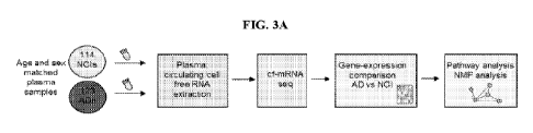
[0017] FIGS. 3A-3C show a transcriptional landscape of cf-mRNA in AD patients
and
functional implications based on gene-set analysis and functional annotations.
FIG. 3A shows a
schematic of the study design. FIG. 3B shows a volcano plot of differentially
expressed genes in
cf-mRNA between AD (n = 126) and NCT controls (n = 115) FDR < 0.05 was used as
the cut-off
criteria. FIG. 3C shows the most significant pathways identified using gene
set enrichment
analysis (top, upregulated genes; bottom, downregulated genes). The black
vertical dotted line
represents significance threshold (p <0.05).
[0018] FIGS. 4A-4C show biological processes and signaling pathways that are
associated with
AD. FIG. 4A shows biological processes determined by IPA analysis for genes
that are
upregulated in cf-mRNA of AD as input (left). Most prominent biological
processes determined
by IPA analysis for genes that are downregulated in cf-mRNA of AD as input
(right). FIG. 4B
shows subcategories within nervous system development and function (IPA) for
genes that are
downregulated in cf-mRNA of AD as input. FIG. 4C shows biological processes
determined by
Gene Ontology for genes that are upregulated in cf-mRNA of AD as input (left)
and the most
prominent biological processes determined by Gene Ontology for genes that are
downregulated
in cf-mRNA of AD as input (right).
100191 FIGS. 5A-5C show cf-mRNA transcripts significantly overlap with brain
tissue
transcripts and transcripts that are dysregulated in AD. FIG. SA shows overlap
between the
Genotype-Tissue Expression (GTEx) defined brain enriched genes and
downregulated genes in
cf-mRNA of AD (left) and overlap between GTEx defined liver enriched genes and
downregulated genes in cf-mRNA of AD (right). P-values show comparison between
number of
overlapped genes versus expected number. FIG. 5B shows overlap between genes
that are
upregulated in cf-mRNA of AD compared to NCI against genes that are
upregulated in the brain
tissue of AD patients (left). FIG. SC shows overlap between genes that are
downregulated in cf-
mRNA of AD compared to NCI against genes that are downregulated in the brain
tissue of AD
patients (left).
[0020] FIGS. 6A-6E illustrate that cf-mRNA classifier robustly distinguishes
AD from NCI.
FIG. 6A shows a schematic of classifier establishment. FIG. 6B shows an
evaluation of
classification accuracy using training cohort. The y-axis depicts AUROC of
individual
algorithms. FIG. 6C shows a ROC curve of cf-mRNA classifier for discriminating
AD against
NCI (left) and a waterfall plot of AD and NCI discrimination (right). FIG. 6D
shows a ROC
-6-
CA 03172199 2022- 9- 16
WO 2021/188825
PCT/US2021/023015
curve of a 9-gene mini classifier for discriminating AD against NCI. FIG. 6E
shows read counts
between AD and NCI in total cohort (123 AD and 114 NCI) for 9 mini-classifier
genes.
[0021] FIG. 7A illustrates the expression levels of 1,496 dysregulated genes
in AD patients with
CDR < 1 (FDR < 0.05). FIG. 7B shows genes downregulated in "early stage" AD
patients are
primarily enriched in nervous system function and developmental processes
(e.g., Netrin
signaling, CRER signaling in neurons, Calcium transport, and Regulation of
neurogenesis) and
upregulated genes in immune response and proteostasis (e.g., protein
ubiquitination,
inflammasome pathway, and activation of immune response).
[0022] FIGS. 8A-8G show that cf-mRNA genes correlate with severity of
cognitive impairment.
FIG. 8A shows that a consensus matrix NIVIF clustering identifies size
biologically distinct
clusters. Unsupervised NMF clustering from 2591 differentially expressed
genes. FIG. 8B shows
the expression of "synaptic transmission" and "immune & inflammatory response"
clusters
categorized by CDR rating. FIG. 8C shows a plot between FDR (represented as -
log) and
Pearson's correlation coefficient for CDR and TPM of genes. Red dotted line
represents FDR =
0.05. FIG. 8D shows top canonical pathways identified in IPA pathway analysis
using 706 genes
that correlate with CDR scores. Red dotted line represents FDR = 0.05. FIG. 8E
shows the
expression of SLU7 based on CDR and MMSE scores (CDR scores (top) and MMSE
(bottom)).
FIG. 8F shows an average ROC curve of the cf-mRNA classifier for
distinguishing NCI (CDR =
0) against those with CDR score of 0.5-1. 15 iterations of cross-validation
were performed, and
the curve represents the average of those 15 ROC curves. FIG. 8G shows
unsupervised
clustering of AD patients using their cf-mRNA profile based on NMF clusters
identified in FIG.
8A.
[0023] FIGS. 9A-9C show the expression of cf-mRNA genes against cognitive
impairment
scores. FIG. 9A illustrates cluster values for each of the 5 AD patient
subcategories, Age and
MIVISE distribution among 5 patient groups identified using ANOVA analysis-
Tukey's post-hoc
test. FIG. 9B shows a plot between FDR (represented as -log) and Pearson's
correlation
coefficient for MMSE vs TPM of genes. Red dotted line represents FDR = 0.05.
FIG. 9C shows
top canonical pathways identified in IPA pathway analysis using 520 genes that
correlate with
MMSE scores. Red dotted line represents FDR = 0.05. FIG. 9D shows overlapping
genes
between genes that correlate with MMSE and CDR scores.
[0024] FIG. 10 depicts a computer system consistent with the disclosure
herein.
[0025] FIG. 11 shows the differential expression of TCF7 in Transcripts per
Million (TPM) by
age group.
-7-
CA 03172199 2022- 9- 16
WO 2021/188825
PCT/US2021/023015
[0026] FIG. 12 shows the differential expression of PTK2 (focal adhesion
kinase in senescent
cells) in TPM by age group.
[0027] FIG. 13 shows the differential expression of FER in TPM by age group.
[0028] FIG. 14 shows the differential expression of CD36 in TPM by age group.
CD36 is one of
18 genes of the panel G00000302 "response to reactive oxygen species" function
which
correlates with age.
[0029] FIG. 15 shows the differential expression of WWTR1 in TPM by age group.
WWTR1 is
expressed in the Hippo pathway in connection with the YAP/TAZ complex. WWTR1
is one of
40 non-blood genes which correlate with age.
[0030] FIG. 16 shows the differential expression of CAV1 in TPM by age group.
CAV1 is
Caveolin 1 involved in caveolae formation. CAV1 is one of 40 non-blood genes
which correlate
with age.
100311 FIG. 17 shows a comparison of age-associated genes with other data
sets. Two genes,
NELL2 and LTB, are consistently highly correlated with age.
[0032] FIG. 18 shows a heat map of the expression of 41 age associated genes
which overlap
with non-blood genes with a p-value of 3.93e-11.
[0033] FIG. 19 shows a chart of age associated genes for multiple tissues
using GTEx data.
DETAILED DESCRIPTION
100341 Methods, systems, and kits described herein relate to the rapid,
noninvasive detection of
disorders using a combination of marker types so as to concurrently determine
both a likely
disorder and a likely tissue under duress, taking into account changes in gene
expression brought
about by the natural aging of an individual. In some embodiments, a gene panel
comprising
genes known to be differentially expressed in individuals at the age of a
subject is applied to a
cell-free RNA (cfRNA) expression profile of the subject. Through practice of
the disclosure
herein, one can make predictions as to a disease identity, and the extent of
its impact on one or
more tissues, without invasive investigation of the tissue or tissues
suspected of being impacted.
[0035] There is a need to develop a reliable and non-invasive test to
accurately diagnose
Alzheimer's disease earlier on. Physicians often use a numeric scale, Clinical
Dementia Rating
(CDR), to quantify the severity of a neurodegenerative disorder. Further, the
Mini-Mental State
Exam (MMSE) or the Fol stein test is used in clinical and research settings to
measure cognitive
impairment.
[0036] The identification of disease markers in circulation, such as in a
blood sample, can be a
useful tool allowing for the identification of diseased tissue without the
need for invasive
-8-
CA 03172199 2022- 9- 16
WO 2021/188825
PCT/US2021/023015
procedures such as a biopsy. This can be useful in older populations who may
be less resilient to
such invasive, painful procedures. Factors other than disease which may affect
gene expression
can also be taken into account. The gene expression of some tissues changes as
individual ages It
may be important to identify gene markers associated with age and how they are
differentially
expressed in order to take them into account when diagnosing a diseased tissue
100371 Here, by performing a transcriptome-wide comparison of plasma cf-mRNA
profiles
between age matched AD patients and control individuals, proof-of-concept is
shown that the
circulating transcriptome has the potential to reveal, in a non-invasive
manner, molecular and
functional information of neurodegenerative diseases such as AD. Technical
performance of the
assay is disclosed herein, as well as detection and quantification of
thousands of genes in
circulation to show that genes dysregulated in the plasma of AD patients can
reflect biological
processes and pathways known to be associated with cognitive impairment and
neurodegenerative disorders. For example, disclosed herein is an overall
decline in AD patients
of multiple pathways implicated in the nervous system function and development
(e.g., synapse
loss, GABA signaling, and neurotransmission), accompanied by elevated levels
of genes
involved in inflammation, mitochondrial dysfunction, oxidation, and
proteostasis. Further, the
genes and biological processes found to be dysregulated in the plasma of AD
patients
substantially overlapped with those identified in the RNA-seq datasets from
postmortem brain
biopsy specimens. Cell free-mRNA in plasma can be a surrogate for non-invasive
molecular
evaluation of brain homeostasis in AD patients.
100381 One potential application that would benefit from a better
understanding of the molecular
mechanisms involved in AD, is the development of new therapeutic strategies.
cf-mRNA
sequencing can provide a granular characterization of AD patients' circulating
transcriptome,
including thousands of genes that are either dysregulated in AD patients or
correlated with AD
severity. In addition to showing high resolution on biological processes
already known to be
linked to AD (e.g., 26 dysregulated genes involved in GABA signaling), reduced
levels of genes
associated with neurogenesis in AD patients were observed, which, without
being bound by any
one particular theory, may support the hypothesis of adult neurogenesis being
disrupted in AD.
Further, many factors involved in RNA splicing were identified to be
dysregulated in AD
patients, such as SLU7, whose levels strongly correlate with disease severity.
Evidence points to
a role of alternative RNA splicing in aging and neurodegeneration. A prominent
decrease of
netrin signaling in AD patients, including a significant reduction in the
levels of NETRIN-1,
which binds APP and has been proposed as a master regulator of A13 levels was
observed.
Decreased NETRIN-1 expression is associated with increased AP concentration.
The integrated
-9-
CA 03172199 2022- 9- 16
WO 2021/188825
PCT/US2021/023015
cf-mRNA technology solution can provide an approach to better understand the
heterogeneous
etiology of AD and may aid in the identification of new molecular entities
with therapeutic
potential and increase their probability of technical success in pre-clinical
and clinical stages.
100391 Indeed, the heterogeneous nature of AD, as a complex neurodegenerative
disease
affecting multiple biological pathways and processes during its onset and
progression, represents
one major difficulty for AD dnig development So far, therapeutic dnigs
targeting (1-amyl oi ds
and tau proteins have shown modest results, therefore multiple compounds
targeting commonly
affected pathways in AD, such as inflammation, mitochondrial dysfunction, and
neuroprotective
compounds are currently being developed and tested as alternatives for AD
treatment. Successful
development of therapeutic agents for a heterogeneous AD population may rely
on the ability to
appropriately enrich the trial groups for AD patients likely to respond to the
candidate drugs.
Since molecular characterization of patients based on brain biopsy is
generally not feasible, non-
invasive tools that enable pre-selection of patients best suited for each
therapy can be useful for
clinical trials. The present disclosure indicates that the molecular
information revealed by the
circulating transcriptome may pave the way to personalized characterization of
disease-related
processes, thus enabling more efficient patient management and improving the
probabilities of
success of the interventions. Further, given that cf-mRNA can enable "real
time" monitoring of
organ health and organ system response to therapeutic interventions, and the
repertoire of AD-
related processes identified in circulation, an integration of cf-mRNA
sequencing and clinical
information may also allow monitoring therapy response in AD patients.
100401 Despite post-mortem histology remaining the gold standard for
establishing AD
pathology, currently CSF, PET, and MRI can be used to diagnose AD patients.
However,
imaging modalities can be costly and CSF collection can be invasive.
Therefore, scalable,
accessible, and cost-efficient blood-based tests are desired for the
management of AD patients.
To date, several protein-based blood biomarkers, including those that measure
circulating levels
of Afl peptides, appear to be promising candidates as diagnostic biomarkers
for AD, though not
without limitations considering that AO is also present in individuals without
dementia and its
levels inconsistently predict the rate of cognitive decline. Profiling the cf-
mRNA transcriptome
represents a non-invasive approach for the development of molecular
classifiers to identify AD
patients, as shown by the performance of cf-mRNA based classifiers to
discriminate control
individuals from AD patients. Therefore, cf-mRNA profiling may offer a novel
approach for
more personalized patient management that integrates clinical information of
disease state with
insights on patient-specific molecular characteristics to create solutions for
improved patient
management. cf-mRNA profiling may aid in clinical trials, for instance, as a
potential tool for the
-10-
CA 03172199 2022- 9- 16
WO 2021/188825
PCT/US2021/023015
discrimination of patients with or without AD, reducing the number of patients
who require A13-
PET for AD diagnosis, and for stratification of patients with increased
likelihood to respond to
the therapy based on their molecular characteristics.
100411 Provided herein are noninvasive methods, systems, compositions, and
kits for assessing
or detecting Alzheimer's disease (AD) in a subject, for example, using a
biological sample of the
subject The methods comprise isolating cell-free messenger RNAs (cf-mRNAs)
from the
biological sample. In some embodiments, the biological sample is a plasma or
serum. In other
embodiments, the biological sample is cerebrospinal fluid (CSF).
100421 A first transcriptome-wide comparison of plasma cf-mRNA profile between
AD and NCI
is disclosed herein and cf-mRNA signatures that are distinct to AD are
identified. Gene-set
enrichment analysis showed that cf-mRNA profile of AD reflected signaling
pathways and
biological processes that are commonly dysregulated in AD. Furthermore,
"immune &
inflammatory response" and "synaptic transmission" gene-clusters which
correlated with the
severity of cognitive impairment are disclosed herein. In addition, genes that
are associated with
neuronal function, another attribute of AD, are attenuated in cf-mRNA
transcriptome of AD
patients. Disclosed herein is a set of genes correlated with CDR and MMSE
cognitive
impairment scores, some of which had substantial gene-expression alteration
even in the AD
patients with very mild to mild cognitive impairment compared to those that
are not cognitively
impaired. A classifier which can differentiate AD patients with modest
cognitive impairment
from normal controls without cognitive impairment, indicating that
transcriptional changes in the
circulation may be suitable as an early diagnostic tool for AD, is also
disclosed herein.
100431 The methods can also employ upfront centrifugation to reduce
contamination of
unwanted "blood" transcripts from cf-mRNA sequencing data. The methods herein
can reduce
background noise within the "blood component" blood cells from the tissue-
specific cf-mRNA
signal. Such noise can increase sequencing depth requirements and dilute
signal from tissue-
specific cf-mRNA. With this purification step, the cf-mRNA transcripts can be
said to be more
than likely deriving from a subject's brain. By reducing the background noise
with the "blood
component" transcripts, the detected cf-mRNA transcripts are likely originated
from brain.
100441 Often, serum, plasma, or other biological samples are collected from
subjects and the
samples are optimized by removing cellular debris. In some embodiments, the
samples are
collected from subjects at a remote location and are shipped to a testing cite
via delivery services.
Some subjects are healthy, some experience cognitive impairment, and some are
diagnosed with
AD. In certain instances, the samples may be enriched in non-blood
transcripts. cf-mRNAs
including a mixture of genetic materials from different genomic sources, such
as cerebrum,
-11-
CA 03172199 2022- 9- 16
WO 2021/188825
PCT/US2021/023015
cerebellum, dorsal root ganglion, superior cervical ganglion, pineal gland,
amygdala, trigeminal
ganglion, cerebral cortex, and hypothalamus can be isolated from the optimized
samples.
[0045] A broad range of centrifugation ranges can be used to optimize the
samples so that blood
transcripts are removed. In certain cases, the ranges may include 1,500 g to
20,000 g, 1,900 g to
16,000 g, 4,000 g to 16,000 g, 8,000 g to 16,000 g, 10,000 g to 14,000 g,
11,000 g to 13,000 g,
11,500 g to 12,500 g, or suitable lower or higher ranges Tn some cases, the
sample may be
centrifuges at about 12,000 g, essentially 12,000 g, substantially 12,000g, or
12,000 g. Some
ranges span about 12,000 g. Some ranges are within 100 g of 12,000 g. Some
centrifugation
protocols do not differ substantially from 12,000 g, such as centrifugations
at 12,000 g. Alternate
ranges having a starting point at a low figure listed above or ending at a
high figure listed above
are also contemplated. Such centrifugation protocols can contribute to 2.5x
improvement in
diversity of an RNA library for processing. In various cases, the
centrifugation protocols may
contribute to a 1.1x, 1.2x, 1.3x, 1.4x, 1.5x, 1.6x, 1.7x, 1.8x, 1.9x, 2.0x,
2.1x, 2.2x, 2.3x, 2.4x,
2.5x, 2.6x, 2.7x, 2.8x, 2.9x, 3.0x, 3.1x, 3.2x, 3.3x, 3.4x, 3.5x, 3.6x, 3.7x,
3.8x, 3.9x, 4.0x, or
greater than 4.0x improvement in diversity of an RNA library for processing.
[0046] Further, cDNAs can be converted based on the isolated cf-mRNAs in order
to form a
library of cDNAs including a NGS library. For example, cDNAs can be generated
from reverse
transcription of a cf-mRNA sample. Further, cDNAs can be enriched for
quantification.
100471 After building the library of cDNAs, many methods can be used to
quantify the levels of
different cDNAs. For example, polynucleotide amplification, sequencing, probe
hybridization,
RT-PCR, and microarray hybridization, among other suitable methods, can be
used to quantify
levels of cDNAs. Various methods can be used to enrich the cDNAs. For example,
some of these
methods are based on hybridization to oligonucleotides designed to hybridize
to different
cDNAs. The hybridization may be to oligonucleotides immobilized on high or low
density
microarrays, or solution phase hybridization to oligonucleotides modified with
a ligand which
can be subsequently employed for immobilization of the hybrids to a solid
surface, such as a
bead. Other methods may employ sequence specific amplification (e.g., PCR) to
amplify specific
cDNAs in a droplet, allowing amplification of specific cDNAs for downstream
sequencing. The
droplet-based amplification may enable highly multiplexed PCR without the
potential non-
specific interaction of a large number of PCR primer pairs and the subsequent
generation of non-
specific amplification products and reduced amplification efficiency of the
cDNAs.
[0048] Moreover, differential gene expression can also be identified, or
confirmed, using the
microarray technique. In this method, polynucleotide sequences of interest
(including cDNAs
-12-
CA 03172199 2022- 9- 16
WO 2021/188825
PCT/US2021/023015
and oligonucleotides) can be plated, or arrayed, on a microchip substrate. The
arrayed sequences
can be then hybridized with specific DNA probes from cells or tissues of
interest.
[0049] Further, differential gene expression can also be identified, or
confirmed, using the
sequencing technique. The polynucleotide sequences of interest (including
cDNAs and
oligonucleotides) can be used as templates to synthesize sequencing libraries.
The libraries can
be sequenced, and the reads mapped to an appropriate reference. Exemplary
sequencing
techniques can include, for example, emulsion PCR, pyrosequencing from Roche
454,
semiconductor sequencing from Ion Torrent, SOLiD sequencing by ligation from
Life
Technologies, sequencing by synthesis from Intelligent Biosystems, bridge
amplification on a
flow cell (e.g., Solexa/Illumina), isothermal amplification by Wildfire
technology (Life
Technologies), or rolonies/nanoballs generated by rolling circle amplification
(Complete
Genomics, Intelligent Biosystems, Polonator). Sequencing technologies such as
Heliscope
(Helicos), SMRT technology (Pacific Biosciences), or nanopore sequencing
(Oxford Nanopore),
which can allow direct sequencing of single molecules without prior clonal
amplification, may be
suitable sequencing platforms. Other sequencing methods are also within the
scope of this
disclosure. Sequencing may be performed with or without target enrichment.
Moreover, RT-PCR
can be used to quantify different gene expression levels. Generally, the
reverse transcription
reaction step can be primed using specific primers, random hexamers, or oligo-
dT primers,
depending on the goal of expression profiling. Reverse transcriptases can be
avian myeloblastosis
virus reverse transcriptase (AMV-RT), Moloney murine leukemia virus reverse
transcriptase
(MLV-RT), or other suitable reverse transcriptases.
[0050] Although the PCR step can use a variety of thermostable DNA-dependent
DNA
polymerases, it typically employs the Taq DNA polymerase, which can have a 5'-
3' nuclease
activity but lacks a 3'-5' proofreading endonuclease activity. Thus, TaqManTM
PCR typically
utilizes the 5'-nuclease activity of Taq or Tth polymerase to hydrolyze a
hybridization probe
bound to its target amplicon, but any suitable enzyme with equivalent 5'
nuclease activity can be
used. Two oligonucleotide primers can be used to generate an amplicon typical
of a PCR
reaction. A third oligonucleotide, or probe, can be designed to detect
nucleotide sequence located
between the two PCR primers. The probe can be non-extendible by Taq DNA
polymerase
enzyme, and can be labeled with a reporter fluorescent dye and a quencher
fluorescent dye. Any
laser-induced emission from the reporter dye can be quenched by the quenching
dye when the
two dyes are located close together, for example, as they are on the probe.
During the
amplification reaction, the Taq DNA polymerase enzyme can cleave the probe in
a template-
dependent manner. The resultant probe fragments can disassociate in solution,
and signal from
-13-
CA 03172199 2022- 9- 16
WO 2021/188825
PCT/US2021/023015
the released reporter dye can be freed from the quenching effect of the second
fluorophore. One
molecule of reporter dye can be liberated for each new molecule synthesized,
and detection of the
unquenched reporter dye can provide basis for quantitative interpretation of
the data.
100511 TaqManTM RT-PCR can be performed using commercially available
equipment, such as,
for example, ABI PRISM 7700TM Sequence Detection SystemTM (Perkin-Elmer-
Applied
Bi system s, Foster City, Cal i f , USA) or T ghtcycl er (Roche Molecular Bi
ochemi cal s,
Mannheim, Germany). In certain embodiments, the 5' nuclease procedure is run
on a real-time
quantitative PCR device such as the ABI PRISM 7700TM Sequence Detection
SystemTM. The
system comprises a thermocycler, laser, charge-coupled device (CCD), camera,
and computer.
The system includes software for running the instrument and for analyzing the
data. 5'-nuclease
assay data can initially be expressed as Ct (the threshold cycle).
Fluorescence values can be
recorded during every cycle and represent the amount of product amplified to
that point in the
amplification reaction. The point when the fluorescent signal is first
recorded as statistically
significant can be the threshold cycle (Ct).
Panel of differentially expressed genes
100521 The biomarker panels comprising a plurality of differentially expressed
protein encoding
genes described herein can facilitate a sensitive and non-intrusive testing to
detect whether a
subject has AD or to determine the clinical development stage of AD. Clinical
development
stages of Alzheimer's disease include (1) preclinical Alzheimer's disease, (2)
mild cognitive
impairment due to Alzheimer's disease, (3) mild dementia due to Alzheimer's
disease, (4)
moderate dementia due to Alzheimer's disease, and (5) severe dementia due to
Alzheimer's
disease. Biomarker panels comprising a plurality of differentially expressed
protein encoding
genes are often readily obtained by a blood draw from an individual. Benefits
of using the
biomarker panels disclosed herein can include fast and convenient detecting of
AD without
cumbersome and unreliable testing.
100531 Biomarker panels as disclosed herein can be selected such that their
predictive value as
panels is substantially greater than the predictive value of their individual
members. Panel
members generally do not co-vary with one another, such that panel members
provide
independent contributions to the panel's overall health signal. Biomarker
panels can comprise
genes dysregulated in plasma of AD patients, as well as genes that correlated
with disease
severity, that are enriched in biological processes associated with AD, such
as synaptic
dysfunction, mitochondri al dysfunction, and inflammation. Genes dysregulated
in circulation can
be used to identify AD patient subtypes among a heterogeneous population
patients, and build cf-
mRNA based classifiers that discriminate (e.g., robustly discriminate) age
matched controls from
-14-
CA 03172199 2022- 9- 16
WO 2021/188825
PCT/US2021/023015
AD patients. Cell-free mRNA biomarker panels can non-invasively reveal
molecular
characteristics associated with neurodegenerati on and AD, and support the
potential of
integrating cf-mRNA with clinical information to potentially improve the AD
patient
management, identify new therapeutic targets, and enable patient
stratification to increase the
probability of technical success of the research and development of
therapeutics. Accordingly, a
panel may be able to substantially outperform the performance of any
individual constituent
indicative of an individual's AD status, such that a commercially and
medicinally relevant degree
of confidence (such as sensitivity, specificity, or sensitivity and
specificity) is obtained.
100541 In some cases, panel members vary independently from each other. As a
result, panels
herein often indicate a health risk despite the fact that one or more than one
individual members
of the panel would not indicate that the health risk is present if measured
alone. In other cases,
panels herein indicate a health risk at a significant level of confidence
despite the fact that no
individual panel member indicates the health risk at a significant level of
confidence on its own.
In yet other cases, panels herein can indicate a health risk at a significant
level of confidence
despite the fact that at least one individual member indicates at a
significant level of confidence
that the health risk is not present.
100551 Some biomarker panels comprise some or all of the differentially
expressed protein
encoding genes recited herein (see Table 1A). In some cases, a biomarker panel
may comprise at
least nine protein encoding genes. In some cases, the biomarker panel may
comprise any two
genes from Table 1A. In some cases, the biomarker panel may comprise any three
genes from
Table A. In some cases, the biomarker panel may comprise any four genes from
Table A. In
some cases, the biomarker panel may comprise any five genes from Table 1A. In
some cases, the
biomarker panel may comprise any six genes from Table 1A. In some cases, the
biomarker panel
may comprise any seven genes from Table 1A. In some cases, the biomarker panel
may comprise
any eight genes from Table 1A. In some cases, the biomarker panel may comprise
the nine genes
from Table 1A.
-15-
CA 03172199 2022- 9- 16
WO 2021/188825
PCT/US2021/023015
Table 1A: List of differentially expressed genes
Ref No. Gene names
1 KIAA0100
2 MAG11
3 NNMT
4 MXD1
ZNF75A
6 SELL
7 ASS1
8 MNDA
9 AC132217.4
100561 In addition, some biomarker panels may comprise some or all of the
differentially
expressed protein encoding genes recited herein (see Table 1B). In some cases,
a biomarker panel
may comprise at least 14 protein encoding genes. In some cases, the biomarker
panel may
comprise any two genes from Table 1B. In some cases, the biomarker panel may
comprise any
three genes from Table 1B. In some cases, the biomarker panel may comprise any
four genes
from Table 1B. In some cases, the biomarker panel may comprise any five genes
from Table 1B.
In some cases, the biomarker panel may comprise any six genes from Table 1B.
In some cases,
the biomarker panel may comprise any seven genes from Table 1B. In some cases,
the biomarker
panel may comprise any eight genes from Table 1B. In some cases, the biomarker
panel may
comprise any nine genes from Table 1B. In some cases, the biomarker panel may
comprise any
ten genes from Table 1B. In some cases, the biomarker panel may comprise any
eleven genes
from Table 1B. In some cases, the biomarker panel may comprise any twelve
genes from Table
1B. In some cases, the biomarker panel may comprise any thirteen genes from
Table 1B. In some
cases, the biomarker panel may comprise the fourteen genes from Table 1B.
-16-
CA 03172199 2022- 9- 16
WO 2021/188825
PCT/US2021/023015
Table 1B: List of additional differentially expressed genes
Ref No. Gene names
1 SLU7
2 HNRNPA2B1
3 GGCT
4 NDUFA12
HSPB11
6 ATP6V1B2
7 SASS6
8 SUM01
9 KRCC1
LSM6
11 LCP1
12 SASS6
13 ATP6v1B2
14 MAT2B
100571 After construction of various biomarker panels, the biomarker panels
can be used to
determine whether a subject has AD as described in the non-invasive diagnostic
methods
provided herein. Further, the biomarker panels can also be used to determine a
particular
development stage of AD. Often, different development stages of AD are
assigned with either a
CDR score or a IVEMSE score. Some of the methods herein comprise comparing a
level of a
biomarker panel in a subject to a threshold level of the same biomarker panel.
In some cases, the
threshold level of a biomarker panel equals the level of the biomarker panel
of a control subject.
In some cases, the control subject is a person having a known diagnosis. For
example, the control
subject can be a negative control subject. The negative control subject can be
a subject that does
not have AD. For other example, the control subject can be a positive control
subject. The
positive control subject can be a subject having a confirmed diagnosis of AD.
The positive
control subject can be a subject having a confirmed diagnosis of AD. Further,
the positive control
subject can be a subject having a confirmed diagnosis of any stage of AD. For
example, the
positive control subject may have a CDR score of 0.5, 1, 2, or 3. The positive
control subject may
have a MilVISE score of 1-6, 6-12, 12-18, 18-24, or 24-30. The threshold value
can be a
-17-
CA 03172199 2022- 9- 16
WO 2021/188825
PCT/US2021/023015
predetermined level of the biomarker, wherein the predetermined level is set
based upon a
measured amount of the biomarker in a control subject.
[0058] Diagnostic methods described herein for detection of AD in a subject
can detect AD with
a sensitivity greater than 75%, greater than 80%, greater than 85%, greater
than 90%, greater
than 95%, greater than 96%, greater than 97%, greater than 98%, greater than
99%, or about
100% Such diagnostic methods can detect Alzheimer's Disease (AD) with a
sensitivity that is
70% to 100%, 80% to 100%, or 90% to 100%. Such diagnostic methods can detect
AD with a
specificity greater than 70%, greater than 75%, greater than 80%, greater than
85%, greater than
90%, greater than 95%, greater than 96%, greater than 97%, greater than 98%,
greater than 99%,
or about 100%. Such diagnostic methods can detect AD with a specificity that
is from 50% to
100%, from 60% to 100%, from 70% to 100%, from 80% to 100%, or from 90% to
100%. In
various embodiments, such diagnostic methods can detect AD with a sensitivity
and a specificity
that is 50% or greater, 60% or greater, 70% or greater, 75% or greater, 80% or
greater, 85% or
greater, or 90% or greater. In certain embodiments, such diagnostic methods
can detect AD with
a sensitivity and a specificity that is 50% to 100%, 60% to 100%, 70% to 100%,
80% to 100%,
or 90% to 100%.
Classifier
[0059] Classifiers can be developed using many different technologies. For
example, computer
systems can be used to develop and generate classifiers. Data, such as cf-mRNA
levels, collected
from the plurality of differentially expressed protein coding genes can be
used to train a machine
learning algorithm to obtain a classifier.
[0060] Machine learning can be generalized as the ability of a learning
machine to perform
accurately on new, unseen examples/tasks after having experienced a learning
data set. Machine
learning may include the concepts and methods provided herein. Supervised
learning concepts
may include: AODE; Artificial neural network, such as Backpropagation,
Autoencoders,
Hopfield networks, Boltzmann machines, Restricted Boltzmann Machines, and
Spiking neural
networks; Bayesian statistics, such as Bayesian network and Bayesian knowledge
base; Case-
based reasoning; Gaussian process regression; Gene expression programming;
Group method of
data handling (GMDH); Inductive logic programming; Instance-based learning;
Lazy learning;
Learning Automata; Learning Vector Quantization; Logistic Model Tree; Minimum
message
length (decision trees, decision graphs, etc.), such as Nearest Neighbor
Algorithm and Analogical
modeling; Probably approximately correct learning (PAC) learning; Ripple down
rules, a
knowledge acquisition methodology; Symbolic machine learning algorithms;
Support vector
machines (SVM); Random Forests; Ensembles of classifiers, such as Bootstrap
aggregating
-18-
CA 03172199 2022- 9- 16
WO 2021/188825
PCT/US2021/023015
(bagging) and Boosting (meta-algorithm); Ordinal classification; Information
fuzzy networks
(IFN); Conditional Random Field; ANOVA; Linear classifiers, such as Fisher's
linear
discriminant, Linear regression, Logistic regression, Multinomial logistic
regression, Naive
Bayes classifier, Perceptron, Support vector machines; Quadratic classifiers;
k-nearest neighbor;
Boosting; logistic regression with Li regularization (LASSO); logistic
regression with L2
regularization (ridge classifier); Decision trees, such as C4.5, Random
forests, ID3, CART,
SLIQ, SPRINT; Bayesian networks, such as Naive Bayes; and Hidden Markov
models.
Unsupervised learning concepts may include: Expectation-maximization
algorithm; Vector
Quantization; Generative topographic map; Information bottleneck method;
Artificial neural
network, such as Self-organizing map; Association rule learning, such as,
Apriori algorithm,
Eclat algorithm, and FP-growth algorithm; Hierarchical clustering, such as
Single-linkage
clustering and Conceptual clustering; Cluster analysis, such as K-means
algorithm, Fuzzy
clustering, DBSCAN, and OPTICS algorithm; and Outlier Detection, such as Local
Outlier
Factor. Semi-supervised learning concepts may include: Generative models, Low-
density
separation, Graphbased methods, and Co-training. Reinforcement learning
concepts may include:
Temporal difference learning, Q-learning, Learning Automata, and SARSA. Deep
learning
concepts may include: Deep belief networks, Deep Boltzmann machines, Deep
Convolutional
neural networks, Deep Recurrent neural networks, and Hierarchical temporal
memory.
100611 In some cases, the performance of a classifier is assessed in some
cases via the AUC of
the ROC as reported herein. A ROC considers the performance of the classifier
at all possible
model score cutoff points. However, when a classification decision needs to be
made (e.g., is this
patient sick or healthy?), a cutoff point is used to define the two groups
Classification scores at
or above the cutoff point are assessed as positive (or sick) while points
below are assessed as
negative (or healthy) in various embodiments.
100621 For some classification models disclosed herein, a classification score
cutoff point is
established by selecting the point of maximum accuracy on the validation ROC.
The point of
maximum accuracy on an ROC is the cutoff point or points for which the total
number of correct
classification calls is maximized. Here, the positive and negative
classification calls are weighted
equally. In cases where multiple maximum accuracy points are present on a
given ROC, the point
with the associated maximum sensitivity may be selected.
Clinical outcome score
100631 Machine learning algorithms for sub-selecting discriminating biomarkers
and/or subject
characteristics, and for building classification models, are used in some
methods and systems
herein to determine clinical outcome scores. These algorithms include, but are
not limited to,
-19-
CA 03172199 2022- 9- 16
WO 2021/188825
PCT/US2021/023015
elastic networks, random forests, support vector machines, and logistic
regression. These
algorithms can aid in selection of important biomarker features and transform
the underlying
measurements into a score or probability relating to, for example, clinical
outcome, disease risk,
disease likelihood, presence or absence of disease, treatment response, and/or
classification of
disease status.
100641 A clinical outcome score can be generated by inputting quantified cf-
mRNA levels to a
classifier described herein. Also, a clinical outcome score is determined by
comparing cf-mRNA
levels that corresponds to at least two differentially expressed genes in the
biological sample
obtained from the subject to a reference cf-mRNA level of the two genes.
Alternately or in
combination, a clinical outcome score is determined by comparing a subject-
specific profile of a
panel of cf-mRNA levels correspond to differentially expressed genes to a
reference profile of
the differentially expressed genes. Often, a reference level or reference
profile represents a
known diagnosis. For example, a reference level or reference profile
represents a positive
diagnosis of AD. As another example, a reference level or reference profile
represents a negative
diagnosis of AD. Similarly, a reference level or reference profile represents
a particular score
associated with CDR or 1VI1VISE.
100651 In some cases, an increase in a score indicates an increased likelihood
of one or more of
a: poor clinical outcome, good clinical outcome, high risk of disease, low
risk of disease,
complete response, partial response, stable disease, non-response, and
recommended treatment
(or treatments) for disease management. In some cases, a decrease in the
quantitative score
indicates an increased likelihood of one or more of a: poor clinical outcome,
good clinical
outcome, high risk of disease, low risk of disease, complete response, partial
response, stable
disease, non-response, and recommended treatment (or treatments) for disease
management.
Also, in some embodiments, an increase in a score indicates a higher CDR or
MMSE score.
100661 A similar profile from a patient to a reference profile often indicates
an increased
likelihood of one or more of a: poor clinical outcome, good clinical outcome,
high risk of
disease, low risk of disease, complete response, partial response, stable
disease, non-response,
and recommended treatment (or treatments) for disease management. In some
applications, a
dissimilar biomarker profile from a patient to a reference profile may
indicate one or more of: an
increased likelihood of a poor clinical outcome, a good clinical outcome, a
high risk of disease, a
low risk of disease, a complete response, a partial response, a stable
disease, a non-response, and
a recommended treatment (or treatments) for disease management.
100671 An increase threshold values of cf-mRNA levels corresponding to one or
more
differentially expressed genes often indicates an increased likelihood of one
or more of a: poor
-20-
CA 03172199 2022- 9- 16
WO 2021/188825
PCT/US2021/023015
clinical outcome, good clinical outcome, high risk of disease, low risk of
disease, complete
response, partial response, stable disease, non-response, and recommended
treatment (or
treatments) for disease management. In some applications, a decrease in one or
more biomarker
threshold values may indicate an increased likelihood of one or more of a:
poor clinical outcome,
good clinical outcome, high risk of disease, low risk of disease, complete
response, partial
response, stable disease, non-response, and recommended treatment (or
treatments) for disease
management.
100681 An increase in at least one of a quantitative score, one or more
thresholds, or similar
biomarker profile values indicates an increased likelihood of one or more of
a: poor clinical
outcome, good clinical outcome, high risk of disease, low risk of disease,
complete response,
partial response, stable disease, non-response, and recommended treatment (or
treatments) for
disease management. Similarly, a decrease in at least one of a quantitative
score, one or more
biomarker thresholds, similar biomarker profile values or combinations thereof
indicates an
increased likelihood of one or more of a: poor clinical outcome, good clinical
outcome, high risk
of disease, low risk of disease, complete response, partial response, stable
disease, non-response,
and recommended treatment (or treatments) for disease management.
Treatment and monitoring regimens
100691 Provided herein are diagnostic, monitoring, and treatment regimens for
implementing any
of the methods described herein for detecting a presence or absence of AD
and/or treatment of
the same.
100701 For example, Mini-Mental State Exam (MMSE) can be administered to
assess whether
there are problems with areas of a subject's brain involved in learning,
memory, thinking, or
planning skills. Alternatively or additionally, computed tomography (CT) scan
can be used to
monitor brain changes that are common in the later stages of Alzheimer's.
Similarly, magnetic
resonance imaging (MRI), CSF, and PET can be helpful to measure amyloid
markers to monitor
the brain changes that are linked to AD. Alternatively or additionally,
neuropsychological testing
can be administered to monitor the relationship between the brain and
behavior.
Neuropsychological testing can help diagnosis of conditions that affect
thinking, emotion, and
behavior, including AD.
100711 A number of treatment methods are contemplated here as well. Different
types of drugs
can treat memory loss, behavior changes, sleep problems, and other AD's
symptoms. For
example, citalopram, fluoxetine, paroxetine, and sertraline can be used to
treat problems with
mood, depression, and irritability experienced by AD patients. Alprazolam,
buspirone,
iorazepam, and oxazepam can be used to treat anxiety or restlessness
associated with AD.
-21-
CA 03172199 2022- 9- 16
WO 2021/188825
PCT/US2021/023015
Alternatively or additionally, cholinesterase inhibitors and/or memantine can
be administered to
alleviate symptoms associated with AD. Further, unconventional therapies, such
as hormone
replacement therapy, art and music therapies, and supplements (e.g., vitamin
E) can be used
alternatively or additionally to treat AD.
100721 Methods, systems, and kits disclosed herein can be intended to non-
invasively detect a
tissue or organ in a subject that is under duress as well as determine which
disease or condition is
affecting the tissue or organ under duress. In some instances, the methods,
systems and kits can
provide for treating a subject for a disease or condition. Some methods
disclosed herein can
comprise selecting a method or therapy for treating a subject for a disease or
condition. Some kits
and systems disclosed herein can provide for selecting a method or therapy for
treating a subj ect
for a disease or condition. Some methods disclosed herein comprise monitoring
a disease or
condition in a subject, or administering a test for a disease or condition.
Some kits and systems
disclosed herein provide for monitoring a disease or condition in a subject,
or administering a test
for a disease or condition. Some methods disclosed herein comprise treating a
subject for a
disease or condition, monitoring a disease or condition in a subject, or
administering a test for a
disease or condition. In some instances, the methods disclosed herein comprise
determining the
subject has a disease or condition, thereby informing the subject or their
healthcare provider that
a treatment or test would be appropriate, suitable, or beneficial to the
subject. In some instances,
the methods disclosed herein comprise determining the subject has a disease or
condition and
recommending a treatment for the disease or condition. In some instances, the
methods disclosed
herein comprise determining the subject has a disease or condition and
treating the subject for the
disease or condition. In some instances, the methods disclosed herein comprise
determining the
subject has a disease or condition and monitoring the subject for the disease
or condition. In
some instances, the methods disclosed herein comprise determining the subject
has an increased
risk or possibility of having the disease or condition relative to an
individual within the same age
range without the disease or condition, and administering a test specific for
the disease or
condition to the subject. In some instances, the methods disclosed herein
comprise determining
the subject has an increased risk or possibility of having the disease or
condition relative to an
individual within the same age range without the disease or condition, and
recommending a test
specific for the disease or condition to the subject.
100731 Provided herein are therapeutic agents, compositions, compounds, and
agents for the
treatments of diseases and conditions. Combinations and analogs of these
agents are
contemplated and intended herein even if each combination and analog is not
explicitly
described. An "analog," as used herein, generally refers to a modified or
synthetic compound that
-22-
CA 03172199 2022- 9- 16
WO 2021/188825
PCT/US2021/023015
resembles a naturally occurring compound, wherein at least 50% of the analog
structure is
identical to at least 50% of the naturally occurring compound.
[0074] Disease presence and location in a subject can be determined at an
early stage of disease
with greater accuracy, because the systems and methods described herein
provide rapid results,
take into account gene expression variations by age, and are non-invasive and
inexpensive. Thus,
the subject can be advantageously treated before the disease progresses to
advanced stages that
are relatively more difficult to control or treat as compared to early stages.
For example, the
systems and methods disclosed herein may allow for determining which tissue(s)
or organ(s) are
showing signs of neurodegeneration before the onset of symptoms. In this way,
the methods and
systems disclosed herein can provide for focused analysis and targeted
therapies at early stages of
disease.
[0075] The methods and systems can provide for treating a subject with a
therapy that is suitable
or optimal for the extent of tissue damage. In some instances, the methods may
comprise
detecting the markers and/or tissue-specific polynucleotides to assess the
effectiveness or toxicity
of a therapy. In certain instances, the methods may comprise quantifying the
markers and/or
tissue-specific polynucleotides to assess the effectiveness or toxicity of a
therapy. In some
instances, the therapy is continued. In various instances, the therapy is
discontinued. In certain
instances, the therapy is replaced with another therapy. Regardless, due to
the rapid and non-
invasive nature of the methods and systems, therapeutic effects can be
assessed and optimized
more often relative to conventional treatment optimization.
[0076] In some aspects, the present disclosure provides for uses of the
systems, samples,
markers, and tissue-specific polynucleotides disclosed herein. In some
instances, disclosed herein
are uses of an in vitro sample for non-invasively detecting a tissue or organ
in a subject that is
under duress and as well as a disease or condition that is the cause of the
duress. In some
instances, disclosed herein are uses of an ex vivo sample for non-invasively
detecting a tissue or
organ in a subject that is under duress and as well as a disease or condition
that is the cause of the
duress by comparing the gene expression data to an age-dependent expression
control. Generally,
uses disclosed herein comprise quantifying markers and tissue-specific
polynucleotides in
samples, including ex vivo samples and in vitro samples. Some uses disclosed
herein comprise
comparing a quantity of a marker and a quantity of tissue-specific
polynucleotide in a first
sample and comparing the quantities to respective quantities in a second
sample. In some
instances, the first sample is from a first subject and the second sample is
from a control subject
(e.g., a healthy subject or subject with a condition wherein the subject is in
the same age range as
the first subject). In some instances, the first sample is from a subject at a
first time point and the
-23-
CA 03172199 2022- 9- 16
WO 2021/188825
PCT/US2021/023015
second sample is from the same subject at a second time point. The first time
point may be
obtained before the subject is administered a therapy and the second time
point may be obtained
after the therapy. Thus, also provided herein are uses of samples, markers,
tissue-specific
polynucleotides, kits, and systems disclosed herein to monitor or evaluate a
condition of a
subject, tissue health state of a subject, or an effect of a therapeutic
agent.
[0077] Tn some aspects, the disclosure provides for methods of monitoring a
human subject with
a chronic condition for a presence of at least one complication of at least
one tissue. In some
aspects, the disclosure provide for methods of monitoring a human subject with
a chronic
condition for an increased risk of at least one complication of at least one
tissue.
[0078] Some methods comprise monitoring the human subject for a complication
in any one of at
least three tissues Some methods comprise monitoring the human subject for an
increased risk of
a complication in any one of at least three tissues.
[0079] Gene expression panels as disclosed herein can share a property that
sensitive, specific
conclusions regarding an individual's tissue disease state are made using
cfRNA expression level
information derived from circulating blood in combination with knowledge of
the individual's
age. A benefit of the present gene marker panels is that they provide a
sensitive, specific tissue
health assessment using conveniently, noninvasively obtained samples. There
may be no need to
rely upon additional data obtained from intrusive biopsies. As a result,
compliance rates may be
substantially higher and tissue health issues are more easily recognized early
in their progression,
so that they may be more efficiently treated.
Cell type and tissue type specific polynucleotides
100801 Provided herein are kits, devices, systems, and methods employing cell
type-specific gene
expression, cell type-specific nucleic acids (e.g., RNAs) and cell type-
specific nucleic acid
modifications (e.g., methylation patterns) disclosed herein. The terms, "cell
type-specific nucleic
acid," "cell type-specific polynucleotide," "tissue-specific nucleic acid,"
and "tissue-specific
polynucleotide" are interchangeable as used herein. The term -cell type-
specific" may be used to
characterize a nucleic acid that is expressed in a single tissue of the
subject. Alternatively, the
term "cell type-specific" may be used to characterize a nucleic acid that is
predominantly
expressed in a specific cellular function or signaling pathway disclosed
herein. The cellular
function or pathway can include neuroinflammation, immune response, hypoxia
signaling,
production of nitric oxide, systemic lupus erythematosus signaling, toll-like
receptor signaling,
NG-kappaB signaling, inflammasome pathway, mitochondrial dysfunction, protein
ubiquitination, etc. For the purposes of this application, predominantly
expressed may mean that
the tissue-specific nucleic acid is expressed at an RNA level that is at least
50% greater in the
-24-
CA 03172199 2022- 9- 16
WO 2021/188825
PCT/US2021/023015
specific tissue than the RNA level of the tissue-specific nucleic acid in any
other tissue of the
subject. However, in some cases, a tissue-specific nucleic acid expressed at
an RNA level that is
at least 30% greater in the specific tissue than that of any other tissue may
be sufficient for the
methods disclosed herein. In other cases, a tissue-specific nucleic acid
expressed at an RNA level
that is at least 80% greater in the specific tissue than that of any other
tissue may be required by
the methods disclosed herein. Predominantly expressed may mean that the tissue-
specific nucleic
acid is expressed at an RNA level that is at least 2-fold greater in the
specific tissue of interest
than the RNA level of the tissue-specific nucleic acid in any other tissue of
the subject.
Predominantly expressed may mean that the tissue-specific nucleic acid is
expressed at an RNA
level that is at least 5-fold greater in the specific tissue of interest than
the RNA level of the
tissue-specific nucleic acid in any other tissue of the subject. Predominantly
expressed may mean
that the tissue-specific nucleic acid is expressed at an RNA level that is at
least 10-fold greater in
the specific tissue of interest than the RNA level of the tissue-specific
nucleic acid in any other
tissue of the subject. Predominantly expressed may mean that a detectable
amount of the tissue-
specific nucleic acid would occur in a biological fluid (e.g., plasma) of the
subject only when
damage occurs to the specific tissue where the tissue-specific nucleic acid is
predominantly
expressed.
100811 Provided herein are kits, systems, and methods for detecting or
quantifying a biological
molecule in a sample from a subject, including by way of non-limiting example,
polynucleotides,
peptides/proteins, lipids, and sterols. Biological molecules disclosed herein
may be tissue-
specific. The term "tissue-specific," as used herein, generally refers to a
biological molecule, or
modification thereof, that is expressed at a higher level in the single tissue
than in any other
tissue in the subject. In some instances, it is expressed at least 10% higher
in the single tissue
than in any other tissue in the subject. In some instances, it is expressed at
least 20% higher in the
single tissue than in any other tissue in the subject. In some instances, it
is expressed at least 30%
higher in the single tissue than in any other tissue in the subject. In some
instances, it is expressed
at least 40% higher in the single tissue than in any other tissue in the
subject. In some instances,
it is expressed at least 50% higher in the single tissue than in any other
tissue in the subject.
Thus, the tissue-specific biological molecule may be considered predominantly
present or
predominantly expressed in a single tissue. Tissue-specific biological
molecules disclosed herein
may be tissue-specific polynucleotides. Tissue-specific polynucleotides are
nucleic acids that are
expressed or modified in a tissue-specific manner. For example, there may be
only a single tissue
or organ, or small set of tissues or organs that predominantly accounts for
the expression of a
-25-
CA 03172199 2022- 9- 16
WO 2021/188825
PCT/US2021/023015
particular gene (e.g., at least 60%, 70%, 80%, 90%, 95%, or more of a gene's
total expression in
the subject).
[0082] Provided herein are kits, systems, and methods for detecting or
quantifying a tissue-
specific polynucleotide in a sample. At least one database of genetic
information can be used to
identify a tissue-specific polynucleotide or a panel of tissue-specific
polynucleotides.
Accordingly, aspects of the disclosure provide systems and methods for the use
and development
of a database. Methods of the disclosure may utilize databases containing
existing data generated
across tissue types to identify the tissue-specific genes. Such databases may
be utilized for
identification of tissue-specific genes. The database may be a web-based gene
expression profile.
Non-limiting examples of web-based gene expression repositories are publicly
available, e.g.,
The Human Protein Atlas at www proteinatlas org, BioGPS at biogps org and The
European
Bioinformatics Institute Expression Atlas at www ebi ac uk/gxa/, Gene
Expression Omnnibus
(GEO) at ncbi nlm nih Gov/geo/, the content of all of which are incorporated
herein by
reference. Such databases are also publicly available as published articles in
printed and on-line
journals. Databases may also include atlases, e.g., the Human 133A/GNF1H Gene
Atlas (see Su
et al., Proc Natl Acad Sci USA, 2004, vol. 101, pp. 6062-7 for original
publication) and RNA-
Seq Atlas (see Krupp et al., Bioinformatics, 2012, vol. 15, pp. 1184-5 for
original publication),
which are both incorporated herein by reference. These databases and web sites
incorporate data
from many independent studies and often corroborate tissue-specific gene
expression patterns
amongst a species. Such cross-validation can provide useful tissue-specific
polynucleotides for
methods, systems, and kits disclosed herein. In some instances, a tissue-
specific polynucleotide
disclosed herein is identified as having tissue-specific expression by at
least two published
datasets. In some instances, a tissue-specific polynucleotide disclosed herein
is identified as
having tissue-specific expression by at least three published datasets. In
some instances, a tissue-
specific polynucleotide disclosed herein is identified as having tissue-
specific expression by at
least four published datasets. In some instances, a tissue-specific
polynucleotide disclosed herein
is identified as having tissue-specific expression by at least five published
datasets. In order to
identify tissue-specific transcripts from at least one database, certain
embodiments employ a
template-matching algorithm to the databases. Template matching algorithms
used to filter data
can be used, see, e.g., Pavlidis P, Noble WS (2001) Analysis of strain and
regional variation in
gene expression in mouse brain. Genome Blot 2:research0042.1-0042.15. Examples
of tissue-
specific genes include those appearing in FIG. 18 of US20130252835, which is
incorporated
herein by reference.
-26-
CA 03172199 2022- 9- 16
WO 2021/188825
PCT/US2021/023015
100831 Provided herein are kits, systems, and methods for detecting or
quantifying a tissue-
specific polynucleotide in a sample. The tissue-specific nucleic acid may
refer to a nucleic acid
that is expressed in a single tissue of each subject in a population of
subjects. The tissue-specific
nucleic acid may refer to a nucleic acid that is predominantly expressed in a
specific tissue of
each subject in a population of subjects. The population of subjects may be
healthy. The
population of subjects may have a common disease or condition. The population
of subjects may
comprise two subjects. The population of subjects may comprise five subjects.
The population of
subjects may comprise ten subjects. The population of subjects may comprise
twenty subjects.
The population of subjects may have a common ethnicity, a common genetic
background, a
common gender, a common age, or a combination thereof. The tissue-specific
nucleic acid may
refer to a nucleic acid that is expressed in a single tissue or predominantly
expressed in a specific
tissue as shown by a published study or database. The published study may have
employed
microarray technology or RNA-seq profiling to measure tissue-specific nucleic
acid levels. In
some instances, damage of the specific tissue is caused by a disease or
condition resulting in
apoptosis of cells in the specific tissue, releasing cell-free tissue-specific
nucleic acids into a
circulating fluid of the subject. The tissue-specific nucleic acid may be a
nucleic acid that is
expressed highly enough in the specific tissue that it can be detected in a
circulating biological
fluid (e.g., blood, plasma) when damage to the specific tissue occurs. The
tissue-specific nucleic
acid may be a nucleic acid that is expressed highly enough in the specific
tissue that it can be
detected in a circulating biological fluid (e.g., blood, plasma) when damage
to at least 10%, at
least 20%, at least 30%, at least 40%, or at least 50% of the specific tissue
occurs.
100841 Disclosed herein are methods, kits, and systems for detecting,
quantifying, and/or
analyzing tissue-specific polynucleotides. In general, the tissue-specific
polynucleotides are cell-
free polynucleotides, released into a biological fluid (e.g., blood,
cerebrospinal fluid, lymphatic
fluid, and urine), upon damage or injury to a cell, tissue, or organ. As used
herein, damage or
injury to the cell, tissue, or organ may be due to a disease or condition that
results in disruption of
a cell membrane or a loss of cell membrane integrity of the cell or at least
one cell within or on
the surface of the tissue or organ. Disruption of the cell membrane or loss of
cell membrane
integrity may result in a release of polynucleotides within the cell.
Disruption of the cell
membrane may be due, for instance, to necrosis, autolysis, or apoptosis. Non-
limiting examples
of tissue-specific polynucleotides include tissue-specific RNA, and DNA
comprising a tissue-
specific methylation pattern. Tissue-specific RNAs may include, but are not
limited to,
messenger RNA (mRNA), microRNA (miRNA), pre-miRNA, pri-miRNA, pre-mRNA,
circular
-27-
CA 03172199 2022- 9- 16
WO 2021/188825
PCT/US2021/023015
RNA (circRNA), long non-coding RNA (lncRNA), and exosomal RNA. Examples of
genes
having tissue-specific expression are provided herein.
[0085] Provided herein are kits, systems, and methods for detecting or
quantifying a biological
molecule in a sample from a subject. Biological molecules disclosed herein may
be tissue-
specific. The term "tissue-specific," as used herein, generally refers to a
biological molecule, or
modification thereof, that is expressed at a higher level in the single tissue
than in any other
tissue in the subject. In some instances, it is expressed at least 10% higher
in the single tissue
than in any other tissue in the subject. In some instances, it is expressed at
least 20% higher in the
single tissue than in any other tissue in the subject. In some instances, it
is expressed at least 30%
higher in the single tissue than in any other tissue in the subject. In some
instances, it is expressed
at least 40% higher in the single tissue than in any other tissue in the
subject. In some instances,
it is expressed at least 50% higher in the single tissue than in any other
tissue in the subject.
Thus, the tissue-specific biological molecule may be considered predominantly
present or
predominantly expressed in a single tissue. Tissue-specific biological
molecules disclosed herein
may be tissue-specific polynucleotides. Tissue-specific polynucleotides are
nucleic acids that are
expressed or modified in a tissue-specific manner. For example, there may be
only a single tissue
or organ, or small set of tissues or organs that predominantly accounts for
the expression of a
particular gene (e.g., at least 60%, 70%, 80%, 90%, 95%, or more of a gene's
total expression in
the subject).
[0086] In some instances, methods disclosed herein comprise comparing the
level of a single
tissue-specific polynucleotide to a corresponding reference level of the
tissue-specific
polynucleotide is sufficient to determine whether a tissue has been damaged by
a disease or
condition. In other instances, the level of multiple tissue-specific
polynucleotides may be
compared to corresponding reference levels of the tissue-specific
polynucleotides to determine
whether a tissue has been damaged by a disease or condition The methods
disclosed herein may
comprise comparing the level of as few as 1, 2, 3, 4, 5, 6, 7, 8, 9, or 10
tissue-specific
polynucleotides to corresponding reference levels to determine whether a
tissue that has been
damaged by a disease or condition. There may be an advantage to comparing as
few as 1, 2, or 3
tissue-specific polynucleotides to corresponding reference levels.
[0087] In some instances, methods disclosed herein comparing the level of a
tissue-specific
polynucleotide to a corresponding reference level of the tissue-specific
polynucleotide can result
in determining that the level of the tissue-specific polynucleotide is greater
than the
corresponding reference level. In some cases, the corresponding reference
level is the level of the
tissue-specific polynucleotide in a healthy individual and the level of the
tissue-specific
-28-
CA 03172199 2022- 9- 16
WO 2021/188825
PCT/US2021/023015
polynucleotide being greater than the corresponding reference level is
indicative of damage or
injury to a specific tissue, organ, or cell in the subject. The level of the
tissue-specific
polynucleotide may be at least 5%, at least 10%, at least 20%, at least 30%,
at least 40%, at least
50%, at least 60%, at least 70%, at least 80%, at least 90%, at least 100%, at
least 150%, or at
least 200% greater than the corresponding reference level.
100881 Tn some instances, methods disclosed herein comparing the level of a
tissue-specific
polynucleotide to a corresponding reference level of the tissue-specific
polynucleotide can result
in determining that the level of the tissue-specific polynucleotide is lower
than the corresponding
reference level. In some cases, the corresponding reference level is the level
of the tissue-specific
polynucleotide in an individual or population having the disease or condition,
and the level of the
tissue-specific polynucleotide being lower than the corresponding reference
level is indicative of
the absence or minimal amount of damage or injury to a specific tissue, organ,
or cell in the
subject. The level of the tissue-specific polynucleotide may be at least 5%,
at least 10%, at least
20%, at least 30%, at least 40%, at least 50%, at least 60%, at least 70%, at
least 80%, at least
90%, or at least 95% lower than the corresponding reference level.
100891 Tissue-specific polynucleotides disclosed herein may be described as
"corresponding to a
gene." In some instances, the phrase "corresponding to a gene" means the
tissue-specific
polynucleotide is transcribed from a gene. Thus, in some instances, tissue-
specific
polynucleotides are tissue-specific RNA transcripts. Tissue-specific RNA
transcripts include full-
length transcripts, transcript fragments, transcript splice variants,
enzymatically or chemically
cleaved transcripts, transcripts from two or more fused genes, and transcripts
from mutated
genes. Fragments and cleaved transcripts must retain enough of the full-length
polynucleotide to
be recognizable as correspond to the gene. In some instances, 5% of the full-
length
polynucleotide is enough of the full-length polynucleotide. In some instances,
10% of the full-
length polynucleotide is enough of the full-length polynucleotide. In some
instances, 15% of the
full-length polynucleotide is enough of the full-length polynucleotide. In
some instances, 20% of
the full-length polynucleotide is enough of the full-length polynucleotide. In
some instances,
25% of the full-length polynucleotide is enough of the full-length
polynucleotide. In some
instances, 30% of the full-length polynucleotide is enough of the full-length
polynucleotide. In
some instances, 40% of the full-length polynucleotide is enough of the full-
length
polynucleotide. In some instances, 50% of the full-length polynucleotide is
enough of the full-
length polynucleotide. In some instances, the phrase "corresponding to a gene"
means the tissue-
specific polynucleotide is a modified form of the gene (e.g., tissue-specific
DNA modification
pattern).
-29-
CA 03172199 2022- 9- 16
WO 2021/188825
PCT/US2021/023015
Isolating, Quantifying, and Detecting
100901 Often, methods disclosed herein comprise detecting or quantifying an
amount of a marker
of a disease or condition disclosed herein in to determine that the subject is
affected by a
respective disease or condition or that the subject is at a risk of being
affected by a respective
disease or condition. In some instances, detecting or quantifying at least 1
copy/ml of the marker
is sufficient to determine that the subject is affected by, or at risk of
being affected by, a
respective disease or condition. In some instances, detecting or quantifying
at least 5 copies/ml of
the marker is sufficient to determine that the subject is affected by, or at
risk of being affected by,
a respective disease or condition. In some instances, detecting or quantifying
at least 10
copies/ml of the marker is sufficient to determine that the subject is
affected by, or at risk of
being affected by, a respective disease or condition. In some instances,
detecting or quantifying
at least 15 copies/ml of the marker is sufficient to determine that the
subject is affected by, or at
risk of being affected by, a respective disease or condition. In some
instances, detecting or
quantifying at least 20 copies/ml of the marker is sufficient to determine
that the subject is
affected by, or at risk of being affected by, a respective disease or
condition. In some instances,
detecting or quantifying at least 25 copies/ml of the marker is sufficient to
determine that the
subject is affected by, or at risk of being affected by, a respective disease
or condition. In some
instances, detecting or quantifying at least 30 copies/ml of the marker is
sufficient to determine
that the subject is affected by, or at risk of being affected by, a respective
disease or condition. In
some instances, detecting or quantifying at least 40 copies/ml of the marker
is sufficient to
determine that the subject is affected by, or at risk of being affected by, a
respective disease or
condition In some instances, detecting or quantifying at least 50 copies/ml of
the marker is
sufficient to determine that the subject is affected by, or at risk of being
affected by, a respective
disease or condition. In some instances, detecting or quantifying at least 100
copies/ml of the
marker is sufficient to determine that the subject is affected by, or at risk
of being affected by, a
respective disease or condition.
100911 Often, methods disclosed herein comprise detecting or quantifying an
amount of a tissue-
specific polynucleotide disclosed herein in to determine that a respective
tissue is being affected
by a disease or condition. In some instances, methods comprise detecting or
quantifying at least 1
copy/ml of the tissue-specific polynucleotide. In some instances, methods
comprise detecting or
quantifying at least 5 copies/ml of the tissue-specific polynucleotide. In
some instances, methods
comprise detecting or quantifying at least 10 copies/ml of the tissue-specific
polynucleotide. In
some instances, methods comprise detecting or quantifying at least 15
copies/ml of the tissue-
specific polynucleotide. In some instances, methods comprise detecting or
quantifying at least 20
-30-
CA 03172199 2022- 9- 16
WO 2021/188825
PCT/US2021/023015
copies/ml of the tissue-specific polynucleotide. In some instances, methods
comprise detecting or
quantifying at least 25 copies/ml of the tissue-specific polynucleotide. In
some instances,
methods comprise detecting or quantifying at least 30 copies/ml of the tissue-
specific
polynucleotide. In some instances, methods comprise detecting or quantifying
at least 35
copies/ml of the tissue-specific polynucleotide. In some instances, methods
comprise detecting or
quantifying at least 40 copies/ml of the tissue-specific polynucleotide. In
some instances,
methods comprise detecting or quantifying at least 45 copies/ml of the tissue-
specific
polynucleotide. In some instances, methods comprise detecting or quantifying
at least 50
copies/ml of the tissue-specific polynucleotide. In some instances, methods
comprise detecting or
quantifying at least 100 copies/ml of the tissue-specific polynucleotide.
100921 Some methods disclosed herein comprise detecting or quantifying at
least a certain
amount of a marker or tissue-specific polynucleotide to determine that a
disease or condition is
affecting a respective tissue. In some cases, the amount of the marker,
wherein the marker is a
polynucleotide, or tissue-specific polynucleotide, is at least 1 copy/ mL, at
least 10 copies/ mL, at
least 20 copies/mL, at least 30 copies/mL, at least 40 copies/mL, or at least
50 copies/mL, at least
80 copies/cell, at least 100 copies/cell, at least 120 copies/cell, at least
150 copies/cell, or at least
200 copies/cell. In some cases, the amount of the marker, wherein the marker
is a protein, lipid,
or other non-polynucleotide biological molecule, is at least 5 pg/mL, at least
10 pg/mL, at least
20 pg/mL, at least 30 pg/mL, at least 50 pg/mL, at least 60 pg/mL, at least 80
pg/mL, at least 100
pg/mL, at least 150 pg/mL, at least 200 pg/mL, or at least 500 pg/mL.
100931 As discussed in the foregoing and following description, methods and
systems disclosed
herein are intended to non-invasively detect a tissue or organ in a subject
that is under duress as
well as determine which disease or condition is affecting the tissue or organ
under duress by
detecting, quantifying, or otherwise analyzing at least one marker and at
least one tissue-specific
polynucleotide disclosed herein. In some cases, the at least one marker
comprises a
polynucleotide (e.g., cell-free polynucleotide) or a polypeptide. Some methods
comprise
detecting the polynucleotide or polypeptide by contacting the polynucleotide
or polypeptide with
at least one probe. In some cases, the at least one probe is only capable of
binding to a wildtype
version of the polynucleotide or polypeptide. In some cases, the at least one
probe is only capable
of binding to a mutant version of the polynucleotide or polypeptide. In some
cases, for example,
wherein the marker is a polynucleotide, detection comprises sequencing.
100941 Some methods disclosed herein comprise isolating at least one marker
and/or at least one
tissue-specific polynucleotide. In some cases, the at least one marker and/or
at least one tissue-
specific polynucleotide comprise a cell-free polynucleotide. In some cases,
isolating the cell-free
-31-
CA 03172199 2022- 9- 16
WO 2021/188825
PCT/US2021/023015
polynucleotide comprises fractionating the sample from the subject. Some
methods comprise
removing intact cells from the sample. For example, some methods comprise
centrifuging a
blood sample and collecting the supernatant that is serum or plasma, or
filtering the sample to
remove cells. In some embodiments, cell-free polynucleotides are analyzed
without fractionating
the sample from the subject. For example, urine, cerebrospinal fluid or other
fluids that contain
little to no cells may not require fractionating. Some methods comprise
sufficiently purifying the
cell-free polynucleotides in order to detect, quantify, and/or analyze the
cell-free polynucleotides.
Various reagents, methods, and kits can be used to purify the cell-free
polynucleotides. Reagents
can include, but are not limited to, Trizol, phenol-chloroform, glycogen,
sodium iodide, and
guanidine resin. Kits include, but are not limited to, Thermo Fisher
ChargeSwitch Serum Kit,
Qiagen RNeasy Kit, ZR serum DNA kit, Puregene DNA purification system, QIAamp
DNA
Blood Midi kit, QIAamp Circulating Nucleic Acid Kit, and QIAamp DNA Mini kit.
100951 Some methods disclosed herein comprise enriching a sample for cell-free
polynucleotides. For example, a sample of interest may contain RNA/DNA from
bacteria. Some
methods comprise exomal capture, thereby eliminating unwanted sequences and
enriching the
sample for polynucleotides of interest. In some cases, exomal capture
comprises array-based
capture or in-solution capture, fragments of DNA corresponding to RNAs of
interest tethered to a
surface or beads, respectively. Some methods also comprise filtering or
removing other
biological molecules or cells from the sample, such as proteins or platelets.
In some instances,
enriching the sample for cell-free polynucleotides includes preventing blood
cell RNA
contamination of a plasma sample. In some instances, using tubes free of EDTA
prevents or
reduces the presence of blood cell RNA in a plasma/serum sample
100961 Generally, methods disclosed herein comprise detecting or quantifying
at least one
marker and/or at least one tissue-specific polynucleotide. In some instances,
quantifying and/or
detecting the at least one marker and/or at least one tissue-specific
polynucleotide comprises
amplifying the at least one marker and/or at least one tissue-specific
polynucleotide. In some
cases involving cell-free RNA, quantifying and/or detecting the at least one
marker and/or at
least one tissue-specific polynucleotide comprises reverse transcribing the
cell-free RNA. Any of
a variety of processes can be employed to detect and/or quantify the marker or
tissue-specific
polynucleotide in a sample. In some cases involving cell-free, tissue-specific
RNAs, RNA is
isolated from a sample and reverse transcribed to produce cDNA prior to
further manipulation,
such as amplification and/or sequencing. In some embodiments, amplification is
initiated at the 3'
end as well as randomly throughout the whole transcriptome in the sample to
allow for
amplification of both mRNA and non-polyadenylated transcripts. Suitable kits
for amplifying
-32-
CA 03172199 2022- 9- 16
WO 2021/188825
PCT/US2021/023015
cDNA include, for example, the Ovation RNA-Seq System. Tissue-specific RNAs
can be
identified and quantified by a variety of techniques such as array
hybridization, quantitative PCR,
sequencing, etc.
100971 Some methods disclosed herein comprise quantifying at least one marker
and/or at least
one tissue-specific polynucleotide described herein. In some cases,
quantifying is useful for
determining the severity of a condition For example, some methods comprise
comparing a
quantity of marker and/or tissue-specific polynucleotide to a quantity of
marker and/or tissue-
specific polynucleotide in a first sample at a first time in the subject and
quantifying the marker
and/or tissue-specific polynucleotide in a second sample at a second time,
wherein the subject
was subjected to a therapy between the first time and the second time. Some
methods comprise
maintaining the therapy or changing the therapy (e.g., type, dose) based on
information that
resulted from the quantifying. Some methods comprise quantifying the marker
and/or tissue-
specific polynucleotide in additional samples at additional times, in between
which the therapy is
modulated.
100981 Some methods of quantifying nucleic acids disclosed herein comprise
sequencing at least
one nucleic acid. Sequencing may be targeted sequencing. In some cases,
targeted sequencing
comprises specifically amplifying a select marker or a select tissue-specific
polynucleotide
disclosed herein and sequencing the amplification products. In some cases,
targeted sequencing
comprises specifically amplifying a subset of selected markers or a subset of
select tissue-
specific polynucleotides disclosed herein and sequencing the amplification
products.
Alternatively, some methods comprising targeting sequencing do not comprise
amplifying the
markers or tissue-specific polynucleotides. Some methods comprise untargeted
sequencing. In
some instances, untargeted sequencing comprises sequencing the amplification
products, wherein
a portion of the cell-free nucleic acids are not markers or tissue-specific
polynucleotides. In some
instances, untargeted sequencing comprises amplifying cell-free nucleic acids
in a sample from
the subject and sequencing the amplification products, wherein a portion of
the cell-free nucleic
acids are not markers or tissue-specific polynucleotides. In some instances,
untargeted
sequencing comprises amplifying cell-free nucleic acids comprising a marker or
tissue-specific
polynucleotide described herein. Sequencing may provide a number of reads that
corresponds to
a relative quantity of the marker or tissue-specific polynucleotide. In some
instances, sequencing
provides a number of reads that corresponds to an absolute quantity of the
marker or tissue-
specific polynucleotide. In some embodiments, the amplified cDNA is sequenced
by whole
transcriptome shotgun sequencing (also referred to as "RNA-Seq"). Whole
transcriptome
shotgun sequencing (RNA-Seq) can be accomplished using a variety of next-
generation
-33-
CA 03172199 2022- 9- 16
WO 2021/188825
PCT/US2021/023015
sequencing platforms such as the Illumina Genome Analyzer platform, ABI Solid
Sequencing
platform, or Life Science's 454 Sequencing platform. In some instances,
identification of specific
targets is performed by microarray, such as a peptide array or oligonucleotide
array, in which an
array of addressable binding elements specifically bind to corresponding
targets, and a signal
proportional to the degree of binding is used to determine quantity of the
target in the sample. In
some cases, the method of quantifying may include sequencing. In some
instances, sequencing
allows for parallel interrogation of thousands of genes without amplicon
interference. In some
instances, the method of quantifying may include quantitative PCR (qPCR). In
some instances,
there are so many control genes required to accurately quantify gene
expression by qPCR, that
quantifying with qPCR is inefficient. In other instances, sequencing
efficiency and accurate
quantification by sequencing may not be affected by the number of (control)
genes analyzed. For
at least the foregoing reasons, sequencing may be useful for some methods
disclosed herein,
wherein the health status of multiple organs (e.g., brain, heart, kidney,
liver, etc.) is assessed.
100991 Some methods of quantifying a nucleic acid disclosed herein comprise
quantitative PCR
(qPCR). In some instances, qPCR comprises a reverse transcription reaction of
cell-free RNAs
described herein to produce corresponding cDNAs. In some instances, cell-free
RNA comprises a
marker, a tissue-specific polynucleotide, and a cell-free RNA that is neither
a marker nor a
tissue-specific polynucleotide. Some cell-free RNA comprises a marker
described herein, a
tissue-specific polynucleotide described herein, and a cell-free RNA that is
neither a marker nor a
tissue-specific polynucleotide described herein. In some cases, qPCR comprises
contacting the
cDNAs that correspond to a marker, a tissue-specific polynucleotide, or a
housekeeping gene
(e g , ACTB, ALB, GAPDH) with PCR primers specific to the marker, tissue-
specific
polynucleotide, or housekeeping gene.
101001 Some methods disclosed herein comprise quantifying a blood cell-
specific
polynucleotide. Methods comprising qPCR disclosed herein may comprise
contacting cDNA
with primers corresponding to a blood cell-specific polynucleotide. Some blood
cell-specific
polynucleotides disclosed herein are nucleic acids that are predominantly
expressed or even
exclusively expressed by one or more types of blood cells. Types of blood
cells can be generally
categorized as white blood cells (also referred to as leukocytes), red blood
cells (also referred to
as erythrocytes), and platelets. In some instances, the blood cell-specific
polynucleotide is used
as a control in methods comprising quantifying tissue-specific polynucleotides
and disease
markers disclosed herein. In some cases, absence of an amplification product
with primers
corresponding to a blood cell-specific polynucleotide may be used to confirm
the method is
detecting cell-free RNAs in a blood, plasma, or serum sample and not RNA
expressed in blood
-34-
CA 03172199 2022- 9- 16
WO 2021/188825
PCT/US2021/023015
cells. By way of non-limiting example, blood-cell specific polynucleotides
include
polynucleotides expressed in white blood cells, platelets, or red blood cells,
and combinations
thereof. White blood cells include, but are not limited to lymphocytes, T-
cells, B cells, dendritic
cells, granulocytes, monocytes, and macrophages. By way of non-limiting
example, the blood-
specific polynucleotide may be encoded by a gene selected from CD4, TMSB4X,
MPO, SOX6,
HBAL HBA2, HBB, DEFA4, GP1BA, CD19, AHSP, and ALAS2. The blood cell-specific
polynucleotide may be encoded by CD4 and predominantly expressed by white
blood cells. The
blood cell-specific polynucleotide may be encoded by TMSB4X and expressed by
multiple blood
cell types (whole blood). The blood cell-specific polynucleotide may be
encoded by MPO and
predominantly expressed by neutrophil granulocytes. The blood cell-specific
polynucleotide may
be encoded by DEFA4 and predominantly expressed by neutrophils. The blood cell-
specific
polynucleotide may be encoded by GP1BA and predominantly expressed by
platelets. The blood
cell-specific polynucleotide may be encoded by CD19 and predominantly
expressed by B cells.
The blood cell-specific polynucleotide may be encoded by ALAS2, SOX6, HBAL
HBA2, or
HBB and predominantly expressed by erythrocytes.
101011 In some cases, the method of quantifying may be qPCR. qPCR may be a
more sensitive
method and therefore more accurately quantify RNA present at very low levels.
In some
instances, the method of quantifying may be sequencing. In some instances,
sequencing requires
more complex preparation of RNA samples and requires depletion or enrichment
of nucleic acids
in order to provide accurate quantification.
101021 Often, methods disclosed herein comprise detecting or quantifying a
combination of
markers or a combination of tissue-specific polynucleotides. In some cases, a
more conclusory
diagnosis or assessment of the subject can be performed if multiple tissue-
specific
polynucleotides are detected. In some cases, the presence of each of the
tissue-specific
polynucleotides in a blood sample of the subject would not be indicative of
damage to the tissue
or origin of interest. However, their presence may collectively indicate
damage to the tissue or
origin of interest. Similarly, a more conclusory diagnosis or assessment of
the subject can be
performed if multiple markers are detected. In some cases, the presence of
each of the markers in
a blood sample of the subject would not be indicative of damage to the tissue
or origin of interest.
However, their presence may collectively indicate the condition in the tissue
or origin of interest.
The methods may comprise detecting or quantifying 2, 3, 4, 5, 6, 7, 8, 9, or
10 tissue-specific
polynucleotides. The methods may comprise detecting or quantifying 2, 3, 4, 5,
6, 7, 8, 9, or 10
markers. Two or more of the markers may be known to interact in a common
genetic pathway or
common molecular signaling pathway. The common molecular signaling pathway may
be a
-35-
CA 03172199 2022- 9- 16
WO 2021/188825
PCT/US2021/023015
network of several proteins interacting to enact a cellular function, such as,
by way of non-
limiting example, an inflammatory response, apoptosis, cholesterol uptake,
etc.
[0103] Similarly, in the case of cell-free DNAs, some methods disclosed herein
employ tissue-
specific modifications of DNA or chromatin to identify the tissue-specific
polynucleotide in the
sample. For example, a tissue-specific cell-free DNA may comprise a tissue-
specific methylation
pattern A tissue-specific cell-free DNA may be complexed with a protein that
is indicative of a
specific tissue of origin (e.g., a transcription factor known to transcribe
the gene in a particular
tissue). Cell-free or circulating chromatin or chromatin fragments may have
tissue-specific
histone modifications (e.g., methylation, acetylation, and phosphorylation).
In some of these
cases, a method such as chromatin immunoprecipitation may be suitable for
detecting-/quantifying the tissue-specific polynucleotide. Cell-free tissue-
specific DNA may be
single-stranded or double-stranded DNA.
[0104] Some methods disclosed herein comprise use of a variety of methods of
detecting the
methylation pattern. Typically, the DNA will be subjected to a chemical
conversion process that
selectively modified either methylated or unmethylated nucleotides. For
example, the DNA may
be treated with bisulfite, which converts cytosine residues to uracil (which
are converted to
thymidine following PCR), but leaves 5-methylcytosine residues unaffected.
Thus, bisulfite
treatment introduces specific changes in the DNA sequence that depend on the
methylation status
of individual cytosine residues ("methylation-specific modification"),
yielding single-nucleotide
resolution information about the methylation status of a segment of DNA.
Various analyses can
be performed on the altered sequence to retrieve this information.
101051 Some methods disclosed herein comprise subjecting DNA to oxidizing or
reducing
conditions prior to bisulfite treatment, so as to identify patterns of other
epigenetic marks. For
example, an oxidative bisulfite reaction can be performed. 5-methylcytosine
and 5-
hydroxymethylcytosine both read as a C in bisulfite sequencing. An oxidative
bisulfite reaction
allows for the discrimination between 5-methylcytosine and 5-
hydroxymethylcytosine at single
base resolution. Typically, the method employs a specific chemical oxidation
of 5-
hydroxymethylcytosine to 5-formylcytosine, which subsequently converts to
uracil during
hi sulfite treatment. The only base that then reads as a C is 5-methyl
cytosine, giving a map of the
true methylation status in the DNA sample. Levels of 5-hydroxymethylcytosine
can also be
quantified by measuring the difference between bisulfite and oxidative
bisulfite sequencing.
DNA may also be subjected to reducing conditions prior to bisulfite treatment.
Reduction
converts 5-formylcytosine residues in the sample nucleotide sequence into 5-
hydroxymethylcytosine. As noted above, 5-formylcytosine converts to uracil
upon bisulfite
-36-
CA 03172199 2022- 9- 16
WO 2021/188825
PCT/US2021/023015
treatment, but 5-hydroxymethylcytosine does not. By comparing a first portion
of a sample
subjected to reductive bi sulfite treatment to a second portion of a sample
subjected to bi sulfite
treatment alone, locations of 5-formylcytosine marks can be identified.
101061 As an alternative to inducing sequence changes based on methylation,
methods disclosed
herein may comprise inferring methylation status may by isolating or enriching
polynucleotides
comprising methylation, and identifying the methylated polynucleotides based
on their sequences
(e.g., by sequencing or probe hybridization). One process for enriching
methylated sequences
comprises modifying bases in a methylation-specific fashion, enriching for
polynucleotides
comprising the modification (e.g., by purification), and/or amplifying the
enriched
polynucleotides, and then identifying the polynucleotides. For example, 5-
hydroxymethyl-
modified cytosines (5hmC) may be selectively glycosylated in the presence of a
UDP-glucose
molecules and a beta-glucosyltransferase. The UDP-glucose molecules may
comprise a label,
such that the label becomes conjugated to the 5hmC-containing polynucleotide
upon reaction
with the UDP-glucose. The label can be a member of a binding pair (e.g.,
streptavidin/biotin or
antigen/antibody), which allows isolation of modified fragments upon binding
to the
corresponding member of the binding pair. Isolated polynucleotides may be
further enriched,
such as in an amplification reaction (e.g., PCR), prior to identification.
101071 Presence and/or quantity (relative or absolute) of a polynucleotide, as
well as changes in
sequence resulting from bisulfite treatment, can be detected using any
suitable sequence
detection method disclosed herein. Examples include, but are not limited to,
probe hybridization,
primer-directed amplification, and sequencing. Polynucleotides may be
sequenced using any
convenient low or high-throughput sequencing technique or platform, including
Sanger
sequencing, Solexa-Illumina sequencing, Ligation-based sequencing (SOLiD),
pyrosequencing;
strobe sequencing (SMR); and semiconductor array sequencing (Ion Torrent). The
Illumina or
Solexa sequencing is based on reversible dye-terminators. DNA molecules are
typically attached
to primers on a slide and amplified so that local clonal colonies are formed.
Subsequently, one
type of nucleotide at a time may be added, and non-incorporated nucleotides
are washed away.
Subsequently, images of the fluorescently labeled nucleotides may be taken and
the dye is
chemically removed from the DNA, allowing a next cycle. The Applied
Biosystems' SOLiD
technology employs sequencing by ligation. This method is based on the use of
a pool of all
possible oligonucleotides of a fixed length, which are labeled according to
the sequenced
position. Such oligonucleotides are annealed and ligated. Subsequently, the
preferential ligation
by DNA ligase for matching sequences typically results in a signal informative
of the nucleotide
at that position. Since the DNA is typically amplified by emulsion PCR, the
resulting bead, each
-37-
CA 03172199 2022- 9- 16
WO 2021/188825
PCT/US2021/023015
containing only copies of the same DNA molecule, can be deposited on a glass
slide resulting in
sequences of quantities and lengths comparable to lumina sequencing. Another
example of an
envisaged sequencing method is pyrosequencing, in particular 454
pyrosequencing, e.g., based
on the Roche 454 Genome Sequencer. This method amplifies DNA inside water
droplets in an oil
solution with each droplet containing a single DNA template attached to a
single primer-coated
bead that then forms a clonal colony. Pyrosequencing uses luciferase to
generate light for
detection of the individual nucleotides added to the nascent DNA, and the
combined data are
used to generate sequence read-outs. A further method is based on Helicos'
Heliscope
technology, wherein fragments are captured by polyT oligomers tethered to an
array. At each
sequencing cycle, polymerase and single fluorescently labeled nucleotides are
added and the
array is imaged. The fluorescent tag is subsequently removed, and the cycle is
repeated. Further
examples of suitable sequencing techniques are sequencing by hybridization,
sequencing by use
of nanopores, microscopy-based sequencing techniques, microfluidic Sanger
sequencing, or
microchip-based sequencing methods. High-throughput sequencing platforms
permit generation
of multiple different sequencing reads in a single reaction vessel, such as
103, 104, 105, 106, 107,
or more.
Computer control systems
101081 The present disclosure provides computer control systems that are
programmed to
implement methods of the disclosure. FIG. 10 shows a computer system 1001 that
is
programmed or otherwise configured to assess or detect AD in a subject. The
computer system
1001 can regulate various aspects of the present disclosure, such as, for
example, receiving or
obtaining a biological sample; quantifying cell-free messenger RNA (cf-mRNA)
levels of a
plurality of cf-mRNAs in a biological sample, wherein said plurality of cell-
free mRNAs
corresponds to a first plurality of genes comprising KIAA0100, MAGI1, NNMT,
MXD1,
ZNF75A, SELL, ASS1, MNDA, and AC132217.4 or a second plurality of genes
comprising
SLU7, HNRNPA2B1, GGCt, NDUFA12, HSPB11, ATP6V1B2, SASS6, SUM01, KRCC1, and
LSM6; inputting said cf-mRNA levels to a classifier to obtain a risk score;
generating a report
based on the risk score; etc. The computer system 1001 can be an electronic
device of a user or a
computer system that is remotely located with respect to the electronic
device. The electronic
device can be a mobile electronic device.
101091 The computer system 1001 includes a central processing unit (CPU, also
"processor" and
"computer processor" herein) 1005, which can be a single core or multi core
processor, or a
plurality of processors for parallel processing. The computer system 1001 also
includes memory
or memory location 1010 (e.g., random-access memory, read-only memory, flash
memory),
-38-
CA 03172199 2022- 9- 16
WO 2021/188825
PCT/US2021/023015
electronic storage unit 1015 (e.g., hard disk), communication interface 1020
(e.g., network
adapter) for communicating with one or more other systems, and peripheral
devices 1025, such
as cache, other memory, data storage and/or electronic display adapters. The
memory 1010,
storage unit 1015, interface 1020 and peripheral devices 1025 are in
communication with the
CPU 1005 through a communication bus (solid lines), such as a motherboard. The
storage unit
1015 can be a data storage unit (or data repository) for storing data. The
computer system 1001
can be operatively coupled to a computer network ("network-) 1030 with the aid
of the
communication interface 1020. The network 1030 can be the Internet, an
internet and/or extranet,
or an intranet and/or extranet that is in communication with the Internet. The
network 1030 in
some cases is a telecommunication and/or data network. The network 1030 can
include one or
more computer servers, which can enable distributed computing, such as cloud
computing. The
network 1030, in some cases with the aid of the computer system 1001, can
implement a peer-to-
peer network, which may enable devices coupled to the computer system 1001 to
behave as a
client or a server.
[0110] The CPU 1005 can execute a sequence of machine-readable instructions,
which can be
embodied in a program or software. The instructions may be stored in a memory
location, such as
the memory 1010. The instructions can be directed to the CPU 1005, which can
subsequently
program or otherwise configure the CPU 1005 to implement methods of the
present disclosure.
Examples of operations performed by the CPU 1005 can include fetch, decode,
execute, and
writeback.
101111 The CPU 1005 can be part of a circuit, such as an integrated circuit.
One or more other
components of the system 1001 can be included in the circuit. In some cases,
the circuit is an
application specific integrated circuit (ASIC).
101121 The storage unit 1015 can store files, such as drivers, libraries, and
saved programs. The
storage unit 1015 can store user data, e.g., user preferences and user
programs. The computer
system 1001 in some cases can include one or more additional data storage
units that are external
to the computer system 1001, such as located on a remote server that is in
communication with
the computer system 1001 through an intranet or the Internet.
101131 The computer system 1001 can communicate with one or more remote
computer systems
through the network 1030. For instance, the computer system 1001 can
communicate with a
remote computer system of a user (e.g., a medical worker that is inquiring a
risk score).
Examples of remote computer systems include personal computers (e.g., portable
PC), slate or
tablet PC's (e.g., Apple iPad, Samsung Galaxy Tab), telephones, Smart phones
(e.g., Apple
-39-
CA 03172199 2022- 9- 16
WO 2021/188825
PCT/US2021/023015
iPhone, Android-enabled device, Blackberry ), or personal digital assistants.
The user can
access the computer system 1001 via the network 1030.
[0114] Methods as described herein can be implemented by way of machine (e.g.,
computer
processor) executable code stored on an electronic storage location of the
computer system 1001,
such as, for example, on the memory 1010 or electronic storage unit 1015. The
machine
executable or machine readable code can be provided in the form of software
During use, the
code can be executed by the processor 1005. In some cases, the code can be
retrieved from the
storage unit 1015 and stored on the memory 1010 for ready access by the
processor 1005. In
some situations, the electronic storage unit 1015 can be precluded, and
machine-executable
instructions are stored on memory 1010.
[0115] The code can be pre-compiled and configured for use with a machine
having a processer
adapted to execute the code, or can be compiled during runtime. The code can
be supplied in a
programming language that can be selected to enable the code to execute in a
pre-compiled or as-
compiled fashion.
[0116] Aspects of the systems and methods provided herein, such as the
computer system 1001,
can be embodied in programming. Various aspects of the technology may be
thought of as
"products" or "articles of manufacture" typically in the form of machine (or
processor)
executable code and/or associated data that is carried on or embodied in a
type of machine
readable medium. Machine-executable code can be stored on an electronic
storage unit, such as
memory (e.g., read-only memory, random-access memory, flash memory) or a hard
disk.
"Storage" type media can include any or all of the tangible memory of the
computers, processors
or the like, or associated modules thereof, such as various semiconductor
memories, tape drives,
disk drives and the like, which may provide non-transitory storage at any time
for the software
programming. All or portions of the software may at times be communicated
through the Internet
or various other telecommunication networks. Such communications, for example,
may enable
loading of the software from one computer or processor into another, for
example, from a
management server or host computer into the computer platform of an
application server. Thus,
another type of media that may bear the software elements includes optical,
electrical, and
electromagnetic waves, such as used across physical interfaces between local
devices, through
wired and optical landline networks and over various air-links. The physical
elements that carry
such waves, such as wired or wireless links, optical links or the like, also
may be considered as
media bearing the software. As used herein, unless restricted to non-
transitory, tangible "storage"
media, terms such as computer or machine "readable medium" refer to any medium
that
participates in providing instructions to a processor for execution.
-40-
CA 03172199 2022- 9- 16
WO 2021/188825
PCT/US2021/023015
101171 Hence, a machine readable medium, such as computer-executable code, may
take many
forms, including but not limited to, a tangible storage medium, a carrier wave
medium or
physical transmission medium. Non-volatile storage media include, for example,
optical or
magnetic disks, such as any of the storage devices in any computer(s) or the
like, such as may be
used to implement the databases, etc. shown in the drawings. Volatile storage
media include
dynamic memory, such as main memory of such a computer platform. Tangible
transmission
media include coaxial cables; copper wire and fiber optics, including the
wires that comprise a
bus within a computer system. Carrier-wave transmission media may take the
form of electric or
electromagnetic signals, or acoustic or light waves such as those generated
during radio
frequency (RF) and infrared (IR) data communications. Common forms of computer-
readable
media therefore include for example: a floppy disk, a flexible disk, hard
disk, magnetic tape, any
other magnetic medium, a CD-ROM, DVD or DVD-ROM, any other optical medium,
punch
cards paper tape, any other physical storage medium with patterns of holes, a
RAM, a ROM, a
PROM and EPROM, a FLASH-EPROM, any other memory chip or cartridge, a carrier
wave
transporting data or instructions, cables or links transporting such a carrier
wave, or any other
medium from which a computer may read programming code and/or data. Many of
these forms
of computer readable media may be involved in carrying one or more sequences
of one or more
instructions to a processor for execution.
101181 The computer system 1001 can include or be in communication with an
electronic display
1035 that comprises a user interface (UI) 1140 for providing, for example, a
report based on the
risk score containing information direct to monitoring and/or treating AD
progression. Examples
of UT's include, without limitation, a graphical user interface (GUI) and web-
based user
interface.
101191 Methods and systems of the present disclosure can be implemented by way
of one or
more algorithms. An algorithm can be implemented by way of software upon
execution by the
central processing unit 1005. The algorithm can, for example, be used to
generate the classifier to
calculate a risk score of having AD or cognitive impairment.
Kits
101201 The present disclosure also provides kits. In some cases, a kit
described herein comprises
one or more compositions, reagents, and/or device components for measuring
and/or detecting
cf-mRNAs corresponding to one or more genes described herein. A kit as
described herein can
further comprise instructions for practicing any of the methods provided
herein. The kits can
further comprise reagents to enable the detection of the cf-mRNAs by various
assays types such
as reverse transcription, polynucleotide amplification, sequencing, probe
hybridization, and
-41 -
CA 03172199 2022- 9- 16
WO 2021/188825
PCT/US2021/023015
microarray hybridization. Kits can also comprise a computer readable medium
comprising
computer executable code for implementing a method described herein.
[0121] In some embodiments, a kit provided herein comprises a plurality of
oligonucleotide
primers that hybridize to cDNA sequences transcribed from cf-mRNAs
corresponding to a list of
differentially expressed genes disclosed herein.
[0122] In some embodiments, kits described herein include a packaging
material. As used herein,
the term "packaging material" can refer to a physical structure housing the
components of the kit.
The packaging material can maintain sterility of the kit components, and can
be made of material
commonly used for such purposes (for example, paper, corrugated fiber, glass,
plastic, foil,
ampules, etc.). Kits can also include a buffering agent, a preservative, or a
protein/nucleic acid
stabilizing agent. Kits can include components for obtaining a biological
sample from a patient
Non-limiting examples of such components can be gloves, hypodermic needles or
syringes,
tubing, tubes or vessels to hold the biological sample, sterilization
components (e.g., isopropyl
alcohol wipes or sterile gauze), and/or cooling material (e.g., freezer pack,
dry ice, or ice). In
some cases, kits disclosed herein are used in accordance of any of the
disclosed methods.
[0123] Systems and kits can be provided herein to non-invasively detect a
tissue or organ in a
subject that is under duress as well as determine which disease or condition
is affecting the tissue
or organ under duress while taking into account changes in gene expression
resultant of the aging
process. Disclosed herein are kits for use in detecting a disease or condition
in a subject, the kit
comprising at least one reagent for detecting at least one marker, and at
least one reagent for
detecting at least one tissue-specific polynucleotide. Additionally or
alternatively, the kits
disclosed herein may be used to determine the location (e.g., tissue) and/or
progression of a
disease or condition in the subject. Additionally or alternatively, the kits
disclosed herein may be
used to determine if a therapy administered to the subject has affected the
progression or stage of
the disease or condition. Additionally or alternatively, the kits disclosed
herein may be used to
determine if a therapy administered to the subject has resulted in any
unintended toxicity or side
effects.
[0124] Provided herein are kits that comprise at least one reagent disclosed
herein. The at least
one reagent for detecting tissue-specific polynucleotides may comprise at
least one reagent for
detecting a cell-free polynucleotide. The at least one reagent for detecting
at least one marker
may comprise at least one reagent for a detecting cell-free polynucleotide.
The at least one cell
free polynucleotide may comprise cell-free DNA or cell-free RNA. The cell-free
DNA may have
a tissue-specific methylation pattern. The cell free polynucleotide may be a
tissue-specific gene
transcript. The at least one reagent for detecting at least one marker and/or
the at least one
-42-
CA 03172199 2022- 9- 16
WO 2021/188825
PCT/US2021/023015
reagent for detecting the tissue-specific polynucleotide may comprise a
polynucleotide probe.
The polynucleotide probe may bind to the cell-free polynucleotide. The
polynucleotide probe
may bind to the cell-free polynucleotide in a sequence-dependent manner. The
polynucleotide
probe may bind to a cell-free polynucleotide corresponding to a wildtype
version of a gene, but
not a mutant version of the gene. Alternatively, the polynucleotide probe may
bind to a cell-free
polynucleotide corresponding to a mutant version of a gene, but not a wildtype
version of the
gene. The polynucleotide probe may be attached to a signaling moiety. By way
of non-limiting
example, the signaling moiety may be selected from a hapten, a fluorescent
molecule, and a
radioactive isotope. The kit may be specific for one disease or condition. The
kit may comprise
as few as 1, 2, 3, 4, or 5 polynucleotide probes in order to detect a disease
or condition in a
subject. The kit may be specific for multiple diseases or conditions. The kit
may comprise 5 to
10, 10 to 20, 10 to 100, 10 to 1000, 100 to 1000, 100 to 10,000, or more4
polynucleotide probes.
101251 Provided herein are kits that comprise at least one reagent disclosed
herein. The at least
one reagent for detecting at least one marker and/or the at least one reagent
for detecting the
tissue-specific polynucleotide may comprise a primer. The primer may be a
reverse transcriptase
primer. The primer may be a PCR primer. The primer may amplify the at least
one marker, at
least one tissue-specific polynucleotide, or portions thereof. The primer may
amplify the cell-free
polynucleotide in a sequence-dependent manner. The primer may amplify a cell-
free
polynucleotide or portion thereof corresponding to a wildtype version of a
gene, but not a mutant
version of the gene. Alternatively, the primer may amplify a cell-free
polynucleotide or portion
thereof corresponding to a mutant version of a gene, but not a wildtype
version of the gene. The
kit may further comprise an amplification reporter that provides a user of the
kit with the quantity
of the at least one marker and/or the at least one reagent for detecting the
tissue-specific
polynucleotides. Typically, the quantity is a relative quantity based on a
reference sample. The
amplification signaling reagent may be selected from intercalating
fluorochromes or dyes. The
amplification signaling reagent may be SYBR Green.
101261 Provided herein are kits that comprise at least one reagent disclosed
herein. The at least
one reagent for detecting at least one marker and/or the at least one reagent
for detecting the
tissue-specific polynucleotide may comprise a peptide that binds to the at
least one marker or
tissue-specific polynucleotide. The peptide may be part of an antibody, or a
polynucleotide
binding protein (e.g., transcription factor, histone). The at least one
reagent for detecting at least
one marker and/or the at least one reagent for detecting the tissue-specific
polynucleotide may
comprise a signaling moiety that emits a signal, wherein the signal being
emitted or lost is
indicative of a presence or a quantity of a marker or a tissue-specific
polynucleotide. Examples of
-43-
CA 03172199 2022- 9- 16
WO 2021/188825
PCT/US2021/023015
signaling moieties include, but are not limited to, dyes, fluorophores,
enzymes, and radioactive
particles. The at least one reagent may further comprise a signaling moiety
detector for detecting
the signal or absence thereof.
101271 Disclosed herein are kits for use in detecting whether or not a tissue
or organ is affected
by a condition, wherein the kits comprise at least one probe or primer for a
marker of the
condition Further disclosed herein are kits for use in detecting the location
of a tumor, pathogen
or disease, wherein the kits comprise at least one probe or primer for a
marker of the condition.
In some instances, the kits comprise at least one probe and at least one
primer. In some instances,
the marker is a polynucleotide and the primer or probe is a polynucleotide
that hybridizes to a
target of interest. In some instances, the marker is a peptide or protein and
the probe is an
antibody or antibody fragment capable of binding the peptide or protein. In
some instances, the
probe is a small molecule that binds to the marker. In some instances, the
probe is conjugated to a
tag that can be used to retrieve the marker, quantify the marker or detect the
marker. The at least
one condition or disease may be at least one of: inflammation, apoptosis,
necrosis, fibrosis,
infection, autoimmune disease, arthritis, liver disease, neurodegenerative
disease, and cancer.
101281 Disclosed herein are kits for use in detecting a disease or condition
in a subject, the kit
comprising at least one reagent for detecting at least one marker, and at
least one reagent for
detecting at least one tissue-specific polynucleotide. The kit may further
comprise a solid
support, wherein the polynucleotide probe, the primer and/or the peptide is
attached to a solid
support. The solid support may be selected from a bead, a chip, a gel, a
particle, a well, a column,
a tube, a probe, a slide, a membrane, and a matrix.
101291 Disclosed herein are kits for use in detecting a disease or condition
in a subject, the kit
comprising at least one reagent for detecting at least one marker, and at
least one reagent for
detecting at least one tissue-specific polynucleotide. Two or more components
of the kits
disclosed herein may be separate. Two or more components of the kits disclosed
herein may be
integrated. Two or more components of the kits disclosed herein may be
integrated into a device.
The device may allow for a user to simply add at least one sample from the
subject to the device
and receive a result indicating whether or not the subject has the disease or
condition and/or
which tissue(s) of the subject is affected by the disease or condition. In
some cases, the user may
add at least one reagent to the device. In other cases, the user does not have
to add any reagents
to the device.
101301 Disclosed herein are kits for use in detecting a disease or condition
in a subject, the kit
comprising at least one reagent for detecting at least one marker, and at
least one reagent for
detecting at least one tissue-specific polynucleotide. The at least one tissue-
specific
-44-
CA 03172199 2022- 9- 16
WO 2021/188825
PCT/US2021/023015
polynucleotide or marker may comprise a cell free polynucleotide. The at least
one marker may
comprise RNA. The at least one tissue-specific polynucleotide may comprise at
least one tissue-
specific RNA, wherein a tissue-specific RNA is an RNA expressed only in a
specific tissue or at
a level in a specific tissue that is substantially higher than the level at
which it is expressed in
other tissues. For example, a tissue-specific gene may be a gene for which
expression in a
particular tissue or group of tissues is at least 2-fold, 5-fold, 10-fold, or
25-fold greater than any
other tissue or group of tissues (e.g., any individually, or all other tissues
or group of tissues
combined). The at least one tissue-specific polynucleotide or marker may
comprise at least one
tissue-specific methylated DNA, wherein the tissue-specific methylated DNA
comprises a tissue-
specific methylation pattern. Alternatively or additionally, the tissue-
specific methylated DNA
may comprise DNA with a methylation pattern that occurs in only one tissue or
at a level in a
tissue that is substantially higher than the level at which it occurs in other
tissues. The tissue may
be determined to be damaged by the condition if: (a) the level of at least one
of the marker is
above the reference level of the at least one marker and (b) the level of at
least one of the tissue-
specific polynucleotide is above the reference level of the at least one
tissue-specific
polynucleotide. The at least one tissue-specific polynucleotide may comprise
two or more
polynucleotides each of which is specific for a different tissue (e.g., 2, 3,
4, 5, 10, 15, 25, or more
different tissues). The tissue may be at least one of: whole blood, bone,
epithelium,
hypothalamus, smooth muscle, lung, thymus, lymph node, thyroid, heart, kidney,
brain,
cerebellum, liver, and skin. The marker and/or tissue-specific polynucleotide
may correspond to
a gene. In general, a marker or tissue-specific polynucleotide "corresponds to
a gene" if it is a
DNA molecule comprising the gene (or an identifiable portion thereof), or is
an expression
product of the gene (e.g., an RNA transcript or a protein product).
101311 Further disclosed herein are systems for carrying out methods of the
present disclosure. In
general, a system may comprise various units capable of performing the steps
of methods
disclosed herein, for example, a sample processing unit, an amplification
unit, a sequencing unit,
a detection unit, a quantifying unit, a comparing unit, and/or a reporting
unit. In some
embodiments, the system comprises: a memory unit configured to store results
of: (i) an assay for
detecting at least one marker of at least one condition in a first sample of a
subject and (ii) an
assay for detecting at least one tissue-specific RNA in a second sample of a
subject, wherein the
at least one tissue-specific RNA is a cell-free RNA specific to a tissue; at
least one processors
programmed to: (i) quantify a level of the at least one marker; (ii) quantify
a level of the at least
one tissue-specific polynucleotide; (iii) compare the level of the at least
one marker to a
corresponding reference level of the marker; (iv) compare the level of the at
least one tissue-
-45-
CA 03172199 2022- 9- 16
WO 2021/188825
PCT/US2021/023015
specific polynucleotide to a corresponding age-dependent reference level of
the tissue-specific
polynucleotide; and (v) determine presence of or relative change in damage of
the tissue by the at
least one condition based on the comparing; and an output unit that delivers a
report to a
recipient, wherein the report provides results of step (b). The system may
provide a
recommendation for medical action based on the results of step (b). The
medical action may
comprise a treatment. The first sample and the second sample may be the same.
The first sample
and the second sample may be different. The first sample and the second sample
may be different
in that they were obtained at different times. The first sample and the second
sample may be
different in that they are different fluids. The first and/or second sample
may be a fluid selected
from the group consisting of: blood, a blood fraction, saliva, sputum, urine,
semen, a transvaginal
fluid, a cerebrospinal fluid, sweat, or a breast fluid. The first and/or
second sample may be
plasma.
101321 The systems disclosed herein may be used with any one of the kits or
devices disclosed
herein. The systems may be integrated with any one of the kits or devices
disclosed herein. The
devices disclosed herein may comprise any one of the systems disclosed herein.
In some
embodiments, the system comprises a computer system. A computer for use in the
system may
comprise at least one processor. Processors may be associated with at least
one controller,
calculation unit, and/or other unit of a computer system, or implanted in
firmware as desired. If
implemented in software, the routines may be stored in any computer readable
memory such as
in RAM, ROM, flashes memory, a magnetic disk, a laser disk, or other suitable
storage medium.
Likewise, this software may be delivered to a computing device via any known
delivery method
including, for example, over a communication channel such as a telephone line,
the Internet, a
wireless connection, etc., or via a transportable medium, such as a computer
readable disk, flash
drive, etc. The various steps may be implemented as various blocks,
operations, tools, modules
and techniques which, in turn, may be implemented in hardware, firmware,
software, or any
combination of hardware, firmware, and/or software. When implemented in
hardware, some or
all of the blocks, operations, techniques, etc. may be implemented in, for
example, a custom
integrated circuit (IC), an application specific integrated circuit (ASIC), a
field programmable
logic array (FPGA), a programmable logic array (PLA), etc. A client-server,
relational database
architecture can be used in embodiments of the system. A client-server
architecture is a network
architecture in which each computer or process on the network is either a
client or a server.
Server computers are typically powerful computers dedicated to managing disk
drives (file
servers), printers (print servers), or network traffic (network servers).
Client computers include
PCs (personal computers) or workstations on which users run applications, as
well as example
-46-
CA 03172199 2022- 9- 16
WO 2021/188825
PCT/US2021/023015
output devices as disclosed herein. Client computers can rely on server
computers for resources,
such as files, devices, and even processing power. In some embodiments, the
server computer
handles all of the database functionality. The client computer can have
software that handles all
the front-end data management and can also receive data input from users.
101331 Systems disclosed herein may be configured to receive a user request to
perform a
detection reaction on a sample The user request may be direct or indirect
Examples of direct
request include those transmitted by way of an input device, such as a
keyboard, mouse, or touch
screen. Examples of indirect requests include transmission via a communication
medium, such as
over the Internet (either wired or wireless).
101341 Systems disclosed herein may further comprise a report generator that
sends a report to a
recipient, wherein the report contains results of a method described herein. A
report may be
generated in real-time, such as during a sequencing read or while sequencing
data is being
analyzed, with periodic updates as the process progresses. In addition, or
alternatively, a report
may be generated at the conclusion of the analysis. In some embodiments, the
report is generated
in response to instructions from a user. In addition to the results of
detection or comparison, a
report may also contain an analysis, conclusion or recommendation based on
such results. For
example, markers associated with a disease or condition are detected and
levels of a tissue-
specific polynucleotide are above a normal range, the report may include
information concerning
this association, such as a likelihood that subject has the disease or
condition, which tissues are or
are not affected, and optionally a suggestion based on this information (e.g.,
additional tests,
monitoring, or remedial measures). The report can take any of a variety of
forms. It is envisioned
that data relating to the present disclosure can be transmitted over such
networks or connections
(or any other suitable means for transmitting information, including but not
limited to mailing a
physical report, such as a print-out) for reception and/or for review by a
receiver. The receiver
can be but is not limited to an individual, or electronic system (e.g., at
least one computers and/or
at least one servers).
101351 The disclosure provides a computer-readable medium comprising code
that, upon
execution by at least one processor, implements a method of the present
disclosure. A machine
readable medium comprising computer-executable code may take many forms,
including but not
limited to, a tangible storage medium, a carrier wave medium or physical
transmission medium.
Non-volatile storage media include, for example, optical or magnetic disks,
such as any of the
storage devices in any computers) or the like, such as may be used to
implement the databases,
etc. Volatile storage media include dynamic memory, such as main memory of
such a computer
platform. Tangible transmission media include coaxial cables; copper wire and
fiber optics,
-47-
CA 03172199 2022- 9- 16
WO 2021/188825
PCT/US2021/023015
including the wires that comprise a bus within a computer system. Carrier-wave
transmission
media may take the form of electric or electromagnetic signals, or acoustic or
light waves such as
those generated during radio frequency (RF) and infrared (IR) data
communications. Common
forms of computer-readable media therefore include for example: a floppy disk,
a flexible disk,
hard disk, magnetic tape, any other magnetic medium, a CD-ROM, DVD or DVD-ROM,
any
other optical medium, punch cards paper tape, any other physical storage
medium with patterns
of holes, a RAM, a ROM, a PROM and EPROM, a FLASH-EPROM, any other memory chip
or
cartridge, a carrier wave transporting data or instructions, cables or links
transporting such a
carrier wave, or any other medium from which a computer may read programming
code and/or
data. Many of these forms of computer readable media may be involved in
carrying at least one
sequence of at least one instruction to a processor for execution.
101361 Unless otherwise defined, all technical terms used herein have the same
meaning as
commonly understood by one of ordinary skill in the art to which this
disclosure belongs.
[0137] As used herein, the singular forms "a," "an," and "the" include plural
references unless
the context clearly dictates otherwise. Any reference to "or- herein is
intended to encompass
"and/or" unless otherwise stated.
[0138] As used herein, the term "about" in the context of a number refers to a
range spanning
from 10% greater than the number to 10% less than the number.
[0139] As used herein, the phrases "at least one," "one or more," and "and/or"
are open-ended
expressions that are both conjunctive and disjunctive in operation. For
example, each of the
expressions "at least one of A, B and C," "at least one of A, B, or C," "one
or more of A, B, and
C," "one or more of A, B, or C," and "A, B, and/or C" means A alone; B alone;
C alone; A and B
together; A and C together; B and C together; or A, B, and C together.
[0140] The terms "determining," "measuring," "evaluating," "assessing,"
"assaying," and
"analyzing" are often used interchangeably herein to refer to forms of
measurement, and include
determining if an element is present or not (for example, detection). These
terms can include
quantitative, qualitative or quantitative and qualitative determinations.
Assessing is alternatively
relative or absolute. "Detecting the presence of' includes determining the
amount of something
present, as well as determining whether it is present or absent.
[0141] The terms "panel," "biomarker panel," "protein panel," "classifier
model," and "model"
are used interchangeably herein to refer to a set of biomarkers, wherein the
set of biomarkers
comprises at least two biomarkers. Exemplary biomarkers are cf-mRNAs mapped to
a list of
differentially expressed genes disclosed herein. However, additional
biomarkers are also
-48-
CA 03172199 2022- 9- 16
WO 2021/188825
PCT/US2021/023015
contemplated, for example, age or gender of the individual providing a sample.
The biomarker
panel is often predictive and/or informative of a subject's health status,
disease, or condition.
[0142] The "level" of a biomarker panel refers to the absolute and relative
levels of the panel's
constituent markers and the relative pattern of the panel's constituent
biomarkers.
[0143] The terms "subject," "individual," or "patient" are often used
interchangeably herein. A
"subject" can be a biological entity containing expressed genetic materials.
The biological entity
can be a plant, animal, or microorganism, including, for example, bacteria,
viruses, fungi, and
protozoa. The subject can be tissues, cells and their progeny of a biological
entity obtained in
vivo or cultured in vitro. The subject can be a mammal. The mammal can be a
human. The
subject may be diagnosed or suspected of being at high risk for a disease. The
disease can be
cognitive impairment. The cognitive impairment can be a symptom for AD. In
some cases, the
subject is not necessarily diagnosed or suspected of being at high risk for
the disease.
[0144] The term sensitivity, or true positive rate, can refer to a test's
ability to identify a
condition correctly. For example, in a diagnostic test, the sensitivity of a
test is the proportion of
patients known to have the disease, who will test positive for it. In some
cases, this is calculated
by determining the proportion of true positives (i.e., patients who test
positive who have the
disease) to the total number of individuals in the population with the
condition (i.e., the sum of
patients who test positive and have the condition and patients who test
negative and have the
condition).
101451 The quantitative relationship between sensitivity and specificity can
change as different
diagnostic cut-offs are chosen. This variation can be represented using ROC
curves. The x-axis
of a ROC curve shows the false-positive rate of an assay, which can be
calculated as (1 ¨
specificity). The y-axis of a ROC curve reports the sensitivity for an assay.
This allows one to
easily determine a sensitivity of an assay for a given specificity, and vice
versa.
[0146] As used herein, the terms "treatment" or "treating" are used in
reference to a
pharmaceutical or other intervention regimen for obtaining beneficial or
desired results in the
recipient. Beneficial or desired results include but are not limited to a
therapeutic benefit and/or a
prophylactic benefit. A therapeutic benefit may refer to eradication or
amelioration of symptoms
or of an underlying disorder being treated. Also, a therapeutic benefit can be
achieved with the
eradication or amelioration of one or more of the physiological symptoms
associated with the
underlying disorder such that an improvement is observed in the subject,
notwithstanding that the
subject may still be afflicted with the underlying disorder. A prophylactic
effect includes
delaying, preventing, or eliminating the appearance of a disease or condition,
delaying or
eliminating the onset of symptoms of a disease or condition, slowing, halting,
or reversing the
-49-
CA 03172199 2022- 9- 16
WO 2021/188825
PCT/US2021/023015
progression of a disease or condition, or any combination thereof For
prophylactic benefit, a
subject at risk of developing a particular disease, or to a subject reporting
one or more of the
physiological symptoms of a disease may undergo treatment, even though a
diagnosis of this
disease may not have been made.
101471 As used herein, the terms "machine learning," "machine learning
procedure," "machine
learning operation," and "machine learning algorithm" generally refer to any
system or analytical
and/or statistical procedure that may progressively improve computer
performance of a task.
Machine learning may include a machine learning algorithm. The machine
learning algorithm
may be a trained algorithm. Machine learning (ML) may comprise one or more
supervised, semi-
supervised, or unsupervised machine learning techniques. For example, an ML
algorithm may be
a trained algorithm that is trained through supervised learning (e.g., various
parameters are
determined as weights or scaling factors). ML may comprise one or more of
regression analysis,
regularization, classification, dimensionality reduction, ensemble learning,
meta learning,
association rule learning, cluster analysis, anomaly detection, deep learning,
or ultra-deep
learning. ML may comprise, but is not limited to: k-means, k-means clustering,
k-nearest
neighbors, learning vector quantization, linear regression, non-linear
regression, least squares
regression, partial least squares regression, logistic regression, stepwise
regression, multivariate
adaptive regression splines, ridge regression, principle component regression,
least absolute
shrinkage and selection operation, least angle regression, canonical
correlation analysis, factor
analysis, independent component analysis, linear discriminant analysis,
multidimensional scaling,
non-negative matrix factorization, principal components analysis, principal
coordinates analysis,
projection pursuit, Sammon mapping, t-distributed stochastic neighbor
embedding, AdaBoosting,
boosting, gradient boosting, bootstrap aggregation, ensemble averaging,
decision trees,
conditional decision trees, boosted decision trees, gradient boosted decision
trees, random
forests, stacked generalization, Bayesian networks, Bayesian belief networks,
naive Bayes,
Gaussian naive Bayes, multinomial naive Bayes, hidden Markov models,
hierarchical hidden
Markov models, support vector machines, encoders, decoders, auto-encoders,
stacked auto-
encoders, perceptrons, multi-layer perceptrons, artificial neural networks,
feedforward neural
networks, convolutional neural networks, recurrent neural networks, long short-
term memory,
deep belief networks, deep Boltzmann machines, deep convolutional neural
networks, deep
recurrent neural networks, or generative adversarial networks.
EXAMPLES
101481 The following illustrative examples are representative of embodiments
of the
compositions and methods described herein and are not meant to be limiting in
any way.
-50-
CA 03172199 2022- 9- 16
WO 2021/188825
PCT/US2021/023015
Example 1 ¨ Clinical specimens
101491 A total of 242 plasma specimens, comprising 126 Alzheimer's disease
patients and 116
age matched controls, from five independent patient cohorts of AD and NCIs
were examined.
These cohorts included: University of California San Diego, University of
Kentucky, University
of Washington St Louis, GEMS (Indiana), and BioIVT. The detailed patient
demographics and
clini copathol ogi cal characteristics are shown in Table 2 Written informed
consent was obtained
from all patients, and the study was approved by the institutional review
boards of all the
participating institutions.
Table 2: Overall patient characteristics
e AD NCI
Patient number 125 116
Age Average ( SEM) 75.0 0.9 75.9
0.9
Sex Female (%) 74 (59%) 67 (58%)
Male (%) 51(41%) 54 (42%)
Cognitive impairment test MMSE (patient number) 125 62
CDR (patient number) 66 76
101501 All clinical diagnoses were performed according to the NINCDS-ADRDA
(Criteria of
National Institute of Neurological and Communicative Disorders and Stroke and
the Alzheimer's
Disease and Related Disorders Association) and according to the
recommendations from the
National Institute of Ageing ¨ Alzheimer's Association workgroups on
diagnostic guidelines for
Alzheimer's disease.
Example 2 ¨ RNA extraction, library preparation, and whole-transcriptome RNA-
seq
101511 RNA was extracted from up to 1 mL of plasma using QIA amp Circulating
Nucleic Acid
Kit (Qiagen) and eluted in 15 [1.1 volume. ERCC RNA Spike-In Mix (Thermo
Fisher Scientific,
Cat. # 4456740) was added to RNA as an exogenous spike-in control according to
manufacturer's instruction (Ambion). Agilent RNA 6000 Pico chip (Agilent
Technologies, Cat. #
5067-1513) was used to assess the integrity of extracted RNA. RNA samples were
converted into
a sequencing library. Qualitative and quantitative analysis of the NGS library
preparation process
was conducted using a chip-based electrophoresis and libraries were quantified
using a qPCR-
based quantification kit. Sequencing was performed using Illumina NextSeq500
platform
(Illumina Inc.), using paired-end sequencing, 75-cycle sequencing. Base-
calling was performed
on an Illumina BaseSpace platform (Illumina Inc,), using the FASTQ Generation
Application.
-51-
CA 03172199 2022- 9- 16
WO 2021/188825
PCT/US2021/023015
For sequencing data analysis, adaptor sequences were removed, and low-quality
bases were
trimmed using cutadapt (v1.11). Reads shorter than 15 base-pairs were excluded
from subsequent
analysis. Read sequences greater than 15 base-pairs were compared to the human
reference
genome GRCh38 using STAR (v2.5.2b) with GENCODE v24 gene models. Duplicated
reads
were removed using the samtools (v1.3.1) rmdup command. Gene-expression levels
were
calculated from de-duplicated BAM files using RSEM (v1.3.0).
101521 Differential expression analysis was implemented with DESeq2 (v1.12.4)
using read
counts as input. Genes with fewer than 250 total reads across the entire
cohort were excluded
from subsequent analysis. Technical replicates were averaged and combined
before the DE
analysis.
101531 Samples were obtained from five different sources described in Table 3.
In order to
correct for batch-effects associated with sample sources, a multi-factor model
"¨ source + disease
status," was implemented including sample source as a potential confounder.
The batch
correction was effective as indicated by the PCA plot after correction.
Benjamin-Hochberg
correction was used to correct for multiple testing and obtain adjusted p-
values (FDR cutoff of
0.05 was used to select dysregulated genes).
101541 Pathway enrichment analysis was conducted using Ingenuity Pathway
Analysis (IPA)
software version 47547484. Complete list of differentially expressed and genes
correlated with
M1VISE and CDR were uploaded to IPA and Expression Analysis was used to
determine
pathways that are highly enriched. IPA categories including: Canonical
pathways and "Top
diseases and bio functions" were examined.
Example 3 ¨ Brain-specific gene establishment
101551 Genes that show substantially higher expression in a particular tissue
(cell-type)
compared to other tissue types (cell-types) are considered tissue (cell-type)
specific genes, such
as brain-specific genes. Tissue (cell-type) transcriptome expression levels
were obtained from the
following two public databases: GTEx (www gtexportal org/home/) for gene
expression across
51 human tissues and Blueprint Epigenome (www blueprint-epigenome eu/) for
gene
expression across 56 human hematopoietic cell types. For each individual gene,
the tissues (cell-
types) were ranked by their expression of that particular gene and if the
expression in the top
tissue (cell-type) is greater than about 20-fold higher than all the other
tissues (cell-types) the
gene was considered specific to the top tissue (cell-type).
-52-
CA 03172199 2022- 9- 16
WO 2021/188825
PCT/US2021/023015
Example 4 ¨ Bioinformatic analysis/classifier development
101561 To build a gene-expression classifier, the cohort was split to 65% and
35%, with first
65% assigned as the "training cohort" and the second 35% as the "validation
cohort." These
gene-expression values and logistic regression models with ridge
regularization were applied to
identify AD samples. Logistic regression analysis with Li regularization
within the scikit-leam
Python library was used for implementation of the classifkation. Meta-
parameters are
determined by cross-validation performed 15 times, by randomly withholding 40%
of the
samples for validation within the "training cohort."
101571 In order to ensure an unbiased evaluation of classifier performance,
the samples sourced
from University of Kentucky were used as "training cohort" and samples from
all other sources
as "validation cohort." None of the samples in the validation cohort was used
in any way during
model training. At the feature selection step, DESeq2 was run on the training
cohort and the top
1,476 genes differentially expressed between AD and NCI samples were selected.
The expression
levels (TPM) of those 1,658 genes were then used in the subsequent training of
the classifiers.
The training of the classifiers was implemented using Python library Scikit-
learn (scikit-
learn org/stable/, v0.20.1). Logistic regression, random forest, support
vector machine (SVM),
K-nearest neighbors classifiers were implemented with classes
sklearnlinear model.LogisticRegression,
sklearn.ensemble.RandomForestClassifier,
skleam.svm.SVC, and sklearn.neighbors.KNeighborsClassifier, respectively. Meta-
parameters
were determined by 15-fold cross-validation on the training cohort. Next, the
trained classifiers
were applied to the validation cohort and the predicted risk score was
obtained for each sample in
the validation cohort. By comparing the risk score with the true disease
status of the samples the
receiver-operating-characteristic (ROC) curves were able to be plotted and the
area under the
curve (AUCs) were calculated. Confidence intervals for the ROC curves were
calculated
according to DeLong.
101581 A normalization was first implemented whereby the expression levels of
each gene were
divided by its maximum value across the samples. This step is designed to
rescale the expression
levels among different genes so as to avoid a few highly expressed genes
dominating the
decomposition process. The normalized expression matrix was then subject to
NMF
decomposition using skleam.decomposition.NMF within the Python library Scikit-
learn (//scikit-
learn org/stable/). NMF decomposition achieves a more parsimonious
representation of the data
by decomposing expression matrix into the product of two matrices X = WH. X is
the expression
matrix with n rows (n samples) and m columns (m genes); W is the coefficient
matrix with n
rows (n samples) and p columns (p components); H is the loading matrix with p
rows (p
-53-
CA 03172199 2022- 9- 16
WO 2021/188825
PCT/US2021/023015
components) and m columns (m genes). W is in a sense a summarization of the
original matrix H
with reduced number of dimensions. H contains information about how much each
gene
contribute to the components. Biological interpretation of the derived
components was achieved
by performing pathway analysis on the top genes that contribute the most to
each component.
Patient grouping was conducted by performing hierarchical clustering on the
coefficient matrix
W. Hierarchical clustering was implemented using Python library SciPy (v1.3.0)
class
scipy.cluster.hierarchy.linkage with parameters method = "average" and metric
= "correlation."
101591 To ensure that classifier performance is assessed unbiasedly, a
classifier is first built using
exclusively samples from University of Kentucky (control n=24, AD n=66) (FIG.
6A).
Differentially expressed genes identified in this University of Kentucky (UKy)
only cohort
(1,658 genes with FDR <0.05) were selected as input features for the
classifier. This set of genes
significantly overlap with the 2,591 dysregulated genes identified using the
entire cohort (i.e.,
942 out of the 1,094 down-regulated genes identified using UKy cohort overlap
with those
identified using the entire cohort, p-value < 10e-8, 451 out of the 564 up-
regulated genes
identified using UKy cohort overlap with those identified using the entire
cohort, p-value < 10e-
8; hypergeometric test). The classifier model was then tested on the testing
set comprised of the
remainder of the AD (n=60) and control samples (n=92) derived from four
independent sources.
Classification performance evaluated by calculating AUROC (Area Under the
Receiver
Operating Characteristics) in the testing cohort was AUROC: 0.83 (95% Cl: 0.77-
0.89) (FIG.
6B). Youden Index was used to establish the cutoff at 0.868 with a sensitivity
of 83.3 (95% CI:
71.5-91.7%), specificity of 68.5 (95% CI: 58.0-77.8%).
Example 5 ¨ Statistical analysis
101601 Risk scores derived from the gene-classifier multivariate logistic
regression model were
used to plot receiver-operating-characteristic (ROC) curves and calculate area
under the curves
(AUCs). Area under the ROC curve (AUC) is calculated for each of the 15
iterations of cross-
validation. Average ROC curves are calculated from these 15 cross-validations.
Confidence
intervals for the ROC curves were calculated using the method of DeLong.
Pearson's correlation
analysis was used to examine correlation between two variables. Student's t-
test was used to
evaluate the difference between two variables. All statistical analyses were
performed using R
(3.3.3, R Development Core Team, //cran r-project org/) and MedCalc
statistical software
version 19 (MedCalc Software bvba, Ostend, Belgium).
-54-
CA 03172199 2022- 9- 16
WO 2021/188825
PCT/US2021/023015
Example 6 ¨ Robust characterization of cf-RNA transcriptome using low in-put
plasma
RNA
[0161] RNA extracted from 400 lam to 1 ml of plasma obtained from 126 patients
with AD and
116 age-matched controls were sequenced. The average plasma cf-RNA yield did
not differ
between AD and NCI controls (8.55 and 9.55 ng respectively) (FIG. 1A).
Following sequencing
runs, the average protein-coding genes identified were 11,714 (transcripts
detected at >5 TPM)
(FIG. 2A). Using external RNA spike-in mix controls, ERCC (External RNA
Controls
Consortium), the accuracy of the present protocol was confirmed with the
observed levels of
ERCC transcripts correlated with the expected spiked-in copy numbers (mean r =
0.92, FIG.
2B). In addition, comparison of the transcript levels between technical
replicates in 96 samples
correlated tightly (mean r = 0.87), highlighting robust technical
reproducibility of the protocol
(FIGS. 2C and 1B). Finally, the read distribution across exon-intron splice
junctions showed that
DNA contamination was negligible (FIG. 2D). Together these results demonstrate
reliable
technical performance of the cf-mRNA sequencing protocol for generating
diverse, quantitative,
and reproducible sequencing data regardless of the AD status of the patient.
Example 7 ¨ Identification of Alzheimer's disease associated cf-mRNA gene-
expression
profile
[0162] To identify differences in the circulating transcriptome between AD
patients and controls,
cf-mRNA isolated from 241 plasma samples, comprised of 126 AD patients and 115
age-
matched NCI controls, from five independent sources, were sequenced (FIG. 3A;
see Tables 2
and 3 for participant characteristics).
Table 3: Cohort characteristics
AD NCI
Variable UCSD U of UW St. U of GEMS
BioIVT Total
Kentucky Louis Kentucky (Indiana)
Patient 59 66 39 23 26 28
241
number
Sex Female 37 37 23 14 14
16 141
Male 22 29 16 9 12 12 100
Age Overall 74.2
73.1 75.5
(average + 1.1 77.2 + 1.5 72.3 + 0.7 83.9 1 1.4 80.21 0.6
1.8 0.6
-55-
CA 03172199 2022- 9- 16
WO 2021/188825
PCT/US2021/023015
SEM)
Female 74.2
73.7 75.5
1.4 77.2 2.1 72.3 0.9 84 1.9 80.2 0.9
2.4 0.8
Male 74.6
72.0 75.8
1.6 76.5 2.3 72.1 1.1 83.7 2.4 80.3 1.0
2.3 1.0
MM SE Overall
(average 20.5
SEM) 0.6 19.0 1.0 29.4 0.1 26.5 1.1
Female 20.5
0.7 19.0 1.4 29.4 0.1 26.4 1.4
Male 20.6
1.6 18.8 1.6 29.4 0.2 26.3 1.9
CDR Overall - 1.16
0.12 0 0
Female - 1.16
0.16 0 0
Male 1.16
0.18 0 0
2,591 differentially expressed genes were identified between AD and NCI (FDR <
0.05, FIG.
3B), of which 2,057 transcripts were downregulated while 534 transcripts were
upregulated in
the circulation of AD patients. The terms "upregulated" and "downregulated"
were used to
describe changes in the number of transcripts in the circulation of AD
patients compared to NCI
controls. To evaluate the functional roles of these differentially expressed
genes, IPA pathway
analysis was used to determine the pathways and biological processes that are
most affected by
AD. IPA analysis revealed that many of the canonical pathways that are
identified by
downregulated transcripts of AD patients were associated with neuronal
signaling pathways
including: GABA receptor signaling, netrin signaling, synaptic long-term
depression and opioid
signaling pathway, while upregulated transcripts were enriched in canonical
pathways that are
associated with immune response (e.g., IL-8 signaling, inflammasome, and
neuroinflammation
signaling pathway), mitochondrial activity (e.g., sirtuin signaling pathway
and mitochondrial
dysfunction) and proteostasis (e.g., sumoylation). The top canonical pathways
identified using
-56-
CA 03172199 2022- 9- 16
WO 2021/188825
PCT/US2021/023015
the transcripts downregulated in AD patients were associated with neuronal
functions, including
GABA receptor signaling, CREB signaling in neurons, netrin signaling and
synaptogenesis
signaling pathway, among others (FIG. 3C).
101631 Further, using IPA analysis, the biological processes that are
dysregulated in AD were
examined. Consistent with the canonical pathway analysis, upregulated
transcripts in AD patients
were significantly enriched in pathways associated with immune response
activation (e g , TL¨S
signaling and inflammasome pathway), mitochondrial activity (e.g.,
mitochondrial dysfunction,
oxidative phosphorylation and sirtuin signaling pathway) and proteostasis
(e.g., sumoylation,
protein ubiquitination and unfolded protein response) (FIG. 3C). Regarding the
biological
processes, genes downregulated in AD patients were enriched in "nervous system
development
and function- category. Biological processes which are associated with the
loss of neurons and
synapse including -development of neurons," -neurotransmission," and -synaptic
transmission"
were the most significantly enriched terms, indicating overall decline of
neurons and synaptic
connections associated transcripts in the cf-mRNA transcriptome of AD patients
(FIG. 4B).
Consistently, it was observed that a significant portion of genes
downregulated in cf-mRNA of
AD patients were brain specific-genes (p = 6.17 x 10¹⁰, FIG. 4A). Last, Gene
Ontology
enrichment analysis confirmed that the genes that are downregulated in AD
patients are
associated with neuronal function, while upregulated genes are enriched in
immune response and
RNA splicing related processes, all consistent with AD pathophysiology (FIG.
4C).
101641 In addition, a portion of brain-specific genes were downregulated in cf-
mRNA of AD
patients (p = 6.17 x 10⁻¹⁰, FIG. 5A). To further ascertain that AD-associated
transcriptional
alterations in cf-mRNA corresponded to the gene-expression changes in the
brain tissues, the
differentially expressed genes identified in AD cf-mRNA were compared with a
previous RNA-
seq dataset which examined transcriptional changes in the hippocampal autopsy
tissues (FIG.
5B). An overlap between the differentially expressed genes of AD and NCI was
observed in the
brain tissue against those of cf-mRNA for both up and down-regulated genes
(both p < 10-5).
Furthermore, there was overlap of identified pathways between cf-mRNA and
brain tissues (FIG.
5C). These data collectively support that cf-mRNA transcriptome captures
transcriptional
changes associated with AD.
Example 8 ¨ Robust classification of Alzheimer's disease patients versus non-
cognitively
impaired controls based on cf-mRNA profile
101651 Machine learning algorithms were used to build cf-mRNA-based
classifiers that can
distinguish AD patients from NCI individuals. To ensure that classifier
performance is assessed
unbiasedly, the cohort was first randomly split into a training set (65% of
the cohort) and a
-57-
CA 03172199 2022- 9- 16
WO 2021/188825
PCT/US2021/023015
testing set (35% of the cohort) (FIG. 6A). Subsequently, differential
expression analysis was
performed on the training set and all of the differentially expressed genes
(1,476 genes, FDR <
0.05) were selected as input features. Classification models were trained
using the following
algorithms: logistic regression with Li regularization (LASSO), random forest,
logistic
regression with L2 regularization (ridge classifier), nearest neighbor
classifier, and support vector
machine (SVM) (FIG. 6B). Models trained on the training dataset were then
applied to the
testing set and their performance was evaluated by calculating AUROC (Area
Under the
Receiver Operating Characteristics). Of all the algorithms evaluated, ridge
classifier provided the
optimal classification performance with an AUROC of 0.902 (FIG. 6C), and a
mean AUROC of
0.844 (FIG. 6D). Using the disease risk score cutoff of 0.44, the classifier
had a sensitivity of
0.81 and a specificity of 0.85. By tuning the regularization parameter in the
LASSO logistic
regression classifiers, the number of features that are incorporated into the
classifier was reduced.
After incorporating varying numbers of genes in the classifier and assessing
their performance on
the testing set, the number of genes used in the classifier was reduced to 9
while maintaining high
classification performance (AUROC = 0.861). The expression of each of 9 genes
(KIAA0100,
MAGI1, NNNT, MXD1, ZNF'75A, SELL, AS Sl, MNDA, and AC132217.4 (non-coding
RNA))
in the total patient cohort is shown in FIG. 6E.
Example 9 ¨ Identification of cf-mRNA signatures that correlate with AD
severity
101661 Unsupervised clustering on herein disclosed genes using non-negative
matrix
factorization (NMF) was used to identify six clusters of genes that were
associated with distinct
biological processes (FIGS. 8A and 9A).
The normalized expression values of two clusters, synaptic transmission, and
immune and
inflammatory response showed significant correlation with the CDR score (FIGS.
8B and 9A).
Synaptic transmission cluster genes showed decreased expression with
increasing CDR scores (r
= -0.48,p <0.0001), and between CDR score 0 and 0.5 (p = 0.001). In contrast,
the expression
levels of immune and inflammatory response cluster increased with CDR score up
to 1, but the
expression values did not increase for patients with higher CDR (r = 0.54, p
<0.0001).
101671 Unsupervised decomposition using non-negative matrix factorization
(NMF) identified
six clusters of genes (FIG. 8A). IPA pathway analyses revealed association
with processes
involved in AD onset and progression, (FIG. 8A). For instance, Cluster 3 is
enriched in genes
associated with synaptic transmission pathways, while Cluster 5 is enriched in
genes associated
with immune response and neuroinflammation (FIG. 8A). A heterogeneous AD
patient
population was be stratified into subtypes based on the molecular profiles of
these six gene-
clusters. In particular, unsupervised hierarchical clustering of all 126 AD
patients based on the
-58-
CA 03172199 2022- 9- 16
WO 2021/188825
PCT/US2021/023015
magnitudes of the six gene clusters revealed five distinct groups (FIG. 8G).
For example,
"Group D" patients are characterized by elevated levels of Cluster 5 genes
(e.g., immune
response and neuroinflammation). The observed patient grouping was not due to
sample source,
age differences or the severity of cognitive impairment (FIG. 9A) and suggests
that cf-mRNA
profiling could be used for non-invasive subtyping of AD patients.
[0168] Next, in order to better understand the relationship between changes in
these
pathways/processes and the progression of AD, it was investigated whether any
of these clusters
correlate with the patient Clinical Dementia Rating (CDR) scores. The analysis
revealed that the
normalized expression values of two clusters of genes, Clusters 3 ("synaptic
transmission") and 5
("immune response, neuroinflammation") significantly correlated with the CDR
score (FIG. 3D).
In particular, "synaptic transmission- gene-cluster showed decreased
expression with increasing
CDR scores (r = -0.48, p value of correlation p < 0.0001), and significant
differences were
observed even between individuals with no dementia (CDR = 0) and patients with
very mild
dementia (CDR =0.5) (p = 0.001). In contrast, the expression levels of "immune
response and
neuroinflammation" cluster increased with CDR score (r = 0.54, p value of
correlation p <
0.0001), with most acute changes happening between CDR stages 0 to 1.
101691 Based on these observations, individual genes whose expression levels
significantly
correlate with disease severity were sought. 707 genes were identified that
correlated with CDR
score (FDR < 0.05, FIG. 9B). Gene ontology analyses revealed that these genes
are primarily
involved in proteostasis, oxidative phosphorylation and mitochondrial
dysfunction (FIG. 9C), all
well-known to be related to AD. To ensure that the genes correlate with
cognitive impairment
consistently, the same analysis was repeated using MMSE score, another widely
used clinical
metric for cognitive impairment assessment. 519 genes correlated with MMSE
score (FIG. 9B).
Genes identified correlating with CDR and MMSE scores overlapped
significantly, as well as the
molecular pathways identified using these genes (FIG. 9C and 9D).
Intriguingly, SLU7, a gene
involved in pre-mRNA splicing, which has been shown to be dysregulated in the
brain tissues of
aging individuals and patients with neurodegenerative disorders (26), highly
correlated with both
CDR and MIVISE scores (FIG. 9D and 8E).
Example 10 ¨ Aging study based on cfRNA
[0170] Gene expression, cfRNA data was gathered for 294 individuals who each
had cfRNA
expression data measured previously. The age of the subject at which the
expression data was
gathered was noted and the data was split into five bins based on
chronological age ranges: 20-
35, 35-50, 50-66, 66-81, and 81-96. A spearman correlation was calculated
between the
expression data and the age of the individual. A false discovery rate (FDR)
cutoff of 0.05 was
-59-
CA 03172199 2022- 9- 16
WO 2021/188825
PCT/US2021/023015
applied, yielding 774 genes found to be correlated with age. Of these 774
genes, 660 were
positively correlated with age (upregulated), and 114 were negatively
correlated with age
(downregulated). FIGS. 11-16 show the differential expression of six genes
found to be
correlated with age: TCF7, PTK2, FER, CD36, WWTR1 and CAV1.
Example 11 ¨ Relationship of gene expression with proteins reactive to oxygen
species
[0171] The 774 genes identified in Example 10 were compared to gene set
G00000302
"response to reactive oxygen species." Interestingly, 18 of these genes
correlated with age. This
overlap between gene sets if significantly higher than would be expected by
chance alone with a
p-value of 4.99e-
Example 12 ¨ Non-blood genes that correlate with age
[0172] Whole blood, buffy coat, and cf-RNA alone were sequenced for three
individuals. Out of
the 512 non-blood genes sequenced, meaning that they are only found in the cf-
RNA fraction, 40
showed a correlation with age by the method used in Example 10.
Example 13 ¨ Comparison of age-associated genes with other datasets
[0173] The 774 age-associated genes identified in Example 10 were compared
with age-
associated genes identified in other data sets, summarized in FIG. 17. The 774
genes identified
overlap well with the gene sets, even prior to adjustment for confounding
clinical parameters.
Two genes, NELL2 and TLB are consistently highly correlated with age in all
datasets in that
they are in the top 30 differentially expressed genes in all datasets.
Example 14 ¨ Correction for confounder effects
[0174] A multivariate regression was applied to correct the effects of
confounders on the
expression data gathered for the 774 age-associated genes. Exemplary
confounders include but
are not limited to pre-processing protocols (spins, filtering, etc.), types of
bio fluid (serum v.
plasma), and source of the sample (which center/university/hospital). The
result of the regression
was that 120 genes were significantly associated with age (FDR<0.1).
[0175] Of the 120 genes associated with age, 15 genes showed an expression
decrease with age.
These 15 genes include: LEF1, TCF7, and BCL11B.
[0176] Of the 120 genes associated with age, 105 genes showed an expression
increase with age.
These 105 genes include: ID1, CDKN1C, CDH5, and PPARG.
[0177] Example 15 ¨ Overlap of 120 genes displaying increased expression with
age and
non-blood genes
101781 Out of the 120 genes from Example 14 showing a correlation with age, 41
overlapped
with the non-blood genes sequenced in Example 12. FIG. 18 shows a heat-map of
the 41 genes.
-60-
CA 03172199 2022- 9- 16
WO 2021/188825
PCT/US2021/023015
The p-value of the relationship is 3.93e-11. The 41 genes include HMGN5,
PPARG, FABP4,
Clorf115, RAPGEF3, AFAP1L1, RAPGEF5, ERG, LIMCH1, ID1, LMCD1, NNN4T, PALM,
PRKCDBP, PTRF, FAM167B, RAMP2, TINAGL1, SNCG, RBPi, MGP, IL33, S100A16,
NRN1, TEAD4, RAI14, MPDZ, CDH5, LAMA4, C8orf4, PALMD, SHROOM4, CALCRL, and
CYYR1.
Example 16 ¨ Overlap of age-associated genes with GTEx data
[0179] The 120 genes from Example 15 were compared to age-associated genes
from GTEx data
reported by Yang et al. summarized in FIG. 19.
[0180] While preferred embodiments of the present invention have been shown
and described
herein, it will be obvious to those skilled in the art that such embodiments
are provided by way of
example only. Numerous variations, changes, and substitutions will now occur
to those skilled in
the art without departing from the invention. It should be understood that
various alternatives to
the embodiments of the invention described herein may be employed in
practicing the invention.
It is intended that the following claims define the scope of the invention and
that methods and
structures within the scope of these claims and their equivalents be covered
thereby.
-61-
CA 03172199 2022- 9- 16