Note: Descriptions are shown in the official language in which they were submitted.
1
DESCRIPTION
EXTRACELLULAR VESICLE SECRETION REDUCING
AGENT FOR REDUCING EXTRACELLULAR VESICLE SECRETION, AND
USE OF THE SAME
Technical Field
[0001] The present invention relates to an extracellular vesicle secretion
reducing agent for reducing extracellular vesicle secretion from cells and the
use of the same.
Background Art
[0002] In recent years, extracellular vesicles such as exosomes secreted from
cells have been attracting attention. Extracellular vesicles contain nucleic
acids, such as microRNA (miRNA), and proteins. The extracellular vesicles
mediate the transfer of their inclusions to a recipient cell from a cell that
has
secreted these extracellular vesicles and thus are considered to function as a
cell-to-cell communication tool. Specifically, for example, it has been
reported
that extracellular vesicles secreted from the primary cancer are involved in
cancer metastasis.
Summary of Invention
Technical Problem
[0003] However, the mechanism of their secretion, namely, how the
extracellular vesicle secretion from cells is regulated, has not yet been
clarified.
If the mechanism of extracellular vesicle secretion is clarified, it becomes
possible to easily analyze the influence of secretion of extracellular
vesicles on a
living organism by, for example, reducing the secretion of the extracellular
vesicles on the basis of this mechanism. Further, reducing the secretion of
extracellular vesicles on the basis of the mechanism also enables the
treatment
CA 03172262 2022- 9- 19
2
of diseases and the like caused by the secretion of the extracellular
vesicles.
[00041 In light of the foregoing, it is an object of the present invention to
provide a novel secretion reducing agent and novel secretion reducing method
for reducing extracellular vesicle secretion from cells.
Solution to Problem
[0005] In order to achieve the above object, the present invention provides an
extracellular vesicle secretion reducing agent for reducing extracellular
vesicle
secretion from a cell, containing: an inhibitor of a serine synthesis pathway.
[00061 The present invention also provides a method for reducing extracellular
vesicle secretion from a cell, including: administering, to an administration
target, the extracellular vesicle secretion reducing agent of the present
invention.
[00071 The present invention also provides a screening method for a candidate
substance for an extracellular vesicle secretion reducing agent for reducing
extracellular vesicle secretion from a cell, including: selecting, out of test
substances, an inhibitory substance that inhibits a serine synthesis pathway
as
a candidate substance that reduces extracellular vesicle secretion from a
cell.
Advantageous Effects of Invention
[00081 The inventors of the present invention found through in-depth studies
that the serine synthesis pathway is involved in extracellular vesicle
secretion
from cells, and thus inhibiting the serine synthesis pathway can reduce the
extracellular vesicle secretion from cells. The mechanism of extracellular
vesicle secretion from cells has not yet been clarified as described above,
and
the above finding was first discovered by the inventors of the present
invention.
According to the present invention, extracellular vesicle secretion from cells
can
be reduced by inhibiting the serine synthesis pathway. Thus, reducing the
secretion according to the present invention also enables, for example,
analysis
of the influence of the extracellular vesicle secretion or the influence of
reducing
CA 03172262 2022- 9- 19
3
the extracellular vesicle secretion on a living organism. Therefore, it can be
said that the present invention provides very useful technology in the medical
field, for example.
Brief Description of Drawings
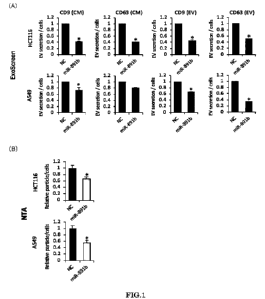
[0009] [FIG. 1] In FIG. 1, (A) shows graphs showing, regarding transformants
(miR-891b) transfected with miR-891b and transformants (NC) as negative
controls, the relative values of the amounts of EVs measured by the ExoScreen
method; and (B) shows graphs showing the relative values of the amounts of
EVs measured by the NTA method.
[FIG. 21 In FIG. 2, (A) shows graphs showing, regarding transformants
(siPSAT1) transfected with siPSAT1 and transformants (NC) as negative
controls, the relative values of the amounts of EVs measured by the ExoScreen
method; and (B) shows graphs showing the relative values of the amounts of
EVs measured by the NTA method.
[FIG. 31 In FIG. 3, (A) shows graphs showing, regarding the above-
described transformants (miR-891b) and the above-described transformants
(NC), the relative values of PSAT1 gene expression levels, (B) shows graphs
showing the relative value of a PSAT1 protein, (C) shows the relationship
between the 3' UTR of the PSAT1 gene and miR-891b, and (D) shows a graph
showing the PSAT1 expression levels in transformants transfected with the
PSAT1 gene and miR-891b.
[FIG. 41 FIG. 4 shows a graph showing, regarding transformants
(siPSAT1) obtained by transfecting various types of cancer cells with siPSAT1
and transformants (NC) as negative controls, the relative values of the
amounts
of secreted EVs measured by the NTA method.
[FIG. 51 FIG. 5 shows graphs showing, regarding transformants
(siPSAT1) obtained by transfecting cancer cells with siPSAT1 and
transformants (NC) as negative controls, the relative amounts of CD63-positive
EVs in the cells.
CA 03172262 2022- 9- 19
4
[FIG. 61 FIG. 6 shows results obtained when transformants (siPSAT1)
obtained by transfecting cancer cells with siPSAT1 and transformants (NC) as
negative controls were cultured in a serine-deficient medium or a serine-
containing medium. (A) shows graphs showing the relative values of the
amounts of secreted EVs measured by the ExoScreen method, and (B) shows
graphs showing the relative values of the amounts of secreted EVs measured by
the NTA method.
[FIG. 71 FIG. 7 shows graphs showing the relative amounts of CD63-
positive EVs in cells.
[FIG. 81 FIG. 8 shows graphs showing, regarding cancer cells cultured
in the presence of an inhibitor of the serine synthesis, the relative values
of the
amounts of secreted EVs measured by the NTA method.
[FIG. 91 FIG. 9 shows graphs showing, regarding transformants
(siPSAT1) obtained by transfecting various types of cancer cells with siPSAT1
and transformants (NC) as negative controls, the relative values of the
amounts
of secreted EVs measured by the NTA method.
[FIG. 101 FIG. 10 shows the results regarding a breast cancer
metastatic cell line MDA-MB-231_Luc_D3H2LN. (A) shows a graph showing
the relative values of the amounts of secreted EVs measured by the NTA
method, and (B) shows Western blot pictures indicating the expression of a
PSAT1 protein.
[FIG. 11] FIG. 11 shows the results regarding mice implanted with a
breast cancer metastatic cell line. (A) shows a graph showing the tumor
volume of the primary lesion (mammary gland) of the mice, and (B) is a graph
showing the tumor weight of the primary lesion (mammary gland).
[FIG. 121 FIG. 12 shows the results regarding lung cancer cells after
silencing of PSAT1 or PHGDH. (A) shows the results regarding the cell
viability, (B) shows a graph showing the relative values of the amounts of
secreted EVs measured by the NTA method, and (C) is a graph showing the
relative values of the amounts of secreted EVs measured by the ExoScreen
CA 03172262 2022- 9- 19
5
method.
[FIG. 131 FIG. 13 shows the results regarding colorectal cancer cells
after silencing of PSAT1 or PHGDH. (A) shows the results regarding the cell
viability, (B) shows a graph showing the relative values of the amounts of
secreted EVs measured by the NTA method, and (C) is a graph showing the
relative values of the amounts of secreted EVs measured by the ExoScreen
method.
[FIG. 141 FIG. 14 shows graphs showing the amount of secreted EVs
per cell. The graph shown in (A) shows the result regarding the normal
epithelial cells in the large intestine, and the graph shown in (B) shows the
result regarding the normal epithelial cells in the lung.
Description of Embodiments
[00101 The extracellular vesicle (EV) secretion reducing agent of the present
invention is hereinafter referred to as "EV secretion reducing agent".
[00111 In the EV secretion reducing agent of the present invention, for
example, the inhibitor is an expression reducing substance that reduces
expression of an enzyme protein in the serine synthesis pathway or a catalytic
function reducing substance that reduces a catalytic function of an enzyme
protein in the serine synthesis pathway.
[0012] In the EV secretion reducing agent of the present invention, for
example, the serine synthesis pathway is a synthesis pathway that includes
PSAT1.
[0013] In the EV secretion reducing agent of the present invention, for
example, the inhibitor of the serine synthesis pathway is an expression
reducing substance or catalytic function reducing substance for a PSAT1
protein.
[00141 In the EV secretion reducing agent of the present invention, for
example, the inhibitor of the serine synthesis pathway is an expression
reducing substance or catalytic function reducing substance for a PHGDH
CA 03172262 2022- 9- 19
6
protein.
[0015] In the EV secretion reducing agent of the present invention, for
example, the inhibitor of the serine synthesis pathway is an expression
reducing substance or catalytic function reducing substance for a PSPH
protein.
[00161 In the EV secretion reducing agent of the present invention, for
example, the cell is a cancer cell.
[00171 In the EV secretion reducing agent of the present invention, for
example, the cancer cell is at least one selected from the group consisting of
colorectal cancer cells, lung cancer cells, melanoma cells, breast cancer
cells,
pancreas cancer cells, and multiple myeloma cells.
[00181 In the EV secretion reducing agent of the present invention, for
example, the cell is a virus-infected cell.
[00191 In the EV secretion reducing agent of the present invention, for
example, the inhibitor is a low molecular weight compound, a protein, or a
peptide.
[00201 In the EV secretion reducing agent of the present invention, for
example, the expression reducing substance for the enzyme protein is at least
one selected from the group consisting of substances that reduce transcription
from a gene encoding the enzyme protein, substances that degrade a
transcription product resulting from transcription, and substances that reduce
translation of the transcription product into a protein.
[0021] In the EV secretion reducing agent of the present invention, for
example, the expression reducing substance is at least one nucleic acid
substance selected from the group consisting of miRNAs, siRNAs, antisenses,
and ribozymes.
[0022] In the EV secretion reducing agent of the present invention, for
example, the expression reducing substance is an expression vector for
expression of the nucleic acid substance.
[0023] In the EV secretion reducing agent of the present invention, for
example, the function reducing substance for the protein is an activity
CA 03172262 2022- 9- 19
7
inhibitory substance or an activity neutralizing substance for the enzyme
protein.
[00241 In the EV secretion reducing agent of the present invention, for
example, the activity neutralizing substance is an antibody or antigen-binding
fragment against the protein.
[0025] In the EV secretion reducing agent of the present invention, for
example, the function reducing substance is an expression vector for
expression
of the activity neutralizing substance.
[00261 In the EV secretion reducing method of the present invention, for
example, the cell is a cancer cell.
[00271 In the EV secretion reducing method of the present invention, for
example, the cancer cell is at least one selected from the group consisting of
colorectal cancer cells, lung cancer cells, melanoma cells, breast cancer
cells,
pancreas cancer cells, and multiple myeloma cells.
[00281 In the EV secretion reducing method of the present invention, for
example, the cell is a virus-infected cell.
[00291 In the EV secretion reducing method of the present invention, for
example, the administration target is a human or a non-human animal.
[00301 In the EV secretion reducing method of the present invention, for
example, the administration is performed in vivo or in vitro.
[0031] In the screening method for a candidate substance for the EV secretion
reducing agent of the present invention, for example, the cell is a cancer
cell.
[0032] In the screening method for a candidate substance for the EV secretion
reducing agent of the present invention, for example, the cancer cell is at
least
one selected from the group consisting of colorectal cancer cells, lung cancer
cells, melanoma cells, breast cancer cells, pancreas cancer cells, and
multiple
myeloma cells.
[0033] In the screening method for a candidate substance for the EV secretion
reducing agent of the present invention, for example, the cell is a virus-
infected
cell.
CA 03172262 2022- 9- 19
8
[00341 <Extracellular Vesicle Secretion Reducing Agent>
The extracellular vesicle (EV) secretion reducing agent (hereinafter
referred to as "EV secretion reducing agent") of the present invention is an
agent for reducing extracellular vesicle secretion from cells. As described
above, the EV secretion reducing agent of the present invention is
characterized
in that it contains an inhibitor of a serine synthesis pathway. The EV
secretion reducing agent of the present invention is characterized in that it
contains the inhibitor, and there is no particular limitation on other
configurations and conditions. As to the EV secretion reducing agent of the
present invention, reference can be made to the following descriptions
regarding the EV secretion reducing method and the like of the present
invention.
[0035] In the present invention, the inhibitor may be an expression reducing
substance for an enzyme protein in the serine synthesis pathway or a catalytic
function reducing substance for an enzyme protein in the serine synthesis
pathway. The present invention is characterized in that it is based on the
finding that the expression behavior of the serine synthesis pathway regulates
EV secretion and thus the EV secretion from cells can be reduced by inhibiting
the synthesis of serine, as described above. Accordingly, the type of
substance
used for inhibiting the serine synthesis and the method for inhibiting the
serine
synthesis are not limited by any means.
[00361 The inhibitor need only be capable of inhibiting the serine synthesis
pathway, and there is no particular limitation on how the inhibitor inhibits
the
serine synthesis pathway. In other words, the inhibitor may inhibit the serine
synthesis pathway by reducing the expression of an enzyme protein in the
serine synthesis pathway or by reducing the catalytic function of an enzyme
protein in the serine synthesis pathway. In the former case, the inhibitor is
an
expression reducing substance for the enzyme protein in the serine synthesis
pathway, for example. In the latter case, the inhibitor is a catalytic
function
reducing substance for the enzyme protein in the serine synthesis pathway, for
CA 03172262 2022- 9- 19
9
example. The EV secretion reducing agent of the present invention may
contain, for example, either one of the expression reducing substance and the
catalytic function reducing substance or both the expression reducing
substance
and the catalytic function reducing substance as the above-described
inhibitor,
which is an active ingredient.
[00371 The inhibitor is not limited to particular types of substances, and may
be, for example, a low molecular weight compound such as a nucleic acid
substance, a protein such as an antibody, or a peptide such as an antigen-
binding fragment.
[00381 The expression reducing substance is not limited to particular
substances, and may be, for example, a substance that reduces either
transcription or translation during expression of the enzyme protein
(hereinafter also referred to as "target protein") from the gene encoding the
enzyme protein (hereinafter also referred to as "target gene"). Examples of
the
reduction of transcription include inhibition of transcription from DNA to an
mRNA precursor, inhibition of RNA processing to form a mature mRNA from an
mRNA precursor, and degradation of an mRNA precursor or a mature mRNA.
Examples of the reduction of translation include inhibition of translation
from a
mature mRNA and inhibition of modification of a translation product.
[00391 The expression reducing substance is, for example, a nucleic acid
substance (hereinafter also referred to as "nucleic acid-type reducing
substance"), and may be embodied as a nucleic acid substance that inhibits the
expression as it is (first embodiment) or may be embodied in the form of a
precursor that converts into a state capable of reducing the expression when
it
is in vivo, in vitro, or ex vivo (second embodiment).
[00401 The expression reducing substance of the first embodiment is, for
example, an antigene substance, an antisense (antisense oligonucleotide), an
RNA interference (RNAD substance, or a ribozyme. The RNAi substance is, for
example, siRNA or miRNA. The antigene substance inhibits mRNA
transcription, for example. The antisense and miRNA inhibit translation from
CA 03172262 2022- 9- 19
10
mRNA, for example. The siRNA and ribozyme degrade mRNA, for example.
These expression reducing substances may target either the entire region or a
partial region of the target gene, for example. As specific examples, the
antisense and miRNA can be designed, for example, so as to bind to the 3' UTR
region of mRNA transcribed from the target gene, and the siRNA and ribozyme
can be designed, for example, so as to fully complementarily bind to a partial
region of mRNA transcribed from the target gene.
[0041] The expression reducing substance of the first embodiment can be
obtained by a screening method to be described below or can be designed from
the sequence of the target gene, for example.
[0042] The expression reducing substance of the first embodiment may be
either a single strand or a double strand, for example. There is no particular
limitation on the structural units of the expression reducing substance.
Examples of the structural unit include a deoxyribonucleotide backbone and a
ribonucleotide backbone, each including a sugar, a base such as purine or
pyrimidine, and phosphoric acid. Other examples of the structural unit
include non-nucleotide backbones including a base such as pyrrolidine or
piperidine. These backbones may be either modified or unmodified. The
structural units may be natural structural units or unnatural, i.e.,
artificial
structural units, for example. The expression reducing substance may be
composed of the same structural units or two or more types of structural
units,
for example.
[0043] The expression reducing substance of the second embodiment is, as
described above, the precursor, and specific examples thereof include
precursors
that express an expression reducing substance of the first embodiment. By
administering the precursor to a target, the expression reducing substance of
the first embodiment can be expressed and allowed to function in, for example,
an in vivo, in vitro, or ex vivo environment.
[00441 The precursor may be in a form that includes the expression reducing
substance of the first embodiment and a linker, for example. As a specific
CA 03172262 2022- 9- 19
11
example, the precursor may be in a form in which both strands of siRNA are
linked together via the linker. The precursor in such a form can generate
(express) a double-stranded siRNA when, for example, the linker is removed
from the precursor upon cleavage of the precursor in an in vivo, in vitro, or
ex
vivo environment. A specific example of the precursor is shRNA that
generates siRNAs when, for example, it is cleaved.
[0045] Alternatively, the precursor may be, for example, an expression vector
with the coding sequence of the expression reducing substance of the first
embodiment inserted therein. Such an expression vector can cause the
expression of the expression reducing substance of the first embodiment in,
for
example, an in vivo, in vitro, or ex vivo environment. The expression vector
may have the coding sequence of, for example, the above-described precursor
such as shRNA inserted therein. The expression vector is not limited to
particular types of expression vectors, and may be, for example, a plasmid
vector or a viral vector. Examples of the viral vector include adenovirus
vectors and Sendai virus vectors.
[00461 The catalytic function reducing substance is, for example, an activity
inhibitory substance that inhibits the activity of the enzyme protein or an
activity neutralizing substance that neutralizes the activity of the enzyme
protein.
[00471 The activity inhibitory substance is not limited to particular
substances,
and may be a low molecular weight compound or the like.
[00481 The activity neutralizing substance may be, for example, an antibody or
antigen-binding fragment (antigen-binding peptide) against the enzyme protein
(such an antibody and antigen-binding fragment are also collectively referred
to
as "antibody-type reducing substances" hereinafter). Since the antibody-type
reducing substance can inhibit the function of the enzyme protein by, for
example, binding to the enzyme protein, it is also referred to as a
neutralizing
antibody or a neutralizing antigen-binding fragment. The antibody-type
reducing substance can also be obtained by, for example, a screening method to
CA 03172262 2022- 9- 19
12
be described below.
[00491 The antibody may be either a monoclonal antibody or a polyclonal
antibody, for example. There is no particular limitation on the isotype
thereof,
and examples of the isotype include IgG, IgM, and IgA. In the case where the
antibody is administered to a human, it preferably is, for example, a fully
human antibody, a humanized antibody, or a chimeric antibody.
[00501 The antigen-binding fragment need only be capable of, for example,
recognizing a target site of the target protein and binding thereto, and
examples of the antigen-binding fragment include fragments that include a
complementarity-determining region (CDR) of the antibody. Specific examples
of the antigen-binding fragment include fragments such as Fab, Fab', and
F(ab').
[0051] The catalytic function reducing substance may be, for example, of the
first embodiment in which the catalytic function reducing substance inhibits
the catalytic function of the enzyme protein as is, or may be of the second
embodiment in which the catalytic function reducing substance is a precursor
that converts into a state capable of reducing the expression in an in vivo,
in
vitro, or ex vivo environment. The catalytic function reducing substance of
the
first embodiment is, for example, the above-described antibody-type reducing
substance. The precursor of the second embodiment may be, for example, an
expression vector with the coding sequence of a protein or peptide that
inhibits
the catalytic function of the enzyme protein inserted therein. The expression
vector is not limited to particular types of expression vectors, and may be,
for
example, a plasmid vector or a viral vector, as with the expression vector
described above in connection with the expression reducing substance.
[0052] The catalytic function reducing substance may be, for example, a
substance that allows the enzyme protein to have catalytic activity and to
maintain its function as a catalyst but inhibits the conditions under which
the
enzyme protein can exhibit the function. As a specific example, the catalytic
function reducing substance may be a reducing substance that reduces a
CA 03172262 2022- 9- 19
13
substrate that the enzyme protein needs in order to exhibit its function or a
reducing substance that alters the substrate. The reduction of the substrate
may be achieved by, for example, inhibiting the generation of the substrate or
degrading the substrate.
[0053] The serine synthesis pathway may be, for example, a synthesis pathway
(I) represented by the following formula. The EV secretion reducing agent of
the present invention preferably contains, for example, an inhibitor of the
serine synthesis pathway (I) that includes PSAT1. As described above, the
inventors of the present invention found that the expression behavior of the
serine synthesis pathway regulates the EV secretion. Specifically, the
inventors found that, for example, abnormal cells such as cancer cells exhibit
a
higher level of EV secretion than normal cells owing to overexpression of
genes
and proteins encoded by these genes in the serine synthesis pathway. The
inventors confirmed that an increase in EV secretion from the abnormal cells
can be reduced by reducing (inhibiting) the expression of the gene encoding a
protein in this serine synthesis pathway, specifically, for example, a protein
in
the serine synthesis pathway (I) shown below or by reducing (inhibiting) the
function of such a protein, thereby achieving the present invention. Reduction
of EV secretion in the present invention can also be referred to as, for
example,
reduction of an increase in EV secretion, and more specifically, it can also
be
referred to as, for example, reduction of an increase in EV secretion so as
not to
exceed the normal EV secretion level.
(I)
PHGDH PSAT1 PSPH
3-PG -. P-Pyr -. P-Ser-. Serine
[00541 PHGDH stands for D-3-phosphoglycerate dehydrogenase. Human
PHGDH protein and the PHGDH gene encoding it are registered under Gene
CA 03172262 2022- 9- 19
14
ID: 26227 in a database (the Genetic Testing Registry (GTR)). An expression
reducing substance for PHGDH can be set, for example, based on the sequence
of the PHGDH gene. As a catalytic function reducing substance for PHGDH,
an inhibitor such as NCT-503 or CBR-5884 or a neutralizing antibody such as a
PHGDH antibody can be used, for example.
[0055] PSAT1 stands for phosphoserine aminotransferase 1. Human PSAT1
protein and the PSAT1 gene encoding it are registered under Gene ID: 29968 in
the database (GTR). An expression reducing substance for PSAT1 can be set,
for example, based on the sequence of the PSAT1 gene, and specific examples
thereof include miR-891b. As a catalytic function reducing substance for
PSAT1, a neutralizing antibody such as a PSAT1 antibody can be used, for
example.
[00561 PSPH stands for phosphoserine phosphatase. Human PSPH protein
and the PSPH gene encoding it are registered under Gene ID: 5723 in the
database (GTR). An expression reducing substance for PSPH can be set, for
example, based on the sequence of the PSPH gene. As a catalytic function
reducing substance for PSPH, a neutralizing antibody such as a PSPH antibody
can be used, for example.
[00571 In the present invention, the inhibitor of the serine synthesis pathway
may be, for example, any one of expression reducing substances and catalytic
function reducing substances for a PSAT1 protein, expression reducing
substances and catalytic function reducing substances for a PHGDH protein,
and expression reducing substances and catalytic function reducing substances
for a PSPH protein. The inhibitor of the serine synthesis pathway may include
any one of them or may include two or more of them.
[00581 The EV secretion reducing agent of the present invention may contain,
for example, only the above-described active ingredient or may further contain
other additive ingredients. The additive ingredients are not limited to
particular ingredients, and examples thereof include ingredients to be
described
below, which are preferably pharmacologically acceptable. The additive
CA 03172262 2022- 9- 19
15
ingredients can be set as appropriate according to, for example, the method
for
administering the EV secretion reducing agent, the administration target, and
the dosage form.
[00591 One example of the additive ingredient is a vehicle, for example.
Examples of the vehicle include liquid media such as aqueous solvents, alcohol
solvents, polyalcohol solvents, lipid solvents, and mixed solvents thereof
(e.g.,
emulsifying solvents), lactose, and starch. Examples of the aqueous solvents
include water, physiological saline, and isotonic solutions containing sodium
chloride and the like. Examples of the lipid solvents include soybean oil.
Other examples of the additive ingredients include: binding agents such as
starch glue; disintegrants such as starch and carbonate; and lubricants such
as
talc and wax. The additive ingredients may also include, for example, a DDS
agent for delivering the active ingredient to a target site.
[00601 In the present invention, cells to be subjected to reduction of EV
secretion are not limited to particular cells, and may be cells to be
subjected to
reduction of extracellular vesicle secretion therefrom in order to examine the
influence of the secretion or the influence of reducing the secretion. The
cells
may be, for example, normal cells or cells that are abnormal for an item of
interest. The state of being abnormal for the item of interest is not limited
to
particular states. For example, when the item of interest is cancer, the
abnormal cells may be cancer cells, and when the item of interest is viral
infection, the abnormal cells may be virus-infected cells. Since the present
invention can analyze the mechanisms of development, metastasis, treatment,
and the like of cancer, the cells are preferably cancer cells. In particular,
since
extracellular vesicles play a role in cell-to-cell information transmission as
described above, the cells are preferably cancer cells of primary cancer that
may
metastasize to other organs (parts of the body). The cancer cells are not
limited to particular cancer cells, and examples thereof include colorectal
cancer
cells, lung cancer cells, melanoma cells, breast cancer cells, pancreas cancer
cells, and multiple myeloma cells. Also, since the present invention can
CA 03172262 2022- 9- 19
16
analyze the mechanism of viral infection, the cells are preferably virus-
infected
cells. The virus is not limited to particular types of viruses, and examples
thereof include influenza viruses and coronaviruses.
[0061] In the present invention, extracellular vesicles are, for example,
exosomes, microvesicles (submicroscopic vesicles), and apoptotic bodies
secreted
through endocytosis pathways. In particular, the extracellular vesicles in the
present invention are exosomes, for example. In general, exosomes can be
detected using a marker molecule such as Alix, Tsg101, CD81, CD63, CD9, or
flotillin.
[0062] The method for using the EV secretion reducing agent of the present
invention is not limited particular methods. For example, the EV secretion
reducing agent may be added to a target to be subjected to reduction of
extracellular vesicle secretion. The method for adding the EV secretion
reducing agent is not limited to particular methods, and the EV secretion
reducing agent may be added in vivo, in vitro, or ex vivo, for example. A
target
to which the EV secretion reducing agent of the present invention is to be
added
is, for example, cells or tissue, or may be a living organism. There is no
particular limitation on the type of the cells and tissue and the part (organ)
of
the living organism, and examples thereof include the large intestine, lung,
skin, breast, breast duct, mammary gland, pancreas, and bone marrow. The
cells and tissue may be, for example, those isolated from a living organism,
or
may be a cell line or a culture thereof. The cells and tissue as the target
may
be, for example, derived from a human or a non-human animal. The living
organism as the target may be, for example, a human or a non-human animal.
Examples of the non-human animal include mammals such as mice, rats,
rabbits, horses, sheep, cows, and camels. When the target to which the EV
secretion reducing agent is to be added is derived from a non-human animal or
is a non-human animal, the EV secretion reducing substance is, for example,
preferably a reducing substance that relatively specifically acts on a target
protein (the enzyme protein) or target gene derived from this particular non-
CA 03172262 2022- 9- 19
17
human animal, and when the target is derived from a human or is a human,
the reducing substance is, for example, preferably an reducing substance that
relatively specifically acts on a target protein or target gene derived from a
human.
[0063] <EV Secretion Reducing Method>
The EV secretion reducing method of the present invention is a method
for reducing EV secretion from cells, and is characterized in that it includes
administering the EV secretion reducing agent of the present invention to an
administration target. The point of the present invention lies in using the EV
secretion reducing agent of the present invention, and there is no particular
limitation on other steps and conditions. As to the EV secretion reducing
method of the present invention, reference can be made to the above
description
regarding the EV secretion reducing agent of the present invention.
[00641 When the administration target is cells, EV secretion from the cells
can
be reduced by, for example, adding the EV secretion reducing agent in the
presence of a medium and incubating the cells. Conditions for the incubation
are not limited to particular conditions, and the medium, temperature, time,
humidity, and other conditions can be set according to, for example, the type
of
the cells.
[00651 When the administration target is tissue, EV secretion from cells that
constitute the tissue can be inhibited by, for example, adding the EV
secretion
reducing agent in the presence of a medium and incubating the tissue.
Conditions for the incubation are not limited to particular conditions. For
example, the medium, temperature, time, humidity, and the like can be set
according to, for example, the type and the size of the tissue.
[00661 When the administration target is a living organism, the method for
administering the EV secretion reducing agent is not limited to particular
methods, and may be parenteral administration, oral administration,
intravenous administration, or the like. Administration conditions are not
limited to particular conditions and can be determined as appropriate
according
CA 03172262 2022- 9- 19
18
to, for example, the type of the living organism and the type of the organ as
the
administration target.
[00671 When the administration method is parenteral administration, an
administration site may be, for example, a target organ, i.e., an organ that
includes cells to be subjected to reduction of EV secretion (such an organ is
also
referred to as "target site"), or a site from which the inhibitor as the
active
ingredient of the EV secretion reducing agent can be delivered to the target
site.
As a specific example, when the target cells are, for example, large
intestinal
cells, the administration site may be the large intestine as the target site
or a
site from which the inhibitor can be delivered to the large intestine. When
the
target cells are, for example, lung cells, the administration site may be the
lung
as the target site, or a site from which the inhibitor can delivered to the
lung.
The same applies to other organs. Examples of the parenteral administration
method include injection to an affected area, intravenous injection,
subcutaneous injection, intradermal injection, intravenous infusion, and
transdermal administration. The form of the EV secretion reducing agent is
not limited to particular forms, and can be set as appropriate according to
the
administration method and the like, as described above. As to the form of the
EV secretion reducing agent, reference can be made to the above description
thereon.
[00681 In the case of parenteral administration, the dosage form is not
limited
to particular forms, and can be determined as appropriate according to the
administration method. For example, the EV secretion reducing agent may be
in the form of liquid, cream, or gel, and can be prepared by mixing the
inhibitor
with a medium. Of the above-described examples of the medium, the aqueous
solvent is, for example, physiological saline or an isotonic solution, the
lipid
solvent is, for example, soybean oil, and the emulsifying solvent is, for
example,
a mixture thereof. Such a parenteral administration agent may further
contain, for example, alcohol, polyalcohol, and/or a surfactant. Also, the
parenteral administration agent may contain a DDS agent for effectively
CA 03172262 2022- 9- 19
19
delivering the inhibitor to a target site from a site other than the target
site.
In particular, in order to effectively deliver the inhibitor to, for example,
cancer
cells in tissue as the target site, the parenteral administration agent may
contain, for example, a DDS agent that specifically recognizes the cancer
cells.
[00691 In the case of oral administration, the dosage form of an oral
administration agent is not limited to particular forms, and examples thereof
include tablets, pills, granules, powder medicines, capsules, and syrups. The
oral administration agent may contain, for example, a diluent, a vehicle,
and/or
a carrier. The oral administration agent may also contain, for example, a DDS
agent for effectively delivering the inhibitor to the target site. In
particular, in
order to effectively deliver the inhibitor to, for example, cancer cells in
tissue as
the target site, the oral administration agent may contain, for example, a DDS
agent that specifically recognizes the cancer cells.
[00701 In administration to a living organism, the administration conditions
of
the EV secretion reducing agent of the present invention can be determined as
appropriate according to, for example, the age, the body weight, the type of
organ as the administration target, and the sex.
[0071] The inventors of the present invention have reported, as the mechanism
of cancer metastasis, that extracellular vesicles are secreted from cancer
cells of
primary cancer and the cancer metastasis is caused via cell-to-cell
information
transmission mediated by the extracellular vesicles. As described above, by
administering the EV secretion reducing agent of the present invention to a
target site (cancerous organ) of a patient affected by cancer, extracellular
vesicle
secretion from cancer cells in the target site is reduced, whereby progression
of
cancer and metastasis of cancer to other organs can be reduced. Accordingly,
the EV secretion reducing agent of the present invention can also be used as a
therapeutic agent for cancer, for example. The term "treatment" as used in the
present specification encompasses, for example, not only what are called
practices to alleviate the progression of cancer, to treat cancer, and the
like but
also preventive practice to prevent the onset or recurrence of cancer. The EV
CA 03172262 2022- 9- 19
20
secretion reducing agent of the present invention may be used for, for
example,
any one of the above-described purposes or two or more of the above-described
purposes.
[0072] In this case, in the administration to a living organism, the
administration conditions of the EV secretion reducing agent of the present
invention can be determined as appropriate according to, for example, in
addition to the items given above as examples, the type of cancer (e.g.,
colorectal cancer, lung cancer, melanoma, breast cancer, pancreas cancer, and
multiple myeloma) and the degree of progression of cancer. The living
organism may be, for example, a subject affected by cancer from the viewpoint
of treating the cancer, or may be a subject not affected by cancer or a
subject
who may or may not affected by cancer from the viewpoint of preventing the
cancer.
[0073] It is to be noted, however, that the intended use of the EV secretion
reducing agent of the present invention is not limited to those given above as
examples. In recent years, for example, it has been reported that EVs such as
exosomes are involved in cell-to-cell communication in the body. Specific
examples thereof include transmission of a virus infecting a subject (for
example, an influenza virus or a coronavirus) in the body. Accordingly, the EV
secretion reducing agent of the present invention reduces, for example, viral
transmission through reduction of EV secretion, thereby enabling inhibition of
the spread of viral infection in the body.
[00741 EVs whose secretion is reduced by the EV secretion reducing agent of
the present invention are known to play a role in cell-to-cell information
transmission by, for example, transferring an information cargo in a certain
cell
to another cell, as described above. Accordingly, the EV secretion reducing
agent of the present invention also can be referred to as, for example, a cell-
to-
cell information transmission reducing agent, and the EV secretion reducing
method of the present invention also can be referred to as a cell-to-cell
information transmission reducing method.
CA 03172262 2022- 9- 19
21
[0075] <Screening Method>
The screening method of the present invention is a screening method for
a candidate substance for an EV secretion reducing agent for reducing EV
secretion from cells, and the screening method is characterized in that it
includes selecting, out of test substances, an inhibitory substance that
inhibits
a serine synthesis pathway as a candidate substance that reduces extracellular
vesicle secretion from cells. The cells are not limited to particular types of
cells
and are as described above. Examples of the cells include cancer cells. As
described above, examples of the cancer cells include colorectal cancer cells,
lung cancer cells, melanoma cells, breast cancer cells, pancreas cancer cells,
and
multiple myeloma cells.
[00761 In the case where a candidate substance for the expression reducing
substance is selected through screening, the screening method of the present
invention includes, for example: in an expression system for expressing the
above-described enzyme protein (target protein) from the above-described gene
(target gene) encoding the enzyme protein, expressing the target protein in
the
presence of each of the test substances; detecting expression of the target
protein in the expression system; and selecting, as the candidate substance, a
test substance in the presence of which the expression level of the target
protein
is relatively low as compared with the expression level of the target protein
in a
control expression system without the test substance.
[00771 In the case where the catalytic function reducing substance is selected
through screening, the screening method of the present invention includes, for
example: bringing each of the test substances into contact with the above-
described enzyme protein (target protein); detecting the catalytic activity of
the
target protein; and selecting, as the candidate substance, a test substance
that
causes the enzyme protein to exhibit relatively low catalytic activity as
compared with the enzyme protein in a control system without the test
substance.
[00781 In the case where the activity neutralizing substance is selected
CA 03172262 2022- 9- 19
22
through screening as a candidate substance for the catalytic function reducing
substance, the screening method of the present invention includes, for
example:
bringing each of the test substances into contact with the above-described
enzyme protein (target protein); detecting binding of the target protein and
the
test substance; and selecting, as the candidate substance, a test substance
that
has bound to the target protein.
[00791 <Use>
The present invention relates to the above-described inhibitor for use in
reduction of extracellular vesicle secretion from cells. As to the above-
described reducing substances, reference can be made to the above description
regarding the EV secretion reducing agent of the present invention and the EV
secretion reducing method of the present invention.
Examples
[00801 Next, examples of the present invention will be described. It is to be
noted, however, that the present invention is by no means limited by the
following examples. Commercially available reagents were used in accordance
with their protocols, unless otherwise stated.
[0081] (Cells)
A human colon adenocarcinoma cell line HCT116 (ATCC CCL-247) was
used as colorectal cancer cells, and an adenocarcinomic human alveolar basal
epithelial cell line A549 (ATCC CCL-185) was used as lung cancer cells. A
human melanoma cell line A375 (ATCC CRL-1619) was used as melanoma
cells. A human breast cancer cell line MM231 (ATCC HTB-26) was used as
breast cancer cells. A human pancreatic adenocarcinoma cell line Panc-1
(ATCC CRL-1469) was used as pancreas cancer cells. A human multiple
myeloma cell line RPMI 8226 (ATCC CCL-155) was used as human multiple
myeloma cells.
[0082] (Nucleic Acid Molecules)
miR-891b (also referred to as "miR-891b mimic", SEQ ID NO: 1:
CA 03172262 2022- 9- 19
23
GCAACUUACCUGAGUCAUUGA) (Product No. 4464066, Ambion) was used as
a nucleic acid molecule miRNA, and miRNA Mimic Negative Control #1
(4464058) (Ambion) was used as a negative control nucleic acid molecule
miRNA. siPSAT1 (Product No. siGENOME SMART pool siRNA M-010398,
Dharmacon) was used as a nucleic acid molecule siRNA, and ALL STAR
Negative Control siRNA (SI03650318) (Qiagen) was used as a negative control
nucleic acid molecule siRNA.
[0083] (Cell Culture)
The medium used for MM231 was an RPMI complete medium obtained
by adding 10% heat-inactivated fetal bovine serum (FBS) and an antibiotic-
antimycotic solution (Gibco) to an RPMI 1640 medium (Gibco). The medium
used for HCT116 was a Maccoy 5A complete medium obtained by adding 10%
heat-inactivated FBS and an antibiotic-antimycotic agent to a Maccoy 5A
medium. The medium used for each of the other types of cells was a DMEM
complete medium obtained by adding 10% heat-inactivated FBS and an
antibiotic-antimycotic agent to a DMEM medium (Gibco). The culture
conditions were set to 37 C, 5% carbon dioxide, and 95% relative humidity
(RH). About 100,000 cells were seeded in 18 mL of each of the above-described
complete media and incubated for 3 to 4 days. Cells that had undergone less
than 20 passages were used.
[00841 (Collection of Secreted EVs)
The cultured cancer cells were washed with phosphate buffered saline
(PBS), and the medium was replaced with advanced RPMI or advanced DMEM
containing the above-described antibiotic-antimycotic agent and 2 mmol/L L-
glutamine (Gibco). The adjusted culture solution after the replacement was
centrifuged at 2,000 x g for 10 minutes to remove the cells, and the
supernatant
obtained was filtered through a 0.22 pm filter (Millipore). The resulting
filtrate was centrifuged at 110,000 x g for 70 minutes, whereby pellets
containing concentrated EVs were obtained. The pellets were washed with 11
mL of PBS, further ultracentrifuged at 110,000 x g for 70 minutes, and
collected
CA 03172262 2022- 9- 19
24
again.
[0085] (ExoScreen Method)
EV detection was performed using an AlphaLISA reagent (Perkin
Elmer) composed of AlphaScreen streptavidin-coated donor beads (6760002),
AlphaLISA unconjugated acceptor beads (6062011), and an AlphaLISA
universal buffer (AL001F), a 96-well half-area white plate (6005560, Perkin
Elmer), and a detection device EnSpire Alpha 2300 Mutilabel plate reader
(Perkin Elmer). Specifically, the EV detection was performed as follows.
Each well of the plate was filled with 5 pL of EVs or 10 pL of CM (cell
culture
supernatant), 10 pL of a 5 nmol/L biotinylated antibody solution prepared
using
the above-described buffer, and 10 pL of 50 pg/mL AlphaLISA acceptor bead-
conjugated antibody. For detection of CD9/CD9 double-positive EVs, a
biotinylated anti-human CD9 antibody and an anti-human CD9 antibody
conjugated with AlphaLISA acceptor beads were used. For detection of
CD9/CD63 double-positive EVs, a biotinylated anti-human CD9 antibody and
an anti-human CD63 antibody conjugated with AlphaLISA acceptor beads were
used. For detection of CD63/CD63 double-positive EVs, a biotinylated anti-
human CD63 antibody and an anti-human CD63 antibody conjugated with
AlphaLISA acceptor beads were used. The plate was incubated at 37 C for 1
hour, and then, 25 pL of 80 pg/mL AlphaScreen streptavidin-coated donor beads
were added thereto. The plate was incubated at 37 C for another 30 minutes
in the dark. Next, luminescence in the wells of the plate was measured using
the detection device with the excitation wavelength set to 680 nm and the
emission detection wavelength set to 615 nm. The antibodies used were both
commercially available products, namely, a mouse monoclonal anti-human CD9
(Clone 12Al2) and a CD63 antibody (Clone 8Al2) (both available from Cosmo
Bio).
[00861 (Analysis of EVs by Nanoparticle Tracking Analysis NTA)
The secreted EVs were collected and then suspended in PBS. Further,
a series of diluted solutions were prepared using PBS and subjected to
CA 03172262 2022- 9- 19
25
NanoSight particle tracking analysis (LM10, software ver. 2.03). In the above-
described particle tracking, at least five 60-second videos were taken for
each
sample with the camera level set to 14. Analysis settings were optimized and
kept constant between the samples. An EV concentration was calculated as
particles/cells in the culture solution, whereby a net EV secretion rate was
obtained. The results of EV measurement by the ExoScreen method correlated
with the results of EV measurement by the NTA analysis. This confirmed that
the ExoScreen method is capable of measuring the amount of secreted EVs.
[00871 (Transient Transfection Assay)
In a 6-well plate, 2 mL of the cancer cell suspension was seeded at 1.0 x
105 cells/well and incubated for 24 hours. Thereafter, 10 nmol]L of the
intended nucleic acid molecules were added thereto, and the cells were
transfected with the nucleic acid molecules using a transfection reagent
(product name: DharmaFECT Transfection Reagent 1). The nucleic acid
molecules used were the above-described miRNA and the above-described
siRNA. After the incubation for 24 hours, the medium was replaced with the
above-described advanced RPMI 1640 medium or advanced DMEM medium
containing the antibiotic-antimycotic agent and 2 mmol/L-glutamine (Gibeo).
48 hours after the replacement, total RNA was extracted, and the expression of
the gene of interest was measured by qPCR.
[00881 (RNA Extraction and qPCR Analysis)
The total RNA was extracted from the cultured cells using commercially
available reagents (trade name: QIAzol, trade name: miRNeasy Mini Kit,
Qiagen). Reverse transcription reactions were caused using a commercially
available kit (trade name: High-Capacity cDNA Reverse Transcription Kit,
Applied Biosystems) and random hexamer primers. Real-time PCR analysis
was performed using commercial kits (trade name: StepOne Plus, trade name:
TaqMan Universal PCR MasterMix, Thermo Fisher Scientific). mRNA
expression was normalized using 6-actin. As a probe for PSAT1, a TaqMan
probe (Applied Biosystems) was used.
CA 03172262 2022- 9- 19
26
[00891 Unless otherwise stated, data presented in the examples were each
expressed as the mean value the standard error. The statistical significance
was determined using a Student's t-test. In dot plots, each bar indicates the
median and the interquartile range, and the statistical significance was
determined by a Student's t-test. P < 0.05 was considered as statistically
significant.
* P < 0.05, ** P < 0.01.
[00901 [Example Al
[Example Al]
The present example identified target genes involved in regulation of
EV secretion in colorectal cancer and lung cancer.
[0091] (1) Detection of EVs secreted from cancer cells
Colorectal cancer cells HCT116 and lung cancer cells A549 were each
transfected with miR-891b. Culture supernatants (CM) of the resulting
transformants (miR-891b) and EV fractions containing EVs secreted from the
transformants (miR-891b) were collected. Then, according to the ExoScreen
method described above, the amounts of the secreted EVs were measured by
measuring the signal intensities. Also, the amounts of the secreted EVs
measured for the respective transformants were confirmed by the NTA analysis.
As a negative control, regarding a transformant (NC) obtained by transfecting
the cells A549 with miR-891b miRNA Mimic Negative Control #1, the amount
of secreted EVs was measured in the same manner. Then, assuming that the
amount of secreted EVs in the transformant (NC) was 1, the relative values (n
=
3) of the amounts of secreted EVs in the transformants (miR-891b) were
determined.
[0092] The results obtained are shown in FIG. 1. In FIG. 1, (A) shows graphs
showing the relative values of the amounts of secreted EVs measured by the
ExoScreen method, and (B) shows graphs showing the relative values of the
amounts of secreted EVs measured by the NTA method. As can be seen from
(A) in FIG. 1, in the transformants (miR-891b) obtained by transfecting the
CA 03172262 2022- 9- 19
27
colorectal cancer cells HCT116 and the lung cancer cells A549 with miR-891b,
the amounts of the secreted EVs observed in both the CMs and the EV fractions
were significantly lower than those observed in the transformants (NC). Also,
as can be seen from (B) in FIG. 1, similar results were obtained also by the
NTA
method. These results demonstrate that miR-891b reduces EV secretion in
colorectal cancer cells and lung cancer cells.
[0093] (2) Target Gene of miR-891b
In the above item (1), EV secretion was reduced by transfection with
miR-891b. The inventors of the present invention conducted further in-depth
studies and found that the PSAT1 gene is a target gene whose expression is
reduced by miR-891b. To support this finding, colorectal cancer cells HCT116
and lung cancer cells A549 were transfected with siRNA (siPSAT1) for reducing
the expression of the PSAT1 gene, and the amounts of secreted EVs were
measured by the ExoScreen method and the NTA method. Also, as negative
controls, the amounts of secreted EVs were measured in the same manner,
except that these cancer cells were transfected with ALL STAR negative control
siRNA. The results obtained are shown in FIG. 2. In FIG. 2, (A) shows
graphs showing the relative values of the amounts of secreted EVs measured by
the ExoScreen method, and (B) shows graphs showing the relative values of the
amounts of secreted EVs measured by the NTA method.
[00941 As can be seen from (A) in FIG. 2, in the transformants (siPSAT1)
obtained by transfecting the colorectal cancer cells HCT116 and the lung
cancer
cells A549 with siPSAT1, the amounts of the secreted EVs observed in both the
CMs and the EV fractions were significantly lower than those observed in the
transformants (NC). Also, as can be seen from (B) in FIG. 2, similar results
were obtained also by the NTA method. The fact that the downregulation of
the PSAT1 gene with siPSAT1 reduced the EV secretion and also the results in
the above item (1) demonstrate that the PSAT1 gene is the target gene of miR-
891b.
[0095] (3) Confirmation of Target Gene
CA 03172262 2022- 9- 19
28
The following test was conducted to examine whether the PSAT1 gene
described in the item (2) is a direct target gene of miR-891b.
[00961 First, expression of the PSAT1 gene was detected in the above-described
transformants (miR-891b) and the control transformants (NC) therefor. Then,
assuming that the expression levels in the transformants (NC) were 1, the
relative values of the expression levels in the respective transformants (miR-
891b) were determined. The results obtained are shown in (A) of FIG. 3. In
FIG. 3, (A) shows graphs showing the relative values of the PSAT1 gene
expression levels. Also, for the transformants (miR-891b) and the control
transformants (NC), the PSAT1 protein expression was detected by Western
blotting. As a control, 6-actin was also detected. The results obtained are
shown in (B) in FIG. 3. In FIG. 3, (B) shows Western blot pictures indicating
the PSAT1 protein expression.
[00971 Next, as can be seen from (C) in FIG. 3, miR-891b has a sequence that
perfectly matches a region consisting of seven contiguous bases in the 3' UTR
of
the human PSAT1 mRNA (SEQ ID NO: 2: UGGACUUAAUAAUGCAAGUUGC
is a partial region of the 3' UTR). Thus, the effect of miR-891b was examined
using the wild-type PSAT1 gene in which the above-described 7-base region in
the 3' UTR is the wild-type sequence (SEQ ID NO: 3: AAGTTGC) and a mutant-
type PSAT1 gene in which the above-described 7-base region in the 3' UTR is a
mutated sequence (SEQ ID NO: 4: TTCAACG). Specifically, the following
experiment was performed. The wild-type PSAT1 gene or the mutant-type
PSAT1 gene was subcloned into a plasmid vector psiCHECK2, and human
embryonic kidney cells (HEK293 cells) were transfected with this recombinant
vector together with miR-891b or the negative control miRNA using
Lipofectamine 3000. 48 hours after the transfection, the luciferase activity
of
the transformants was quantified using a plate reader according to the
operating procedure of a Dual-Luciferase Reporter Assay System.
[00981 The results obtained are shown in (D) of FIG. 3. In FIG. 3, (D) shows a
graph showing the PSAT1 expression levels as the relative luciferase activity.
CA 03172262 2022- 9- 19
29
[00991 As can be seen from (D) in FIG. 3, transfection with miR-891b reduced
the PSAT1 gene expression and the PSAT1 protein expression. Furthermore,
transfection with miR-891b significantly reduced the PSAT1 expression level in
the case where the wild-type PSAT1 in which the sequence of the region
recognized by miR-891b is the wild-type sequence was used, whereas
transfection with miR-891b did not reduce the PSAT1 expression level in the
case where the recognition region of miR-891b had a mutated sequence. From
these results, it was found that miR-891b cleaves PSAT1 upon recognition of
the 3' UTR of PSAT1. In other words, these results attest that the PSAT1 gene
is a direct target of miR-891b.
[01001 [Example A21
The present example examined whether reducing the expression of the
PSAT1 gene reduces EV secretion from various types of cancer cells.
[0101] Transfection with siRNA (siPSAT1) and measurement of the amount of
secreted EVs by the nanoparticle tracking analysis (NTA) method were
performed in the same manner as in the item (2) of Example Al, except that
melanoma cells A375, breast cancer cells M1V1231, pancreas cancer cells Panc-
1,
and multiple myeloma cells RPMI8226 were used. The results obtained are
shown in FIG. 4. FIG. 4 shows a graph showing the relative values of the
amounts of secreted EVs measured by the NTA method.
[0102] As can be seen from FIG. 4, in a transformant (siPSAT1) obtained by
transfecting each type of the cancer cells with siPSAT1, the amount of the
secreted EVs was significantly lower than that observed in the transformant
(NC) as the negative control therefor. From these results, it was found that
reducing the expression of PSAT1 can reduce EV secretion not only in
colorectal
cancer cells and lung cancer cells but also in various types of other cancer
cells.
From these results, it can be said that EV secretion from various types of
cancer
cells can be reduced by reducing the expression of PSAT1, thus enabling
treatment of these cancers and prevention of metastasis of these cancers to
other organs.
CA 03172262 2022- 9- 19
30
[0103] [Example A31
Colorectal cancer cells HCT116 and lung cancer cells A549 were
transfected with siRNA (siPSAT1) in the same manner as in the item (2) in
Example Al, and EV biogenesis after PSAT1 silencing was examined. For the
respective silenced transformants, CD63 and PSAT1 were examined by
immunofluorescence. As a result, accumulation of CD63 as an EV marker was
observed in cytoplasm after the PSAT1 silencing. Also, when these cells were
transfected with miR-891b, accumulation of CD63 was observed, as with the
above-described case. These results confirmed that, although the EV
production itself is maintained even when the PSAT1 expression is reduced, the
produced EVs accumulate in cytoplasm and reduce the secretion of the EVs
from the cells to the outside.
[0104] Further, coimmunostaining of the EV marker CD63 and also an early
endosome marker EEA1 or a late endosome marker Rab7 was conducted for the
PSAT1-silenced transformants. As a result, no overlap was observed between
EEA1 and CD63 in either the PSAT1-silenced transformants or the negative
control transformants. On the other hand, an almost complete overlap was
observed between CD63 and Rab7 in the negative control transformants,
whereas a slight overlap was observed between CD63 and Rab7 in the PSAT1-
silenced transformants. On this account, the CD63 single-positive area was
measured based on the intensity. The results obtained are shown in FIG. 5.
As can be seen from FIG. 5, in both the HCT116 and A549, the PSAT1 silencing
greatly increased the CD63 single-positive area. This confirmed that EVs had
accumulated in the cells. These results suggest the possibility that PSAT1
may play a role in EV secretion during the synthesis of late endosome.
[0105] [Example 13]
Example A described above confirmed that PSAT1 is involved in EV
secretion and that EV secretion can be reduced by reducing the expression of
PSAT1. Since PSAT1 is an enzyme protein in the serine synthesis pathway, it
was speculated that the serine synthesis pathway is involved in EV secretion
CA 03172262 2022- 9- 19
31
and inhibiting the serine synthesis pathway, i.e., inhibiting serine synthesis
can
reduce the EV secretion. Thus, Example B examined whether EV secretion
can be reduced by inhibiting any of the steps in the serine synthesis pathway
other than PSAT1.
[01061 [Example B11
Ordinary DMEM media contain serine. Accordingly, a serine-deficient
medium was used to examine the effect brought about by adding serine on the
EV secretion.
[01071 A serum-free serine-containing MEM medium containing 1 x insulin,
transferrin, selenium solution (100 x ITS-G), 1 x MEM vitamin solution, and
serine (4 mmol/L) and a serum-free serine-deficient MEM medium having the
same composition as this serum-free serine-containing MEM medium except
that serine was not contained therein were used in the present example.
Colorectal cancer cells HCT116 and lung cancer cells A549 were each seeded in
a 96-well plate at 5 x 10³ cells/well and in a 6-well plate at 1.5 x 10⁵
cells/well
(Day 0), and incubated for 24 hours in a medium (DMEM containing 10% FBS
and 1 x antibiotic-antimycotic). After the incubation (Day 1), each type of
cells
were transfected with siRNA (siPSAT1) or ALL STAR negative control siRNA
(NC) and incubated for another 24 hours. After the incubation (Day 2), the
medium in the wells was replaced with the above-described serine-deficient
MEM medium or serine-containing MEM medium. After another 48 hours of
incubation (Day 4), the 96-well plate was used in the ExoScreen method and
the post-culture medium was collected from the 6-well plate. Secreted EVs
were collected from the collected medium and then subjected to the NTA.
[01081 As a result, the growth of the PSAT1-silenced transformants in the
serine-deficient MEM medium was slower than the growth thereof in the
serine-containing MEM medium.
[01091 The results of the measurement by the ExoScreen method and the NTA
are shown in FIG. 6. In the ExoScreen method, the amount of secreted EVs
was determined as a relative value calculated assuming that the measurement
CA 03172262 2022- 9- 19
32
result for the transformant transfected with siRNA (siPSAT1) was 1. In the
NTA, the amount of secreted EVs was determined as a relative value calculated
assuming that the amount of secreted EVs in the transformant transfected with
siRNA (siPSAT1) was 1. In FIG. 6, (A) shows graphs showing the relative
values of the amounts of secreted EVs measured by the ExoScreen method, and
(B) shows graphs showing the relative values of the amounts of secreted EVs
measured by the NTA method.
[01101 As can be seen from FIG. 6, in the PSAT1-unsilenced negative controls
(NC), the results of using the serine-deficient MEM medium and the results of
using the serine-deficient MEM medium were approximately the same. In
contrast, when the transformants transfected with siRNA (siPSAT1) were
cultured in the serine-deficient MEM medium, the amounts of secreted EVs
were smaller than those in the negative control transformants (NC). Also,
when the transformants transfected with siRNA (siPSAT1) were cultured using
the serine-containing MEM medium (i.e., when serine was added to the
transformants), the amounts of secreted EVs were significantly increased as
compared with the case where they were cultured using the serine-deficient
MEM medium and were roughly equivalent to those in the negative controls
(NC). From these results, it was found that serine synthesis is involved in EV
secretion and that EV secretion can be reduced by inhibiting the serine
synthesis.
[01111 [Example B21
An inhibitor of the serine synthesis pathway was used to examine the
effect thereof on EV secretion.
[0112] As the inhibitor, an inhibitor NCT-503 of an enzyme protein (PHGDH)
in the serine synthesis pathway was used.
CA 03172262 2022- 9- 19
33
NCT-503
-0-
PHGDH PSAT1 PSPH
3-PG -,- P-Pyr -.. P-Ser-. Serine
[0113] Specifically, the test was performed in the following manner.
Colorectal cancer cells HCT116 and lung cancer cells A549 were each seeded in
a 6-well plate at 1.5 x 10⁵ cells/well (Day 0), and incubated for 24 hours in
a
medium (DMEM with 10% FBS, 1 x anti-anti medium). After the incubation
(Day 1), the above-described medium was removed, and thereafter, a serine-
deficient medium and an inhibitor solution were added to the wells. The
inhibitor solution was prepared by dissolving NCT-503 in DMSO, and the
concentration of the inhibitor per well was set to 2.5 pmol/L. Then, after
another 48 hours of incubation (Day 3), the medium in the wells was collected
and the cells in the wells were counted. As negative controls (NC), the test
was performed in the same manner, except that DMSO was added instead of
the inhibitor solution.
[0114] EVs accumulated in the cells were analyzed in the following manner.
Specifically, the collected cells were immunostained, the CD63 single-positive
area was measured based on the fluorescence intensity, and the fluorescence
intensity measured in the area was divided by the number of nuclei to
determine the amount of EVs accumulated in each cell. The results obtained
are shown in FIG. 7. On the other hand, secreted EVs were collected from the
collected medium and then subjected to the NTA. In the NTA, the amount of
secreted EVs was determined as a relative value calculated assuming that the
amount of the secreted EVs in the negative control was 1. The results thereof
are shown in FIG. 8.
[0115] First, as a result of immunofluorescence observation, intracellular
accumulation of CD63-positive EVs resulting from the coexistence of the cancer
cells with the inhibitor was confirmed, as with the results regarding the
PSAT1-
CA 03172262 2022- 9- 19
34
silenced transformants in Example A3 above. These results agree with the
results shown in FIG. 7. That is to say, also in FIG. 7, the amounts of EVs in
the cells with the coexisting inhibitor were larger than those in NCs, and
this
confirmed that EVs had accumulated in the cells and the secretion of these EVs
was inhibited. Correspondingly, as can be seen from FIG. 8, the amounts of
EVs in the media of the cells with the coexisting inhibitor were smaller than
those in NCs, and this indicates that EV secretion from the cells was
inhibited.
Also from these results, it was found that EV secretion can be reduced by
inhibiting serine synthesis.
[01161 [Example Cl
Using various types of cancer cells, the regulation of EV secretion by
serine synthesis was examined. Although the present example used PSAT1
silencing to inhibit the serine synthesis pathway, inhibition of the serine
synthesis pathway is not limited to PSAT1 silencing, and it had been confirmed
that any inhibition of the serine synthesis can similarly reduce EV secretion,
as
described above.
[01171 Cells used in the present examples were colorectal cancer cell lines
(HCT15, COL0201, C0L0205, and HT-29), normal colorectal fibroblast cells
(CCD-18co), lung cancer cell lines (A427, H1650, and H2228), and normal lung
epithelial cells. Total RNAs were prepared from the respective types of cells
and the expression levels of PSAT1 were examined. As a result, it was found
that, in both the large intestinal cells and the lung cells, the expression
levels of
PSAT1 in the cancer cells were significantly higher than those in the normal
cells. Not only for the colorectal cancer cells and the lung cancer cells but
also
for cells of ovarian cancer, breast cancer, melanoma, head and neck cancer,
multiple myeloma, and pancreas cancer, the present example confirmed that
the expression levels of PSAT1 in cancer cells were significantly higher than
those in normal cells in the same manner as in the above. The expression level
of PSAT1 in lung cancer patients highly correlated with the survival rate.
Specifically, the higher the expression level, the lower the survival rate.
CA 03172262 2022- 9- 19
35
[01181 Accordingly, in the present example, the above-described respective
types of cancer cells were transfected with siRNA (siPSAT1) in the same
manner as in Example A2, and the amounts of secreted EVs in the PSAT1-
silenced transformants were measured by the NTA. The results obtained are
shown in FIG. 9. FIG. 9 shows graphs showing the relative values of the
amounts of secreted EVs measured by the NTA method.
[01191 As can be seen from FIG. 9, in the respective types of cancer cells,
the
amounts of the secreted EVs in the transformants (siPSAT1) transfected with
siPSAT1 were significantly lower than those in transformants (NC) as negative
controls. From these results, it was found that EV secretion in various types
of
cancer cells can be reduced by reducing the expression of PSAT1. These
results confirmed that synthesis of serine is inhibited by reducing the
expression of PSAT1, whereby EV secretion from various types of cancer cells
can be reduced. Accordingly, it can be said that inhibiting serine synthesis
by,
for example, reducing the expression of PSAT1 enables treatment of these
various types of cancers and prevention of metastasis of these cancers to
other
organs.
[01201 [Example D]
The present example examined reduction in tumor volume, inhibition of
EV secretion, and the like in vivo using an inhibitor NCT-503 of an enzyme
protein (PHGDH) in the serine synthesis pathway.
[0121] As a breast cancer metastatic cell line, an MDA-MB-231_Luc_D3H2LN
cell line (hereafter referred to as D3H2LN) was used. This cell line is a
modified strain of the parent breast cancer cell line MDA-MB-231, and this
cell
line had been confirmed to exhibit a significantly larger amount of exosome
secretion and a higher level of PSAT1 expression than the parent cell line.
Specifically, the amounts of secreted EVs in the parent cell line and the
D3H2LN were measured by the NTA analysis in the same manner as in
Example Al, and also, expression of PSAT1 in the parent cell line and the
D3H2LN were measured by Western blotting. The results obtained are shown
CA 03172262 2022- 9- 19
36
in FIG. 10. In FIG. 10, (A) shows a graph showing the relative values of the
amounts of secreted EVs measured by the NTA method, and (B) shows Western
blot pictures indicating the expression of the PSAT1 protein. As can be seen
from FIG. 10, the D3H2LN had been confirmed to exhibit a significantly larger
amount of exosome secretion and a higher level of PSAT1 expression than the
parent cell line.
[0122] Immunodeficient mice C. B-17 scid mice (female, 6-week old) were used
as mice. The D3H2LN was suspended in PBS to prepare a cell suspension of 1
x 106 cells/100 pL. Then, on the 0th day (Day 0), 100 pL of the cell
suspension
was subcutaneously implanted in the mammary gland of each mouse by
injection. Then, on the 7th day (Day7), the D3H2LN cells had engrafted, and
to mice of an inhibitor administration group (n = 6), an NCT-503 solution
(solvent: PBS) was administered by intraperitoneal injection every day at 40
mg/kg of body weight. To mice of a non-administration group (n = 6, Vehicle),
the same amount of PBS was administered every day instead of the NCT-503.
For each of the mice in both the administration group and the non-
administration group, the tumor volume in the primary lesion (mammary
gland) was measured every week using an IVIS-Spectrum (Summit
Pharmaceuticals International Corporation). Specifically, the tumor volume
was calculated as per the mathematical expression using the longest diameter
and the shortest diameter (shortest diameter x shortest diameter x longest
diameter x 0.5). Also, the body weight of each mouse was measured every day
to determine the toxicity. On the 35th day (Day 35), the mice were sacrificed
and the mammary gland as the primary lesion and the lungs as the metastatic
lesion were excised from each mouse, after which the weight of the primary
lesion was measured.
[0123] The results obtained are shown in FIG. 11. In FIG. 11, (A) shows a
graph showing the tumor volume (calculated value) of the primary lesion
(mammary gland) of the mice on Day 21 and Day 35, and (B) is a graph
showing the tumor weight (actual measured value) of the primary lesion
CA 03172262 2022- 9- 19
37
(mammary gland) of the mice on Day 35.
[0124] As can be seen from (A) in FIG. 11, the volume of the tumor in the
primary lesion in the administration group to which the inhibitor had been
administered was significantly reduced with time as compared with that in the
non-administration group. Also, as can be seen from (B) in FIG. 11, the weight
of the tumor in the primary lesion in the administration group to which the
inhibitor had been administered was significantly reduced as compared with
that in the non-administration group. In addition, the lungs as the metastatic
lesion of each mouse were HE-stained. As a result, it was found that the lungs
in the administration group tended to have fewer metastatic lesions than the
lungs in the non-administration group.
[0125] [Example El
PSAT1, which had been confirmed to reduce EV secretion in the above-
described example, was used as a positive control. Regarding an enzyme
protein PHGDH in the serine synthesis pathway, the present example
examined the reduction of EV secretion from cancer cells by reducing the
expression of the respective genes (the PHGDH gene and the PSAT1 gene).
Unless otherwise stated, the examination was performed in the same manner
as in Example B1 described above.
[0126] For the PHGDH gene, siPHGDH (Product No. M-9518-01-0010,
Dharmacon) was used as siRNA. For the PSAT1 gene, the above-described
siPSAT1 was used as in Example A.
[0127] Lung cancer cells A549 and colorectal cancer cells HCT116 were each
seeded in a 96-well plate at 5 x 10³ cells/well and in a 6-well plate at 1.5 x
105
cells/well (Day 0), and incubated for 24 hours. The medium used was a DMEM
complete medium, which was a DMEM medium (Gibco) containing 10% heat-
inactivated FBS and an antibiotic-antimycotic agent. Then, on Day 1, the
respective types of cells were transfected with siRNA (siPSAT1 or siPHGDH) or
the above-described ALL STAR negative control siRNA and incubated for
another 24 hours. After the incubation (Day 2), the medium in the wells was
CA 03172262 2022- 9- 19
38
replaced with an advanced DMEM medium. After another 48 hours of
incubation (Day 4), the 96-well plate was used in the ExoScreen method, and
the post-culture medium was collected from the 6-well plate. Secreted EVs
were collected from the collected medium and then subjected to the NTA. The
number of cells on the 6-well plate on Day 4 after the start of the culture
were
counted, and the viability was calculated assuming that the number of cells in
the negative control was a relative value of 1.
[01281 FIG. 12 shows the results regarding the lung cancer cells A549, and
FIG. 13 shows the results regarding the colorectal cancer cells HCT116. In
each of FIGs. 12 and 13, (A) shows the results regarding the cell viability,
and
(B) shows a graph showing the relative values of the amounts of secreted EVs
measured by the NTA method.
[01291 As can be seen from (A) in FIG. 12 and (A) in FIG. 13, in the both
types
of cancer cells, the silenced transformants showed little change in viability
as
compared with their unsilenced controls. Also, as can be seen from (B) and (C)
in FIG. 12, the silenced transformants exhibited reduced EV secretion as
compared with their controls.
[01301 [Example F]
Example B2 described above confirmed that EV secretion in cancer cells
can be reduced by using the inhibitor NCT-503 for inhibiting PHGDH in the
serine synthesis pathway. The present example presents supplemental data
showing that the reduction of EV secretion was achieved not because the
addition of NCT-503 caused cell death but because EV secretion specific to
abnormal cells such as cancer cells is reduced by inhibiting the serine
synthesis
pathway.
[0131] The present example examined the effect of NCT-503 on normal cells to
provide indirect data showing that NCT-503 reduced EV secretion specific to
cancer cells. In Example B2 described above, NCT-503 was added to the
medium in each well at 2.5 pmol/L. Thus, normal epithelial cells in each of
the
lung (HBEC) and the large intestine (HCoEpiC) were cultured using NCT-503-
CA 03172262 2022- 9- 19
39
free media (containing DMSO) or NCT-503-containing media containing NCT-
503 at predetermined concentrations (0.15625 to 2.5 pmol/L). Thereafter, the
cell viability was determined and also the amount of secreted EVs was
measured by the ExoScreen method.
[0132] Specifically, the test was performed in the following manner. The
normal epithelial cells were seeded in a 96-well plate at 5000 cells/well (Day
0),
and incubated for 24 hours in a medium (Day 1). The media used for the large
intestinal cells were CoEpiCM 1 x anti-anti media, and the media used for the
lung cells were BEBM 1 x anti-anti media. After the incubation, cell adhesion
was confirmed, and further, the inhibitor solution (NCT-503/DMS0) was added
in the same manner as in Example B2 such that the concentration of NCT-503
was 2.5 pmol/L. Further, the cells were incubated for another 48 hours (Day
3). Then, the medium in the wells was collected and the cells in the wells
were
counted. As a control, the same procedures as described above were performed
except that DMSO was added instead of the inhibitor solution, and the cells in
the wells were counted. Then, the cell viability was calculated assuming that
the number of cells in the NCT-free (0 M) medium (i.e., DMSO-containing
medium) of the control was a relative value of 1. Also, the amount of secreted
EVs was measured by ExoScreen.
[0133] The results obtained are shown in FIG. 14. FIG. 14 shows graphs
showing the amount of secreted EVs per cell. (A) shows the results regarding
the normal epithelial cells in the large intestine, and (B) shows the results
regarding the normal epithelial cells in the lung. As a result, regarding the
normal epithelial cells in both the large intestine and the lung, there was
almost no difference between the amount of secreted EVs in the NCT-503-
containing medium and the amount of secreted EVs in the DMSO-containing
medium without NCT (0 M). Specifically, for example, when NCT-503 was
added at a concentration of 2.5 pmol/L, the relative value of the amount of
secreted EVs in the NCT-503-containing medium with respect to the amount of
secreted EVs in the DMSO-containing medium without NCT (0 M) was 0.98 in
CA 03172262 2022- 9- 19
40
the case of the normal epithelium cells in the large intestine and 1.17 in the
case of the normal epithelium cells in the lung, and these relative values
indicate that there was almost no difference in the amount of secreted EVs
between these media. In other words, the addition of NCT-503 did not affect
EV secretion in the normal epithelial cells. In contrast to these results, in
Example B2 described above, adding 2.5 pmol/L NCT-503 to the cancer cells
could significantly reduce EV secretion as compared with the control in which
only DMSO as the solvent for NCT-503 was added. These results confirm that
the reduction of EV secretion by adding NCT-503 in cancer cells was achieved
not because the addition of NCT-503 caused cell death but because the
expression of the serine synthesis system was upregulated owing to canceration
of the cells and the inhibitor NCT-503 addressed this upregulation by
inhibiting
PHGDH in this synthesis system, thereby reducing the EV secretion.
[01341 While the present invention has been described above with reference to
illustrative embodiments and examples, the present invention is by no means
limited thereto. Various changes and modifications that may become apparent
to those skilled in the art may be made in the configuration and specifics of
the
present invention without departing from the scope of the present invention.
[0135] This application claims priority from Japanese Patent Application No.
2020-069392 filed on April 7, 2020 and Japanese Patent Application No. 2021-
022825 filed on February 16, 2021. The entire disclosures of these Japanese
patent applications are incorporated herein by reference.
CA 03172262 2022- 9- 19
41
Industrial Applicability
[0136] According to the present invention, extracellular vesicle secretion
from
cells can be reduced by inhibiting the serine synthesis pathway. Thus,
reducing the secretion according to the present invention enables the analysis
of the influence of the extracellular vesicle secretion or the influence of
reducing
the extracellular vesicle secretion on a living organism. Therefore, it can be
said that the present invention is very useful in the medical field, for
example.
[Sequence Listing]
THS20004WO_5T25.txt
CA 03172262 2022- 9- 19