Note: Descriptions are shown in the official language in which they were submitted.
CA 03172424 2022-08-22
WO 2022/155285
PCT/US2022/012238
PROSTHETIC IMPLANT REMOVAL TOOL AND TOOL
SET
CROSS REFERENCE TO RELATED APPLICATIONS
This application claims priority to provisional Application No. 63/199,654
filed on January
14, 2021 and entitled "Prosthetic Implant Removal Tool and Associated Method"
and
provisional Application No. 63/202,053 filed on May 25, 2021 and entitled
"Implant Removal
Tool Set", and nonprovisional Application No. 17/387,805 filed on July 28,
2021 and entitled
"Prosthetic Implant Removal Tool and Tool Set."
TECHNICAL FIELD
This disclosure relates to a tool set for removing a prosthetic implant. More
particularly, the
present disclosure relates to tools and associated methods for minimizing bone
loss during
the removal of a prosthetic.
BACKGROUND
Joint arthroplasty is increasingly common in the United States and around the
world.
Arthroplasty can involve the complete or partial replacement of hips, knees,
or shoulders.
Of these, hip replacements are the most common form of surgery. During a hip
replacement, the surgeon replaces the socket of the hip bone, known as the
acetabulum,
with an acetabular cup. The head of the femur is also replaced with a femoral
implant.
Femoral implants include a stem that is inserted into the superior end of the
femur and an
angled neck that extends upwardly. The neck mimics the natural neck of the
femur and
provides an attachment point for a head to be attached. These implants include
coatings
and texturing to promote bone growth to affix the implant to the femur and hip
socket.
Most hip replacements last for approximately 25 years. After this time the
acetabular cup
and femoral implants may fail and need to be repaired or replaced. As life
expectancy in
general increases, people are living with artificial hips for longer periods
of time. As a result,
hip revision surgeries are on the rise. Hip revisions surgeries can be
complicated and often
1
CA 03172424 2022-08-22
WO 2022/155285
PCT/US2022/012238
pose more risk than the original hip replacement. During revision surgeries
surgeons
attempt to remove the existing implants while minimizing damage to surrounding
bone and
tissue. This is often a difficult task as implants are designed to join with
the surrounding
bone over time. Minimizing the loss of this bone during a revision helps the
new implant to
be properly affixed. It also reduces the length and cost of the revision
surgery and further
reduces recovery time. Efforts have been made over the years to provide tools
that aid in
the efficient removal of a prosthetic.
One example of this is disclosed in U.S. Patent 9,867,628 to Macke. Macke
relates to a
method for the extraction of medical implants. In accordance with the method,
a surgical
cutting guide is attached to an implanted prosthesis. An osteotome is directed
through a
slot in the surgical cutting guide to a specified location at the interface
between the
prosthesis and the bone. The prosthesis is dislodged using the osteotome. The
osteotome
is then withdrawn through the slot. The slot can include a curvature to assist
with minimizing
bone loss.
Another implant removal tool is disclosed in U.S. Patent 6,187,012 to Masini.
Masini
discloses a guide means for directing a cutting tool into the interface
between a prosthesis
and the surrounding bone. The guide means is used to bring about a more
controlled
separation and removal of the prosthesis. The guide may be placed on the
prosthesis itself
or it may be placed on a separate component. In the case of a femoral implant,
the guide
can include tracks, channels, or groves that are oriented along the stem of
the implant.
U.S. Patent 5,257,995 to Umber discloses an apparatus for removing a
prosthesis from a
bone. The apparatus includes a cutting tool having a cutting tip and an
elongated shank
that is designed to allow significant lateral flexing. A motor is included to
provide rotational
motion to the cutting tool. A handle is also provided that is designed to be
held in the hand
opposite the cutting tool. The handle includes a bearing carrier with a hole
for receiving the
shank of the cutting tool. The surgeon manipulates the handle and cutting tool
to cut a
perimeter around the prosthesis.
A further example is illustrated in U.S. Pat. 10,751,070 to Pendleton.
Pendleton discloses
a device that has at least one blade connected to a handle. The shape of the
blade
conforms to a portion of an implant so that a cutting tip of the blade can be
positioned in a
2
CA 03172424 2022-08-22
WO 2022/155285
PCT/US2022/012238
desired position relative to the implant and the femur. Force is applied to
the handle so that
the cutting tip of the blade cuts through bone growth from the femur into the
implant.
Although the background art illustrates various devices and techniques for
removing
prosthetics, all suffer from significant drawbacks. Namely, the devices of the
background
art rely heavily upon the technique of the surgeon and do not include tools
that adequately
accommodate the shape of the prosthetic being removed or that otherwise
minimizes bone
loss. The implant removal tool of the present disclosure is aimed at
overcoming these and
other shortcomings present in the background art.
SUMMARY
In accordance with the present disclosure, there is provided a tool for
removal of a femoral
implant. The tool comprises a proximal end; a proximal connector near the
proximal end; a
distal end having a leading edge; a lateral wall that is arcuate, allowing the
distal end to
penetrate into a femur substantially closely along a lateral side of the
femoral implant; and
side edges extending from the proximal end to the distal end and configured
for cutting.
Also in accordance with the present disclosure, there is provided a tool for
removal of a
femoral implant. The tool comprises a proximal end; a proximal connector near
the proximal
end; a distal end having a leading edge; and a hook connected to the distal
end, wherein
all edges of the hook are configured for cutting.
Further in accordance with the present disclosure, there is provided a tool
set for removal
of a femoral implant. The tool set comprises at least one lateral tool; at
least one medial
tool; a J-shaped tool; or an L-shaped tool. The at least one lateral tool
comprises a lateral
tool proximal end; a lateral tool proximal connector near the lateral tool
proximal end; a
lateral tool distal end having a lateral tool leading edge; a lateral tool
lateral wall that is
arcuate, allowing the lateral tool distal end to penetrate into a femur
substantially closely
along a lateral side of the femoral implant; and lateral tool side edges
extending from the
lateral tool proximal end to the lateral tool distal end configured for
cutting. The at least one
medial tool comprises a medial tool proximal end; a medial tool proximal
connector near
the medial tool proximal end; a medial tool distal end having a medial tool
leading edge; a
medial tool lateral wall that is arcuate; side edges extending from the
proximal end to the
3
CA 03172424 2022-08-22
WO 2022/155285
PCT/US2022/012238
distal end configured for cutting; and at least one opening located in the
medial tool lateral
wall allowing a femoral implant to partially pass through as the medial tool
advancing into
a femur. The J-shaped and the L-shaped tool each comprises a side tool
proximal end; a
side tool proximal connector near the side tool proximal end; a side tool
distal end having
a side tool leading edge; and a hook connected to the side tool distal end,
wherein all edges
of the hook are configured for cutting.
BRIEF DESCRIPTION
For a more complete understanding of the present disclosure and its
advantages,
reference is now made to the following descriptions, taken in conjunction with
the
accompanying drawings, in which:
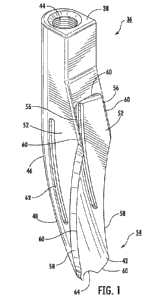
Fig. 1 is a perspective view of a lateral implant removal tool in accordance
with some
embodiments of the present disclosure.
Fig. 2 is a side view of the lateral implant removal tool in accordance with
some
embodiments of the present disclosure.
Fig. 3 is a perspective view of a medial implant removal tool in accordance
with some
embodiments of the present disclosure.
Fig. 4 is a side view of the medial implant removal tool in accordance with
some
embodiments of the present disclosure.
Fig. 5 is a bottom view of the medial implant removal tool in accordance with
some
embodiments of the present disclosure.
Fig. 6 is a perspective view of an alternative embodiment of the medial
implant removal
tool.
Fig. 7 is a side view of the alternative embodiment of the medial implant
removal tool.
Fig. 8 is a bottom view of the alternative embodiment of the medial implant
removal tool.
Fig. 9 is a perspective view of an alternative embodiment of the lateral
implant removal
tool.
Fig. 10 is a side view of the alternative embodiment of the lateral implant
removal tool.
Figs. 11 is a sectional view of the alternative embodiment of the lateral
implant removal
tool taken along line 11-11 of Fig. 10.
Figs. 11A-11E are alternative embodiments showing different cross sections of
the lateral
4
CA 03172424 2022-08-22
WO 2022/155285
PCT/US2022/012238
implant removal tool of Fig. 10, each of which being taken along section line
11-11.
Fig. 12 illustrates a femoral implant prior to insertion of a lateral or
medial tool.
Figs. 13-15 illustrate the insertion of the lateral implant removal tool of
Figs. 1-2.
Fig. 16-17 illustrate the insertion of the medial implant removal tool of Fig.
3-5.
Fig. 18 illustrates the full insertion of both the lateral and medial tools.
Fig. 19 illustrates the femoral implant following the removal of the lateral
and medial tools.
Fig. 20 is a view of an impact hammer secured to the medial tool of Fig. 3-5.
Fig. 21 is a view of an impact hammer being used to insert the lateral tool of
Fig. 1-2.
Fig. 22 is a perspective view of another alternative embodiment of the lateral
implant
removal tool.
Fig. 23 is a front view of the lateral implant removal tool shown in Fig. 22.
Fig. 24 is a back view of the lateral implant removal tool shown in Fig. 22.
Fig. 25 is a side view of the lateral implant removal tool shown in Fig. 22.
Fig. 26 is a sectional view of the lateral implant removal tool shown in Fig.
22 taken along
line 26-26 in Fig. 23.
Fig. 27 is a perspective view of a further alternative embodiment of the
lateral implant
removal tool.
Fig. 28 is a front view of the lateral implant removal tool shown in Fig. 27.
Fig. 29 is a back view of the lateral implant removal tool shown in Fig. 27.
Fig. 30 is a side view of the lateral implant removal tool shown in Fig. 27.
Fig. 31 is a sectional view of the lateral implant removal tool shown in Fig.
27 taken along
line 31-31 in Fig. 28.
Fig. 32 is an illustration of the lateral implant removal tool shown in Fig.
27 working in
combination with a Gigli saw.
Fig. 33 is a perspective view of yet another alternative embodiment of the
lateral implant
removal tool.
Fig. 34 is an end view of the lateral implant removal tool shown in Fig. 33.
Fig. 35 is a front view of the lateral implant removal tool shown in Fig. 33.
Fig. 36 is a back view of the lateral implant removal tool shown in Fig. 33.
Fig. 37 is a side view of the lateral implant removal tool shown in Fig. 33.
5
CA 03172424 2022-08-22
WO 2022/155285
PCT/US2022/012238
Fig. 38 is an illustration of full insertion of the lateral implant removal
tool shown in Fig. 33.
Fig. 39 is a perspective view of an embodiment of a J-shaped tool.
Fig. 40 is an end view of the J-shaped tool shown in Fig. 39.
Fig. 41 is a front view of the J-shaped tool shown in Fig. 39.
Fig. 42 is a back view of the J-shaped tool shown in Fig. 39.
Fig. 43 is a side view of the J-shaped tool shown in Fig. 39.
Fig. 44 is a perspective view of an embodiment of an L-shaped tool.
Fig. 45 is an end view of the L-shaped tool shown in Fig. 44.
Fig. 46 is a front view of the L-shaped tool shown in Fig. 44.
Fig. 47 is a back view of the L-shaped tool shown in Fig. 44.
Fig. 48 is a side view of the L-shaped tool shown in Fig. 44.
Fig. 49 is an illustration of a femoral implant with a flange.
Fig. 50 is an illustration of the J-shaped tool shown in Fig. 39 cutting under
the flange of
the femoral implant illustrated in Fig. 49.
Similar reference numerals refer to similar parts throughout the several views
of the
drawings.
pETAILED DESCRIPTION
The present disclosure relates to tools and associated methods for removing a
prosthetic
implant. Although the tool can be used to remove a variety of different
prosthetic implants,
it finds particular application in the removal of femoral implants. In one
embodiment, both
lateral and medial tools are utilized. In some embodiments, at least one
lateral tool is utilized
with at least one of a medial tool or medial tools, a J-shaped tool, or an L-
shaped tool. In
an illustrative non-limiting embodiment, the lateral tool includes a generally
arcuate shape
with upstanding sidewalls that define an arcuate interior. The lateral tool is
thus
dimensioned to follow the contour of the lateral side of a femoral implant.
The medial tool,
in one embodiment, includes opposing side walls that define an interior
opening. The
opening is sized to receive a neck of the femoral implant, thereby allowing
the tool to closely
follow the medial bone/implant interface. The details of these tools, and the
manner in which
they can be employed, are discussed in greater detail hereinafter.
The disclosed tools are specifically configured to release an implanted
prosthesis by
6
CA 03172424 2022-08-22
WO 2022/155285
PCT/US2022/012238
closely following the bone/implant interface. The tools can be employed to
remove a wide
variety of different prosthetics, such as shoulder and hip implants. However,
in the
illustrated embodiments, the tools are used to cut around, dislodge, and
remove afemoral
implant 20 shown in Fig. 12. As depicted in Fig. 12, femoral implant 20
generally includes
a lateral (or outer) side 22 and a medial (or inner) side 24. Implant 20
further include a stem
26 that is inserted into a superior end of a femur bone 28. Various coatings
and texturing can
be employed for promoting bone growth and the grafting of implant 20 to femur
28. As
illustrated, implant 20 includes a textured portion 32 at its upper portion
where bone growth
and proper affixation are important. Femoral implant 20 also includes a neck
34 that is
angled with respect to the body of implant 20. A head (not shown) is then
secured to the end
of neck 34, with the head ultimately being fitted into an acetabular cup (not
shown).
Lateral Implant Removal Tool,
With reference to Figs. 1-2, a lateral tool 36 includes proximal and distal
ends (38 and 42),
with distal end 42 forming a leading edge that is inserted into femur 28. In
order to allow tool
36 to be connected to an impact hammer (Figs. 20-21), proximal end 38 includes
a threaded
aperture 44. It is also possible to couple tool 36 to the impact hammer via a
quick release
mechanism. The use of the impact hammer is described in greater detail
hereinafter.
Although the size and shape of femoral implants vary, often times lateral side
22 is curved
to match the contour of femur 28. As
such, lateral tool 36 includes a lateral wall 46 with an arcuate extent 48.
Lateral tool 36 further
includes opposing side walls 52. A curved or arcuate interior portion 54 is
defined in the
area between the opposing side walls 52. The shape and geometry of tool 36 may
be
changed to accommodate different types of prosthetics.
In one embodiment, each side wall 52 of lateral tool 36 includes a first
angled extent 56
and a second curved extent 58. As illustrated, angled extent 56 is located
nearer to proximal
end 38 of tool 36 while curved extent 58 is located at distal end 42 of tool
36. Curved extents
58 of tool 36 are preferably angled and sharpened. All edges 60 surrounding
interior portion
54 may be sharpened to facilitate insertion of tool 36. These sharpened edges
60 cut the
bone growth along the bone/implant interface and otherwise allow for the
insertion of tool
36. In order to allow the surgeon to gauge how far tool 36 has been inserted,
a window 62
7
CA 03172424 2022-08-22
WO 2022/155285
PCT/US2022/012238
can be formed within one or both of the side walls 52. Distal end 42 of tool
36 optionally
includes a curved and sharpened leading edge 64. Sharpened leading edge 64 and
sharpened edges 60 allow lateral tool 36 to be inserted as closely as possible
along the
interface between the femur and implant. This, in turn, allows for efficient
removal of femoral
implant 20.
Medial Implant Removal Tool
With reference to Figs. 3-5, a medial tool 66 includes proximal and distal
ends (68 and 72)
as well as opposing side walls 74. Side walls 74 are defined by inner and
outer edges (76
and 78), and in a preferred embodiment, outer edges 78 of walls 74 are
sharpened.
However, unlike lateral tool 36, medial tool 66 is not closed. Rather, medial
tool 66 includes
a generally central opening 82. The purpose of opening 82 is described
hereinafter. All of
inner and outer edges 80 surrounding central opening 82 are preferably
sharpened. A U-
shaped trough 84 with a sharpened leading edge 86 is formed at distal end 72
of medial tool
66. Medial tool 66 is adapted to be inserted
between femur 28 and medial side 24 of femoral implant 20. All of sharpened
edges 80
assist with insertion, including outer edges 78, inner edges 76, and leading
edge 86.
Furthermore, neck 34 of femoral implant 20 is allowed to extend through
opening 82 of
medial tool 66. In this regard, opening 82 is specifically sized to
accommodate neck 34 and
end of implant 20. The sharpened edges surrounding opening 82 allows tool 66
to cut along
the anterior and posterior sides as well as the medial aspect of implant 20.
In another exemplary embodiment, as depicted in Figs 3-4, side walls 74 can
have different
length to make the overall length of medial tool 66 accommodate different
implants.
Method of Usina Lateral and Medial Tools
A method of using lateral tools 36 and medial tool 66 is next described in
connection with
Figs. 13-19. Both lateral and medial tools 36 and 66 can be used in
conjunction with one
another to remove femoral implant 20. However, the present disclosure is not
limited to the
use of both tools (36 and 66) and advantages disclosed herein can be realized
by using
either tool 36 or 66 individually. Each tool is inserted into the bone via an
associated impact
8
CA 03172424 2022-08-22
WO 2022/155285
PCT/US2022/012238
tool (88 and 96)(Figs. 20-21). More specifically, a first impact tool 88 (Fig.
21) includes a
threaded extent 92 that is secured to threaded aperture 44 of lateral tool 36.
A nut 120 can
be secured immediately above threaded extent 92 to prevent unintended rotation
of impact
tool 88 relative to lateral tool 36. Impact tool 88 includes a textured extent
94 that allows
the surgeon to manipulate lateral tool 36 during insertion. The surgeon uses
first impact tool
88 to guide the leading edge 64 and curved extents 58 of lateral tool 36 into
femur 28. A
weighted slide 90 is used as a hammer to apply force to the top of lateral
tool 36. During
this insertion of lateral tool 36, bone growth between the femoral implant 20
and femur 28
is cut.
Second impact tool 96 (Fig. 20) is substantially similar to the first impact
tool 88 and is
likewise used to position and insert medial tool 66. Namely, second impact
tool 96 allows
leading edge 86 and outer and inner edges (78, 76) of medial tool 66 (as well
as all edges
80 surrounding opening 82) to cut bone growth between femoral implant 20 and
femur 28
during the process of insertion. Second impact tool 96 likewise includes a
threaded extent
98, a sliding weight 100, and a guide 102. Each impact tool (88, 96) can be
manually inserted
or can optionally be inserted via a pneumatic hammer or other striking tool.
As described, the lateral and medial implant removal tools (36 and 66) can be
used in
conjunction with one another. It is preferred that lateral tool 36 is inserted
and removed
prior to the insertion and removal of medial tool 66. Fig. 18 illustrates that
in the preferred
embodiment, lateral and medial tools (36 and 66) are inserted into femur 28
such that
curved extents 58 of lateral tool 36 overlap outer edges 78 of medial tool 66.
The overlapping edges (58 and 78) ensure that all bone growth immediately
surrounding
implant 20 is removed. This ensures the efficient removal of implant 20 with
minimal bone
loss.
Alternative Embodiments of Medial Tool
An alternative embodiment of a medial tool 112 is depicted in Figs. 6-8. Tool
112 is
generally the same as medial tool 66 (Figs. 3-4), but includes a side cut out
114 leading to
a narrower distal size when compared to the opening of 118. Medial tool 112
also includes
an opening 118 to accommodate different neck geometries and has a lower
rounded and
9
CA 03172424 2022-08-22
WO 2022/155285
PCT/US2022/012238
sharpened edge 116. Medial tool 112 also includes opposing side walls 124 with
inner
sharpened edges 122. Side walls 124 can have a different length to make the
overall length
of medial tool 66 accommodating different implants.
Alternative Embodiments of Lateral Tools
Figs. 9 and 10 depict an alternative embodiment of a lateral tool as a lateral
tool 104.
Lateral tool 104 is the same in most respects as lateral tool 36. Lateral tool
104 includes a
generally straight back wall 106 and a leading edge 108, more curved than
leading edges
60 and 64 of lateral tool 36. This geometry may be preferred for the lateral
tool depending
upon the shape and size of the implant being removed. Figs. 11 and 11A-11E
illustrate a U-
shaped cross section that makes up the body of lateral tool 104. However, any
of a variety
of cross-sectional shapes can be used. Figs. 11 and 11A-11E illustrate some
possible
cross-sectional shapes for the lateral tool.
Figs. 22-26 depict an alternative embodiment of a lateral tool as a lateral
tool 200. Lateral
tool 200 includes a proximal end 202 and a distal end 204, with distal end 204
forming a
leading edge 206 for inserting into a femur. Proximal end 202 includes a
proximal connector
208 and may include an indicia 210. Two opposing side walls 212 extending from
proximal
end 202 to distal end 204, and a lateral wall 214 extending from proximal end
202 to distal
end 204, provide lateral tool 200 with a generally U-shaped or C-shaped cross
section at
most locations perpendicular to a longitudinal direction. Two opposing side
walls 212 may
be substantially parallel to each other. The inside surfaces of opposing side
walls 212 and
lateral wall 214 define an interior area 216, having a generally arcuate
shape. In some
embodiments, there may be an opening 218 in lateral wall 214. Opening 218 may
be
entirely in lateral wall 214, or partially in lateral wall 214 and partially
in side walls 212. At
some locations along the longitudinal direction, opening 218 may effectively
remove lateral
wall 214 and leave only side walls 212 or portions of side walls 212.
As shown in Fig. 22-25, leading edge 206 may have a curvature and two pointed
ends 220.
Leading edge 206 may be sharpened, rounded, or blunt.
In order to allow lateral tool 200t0 be connected to an impact hammer, such as
shown in Figs.
20-21, proximal connector 208 may include a threaded aperture, a friction-fit
connection, a
CA 03172424 2022-08-22
WO 2022/155285
PCT/US2022/012238
twist lock, or other connector that allows for releasable connection to an
impact hammer.
In other aspects, proximal connector 208 may allow for releasable connection
to a handle
or other instruments, such as a vibration generating device. Proximal
connector 208 may also
include a quick release mechanism or a more permanent securement mechanism.
Indicia 210 may be used for identification. As shown in Figs. 22 and 24, as a
non-limiting
example, an indicia "14" is used to indicate that the opening along the
arcuate blade is 14
mm wide. Other sizes are possible, as are indicia identifying the tool by
another criterion.
Although the shape of femoral implants varies, oftentimes the inside surface
of lateral wall
214 has an extent that is curved to match the lateral side contour of a
femoral implant. In
some embodiments, side edges 222 are sharpened or configured for cutting and
are curved
or arcuate. Side edges 222 should be understood as the edges on the sides of
lateral tool
200 and could be edges of side walls 212 or lateral wall 214. In some
embodiments, lateral
tool 200 may have side walls 212 having height tapered from proximal end 202
to distal
end 204.
Still with reference to lateral tool 200 in Figs. 22-26, all edges surrounding
interior area 216
may be sharpened or configured for cutting to facilitate insertion of lateral
tool 200 into a
femur. These edges cut along bone and implant interface and otherwise allow
for the
insertion of lateral tool 200 into a femur. In some aspects, these edges may
be configured
with teeth or scalloped edges to aid in the cutting of bone.
While, as described above, leading edge 206 may be curved between two pointed
ends 220,
alternatively, leading edge 206 may instead comprise one or more straight
edges between
pointed ends 220. Leading edge 206 may include one or more teeth or scalloped
edges to
facilitate insertion into the femur. Leading edge 206, side edges 222, and the
extent of the
interior of lateral wall 214 allow lateral tool 200 to be inserted as closely
as possible along
the interface between a femur and implant. This advantageously allows for the
efficient
removal of the implant.
In Fig. 23-24, opening 218 is shown as rectangular in shape, having a length
along a
longitudinal axis of lateral tool 200 that is greater than a width. Corners of
opening 218 may
include chamfers or may be rounded as illustrated. A proximal opening edge
228, a distal
opening edge 230, or side opening edges 232 may include sharpened edges,
rounded
11
CA 03172424 2022-08-22
WO 2022/155285
PCT/US2022/012238
edges, or blunt edges. Opening 218 is shaped so that certain features of an
implant may
pass through as lateral tool 200 advances into a femur. More particularly,
opening 218
allows the shape of interior area 216 of lateral tool 200 to not completely
conform to the
shape of the lateral side of the implant so that side edges 222 may cut
closely along the
implant/bone interface. Therefore, the shape of opening 218 can be customized
for an
implant to be removed. For example, while opening 218 is shown as being
rectangular, it
may have another shape such as an ellipse or oval. Lateral tool 200 may also
include more
than one opening. Opening 218 may be arranged over a smaller portion of
lateral tool 200.
As described with reference to previous embodiments, in order to allow a
surgeon to gauge
how far a lateral tool has been inserted, a window (not shown) or other
measuring feature
can be formed within one or both of side walls 212, or on lateral wall 214.
In some exemplary embodiments, as shown in Fig. 27-31, lateral tool 300 is
similar to lateral
tool 200, except that lateral tool 300 has side walls 312 with height that
tapers off quickly
so that side edges 322 become edges of lateral wall 314, and interior area 316
becomes
the area defined by the inside surface of the arcuate lateral wall 314. Bevels
324 may be
formed on the side walls 312 or lateral wall 314 and angled to pointed ends
320. In some
embodiments, the bevels 324 are sharpened or configured for cutting.
In one embodiment, as depicted in Fig. 32, a distal end 304 of lateral tool
300 can be
configured to accommodate a Gigli saw 334, or other instrument, such that once
lateral tool
300 is inserted along an implant 336, Gigli saw 334 can be used by a surgeon
to cut along
the implant 336 to facilitate removal of the implant. Advantageously, saw wire
338 can be
crossed around the implant before introduction which can allow Gigli saw 334
to help
release the lateral, medial, anterior, and posterior aspects of the implant
336 from the bone
340. Although Gigli saw 334 is shown only in Fig. 32, it can be used with
other examples
in this disclosure as well.
Figs. 33-37 depicts yet another alternative embodiment of a lateral tool as a
lateral tool
400. Lateral tool 400 has a proximal end 402 and a distal end 404. As shown in
a front view
of lateral tool 400 in Fig. 34, lateral tool 400 has two parallel side walls
406 and a lateral
wall 408 with a curved shape cross section, defining an interior area 410. A
leading edge
412 and side edges 414 around interior area 410 may be sharpened or configured
for
12
CA 03172424 2022-08-22
WO 2022/155285
PCT/US2022/012238
cutting the lateral side, anterior side, and posterior side of an implant
together as lateral
tool 400 advances into a femur.
Lateral wall 408 of lateral tool 400 may be substantially straight
longitudinally, having a
longitudinal axis at an angle to side edges 414. An opening 416 may displace a
substantial
portion of lateral wall 408 as well as sections of adjacent side walls 406,
leaving lateral wall
408 present only near distal end 404 of lateral tool 400. The cross sectional
shape of lateral
wall 408 can be customized to conform the shape of the lateral side of an
implant to be
removed. Advantageously, a shorter lateral wall 408 may enable lateral tool
400 to conform
to implants with a larger variety of lateral curvature. Side edges 414 may be
sharpened or
configured for cutting along an anterior side and a posterior side of an
implant. In this
exemplary embodiment, side edges 414 are straight. However, side edges 414 can
be
curved or arcuate, or have segments at different angles to facilitate cutting
the anterior and
posterior sides of the implant.
Opening 416 may have edges sharpened or configured for cutting and is large
enough to
allow features of implant to pass through as lateral tool 400 advances into a
femur, as
illustrated in Fig. 38. In this exemplary embodiment, a portion of the implant
does not
conform to the shape of lateral tool 400 interior area 410 and extends out
through opening
416. Opening 416 may be shorter than side edges 414. A proximal edge 418 of
opening
416 is on the distal side of proximal ends 420 of side edges 414. However,
proximal edge
418 of opening 416 can be proximate to proximal end of side edge 414.
Proximal end 402 of lateral tool 400 may have a proximal connector 422. In
some
embodiments, proximal connector 422 may include a threaded aperture, a
friction-fit
connection, a twist lock, or other connector that allows for releasable
connection to an
impact hammer. In other aspects, proximal connector 422 may allow for
releasable
connection to a handle or other instruments, such as a vibration generating
device.
Proximal connector 422 may also include a quick release mechanism or a more
permanent
securement mechanism. As depicted in Figs. 33-37, proximal connector 422 may
be in-
line or parallel to the axis of straight lateral wall 408 with an offset.
Advantageously, a
surgeon or an operator of lateral tool 400 may apply force through an
attachment to
proximal connector 422 in an optimal orientation to facilitate leading edge
412 advancing
13
CA 03172424 2022-08-22
WO 2022/155285
PCT/US2022/012238
into a femur. However, proximal connector 422 may be customized to be at an
angle to
the axis of straight lateral wall 408 to optimize for a specific type of
implant to be
removed.
Undercut Tools
In some embodiments, a J-shaped tool 500 depicted in Figs. 39-43 or an L-
shaped tool 600
depicted in Figs. 44-48 can be used when an implant has a flange near its
stem. J-shaped
tool 500 and L-shaped tool 600 are each essentially formed from a portion of a
cylindrical
shell. J-shaped tool 500 has a straight blade 502 with a proximal end 504 and
a distal end
506 and a hook 508 connected to distal end 506 of straight blade 502. Proximal
end 506 is
connected to a cylindrical tool base 510. Hook 508 includes a proximal edge
512 sharpened
or configured for cutting. L-shaped tool 600 has a straight blade 602 with a
proximal end
604 and distal end 606 of straight blade 602. Proximal end 606 is connected to
a cylindrical
tool base 610. Tool base 510 or 610 can be any other shape and can include
features to
facilitate handling. Hook 608 includes a proximal edge 512 sharpened or
configured for
cutting. Hook 508 or 608 respectively extends from distal end 506 or 606
circumferentially,
following the same cylindrical curvature. All edges of straight blade 502 or
602 and hook
508 or 608 may be sharpened or configured for cutting.
Fig. 49 illustrates a femoral implant 700. In some embodiments as illustrated
in Fig. 49, an
implant 700 may include a flange 710 near its stem 720, leaving a medial side
730 being
an undercut. Implant 700 may be removed when J-shaped tool 500 or L-shaped
tool 600
is advanced into a femur before or after using other lateral and/or medial
tools, rotated to
have hook 508 or 608 below flange 710, then pulled along medial side 730 while
cutting
along the implant/bone interface under flange 710 of implant 700, using
proximal edge 512
or 612 of hook 508 or 608. This allows J-shaped tool 500 or L-shaped tool 600
to cut around
the undercut of an implant.
Fig. 50 illustrates J-shaped tool 500 cutting along medial side 730 of implant
700. One
skilled in the art will now appreciate how L-shaped tool 600 is utilized to
perform similar
tasks. One skilled in the art would also appreciate how J-shaped tool 500 and
L-shaped
tool 600 can cut around flanges at a different location of an implant.
14
CA 03172424 2022-08-22
WO 2022/155285
PCT/US2022/012238
The disclosed tools and tool set have several advantages. For example, the
tools in the
tool set are shaped to conform to the interface between bone and a prosthetic,
such as a
femoral implant. The tools may also include an opening to accommodate a neck
or other
features of the prosthetic to pass through, so that the shape of an interior
area of the tools
does not have to fully conform to the shape of the implant to allow cutting
along the
implant/bone interface. Edges on both sides and around the opening may be
sharpened or
configured for cutting, so the tool may cut along anterior and posterior sides
of the implant
while at the same time cutting along the lateral or medial aspect. All of this
allows inserting
tools along an edge of a prosthetic that is immediately adjacent to a stem or
other feature
of a prosthetic, enabling efficient removal of the prosthetic.
An advantage of the tools of the present disclosure is that they allow
prosthetics to be
removed efficiently and in a minimal amount of time.
A further advantage of the tools is that they allow prosthetics to be removed
while
minimizing the loss of existing bone.
Yet a further advantage of the tools is that the efficient removal of
prosthetics greatly
decreases recovery time.
Another advantage is that the efficient removal of prosthetics reduces both
the need for
anesthesia and operating room costs in general.
In one embodiment, a tool provides a groove or recess for accommodating an
additional
cutting element such as a Gigli saw to further facilitate removal of an
implant, which allows
for the efficient removal of the prosthetic.
Various embodiments of the disclosure may have none, some, or all of these
advantages.
Other technical advantages of the present disclosure will be readily apparent
to one skilled
in the art.
Although this disclosure has been described in terms of certain embodiments
and
generally associated methods, alterations and permutations of these
embodiments and
methods will be apparent to those skilled in the art. Accordingly, the above
description of
example embodiments does not define or constrain this disclosure. Other
changes,
substitutions, and alterations are also possible without departing from the
spirit and scope
of this disclosure.
15