Note: Descriptions are shown in the official language in which they were submitted.
PULMONARY FIBROSIS MEDICINE CONTAINING PYRAZOL
DERIVATIVE
Technical Field
This application claims the benefit of Korean Patent
Application No. 10-2020-0044598 filed on April 13, 2020, and
Korean Patent Application No. 10-2021-0036863 filed on March
22, 2021, with the Korean Intellectual Property Office, the
disclosure of which are herein incorporated by
reference in their entirety.
The present invention relates to a pyrazole derivative
useful for preventing or treating pulmonary fibrosis, a method
for preparing the same, and a pharmaceutical composition
thereof.
Background Art
Pulmonary fibrosis (PF) is a type of chronic interstitial
lung disease and characterized by infiltration of inflammatory
cells such as lymphocytes and macrophages into the lung
interstitium, proliferation of fibroblasts, and deposition of
fibrous connective tissue into the lung interstitium.
Pulmonary fibrosis is caused by various internal and external
etiology of the lungs, is a result of chronic lung damage or
disease progressing to the end, and seriously threatens human
health.
The etiology of pulmonary fibrosis includes factors such
as immune dysfunction, viral or bacterial infection, drugs and
chemicals, radiation, and air pollution (smog, cigarette smoke,
dust, and the like).
As mentioned above, there are patients who may diagnose
the clear cause of pulmonary fibrosis, but there are also
cases where the cause cannot be elucidated, wherein such cases
are called idiopathic pulmonary fibrosis (IPF). Idiopathic
1
CA 03172596 2022- 9- 21
pulmonary fibrosis is a type of interstitial pneumonia in
which fibrosis of the lung parenchyma is progressively
progressing, and is known to have a high risk of death due to
respiratory failure within a few years after diagnosis and a
very poor prognosis. The 5-year survival rate is about 20%,
similar to that of lung cancer. In addition, the incidence and
prevalence of pulmonary fibrosis are rapidly increasing with
the aging population.
Pulmonary fibrosis is a complex pathological and
physiological process, wherein in the early stage, a large
number of inflammatory cells infiltrate around the
inflammatory site of the lung to cause the alveolar wall to
become chronically thickened, and in the middle/end stage,
normal lung tissue structure is destroyed due to overgrowth,
alveolar deformation, hardening and scarring of the lung
tissue caused by excessive deposition of extracellular matrix
elements such as collagen by fibroblasts to result in loss of
function.
Fibroblasts play a role in the recruitment of immune
cells to sites of inflammation and tissue damage. In addition,
fibroblasts produce and respond to many inflammatory cytokines.
Thus, fibroblasts may contribute to chronic inflammation, and
conversely, inflammatory cytokines promote the conversion of
fibroblasts to myofibroblasts, thereby promoting fibrosis.
Therefore, injury or inflammation of the lung tissue may lead
to pulmonary fibrosis.
Lung transplantation is the only method to repair lung
tissue with progressive fibrosis due to pulmonary fibrosis,
and the 5-year survival rate after diagnosis is only 43%.
Although many clinical studies are being conducted to
develop a therapeutic agent for pulmonary fibrosis, there is
still no therapeutic agent for lung fibrosis, and
immunosuppressants, which are steroids or cytotoxic drugs, are
2
CA 03172596 2022- 9- 21
mainly used as a first-line. Among steroids and cytotoxic
drugs, steroids are used first, and a combination therapy of
steroids and azathioprine or cyclophosphamide is currently
being used.
In addition, pirfenidone and nintedanib are the only
approved drugs for the treatment of pulmonary fibrosis. It has
been reported that pirfenidone has a mild therapeutic effect
and is used at a very high dose of about 2.4 g/day, but there
is little or no significant improvement in survival, and since
it tends to decrease the quality of life of patients due to
severe side effects such as gastrointestinal disorders (nausea,
diarrhea, dyspepsia), skin disorders (photosensitive rash) and
metabolic and nutritional disorders (anorexia, anepithymia)
and weakens the liver function, continuous administration is
difficult. It has been reported that nintedanib is used at a
dose of 200 to 400 mg/day and reduces the incidence of acute
exacerbations of mild to severe idiopathic pulmonary fibrosis,
but continuous administration is difficult due to many side
effects and gastrointestinal side effects.
Therefore, there is an urgent need to develop new drugs
that may treat the underlying cause rather than alleviate the
progression of the symptoms of a disease.
Although many studies are being conducted on the causes
of pulmonary fibrosis, the pathogenesis is still unclear,
making it difficult to develop therapeutic agents, but
research results have been reported that oxidative stress due
to the generation of excessive activated oxygen according to
changes in redox homeostasis in vivo plays an important role
in the progression and exacerbation of pulmonary fibrosis.
Oxidative stress refers to tissue damage caused by a
relatively excessive production of reactive oxygen species as
the balance between the production of reactive oxygen species
(ROS) and the antioxidant defense mechanism for biomolecules,
3
CA 03172596 2022- 9- 21
cells and tissues is broken. In particular, it has been
reported that oxidative stress generated in the lung tissue
induces and worsens pulmonary fibrosis. It has been reported
that TGF-13 stimulation in the lung tissue of patients with
progressive pulmonary fibrosis induces an increase in the
generation of reactive oxygen species and increases the
expression of collagen and a-smooth muscle actin (a-SMA),
which are important for fibrosis. In particular, it has been
reported that pulmonary fibrosis worsens due to reactive
oxygen species in the lung tissue of patients with idiopathic
pulmonary fibrosis.
RNA virus or DNA virus infection also causes fatal lung
damage through pneumonia and pulmonary fibrosis, leading to
death. It has been reported that when single-stranded viruses
(corona virus, influenza virus, respiratory syncytial virus,
rhinovirus, dengue virus, HIV, and the like) and DNA viruses
(adenovirus, vaccinia virus, herpes simplex virus, and the
like)invade cells and form endosomes, reactive oxygen species
are generated to promote virus replication, and the rapidly
amplified virus penetrates into the lung tissue to cause lung
damage while promoting inflammation and fibrosis.
Currently, in the case of viral infection, rapid
amplification in the human body causes rapid lung damage,
making treatment difficult, and thus, if a therapeutic agent
for inhibiting the generation of reactive oxygen species and
an antiviral agent are used in combination in order to treat
and alleviate lung damage caused by viral infection and rapid
amplification, it will be possible to more effectively treat
viral pneumonia and pulmonary fibrosis. In this case, as
antiviral agents that may be used in combination,
representative drugs include remdesivir, ritonavir, lopinavir,
favilavir, and the like.
4
CA 03172596 2022- 9- 21
It has been reported that TGF-13 stimulation in the lung
tissue of patients with progressive pulmonary fibrosis induces
an increase in the generation of reactive oxygen species and
increases the expression of collagen and a-smooth muscle actin
(a-SMA), which are important for fibrosis, and it has been
reported that pulmonary fibrosis worsens due to reactive
oxygen species in the lung tissue of patients with idiopathic
pulmonary fibrosis.
On the other hand, none of the prior art document
discloses that the pyrazole-based compound of the present
invention is effective in preventing and treating pulmonary
fibrosis.
[Prior Art Documents]
(Patent Document 1) Korean Patent No. 10-1280160
(Patent Document 2) Korean Patent Application Laid-Open
No. 10-2019-0122806
(Patent Document 3) Korean Patent Application Laid-Open
No. 10-2019-0136079
(Non-Patent Document 1) Gabriel Laghlali, et al.
Respiratory 2019, 13629.
(Non-Patent Document 3) Eunice E. To et al. Nature
communications, 8(69), 1-17.
(Non-Patent Document 3) Alessandro G. Fois, Panagiotis
Paligiannis et al., Respir Res. 2018, 19:51.
Disclosure
Technical Problem
It is an object of the present invention to provide a
pharmaceutical composition comprising a compound of Formula 1
or a pharmaceutically acceptable salt thereof.
It is another object of the present invention to provide
a pharmaceutical composition for effectively inhibiting the
5
CA 03172596 2022- 9- 21
generation of reactive oxygen species, comprising a compound
of Formula 1 or a pharmaceutically acceptable salt thereof.
It is another object of the present invention to provide
a pharmaceutical composition for treating or preventing
pulmonary fibrosis, comprising a compound of Formula 1 or a
pharmaceutically acceptable salt thereof.
It is another object of the present invention to provide
a method of preventing or treating pulmonary fibrosis by
administering a compound of Formula 1 or a pharmaceutically
acceptable salt thereof to an individual.
It is another object of the present invention to provide
the use of a compound of Formula 1 or a pharmaceutically
acceptable salt thereof for preventing or treating pulmonary
fibrosis.
It is another object of the present invention to provide
a pharmaceutical composition for treating and preventing
pulmonary fibrosis, further comprising a compound of Formula
1 or a pharmaceutically acceptable salt thereof, and an
antibiotic, an antifungal agent, an antiviral agent, an anti-
inflammatory agent or any combination thereof.
It is another object of the present invention to provide
a method of preventing or treating pulmonary fibrosis by
further administering to an individual a compound of Formula
1 or a pharmaceutically acceptable salt thereof, and an
antibiotic, an antifungal agent, an antiviral agent, an anti-
inflammatory agent or any combination thereof.
It is another object of the present invention to provide
the use of a compound of Formula 1 or a pharmaceutically
acceptable salt thereof, and an antibiotic, an antifungal
agent, an antiviral agent, an anti-inflammatory agent or any
combination thereof for preventing or treating pulmonary
fibrosis.
6
CA 03172596 2022- 9- 21
It is another object of the present invention to provide
an antiviral agent comprising a compound of Formula 1 or a
pharmaceutically acceptable salt thereof.
It is another object of the present invention to provide
a method of preventing or treating a viral disease by
administering a compound of Formula 1 or a pharmaceutically
acceptable salt thereof to an individual.
It is another object of the present invention to provide
the use of a compound of Formula 1 or a pharmaceutically
acceptable salt thereof for preventing or treating a viral
disease.
Technical Solution
In order to achieve the above objects, the present
invention provides a pharmaceutical composition for preventing
and improving or treating pulmonary fibrosis or an viral
disease, comprising a pyrazole-based compound represented by
following Formula 1 or a pharmaceutically acceptable salt
thereof:
Formula 1
R
\ OH
/ N
\
-------
wherein R is a linear or branched alkyl group having 1
to 10 carbon atoms.
[Advantageous Effects]
7
CA 03172596 2022- 9- 21
The pyrazole-based compound according to the present
invention or a pharmaceutically acceptable salt thereof may
effectively inhibit the generation of reactive oxygen species
generated in the lungs, and thus may be usefully used for the
prevention or treatment of oxidative stress-induced pulmonary
fibrosis without any particular side effects.
In addition, the pyrazole-based compound according to
the present invention or a pharmaceutically acceptable salt
thereof has antiviral activity, and thus may be usefully used
for the prevention or treatment of a viral disease.
Description of Drawings
Figure 1 shows the result of effectively inhibiting the
expression of PMA stimulation-induced reactive oxygen species
in normal human lung fibroblasts (NHLFs) when treated with
Compound 1.
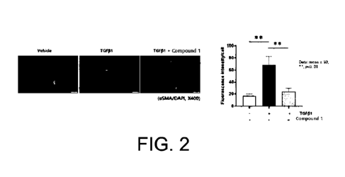
Figure 2 shows the results of inhibiting the expression
of aSMA by Compound 1 during the differentiation of normal
human lung fibroblasts into myofibroblasts by TGF-131.
Figure 3 shows the results of inhibiting the expression
of collagen I by Compound 1 during the differentiation of
normal human lung fibroblasts into myofibroblasts by TGF-131.
Figure 4 shows the result of effectively inhibiting the
expression of LPS treatment-induced IL-113 in normal human lung
fibroblasts when treated with Compound 1.
Figure 5 is photographs showing H&E staining of alveolus
and bronchiole when treated with nintedanib or Compound 1 in
an animal model with bleomycin administration-induced
pulmonary fibrosis (BLM), and shows the result of reducing the
amount of immune cells penetrating into the alveolus and
bronchiole when treated Compound 1.
Figure 6 is photographs showing collagen deposition
staining of bronchiole when treated with nintedanib or
8
CA 03172596 2022- 9- 21
Compound 1 in an animal model with bleomycin administration-
induced pulmonary fibrosis (BLM), and shows the result of
reducing collagen deposition when treated Compound 1.
Figure 7 is photographs showing the staining of aSMA
when treated with nintedanib or Compound 1 in an animal model
with bleomycin administration-induced pulmonary fibrosis
(BLM), and shows a decrease in the expression of aSMA when
treated Compound 1.
Figure 8 is photographs showing the staining of collagen
I when treated with nintedanib or Compound 1 in an animal
model with bleomycin administration-induced pulmonary
fibrosis(BLM), and shows a decrease in the expression of
collagen I when treated Compound 1.
Figure 9 is the result of quantifying the degree of lung
fibrosis with the improved Ashcroft scale when treated with
nintedanib or Compound 1 in an animal model with bleomycin
administration-induced pulmonary fibrosis (BLM), and shows
effective improvement of pulmonary fibrosis compared to
nintedanib when treated with compound 1.
Figure 10 shows that the antiviral efficacy is excellent
and the cytotoxicity is low, when treated with Compound 1 in
human lung epithelial cells.
Best Mode
Hereinafter, the present invention will be described in
more detail with reference to embodiments.
However, the present invention is not limited by the
embodiments that have been represented by way of example, and
the present invention is defined only by the scope of the
appended claims. In addition, even if it is a constitution
essential for practicing the present invention, a specific
description of the constitution that may be easily practiced
by the skilled artisan will be omitted.
9
CA 03172596 2022- 9- 21
The terms and words as used in the present specification
and claims should not be construed as limited to conventional
or dictionary meanings, but should be construed as the meaning
and concept consistent with the technical idea of the present
invention based on the principle that the inventor can
appropriately define the concept of the term to describe its
own invention in the best way.
The terms used in the present invention are for the
purpose of describing specific embodiment only and are not
intended to limit the present invention. Singular expressions
include plural expressions unless the context clearly
indicates otherwise. In the present invention, terms such as
"comprise" and "have" are intended to indicate that there is
a feature, number, step, operation, component, part, or
combination thereof described in the specification, and it
should be understood that the terms do not exclude in advance
the possibility of the presence or addition of one or more
other features, numbers, steps, operations, components, parts,
or combinations thereof.
Pulmonary fibrosis is a fibrosis of the lung parenchyma
caused by infiltration of inflammatory cells into the lung
interstitium, proliferation of fibroblasts, and deposition of
fibrous connective tissue into the lung interstitium due to
inflammation of the lung, and normal lung tissue structure is
destroyed to result in loss of lung function. That is,
Pulmonary fibrosis is caused by various internal and external
etiology of the lungs and is a result of chronic lung damage
or disease progressing to the end, and the incidence and
prevalence are rapidly increasing in line with the recent
aging trend. Recently, in particular, idiopathic pulmonary
fibrosis of unknown cause of disease has become a problem.
Lung transplantation is the only method to repair lung
tissue with progressive fibrosis due to pulmonary fibrosis,
CA 03172596 2022- 9- 21
and pirfenidone and nintedanib are the only approved drugs for
pulmonary fibrosis, but both drugs have a problem that
continuous administration is difficult due to many side
effects and gastrointestinal disorders.
Therefore, a treatment method for pulmonary fibrosis is
generally symptomatic therapy to relieve symptoms using
steroids or immunosuppressants, but there is a need for a
fundamental treatment method.
As a result of research focusing on the fact that a
therapeutic agent for pulmonary fibrosis may be developed if
the generation of oxidative stress in the lung tissue is
effectively inhibited, the present inventors have completed
the present invention by discovering that the pyrazole
derivative of the present invention reduces bleomycin
administration-induced lung damage and inhibits uSMA and
collagen I accumulation-induced pulmonary fibrosis, thereby
reducing pulmonary fibrosis, and confirming that it may be
used as a therapeutic agent for pulmonary fibrosis.
Accordingly, the present invention provides a
pharmaceutical composition capable of preventing or treating
pulmonary fibrosis, comprising one or more compounds selected
from the pyrazole-based compound represented by Formula 1 or
a pharmaceutically acceptable salt thereof.
In addition, the present invention provides a
pharmaceutical composition capable of preventing or treating
idiopathic pulmonary fibrosis, comprising one or more
compounds selected from the pyrazole-based compound
represented by Formula 1 or a pharmaceutically acceptable salt
thereof.
The pyrazole-based compound used in the present
invention is represented by the following Formula 1:
11
CA 03172596 2022- 9- 21
<Formula 1>
R
\ OH
/ N
\
-------
wherein R is a linear or branched alkyl group having 1
to 10 carbon atoms.
The pharmaceutically acceptable salt of the pyrazole-
based compound included in the pharmaceutical composition of
the present invention refers to salts that retain the
biological effectiveness and properties of the parent compound
and are not harmful biologically or otherwise when
administered in a single dosage. In addition, it refers to a
salt commonly used in the pharmaceutical industry.
Specifically, pharmaceutically acceptable addition salts
may be prepared from inorganic and organic bases. Salts
derived from inorganic bases may include, but are not limited
to, sodium, potassium, lithium, ammonium, calcium, and
magnesium salts. Salts derived from organic bases include, but
are not limited to, salts of primary, secondary and tertiary
amines; substituted amines including naturally occurring
substituted amines; and isopropylamine, trimethylamine,
diethylamine, triethylamine, tripropylamine, ethanolamine, 2-
dimethylaminoethanol, tromethamine, lysine,
arginine,
histidine, caffeine, procaine, hydrabamine, choline, betaine,
ethylenediamine, glucosamine, N-alkylglucamine, theobromine,
purine, piperazine, piperidine, and/or cyclic amines including
N-ethylpiperidine.
It should be also understood that other carboxylic acid
derivatives, specifically carboxylic acid amides, including
12
CA 03172596 2022- 9- 21
carboxamides, lower alkyl carboxamides, di(lower alkyl)
carboxamides, and the like, are also useful in the practice
of the present invention.
Additionally, pharmaceutically acceptable acid addition
salts may be prepared from inorganic and organic acids. Salts
derived from inorganic acids include hydrochloric acid,
hydrobromic acid, sulfuric acid, nitric acid, phosphoric acid,
perchloric acid, iodic acid, tartaric acid, and the like.
Salts derived from organic acids may include, but are not not
limited to, acetic acid, trifluoroacetic acid, propionic acid,
glycolic acid, gluconic acid, galacturonic acid, glutamic acid,
glutaric acid, glucuronic acid, aspartic acid, ascorbic acid,
carbonic acid, vanillic acid, hydroiodic acid, pyruvic acid,
oxalic acid, malic acid, malonic acid, lactic acid, succinic
acid, maleic acid, fumaric acid, tartaric acid, citric acid,
benzoic acid, cinnamic acid, mandelic acid, methanesulfonic
acid, ethanesulfonic acid, benzenesulfonic acid, p-
toluenesulfonic acid, naphthalenesulfonic acid and/or
salicylic acid, and the like.
The pharmaceutically acceptable salt may be a
hydrochloride salt.
The pyrazole-based compound represented by Formula 1 or
a pharmaceutically acceptable salt thereof included in the
pharmaceutical composition of the present invention is
specifically exemplified as follows:
3-phenyl-4-methyl-1-(pyridin-2-y1)-1H-pyrazol-5-ol or a
hydrochloride salt thereof;
3-phenyl-4-ethyl-1-(pyridin-2-y1)-1H-pyrazol-5-ol or a
hydrochloride salt thereof;
3-phenyl-4-n-propy1-1-(pyridin-2-y1)-1H-pyrazol-5-ol or
a hydrochloride salt thereof;
3-phenyl-4-isopropyl-1-(pyridin-2-y1)-1H-pyrazol-5-ol
or a hydrochloride salt thereof;
13
CA 03172596 2022- 9- 21
3-phenyl-4-n-butyl-1-(pyridin-2-y1)-1H-pyrazol-5-ol or
a hydrochloride salt thereof;
3-phenyl-4-tert-butyl-1-(pyridin-2-y1)-1H-pyrazol-5-ol
or a hydrochloride salt thereof;
3-phenyl-4-n-penty1-1-(pyridin-2-y1)-1H-pyrazol-5-ol or
a hydrochloride salt thereof;
3-phenyl-4-n-hexy1-1-(pyridin-2-y1)-1H-pyrazol-5-ol or
a hydrochloride salt thereof.
Specifically, the pyrazole-based compound included in
the pharmaceutical composition of the present invention may
be 3-phenyl-4-n-propy1-1-(pyridin-2-y1)-1H-pyrazol-5-ol or a
hydrochloride salt thereof.
The compound of Formula 1 of the present invention may
inhibit the generation of reactive oxygen species.
In the present invention, oxidative stress refers to
tissue damage caused by a relatively excessive production of
reactive oxygen species when the balance between the
production of reactive oxygen species (ROS) and the
antioxidant defense mechanism for biomolecules, cells and
tissues is broken. In this case, "reactive oxygen species" may
refer to activated oxygen, active oxygen, and activated oxygen
species, which refer to the same substance.
The pyrazole-based compound of the present invention, in
particular, the hydrochloride salt of 3-pheny1-4-n-propy1-1-
(pyridin-2-y1)-1H-pyrazol-5-ol (Compound 1) effectively
reduced reactive oxygen species in human lung epithelial cells,
and effectively inhibited the expression of aSMA and collagen
I, which are myofibroblast differentiation markers, when lung
fibroblasts were differentiated into myofibroblasts.
In addition, as confirmed by experiments in animal models
with bleomycin-administered pulmonary fibrosis, the compound
of the present invention reduced the infiltration of
inflammatory cells into the lung tissue, decreased lung
14
CA 03172596 2022- 9- 21
epithelial cell hypertrophy, reduced deformation of the lung
structure, and reduced the site of abnormal tissue deposition,
compared to nintedanib, which was approved as a conventional
therapeutic agent for pulmonary fibrosis.
The compound of the present invention decreased the
expression and accumulation of collagen I and aSMA in the lung
tissue, and also decreased the expression of reactive oxygen
species in the lung tissue, compared to nintedanib. Based on
these results, when the severity of fibrosis was quantified
with the improved Ashcroft scale, it showed a remarkable
improvement effect in pulmonary fibrosis compared to
nintenadib, which was approved as a conventional therapeutic
agent for pulmonary fibrosis.
Therefore, it was confirmed that the compound of the
present invention not only suppresses the inflammatory
response by inhibiting reactive oxygen species in the lung
tissue in human lung cells and lung fibrosis models, but also
prevents or alleviates pulmonary fibrosis by reducing the
expression and accumulation of collagen I and aSMA.
The pulmonary fibrosis may be caused by pulmonary
inflammatory fibrosis, chronic obstructive pulmonary disease
(COPD) combined pulmonary fibrosis, idiopathic pulmonary
fibrosis (IPF) or asthma.
In addition, the pneumonia, which may be said to be the
main cause of pulmonary fibrosis, may be caused by viral
pneumonia, bacterial pneumonia, fungal
pneumonia,
hypersensitivity pneumonitis, aspiration
pneumonia,
interstitial pulmonary disease, pneumoconiosis, and the like.
The viral pneumonia may be caused by a virus selected
from adenovirus, vaccinia virus, herpes simplex virus,
parainfluenza virus, rhinovirus, varicella zoster virus,
measle virus, respiratory syncytial virus, dengue virus, human
immunodeficiency virus (HIV), influenza virus, coronavirus,
CA 03172596 2022- 9- 21
severe acute respiratory syndrome-related coronavirus (SARS-
CoV), severe acute respiratory syndrome-related coronavirus 2
(SARS-CoV2), middle east respiratory syndrome coronavirus
(MERS-CoV), or variant viruses of these viruses.
The pharmaceutical composition of the present invention
may further comprise an antibiotic, an antifungal agent, an
antiviral agent, an anti-inflammatory agent or any combination
thereof as a second therapeutic agent, in addition to the
compound of Formula 1 or a salt thereof.
Specifically, the antibiotic included in the second
therapeutic agent is gentamycin, kanamycin, streptomycin,
amikacin, neomycin, and the like, among aminoglycoside
antibiotics; erythromycin, azithromycin, clarithromycin, and
the like, among macrolide antibiotics; penicillin,
cephalosporin, carbapenem, monobactam, and the like, among the
beta-lactam antibiotics; clindamycin, and the like, as
lincomycin antibiotics; linezolid, and the like, as
oxazolidinone antibiotics; ciprofloxacin, lebofloxacin,
moxifloxacin, fluoroquinolone, and the like, as a quinolone
antibiotic; tetracycline, doxycycline, tigecycline, and the
like, as tetracycline
antibiotics;
trimethoprime/sulfamethoxazole (TMX/SMX), as sulfonamide
antibiotics; or a combination thereof.
The antiviral agent included in the second therapeutic
agent is thiosemicarbazone, metisazone, acyclovir, remdecivir,
ritonavir, lopinavir, faviravir, idoxuridine, vidarabine,
ribavirin, ganciclovir, famciclovir, valaciclovir, cidofovir,
valganciclovir, brivudine, ribavirin,
rimantadine,
tromantadine, foscarnet, saquinavir, indinavir, nelfinavir,
amprenavir, fosamprenavir, atazanavir, tipranavir, zidovudine,
didanosine, zalcitabine, stavudine, lamivudine, abacavir,
tenofovir disoproxil, adefovir disoproxil, emtricitabine,
entecavir, nevirapine, delavirdine, efavirenz, zanamivir,
16
CA 03172596 2022- 9- 21
oseltamivir, inosine pranobex, pleconaril, enfuvirtide, or a
combination thereof.
The antifungal agent included in the second therapeutic
agent is allylamine, terbinafine, 5-fluoro cytosine,
fluconazole, itraconazole, ketoconazole, ravuconazole,
posaconazole, voriconazole, caspofungin,
micafungin,
anidulafungin, amphotericin B, amphotericin B lipid complex
(ABLC), amphotericin B colloidal dispersion (ABCD), liposome-
amphotericin B (L-AMB), liposome nystatin, griseofulvin, or a
combination thereof.
The compound of Formula 1 of the present invention has
antiviral activity against various viruses, and thus, the
compound of the present invention may be itself used as an
antiviral agent. Accordingly, the compound of Formula 1 of the
present invention may be effective in preventing or treating
a viral disease. The viral disease may be caused by a virus
selected from, but is not limited to, adenovirus, vaccinia
virus, herpes simplex virus, parainfluenza virus, rhinovirus,
varicella zoster virus, measle virus, respiratory syncytial
virus, dengue virus, human immunodeficiency virus (HIV),
influenza virus, coronavirus, severe acute respiratory
syndrome coronavirus (SARS-CoV), severe acute respiratory
syndrome-related coronavirus 2 (SARS-CoV2), middle east
respiratory syndrome coronavirus (MERS-CoV), or variant
viruses of these viruses. In particular, the compound 1 of the
present invention may effectively inhibit the proliferation
of severe acute respiratory syndrome-related coronavirus 2
(SARS-CoV2), and thus may also inhibit the proliferation of
its related viruses, SARS-CoV, MERS-CoV, or variant viruses
of these coronaviruses.
The pharmaceutical composition of the present invention
may comprise a pharmaceutically acceptable carrier within a
range that does not impair the effects of the present invention.
17
CA 03172596 2022- 9- 21
The "pharmaceutically acceptable carrier" includes any
and all kinds of solvents, dispersion media, coatings,
surfactants, antioxidants, preservatives (antibacterial or
antifungal agents), isotonic agents, diluents, absorption
delaying agents, salts, preservatives, stabilizers, binders,
excipients, disintegrants, lubricants, sweetening agents,
flavouring agents, dyes, and the like, and combinations
thereof, as known to those skilled in the art. Except that any
conventional carrier is not compatible with the active
ingredient, its use in therapeutic or pharmaceutical
compositions is contemplated.
The diluent may be selected from the group consisting
of, but is not limited to, microcrystalline cellulose, lactose
monohydrate, lactose anhydride, lactose, starch, mannitol,
carboxymethylcellulose, sorbitol, and combinations thereof.
The disintegrant may be selected from the group
consisting of, but is not limited to, low-substituted
hydroxypropyl cellulose, crospovidone, croscarmellose sodium,
sodium starch glycolate, F-melt, and combinations thereof.
The binder may be selected from the group consisting of,
but is not limited to, hydroxypropyl cellulose, hydroxypropyl
methylcellulose, hypromellose, polyvinyl acetic acid,
povidone, polyvinylpyrrolidone, copovidone, macrogol, sodium
lauryl sulfate, light anhydrous silicic acid, synthetic
aluminum silicate, silicate derivatives such as calcium
silicate or magnesium metasilicate aluminate, phosphates such
as calcium hydrogen phosphate, carbonates such as calcium
carbonate, pregelatinized starches, gums such as acacia gum,
gelatin, cellulose derivatives such as ethyl cellulose, and
mixtures thereof.
The lubricant may be selected from the group consisting
of, but is not limited to, magnesium stearate, silicon dioxide,
18
CA 03172596 2022- 9- 21
talc, light anhydrous silicic acid, sodium stearyl fumarate,
and combinations thereof.
As a pH adjusting agent, an acidifying agent such as
acetic acid, adipic acid, ascorbic acid, sodium ascorbate,
sodium etherate, malic acid, succinic acid, tartaric acid,
fumaric acid and citric acid, and a basifying agent such as
aqueous ammonia, sodium carbonate, magnesium oxide, magnesium
carbonate, sodium citrate and tribasic calcium phosphate may
be used.
As the antioxidant, dibutyl hydroxy toluene, butylated
hydroxyanisole, tocopherol acetate, tocopherol, propyl
gallate, sodium hydrogen sulfite, sodium pyrosulfite and the
like may be used.
In addition, it is possible to formulate the agents of
the present invention by selectively using various additives
selected from colorants and flavourings as pharmaceutically
acceptable additives.
In the present invention, the scope of the additives is
not limited to using the additives, and it may be formulated
to selectively contain a dose within a normal range using the
additives.
The pharmaceutical composition according to the present
invention may be formulated and used in the form of oral
formulations such as powders, granules, tablets, capsules,
suspensions, emulsions, syrups and aerosols, external
preparations, suppositories, or sterile injectable solutions.
In one aspect of the present invention, it may be a
pharmaceutical composition for preventing, improving or
treating pulmonary fibrosis, comprising the active ingredient
in the range of 0.00001 to 100% by weight, 0.0001 to 95% by
weight, or 0.001 to 90% by weight based on the total weight
of the pharmaceutical composition.
19
CA 03172596 2022- 9- 21
In the preventive or therapeutic agent for pulmonary
fibrosis according to the present invention, the dosage of the
pyrazole-based compound represented by Formula 1 or a
pharmaceutically acceptable salt thereof may be appropriately
changed depending on the age of the patient, the body weight,
the symptom, the route of administration, and the like.
The dosage of the pyrazole-based compound represented by
Formula 1 or a pharmaceutically acceptable salt thereof of the
present invention may be 0.00001 mg/kg/day to 2000 mg/kg/day,
0.0001 mg/kg/day to 1000 mg/kg/day, 0.001 mg/kg/day to 800
mg/kg/day, 0.001 mg/kg/day to 500 mg/kg/day, 0.001 mg/kg/day
to 100 mg/kg/day, 0.001 mg/kg/day to 80 mg/kg/day, or 0.01
mg/kg/day to 70 mg/kg/day.
The content of the pyrazole-based compound represented
by Formula 1 or a pharmaceutically acceptable salt thereof of
the present invention may be 0.00001 to 100% by weight, 0.0001
to 95% by weight, 0.0001 to 90% by weight, 0.001 to 70% by
weight, or 0.001 to 50% by weight per unit dosage form.
The administration concentration of the pyrazole-based
compound represented by Formula 1 or a pharmaceutically
acceptable salt thereof of the present invention may be 0.0001
to 500 pM, 0.001 to 300 pM, 0.001 to 150 pM, 0.001 to 130 pM,
0.001 to 100 pM, 0.001 to 80 pM, or 0.01 to 70 pM.
The pharmaceutical composition of the present invention
may be administered through a general route, and may be
specifically formulated for intramuscular, intrathecal,
intra-digestive, intracardiovascular, intrarenal,
or
intravenous administration. Formulation methods employ
conventional methods known to those skilled in the art.
A conventional composition for intramuscular or
intrathecal administration may consist of, but not limited to,
for example, the active ingredient and a sterile isotonic
aqueous solution containing dextrose, sodium chloride, or both
CA 03172596 2022- 9- 21
dextrose and sodium chloride. Other examples include, but are
not limited to, lactated Ringer's injection, lactated Ringer's
injection + dextrose injection, Normosol-M and dextrose,
Isolyte E, acylated Ringer's injection, and the like.
Optionally, the present formulation may comprise, but is not
limited to, a cosolvent such as polyethylene glycol; chelating
agents such as ethylenediamine tetraacetic acid; and
antioxidants such as sodium metabisulphite. Optionally,
without limitation, the solution may be lyophilized and then
reconstituted with a suitable solvent immediately prior to
administration.
Preferred examples are provided to help understanding of
the present invention. The following examples are provided not
to limit the present invention but to facilitate the
understanding of the present invention.
Mode for Carrying out the Invention
<Synthetic Example 1> Synthesis of 3-pheny1-4-ethy1-1-
(pyridin-2-y1)-1H-pyrazol-5-ol
OH
N,N
N
In a round bottom flask, 2-ethyl-3-oxo-3-phenylpropionic
acid ethyl ester (10.7 g, 49 mmol) and 2-hydrazinopyridine
(5.6 g, 51.4 mmol) were heated to reflux under nitrogen
condition without a solvent for 1 day. The resulting solid was
purified with hexane and ethyl acetate and then dried under
vacuum to obtain the title compound in a yield of 70%.
21
CA 03172596 2022- 9- 21
1H NMR(300 MHz, DMSO-d6) 5 8.25-8.24(1H, d), 8.00-
7.97(1H, d), 7.84-7.82(1H, t), 7.73-7.71(2H, m), 7.46-7.37(3H,
m) 7.12-7.11(1H, t), 2.62-2.57(2H, m), 1.23-1.17(3H, m);
ESI(m/z) 266.1[M+H]
<Synthetic Example 2> Synthesis of 3-pheny1-4-buty1-1-
(pyridin-2-y1)-1H-pyrazol-5-ol
1 \ OH
N,N
/ N
\
In a round bottom flask, 2-butyl-3-oxo-3-phenylpropionic
acid ethyl ester (12.1 g, 49 mmol) and 2-hydrazinopyridine
(5.6 g, 51.4 mmol) were heated to reflux under nitrogen
condition without a solvent for 1 day. The resulting solid was
purified with hexane and ethyl acetate and then dried under
vacuum to obtain the title compound in a yield of 75%.
1H NMR(300 MHz, DMSO-d6) 5 8.25-8.24(1H, d), 8.03-
8.02(1H, d), 7.85-7.83(1H, t), 7.70-7.69(2H, m), 7.44-7.35(3H,
m) 7.12-7.11(1H, t), 2.56-2.53(2H, t), 1.58-1.52(2H, m), 1.38-
1.24(2H, m), 0.89-0.86(3H, t); ESI(m/z) 294.0[M+H]+
22
CA 03172596 2022- 9- 21
<Synthetic Example 3> Synthesis of 3-pheny1-4-propy1-1-
(pyridin-2-y1)-1H-pyrazol-5-ol
\ OH
N
2-Propy1-3-oxo-3-phenylpropionic acid ethyl ester (2.52
g, 10.7 mmol) and 10 ml of ethanol were placed in a round
bottom flask, and then a solution of 2-hydrazinopyridine (1.29
g, 1.18 mmol) diluted in 3 ml of ethanol was slowly added
dropwise thereto at 000. It was heated to reflux at 100 C for
3 day. The solvent was removed by distillation under reduced
pressure, and the resulting solid was washed with hexane and
ethyl acetate, and then dried under vacuum to obtain the title
compound in a yield of 82%.
1H NMR(300 MHz, CD013) 5 12.50(1H, s), 8.27-8.25(1H, m),
8.01(1H, d, J = 8.5 Hz), 7.81(1H, m), 7.69(2H, m), 7.48-
7.34(3H, m), 7.12-7.10(1H, m), 2.54(2H, d, J = 7.5 Hz), 1.64(2H,
m), 0.93(3H, t, J = 7.3 Hz); EIMS(70 eV) m/z(rel intensity)
279(M+, 37), 250(100)
23
CA 03172596 2022- 9- 21
<Synthetic Example 4> Synthesis of 3-pheny1-4-propy1-1-
(pyridin-2-y1)-1H-pyrazol-5-ol hydrochloride (Compound 1)
HCI
\ OH
N
N
3-Phenyl-4-propy1-1-(pyridin-2-y1)-1H-pyrazol-5-ol (280
mg, 1.0 mmol) prepared in Synthetic Example 3 above was
dissolved in 4 ml of ethyl ether in a round bottom flask, and
then 0.55 ml of ethyl ether dissolved in 2 M HC1 was slowly
added dropwise thereto at 0 C. The solid produced from the
reaction solution was filtered under reduced pressure, the
solvent was removed, washed with hexane and ethyl acetate, and
then dried under vacuum to obtain the title compound (270 mg,
0.85 mmol).
1H NMR(300 MHz, CDC13) 5 8.44(1H, d, J = 4.2 Hz), 8.0-
8.03(2H, m), 7.66-7.64(2H, m), 7.48-7.42(3H, m), 7.34-7.30(1H,
m), 2.49(2H, brs), 2.43(2H, t, J = 7.5 Hz), 1.48(2H, m),
0.48(3H, t, J = 7.3 Hz)
<Example 1> Analysis of changes in reactive oxygen
species generation in normal human lung fibroblasts
In order to confirm the effect of the compounds of the
synthetic examples on the generation of reactive oxygen
species from lung fibroblasts, the generation of reactive
oxygen species was induced through PMA stimulation in normal
human lung fibroblasts (NHLFs, Lonza), and the inhibitory
effect of Compound 1 on the generation of reactive oxygen
species was observed under this condition.
24
CA 03172596 2022- 9- 21
Each cell was suspended in a culture medium containing
10% FBS, seeded in a 96-well plate, and cultured for 24 hours
under conditions of 5% CO2 and 37 C. After pre-treating
Compound 1 for 30 minutes to 1 hour, TGF-81 or phorbol 12-
myristate 13-acetate (PMA) stimulation was applied to each
well including cells and drugs. After additional culture for
30 minutes or 48 hours, the degree of generation of reactive
oxygen species was confirmed using 8-amino-5-chloro-7-pheny1-
2,3-dihydro-pyrido[3,4-d]pyridazine-1,4-dione (L-012)
or
2',7'-dichlorodihydrofluoresencein diacetate (DCF-DA).
It was confirmed that the generation of PMA stimulation-
induced reactive oxygen species was inhibited in a
concentration-dependent manner when treated with Compound 1
(Figure 1).
<Example 2> Expression pattern of TGF-pl-induced aSMA
and collagen type I in normal human lung fibroblasts
In order to analyze the effect of Compound 1 on the
differentiation of lung fibroblasts into myofibroblasts, the
inhibitory effect of Compound 1 on the increase in the
expression of TGF-81-induced a-smooth muscle actin (aSMA) and
collagen type I (collagen I) in normal human lung fibroblasts
(NHLFs, Lonza) was observed. NHLF cells were suspended in
culture medium (FGM-2 Bulletkit media, Lonza) and inoculated
at a concentration of 1x10⁴ cells/well on a 4-well chamber
slide (Nunc).
After 24 hours of culture in a CO2 incubator, the medium
was replaced with a serum-free medium and further cultured for
12 hours. Thereafter, vehicle and 2 mM Compound 1 were pre-
treated in the corresponding wells for 1 hour, and then 10
ng/ml of TGF-81 was treated in each well except for the
negative control (vehicle group) for 72 hours.
CA 03172596 2022- 9- 21
Confirmation of the expression of aSMA and collagen I in
the prepared cells was performed through immunocytochemistry
as follows. Cells were fixed in 4% paraformaldehyde for 10
minutes and permeated using 0.1% Triton X-100, and primary
antibody (anti-aSMA Ab, 1:200; anti-collagen I Ab, 1:500,
reacted for 3 hours at room temperature) and secondary
antibody (Alexa-594 conjugated Ab, 1:1000, reacted for 1 hour
at room temperature) treatment process was sequentially
performed, and then the expression of aSMA and collagen I was
observed using a fluorescence microscope.
At this time, in order to quantitatively confirm the
degree of expression of aSMA and collagen I, the number of
DAPI (cell nuclear staining marker)-positive cells and aSMA-
and collagen I-positive cells per each field was counted,
respectively, and then the percentage of the number of aSMA-
and collagen I-positive cells was calculated and compared
using the following formula:
% aSMA (or collagen I)-positive cells/field
= aSMA(or collagen I)-positive cells/DAPI-positive cells
x 100/field
As shown in Figures 2 and 3, it was observed that the
expression of aSMA and collagen I was significantly increased
compared to the control group when NHLFs were treated with 10
ng/ml TGF-131, and the increase in the expression of both
markers was remarkably inhibited when treated with Compound 1.
From these results, it was confirmed that Compound 1
could effectively inhibit the differentiation of lung
fibroblasts into myofibroblasts induced by TGF-131 treatment
(see Figures 2 and 3).
<Example 3> Analysis of changes in the expression of
LPS-induced IL-43
26
CA 03172596 2022- 9- 21
In order to confirm the effect of the compounds of the
synthetic examples of the present invention on the
inflammatory response in normal human lung fibroblasts (NHLFs,
Lonza), whether the increased expression of IL-113 by LPS
treatment was inhibited by Compound 1 was observed.
2x10⁵ cells were inoculated in a 6 well plate, cultured
for 24 hours, and further cultured for 16 hours in a serum-
free culture medium, and then Compound 1 was pre-treated for
30 minutes according to each condition and an inflammatory
response was induced with LPS for 6 hours. IL-113 expression
was confirmed using RT-PCR. Total RNA was isolated from cells
using RNeasy mini kit (Qiagen), and cDNA was synthesized from
2 ug of the isolated RNA using PrimeScriptTM II 1st strand cDNA
synthesis kit (TaKaRa), and then PCR was performed with
AccuPower PCR PreMix (Bioneer) to amplify the gene. The base
sequence for the target primer is as follows: IL-113 (forward:
5'-CCACAGACCTTCCAGGAGAATG-3', reverse:
5'-
GTGCAGTTCAGTGATCGTACAGG-3'); GAPDH (forward:
5'-
GTGGCTGGCTCAGAAAAAGG-3', reverse: 5'-GGTGGTCCAGGGGTCTTACT-3');
13-actin (forward: 5'-CACCATTGGCAATGAGCGGTTC-3', reverse: 5'-
AGGTCTTTGCGGATGTCCACGT-3'. The PCR product was confirmed by
electrophoresis on 1.5% agarose gel.
As a result of the experiment, as can be seen in Figure
4, it was observed that LPS-induced IL-113 was effectively
inhibited in a concentration-dependent manner when treated
with Compound 1 (see Figure 4).
<Example 4> Confirmation of therapeutic effect on
pulmonary fibrosis in a mouse model with bleomycin- induced
pulmonary fibrosis
For the mouse model with bleomycin-induced pulmonary
fibrosis, C57BL/6J male mice aged 5 weeks and before and after
body weight of 20 g were used. C57BL/6J mice were anesthetized
27
CA 03172596 2022- 9- 21
by intraperitoneally injection of pentobarbital (40 mg/kg),
the skin of the anterior neck or midline was incised, the
trachea was exposed with a self-retaining retractor, and then
a micro syringe was inserted from the occipital side to the
lungs to slowly administer bleomycin (2 mg/kg) while checking
the state of respiration. Immediately after injection, the
skin of the incised anterior neck or midline was sutured, and
then they were bred in a sterile animal room at constant
temperature (22-26 C) and constant humidity (55-60%). By this
method, remarkable fibrosis occurred in the lungs, usually
after 3 weeks of treatment.
Experimental animals were 7 animals in each group, and
the experiment was conducted by dividing them into the
following groups: a control group (Control) orally
administered with distilled water, a group (BLM) with
bleomycin-induced pulmonary fibrosis, an experimental group
as a positive control group (BLM + Nintedanib) orally
administered with 100 mg/kg of nintedanib daily 3 weeks after
administration of bleomycin, and an experimental group (BLM +
Compound 1) orally administered with 60 mg/kg of Compound 1
daily 3 weeks after administration of bleomycin.
Nintedanib and Compound 1 were orally administered once
a day for 28 days. On the day after the last administration
of nintedanib or compound 1, each animal was anesthetized, and
then was bled by cardiac puncture and sacrificed.
Lung tissues were enucleated from all animals for
histopathological examination. The enucleated lung tissue was
fixed in 10% neutral buffered formalin (NBF). The fixed lung
tissue was embedded in paraffin and sliced to a thickness of
4 pm to prepare a tissue slide.
The tissue slides were subjected to hematoxylin & eosin
(H&E) staining, which is cell staining, and Masson's trichrome
(MT) staining to confirm fibrosis. Histopathological
28
CA 03172596 2022- 9- 21
examination of the prepared tissue slides was confirmed using
an optical microscope (Carl Zeiss, Oberkochen, Germany). The
severity of fibrosis was assessed using the modified Ashcroft
scale, which is a semi-quantitative histopathological scoring.
For immunohistochemical staining, the tissue slides
prepared above were de-paraffinized, and then rabbit anti-a-
smooth muscle actin (SMA) antibody or rabbit anti-collagen
type I antibody was used as a primary antibody. and Vectastatin
ABC kit (Vector Laboratories, Inc, Burlingame, CA) was used
to confirm the expression of the antigen reacted with each
antibody.
After reacting for 5 minutes using 3,3'-diaminobenzidine
(DAB) as a substrate for peroxidase, it was observed under a
400 magnification field of view with an optical microscope.
When reading the staining result, if the cytoplasm is colored
reddish-brown in whole or in part, it is considered that there
is an immune reaction, and thus, it is read as positive.
From the experimental results, the mean and standard
deviation were obtained using the SPSS ver. 22.0 statistical
program (SPSS Inc., Chicago, IL, USA), and the significance
of the difference between the experimental groups was verified
at p<0.05 level by the student t-test.
As a result of histological analysis through H&E staining,
among an experimental group (BLM) with bleomycin-induced lung
fibrosis, a positive control group (BLM + Nintedanib)
administered with nintedanib, and a group administered with
Compound 1 (BLM + Compound 1), it was confirmed that in the
group administered with Compound 1 (BLM + Compound 1), the
degree of infiltration of inflammatory cells into the alveolus
and bronchiole was significantly lower than that of the
experimental group and the positive control group. In addition,
it was confirmed that the hypertrophic epithelial cells were
decreased, the deformation of the lung structure was reduced,
29
CA 03172596 2022- 9- 21
and the site of abnormal tissue deposition was reduced (see
Figure 5).
In Figure 6, the degree of lung fibrosis in the group
administered with Compound 1 (BLM + Compound 1) was analyzed
compared to those of the experimental group with bleomycin-
induced pulmonary fibrosis (BLM) and the positive control
group administered with nintedanib (BLM + Nintedanib) through
Masson's trichrome (MT) staining method specific for lung
fibrosis. As a result, it was confirmed that the size of the
fibrosis area and the deposition of collagen due to fibrosis
were remarkably reduced in the group administered with
Compound 1 (BLM + Compound 1) compared to the experimental
group and the positive control group.
In Figures 7 and 8, it may be conformed that the
expression of a-SMA was reduced in the group administered with
Compound 1 (BLM + Compound 1) compared to the experimental
group with bleomycin-induced pulmonary fibrosis (BLM) and the
positive control group administered with nintedanib (BLM +
Nintedanib) (Figure 7), and it was conformed that the
expression of collagen type I (collagen I) was also remarkably
reduced in the group administered with Compound 1 (BLM +
Compound 1) compared to the experimental group (BLM) and the
positive control group (BLM + Nintedanib) (Figure 8).
Comprehensively, after quantifying the histological and
immunological results with the modified Ashcroft scale widely
used in the field of lung fibrosis histology, the severity of
pulmonary fibrosis in the group administered with Compound 1
(BLM + Compound 1) was evaluated compared to those of the
negative control group (Control), the experimental group (BLM)
and the positive control group (BLM + Nintedanib),
respectively.
As shown in Figure 9, the score was 0 in the negative
control group (PBS) showing normal findings, and the score was
CA 03172596 2022- 9- 21
5.5 0.8 in the experimental group (BLM) with bleomycin-induced
pulmonary fibrosis. The improved Ashcroft score of the
positive control group administered with nintedanib (BLM +
Nintedanib) was reduced to 5.2 0.45, and the improved Ashcroft
score of the experimental group administered with Compound 1
(BLM + Compound 1) was 4.0 1.1, and thus, it was confirmed
that the compound of the present invention effectively
improved pulmonary fibrosis compared to nintedanib, which has
been used as a conventional therapeutic agent for idiopathic
pulmonary fibrosis.
<Example 5> Confirmation of virus inhibitory effect in
human lung epithelial cells
In order to analyze the antiviral activity of the
compounds of the synthetic examples, real-time qRT-PCR
(quantitative RT-PCR) was perfumed using severe acute
respiratory syndrome coronavirus 2 (SARS-CoV2) and Calu-3
cells, which are human lung epithelial cells. Human lung
epithelial cells (Calu-3) were cultured in Dulbecco's Modified
Eagle Medium (DMEM) at 37 C under 5% CO2. The concentration of
Compound 1 was 15, 12.5, 6.25, 3.125, 1.563, 0.781, 0.391,
0.195, 0.098, 0.049, 0.024, 0.012 TIM. Antiviral efficacy (IC5o;
50% inhibition concentration) and toxicity to the cells (CC50;
50% cytotoxic concentration) according to drug concentration
were measured at 24 hours and 48 hours after treatment on the
cells by concentration, and are shown in Table 1 and Figure
10.
[Table 1]
Time (h) CCso IC5o SI
24 435 31.53 13.80
48 164.5 1.67 98.50
As can be seen from Table 1 and Figure 10, the antiviral
efficacy showed an IC50 of 1.67 pM at 48 hours, and the
31
CA 03172596 2022- 9- 21
cytotoxicity showed a very low cytotoxicity as a 0050 of 164.5
pM at 48 hours. Therefore, it was confirmed that Compound 1
could inhibit the replication of SARS-CoV-2 and was a drug
having very low cytotoxicity.
32
CA 03172596 2022- 9- 21