Note: Descriptions are shown in the official language in which they were submitted.
CA 03172878 2022-08-24
WO 2021/173879 PCT/US2021/019739
IDENTIFICATION OF BIOMIMETIC VIRAL PEPTIDES AND USES
THEREOF
CROSS-REFERENCE TO RELATED APPLICATION(S)
[0001] This application claims priority to U.S. Provisional Patent
Application No.
62/981,453, filed February 25, 2020, U.S. Provisional Patent Application No.
63/002,249,
filed March 30, 2020, U.S. Provisional Patent Application No. 62/706,225,
filed August 5,
2020, and U.S. Provisional Patent Application No. 63/091,291, filed October
13, 2020, the
contents of which are hereby incorporated by reference in their entireties.
SEQUENCE LISTING
[0002] This application contains a Sequence Listing, which was submitted in
ASCII
format via EFS-Web, and is hereby incorporated by reference in its entirety.
The ASCII
copy, created on February 25, 2021, is named 2021-02-25 Ligandal 8009.W000
sequence
listing and is 160 KB in size.
BACKGROUND
[0003] SARS-CoV-2, which causes COVID-19, is a global pandemic. SARS-CoV-2
and other coronaviruses including MERS and SARS cause severe respiratory
illnesses in
humans and are believed to have a common origin in viruses that propagate in
bats and
rodents. Some corona viruses with animal hosts have acquired mutations that
extend their
host range to include humans. As of March 2020 SARS-CoV-2 has mutated and
expanded across the human species; a total of 214 haplotypes (i.e. sequence
variations)
and 344 different strains have been identified. Most of these variations,
gained through
mutation, recombination, and natural selection, have been found in the Spike
(S) protein.
Such variations may lead to even more infective and virulent strains.
Exploring the
sequence space associated with viral proteins is a difficult problem with
critically important
implications for evolutionary biology and disease forecasting. While several
past studies
have attempted to address the problem of viral evolution, few have had access
to data
sets similar to those compiled for SARS-CoV-2 or to the rich set of novel
analytical tools
CA 03172878 2022-08-24
WO 2021/173879 PCT/US2021/019739
arising from data science, mathematics, and biophysics that are currently
available to
researchers.
[0004] The long-term health consequences of SARS-CoV-2 infection in
recovered
individuals remain to be seen, however, they include a range of sequelae from
neurological to hematological, vascular, immunological, inflammatory, renal,
respiratory,
and potentially even autoimmune. These long-term effects are particularly
concerning
when factoring in the known neuropsychiatric effects of SARS-CoV-1, whereby
27.1% of
233 SARS survivors exhibited symptoms meeting diagnostic criteria for chronic
fatigue
syndrome 4 years after recovery. Furthermore, 40.3% reported chronic fatigue
problems
and 40% exhibited psychiatric illness. The current preventive approaches
include, for
example, mRNA vaccine approach and recombinant vaccine approaches comprising
virus-
like particles, recombinant spike protein fragments, and the like. These
vaccine
approaches are usually costly, slow to develop, and require live attenuated,
recombinant,
or mRNA-based approaches that require extensive reengineering to approach
novel
antigens. While mRNA costs more than $1000/mg to manufacture at lab-bench
scale, the
peptide approach disclosed herein is a much more cost-effective alternative,
at about
$5/mg at lab-bench scale.
[0005] Rapid and globally scalable vaccine development is of paramount
importance
for protecting the world from SARS-CoV-2, as well as future lethal disease
outbreaks and
pandemics. Accordingly, there is an urgent need to better understand the
potential
variations of genomic sequences of the S protein in SARS-CoV-2 or any other
new viruses
or the like, and to develop an affordable, globally deployable, room
temperature stable,
and repeatedly administrable therapeutic with low risk of complications across
the general
population.
SUMMARY
[0006] In one aspect, disclosed herein is a scaffold comprising a truncated
peptide
fragment from the binding domain of SARS-CoV-2 spike (S) protein or ACE2
receptor,
wherein the scaffold substantially maintains the structure, conformation,
and/or binding
affinity of the native protein. In certain embodiments, the scaffold has a
size of between 40
-2-
CA 03172878 2022-08-24
WO 2021/173879 PCT/US2021/019739
and 200 amino acid residues. In certain embodiments, the scaffold comprises
two critical
binding motifs from the CoV-2 spike protein binding interface. In certain
embodiments, the
scaffold comprises two critical binding motifs from the ACE2 binding
interface. In certain
embodiments, the two critical binding motifs are connected by a linker such as
a GS linker.
In certain embodiments, the linker has a size of between 1 and 20 amino acid
residues. In
certain embodiments, the scaffold comprises one or more modifications
including an
insertion, a deletion, and/or a substitution. In certain embodiments, the
scaffold further
comprises one or more immuno-epitopes, one or more tags, one or more
conjugatable
domains, and/or a polar head or tail. In certain embodiments, one or more
scaffolds are
connected via one or more linkers to form a multi-valent scaffold. In certain
embodiments,
one or more scaffolds are attached to an immune-response eliciting domain such
as an Fc
domain (e.g., a human Fc domain or a humanized Fc domain) to form a fusion
protein. In
certain embodiments, one or more scaffolds are attached to a substrate such as
a
nanoparticle or a chip. In certain embodiments, one or more scaffolds are
conjugated to
another peptide or therapeutic agent.
[0007] In another aspect, disclosed herein is a composition comprising one
or more
scaffolds, one or more conjugates, or one or more fusion proteins disclosed
herein. In
certain embodiments, the composition further comprises one or more
pharmaceutically
acceptable carriers, excipients, or diluents. In certain embodiments, the
composition is
formulated into an injectable, inhalable, oral, nasal, topical, transdermal,
uterine, or rectal
dosage form. In certain embodiments, the composition is administered to a
subject by a
parenteral, oral, pulmonary, buccal, nasal, transdermal, rectal, or ocular
route. In certain
embodiments, the composition is a vaccine composition.
[0008] In another aspect, disclosed herein is a method of treating or
preventing SAR-
CoV-2 infection in a subject comprising administering to the subject a
therapeutically
effective amount of one or more scaffolds, one or more conjugates, one or more
fusion
proteins, or a composition comprising the one or more scaffolds, one or more
conjugates,
or one or more fusion proteins disclosed herein. In certain embodiments, the
subject is a
mammal. In certain embodiments, the subject is human.
-3-
CA 03172878 2022-08-24
WO 2021/173879 PCT/US2021/019739
[0009] In another aspect, disclosed herein is a method of blocking SAR-CoV-
2 virus
entry in a subject comprising administering to the subject a therapeutically
effective
amount of one or more scaffolds, one or more conjugates, one or more fusion
proteins, or
a composition comprising the one or more scaffolds, one or more conjugates, or
one or
more fusion proteins disclosed herein. In certain embodiments, the subject is
a mammal.
In certain embodiments, the subject is human.
[0010] In another aspect, disclosed herein is a method of targeted delivery
of one or
more therapeutic agents comprising conjugating the one or more therapeutic
agents to one
or more scaffolds disclosed herein, and delivering the conjugate to a subject
in need
thereof.
[0011] In another aspect, disclosed herein is a method of obtaining a
scaffold that
mimics the binding of the native protein from which the scaffold is derived.
The method
entails the steps of producing a three-dimensional binding model of a first
binding partner
and a second binding partner, determining the binding interface on each
binding partner
based on the binding model, analyzing the binding interface to preserve the
structure
and/or conformation of each binding partner in its native, free or bound
state, determining
the critical binding residues based on thermodynamic calculation (AG), and
determining
the amino acid sequence of the binding interface of each binding partner to
obtain the
scaffold. In certain embodiments, the three-dimensional binding is produced by
a
computer program such as SWISS-MODEL. In certain embodiments, the three-
dimensional binding is based on homology of either the first binding partner
or the second
binding partner to a protein of known sequence and/or structure. In certain
embodiments,
the method further entails designing scaffolds of various conformations or
folding states to
fit with the corresponding binding partner.
BRIEF DESCRIPTION OF THE DRAWINGS
[0012] This application contains at least one drawing executed in color.
Copies of
this application with color drawing(s) will be provided by the Office upon
request and
payment of the necessary fees.
-4-
CA 03172878 2022-08-24
WO 2021/173879 PCT/US2021/019739
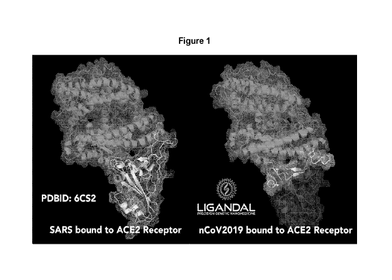
[0013] Figure 1 shows the crystal structure of SARS-CoV-1 (PDBID 6CS2)
bound to
ACE2 (left) compared to simulated structure of SARS-CoV-2 bound to ACE2
(right).
Amino acid residues contributing positively to binding (-AG) are shown in
green, amino
acid residues having about 0 AG are shown in yellow and repulsory amino acid
residues
(+AG) are shown in pink (left) or in orange (right).
[0014] Figure 2A shows the 3D structure of two previously published SARS-
CoV-1
immuno-epitopes. Figures 2B-2D show the 3D structure and the locations of the
deduced
CoV-2 immuno-epitopes based on homology to SARS-CoV-1.
[0015] Figure 3 shows the MHC-I binding prediction results of immuno-
epitopes.
"ImmunoEpitope1" = SEQ ID NO:67; "ImmunoEpitope2" = SEQ ID NO:69;
KMSECVLGQSKRV = SEQ ID NO:71; LLFNKVTLA = SEQ ID NO:7; SFIEDLLFNKV =
SEQ ID NO:68.
[0016] Figure 4 shows the position of CoV-2 S protein antibody epitopes
identified by
others in the CoV-2 S protein (residues 15-1137 of SEQ ID NO:2 pictured). The
CoV-2
scaffold in the wildtype protein is double underlined. The epitopes are shown
in bold while
the epitopes having high antigenicity scores are shown in bold and underlined.
[0017] Figure 5 shows the truncated CoV-2 S protein aligned to ACE2 and the
locations of the antibody epitopes (magenta) and the ACE2 binding residues
(green).
[0018] Figure 6 depicts three-dimensional molecular modeling of three
representative
linkers in the bound conformation. The backbone is depicted as a blue coil.
Side chain
atoms are color coded in PyMol using the command color > by chain > chainbows
and
Color > by > element > HNOS where H = white, N = blue, 0 = red, and S =
yellow.
Representative sequences depicted are
SNNLDSKVGGNYNYLYRLFDGTEIYQAGSTPCNGVEGFNCYFPLQSYGFQPTNGVGYQ
P (SEQ ID NO:116);
SNNLDSKVGGNYNYLYRLFNANDKIYQAGSTPCNGVEGFNCYFPLQSYGFQPTNGVGY
QP (SEQ ID NO:119);
SNNLDSKVGGNYNYLYRLFPGTEIYQAGSTPCNGVEGFNCYFPLOSYGFQPTNGVGYQP
(SEQ ID NO:122).
-5-
CA 03172878 2022-08-24
WO 2021/173879 PCT/US2021/019739
[0019] Figure 7A illustrates the binding of Scaffold #15 (SEQ ID NO:86) to
residues
19-169 of ACE2 (SEQ ID NO:140). The B cell epitopes are shown in magenta, the
T cell
epitopes are shown in orange, and the ACE2 binding sites are shown in green.
Figure 7B
illustrates that a modified CoV-2 scaffold having 59 amino acids (with 18
amino acids
eliminated from the wildtype sequence) preserves the binding affinity to ACE2.
Figure 7C
illustrates that a modified CoV-2 scaffold having 67 amino acids (with 10
amino acids
eliminated from the wildtype sequence) preserves the binding affinity to ACE2.
The B cell
antibody immuno-epitopic regions are shown in magenta, the T cell receptor
binding,
MHC-1 and MHC-2 loading regions are shown in orange, and the ACE2 binding
regions
are shown in green.
[0020] Figures 8A-8B show that S protein Scaffold #9 (SEQ ID NO:80) can be
fitted
to ACE2 (Figure 8A; residues 19-107 of ACE2 shown) to determine its ACE2
binding
affinity and KD prediction based on computer modeling (Figure 8B).
[0021] Figure 9A shows the computer modeling of CoV-1 (cyan) and CoV-2
(navy)
bound to ACE2 (red) based on homology of CoV-1 and CoV-2. Figure 9B shows the
computer modeling of CoV-1 (cyan) bound to ACE2 (red). Figure 9C shows the
computer
modeling of CoV-2 (navy) bound to ACE2 (red).
[0022] Figures 10A and 10B show the AG calculation to determine key binding
residues for CoV-2 and CoV-1, respectively.
[0023] Figures 11A-11B show the thermodynamic modeling of CoV-2 bound to
ACE2,
with the binding interface enlarged in Figure 11B. Figure 11C shows two
critical binding
motifs determined for CoV-2: residues 437-455 (SEQ ID NO:65) and residues 473
to 507
(SEQ ID NO:66. The amino acid residues having a negative AG, a positive AG,
and about
0 AG are shown in green, orange, and yellow, respectively. The backbone
residues are
shown in navy. L455 and P491 are shown in magenta.
[0024] Figures 12A-12J illustrate the folding possibilities (center()
through center9
conformation shown in PyMOL) for CoV-2 Scaffold #1 having an amino acid
sequence of
SEQ ID NO:72.
-6-
CA 03172878 2022-08-24
WO 2021/173879 PCT/US2021/019739
[0025] Figure 13A shows the binding of center0 of CoV-2 Scaffold #9 (SEQ ID
NO:80) with ACE2. Figure 13B shows the binding of center0 and center9 of CoV-2
Scaffold #9 with ACE2. Figures 13C-13D show chaotic assortment of center0-
center9 of
CoV-2 Scaffold #9 with ACE2, showing reasonable average folding and locations
of all
possible folding states given Heisenberg Uncertainty Principle. Figure 13D
shows the
enlarged binding interface of Scaffold #9 and ACE2.
[0026] Figure 14A depicts a simulation of ACE2 bound to CoV-2 S protein.
Figures
14B-14D depict ACE2 Scaffold 1 (SEQ ID NO:141) (purple), simulated via
RaptorX,
overlaid with wildtype hACE2 (red). The critical binding residues of ACE2 at
the interface
with the CoV-2 S protein are highlighted green.
[0027] Figure 15A shows computer modeling of ACE2 Scaffold 1 (SEQ ID NO:141
truncated from the ACE2 protein. Figure 15B depicts the molecular modeling of
ACE2
Scaffold 1 (purple, with critical binding residues shown in green) with the
CoV-2 S protein
(blue, with antibody binding domains shown in teal arrow). The scaffold binds
to the CoV-
2 S protein while preserving the presentation of antibody-binding immuno-
epitopic regions
of the S protein while bound. Figure 15C depicts ACE2 Scaffold 1 bound to the
CoV-2 S
protein. ACE2 Scaffold 1 is not predicted to affect the immune binding domains
(pink) of
the CoV-2 S protein.
[0028] Figure 16A shows the binding to ACE2 by the Cryo-EM structure of CoV-
2 S
protein published by others, and Figure 16B shows the binding to ACE2 by CoV-2
S
protein based on SWISS-MODEL.
[0029] Figure 17A shows the simulated conformation with ACE2 using the
structure
published by others (top) and the computer simulated structure of this
disclosure (bottom).
Figure 17B shows the comparison of the Cryo-EM structure of CoV-2 published by
others
(left) to the disclosed truncated and labeled SWISS-MODEL simulated structure
(right).
The red dotted oval indicates the location of the missing residues from the
Cryo-EM
structure. Purple regions indicate B cell immuno-epitopes determined by
others, while
orange regions indicate ACE2-repulsory regions, green regions indicate ACE2-
binding
regions, and yellow regions indicate ACE2-neutral regions as determined via
PDBePISA.
-7-
CA 03172878 2022-08-24
WO 2021/173879 PCT/US2021/019739
[0030] Figure 18 shows that a custom-built peptide robot completed
synthesis of a 9-
amino acid MHC-1 loading epitope in about 24 minutes, allowing for rapid
prototyping prior
to commercial scale-up.
[0031] Figure 19A shows head-to-tail cyclization of the side chain
protected peptide in
solution by amide coupling using Scaffold #47 ("Liganda1-05," SEQ ID NO:118)
as an
example. Figure 19B shows on resin head-to-tail cyclization by amide coupling
using
Scaffold #48 ("Liganda1-06," SEQ ID NO:119) as an example. Figure 19C shows
cyclization of purified linear thioester peptide by NCL using Scaffold #46
("Liganda1-04,"
SEQ ID NO:117) as an example.
[0032] Figures 20A-20I are charts depicting biolayer interferometry of
Scaffold #4
("Peptide 1," SEQ ID NO:75), Scaffold #7 ("Peptide 4," SEQ ID NO:78), Scaffold
#8
("Peptide 5," SEQ ID NO:79), and Scaffold #9 ("Peptide 6," SEQ ID NO:80)
associated
with ACE2-biotin captured on streptavidin sensor tips (2.5 nm capture) to
determine
dissociation constant of the scaffolds to ACE2. All scaffolds exhibited potent
inhibition of
RBD binding to ACE2 at 10pM concentrations. As shown in Figures 20A-20D, a
clear
binding to ACE2 was observed for each scaffold with increasing concentrations
(blank
values were subtracted). As shown in Figures 20E-20H, a dose-response curve
was also
observed, whereby RBD was able to strongly associate with each sensor at 35 pM
in the
absence of peptide (green, top curve), and experienced a peptide-dose-response-
dependent inhibition of binding (blue, cyan and red represent 10, 3 and 1 pM
concentrations, respectively). Figure 201 corresponds to RBD-biotin captured
on
streptavidin sensor tips (5 nm capture), and subsequently bound to ACE2.
[0033] Figures 21A-21F are charts depicting biolayer interferometry of the
scaffolds
associated with a neutralizing antibody captured on anti-human IgG (AHC)
sensor tips (1
nm capture) was used to determine dissociation constant of Scaffold #4
("Peptide 1," SEQ
ID NO:75), Scaffold #7 ("Peptide 4," SEQ ID NO:78), Scaffold #8 ("Peptide 5,"
SEQ ID
NO:79), and Scaffold #9 ("Peptide 6," SEQ ID NO:80) to the neutralizing
antibody (Figures
21A-21D). The dissociation constant of increasing concentrations of RBD was
determined
with anti-RBD neutralizing antibody (Figure 21E). Figure 21F shows that 117 nM
RBD was
mixed with increasing concentrations of ACE2 prior to introduction to
neutralizing
-8-
CA 03172878 2022-08-24
WO 2021/173879 PCT/US2021/019739
antibodies bound to the sensors to demonstrate ACE2's inhibition of
neutralizing antibody
binding to the RBD.
[0034] Figure 22 shows luminescence (RLU) of ACE2-HEK293 cells following
SARS-
CoV-2 spike infection at 60 hours post-infection when co-transfected with
Scaffold #4
("Peptide 1," SEQ ID NO:75), Scaffold #7 ("Peptide 4," SEQ ID NO:78), Scaffold
#8
("Peptide 5," SEQ ID NO:79), and Scaffold #9 ("Peptide 6," SEQ ID NO:80).
Control
groups included untransfected ACE2-HEK293 cells (no virus) and ACE2-HEK293
cells
transfected with the SARS-CoV2 spike protein.
[0035] Figures 23A-23D show luminescence (RLU) in ACE2-HEK293 cells
transfected with the SARS-CoV-2 spike protein or no virus (control) and
Scaffold #8
("Peptide 5," SEQ ID NO:79) (Figure 23A), soluble ACE2 (Figure 23B), soluble
receptor-
binding domain (RBD) of the SARS-CoV-2 spike protein (Figure 23C), and a SARS-
CoV-2
neutralizing antibody (neuAb) (Figure 23D).
[0036] Figure 24A shows that Scaffold #4 ("LGDL_NIH_001," SEQ ID NO:75),
Scaffold #7 ("LGDL_NIH_004," SEQ ID NO:78), Scaffold #8 ("LGDL_NIH_005," SEQ
ID
NO:79), and Scaffold #9 (LGDL_NIH_006," SEQ ID NO:80) exhibited over 90%
inhibition
of viral load (EC90) in live virus at micromolar concentrations. Figure 24B
shows that the
scaffolds tested in Figure 24A were not toxic at the effective concentrations.
[0037] Figure 25 depicts three-dimensional molecular modeling of Scaffold
#4
("Peptide 1," SEQ ID NO:75 based on a 180ns run (single trajectory) in OpenMM
starting
from the native-like conformation.
[0038] Figure 26 is a chart plotting Rosetta score (REU) of Scaffold #4
(SEQ ID
NO:75 at indicated timepoints.
[0039] Figures 27A and 27B are diagrams of epitopes on the S protein that
are only
exposed during fusion.
[0040] Figures 28A and 28B are diagrams of binding sites which would
prevent the
process from moving to the next step of neutralizing. Figure 28C shows the
enlarged
binding site.
-9-
CA 03172878 2022-08-24
WO 2021/173879 PCT/US2021/019739
[0041] Figures 29A and 29B (enlarged) depict three-dimensional molecular
modeling
of the sequence KMSECVLGQSKRV (SEQ ID NO:8) (shown in red) fitted to the SARS-
CoV-2 spike protein (green). SEQ ID NO:8 corresponds to one of the binding
sites
identified in Figures 27-28 located in the hinge between heptad repeat (HR) 1
(HR1) and
HR2 during the pre-bundle stage.
[0042] Figure 30 depicts a diagram for extein insertion placement.
[0043] Figures 31A-31D are diagrams generated during peptide screening and
optimization.
[0044] Figures 32A-32B show sequence alignments of representative SARS-CoV-
2 S
protein scaffolds disclosed herein. Alignment shows amino acid residues 433-
511 of SEQ
ID NO:2. Critical binding motifs are underlined. Substitutions are double
underlined and
highlighted yellow. GS linkers are bolded and highlighted blue. Epitopes for B
cell and T
cell binding are bolded, italicized, and highlighted green. EPEA C-tags are
italicized and
highlighted gray. Poly charged N- and C-terminal residues are squiggly
underlined and
highlighted pink. Alternative TCR epitopes are highlighted red.
[0045] Figures 33A-33E illustrate the siRNA designing process using the IDT
siRNA
design tool, including the locations and sequences of the selected sense and
anti-sense
strands (SEQ ID NOs:143-148).
[0046] Figure 34 depicts a three-dimensional simulation model of SARS-CoV-1
bound to angiotensin-converting enzyme 2 (ACE2) (PDB ID 6CS2; red) to
approximate the
binding interface of the SWISS-MODEL simulated SARS-CoV-2 (left); and selected
MHC-I
and MHC-II epitope regions for inclusion in Scaffold #8) (pink) represent P807-
K835 and
A1020-Y1047 in the S1 spike protein. The model on the right depicts the
receptor-binding
domain (RBD) of the SARS-CoV-2 spike protein (blue/multi-colored) simulated
binding with
ACE2 (red). The simulation model identifies predicted thermodynamically
favorable
(green), neutral (yellow), and unfavorable (orange) interactions. Outer bounds
of amino
acids used to generate the scaffold (V433 - V511) are shown in cyan on the
right.
[0047] Figure 35 depicts a three-dimensional simulation model of the ACE2
receptor
(red) aligned with Scaffold #4, #7, #8, and #9 (top, from left to right).
Multiple folding states
-10-
CA 03172878 2022-08-24
WO 2021/173879 PCT/US2021/019739
for Peptide 5 are shown in simulated binding to ACE2 (bottom). Predicted
binding
residues are indicated in green (top and bottom).
[0048] Figure 36 shows SARS-CoV-2 genomic sequence (SEQ ID NO: 1).
Nucleotides 21536-25357 (underlined) encode S protein of SEQ ID NO:2.
Nucleotides
26218-26445 (double underlined) encode envelope protein of SEQ ID NO:3.
[0049] Figure 37 shows the amino acid sequence of SARS-CoV-2 spike (S)
protein
(SEQ ID NO: 2).
[0050] Figure 38 shows the amino acid sequence of ACE2 (SEQ ID NO: 140).
DETAILED DESCRIPTION
[0051] As disclosed herein, by combining methods from mathematical data
science,
biophysics, and experimental biology, the sequences of the S protein that are
most likely to
expand the host range and increase the stability of SARS-CoV-2 in the human
population
through natural selection can be predicted. A computational pipeline is
developed to
estimate the mutation landscape of the SARS-CoV-2 S protein. The predicted
sequences
are experimentally engineered and their binding to the human receptor ACE2 is
measured
using biochemical assays and cryo-electron microscopy.
[0052] Novel mathematical approaches, inspired by the structure of genetic
algorithms, are developed for the identification of highly probable sequences
of the SARS-
CoV-2's S-protein. More specifically, the disclosed approach incorporates
descriptors from
graph theory, topological data analysis, and computational biophysics into a
new machine
learning framework that combines neural networks and genetic algorithms. This
powerful
interdisciplinary approach allows the use of existing data from SARS-CoV-2 to
uncover a
few candidate sequences that are most likely to occur in the evolution of its
viral S-protein.
These results are experimentally validated by generating peptides from the
obtained
sequences. The resulting pipeline provides new solution to better understand
the mutation
landscape of viral proteins.
[0053] As disclosed herein, in silico analysis was conducted to generate
and screen
novel peptides ("scaffolds") designed to serve as competitive inhibitors to
the SARS-CoV-2
-11-
CA 03172878 2022-08-24
WO 2021/173879 PCT/US2021/019739
spike (S) protein by predicting 1) ACE2 receptor binding regions, 2) immuno-
epitopic
regions for T cell receptor MHC-I and MHC-II loading, and 3) immuno-epitopic
regions for
B cell receptor or antibody binding. As demonstrated in the working examples,
three-
dimensional modeling and in silico analysis were used to examine predicted
structures of
the novel peptides, various sequence modifications were evaluated (e.g., by
examining
Rosetta energy unit (REU) scores for candidate peptides), and predicted
binding models
were simulated by computer. Based on these results, provided herein are
methods for
generating and optimizing peptide scaffolds for use as competitive inhibitors
in vaccine
development by taking a peptide sequence (e.g., the SARS-CoV-2 spike protein),
introducing sequence modifications, and using three-dimensional modeling
techniques to
predict folding or binding conformation. Also provided herein are optimized
peptide
scaffolds designed using these methods, formulations comprising these peptide
scaffolds,
and methods of using these peptide scaffolds and formulations to competitively
inhibit viral
proteins or treat viral infection, and the use of these peptide scaffolds and
formulations as
vaccines to prevent viral infection.
[0054] Accordingly, this disclosure relates to a breakthrough approach for
rapid
vaccine prototyping. In some aspects, the disclosed vaccine approach provides
a fully
synthetic scaffold for mimicking T-cell receptor and antibody binding
epitopes, which can
be rapidly custom-tailored to new mutant forms of a virus. Additionally, the
synthetic
scaffold can serve as a targeting ligand mimicking viral entry to target
diseased cells and
tissues with therapeutic agents. These "mini viral" scaffolds can be
synthesized in hours,
and rapidly scaled to a scale of over 100 kg to meet global needs.
Additionally, scaffolds
provided herein may separately be used in place of small molecules for
inhibiting binding
cleft interactions.
[0055] The scaffolds disclosed herein are peptides generated by modeling
off the
SARS-CoV-2 spike protein receptor binding motif (RBM) conserved motifs, and
have the
potential utility as a prophylactic, immune-stimulant, and therapeutic agent
against the
virus. Therefore, also disclosed herein are compositions comprising one or
more
scaffolds, which can be used for: 1) inhibiting ACE2-spike interaction and
viral entry into
ACE2-expressing cells, 2) promoting binding to neutralizing antibodies without
-12-
CA 03172878 2022-08-24
WO 2021/173879 PCT/US2021/019739
competitively displacing neutralizing antibody binding to the RBD; and/or 3)
preventing
soluble ACE2 association with the RBD.
[0056] Detailed in this disclosure are the simulation, design, synthesis
and
characterization of peptide scaffolds designed to block viral binding to cells
expressing
ACE2, while also stimulating an immune response and promoting exposure of the
spike
protein for recognition by the immune system. In contrast to neutralizing
antibody
therapies and other approaches that seek to target the virus, a biomimetic
virus decoy
peptide technology is developed to compete for binding with cells and expose
the virus for
binding to neutralizing antibodies.
I. Computer-assisted 3D modeling
A. Analyzing the binding interface
[0057] In one aspect, this disclosure relates to methods of computer-
assisted three-
dimensional (3D) modeling to investigate protein-protein interactions. These
methods
entail producing a 3D model of a first binding partner and a second binding
partner,
determining the amino acid sequence, the 3D structure, and the conformation of
the
interface of each binding partner, truncating the binding interface of each
binding partner
while maintaining the 3D structure of each to obtain a scaffold representing
each binding
partner, determining the binding affinity of each amino acid residue in the
scaffold based
on calculation of thermodynamic energy of each residue, and determining the
location and
sequence of critical binding motifs in the scaffold. In certain embodiments,
the 3D model is
produced with SWISS-MODEL based on protein sequence homology to the first
binding
partner or the second binding partner. Various modifications can be made to
the scaffold
to maintain or improve the structure, conformation, and binding affinity of
the scaffold.
These modifications include but are not limited to insertions, deletions, or
substitutions of
one or more amino acid residues in the scaffold. As detailed in this
disclosure, various
linkers, conjugatable domains, and/or immuno-epitopes can be added to the
scaffold to
obtain multi-functional scaffolds. In certain embodiments, one or more amino
acids that
are not critical for binding can be deleted or substituted. In certain
embodiments, the
binding partners are SARS-CoV-2 S protein and ACE2. In certain embodiments the
S
-13-
CA 03172878 2022-08-24
WO 2021/173879 PCT/US2021/019739
protein has the amino acid sequence set forth in SEQ ID NO:2. In other
embodiments, the
S protein is a variant, including but not limited to B.1.1.7 variant (SEQ ID
NO:137), B.1.351
variant (SEQ ID NO:138), or P.1 variant (SEQ ID NO:139). Other variants of
coronavirus
can be found at nextstrain.org/ncov/global. In certain embodiments ACE2 has
the amino
acid sequence set forth in SEQ ID NO:140.
[0058] As used herein, the term "scaffold" means a continuous stretch of
amino acid
sequence located at the binding interface of a binding partner and involved
with binding to
the other binding partner. In certain embodiment, the scaffold has a size of
less than
about 120 amino acid residues, less than about 110 amino acid residues, less
than about
100 amino acid residues, less than about 90 amino acid residues, less than
about 80
amino acid residues, less than about 70 amino acid residues, less than about
60 amino
acid residues, or less than 50 amino acid residues. In certain embodiments,
the scaffold
maintains the 3D structure and/or conformation of the native, free, or bound
state of the
protein from which its binding sequence(s) is derived. For example, the
scaffold may be
designed to maintain an a-helix and/or 13 sheet structure when truncated from
a wildtype
protein sequence. In certain embodiments, the scaffold may comprise one or
more
modifications such as insertions, deletions, and/or substitutions, provided
those
modifications do not substantially decrease, and in some embodiments actually
increase,
binding affinity of the scaffold to its binding partner.
[0059] As disclosed herein, the protein sequence of the SARS-CoV-2 spike
protein
(SARS-CoV-2 or CoV-2; SEQ ID NO:2) was compared to SARS coronavirus protein
sequence (PDB ID 6CS2) to produce a 3D model of CoV-2 binding to ACE2 (Figure
1).
The binding interface of each of CoV-2 and ACE2 was investigated to determine
a stretch
of amino acid residues involved in binding. This stretch of amino acid
sequence may be
truncated from the remaining protein sequence, and the structure and/or
conformation of
this stretch of amino acid sequence is maintained to simulate that of the
native protein in
free or bound state, thereby to obtain the CoV-2 scaffold or the ACE2 scaffold
of this
disclosure.
[0060] Accordingly, disclosed herein is a CoV-2 scaffold, which has an
amino acid
sequence at least 40%, at least 45%, at least 50%, at least 55%, at least 60%,
at least
-14-
CA 03172878 2022-08-24
WO 2021/173879 PCT/US2021/019739
65%, at least 70%, at least 75%, at least 80%, at least 85%, at least 90%, at
least 95%, at
least 96%, at least 97%, at least 98%, at least 99%, or 100% identical to the
amino acid
sequence of residues 433-511 of SEQ ID NO:2
(VIAWNSNNLDSKVGGNYNYLYRLFRKSNLKPFERDISTEIYQAGSTPCNGVEGFNCYFPL
QSYGFQPTNGVGYQPYRVV).
[0061] In some embodiments, the CoV-2 scaffold has an amino acid sequence
at
least 40%, at least 45%, at least 50%, at least 55%, at least 60%, at least
65%, at least
70%, at least 75%, at least 80%, at least 85%, at least 90%, at least 95%, at
least 96%, at
least 97%, at least 98%, at least 99%, or 100% identical to the amino acid
sequence:
VIAWNSNNLDSKVGGNYNYLYRLFRKSNLKPFERDISTEI YQA GSTPCNGVEGFNC YFPL
QSYGFQP7NGVGYQPYRVV ("Scaffold #1; SEQ ID NO:72).
[0062] In SEQ ID NO: 72 above, amino acid residues in the CoV-2 S protein
backbone are shown in plain letters (including 433V-436W, F456-I472, and Y508-
V511),
amino acid residues having a AG of about 0, which are neutral in binding, are
underlined
(including N437, S438, N440-K444, G447, N448, N450-L452, R454, S477-V483,
G485,
C488, P491, L492, F497, G504, and P507), amino acid residues having a negative
AG,
which are critical binding residues, are shown in bold (including N439, Y449,
Y453, Q474,
E484, N487, Q493-Y495, Q498, P499, N501, and Q506), and amino acid residues
having
a positive AG, which are repulsory residues, are shown in italicized
(including V445, G446,
L455, Y473, A475, G476, F486, Y489, F490, G496, T500, G502, V503, and Y505).
[0063] Based on the computer modeling and the calculation of thermodynamic
energy, it is determined that one CoV-2 scaffold of this disclosure comprises
a first critical
binding motif comprising residues 437 to 455 of SEQ ID NO: 2, a second
critical binding
motif comprising residues 473 to 507 of SEQ ID NO: 2, and a backbone region
comprising
residues 456 to 472 of SEQ ID NO: 2. The first and second critical binding
motif directly
interact with ACE2 on the binding interface, while the backbone region
comprises amino
acid residues that do not directly interact with ACE2.
[0064] The CoV-2 scaffold may further comprise one or more amino acids from
the
CoV-2 S protein backbone at the N-terminus, the C-terminus, or both, to
achieve a desired
-15-
CA 03172878 2022-08-24
WO 2021/173879 PCT/US2021/019739
size. In some embodiments, the CoV-2 scaffold comprises from about 40 to about
200
amino acid residues, from about 50 to about 100 amino acid residues, from
about 55 to
about 95 amino acid residues, from about 60 to about 90 amino acid residues,
from about
65 to about 85 amino acid residues, from about 70 to about 80 amino acid
residues. In
some embodiments, the CoV-2 scaffold comprises about 50 amino acid residues,
about 55
amino acid residues, about 60 amino acid residues, about 65 amino acid
residues, about
70 amino acid residues, about 75 amino acid residues, about 80 amino acid
residues,
about 85 amino acid residues, about 90 amino acid residues, about 95 amino
acid
residues, or about 100 amino acid residues.
[0065] Although the size of the CoV-2 scaffold can vary, and certain
modifications
such as insertions, deletions, and/or substitutions can be incorporated, the
scaffold
maintains the structure and/or conformation of its native state with regard to
the ACE2
binding interface. Preservation of this structure and/or conformation allows
the scaffold to
bind ACE2 with the same or greater affinity than the full-length S protein
despite its
truncation. For example, the 13 sheet structure is maintained and can be
stabilized by
further modifications. In some embodiments, the CoV-2 scaffold comprises L455C
and
P491C substitutions such that a disulfide bond is formed between location 455
and
location 491 to stabilize the 13 sheet structure. These two locations appear
to be in
proximity to each other in the native CoV-2 S protein bound to ACE2 based on
computer
modeling. In some embodiments, the CoV-2 scaffold comprises one or more
mutations to
replace one or more of the existing Cys residues such that the only Cys
residues
remaining are the ones introduced at locations 455 and 491 to avoid any
undesirable
interference of formation of a disulfide bond. For example, Cys can be
substituted with
Gly, Ser, or any other residue as long as the substitution does not compromise
the binding
affinity to ACE2. Some examples of replacing Cys residues include but not
limited to
C480G, and C488G.
[0066] In certain embodiments, the CoV-2 scaffold disclosed herein may
further
comprise a loop to connect the N-terminal residue and the C-terminal residue
using a
linker such as an am ine-carboxy linker to obtain a head-to-tail cyclized
scaffold. In certain
embodiments, cyclization of the scaffold provides increased stability with
lower free
-16-
CA 03172878 2022-08-24
WO 2021/173879
PCT/US2021/019739
energy, enhanced folding, binding, or conjugation to a substrate, and/or
enhanced
solubility. The loop does not directly interact with the scaffold's binding
partner. In certain
embodiments, the loop allows the scaffold to be attached to an siRNA payload
or other
substrates. In certain embodiments, the loop comprises 1-200 amino acid
residues. In
certain embodiments, the loop comprises less than about 150 amino acid
residues.
Depending on the desired conformation of the scaffold, linkers, conjugatable
domains, a
polar head or tail, etc., one can adjust the size of the loop accordingly. In
some
embodiments, the loop comprises 9-15 Arg and/or Lys residues. In some
embodiments,
the loop comprises a conjugatable domain such as maleimide or other linkers to
conjugate
the scaffold to a substrate or a poly amino acid chain. In some embodiments,
the loop
comprises one or more immune-activating poly amino acid chain or immune-
reactive
glycan. The N-terminus and C-terminus can also be connected by forming a
disulfide
bond, any other appropriate linker (flexible or rigid), click chemistry, PEG,
polysarcosine, or
bioconjugated. Thus, the peptides may be cyclized, stabilized, linear,
otherwise click-
chemistry or bioconjugated, or substituted with non-natural amino acids,
peptoids,
glycopeptides, lipids, cholesterol moieties, polysaccharides, or anything that
enhances
folding, binding, solubility, or stability.
[0067] Also
disclosed herein is an ACE2 scaffold, which is at least 40%, at least
45%, at least 50%, at least 55%, at least 60%, at least 65%, at least 70%, at
least 75%, at
least 80%, at least 85%, at least 90%, at least 95%, at least 96%, at least
97%, at least
98%, at least 99%, or 100% identical to the amino acid sequence of residues 19-
84 of
SEQ ID NO:140.
[0068] In
some embodiments, the ACE2 scaffold is at least 40%, at least 45%, at
least 50%, at least 55%, at least 60%, at least 65%, at least 70%, at least
75%, at least
80%, at least 85%, at least 90%, at least 95%, at least 96%, at least 97%, at
least 98%, at
least 99%, or 100% identical to the amino acid sequence:
STIEEQAKTFLDKFNHEAEDLFYQSSLASWNYNTNITEENVQNMNNAGDKWSAFLKEQS
TLAQMYP (SEQ ID NO:151)
-17-
CA 03172878 2022-08-24
WO 2021/173879 PCT/US2021/019739
[0069] In SEQ ID NO:151 above, the amino acid residues having a negative
AG,
which are critical binding residues, are shown in bold (including S19, Q24,
D38, Q42, E75,
Q76, and Y83).
[0070] Based on the computer modeling and the calculation of thermodynamic
energy, it is determined that an ACE2 scaffold of this disclosure comprises a
first critical
binding motif comprising amino acid residues 19 to 42 of SEQ ID NO:140, a
second critical
binding motif comprising residues 64 to 84 of SEQ ID NO:140, and a backbone
region
comprising residues 43 to 63 of SEQ ID NO:140. The first and second critical
binding
motif directly interact with CoV-2 S protein on the binding interface, while
the backbone
comprises amino acid residues on the backbone of ACE2 and does not directly
interact
with CoV-2 S protein.
[0071] In some embodiments, the ACE2 scaffold comprises a linker (shown in
bold)
connecting the two critical binding motifs, see for example, ACE2 Scaffold 1
(SEQ ID
NO:141):
STIEEQAKTFLDKFNHEAEDLFYQGSGSGNAGDKWSAFLKEQSTLAQMYP
[0072] In some embodiments, the ACE2 scaffold further comprises an EPEA C-
tag
(underlined), see for example, ACE Scaffold 2 (SEQ ID NO:142):
STIEEQAKTFLDKFNHEAEDLFYQGSGSGNAGDKWSAFLKEQSTLAQMYPEPEA
[0073] The ACE2 scaffold may further comprise one or more amino acids or
monomeric units from the ACE2 protein backbone or recreating the binding
effect of ACE2
at the appropriate interface with the spike protein as derived from ACE2's N-
terminus, the
C-terminus, or both, to achieve a desired size, folding and affinity. In some
embodiments,
the ACE2 scaffold comprises from about 10 to about 200 amino acid residues,
from about
50 to about 100 amino acid residues, from about 55 to about 95 amino acid
residues, from
about 60 to about 90 amino acid residues, from about 65 to about 85 amino acid
residues,
from about 70 to about 80 amino acid residues. In some embodiments, the ACE2
scaffold
comprises about 50 amino acid residues, about 55 amino acid residues, about 60
amino
acid residues, about 65 amino acid residues, about 70 amino acid residues,
about 75
-18-
CA 03172878 2022-08-24
WO 2021/173879 PCT/US2021/019739
amino acid residues, about 80 amino acid residues, about 85 amino acid
residues, about
90 amino acid residues, about 95 amino acid residues, or about 100 amino acid
residues.
[0074] Although the size of the ACE2 scaffold can vary, and certain
modifications
such as insertions, deletions, and/or substitutions can be incorporated, the
scaffold
maintains the structure and/or conformation of its native state with regard to
the CoV-2 S
protein binding interface. Preservation of this structure and/or conformation
allows the
scaffold to bind the S protein with the same or greater affinity than the full-
length ACE2
protein despite its truncation.
[0075] In certain embodiments, the N-terminus, C-terminus, or both termini
of the
ACE2 scaffold are modified with any number of bioconjugation motifs, linkers,
spacers,
tags (such as his-tag and C-tag) etc. In certain embodiments, one or more
amino acids
that are not critical for binding to CoV-2 S protein are deleted or
substituted.
[0076] Additional scaffolds can be designed to mimic ACE2 binding to the
CoV-2 S
protein. These ACE2 scaffolds can bind to the CoV-2 virus to coat the virus
such that the
virus is unable to bind to ACE2 thereby to inhibit viral entry into human (or
other hosts)
body. Moreover, an ACE2 scaffold can be further modified to include, for
example, a
fragment crystallizable (Fc) domain or an alternate domain that serves to
activate an
immune response.
[0077] ACE2 Scaffold 1 (SEQ ID NO:141), comprising a first critical binding
motif, a
second critical binding motif, and a linker connecting the critical binding
motifs, is predicted
to have a higher affinity for the CoV-2 S protein than wildtype ACE2.
Additionally, in
contrast to ACE2, which binds to CoV-2 S protein and blocks the immuno-
epitopic region
of CoV-2 S protein, ACE2 Scaffold 1 is not expected to affect the immune
binding domains
of the CoV-2 S protein and allows the immune system to identify the CoV-2
virus. Similar
to other scaffolds provided herein, ACE2 Scaffold 1 may be provided in a
nanoparticle or
other suitable substrate and may act to aggregate the virus. For instance, the
N- or C-
termini may be modified with any number of bioconjugation motifs, linkers,
spacers, and
the like; and may have various substrates including buckyballs (e.g., C60/C70
fullerenes),
branched PEGs, hyper-branched dendrimers, single-walled carbon nanotubes,
double-
-19-
CA 03172878 2022-08-24
WO 2021/173879 PCT/US2021/019739
walled carbon nanotubes, KLH, OVA, and/or BSA. ACE2 Scaffold 1 is predicted to
have a
higher affinity for the virus spike proteins than free ACE2.
B. Analyzing the immune-epitopes
[0078] Immune Epitope Database (IEDB) was utilized to predict key epitopes
prior to
clinical data emerging on various T cell receptor (TCR) responses across
populations with
various HLA alleles. These predicted epitopes were compared to known epitopes
for
MHC-I and MHC-II response in SARS-CoV-1. It was previously reported that S5
peptide
having an amino acid sequence of LPDPLKPTKRSFIEDLLFNKVTLADAGFMKQYG (SEQ
ID NO:135) (residues 788-820 of SARS-CoV-1) and S6 peptide having an amino
acid
sequence of ASANLAATKMSECVLGQSKRVDFCGKGYH (SEQ ID NO:136) (residues
1002-1030 of SARS-CoV-1) exhibited immunogenic responses similar to those
found in a
parallel investigation using truncated recombinant protein analogs of the SARS-
CoV S
protein (2). The S5 peptide was defined based on known immunogenicity of the
monovalent peptide in terms of its ability to illicit an MHC-I response and
antibody
response, whereas many other peptides were only immunogenic while present
multivalently. The S6 peptide represents a known MHC-II domain from SARS-CoV-
1.
[0079] These immuno-epitopes of SARS-CoV-1 S protein were aligned to CoV-2
S
protein to determine likely immunogenic sites on CoV-2 S protein. Based on
homology,
the corresponding immuno-epitopes in CoV-2 S protein are identified as
follows, and are
also designed to overlap with the regions of the S2 spike in its pre-fusion
conformation
following TMPRSS2 cleavage of the S1-S2 interface:
(QIL)PDPSKPSKRSFIEDLLFNKVTLADAGFIK (SEQ ID NO:67) (locations 804-835), and
ASANLAATKMSECVLGQSKRVDFCGKGY (SEQ ID NO:69) (locations 1020-1047)
[0080] The 3D structure of the SARS-CoV-1 immuno-epitopes are shown in
Figure
2A, and the 3D structure of the CoV-2 immuno-epitopes and their locations on
CoV-2 S
protein are shown in Figures 2B-2D. IEDB determined that sequences
KMSECVLGQSKRV (SEQ ID NO:71) and LLFNKVTLA (SEQ ID NO:7) of SARS-CoV-2 S
protein, representing MHC-II and MHC-I binding domains for HLA-A*02:01,
respectively,
would be immunogenic with percentile ranks of 0.9 and 1.2, respectively. Lower
percentile
-20-
CA 03172878 2022-08-24
WO 2021/173879 PCT/US2021/019739
rank represents better binding. Figure 3 shows the MHC-I binding prediction
results of
these immuno-epitopes. Accordingly, the immuno-epitopes having the following
sequences are used in further studies and included in some scaffolds:
KMSECVLGQSKRV (SEQ ID NO:71), and LLFNKVTLA (SEQ ID NO:7).
[0081] Additional epitopes can be identified from various databases. For
example, in
the TepiTool results, YLQPRTFLL (SEQ ID NO:9), FIAGLIAIV (SEQ ID NO:22), and
FVFLVLLPL (SEQ ID NO:21) are the top scoring HLA-A*0201 epitopes. These top-
scoring epitopes are very hydrophobic. Some of the top-scoring epitopes, or
alternately if
available, epitopes that demonstrate immunogenicity in vivo or in vitro can be
included in
the scaffolds disclosed herein.
[0082] Figure 4 shows that the antibody epitopes such as B cell epitopes in
CoV-2 S
protein identified by others (12) are aligned to the CoV-2 S protein amino
acid sequence.
[0083] As shown in Figure 5, the truncated CoV-2 S protein having the amino
acid
sequence of SEQ ID NO:4 (below) is aligned to ACE2 to show the locations of
antibody
epitopes (magenta), and ACE2 binding residues (green).
CPFGEVFNATRFASVYAWNRKRISNCVADYSVLYNSASFSTFKCYGVSPTKLNDLCFTNV
YADSFVIRGDEVRQIAPGQTGKIADYNYKLPDDFTGCVIAWNSNNLDSKVGGNYNYLYRL
FRKSNLKPFERDISTEIYQAGSTPCNGVEGFNCYFPLQSYGFQPTNGVGYQPYRVVVLSF
ELLHAPATVCGPKKST (SEQ ID NO:4; some immune-epitopes highlighted in bold).
[0084] Sequence search using the Bepipred tool indicates that most of the
receptor-
binding motif (residues 440-501) is predicted as being a B cell linear
epitope.
[0085] PDB can be used to identify B-cell epitopes as well. For example,
PDB lists
eight epitopes which were previously explored by experiments. Two linear
epitopes on the
SARS-CoV-2 S protein were demonstrated to elicit neutralizing antibodies in
COVID-19
patients (12). Some examples of the B-cell epitopes include:
PSKPSKRSFIEDLLFNKV
(S21P2) (SEQ ID NO:30), TESNKKFLPFQQFGRDIA (S14P5) (SEQ ID NO:25),
PATVCGPKKSTNLVKNKC (SEQ ID NO:24), GIAVEQDKNTQEVFAQVK (SEQ ID NO:26),
NTQEVFAQVKQIYKTPPI (SEQ ID NO:27), PIKDFGGFNFSQILPDPS (SEQ ID NO:29),
-21-
CA 03172878 2022-08-24
WO 2021/173879 PCT/US2021/019739
PINLVRDLPQGFSALEPL (SEQ ID NO:23), and VKQIYKTPPIKDFGGFNF (SEQ ID
NO:28).
[0086] As disclosed in detail below, the scaffolds disclosed herein can be
modified to
include one or more immuno-epitopes including T cell epitopes and/or B cell
epitopes.
[0087] These results demonstrate that binding pockets can be predicted in a
way that
is consistent with Cryo-EM and other high-resolution structural data. This
technique can
be used to rapidly address future mutations of any known or new viruses, even
when
genomic data of the entire virus suggests as little as 80% similarity. The
technology
disclosed herein also incorporates a bioinformatics-driven approach for
mapping TCR and
BCR/antibody epitopes, allowing for a "compression algorithm" of protein size.
In contrast
to recombinant technique and other approaches, the technology disclosed herein
utilizes a
small peptide, such as a peptide of fewer than 70 amino acids out of an about
1200 amino-
acid spike protein, to generate a multi-functional scaffold for ACE2 binding
and
TCR/antibody recognition.
II. Scaffold/peptide modifications
[0088] Disclosed herein are CoV-2 scaffolds or ACE2 scaffolds comprising
one or
more fragments of amino acid sequence from the binding interface of each of
CoV-2 S
protein and ACE2 while substantially maintaining the structure and/or
conformation of the
native protein in its free or bound state. The scaffolds disclosed herein
substantially
maintain or improve the binding affinity to the corresponding binding partner.
For example,
the CoV-2 scaffolds disclosed herein substantially maintain or improve the
binding affinity
to wildtype ACE2; and the ACE2 scaffolds disclosed herein substantially
maintain or
improve the binding affinity to wildtype CoV-2 S protein. The CoV-2 scaffold
or the ACE2
scaffold comprises from about 10 to about 100 amino acid residues, from 15 to
about 30
amino acid residues, from about 55 to about 95 amino acid residues, from about
60 to
about 90 amino acid residues, from about 65 to about 85 amino acid residues,
from about
70 to about 80 amino acid residues. In some embodiments, the CoV-2 scaffold or
the
ACE2 scaffold comprises about 50 amino acid residues, about 55 amino acid
residues,
about 60 amino acid residues, about 65 amino acid residues, about 70 amino
acid
-22-
CA 03172878 2022-08-24
WO 2021/173879 PCT/US2021/019739
residues, about 75 amino acid residues, about 80 amino acid residues, about 85
amino
acid residues, about 90 amino acid residues, about 95 amino acid residues, or
about 100
amino acid residues. In some embodiments, the CoV-2 scaffold or ACE2 scaffold
comprises two or more sequences that enhance binding, displacement or
immunogenicity
of the scaffold(s). In some embodiments, the viral-mimetic or pathogen-mimetic
scaffolds
need not be related to SARS-CoV-2 and its binding to ACE2, and can be derived
from any
pathogen binding to its concomitant human or host protein binding partner(s),
including
eukaryote and prokaryote species.
[0089] In certain embodiments, disclosed herein is a CoV-2 scaffold or an
ACE2
scaffold, each comprising two critical binding motifs, wherein the critical
binding motifs are
involved with direct binding to the binding partner. In some embodiments, the
scaffold
further comprises one or more backbone regions comprising amino acid residues
not
involved with direct binding to the binding partner. In some embodiments, the
backbone
region is located between the two critical binding motifs. In some
embodiments, the
backbone region is located at the N-terminus of the first critical binding
motif. In some
embodiments, the backbone region is located at the C-terminus of the second
critical
binding motif.
[0090] In certain embodiments, the CoV-2 scaffold disclosed herein
comprises a first
critical binding motif having at least 50%, at least 55%, at least 60%, at
least 65%, at least
70%, at least 75%, at least 80%, at least 85%, at least 90%, at least 95%, at
least 98%, or
100% identity to an amino acid sequence of NSNNLDSKVGGNYNYLYRL (SEQ ID
NO:65), and a second critical binding motif having at least 50%, at least 55%,
at least
60%, at least 65%, at least 70%, at least 75%, at least 80%, at least 85%, at
least 90%, at
least 95%, at least 98%, or 100% identity to an amino acid sequence of
YQAGSTPCNGVEGFNCYFPLQSYGFQPTNGVGYQP (SEQ ID NO:66).
[0091] In certain embodiments, the ACE2 scaffold disclosed herein comprises
a first
critical binding motif having at least 50%, at least 55%, at least 60%, at
least 65%, at least
70%, at least 75%, at least 80%, at least 85%, at least 90%, at least 95%, at
least 98%, or
100% identity to an amino acid sequence of STIEEQAKTFLDKFNHEAEDLFYQ (SEQ ID
NO:149), and a second critical binding motif having at least 50%, at least
55%, at least
-23-
CA 03172878 2022-08-24
WO 2021/173879 PCT/US2021/019739
60%, at least 65%, at least 70%, at least 75%, at least 80%, at least 85%, at
least 90%, at
least 95%, at least 98%, 01 100% identity to an amino acid sequence of
NAGDKWSAFLKEQSTLAQMYP (SEQ ID NO:150).
[0092] The scaffolds disclosed herein may have different sizes depending on
the
number of the amino acid residues from the backbone included between the two
critical
binding motifs, at the N-terminus of the first critical binding motif and/or
at the C-terminus
of the second critical binding motif. See, for example, Scaffold #1 (SEQ ID
NO:72) (top)
and Scaffold #10 (SEQ ID NO:81) (bottom), amino acid sequences aligned below.
SEQ ID NO: 72
VIAWNSNNLDSKVGGNYNYLYRLFRKSNLKPFERDISTEIYQAGSTPCNGVEGFNCYFPLQSYGEOPTNGVGYQPYRVV
SEQ ID NO: 81
NSNNIDSKVGGNYNYLYRLFRKSNLKPFERDISTEIYQAGSTPCNGVEGFNCYFPLQSYGFOPTNGVGYOPY
[0093] In certain embodiments, one or more amino acid residues in the
scaffold are
deleted or substituted. For example, one or more repulsory amino acid residues
having a
positive AG are deleted or substituted, one or more neutral amino acid
residues having a
AG of about 0 are deleted or substituted, and/or one or more amino acid
residues outside
of the critical binding motifs, e.g., in the backbone region, are deleted or
substituted.
Although not desirable, one or more critical amino acid residues having a
negative AG can
be deleted or substituted.
[0094] In certain embodiments, the scaffold is modified by replacing the
amino acid
residues in the backbone region between the critical binding motifs with a
linker such as a
GS linker having various lengths. In some embodiments, the scaffold comprises
a linker
having about 1 to about 20 amino acid residues, for example, the linker has 1,
2, 3, 4, 5, 6,
7, 8, 9, 10, 11, 12, 13, 14, 15, 16, 17, 18, 19, 0r20 amino acid residues. One
can optimize
the size of the linker to achieve a desired structure and/or conformation of
the scaffold.
See, for example, Scaffold #1 (SEQ ID NO:72), Scaffold #3 (SEQ ID NO:74),
Scaffold #11
(SEQ ID NO:82), Scaffold #12 (SEQ ID NO:83), Scaffold #13 (SEQ ID NO:84), and
Scaffold #14 (SEQ ID NO:85), from top to bottom in the order of appearance,
amino acid
sequences aligned below. The GS linkers are shown in bold.
VIAWNSNNLDSKVGGNYNYLYRLFRKSNLKPFERDISTEIYQAGSTPCNGVEGFNCYFPLQSYGFc:?TNGVGYQPYRV
V
VIAWNSNILDSKVGGNYNYLYRLGSGSG QAGSTPCNGVEGFNCYFPLQSYGFUTNGVGYQPYRVV
N.SNILDSKVGGNYT,TYLYRLGSGSGS QAGSTPCNGVEGFNCYFPLQSYGFUTNGVGYQPY
NSNNLDSKVGGNYNYLYRLGSGSG QAGSTPCINGVEGFNCYFPLQSYGFUTNGVGYQPY
NSNNLDSKVGGNYNYLYRIGSGS QAGSTPC.NGVEGFNC'YFFLOYGFQFTNGVGYQPY
NSNNLDSKVGGNYNYLYRLGSG QAGSTPCNGVEGFNCYFFLOYGFQPTNGVGYQPY
-24-
CA 03172878 2022-08-24
WO 2021/173879 PCT/US2021/019739
[0095] In certain embodiments, the scaffold comprises one or more immuno-
epitopes
such as one or more T cell epitopes, one or more B cell epitopes, or both. The
immuno-
epitopes can be included within a non-interfacing loop structure which
replaces the entire
or partial sequence of the backbone region between the two critical binding
motifs of the
scaffold. For example, one or more amino acid residues in the backbone region
can be
replaced by one or more immuno-epitopes. In another example, one or more amino
acid
residues in the critical binding motif, preferably, the repulsory or neutral
amino acid
residues, can be replaced by one or more immuno-epitopes. Depending on the
desirable
size and structure of the scaffold, one can choose which amino acid residues
to be
replaced by one or more immuno-epitopes.
[0096] In certain embodiments, the immuno-epitopes are 9 or 13 amino acid
residues
long corresponding to MHC-I and MHC-Il binding. For example, the T cell
epitopes include
but are not limited to KMSECVLGQSKRV (SEQ ID NO:8), and LLFNKVTLA (SEQ ID
NO:7). Other known epitopes may be included in the scaffold as well. For
example,
dominant TCR epitopes including KLWAQCVQL (SEQ ID NO:10) (ORF1ab, 3886-3894,
17.7 nM, mostly for A*02), YLQPRTFLL (SEQ ID NO:9) (S, 269-277, 5.4 nM, mostly
for
A*02), and LLYDANYFL (SEQ ID NO:11) (ORF3a, 139-147, mostly for A*02) (3).
Other
known TCR epitopes include PRWYFYYLGTGP (SEQ ID NO:12) (nucleocapsid),
SPRVVYFYYL (SEQ ID NO:13) (nucleocapsid, mostly for B*07:02, A*11:01,
A*03:01),
WSFNPETN (SEQ ID NO:14) (membrane protein), QPPGTGKSH (SEQ ID NO:15)
(ORF1ab polyprotein), and VYTACSHAAVDALCEKA (SEQ ID NO:16) (ORF1ab
polyprotein) (1, 4-6). Some epitopes are strong but may be HLA restricted such
as
KTFPPTEPK (SEQ ID NO:17) (N protein; 20.8 nM), CTDDNALAYY (SEQ ID NO:18)
(ORF1ab; 5.3 nM), TTDPSFLGRY (SEQ ID NO:19) (ORF1ab; 7.2 nM), and FTSDYYQLY
(SEQ ID NO:20) (ORF3a; 3.2 nM).
[0097] In certain embodiments, the B cell epitopes include but are not
limited to
FDEDDS (SEQ ID NO:63), IQKEIDRL (SEQ ID NO:62), KYFKNHTSP (SEQ ID NO:61),
MAYR (SEQ ID NO:56), NVLYENQ (SEQ ID NO:57), QSKR (SEQ ID NO:58), YQPY (SEQ
ID NO:45), SEFR (SEQ ID NO:36), TPGDSS (SEQ ID NO:38), TTKR (SEQ ID NO:64),
YYHKNNKSVVM (SEQ ID NO:35), ASTEK (SEQ ID NO:33), AWNRKR (SEQ ID NO:41),
-25-
CA 03172878 2022-08-24
WO 2021/173879 PCT/US2021/019739
DPSKPSKRSF (SEQ ID NO:55), DQLTPTWRVY (SEQ ID NO:50), EQDKNTQ (SEQ ID
NO:54), ESNKK (SEQ ID NO:47), FPQSA (SEQ ID NO:59), GFQPT (SEQ ID NO:44),
GTNTSN (SEQ ID NO:49), HVNNSY (SEQ ID NO:51), IADTTDAVRDPQT (SEQ ID
NO:48), IYSKHT (SEQ ID NO:37), KYNENGT (SEQ ID NO:39), LDSKTQ (SEQ ID NO:34),
LKPFERDI (SEQ ID NO:43), LTTRTQLPPAYTNS (SEQ ID NO:31), NSNNLD (SEQ ID
NO:42), PKKS (SEQ ID NO:46), QTSNFRVQPT (SEQ ID NO:40), SMTKT (SEQ ID
NO:53), TNGTKRFD (SEQ ID NO:32), VPAQEKNFT (SEQ ID NO:50), and
YQTQTNSPRRAR (SEQ ID NO:52). The locations of the B cell epitopes in CoV-2 S
protein are shown in Figure 4.
[0098] See, for example, Scaffold #10 (SEQ ID NO:81), Scaffold #15 (SEQ ID
NO:86), Scaffold #16 (SEQ ID NO:87), Scaffold #17 (SEQ ID NO:88), Scaffold #18
(SEQ
ID NO:89), Scaffold #19 (SEQ ID NO:90), and Scaffold #20 (SEQ ID NO:91), from
top to
bottom in the order of appearance, amino acid sequences aligned below.
Scaffold #1
(SEQ ID NO:72), Scaffold #3 (SEQ ID NO:74), Scaffold #11 (SEQ ID NO:82),
Scaffold #12
(SEQ ID NO:83), Scaffold #13 (SEQ ID NO:84), Scaffold #14 (SEQ ID NO:85), from
top to
bottom in the order of appearance, amino acid sequences aligned below. Amino
acid
residues in the critical binding motifs are underlined, and the immuno-
epitopes are shown
in bold. Depending on the desired size and/or structure of the scaffold, the
immuno-
epitopes can replace the entire or partial sequence of the backbone region
between the
critical binding motifs, and/or can replace partial sequence of the critical
binding motif, in
particular the repulsory and/or neutral amino acid residues in the critical
binding motif.
NSNNL DS nTGGNYNY
FRKSNL K P FER DI STE I YQAG ST P C: NGVE G FN C Y FP ',QS YGFQPINGVGYQ PY
NSNNLDS KVG GNYN L YRL
KMSECVLGQSKRVQALLFNKVTLAGFNCYF?LQS YGFQ PTNG cIGYQPY
NSNNI, LIS KVGGNYNYLYRI FR
SECVLGQSKRVQALLFNKVTLA.GFNCY FPLQSIGFQPTNGVGYQPY
N S NNL DS KVGGNYNYL YRL FRKS ICKSECVLGQSKRVQALLFNICVTLA.GFNCYF P LQS YGFQ
PTNGVGYQPY
NSNNLDS KVG.GNYNYLYRLFRKSN IZECVLGQSKRVQALLFNVTLGFCYRPLQ3YGFQPTNGVGYQ PY
NSNNL DS KVG GWYN YL YPI FRKSNL KMSECVLGQSKRVQALLFNKVTLAGFNC PLQSYGFQ
PTNGUGYQPY
NSNNL DS K.VGGNYNYLYPI FPESNLKICNISECTILGQSKRVQALISNICVTLAGFT,TCYF PLQS YGFQ
PT NGVGYQ.PY
[0099] In
certain embodiments, the scaffold comprises one or more Cys substitutions
such that a Cys-Cys bridge can be formed at a desired location via a disulfide
bond. For
example, L455C and P491C substitutions are made to introduce a Cys-Cys bridge
to
maintain or stabilize the p sheet structure of the scaffold. In some
embodiments, the Cys
-26-
CA 03172878 2022-08-24
WO 2021/173879 PCT/US2021/019739
residues at other locations can be substituted by Gly or other residues to
avoid
interference of Cys-Cys bridge at the desired location. In other embodiments,
other click
chemistry or diselenide chemistry techniques can be used to bridge two amino
acids or
monomeric regions of the scaffold(s) to recreate a desired structure.
[0100] In certain embodiments, the scaffold further comprises a head and/or
a tail
comprising one or more charged amino acids such as poly(Arg), poly(Lys),
poly(His), poly
(Glu) or poly(Asp) attached to the N-terminus, C-terminus, or both. These
cationic or
anionic sequences are added to make an electrostatic nanoparticle of the
scaffolds
disclosed herein.
[0101] In certain embodiments, the scaffold comprises one or more amino
acid
substitutions to increase ACE2 binding affinity, antibody affinity, or both.
For example,
substitutions that increase ACE2 binding affinity include but are not limited
to: N439R,
L452K, 1470N, E484P, Q498Y, N501T. For example, substitutions that alter
antibody
affinity include but are not limited to: A372T, 5373F, T3935, 1402V, S4381,
N439R, L441 I,
S443A, G446T, K452K, L455Y, F456L, S459G, T470N, E471V, Y473F, Q474S, S477G,
E484P, F490W, Q493N, S494D, Q498Y, P499T, and N501T. Substitutions that
increase
ACE2 binding affinity while decreasing or potentially displacing antibody
binding and B cell
binding to those sequences can contribute to immune evasion and immune escape.
In
certain embodiments, the scaffold comprises one or more amino acid
substitutions include
N501Y, N501T, E484K, S477N, 1478K, L452R, and N439K, sequences and other
snippets of sequences that need not necessarily be from the S-protein, versus
an active
sequence that has selection pressure for enhanced pathogenicity or
transmissibility, or
contributes to antigenic drift/escape. These peptides can be rapidly designed
and
distributed in advance of worldwide spread to cover specific zones as new
strains emerge,
and this principle can also be applied to other pathogens (including bacteria,
fungi,
protozoans, etc.) and viruses. Some exemplary pathogenic variants that enhance
ACE2
binding, which may or may not correspondingly increase infectivity,
pathogenicity, and
antibody escape. For example, N426-F443 and Y460-Y491 can be maintained.
[0102] In certain embodiments, the scaffold comprises a His tag or a C-tag
having an
amino acid sequence of EPEA.
-27-
CA 03172878 2022-08-24
WO 2021/173879 PCT/US2021/019739
[0103] The scaffolds disclosed herein can be linear peptides.
Alternatively, the
scaffolds disclosed herein can be cyclic peptides, for example, the linear
peptides can be
head-to-tail cyclized via an amide bond. Some examples of the head-to-tail
cyclic
scaffolds include Scaffold #43 (SEQ ID NO:114) and Scaffold #44 (SEQ ID
NO:115)
having the following amino acid sequences (GS linker in bold):
SNNLDSKVGGNYNYLYRLGSGSGQAGSTPCNGVEGFNCYFPLQSYGFQPINGVGYQP
(SEQ ID NO:114); and
SNNLDSKVGGNYNYLYRCGSGSGQAGSTPGNGVEGFNGYFCLQSYGFQPTNGVGYQP
(SEQ ID NO:115).
[0104] These cyclic scaffolds are predicted to be non-aggregating and non-
toxic and
have a binding affinity equivalent to or better than the linear scaffolds. In
certain
embodiments, alternative linkers can be used to further optimize the cyclic
scaffolds.
Some examples of the head-to-tail cyclic scaffolds have the following amino
acid
sequences (linker in bold):
SNNLDSKVGGNYNYLYRLFDGTEIYQAGSTPCNGVEGFNCYFPLQSYGFQPTNGVGYQ
P (Scaffold #45; SEQ ID NO:116); and
SNNLDSKVGGNYNYLYRLFPKPEIYQAGSTPCNGVEGFNCYFPLQSYGFQPTNGVGYQP
(Scaffold #53; SEQ ID NO:124).
[0105] In certain embodiments, additional amino acid residues can be added
to the
scaffold to achieve a desired size or structure. Likewise, these scaffolds can
be linear
peptides or head-to-tail cyclic peptides. Some examples of the scaffolds have
the
following amino acid sequences (added residues shown in bold):
SNNLDSKVGGNYNYLYRLFNANDEIYQAGSTPCNGVEGFNCYFPLQSYGFQPINGVGY
QP (Scaffold #46; SEQ ID NO:117);
SNNLDSKVGGNYNYLYRLFNAHDKIYQAGSTPCNGVEGFNCYFPLQSYGFQPINGVGY
QP (Scaffold #47; SEQ ID NO:118);
SNNLDSKVGGNYNYLYRLFNANDKIYQAGSTPCNGVEGFNCYFPLQSYGFQPINGVGY
QP (Scaffold #48; SEQ ID NO:119); and
-28-
CA 03172878 2022-08-24
WO 2021/173879 PCT/US2021/019739
SNNLDSKVGGNYNYLYRLFDAHDKIYQAGSTPCNGVEGFNCYFPLOSYGFQPINGVGY
QP (Scaffold #49; SEQ ID NO:120).
[0106] In certain embodiments, the scaffold is modified to include a linker
comprising
a Pro residue to obtain a more rigid structure. Some examples of such rigid
scaffolds have
the following amino acid sequences (Pro-containing linker shown in bold):
SNNLDSKVGGNYNYLYRLFPKPEQAGSTPCNGVEGFNCYFPLQSYGFQPTNGVGYQP
(Scaffold #50; SEQ ID NO:121);
SNNLDSKVGGNYNYLYRLFPGTEIYQAGSTPCNGVEGFNCYFPLQSYGFQPTNGVGYQP
(Scaffold #51; SEQ ID NO:122);
SNNLDSKVGGNYNYLYRLFPATEIYQAGSTPCNGVEGFNCYFPLQSYGFQPTNGVGYQP
(Scaffold #52; SEQ ID NO:123);
SNNLDSKVGGNYNYLYRLFPGTDIYQAGSTPCNGVEGFNCYFPLQSYGFQPINGVGYQ
P (Scaffold #54; SEQ ID NO:125); and
SNNLDSKVGGNYNYLYRLFPAHDKIYQAGSTPCNGVEGFNCYFPLQSYGFQPINGVGY
QP (Scaffold #55; SEQ ID NO:126).
[0107] Figure 6 depicts the three-dimensional molecular modeling of three
representative linkers in the bound conformation.
[0108] In certain embodiments, the scaffold comprises a PEG chain such as
PEG2000 (45-unit) to allow binding to both units of dimeric ACE2. In some
embodiments,
the PEG chain has a length of between 30 and 60 unit, between 35 and 55 units,
or
between 40 and 50 units. In some embodiments, the PEG chain has a length of
about 30,
about 31, about 32, about 33, about 34, about 35, about 36, about 37, about
38, about 39,
about 40, about 41, about 42, about 43, about 44, about 45, about 46, about
47, about 48,
about 49, about 50, about 51, about 52, about 53, about 54, about 55, about
56, about 57,
about 58, about 59, or about 60 units.
[0109] In certain embodiments, the scaffold comprises one or more amino
acid
substitutions with hydrophilic amino acids or polymeric sequences to reduce
aggregation.
In certain embodiments, the scaffold comprises modifications to increase the
number of
-29-
CA 03172878 2022-08-24
WO 2021/173879 PCT/US2021/019739
hydrophilic amino acids. In certain embodiments, the scaffold is configured
with
hydrophobic amino acids facing inward. In some embodiments, PEG,
poly(sarcosine), or
hydrophilic polymer sequences can be added to increase scaffold solubility.
Some
examples of such scaffolds have the following amino acid sequences (K
substitution
shown in bold):
VKAWNSNNLDSKVGGNYNYLYRLGSGSGQAGSTPCNGVEGFNCYFPLQSYGFQPTNG
VGYQPYRVV (Scaffold #41; SEQ ID NO:112); and
VIKWNSNNLDSKVGGNYNYLYRLGSGSGQAGSTPCNGVEGFNCYFPLQSYGFQPTNGV
GYQPYRW (Scaffold #42; SEQ ID NO:113).
[0110] The scaffolds disclosed herein can be joined to one or more
additional
scaffolds or other peptides using an appropriate linker to generate multimeric
structures.
In certain embodiments, dimers may be formed by linking two scaffolds or
peptides
together with a linker. In certain embodiments, trimers may be formed by
linking three
scaffolds or peptides with two linkers. Larger multimeric structures, e.g.,
assemblies
including four, five, six, seven, or eight scaffolds or peptides linked
together may be
generated. Examples of linkers include, but are not limited to, PEG or
poly(sarcosine).
[0111] In certain embodiments, the scaffold comprises a native F residue at
the N-
terminus, residues IYQ at the C-terminus, or both. In some embodiments, the
scaffold
comprises a closing of the head-to-tail cyclic peptide at the residues YQP.
[0112] Representative examples of scaffolds derived from SARS-CoV-2 S
protein are
set forth in Table 1 below. Critical binding motifs are underlined, immune-
epitopes are
bolded and italicized, and linkers are bolded. Alignments of these
representative scaffold
sequences are set forth in Figures 32A-32B. The obtained scaffolds can be
fitted to ACE2
to investigate the binding affinity, as shown in Figures 7A-7C.
[0113] As shown in Figures 8A-8B, the CoV-2 scaffolds, with or without
modifications,
can be fitted to ACE2 to determine their ACE2 binding affinity and KD
prediction based on
computer modeling.
[0114] The selected scaffolds are subject to further structural analysis
and
modification to achieve higher binding affinity, better efficacy, and/or
improved stability.
-30-
CA 03172878 2022-08-24
WO 2021/173879 PCT/US2021/019739
The scaffolds disclosed herein, with or without modifications, can be obtained
by any
existing technology, for example, by peptide synthesis or by recombinant
technology.
III. In vitro assay of scaffolds
[0115] Biolayer interferometry assay is performed, as demonstrated in the
working
examples, to screen for scaffolds having high binding affinity to ACE2 and
significant
inhibition of CoV-2 infection in vitro. Biolayer interferometry ("BLI") is a
method for
measuring the wavelength shift of incident white light following loading of a
ligand upon a
sensor tip surface, and/or binding of a soluble analyte to that ligand on the
sensor tip
surface. The wavelength shift corresponds to the amount of an analyte present
and can
be used to determine dissociation constants and competition between multiple
analytes
and the immobilized ligand period the wavelength shift corresponds to the
amount of an
analyte presents and can be used to determine dissociation constants and
competition
between multiple analytes and an immobilized ligand.
[0116] Specifically, interactions of the scaffolds with ACE2 and SARS-CoV-2
neutralizing antibodies are characterized by biolayer interferometry, as well
as by
pseudotyped lentiviral infection of ACE2-HEK293 cells. As demonstrated in the
working
examples, a statistically significant inhibition of infection was observed
with doses as low
as 30 nM of Scaffold #8 (SEQ ID NO:79), with 95% or greater inhibition of
infection in the
6.66 pM range.
[0117] ACE2, commonly known as the viral entry receptor for SARS-CoV-2,
exists in
both membrane-bound and soluble forms. While ACE2 prevents infection in vitro
when
presented in soluble form, it may contribute to the immune cloaking and
immunoevasive
properties of the virus in vivo, essentially shielding the spike protein in
its open
conformation from recognition by the adaptive immune system. As demonstrated
in the
working example, a statistically significant inhibition of SARS-CoV-2
pseudotyped lentiviral
infections was observed with ACE2 concentrations as low as 4 nM. Yet the
working
examples further demonstrate that soluble ACE2 prevented neutralizing
antibodies from
binding to the spike protein's receptor binding domain (RBD).
-31-
CA 03172878 2022-08-24
WO 2021/173879 PCT/US2021/019739
[0118] In patients with heart failure, soluble ACE2 exists in plasma
concentrations of
16.6-41.1 ng/mL (1st and 4th quartile ranges), which corresponds to
approximately 193-
478 pM, while some studies report concentrations of 7.9 ng/mL in acute heart
failure
patients and 4.8 ng/mL in healthy volunteers, which corresponds to
approximately 92 and
approximately 56 pM, respectively (4, 6).
[0119] Other studies report that male and female patients with type 1
diabetes
(approximately 27.0 ng/mL) with comorbidities of diabetic nephropathy
(approximately 25.6
ng/mL) and/or coronary heart disease (approximately 35.5 ng/mL) had higher
circulating
ACE2 concentrations than male controls (approximately 27.0 ng/mL), with higher
arterial
stiffness and microvascular or macrovascular disease being positively
associated with
soluble ACE2 concentrations (14). In such ranges, ACE2 may enhance infection
in vivo
due to occluding the receptor binding domains of the S1 spike protein in open
conformation, given that an individual virus spike only takes on this "open"
conformation
after exposure to furin (during biosynthesis) and TMPRSS2 (during membrane
association) (7; 17). Additionally, the higher concentrations of ACE2 in
patients with
cardiovascular, diabetic, renal, and vascular disease may further be
associated with
increased pathogenicity of SARS-CoV-2. Because ACE2 exhibits extremely potent
binding affinity for the SARS-CoV-2 receptor binding domain (RBD), which may
interfere
with neutralizing antibody binding to the virus, the virus may avoid detection
by the
immune system as a function of soluble ACE2. SARS-CoV-2 viral titers in the
blood of
clinical specimens are lower relative to bronchoalvaeolar lavage,
fibrobronchoscope brush
biopsy, sputum, nasal swab, pharyngeal swab, and feces (an average of 246
reduction
versus a cycle threshold of 30 corresponding to <2.6 x 10⁴ copies/mL),
corresponding to
approximately 1000 viral copies per mL in the blood (20). Assuming about 100
spikes per
virus, this corresponds to approximately 100,000 possible ACE2-binding sites
per mL of
blood if all spikes are in open conformation. However, given that the open
conformation
only occurs after TMPRSS2 cleavage, the starting position of each spike must
be assumed
as being closed, and likely only a fraction of these about 100,000 sites are
exposed for
ACE2 or neutralizing antibody binding at any given point in time. Therefore,
an
approximately 193-478 pM soluble ACE2 concentration corresponds to 1.6 x 10¹⁴
to 2.9 x
1014 molecules/mL, which, when coupled to the approximately 720 pM to 1.2 nM
Kd of
-32-
CA 03172878 2022-08-24
WO 2021/173879 PCT/US2021/019739
ACE2 to the spike protein in open conformation, suggests that SARS-CoV-2 would
primarily exist with its "open" spikes occluded by ACE2 in blood. ACE2 is
predicted to bind
to certain SARS-CoV-2 RBD mutants with as little as 110 to 130 pM Kd and,
importantly¨
when in fully "open" conformation¨the SARS-CoV-2 spike protein exhibits
comparable
binding affinity to neutralizing antibodies that compete for this same binding
site (25; 26).
This is particularly troubling when considering the ability of ACE2 to hinder
neutralizing
antibody binding to this site, and that neutralizing antibodies are a product
of B cell
maturation, whereby B cells must mature antibodies and BCRs to reach single-
digit
nanomolar or picomolar binding affinities comparable in strength to ACE2-spike
binding.
[0120] Indeed, SARS-CoV-2 has a binding affinity for ACE2 that is
comparable to that
of even potently neutralizing antibodies, and according to results provided
herein. As
demonstrated herein, ACE2 severely abrogates antibody binding to the SARS-CoV-
2 spike
RBD as well as serving as potent inhibitor of infection of SARS-CoV-2
pseudotyped
lentivirus in ACE2-expressing cells in vitro. Together, these data indicate
that ACE2
serves both a protective function against infection and inhibitory function on
immune
recognition of the virus, acting as a competitive inhibitor of neutralizing
antibody
recognition against the spike protein, with binding affinities ranging from
about 676 pM to
about 33.97 nM (1).
[0121] As demonstrated in the working examples, the receptor binding domain
(RBD)
of SARS-CoV-2 spike bound to ACE2 with an affinity of about 3nM, and ACE2 was
able to
prevent association of a neutralizing antibody with the RBD that would
otherwise have
about a binding affinity of about 6 nM. In sum, ACE2 binding to "open"
conformation spike
proteins is a viable mechanism at physiological ACE2 concentrations for
inhibiting
neutralizing antibody formation and binding against the spike protein RBD, and
the virus
has multiple mechanisms for avoiding detection by neutralizing antibodies as a
result.
[0122] The most recent spike protein mutation, D614G, seems to further
increase the
density of "open" spike proteins on the surface versus the original sequence,
as well as the
density of spikes in general, which notably makes this mutant likely to be
more sensitive to
neutralizing antibodies versus the aspartic acid (D) containing variant, while
also
increasing infectivity (9; 23). In fact, the D614G variant seems to display
>1A log10 (-3x)
-33-
CA 03172878 2022-08-24
WO 2021/173879 PCT/US2021/019739
increased infectivity in ACE2-expressing cells with SARS-CoV-2 pseudotyped
lentiviral
infection assays (11).
[0123] As SARS-CoV-2 and C0VI019 continue to ravage the world, it will be
important to monitor the emergence and susceptibility of various mutants to
"immune
cloaking" by avoidance of neutralizing antibody recognition or recognition of
the spike
protein in "open" conformation.
IV. Applications of scaffolds/peptides and compositions comprising the same
[0124] Also disclosed herein are compositions comprising one or more
scaffolds, a
conjugate comprising one or more scaffolds, or a fusion protein comprising one
or more
scaffolds. In some embodiments, the composition further comprises one or more
pharmaceutically acceptable carriers, excipients, or diluents. In some
embodiments, the
composition can be formulated into an injectable, inhalable, oral, nasal,
topical,
transdermal, uterine, lubricant, oil, candy, gummy bear, and/or vaginal and
rectal dosage
form. In some embodiments, the composition is administered to a subject by a
parenteral,
oral, pulmonary, buccal, nasal, transdermal, rectal, vaginal, catheter,
urethral, or ocular
route.
[0125] As disclosed in this document, the scaffolds can be modified by
adding a polar
head or a polar tail comprising 2-150 amino acid residues, e.g., comprising
poly(Arg),
poly(K), poly(His), poly(Glu), or poly(Asp) to the N-terminus or the C-
terminus, as well as
non-natural amino acids and other polymeric species including glycopeptides,
polysaccharides, linear and branched polymers, and the like. Examples of non-
natural
amino acids and other polymeric species that may be suitable for use with the
scaffolds of
the present disclosure include polymeric molecules described in U.S.
Provisional Patent
Application No. 62/889,496, which is incorporated herein by reference. A
recombinant
membrane-fusion domain may also be added to the scaffolds via a linker. Thus,
the
scaffolds can be assembled into electrostatic nanoparticles. Additionally, the
scaffolds can
be immobilized on chips for surface plasmon resonance (SPR). The scaffolds can
serve
as ligands for targeted delivery for various therapeutics such as siRNAs,
CRISPR based
technology and small molecules as part of both synthetic and
naturally/recombinantly
-34-
CA 03172878 2022-08-24
WO 2021/173879 PCT/US2021/019739
derived delivery systems, gene and protein-based payloads, and the like. The
scaffolds
can be either synthetic or recombinant, and can include linkers and synthetic
or
recombinant modifications to the N-terminus or the C-terminus to further
enhance
membrane fusion or delivery substrate fusion. Optionally, the targeted
delivery can be
nanoparticle-based. Various tags known in the art can be attached to the
scaffolds as
well, e.g., His-tag and C-tag.
[0126] Also disclosed in this document, the scaffolds may comprise a loop
which
allows attachment of a conjugatable domain using the existing peptide
conjugate
technology. In some embodiments, the scaffold disclosed herein may be
conjugated via
maleimide, which is commonly used in bioconjugation, and which reacts with
thiols, a
reactive group in the side chain of Cys residue. Maleimide may be used to
attach the
scaffolds disclosed herein to any SH-containing surface as illustrated below:
H S
_____ (OCH ,CH 9 ¨0
0
/¨(-0CH SH 2) n ,CHp
HS ='
¨(CH
- ¨SH
[0127] The scaffolds disclosed herein may comprise one or more immuno-
epitopes.
Further, one or more scaffolds disclosed herein may be conjugated together via
linkers or
other conjugatable domains to obtain multi-epitope, multi-valent scaffolds.
Additionally, the
scaffolds disclosed herein can be attached to other immune-response eliciting
domains or
fragments. In some embodiments, one or more of the scaffolds disclosed herein
can be
attached to an Fc fragment to form a fusion protein.
[0128] Both the ACE-2 scaffolds and the CoV-2 scaffolds disclosed herein
can be
used in compositions as a "coating" to block or inhibit virus entry. In some
embodiments,
the ACE-2 scaffolds can bind to the RBD of the CoV-2 virus to coat the virus
thereby to
block virus entry into human body. In some embodiments, the CoV-2 scaffolds
can bind to
the ACE2 binding domain to coat the ACE2 receptor thereby to block virus entry
into
human body.
-35-
CA 03172878 2022-08-24
WO 2021/173879 PCT/US2021/019739
[0129] The scaffolds disclosed herein are small peptides having a size of
less than
100 amino acids, e.g., about 70 amino acid residues or less and comprising: 1)
immuno-
epitopic regions for T cell receptor MHC-I and MHC-II loading, 2) immuno-
epitopic regions
for B cell receptor or antibody binding, and 3) ACE2 receptor binding regions.
Not only
can these synthetic or recombinant scaffolds serve as competitive inhibitors
for ACE2
binding by the SARS-CoV-2 virus, they are also designed to trigger immune
learning and
be able to be presented on a variety of immunologically active scaffolds and
adjuvants.
Additionally, these scaffolds can readily be conjugated to a variety of
immunoadjuvants as
well as known and novel substrates for multivalent display. These scaffolds
may also be
used for a variety of infectious disease-causing agents, ranging from
bacteria, fungi,
protozoans, amoebas, parasites, viruses, sexually-transmitted diseases, and
the like.
[0130] Additionally, the disclosed technology allows for targeted delivery
of a variety
of therapeutic agents, including silencing RNAs, CRISPR and other gene editing
based
technologies, and small molecule agents to virally-infected cells. For
example, the
scaffolds disclosed herein can serve as a ligand for nanoparticle-based siRNA
delivery and
small molecule conjugate approaches in therapeutic design and development.
Further, the
scaffolds comprising immuno-epitopes can also present key residues for immuno-
epitopic
recognition by antibodies and T cell receptors through MHC-I and MHC-II
loading as
determined by predicted antibody binding regions for the most distal loop
structures of the
entire SARS-CoV-2 protein based on crystal structure data of SARS-CoV-1 with a
neutralizing antibody, in addition to IEDB immuno-epitope prediction
approaches.
[0131] Due to their binding to neutralizing antibodies against the RBD, the
scaffolds
disclosed herein are also expected to enhance immune response to SARS-CoV-2
rather
than blunting it. In contrast, approaches such as ACE2-mimetic and antibody
therapies
are likely to reduce neutralizing antibody response to the virus, since they
coat the virus
and prevent binding of the adaptive immune system to the portion that is
bound, which is
the same segment of the spike protein necessary for B cell receptor (BCR)
maturation into
neutralizing antibodies targeting the spike protein RBD in its "open"
conformation.
[0132] Importantly, the scaffolds disclosed herein are not expected to
interfere in the
activity of ACE2, due to binding to the face of the enzyme that does not
metabolize
-36-
CA 03172878 2022-08-24
WO 2021/173879
PCT/US2021/019739
angiotensin II. Critically for vaccine design and immune response promotion,
these
peptides are also designed to have modular epitopes for MHC-I and MHC-Il
recognition,
which can be customized to the haplotypes of various patient populations, in
addition to
the inclusion of antibody-binding epitopes within the peptide sequences.
Promisingly,
recovered COVID-19 patients form dominant CD8+ T cell responses against a
conserved
set of epitopes, with 94% of 24 screened patients across 6 HLA types
exhibiting T cell
responses to 1 or more dominant epitopes, and 53% of patients exhibiting
responses to all
3 dominant epitopes (5; 27). Furthermore, previous studies demonstrate that
patients with
various HLA genotypes form MHC-I mediated responses to varying SARS-CoV-2
epitopes,
and this can be predicted with bioinformatics approaches (10). While
bioinformatic
predictions of MHC loading corresponding to various HLA genotypes do not
predictively
reveal which peptide sequences will or will not be loaded, they do create a
comprehensive
overview of the possible state-spaces for empirical validation. The scaffolds
disclosed
herein are designed to display modular motifs for priming clonal expansion of
selective
TCR repertoire, which can be facilitated by sequencing of recovered patient
TCR
repertoires and insertion into these scaffolds and assessing HLA genotypes of
target
populations (28). This affords a facilitated method for rapid vaccine and
antidote design,
coupling bioinformatics with structural and patient-derived omics data to
create an iterative
design approach to treating infectious agents.
[0133] The
in vitro studies provide proof of principle for antibody recognition and
effective viral blockade. The synthetic nature of the scaffolds affords
utility in tethering
these peptides to a variety of substrates via click chemistry, which include
but are not
limited to C60 buckminsterfullerene, single and multi-walled carbon nanotubes,
dendrimers, traditional vaccine substrates such as KLH, OVA and BSA, and the
like¨
though the bare alkyne-terminated peptides are examined in the present
example. The
synthetic nature, in silico screening, and precise conformation of these
peptides allows for
rapid synthesis without traditional limitations of recombinant, live-
attenuated, gene delivery
system, viral vector, or inactivated viral vaccine approaches. Due to the
click chemistry
nature of these peptides, they may also serve as drug and gene delivery
carriers by
modifications with electrostatic sequences, or by click chemistry or membrane
fusion onto
lipidic particles. Compositions provided herein comprising the scaffolds
disclosed herein
-37-
CA 03172878 2022-08-24
WO 2021/173879 PCT/US2021/019739
and future permutations of these peptides may be used to facilitate the
design,
development and scale-up of precise therapeutic agents and vaccines against a
variety of
infectious agents as part of broader biodefense initiatives. The peptides need
not be
synthetic, and may optionally comprise recombinant variants or a fusion
between a
recombinant protein and a moiety selected from the group consisting of
synthetic peptides,
polymers, peptoids, glycoproteins, polysaccharides, lipopeptides, and
liposugars.
[0134] The scaffolds disclosed herein are designed to overcome many
limitations
associated with antibody therapies, ACE2-Fc therapies, and other antiviral
therapeutics.
Though neutralizing antibodies may be used as "stopgap" therapeutics to
prevent the
progression of disease, the transient nature of administered antibodies leaves
the
organism susceptible to reinfection. Furthermore, as demonstrated in the
present
example, ACE2 is a potent inhibitor of neutralizing antibody binding to the
SARS-CoV-2
spike protein receptor binding domain. Therapeutics that mimic ACE2 and shield
this key
epitope are likely to bias antibody formation towards off-target sites, which
could contribute
to antibody-dependent enhancement (ADE), vaccine-associated enhanced
respiratory
distress (VAERD), and a host of other immunological issues upon repeat viral
challenge.
These key issues are also important to consider in vaccine development, as
there is
precedent for enhanced respiratory disease in vaccinated animals with SARS-CoV-
1 (29).
With SARS-CoV-1, a marked lack of peripheral memory B cell responses was
observed in
patients 6 years following infection (30). Thus, any approach that promotes a
specific and
neutralizing immune response, whether freestanding or in conjunction with
another vaccine
approach or infection, should be considered as an alternative to
immunosuppressant and
potentially off-target antibody forming approaches.
[0135] In particular, any approaches that have potential to limit
endogenous antibody
formation should be carefully reconsidered, due to the viral immuno-evasive
techniques
spanning a gamut of mechanisms, including but not limited to the spike protein
switching
between "open" and "closed" conformations, heavy glycosylation limiting
accessible
regions, and also the presentation of T cell evasion due to MHC downregulation
on
infected cells and potential MHC-Il binding of the SARS-COV-2 spike protein
limiting CD4+
T cell responses, which all may be factors in contributing to T cell
exhaustion and
-38-
CA 03172878 2022-08-24
WO 2021/173879 PCT/US2021/019739
ineffective and/or transient antibody and memory B cell responses in infected
patients. An
ideal therapeutic strategy should enhance neutralizing antibody formation, not
blunt it,
while also preventing the virus from entering cells and replicating(31; 32;
33; 34). Indeed,
severe and critically ill patients exhibit extreme B cell activation and,
presumably, antibody
responses. Yet, poor clinical outcomes are seen, suggesting that immune
evasion and/or
off-target antibody formation is dominant (35; 36).
[0136] The extent to which various factors individually play in
contributing to this
phenomenon remains poorly understood. Surely, COVID19 presents itself as a
multifactorial disease with a cascade of deleterious effects. Also, the
potential for
reinfection across cohorts of varying disease severity remains to be fully
elucidated,
though numerous clinical and anecdotal reports indicate that immunity to
coronaviruses is
markedly short-lived, with seasonal variations in susceptibility to
reinfection with alpha- and
beta-coronaviruses being frequently observed, and some antibody responses
lasting for no
longer than 3 months (37).
[0137] With SARS-CoV-2 in particular, patients developing moderate antibody
responses are seen to have undetectable antibodies in as little as 50 days
(38).
Additionally, one study on 149 recovered individuals reported that 33% of
study
participants did not generate detectable neutralizing antibodies 39 days
following symptom
onset, and that the majority of the cohort did not have high neutralizing
antibody activity
(39).
[0138] Importantly, results provided herein indicate that the scaffolds
disclosed herein
can be used as both therapeutic agents and vaccines due to the presence of key
epitopes
for antibody formation, and the performance of Scaffold #8 (SEQ ID NO:79)
which exhibits
one MHC-I epitope and one MHC-II epitope in these experiments. The MHC-I and
MHC-II
domains can be flexibly substituted to match H LA types in various populations
or pooled
across panels of peptides exhibiting multiple domains. Because the disclosed
scaffolds
mimic the virus, rather than binding to it, and also due to its ability to
displace ACE2 from
cloaking the virus spike protein, compositions provided herein may prove to be
an effective
immune-enhancing strategy in infected patients, with additional potential to
serve as a
prophylactic vaccine.
-39-
CA 03172878 2022-08-24
WO 2021/173879 PCT/US2021/019739
[0139] Therefore, the scaffolds disclosed herein and the compositions
comprising the
same can be used to prevent viral association with ACE2 and infection, while
also
contribute to a decrease in soluble ACE2 shielding of the virus. The
thermodynamically
favorable interaction of an antibody with the virus (about 6 nM KD with the
neutralizing
antibody studied herein) versus scaffolds provided herein (about 1 pM KD)
suggests that
the scaffolds can dissociate ACE2, promote antibody formation against the
virus during
infection, and preferentially train the immune system to eliminate the virus.
Example 1. Simulation and docking of SARS-CoV-2 spike (S) protein in the
absence
of structural data
[0140] This working example demonstrates building a structural simulation
of the
novel virus SARS-CoV-2 using SWISS-MODEL based on a SARS-CoV-2 spike protein
sequence (UniProt ID PODTC2) and its homology to SARS-CoV-1 (PDB ID 6CS2) in
the
absence of crystallographic or cryo-EM data determining the atomic-resolution
structure of
SARS-CoV-2 and in the absence of any data on the binding cleft of the CoV-2
virus to the
ACE2 receptor.
[0141] To elucidate the binding motif of the CoV-2 receptor binding domain
(RBD), in
the absence of structural data, the results of prior crystallography
experiments on SARS-
CoV-1 with ACE2 were relied upon. SWISS-MODEL was utilized to generate a SARS-
CoV-2 spike protein structure prior to the availability of Cryo-EM or X-ray
crystallography
data in February of 2020 (20-3).
[0142] The SARS-CoV-2 spike protein structure was aligned with SARS-CoV-1
spike
protein bound to ACE2 (PDB ID 6CS2) using PyMOL (The PyMOL Molecular Graphics
System, Version 2.3.5 Schradinger, LLC.). This structure was then run through
PDBeP ISA
to determine the Gibbs free energy (AG) and predicted amino acid interactions
between
the SARS-CoV-2 spike protein and the ACE2 receptor (10). PyMOL was also used
to
align a truncated sequence of SARS-CoV-1 (locations 322-515) in its native
conformation
with the ACE2 receptor to SARS-CoV-2 S protein (locations 336-531) and
thermodynamic
AG calculations of the simulated binding pocket of SARS-CoV-2 S protein with
ACE2 were
performed utilizing PDBeP ISA. Upon the availability of structural data, this
approach was
-40-
CA 03172878 2022-08-24
WO 2021/173879 PCT/US2021/019739
compared and determined to have correctly identified the stretches of amino
acids
necessary for binding to ACE2, as detailed in Example 4.
[0143] As shown in Figure 1, SARS coronavirus ("SARS-CoV"; "SARS-CoV-1"; or
"Coy-I") protein sequence (PDB ID 6CS2) was compared to SARS-CoV-2 S protein
sequence (hereinafter "CoV-2"; SEQ ID NO:2, encoded by nucleotides 21536-25357
of
SEQ ID NO:1) and a homology model was generated using SWISS-MODEL, which was
then imported into PyMOL as a PDB file.
[0144] Chain A of CoV-2 was aligned with Chain A of SARS-CoV-1 (PDB ID
6CS2) in
the bound state to the ACE2 receptor (see Figures 9A-9C). PDB-PISA was run on
the
binding interface of CoV-2 S protein with the ACE2 receptor to determine the
critical
binding residues. Figures 10A and 10B show the results of AG calculation for
each
residue on the binding interface for CoV-2 and CoV-1, respectively. As shown
in Figures
11A-11C, the key residues of CoV-2 S protein for binding to ACE2 (negative AG)
are
highlighted in green, while residues having about 0 AG are shown in yellow,
and repulsory
residues have a positive AG are shown in orange. L455 and P491, shown in
magenta, are
in proximity based on the computer model, and therefore, maybe replaced by Cys
residues
to introduce a disulfide bond between these two locations to stabilize the p
sheet structure
in the CoV-2 binding interface.
Example 2. Design and simulation of synthetic peptide scaffolds mimicking S
protein receptor binding motif
[0145] Following the simulation of structure and binding of the SARS-CoV-2
S protein
RBD, a peptide scaffold comprising the truncated receptor binding motif (RBM)
was
designed. This sequence was designed to recreate the structure of the large
protein in this
motif, with key modifications performed to facilitate p sheet formation.
Various deep
learning-based approaches were used to simulate the structure of the peptide
scaffolds.
For example, a SUMMIT supercomputer-based modeling approach can be used to
simulate binding of putative scaffolds. Additionally, a PDBePISA and Prodigy
combined
approach can be added to the supercomputer's heuristics for assessing binding
cleft
interactions. The modeling techniques can also include the CD147-SPIKE
interactions as
components such that the supercomputer's molecular dynamics simulations can
predict in
-41-
CA 03172878 2022-08-24
WO 2021/173879 PCT/US2021/019739
the absence of pre-biased alignment. Furthermore, modeling with or without use
of the
supercomputer can be coupled with the use of RaptorX (or AlphaFold or
equivalent) to
model a free energy folding state of a random peptide sequence. This allows
for
combinatorial screening of random peptide sequences using the supercomputer
and then
running molecular dynamics simulations on a target receptor. These folding
techniques
are uniquely distinguished from homology modeling, which does not take into
account free
energy of the peptide and full gamut of possible folded states of a smaller
truncated protein
fragment, which has different free energy than a larger protein. Approaches
provided
herein allow for generation of de novo peptide sequences that are then
simulated in their
folding and binding states.
[0146] For illustration, the peptide scaffolds with or without
modifications were
simulated using RaptorX, which is an efficient and accurate protein structure
prediction
software package, building upon a powerful deep learning technique (19). Given
a
sequence, RaptorX is used to run a homology search tool HHblits to find its
sequence
homologs and build a multiple sequence alignment (MSA), and then derive
sequence
profile and inter-residue coevolution information (13). Afterwards, RaptorX is
used to feed
the sequence profile and coevolution information to a very deep convolutional
residual
neural network (of about 100 convolution layers) to predict inter-atom
distance (i.e., Ca-Ca,
Cb-Cb and N-0 distance) and inter-residue orientation distribution of the
protein under
prediction. To predict inter-atom distance distribution, RaptorX discretizes
the Euclidean
distance between two atoms into 47 intervals: 0-2, 2-2.4, 2.4-2.8, 2.8-3.2 ...
19.6-20, and >
20A. To predict inter-residue orientation distribution, RaptorX discretizes
the orientation
angles defined previously (13) into bins of 10 degrees. Finally, RaptorX
derives distance
and orientation potential from the predicted distribution and builds 3D models
of the protein
by minimizing the potential. Experimental validation indicates that such a
deep learning
technique is able to predict correct folding for many more proteins than ever
before and
outperforms comparative modeling unless proteins under prediction have very
close
homologs in Protein Data Bank (PDB).
-42-
CA 03172878 2022-08-24
WO 2021/173879 PCT/US2021/019739
[0147] The scaffolds disclosed herein were analyzed with RaptorX to obtain
their
possible folding states. Figures 12A-12J illustrate the folding possibilities
(center() through
center9 conformation shown in PyMOL) for Scaffold #1 having the amino acid
sequence:
VIAWNSNNLDSKVGGNYNYLYRLFRKSNLKPFERDISTEIYQAGSTPCNGVEGFNCYFPL
QSYGFQPTNGVGYQPYRVV (SEQ ID NO:72).
[0148] Each scaffold in its 10 possible folding states were overlaid with
the CoV-2
RBD docked to ACE2 using PyMOL align commands to approximate binding. Figures
13A-13D illustrate various conformations of Scaffold #9 binding to ACE2.
Scaffold #9 has
the amino acid sequence:
EEVIAWNSNNLDSKVGGNYNYLYRCGSGSGQAGSTPGNGVEGFNGYFCLQSYGFQPTN
GVGYQPYRVVRRR ((SEQ ID NO:80).
Example 3. Design and simulation of synthetic peptide scaffolds mimicking ACE2
binding domain
[0149] The binding interface of ACE2 was investigated in a similar way as
disclosed
in Example 2. A stretch of amino acid sequence from locations 19-84 of ACE2
(SEQ ID
NO:140) appeared to be involved with binding to CoV-2 S protein. The critical
binding
residues include S19, Q24, 038, Q42, E75, Q76, and Y83, shown in green in
Figures 14A-
14D. Based on this analysis, ACE2 Scaffold 1 having the amino acid sequence
STIEEQAKTFLDKFNHEAEDLFYQGSGSGNAGDKWSAFLKEQSTLAQMYP (SEQ ID
NO:141) was synthesized (the GS linker is italicized and underlined). ACE2
Scaffold 1
appeared to have two critical binding motifs: STIEEQAKTFLDKFNHEAEDLFYQ (SEQ ID
NO:149) (locations 19-42), and NAGDKWSAFLKEQSTLAQMYP (SEQ ID NO:150)
(locations 64-84). Figure 15A shows computer modeling of ACE2 Scaffold 1
truncated
from the ACE2 protein, and Figures 15B-15C shows a simulation of ACE2 Scaffold
1
binding to CoV-2 S protein.
Example 4. Comparison of Cryo-EM structure and simulated structure
[0150] The computer simulated binding model disclosed in Example 1 was
compared
to the actual Cryo-EM solution structure of CoV-2 S protein determined and
published by
others (e.g., 22; Veesler Lab, Univ. of Washington
-43-
CA 03172878 2022-08-24
WO 2021/173879 PCT/US2021/019739
(faculty.washington.edu/dveesler/publications/) (Figures 16A-16B). It was
found that the
binding interfaces were largely in agreement in terms of the stretches of the
amino acid
residues involved with binding with ACE2, with some discrepancies in the exact
critical
amino acids. Surprisingly, the cryo-EM structure published by others lacked
these critical
binding residues identified by computer modeling (Figures 17A-17B). The
published
structure did not contain residues 444-502, and therefore, lacked the critical
binding motifs
from locations 437 to 453 and from locations 473 to 507.
[0151] This example suggests that simulation of protein binding interfaces
based on
homologous binding scaffolds is an effective means to rapidly design binding
scaffolds,
inhibitors, and aid in drug discovery.
Example 5. Producing CoV-2 scaffolds
[0152] This example illustrates the design and production of CoV-2
scaffolds.
[0153] Simulation of SARS-CoV-2 S protein and determination of its ACE2-
binding
region. SWISS-MODEL was used to create a structural simulation of the CoV-2
virus in
comparison to SARS-CoV-1 (PDB ID 6CS2). Next, PyMOL was used to align a
truncated
sequence of SARS-CoV-1 (locations 322 ¨515) in its native conformation with
the ACE2
receptor to SARS-CoV-2 (locations 336 ¨ 531).
[0154] Mapping minimum interfacial sequences. Thermodynamic AG calculations
of
the simulated binding pocket of SARS-CoV-2 S protein with ACE2 were performed
utilizing
PDBePISA to determine the CoV-2 scaffold that binds to ACE2 and the critical
binding
residues in the scaffold.
[0155] Mapping immuno-epitopes. The entire sequence of the spike
glycoprotein, as
well as previously defined stretches of SARS-CoV-1 immunogenic sites were
compared
with similar sites on SARS-CoV-2. IEDB was used to recommend 2.22 antigenicity
scoring
to determine whether the homologous sites on SARS-CoV-2 were immunogenic.
[0156] Simulating truncated CoV-2 scaffolds. SWISS-MODEL and additional
deep
learning driven protein simulation approaches were used to perform structural
simulations
of the novel scaffolds. Various modifications were made to the scaffolds, such
as adding
linkers, replacing non-ACE2-binding and non-antibody-binding regions of the
most
-44-
CA 03172878 2022-08-24
WO 2021/173879 PCT/US2021/019739
proximal ACE2-interfacial fragment of the SARS-CoV-2 glycoprotein to
incorporate
antibody-binding regions. These domains can be variable and made in parallel
to
encompass a holistic screening of known and predicted immunogenic sites.
[0157] Peptide synthesis. Peptide scaffold sequences were designed and
synthesized in-house or custom synthesized by third-party commercial providers
such as
sb-PEPTIDE (France). Mass spectrometry was used to confirm the appropriate
peptide
molecular weights.
[0158] In the case where targeting ligands were manufactured in-house, the
method
and materials were as follows. The peptides were synthesized using standard
Fmoc-
based solid-phase peptide synthesis (SPPS) utilizing a custom-built peptide
robot,
demonstrating about 120 second per amino acid coupling of a 9 amino acid
sequence.
Previously, 30-50 amino-acid peptides were synthesized in as little as 2 hours
(Figure 18).
Synthesis may occur by any suitable means. For example, in the alternative to
the peptide
robot, yeast may be used to synthesize proteins. The peptides were synthesized
on Rink-
amide AM resin. Amino acid couplings were performed with 0-(1H-6-
Chlorobenzotriazole-
1-y1)-1,1,3,3-tetramethyluronium hexafluorophosphate (HCTU) coupling reagent
and N-
methylmorpholine (NMM) in dimethyl formamide (DMF). Deprotection and cleavage
of the
peptides was performed with trifluoroacetic acid (TFA), tri-isopropyl silane
(TIPS), and
water. Crude peptide mixtures were purified by reverse-phase HPLC (RP-HPLC).
Pure
peptide fractions were frozen and lyophilized to yield purified peptides.
Example 6. Cyclization of CoV-2 scaffolds
[0159] This example illustrates various strategies of head-to-tail
cyclization of CoV-2
scaffolds, including: (1) head-to-tail cyclization of the side chain protected
peptide in
solution by amide coupling (Figure 19A), (2) on resin head-to-tail cyclization
by amide
coupling (Figure 19B), and (3) cyclization of purified linear thioester
peptide by NCL
(Figure 19C). For Strategy (1), the synthesis was completed with an HPLC
purity of -30%
of the globally deprotected peptide. For Strategy (2), the synthesis was
completed, and
after de-allylation and global deprotection, the HPLC purity was -25%. For
Strategy (3),
the microwave synthesis was completed with -20% crude product obtained with 0-
Ally1
protecting group. After de-allylation on resin cyclization was attempted with
PyBOP/DIPEA
-45-
CA 03172878 2022-08-24
WO 2021/173879 PCT/US2021/019739
for 16 hours. Desired product mass was not observed but thioesterification
would be the
next step.
Example 7. Biolayer interferometry of CoV-2 scaffolds
[0160] Biolayer interferometry ("BLI") directly interrogates binding
between two or
more analytes. This example demonstrates in vitro analysis of CoV-2 scaffolds
using BLI
to characterize binding dynamics by determining dissociation constants of the
scaffolds
associated with dimeric ACE2, and the inhibitory effects of the scaffolds on
ACE2 to
binding to the receptor binding domain ("RBD"). BLI was also used to determine
the
dissociation constants of the scaffolds associated with IgG neutralizing
antibody (NAb).
[0161] An Octet RED384 biolayer interferometer (Fortebio) was used with
sensor
tips displaying anti-human IgG Fc (ACH), streptavidin (SA), nickel-charged
tris-nitriloacetic
acid (NTA), or anti-penta-his (HIS1K) in 96-well plates. For streptavidin
tips, 1 mM biotin
was used to block the surface after saturation with a given immobilized
ligand. After
protocol optimization with His-tagged versus biotin-tagged variants of ACE2
and RBD, the
scaffold analytes in solution exhibited nonspecific binding to the sensor tip
surface with
NTA and NISI K tips whereas biotinylated surfaces minimized this nonspecific
binding.
Furthermore, ACE2-His (Sino Biological) and RBD-his (Sino Biological)
exhibited
extremely weak binding to HIS1K tips. Therefore, dimeric-ACE 2-biotin (UCSF)
and RBD-
biotin (UCSF) were used on SA tips, and neutralizing monoclonal IgG antibody
against the
SARS-CoV-2 spike glycoprotein (CR3022, antibodies-online) were used on AHC
tips for all
studies. Nonspecific binding was still observed with Scaffold #8 (SEQ ID
NO:79) binding
to a neutralizing antibody on AHC tips, which complicated efforts to determine
the KD using
Scaffold #8 as the analyte compared to the neutralizing antibody ligand. All
stock solutions
were prepared in a 1X PBS containing 0.2% BSA and 0.02% Tween20. The following
ligands and analytes were studied:
1) Dimeric ACE2-biotin was immobilized on SA tips (-2.5 nm capture).
a. Scaffold #4 ("Peptide 1," SEQ ID NO:75), Scaffold #7 ("Peptide 4," SEQ ID
NO:78), Scaffold #8 ("Peptide 5," SEQ ID NO:79), and Scaffold #9 ("Peptide
-46-
CA 03172878 2022-08-24
WO 2021/173879 PCT/US2021/019739
6," SEQ ID NO:80) were introduced to immobilize ACE2 in concentrations of
1, 3 and 10 pM (Figures 20A to 20D).
b. Sensor tips were removed from peptide solutions and introduced to 35 pM
RBD-His (Sino Biological) (Figures 20E to 20H).
2) RBD-biotin was immobilized on SA tips (-5 nm capture).
a. ACE2-His (Sino Biological) was introduced to immobilized RBD at 1.3, 3.9,
11.7, 35, and 105 pM concentrations (Figure 201).
3) Neutralizing IgG antibody was immobilized on AHC tips (-1 nm capture).
a. Scaffold #4, Scaffold #7, Scaffold #8, and Scaffold #9 were introduced to
immobilized ACE2 at 0.37, 1.1, 3.33, and 10 pM concentrations (Figure 20A
to 20D).
b. RBD-His (Sino Biological) was introduced to immobilized neutralizing
antibody (CR3022, antibodies-online) at 1, 3, 9, 27, and 81 pM
concentrations.
c. 117 nM RBD-His (Sino Biological) was mixed with ACE2-His at 0 (RBD only),
2.88, 8.63, 25.9, and 77.7 pM concentrations. Next, immobilized neutralizing
antibody (CR3022, antibodies-online) was introduced.
[0162] BLI was used to determine dissociation constants of selected
scaffolds
associated with dimeric ACE2 and the inhibitory effects of the scaffolds on
ACE2 binding
to RBD. As demonstrated in Figures 20A-20I, the CoV-2 scaffolds tested in this
experiment prevented ACE2 from binding to S protein RBD in a concentration-
dependent
manner. All scaffolds tested exhibited potent inhibition of RBD binding to
ACE2 at 10 pM
concentrations. The scaffolds were associated with ACE2 at 1, 3 and 10 pM
concentrations until saturation (Figures 20A-20D). After binding to ACE2, the
ACE2
association of SARS-CoV-2 RBD at 35 pM was measured in the absence of
scaffolds
(Figures 20E-20H), and scaffolds were shown to act as strong antagonists even
after a
saline + FBS wash. Interestingly, association of ACE2 with Scaffold #8 at 1 pM
and 3 pM
enhanced RBD binding, while 10 pM concentrations strongly abrogated binding
(Figure
-47-
CA 03172878 2022-08-24
WO 2021/173879 PCT/US2021/019739
20G). All other peptides exhibited a dose-response-like behavior in preventing
RBD
binding, including at 1 pM and 3 pM concentrations (Figures 20E, 20G, and
20H). To
assess competitive irreversible antagonism, the scaffolds were not included
within the final
solution of 35 pM RBD as depicted in Figure 201.
[0163] BLI was also used to determine dissociation constants of selected
scaffolds
associated with an IgG neutralizing antibody. Scaffold #8 exhibited
nonspecific binding to
the sensor tip (Figure 210), preventing determination of KD against the
neutralizing
antibody. This nonspecific binding with Scaffold #8 was observed in all
studies that did not
utilize biotinylated substrates with biotin blocking of the sensor surface.
However, single-
m icromolar binding affinities for all other scaffolds were determined with
the neutralizing
antibody (Figures 21A, 21B, and 21D). Next, the dissociation constant for
increasing
concentrations of RBD with anti-RBD neutralizing antibody was measured (Figure
21E).
To examine ACE2's inhibition of neutralizing antibody binding to the RBD, 117
nM RBD
was mixed with increasing concentrations of ACE2 prior to introduction to
immobilized
neutralizing antibody (Figure 21F). The half-maximal inhibitory concentration
(I050) of
ACE2 inhibiting interaction between RBD and the neutralizing antibody was
interpolated to
be about 30-35 nM for ACE2 when the RBD concentration was 117 nM (Figure 21F).
These data indicate that ACE2 binds more potently to the RBD than the
neutralizing
antibody does, and that soluble ACE2 can act as a potent "cloak" against
neutralizing
antibody recognition even at fractional molarities to SARS-CoV-2 spike RBDs.
All
scaffolds tested in this experiment were immunogenic.
[0164] Using the BLI data presented in the Figures above, the dissociation
constants
(KD) and RMax values (steady-state binding analyses) were determined for 1)
Scaffold #4,
Scaffold #7, Scaffold #8, and Scaffold #9 binding to ACE2, 2) ACE2 and
neutralizing
antibodies binding to RBD, 3) scaffolds' binding to neutralizing antibodies,
and 4) RBD
binding to neutralizing antibodies in the presence of increasing
concentrations of ACE2.
The KD and RMax values are presented in Table 2 below.
-48-
CA 03172878 2022-08-24
WO 2021/173879 PCT/US2021/019739
Table 2:
Binding Partners RMax KD
Scaffold #4 Binding to ACE2 0.12111318 0.0239083 1.80E-06 1.1E-06M
Scaffold #7 Binding to ACE2 0.22716674 0.0339753 5.20E-06 1.7E-06M
Scaffold #8 Binding to ACE2 0.57363623 0.1333544 4.00E-06 2.2E-06M
Scaffold #9 Binding to ACE2 0.13006174 0.032393 2.40E-06 1.7E-06M
Scaffold #4 Binding to NAb 0.71962807 0.0759471 4.30E-06 1.0E-06M
Scaffold #7 Binding to NAb 0.22716674 0.0339753 5.20E-06 1.7E-06M
Scaffold #8 Binding to Nab co N/A
Scaffold #9 Binding to NAb 1.18656192 0.0552815 3.30E-06 3.9E-07M
ACE2 Binding to RBD 0.57847430 0.0155693 2.30E-09 3.0E-10M
NAb Binding to RBD 4.40220793 0.159029 8.60E-09 1.1E-09M
[0165] The data presented in Table 2 demonstrates that Scaffold #s 4, 7, 8
and 9
have dissociation constants of 1.8 1.1 pM (Scaffold #4), 5.2 1.7 pM
(Scaffold #7), 2.4
1.7 pM (Scaffold #8), and 2.4 1.7 pM (Scaffold #9) with ACE2, and 4.3 1.0
pM
(Scaffold #4), 5.2 1.7 pM (Scaffold #7), unknown (Scaffold #8), and 3.3
1.19 pM
(Scaffold #9) with a neutralizing antibody, respectively. Scaffold #8 binding
to the
neutralizing antibody was undetermined due to a technical error caused by
nonspecific
interactions with the sensor tip. The dissociation constant of ACE2 with RBD
is 2.3 0.3
nM, while the dissociation constant of the neutralizing antibody with RBD is
8.6 1.1nM.
These data indicate that Scaffold #4 exhibited the strongest affinity for both
the neutralizing
antibody and ACE2.
Example 8. Infection of ACE2-HEK293 cells with SARS-CoV-2 spike protein
pseudotyped lentivirus
[0166] ACE2-HEK293 cells (BPS Bioscience) were transduced with pseudotyped
lentivirions displaying the SARS-CoV-2 spike glycoprotein (BPS Bioscience) and
assessed
-49-
CA 03172878 2022-08-24
WO 2021/173879 PCT/US2021/019739
for luciferase activity and trypan blue toxicity 60 hours post-infection. A
neutralizing
monoclonal IgG antibody against SARS-CoV-2 spike glycoprotein (CR3022,
antibodies-
online), ACE2 (Sino Biological), receptor-binding domain (RBD) of spike
glycoprotein (Sino
Biological), and selected scaffolds of the present disclosure were used as
inhibitors of
infection. Infection was quantitated via bioluminescence, and toxicity was
characterized
via a trypan blue absorbance assay utilizing a Synergy TM H1 BioTek
spectrophotometer.
[0167] As shown Figure 22, Scaffold #4 and Scaffold #7 did not block SARS-
CoV-2
spike protein pseudotyped virus infection of ACE2-HEK293 cells at
concentrations below
20 pM, as assessed by luciferase activity 60 hours post-infection. Yet,
Scaffold #8 and
Scaffold #9 both impeded viral infection at 6.66 pM, with Scaffold #8
significantly exhibiting
this blocking effect in the nanomolar range (80 nM and 30 nM, p < 0.05, t-test
comparison
with virus only). (*, p < 0.05; ", p < 0.001; unpaired student's t-test,
technical triplicates).
[0168] Figures 23A-23D show a virtually complete inhibition of SARS-CoV-2
spike
protein pseudotyped virus infection by soluble RBD and soluble ACE2 at 0.33uM,
while a
SARS-CoV-2 neutralizing antibody inhibited infection to a similar extent at
concentration as
low as 6 nM. Intriguingly, 12 nM RBD enhanced infection. (*, p <0.05; ', p
<0.001;
unpaired student's t-test, technical triplicates).
[0169] Importantly, with the exceptions of 20 pM dose of Scaffold #8
causing cell
death and leading to visible aggregation of the scaffold in solution and 166
nM neutralizing
antibody enhancing cell survival, the addition of the scaffolds, soluble ACE2,
soluble RBD,
or SARS-CoV-2 neutralizing antibody at different concentrations did not result
in
statistically significant changes in cell viability in the presence of virus,
ca. 50%.
[0170] Accordingly, the novel synthetic peptide scaffolds disclosed herein
have been
demonstrated to block a virus from associating with cells, while also
demonstrating
epitopes for antibody and T cell receptor formation. This experiment
demonstrates that the
tested scaffolds effectively blocked >95% of infection of pseudotyped
lentiviruses
displaying the SARS-CoV-2 spike protein infecting ACE2-expressing cells
without toxicity
at EC95 dose, and that the tested scaffolds prevented association of the SARS-
CoV-2
receptor binding domain (RBD) with ACE2 even at extremely high RBD
concentrations (35
-50-
CA 03172878 2022-08-24
WO 2021/173879 PCT/US2021/019739
pM). The tested scaffolds exhibited an IC50 in sub-micromolar range with
statistically
significant viral inhibition at 30 nM.
Example 9. Effects of CoV-2 scaffolds in live virus
[0171] The inhibitory effects of Scaffold #4, Scaffold #7, Scaffold #8, and
Scaffold #9
were tested in live virus in CaCo2 cells, followed by toxicity test. See Table
3 for the
antiviral activity of the tested scaffolds against SARS-CoV-2 shown as 50%
cell culture
infectious dose (C0ID50) determined by endpoint dilution on Vero 76 cells, and
the
percent toxicity of the tested scaffolds determined by neutral red dye uptake
on Caco-2
cells.
[0172] Figure 24A shows that the tested scaffolds exhibited over 90%
inhibition of
viral load (EC90) in live virus at micromolar concentrations. Figure 24B shows
no toxicity
with up to 2.5I0g inhibition of viral load with live SARS-CoV-2 virus.
Example 10. Molecular dynamics and modeling of scaffolds
[0173] As shown in Figure 25, molecular dynamics modeling was used to model
the
folding of Scaffold #4 having the amino acid sequence
VIAWNSNNLDSKVGGNYNYLYRCFRKSNLKPFERDISTEIYQAGSTPGNGVEGFNGYFCL
QSYGFQPTNGVGYQPYRVV (SEQ ID NO:75). It was investigated whether the best-
scoring structure was stable by itself in solution, and how flexible it would
be. Peptides
can sometimes refold very quickly; if the starting point is not near a local
minimum that
would be evident from modeling. Whether the peptide is stable at short to
intermediate
time scales is a much easier question than whether that is the global minimum.
[0174] Qualitatively, the big loop (RKSNLKPFERDISTE; SEQ ID NO:128) folded
under and up until it touched the 13 sheet in the middle, <lns; the TNG
binding loop folded
down and towards the middle, about 20ns; then the structure was basically
stable for the
rest of the simulation but remained very flexible. The binding loops were
highly flexible¨
they unfolded and refolded constantly, the motion was 5-6A rmsd; and the
middle of the
structure was fairly rigid, <1A rmsd. Rosetta scores during folding are shown
in Figure 26.
These data indicate that about 20 units were lost relative to the idealized
structure.
Rosetta has a not-entirely physical energy function which is optimized for
well-ordered
-51-
CA 03172878 2022-08-24
WO 2021/173879 PCT/US2021/019739
proteins with stable folds but does not perfectly model solvent effects, which
drive the loop
to fold under. It is possible that there are not particularly good specific
binding interactions
and that the analysis has trouble with disordered regions. Annealing
structures from near
the end of the run carefully may be able to find even better Rosetta scores.
[0175] All scaffold structures can be run through the above-disclosed steps
in
replicas. Improvements can be made to sample longer time scales efficiently.
For
example, AMD can give 100x effective speedup vs plain MD, such that even
folding from
scratch or tracing binding pathways can be analyzed.
[0176] In designing peptides, rigidity may be the most important
consideration.
Crosslinking chains, either by H-bond or by covalent link (e.g., stapled
peptides), can
increase the effective concentration of peptides in a ready-to-bind
conformation, and
reduce the likelihood of unbinding of a peptide due to flexing. There is
probably strong
selection pressure to make the biological designs more flexible, especially in
surface-
exposed regions. The flexibility is taken into consideration in subsequent
designs, for
example, by adding multiple prolines, or determining how to make the two p
sheet bits into
one bigger p sheet. Some exemplary peptides including Scaffold #s 4, 5, 6, 7,
8, and 9
(SEQ ID NOs:75-80, respectively) were used in further structural analysis and
modification.
[0177] The sequence or partial sequence of the scaffolds was tested
initially without
the receptor binding domain (RBD) to determine whether it produces expected
structure.
An initial test can be performed using the sequence
CKMSECVLGQSKRVQALLFNKVTLAGFNGYFC (SEQ ID NO:129), which is the loop only
from Scaffold #8, with cysteine residues at the N-terminus and C-terminus to
ensure
closure,; and the partial sequence of KMSECVLGQSKRVDFC (SEQ ID NO:70), which
is
slightly larger than the immuno-epitope by adding three amino acid residues to
the C-
terminus (bold and underlined), also looped.
[0178] As shown in Figure 27, the unique epitopes on S protein that are
only exposed
during fusion were examined. The binding sites which would prevent the process
from
moving to the next step of neutralizing were also examined and some hidden
epitopes
were exposed indefinitely (Figure 28). The sequence 6XRA from Protein Data
Bank (PDB,
-52-
CA 03172878 2022-08-24
WO 2021/173879 PCT/US2021/019739
distinct conformational shapes of SARS-CoV-2 spike protein,
wmv.rcsb.or, istructure/6XRA), which is the bundle configuration of the S
protein during
fusion, was investigated. The sequence of KMSECVLGQSKRV (SEQ ID NO:71) was
fitted to the protein structure as depicted in Figure 29A and shown enlarged
in Figure 29B.
It was determined that this was exactly the location of one of the binding
sites identified in
Figures 27-28 located in the hinge between HR1 and HR2 during the pre-bundle
stage,
i.e., the binding site enlarged in Figure 29B. Thus, it was predicted that
Scaffold #8
prevented fusion/infection with pseudo-typed virus at nanomolar concentrations
because
some of it bound at this site, using a mechanism completely independent of
ACE2. This
hypothesis is supported by the determination that the binding of Scaffold #8
to ACE2 was
not much better than for the other peptides; but the effects at very low (nM)
concentrations
were notably different, which suggests a second mechanism of action. The
second
mechanism of action only kicks in with actual spike protein and actual virus.
Therefore, it
is probably binding to the spike protein. This binding pocket is surrounded by
the other
two chains in the bundle. Any peptide that manages to get in the binding
pocket would
likely have an ultra-tight binding, maybe at a concentration of single digit
nanomolar or less
and it would also completely disrupt fusion. Additional analysis is required
to determine
the series of rearrangements the spike protein goes through to go from its
original folded
up form to the bound form. There may be multiple pathways, and only some of
them may
have this site temporarily open during binding. Also, there are many other
binding sites
where a small fragment of the 6XRA structure may compete with the whole
structure. An
in silico or in vitro screen of every 10-20 amino acids linear fragments may
find more sites.
[0179] While Scaffold #8 itself is unlikely to be optimal because the RBD
bits do not
appear to do anything here other than provide bulk or steric hindrance, which
probably
makes binding less tight, although also disrupts the hairpin structure more,
it serves as
proof of concept. The sequence of KMSECVLGQSKRVDFC (SEQ ID NO:70) having a
disulfide bond was tested initially and subsequently optimized.
[0180] The genetically encoded cyclic peptides, self-catalyzing as
described
previously, were also utilized (16). An extein of choice is inserted in the
region identified in
Figure 30 with a Cys or Ser residue in position 1, which is necessary for
intein splicing).
-53-
CA 03172878 2022-08-24
WO 2021/173879 PCT/US2021/019739
An example of the sequence is as follows:
HHHHHHGENLYFKLQAMGMIKIATRKYLGKQNVYDIGVERYHNFALKNGFIASNCAAAAA
CLSYDTEILTVEYGILPIGKIVEKRIECTVYSVDNNGNIYTQPVAQWHDRGEQEVFEYCLED
GCLIRATKDHKFMTVDGQMMPIDEIFERELDLMRVDNLPNGTAANDENYALA (SEQ ID
NO:152). The bold sequence is the resulting cyclic peptide, the rest splices
itself out¨
cyclic 6 amino acids or longer, and the first amino acid can be Cys or Ser.
Intein-extein
fusion can be used as a mechanism for fusing a peptide or
recombinant/synthetic
sequence with a self-catalyzing and self-spliced out sequence to create fusion
between
two peptide sequences.
Example 11. Further optimization of scaffold sequences
[0181] Additional sequences for designing the scaffold were identified
based on
consensus of the highest scores, and the scores were a combination of
stability and
binding affinity, with heavy emphasis on the affinity.
[0182] For a peptide to act as a "super binder" having very high affinity,
it is desirable
to have a longer loop that sticks out on the ACE2 side and makes more contacts
with it.
The goal is to improve stability of the scaffold by itself without
compromising binding
affinity. Preferably, binding affinity is improved by nudging the same binding
residues into
better positions.
[0183] Figures 31A-31D illustrate how to screen and optimize the peptide
sequence.
Step 1: Length 5 loop in RLxxxxxQA, about 60k tries. F at the first position
was most
prevalent. Figure 31A. Step 2: Fix the first F - at this point -YQA seemed to
be closer to
the next residue than -QA, and therefore, a length 5 loop RLFxxxxxYQA was
built, about
15k tries. Very nicely, this reproduced the native -TEIYQA bit; the single
best sequence
from that run was the RLFDGTEIYQA. Figure 31B. Step 3: The geometry of the
second
residue was not too incompatible with proline, but the loop builder algorithm
had trouble
inserting that; tried fixing a proline in that position. Build length 4 loop
with RLFPxxxxYQA,
about 22k tries. The loop sequence including RLFPGTEIYQA scored equal to
RLFDGTEIYQA. The loop sequence including RLFPGTDIYQA was good as well. Figure
31C. Step 4. Build a slightly longer loop, 5 residues with RLFxxxxxlYQA, about
41k tries.
This run was less conclusive. The scoring function favored D or E at every
position. This
-54-
CA 03172878 2022-08-24
WO 2021/173879 PCT/US2021/019739
may be because they can form hydrogen bond to their own backbone when the loop
is
facing out into the solvent. Additionally, they did not seem to be necessarily
interacting
with non-adjacent residues, but they may still stabilize the loop. The best
candidate from
this batch was RLFNANDKIYQA or RLFNANDEIYQA. Figure 31D.
Example 12. Use of scaffold for siRNA delivery
An siRNA was designed for the envelope protein of SARS-CoV-2 using IDT's
silencing
RNA design tool. The envelope protein is encoded by nts 26,191-26,288 of SEQ
ID NO:1.
The following sequences were utilized: 13.4 sense (SEQ ID NO:143) and 13.4
antisense
(SEQ ID NO:144) (corresponding to nts 26,200-26,224 of SEQ ID NO:1); 13.10
sense
(SEQ ID NO:145) and 13.10 antisense (SEQ ID NO:146 (corresponding to nts
26,235-
26,259 of SEQ ID NO:1); and 13.5 sense (SEQ ID NO:147) and 13.5 antisense (SEQ
ID
NO:148) (corresponding to nts 26,207-26,231 of SEQ ID NO:1Figures 33A-33E
illustrate
the process of designing using the IDT siRNA design tool, including the
locations and
sequences of selected sense and anti-sense strands.
[0184] The CoV-2 scaffold, with or without modification, or with or without
immuno-
epitope(s), is mixed with the siRNA according to previously developed methods
to create a
gene vector with a) immune priming activity and vaccine behavior, and b)
silencing RNA
behavior for the viral replication. See U.S. Provisional Patent Application
No. 62/889,496.
This approach can also be used for gene editing, RNA editing, and other
protein-based
Cas tools to treat a variety of viruses.
Example 13. Computer simulation of CoV-2 scaffold binding to ACE2
[0185] This example demonstrates simulation of Scaffold #4, Scaffold #7,
Scaffold
#8, and Scaffold #9 binding to ACE2.
[0186] An "align" command was utilized in PyMOL with SARS-CoV-1 bound to
ACE2
(PDB ID 6CS2) to approximate the binding interface of the SWISS-MODEL
simulated
SARS-CoV-2 (left); selected MHC-I and MHC-II epitope regions for inclusion in
Scaffold #4
were colored pink and represent P807-K835 and A1020-Y1047 in the S1 spike
protein,
and were further refined by IEDB immune epitope analysis. Figure 34. Next, the
receptor-binding domain (RBD, blue and multicolored) binding to ACE2 (red)
shown on the
-55-
CA 03172878 2022-08-24
WO 2021/173879
PCT/US2021/019739
right in Figure 34 of the SARS-CoV-2 S1 spike protein was truncated from the
larger
structure. The resulting RBD structure was run through PDBePISA to determine
interacting residues. In the model on the right (Figure 34), green residues
indicate
predicted thermodynamically favorable interactions between ACE2 and the S1
spike
protein RBD, while yellow indicate predicted thermodynamically neutral and
orange
indicate predicted thermodynamically unfavorable interactions. Cyan residues
indicate the
outer bounds of amino acids used to generate SARS-BLOCKTm peptides (V433-
V511).
While the predicted binding residues did not overlap completely with
subsequently
empirically-validated sequences, the stretches of amino acids reflected in the
simulated
motifs accurately reflected binding behavior, whereby N439, Y449, Y453, Q474,
G485,
N487, Y495, Q498, P499, and Q506 were suggested to be critical ACE2-
interfacing
residues by the disclosed PDBePISA simulation. Other mutagenesis studies have
determined that G446, Y449, Y453, L455, F456, Y473, A475, G476, E484, F486,
N487,
Y489, F490, 0493, G496, Q498, T500, N501, G502, and Y505 are critical for
binding
within the stretch of 5425-Y508. (40). Accordingly, residue predictions
provided herein
can be assessed as being precise, and accurate to within a few amino acids of
actual
binding behaviors-and represent a rapid and computationally minimalistic way
to predict
binding protein stretches without a structure when sufficiently long amino
acid sequences
are employed.
[0187] The
scaffolds simulated via RaptorX were aligned with the ACE2 receptor
(red, with PDBePISA-predicted binding interfaces in green) using the "align"
command in
PyMOL. See Figure 35, shown from left to right (top) are Scaffold #4, Scaffold
#7, Scaffold
#8, and Scaffold #9. Scaffold #4 and Scaffold #7included the wildtype sequence
with two
cross-linking motif substitutions. Scaffold #8 included MHC-I and MHC-II
epitopes, and
Scaffold #9 included a GSGSG linker (white) in one of its non-ACE2-interfacing
loop
regions. Taking into account all possible folded states generated for each
peptide (shown
for Scaffold #8 on bottom), these simple align commands can take into account
multiple
potential conformations of each peptide and may serve as a basis for future
studies
exploring more advanced molecular dynamics approaches for relaxing and
simulating
intramolecular interactions at the binding interface. The overlay of many
possible folded
states represents an electron distribution cloud of possible states that can
be simulated for
-56-
CA 03172878 2022-08-24
WO 2021/173879
PCT/US2021/019739
their minimal interfacial free energies with vastly fewer computational
resources than are
typically required for modeling binding pockets of de novo peptides or protein-
protein
interfaces that lack existing structures.
[0188] From
the foregoing, it will be appreciated that specific embodiments of the
invention have been described herein for purposes of illustration, but that
various
modifications may be made without deviating from the scope of the invention.
Accordingly,
the invention is not limited except as by the appended claims.
-57-
CA 03172878 2022-08-24
WO 2021/173879 PCT/US2021/019739
REFERENCES
1. Chi et al. Humanized Single Domain Antibodies Neutralize SARS-CoV-2 by
Targeting Spike Receptor Binding Domain. Nat Commun 11(1):4528 (2020)
2. Choy et al. Synthetic peptide studies on the Severe Acute Respiratory
Syndrome
(SARS) corona virus spike glycoprotein: Perspective for SARS vaccine
development. Clinical Chemistry 50(6):1036-1042 (2004)
3. Bertoni et al. Modeling protein quaternary structure of homo- and hetero-
oligomers
beyond binary interactions by homology. Scientific Reports 7:10480 (2017)
4. Epelman et al. Detection of soluble angiotensin-converting enzyme 2 in
heart
failure: insights into the endogenous counter-regulatory pathway of the renin-
angiotensin-aldosterone system. J Am Coll Cardiol 52(9):750-754 (2008)
5. Ferretti et al. CO VID-19 patients form memory CD8+ T cells that recognize
a small
set of shared immunodominant epitopes in SARS-CoV-2. Immunity 53(5):P1095-
1107 (2020)
6. Hisatake et al. Serum angiotensin-converting enzyme 2 concentration and
angiotensin-(1-7) concentration in patients with acute heart failure patients
requiring
emergency hospitalization. Heart and Vessels 32(3):303-308 (2017)
7. Hoffmann et al. A multibasic cleavage site in the spike protein of SARS-CoV-
2 is
essential for infection of human lung cells. Mol Cell 78(4):779-784 (2020)
8. Krissinel E and Henrick K. Interference of macromolecular assemblies from
crystalline state. J Mol Biol 372(3):774-797 (2007)
9. Mansbach et al. The SARS-CoV-2 Spike Variant D614G Favors an Open
Conformational State. bioRxiv (preprint) 2020.07.26.219741 (2020)
10. Nguyen et al. Human leukocyte antigen susceptibility map for Severe Acute
Respiratory Syndrome coronavirus 2. J Virol 94(13):e00510-20 (2020)
11. Ogawa et al. The D614G mutation in the SARS-CoV2 Spike protein increases
infectivity in an ACE2 receptor dependent manner. bioRxiv (preprint)
2020.07.21.214932 (2020)
-58-
CA 03172878 2022-08-24
WO 2021/173879 PCT/US2021/019739
12. Poh et al. Two linear epitopes on the SARS-CoV-2 spike protein that elicit
neutralizing antibodies in COVID-19 patients. Nature Commun 11:2806 (2020)
13. Rem mert et al. HHblits: lightning-fast iterative protein sequence
searching by HMM-
HMM alignment. Nature Methods 9:173-175 (2012)
14. Soro-Paavonen et al. Circulating ACE2 activity is increased in patients
with type 1
diabetes and vascular complications. J Hypertens 30(2):375-383 (2012)
15. Studer et al. QMEANDisCo¨distance constraints applied on model quality
estimation. Bioinformatics 36(6):1765-1771 (2020)
16. Townend & Tavassoli. Traceless production of cyclic peptide libraries in
E. coli.
ACS Chemical Biology 11:1624-1630 (2016)
17. Walls et al. Structure, function, and antigenicity of the SARS-CoV-2 spike
glycoprotein. Cell 181(2):281-292 (2020)
18. Wang et al. Identification of an HLA-A*0201-restricted CD8+ T-cell epitope
SSp-1 of
SARS-CoV spike protein. Blood 104(1):200-206 (2004)
19. Wang et al. Accurate de novo prediction of protein contact map by ultra-
deep
learning model. PLoS Computational Biology 13(1):e1005324 (2017)
20. Wang et al. Detection of SARS-CoV-2 in different types of clinical
specimens. JAMA
323(18):1843-1844 (2020)
21. Waterhouse et al. SWISS-MODEL: homology modelling of protein structures
and
complexes. Nucl Acids Res 46:W296¨W303 (2018)
22. Wrapp et al. Cryo-EM structure of the 2019-nCoV spike in the prefusion
conformation. Science 367:1260-1263 (2020)
23. Zhang et al. The D614G mutation in the SARS-CoV-2 spike protein reduces S1
shedding and increases infectivity. bioRxiv (preprint) 2020.06.12.148726
(2020)
24. Zheng & Song. Novel antibody epitopes dominate the antigenicity of spike
glycoprotein in SARS-CoV-2 compared to SARS-CoV. Cell Mol Immunol 17:538-
538 (2020)
-59-
CA 03172878 2022-08-24
WO 2021/173879 PCT/US2021/019739
25. Ou et al. Emergence of RBD mutations in circulating SARS-CoV-2 strains
enhancing the structural stability and human ACE2 receptor affinity of the
spike
protein (2020)
26. Nam i et al. The effect of ACE2 inhibitor MLN-4760 on the interaction of
SARS-CoV-
2 spike protein with human ACE2: a molecular dynamics study (2020)
27. Chour et al. Shared antigen-specific CD8+ T cell Responses against the
SARS-
CoV-2 spike protein in HLA-A*02:01 COVID-19 participants. medRxiv preprint
2020.05.04.20085779 (2020)
28. Gutierrez et al. Deciphering the TCR repertoire to solve the COVID-19
mystery.
Trends in Pharmacological Science 41(8) (2020)
29. Tseng et al. Immunization with SARS coronavirus vaccines leads to
pulmonary
immunopathology on challenge with the SARS virus. PLoS ONE 7(4):e35421
(2012)
30. Tang et al. Lack of peripheral memory B cell responses in recovered
patients with
severe acute respiratory syndrome: a six-year follow-up study. The Journal of
Immunology 186(12):7264-7268 (2011)
31. Zhang et al. The ORF8 Protein of SARS-CoV-2 Mediates Immune Evasion
through
Potently Downregulating MHC-I. bioRxiv preprint 2020.05.24.111823 (2020)
32. Diao et al. Reduction and functional exhaustion of T cells in patients
with
coronavirus disease 2019 (COV1D-19). Frontiers in Immunology 11(827) (2020)
33. Zheng et al. Elevated exhaustion levels and reduced functional diversity
of T cells in
peripheral blood may predict severe progression in COVID-19 patients. Cellular
&
molecular immunology 17(5):541-543 (2020)
34. Kumar et al. An in-silico based clinical insight on the effect of
noticeable CD4
conserved residues of SARS-CoV-2 on the CD4-MHC-II interactions. bioRxiv
preprint 2020.06.19.161802 (2020)
35. Woodruff et al. Critically ill SARS-CoV-2 patients display lupus-like
hallmarks of
extrafofficular B cell activation. medRxiv preprint 2020.04.29.20083717 (2020)
-60-
CA 03172878 2022-08-24
WO 2021/173879 PCT/US2021/019739
36. Woodruff et al. Extra follicular B cell responses correlate with
neutralizing antibodies
and morbidity in CO VID-19. Nature Immunol 21:1506-1516 (2020)
37. Kellam & Barclay. The dynamics of humoral immune responses following SARS-
Co V-2 infection and the potential for reinfection. Journal of General
Virology
101:791-797 (2020)
38. Seow et al. Longitudinal evaluation and decline of antibody responses in
SARS-
CoV-2 infection. medRxiv preprint 2020.07.09.20148429 (2020)
39. Robbiani et al. Convergent antibody responses to SARS-CoV-2 infection in
convalescent individuals. Nature 584(7821):437-442 (2020)
40. Yi et al. Key residues of the receptor binding motif in the spike protein
of SARS-
CoV-2 that interact with ACE2 and neutralizing antibodies. Cellular &
Molecular
Immunology 17:621-630 (2020)
61
Table 1: Representative SARS-CoV-2 scaffolds
Representative examples of scaffolds derived from SARS-CoV-2 S protein are set
forth in Table 1 below. Critical binding 9,
motifs are underlined, immune-epitopes are bolded and italicized, linkers are
bolded, substitutions are double underlined,
poly charged N- and C-terminus amino acid residues are squiggly underlined,
and EPEA C-term tags are italicized. (44
Name Sequence SEQ ID
Modifications
NO
Scaffold #1 V IAWNSNNLDS KVGGNYNYLYRLFRKSNL 72
Corresponds to residues 433-511 of wildtype
KPFERDISTEIYQAGSTPCNGVEGFNCYF SARS-
CoV-2 S protein.
P LQSY GFQPTNGVGYQPYRVV
Scaffold #2 V IAWNSNNLDS KVGGNYNYLYRLKMSEC 73
Corresponds to residues 433-511 of CoV-2 S
t.4
VLGQSKRVQALLFNKVTLAGFNCYFPLQS
protein, but with backbone region between two
YGFQPTNGVG YQPYRVV
critical binding motifs and partial sequence of the
second critical binding motif replaced with two
immuno-epitopes.
Scaffold #3 VIAWNSNNLDSKVGGNYNYLYRLGSGSG 74
Corresponds to residues 433-511 of CoV-2 S
QAGSTPCNGVEGFNCYFPLQSYGFQPTN
protein, but with backbone region between two
GVG YQPYRVV
critical binding motifs replaced with GS linker and n
repulsory residue Y473 deleted.
t.4
Identical to Scaffold #41 but for absence of I434K
/t2
substitution.
(44
Identical to Scaffold #42 but for absence of
A435K substitution.
Scaffold #4 VIAWNSNNLDSKVGGNYNYLYRCFRKSNL 75
Corresponds to residues 433-511 of CoV-2 S
(44
KPFERDISTEIYQAGSTPGNGVEGFNGYF
protein, but with L455C and P491C substitutions Et
C LQSYGFQPTNGVG YQPYRVV to
introduce a disulfide bond and C480G and
C488G substitutions.
Identical to Scaffold #7 but for absence of two
poly charged amino acid residues added to the
N-terminus and three poly charged amino acid
residues added to the C-terminus.
r.o4
oo
Scaffold #5 VIAWNSNNLDSKVGGNYNYLYRCKMSEC 76
Corresponds to residues 433-511 of CoV-2 S
VLGQSKRVQALLFNKVTLAGFNGYFCLQ
protein, but with backbone region between two
SYGFQPTN GVG YQPYRVV
critical binding motifs and partial sequence of the
second critical binding motif replaced with two
immuno-epitopes, L455C and P491C
substitutions to introduce a disulfide bond, and
C488G substitution.
Scaffold #6 VIAWNSNNLDSKVGGNYNYLYRCGSGSG 77
Corresponds to residues 433-511 of CoV-2 S
QAGSTPGNGVEGFNGYFCLQSYGFQPTN
protein, but with backbone region between two
GVG YQPYRVV
critical binding motifs replaced with GS linker,
(44
repulsory residue Y473 deleted, L4550 and
P491C substitutions to introduce a disulfide bond,
0
and 0480G and C488G substitutions.
Identical to Scaffold #9 but for absence of two
(44
poly charged amino acid residues added to the
N-terminus and three poly charged amino acid
residues added to the C-terminus.
Scaffold #7 EEVIAWNSNNLDSKVGGNYNYLYRCFRK 78 Corresponds
to residues 433-511 of CoV-2 S
SNLKPFERDISTEIYQAGSTPQNGVEGFN protein, but
with L455C and P491C substitutions
GYFCLQSYGFQPTNGVG YQPYRVVRRR to introduce
a disulfide bond, C480G and C488G
substitutions, and two poly charged amino acid
residues added to the N-terminus and three poly
charged amino acid residues added to the C-
term inus.
Identical to Scaffold #4 but for addition of two
poly charged amino acid residues added to the
N-terminus and three poly charged amino acid
residues added to the C-terminus.
Scaffold #8 EEVIAWNSNNLDSKVGGNYNYLYRCKMS 79 Corresponds
to residues 433-511 of CoV-2 S
ECVLGQSKRVQALLFNKVTLAGFNGYFC protein, but
with backbone region between two
LQSYGFQPTNGVG YQPYRVVRE_R, critical
binding motifs and partial sequence of the
second critical binding motif replaced with two
immuno-epitopes, L455C and P491C
0
substitutions to introduce a disulfide bond,
C488G substitution, and two poly charged amino
(44
acid residues added to the N-terminus and three
poly charged amino acid residues added to the
C-terminus.
Identical to Scaffold #40 but for absence of C to
S substitution in the first inserted immune-
epitope.
Scaffold #9 EEVIAWNSNNLDSKVGGNYNYLYRCGSG 80 Corresponds
to residues 433-511 of CoV-2 S
SGQAGSTPGNGVEGFNGYFCLQSYGFQP protein, but
with backbone region between two
TN GVG YQPYRVVRRR critical
binding motifs replaced with GS linker,
repulsory residue Y473 deleted, L455C and
P491C substitutions to introduce a disulfide bond,
C480G and C488G substitutions, and two poly
charged amino acid residues added to the N-
terminus and three poly charged amino acid
residues added to the C-terminus.
Identical to Scaffold #6 but for addition of two
poly charged amino acid residues added to the
(44
N-terminus and three poly charged amino acid
residues added to the C-terminus.
0
Scaffold NSNNLDSKVGGNYNYLYRLFRKS NLKPFE 81 Corresponds to
residues 437-508 of wildtype
#10 RD/STEIYQAGSTPCNGVEGFNCYFPLQS SARS-CoV-2 S
protein. (44
YGFQPTNGVG YQPY
Scaffold NSNNLDSKVGGNYNYLYRLGSGSGSQAG 82 Corresponds to
residues 437-508 of SARS-CoV-
#11 STPCNGVEGFNCYFPLQSYGFQPTNGVG 2 S protein,
but with backbone region between
YQPY two critical
binding motifs replaced with GS linker
and repulsory residue Y473 deleted.
Scaffold NSNNLDSKVGGNYNYLYRLGSGSGQAGS 83 Corresponds to
residues 437-508 of SARS-CoV-
#12 TPCNGVEGFNCYFPLQSYGFQPTNGVGY 2 S protein,
but with backbone region between
QEY two critical
binding motifs replaced with GS linker
and repulsory residue Y473 deleted.
Scaffold NSNNLDSKVGGNYNYLYRLGSGSQAGST 84 Corresponds to
residues 437-508 of SARS-CoV-
#13 PCNGVEGFNCYFPLQSYGFQPTNGVGYQ 2 S protein,
but with backbone region between
PY two critical
binding motifs replaced with GS linker
and repulsory residue Y473 deleted.
Scaffold NSNNLDSKVGGNYNYLYRLGSGQAGSTP 85 Corresponds to
residues 437-508 of SARS-CoV- c7,
#14 CNGVEGFNCYFPLOSYGFQPTNGVGYQP 2 S protein,
but with backbone region between
(44
two critical binding motifs replaced with GS linker
and repulsory residue Y473 deleted.
0
Scaffold NSNNLDSKVGGNYNYLYRLKMSECVLGQ 86 Corresponds to
residues 437-508 of SARS-CoV-
#15 SKRVQALLFNKVTLAGFNCYFPLQSY GFQ 2 S protein,
but with backbone region between (44
PTNGVGYQPY two critical
binding motifs and partial sequence of
the second critical binding motif replaced with two
immuno-epitopes.
Identical to Scaffold #2 but for absence of N-
terminal VIAW and C-terminal RVV.
Scaffold NSNNLDSKVGGNYNYLYRLFRKMSECVL 87 Corresponds to
residues 437-508 of SARS-CoV-
#16 GQSKRVQALLFNKVTLAGFNCYFPLQSY 2 S protein,
but with majority of backbone region
GFQPTNGVG YQPY between two
critical binding motifs and partial
sequence of the second critical binding motif
replaced with two immuno-epitopes.
Scaffold NSNNLDSKVGGNYNYLYRLFRKSKMSEC 88 Corresponds to
residues 437-508 of SARS-CoV-
#17 VLGQSKRVQALLFNKVTLAGFNCYFPLQS 2 S protein,
but with majority of backbone region
YGFQPTNGVG YQPY between two
critical binding motifs and partial
sequence of the second critical binding motif
replaced with two immuno-epitopes.
(44
Scaffold NSNNLDSKVGGNYNYLYRLFRKSNKMSE 89 Corresponds
to residues 437-508 of SARS-CoV-
#18 CVLGQSKRVQALLFNKVTLAGFNCYFPL 2 S protein,
but with majority of backbone region 2
QSYGFQPTNGVGYQPY between two
critical binding motifs and partial
sequence of the second critical binding motif
(44
replaced with two immuno-epitopes.
Scaffold NSNNLDSKVGGNYNYLYRLFRKSNLKMS 90 Corresponds
to residues 437-508 of SARS-CoV-
#19 ECVLGQSKRVQALLFNKVTLAGFNCYFPL 2 S protein,
but with majority of backbone region
QSYGFQPTNGVGYQPY between two
critical binding motifs and partial
sequence of the second critical binding motif
replaced with two immuno-epitopes.
Scaffold NSNNLDSKVGGNYNYLYRLFRKSNLKKM 91 Corresponds
to residues 437-508 of SARS-CoV-
#20 SECVLGQSKRVQALLFNKVTLAGFNCYF 2 S protein,
but with majority of backbone region
PLQSYGFQPTNGVGYQPY between two
critical binding motifs and partial
sequence of the second critical binding motif
replaced with two immuno-epitopes.
Scaffold VIAWNSRNLDSKVGGNYNYKYRLFRKSNL 92 Corresponds
to residues 433-511 of SARS-CoV-
#21 KPFERDISNEIYQAGSTPCNGVPGFNCYF 2 S protein,
but with N439R, L452K, T470N,
PLQSYGFQPTTGVG YQPYRVV E484P, and
N501T substitutions to increase
affinity for ACE2 and antibodies.
(44
Scaffold EEVIAWNSNNLDSKVGGNYNYLYRCKMS 93 Corresponds
to residues 433-511 of SARS-CoV-
#22 ECVLGQSKRVQALLFNKVTLAQAGFNGY 2 S protein,
but with backbone region between 0
Fe. LQSYGFQPTN GVG YQFYRVVR RR two critical
binding motifs and partial sequence of
the second critical binding motif replaced with two
'at
immuno-epitopes, L455C and P491C
substitutions to introduce a disulfide bond,
C488G substitution, and two poly charged amino
acid residues added to the N-terminus and three
poly charged amino acid residues added to the
C-terminus.
Identical to Scaffold #8 but for addition of QA
between second inserted immune-epitope and
second critical binding motif.
Identical to Scaffold #24 but for absence of EPEA
C-term tag.
Scaffold E_EVIAWNSNNLDSKVGGNYNYLYRCFRK 94 Corresponds
to residues 433-511 of CoV-2 S
#23 SNLKPFERDISTEIYQAGSTPCNGVEGFN protein, but
with L455C and P491C substitutions
GYFCLQSYGFQPTNGVG YQPYRVVRRR to introduce
a disulfide bond, C488G substitution, g
and two poly charged amino acid residues added
to the N-terminus and three poly charged amino
-I
acid residues added to the C-terminus.
(44
Identical to Scaffold #7 but for absence of C480G
substitution.
Scaffold EEVIAWNSNNLDSKVGGNYNYLYRCKMS 95 Corresponds
to residues 433-511 of SARS-CoV-
(44
#24 ECVLGQSKRVQALLFNKVTLAQAGFNGY 2 S protein,
but with backbone region between c'e
FCLQSYGFQPTNGVG YQPYRVVRRREPE two critical
binding motifs and partial sequence of
A the second
critical binding motif replaced with two
immuno-epitopes, L455C and P491C
substitutions to introduce a disulfide bond,
C488G substitution, two poly charged amino acid
residues added to the N-terminus and three poly
charged amino acid residues added to the C-
terminus, and EPEA C-tag.
Identical to Scaffold #22 but for addition of EPEA
C-term tag.
Scaffold EEVIAWNSNNLDSKVGGNYNYLYROKMS 96 Corresponds
to residues 433-511 of SARS-CoV-
#25 ESVLGQSKRVQALLFNKVTLAQA,GFNGY, 2 S protein,
but with backbone region between
FCLQSYGFQPTNGVG YQPYRVVRRREPE two critical
binding motifs and partial sequence of A
A the second
critical binding motif replaced with two
immuno-epitopes, C to S substitution in the first
inserted immune-epitope, L455C and P491C
substitutions to introduce a disulfide bond,
(44
C488G substitution, two poly charged amino acid
residues added to the N-terminus and three poly
o
charged amino acid residues added to the C-
terminus, and [PEA C-tag.
(44
Scaffold EEVIAWNSNNLDSKVGGNYNYLYRLKMS 97 Free self-
folded peptide.
#26 ECVLGQSKRVQALLFNKVTLAQAGFNGY Corresponds to
residues 433-511 of SARS-CoV-
FCLQSYGFQPTIAGVG YQPYRVVR,RREPE 2 S protein,
but with backbone region between
A two critical
binding motifs and partial sequence of
the second critical binding motif replaced with two
immuno-epitopes, P491C substitution, C488G
substitution, two poly charged amino acid
residues added to the N-terminus and three poly
charged amino acid residues added to the C-
terminus, and [PEA C-tag.
Scaffold EVEVEFEVEVIAWNSNNLDSKVGGNYNYL 98 Free self-
folded peptide.
#27 YRLFGSGSGSGSGSGSGSGSYQAGSTPC Corresponds to
residues 433-511 of SARS-CoV-
NGVEGFNSYFPLQSYGFQPTNGVGYQPY 2 S protein,
but with majority of backbone region A
RVVRVRFRVRVREPEA
between two critical binding motifs replaced with
GS linker, C488S substitution, filler residues
added to N- and C-termini, and EPEA C-tag.
(44
Identical to Scaffold #28 but for absence of
L455C and P491C substitutions.
0
Scaffold EVEVEFEVEVIAWNSNNLDSKVGGNYNYL 99 Free
disulfide bonded peptide.
(44
#28 YRCFGSGSGSGSGSGSGSGSYQAGSTP
c'e
Corresponds to residues 433-511 of SARS-CoV-
CNGVEGFNSYFCLQSYGFQP7NGVG YQP 2 S protein,
but with majority of backbone region
YRVVRVRF RVRVREPEA between two
critical binding motifs replaced with
GS linker, L455C and P491C substitutions to
introduce a disulfide bond, C4885 substitution,
filler residues added to N- and C-termini, and
EPEA C-tag.
Identical to Scaffold #27 but for addition of L455C
and P491C substitutions.
Identical to Scaffold #30, but with GS linker in
place of three inserted TCR epitopes.
Scaffold EVEVEFEVEVIAWNSNNLDSKVGGNYNYL 100 Free self-
folded peptide.
#29 YRLFKLWAQCVQLYLQPRTFLLLLYDANY Corresponds
to residues 433-511 of SARS-CoV-
FLYQAGSTPCNGVEGFNSYFPLQSYGFQ
2 S protein, but with majority of backbone region
Lt
P7NGVG YQPYRVVRVRF RVRVREPEA
between two critical binding motifs replaced with
a'
three TCR epitopes (KLWAQCVQL,
LLYDANYFL, and YLQPRTFLL), C4885
(44
substitution, filler residues added to N- and C-
termini, and EPEA C-tag.
0
Identical to Scaffold #30 but for absence of
L455C and P491C substitutions.
(44
Scaffold EVEVEFEVEVIAWNSNNLDSKVGGNYNYL 101 Free
disulfide bonded peptide.
#30 YRCFKLWAQCVQLYLQPRTFLLLLYDANY
Corresponds to residues 433-511 of SARS-CoV-
FLYQAGSTPCNGVEGFNSYFCLQSYGFQ 2 S
protein, but with majority of backbone region
PTNGVGYQPYRVVRVRFRVRVREPEA
between two critical binding motifs replaced with
three TCR epitopes (KLWAQCVQL,
LLYDANYFL, and YLQPRTFLL), L455C and
r.o4 P491C
substitutions to introduce a disulfide bond,
C488S substitution, filler residues added to N-
and C-termini, and EPEA C-tag.
Identical to Scaffold #28, but with three TCR
epitopes in place of inserted GS linker.
Identical to Scaffold #29 but for addition of L455C
and P491C substitutions.
Scaffold C EVEVEFEVEVIAWNSRNLDSKVGGNYN 102
Conjugatable peptide.
#31 YKYRLFKLWAQCVQLYLQPRTFLLLLYDA
Corresponds to residues 433-511 of SARS-CoV-
2 S protein, but with majority of backbone region
CI!
NYFLYQAGSTPCNGVEGFNSYFPLQSYG between two
critical binding motifs replaced with
FQPTTGVG YQPYRRREPEA three TCR
epitopes (KLWAQCVQL,
0
LLYDANYFL, and YLQPRTFLL), N439R, L452K,
C488S, and N501T substitutions, filler residues
(44
added to N- and C-termini, and EPEA C-tag.
Identical to Scaffold #32, but with three TCR
epitopes in place of inserted GS linker.
Scaffold CEVEVEFEVEVIAWNSRNLDSKVGGNYN 103 Conjugatable
peptide.
#32 YKYRLFGSGSGSGSGSGSGSGSYQAGST Corresponds to
residues 433-511 of SARS-CoV-
PCNGVEGFNSYFPLQSYGFQPTTGVGYQ
2 S protein, but with majority of backbone region
PYRRREPEA
between two critical binding motifs replaced with
"0
GS linker, N439R, L452K, C488S, and N501T
substitutions, filler residues added to N- and C-
termini, and EPEA C-tag.
Identical to Scaffold #31, but with GS linker in
place of three inserted TCR epitopes.
Scaffold CEVEVEFEVEVIAWNSRNLDSKVGGNYN 104 Conjugatable
peptide.
#33 YKYRLFKLWAQCVQLYLQPRTFLLLLYDA
Corresponds to residues 433-511 of SARS-CoV-
NYFLNEIYQAGSTPCNGVEGFNSYFPLQS
2 S protein, but with majority of backbone region
YGFQPTTGVGYQPYRRREPEA between two
critical binding motifs replaced with `,1
three TCR epitopes (KLWAQCVQL,
LLYDANYFL, and YLQPRTFLL) and N residue,
0
N439R, L452K, 0488S, and N501T substitutions,
2
filler residues added to N- and C-termini, and
(44
EPEA C-tag.
Identical to Scaffold #34, but with three TCR
epitopes in place of inserted GS linker.
Scaffold CEVEVEFEVEVIAWNSRNLDSKVGGNYN 105 Conjugatable
peptide.
#34 YKYRLFGSGSGSGSGSGSGSGSNEIYQA Corresponds
to residues 433-511 of SARS-CoV-
GSTPCNGVEGFNSYFPLQSYGFQPTTGV
2 S protein, but with majority of backbone region
GYQPYRRREPEA
between two critical binding motifs replaced with
"0
GS linker and N residue, N439R, L452K, C488S,
and N501T substitutions, filler residues added to
N- and C-termini, and [PEA C-tag.
Identical to Scaffold #33, but with GS linker in
place of three inserted TCR epitopes.
Scaffold EVEVEFEVEVIAWNSNNLDSKVGGNYNYL 106 Free self-
folded peptide.
#35 YRLFKMSE5VLGQSKRVQALLFNKVTLA
Corresponds to residues 433-511 of SARS-CoV-
QYDA557{EQ_N_5,YEQENEaci5X_G_ES/E
2 S protein, but with majority of backbone region
TN GVG YQPYRVVRVRFRVRVREPEA between two
critical binding motifs replaced with `,1
-0
Attorney Docket No. 134554-8009.W000
N)
two immune-epitopes, C to S substitution in the
tip
first inserted immune-epitope, C488S
0
W
substitution, filler residues added to N- and C-
N)
termini, and EPEA C-tag.
01
(44
oe
¨n
CD
Identical to Scaffold #36 but for absence of
0-
-s
L455C and P491C substitutions.
cu
Scaffold EVEVEFEVEVIAWNSNNLDSKVGGNYNYL 107 Free disulfide
bonded peptide. N)
0
#36 YRCFKMSEVLGQSKRVQALLFNKVTLA
N)
Corresponds to residues 433-511 of SARS-CoV-
OYQAGSTPCNGVEGFNSYFCLQSYGFQ12
2 S protein, but with majority of backbone region
TN GVG YQPYRVVRVRFRVRVREPEA
-
between two critical binding motifs replaced with
o
NJ
two immune-epitopes, C to S substitution in the
NJ
0
first inserted immune-epitope, L455C and P491C
substitutions to introduce a disulfide bond, C488S
substitution, filler residues added to N- and C-
termini, and EPEA C-tag.
Identical to Scaffold #35 but for addition of L455C
and P491C substitutions.
(-)
Scaffold CEVEVEFEVEVIAWNSRNLDSKVGGNYN 108 Conjugatable
peptide.
#37 YKYRLFKMSE5VLGQSKRV0ALLFNKVTL
Corresponds to residues 433-511 of SARS-CoV-
A0ygAGSTPCNGVEGFN,SYFPL0SYGFQ 2 S protein,
but with majority of backbone region
PTIGVG YQPYRRREPEA
151466847.2
between two critical binding motifs replaced with
two immune-epitopes, C to S substitution in the
0
first inserted immune-epitope, N439R, L452K,
C488S, and N501T substitutions, filler residues
(44
added to N- and C-termini, and EPEA C-tag.
Scaffold C EVEVEFEVEVIAWNSRNLDSKVGGNYN 109 Conjugatable
peptide.
#38 YKYRLFKMSE5VLGQSKRVQALLFNKVTL Corresponds to
residues 433-511 of SARS-CoV-
AQNEIYQAGSTPCNGVEGFNSYFPLQSY 2 S protein,
but with majority of backbone region
GFQPTIGVG YQPYRRREPEA between two
critical binding motifs replaced with
two immune-epitopes, C to S substitution in the
first inserted immune-epitope, N439R, L452K,
C4885, and N501T substitutions, filler residues
added to N- and C-termini, and EPEA C-tag.
Scaffold EEVIAWNSNNLDSKVGGNYNYLYRCGSG 110 Corresponds to
residues 433-511 of SARS-CoV-
#39 SGQAGSTPCNGVEGFNGYFCLQSYGFQP 2 S protein,
but with backbone region between
TN GVG YQPYRVVRRREPEA two critical
binding motifs replaced with GS linker,
repulsory residue Y473 deleted, L455C and
P491C substitutions to introduce a disulfide bond,
C488G substitution, two poly charged amino acid
6
residues added to the N-terminus and three poly
(44
charged amino acid residues added to the C-
terminus, and [PEA C-tag.
0
Scaffold EEVIAWNSNNLDSKVGGNYNYLYRCKMS 111 Corresponds to
residues 433-511 of SARS-CoV-
#40 ES VLGQSKRVQALLFNKVTLAG F NGYF,C.L. 2 S protein,
but with backbone region between (44
QSYGFQPTNGVGYQPYRVVRRR two critical
binding motifs and partial sequence of
the second critical binding motif replaced with two
immuno-epitopes, C to S substitution in the first
inserted immune-epitope, L455C and P491C
substitutions to introduce a disulfide bond,
C488G substitution, and two poly charged amino
acid residues added to the N-terminus and three
poly charged amino acid residues added to the
C-terminus.
Identical to Scaffold #8 but for inclusion of C to S
substitution in the first inserted immune-epitope.
Scaffold VKAWNSNNLDSKVGGNYNYLYRLGSGSG 112 Corresponds to
residues 433-511 of SARS-CoV-
#41 QAGSTPCNGVEGFNCYFPLQSYGFQPTN 2 S protein,
but with backbone region between
GVG YQPYRVV two critical
binding motifs replaced with GS linker,
repulsory residue Y473 deleted, and I434K.
Identical to Scaffold #3 but for I434K substitution.
(44
Scaffold VIKWNSNNLDSKVGGNYNYLYRLGSGSG 113 Corresponds to
residues 433-511 of SARS-CoV-
#42 OAGSTPCNGVEGFNCYFPLQSYGFQPTN 2 S protein,
but with backbone region between 0
GVG YQPYRVV two critical
binding motifs replaced with GS linker,
repulsory residue Y473 deleted, and A435K.
(44
Identical to Scaffold #3 but for A435K
substitution.
Scaffold SNNLDSKVGGNYNYLYRLGSGSGQAGST 114 Corresponds to
residues 438-507-OF SARS-
#43 PCNGVEGFNCYFPLQSYGFQPTNGVGYQ CoV-2 S
protein, but with backbone region
between two critical binding motifs replaced with
GS linker and repulsory Y473 deleted.
Truncated version of Scaffolds #3, #12, #41, and
#42.
Scaffold SNNLDSKVGGNYNYLYRCGSGSGQAGST 115 Corresponds to
residues 438-507-OF SARS-
#44 PGNGVEGFNGYFCLQSYGFQPTNGVGYQ CoV-2 S
protein, but with backbone region
between two critical binding motifs replaced with
GS linker, repulsory Y473 deleted, L455C and
P491C substitutions to introduce a disulfide bond,
and C480G and C488G substitutions.
(44
Scaffold SNNLDSKVGGNYNYLYRLFDGTEIYQAGS 116 Corresponds to
residues 438-507-OF SARS-
#45 TPCNGVEGFNCYFPLQSYGFQPTNGVGY CoV-2 S
protein, but with majority of backbone 0
region between two critical binding motifs
replaced with DGT.
(44
Scaffold SNNLDSKVGGNYNYLYRLFNANDEIYQAG 117 Corresponds to
residues 438-507-OF SARS-
#46 STPCNGVEGFNCYFPLQSYGFQPTNGVG CoV-2 S
protein, but with majority of backbone
YQP region between
two critical binding motifs
replaced with NAND.
Scaffold SNNLDSKVGGNYNYLYRLFNAHDKIYQAG 118 Corresponds to
residues 438-507-OF SARS-
#47 STPCNGVEGFNCYFPLQSYGFQPTNGVG CoV-2 S
protein, but with majority of backbone
YQP region between
two critical binding motifs
replaced with NAHDK.
Scaffold SNNLDSKVGGNYNYLYRLFNANDKIYQAG 119 Corresponds to
residues 438-507-OF SARS-
#48 STPCNGVEGFNCYFPLQSYGFQPTNGVG CoV-2 S
protein, but with majority of backbone
YQP region between
two critical binding motifs
replaced with NANDK.
Scaffold SNNLDSKVGGNYNYLYRLFDAHDKIYQAG 120 Corresponds to
residues 438-507-OF SARS-
#49 STPCNGVEGFNCYFPLQSYGFQPTNGVG CoV-2 S
protein, but with majority of backbone
YQP region between
two critical binding motifs
replaced with DAHDK.
(44
Scaffold SNNLDSKVGGNYNYLYRLFPKPEQAGST 121 Corresponds to
residues 438-507-OF SARS-
#50 PCNGVEGFNCYFPLQSYGFQPTNGVGYQ CoV-2 S
protein, but with majority of backbone 0
region between two critical binding motifs
replaced with PKPE and repulsory Y473 deleted.
'at
Scaffold SNNLDSKVGGNYNYLYRLFPGTEIYQAGS 122 Corresponds to
residues 438-507-OF SARS-
#51 TPCNGVEGFNCYFPLQSYGFQPTNGVGY CoV-2 S
protein, but with majority of backbone
QL2 region between
two critical binding motifs
replaced with PGT.
Scaffold SNNLDSKVGGNYNYLYRLFPATEIYQAGS 123 Corresponds to
residues 438-507-OF SARS-
#52 TPCNGVEGFNCYFPLQSYGFQPTNGVGY CoV-2 S
protein, but with majority of backbone
region between two critical binding motifs
replaced with PAT.
Scaffold SNNLDSKVGGNYNYLYRLFPKPEIYQAGS 124 Corresponds to
residues 438-507-OF SARS-
#53 TPCNGVEGFNCYFPLQSYGFQPTNGVGY CoV-2 S
protein, but with majority of backbone
region between two critical binding motifs
replaced with PKP.
Scaffold SNNLDSKVGGNYNYLYRLFPGTDIYQAGS 125 Corresponds to
residues 438-507-OF SARS-
#54 TPCNGVEGFNCYFPLQSYGFQPTNGVGY CoV-2 S
protein, but with majority of backbone
region between two critical binding motifs
replaced with PGTD.
(44
Scaffold SNNLDSKVGGNYNYLYRLFPAHDKIYQAG 126 Corresponds to
residues 438-507-OF SARS-
#55 STPCNGVEGFNCYFPLQSYGFQPTNGVG CoV-2 S
protein, but with majority of backbone 0
YQP region between
two critical binding motifs
replaced with PAHDK.
(44
Scaffold UVIAWNSNNLDSKVGGNYNYLYRLFXXX 127 Corresponds to
residues 433-511 of SARS-CoV-
#56 )0000000(XX)O)00000000000000(AG 2 S protein,
but with majority of backbone region
FNGYFSLQSYGFQPTNGVGYQPYRVVRR between two
critical binding motifs and partial
R. sequence of
the second critical binding motif
replaced with 5-30 AAs, C488G and P491S
substitutions, and two poly charged amino acid
residues added to the N-terminus and three poly
charged amino acid residues added to the C-
terminus.
(44
Table 3: Toxicity and antiviral activity of various CoV-2 scaffolds
Table 3. Toxicity and antiviral activity of Ligandal compounds against SARS-
CoV-2 0
Percent Toxicity Virus Titer -
CCID50/mL (Log10) Percent Virus t--)
o
Toxicity Titer -
CCID50/ !I
mL oe
-4
(Log10)
Concentra Scaffold Scaffold Scaffold Scaffold Scaffold Scaffold Scaffold
Scaffold Concentra M12853 M128533
tion #4 #7 #8 #9 #4 #7
#8 #9 tion 3
( g/m1) (SEQ ID (SEQ ID (SEQ ID (SEQ ID (SEQ ID (SEQ ID (SEQ ID (SEQ
ID (pg/m1)
NO:75) NO:78) NO:79) NO:80) NO:75) NO:78) NO:79) NO:80)
20 6.3% 21.0% 0.0% 0.0% 4.3 3.0
4.3 2.5 100 16.1% <1.7
6.3 17.7% 11.9% 0.0% 0.0% 4.0 3.7
4.7 4.3 32 0.0% <1.7
2 13.8% 10.8% 0.0% 0.0% 4.7 5.3
5.3 4.7 10 0.0% 3.0
0.63 1.9% 0.0% 10.7% 0.8% 4.7 5.3
5.3 4.7 3.2 0.0% 4.3 P
0.2 15.1% 22.2% 6.9% 9.2% 4.7 5.0
5.0 5.0 1 8.4% 4.7
,
0.063 4.9% 13.4% 6.5% 2.8% 5.0 4.7 4.7
5.3 0.32 10.7% 5.0
.3
oe
,
0.02 11.3% 9.8% 13.2% 18.2% 4.5 5.0
5.5 5.3 0.1 9.5% 5.0 .3
r.,
0.0063 0.0% 7.8% 12.3% 0.0% 4.7 4.7
5.0 5.0 0.032 10.0% 4.7 2
r.,
,
Virus 4.5 4.7
4.5 5.0 2
Control
.
Percent toxicity determined by neutral red dye uptake on Caco-2 cells
50% cell culture infectious dose (CCID50) determined by endpoint dilution on
Vero 76 cells
Iv
n
,-i
cp
t..,
=
t..,
'a
-4
v:,