Note: Descriptions are shown in the official language in which they were submitted.
DEMANDE OU BREVET VOLUMINEUX
LA PRESENTE PARTIE DE CETTE DEMANDE OU CE BREVET COMPREND
PLUS D'UN TOME.
CECI EST LE TOME 1 DE 2
CONTENANT LES PAGES 1 A 433
NOTE : Pour les tomes additionels, veuillez contacter le Bureau canadien des
brevets
JUMBO APPLICATIONS/PATENTS
THIS SECTION OF THE APPLICATION/PATENT CONTAINS MORE THAN ONE
VOLUME
THIS IS VOLUME 1 OF 2
CONTAINING PAGES 1 TO 433
NOTE: For additional volumes, please contact the Canadian Patent Office
NOM DU FICHIER / FILE NAME:
NOTE POUR LE TOME / VOLUME NOTE:
CA 03173545 2022-08-18
WO 2021/168478 PCT/US2021/070163
COMPOSITIONS, KITS, AND METHODS FOR DETECTION OF VIRAL SEQUENCES
CROSS-REFERENCE TO RELATED APPLICATIONS
[0001] This application claims priority to and the benefit of United States
Provisional Patent
Application Serial No. 63/199,570, filed January 8, 2021 and titled
"COMPOSITIONS, KITS
AND METHODS FOR DETECTION OF VIRAL SEQUENCES"; United States Provisional
Patent Application Serial No. 63/199,076, filed December 4, 2020 and titled
"COMPOSITIONS,
KITS AND METHODS FOR DETECTION OF VIRAL SEQUENCES"; United States
Provisional Patent Application Serial No. 63/198,421, filed October 16, 2020
and titled
"COMPOSITIONS, KITS AND METHODS FOR DETECTION OF VIRAL SEQUENCES";
United States Provisional Patent Application Serial No. 63/198,134, filed
September 30, 2020 and
titled "COMPOSITIONS, KITS AND METHODS FOR DETECTION OF VIRAL
SEQUENCES"; United States Provisional Patent Application Serial No.
62/706,081, filed July 30,
2020 and titled "COMPOSITIONS, KITS AND METHODS FOR DETECTION OF VIRAL
SEQUENCES"; United States Provisional Patent Application Serial No.
63/052,385, filed July 15,
2020 and titled "COMPOSITIONS, KITS AND METHODS FOR DETECTION OF VIRAL
SEQUENCES"; United States Provisional Patent Application Serial No.
63/044,160, filed June 25,
2020 and titled "COMPOSITIONS, KITS AND METHODS FOR DETECTION OF VIRAL
SEQUENCES"; United States Provisional Patent Application Serial No.
62/981,938, filed
February 26,2020 and titled "COMPOSITIONS, KITS AND METHODS FOR DETECTION OF
VIRAL SEQUENCES"; and United States Provisional Patent Application Serial No.
62/978,274,
filed February 18, 2020 and titled "COMPOSITIONS, KITS AND METHODS FOR
DETECTION OF VIRAL SEQUENCES." Each of the foregoing applications are
incorporated
herein by this reference in their entirety.
SEQUENCE LISTING
[0002] The instant application contains a Sequence Listing which has been
submitted
electronically in ASCII format and is hereby incorporated by reference in its
entirety. Said ASCII
copy, created on January 15, 2021, is named LT01529PCT SEtxt and is 665,718
bytes in size.
FIELD
[0003] The present teachings relate to compositions, methods, systems and
kits for specific
detection, diagnosis and differentiation of viruses involved in infectious
diseases. Differential
1
CA 03173545 2022-08-18
WO 2021/168478 PCT/US2021/070163
detection of specific viral agents allows accurate diagnosis so that
appropriate treatment and
infection control measures can be provided in a timely manner.
BACKGROUND
[0004] Infectious diseases are caused by pathogenic microbes or infectious
agents (e.g.,
viruses). Early and accurate diagnosis of infectious disease is important for
several reasons. For
example, proper diagnosis can lead to earlier, more effective treatment which
improves outcomes
for the infected individual. On the other hand, individuals who are
undiagnosed or misdiagnosed
may unknowingly transmit diseases to others. Accurate diagnoses also help
ensure proper
treatments are applied, particularly with respect to certain disease
categories with multiple
pathogenic causes and similar symptom profiles, such as respiratory diseases.
[0005] One example of a problematic virus associated with infectious
diseases are
coronaviruses. Coronaviruses are a family of viruses having a positive-sense
single stranded RNA
genome of about 30 kilobases in length. Human coronaviruses were first
identified in the mid
1960's as being one of the many etiologic agents of the common cold. People
around the world
commonly get infected with human coronavirus strains 229E (an alpha
coronavirus), NL63 (an
alpha coronavirus), 0C43 (a beta coronavirus), and HKU1 (a beta coronavirus).
These infections
present with mild clinical symptoms and are associated with an extremely low
mortality rate.
[0006] Some coronaviruses infect non-human animals where they can evolve
and undergo
zoonosis, expanding their tropism to humans. Such crossover events have proven
devastating in
years past. For example, the Middle East Respiratory Syndrome (MERS) was
caused by MERS-
CoV, a beta coronavirus that crossed over from dromedary camels to humans.
MERS-CoV was
associated with a high mortality rate of approximately 35%, but its low
transmissibility rate helped
to limit its spread and potential for devastation. As another example, Severe
Acute Respiratory
Syndrome (SARS), which was caused by SARS-CoV, another beta coronavirus, was
believed to
have been transmitted from bats to civet cats who then transmitted the virus
to humans. Although
not as deadly as MERS-CoV, SARS-CoV was nevertheless associated with a
moderately high
mortality rate of approximately 9.6%. Likely due, at least in part, to the
lifecycle of SARS-CoV
within humans, the spread of this virus was limited mostly to southeast Asian
countries. Human
infected with SARS-CoV often became symptomatic prior to shedding infectious
virions, making
quarantining a particularly useful tool for limiting exposure and spread of
the infection.
[0007] More recently, a new variant beta coronavirus, SARS-CoV-2 (also
known as 2019-
nCoV), has emerged, potentially from a crossover event between pangolins and
humans in Wuhan,
China. While the epidemiological data are incomplete, reports so far indicate
that over 85 million
2
CA 03173545 2022-08-18
WO 2021/168478
PCT/US2021/070163
people worldwide are believed to have already been infected by SARS-CoV-2.
However, unlike
MERS-CoV and SARS-CoV before it, SARS-CoV-2 appears to be significantly less
lethal on
average with a mortality rate of about 2.3%. Due to its increased
transmissibility, the seemingly
small percentage of deaths associated with SARS-CoV-2 belies its worldwide
impact, having
caused an estimated 1.9 million deaths in the worldwide pandemic at the time
of this filing, and
currently continuing to grow. The raw number of humans impacted by SARS-CoV-2
dwarfs the
total number of deaths caused by MERS-CoV and SARS-CoV combined __ reportedly
around
1,600.
[0008] Given the
present and continuing emergence of new coronavirus strains, there is an
urgent need to develop methods for the rapid detection and characterization of
existing and novel
coronavirus strains so that appropriate treatment and infection control
measures can be properly
instituted in a timely manner. Problematically, many of the SARS-CoV-2
detection assays are non-
specific with respect to detecting and differentiating SARS-CoV-2 from other
respiratory
pathogens, particularly other coronaviruses, which has led to a lack of
patient confidence in the
diagnostic potential of current SARS-CoV-2 detection assays. Further, because
individuals
infected with SARS-CoV-2 often experience symptoms similar to those infected
with Influenza
Types A or B (Flu A or Flu B) and/or Respiratory Syncytial Virus (RSV), there
is an additional
need to be able to simultaneously test for each of these respiratory viruses
in order to provide an
accurate diagnosis before seeking/providing treatment and/or confining the
individual to a
quarantined area under the potentially mistaken belief that they are infected
with SARS-CoV-2.
Each misidentified or misdiagnosed instance of SARS-CoV-2 infection further
convolutes the
epidemiological data and prevents the implementation of appropriate, informed
solutions.
[0009] Accordingly,
there are a number of disadvantages with current methods, systems,
compositions, and kits for detecting existing and novel coronavirus strains
among other common
respiratory tract viral pathogens, which can be addressed, and the methods,
systems, compositions,
and kits of the present disclosure address and overcome at least some of the
foregoing problems in
the art.
BRIEF DESCRIPTION OF THE DRAWINGS
[0010] In order to
describe the manner in which the above recited and other advantages and
features of the disclosure can be obtained, a more particular description of
the disclosure briefly
described above will be rendered by reference to specific embodiments thereof,
which are
illustrated in the appended drawings. It is appreciated that these drawings
depict only typical
embodiments of the disclosure and are not therefore to be considered to be
limiting of its scope.
3
CA 03173545 2022-08-18
WO 2021/168478 PCT/US2021/070163
100111 The disclosure will be described and explained with additional
specificity and detail
through the use of the accompanying drawings in which:
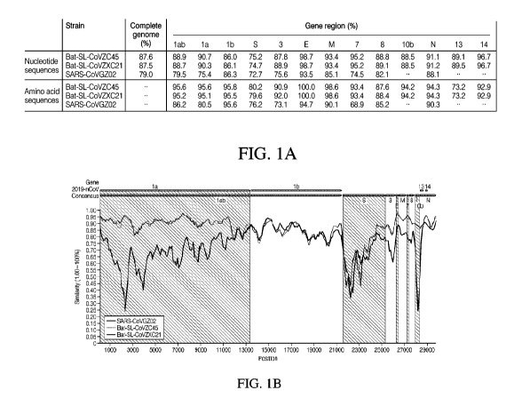
[0012] FIG. lA illustrates the sequence identity between the consensus SARS-
CoV-2
sequence and three closely related coronaviruses, namely, Bat-SL-CoVZC45, Bat-
SL-
CoVZXC21, and SARS-CoVGZ02, across the complete genome and across specific
gene regions
within the coronavirus genome.
[0013] FIG. 1B illustrates the tabular information of FIG. lA in graphical
form with the x-axis
being the base pair position within the viral genome and the y-axis being the
percent similarity of
each related virus to the corresponding SARS-CoV-2 consensus sequence.
[0014] FIG. 2A illustrates the amplification plot of an ORF lab singleplex
qPCR assay
performed on an exemplary sample containing SARS-CoV-2 nucleic acid, the
reagents for the
qPCR assay being part of a kit for the detection of SARS-CoV-2 disclosed
herein.
[0015] FIG. 2B illustrates the standard curve for the singleplex qPCR assay
of FIG. 2A.
[0016] FIG. 2C illustrates the amplification plot of an N protein
singleplex qPCR assay
performed on an exemplary sample containing SARS-CoV-2 nucleic acid, the
reagents for the
qPCR assay being part of a kit for the detection of SARS-CoV-2 disclosed
herein.
[0017] FIG. 2D illustrates the standard curve for the singleplex qPCR assay
of FIG. 2C.
[0018] FIG. 2E illustrates the amplification plot of an S protein
singleplex qPCR assay
performed on an exemplary sample containing SARS-CoV-2 nucleic acid, the
reagents for the
qPCR assay being part of a kit for the detection of SARS-CoV-2 disclosed
herein.
[0019] FIG. 2F illustrates the standard curve for the singleplex qPCR assay
of FIG. 2E.
[0020] FIGs. 3A-3C illustrate comparative amplification plots of ORF 1 ab
(FIG. 3A), N
protein (FIG. 3B), and S protein (FIG. 3C) qPCR assays performed on a 7500
Fast Dx instrument
(Thermo Fisher Scientific) running the 7500 Standard Protocol or the 7500 Fast
Protocol.
[0021] FIGs. 4A-4C illustrate comparative amplification plots of ORF lab
(FIG. 4A), N
protein (FIG. 4B), and S protein (FIG. 4C) qPCR assays generated when running
identical standard
protocols on a 7500 Fast Dx instrument (Thermo Fisher Scientific) or a
QuantStudio 5 Real-Time
PCR System (Thermo Fisher Scientific).
[0022] FIGs. 5A-5C illustrate comparative amplification plots of ORF lab
(FIG. 5A), N
protein (FIG. 5B), and S protein (FIG. 5C) qPCR assays using the TaqPath 1-
Step RT-qPCR
Master Mix (Thermo Fisher Scientific) or the TaqMan Fast Virus 1-Step Master
Mix (Thermo
Fisher Scientific) on the QuantStudio 5 Real-Time PCR System (Thermo Fisher
Scientific).
4
CA 03173545 2022-08-18
WO 2021/168478 PCT/US2021/070163
DETAILED DESCRIPTION
[0023] Before describing various embodiments of the present disclosure in
detail, it is to be
understood that this disclosure is not limited to the parameters of the
particularly exemplified
systems, methods, apparatus, products, processes, and/or kits, which may, of
course, vary. Thus,
while certain embodiments of the present disclosure will be described in
detail, with reference to
specific configurations, parameters, components, elements, etc., the
descriptions are illustrative
and are not to be construed as limiting the scope of the claimed invention. In
addition, the
terminology used herein is for the purpose of describing the embodiments and
is not necessarily
intended to limit the scope of the claimed invention.
[0024] Furthermore, it is understood that for any given component or
embodiment described
herein, any of the possible candidates or alternatives listed for that
component may generally be
used individually or in combination with one another, unless implicitly or
explicitly understood or
stated otherwise. Additionally, it will be understood that any list of such
candidates or alternatives
is merely illustrative, not limiting, unless implicitly or explicitly
understood or stated otherwise.
[0025] In addition, unless otherwise indicated, numbers expressing
quantities, constituents,
distances, or other measurements used in the specification and claims are to
be understood as being
modified by the term "about," as that term is defined herein. Accordingly,
unless indicated to the
contrary, the numerical parameters set forth in the specification and attached
claims are
approximations that may vary depending upon the desired properties sought to
be obtained by the
subject matter presented herein. At the very least, and not as an attempt to
limit the application of
the doctrine of equivalents to the scope of the claims, each numerical
parameter should at least be
construed in light of the number of reported significant digits and by
applying ordinary rounding
techniques. Notwithstanding that the numerical ranges and parameters setting
forth the broad scope
of the subject matter presented herein are approximations, the numerical
values set forth in the
specific examples are reported as precisely as possible. Any numerical values,
however, inherently
contain certain errors necessarily resulting from the standard deviation found
in their respective
testing measurements.
[0026] All publications and patent applications cited herein, as well as
the Appendices attached
hereto, are incorporated by reference in their entirety for all purposes to
the same extent as if each
were specifically and individually indicated to be so incorporated by
reference. Although the
present invention has been described in some detail by way of illustration and
example for purposes
of clarity and understanding, it will be apparent that certain changes and
modifications may be
practiced within the scope of the spirit and substance of this disclosure and
of the appended claims.
CA 03173545 2022-08-18
WO 2021/168478 PCT/US2021/070163
[0027] Any headings and subheadings used herein are for organizational
purposes only and are
not meant to be used to limit the scope of the description or the claims.
Overview of Compositions. Systems, and Kits for Detection of Target Sequences
[0028] As discussed above, a new variant beta coronavirus, SARS-CoV-2 (also
known as
2019-nCoV), has recently emerged as the newest pandemic virus. Current
epidemiological data is
potentially disadvantaged in view of existing non-specific detection assays
used to identify SARS-
CoV-2 infections. The lack of a reliable assay for accurately and specifically
identifying SARS-
CoV-2 from a sample (e.g., a clinical sample obtained from nasopharyngeal
swab, nasopharyngeal
aspirate, bronchoalveolar lavage, buccal swab, saliva, or urine) and/or
differentiating this virus
from other common respiratory pathogens may prevent healthcare professionals
from properly
treating and advising patients. Further, by not being able to accurately and
quickly identify
individuals infected with SARS-CoV-2, it can be quite difficult to establish a
systematic treatment
campaign or implement successful preventative measures.
[0029] Given the present and continuing emergence of new coronavirus
strains, there is an
urgent need to develop methods for the rapid detection and characterization of
existing and novel
coronavirus strains so that appropriate treatment and infection control
measures can be properly
instituted in a timely manner. Problematically, many of the available SARS-CoV-
2 detection
assays are non-specific with respect to detecting and differentiating SARS-CoV-
2 from other
respiratory pathogens, particularly other coronaviruses, which has potential
to lead to a lack of
patient confidence in the diagnostic potential of current SARS-CoV-2 detection
assays. Further,
because individuals infected with SARS-CoV-2 often experience symptoms similar
to those
infected with Influenza Types A or B and/or Respiratory Syncytial Virus (RSV),
and/or other
respiratory microbes, there is an additional need to be able to simultaneously
test for each of these
respiratory infectious agents in order to provide an accurate diagnosis before
seeking/providing
treatment and/or confining the individual to a quarantined area under the
potentially mistaken
belief that they are infected with SARS-CoV-2. Each misidentified or
misdiagnosed instance of
SARS-CoV-2 infection can further convolute the epidemiological data and
prevent the
implementation of appropriate, informed solutions that may help reign in the
pandemic.
[0030] A number of assays are available for allegedly detecting the
presence of SARS-CoV-2,
such as those provided by the United States Centers for Disease Control and
Prevention (US-CDC)
that contain probes targeting the N protein, the assay developed by the
Chinese CDC targeting the
coding regions of the N protein and the ORF lab protein, and the WHO kit
targeting the coding
regions of the N protein, the E protein, and the closely related RdRp
SARS/Wuhan coronavirus.
6
CA 03173545 2022-08-18
WO 2021/168478
PCT/US2021/070163
While each of the foregoing assays have 100% coverage of all published SARS-
CoV-2 genomes
to date _______________________________________________________ meaning these
assays are each theoretically capable of identifying the presence of SARS-
CoV-2 from a nucleic acid sample ______________________________ the design of
these assays is such that the detection is non-
specific, which can perpetuate the issues discussed above instead of
alleviating them.
[0031] For example,
the probes used in the WHO kit for the E and N proteins map perfectly to
hundreds of non-SARS-CoV-2 coronavirus strains. Furthermore, the confirmatory
probe
identifying RdRp-SARS/Wuhan was designed to detect both SARS and SARS-CoV-2,
making it
non-specific by design. These assays lack endogenous controls as well. In
total, this assay is non-
specific for SARS-CoV-2 and is prone to providing false positive results.
Similarly, the US-CDC
kit for detecting SARS-CoV-2 has also exhibited some non-specificity. It
relies on three separate
probes to the coding region of the N protein, and two of these probes can
generate a false positive
signal in the presence of non-SARS-CoV-2 coronaviruses, such as many SARS
strains and even
the bat-SARS-CoV strain, particularly when present in higher concentrations.
Accordingly, even
this kit fails to provide an assay having the desired SARS-CoV-2 specificity,
and there remains an
unmet need in the market for a SARS-CoV-2 detection assay that is accurate and
specific and that,
preferably, can be implemented quickly with a short turnaround time between
obtaining the sample
and receiving the results.
[0032] Disclosed
herein are compositions, kits, and methods for specifically detecting viral
sequences, in particular SARS-CoV-2. Additional compositions, kits, and
methods are disclosed
that enable the detection and differentiation of SARS-CoV-2 from other related
coronaviruses,
from respiratory tract microbiota, and from common respiratory pathogens that
produce similar
symptomatic infections in humans, including Influenza Type A (Flu A),
Influenza Type B (Flu B),
and Respiratory Syncytial Virus (e.g., RSV A and RSV B). As demonstrated
throughout the present
description, many of the probes disclosed herein include nucleic acid binding
portions that exhibit
100% identity (i.e., no mismatches) to all 52 genomic sequences of SARS-CoV-2
reported in the
literature. Further, the kits and methods provided herein specifically target
all 71 complete
genomes currently available at GISAID, and do not target any of the 2,116
complete genomes of
other coronaviruses currently available at NCBI, underscoring the beneficial
specificity of the
disclosed methods and kits for detecting SARS-CoV-2. Indeed, the disclosed
embodiments solve
at least some of the unmet needs in the field of viral identification and
provide meaningful
improvements over prior viral detection compositions, kits, and methods.
[0033] In some
embodiments, the strain coverage for the compositions, kits, and methods
described herein for detecting SARS-CoV-2, including those with primers and
probes selected
from SEQ 1.13 NO:4 ¨ SEQ 1.13 NO:2533, is 99.9% based on an in silico analysis
of 35,833 high
7
CA 03173545 2022-08-18
WO 2021/168478 PCT/US2021/070163
quality complete sequences available from GISAID as of July 6, 2020.
Additionally, the
compositions, kits, and methods disclosed herein for the detection of viral
sequences, particularly
those multiplex assays for identifying the specific presence of SARS-CoV-2,
Flu A, Flu B, RSV
A, and/or RSV B using primers and probes selected from SEQ ID NO:4 ¨SEQ ID
NO:2533, retain
the 99.9% specificity and precision for identifying SARS-CoV-2 strains, and
the strain coverage
is 98.2% (6730/6854) for Flu A and 99.3% (3105/3127) for Flu B, based on data
available from
NCBI as of April 13, 2020.
[0034] SEQ ID NO:4 ¨ SEQ ID NO:257 includes a list of sequences which are
amenable for
use as forward primers targeting the ORFlab, S protein, or N protein coding
regions of the SARS-
CoV-2 genome, regions of the human influenza (Flu) type A or type B viral
genome, regions of
the Respiratory Syncytial Virus (RSV) type A or type B viral genome, or
control sequences, such
as M52 Phage and RNase P.
[0035] SEQ ID NO:267 ¨ SEQ ID NO:510 includes a list of sequences which are
amenable
for use as reverse primers targeting the ORF 1 ab, S protein, or N protein
coding regions of the
SARS-CoV-2 genome, regions of the human influenza (Flu) type A or type B viral
genome, regions
of the Respiratory Syncytial Virus (RSV) type A or type B viral genome, or
control sequences,
such as M52 Phage and RNase P.
[0036] SEQ ID NO:520 ¨SEQ ID NO:2533 includes a list of sequences which are
nucleic acid
portions of probes targeting the ORFlab, S protein, or N protein coding
regions of the SARS-CoV-
2 genome, regions of the human influenza (Flu) type A or type B viral genome,
regions of the
Respiratory Syncytial Virus (RSV) type A or type B viral genome, or control
sequences, such as
M52 Phage and RNase P.
[0037] Further, because SARS-CoV-2 is an RNA virus, it can mutate with
relatively high
frequency, making it difficult to consistently detect over time. Specific
detection can be ensured
even in the case of future variants by using multiple assays targeting
different regions of the same
target genes to ensure redundancy and specificity. Unlike other published
assay designs, which
require multiple assay designs and separate reactions for each loci to enhance
specificity, the
disclosed primers and probes can differentiate SARS-CoV-2 strains using a
single and specific
assay conducted in a single, or at the most two, reaction volumes.
[0038] The embodiments of the present disclosure beneficially provide
improved
compositions, kits, and methods for detecting and differentiating viral
respiratory tract pathogens
that share similar symptomatic presentations in humans. As such, these
disclosed embodiments
advantageously improve the efficiency and accuracy of evaluating respiratory
samples (e.g., in a
8
CA 03173545 2022-08-18
WO 2021/168478
PCT/US2021/070163
laboratory setting, at point of sale location, and/or at point of care
location) for the presence of
viral sequences and can improve the diagnosis and treatment of affected
individuals.
[0039] The disclosed
compositions, kits, and methods for the detection of viral sequences can
also improve the accuracy of epidemiological studies related to SARS-CoV-2,
Flu A, Flu B, and/or
RSV infections. Additional embodiments disclosed herein include assay panels
(e.g. in the format
of an array card) for determining the presence of viral, bacterial, and fungal
nucleic acid sequences
that can, among other things, improve syndromic evaluations and
epidemiological studies by being
able to detect and differentiate SARS-CoV-2 from related coronaviruses,
influenza viruses,
rhinoviruses, adenoviruses, and other viral, bacterial, and fungal microbes.
Sample Collection
[0040] The disclosed
compositions, kits, and methods are configured to detect viral nucleic
acid from a sample, preferably a specific and differential detection of SARS-
CoV-2 from a sample.
The sample may be a veterinary sample (e.g., from non-human animals like
mink), a clinical
sample (e.g., from a symptomatic or asymptomatic human), a food sample, a
forensic sample, an
environmental sample (e.g., soil, dirt, garbage, sewage, air, or water),
including food processing
and manufacturing surfaces, or any other biological sample. In most instances,
SARS-CoV-2 or
other coronaviruses and respiratory tract pathogens are detected by analysis
of swabs or fluid
obtained from swabs, such as throat swabs, nasal swabs, nasopharyngeal swabs,
nasal mid-
turbinate swabs, oropharyngeal swabs, cheek swabs, saliva swabs, or other
swabs, though it should
be appreciated that SARS-CoV-2 or other coronaviruses and/or respiratory tract
pathogens may
also be detected by analysis of urine samples, saliva samples, or other
clinical samples.
[0041] The sample can
be collected by a healthcare professional in a healthcare setting, but in
some instances, the sample may also be collected by the subject themselves or
by an individual
assisting the subject in self-collection. For example, a nasopharyngeal swab
has heretofore served
as the gold standard for obtaining a sample to be used in clinical diagnostics
or screening. Such
swabs are often used by a healthcare professional in a healthcare setting.
Other samples, such as a
saliva sample, can similarly be obtained in a healthcare setting with the
assistance or oversight of
a healthcare professional. However, in some instances, self-collection of a
sample can be more
efficient and can be done outside of a healthcare setting.
[0042] In some embodiments, the sample is a raw saliva sample collected
whether by self-
collection or assisted/supervised collection __________________ in a sterile
tube or specifically-designed saliva
collection device. The saliva collection tube/device may be a component of a
self-collection kit
having instructions for use, such as sample collection instructions, sample
preparation or storage
9
CA 03173545 2022-08-18
WO 2021/168478 PCT/US2021/070163
instructions, and/or shipping instructions. In some embodiments, the raw
saliva sample can be
collected directly into a sealable container without any preservation solution
or other fluid or
substance in the container prior to receipt of the saliva sample within the
container or as a result of
closing/sealing the container. In some other embodiments, the raw saliva
sample is collected into
a container which already contains some amount of a preservation or treatment
solution or other
fluid or substance.
[0043] Traditionally, a nucleic
acid fraction of the sample is extracted or purified from the
sample ____________________________________________ whether obtained via swab,
from raw saliva, or other bodily fluid prior to any detection
of viral nucleic acids therein. Surprisingly, the disclosed embodiments for
detecting viral nucleic
acid from a sample can be adapted to detect viral nucleic acid directly from a
raw saliva sample
without a specific nucleic acid purification and/or extraction step prior to
its use in downstream
detection assays (e.g., RT-qPCR). In some embodiments, the saliva sample is
pre-treated prior to
use (see, for example, Example 8 herein). This can include, for example,
heating the saliva sample,
such as by placing the raw saliva sample on a heat block/water bath set to a
temperature of 95 C
for 30 minutes, followed by combining the heat-treated saliva with a buffer or
lysis solution. The
buffer or lysis solution can include, for example, any nucleic-acid-amenable
buffer such as TBE
and may further include a detergent and/or emulsifier such as Triton-X-100, NP-
40, or the
polysorbate-type nonionic surfactant, Tween-20.
[0044] It should be appreciated
that in some embodiments, the disclosed compositions can
include the sample mixed with a buffer and detergent/emulsifier. The sample
can be added to a
buffer/detergent mixture or vice versa. As a non-limiting example, a set of
subject samples can be
prepared as compositions for downstream analysis and detection of viral
sequence by adding a
volume of heat-treated sample for each subject into one (or a plurality) of
wells in a multi-well
plate. A volume of a buffer/detergent mixture (e.g., TBE + Tween-20) can then
be added to each
well containing a subject sample. Alternatively, a multi-well plate can be
loaded with a volume of
a buffer/detergent mixture into which a volume of heat-treated saliva is
added. Once combined,
this probative template solution can be used immediately or stored for later
analysis. Such
probative template solutions can also be combined with PCR reagents (e.g.,
buffers, dNTPs, master
mixes, etc.) prior to or after storage.
Compositions, Kits, and Methods for Detection of SARS-CoV-2 Viral Sequences
[0045] The primers and probes
disclosed herein are useful for the detection of SARS-CoV-2
from a sample, such as a biological sample obtained from a human or non-human
(e.g., mink)
subject. Such primers and/or probes can be used within a kit for performing a
nucleic-acid-based
CA 03173545 2022-08-18
WO 2021/168478 PCT/US2021/070163
assay for the detection and identification of one or more target nucleic acids
in the sample, which
may be single stranded or double stranded of any size. For example, the
primers and probes
provided in SEQ ID NO:4 ¨ SEQ ID NO:2533 can be used to amplify and/or analyze
one or more
specific target sequences present in the SARS-CoV-2 viral genome or within one
or more of the
Flu A, Flu B, RSV A, RSV B, other target respiratory microbes and/or controls
(see, e.g., Tables
3A and 3B), as described herein. The amplified products ("amplicons") can be
detected and/or
analyzed using any suitable method and on any suitable platform.
[0046] Polymerase chain reaction (PCR) and related methods are common
methods of nucleic
acid amplification. PCR is one, but not the only, example of a nucleic acid
polymerase reaction
method for amplifying a nucleic acid test sample comprising the use of a known
nucleic acid as a
primer and a nucleic acid polymerase to amplify or generate a specific target
nucleic acid. In
general, PCR utilizes a primer pair that consists of a forward primer and a
reverse primer
configured to amplify a target segment of a nucleic acid template. Typically,
but not always, the
forward primer hybridizes to the 5' end of the target sequence and the reverse
primer will be
identical to a sequence present at the 3' end of the target sequence. The
reverse primer will typically
hybridize to a complement of the target sequence, for example an extension
product of the forward
primer and/or vice versa. PCR methods are typically performed at multiple
different temperatures,
causing repeated temperature changes during the PCR reaction ("thermal
cycling"). Other
amplification methods, such as, e.g., loop-mediated isothermal amplification
("LAMP"), and other
isothermal methods, such as those listed in Table 1, may require less or less
extensive thermal
cycling than does PCR, or require no thermal cycling. Such isothermal
amplification methods are
also contemplated for use with the assay compositions, reaction mixtures, kits
described herein.
Table 1. Summary of optional isothermal amplification methods.
NASBA Nucleic acid sequence-based amplification (NASBA) is a method used to
amplify RNA.
LAMP Loop-mediated isothermal amplification (LAMP) is a single tube technique
for
the amplification of DNA. It typically uses 4-6 primers, which form loop
structures to facilitate subsequent rounds of amplification.
RDA Helicase-dependent amplification (HDA) uses the double-
stranded DNA
unwinding activity of a helicase to separate strands for in vitro DNA
amplification at constant temperature.
RCA Rolling circle amplification (RCA) starts from a circular DNA
template and a
short DNA or RNA primer to form a long single stranded molecule.
11
CA 03173545 2022-08-18
WO 2021/168478 PCT/US2021/070163
iviDA Multiple displacement amplification (MDA) is a technique that
initiates when
multiple random primers anneal to the DNA template and the polymerase
amplifies DNA at constant temperature.
WGA When MDA is used to amplify DNA from a whole genome of a cell
it is called
whole genome amplification (WGA). (Other methods of WGA include
MALBAC, LIANTI, DOP-PCR.)
RPA Recombinase polymerase amplification (RPA) is a low
temperature DNA and
RNA amplification technique.
[0047] Methods of performing PCR, including those in Table 1, are well
known in the art;
nevertheless, further discussion of PCR and other methods may be found, for
example, in
Molecular Cloning: A Laboratory Manual by Green and Sambrook, Cold Spring
Harbor
Laboratory Press, 4th Edition 2012, which is incorporated by reference herein
in its entirety.
[0048] SARS-CoV-2 has a single-stranded positive-sense RNA genome. Other
viruses, such
as Flu A, Flu B, RSV A, and RSV B also have RNA-based genomes. In some
embodiments,
therefore, the amplification reaction (e.g., LAMP or PCR) can be combined with
a reverse
transcription (RT) reaction, such as in RT-LAMP or RT-PCR to convert the RNA
genome to a
cDNA template. The cDNA template is then used to create amplicons of the
target sequences in
the subsequent amplification reactions.
[0049] In some embodiments, the amplifying step can include performing
qPCR, as that term
is defined herein. qPCR is a sensitive and specific method for detecting and
optionally quantifying
amounts of starting nucleic acid template (e.g., coronaviral nucleic acid) in
a sample. Methods of
qPCR are well known in the art; one leading method involves the use of a
specific hydrolysis probe
in conjunction with a primer pair. The hydrolysis probe can include a
detectable label (e.g.,
fluorophore) at one end and a quencher that quenches the detectable label at
the other end. In some
embodiments, the label is at the 5' end of the probe and cleavage of the 5'
label occurs via 5'
hydrolysis of the probe by the nucleic acid polymerase as it extends the
forward primer towards
the probe binding site within the target sequence. The separation of the probe
label from the probe
quencher via cleavage (or unfolding) of the probe results in an increase in
signal which can be
detected and optionally quantified. The detectable signal can be monitored
over time and analyzed
to determine the relative or absolute amount of starting nucleic acid template
present in the sample.
Suitable labels are described herein. In some embodiments, the dye-quencher
combinations are
used, such as those described in the Examples. It should be appreciated that
qPCR and RT-qPCR
methods are known to those having skill in the art. Nevertheless, particular
embodiments are
12
CA 03173545 2022-08-18
WO 2021/168478 PCT/US2021/070163
provided in the Examples and provide further details regarding qPCR as well as
related
compositions and methods of use thereof.
[0050] .. The reaction vessel or volume can optionally include a tube,
channel, well, cavity, site
or feature on a surface, or alternatively a droplet (e.g., a microdroplet or
nanodroplet) that may be
deposited onto a surface or into a surface well or cavity or suspended within
(or partially bounded
by) a fluid stream. In some embodiments, the reaction volume includes one or
more droplets
arrayed on a surface or present in an emulsion. The reaction volumes can
optionally be formed by
fusion of multiple pre-reaction volumes containing different components of an
amplification
reaction. For example, pre-reaction volumes containing one or more primers can
be fused with pre-
reaction volumes containing human nucleic acid samples and/or polymerase
enzymes, nucleotides,
and buffer. In some embodiments involving performing qPCR reactions in array
format, a surface
contains multiple grooves, channels, wells, cavities, sites, or features
defining a reaction volume
containing one or more amplification reagents (e.g., primers, probes, buffer,
polymerase,
nucleotides, and the like). In some array-formatted singleplex embodiments,
the reaction volume
within the selected tubes, grooves, channels, wells, cavities, sites, or
features contains only a single
forward primer sequence and a single reverse primer sequence. Optionally, a
probe sequence is
also included in the singleplex reaction volume.
[0051] .. In some array-formatted multiplex embodiments, the reaction volume
within the
selected tubes, grooves, channels, wells, cavities, sites, or features
contains multiple (e.g., 2, 3, 4,
5, 6, etc.) forward primer sequences and multiple reverse primer sequences.
Optionally, one or
more probe sequences is also included in the multiplex reaction volume.
[0052] .. For instance, exemplary methods for polymerizing and/or amplifying
and detecting
nucleic acids suitable for use as described herein are commercially available
as TaqMan assays
(see, e.g., U.S. Patent Nos. 4,889,818; 5,079,352; 5,210,015; 5,436,134;
5,487,972; 5,658,751;
5,210,015; 5,487,972; 5,538,848; 5,618,711; 5,677,152; 5,723,591; 5,773,258;
5,789,224;
5,801,155; 5,804,375; 5,876,930; 5,994,056; 6,030,787; 6,084,102; 6,127,155;
6,171,785;
6,214,979; 6,258,569; 6,814,934; 6,821,727; 7,141,377; and/or 7,445,900, all
of which are hereby
incorporated herein by reference in their entirety). TaqMan assays are
typically carried out by
performing nucleic acid amplification on a target polynucleotide using a
nucleic acid polymerase
having 5'-to-3' nuclease activity, a primer capable of hybridizing to the
target polynucleotide, and
an oligonucleotide probe capable of hybridizing to said target polynucleotide
3' relative to the
primer. The oligonucleotide probe typically includes a detectable label (e.g.,
a fluorescent reporter
molecule) and a quencher molecule capable of quenching the fluorescence of the
reporter
molecule. Typically, the detectable label and quencher molecule are part of a
single probe. As
13
CA 03173545 2022-08-18
WO 2021/168478 PCT/US2021/070163
amplification proceeds, the polymerase digests the probe to separate the
detectable label from the
quencher molecule. The detectable label is monitored during the reaction,
where detection of the
label corresponds to the occurrence of nucleic acid amplification (e.g., the
higher the signal the
greater the amount of amplification). Variations of TaqMan assays are known in
the art and would
be suitable for use in the methods described herein.
[0053] For example, a singleplex or multiplex qPCR can include a single
TaqMan dye
associated with a locus-specific primer or multiple TaqMan dyes respectively
associated with a
plurality of loci in a multiplex format. As a non-limiting example, a 4-plex
reaction can include
FAM (emission peak ¨517 rim), VIC (emission peak ¨551 rim), ABY (emission peak
¨580 rim),
and JUN (emission peak ¨617 rim) dyes, each dye being associated with a
different target sequence
and each dye being quenched by QSY, can allow up to 4 targets to be amplified
and tracked real-
time within a single reaction vessel. These aforementioned reporter dyes are
optimized to work
together with minimal spectral overlap for improved performance. These dyes
can additionally be
combined with Mustang Purple (emission peak ¨654 rim) for use monitoring
fluorescence of a
control or for use in a non-emission-spectrum-overlapping 5-plex assay. In
addition, the QSY
quencher is fully compatible with probes that have minor-groove binder
quenchers.
[0054] Detector probes may be associated with alternative quenchers,
including without
limitation, dark fluorescent quencher (DFQ), black hole quenchers (BHQ), Iowa
Black, QSY
quencher, and Dabsyl and Dabcel sulfonate/carboxylate Quenchers. Detector
probes may also
include two probes, wherein, for example, a fluorophore is associated with one
probe and a
quencher is associated with a complementary probe such that hybridization of
the two probes on a
target quenches the fluorescent signal or hybridization on the target alters
the signal signature via
a change in fluorescence. Detector probes may also include sulfonate
derivatives of fluorescein
dyes with S03 instead of the carboxylate group, phosphoramidite forms of
fluorescein,
phosphoramidite forms of Cy5.
[0055] It should be appreciated that when using more than one detectable
label, particularly in
a multiplex format, each detectable label should differ in its spectral
properties from the other
detectable labels used therewith such that the labels may be distinguished
from each other, or such
that together the detectable labels emit a signal that is not emitted by
either detectable label alone.
Exemplary detectable labels include, for instance, a fluorescent dye or
fluorophore (e.g., a chemical
group that can be excited by light to emit fluorescence or phosphorescence),
"acceptor dyes"
capable of quenching a fluorescent signal from a fluorescent donor dye, and
the like, as described
above. Suitable detectable labels may include, for example, fluoresceins
(e.g., 5-carboxy-2,7-
dichlorofluorescein; 5-Carboxyfluorescein (5-FANI); 5-Hydroxy Tryptamine (5-
HAT); 6-JOE; 6-
14
CA 03173545 2022-08-18
WO 2021/168478 PCT/US2021/070163
carboxyfluorescein (6-FANI); Mustang Purple, VIC, ABY, JUN; FITC; 6-carboxy-
4',5'-dichloro-
2',7'-dimethoxy-fluorescein (JOE)); 6-carboxy-1,4-dichloro-2',7'-dichloro-
fluorescein (TET);
6-carboxy-1,4-dichloro-2',4',5',7'-tetra-chlorofluorescein (HEX); Alexa Fluor
fluorophores (e.g.,
350, 405, 430, 488, 500, 514, 532, 546, 555, 568, 594, 610, 633, 635, 647,
660, 680, 700, 750);
BODIPY fluorophores (e.g., 492/515, 493/503, 500/510, 505/515, 530/550,
542/563, 558/568,
564/570, 576/589, 581/591, 630/650-X, 650/665-X, 665/676, FL, FL ATP, FI-
Ceramide, R6G SE,
TMR, TMR-X conjugate, TMR-X, SE, TR, TR ATP, TR-X SE), Cascade Blue, Cascade
Yellow;
Cylm dyes (e.g., 3, 3.18, 3.5, 5, 5.18, 5.5, 7), cyan GFP, cyclic AMP
Fluorosensor (FiCRhR),
fluorescent proteins (e.g., green fluorescent protein (e.g., GFP. EGFP), blue
fluorescent protein
(e.g., BFP, EBFP, EBFP2, Azurite, mKalamal), cyan fluorescent protein (e.g.,
ECFP, Cerulean,
CyPet), yellow fluorescent protein (e.g., YFP, Citrine, Venus, YPet), FRET
donor/acceptor pairs
(e.g., fluorescein/fluorescein, fluorescein/tetramethylrhodamine,
IAEDANS/fluorescein,
EDANS/dabcyl, BODIPY FL/BODIPY FL, Fluorescein/QSY7 and QSY9), LysoTracker and
LysoSensor (e.g., LysoTracker Blue DND-22, LysoTracker Blue-White DPX,
LysoTracker
Yellow HCK-123, LysoTracker Green DND-26, LysoTracker Red DND-99, LysoSensor
Blue
DND-167, LysoSensor Green DND-189, LysoSensor Green DND-153, LysoSensor
Yellow/Blue
DND-160, LysoSensor Yellow/Blue 10,000 MW dextran), Oregon Green (e.g., 488,
488-X, 500,
514); rhodamines (e.g., 110, 123, B, B 200, BB, BG, B extra, 5-
carboxytetramethylrhodamine (5-
TAMRA), 5 GLD, 6-Carboxyrhodamine 6G, Lissamine, Lissamine Rhodamine B,
Phallicidine,
Phalloidine, Red, Rhod-2, ROX (6-carboxy-X-rhodamine), 5-ROX (carboxy-X-
rhodamine),
Sulphorhodamine B can C, Sulphorhodamine G Extra, TAMRA (6-
carboxytetramethyl-rhodamine), Tetramethylrhodamine (TRITC), WT), Texas Red,
Texas Red-
X, among others as would be known to those of skill in the art.
[0056] Other detectable labels may also be used. For example, primers can
be labeled and used
to both generate amplicons and to detect the presence (or concentration) of
amplicons generated in
the reaction, and such may be used in addition to or as an alternative to
labeled probes described
herein. As a further example, primers may be labeled and utilized as described
in Nazarenko et al.
(Nucleic Acids Res. 2002 May 1; 30(9): e37), Hayashi et al. (Nucleic Acids
Res. 1989 May 11;
17(9): 3605), and/or Neilan et al. (Nucleic Acids Res. Vol. 25, Issue 14, 1
July 1997, pp. 2938-
39). Those of skill in the art will also understand and be capable of
utilizing the PCR processes
(and associated probe and primer design techniques) described in Zhu et al.
(Biotechniques. 2020
Jul: 10.2144/btn-2020-0057).
[0057] In some embodiments, intercalating labels can be used such as
ethidium bromide,
SYBR Green I, SYBR GreenER, and PicoGreen (Life Technologies Corp., Carlsbad,
CA), thereby
CA 03173545 2022-08-18
WO 2021/168478 PCT/US2021/070163
allowing visualization in real-time, or end point, of an amplification product
in the absence of a
detector probe. It should be appreciated, however, that use of intercalating
labels may limit
multiplexing capabilities, as many intercalating labels are non-specific for a
given sequence and
merely report the total (or proportional) nucleic acid content within a
reaction. In some
embodiments, real-time visualization may include both an intercalating
detector probe and a
sequence-based detector probe. The detector probe can be at least partially
quenched when not
hybridized to a complementary sequence in the amplification reaction and is at
least partially
unquenched when hybridized to a complementary sequence in the amplification
reaction. In some
embodiments, probes may further comprise various modifications such as a minor
groove binder
to further provide desirable thermodynamic characteristics.
[0058] The genetic sequence of SARS-CoV-2 is available as NCBI accession
no.
NC 045512.2 and as GenBank accession no. MN908947.3, describing a positive-
sense, single-
stranded RNA genome of 29,844 base pairs; occasionally, such sequence is
referred to herein as
the 'normal', 'wild type' or 'reference' sequence for SARS-CoV-2, as opposed
to SARS-CoV-2
variant or mutant sequences. Initial genetic characterizations of SARS-CoV-2
identified three
coronaviruses having close homology to SARS-CoV-2, namely Bat-SL-CoVZC45, Bat-
SL-
CoVZXC21, and SARS-CoVGZ02. The sequence identity between these strains is
depicted in
FIGs. lA and 1B. In particular, FIG. lA illustrates the sequence identity
between the consensus
SARS-CoV-2 sequence as compared to each of Bat-SL-CoVZC45, Bat-SL-CoVZXC21,
and
SARS-CoVGZ02, across the complete genome as well as across each specific gene
region within
the coronavirus genome. FIG. 1B illustrates the tabular information of FIG. lA
in graphical form
with the x-axis being the base pair position within the viral genome and the y-
axis being the percent
similarity of each related virus to the corresponding SARS-CoV-2 consensus
sequence.
[0059] The analysis illustrated in FIGs. lA and 1B identified at least
three genetic regions with
significant variability between SARS-CoV-2 and the other related viruses,
specifically within the
viral genes encoding the ORF lab protein (SEQ ID NO:1; base pair 1 corresponds
to base pair 1000
of MN908947.3), the S protein (SEQ ID NO:2; base pair 1 corresponds to base
pair 21564 of
MN908947.3), and the N protein (SEQ ID NO:3; base pair 1 corresponds to base
pair 28275 of
MN908947.3). The region comprising the coding sequence for the ORF lab protein
is between base
pairs 1000-3000 of the SARS-CoV-2 genome; this sequence corresponds to SEQ ID
NO: 1. The
2,000 base pair region of the SARS-CoV-2 genome that includes the coding
sequence for the S
protein is between base pairs 21,564-23,564; this sequence corresponds to SEQ
ID NO:2. Finally,
the 1,283 base pair region of the SARS-CoV-2 genome that includes the coding
sequence for the
N protein is between base pairs 28,275-29,558; this sequence corresponds to
SEQ ID NO:3.
16
CA 03173545 2022-08-18
WO 2021/168478 PCT/US2021/070163
[0060] In some embodiments, detecting amplification of target sequences
includes measuring
one or more signals emitted by a detectable label attached to, or associated
with, one or more
primers or probes. Optionally, the one or more signals are measured multiple
times as the
amplification reaction progresses, in some embodiments at least once per
thermal cycle (e.g. during
or just after an annealing or extension phase of a thermal cycle), thus
allowing for amplification to
be detected in 'real time'. In 'multiplex' amplification embodiments, the
formation of a plurality
of separate and different amplification products can be tracked over time by
measuring a signal in
one or more detection channels. The signal can be emitted by a detectable
label, optionally a
fluorescent label, attached to a primer and/or probe that selectively
hybridizes to the amplification
product. In some embodiments, each channel is calibrated to preferentially or
selectively detect a
corresponding amplification product and the signal in each channel is used as
a measure of
concentration of the corresponding amplification product. For example, in some
embodiments, an
amplification product of the S gene from SARS-CoV-2 is detected in a first
detection channel
based on a first signal emitted by a first label attached to, or associated
with, a first primer and/or
first probe that selectively hybridizes to the S gene amplification product,
and an amplification
product of the N gene is detected in a second detection channel based on a
second signal emitted
by a second label attached to, or associated with, a second primer and/or
second probe that
selectively hybridizes to the N gene amplification product. Optionally, in
'triplex' embodiments
involving amplification of the Orflab gene, the amplification product of the
ORF lab gene is
detected in a third channel based on a third signal emitted by a third label
attached to a third primer
and/or third probe that selectively hybridizes to the ORF lab amplification
product or to the
ORF lab target region. In some embodiments, the amplification product of a
control or reference
sequence is detected in a fourth channel based on a fourth signal emitted by a
fourth label attached
to a fourth primer and/or fourth probe that selectively hybridizes to an
amplification product of, or
to a target sequence within the control or reference sequence.
[0061] In some embodiments (e.g., in the well-known and widely used
TaqManTm line of
qPCR assays), detecting an amplification product includes detecting a signal
emitted by a
fluorescent label attached to the 5' end of a cleavable probe that selectively
hybridizes to the
amplification product during amplification. The cleavable probe further
includes a quencher that
quenches the fluorescent label to a 'baseline' fluorescence level. The 5' end
of the cleavable probe
is cleaved by the polymerase during the extension step, resulting in the
separation of the fluorescent
label from the quencher and a corresponding increase in fluorescence over
baseline. As the PCR
reaction progresses, the continuing increase in fluorescence over baseline is
measured at each
cycle. In some embodiments, an amplification product from the N gene of SARS-
CoV-2 is
17
CA 03173545 2022-08-18
WO 2021/168478 PCT/US2021/070163
detected in a first channel based on a first signal emitted by the VIC dye
attached to a probe that
selectively hybridizes to the corresponding amplification product from the N
gene. Optionally, a
second amplification product from the S gene of SARS-CoV-2 is detected in a
second channel
based on a second signal emitted by the ABY dye attached to a probe that
selectively hybridizes to
the corresponding amplification product from the S gene. Optionally, a third
amplification product
from the ORF lab gene of SARS-CoV-2 is detected in a third channel based on a
third signal
emitted by the FAM dye attached to a probe that selectively hybridizes to the
corresponding
amplification product from the ORF lab gene. Optionally, a fourth
amplification product from a
control or reference sequence is detected in a fourth channel based on a
fourth signal emitted by
the JUN dye attached to a probe that selectively hybridizes to the
corresponding amplification
product from the control or reference sequence.
[0062] In some embodiments, a passive reference dye, such as ROXTM is
included in the
reaction mixture. The metric "Rn" is optionally used to track progress of the
amplification reaction
and to determine the amount of target sequence originally present in the
reaction mixture prior to
amplification. Rn can be calculated as the fluorescence of the reporter dye
divided by the
fluorescence of a passive reference dye present in the reaction mixture; i.e.,
Rn is the reporter signal
normalized to the fluorescence signal of the passive reference dye. In some
embodiments, Rn is
plotted against PCR cycle number. In some embodiments, ARn (calculated as Rn
minus the
baseline) can be plotted against PCR cycle number. In some embodiments, an
amplification plot
shows the variation of log (ARn) with PCR cycle number. Ct (threshold cycle)
is the intersection
between an amplification curve and a threshold line. The lower the Ct value
for a given
amplification product, the earlier the amplification is detectable and the
higher the absolute
amount, and the relative concentration, of the corresponding target sequence
originally present in
the reaction mixture. In some embodiments, cutoffs for Ct are used to
determine whether a target
sequence was originally present or absent in the reaction mixture prior to
amplification. For
example, in some embodiments a target sequence is determined to be present if
the Ct value is less
than or equal to 37.
[0063] In some embodiments, emerging variants of SARS-CoV-2 are detectable
even if such
variants include mutations in one or more of the target regions described
above (i.e., ORF lab
protein, S protein, or N protein regions). By looking at multiple target
regions within the SARS-
CoV-2 genome, accurate detection is achievable even in situations where
mutations are significant
enough to lead to a negative test result in one (or even two) of the target
regions. For example,
newly emerging variant B.1.1.7 (often referred to as "the UK variant") has an
unusual number of
mutations associated with the S protein region. These mutations are
substantial enough that some
18
CA 03173545 2022-08-18
WO 2021/168478 PCT/US2021/070163
test components and protocols designed for earlier SARS-CoV-2 variants will
show a negative
result for the S protein region. However, the built-in redundancy of looking
at multiple regions
ensures that the overall test is still capable of detecting the SARS-CoV-2
variant B.1.1.7 (based on
positive ORF lab and/or N protein region detection) without significant
effects on the overall
accuracy of the test. In another example, the 501Y.V2 variant (discovered in
South Africa) has not
been found to affect detection of the S protein region or any of the other
tested regions described
herein. Nevertheless, the robustness and redundancy of embodiments that target
multiple regions
of the SARS-CoV-2 genome limit the risk that these variants, or others that
emerge in the future,
will significantly impact the overall accuracy of SARS-CoV-2 detection.
[0064] .. To maximize the specificity of a genetic assay for SARS-CoV-2,
primers and probes
were designed that targeted the coding regions for ORFlab, S protein, and N
protein. In particular,
the viral detection kits, arrays, assays, etc. disclosed herein include
primers and/or probes specific
for the SARS-CoV-2 genetic sequence encoding the S protein; none of the SARS-
CoV-2 viral
detection kits currently available target the S gene. Targeting the S gene (in
addition to the coding
regions associated with the N protein and ORF lab) provides several advantages
in terms of
specificity and reliability. For example, at least some of the disclosed
assays targeting the coding
region for the S protein have been shown to differentiate between SARS-CoV and
SARS-CoV-2
at the receptor binding level. Inclusion of these primers and/or probes
targeting the S gene
sequences offers higher specificity in detection of SARS-CoV-2 strains against
other similar
coronaviruses, especially in geographical regions where subjects present with
co-infections of
SARS-CoV and SARS-CoV-2.
[0065] The specificity of the primers and probes provided in SEQ ID NO:4
¨SEQ ID NO:2533
was estimated in silico using the standard mapping algorithm zper3p. These
primers and probes
were found to exhibit higher specificity for SARS-CoV-2 in silico than the
primers and probes of
other commercially available SARS-CoV-2 qPCR-based assays. Accordingly, the
disclosed
compositions, kits, and methods for detecting viral sequences include at least
one primer and/or
probe having a sequence defined by SEQ ID NO:4 ¨ SEQ ID NO:2533, which enables
the
singleplex and multiplex assays described herein to demonstrate a high level
of sensitivity,
specificity, and accuracy. In some embodiments, a first forward primer is SEQ
ID NO: 160. In
some embodiments, a second forward primer is SEQ ID NO: 100. In some
embodiments, a third
forward primer is SEQ ID NO: 211. In some embodiments, a first reverse primer
is SEQ ID NO:
468. In some embodiments, a second reverse primer is SEQ ID NO: 337. In some
embodiments, a
third reverse primer is SEQ ID NO: 501 and/or 510. In some embodiments, the
probes are SEQ ID
NOs: 1049, 864 and/or 833. In some embodiments of the disclosed compositions,
kits, and
19
CA 03173545 2022-08-18
WO 2021/168478 PCT/US2021/070163
methods, the first forward primer is SEQ ID NO: 160, the second forward primer
is SEQ ID NO:
100, the third forward primer is SEQ ID NO: 211, the first reverse primer is
SEQ ID NO: 468, the
second reverse primer is SEQ ID NO: 337, the third reverse primer is SEQ ID
NO: 501 and/or 510,
and the complementary probes are SEQ ID NOs 1049, 864 and 833, respectively.
[0066] For example, the sensitivity for the assays described herein can be
at least 30 GCE/rxn,
25 GCE/rxn, 20 GCE/rxn, 15 GCE/rxn, or 10 GCE/rxn, including all ranges and
numbers in
between, for simultaneous detection of multiple targets or pathogens including
any of those
described herein. In some embodiments, the multiplex assays described herein
demonstrate a linear
dynamic range (LDR) of detection from 107 to 10 GCE/rxn when calculated from a
serial dilution.
As used herein, "linear dynamic range (LDR)" refers to the range of input
template (between the
highest and lowest input RNA or DNA) for which acceptable linearity (R2 is >
0.980) and
efficiency (preferably between 90-110%) are observed.
[0067] .. In some embodiments, the singleplex assays described herein can
demonstrate a
sensitivity level down to 1-10 copies/nL input per reaction. For example, the
sensitivity for the
assays described herein can be as low as 20 copies/nL, 15 copies/nL, 10
copies/nL, 5 copies/nL, 4
copies/nL, 3 copies/nL, 2 copies/nL or 1 copy/nL per reaction, including all
numbers and ranges
in between. In some embodiments, the singleplex assays described herein can
demonstrate an LDR
over a range of at least 5 to 6 orders of magnitude with an R2 > 0.99 and a
PCR efficiency near
100% using serial dilutions. For example, the singleplex assays described
herein can demonstrate
an LDR increase of at least 103, 103, 105, or 106. In some embodiments, a
sample input range from
107 down to 10' copies, by serial dilution, is linear using the assays
described herein.
[0068] In some embodiments, amplification of RNA viral genomes is achieved
by performing
reverse transcription followed by amplification of at least a portion of the
resultant cDNA. Suitable
methods are known in the art and include, for example, RT-PCR or RT-LAMP
methods where the
target sequence (e.g., viral RNA genome) is reverse transcribed to form a
first cDNA strand, which
is then copied in a template-dependent fashion to form a double stranded DNA
sequence. The
target sequence is then amplified from this double-stranded cDNA.
[0069] In some embodiments, RT-PCR is performed using samples comprising
virus particles
or suspected of comprising virus particles, which may be infectious virions,
non-infectious or
inactivated viral capsids enclosing the viral nucleic acid, or viral genomic
RNA obtained from an
infected cell. In such embodiments, the compositions, reaction mixtures, and
kits disclosed herein
can include at least one RNA-dependent DNA polymerase, generally termed a
reverse transcriptase
(RT), and related components for carrying out reverse transcription. RT-PCR
may be performed
CA 03173545 2022-08-18
WO 2021/168478 PCT/US2021/070163
using the compositions, reaction mixtures, and kits described herein, when,
for example, RNA is
the starting material for subsequent analysis.
[0070] In some embodiments, the RT-PCR may be a one-step procedure using
one or more
primers and one or more probes as described herein. In some embodiments, the
RT-PCR may be
carried out in a single reaction tube, reaction vessel (e.g., "single-tube" or
"1-tube" or "single-
vessel" reaction). In some embodiments, the RT-PCR may be carried out in a
multi-site reaction
vessel, such as a multi-well plate or array. In some embodiments, RT and PCR
are performed in
the same reaction vessel or reaction site, such as in 1-step or 1-tube RT-
qPCR. Suitable exemplary
RTs can include, for instance, a Moloney Murine Leukemia Virus (M-MLV) Reverse
transcriptase,
SuperScript Reverse Transcriptases (Thermo Fisher Scientific), SuperScript IV
Reverse
Transcriptases (Thermo Fisher Scientific), or Maxima Reverse Transcriptases
(Thermo Fisher
Scientific), or modified forms of any such RTs.
[0071] In some embodiments, only a single RT-qPCR assay (consisting of a
given forward
primer and a given reverse primer sequence) is included within a reaction
vessel or volume, a
reaction mode referred to as "singleplex" herein. Optionally, the singleplex
qPCR assay can also
include a single probe sequence in addition to the forward primer sequence and
the reverse primer
sequence. The probe sequence can be a hydrolysis probe sequence. Optionally,
the probe includes
an MGB (minor groove binding protein), as in TaqMan probes.
[0072] In some embodiments, the disclosed compositions and methods can be
used in
multiplex format, wherein two or more qPCR assays, each capable of amplifying
and detecting a
different target sequence, are present in a single reaction volume. In some
embodiments, different
assays in the same reaction volume will cause a corresponding different
amplification product to
be generated when the reaction volume is subjected to appropriate
amplification conditions and
multiple amplicons may be formed in the same reaction volume. The different
amplification
products can be produced simultaneously when the reaction volume is subjected
to amplification
conditions; alternatively, different amplification products may be produced
serially or
consecutively. For example, some assay reaction products may take longer to
appear than others
due to initial starting concentration of template or may benefit from
different reaction conditions
for optimal production.
[0073] In some embodiments, different assay products can be independently
detected or at
least discriminated from each other. For example, different assay products may
be distinguished
optically (e.g., using optically different labels for each qPCR assay whose
emission spectra can be,
for example, on the light spectrum, inclusive of infrared, UV, and visible
light) or can be
discriminated using some other suitable method, including as described in U.S.
Patent Publication
21
CA 03173545 2022-08-18
WO 2021/168478
PCT/US2021/070163
No. 2019/0002963, which is incorporated herein by reference in its entirety.
In some embodiments,
specific combinations of labels are used to differentiate between different
pathogens, strains,
and/or types of pathogens. For example, different respiratory pathogens or
viruses may be
differentiated from one another using different labels specific to each
pathogen or virus such that
the label is detectable only in the presence __________________ and
amplification¨of the pathogen- or viral-specific
nucleic acid sequence.
[0074] In some array-
based embodiments, two or more different qPCR assays (each containing
a forward primer, a reverse primer and optionally a probe) are present in a
single well, cavity, site
or feature of the array and products of each assay can be independently
detected. For example,
different assay products may be discriminated optically (e.g., using different
labels present as
components of each assay) or using some other suitable method, including as
described in U.S.
Patent Publication No. 2019/0002963. In some embodiments, at least one primer
of each assay
contains an optically detectable label that can be discriminated from the
optical label of at least
one other assay. For the purposes of this disclosure, a PCR assay, which for
the sake of clarity is
inclusive of any polymerase-driven amplification reaction disclosed herein
(e.g., qPCR and RT-
qPCR), is considered different from another PCR assay if the respective
amplicons differ in nucleic
acid sequence by at least one nucleotide.
[0075] In a preferred
multiplex format, at least two different assays are combined into a single
reaction volume to determine at least the presence of SARS-CoV-2 from a
nucleic acid sample
obtained or derived from a clinical or laboratory source.
[0076] It should be
appreciated that the subject and type of sample may vary. For example, a
nasopharyngeal swab has traditionally served as the gold standard in many
clinical diagnostic
situations, particularly those associated with upper respiratory tract
infections. A nucleic acid
fraction of the sample obtained by the nasopharyngeal swab can be extracted
and used for
downstream analysis, such as RT-qPCR. The swab (or other samples disclosed
herein) may be
obtained or collected from a human subject, but it should be appreciated that
the disclosed
embodiments can additionally extend to the processing of samples from non-
human subjects, such
as non-human animals. In some embodiments, the non-human animal subject can be
a mink or
other domesticated (or non-domesticated) animal, and the disclosed
compositions, kits, and
methods can be used to detect the presence of SARS-CoV-2 within the non-human
animal (e.g.,
for diagnostic or screening purposes).
[0077] As an
additional example, the collected sample may be a raw saliva sample. As
provided herein, the raw saliva sample can be self-collected (e.g., within a
saliva collection device
or sterile tube) or collected from the subject by any other individual in
proximity to the subject. In
22
CA 03173545 2022-08-18
WO 2021/168478 PCT/US2021/070163
some embodiments, the raw saliva sample is collected directly into a sealable
container without
any preservation solution or other fluid or substance in the container prior
to receipt of the saliva
sample or as a result of closing/sealing the container. The disclosed
embodiments for detecting
viral nucleic acid from a sample can be adapted to detect viral nucleic acid
directly from the saliva
sample, or in alternative embodiments, the sample can undergo a specific RNA
purification and/or
extraction step prior to its use in a detection assay (e.g., RT-qPCR). Thus,
it should be appreciated
that in some embodiments, a subject sample (e.g., saliva) can directly serve
as sample input for
subsequent downstream analyses, such as PCR, and this can be accomplished, in
some
embodiments, with no nucleic acid purification and/or extraction step prior to
its use.
[0078] In some embodiments, a method for detecting viral nucleic acid,
particularly SARS-
CoV-2, directly from a raw saliva sample can include collecting or receiving a
saliva sample from
a subject and heat treating the sample such as by placing the raw saliva
sample on a heat
block/water bath set to a temperature of 95 C for 30 minutes. The heating step
can provide many
benefits, including, for example, denaturing nucleases such as RNase within
the saliva that may
interfere with accurate assessments of viral presence. Heating the raw saliva
sample can also break
down the mucus, making the sample more amenable to manipulation with
laboratory equipment
such as pipettes. The high heat can also cause thermal disruption of any
prokaryotic and eukaryotic
cells present in the sample and can also disrupt enveloped viruses and/or
viral capsids present in
the sample and thereby increase accessibility to any viral nucleic acid.
[0079] The method can additionally include mixing the heat-treated sample
(e.g., via vortexing
the sample for at least 10 seconds) before and/or after equilibrating the heat-
treated sample to room
temperature. A lysis solution can then be prepared and combined (e.g., in 1:1
proportions) with the
heat-treated sample to create a probative template solution for detecting the
presence of viral
nucleic acid within the sample via nucleic acid amplification reactions (e.g.,
PCR, RT-PCR, qPCR,
RT-qPCR, or the like). The lysis solution can include a nucleic-acid-amenable
buffer such as TBE
combined with a detergent and/or emulsifier (e.g., Tween-20, Triton-X-100, NP-
40, or the like),
the polysorbate-type nonionic surfactant. The detergent and/or emulsifier can
promote better
mixing of the reagents and may also act to increase accessibility to any viral
nucleic acid within
the sample (e.g., by removing lipid envelopes from virions).
[0080] Once the probative template solution is formed, it can be combined
with PCR reagents
and subjected to conditions suitable to generate viral-specific (e.g., SARS-
CoV-2-specific, Flu
A/B-specific, and/or RSV-A/B-specific) amplicons if the viral nucleic acid is
present. In some
embodiments, the primers can be selected from SEQ ID NO:4 ¨ SEQ ID NO:510 and
can be
coupled with one or probes generated from sequences disclosed in SEQ ID NO:520
¨ SEQ ID
23
CA 03173545 2022-08-18
WO 2021/168478 PCT/US2021/070163
NO:2533 to specifically identify the amplified coding regions associated with
the SARS-CoV-2 N
protein and S protein and/or ORF lab region in addition to any other selected
viral sequence to be
identified. In some embodiments, the primers and/or probes can be included
within a kit, which
may additionally include an internal control primer and probe set for
identifying a positive control
coding sequence (e.g., endogenously present human RNase P RPP30 sequence). The
foregoing
and similar methods are beneficially compatible with some currently available
assays/kits, such as
the TaqCheck SARS-CoV-2 Fast PCR Assay, TaqCheck SARS-CoV-2 Control, and
TaqCheck
SARS-CoV-2 Control Dilution Buffer, and may additionally be compatible with
preexisting
thermal cycling master mixes, such as TaqPath 1-Step RT-qPCR Master Mix, CG.
[0081] .. Notably, in some embodiments, the nucleic acid amplification
protocol can be
configured for rapid processing (e.g., in less than about 45 minutes) and high
throughput, allowing
for a minimally invasive method to quickly screen large numbers of individuals
in a scalable way.
This can be particularly useful to perform asymptomatic testing (e.g., high
frequency/widespread
testing at schools, workplaces, conventions, sporting events, large social
gatherings, etc.) or for
epidemiological purposes. The disclosed embodiments can also beneficially
provide a lower cost
sample collection system and method that enables self-collection (reducing
health care professional
(I-ICP) staffmg needs) using a low-cost collection device. This eliminates the
requirements for
swabs, buffers, virus transmission media (or other specialized transport
medium), and the like. The
disclosed embodiments also allow for a reduction in Personal Protective
Equipment (PPE)
requirements and costs. Because the reagents and methods are streamlined
(e.g., no precursor
nucleic acid purification and/or extraction step), there is a reduced use of
nucleic acid preparation
plastics which brings a coincident reduction in reagent costs and inventory
costs. There is also a
beneficial reduced dependence on supply-constrained items, and the
compatibility of these
methods and kit components with existing equipment improves the flexibility
and simplicity of
their implementation to the masses. Overall, such embodiments allow for a less
expensive assay
that can be accomplished more quickly from sample collection through result
generation.
[0082] .. The disclosed methods for the direct detection of viral nucleic
acids, particularly SARS-
CoV-2, from a raw saliva sample are beneficially robust. For example, the
probative template
obtained from the raw saliva sample can be used and/or is compatible with many
varied PCR
reagents and/or commercially available kits. That is, in some embodiments, a
saliva sample that is
received directly from the subject (whether through self-collection or
assisted collection), heat
treated, and combined with a buffer/detergent mixture (e.g., a TBS/Tween-20
solution) for use as
the sample input for existing PCR/LAMP kits and is otherwise compatible with
many
commercially available PCR/LAMP buffers, polymerases, and other PCR/LAMP
reagents. Indeed,
24
CA 03173545 2022-08-18
WO 2021/168478 PCT/US2021/070163
in some embodiments, no additional steps or modifications to the existing
PCR/LAMP protocols
are needed when using the heat-treated saliva sample in TBS/Tween-20 (or other
buffer/detergent
mixture) as the nucleic acid source for amplification. This allows for rapid
adoption and
implementation while maintaining the desired (and often requisite) specificity
and reliability of a
diagnostic test. In some embodiments, the disclosed methods for the direct
detection of SARS-
CoV-2 from a raw saliva sample has a sensitivity of <10,000 GCE/mL. In some
embodiments, the
methods and kits have a sensitivity of <5,000 GCE/mL, <2,500 GCE/mL, <1,000
GCE/mL, or any
value or range therebetween. For example,
[0083] .. An exemplary method for detecting viral nucleic acid, particularly
SARS-CoV-2,
directly from a raw saliva sample is provided in the Examples section below.
[0084] As provided herein, it should be appreciated that the nucleic acid
sample can obtained
from other body tissues or sources, including but not limited to bodily fluids
other than saliva (e.g.,
blood, urine, sputum, and the like). In some embodiments, SARS-CoV-2 or other
viruses are
detected by analysis of swabs, or fluid obtained from swabs, such as throat
swabs, nasal swabs,
nasopharyngeal swabs, cheek swabs, saliva swabs, or other swabs. In some
embodiments, the
nucleic acid sample includes a total nucleic acid content isolated from a
subject via nasopharyngeal
swab, nasopharyngeal aspirate, and/or bronchoalveolar lavage. However, it
should be appreciated
that SARS-CoV-2 or other coronaviruses and/or other viruses may also be
detected by analysis of
urine samples, saliva samples, or other suitable clinical samples.
[0085] .. In some embodiments, particularly where the sample is not used
directly to identify the
presence of viral sequences, an initial step for detecting viral sequences can
include subjecting
each sample to a nucleic acid purification assay. The purified or otherwise
extracted nucleic acid
fraction of the subject sample is then added to the downstream detection
assays. The total nucleic
acid content can be isolated by any means known in the art, including, for
example, using a
MagMAX Viral/Pathogen Ultra Nucleic Acid Isolation Kit (sold by Thermo Fisher
Scientific
under Cat. No. A42356). In short, the MagMAX kit provides nucleic-acid-binding
beads and other
reagents for binding, washing, and eluting nucleic acid from a
clinicaUlaboratory sample. These
eluted nucleic acids preferably include the total nucleic acid content
purified from the sample.
[0086] .. In some embodiments, the total nucleic acid content includes any
genomic DNA and
transcribed RNA derived from subject cells captured within the sample in
addition to any
genomic/transcribed nucleic acids derived from the subject's eukaryotic and/or
prokaryotic
microbiota captured during the sampling process. Any single-stranded (plus or
minus) RNA,
double-stranded RNA, cDNA derivatives thereof, single-stranded (plus or minus)
DNA, or double-
stranded DNA derived from viruses captured in the sample are also, preferably,
included within
CA 03173545 2022-08-18
WO 2021/168478 PCT/US2021/070163
the total nucleic acid content eluted from the clinicaUlaboratory sample. This
eluted total nucleic
acid content can then be used as the source of template nucleic acid within
the disclosed RT-qPCR
assays.
[0087] The compositions, reaction mixtures, and kits may also comprise any
other components
necessary for carrying out such RT-qPCR reactions, such as may be found in
SuperScript IV VILO
Master Mix (Thermo Fisher Scientific), TaqPath 1-Step RT-qPCR Master Mix
(Thermo Fisher
Scientific), TaqMan Fast Virus 1-Step Master Mix (Thermo Fisher Scientific) or
any other suitable
RT-PCR master mixes which may be commercially available. RT-PCR protocols are
known within
the art. Notwithstanding this, an exemplary RT-PCR protocol for use in the
multiplex viral
detection protocols described herein is set forth in the Examples.
[0088] In some embodiments, the reverse transcription and/or nucleic acid
amplification
assays as described herein are performed using a real-time quantitative PCR
(qPCR) instrument,
including for example a QuantStudio Real-Time PCR system, such as the
QuantStudio 5 RealTime
PCR System (Q55) and QuantStudio 12K Flex System (QS12K), or a 7500 Real-Time
PCR
system, such as the 7500 Fast Dx system, from Thermo Fisher Scientific.
[0089] The disclosed kits for detecting viral sequences contain, in some
embodiments, one or
more of the forward primers, reverse primers, and/or probes for detecting
target nucleic acid
sequences in the SARS-CoV-2 genome, such as disclosed in SEQ ID NO:4 ¨ SEQ ID
NO:251;
SEQ ID NO:267 ¨SEQ ID NO:504 and SEQ ID NO:510; and SEQ ID NO:520 ¨ SEQ ID
NO:1295
and SEQ ID NO:1466 ¨ SEQ ID NO:2359, respectively. In some embodiments, the
primers
described herein are included in the kits, array cards, and similar at a
concentration from about 100
nM to 1 mM (e.g. 300nM, 400nM, 500 nM, etc.), including all concentration
amounts and ranges
in between. In some embodiments, the probes described herein are used in a
nucleic acid assay at
a concentration from about 50 nM to 500 nM (e.g., 75 nM, 125 nM, 250 nM,
etc.), including all
concentration amounts and ranges in between.
[0090] The disclosed kits, arrays, etc. disclosed herein can include
reagents for performing a
nucleic acid amplification method, as discussed herein, disclosed herein
typically include at least
one primer pair (e.g., a forward primer selected from SEQ ID NO:4 ¨ SEQ ID
NO:251 and a
reverse primer selected from SEQ ID NO:267 ¨ SEQ ID NO:504 and SEQ ID NO:510)
directed
to a SARS-CoV-2 target nucleic acid sequence, a nucleic acid polymerase (e.g.,
temperature-
tolerant DNA-dependent DNA polymerases such as Taq or RNA-dependent DNA
polymerases,
also known as reverse transcriptase), and a pool of deoxynucleotide
triphosphates (dNTPs) for
synthesizing cDNA templates and/or amplicons. Each of the foregoing can be
included
individually within the disclosed kits, as needed, in concentrations suitable
for the various thermal
26
CA 03173545 2022-08-18
WO 2021/168478
PCT/US2021/070163
cycling reactions in which they are used, as known in the art. In some
embodiments, the kits can
additionally include buffers and/or salts at an appropriate concentration for
the generation of a
reaction mixture that, for example, increases levels of polymerase activity in
the reaction mixture,
as known in the art or otherwise disclosed herein.
[0091] In some
embodiments, optimal amplification and detectability for viral genomes is
achieved by the disclosed kits using a master mix included therein and which
can be added to the
reaction volume prior to amplification. A master mix can include, for example,
one or more of a
nucleic acid polymerase, dNTPs, buffers, and salts ____________ all of which
are included in the master mix at
the appropriate concentration such that when the desired volume of sample is
added (with or
without balancing volumes of PCR-grade water). In some embodiments
(particularly multiplex
assays), the disclosed kits and/or reaction volumes can include TaqMan Fast
Virus 1-Step Master
Mix (sold by Thermo Fisher Scientific under Catalog No. 44444432), or
alternatively, TaqPath 1-
Step RT-qPCR Master Mix, CG (sold by Thermo Fisher Scientific under Catalog
No. A15299). In
other embodiments the master mix is TaqPath 1 Step Multiplex Master Mix (No
ROX) (sold by
Thermo Fisher Scientific under Catalog Nos. A48111 and A28521) or similar
master mix known
in the art. In some embodiments, the kit includes primers, probes, and master
mix sufficient to
constitute a reaction mixture supporting multiplex amplification of one or
more SARS-CoV-2
regions encoding the N protein, the S protein and/or ORF lab protein in a
single reaction volume.
[0092] In some
embodiments, the kit includes at least one qPCR assay (including forward
primer, reverse primer, probe, and optionally a master mix or components
thereof) configured to
amplify a region of the gene encoding the ORF lab protein. In some
embodiments, the kit includes
at least one qPCR assay (including forward primer, reverse primer, probe, and
optionally a master
mix or components thereof) configured to amplify a region of the gene encoding
the N protein. In
some embodiments, the kit includes at least one qPCR assay (including forward
primer, reverse
primer, probe, and optionally a master mix or components thereof) configured
to amplify a region
of the gene encoding the S protein. In some embodiments, the kit includes at
least one qPCR assay
(including forward primer, reverse primer and optionally a probe) configured
to amplify a region
of the gene encoding the N protein, at least one qPCR assay (including forward
primer, reverse
primer, probe, and optionally a master mix or components thereof) configured
to amplify a region
of the gene encoding the S protein, and/or at least one qPCR assay (including
forward primer,
reverse primer, probe, and optionally a master mix or components thereof)
configured to amplify
a region of the gene encoding the ORF lab protein.
[0093] In some
embodiments, the primers and/or probes associated with SEQ ID NO:4 ¨ SEQ
ID NO:2533 and/or Table 2 may further comprise a fluorescent or other
detectable label and/or a
27
CA 03173545 2022-08-18
WO 2021/168478 PCT/US2021/070163
quencher or minor groove binder, such as those described above. As a non-
limiting example, said
primers and/or probes can be associated with FAM, ABY, VIC, or JUN as
detectable labels and
QSY as a quencher. As such, it should be appreciated that the primers and
probes of SEQ ID NO:4
¨ SEQ ID NO:2533 and/or Table 2 may be included with disclosed kits, arrays,
etc. for use in either
singleplex or multiplex assay formats.
[0094] In some embodiments of multiplex assay formats described herein, one
or more various
SARS-CoV-2 genomic regions are detected, including assays for detecting the
coding regions of
ORF lab (e.g., FAM-labeled), N Protein (e.g., VIC-labeled), and/or S Protein
(e.g., ABY-labeled).
As described above, one or more labelled primers may be used, in addition to
or as an alternative
to labelled probes, for detecting one or more target nucleic acids. Thus, in
some embodiments, no
probes are utilized.
[0095] In some embodiments of multiplex assay formats described herein,
various SARS-
CoV-2 genomic regions are detected, including assays for the coding regions of
N Protein and S
Protein (e.g., both FAM-labeled), optionally combined with assays for Flu A
(e.g., VIC-labeled)
and/or Flu B (e.g., ABY-labeled). In some embodiments of multiplex assay
formats described
herein, various SARS-CoV-2 genomic regions are detected, including assays for
detecting the
coding regions of N Protein and S Protein (e.g., VIC-labeled), optionally
combined with assays for
Flu A and/or B (e.g., both FAM-labeled) and/or for RSV Type A and/or Type B
(e.g., both ABY-
labeled). Optionally, in some embodiments, a control (e.g., JUN-labeled), such
as bacteriophage
M52 or RNase P control, is included in the kit, array, etc. comprising the
multiplex assay. It should
be appreciated that although particular examples are provided above indicating
a given fluorophore
associated with detection of a given viral sequence, the primers and/or probes
can be modified to
include a functionally similar fluorophore described herein or as otherwise
known in the art.
Further, quenchers, such as QSY, can be included in any of the foregoing
examples, and the
detectable label and/or quencher can be selected based on the singleplex or
multiplex requirements
of the given qPCR assay in accordance with the constraints and considerations
discussed above or
otherwise understood by those having skill in the art.
[0096] .. In some embodiments, at least one of the qPCR assays targets a
sequence within a
SARS-CoV-2 gene selected from the group consisting of: the N protein, the ORF
lab protein, and
the S protein. In some embodiments, the reaction volume further includes a
second qPCR assay
targeting a different gene of the aforementioned group. In some embodiments,
the reaction volume
further includes a third qPCR assay that targets the remaining third gene from
the aforementioned
group, such that when the reaction volume is subjected to amplification
conditions, and if the
sample includes SARS-CoV-2 genomic RNA, at least one amplicon is produced from
genetic
28
CA 03173545 2022-08-18
WO 2021/168478
PCT/US2021/070163
sequence encoding the S protein, at least one amplicon from genetic sequence
encoding the N
protein and at least one amplicon from the genetic sequence encoding the ORF
lab protein.
[0097] In a further multiplex format, at least two qPCR assays are
combined into a single
reaction volume that includes a nucleic acid sample obtained or derived from
body tissue, as
described herein, and at least one exogenous positive control nucleic acid
sequence. The exogenous
positive control sequence may comprise an MS2 phage sequence/gene or other
nucleic acid
sequence, preferably RNA sequence. Additionally, or alternatively, the
multiplex qPCR assays
include an endogenous positive control, such as human RNase P. In some
embodiments, the primer
and/or probe sequences described by the United States Centers for Disease
Control and Prevention
(CDC) may be utilized (https://www.cdc.gov/coronavirus/2019-ncov/lab/rt-per-
panel-primer-
probes.html). For example, assays described herein may include one or more
primers and/or probes
shown in Table 2.
Table 2: CDC 2019-Novel Coronavirus (2019-nCoV) Real-time RT-PCR Primers and
Probes
Name/Description SEQ ID NO Labels1.2 Oligonucleotide
Sequence Final
(5' >3') Conc.*
2019-nCoV N1
SEQ ID NO:2544 None GAC CCC AAA ATC AGC GAA AT 500 nM
Forward Primer
2019-nCoV N1 TCT GGT TAC TGC CAG TTG AAT
SEQ ID NO:2545 None 500 nM
Reverse Primer CTG
FAM, FAM-ACC CCG CAT TAC GTT
2019-nCoV N1 Probe SEQ ID NO:2546 125 nM
BHQ-1 TGG TGG ACC-BHQ1
FAM,
FAM-ACC CCG CAT /ZEN/ TAC
2019-nCoV N1 Probe SEQ ID NO:2547 ZEN, 125 nM
Gil TGG TGG ACC-3IABkFQ
3IABkFQ
2019-nCoV N2
SEQ ID NO:2548 None TTA CAA ACA TTG GCC GCA AA 500 nM
Forward Primer
2019-nCoV N2
SEQ ID NO:2549 None GCG CGA CAT TCC GAA GAA 500 nM
Reverse Primer
FAM, FAM-ACA AU TGC CCC CAG
2019-nCoV N2 Probe SEQ ID NO:2550 125 nM
BHQ-1 CGC TTC AG-BHQ1
FAM,
FAM-ACA AU TGC /ZEN/ CCC
2019-nCoV N2 Probe SEQ ID NO:2551 ZEN, 125 nM
CAG CGC TTC AG-3IABkF
3IABkFQ
RNase P Forward
SEQ ID NO:2552 None AGA TTT GGA CCT GCG AGC G 500 nM
Primer
RNase P Reverse
SEQ ID NO:2553 None GAG CGG CTG TCT CCA CAA GT 500 nM
Primer
FAM, FAM - TTC TGA CCT GAA GGC
RNase P Probe SEQ ID NO:2554 125 nM
BHQ-1 TCT GCG CG - BHQ-1
29
CA 03173545 2022-08-18
WO 2021/168478
PCT/US2021/070163
Oligonucleotide Sequence Final
Name/Description SEQ ID NO Labels1.2
(5' >3') Conc.
*
FAM,
FAM-TTC TGA CCT /ZEN/ GAA
RNase P Probe SEQ ID NO:2555 ZEN, 125nM
3IABkFQ GGC TCT GCG CG-3IABkFQ
RNase P Probe SEQ ID NO:2556 VIC, QSY VIC-TTC TGA CCT GAA GGC TCT125nM
GCG CG-QSY
1¨ The labels shown are exemplary to some embodiments of the compositions,
reactions mixtures, kits,
or methods described herein and are in no way meant to limit other possible
labels, including various
fluorophores and quenchers, contemplated for use in the primers and probes
described herein.
2 - TaqMan probes are labeled at the 5'-end with the reporter molecule 6-
carboxyfluorescein (FAM)
and with the quencher, Black Hole Quencher 1 (BHQ-1) (Biosearch Technologies,
Inc., Novato, CA) at
the 3'-end. TaqMan probes can also be labeled at the 5'-end with the reporter
molecule 6-
carboxyfluorescein (FAM) and with a double quencher, ZENTm Internal Quencher
positioned between
the ninth (9th) and tenth (10th) nucleotide base in the oligonucleotide
sequence and Iowa Black FQ
(3IABkFQ) located at the 3'-end (Integrated DNA Technologies, Coralville, IA).
* ¨ The fmal concentrations shown are exemplary to some embodiments of the
compositions, reactions
mixtures, kits, or methods described herein and are in no way meant to limit
other possible concentrations
or concentration ranges for use in the compositions, reactions mixtures, kits,
or methods contemplated
herein.
[0098] In some embodiments, kits are provided that include reagents for
a multiplex qPCR
assay targeting a SARS-CoV-2 sequence selected from the coding regions of the
ORFlab protein,
the N protein, and/or the S protein. In some embodiments, the kits
additionally include reagents
for a second qPCR assay targeting a different, SARS-CoV-2 sequence selected
from the coding
regions of the ORF lab protein, the N protein, and the S protein. In some
embodiments, the kits
additionally include reagents for a third qPCR assay targeting the third SARS-
CoV-2 sequence
selected form the coding regions associated with the ORF lab protein, the N
protein, and the S
protein, such that when a reaction volume containing reagents from the first,
second, and third
qPCR assays is subjected to amplification conditions and if the nucleic
acid sample being tested
includes SARS-CoV-2 genomic RNA (or cDNA reverse transcribed therefrom)¨at
least one
amplicon is produced from the coding region associated with the ORF lab
protein, at least one
amplicon is produced from the coding region associated with the S protein, and
at least one
amplicon is produced from the coding region associated with the N protein
(e.g., using a first
forward primer selected from SEQ ID NO:4, SEQ ID NO:34, and SEQ ID NO:160, a
second
forward primer selected from SEQ ID NO:5 and SEQ ID NO:100, and a third
forward primer
selected from SEQ ID NO:211 and SEQ ID NO:248; using a first reverse primer
selected from
SEQ ID NO:320, SEQ ID NO:423, and SEQ ID NO:468, a second reverse primer
selected from
SEQ ID NO:337 and SEQ ID NO:441, and a third reverse primer selected from SEQ
ID NO:487
CA 03173545 2022-08-18
WO 2021/168478
PCT/US2021/070163
SEQ ID NO:501, and 510; and using complementary probes selected from SEQ ID
NO:520 -SEQ
ID NO:1295 and/or SEQ ID NO:1466 - SEQ ID NO:2359; preferably one or more
probes selected
from SEQ ID NO: 821 - 1054, SEQ ID NO: 1853 -2027, SEQ ID NO: 2028 -2220,
and/or SEQ
ID NO: 2221 - 2359; and more preferably one or more probes selected from SEQ
ID NO:565,
SEQ ID NO:599, SEQ ID NO:833, SEQ ID NO:864, SEQ ID NO:930, SEQ ID NO:971, SEQ
ID
NO:1049, SEQ ID NO:1106, SEQ ID NO:1160, SEQ ID NO:1203, SEQ ID NO:1866, SEQ
ID
NO:1891, SEQ ID NO:1962, SEQ ID NO:2031, SEQ ID NO:2188, SEQ ID NO:2203, SEQ
ID
NO:2216, SEQ ID NO:2248, SEQ ID NO:2291, and/or SEQ ID NO:2345). In some
embodiments,
the first forward primer is SEQ ID NO: 160. In some embodiments, the second
forward primer is
SEQ ID NO: 100. In some embodiments, the third forward primer is SEQ ID NO:
211. In some
embodiments, the first reverse primer is SEQ ID NO: 468. In some embodiments,
the second
reverse primer is SEQ ID NO: 337. In some embodiments, the third reverse
primer is SEQ ID NO:
501 and/or 510. In some embodiments, the complementary probes are SEQ ID NOs:
1049, 864
and/or 833. In some embodiments the first forward primer is SEQ ID NO: 160,
the second forward
primer is SEQ ID NO: 100, the third forward primer is SEQ ID NO: 211, the
first reverse primer
is SEQ ID NO: 468, the second reverse primer is SEQ ID NO: 337, the third
reverse primer is SEQ
ID NO: 501 and/or 510, and the complementary probes are SEQ ID NOs 1049, 864
and 833,
respectively.
[0099] The primer and
probe sequences described herein need not have 100% homology to
their targets to be effective, though in some embodiments, homology is
substantially 100%. In
some embodiments, one or more of the disclosed primer and/or probe sequences
have a homology
to their respective target of about 50%, about 60%, about 70%, about 80%,
about 85%, about 90%,
about 95%, about 97%, about 98%, about 99%, or up to substantially 100%. Some
combinations
of primers and/or probes may include primers and/or probes each with different
homologies to
their respective targets, and the homologies may be, for example, within a
range with endpoints
defined by any two of the foregoing values.
[0100] In some
embodiments, the multiplex qPCR kits disclosed herein additionally include
reagents for a fourth qPCR assay targeting an exogenous/endogenous positive
control sequence,
such that when the multiplex reaction volume is subjected to amplification
conditions and if the
sample includes SARS-CoV-2 genomic RNA and subject-derived nucleic acid or
exogenously
added template nucleic acid ___________________________________ at least one
amplicon is produced from the coding region associated
with the ORF lab protein, at least one amplicon is produced from the coding
region associated with
the S protein, at least one amplicon is produced from the coding region
associated with the N
protein, as above, and at least one amplicon is produced from the
exogenous/endogenous positive
31
CA 03173545 2022-08-18
WO 2021/168478
PCT/US2021/070163
control sequence. If the positive control sequence is an endogenously-derived
control, such as
RNase P, the presence of subject-derived nucleic acid (e.g., genomic DNA
coding for RNase P,
RNase P RNA, and/or reverse transcribed RNase P transcript), can be used as
the template for an
RNase P qPCR assay. Exemplary primers and probes for such an RNase P qPCR
positive control
can include SEQ ID NO:2552 ¨ SEQ ID NO:2556, although those having skill in
the art should
appreciate that other RNase-P-specific primers and/or probes could be used. If
the positive control
sequence is an exogenously-derived control, such as a component of the M52
bacteriophage, a
known or predetermined concentration of template nucleic acid is added to the
reaction volume to
serve as the requisite template for an M52 qPCR assay. Exemplary primers and
probes for an
exogenous M52 qPCR positive control can include SEQ ID NO:256, SEQ ID NO:509,
and SEQ
ID NO:1300, although those having skill in the art should appreciate that
other M52-specific
primers and/or probes could be used.
[0101] In some
embodiments, the singleplex and multiplex assays can include synthetic DNA
controls and/or genomic RNA controls for the pathogenic targets being tested,
clinical isolate
research samples, and/or organism samples. The compositions and kits can also
include a control
DNA construct comprising a plasmid carrying the target sequences of the N
protein, the S protein
and the ORF lab protein. The control plasmid can additionally include an RNase
P coding sequence
for use as a positive control and/or a Xeno cassette for monitoring sample
contamination. In some
embodiments, the synthetic control construct is provided in a separate kit
with the same primers
and probes included within a complementary kit used to detect SARS-CoV-2 viral
nucleic acid
from a biological sample. These two kits can be manufactured, in some
embodiments, at different
locations to prevent any possible cross-contamination of the control plasmid
within the detection
kits (which would result in false positive sample results).
[0102] In some
embodiments, the multiplex RT-qPCR kits disclosed herein additionally
include reagents for a fifth qPCR assay that targets two separate
(exogenous/endogenous) positive
control sequences, such that when the reaction volume is subjected to
amplification conditions
and if the sample includes SARS-CoV-2 genomic RNA and subject-derived nucleic
acid or
exogenously added template nucleic acid _______________________ at least one
amplicon is produced from the coding
region associated with the ORF lab protein, at least one amplicon is produced
from the coding
region associated with the S protein, at least one amplicon is produced from
the coding region
associated with the N protein, as above, and at least two amplicons are
produced from the
exogenous and/or endogenous positive control sequence(s) (e.g., using primers
and probes for
RNase P and/or M52 described above, or other selective and specific primers
and probes that can
serve as a positive control in the qPCR assays described herein).
32
CA 03173545 2022-08-18
WO 2021/168478 PCT/US2021/070163
[0103] In some embodiments, the disclosed primers and probes (including
those listed in SEQ
ID NO:4 ¨ SEQ ID NO:2533) are used in a multiplex assay format that targets at
least two different
loci in the SARS-CoV-2 genome, namely: the gene encoding ORF lab, the gene
encoding the S
protein, and/or the gene encoding the N protein. In some embodiments, the
disclosed primers and
probes are used in a multiplex assay format that targets at least three loci
in the SARS-CoV-2
genome, namely: the gene encoding ORF lab, the gene encoding the S protein,
and the gene
encoding the N protein. A single qPCR reaction volume includes multiple assays
for each of these
target genes to ensure redundancy and specific identification of the target
virus.
[0104] For example, a multiplex assay of the present disclosure includes a
4-plex assay where
three of the four dye channels are each dedicated to the identification of a
different SARS-CoV-2
coding region, particularly portions of the coding regions associated with the
N protein, the S
protein, or ORF lab that allow for the specific and selective identification
of SARS-CoV-2. The
fourth dye channel can be used for the identification of a positive control
(e.g., an exogenously
added control like M52 or an endogenously present control like RNase P). That
is, multiplex assays
of the present disclosure can include probes having sequence specificity for
coding regions of the
N protein, S protein, or ORF lab coupled to a detectable label and/or quencher
as proved herein.
As a non-specific example, a multiplex assay can include a first probe
targeting a SARS-CoV-2-
specific N protein coding sequence and having a VIC dye associated therewith.
A second probe of
the multiplex assay can target a SARS-CoV-2-specific S protein coding sequence
and have an
ABY dye associated therewith. A third probe of the multiplex assay can target
a SARS-CoV-2-
specific ORF lab coding sequence and have a FAM dye associated therewith. A
fourth probe of
the multiplex assay can be specific for a positive control sequence and have a
JUN dye associated
therewith. One having skill in the art will recognize that the foregoing dyes
can be interchanged or
exchanged for another detectable label known in the art.
[0105] In some embodiments, a 4-plex assay includes at least two SARS-CoV-2-
specific
probes sharing the same dye channel. Second and third dye channels of the 4-
plex assay can be
associated with Flu A- and Flu B-specific probes, respectively, and the fourth
dye channel can be
associated with RSV A and/or RSV B specific probes or a positive control
probe. Alternatively, a
second dye channel can be shared by Flu A and Flu B specific probes, a third
channel can be
associated with RSV A and/or RSV B specific probes, and a fourth dye channel
can be associated
with a positive control probe. Alternatively, the third dye channel can be
associated with a Flu A-
specific probe, and the fourth channel can be associated with a Flu B-specific
probe.
[0106] In some embodiments, a 4-plex assay includes at least two SARS-CoV-2-
specific
probes sharing the same dye channel. Second and third dye channels of the 4-
plex assay can be
33
CA 03173545 2022-08-18
WO 2021/168478 PCT/US2021/070163
associated with RSV A-specific and RSV B-specific probes, respectively, and
the fourth dye
channel can be associated with Flu A- and/or Flu B-specific probes or a
positive control probe.
Alternatively, a second dye channel can be shared by RSV A and RSV B specific
probes, a third
channel can be associated with Flu A- and/or Flu B-specific probes, and a
fourth dye channel can
be associated with a positive control probe. Alternatively, the third dye
channel can be associated
with an RSV A-specific probe, and the fourth channel can be associated with an
RSV B-specific
probe.
[0107] The disclosed primers and probes can be included within additional
and/or alternative
multiplex assays. For example, a 5-plex assay can include at least two SARS-
CoV-2-specific
probes (e.g., for N protein, S protein, and/or ORF 1 ab amplicons). The probes
can be associated
with different dye channels or can share the same dye channel. In some
embodiments, detection
between CoV-2 N Protein and S Protein genomic sequences is not distinguished.
In some
embodiments, detection between CoV-2 N Protein genomic sequence and S Protein
genomic
sequence is distinguished. Additional, second, third, and/or fourth, dye
channels can be associated
with, for example, Flu A-specific and/or Flu B-specific probes and/or RSV A-
specific and/or RSV
B-specific probes. A positive control probe can additionally be included in
some embodiments. As
a particular, non-limiting example, regions within the genes encoding SARS-CoV-
2 N protein, S
protein, and/or ORF lab may be detected using a single dye channel (e.g., VIC)
while combined
assays for Influenza Type A and/or B and/or for RSV Type A and/or B are
simultaneously detected
in the same reaction using one or more different dye channels (e.g., FAM, ABY
and/or JUN). In
one embodiment, at least two SARS-CoV-2-specific detectable primers and/or
probes can be
associated with a single dye channel, such as by labeling with a VIC dye, and
optionally, a Flu A-
specific detectable primer and/or probe can be associated with second dye
channel, such as by
labeling with an ABY dye and/or a Flu B-specific detectable primer and/or
probe can be associated
with a third dye channel, such as by labeling with a FAM dye. In another
embodiment, assays for
SARS-CoV-2 and/or Influenza Type A and/or Type B can be further combined with
RSV A and
RSV B detectable primers and/or probes to include detection through yet
another dye channel, such
as through respective association a JUN dye. Optionally, a fifth channel can
be reserved for an
M52 or RNase P positive control primers and/or probes associated with an ALEXA
(AF) dye.
Compositions, Kits, and Methods for Detection of Multiple Respiratory Tract
Pathogens
[0108] The compositions, kits, and methods for detecting SARS-CoV-2,
described above and
elsewhere herein, can form the basis of an assay for detecting multiple
respiratory tract pathogens.
For example, the qPCR assays described above (whether singleplex or multiplex)
can be included
34
CA 03173545 2022-08-18
WO 2021/168478 PCT/US2021/070163
as a component of a panel of qPCR assays or as a multiplex assay for detecting
a plurality of
pathogens from a subject sample.
[0109] In some embodiments, a panel of qPCR assays includes one or more
assays for
detecting SARS-CoV-2 in addition to one or more assays for detecting Flu A
and/or Flu B viruses.
In some embodiments, the panel of qPCR assays includes one or more assays for
SARS-CoV-2 in
addition to one or more assays for RSV A and/or RSV B. In some embodiments,
the panel of qPCR
assays includes one or more assays for SARS-CoV-2 in addition to one or more
assays for Flu A
and/or Flu B viruses, and one or more assays for RSV A and/or RSV B. In some
embodiments, the
panel of qPCR assays includes at least two assays for SARS-CoV-2 in addition
to at least two
assays for Flu A and/or Flu B viruses, and at least two assays for RSV A
and/or RSV B.
[0110] It should be appreciated that the panel of qPCR assays can be
performed in a singleplex
format, allowing the probes for each qPCR target to be shared between the
various assays.
Alternatively, the qPCR assays can be performed in a multiplex format with
each component of
the panel being associated with a different probe whose emission spectra do
not substantially
overlap (i.e., the emission spectra of each probe is uniquely identifiable
from the other probes).
[0111] In some embodiments, the panel of qPCR assays includes one or more
assays for SARS-
CoV-2 in addition to one or more assays for a control sequence. In some
embodiments, the control
sequence is an RNase P sequence and/or a MS2 Phage sequence. In some
embodiments, the panel
of qPCR assays includes one or more assays comprising any of the forward
primers, reverse
primers, and probe sequences listed in SEQ ID NO:4 ¨ SEQ ID NO:2533. It should
be appreciated
that qPCR and RT-qPCR methods are known to those having skill in the art.
Nevertheless,
particular embodiments are provided in the Examples illustrating use of qPCR
assays for detecting
SARS-CoV-2, Flu A and/or Flu B, and/or RSV A, and/or RSV B targets.
[0112] In some embodiments, the panel of qPCR assays includes four or more
assays
comprising any of the forward primers, reverse primers, and probe sequences
listed in SEQ ID
NO:4 ¨ SEQ ID NO:2533. In some embodiments, the panel of qPCR assays includes
six or more
assays comprising any of the forward primers, reverse primers and probes
sequences listed in SEQ
ID NO:4 ¨SEQ ID NO:2533. Each qPCR assay can include a forward primer and a
reverse primer.
Optionally, the assay further includes a probe, which can be a hydrolysis
probe. In some
embodiments, each qPCR assay includes at least one forward primer, at least
one reverse primer,
and at least one probe selected from any of those listed in SEQ ID NO:4 ¨SEQ
ID NO:257, SEQ
ID NO:267 ¨SEQ ID NO:510, and SEQ ID NO:520 ¨SEQ ID NO:2533, respectively.
[0113] In some embodiments, the panel of qPCR assays includes at least one
qPCR assay for
detecting SARS-CoV-2, plus at least one qPCR assay for detecting at least one
assay for a
CA 03173545 2022-08-18
WO 2021/168478
PCT/US2021/070163
respiratory microorganism listed in Table 3A, below. In some embodiments, the
panel of qPCR
assays includes at least one qPCR assay for detecting SARS-CoV-2, plus at
least one qPCR assay
for detecting each of the microorganisms listed in Table 3A. In some
embodiments, the assay
detects two or more (e.g., 2, 3, 4, 5, 6, etc.) of the targets of Table 3A. In
some embodiments, the
assay detects at least two targets within the SARS-CoV-2 genome as well an
internal positive
control (see, e.g., Table 3B), such as human RNase P. In some embodiments, the
assay
simultaneously detects every target of Table 3A in addition to one or more
positive control, such
as for RNase P and/or 18S rRNA and/or an exogenous control like bacteriophage
MS2 (see, e.g.,
Table 3A). In some embodiments, the panel of assays is formatted as an open
array card. In other
embodiments, the panel of assays is formatted as an open array plate. In some
embodiments, the
panel of assays are used in a plurality of singleplex assays. In some other
embodiments, the panel
of assays are used in one or more multiplex assays. In some embodiments, the
panel of assays
includes pooled assays. In some embodiments, the panel of assays includes
dried down and/or
lyophilized assays.
Table 3A. List of targets and assays for respiratory tract microbiota (RTM)
Assay ID (Thermo
Target organism Assay name Nucleic acid
Fisher Scientific)
Bacteria
Bordetella bronchiseptica I
Bordetella DNA Ba06439624 sl
parapertussis I pertussis
Bordetella pertussis B. pertussis DNA Ba06439623 sl
Chlamydophila pneumoniae C. pneumoniae DNA Ba06439616 sl
Haemophilus influenzae H. influenzae DNA Ba06439625 sl
Klebsiella pneumoniae K. pneumoniae DNA Ba04932083 sl
Legionella pneumophila L. pneumophila DNA Ba06439617 sl
Mycoplasma pneumoniae M pneumoniae DNA Ba06439620 sl
Staphylococcus aureus S. aureus DNA Ba04646259 sl
Streptococcus pneumoniae S. pneumoniae DNA Ba06439619 sl
Bordetella holmesii B. holmesii DNA Ba06439621 sl
Coxiella burnetii C. burnetii DNA Ba06439618 sl
Moraxella catarrhalis M catarrhalis DNA Ba06439622 sl
Virus
Adenovirus AdV lof2 DNA Vi99990001_po
Adenovirus AdV 2of2 DNA Vi99990002_po
Human Bocavirus HBoV DNA Vi99990003_po
Human Coronavirus 229E CoV 229E RNA Vi06439671 sl
36
CA 03173545 2022-08-18
WO 2021/168478
PCT/US2021/070163
Human Coronavirus HKU1 CoV HKU1 RNA Vi06439674 sl
Human Coronavirus NL63 CoV NL63 RNA Vi06439673 sl
Human Coronavirus 0C43 CoV 0C43 RNA Vi06439646 sl
Human Enterovirus (pan assay) EV_pan RNA Vi06439631 sl
Human Enterovirus D68 EV D68 RNA Vi06439669 sl
Human Metapneumovirus (hMPV) hMPV RNA Vi99990004_po
Human Parainfluenza virus 1 hPIV1 RNA Vi06439642 sl
Human Parainfluenza virus 2 hPIV2 RNA Vi06439672 sl
Human Parainfluenza virus 3 hPIV3 RNA Vi06439670 sl
Human Parainfluenza virus 4 hPIV4 RNA Vi99990005_po
Human Respiratory Syncytial
RSVA RNA Vi99990014_po
Virus A (RSVA)
Human Respiratory Syncytial
RSVB RNA Vi99990015_po
Virus B (RSVB)
Human Rhinovirus 1/2 RV lof2 RNA Vi99990016_po
Human Rhinovirus 2/2 RV 2of2 RNA Vi99990017_po
Human herpesvirus 3 (HFIV3 ¨ miv3
DNA Vi06439647 sl
Varicella zoster Virus)
Human herpesvirus 4 (HFIV4 ¨ mivzi
DNA Vi06439675 sl
Epstein-Ban Virus)
Human herpesvirus 5 WRVS ¨ miv5
DNA Vi06439643 sl
Cytomegalovirus)
Human herpesvirus 6 (IFFIV6) IFFIV6 DNA Vi06439627 sl
Influenza A Flu A_pan RNA Vi99990011_po
Influenza A/H1-2009 Flu A H1 RNA Vi99990009_po
Influenza A/H3 Flu A H3 RNA Vi99990010_po
Influenza B Flu B_pan RNA Vi99990012_po
Human Parechovirus HPeV RNA Vi99990006_po
Measles virus Measles RNA Vi99990013_po
Middle East Respiratory
MERS CoV RNA Vi06439644 sl
Syndrome coronavirus (MERS)
Mumps virus Mumps RNA Vi06439657 sl
Severe Acute Respiratory
SARS CoV RNA Vi06439634 sl
Syndrome coronavirus (SARS)
Fungus
Pneumocystis jirovecii P. jirovecii DNA Fn06439626 sl
37
CA 03173545 2022-08-18
WO 2021/168478
PCT/US2021/070163
Table 3B: Assays for respiratory tract microbiota controls
Assay ID (Thermo
Control name Assay name Nucleic acid
Fisher Scientific)
TaqMan Universal Extraction Control B. atrophaeus DNA Ba06596576 sl
Organism (B. atrophaeus)
TaqMan Universal RNA Spike Xeno RNA Ac00010014 al
In/Reverse Transcription (Xeno) Control
Human RNase P RPPH1 gene RPPH1 DNA Hs04930436 gl
[0114] In some embodiments, a panel of different qPCR assays can be
used to test for multiple
strains or types of pathogens in addition to SARS-CoV-2, including, but not
limited to, other viral
pathogens, such as Flu A and/or Flu B, and RSV Type A and/or Type B, bacterial
pathogens, and/or
fungal pathogens. In some embodiments, the panel of qPCR assays can be used
simultaneously to
test a single subject sample or a pooled sample comprising multiple subject
samples, with each
assay run in parallel in an array format. Optionally, different qPCR assays
specific for each of the
target nucleic acids can be plated into individual wells of a single array or
multi-well plate, similar
to, for example, a TaqMan Array Card (e.g., sold by Thermo Fisher Scientific
under Catalog Nos.
4346800 and 4342265) or a MicroAmp multi-well reaction plate (e.g., sold by
Thermo Fisher
Scientific under Catalog Nos. 4346906, 4366932, 4306737, 4326659 and
N8010560). Optionally,
the different qPCR assays present in different wells of an array or plate can
be dried or freeze-dried
in situ and the array or plate can be stored or shipped prior to use. In some
embodiments the array-
formatted assays can be run as a singleplex or as a multiplex assay.
[0115] An exemplary array card disclosed herein can include some or all
of the assays present
in the TaqMan Array Respiratory Tract Microbiota Comprehensive Card (sold by
Thermo Fisher
Scientific under Catalog No. A41238), particularly those directed to
identifying viral, bacterial, or
fungal microbes from a sample, along with one or more assays for detecting
SARS-CoV-2, as
disclosed herein, present in at least one well of the array. The SARS-CoV-2
detection assays can
include at least one primer or probe selected from SEQ ID NO:4 ¨ SEQ ID
NO:2533.
[0116] In some embodiments, the panel includes assays for other
circulating coronavirus
strains, including but not limited to the 229E, KHU1, NL63, and 0C43
coronaviruses. The
panel/array card can include any number of individual assays, but in some
embodiments, the
panel/array card includes 48 separate assays, at least one of which is a
control assay (e.g., RNase P,
18S rRNA, or the like). In some embodiments, the disclosed methods include
using the panel to
profile respiratory microorganisms present in a sample taken from an organism
(e.g., human) and
determining the profile of respiratory microbiota present in the organism's
sample. Optionally, the
38
CA 03173545 2022-08-18
WO 2021/168478 PCT/US2021/070163
disclosed methods can include diagnosing an infection present in an organism
(e.g., human) from
which a sample is taken.
[0117] In any of the foregoing embodiments, it should be appreciated that
the compositions,
kits, and methods for detecting viral nucleic acid can be implemented at or be
included within a
"point-of-service" (POS) system. Additionally, or alternatively, samples may
be collected and/or
analyzed at a "point-of-care" (POC) location. In some embodiments, analysis at
a POC location
typically does not require specialized equipment and has rapid and easy-to-
read visual results. In
some embodiments, analysis can be performed in the field, in a home setting,
and/or by a lay person
not having specialized skills. In certain embodiments, for example, the
analysis of a small-volume
clinical sample may be completed using a POS system in a short period of time
(e.g., within hours
or minutes).
[0118] Optionally, a "point of service" (POS) or a "point of service
system," is performed at a
location, using a system at that location, which is capable of providing a
service (e.g. testing,
monitoring, treatment, diagnosis, guidance, sample collection, verification of
identity (ID
verification), and other services) at or near the site or location of the
subject. A service may be a
medical service or may be a non-medical service. In some situations, a POS
system provides a
service at a predetermined location, such as a subject's home, school, or
work, or at a grocery store,
a drug store, a community center, a clinic, a doctor's office, a hospital, an
outdoor triage tent, a
makeshift hospital, a border check point, etc. A POS system can include one or
more point of
service devices, such as a portable virus/pathogen detector. In some
embodiments, a POS system
is a point of care system. In some embodiments, the POS system is suitable for
use by non-
specialized workers or personnel, such as nurses, police officers, civilian
volunteers, or the subject
herself.
[0119] In certain embodiments, the assays described herein can be performed
at a "point of
care" (POC), e.g., a location at which medical-related care (e.g. treatment,
testing, monitoring,
diagnosis, counseling, etc.) is provided. A POC may be, e.g. at a subject's
home, work, or school,
or at a grocery store, a community center, a drug store, a doctor's office, a
clinic, a hospital, an
outdoor triage tent, a makeshift hospital, a border check point, etc. A POC
system is a system
which may aid in, or may be used in, providing such medical-related care, and
may be located at
or near the site or location of the subject or the subject's health care
provider (e.g. subject's home,
work, or school, or at a grocery store, a community center, a drug store, a
doctor's office, a clinic,
a hospital, etc.).
[0120] In embodiments, such a system is a point-of-service system (POS
system), wherein a
POS system is located at a point of service location. In embodiments, a POS
system is located at a
39
CA 03173545 2022-08-18
WO 2021/168478 PCT/US2021/070163
point of service location and is configured to accept a clinical sample
obtained from a subject at
the POS location. In embodiments, a POS system is located at a point of
service location and is
configured to accept a clinical sample obtained from a subject at the POS
location, and is further
configured to analyze the clinical sample at the POS location. In embodiments,
the clinical sample
is a small volume clinical sample. In embodiments, the clinical sample is
analyzed in a short period
of time. In embodiments, the short period of time is determined with respect
to the time at which
sample analysis began. In embodiments, the short period of time is determined
with respect to the
time at which the sample was inserted into a device for the analysis of the
sample. In embodiments,
the short period of time is determined with respect to the time at which the
sample was obtained
from the subject.
[0121] In some embodiments, a POS can include the amplification-based
methods,
compositions and kits disclosed herein, including any of the described assays
and/or assay panels.
Such assays are contemplated for use with both thermal cycling amplification
workflows and
protocols, such as in PCR, as well as isothermal amplification workflows and
protocols, such as in
LAMP.
[0122] In some embodiments, a POS or a POC comprises self-collection of a
biological
sample, such as a nasal swab or a saliva sample. In some embodiments, the self-
collection may
comprise the use of a self-collection kit and/or device, such as a swab or a
tube. In some
embodiments, the self-collection kit comprises instructions for use, including
collection
instructions, sample preparation or storage instructions, and/or shipping
instructions. For example,
the self-collection kit and/or device may be used by an individual, such as
lay person, not having
specialized skills or medical expertise. In some embodiments, self-collection
may be performed by
the subject themselves or by any another individual in proximity to the
subject, such as but not
limited to a parent, a care giver, a teacher, a friend, or other family
member.
[0123] The POS/POC implementations can beneficially provide a convenient
way to monitor
and/or detect individuals who may be infected by any of the viruses sought by
the disclosed kits
and methods. This includes, for example, screening for asymptomatic
individuals (e.g., those
individuals who are infected with SARS-CoV-2 and who may be shedding
infectious virions prior
to the presentation of the hallmark symptoms associated with respiratory tract
infections (e.g.,
fever, coughing, malaise). Once detected, the infected individuals can be
quarantined to prevent
further spread of the infection. Additionally, or alternatively, the
individual can be provided
appropriate medical care, which in some embodiments can be initiated directly
at the POC facility
and/or following notification of a healthcare professional by the POS
system/POC facility.
CA 03173545 2022-08-18
WO 2021/168478 PCT/US2021/070163
[0124] The disclosed kits and methods for detecting viral sequences
beneficially provides
companies and/or venues that are decentralized from hospitals and/or
traditional clinical laboratory
services with the ability to quickly screen samples on site and to take action
where appropriate and
with only those individuals where action should be taken. For example,
spectators of a sporting
event may arrive at the corresponding venue in advance of the event where they
can be screened
for SARS-CoV-2 (in addition to any other disclosed virus). Those spectators
who test negative
may then enter the venue and may not be required to practice social distancing
or to wear personal
protective equipment, such as wearing a face covering. On the other hand,
those spectators who
test positive can be rejected entry and directed to a healthcare professional,
thereby limiting the
exposure and/or spread of SARS-CoV-2 (or other virus detected by the disclosed
kits and methods
disclosed herein) at the venue.
[0125] Because the disclosed kits and methods enable screening of
individuals at locations that
are decentralized from hospitals and/or traditional clinical laboratory
services, the disclosed kits
and/or methods can additionally be used to collect accurate epidemiological
data from a population
more efficiently and more quickly. Such data can be used to identify hot spots
within a community
and/or behaviors or cohorts that perpetuate the spread of such viral
pathogens, which can enable
directed or more effective solutions.
[0126] When conducting screenings, as discussed above, the samples from
individuals who
report to be healthy (i.e., asymptomatic for the hallmarks of SARS-CoV-2, or
other respiratory
tract, infection) can be combined into a single pooled sample. Because most of
the samples are
expected to be negative, such methods can beneficially enable the screening of
large populations
quickly and efficiently, though it may require a minority of people to be
retested to determine
which sample(s) in a given pooled sample is responsible for the pooled sample
testing positive for
the presence of the target nucleic acid (e.g., SARS-CoV-2). Less assay
resources and/or
instruments would be required to process each sample separately and/or the
same amount of assay
resources and/or instruments can be used to screen a larger number of samples.
[0127] That is, in some embodiments, a sample is obtained from multiple
organisms (e.g., a
plurality of subjects or patients) and the multiple samples are pooled
together to make a single
pooled sample for testing. A sample may be obtained from at least two
different organisms or
individuals for pooling together to form a single pooled sample for testing.
In some embodiments,
a sample may be obtained from between 2-10 different organisms or individuals
and pooled
together to form a single sample for testing. In some embodiments, a sample
may be obtained from
2, 3, 4, 5, 6, 7, 8, 9, or 10 different organisms or individuals for pooling
together to form a single
sample for testing. In some embodiments, a sample may be obtained from up to
and including 5
41
CA 03173545 2022-08-18
WO 2021/168478 PCT/US2021/070163
different organisms or individuals for pooling together to form a single
sample for testing. For
example, a sample used for testing, according to the methods and compositions
described herein,
may comprise a multiplicity of samples obtained from different organisms or
individuals (e.g. 2,
3, 4, 5 different individuals) which are combined together to form a single
samples used for
subsequent detection of a pathogen such as SARS-CoV-2.
[0128] Regardless of whether the samples are pooled or evaluated
individually, the inventors
have unexpectedly discovered that mixing RT-qPCR reaction volumes can play a
critical role in
preventing optical mixing and/or the false classification of samples.
Accordingly, a step of mixing
(e.g., vortexing) the reaction volume prior to RT-qPCR is included in
preferred methods for
detecting viral sequences disclosed herein. This can include, for example,
vortexing the reaction
volume (e.g., single tube, array, and/or plate-based). In some embodiments,
the reaction volume is
vortexed for at least 5 seconds, preferably at least 10 seconds. In some
embodiments, the reaction
volume is vortexed for about one minute or longer, preferably less than about
one minute, or more
preferably less than about 30 seconds. In some embodiments, the reaction
volume is vortexed for
a period of time between 5 seconds and about one minute, preferably between 10
¨30 seconds.
Abbreviated list of defined terms
[0129] To assist in understanding the scope and content of the foregoing
and forthcoming
written description and appended claims, a select few terms are defined
directly below.
[0130] The SARS-CoV-2 virus, also known as 2019-nCoV, is associated with
the human
respiratory disease COV1D-19. The virus isolated from early cases of COV1D-19
was provisionally
named 2019-nCoV, and the Coronavirus Study Group of the International
Committee on
Taxonomy of Viruses subsequently designated 2019-nCoV as SARS-CoV-2. For the
purposes of
this disclosure, the term "SARS-CoV-2" and "2019-nCoV" are considered to refer
to the same
virus and may be used interchangeably to refer to the etiologic agent for
COV1D-19. As used
herein, these terms are also inclusive of separate variants of SARS-CoV-2,
including variant
B.1.1.7, variant 501Y.V2, and other variants that may emerge in the future.
[0131] As used herein, the term "kit" refers to any delivery system for
delivering materials. In
the context of reaction assays, such delivery systems include systems that
allow for the storage,
transport, or delivery of reaction reagents (e.g., oligonucleotides, enzymes,
primer set(s), etc. in
the appropriate containers) and/or supporting materials (e.g., buffers,
written instructions for
performing the assay etc.) from one location to another. For example, kits can
include one or more
enclosures (e.g., boxes) containing the relevant reaction reagents and/or
supporting materials. As
used herein, the term "fragmented kit" refers to a delivery system comprising
two or more separate
42
CA 03173545 2022-08-18
WO 2021/168478 PCT/US2021/070163
containers that each contain a sub-portion of the total kit components. The
containers may be
delivered to the intended recipient together or separately. For example, a
first container may
contain an enzyme for use in an assay, while a second container contains
oligonucleotides. Indeed,
any delivery system comprising two or more separate containers that each
contains a sub-portion
of the total kit components are included in the term "fragmented kit." In
contrast, a "combined kit"
refers to a delivery system containing all of the components of a reaction
assay in a single container
(e.g., in a single box housing each of the desired components). The term "kit"
includes both
fragmented and combined kits.
[0132] Components described herein may be combined with one another and
provided as such
a combined kit or fragmented kit, or alternatively each component may be
provided separately and
utilized as desired by a user. For example, a treatment solution as described
herein may be included
in a kit or may be provided as a "stand-alone" item (in an appropriate
container) for use as desired
by the user.
[0133] As used herein, the term "patient" generally refers to any animal,
for example a
mammal, under the care, observation, or treatment of a healthcare provider,
with particular
reference to humans under the care of a primary care physician, infectious
disease specialist, or
other relevant medical professional that may diagnose or treat viral
infections. For the purpose of
the present application, a "patient" may be interchangeable with an
"individual" or "subject."
Accordingly, in some embodiments, the subject is a human patient. It should be
appreciated,
however, that a "subject" does not necessarily have to be a "patient," as that
term is described
herein. For example, a subject may be an asymptomatic carrier or uninfected
person (or animal)
being screened for one or more viruses. A "subject" can also be a non-human
animal, such as a
mink.
[0134] As used herein, the terms "real-time PCR" or "quantitative real-time
PCR" or "qPCR"
refer to the measurable amplification of nucleic acids via PCR in real time,
typically by monitoring
fluorescent probes in the reaction volume and enabling the optional
quantitation of the PCR
product. The terms "real-time" and "real-time continuous" are interchangeable
and refer to a
method where data collection occurs through periodic monitoring during the
course of the
amplification reaction. Thus, real-time methods combine amplification and
detection into a single
step. It should be appreciated that the data collection may occur through
periodic monitoring during
the course of PCR while the analysis of such data may occur later in time.
[0135] The terms "reverse transcription PCR" or simply "RT-PCR" are
intended to include
those PCR methods that first transcribe an RNA template (such as a viral RNA
genomic template)
into complementary DNA (cDNA) using an RNA-dependent DNA polymerase generally
referred
43
CA 03173545 2022-08-18
WO 2021/168478 PCT/US2021/070163
to as a reverse transcriptase. The cDNA is then used by any of the DNA-
dependent DNA
polymerases commonly used in PCR methods as a template for PCR amplification
of the target
nucleic acid sequence. For ease of use within the specification, the terms "RT-
PCR" and "RT-
qPCR" may be used interchangeably, as it is understood by those having skill
in the art that
methods and reagents for monitoring amplicon production at the endpoint, such
as is done in
traditional PCR methods, can be adjusted such that amplicon production can be
monitored during
and/or between thermal cycles of PCR, such as is done in traditional qPCR
methods.
[0136] Further, it should be appreciated that when the term "qPCR" is used
herein, it does not
necessarily exclude methods and/or kits that include an initial reverse
transcription step. As such,
any indication within the specification of a "qPCR" method, kit, array, and/or
assay for performing
qPCR is understood to include the same or similar method, kit, array, and/or
assay having an initial
reverse transcription step with any attendant reagents (e.g., reverse
transcriptase, buffers, dNTPs,
salts, etc.).
[0137] Unless defined otherwise, all technical and scientific terms used
herein have the same
meaning as commonly understood by one of ordinary skill in the art to which
the present disclosure
pertains.
[0138] Various aspects of the present disclosure, including devices,
systems, and methods may
be illustrated with reference to one or more embodiments or implementations,
which are exemplary
in nature. As used herein, the term "exemplary" means "serving as an example,
instance, or
illustration," and should not necessarily be construed as preferred or
advantageous over other
embodiments disclosed herein. In addition, reference to an "implementation" of
the present
disclosure or invention includes a specific reference to one or more
embodiments thereof, and vice
versa, and is intended to provide illustrative examples without limiting the
scope of the invention,
which is indicated by the appended claims rather than by the following
description.
[0139] As used throughout this application the words "can" and "may" are
used in a permissive
sense (i.e., meaning having the potential to), rather than the mandatory sense
(i.e., meaning must).
Additionally, the terms "including," "having," "involving," "containing,"
"characterized by," as
well as variants thereof (e.g., "includes," "has," "involves," "contains,"
etc.), and similar terms as
used herein, including within the claims, shall be inclusive and/or open-
ended, shall have the same
meaning as the word "comprising" and variants thereof (e.g., "comprise" and
"comprises"), and
do not exclude additional un-recited elements or method steps, illustratively.
[0140] It will be noted that, as used in this specification and the
appended claims, the singular
forms "a," "an" and "the" include plural referents unless the context clearly
dictates otherwise.
Thus, for example, reference to a singular referent (e.g., "widget") includes
one, two, or more
44
CA 03173545 2022-08-18
WO 2021/168478 PCT/US2021/070163
referents. Similarly, reference to a plurality of referents should be
interpreted as comprising a
single referent and/or a plurality of referents unless the content and/or
context clearly dictate
otherwise. For example, reference to referents in the plural form (e.g.,
"widgets") does not
necessarily require a plurality of such referents. Instead, it will be
appreciated that independent of
the inferred number of referents, one or more referents are contemplated
herein unless stated
otherwise.
[0141] To facilitate understanding, like reference numerals (i.e., like
numbering of components
and/or elements) have been used, where possible, to designate like elements
common to the figures.
Specifically, in the exemplary embodiments illustrated in the figures, like
structures, or structures
with like functions, will be provided with similar reference designations,
where possible. Specific
language will be used herein to describe the exemplary embodiments.
Nevertheless, it will be
understood that no limitation of the scope of the disclosure is thereby
intended. Rather, it is to be
understood that the language used to describe the exemplary embodiments is
illustrative only and
is not to be construed as limiting the scope of the disclosure (unless such
language is expressly
described herein as essential).
[0142] Any headings used herein are for organizational purposes only and
are not meant to be
used to limit the scope of the description or the claims.
[0143] Various aspects of the present disclosure can be illustrated by
describing components
that are bound, coupled, attached, connected, and/or joined together. As used
herein, the terms
"bound," "coupled", "attached", "connected," and/or "joined" are used to
indicate either a direct
association between two components or, where appropriate, an indirect
association with one
another through intervening or intermediate components. In contrast, when a
component is referred
to as being "directly bound," "directly coupled", "directly attached",
"directly connected," and/or
"directly joined" to another component, no intervening elements are present or
contemplated.
Furthermore, binding, coupling, attaching, connecting, and/or joining can
comprise mechanical
and/or chemical association.
EXAMPLES
Example 1: Singleplex Assay for detecting SARS-CoV-2
[0144] .. An exemplary protocol for detecting SARS-CoV-2 from a biological
sample via a
singleplex assay was performed., as The assay used primers and FAM-labeled
probes for detecting
the ORF lab, as well as additional primers and process for detecting S
protein, and N protein coding
sequences for SARS-CoV-2 selected from the primers and probes disclosed
herein. The assay also
used general-purpose components (e.g., master mix and other non-
oligonucleotide components)
CA 03173545 2022-08-18
WO 2021/168478 PCT/US2021/070163
from TaqMan 2019-nCoV Assay Kit (Thermo Fisher Scientific, Catalog No.
A47532). An optional
VIC-labeled internal control directed to RNase P was also included. In a
separate kit, the same
primers/probes were included and used as positive controls to detect the
target sequences from a
synthetic DNA construct encoding the target sequences for ORF lab, S protein,
N protein, and
RNase P.
[0145] The total nucleic acid content was isolated from samples collected
via nasopharyngeal
swab, nasopharyngeal aspirate, or bronchoalveolar lavage using the MagMAX
Viral/Pathogen
Nucleic Acid Isolation Kit (sold by Thermo Fisher Scientific under Cat. No.
A42356) in
accordance with the instructions provided therewith.
[0146] For each assay, the components in Table 4 were combined for the
number of reactions,
plus 10% overage:
Table 4. RT-qPCR Reaction Mix
Component Volume / reaction
RT-qPCR Master Mix, CG (4X) 6.25 aL
2019 nCoV TaqMan Assay (20X) (FAM) 1.25 aL
TaqMan RNase P Assay, VIC dye/QSY assay (20X) 1.25 aL
Nuclease-free water 11.25 aL
Total Reaction Mix Volume 20.00 aL
[0147] The reaction mixes were vortexed for about 10-30 seconds and
centrifuged briefly. For
each reaction, the components in Table 5, below, were combined in a MicroAmp
Optical 96-Well
Reaction Plate (0.2 mL/well):
Table 5. RT-qPCR Reactions
Component Volume / reaction
Reaction Mix (see Table 4) 20.00 aL
= Nucleic acid sample or
= 1 aL 2019-nCoV Control construct + 4 aL
5.00 al,
PCR-grade water or
= No template control (5 aL PCR-grade water)
Total Reaction Volume 25.00 aL
[0148] The plate was sealed with a MicroAmp Optical Adhesive Film and
vortexed briefly to
mix the contents. The plate was centrifuged briefly to collect the contents at
the bottom of the
wells. The plate was loaded into a 7500 Real-Time PCR Instrument and the
protocol in either Table
6 or Table 7 was run, depending on the respective RT-qPCR Master Mix used to
create the reaction
mix.
46
CA 03173545 2022-08-18
WO 2021/168478 PCT/US2021/070163
Table 6. RT-qPCR Protocol using TaqPath 1-Step RT-qPCR Master Mix
RT-qPCR Protocol
TaqPath 1-Step RT-qPCR Master Mix
Step Stage it of cycles
Temp. Time
UNG incubation 1 1 25 C 2 min
Reverse transcriptionS 2 1 50 CS 15 min
Polymerase activations 3 1 95 C 2 min
95 C 3 sec
Amplification 4 40
60 C 30 sec
Preferably any temperature between 48 C ¨ 55 C.
RT inactivation, initial denaturation, and activation of DNAP.
Table 7. RT-qPCR Protocol using TaqMan Fast Virus 1-Step Master Mix
RT-qPCR Protocol
TaqMan Fast Virus 1-Step Master Mix
Step Stage it of cycles
Temp. Time
Reverse transcription 1 1 50 CS 5 min
RT inactivation/initial
2 1 95 C 20 sec
Denaturation
95 C 3 sec
Amplification 3 40
60 C 30 sec
Preferably any temperature between 48 C ¨ 55 C.
[0149] The resulting data were analyzed using the included 7500 Software
v2.3 because this
program has updated algorithms with improved sensitivity for detecting low-
copy samples. The
analysis was performed using the Auto Baseline and Auto Threshold analysis
settings of the
aforementioned software. For each plate, the control reactions were confirmed
to perform as
expected (i.e., the no template control had an undetermined Ct value and the
positive control had a
Ct value less than or equal to 30).
[0150] The Ct value for each individual assay was also analyzed in
accordance with Table 8.
Table 8. Individual assay results guide
2019-nCoV RNase P
Interpreted Result
assay (FA1VI) assay (VIC)
Ct < 37 Any Value Positive.
37 < Ct <40 Any Value Inconclusive. Repeat the test.
Ct = 40 or
Ct <40 Negative.
undetermined
47
CA 03173545 2022-08-18
WO 2021/168478 PCT/US2021/070163
[0151] The results for each tested sample was interpreted to have SARS-CoV-
2 RNA present
if either (i) any two of the three 2019-nCoV assays were positive or (ii) any
one of the 2019-nCoV
assays were positive in two different samples collected from the same subject.
SARS-CoV-2 RNA
was not present in the sample if all three of the 2019-nCoV assays were
negative.
[0152] Exemplary results illustrating amplification plots and associated
standard curves for
ORF 1 ab are provided in FIGs. 2A and 2B, respectively. Similarly, an
amplification plot and
associated standard curve for S protein are provided in FIGs. 2C and 2D,
respectively, and an
amplification plot and associated standard curve for N protein are provided in
FIGs. 2E and 2F,
respectively.
Example 2: Multiplex Assay for detecting SARS-CoV-2
[0153] An exemplary protocol for detecting SARS-CoV-2 from a biological
sample via a
multiplex assay was performed. . The assay kit included primers and FAM-
labeled probes for
detecting ORF 1 ab (SEQ ID NOs: 160, 468 and 1049, respectively), primers and
ABY-labeled
probes for detecting S protein (SEQ ID NOs: 100, 337 and 864, respectively),
and primers and
VIC-labeled probes for detecting N protein (SEQ ID NOs: 211, 501and 833,
respectively) coding
sequences for SARS-CoV-2. A JUN-labeled internal positive control directed to
either endogenous
RNase P or an exogenous M52 RNA template was also included. In a separate kit,
the same
primers/probes were included and used as positive controls to detect the
target sequences from a
synthetic DNA construct encoding the target sequences for ORF 1 ab, S protein,
N protein, and
RNase P/M52 RNA. The remaining amplification reagents were obtained from the
TaqPathTm
COVID-19 Combo kit (Thermo Fisher Scientific, Catalog No. A47814).
[0154] The total nucleic acid content was isolated from samples collected
via nasopharyngeal
swab, nasopharyngeal aspirate, or bronchoalveolar lavage using the MagMAX
Viral/Pathogen
Nucleic Acid Isolation Kit (sold by Thermo Fisher Scientific under Cat. No.
A42356) in
accordance with the instructions provided therewith.
[0155] For each assay, the components in Table 9 were combined for the
number of reactions,
plus 10% overage:
Table 9. RT-qPCR Reaction Mix
Component Volume / reaction
TaqPath 1-Step Multiplex Master Mix, CG (4X) 6.25 uL
nCoV Multiplex TaqMan Assay (20X) 1.25 uL
Internal positive control template 1.00 uL
Nuclease-free water 11.50 uL
48
CA 03173545 2022-08-18
WO 2021/168478 PCT/US2021/070163
Total Reaction Mix Volume 20.00 uL
[0156] The reaction mixes were vortexed for about 10-30 seconds and
centrifuged briefly. For
each reaction, the components in Table 10, below, were combined in a MicroAmp
Optical 96-Well
Reaction Plate (0.2 mL/well):
Table 10. RT-qPCR Reactions
Component Volume / reaction
Reaction Mix (see Table 4) 20.00 uL
= Nucleic acid sample or
= 1 uL 2019-nCoV Control construct + 4 uL
5.00 uL
PCR-grade water or
= No template control (5 uL PCR-grade water)
Total Reaction Volume 25.00 uL
[0157] The plate was sealed with a MicroAmp Optical Adhesive Film and
vortexed briefly to
mix the contents. The plate was centrifuged briefly to collect the contents at
the bottom of the
wells, reaction mixes were vortexed for about 10-30 seconds) and centrifuged
briefly. The plate
was loaded into a QuantStudio 5 Real-Time PCR System and the protocol in Table
11 was run.
Table 11. RT-qPCR Protocol for Multiplex Assay
Step Stage it of cycles
Temp. Time
UNG incubation 1 1 25 C 2 min
Reverse transcriptiont 2 1 50 Ct 15 min
Polymerase activations 3 1 95 C 2 min
95 C 3 sec
Amplification 4 40
60 C 30 sec
t Preferably any temperature between 48 C ¨ 55 C.
RT inactivation, initial denaturation, and activation of DNAP.
[0158] The resulting data were analyzed using the QuantStudio Design and
Analysis Software
v1.5.1 included with the QuantStudio 5 Real-Time PCR System because this
program has updated
algorithms with improved sensitivity for detecting low-copy samples. For each
plate, the control
reactions were confirmed to perform as expected (i.e., the no template control
had an undetermined
Ct value and the positive control had a Ct value less than or equal to 30).
[0159] The Ct value for each individual assay was also analyzed in
accordance with Table 8.
Table 12. Individual assay multiplex assay results guide
49
CA 03173545 2022-08-18
WO 2021/168478 PCT/US2021/070163
Ct <37 37 < Ct < 40 Ct = 40 or undetermined
ORF lab (FAM) Positive Repeat test Negative
N protein (VIC) Positive Repeat test Negative
S protein (ABY) Positive Repeat test Negative
Pos. Control (JUN) Positive Positive Negative
[0160] The results for each tested sample was interpreted to have SARS-CoV-
2 RNA present
if either (i) any two of ORFlab, S protein, or N protein were positive or (ii)
any one of ORFlab, S
protein, or N protein were positive in two different samples collected from
the same subject. SARS-
CoV-2 RNA was not present in the sample if all three of ORFlab, S protein, and
N protein were
negative.
Example 3: Robustness Testing of SARS-CoV-2 Detection Assays
[0161] To ensure the specificity observed when performing the singleplex
and multiplex
assays described in Examples 1 and 2, the robustness of each assay was tested
based on RT-qPCR
protocol, qPCR instrument, and RT-qPCR master mix used. Exemplary results
shown in FIGs.
3A-3C illustrate robust assay performance under both the 7500 Standard
Protocol and the 7500
Fast Protocol and indicate that either protocol can be used effectively to
identify samples with
SARS-CoV-2 RNA.
[0162] The robustness of assay performance between qPCR instruments was
also confirmed.
As shown in FIGs. 4A-4C, the disclosed SARS-CoV-2 assays are likely to be
effective in
identifying SARS-CoV-2 RNA within a sample regardless of whether the 7500
Instrument or the
QuantStudio 5 system is used.
[0163] Although assay robustness was not predicted to vary with respect to
the type of master
mix used, the results shown in FIGs. 4A-4C were specific to assays using
TaqMan Fast Virus 1-
Step Master Mix. Thus, assay robustness with respect to master mix was also
tested. As shown in
FIGs. 5A-5C, both the TaqPath 1-Step Master Mix and the Fast Virus 1-Step
Master Mix show
robust assay performance run on the Quant Studio 5 instrument, making either
master mix effective
for identifying samples containing SARS-CoV-2 RNA.
[0164] A summary of these experiments is shown in Table 13, below,
indicating that the
disclosed assays are robust enough to identify samples having SARS-CoV-2 RNA
regardless of
the qPCR master mix, qPCR protocol, or qPCR instrument used.
Table 13. Summary of assay robustness survey
Applied Biosystems Applied Biosystems
Target Slope 122 Efficiency
Instrument Master mix
CA 03173545 2022-08-18
WO 2021/168478 PCT/US2021/070163
7500 Real-Time PCR TaqMan Fast Virus 1-Step
-3.47 0.998 94.15%
System Master Mix (Standard)
TaqMan Fast Virus 1-Step
ORFlab -3.321 0.998
100.02%
QuantStudioTM 5 Real- Master Mix (Standard)
Time PCR System TaqPath 1-Step RT-qPCR
-3.385 0.999 97.42%
Master Mix, CG
7500 Real-Time PCR TaqMan Fast Virus 1-Step
-3.443 0.999 95.18%
System Master Mix (Standard)
TaqMan Fast Virus 1-Step
N protein -3.417 0.999 96.18%
QuantStudioTM 5 Real- Master Mix (Standard)
Time PCR System TaqPath 1-Step RT-qPCR
-3.324 0.995 99.91%
Master Mix, CG
7500 Real-Time PCR TaqMan Fast Virus 1-Step
-3.364 0.999 98.26%
System Master Mix (Standard)
TaqMan Fast Virus 1-Step
S Protein -3.408 0.997 96.51%
QuantStudioTM 5 Real- Master Mix (Standard)
Time PCR System TaqPath 1-Step RT-qPCR
-3.319 0.998 100.11%
Master Mix, CG
Example 4: Specificity test of singleplex and multiplex assays for SARS-CoV-2
[0165] Nucleic acids of 22 viruses and bacteria (listed below) were
extracted with MagMAX
Viral/Pathogen Ultra Nucleic Acid Isolation Kit (sold by Thermo Fisher
Scientific under Catalog
No. A42356) on KingFisher and used for this test. Extracted RNA and DNA were
pre-amplified
with TaqPath 1-Step RT-qPCR Master Mix, CG and a PreAmp pool containing
primers for both
SARS-CoV-2 and MEGAPLEX PREAMP PRIMERS, RTM (sold by Thermo Fisher Scientific
Catalog No. A41374). Pre-amplification was carried out using the thermal
cycling protocol of
described in Table 14 below.
Table 14. Pre-amplification protocol
Step Stage it of cycles Temp. Time
UNG incubation 1 1 25 C 2 min
Reverse transcriptionS 2 1 50 CS 30 min
Polymerase activations 3 1 95 C 2 min
95 C 15 sec
Amplification 4 14
60 C 2 min
Denaturation 5 1 99 C 10 min
Preferably any temperature between 48 C ¨ 55 C.
RT inactivation, initial denaturation, and activation of DNAP.
[0166] The product of the PreAMP reaction was diluted in a 1:10 ratio with
water (preferably
RT-PCR grade water), and 5 uL of the diluted PreAmp reaction was added to a 25
uL reaction
volume for singleplex and multiplex reactions containing RT-PCR enzymes and
assay reagents.
51
CA 03173545 2022-08-18
WO 2021/168478 PCT/US2021/070163
The recipe for singleplex reactions is provide in Table 15, and the recipe for
multiplex reactions is
provided in Table 16.
[0167] 5 nt, of diluted PreAmp reaction (i.e., sample), and 11 nt, of water
(preferably RT-PCR
grade water). Reactions were run on QS5 instrument following recommended
protocols of
respective master mixes. Three replicates were performed for each sample.
[0168]
Table 15. RT-qPCR Mix for singleplex reactions
Component Volume / reaction
TaqPath 1-Step Multiplex Master Mix, CG (4X) 6.25 nt,
SARS-CoV-2 viral gene singleplex assay (20X) 1.25 nt,
RNase P positive control assay (20X) 1.25 nt,
Nuclease-free water 11.25 nt,
Total Reaction Mix Volume 20.00 nt,
Table 16. RT-qPCR Mix for multiplex reactions
Component Volume / reaction
TaqPath 1-Step Multiplex Master Mix, (No ROX) 6.25 nt,
SARS-CoV-2 viral gene multiplex assay (20X) 1.25 nt,
Internal positive control template 1.00 nt,
Nuclease-free water 11.50 nt,
Total Reaction Mix Volume 20.00 nt,
[0169] The reaction mixes were each vortexed for about 10-30 seconds and
centrifuged
briefly. The preamplified and diluted samples for each of 22 viruses and
bacteria were used for
specificity testing. All organisms were obtained from ZeptoMetrix with
exception of Coronavirus
strain HKU1, which was a clinical isolate. For each reaction mixture and
genomic sample to be
tested, the components in Table 17, below, were combined within a MicroAmp
Optical 96-Well
Reaction Plate (0.2 mL/well) in triplicate:
Table 17. RT-qPCR Reactions
Component Volume / reaction
Reaction Mix (see Tables 15 and 16) 20.00 nt,
= Nucleic acid sample or
= 1 nt, 2019-nCoV Control construct + 4 nt,
5.00 nt,
PCR-grade water or
= No template control (5 nt, PCR-grade water)
Total Reaction Volume 25.00 nt,
52
CA 03173545 2022-08-18
WO 2021/168478
PCT/US2021/070163
[0170] Results: None of the 22 organisms tested show cross reactivity
to either singleplex or
multiplex assays for SARS-CoV-2 detection, as summarized in Table 18 below.
Thus, the assays
are specific.
Table 18. Specificity test results for 2019-nCoV singleplex and multiplex
assays
Amplification Detected (singleplix assay I multiplex assay)
Sample Tested ORFlab N Protein S Protein Multiplex
control
Coronavirus 229E 0 of 3 I 0 of 3 0 of 3 I 0 of 3 0 of 3 I 0 of 3
N/A I 3 of 3
Coronavirus HKU 0 of 3 I 0 of 3 0 of 3 I 0 of 3 0 of 3 I 0 of 3
N/A I 3 of 3
Coronavirus NL63 0 of 3 1 0 of 3 0 of 3 1 0 of 3 0 of 3 1 0 of 3
N/A I 3 of 3
Coronavirus 0C43 0 of 3 I 0 of 3 0 of 3 I 0 of 3 0 of 3 I 0 of 3
N/A I 3 of 3
Flu A (H3) 0 of 3 I 0 of 3 0 of 3 I 0 of 3 0 of 3 I 0 of 3
N/A I 3 of 3
Flu A (H1-2009) 0 of 3 I 0 of 3 0 of 3 I 0 of 3 0 of 3 I 0 of 3
N/A I 3 of 3
Flu B (Pan) 0 of 3 I 0 of 3 0 of 3 I 0 of 3 0 of 3 I 0 of 3
N/A I 3 of 3
RSV A 0 of 3 I 0 of 3 0 of 3 I 0 of 3 0 of 3 I 0 of 3
N/A I 3 of 3
RSV B 0 of 3 I 0 of 3 0 of 3 I 0 of 3 0 of 3 I 0 of 3
N/A I 3 of 3
Rhinovirus 0 of 3 I 0 of 3 0 of 3 I 0 of 3 0 of 3 I 0 of 3
N/A I 3 of 3
Human parainfluenza virus 1 0 of 3 I 0 of 3 0 of 3
I 0 of 3 0 of 3 I 0 of 3 N/A 13 of 3
Human parainfluenza virus 2 0 of 3 I 0 of 3 0 of 3
I 0 of 3 0 of 3 I 0 of 3 N/A 13 of 3
Human parainfluenza virus 3 0 of 3 I 0 of 3 0 of 3
I 0 of 3 0 of 3 I 0 of 3 N/A 13 of 3
Human parainfluenza virus 4 0 of 3 I 0 of 3 0 of 3
I 0 of 3 0 of 3 I 0 of 3 N/A 13 of 3
Enterovirus (Pan) 0 of 3 1 0 of 3 0 of 3 1 0 of 3 0 of 3 1 0 of 3
N/A I 3 of 3
Enterovirus (D68) 0 of 3 I 0 of 3 0 of 3 I 0 of 3 0 of 3 I 0 of 3
N/A I 3 of 3
Human metapneumovirus 0 of 3 1 0 of 3 0 of 3 1 0 of 3 0 of 3 1 0 of 3
N/A I 3 of 3
Adenovirus 0 of 3 I 0 of 3 0 of 3 I 0 of 3 0 of 3 I 0 of 3
N/A I 3 of 3
Mycoplasma pneumoniae 0 of 3 1 0 of 3 0 of 3 1 0 of 3 0 of 3 1 0 of 3
N/A I 3 of 3
Legionella pneomophila 0 of 3 1 0 of 3 0 of 3 1 0 of 3 0 of 3 1 0 of 3
N/A I 3 of 3
Bordetella pertussis 0 of 3 1 0 of 3 0 of 3 1 0 of 3 0 of 3 1 0 of 3
N/A I 3 of 3
Bordetella parapertussis 0 of 3 1 0 of 3 0 of 3 1 0 of 3 0 of 3 1 0 of 3
N/A I 3 of 3
No template control 0 of 3 1 0 of 3 0 of 3 1 0 of 3 0 of 3 1 0 of 3
N/A I 3 of 3
2019nCoV positive control 0 of 3 1 0 of 3 0 of 3 1 0 of 3 0 of 3 1 0 of 3
N/A I 3 of 3
Example 5: Mixing RT-PCR reaction plates
[0171] To ensure proper analysis of SARS-CoV-2 research samples, it is
essential to mix the
RT-qPCR reaction properly by vortexing the plate. Failure to do so can result
in Optical Mixing, a
phenomenon that is likely to occur when the sample volume exceeds 20% of the
PCR reaction
volume. Optical Mixing can lead to RTPCR baseline instability, resulting in QC
failure of entire
plates and potential false classification of samples.
[0172] Mixing Protocol:
[0173] After master mix, assay, water, samples, and controls were added
to the RT-PCR
reaction plate, the plate wells were sealed with a MicroAmp Optical Adhesive
Film. The
53
CA 03173545 2022-08-18
WO 2021/168478 PCT/US2021/070163
MicroAmp Adhesive Film Applicator was used to make sure all wells were sealed
completely. The
MicroAmp Optical Adhesive Cover uses a pressure- sensitive adhesive backing to
adhere the cover
to the optical 96- or 384-well plate. Using enough force to activate the
pressure-sensitive adhesive
will prevent evaporation from the wells.
[0174] The speed of a vortex mixer, such as the Vortex-Genie 2 from
Scientific Industries, was
set to the highest setting with the activation mode set to "Touch." The vortex
mixer was also
outfitted with a platform rather than a tube cup.
[0175] The plate was contacted with the vortex mixer and held in contact
therewith for 10-15
seconds while allowing the vortex platform to move vigorously and cause the
reaction mix to move
freely in the wells. Too much or too little pressure applied to the vortex
platform reduced mixing
efficiency. The plate was moved around during vortexing to ensure that contact
with the platform
has been made at all four quadrants and the center of the plate for equal
time.
[0176] Experimental Design:
[0177] In a first set of experiments, two identical 96-well plates were
created. A first plate was
vortexed for 30 seconds at maximum speed on a Vortex-Genie 2, and the other
plate was not mixed
at all. Each plate contained triplicate reactions of extracted contrived
positive samples and negative
samples. Contrived positive samples consisted of pooled nasopharyngeal
specimens spiked with
SARS-CoV-2 viral RNA at 9X, 3X, or 1X the Limit of Detection (2,250 GCE/mL,
750 GCE/mL
and 250 (iCE/mL, respectively). Samples were extracted with either the MagMAX
Viral/Pathogen
Nucleic Acid Isolation Kit and a 400-uL specimen volume or the MagMAX
Viral/Pathogen II
Nucleic Acid Isolation Kit and a 200-uL specimen volume, and both extraction
workflows were
run on the same RT-PCR plate.
[0178] In a second set of experiments, two identical 384-well plates were
created; one plate
was vortexed for 10 seconds at maximum speed on a Vortex-Genie 2, and the
other plate was not
mixed at all. Each plate contained 48 replicate reactions of extracted SARS-
CoV-2 viral RNA at
GCE/reaction plus the M52 Internal Control. RT-PCR runs were performed on an
Applied
Biosystems QuantStudio 7 Flex system with a 384-well block.
[0179] Results:
[0180] The 96-well and 384-well plates that were not mixed demonstrated
steep downward
slopes in the fluorescent signal during the early cycles of the thermal
protocol. By contrast, the 96-
well and 384-well plates that were mixed revealed that proper mixing produces
flatter baselines
for the same conditions. Thus, compared with no mixing, vortexing for 10 ¨30
seconds produces
flatter baselines irrespective of extraction protocol, plate type, or sample
type. Because falling
54
CA 03173545 2022-08-18
WO 2021/168478 PCT/US2021/070163
baselines can produce plate failures or inaccurate results, vortex mixing
appears important for
achieving reliable, specific results.
Example 6: SARS-CoV-2, Flu A/B, RSV Multiplex Assay
[0181] An exemplary protocol for detecting the presence of genomic nucleic
acid associated
with SARS-CoV-2, Flu A/B, or RSV from a biological sample via a multiplex
assay was performed
using primers and FAM-labeled probe for detecting target sequences from Flu
(Influenza) Types
A and B genomes (SEQ ID NOs: 252, 253, 257, 505, 506, 1296 and 1297); primers
and VIC-
labeled probe for detecting SARS-CoV-2 target sequences from the S gene in
SARS-CoV-2 (SEQ
ID NOs: 100, 337 and 864), and N protein (SEQ ID NOs: 211, 510 and 833),
primers and ABY-
labeled probes for detecting target sequences specific for the RSV viral
genome (SEQ ID Nos:
254, 255, 507, 508, 1298 and 1299), and primers and JUN-labeled probes for an
internal positive
control directed to an exogenous M52 RNA template (SEQ ID NOS: 206, 509 and
1300). The
primer of SEQ ID NO: 510 used for this experiment is similar to the primer of
SEQ ID NO: 501,
but also includes an additional "A" nucleotide residue at the 3' end. The
addition of this residue
was found to be helpful in reducing artifacts formed during PCR reactions
containing certain
additional primers and probes.
[0182] In separate wells, the same primers/probes were included and used
together with a
synthetic positive control construct encoding the target sequences for
identifying SARS-CoV-2,
Flu (A and B), and RSV.
[0183] The total nucleic acid content was isolated from samples collected
via nasopharyngeal
swab, nasopharyngeal aspirate, or bronchoalveolar lavage using the MagMAX
Viral/Pathogen
Nucleic Acid Isolation Kit (sold by Thermo Fisher Scientific under Cat. No.
A42356) in
accordance with the instructions provided therewith.
[0184] The components in one of Tables 19A-19C were combined to make the RT-
PCR
Reaction Mix for the total number of reactions, plus 10% overage:
Table 19A. RT-qPCR Reaction Mix
Component Volume / reaction
TaqPath 1-Step Multiplex Master Mix, CG (4X) 6.25 uL
Primers and probes 1.25 uL
Total Reaction Mix Volume 7.50 uL
Table 19B. RT-qPCR Reaction Mix
Component Volume / reaction
TaqPath 1-Step Multiplex Master Mix, CG (4X) 6.25 uL
CA 03173545 2022-08-18
WO 2021/168478 PCT/US2021/070163
Primers and probes 1.25 uL
Nuclease-free Water 7.5 uL
Total Reaction Mix Volume 15.0 uL
Table 19C. RT-qPCR Reaction Mix (400-aL sample input volume)
Component Volume / reaction
TaqPath 1-Step Multiplex Master Mix, CG (4X) 6.25 uL
Primers and probes 1.25 uL
Nuclease-free Water 12.50 uL
Total Reaction Mix Volume 20.0 uL
101851 .. The reaction mixes were vortexed for about 10-30 seconds) and
centrifuged briefly.
For each reaction, the components in Table 20A-20D, below, were combined in a
MicroAmp
Optical 96-Well Reaction Plate:
Table 20A. RT-qPCR Reactions (0.2 mL/well)
Volume / reaction
Component RNA Sample Pos. Control Neg. Control
Reaction Mix (see Table 19A) 7.5 uL 7.5 uL 7.5 uL
Purified sample RNA 17.5 uL
Diluted TaqMan SARS-CoV-2, Flu
A/B, RSV RNA Control 17.5 al,
Negative Control
17.5 LL
(from RNA extraction)
Total Reaction Volume 25.0 uL 25.0 uL 25.0 uL
Table 20B. RT-qPCR Reactions (0.2 mL/well)
Volume / reaction
Component RNA Sample Pos. Control Neg. Control
Reaction Mix (see Table 19A) 7.5 uL 7.5 uL 7.5 uL
Purified sample RNA 17.5 uL
Diluted TaqMan SARS-CoV-2, Flu
A/B, RSV RNA Control 2.0 uL
Nuclease-free Water 15.5 uL
Negative Control
17.5 uL
(from RNA extraction)
Total Reaction Volume 25.0 uL 25.0 uL 25.0 uL
Table 20C. RT-qPCR Reactions (0.2 mL/well)
Volume / reaction
Component RNA Sample Pos. Control Neg. Control
56
CA 03173545 2022-08-18
WO 2021/168478 PCT/US2021/070163
Reaction Mix (see Table 19B) 15.0 aL 15.0 aL 15.0 iL
Purified sample RNA 10.0 aL
Diluted TaqMan SARS-CoV-2, Flu
A/B, RSV RNA Control 2.0 aL
Nuclease-free Water 8 iL
Negative Control
10.0
(from RNA extraction)
Total Reaction Volume 25.0 aL 25.0 aL 25.0 iL
Table 20D. RT-qPCR Reactions (0.4 mL/well)
Volume / reaction
Component RNA Sample Pos. Control Neg. Control
Reaction Mix (see Table 19C) 20.0 aL 20.0 aL 20.0 iL
Purified sample RNA 5 iL
Diluted TaqMan SARS-CoV-2, Flu
A/B, RSV RNA Control 2.0 aL
Nuclease-free Water 3.0 aL
Negative Control
5.0
(from RNA extraction)
Total Reaction Volume 25.0 aL 25.0 aL 25.0 iL
[0186] The plate was sealed with a MicroAmp Optical Adhesive Film and
vortexed briefly to
mix the contents. The plate was centrifuged briefly to collect the contents at
the bottom of the
wells. The plate was loaded into a 7500 Real-Time PCR Instrument and the
protocol in Table 21
was run.
Table 21. RT-qPCR Protocol for Multi-Pathogen Multiplex Assay
Step Stage it of cycles
Temp. Time
UNG incubation 1 1 25 C 2 min
Reverse transcriptiont 2 1 53 Ct 10 min
Preincubation 3 1 85 C 10 min
Polymerase activation 4 1 95 C 2 min
95 C 3 sec
Amplification 5 46
60 C 30 sec
t Preferably any temperature between 48 C ¨ 55 C.
[0187] The results for each tested sample having amplified product in the
positive control and
no amplified product in the negative control were interpreted to have: (i)
SARS-CoV-2 RNA
present if the VIC signal was positive, (ii) Flu A/B if the FAM signal was
positive, and/or (iii)
RSV if the ABY signal was positive.
57
CA 03173545 2022-08-18
WO 2021/168478 PCT/US2021/070163
Example 7
[0188] A 4-plex RT-qPCR assay was developed in 1-tube, for nucleic acid
detection of SARS-
CoV-2, Flu A, and Flu B viruses along with a process control. This SARS-CoV-2
portion of the 4-
plex assay targets both S protein and N protein regions having higher
specificity and exhibiting
lower risk for mutation. The primers targeting the S protein region were SEQ
ID NOs: 100 and
337, with the associated probe of SEQ ID NO: 864. The primers targeting the N
protein region
were SEQ ID NOs: 211 and 510, with the associated probe of SEQ ID NO: 833. A
proprietary
bioinformatics pipeline was used to design specific assays for Flu A and Flu B
with great coverage.
The resulting strain coverage for the primer and probe sets was 99.9% for SARS-
CoV-2 with
35,833 high quality complete sequences. The resulting strain coverage for the
primer and probe
sets for Flu A and Flu B is 6,730/6,854 or 98.2%, and 3,105/3,127 or 99.3%,
respectively. Assays
were tested with qPCR instruments such as Q55 and 7500 Fast Dx using a
modified RT-PCR
protocol based on TaqPath COV1D-19 Combo Kit (Appendix 2) and using the same
master mix,
water and dNTPs provided therein.
[0189] DNA, in vitro transcribed RNA, genomic RNA, and viral organism
controls are used at
different stages of feasibility testing and development. DNA and in vitro
transcribed RNA controls
include SARS-CoV-2, Flu A and Flu B. Viral RNA and viral organism controls
include SARS-
CoV-2, genomic RNA, and gamma irradiation inactivated virus Influenza A: H1N1
(Brisbane/59/2007) and H3N2 (Perth/16/2009), and Influenza B: Victoria lineage
(Wisconsin/01/2010) and Yamagata lineage (Florida/04/2006).
[0190] PCR thermal cycling protocol was done according to our previous
TaqPath COV1D-19
Combo Kit with two modifications: (1) a preincubation step (85 C, 10min) was
added after RT
(preincubation reduces ABY channel's abnormal amplification curves); and (2)
the number of
cycles was increased from 40 to 46 (increases ARn of true amplification above
maximum crosstalk
level).
[0191] Criteria for the analytical validation studies:
[0192] The workflow limit of detection (LoD) will be established as the
lowest concentration
of GCE of the virus that can be detected > 95% of the time (e.g., > 19/20
replicates).
[0193] Reactivity/inclusivity: 100% of strains tested must by detected by
the assay; Strains not
detected at 3X LoD will be retested at higher concentrations until 100% hit
rate is achieved; 100%
of replicates must be positive.
[0194] .. Interference substance: A substance will be determined to be non-
interfering at the
tested concentration if 100% of replicates produce the expected results for a
given interferent.
58
CA 03173545 2022-08-18
WO 2021/168478 PCT/US2021/070163
[0195] Competitive interference: At least 95% of positive samples at 4X LoD
or lower
produce a result of Positive in the presence of competitors at 1,000X LoD or
higher ACt = Mean
Combination Ct ¨ Mean Single Target Ct < 1.5 Cycles
[0196] The 4-plex real-time PCR assays can simultaneously detect and
differentiate SARS-
CoV-2, Flu A, and Flu B viral nucleic acids. In the tested examples, Flu A
primers and probe sets
were associated with the FAM channel, SARS-CoV-2 N protein and S protein
primers and probe
sets were associated with the VIC channel, Flu B primers and probe sets were
associated with the
ABY channel, and the internal control M52 primers and probe sets were
associated with the JUN
channel.
[0197] One strain of SARS-CoV-2, two strains of Flu A (one H1N1 and one
H3N2) and two
strains of Flu B (one Victoria lineage and one Yamagata lineage) were examined
for workflow
LoD. Viral RNA extraction using MagMAX Viral/Pathogen II (MVP II) Nucleic Acid
Isolation
Kit and KingFisher Flex 96 Deep Well. RT-qPCR was performed on the 7500 Fast
Dx Real-Time
PCR system and the QuantStudio 5 Real-Time PCR Instrument. The results from
the workflow
limits of detection are provided in Table 22 below.
Table 22. Workflow Limits of Detection (LoD)
Virus Subtype Strain Limit of Detection
SARS-CoV-2 N/A USA-WA1/2020 100 GCE/mL 0.16 TC11350/mL
Flu A (Perth) H3N2 A/Perth/16/2009 200 GCE/mL 0.00249 TCI.D5o/mL
Flu A (Brisbane) H1N1 A/Brisbane/59/2007 500 GCE/mL 0.00159
TCI.D5o/mL
Flu B (Florida) Yamagata B/Florida/04/2006 500 GCE/mL 0.0588
TC11350/mL
Flu B (Wisconsin) Victoria B/Wisconsin/01/2010 1000 GCE/mL 0.00560
TCI.D5o/mL
[0198] The primers and probes used in the 4-plex assay did not cross-react
with any of the 41
respiratory pathogens tested and listed in Table 23 below.
Table 23. RT-qPCR Protocol for Multi-Pathogen Multiplex Assay
Bacteria and Fungi Viruses Purified DNA
Chlamydia pneumoniae Adenovirus Bacillus anthracis
Bordetella pertussis Enterovirus Leptospira interrogans
Candida albicans Coronavirus 229E Moraxella catarrhalis
Corynebacterium diphtheriae Coronavirus NL63 Mycobacterium tuberculosis
Haemophilus influenzae Coronavirus 0C43 Streptococcus pneumoniae
Legionella non- pneumophila Human Metapneumovirus Streptococcus pyogenes
Legionella pneumophila Influenza C virus Coronavirus HKU1
Neisseria elongata M ERS-CoV Chlamydia psittaci
Neisseria meningitidis Parainfluenza virus 1 Coxiella burnetii
Pseudomonas aeruginosa Parainfluenza virus 2
59
CA 03173545 2022-08-18
WO 2021/168478 PCT/US2021/070163
Staphylococcus aureus Parainfluenza virus 3
Staphylococcus epidermidis Parainfluenza virus 4
Streptococcus salivarius Parechovirus
Pneumocystis carinni RSV A
Mycoplasma pneumoniae RSV B
Rhinovirus
SARS-CoV
[0199] Possible interference substances were tested to demonstrate the
ability of this 4-plex
test to detect each target virus at a low concentration in the presence of
potentially interfering
substances and to demonstrate that the potential interferents alone do not
produce false positive
results. Blood, corticosteroid nasal spray, nasal gel, homeopathic allergy
relief nasal spray, throat
lozenges, Oseltamivir, antibiotic ointment, and systemic antibiotics tested
show no interference.
Afrin Original nasal spray showed interference at 10% v/v, so it was titrated
down to find the
concentration at which no inhibition was observed. The maximum, non-inhibitory
concentration
was 0.6% on both instruments and all viruses tested. The results are
summarized in Table 24 below.
Table 24. RT-qPCR Protocol for Multi-Pathogen Multiplex Assay
Potential Interfering Substance Final Concentration Result
Mucin 0.1 mg/mL Pass
Blood 1% v/v Pass
Nasal spray 10% v/v Pass
Nasal corticosteroid 5 ug/mL Pass
Nasal gel 1% w/v Pass
Homeopathic allergy relief medicine 10% v/v Pass
Throat lozenges 1% w/v Pass
Oseltamivir phosphate 33 ug/mL Pass
Antibiotic, nasal ointment 5 ug/mL Pass
Systemic Antibiotic 0.6 mg/mL Pass
[0200] A competitive interference study was also performed to assess the
ability of the 4-plex
SARS-CoV-2, Flu A, and Flu B test to detect each target virus at a low
concentration in the
presence of another target virus at a high concentration. Competitive
interference testing was
performed using contrived NP samples with SARS-CoV-2, Flu A, and Flu B with
one virus at a
concentration less than or equal to three times its LoD and the other tested
virus at a concentration
greater than or equal to 105 TC1D5o/mL. A summary of the results is in Table
25 below.
Table 25. Competitive Interference Results
Virus combination Concentration Passed
SARS-CoV-21 FluBh' 3X LoD
CA 03173545 2022-08-18
WO 2021/168478 PCT/US2021/070163
FluBl SARS-CoV-21' 3X LoD
FluAl Flul31' 3X LoD
FluBl FluAh' 3X LoD
SARS-CoV-21 FluA1 FluBle 3X LoD
SARS-CoV-21 FluAh' 4X LoD
FluAl SARS-CoV-21' 4X LoD
Example 8: Limit of Detection
[0201] The purpose of this study was to establish the Limits of Detection
(LoD) for SARS-
CoV-2, influenza A, influenza B, RSV A, and RSV B for use on a 96-well real-
time PCR platform.
The experiment uses primers targeting the S protein and N protein of SARS-CoV-
2. The primers
targeting the S protein region were SEQ ID NOs: 100 and 337, with the
associated probe of SEQ
ID NO: 864. The primers targeting the N protein region were SEQ ID NOs: 211
and 510, with the
associated probe of SEQ ID NO: 833. The LoD will be established as the lowest
number
(concentration) of Genomic Copy Equivalents (GCE) of each virus that can be
detected at least
95% of the time.
[0202] Individual LoD for each of the viruses detected by the COVID/Flu/RSV
test were
determined using contrived specimens comprising inactivated SARS-CoV-2 virus
and live
influenza A and B, and respiratory syncytial viruses spiked at various levels
into pooled
nasopharyngeal (NP) swab specimens. Inactivated SARS-CoV-2 virus was obtained
from BEI
Resources (PN NR-52287, LN 70033322). Two strains of live influenza A virus
(referred to as
Perth and Brisbane, respectively) were obtained from ZeptoMetrix: Influenza A
H3N2 (strain
A/Pertb/16/2009; PN 0810251CF, LN 313219) and Influenza A H1N1 (strain
A/Brisbane/59/2007;
PN 0810244CF, LN 323919). Two strains of live influenza B virus (referred to
as Florida and
Wisconsin, respectively) were obtained from ZeptoMetrix: Influenza B Yamagata
lineage (strain
B/Florida/04/2006; PN 0810255CF, LN 312479) and Influenza B Victoria lineage
(strain
B/Wisconsin/01/2010; PN 0810241CF, LN 324993). One strain of RSV A virus was
obtained from
ZeptoMetrix (PN 0810040ACF, LN 324695). One strain of RSV B virus was obtained
from
ZeptoMetrix (PN 0810480CF, LN 322742). The quantitated GCE/mL values for the
stock
materials were determined by dPCR, and dilutions were appropriately formulated
based on this
information.
[0203] Sample extraction was performed using the MagMAX Viral/Pathogen II
Nucleic Acid
Isolation Kit and KingFisher Flex system. Samples were tested using the
COVID/Flu/RSV test
with the TaqPath 1-Step Multiplex Master Mix (No ROX) on the Applied Biosystem
7500 Fast
Real-Time PCR Instrument. The study was conducted in three phases: a
preliminary LoD was
61
CA 03173545 2022-08-18
WO 2021/168478 PCT/US2021/070163
determined in Phase I of the study, a refined LoD was determined in Phase II,
and the LoD was
confirmed in Phase III. A preliminary LoD was determined in Phase I by testing
contrived samples
with known GCEs at six levels, beginning from sample extraction. Preliminary
LoD testing was
performed with three replicate contrived samples at each GCE level. Following
determination of
the preliminary LoD, Phase II refined the LoD with five replicate specimens
per five levels at,
below, and above, the preliminary LoD, and Phase III confirmed the LoD with at
least 95%
detection across 20 replicate samples.
[0204] Testing was performed under manufacturer-recommended conditions for
the
KingFisher Flex and Real-Time PCR instruments. Room temperature steps were
performed in
laboratories with a temperature between 15 C and 30 C.
[0205] All remaining non-oligonucleotide reagents (e.g., master mix, water,
dNTPs, etc.) were
obtain from the TaqPathTm COVID-19 Combo kit (Thermo Fisher Scientific,
Catalog No. A47814)
and RT-PCR was conducted according to the protocol supplied therewith.
[0206] Procedure
[0207] A pool of at least 175 mL of NP specimens was prepared and divided
equally for the
five viruses; the same pool of each virus was used for the entire study phase.
[0208] Inactivated SARS-CoV-2 virus was obtained and diluted in Nucleic
Acid Dilution
Solution (NADS) or VTM just prior to extraction in accordance with Table 26;
each dilution was
mixed gently but thoroughly. To prevent cross-contamination, gloves were
changed after making
the intermediate dilutions and before beginning the LoD dilutions.
Calculations in the following
table were based on a stock concentration of 1.75x 10⁹ GCE/mL (2.8x 10⁹
TCIDso/mL); if
inactivated SARS-CoV-2 virus was not formulated at this concentration, the
calculations were
amended to produce the following concentrations.
Table 26.
Dilution ID Final Conc. Virus Vol. Virus Stock Vol. Diluent Vol. NP
Pool
Intermed-1 5.0 x 10⁷ GCE/mL 2.0 p.L of stock 68.0 jiL
Intermed-2 5.0 x 10⁵ GCE/mL 10 p.L of Intermed-1 990 p.L
Intermed-3 5.0 x 10³ GCE/mL 20 p.L of Intermed-2 1980
jiL
1 2.5 x 10³ GCE/mL 1500 p.L of
Intermed- 3 1500 p.L
2 1.25 x 10³ GCE/mL 1500 p.L of
Dilution 1 1500 p.L
3 5.0 x 10² GCE/mL 1200 p.L of
Dilution 2 1800 p.L
4 2.5 x 10² GCE/mL 1500 p.L of
Dilution 3 1500 p.L
100 GCE/mL 1200 p.L of Dilution 4 1800 p.L
6 50 GCE/mL 1500 p.L of Dilution 5 1500 p.L
7 0 GCE/mL 1500 p.L
62
CA 03173545 2022-08-18
WO 2021/168478 PCT/US2021/070163
[0209] Live RSV A, RSV B, Flu A and Flu B viruses were obtained and viral
genomic RNA
was quantitated by digital PCR. Live RSV A, RSV B, Flu A virus, and Flu B
virus were diluted to
the same concentrations based upon the concentrations determined by digital
PCR. Either NADS
or VTM were used as the virus diluent.
[0210] LoD Phase I
[0211] Each test level included at least three replicate extractions. The
preliminary LoD was
the lowest concentration at which all three extraction replicates produce a
result of Positive.
[0212] __ Using the specimens prepared in steps 12.2 and 12.3, Contrived
samples were extracted
in triplicate from the specimens prepared and were tested by RT-qPCR on a 7500
Fast platform.
The preliminary LoD was calculated as the lowest concentration (highest Test
Level ID) at which
all three extraction replicates produced a result of Positive.
[0213] __ On the 7500 Fast real-time PCR instrument, the Preliminary LoD for
SARS-CoV-2 was
established in Phase I as 50 GCE/mL, the Preliminary LoD for Flu A (Perth) was
established in
Phase I as 250 GCE/mL, the Preliminary LoD for Flu A (Brisbane) was
established in Phase I as
384 GCE/mL, the Preliminary LoD for Flu B (Florida) was established in Phase I
as 500 GCE/mL,
the Preliminary LoD for Flu B (Wisconsin) was established in Phase I as 250
GCE/mL, the
Preliminary LoD for RSV A was established in Phase I as 50 GCE/mL, and the
Preliminary LoD
for RSV B was established in Phase I as 250 GCE/mL.
[0214] __ LoD Phase II
[0215] Each test level included at least five replicate extractions. The
refined LoD was the
lowest concentration at which all five extraction replicates produced a result
of Positive.
[0216] __ Contrived samples were prepared to the levels in Table 27 below with
at least five
replicates per Test Level. Virus dilutions compatible with the concentrations
being tested were
formulated, and contrived samples were extracted and tested by RT-qPCR on a
7500 Fast platform.
The refined LoD was calculated for each virus as the lowest concentration
(highest Test Level ID)
at which all five extraction replicates produced a result of Positive.
Table 27.
Test Level ID Final Concentration Virus Replicates
1 3X Preliminary LoD __ 5
2 2X Preliminary LoD __ 5
3 Preliminary LoD 5
4 0.5X Preliminary LoD 5
0.33X Preliminary LoD 5
6 0 GCE 5
63
CA 03173545 2022-08-18
WO 2021/168478 PCT/US2021/070163
[0217] On the 7500 Fast real-time PCR instrument, the Refined LoD for SARS-
CoV-2 was
established in Phase II as 50 GCE/mL, the Refined LoD for Flu A (Perth) was
established in Phase
II as 250 GCE/mL, the Refined LoD for Flu A (Brisbane) was established in
Phase II as 768
GCE/mL, the Refined LoD for Flu B (Florida) was established in Phase II as
1000 GCE/mL, the
Refined LoD for Flu B (Wisconsin) was established in Phase II as 250 GCE/mL,
the Refined LoD
for RSV A was established in Phase II as 150 GCE/mL, and the Refined LoD for
RSV B was
established in Phase II as 200 GCE/mL.
[0218] LoD Phase III
[0219] The refined LoD determined in Phase II was confirmed with at least
20 replicate
extractions. The LoD was confirmed if at least 19 of 20 extraction replicates
produced a result of
Positive.
[0220] Contrived samples were prepared to the levels in Table 28 below with
at least twenty
replicates per Test Level. Virus dilutions compatible with the concentrations
being tested were
formulated, and contrived samples were extracted and tested by RT-qPCR on a
7500 Fast platform.
The LoD for each specimen was confirmed if at least 95% of extraction
replicates produced a result
of Positive. If the LoD was not confirmed for any of the five viruses, Phase
III was repeated for
that virus at a higher concentration.
Table 28.
Test Level ID Final Concentration Virus Replicates
1 Refmed LoD 20
2 0 GCE 1
[0221] On the 7500 Fast real-time PCR instrument, the Confirmed LoD for
SARS-CoV-2
established in Phase III was 50 GCE/mL, the Confirmed LoD for Flu A (Perth)
established in Phase
III was 350 GCE/mL, the Confirmed LoD for Flu A (Brisbane) established in
Phase III was 384
GCE/mL, the Confirmed LoD for Flu B (Florida) established in Phase III was
1250 GCE/mL, the
Confirmed LoD for Flu B (Wisconsin) established in Phase III was 350 GCE/mL,
the Confirmed
LoD for RSV A established in Phase III was 200 GCE/mL, and the Confirmed LoD
for RSV B
established in Phase III was 200 GCE/mL.
[0222] LoDs for the TaqPath COVID-19, Flu A / Flu B, RSV Combo Kit
(COVID/Flu/RSV
test) were established using the 7500 Fast real-time PCR instrument (e.g.,
7500 Fast Dx for 96-
well plates using 17.5 uL of reaction volume input and QS7 Flex for 384-well
plates using 14 uL
of reaction volume input); the results are summarized in Table 29 below.
64
CA 03173545 2022-08-18
WO 2021/168478 PCT/US2021/070163
Table 29. Summary Workflow Limits of Detection (LoD)
Virus Subtype Strain Limit of Detection
SARS-CoV-2 N/A USA-WA1/2020 50 GCE/mL 0.00824
TCID5o/mL
Flu A (Perth) H3N2 A/Perth/16/2009 350 GCE/mL 0.0221
TC11350/mL
Flu A (Brisbane) H1N1 A/Brisbane/59/2007 384 GCE/mL
0.00123 TCID5o/mL
Flu B (Florida) Yamagata B/Florida/04/2006 1250 GCE/mL
0.147 TC11350/mL
Flu B (Wisconsin) Victoria B/Wisconsin/01/2010 350 GCE/mL
0.00424 TCID5o/mL
RSV A N/A A/2006 200 GCE/mL 0.0136 TC11350/mL
RSV B N/A B/3/2015 Isolate #2 200 GCE/mL 0.0131
TC11350/mL
Example 9: SARS-CoV-2 Fast PCR Assay
[0223] Raw saliva samples were heated in a 95 C water bath for 30 minutes
and subsequently
allowed to equilibrate to room temperature. Each heat-treated sample was
vortexed at maximum
speed for 10 seconds or until the sample appeared homogenous. 100 uL of each
heated treated
saliva sample was transferred to individual wells of a 96-well plate having
100 uL of a TBE-T mix
prepared therein. The TBE-T mix included 50 uL TBE buffer and 50 uL Tween-20
detergent. Each
well was mixed by pipetting gently. Any storage prior to RT-PCR was done at 4
C or on ice for
up to 2 hours.
[0224] The components in Table 30 were combined to make the RT-PCR Reaction
Mix for the
total number of reactions, plus 10% overage (with the Multiplex Reagents and
Control reagents
obtained from Thermo Fisher Catalog Nos. A47701 and 956125, respectively.
Table 30. RT-qPCR Reaction Mix
Component Volume / reaction
TaqPath 1-Step Multiplex Master Mix, CG (4X) 2.5 uL
TaqMan SARS-CoV-2, Flu A, Flu B Multiplex Reagents 0.5 uL
Nuclease-free water 2.0 uL
Total Reaction Mix Volume 5.0 uL
[0225] The reaction mixes were vortexed for about 10-30 seconds) and
centrifuged briefly.
For each reaction, the components in Table 31, below, were combined in a
MicroAmp Optical 384-
Well Reaction Plate (0.2 mL/well):
Table 31. RT-qPCR Reactions
Volume / reaction
Component RNA Sample Pos. Control Neg. Control
Reaction Mix (see Table 30) 5.0 uL 5.0 uL 5.0 uL
Prepared sample (saliva + TBE-T) 5.0 uL
CA 03173545 2022-08-18
WO 2021/168478
PCT/US2021/070163
Diluted TaqMan SARS-CoV-2, Flu
17.5 ut,
A, Flu B, RNA Control
Nuclease-free water 5.0 uL
Total Reaction Volume 10.00 uL 10.0 uL 10.0 uL
[0226] The plate was
sealed with a MicroAmp Optical Adhesive Film and vortexed briefly to
mix the contents. The plate was centrifuged briefly to collect the contents at
the bottom of the
wells. The plate was loaded into a 7500 Real-Time PCR Instrument and the
protocol in Table 32
was run.
Table 32. RT-qPCR Protocol for Multi-Pathogen Multiplex Assay
Step Stage it of cycles Temp. Time
Reverse transcription 1 1 50 C 4 min
Polymerase activation 2 1 95 C 2 min
Amplification 3 40 95 C 1 sec
60 C 20 sec
[0227] Appropriate
analysis parameters were identified using the baseline threshold algorithm
with manual threshold settings on Applied Biosystems qPCR instruments to
calculate Ct values.
With this algorithm, there were two primary analysis settings impacting Ct
value baseline and
threshold. The baseline is set individually for each amplification curve and
defines the region of
baseline significant fluorescence signal detected, which can help normalize
well-to-well variance
in background noise during early cycles. Automatic baselining using a start
cycle of 5 was initially
used for the TaqCheck SARS-CoV-2 Fast PCR Assay.
[0228] Once the
primary analysis settings were established, Ct cutoffs were defined for each
target for both samples and controls. Cutoffs were evaluated using data
generated with no template
controls and other negative controls to exclude spurious amplification such as
contamination
introduced from the environment. Cutoffs were also assessed in context of data
assessing the
dynamic range of the assay. For example, an acceptable Ct cutoff excludes
background
contamination in the NTC while capturing true amplification within the
verified dynamic range.
[0229] The first
experiment determined the maximum level of background fluorescence signal
and therefore the lowest that ARn thresholds could be set. This experiment
consisted of running
single-amplification wells from SARS-CoV-2 in vitro transcribed RNA (1x10⁷
copies/well) and
human universal human reference RNA (1 mg) in 4 corner wells and 4 wells in
the center of a 384-
well plate on 8 different QuantStudio 5 instruments.
[0230] The second
experiment assessed variability of RNAse P Ct values and the real-world
effects of inhibition on the overall strength of qPCR amplification and
therefore how high ARn
66
CA 03173545 2022-08-18
WO 2021/168478
PCT/US2021/070163
thresholds could be set. This experiment was accomplished using frozen
negative saliva samples
(in triplicate) and spiking in dilutions of inactivated virus at 10,000
GCE/mL.
[0231] The final
experiment established ARn and Ct cutoffs for assay targets. This was
accomplished by running a total of 8, 384-well NTC plates at 3 different lab
sites to determine the
variability of RNase P background signal between different labs and establish
a Ct cutoff settings
for the assay. There is inherent variability between labs since human cells,
though ubiquitous, exist
at varying levels between different laboratories.
[0232] Results: once
the data were collected, the ARn of representative present and absent
samples for RNase P was plotted at various cycles to determine a preliminary
Ct cutoff and
threshold that separates the RNase P present from the RNase P absent samples.
Preliminary
experiments indicated that a Ct cutoff of 32 for RNase P for samples provided
analytical sensitivity
and guarded against false calls due to sample inadequacy. Based on these
experiments a Ct cutoff
of 35 was selected for RNase P in the No Template Control (NTC) and Positive
Control (PC) to
control for RNase P contamination as these controls should not contain human
genomic material.
Preliminary experiments further indicated that a Ct cutoff of 37 for SARS-CoV-
2 provides
analytical sensitivity while addressing low levels of SARS-CoV-2
contamination. It is important
to note that high levels of contamination (such as from a cross-contamination
event) are difficult
to address by thresholds and Ct cutoffs _______________________ best practices
should be implemented in the lab SOP to
prevent contamination.
[0233] A summary of the thresholds and Ct cutoffs identified are
provided in Table 33 below.
Table 33.
Target Sample Type Threshold (ARn) Ct cutoff
RNase P Sample 0.2 32
SARS-CoV-2 Sample, NTC 0.1 37
RNase P PC, NTC 0.2 35
SARS-CoV-2 PC 0.1 37
[0234] Based on the
foregoing, secondary analysis Ct cutoffs were applied for samples (as
described in Table 34 below) and for NTC and PC (as described in Table 35
below).
Table 34.
Ct Value
SARS-CoV-2 N and RNase P Result
S genes (VIC) (FAM)
<37 <32 Present
67
CA 03173545 2022-08-18
WO 2021/168478 PCT/US2021/070163
<37 >32 Present
>37 <32 Absent
>37 >32 Re-Test
Table 35.
Ct Value
Control SARS-CoV-2 N and RNase P
S genes (VIC) (FAM)
NTC >37 >35
PC < 37 >35
[0235] .. To determine the analytical sensitivity, an experiment was performed
to determine the
genome copy number equivalents per mL (GCE/mL) where greater than 95% of the
expected
present samples were detected. Gamma irradiated virus was spiked into SARS-CoV-
2 negative
saliva samples. Samples were then prepared as described in TaqCheckTm SARS-CoV-
2 Fast PCR
Assay Quick Reference Guide and analyzed by RT-PCR. Data from a representative
experiment,
analyzed based on the determined threshold and Ct cutoffs contained herein,
are presented in Table
36. Based on the results of the experiment, analytical sensitivity was
established at 6,000
GCE/mL.
Table 36.
Copies # Pos Samples
(GCE/mL)
# Replicates Detected % Pos
1000 92 46 50%
3320 96 87 91%
4000 300 192 64%
6000 80 79 99%
6680 96 93 97%
9000 60 60 100%
10000 280 274 98%
12000 60 60 100%
20000 161 161 100%
Example 10: Discrimination of variants of SARS-CoV-2 in biological samples
[0236] .. In some embodiments, one inherent advantage possessed by the
disclosed primers and
probes is the ability to discriminate between patient samples that contain the
'normal' or
'reference' version of SARS-nCoV-2, as exemplified in GenBank Accession No:
MN908947.3,
and patient samples infected by certain viral variants, particularly variants
involving deletion of
amino acid residue 69 and/or 70 of the Spike protein encoded by the S gene.
The disclosed
primers and probes can accordingly be used to quickly and cheaply determine
whether certain
68
CA 03173545 2022-08-18
WO 2021/168478 PCT/US2021/070163
SARS-CoV-2 variants are potentially present in clinical samples during patient
intake or triage.
Samples that test positive for two out of three viral target sequences (N, S
and ORFlab) are
initially classified as positive and selected for further assessment using
more extensive and
expensive confirmatory methods such as sequencing of the viral genome. As
reported in the
literature, such primers and probes have been widely used by health facilities
in the United
Kingdom to obtain an initial indication of whether the B.1.1.7 variant is
present in patients.
[0237] See, e.g., Public Health
England, Technical Briefing 1, Investigation of Novel SARS-
COV-2 Variant, Variant of Concern 202012/01, available
at
https://assets.publishing.service.gov.uldgovemment/uploads/system/uploads/attac
hment data/file
/959438/Technical Briefing VOC SH NJL2 SH2.pdf.
[0238] As an example, samples
collected from humans via nasopharyngeal swab,
nasopharyngeal aspirate, or bronchoalveolar lavage are tested to determine
whether certain variants
of SARS-CoV-2 are potentially present. Viral DNA from patient samples is
isolated using the
MagMAX Viral/Pathogen Nucleic Acid Isolation Kit (sold by Thermo Fisher
Scientific under Cat.
No. A42356) in accordance with the instructions provided therewith.
[0239] The samples are then
subjected to multiplex amplification using the exemplary primers
and probes specified below and using the master mix and other general
components from the
TaqPath COV1D-19 Combo Kit (Thermo Fisher Scientific, Catalog No. A47814) in
accordance
with the protocol supplied therewith. Amplification is performed on a
QuantStudio 7500 (Thermo
Fisher Scientific) and amplification of the N, S and ORF lab target sequences
monitored using the
VIC, ABY and FAM probe labels, respectively, while the positive control (M52)
was monitored
using a JUN probe label.
Table 37.
FOR primer REV primer Probe
ORF1ab SEQ ID NO: 160 SEQ ID NO: 468 SEQ ID NO: 1049
SEQ ID NO: 100 SEQ ID NO: 337 SEQ ID NO: 864
SEQ ID NO: 211 SEQ ID NO: 510(mod) SEQ ID NO: 833
69
CA 03173545 2022-08-18
WO 2021/168478
PCT/US2021/070163
[0240] The resulting data are analyzed using the COVID Interpretive
Software released by
Thermo Fisher Scientific. For each plate, the control reactions are confirmed
to perform as
expected (i.e., the no template control have an undetermined Ct value and the
positive control has
a Ct value less than or equal to 30).
[0241] The Ct cutoff for viral samples is set to 37. If the Ct value
for each of the N, S and
ORF lab target sequences is determined, the sample called as positive for a
target sequence where
the Ct value for that target sequence is equal to, or less than, 37. In
several samples, the results
were positive for the N and ORF lab gene target sequences but negative for the
S gene target
sequence.
[0242] Samples that test positive for two or more SARS-CoV-2 target
sequences are classified
as having a valid result; those only showing positive for two of the three
SARS-CoV-2 target
sequences are recommended for further testing by the health authorities. These
can typically, but
not necessarily, include samples that test positive for both the N and ORF lab
gene target sequences
while testing negative for the S gene target sequence.
Conclusion
[0243] The present disclosure may be embodied in other specific forms
without departing from
its spirit or essential characteristics. The described embodiments are to be
considered in all respects
only as illustrative and not restrictive. The scope of the invention is,
therefore, indicated by the
appended claims rather than by the foregoing description. While certain
embodiments and details
have been included herein and in the attached disclosure for purposes of
illustrating embodiments
of the present disclosure, it will be apparent to those skilled in the art
that various changes in the
methods, products, devices, and apparatuses disclosed herein may be made
without departing from
the scope of the disclosure or of the invention. Thus, while various aspects
and embodiments have
been disclosed herein, other aspects and embodiments are contemplated. All
changes that come
within the meaning and range of equivalency of the claims are to be embraced
within their scope.
[0244] The subsequent items are a list of preferred embodiments:
1. A method for detecting SARS-CoV-2 in a nucleic acid sample,
comprising:
(a) creating a reaction mixture containing the nucleic acid sample, a forward
primer, and
a reverse primer; and
(b) subjecting the reaction mixture to reaction conditions suitable to perform
a
polymerase chain reaction (PCR).
2. The method of item 1, further including generating one or more
amplicons via PCR.
CA 03173545 2022-08-18
WO 2021/168478
PCT/US2021/070163
3. The method of item 1 or item 2, wherein the reaction mixture further
includes a probe
containing a fluorescent reporter and a corresponding quencher.
4. The method of any one of items 1-3, further including monitoring the
fluorescence
produced during the PCR.
5. The method of any preceding item, further including determining the
amount of nucleic
acid present in the sample.
6. The method of any preceding item, wherein the forward primer and the
reverse primer
bind to a region of the coronavirus genome wherein the homology of the region
between
SARS-CoV-2 and bat-SL-CoVZC45 is less than 50%, 20%, 10% or 5%.
7. The method of item 6, wherein the region is within the ORFlab gene, the
S protein gene,
or the N protein gene of the SARS-CoV-2 genome.
8. The method of any preceding item, wherein the forward primers are
selected from SEQ
ID NO:4 ¨ SEQ ID NO:251.
9. The method of any preceding item, where in the reverse primers are
selected from SEQ
ID NO:267 ¨ SEQ ID NO:504.
10. The method of item any preceding item, wherein the probe sequence is
selected from
SEQ ID NO:520 ¨SEQ ID NO:1295.
11. The method of any one of items 3-10, wherein the probe is labeled at
the 5' end with a
dye selected from 6FAM, ABY, VIC, JUN, and FAM.
12. The method of item 11, wherein the probe is labeled at the 3' end with
a quencher
selected from QSY, BHQ (Black Hole Quencher), and DFQ (Dark Fluorescent
Quencher).
13. The method of any preceding item, wherein a positive control and a
negative control are
analyzed in conjunction with the sample.
14. The method of item 13, wherein a nucleic acid template for the positive
control is a
synthetic plasmid comprising sequences from the SARS-CoV-2 ORF lab gene, the
SARS-CoV-2 S protein gene, the SARS-CoV-2 N protein gene, and/or a human RNase
P
gene.
15. A composition for detecting the presence of SARS-CoV-2 from a nucleic
acid sample,
comprising a nucleic acid primer and/or probe containing a nucleic acid
sequence of a
target region, the nucleic acid primer and/or probe comprising a primer and/or
probe
within SEQ ID NO:4 ¨SEQ ID NO:251. SEQ ID NO:267 ¨SEQ ID NO:504, and SEQ
ID NO:520 ¨ SEQ ID NO:1295.
71
CA 03173545 2022-08-18
WO 2021/168478
PCT/US2021/070163
16. The composition of item 15, wherein the nucleic acid primer is a first
forward primer
configured to hybridize to one end of a first target sequence within a first
target region in
the SARS-CoV-2 viral RNA genome, or to a complement of the first target
sequence.
17. The composition of item 15 or 16, wherein the nucleic acid sequence of
the target region
is SEQ ID NO:l.
18. The composition of item 15 or 16, wherein the nucleic acid sequence of
the target region
is SEQ ID NO:2.
19. The composition of item 15 or 16, wherein the nucleic acid sequence of
the target region
is SEQ ID NO:3.
20. The composition of any one of items 15-19, further including a first
reverse primer
configured to hybridize to the other end of the first target sequence or its
complement.
21. The composition of any one of items 15-20, further including the
nucleic acid sample, a
polymerase, a buffer, and dNTPs.
22. The composition of any one of items 15-21, further including a first
probe containing a
detectable label.
23. The composition of item 22, wherein the detectable label is a
fluorescent label and the
first probe further includes a quencher that quenches the fluorescent label.
24. The composition of item 22 or item 23, wherein the first probe is
configured to hybridize
to a first target subsequence that is complementary or identical to at least
10 contiguous
nucleotides within the first target sequence, a DNA copy thereof, or to a
complement of
the first target sequence or its DNA copy.
25. A composition for amplifying one or more target sequences in the SARS-
CoV-2 genome,
comprising: a first forward primer and a first reverse primer configured to
amplify a first
target sequence present in a first target region of the SARS-CoV-2 genome,
wherein the
first target sequence includes at least 10 contiguous nucleotides of the first
target region,
the first target region having less than 50%, 40%, 30%, 20%, or 10% identity
with an
analogous region in bat-SL-CoVZC45.
26. The composition of item 25, wherein the first forward primer and the
first reverse primer
are configured to hybridize to different ends of the first target sequence, a
DNA copy
thereof, or their respective complements, and form an amplicon therebetween.
27. The composition of item 25 or item 26, wherein the nucleic acid
sequence of the first
target region is SEQ ID NO:l.
28. The composition of item 25 or item 26, wherein the nucleic acid
sequence of the first
target region is SEQ ID NO:2.
72
CA 03173545 2022-08-18
WO 2021/168478
PCT/US2021/070163
29. The composition of item 25 or item 26, wherein the nucleic acid
sequence of the first
target region is SEQ ID NO:3.
30. The composition of any one of items 25-29, further including a nucleic
acid sample, a
polymerase, a buffer, and dNTPs.
31. The composition of any one of items 25-30, further including a first
probe containing a
detectable label.
32. The composition of item 31, wherein the detectable label is a
fluorescent label and the
first probe further includes a quencher that quenches the fluorescent label.
33. The composition of item 31 or 32, wherein the first probe is configured
to hybridize to a
first target subsequence that is complementary or identical to at least 10
contiguous
nucleotides of the first target sequence, a DNA copy thereof, or their
respective
complements.
34. The composition of item 33, further including a second forward primer
and a second
reverse primer configured to amplify a second target sequence within a second
target
region of the SARS-CoV-2 genome.
35. The composition of item 34, wherein the second forward primer and the
second reverse
primer are configured to bind to different ends of the second target sequence
or to a
cDNA complement thereof.
36. The composition of item 34 or 35, wherein the nucleic acid sequence of
the first target
region and the second target region are different and are selected from SEQ ID
NO:1,
SEQ ID NO:2, and SEQ ID NO:3.
37. The composition of item 36, further including a third forward primer
and a third reverse
primer configured to amplify a third target sequence within a third target
region of the
SARS-CoV-2 genome.
38. The composition of item 37, wherein the third forward primer and the
third reverse
primer are configured to bind to different ends of the third target sequence
or to a DNA
copy or DNA complement thereof.
39. The composition of item 38, wherein the nucleic acid sequence of the
first target region,
the second target region, and the third target region are different and are
selected from
SEQ ID NO:1, SEQ ID NO:2, and SEQ ID NO:3.
40. The composition of any one of items 26-39, wherein the primer sequences
specific for the
first target sequence are selected from SEQ ID NOs: 4, 320, 34, 423, 160 and
468.
41. The composition of any one of items 34-40, wherein the primer sequences
specific for the
second target sequence are selected from SEQ ID NOs: 5, 441, 100 and 337.
73
CA 03173545 2022-08-18
WO 2021/168478
PCT/US2021/070163
42. The composition of any one of items 37-41, wherein the primer sequences
specific for the
third target sequence are selected from SEQ ID NOs : 248,487,211 and 501.
43. A composition useful as an amplification control in a reaction to
detect SARS-CoV-2
nucleic acid, Influenza (Flu) type A and/or Influenza (Flu) type B nucleic
acid, and/or
Respiratory Syncytia Virus (RSV) nucleic acid in a sample, comprising a linear
or
circular nucleic acid molecule including, in any order, at least one target
sequence derived
from the N gene of SARS-CoV-2, at least one further target sequence derived
from the S
gene of SARS-CoV-2, and optionally at least one target sequence derived from
the
ORF lab gene of SARS-CoV-2.
44. A kit for detecting SARS-CoV-2 nucleic acid, Influenza (Flu) type A
and/or Influenza
(Flu) type B nucleic acid, and/or Respiratory Syncytia Virus (RSV) nucleic
acid in a
sample, comprising a composition of any one of items 15-43, or any
combinations
thereof.
45. The kit of item 44, further including a PCR master mix.
46. The kit of item 45, wherein the master mix is TaqMan Fast Virus 1-Step
Master Mix or
TaqPath 1-Step RT-qPCR Master Mix, CG.
47. The kit of any one of items 44-46, wherein at least one of the
components is dried or
freeze dried.
48. The kit of any of items 44-47, further including an array of qPCR
assays, each qPCR
assay situated in a different locus of the array.
49. The kit of item 48, wherein the different locus includes a well,
channel, groove, cavity,
site, or feature formed on a surface of the array.
50. A method of detecting SARS-CoV-2 viral nucleic acid present in a
sample, comprising:
(a) providing a composition according to any one of items 15-43;
(b) forming a reaction volume by contacting the composition, in any order or
combination, with a polymerase, dNTPs, and a nucleic acid sample obtained from
bodily tissue taken from an organism; and
(c) forming one or more amplification products containing amplified SARS-CoV-2
sequences in the reaction volume, wherein the forming includes subjecting the
reaction volume to amplification conditions suitable to amplify target SARS-
CoV-2
sequences from SARS-CoV-2 nucleic acid present in the nucleic acid sample
prior to
amplification.
51. The method of item 50, further including detecting at least one of the
amplification
products during or after the forming step.
74
CA 03173545 2022-08-18
WO 2021/168478
PCT/US2021/070163
52. The method of item 50 or 51, further including diagnosing a SARS-CoV-2
infection in
the organism.
53. The method of any one of items 50-52, wherein the organism is a human
subject and the
nucleic acid sample is derived from SARS-CoV-2.
54. The method of any one of items 50-53, wherein the forming includes
amplifying at least
three different and specific SARS-CoV-2 target sequences from the nucleic acid
sample.
55. The method of item 54, wherein the at least three different three
different SARS-CoV-2
target sequences include one target sequence derived from the N gene, one
target
sequence derived from the S gene, and one target sequence derived from the ORF
lab
gene.
56. The method of item 13 or any one of items 50 to 54, wherein the
positive control is an
exogenous RNA sequence or an endogenous DNA or RNA sequence.
57. The method of item 56, wherein the exogenous RNA sequence is an MS2
bacteriophage
sequence and wherein the endogenous DNA or RNA sequence is a human RNase P
sequence.
58. The method of item 56 or 57, further comprising a second positive
control selected from
an exogenous RNA sequence and an endogenous nucleic acid sequence.
59. The method of any one of items 1-14 or 50-58, wherein the method
further includes
detecting a target sequence derived from the influenza type A (Flu A) and/or
influenza
type B (Flu B) virus in the nucleic acid sample.
60. The method of item 59, wherein the detecting comprises the use of a
forward primer
selected from SEQ ID NO:252, SEQ ID NO:253, or SEQ ID NO:257; a reverse primer
selected from SEQ ID NO:505 and SEQ ID NO:506; and/or a probe selected from
SEQ
ID NO:1296 and SEQ ID NO:1297.
61. The method of any one of items 1-14 or 50-60, wherein detection of SARS-
CoV-2
and/or Flu A and/or Flu B in the nucleic acid sample is detected down to at
least a 10
genomic copy equivalent per reaction (GCE/rxn).
62. The method of any one of items 1-14 or 50-60, wherein detection of SARS-
CoV-2
and/or Flu A and/or Flu B in the nucleic acid sample is detected over a linear
dynamic
range (LDR) of detection from 107 to 10 GCE/rxn.
63. The method of any one of items 1-14 or 50-60, wherein detection of SARS-
CoV-2 in the
nucleic acid sample is detected down to 1-10 copies/uL per reaction.
64. The method of any one of items 1-14 or 50-60, wherein detection of SARS-
CoV-2 in the
nucleic acid sample is detected over a linear dynamic range (LDR) of at least
5 logs.
CA 03173545 2022-08-18
WO 2021/168478
PCT/US2021/070163
65. A composition for detecting the presence of SARS-CoV-2 and Influenza
type A (Flu A)
and/or Influenza type B (Flu B) in a nucleic acid sample, comprising at least
two pairs of
nucleic acid primers, each pair of nucleic acid primers having a forward
primer and a
reverse primer selected, respectively, from SEQ ID NO:4 ¨ SEQ ID NO:257 and
SEQ ID
NO:267 ¨SEQ ID NO:510, and, optionally, at least two nucleic acid probes
containing a
nucleic acid sequence selected from any of SEQ ID NO:520 ¨ SEQ ID NO:2533.
66. The method, kit or composition of any one of the preceding items,
wherein the probe is a
FAM-labeled probe directed to the ORF lab gene of SARS-CoV-2.
67. The method, kit or composition of any one of the preceding items,
wherein the probe is a
VIC-labeled probe directed to the N protein gene of SARS-CoV-2.
68. The method, kit or composition of any one of the preceding items 1,
wherein the probe is
an ABY-labeled probe directed to the S protein gene of SARS-CoV-2.
69. The method, kit or composition of any one of the preceding items,
wherein the positive
control is an M52 qPCR assay comprising a JUN-labeled probe directed to a
portion of
the M52 nucleic acid present in the M52 qPCR assay.
70. A method for the detection of SARS-CoV-2 in a nucleic acid sample,
comprising:
(a) creating a reaction mixture containing the nucleic acid sample, a forward
primer, and a
reverse primer; and
(b) subjecting the reaction mixture to reaction conditions suitable to perform
a loop-
mediated isothermal amplification (LAMP).
71. The method of any one of items 1-14,50-64, and 66-70, wherein the
method comprises
a point-of service (POS) system.
72. The method of any one of items 1-14,50-64, and 66-70, wherein the
nucleic acid sample
is collected at a point of care (POC) location, and/or is analyzed in a device
at the POC
location.
73. The method of item 71, wherein the device at the POC location is
configured to analyze a
small-volume clinical sample in a short period of time, such as less than 1-2
hours.
74. The method of item 71 or item 72, wherein the method is performed on
the POS system at
the POC location.
75. The method of item 71 or item 72, wherein the nucleic acid sample is
obtained at the POC
location.
76. The method of item 71 or item 72, wherein the method is used for
analyzing a clinical
sample at the POC location.
76
CA 03173545 2022-08-18
WO 2021/168478
PCT/US2021/070163
77. The method of item 71 or item 72, wherein the POS method includes
performing a
plurality of assays on a single small volume clinical sample, or on aliquots
thereof.
78. The method of item 71, wherein the POS system is implemented at a POS
location, and
wherein the method is performed in a short period of time.
79. The method of item 71, wherein the methods are implemented at the POS
system at the
POS location, wherein the methods are for performing a plurality of assays on
a single
small volume clinical sample, or on aliquots thereof, and may be performed in
a short
period of time.
80. The method of item 78 or item 79, wherein the short period of time is
less than 24 hours.
81. The method, kit or composition of any one of the preceding items,
wherein the PCR is a
reverse transcription PCR (RT-PCR).
82. The method, kit or composition of any one of the preceding items,
wherein a forward
RNase P primer comprises SEQ ID NO:2552, a reverse RNase P primer comprises
SEQ
ID NO:2553, and/or an RNase P probe is selected from SEQ ID NO:2554 ¨ SEQ ID
NO:2556.
83. The method of any one of items 1-14 or 50-64 or 66-82, wherein the
method further
includes detection of a Respiratory Syncytial Virus (RSV) specific target
within the
nucleic acid sample.
84. The method of item 83, wherein detection of RSV comprises detecting RSV
type A
and/or RSV type B using a forward primer selected from SEQ ID NO:254 and SEQ
ID
NO:255, a reverse primer selected from SEQ ID NO:507 and SEQ ID NO: 508,
and/or a
probe selected from SEQ ID NO:1298 and SEQ ID NO:1299.
85. The method of any one of items 1-14 or 50-64 or 66-84, wherein
detection of SARS-
CoV-2, Flu A and/or Flu B, and/or RSV A and/or RSV B in the nucleic acid
sample is
detected down to at least a 10 genomic copy equivalent per reaction (GCE/rxn).
86. The method of any one of items 1-14 or 50-64 or 66-84, wherein
detection of SARS-
CoV-2, Flu A and/or Flu B, and/or RSV A and/or RSV B in the nucleic acid
sample is
detected over a linear dynamic range of detection from 10.7 to 10 GCE/rxn.
87. The method of any one of items 1-14 or 50-64 or 66-84, wherein the
method further
includes detection of influenza type A (Flu A) virus, influenza type B (Flu B)
virus,
Respiratory Syncytial Virus type A (RSV A), and/or Respiratory Syncytial Virus
type B
(RSV B) in the nucleic acid sample.
77
CA 03173545 2022-08-18
WO 2021/168478
PCT/US2021/070163
88. The method of item 87, wherein the SARS-CoV-2 probes comprise a VIC dye
and QSY
quencher, the Flu A/B probes comprise FAM dye and QSY quencher, and the RSV
A/B
probes comprise an ABY dye and QSY quencher.
89. A method for detecting SARS-CoV-2 as disclosed herein.
90. The method of any one of items 1-14,50-64, or 66-89, wherein the sample
comprises a
saliva sample.
91. The method of item 89 or item 90, wherein the method does not include a
step for
purifying or extracting a nucleic-acid-containing portion away from the
sample.
92. The method of any one of items 89-91, wherein the detection of SARS-CoV-
2 within the
sample is performed via a nucleic acid amplification reaction utilizing the
unpurified
sample as a probative template.
93. The method of item 92, further comprising heating the sample for a
period of time
sufficient to inactivate nucleases within the sample and/or to rupture
eukaryotic cells,
denature viral capsids, and/or disrupt a membrane portion of enveloped virions
therein.
94. The method of any one of items 89-93, further comprising heating the
unpurified sample
to a temperature at or above about 80 C, preferably at or above about 90 C,
more
preferably at or above about 95 C.
95. The method of item 94, wherein the unpurified sample is heated for at
least 5,10,15,20,
25,30,35, or 40 minutes or for any range of time formed by an upper and lower
bound
selected therefrom.
96. The method of any one of items 90-95, further comprising combining the
sample with a
lysis buffer.
97. The method of any one of items 93-96, further comprising mixing the
heat-treated sample
prior to and/or after combining the heat-treated sample with the lysis buffer.
98. The method of item 96 or 97, wherein the lysis buffer comprises a
nucleic-acid-amenable
buffer and a detergent and/or emulsifier.
99. The method of any one of items 96-98, wherein the lysis buffer
comprises a combination
of TBE buffer and a polysorbate-type nonionic surfactant, such as Tween-20.
100. The method of any one of items 89-99, wherein the sample comprises a
pooled-subject
sample.
101. The method of any one of items 89-100, wherein detecting SARS-CoV-2
within the
sample occurs in less than about 3 hours from the time the sample is received,
preferably
less than about 2 hours.
78
CA 03173545 2022-08-18
WO 2021/168478
PCT/US2021/070163
102. A method for the detection of the SARS-CoV-2 coronavirus in a nucleic
acid sample
comprising:
(a) heating a sample for 15-45 minutes, preferably about 30 minutes, at 95 C;
(b) mixing the heat-treated sample with a lysis solution to form a volume of
probative
template;
(c) creating a nucleic acid amplification reaction mixture comprising at least
a portion of
the volume of probative template, one or more primers specific and/or
diagnostic for
SARS-CoV-2, and a nucleic acid polymerase; and
(d) subjecting the nucleic acid amplification reaction mixture to conditions
suitable to
generate SARS-CoV-2-specific amplicons if SARS-CoV-2 nucleic acid is present
in
the sample.
103. The method of item 102, further comprising receiving a sample.
104. The method of item 103, wherein receiving the sample comprises receiving
a sample
collection device or other container comprising the sample.
105. The method of item 104, wherein the sample collection device is a
sealable tube.
106. The method of any one of items 103-105, wherein the sample is received
following self-
collection of the sample by the subject.
107. The method of any one of items 103-106, wherein receiving the sample
comprises
receiving a raw saliva sample.
108. The method of any one of items 102-107, further comprising vortexing the
heat-treated
sample.
109. The method of any one of items 102-108, further comprising detecting the
amplicons, or
one or more detectable labels associated with generation of the amplicons,
while
subjecting the nucleic acid amplification reaction mixture to the conditions
suitable for
generating the amplicons.
110. The method of any one of items 102-108, further comprising detecting the
amplicons, or
one or more detectable labels associated with generation of the amplicons,
after the
nucleic acid amplification reaction mixture is subjected to the conditions
suitable for
generating the amplicons.
111. The method of any one of items 102-110, further comprising equilibrating
the heat-
treated sample to room temperature prior to mixing the heat-treated sample
with the lysis
solution.
112. The method of any one of the preceding items, wherein the sample is
received following
self-collection by the sample provider.
79
CA 03173545 2022-08-18
WO 2021/168478
PCT/US2021/070163
113. The method of any one of the preceding items, wherein the sample is
received following
collection by an individual other than the sample provider.
114. The method of any one of the preceding items, wherein one or more of the
method steps
are performed using a sample collection device.
115. The method of item 114, wherein at least the steps of receiving and
heating are performed
using the sample collection device.
116. The method of any one of the preceding items, wherein the method is used
for
asymptomatic testing and/or high-frequency or widespread screening.
117. A composition for use in methods of detecting viral nucleic acid, the
composition
comprising a heat-treated sample.
118. The composition of item 117, wherein the heat-treated sample comprises
heat-treated raw
saliva.
119. The composition of item 117 or item 118, further comprising a buffer.
120. The composition of item 119, wherein the buffer comprises TBE.
121. The composition of any one of items 117-120, further comprising a
detergent and/or
emulsifier.
122. The composition of item 121, wherein the detergent and/or emulsifier
comprises a
polysorbate-type nonionic surfactant.
123. The composition of any one of items 117-122, further comprising one or
more PCR
reagents.
124. The composition of item 123, wherein the one or more PCR reagents
comprise one or
more primers or probes for amplifying specific viral nucleic acid sequences as
described
herein.
125. The composition of item 123 or item 124, wherein the one or more PCR
reagents
comprise a PCR master mix or components thereof.
126. The method, composition or kit of any one of the preceding items, wherein
the nucleic
acid sample is derived from a non-human animal.
127. The method, composition or kit of any one of the preceding items, wherein
the nucleic
acid sample is derived from a mammal.
128. The method, composition or kit of any one of item 127, wherein the
nucleic acid sample
is derived from a mink, cat, dog, ferret, hamster, bat, primate, such as
Rhesus macaques,
cynomolgus macaques, grivets, and common marmosets, zoo animal, laboratory
animal
or farm animal.
CA 03173545 2022-08-18
WO 2021/168478
PCT/US2021/070163
129. The method, composition or kit of any one of the preceding items, wherein
the reverse
and forward primer sequences specific for the first target sequence are
selected from SEQ
ID NOs: 248 and 487, or 211 and 501.
130. The method, composition or kit of any one of the preceding items wherein
the reverse and
forward primer sequences specific for the second target sequence are selected
from SEQ
ID NOs: 5 and 441, or 100 and 337.
131. The method, composition or kit of any one of the preceding items, wherein
the reverse
and forward primer sequences specific for the third target sequence are
selected from
SEQ ID NOs: 4 and 320, or 34 and 423, or 160 and 468.
132. The method, composition or kit of any one of the preceding items, wherein
the reverse
and forward primer sequences specific for the first, second and/or third
target sequence
are:
SEQ ID NOs: 4 and 320, SEQ ID NOs: 5 and 441 and/or SEQ ID NOs: 248 and 487;
SEQ ID NOs: 34 and 423, SEQ ID NOs: 5 and 441 and/or SEQ ID NOs: 248 and 487;
SEQ ID NOs: 160 and 468, SEQ ID NOs: 5 and 441 and/or SEQ ID NOs: 248 and 487;
SEQ ID NOs: 4 and 320, SEQ ID NOs: 100 and 337 and/or SEQ ID NOs: 248 and 487;
SEQ ID NOs: 34 and 423, SEQ ID NOs: 100 and 337 and/or SEQ ID NOs: 248 and
487;
SEQ ID NOs: 160 and 468, SEQ ID NOs: 100 and 337 and SEQ ID NOs: 248 and 487
SEQ ID NOs: 4 and 320, SEQ ID NOs: Sand 441 and SEQ ID NOs: 211 and 501;
SEQ ID NOs: 34 and 423, SEQ ID NOs: 5 and 441 and SEQ ID NOs: 211 and 501;
SEQ ID NOs: 160 and 468, SEQ ID NOs: Sand 441 and SEQ ID NOs: 211 and 501;
SEQ ID NOs: 4 and 320, SEQ ID NOs: 100 and 337 and SEQ ID NOs: 211 and 501;
SEQ ID NOs: 34 and 423, SEQ ID NOs: 100 and 337 and SEQ ID NOs: 211 and 501;
or
SEQ ID NOs 160 and 468, SEQ ID NOs: 100 and 337 and SEQ ID NOs: 211 and 501.
133. The method, composition or kit of any one of the preceding items, wherein
the probe is
selected from the group consisting of SEQ ID NO: 565, 599, 971, 930, 1160,
1106, 1203,
1049, 864 and/or 833.
81
CA 03173545 2022-08-18
WO 2021/168478
PCT/US2021/070163
134. The method, composition or kit of any one of the preceding items, wherein
any one of the
reverse and forward primer sequences specific for the first, second or third
target
sequence is SEQ ID NO: 248.
135. The method, composition or kit of any one of the preceding items, wherein
any one of the
reverse and forward primer sequences specific for the first, second or third
target
sequence is SEQ ID NO: 487.
136. The method, composition or kit of any one of the preceding items, wherein
any one of the
reverse and forward primer sequences specific for the first, second or third
target
sequence is SEQ ID NO: 211.
137. The method, composition or kit of any one of the preceding items, wherein
any one of the
reverse and forward primer sequences specific for the first, second or third
target
sequence is SEQ ID NO: 501.
138. The method, composition or kit of any one of the preceding items, wherein
any one of the
reverse and forward primer sequences specific for the first, second or third
target
sequence is SEQ ID NO: 5.
139. The method, composition or kit of any one of the preceding items, wherein
any one of the
reverse and forward primer sequences specific for the first, second or third
target
sequence is SEQ ID NO: 441.
140. The method, composition or kit of any one of the preceding items, wherein
any one of the
reverse and forward primer sequences specific for the first, second or third
target
sequence is SEQ ID NO: 100.
141. The method, composition or kit of any one of the preceding items, wherein
any one of the
reverse and forward primer sequences specific for the first, second or third
target
sequence is SEQ ID NO: 337.
142. The method, composition or kit of any one of the preceding items, wherein
any one of the
reverse and forward primer sequences specific for the first, second or third
target
sequence is SEQ ID NO: 160.
143. The method, composition or kit of any one of the preceding items, wherein
any one of the
reverse and forward primer sequences specific for the first, second or third
target
sequence is SEQ ID NO: 468.
144. The method, composition or kit of any one of the preceding items, wherein
any one of the
reverse and forward primer sequences specific for the first, second or third
target
sequence is SEQ ID NO: 4.
82
CA 03173545 2022-08-18
WO 2021/168478
PCT/US2021/070163
145. The method, composition or kit of any one of the preceding items, wherein
the probe is
SEQ ID NO: 565.
146. The method, composition or kit of any one of the preceding items, wherein
the probe is
SEQ ID NO: 599.
147. The method, composition or kit of any one of the preceding items, wherein
the probe is
SEQ ID NO: 971.
148. The method, composition or kit of any one of the preceding items, wherein
the probe is
SEQ ID NO: 930.
149. The method, composition or kit of any one of the preceding items, wherein
the probe is
SEQ ID NO: 1160.
150. The method, composition or kit of any one of the preceding items, wherein
the probe is
SEQ ID NO: 1106.
151. The method, composition or kit of any one of the preceding items, wherein
the probe is
SEQ ID NO: 1203.
152. The method, composition or kit of any one of the preceding items, wherein
the probe is
SEQ ID NO: 1049.
153. The method, composition or kit of any one of the preceding items, wherein
the probe is
SEQ ID NO: 864.
154. The method, composition or kit of any one of the preceding items, wherein
the probe is
SEQ ID NO: 833.
155. The method, composition or kit of any one of the preceding items, wherein
any one of the
reverse and forward primer sequences specific for the first, second or third
target
sequence is SEQ ID NO: 320.
156. The method, composition or kit of any one of the preceding items, wherein
any one of the
reverse and forward primer sequences specific for the first, second or third
target
sequence is SEQ ID NO: 34.
157. The method, composition or kit of any one of the preceding items, wherein
any one of the
reverse and forward primer sequences specific for the first, second or third
target
sequence is SEQ ID NO: 423.
158. The method, composition or kit of any one of the preceding items,
comprising a first
forward primer of SEQ ID NO: 160 and a first reverse primer of SEQ ID NO: 468.
159. The method, composition or kit of item 134, further comprising a probe of
SEQ ID NO:
1049.
83
CA 03173545 2022-08-18
WO 2021/168478
PCT/US2021/070163
160. The method, composition or kit of any one of the preceding items,
comprising a second
forward primer of SEQ ID NO: 100 and a second reverse primer of SEQ ID NO:
337.
161. The method, composition or kit of item 136, further comprising a probe of
SEQ ID NO:
864.
162. The method, composition or kit of any one of the preceding items,
comprising a third
forward primer of SEQ ID NO: 211 and a third reverse primer of SEQ ID NO: 501
and/or
510.
163. The method, composition or kit of item 138, further comprising a probe of
SEQ ID NO:
833.
164. The method, composition or kit of any one of the preceding items useful
for amplifying
and/or detecting a region of the ORF lab gene of SARS-CoV-2, comprising: a
forward
primer of SEQ ID NO: 160, a reverse primer of SEQ ID NO: 468, and a probe of
SEQ ID
NO: 1049.
165. The method, composition or kit of any one of the preceding items useful
for amplifying
and/or detecting a region of the S gene of SARS-CoV-2, comprising: a forward
primer of
SEQ ID NO: 100, a reverse primer of SEQ ID NO: 337, and a probe of SEQ ID NO:
864.
166. The method, composition or kit of any one of the preceding items useful
for amplifying
and/or detecting a region of the N gene of SARS-CoV-2, comprising: a forward
primer of
SEQ ID NO: 211, a reverse primer of SEQ ID NO: 501, and a probe of SEQ ID NO:
833.
167. The method, composition or kit of any one of the preceding items useful
for amplifying
and/or detecting a region of the N gene of SARS-CoV-2, comprising: a forward
primer of
SEQ ID NO: 211, a reverse primer of SEQ ID NO: 510, and a probe of SEQ ID NO:
833.
168. The method, composition or kit of any one of the preceding items useful
for multiplex
detection of target sequences derived from the S gene and the N gene of SARS-
CoV-2,
comprising:
(i) a first forward primer of SEQ ID NO: 211, a first reverse primer of SEQ
ID NO: 501, and a first probe of SEQ ID NO: 833; and
(ii) a second forward primer of SEQ ID NO: 100, a second reverse primer of
SEQ ID NO: 337, and a second probe of SEQ ID NO: 864.
169. The method, composition or kit of any one of the preceding items useful
for multiplex
detection of target sequences derived from the S gene and the N gene of SARS-
CoV-2,
comprising:
(i) a first forward primer of SEQ ID NO: 211, a first reverse
primer of SEQ
ID NO: 510, and a first probe of SEQ ID NO: 833; and
84
CA 03173545 2022-08-18
WO 2021/168478
PCT/US2021/070163
(ii) a second forward primer of SEQ ID NO: 100, a second reverse
primer of SEQ ID
NO: 337, and a second probe of SEQ ID NO: 864.
170. The method, composition or kit of any one of the preceding items useful
for multiplex
detection of target sequences derived from the S gene, N gene and ORF lab
genes of
SARS-CoV-2, comprising:
(i) a first forward primer of SEQ ID NO: 160, a first reverse primer of SEQ ID
NO:
468, and a first probe of SEQ ID NO: 1049;
(ii) a second forward primer of SEQ ID NO: 100, a second reverse primer of SEQ
ID
NO: 337, and a second probe of SEQ ID NO: 864; and
(iii) a third forward primer of SEQ ID NO: 211, a third reverse primer of SEQ
ID NO: 501, and a third probe of SEQ ID NO: 833.
171. The method, composition or kit of any one of the preceding items useful
for multiplex
detection of target sequences derived from the S gene, N gene and ORF lab
genes of
SARS-CoV-2, comprising:
(i) a first forward primer of SEQ ID NO: 160, a first reverse primer of SEQ
ID NO: 468, and a first probe of SEQ ID NO: 1049;
(ii) a second forward primer of SEQ ID NO: 100, a second reverse primer of
SEQ ID NO: 337, and a second probe of SEQ ID NO: 864; and
(iii) a third forward primer of SEQ ID NO: 211, a third reverse primer of SEQ
ID NO: 510, and a third probe of SEQ ID NO: 833.
172. The composition or kit of any one of the preceding items, further
comprising a primer
selected from SEQ ID NOs:252, 253, or 257.
173. The composition or kit of any one of the preceding items, further
comprising a primer
selected from SEQ ID NOs: 505 and 506.
174. The composition or kit of any one of the preceding items, further
comprising a probe
selected from SEQ ID NOs:1296 and 1297.
175. The composition or kit of any one of the preceding items, further
comprising a primer
selected from SEQ ID NOs:254 and 255.
176. The composition or kit of any one of the preceding items, further
comprising a primer
selected from SEQ ID NOs:507 and:508.
177. The composition or kit of any one of the preceding items, further
comprising an
oligonucleotide selected from SEQ ID NOs:1298 and 1299.
178. The composition or kit of any one of the preceding items, further
comprising an
oligonucleotide selected from SEQ ID NOs:2552 and 2553.
CA 03173545 2022-08-18
WO 2021/168478
PCT/US2021/070163
179. The composition or kit of any one of the preceding items, further
comprising an
oligonucleotide selected from SEQ ID NOs:2554, 2555 and 2556.
180. The composition or kit of any one of the preceding items, further
comprising one or more
oligonucleotides selected from SEQ ID NO:256, 509 and 1300.
181. The composition or kit of any one of the preceding items, wherein any one
or more of the
primers and probes includes a fluorescent dye label.
182. The composition or kit of any one of the preceding items, wherein any one
or more of the
primers and probes includes a fluorescent dye label selected from the group
consisting of:
VIC, ABY, FAM and JUN.
183. A method for using the composition or kit of any one of the preceding
items, comprising
amplifying a target sequence using the composition or kit; and detecting the
target
sequence.
184. The method of item 183, further including determining whether a
biological sample
includes DNA or RNA from a virus.
185. The method of item 184, further including diagnosing a viral infection in
the subject from
which the biological sample was derived.
186. The method of item 185, further including diagnosing a specific viral
infection in the
subject from which the biological sample was derived.
187. The method of items any of items 183-186, further including ruling out a
specific viral
infection in the subject from which the biological sample was derived.
188. A method for the detection of the SARS-CoV-2 coronavirus in a nucleic
acid sample
comprising:
(a) creating a reaction mixture containing the sample, a forward primer and a
reverse
primer; and
(b) subjecting the reaction mixture to reaction conditions suitable to perform
a
polymerase chain reaction (PCR).
189. The method of item 188, further including generating one or more
amplicons via PCR.
190. The method of item 188 or 189, wherein the reaction mixture further
includes a probe
containing a fluorescent reporter and a corresponding quencher.
191. The method of any of items 188-190, further including monitoring and/or
detecting the
fluorescence produced during the PCR.
86
CA 03173545 2022-08-18
WO 2021/168478
PCT/US2021/070163
192. The method of item 191, further including determining the amount of
nucleic acid present
in the sample.
193. The method of item 188, wherein the forward primer and the reverse primer
bind to a
region of the coronavirus genome wherein the homology between the SARS-CoV-2
coronavirus and the bat-SL-CoVZC45 coronavirus is less than 50%, 20%, 10% or
5%.
194. The method of item 193, wherein the region of the coronavirus having less
than 50%
homology between the SARS-CoV-2 coronavirus and the bat-SL-CoVZC45 coronavirus
is within the orflab gene, the S protein gene or the N protein gene of the
coronavirus
genome.
195. The method of item 193 or 194, wherein the forward primers are selected
from SEQ ID
NO. 4-SEQ ID NO. 251.
196. The method of item 193 or 194, where in the reverse primers are selected
from SEQ ID
NO. 267-SEQ ID NO. 504.
197. The method of item 193 or 194, wherein the probe sequence is selected
from SEQ ID NO.
520-SEQ ID NO. 1295.
198. The method of item 197, wherein the probe is labeled at the 5' end with a
dye selected
from 6FAM, ABY, VIC, JUN and FAM.
199. The method of item 198, wherein the probe is labeled at the 3' end with a
quencher
selected from QSY, BHQ (Black Hole Quencher) and DFQ (Dark Fluorescent
Quencher).
200. The method of item 193 or 194, wherein a positive control and a negative
control are
analyzed in conjunction with the sample.
201. The method of item 200, where in the positive control is a synthetic
plasmid comprising
targets from the coronavirus orflab gene, the S protein gene, the N protein
gene and
RNase P.
202. A composition for detecting the presence of SARS-CoV-2 in a DNA sample,
comprising
a nucleic acid primer containing a nucleic acid sequence selected from the
group
comprising any of the primers disclosed herein.
87
CA 03173545 2022-08-18
WO 2021/168478
PCT/US2021/070163
203. The composition of item 202, wherein the nucleic acid primer is a first
forward primer
configured to hybridize to one end of a first target sequence within a first
target region in
the SARS-CoV-2 viral RNA genome, or to a complement of the first target
sequence.
204. The composition of item 202, wherein the nucleic acid sequence of the
target region is
SEQ ID NO: 1.
205. The composition of item 202, wherein the nucleic acid sequence of the
target region is
SEQ ID NO: 2.
206. The composition of item 202, wherein the nucleic acid sequence of the
target region is
SEQ ID NO: 3.
207. The composition of item 202-206, further including a first reverse primer
configured to
hybridize to the other end of the first target sequence or its complement.
208. The composition of any of the preceding items, further including a
nucleic acid sample, a
polymerase, a buffer and nucleotides.
209. The composition of any of items 202-208, further including a first probe
containing a
fluorescent or other detectable label.
210. The composition of item 209, wherein the label of the first probe is a
fluorescent label
and the oligonucleotide probe further includes a quencher that quenches the
fluorescent
label.
211. The composition of item 209 or 210, wherein the first probe is configured
to hybridize to
a first target subsequence that is complementary or identical to at least 10
contiguous
nucleotides within the first target sequence, a DNA copy thereof, or to a
complement of
the first target sequence or its DNA copy.
212. A composition for amplifying one or more target sequences in the SARS-CoV-
2 genome,
comprising: a first forward primer and a first reverse primer configured to
amplify a first
target sequence present in a first target region of the SARS-CoV-2 genome,
wherein the
first target sequence includes at least 10 contiguous nucleotides of the first
target region,
the first target region having less than 50%, 40%, 30%, 20% or 10% identity
with bat-SL-
CoVZC45 coronavirus and with SARS-CoV-2.
88
CA 03173545 2022-08-18
WO 2021/168478
PCT/US2021/070163
213. The composition of item 212, wherein the first forward primer and the
first reverse primer
are configured to hybridize to different ends of the first target sequence, a
DNA copy
thereof, or their respective complements.
214. The composition of item 212 or 213, wherein the nucleic acid sequence of
the first target
region is SEQ ID NO: 1.
215. The composition of item 212 or 213, wherein the nucleic acid sequence of
the first target
region is SEQ ID NO: 2.
216. The composition of item 212 or 213, wherein the nucleic acid sequence of
the first target
region is SEQ ID NO: 3.
217. The composition of any of items 212-217, further including a nucleic acid
sample, a
polymerase, a buffer and nucleotides.
218. The composition of any of items 212 to 217, further including a first
probe containing a
fluorescent or other detectable label.
219. The composition of item 218, wherein the label of the first probe is a
fluorescent label
and the probe further includes a quencher that quenches the fluorescent label.
220. The composition of item 218 or 219, wherein the first probe is configured
to hybridize to
a first target subsequence that is complementary or identical to at least 10
contiguous
nucleotides of the first target sequence, a DNA copy thereof, or their
respective
complements.
221. The composition of any of items 212-220, further including a second
forward primer and
a second reverse primer configured to amplify a second target sequence within
a second
target region of the SARS-CoV-2 genome.
222. The composition of item 221, wherein the second forward primer and the
second reverse
primer are configured to bind to different ends of the second target sequence
or to a
cDNA complement thereof.
223. The composition of items 221 or 222, wherein the nucleic acid sequence of
the first target
region and the second target region are different and are selected from the
group
consisting of: SEQ ID NO: 1, SEQ ID NO: 2, and SEQ ID NO: 3.
89
CA 03173545 2022-08-18
WO 2021/168478
PCT/US2021/070163
224. The composition of items 221-223, further including a third forward
primer and a third
reverse primer configured to amplify a third target sequence within a third
target region of
the SARS-CoV-2 genome.
225. The composition of item 224, wherein the third forward primer and the
third reverse
primer are configured to bind to different ends of the third target sequence
or to a DNA
copy or DNA complement thereof.
226. The composition of items 224 and 225, wherein the nucleic acid sequence
of the first
target region, the second target region and the third target region are
different and are
selected from the group consisting of: SEQ ID NO: 1, SEQ ID NO: 2, and SEQ ID
NO: 3.
227. The composition of item 226, wherein the primer sequences specific for
the first target
sequence are selected from SEQ ID NOs: 248 and 487, or 211 and 501.
228. The composition of item 226 or 227, wherein the primer sequences specific
for the
second target sequence are selected from SEQ ID NOs: 5 and 441, or 100 and
337.
229. The composition of any of items 226-228, wherein the primer sequences
specific for the
third target sequence are selected from SEQ ID NOs: 4 and 320, or 34 and 423,
or 160
and 468.
230. A composition useful as an amplification control in a reaction to detect
SARS-CoV-2
viral nucleic acids in a sample, comprising a linear or circular nucleic acid
molecule
including, in any order, at least one target sequence derived from the N gene
of SARS-
CoV-2, at least one further target sequence derived from the S gene of SARS-
CoV-2, and
at least one target sequence derived from the orflab gene of SARS-CoV-2.
231. A kit for detecting SARS-CoV-2 viral nucleic acid in a sample, comprising
a composition
of any of items 202-230, or any combinations thereof.
232. The kit of item 231, further including a master mix.
233. The kit of item 232, wherein the master mix is TaqMan Fast Virus 1-Step
Master Mix or
TaqPath 1-Step RT-qPCR Master Mix, CG.
234. The kit of any of items 231-233, wherein at least one of the components
is dried or freeze
dried.
CA 03173545 2022-08-18
WO 2021/168478
PCT/US2021/070163
235. The kit of any of items 231-234, further including an array of qPCR
assays, each qPCR
assay situated in a different locus of the array.
236. The kit of item 235, wherein the different locus includes a well,
channel, groove, cavity,
site or feature on the surface of the array.
237. A method of detecting SARS-CoV-2 viral nucleic acid present in a sample,
comprising:
(c) providing a composition according to any of items 202-230;
(d) forming a reaction volume by contacting the composition, in any order or
combination, with a polymerase, nucleotides and a sample obtained from bodily
tissue
taken from an organism; and
(e) forming one or more amplification products containing amplified
coronaviral
sequences in the reaction volume, wherein the forming includes subjecting the
reaction volume to amplification conditions suitable to amplify target
coronaviral
sequences from coronaviral nucleic acid, wherein the coronaviral nucleic acid
are
present in the sample prior to amplification.
238. The method of item 237, further including detecting at least one of the
amplification
products during or after the forming step.
239. The method of item 238, further including diagnosing a coronaviral
infection in the
organism.
240. The method of items 237-239, wherein the organism is a human patient and
the
coronaviral nucleic acid is derived from SARS-CoV-2.
241. The method of items 237-240, wherein the forming includes amplifying at
least three
different coronaviral target sequences from the coronaviral nucleic acid.
242. The method of item 241, wherein the at least three different coronaviral
target sequences
include one target sequence derived from the N gene, one target sequence
derived from
the S gene, and one target sequence derived from the orflab gene.
91
CA 03173545 2022-08-18
WO 2021/168478
PCT/US2021/070163
APPENDIX 1
Page 92 DOCKET NO. LT01529PCT
92
CA 03173545 2022-08-18
WO 2021/168478
PCT/US2021/070163
applied biosystems PRODUCT
INFORMATION SHEET
TaqMan' 2019 nCoV Assay Kit v1
For use with TaqMan- 2019 nCoV Control Kit vi for detection of the 2019 novel
coronavirus
Catalog Number A47532
Pub. No. MAN0019096 Rev. A.0
AWARNING! Read the Safety Data Sheets (SDSs) and follow the handling
instructions. Wear appropriate protective eyewear,
__ clothing, and gloves. Safety Data Sheets (SDSs) are available from
thermofisher.com/support.
Product descriptions
Applied Biosystems- TaqMan- 2019 nCoV Assay Kit vi (Cat. No. A47532) contains
a set of TaqMan- RT-PCR assays for the in vitro
qualitative detection and characterization of the 2019 novel coronavirus
(nCoV) in respiratory samples. The kit includes three assays that
target nCoV genes, and one positive control assay that targets the Human RNase
P gene:
= nCoV Assays target three different virus genomk regions, reducing the
risk of false negatives.
= nCoV Assays have undergone bioinformatics selection and analysis to
specifically target sequences that are unique to 2019 nCoV.
= The RNase P Assay serves as an internal positive control for each
reaction, enabling relative quantification of viral genes.
= The nCoV Assays perform well with total nucleic acid that is isolated
from nasopharyngeal swab, nasopharyngeal aspirate, and
bronchoalveolar lavage (BAL) samples.
The TaqMan- 2019 nCoV Assay Kit vi is used with the TaqMan- 2019 nCoV Control
Kit vi (Cat. No. A47533) to monitor assay-specific
amplification.
Contents and storage
Contents Dye Amountril Concentration Storage
nCoV lOrf-1abl (Tube 1) FAM- dye 75 pL 20X
nCoV IS Protein) ITube FAM- dye 75 pL 20X
-30 C to -10 C
nCoV IN Protein) (Tube 3) FAM- dye 75 pL 20X
RNase P Assay (Tube 41 VIC- dye 250 pL 20X
1" Sufficient for 50.25-pL reactions.
Required materials not supplied
Unless otherwise indicated, all materials are available through
thermofisher.com. MLS: Fisher Scientific (fisherscientific.com) or other
major laboratory supplier.
Item Source
Real-Time PCR Instrument lone of the following)
7500 Real-Time PCR Instrument
QuantStudio- 5 Real-Time PCR System (96-well, 0.2-mL Block) Contact
your local sales office
QuantStudio- 3 Real-Time PCR System (96-well, 0.2-mL Block)
Equipment
Vortex mixer
Micropipettes MLS
Microcentrifuge
Thermo Fisher
For Research Use Only. Not for use in diagnostic procedures.
Page 93 DOCKET NO. LT0qtrECITTIFIC
CA 03173545 2022-08-18
WO 2021/168478
PCT/US2021/070163
Item Source
Reagents
TaqMan- 2019 nCoV Control Kit v1 A47533
Master Mix lone of the fo1lowing)01.
= TaqPath- 1-Step
RT-qPCR Master Mix, CG = A15299, A15300
= TaqMan- Fast Virus 1-Step
Master Mix (contains dUTP/UNG) = 4444432, 4444434, 4444436
RT-PCR Grade Water AM9935
Tubes, plates, and other consumables
MicroAmp- Optical 96-Well Reaction Plate (0.2-mL) N8010560, or
equivalent[2]
MicroAmp- Optical Adhesive Film 4306311
Aerosol-resistant barrier pipette tips MLS
Disposable gloves MLS
Software
(Recommended QuantStudie Design and Analysis Software v2
thermofishencom/qpersoftware
III Other Master Mixes are supported, but have not been tested.
121 See thermofishencom/plastics.
Procedural guidelines
Guidelines for nucleic acid isolation
= The assays perform well with total nucleic acid that is isolated from
nasopharyngeal swab, nasopharyngeal aspirate, and
bronchoalveolar lavage (BAL) samples.
= Use your preferred method of nudeic acid isolation.
= To perform nudeic acid isolation from respiratory tract samples using the
MagMAX¨ Viral/Pathogen Nucleic Acid Isolation Kit, see
"Related documentation" on page 2.
Guidelines for 1-Step RT-PCR
= Use purified, nondegraded total nucleic acid that is dissolved in PCR-
compatible buffer.
= Ensure that the input nucleic acid is free of RNase activity and
inhibitors of reverse transcription (RT) and PCR.
= Protect the assays from light and store as indicated until ready for use.
Excessive light exposure can negatively affect the fluorescent
probes.
= To avoid false negatives, test each sample with all three of the nCov
Assays.
= Include the RNase P Assay as an internal positive control_
= Before you begin, determine the number of reactions that are required.
Include the following reactions for each assay:
Table 1 Determine the number of reactions for each nCoV Assay
Reaction type Number of reactions
Nucleic acid isolated from respiratory sample Number of samples x Number of
replicates[11
nCoV Control v1 1 per nCoV Assayper plate
No-template control (Nrcl or negative extraction control (NEC) 1 per nCoV
Assayper plate
Optiona4 Run technical replicates in triplicate to identify outliers.
Perform 1-Step RI PCR
Prepare 1-Step RT-PCR reactions
IMPORTANT! For optimal results, prepare the reactions on ice.
Thaw the master mix and purified nudeic acid samples on ice.
2 Page 94 TaqManm 20004'aTs4'ilevt
LiVe1(520PIS5Fiation Sheet
94
CA 03173545 2022-08-18
WO 2021/168478 PCT/US2021/070163
1. Resuspend the samples by inverting the tube, then gently vortexing.
2. Gently swirl the master mix tube to mix thoroughly. Do not invert the tube,
as this can introduce bubbles.
3. For each nCoV Assay, combine the following components for the number of
required reactions, plus 10% overage:
Component Volume for 1 reaction
Master Mix (4M 6.25 pL
nCoV Assay (20X) 1.25 pL
RNase P Assay (20X) 1.25 pL
RT-PCR Grade Water 11.25 pL
Total RT-PCR Reaction Mix volume 20.0 pL
4. Vortex the RT-PCR Reaction Mix tubes (1 for each nCoV Assay), then
centrifuge briefly.
5. For each nCoV Assay, combine the following components in the appropriate
wells of a MicroAmp- Optical 96-Well Reaction Plate
(0.2-mL).
Component Volume per well
RT-PCR Reaction Mix (see step 3) 20.0 pL
One of the following:
= Nucleic acid sample or
5.0 pL
= nCoV Control vi + 4 pL Nuclease-free Wateror
= Negative control (NTC or NEC)
Total volume 25.0 pL
6. Seal the plate with a MicroAmp- Optical Adhesive Film, then vortex briefly
to mix the contents.
7. Centrifuge the plate briefly to collect the contents at the bottom of the
wells.
IMPORTANT! Rim the plate within 2 hours of preparation, or store the plate in
the dark at 2-8 C for up to 24 hours.
Set up and run the real-time PCR instrument
See the appropriate instrument documentation for detailed instructions to
program the thermal-cycling conditions or to run the plate.
1. Select the cyding mode: Standard.
2. Set the ramp rate:
= QuantStudio- 3 or 5 System: 1.6 C/second
= 7500 Real-Time PCR System: 100%
3. Set up the thermal protocol.
Stage Step Temperature Time
Hold UNG incubation[11 25 C 2 minutes
= TaciPath- 1-Step RT-qPCR Master Mix, CG: 15 minutes
Hold Reverse transcription 50 C
= TaciMae Fast Virus 1-Step Master Mix: 5 minutes
= TaciPath- 1-Step RT-qPCR Master Mix, CG: 2 minutes
Hold Activationi2) 95 C
= TaqManm Fast Virus 1-Step Master Mix: 20 seconds
Cycling Denaturation 95 C 3 seconds
(40 cycles) Anneal/Extension 60 C 30 secondl
Heat-labile UNG is completely inactivated during the first ramp to 95 C.
12' Required for RI inactivation, first denaturation, and activation of the
DNA polymerase.
TaqMan- 2019 nCoVAssay Kit vi Product Information Sheet Page 95 DOCKET
NO. LT01529PCT 3
CA 03173545 2022-08-18
WO 2021/168478 PCT/US2021/070163
4. Set the reaction volume: 25 L.
5. Load the plate into the real-time PCR instrument, then start the run.
Guidelines for data analysis
For detailed information about data analysis, refer to the software Help or
user guide.
1. (Recommended) Use QuantStudio¨ Design and Analysis Software v2 for data
analysis.
2. Analyze data using the Baseline Threshold Algorithm, Automatic Threshold,
and Automatic Baseline analysis settings.
3. Review results for each reaction in the amplification plot (in log or
linear view).
Assess the overall shape of the curve, then adjust baseline and threshold
values, if needed.
4. Confirm that the control reactions for each nCoV Assay perform as expected:
Table 2 Expected results of the control reactions for each nCoV Assay
Reaction Expected C, value Expected Amp Status About
unexpected results
If the NTC or NEC have an amplification curve that
crosses the threshold (false positive), sample
NTC or NEC Undetermined No Amp contamination may have occurred.
Repeat the test with new reagents, following good RT-
PCR practices.
If expected positive reactivity is not achieved, repeat the
nCoV Control v1 Ct 37 Amp Amp
test with new reagents.
5. Classify the results for the individual nCoV Assays, according to the Cj
value and amplification status, as indicated in the table:
Table 3 Individual assay result interpretation
C., value Amp status nCoV Assay Result
Amp Positiveill
37
Inconclusive Inconclusive. Repeat the test.
nCoV Assay
37 <C, <40 Amp or Inconclusive Inconclusive. Repeat the test
using >5 pL of the sample.
Ct = Undetermined No Amp Negative
Ct 37 Amp Positive
RNAseP Assay
Ct = Undetermined No Amp Negative
0, A Cõ value of 35 2 indicates the presence of10 copies of the viral gene.
This corresponds to 2 copies/pL in the extracted nucleic acid sample.
6. Review all of the assays for a single sample, then interpret the results
using the table:
Table 4 Combined Assay results interpretation
nCoV Assay results RNAse P Assay results .. Interpretation of results
All three positive Positive or Negative nCoV is present in the
sample.
Positive nCoV is not present in the sample.
ALL three negative
Negative Invalid result.
Interpret results according to guidelines established in
One or two positive Positive or Negative
your Lab.
4 Page 96 TaqManm 20004'aTs4'ilevt
LIV04620PVJ5Fiation Sheet
96
CA 03173545 2022-08-18
WO 2021/168478 PCT/US2021/070163
Related documentation
Document Publication Number
TagMan- 2019 nCoV Control Kit vi Product Information Sheet MAN 0019097
TagMan- Gene Expression Assays User Guide¨single-tube assays 4333458
Isolation of Nucleic Acid for Respiratory Tract Microbiota Profiling
Experiments Quick Reference MAN00018526
TagMair Multiplex PCR Optimization User Guide MAN 0010189
Limited product warranty
Life Technologies Corporation and/or its affiliate(s) warrant their products
as set forth in the Life Technologies General Terms and
Conditions of Sale at www.therrnofisher.comiusien/homeiglobalfterms-and-
conditions.html. If you have any questions, please contact
Life Technologies at www.thermofisher.conilsupport.
idLife Technologies Corporation I 6055 Sunol Blvd I Pleasanton, CA 94566
For descriptions of symbols on product labels or product documents, go to
thermofishencom/symbots-definition.
The information in this guide is subject to change without notice.
DISCLAIMER: TO THE EXTENT ALLOWED BY LAW, THERMO FISHER SCIENTIFIC INC. AND/OR
ITS AFFILIATEISI WILL NOT BE LIABLE FOR SPECIAL INCIDENTAL, INDIRECT,
PUNITIVE, MULTIPLE, OR CONSEQUENTIAL DAMAGES IN CONNECTION WITH OR ARISING
FROM THIS DOCUMENT, INCLUDING YOUR USE OF IT.
Revision history: Pub. No. MAN0019096
Revision Date Description
A.0 6 February 2020 New document for release of TagMan"
2019 nCoV Assay Kit v1.
Important Licensing Information: This product may be covered by one or more
Limited Use Label Licenses. By use of this product, you accept the terms and
conditions of all
applicable Limited Use Label Licenses.
2020 Thermo Fisher Scientific Inc. All rights reserved. All trademarks are the
property of Thermo Fisher Scientific and its subsidiaries unless otherwise
specified. TagMan is a
registered trademark of Roche Molecular Systems, Inc., used under permission
and License.
thermofishencom/support I thermofishencom/askaquestion Thermo Fisher
thermofishercom SCIENTIFIC
L February 2020 Page 97 DOCKET NO. LT01529PCT
97
CA 03173545 2022-08-18
WO 2021/168478
PCT/US2021/070163
APPENDIX 2
Page 98 DOCKET NO. LT01529PCT
98
CA 03173545 2022-08-18
WO 2021/168478
PCT/US2021/070163
LT01529PRO8
Appendix 2
TaqManTm 2019-nCoV Multiplex Assay Kit
vi
For use with TaqManTm 2019-nCoV Multiplex Control Kit vi for detection
of the 2019 novel coronavirus RNA (2019-nCoV)
TaqMan Assays for detection on 96-well 0.2 ml plates
for use with:
7500 Real-Time PCR System
QuantStudioTM 5 Flex Real-Time PCR Systems
Product description
Applied Biosystems- TaqMan- 2019-nCoV Multiplex Assay Kit v1 (Cat. No. ANNNNN)
contains a set of
TaqMan- RT-PCR assays for the qualitative detection and characterization of
the 2019 novel coronavirus (2019-
nCoV) RNA.
The kit includes three assays that target viral genes, and one positive
control assay that targets an
exogenous control RNA from bacteriophage M2:
= Assays target three different virus genomic regions, (ORF1ab, N and S
genes) reducing the risk of false
negative&
= Assays have undergone bioinformatics selection and analysis to
specifically target sequences that are
unique to 2019-nCoV.
= The MS2 Assay is run in multiplex with each 2019-nCoV Assay as an
exogenous internal positive control.
= The 2019-nCoV Assays perform well with total nucleic acid that is
isolated from research samples collected
via nasopharyngeal swab, nasopharyngeal aspirate, and bronchoalveolar lavage
(BAL).
Page 99 DOCKET NO. LT01529PCT
99
CA 03173545 2022-08-18
WO 2021/168478
PCT/US2021/070163
LT01529PRO8
The kit is used with the TagMan- 2019-nCoV Multiplex Control Kit v1 (Cat. No.
ANNNNN) to monitor assay-
specific amplification.
For more information about genetic analysis solutions available for 2019-nCoV
analysis, go to
thermofishercomicoronavirus.
Contents and storage
DY Acces
nCoV Multiplex Targe A Conce
e Label/ sion Number Storage
Component ts mount ntration
Quencher
Polyprotein ORF 6F ORF
lab Assay AM/QSY lab
S Protein Assay AB S MN90
Y/Q.SY Protein 8947.3
75 -25 C to -
N Protein Assay VIC N 20X
111 15 C
/QSY Protein
Internal Positive Exoge
JU
Control Assay nous MS2 NA
N/QSY
RNA template
Internal Positive 50 -25 C to -
25X
Control RNA Template p1 15 C
Sufficient for 50 .25¨tiL reactions.
Some materials may not be supplied
Item Source
Real-time PCR instrument (one of the following)
QuantStudioTm 5 Real-Time PCR System (96-we I I, 0. 2-mL Block) Contact
your local sales
7500 Real-Time PCR System off ice
TagMan-2019-nCd Multiplex Control Kit v1 ANNNNN
TagPath-1-Step Multiplex Master Mix (NO ROX) A28521, A28522, A28523
Software
Item Documentation
QuantStudioT" Design and Analysis QuantStudio." Design and Analysis
Software v2 USER
Software v2.3 GUIDE, Publication Number MAN0018200
Page 100 DOCKET NO. LT01529PCT
100
CA 03173545 2022-08-18
WO 2021/168478
PCT/US2021/070163
LT01529PRO8
Procedural guidelines
= Use purified, non-degraded total nucleic acid that free of RNase activity
and RT-PCR inhibitors.
Note: To perform nucleic acid isolation using the MagMAX- Viral/Pathogen
Nucleic Acid Isolation Kit,
see "Related documentation."
= Protect the assays from light
= To avoid contamination, do not use the same pipette for positive controls
and samples to avoid
contamination.
= For each reaction, run the 2019-nCov Assay as a 4-plex with the MS2
Assay.
= Before you begin, determine the number of required reaction& Include the
following reactions for each
assay:
Sample type Number of reactions
Nucleic acid test sample Number of samples Number of replicates [1]
2019-nCoV Multiplex Control v1 2 per plate
No-template control (NTC) or negative 2 per plate
extraction control (NEC)
[I] Replicates are optional.
Page 101 DOCKET NO. LT01529PCT
101
CA 03173545 2022-08-18
WO 2021/168478 PCT/US2021/070163
LT01529PRO8
Perform 1-Step RT-PCR
IMPORTANT! For optimal results prepare the plate on ice.
Use a separate pipette for the control template to avoid contamination.
Run the plate within 2 hours of preparation or store the plate at 2-8 C for up
to 24 hours.
t Thaw the purified nucleic acid samples, master mixes, and positive
control template on ice. Resuspend the
samples by gently vortexing and spinning sample tubes in a microcentrifuge.
2. Mix the 1-Step Master Mix thoroughly but gently to avoid bubbles.
3. For each test, combine the following components for the number of
reactions, plus 10% overage:
Volume per Volume per
Volume
Component positive control NTC or negative
per sample
extraction control
TaqPath 1-Step Multiplex
6.254
Master Mix (4X)
nCoV Multiplex Assay (20X) 1.25 pL
Nuclease-free water 12.504
Internal Positive Control
1.00 pL
Template
Total PCR Reaction Mix volume 20.00 pi
Pipette 20 ul of PCR reaction mix to each well of a MicroAmp- Optical 96-Well
Reaction Plate (0.2-mL) well
and combine with sample or control as shown below.
Component Volume per reaction
Reaction Mix (see step 1) 20.0 p L
= Nucleic acid sample or
= Positive control mix containing 1 i L 2019-nCoV Multiplex
5.00 pL
Control vi ['land 4 if L RT-PCR Grade Water or
= Negative control (nuclease-free water for NTC or NEC)
Total reaction volume 25.00
[1] Use a separate pipette for the control template to avoid sample
contamination.
4. Using the instrument software, load and run the reactions on a real-
time PCR instrument using a Standard
ramp (1.6 C /sec) and the following thermal protocol:
Page 102 DOCKET NO. LT01529PCT
102
CA 03173545 2022-08-18
WO 2021/168478
PCT/US2021/070163
LT01529PRO8
Stage Step Temperature Time
Hold UNG incubation 25 C 2 minutes
[1]
Hold Reverse 53 C 10 minutes
transcription
Hold Activation [2] 95 C 2 minutes
Cyclmg Denaturation 95 C 3 seconds
Anneal/Extension 60 C 30 seconds
(40 Cycles)
[I] Heat-labile UNG is comp I ete I y inactivated during the first ramp to 95
C.
[2] Used for RI inactivation, first denaturation, and activation of the DNA po
I ymerase.
Guidelines for data analysis
For detailed information about data analysis, refer to the Software Help or
User Guide.
(Recommended) Use QuantStudio TM Design and Analysis Software v2 for data
analysis.
t Open the eds file in the QuantStudio Design & Analysis v2.3
software.
2. Analyze data for all assays using the default Automatic Threshold and
Automatic Baseline analysis settings.
3. Review results for each positive and negative control reaction to make
sure that amplification is visible in all
positive controls for all assays, and that there is no amplification in the
NTC or NEC reactions. For positive
controls if Ct > 37 for any assay, this indicates qPCR reaction failure.
Amplification in negative samples with
Ct <37 suggests possible reagent or pipette contamination. Repeat the test
with new materials if either type
of control assay fails to meet these criteria
Table 1 Expected results of the control reactions for each 2019-nCoV Assay
Expected CI About unexpected results
Reaction
value
If the NTC or NEC has an amplification
curve that crosses the threshold (false
positive), sample contamination may have
NTC or NEC Undetermined
occurred.
Repeat the test with new reagents,
following good RT-PCR practices.
2019-nCoV Control If expected positive reactivity is not
Ct 37 Amp
v1 achieved, repeat the test with new
reagents
Ct 37 qPCR reagents are functional
MS2 IPC Assay Ct = qPCR reaction failure
Undetermined
Page 103 DOCKET NO. LT01529PCT
103
CA 03173545 2022-08-18
WO 2021/168478
PCT/US2021/070163
LT01529PRO8
4. Omit the positive wells from the plate and analyze the data again to
maximize sensitivity of detection.
5. Under Actions click Export In the next window check the box to combine
all reports in one file.
6. Browse for the directory and export the data.
T Classify the results for the individual assays using the following
table.
Results interpretation
1. FAM channel detects SARS-CoV-2 viral target gene ORF1ab; VIC channel
detects SARS-CoV-2 viral
target gene N gene, and ABY channel detects SARS-CoV-2 viral target gene S
gene. JUN channel detects internal
positive control MS2.
2. Interpretation of viral target genes:
Target Channel Scenario 1 Scenario 2 Scenario 3
ORF1ab FAM Ct va I ue<37 37<Ct va I
ue<40 No Ct value
or Ct value =40
Results Positive* I ndeterm i nate** Negat i
ve***
judged
N gene VIC Ct va I ue<37 37<Ct va I
ue<40 No Ct value
or Ct value =40
Results Positive* I ndeterm i nate**
Negative***
judged
S gene ABY Ct va I ue<37 37<Ct va I
ue<40 No Ct value
or Ct value =40
Results Positive* I ndeterm i nate** Negat i
ve***
judged
* Scenario 1 (Positive): the Ct value is < 37, the amplification curve is S-
shaped.
** Scenario 2 (Indeterminate): the amplification curve is S-shaped, and 37 Ct
<40, which needs to be re-
run; if the re-run results Ct < 37 or are consistent with first run 37 Ct < 40
with S-shaped amplification curve, the
result is determined to be positive for the specific gene target
*"* Scenario 3 (Negative): If M52 (JUN channel) has no Ct value or Ct
value=40, run of multiplex is invalid.
Note: When viral target concentration is 10^5 copies/Rxn, M52 Ct value will be
higher than that of the
positive control.
3. SARS-CoV-2 nucleic acid test positive interpretation criteria:
(1) Any two of the three genes tested are positive [ORF1ab and N genes (and or
S genes); ORF1ab and S
genes (and or N genes); N and S genes (and or ORF1ab genes)].
Page 104 DOCKET NO.
LT01529PCT
104
CA 03173545 2022-08-18
WO 2021/168478
PCT/US2021/070163
LT01529PRO8
(2) Any one of the three genes (ORFlab or N or S genes) is positive in two
different sample collections of
the same subject.
The above set of criteria are recommendations based on analytical analysis.
Please determine the criteria
for your use case based on your accuracy testing in your lab with appropriate
samples.
Page 105 DOCKET NO. LT01529PCT
105
CA 03173545 2022-08-18
WO 2021/168478
PCT/US2021/070163
APPENDIX 3
Page 106 DOCKET NO. LT01529PCT
106
CA 03173545 2022-08-18
WO 2021/168478
PCT/US2021/070163
applied biosystems
Respiratory Tract Microbiota Profiling
Experiments
APPLICATION GUIDE
TaqMan Assays for respiratory tract microbiota profiling
experiments in TaqMan Array Card format
for use with:
TaqMan Array Respiratory Tract Microbiota Comprehensive Card
Custom TaqMan Array Cards
MagMAX- Viral/Pathogen Ultra Nucleic Acid Isolation Kit
QuantStudio- 12K Flex Real-Time PCR System
QuantStudio- 7 Flex Real-Time PCR System
Catalog Number A41238
Publication Number MAN0017951
Revision C 0
Thermo Fisher
For Research Use Only. Not for use in diagnostic procedures. SCIENTIFIC
Page 107 DOCKET NO. LT01529PCT
107
CA 03173545 2022-08-18
WO 2021/168478 PCT/US2021/070163
maLife Technologies Corporation I 6055 Sunol Blvd I Pleasanton, CA 94566
For descriptions of symbols on product labels or product documents, go to
thermofishencom/symbols-definition.
The information in this guide is subject to change without notice.
DISCLAIMER: TO THE EXTENT ALLOWED BY LAW, THERMO FISHER SCIENTIFIC INC. AND/OR
ITS AFFILIATE'S) WILL NOT BE LIABLE FOR SPECIAL,
INCIDENTAL, INDIRECT, PUNITIVE, MULTIPLE, OR CONSEQUENTIAL DAMAGES IN
CONNECTION WITH OR ARISING FROM THIS DOCUMENT,
INCLUDING YOUR USE OF IT.
Revision history: Pub. No. MAN0017951
Revision Date Description
C.0 09 September 2019 = Updated product information for TaqMan' Array
Respiratory Tract
Microbiota Comprehensive Card and TaqMan Universal Extraction
Control Organism (B. atrophaeus)
= Removed Early Access designation
B.0 25 July 2019 = Updated data analysis recommendations
= Updated nucleic acid isolation kit product information and workflow
= Updated control product information
= Changed master mix for preampUfication to TaqPathm 1-Step RT-
qPCR Master Mix, CO
= Changed master mix for array card real-time PCR to TaqMan Fast
Advanced Master Mix, No UNG
A.0 6 August 2018 New document.
Important Licensing Information: These products may be covered by one or more
Limited Use Label Licenses. By use of these products, you accept
the terms and conditions of all applicable Limited Use Label Licenses.
TRADEMARKS: All trademarks are the property of Thermo Fisher Scientific and
its subsidiaries unless otherwise specified.
2019 Thermo Fisher Scientific Inc. All rights reserved.
Page 108 DOCKET NO. LT01529PCT
108
CA 03173545 2022-08-18
WO 2021/168478
PCT/US2021/070163
Contents
= ............................................................... CHAPTER 1
Overview 5
Product information ............................................ 5
TaqMan assays for respiratory tract microbiota profiling ...... 5
TaqMan Array Card products and formats ........................ 7
TaqMan Array Respiratory Tract Microbiota Comprehensive Card .. 7
Custom TaqMan Array Card formats .............................. 8
Materials required but not supplied ............................ 10
Materials required for nucleic acid isolation .................. 10
Materials required for PCR ..................................... 11
Materials required for data analysis ........................... 12
Optional controls .............................................. 13
TaqMan Universal RNA Spike In/Reverse Transcription (Xeno) Control 13
TaqMan Universal Extraction Control Organism (B. atrophaeus) .. 13
TaqMan Respiratory Tract Microbiota Amplification Control ..... 14
Workflow using TaqMan Array Cards ............................. 14
= CHAPTER 2 Isolate nucleic acid using MagMAX-
Viral/Pathogen Ultra Nucleic Acid Isolation Kit ................ 15
Procedural guidelines .......................................... 15
Before first use of the kit .................................... 15
Set up the KingFisher- Flex instrument ......................... 15
Reconstitute TaqMan Universal Extraction Control Organism (B. atrophaeus)
16
Set up the processing plates ................................... 16
Set up Sample Plate, then start processing ..................... 16
Continue processing to bind, wash, and elute the nucleic acid .. 18
= ............................................................... CHAPTER 3
Perform preamplification and real-time PCR 20
Good laboratory practices for PCR and RT-PCR ................... 20
Perform preamplification ....................................... 20
Dilute the preamplified product ................................ 21
Respiratory Tract Microbiota Profiting Experiments using TaqMan Array Cards
Application Guide 3
Page 109 DOCKET NO. LT01529PCT
109
CA 03173545 2022-08-18
WO 2021/168478
PCT/US2021/070163
Contents
= ............................................................ CHAPTER 4
Prepare and run TaqMan Array Cards 23
= ............................................................ CHAPTER 5
Analyze data 25
Select analysis software .................................... 25
Review results .............................................. 25
Considerations for data analysis ............................ 26
= ............................................................ APPENDIX A
Troubleshooting 28
Troubleshooting: Nucleic Acid Isolation ..................... 28
Troubleshooting: After removing the card from packaging ..... 28
Troubleshooting: After loading PCR reaction mix into the card .. 28
Troubleshooting: After centrifuging the card ................ 30
Troubleshooting: After running the card and reviewing run results 30
= ............................................................ APPENDIX B 1-
step RT-PCR procedure 33
Prepare and run TaqMan Array Cards (alternate 1-step RT-PCR procedure)
33
Data analysis parameters for 1-step RT-PCR .................. 34
= APPENDIX C Detailed procedures for preparation of a
TaqMan Array Card ........................................... 36
TaqMan Array Card overview ................................. 36
Guidelines for preparation of a card ........................ 37
TaqMan Array Card diagram .................................. 38
Load the PCR reaction mix ................................... 39
Centrifuge the card ......................................... 39
Seal the card ............................................... 42
= ............................................................ APPENDIX D
Safety 45
Chemical safety ............................................. 46
Biological hazard safety .................................... 47
Documentation and support ................................... 48
Related documentation ....................................... 48
Symbols that may be displayed on the instrument, in the software, or in this
guide 48
Customer and technical support .............................. 49
Limited product warranty .................................... 49
4 Respiratory
Tract Microbiota Profiting Experiments using TaqMan Array Cards Application
Guide
Page 110 DOCKET NO.
LT01529PCT
110
CA 03173545 2022-08-18
WO 2021/168478 PCT/US2021/070163
Overview
Product information
TaqMan Array Respiratory Tract Microbiota Comprehensive Card (Cat. No.
A4I238)
is an efficient, easy-to-use TaqMan Array Card for the characterization of
key
respiratory tract microbes. The array card includes TaqMan assays that have
been
optimized for detection of 42 respiratory tract viral and bacterial, and
fungal
microbes. The array card also includes control assays for TaqMan Universal
Extraction Control Organism (B. atrophaeus), TaqMan Universal RNA Spike
In/Reverse Transcription (Xeno) Control, the human RNase P RPPH1 gene, and the
human 18S ribosomal RNA gene.
The assays perform well with total nucleic acid that is isolated from
nasopharyngeal
swab, nasopharyngeal aspirate, and bronchoalveolar lavage (BAL) samples using
the
MagMAK Viral/Pathogen Ultra Nucleic Acid Isolation Kit (see Chapter 2,
"Isolate
nucleic acid using MagMAXTm Viral/Pathogen Ultra Nucleic Acid Isolation Kit").
TaqMan assay designs and assay target sequences have undergone rigorous
bioinformatics selection and analysis to maximize strain coverage and minimize
potential for off-target cross-reactivity. Qualified TaqMan assays also
undergo
performance testing to ensure that results are accurate and reproducible with
high
levels sensitivity and specificity.
TaqMan assays for respiratory tract microbiota profiling
The following assays are included in the TaqMan Array Respiratory Tract
Microbiota
Comprehensive Card.
The assays can also be ordered in a Custom TaqMan Array Card.
Table 1 Assays for respiratory tract microbiota targets
Target organism Assay name Nucleic acid type
Assay ID
Bacteria
Bordetella bronchiseptka I parapertussis I
Bordetelta DNA Ba06439624_sl
pertussis
Bordetella holmesii B.holmesii DNA Ba06439621_sl
Bordetella pertussis B.pertussis DNA Ba06439623_sl
Chlamydophila pneumoniae C.pneumoniae DNA Ba06439616_sl
Coxiella burnetii C.burnetii DNA Ba06439618_sl
Haemophilus Mfluenzae Hinftuenzae DNA Ba06439625_sl
Respiratory Tract Microbiota Profiting Experiments using TaqMan Array Cards
Application Guide 5
Page 111 DOCKET NO. LT01529PCT
111
CA 03173545 2022-08-18
WO 2021/168478 PCT/US2021/070163
lil Chapter 1 Overview
TaqMad assays for respfratory tract microbiota profiling
Target organism Assay name Nucleic acid type Assay ID
Klebsiella pneumoniae K.pneumoniae DNA Ba04932083_sl
Leg/one/la pneumophila L.pneumophila DNA Ba06439617_sl
Moraxella catarrhalis M.catarrhalis DNA Ba06439622_sl
Mycoplasma pneumoniae M.pneumoniae DNA Ba06439620_sl
Staphylococcus aureus S.aureus DNA Ba04646259_sl
Streptococcus pneumoniae S.pneumoniae DNA Ba06439619_sl
Fungus
Pneumocystis jirovecii P.jirovecii DNA Fn06439626_sl
Virus
Adenovirus AdV_lof 2 DNA Vi99990001_po
Adenovirus AdV_2of 2 DNA Vi99990002_po
Human Bocavirus HBoV DNA Vi99990003_po
Human Coronavirus 229E CoV_229E RNA Vi06439671_sl
Human Coronavirus HKU1 CoV_HKU1 RNA Vi06439674_sl
Human Coronavirus NL63 CoV_NL63 RNA Vi06439673_sl
Human Coronavirus 0C43 CoV_0C43 RNA Vi06439646_sl
Human Enterovirus (pan assay) EV_pan RNA Vi06439631_sl
Human Enterovirus D68 EV_D68 RNA Vi06439669_sl
Human Metapneumovirus (hMPV) hMPV RNA Vi99990004_po
Human Parainfluenza virus 1 hP1V1 RNA Vi06439642_sl
Human Parainfluenza virus 2 hP1V2 RNA Vi06439672_sl
Human Parainfluenza virus 3 hP1V3 RNA Vi06439670_sl
Human Parainfluenza virus 4 hP1V4 RNA Vi99990005_po
Human Parechovirus HPeV RNA Vi99990006_po
Human Respiratory Syncytial Virus A (RSVA) RSVA RNA Vi99990014_po
Human Respiratory Syncytial Virus B (RSVB1 RSVB RNA Vi99990015_po
Human Rhinovirus 1/2 RV_lof2 RNA Vi99990016_po
Human Rhinovirus 2/2 RV_2of2 RNA Vi99990017_po
Human herpesvirus 3 (HHV3 - Varicella
HHV3 DNA Vi06439647_sl
zoster Virus)
6 Respiratory Tract Microbiota Profiling Experiments using
TagMan Array Cards Application Guide
Page 112 DOCKET NO. LT01529PCT
112
CA 03173545 2022-08-18
WO 2021/168478
PCT/US2021/070163
Chapter 1 Overview
TaqMan Array Card products and formats
Target organism Assay name Nucleic acid type Assay ID
Human herpesvirus 4 (1-IHV4 - Epstein-Barr
HHV4 DNA Vi06439675_sl
Virus'
Human herpesvirus 5 (1-IHV5 -
HHV5 DNA Vi06439643_sl
Cytomegalovirusl
Human herpesvirus 6 (1-IHV6) HHV6 DNA Vi06439627_sl
Influenza A Flu_A_pan RNA Vi99990011_po
Influenza A/H1-2009 Flu_A_Hl RNA Vi99990009_po
Influenza A/H3 Flu_A_H3 RNA Vi99990010_po
Influenza B Flu_B_pan RNA Vi99990012_po
Measles virus Measles RNA Vi99990013_po
Middle East Respiratory Syndrome
MERS_CoV RNA Vi06439644_sl
coronavirus (NIERS)
Mumps virus Mumps RNA Vi06439657_sl
Severe Acute Respiratory Syndrome
SARS_CoV RNA Vi06439634_sl
coronavirus (SARS)
Table 2 Assays for respiratory tract microbiota controls
Control name Assay name Nucleic acid type Assay ID
TaqMan Universal Extraction Control
B.atrophaeus DNA Ba06596576_sl
Organism (B. atrophaeus)
TaqMan Universal RNA Spike In/Reverse
Xeno RNA Ac00010014_al
Transcription (Xenol Control
Human RNase P RPPH1 gene RPPH1 DNA Hs04930436_g1
Human 185 ribosomal RNA geneM 18S DNA Hs99999901_sl
01 Human 185 ribosomal RNA is used as a manufacturing control for the array
card and is not an assay specific to Respiratory Tract Microbiota
profiling.
TaqMan Array Card products and formats
TaqMan Array The TaqMan
Array Respiratory Tract Microbiota Comprehensive Card (Cat. No.
Respiratory Tract A41238) contains pre-plated, dried down TaqMan assays
for respiratory tract
microbiota profiling. For the complete lists of assays included with the array
card, see
Microbiota
"TaqMan assays for respiratory tract microbiota profiling" on page 5.
Comprehensive
Card
Respiratory Tract Microbiota Profiting Experiments using TaqMan Array Cards
Application Guide 7
Page 113 DOCKET NO. LT01529PCT
113
CA 03173545 2022-08-18
WO 2021/168478
PCT/US2021/070163
Chapter 1 Overview
Taghlatf Array Card products and formats
Contents and storage
Table 3 TaqMan Array Respiratory Tract Microb iota Comprehensive Card (Cat.
No. AM 238)
Component Amount Storage
TaqMan Array Respiratory Tract Microb iota 10 cards[13 2 C to 8
C
Comprehensive Card
ill Minimum order.
Custom TaqMan Any of the TaqMan assays for respiratory tract microbiota
profiling can be included
Array Card in a Custom TaqMan Array Card.
formats
Array format Manufacturing controls Number of
assays/replicated Maximum number of samples
24 1 23/2 8
48 1 47/1 8
8 Respiratory Tract Microbiota Profiting Experiments using
TaqMan Array Cards Application Guide
Page 114 DOCKET NO.
LT01529PCT
114
CA 03173545 2022-08-18
WO 2021/168478
PCT/US2021/070163
Chapter 1 Overview II
TaqMan Array Card products and formats
Configure and order Custom TaqMare Array Cards
1. Go to thermofisher.com/arraycards.
2. In the table, click Select to configure a 24 or 48 format array card.
The Custom Array Config-urator screen displays.
3. Enter the custom array name in the Array name text field.
4. Click Import Your Assay List, then upload or copy-paste the assay
information:
= Under Upload a list of Assay IDs, click Choose File, then select a tab-
delimited text file (T)CT) containing Assay IDs.
Or
= Under Enter a list of Assay IDs, paste the Assay IDs, then click Import
Entered List.
5. Follow the on-screen instructions to configure the assays on the plate.
6. (Optional) Click Save Your Array at any time to save the array
configuration to
your Thermo Fisher Scientific account.
7. When the plate is configured, click Complete Your Design, then follow the
on-screen instructions to complete the order.
Respiratory Tract Microbiota Profiling Experiments using TagMan Array Cards
Application Guide 9
Page 115 DOCKET NO. LT01529PCT
115
CA 03173545 2022-08-18
WO 2021/168478
PCT/US2021/070163
Chapter 1 Overview
Materials required but not supplied
Materials required but not supplied
Unless otherwise indicated, all materials are available through
thermofisher.com.
MLS: Fisher Scientific (fisherscientific.com) or other major laboratory
supplier.
Materials required Nucleic acid isolation kit
for nucleic acid Table 4 MagMAXTm Viral/Pathogen Ultra Nucleic Acid
Isolation Kit (Cat. No. A42356)
isolation
Component Amount Storage
Binding Solution 60 mL
Wash Buffer 100 mL
Elution Solution 10 mL 15 C to 25 C
Nucleic Acid Binding Beads 2 mL
Proteinase K 1 mL
Enzyme Mix 5 mL -25 C to -15 C
Additional materials
Item Source
Instrument and equipment
KingFisher- Flex Magnetic Particle Processor 96DW with
5400630
deep-well heat block
Adjustable micropipettors MLS
Multi-channel micropipettors MLS
Plastics and consumables
KingFisher- Deepwell 96 Plate 95040450
KingFisher- 96 KF microplate (200 pL) 97002540
KingFisher- 96 tip comb for DW magnets 97002534
Conical Tubes (15 mLI AM12500
Conical Tubes (50 mLI AM12501
Nonstick, RNase-Free Microfuge Tubes, 1.5 mL AM12450
Nonstick, RNase-Free Microfuge Tubes, 2.0 mL AM12475
MicroAmp- Clear Adhesive Film 4306311
Filtered micropipettor tips MLS
Reagent reservoirs MLS
Respiratory Tract Microbiota Profiting Experiments using TagMan Array Cards
Application Guide
Page 116 DOCKET NO. LT01529PCT
116
CA 03173545 2022-08-18
WO 2021/168478 PCT/US2021/070163
Chapter 1 Overview II
Materials required but not supplied
Item Source
Reagents
Ethanol, 100% (molecular biology grade( MLS
Nuclease-free Water AM9932, or equivalent
Universal Transport Media, for preparation of negative Fisher
Scientific 22-031-14,
extraction control or equivalent
(Optional) 1X PBS (1X), pH 7.4, for reconstitution of
TaciMan Universal Extraction Control Organism 10010023
(B. atrophaeus)
Materials required Table 5 Materials required for preamplification and real-
time PCR
for PCR
Item Source
Equipment
Microcentrifuge MLS
Vortex mixer MLS
Micropipettes MLS
Tubes, plates, and other consumables
N8010560, or equivalent;
MicroAmp- Optical 96-Well Reaction Plate see thermofishercom/
plastics
MicroAmp- Clear Adhesive Film 4306311
Aerosol-resistant barrier pipette tips MLS
Disposable gloves MLS
Reagents
Nuclease-free Water AM9937, or equivalent
Table 6 Additional materials required for preamplification
Item Source
Thermal cycler, one of the following (or equivalent):
= Veriti- Thermal Cycler, 96-
well standard block Contact your local. sales
= SimpliAmp- Thermal
Cycler office
= ProFlex- PCR System
TaqPath- 1-Step RT-qPCR Master Mix, CG A15299
TaqMan PreAmp Pool, Respiratory Tract Microbiota, 4X 4485255E13
01 To order, contact CustomControtsfathermofisher.com.
Respiratory Tract Microbiota Profiling Experiments using TagMan Array Cards
Application Guide 11
Page 117 DOCKET NO. LT01529PCT
117
CA 03173545 2022-08-18
WO 2021/168478
PCT/US2021/070163
Chapter 1 Overview
Materials required but not supplied
Table 7 Additional materials required for real-time PCR with TaqMan Array
Cards
Item Source
Real-time PCR instrument, one of the following; configured with the TaqMan
Array
Card block and heated cover.
QuantStudio- 7 Flex Real-Time PCR System Contact your local sales
QuantStudio- 12K Flex Real-Time PCR System office
Equipment
TagMan Array Card Sealer 4331770
Centrifuge with custom buckets and card holders,
one of the following:
= Sorvall- centrifuge
Contact your local sales
= Heraeus- centrifuge
office
See the Resources section at thermofishercom/
taqmanarrays for a list of compatible centrifuges, rotors,
and buckets.
Blank balance TagMan Array Cards
Contact your local sales
(Included with the instrument block upgrade / installation office
kit)
Reagents
TagMan Fast Advanced Master Mix, No UNG = A44359 (1 mL)
= A44360 (5 mL)
Materials required Item Source
for data analysis
Software, select one of the following:
Relative Quantification App MI (recommended)
apps.thermofishercom
Included with QuantStudio- 12K
QuantStudio- 12K Flex Software
Flex Real-Time PCR System
Included with QuantStudio- 6 and
QuantStudio- 6 and 7 Flex Software
7 Flex Real-Time PCR System
12 Respiratory Tract Microbiota Profiting Experiments using
TagMan Array Cards Application Guide
Page 118 DOCKET NO. LT01529PCT
118
CA 03173545 2022-08-18
WO 2021/168478
PCT/US2021/070163
Chapter 1 Overview II
Optional controls
Optional controls
Control Purpose How to use Cat. No.
TaqMan Universal RNA Control for RNA Nucleic acid isolation:
Spike In/Reverse recovery and Add to samples along
A39179
Transcription (Xeno) purification, and reverse with the
Control transcription Binding/Bead Mix
TaqMan Universal Control for nucleic acid Nucleic acid
isolation:
Extraction Control extraction and Stand-alone sample, or
A39180
Organism purification add to samples after
(B. atrophaeus) Enzyme Mix
TaqMan Respiratory Control for real-time Real-time PCR: Stand-
Tract Microbiota PCR alone sample added A39178
Amplification Control directly to the card
TaqMan TaqMan Universal RNA Spike In/Reverse Transcription
(Xeno) Control serves as an
Universal RNA exogenous process control for nucleic acid isolation and
RNA recovery, reverse
Spike In/Reverse transcription, and preamplification. The control can also
be used as an internal
positive control for real-time PCR. The control is used with the proprietary
TaqMan
Transcription assay for Xenon sequences, which is included in the TaqMan
Array Respiratory Tract
(Xeno) Control Microbiota Comprehensive Card.
TaqMan Universal RNA Spike In/Reverse Transcription (Xeno) Control is
supplied at
a concentration of 10,000 copies/4. During nucleic acid isolation, 10 4 of the
control
can be added to each test sample along with the binding reagents. When carried
through the respiratory tract microbiota workflow, the control is used to
monitor
nucleic acid recovery and to indicate the presence of PCR inhibitors. Using a
control
to monitor sample-specific PCR inhibition reduces the likelihood of false
negatives
and provides confidence that results are accurate.
TaqMan TaqMan Universal Extraction Control Organism (B.
atrophaeus), serves as a process
Universal control for cell lysis and nucleic acid recovery. The
control is used with the
Extraction Control proprietary TaqMan assay for Bacillus atrophaeus
sequences.
Organism Like other gram-positive bacteria, Bacillus atrophaeus has
thick cell walls than can be
(B. atrophaeus) difficult to lyse. This characteristic makes gram-positive
bacteria an ideal control to
monitor the efficiency of cell lysis and subsequent nucleic acid recovery.
TaqMan Universal Extraction Control Organism (B. atrophaeus) is supplied
lyophilized with a quantity of 1 x 10⁹ copies/vial, and is reconstituted in
200 4 of
1X PBS (1X), pH 7.4 to a final concentration 5 x 10⁶ copies/4. During nucleic
acid
isolation, 10 4 of the control is processed as a stand-alone sample in a
background of
universal transport media. It can also be added to one or more test samples at
the start
of the extraction process. The control is carried through the remainder of the
workflow with the test samples. It is recommended that at least one stand-
alone
control sample is run per extraction plate.
Respiratory Tract Microbiota Profiting Experiments using TaqMan Array Cards
Application Guide 13
Page 119 DOCKET NO. LT01529PCT
119
CA 03173545 2022-08-18
WO 2021/168478
PCT/US2021/070163
Chapter 1 Overview
Workflow using TaqMan Array Cards
TaqMan TaqMan Respiratory Tract Microbiota Amplification Control
contains a linearized
Respiratory Tract multi-target plasmid with target sequences for each
available respiratory tract
Microbiota microbiota profiling assay. The plasmid also contains target
sequences for TaqMan
Universal RNA Spike In/Reverse Transcription (Xeno) Control and the human
Amplification RNase P RPPH1 gene. It can be included in respiratory tract
microbiota profiling
Control experiments as a positive control for panel-specific
amplification.
TaqMan Respiratory Tract Microbiota Amplification Control is supplied at a
concentration of 1 x 10⁵ copies/ L. During real-time PCR, 10 I, of the
control is used
as a stand-alone sample in one port of the TaqMan Array Respiratory Tract
Microbiota Comprehensive Card. The control can be used if needed to verify
assay
performance and to help with troubleshooting.
Note: The amplification control RV target sequence is a perfect match to the
RV_lof2
assay target, and contains a mismatch with the RV_2of2 assay target. Lower Amp
Scores and Cq confidence scores can be noted for RV_2of2 versus RV_lof2.
Workflow using TaqMan Array Cards
Start with bronchoalveolar lavage, nasopharyngeal swab, or nasopharyngeal
aspirate samples
V
Isolate nucleic acid using MagMAX- Viral/Pathogen Ultra Nucleic Acid Isolation
Kit (page 151
V
Perform preamplification (page 20)
V
Prepare and run TagMan Array Cards (page 23)
V
Analyze data (page 25)
14 Respiratory Tract Microbiota Profiting Experiments using
TaqMan Array Cards Application Guide
Page 120 DOCKET NO. LT01529PCT
120
CA 03173545 2022-08-18
WO 2021/168478
PCT/US2021/070163
2 Isolate
nucleic acid using MagMAXTM
Viral/Pathogen Ultra Nucleic Acid
Isolation Kit
For required materials, see "Materials required for nucleic acid isolation" on
page 10.
Procedural guidelines
= Perform all steps at room temperature (20-25 C) unless otherwise noted.
= Ensure that Nucleic Acid Binding Beads remain in a homogeneous suspension
while pipetting. Vortex beads before use.
Before first use of the kit
= Download the KingFisher'm Flex script MVP_Ultra_Flex from the product
page,
then install it on the instrument.
See the instrument user guide for instructions to install the script.
= Prepare fresh 80% Ethanol using 100% absolute Ethanol and Nuclease-free
Water,
sufficient for 1.5 mL per sample, plus 10% overage.
Set up the KingFisherTm Flex instrument
= Ensure that the KingFisherr" Flex instrument has the appropriate magnetic
head
and heat block installed.
- 96 deep-well magnetic head
- 96 deep-well heat block
= Ensure that the MVP_Ultra_Flex script is installed on the instrument.
Respiratory Tract Microbiota Profiting Experiments using TaqMan Array Cards
Application Guide 15
Page 121 DOCKET NO. LT01529PCT
121
CA 03173545 2022-08-18
WO 2021/168478
PCT/US2021/070163
EChapter 2 Isolate nucleic acid using MagMAX- Viral/Pathogen Ultra Nucleic
Acid Isolation Kit
Reconstitute TaqMan' Universal Extraction Control Organism la atrophaeusi
Reconstitute TaqMan Universal Extraction Control Organism
(B. atrophaeus)
Use of the TaqMan Universal Extraction Control Organism (B. atrophaeus) is
optional.
1. Remove metal fastener from vial using tweezers and place vial on ice.
2. Remove rubber stopper from vial, then add 200 tL 1X PBS (1X), pH 7.4 to the
vial.
3. Replace the rubber stopper, then vortex the tube to mix.
4. Transfer reconstituted sample to a 1.5-ml tube, then store on ice or at 4
C.
Note: Store the reconstituted control at 4 C for up to 48 hours. For long term
storage, store the reconstituted control at -80 C to -20 C for up to 4 months.
Mix
well to resuspend before use.
The final concentration of the control is 5 x 10⁶ copies/ L.
Set up the processing plates
Set up the processing plates outside the instrument according to the following
table.
Cover the plates with a temporary seal, then store at room temperature for up
to
1 hour while you set up Sample Plate.
Plate type Plate position Plate ID Reagent Volume
per well
2 Wash 1 Plate Wash Buffer 1000 pL
3 Wash 2 Plate 80% Ethanol 1000 pL
Deep well[h]
4 Wash 3 Plate 80% Ethanol 500 pL
Elution Plate Elution Solution 60 pLE21
Standard131 6 Tip Comb 96DW Tip Comb
KingFisher- Deepwell 96 Plate
[2] The elution volume can be increased to a maximum of 100 pL.
[3] KingFisher- 96 KF mirror:Late
Set up Sample Plate, then start processing
(Optional) Reconstitute TaqMan Universal Extraction Control Organism
(B. atrophaeus) before use in step 3 (see "Reconstitute TaqMan Universal
Extraction
Control Organism (B. atrophaeus)" on page 16).
1. Swirl the bottle of Enzyme Mix, then place on ice.
2. Add 50 1, of Enzyme Mix to each well in a KingFishern" Deepwell 96 Plate
(Sample Plate).
16 Respiratory Tract Microbiota Profiting Experiments using
TaqMan Array Cards Application Guide
Page 122 DOCKET NO. LT01529PCT
122
CA 03173545 2022-08-18
WO 2021/168478
PCT/US2021/070163
Chapter 2 Isolate nucleic acid using MagMAX- Viral/Pathogen Ultra Nucleic Acid
Isolation Kit 111
Set up Sample Plate, then start processing
3. Add samples and controls to the appropriate well containing Enzyme Mix.
Sample or control Instructions
Sample Add 200-400 pL of sample to a well.
Negative Extraction
Add 200-400 pL of Universal Transport Media to a well.
Control (NEC)
= Combine 10 pL of reconstituted control with 390 pL
(Optional) TaqMan of Universal Transport Media in a well.
Universal Extraction
or
Control Organism
= ( Add 10 pL of reconstituted control
to one or more B. atrophaeus)
sample wells.
4. On the KingFisherr" Flex instrument, select the MVP_Ultra_Flex script, then
press Start.
5. Follow the instrument prompts to load sample and processing plates, then
press
Start.
Proceed immediately to the next step.
Respiratory Tract Microbiota Profiting Experiments using TaqMan Array Cards
Application Guide 17
Page 123 DOCKET NO. LT01529PCT
123
CA 03173545 2022-08-18
WO 2021/168478
PCT/US2021/070163
EChapter 2 Isolate nucleic acid using MagMAX- Viral/Pathogen Ultra Nucleic
Acid Isolation Kit
Continue processing to bind, wash, and elute the nucleic acid
Continue processing to bind, wash, and elute the nucleic acid
1. During the enzyme treatment incubation on the instrument, prepare the
Binding/Bead Mix.
a. Vortex the tube of Nucleic Acid Binding Beads to fully resuspend the beads.
b. Combine the following components for the required number of samples,
plus 10% overage.
IMPORTANT! Binding Solution is viscous. Pipet slowly to avoid bubbles
and to ensure that the correct volume is delivered.
Component Volume per sample
Binding Solution 530 pL
Nucleic Acid Binding Beads 20 pL
(Optional) TagMan Universal RNA Spike In/Reverse
pL
Transcription (Xeno) Control
Total 550 pL or 560 pL
2. Gently invert the Binding/Bead Mix 5 times to mix, then store at room
temperature until the next step.
3. When prompted by the instrument (approximately 20 minutes after the start
of
the script), remove the Sample Plate from the instrument.
4. Add 10 ITL of Proteinase K to each sample in the Sample Plate.
Note: Add the Proteinase K to the sample separately from and before the
Binding/Bead Mix. Combining the reagents, or adding in a different order can
affect nucleic acid recovery.
5. Gently invert the Binding/Bead Mix 5 times to mix, then use a manual pipet
(single or multi channel) to dispense the appropriate volume to each sample
and
control well in the Sample Plate.
= 550 L: Binding/Bead Mix only or
= 560 L: Binding/Bead Mix + TaqMan Universal RNA Spike In/Reverse
Transcription (Xeno) Control
IMPORTANT! Binding/Bead Mix is viscous. Pipet slowly to avoid bubbles and to
ensure that the correct volume is delivered. Invert the Binding/Bead Mix
regularly to avoid bead settling.
6. Return Sample Plate to the instrument, then press Start to resume the
script.
7. When processing is complete (-30 minutes after adding Binding/Bead Mix),
remove Elution Plate from instrument.
The purified nucleic acid is in Elution Plate.
8. Transfer the nucleic acid samples to a 96-well storage plate or seal
Elution Plate.
18 Respiratory Tract Microbiota Profiling Experiments using
TagMan Array Cards Application Guide
Page 124 DOCKET NO. LT01529PCT
124
CA 03173545 2022-08-18
WO 2021/168478
PCT/US2021/070163
Chapter 2 !soLate nucleic acid using MagMAX- Viral/Pathogen ULtra NucLeic Acid
IsoLation Kit 111
Continue processing to bind, wash, and elute the nucleic acid
Store nucleic acid samples on ice for immediate use or at -20 C for longer-
term
storage.
Respiratory Tract Microbiota Profiting Experiments using TaqMan Array Cards
Application Guide 19
Page 125 DOCKET NO. LT01529PCT
125
CA 03173545 2022-08-18
WO 2021/168478
PCT/US2021/070163
Perform preamplification and real-
time PCR
Good laboratory practices for PCR and RT-PCR
= Wear clean gloves and a clean lab coat.
- Do not wear the same gloves and lab coat that you have previously used
when handling amplified products or preparing samples.
= Change gloves if you suspect that they are contaminated.
= Maintain separate areas and dedicated equipment and supplies for:
- Sample preparation and reaction setup.
- Amplification and analysis of products.
= Do not bring amplified products into the reaction setup area.
= Open and close all sample tubes carefully. Avoid splashing or spraying
samples.
= Keep reactions and components capped as much as possible.
= Use a positive-displacement pipettor or aerosol-resistant barrier pipette
tips.
= Clean lab benches and equipment periodically with 10% bleach solution or
DNA
decontamination solution.
Perform preamplification
Note: Preamplification of the TaqMan Respiratory Tract Microbiota
Amplification
Control is not recommended.
1. Prepare PreAmp Reaction Mix: Combine the following components for the
number of required reactions plus 10% overage, then mix thoroughly by
pipetting up and down.
Component Volume per reaction
TaqPath¨ 1-Step RT-qPCR Master Mix, CG 2.5 pL
TaqMan PreAmp Pool, Respiratory Tract Microbiota, 2.5 pL
4X
20 Respiratory Tract Microbiota Profiting Experiments using
TaqMan Array Cards Application Guide
Page 126 DOCKET NO. LT01529PCT
126
CA 03173545 2022-08-18
WO 2021/168478
PCT/US2021/070163
Chapter 3 Perform preamplification and real-time PCR
Dilute the preamplitied product
2. Distribute PreAmp Reaction Mix, then Nuclease-free Water or sample nucleic
acid to the appropriate wells of a 96-well plate.
No-template control
Component Sample reaction
(NTC) reaction
PreAmp Reaction Mix 5 pL 5 pL
Sample DNA or NEC 5 pL
Nuclease-free Water 5 pL
Total volume per reaction 10 pL 10 pL
3. Seal the plate with adhesive film.
4. Gently vortex the plate for 10 seconds to mix, then briefly centrifuge to
bring
contents to the bottom of the wells.
5. Place the plate in a thermal cycler that is programmed with the following
thermal cycling conditions, then start the run.
Stage Step Temperature Time
Hold UNG incubationill 25 C 2 minutes
Reverse
Hold 50 C 30 minutes
transcription
Hold Activation 95 C 2 minutes
Denaturation 95 C 15 seconds
Cycling (14 cycles( Annealing/
60 C 2 minutes
Extension
Hold Inactivation 99.9 C 10 minutes
Hold 4 C Hold
Heat-labile UNG is completely inactivated during the initial ramp to 95 C.
6. Store the plate on ice until dilution for PCR (see "Dilute the preamplified
product" on page 21).
Dilute the preamplified product
To determine dilution volumes, first determine the total volume of diluted
preamplified sample that is required for PCR (see Chapter 4, "Prepare and run
TaqMan Array Cards"). We recommend that you prepare only the volume of
diluted
preamplified sample that is required for your experiment. The undiluted
preamplified
sample can be stored at ¨20 C long term.
1. Vortex, then briefly centrifuge the plate that contains the completed
preamplification sample reactions.
2. Remove the adhesive film from the plate.
Respiratory Tract Microbiota Profiting Experiments using TaqMan Array Cards
Application Guide 21
Page 127 DOCKET NO. LT01529PCT
127
CA 03173545 2022-08-18
WO 2021/168478
PCT/US2021/070163
EChapter 3 Perform preampLification and reaLtime PCR
Dilute the preamplitied product
3. Prepare a 1:20 dilution of the preamplified samples in a new 96-well plate.
a. Transfer the desired volume of the preamplified samples to a new 96-well
plate (for example, 2
b. Add the appropriate volume of Nuclease-free Water to each sample and
control well (for example, 38
4. Seal the plate with new adhesive film.
5. Vortex the plate for 10 seconds, then briefly centrifuge.
6. Proceed directly to PCR (see Chapter 4, "Prepare and run TaqMan Array
Cards").
Seal the plate that contains the unused portion of the undiluted preamplified
product,
then store at -20 C.
22 Respiratory Tract Microbiota Profiting Experiments using
TagMan Array Cards Application Guide
Page 128 DOCKET NO. LT01529PCT
128
CA 03173545 2022-08-18
WO 2021/168478
PCT/US2021/070163
Prepare and run TaqMan Array
Cards
For detailed instructions for handling TaqMan Array Cards, see Appendix C,
"Detailed procedures for preparation of a TaqMan Array Card".
Download the plate file for the array card at thermofisher.comitaqmanfiles.
1. Allow the card to equilibrate to room temperature.
2. Gently mix the bottle of TaqMan Fast Advanced Master Mix, No UNG.
IMPORTANT! Ensure that you use TaqMan Fast Advanced Master Mix, No
UNG. If you use a master mix that contains UNG, the preamplified samples will
be degraded.
3. For each port, combine the following components.
(Optional) Use 10 L of TaqMan Respiratory Tract Microbiota Amplification
Control plus 10 tL of Nuclease-free Water instead of diluted preamplified
product, as a positive amplification control sample.
Component Volume per port
Diluted preamplified product 20 pL
TaqMan Fast Advanced Master Mix, No
50 pL
UNG
Nuclease-free Water 30 pL
Total volume per port 100 pL
4. Fill each port with 100 I, of prepared mix.
5. Centrifuge the card at 1,200 rpm (301 x g) for 1 minute.
6. Repeat step 5.
7. Seal the card using TaqMan Array Card Sealer (see "Seal the card" on page
42
for detailed instructions).
8. Load the card into the real-time PCR instrument, then set up the experiment
in
the instrument software.
= Experiment type¨Array Card
= Experiment¨Standard curve
= Run type¨Fast
= Sample and assay assignments¨Import the plate file (TXT) for the card,
then assign samples.
Respiratory Tract Microbiota Profiling Experiments using TaqMan Array Cards
Application Guide 23
Page 129 DOCKET NO. LT01529PCT
129
CA 03173545 2022-08-18
WO 2021/168478
PCT/US2021/070163
ElChapter 4 Prepare and run TaqMaeArray Cards
Dilute the preamplitied product
= Run method¨Change the default run method to the following settings:
Step Stage Cycles Temp. Time
Activation 1 1 95 C 10 minutes
95 C 3 seconds
Amptification 2 40
60 C 30 seconds
9. Run the program.
24 Respiratory Tract Microbiota Profiting Experiments using
TagMan Array Cards Application Guide
Page 130 DOCKET NO.
LT01529PCT
130
CA 03173545 2022-08-18
WO 2021/168478
PCT/US2021/070163
Analyze data
Select analysis software
Analysis option
Software
Crt analysis QC metrics
Relative Quantification App El (recommended1[11
Access the app at thermashercom/connect.
QuantStudio- 12K Flex Software
QuantStudio- 6 and 7 Flex Software 21
01 To perform data analysis using the app, you must export your data. For
detailed instructions about exporting
data, see the instrument documentation].
[2] QC metrics are not available v1.3 or earlier.
Review results
1. In the analysis settings of the software, select the relative CA method.
The relative CA method is recommended for dried-down assays. Dried-down
assays can reconstitute at different rates, causing a dip in the early cycles
of the
baseline. Crt can correct for a variable baseline.
= In RQ App
a. Click * (Analysis Settings).
b. In the C, Settings tab, select Use C.
c. In the Endogenous controls tab, select Skip target normalization.
d. Click Finish.
= In QuantStudie 12K Flex Software or QuantStudie 7 Flex Software¨ select
Analysis Settings I Ct Settings I Algorithm Settings = Relative Threshold.
2. Review amplification curves (in log or linear view), Crt values, and
amplification
curve QC metrics (Amp Score and Cq Confidence) for each reaction.
QC metric Description
Amp Score A value to
indicate the quality of the amplification
curve.
C, Confidence A value to reflect the reliability of the
derived C,.
Respiratory Tract Microbiota Profiting Experiments using TaqMan Array Cards
Application Guide 25
Page 131 DOCKET NO.
LT01529PCT
131
CA 03173545 2022-08-18
WO 2021/168478
PCT/US2021/070163
U
Chapter 5 Analyze data
Considerations for data analysis
3. (Optional) Filter data in the order indicated in the following table.
Note: We encourage testing and establishing your own Crt cut-off value and
amplification curve QC metrics for each assay to achieve high sensitivity and
specificity
Parameter to Consider filtering out sample data using the following cut-off
examine values
Crt Crt > 30
Amp Score Amp Score <1.2
Note: The RNase P (RPPH1) assay has a cut-off value of <1.0
C,õ Confidence C,õ Confidence < 0.7
Note: The following assays have a cut-off value of <0.5
= RNase P (RPPH11
= Human Rhinovirus 1/2 (RV_1of21
= Human Rhinovirus 2/2 (RV_2of21
= Bordetella holmesii (B. holmesii)
Note: The Crt method is recommended for analysis. If you select the
comparative Ct
method, use the following analysis settings. Review real-time data as
described above.
- Baseline ¨Auto
- Threshold-0.2 Ct
Considerations for data analysis
Organisms that are covered by more than one assay
For full strain coverage of adenoviruses or rhinoviruses, two assays are used.
A
positive result with either or both of the assays indicates a positive result
for the
organism.
Species-specific assays that are also covered by pan or broad coverage
assays
Species-specific
Considerations for data analysis
assay
= The Flu_A_pan assay detects Influenza A H1 and H3, for
which there are also strain-specific assays.
Flu A assays
= Samples that are positive for the Flu_A_H1 or Flu_A_H3
assay typically are positive for the Flu_A_pan assay.
= The Bordetella assay detects B. pertussis, B. bronchiseptka,
and B. parapertussis strains. Strain-specific assays for B.
Bordetella assays pertussis and B. holmesii are also available.
= Most samples that are positive for the B. pertussis assay are
also positive with the Bordetella assay.
26 Respiratory Tract Microbiota Profiling Experiments using
TagMan Array Cards Application Guide
Page 132 DOCKET NO. LT01529PCT
132
CA 03173545 2022-08-18
WO 2021/168478
PCT/US2021/070163
Chapter 5 Analyze data III
Considerations for data analysis
Species-specific
Considerations for data analysis
assay
= Samples that are positive for the RSVA assay may be detected
RSV assays at a lower efficiency (a difference of
several Crt values) by the
RSVB assay.
= The RV assays detect both RV and EV strains whereas the EV
assays are specific for EV strains. Thus enterovirus positive
samples are detected by both EV and RV assays whereas
rhinovirus positive samples are detected only by the RV
Enterovirus (EV) and assays.
rhinovirus (RV) = The EV_pan assay detects all human
enterovirus species
assays except D68, for which there is a strain-
specific EV_D68 assay.
= Samples that are positive for the EV_D68 assay may be
detected at a lower efficiency (a difference of several C,-
values)by the EV_pan assay.
= It is not unusual to detect M. catarrhalis, H. influenzae, K
pneumoniae, S. pneumoniae, and S. aureus in respiratory
samples as these are commensal or transiently commensal
Bacterial and HHV upper respiratory tract microbes.
assays
= Due to the high prevalence of human infection with HHV4
(EBV) and HHV6 viruses, these viruses can be detected at low
levels in some respiratory samples.
Respiratory Tract Microbiota Profiting Experiments using TaqMan Array Cards
Application Guide 27
Page 133 DOCKET NO. LT01529PCT
133
CA 03173545 2022-08-18
WO 2021/168478
PCT/US2021/070163
00.00
oovel Troubleshooting
AVs.
Troubleshooting: Nucleic Acid Isolation
Observation Possible cause Recommended action
Beads remain in sample after Excessive amount of recovered
Increase elution volume to 100 pL.
elution host genomic DNA/RNA is Reduce the input volume of
starting sample to
preventing nucleic acid 200 pL.
separation from the beads.
Reduced extraction efficiency of Proteinase K enzyme was Always add
Proteinase K enzyme to the sample
TagMan Universal RNA Spike either omitted from the sample separately and
before adding the Binding/Bead
In/Reverse Transcription (Xeno) or added incorrectly. Master mix.
Control TaqMan Universal RNA Spike Ensure that theTagMan
Universal RNA Spike
In/Reverse Transcription (Xenci) In/Reverse Transcription (Xenci) Control is
Control added at the wrong added to the Binding/Bead Master mix
before
step. dispensing into sample wells.
Troubleshooting: After removing the card from packaging
Observation Possible cause Recommended action
Water condenses on the The card was not at room Remove condensation on
the reaction wells by
reaction wells (optical side of temperature before being lightly
blowing room temperature pressurized
the card' removed from the packaging. nitrogen or an air blower
on the wells.
IMPORTANT! Ensure that all water
condensation is removed. The optical side of
the card must be free of water condensation.
Troubleshooting: After loading PCR reaction mix into the card
Observation Possible cause Recommended action
Fill reservoirs have bubbles in When Loading the card with Inspect
the affected rows after centrifuging
the PCR reaction mix PCR reaction mix, air was and sealing the card.
Note wells that contain
introduced into the fill bubbles, then consider omitting these
wells
reservoir, from analysis.
Fill reservoir is not full of PCR The PCR reaction mixture was Be sure
to correctly pipette the entire PCR
reaction mix not correctly pipetted into the reaction mixture (100
pLI into the fill reservoir.
fill reservoir.
Add more sample-specific PCR reaction mix to
the fill reservoir.
28 Respiratory
Tract Microbiota Profiting Experiments using TagMan Array Cards Application
Guide
Page 134 DOCKET NO.
LT01529PCT
134
CA 03173545 2022-08-18
WO 2021/168478
PCT/US2021/070163
Appendix A Troubleshooting FT
Troubleshooting: After loading PCR reaction mix into the card
Observation Possible cause Recommended action
PCR reaction mix leaks from The PCR reaction mixture was Be
sure to correctly pipette the entire PCR
the vent port into the fill not correctly pipetted into the
reaction mixture (100 uLl into the fill reservoir.
reservoir fill reservoir.
Add more sample-specific PCR reaction mix to
the fill reservoir.
Respiratory Tract Microbiota Profiting Experiments using TaqMan Array Cards
Application Guide 29
Page 135 DOCKET NO. LT01529PCT
135
CA 03173545 2022-08-18
WO 2021/168478
PCT/US2021/070163
Appendix A Troubleshooting
- "=µ'' ' Troub(eshooting: After centrifuging the card
Troubleshooting: After centrifuging the card
Observation Possible cause Recommended action
Fill reservoir completely empty Some wells were filled Continue with
running the card. However,
improperly. consider omitting the wells associated
with
that fill reservoir.
R 614 61). 1
PCR reaction mix remains in a Though rare, the fill port is
Inspect the card for blocked fill port or a
fill reservoir blocked, pinched channel. If the fill reservoir
is
defective, contact Support.
111 11111 11 Filling is incomplete or not 9 e the card
consistent. Centrifuge again for 1 minute.
If the filling is still incomplete after the
a (s) cro 1 additional centrifuge cycle,
continue with
running the card. However, consider omitting
the wells associated with that fill reservoir.
Troubleshooting: After running the card and reviewing run results
Observation Possible cause Recommended action
Unexpected Crt values in the amplification plot Unexpectedly low Crt values
Review amplification curves,
(< 10) ¨ Signal variation or Amp Score, and Cq
noise in early PCR cycles Confidence values.
¨0 Consider filtering Crt vaLues
from analysis.
Compare replicates, if
available.
Dilute the samples, then
repeat the experiment.
Unexpectedly high Crt values Review amplification curves,
(27-30) ¨ Sporadic Amp Score, and Cq
amplification Confidence values.
Compare replicates, if
0 Expected Crt value (noted in most repLicatesI available.
0 Unexpected CrtvaLue (too Low' Repeat the experiment.
30 Respiratory Tract Microbiota Profiting Experiments using
TagMan Array Cards Application Guide
Page 136 DOCKET NO. LT01529PCT
136
CA 03173545 2022-08-18
WO 2021/168478
PCT/US2021/070163
Appendix A Troubleshooting FT
Troubleshooting: After running the card and reviewing run results
Observation Possible cause Recommended
action
No amplification in some wells Empty wells due to improper When sealing
the card, use a
card sealing. slow and steady motion to
push the carriage across the
TaqMan Array Card Sealer.
IMPORTANT! Do not move
the carriage back across the
card. See "Seal the card" on
page 42.
Positive sample is not present No action required. Consider
in the wells. using one or more positive
controls to confirm nucleic
acid recovery.
No amplification for portions of the card The card was misaligned in
Inspect the card for crushed
the block during the or distorted feet. If there
are
instrument run, damaged feet, contact
Support.
0
0 Array card feet
Positive sample is not present No action required. Consider
in the wells. using one or more positive
controls to confirm nucleic
acid recovery.
No amplification within or across one or more Empty wells due to improper
When sealing the card, use a
rows card sealing. slow and steady motion to
push the carriage across the
TaqMan Array Card Sealer.
Empty wells due to If the TaqMan Array Card
misalignment of the TaqMan Sealer is misaligned, contact
Array Card Sealer. Support.
PCR reaction mix improperly Ensure that all reaction
prepared. components were added to
the PCR reaction mix.
Positive sample is not present No action required. Consider
in the wells. using one or more positive
controls to confirm nucleic
acid recovery.
Respiratory Tract Microbiota Profiting Experiments using TaqMan Array Cards
Application Guide 31
Page 137 DOCKET NO.
LT01529PCT
137
CA 03173545 2022-08-18
WO 2021/168478
PCT/US2021/070163
Appendix A Troubleshooting
- ' Troubleshooting: After running the card and reviewing run results
Observation Possible cause Recommended action
The baseline is variable The dried-down assays on the Use the relative
threshold
card were reconstituted at algorithm (Crt). Crt can
different rates, causing a dip correct for a variable
in the early cycles of the baseline.
baseline. Use the Relative
Quantification application,
available on the Connect.
The Relative Quantification
application uses Crt if the
software specific to your
instrument does not have the
relative threshold algorithm.
Noise in the amplification plots for portions of the The card was
misaligned in Inspect the card for crushed
card the block during the .. or distorted feet. If
there are
instrument run, damaged feet, contact
Support.
0
0 Array card feet
A gradient signal across the card The card was in a diagonal
Repeat the assay with a new
position during centrifugation card. Ensure that all the
because not all of the positions in the centrifuge
positions in the card holder card holder are filled.
were filled.
32 Respiratory Tract Microbiota Profiting Experiments using
TagMan Array Cards Application Guide
Page 138 DOCKET NO. LT01529PCT
138
CA 03173545 2022-08-18
WO 2021/168478
PCT/US2021/070163
01,10
=41:1040 1-step RT-PCR procedure
A, Willi
411.=
For maximum sensitivity, we strongly recommend using the 2-step procedure as
described in Chapter 3, "Perform preamplification and real-time PCR".
The following 1-step procedure can be used if sensitivity requirements can be
met
without using preamplification.
Note: This 1-step procedure has not been fully optimized for respiratory tract
microorganism detection.
Prepare and run TaqMan Array Cards (alternate 1-step RT-PCR
procedure)
The same thermal cycling program can be used for both RNA and DNA targets. The
reverse transcription step does not affect performance with DNA targets.
For detailed instructions for handling TaqMan Array Cards, see Appendix C,
"Detailed procedures for preparation of a TaqMan Array Card".
1. Allow the card to equilibrate to room temperature.
2. Gently mix the bottle of TaqPathr" 1-Step RT-qPCR Master Mix, CG.
3. For each port, combine the following components.
Component Volume per port
Nucleic acidEll 50 pL
TaqPath¨ 1-Step RT-qPCR Master Mix, CG 25 pL
Nuclease-free Water 25 pL
Total volume per port 100 pL
Ell For more information, see Chapter 2, "Isolate nucLeic acid using MagMAX-
Viral/Pathogen ULtra
Nucleic Acid Isolation Kit'.
10-50 4 of nucleic acid sample can be used. The ratio of nucleic acid sample
to
water can be adjusted based on:
= Sample availability
= Need for sample archiving
= Sample splitting for port replicates
4. Fill each port with 100 4 of prepared mix.
5. Centrifuge the card at 1,200 rpm for 1 minute.
Respiratory Tract Microbiota Profiting Experiments using TaqMan Array Cards
Application Guide 33
Page 139 DOCKET NO.
LT01529PCT
139
CA 03173545 2022-08-18
WO 2021/168478
PCT/US2021/070163
171 Appendix B 1-step RT-PCR procedure
- ' Data analysis parameters for 1-step RT-PCR
6. Repeat step 5.
7. Seal the card using TaqMan Array Card Sealer.
8. Load the card into the real-time PCR instrument, then set up the experiment
in
the instrument software.
Settings:
= Experiment type¨Array Card
= Experiment¨Standard Curve
= Run type¨Fast
= Sample and assay assignments¨Import the plate file (TXT) for the card,
then assign samples
= Run method¨Change the default run method to the following settings:
Step Stage Cycles Temperat Time
ure
UNG incubationill 1 1 25 C 2 minutes
Reverse transcription 2 1 50 C 15
minutes
RT inactivation/denaturation 3 1 95 C 2 minutes
95 C 3 seconds
Amplification 4 40
60 C 1 minute121
M Heat-labile UNG is completely inactivated during the initiaL ramp to 95 C.
[21 For sample volumes <30 pL, adjust the time to 30 seconds.
9. Run the program.
Data analysis parameters for 1-step RT-PCR
We encourage testing and establishing your own Crt cut-off value and
amplification
curve QC metrics for each assay to achieve high sensitivity and specificity.
Parameter to Consider filtering out sample data using the following cut-off
examine values
Crt Crt > 35
Amp Score Amp Score < 1.2
Note: The RNase P (RPPH11 assay has a cut-off value of <1.0
Cq Confidence Cq Confidence <0.7
Note: The following assays have a cut-off value of <0.5
= RNase P (RPPH1)
= Bordetella pertussis (B. pertussisl
= Bordetella holmesii (B. holmesiil
34 Respiratory Tract Microbiota Profiting Experiments using
TagMan Array Cards Application Guide
Page 140 DOCKET NO.
LT01529PCT
140
CA 03173545 2022-08-18
WO 2021/168478
PCT/US2021/070163
Appendix B 1-step RT-PCR procedure ri
Data analysis parameters for 1-step RT-PCR '
For more information about reviewing the results, see the following sections:
= "Review results" on page 25
= "Considerations for data analysis" on page 26
Respiratory Tract Microbiota Profiting Experiments using TaqMan Array Cards
Application Guide 35
Page 141 DOCKET NO. LT01529PCT
141
CA 03173545 2022-08-18
WO 2021/168478
PCT/US2021/070163
e'ele
og del Detailed
procedures for preparation
^ono,
of a TaqMan Array Card
= ............................................................ TaqMan Array
Card overview 36
= ............................................................ Guidelines for
preparation of a card 37
= ............................................................ TaqMan Array
Card diagram 38
= ............................................................ Load the PCR
reaction mix 39
= ............................................................ Centrifuge the
card 39
= ............................................................ Seal the card
42
TaqMan Array Card overview
TaqMan Array Cards are 384-well microfluidic cards that are prepared with
dried-
down TaqMan Assays.
Advantages of using TaqMan Array Cards include:
= Small-volume design that minimizes sample and reagent consumption.
= Streamlined reaction setup that saves time and reduces labor-intensive
steps.
= Access to high-throughput, 384-well format without liquid-handling
robotics.
= Two-fold discrimination detection at the 99.7% confidence level.
= Standardization across multiple samples in multiple laboratories.
Each card can run 1 to 8 samples against 12 to 384 TaqMan Assay targets
(including
controls).
36 Respiratory
Tract Microbiota Profiting Experiments using TaqMan Array Cards Application
Guide
Page 142 DOCKET NO.
LT01529PCT
142
CA 03173545 2022-08-18
WO 2021/168478
PCT/US2021/070163
Appendix C Detailed procedures for preparation of a TagMae Array Card
Guidelines for preparation of a card ro
T
ro-cYconerforrcYcrrYo-cYo-rY*4-cro¨o-o'n-o-o-i,
*,ctct000toctctoctococioctoctoctocococococt**
*00 Ott*******************
*0*(00 00*****0*********0 CO __ =
4***********************
0 ______________________
goo ifio ocio fociocio loop fo fp** lio******* _____________ =
(**0000******************
10(10******000000*********41! ______________________________ =
************************
_______________________ 0======================= a, __ =
.=======================
****00****00**0 0 001111 i* -.4 __ =
CO-,=
- ___________________________________________________________
0 Fill reservoir¨Each reservoir is Loaded with a sample-specific PCR reaction
mix; the
associated reaction weLls fiLL with that sample (8 total reservoirs)
0 Fill reservoir strip¨Support strip for fill reservoirs; removed before
running the card
0 Reaction well¨Each well contains dried-down assay (384 total reaction webs)
0 Reaction well row¨A set of reaction wells that fill with the same sample-
specific PCR
reaction mix (8 totaL rows, each row associated with a single fib reservoir)
Guidelines for preparation of a card
= Keep the card protected from light and stored as indicated until ready
for use.
Excessive exposure to light may affect the fluorescent probes of the dried-
down
assays in the card.
= Before removing the card from its packaging:
- Prepare each sample-specific PCR reaction mix.
- Allow the card to reach room temperature.
= Load each fill reservoir with 100 ttL of sample-specific PCR reaction
mix.
- Each fill reservoir contains a single sample as determined by the card
layout.
- The 100- L volume ensures adequate filling of each reaction well. Volumes
smaller than 100 111, result in insufficiently filled cards.
= Do not allow the micropipette tip to contact the coated foil beneath the
fill port.
Respiratory Tract Microbiota Profiting Experiments using TaqMan Array Cards
Application Guide 37
Page 143 DOCKET NO. LT01529PCT
143
CA 03173545 2022-08-18
WO 2021/168478
PCT/US2021/070163
FMAppendix C Detailed procedures for preparation of a TaqMan Array Card
' TagAlan' Array Card diagram
= Load the card with PCR reaction mix before centrifuging the card.
During centrifugation, the PCR reaction mix resuspends the dried-down assays
in each well of the card. Adding sample after centrifuging disrupts the assay
layout of the card.
= Run the card within the time allowed by the Master Mix.
= If the card is not run immediately, protect it from light and store at 2-
8 C.
TaqMan Array Card diagram
A TaqMan Array Card includes 8 fill reservoirs and 384 reaction wells.
0
0 Fill reservoirs 18 total' 0 Array card barcode
0 Fill reservoir strip 0 Reaction well (384 total)
0 Array card carrier
e*************************************5
iCco
0 Foil
0 Array card feet
The fill reservoir includes a fill port and a vent port. Use the fill port to
load PCR
reaction mix into the card.
38 Respiratory Tract Microbiota Profiting Experiments using
TaqMan Array Cards Application Guide
Page 144 DOCKET NO. LT01529PCT
144
CA 03173545 2022-08-18
WO 2021/168478
PCT/US2021/070163
Appendix C Detailed procedures for preparation of a TaqMaeArray Card
Load the PCR reaction mix ro, =
- _________________________________
0 _____________________________ 5 0
=
0 Fill port
0 Vent port
Load the PCR reaction mix
Before removing the card from its packaging:
= Prepare each sample-specific PCR reaction mix.
= Allow the card to reach room temperature.
1. Carefully remove the card from its packaging.
2. Place the card on the benchtop with its foil-side down.
3. Load 100 FAL of the sample-specific PCR reaction mix into a micropipette.
4. Hold the micropipette in an angled
position, then place the tip into a fill
port of the card.
5. Slowly dispense the entire volume of
reaction mix so that it sweeps in and /
around the fill reservoir toward the /
/
vent port.
Centrifuge the card
1. Load the cards into the centrifuge buckets.
a. Place the bucket on the benchtop with its label facing the front of the
bench.
b. Insert the cards into the card holder, ensuring that:
= The fill reservoirs extend upwards out of the card holder.
= The reaction wells face the label-side of the card holder.
c. Insert blank balance cards into any empty positions of the card holder. All
three positions in the card holder must be filled.
Respiratory Tract Microbiota Profiting Experiments using TaqMan Array Cards
Application Guide 39
Page 145 DOCKET NO. LT01529PCT
145
CA 03173545 2022-08-18
WO 2021/168478 PCT/US2021/070163
rp Appendix h ni
dix CDetailed procedures for preparation of a TagMan Array Card
uge
[I. Place the loaded card holder into the bucket so that the card holder label
faces the front of the bucket.
sjil 14111 li !
:7-155!:Aa41
41?4,1-11,1;!1,
F
--,51
-Ai
2. Configure the centrifuge using its front-panel controls.
a. Set the bucket type to 15679.
b. Set the following parameters according to the control panel type.
EASYSet QUIKSet
Parameter
(touchpad-operatecn knob-operatedi
Increasing ramp rate 9 3
Decreasing ramp rate 9
Rotational speed 1200, rpm (331 x g) 1,200
rpm
Centrifugation time 111 1 minute 1 minute
(1] You will centrifuge the cards twice, each time for 1 minute (see step 41.
IMPORTANT! A speed that is set too high can deform the card.
3. Load the buckets into the centrifuge.
a. Press on the centrifuge
control panel to open the
centrifuge cover.
b. Place each loaded bucket onto
an open rotor arm of the 0)
centrifuge.
iri, 41, i,V-
Ensure that each bucket can
swing easily within its slotted
11*='-'1:4N OP
position on the rotor arm.
c. If there are empty rotor arms,
prepare buckets with blank
balance cards as described in
step 1. Place the balance buckets
onto the rotor arms. Centrifuge is properly loaded and
balanced.
The rotor must be evenly loaded
and opposing buckets must hold the same weight.
40 Respiratory Tract Microbiota Profiting Experiments using
TaqMan Array Cards Application Guide
Page 146 DOCKET NO. LT01529PCT
146
CA 03173545 2022-08-18
WO 2021/168478
PCT/US2021/070163
Appendix C Detailed procedures for preparation of a TaqMaeArray Card M
Centrifuge the card -µF-', '
d. Close the centrifuge cover.
4. Run the centrifuge.
a. Press Pio .
The centrifuge will start, then automatically stop after 1 minute.
b. Repeat substep 4a so that the cards are centrifuged for a total of two,
consecutive, 1-minute centrifugations.
IMPORTANT! Do not centrifuge the cards continuously for 2 minutes. The
ramping up in speed during the two, consecutive 1-minute centrifugations is
important for proper filling.
5. Remove the cards from the centrifuge.
a. Press IQ.
b. Remove the buckets from the centrifuge, then remove the card holders from
the buckets.
c. Remove each card from the card holder by lifting it gently by the card
carrier sides.
6. Examine the cards for proper filling.
When properly filled, the remaining volumes of PCR reaction mix are consistent
reservoir to reservoir. If the card is not properly filled, see Appendix A,
"Troubleshooting".
Consistent filling Inconsistent filling
UNIUU"IlUil UNIIIIIIOUNI
R 1
Respiratory Tract Microbiota Profiting Experiments using TaqMan Array Cards
Application Guide 41
Page 147 DOCKET NO. LT01529PCT
147
CA 03173545 2022-08-18
WO 2021/168478
PCT/US2021/070163
roAppendix C Detailed procedures for preparation of a TaqMan Array Card
Seal the card
Seal the card
The TaqMan Array Card Sealer isolates the wells of an array card after it is
loaded
with PCR reaction mix and centrifuged. The sealer uses a precision stylus
assembly
(under the carriage) to seal the main fluid distribution channels of the array
card.
Note: In some documents, the TaqMan Array Card Sealer is referred to as a
Stylus
Staker.
1. Position the TaqMan Array Card
0
Sealer and its carriage before
inserting a card.
a. Place the sealer on a benchtop
that is approximately 0
waist-high so that the carriage
can be easily maneuvered. ________________________ 0
b. Position the sealer with the 0 Carriage
carriage starting position 0 Carriage starting position
toward the front of the bench. 0 Carriage ending position
Ensure that the engraved
arrows on the sealer point away from you.
c. Ensure that the carriage for the sealer is at the starting position.
IMPORTANT! Do not insert a filled card into the sealer if the carriage is not
in its starting position. The card will be irreparably damaged if the carriage
is moved backwards across the card towards the starting position.
2. Insert a filled, centrifuged card into the sealer.
a. Hold the card with its foil-side up.
b. Orient the card over the sealer with the fill reservoirs of the card toward
the
ending position.
c. Align the rear pin grooves of the card to the alignment pins of the sealer.
0
11111,1111 '
. OW5,...
till I.* 1
. A
iii,..------
_
0 Alignment pins
0 Spring clips
d. Gently place the card on top of the sealer.
42 Respiratory Tract Microbiota Profiting Experiments using
TaqMan Array Cards Application Guide
Page 148 DOCKET NO. LT01529PCT
148
CA 03173545 2022-08-18
WO 2021/168478
PCT/US2021/070163
Appendix C Detailed procedures for preparation of a TaqMaeArray Card
Seat the cani ro,
e. Gently push the card until it is
seated securely in the sealer.
,
When properly seated, the foil
surface of the card is level with
the base of the sealer and the
spring clips hold the card securely
in place.
3. Slowly and steadily push the carriage across the sealer in the direction of
the
engraved arrows.
Push the carriage across the entire length of the card until the carriage
meets the
mechanical stops at the ending position.
411.
1ILF-7 71-.17011
IMPORTANT!
- Do not use excessive force or speed when pushing the carriage across the
card.
- Do not move the carriage back across the card. Leave the carriage at the
ending position while removing the card from the sealer.
4. Remove the sealed card from the sealer by grasping the sides of the card
and
lifting it off.
Use the thumb slot in the middle of the sealer to access the card.
5. Examine the card for proper sealing.
Note: When properly sealed, the indentations from the sealer carriage align
with
the main channels of the card.
If the indentations do not align or if the foil is damaged, do not use the
card.
6. Use scissors to trim the fill reservoir strip from the card. Use the edge
of the card
carrier as a guide.
.40tab*
400Ts
Lo
Respiratory Tract Microbiota Profiting Experiments using TagMan Array Cards
Application Guide 43
Page 149 DOCKET NO. LT01529PCT
149
CA 03173545 2022-08-18
WO 2021/168478
PCT/US2021/070163
Appendix C Detailed procedures for preparation of a TaqMan Array Card
ro, Seal the card
IMPORTANT! Completely remove the fill reservoir strip. Any remaining plastic
that extends beyond the card edge can prevent the card from seating properly
on
the sample block and can affect amplification.
Correct trim Incorrect trim
II(4)111
The card is now ready to run on the instrument.
Run the card within the time allowed by the Master Mix.
44 Respiratory Tract Microbiota Profiting Experiments using
TagMan Array Cards Application Guide
Page 150 DOCKET NO. LT01529PCT
150
CA 03173545 2022-08-18
WO 2021/168478
PCT/US2021/070163
Oe 0 0! Safety
A. IiI*0-
411.=
A WARNING! GENERAL SAFETY. Using this product in a manner not specified
____________________ \ in the user documentation may result in personal injury
or damage to the
instrument or device. Ensure that anyone using this product has received
instructions in general safety practices for laboratories and the safety
information provided in this document.
- Before using an instrument or device, read and understand the safety
information provided in the user documentation provided by the
manufacturer of the instrument or device.
- Before handling chemicals, read and understand all applicable Safety Data
Sheets (SDSs) and use appropriate personal protective equipment (gloves,
gowns, eye protection, and so on). To obtain SDSs, see the "Documentation
and Support" section in this document.
Respiratory Tract Microbiota Profiting Experiments using TaqMan Array Cards
Application Guide 45
Page 151 DOCKET NO. LT01529PCT
151
CA 03173545 2022-08-18
WO 2021/168478
PCT/US2021/070163
1111 Appendix D Safety
- ' Chemical safety
Chemical safety
AWARNING! GENERAL CHEMICAL HANDLING. To minimize hazards,
ensure laboratory personnel read and practice the general safety guidelines
for
chemical usage, storage, and waste provided below. Consult the relevant SDS
for specific precautions and instructions:
- Read and understand the Safety Data Sheets (SDSs) provided by the
chemical manufacturer before you store, handle, or work with any chemicals
or hazardous materials. To obtain SDSs, see the "Documentation and
Support" section in this document.
- Minimize contact with chemicals. Wear appropriate personal protective
equipment when handling chemicals (for example, safety glasses, gloves, or
protective clothing).
- Minimize the inhalation of chemicals. Do not leave chemical containers open.
Use only with sufficient ventilation (for example, fume hood).
- Check regularly for chemical leaks or spills. If a leak or spill occurs,
follow
the manufacturer cleanup procedures as recommended in the SDS.
- Handle chemical wastes in a fume hood.
- Ensure use of primary and secondary waste containers. (A primary waste
container holds the immediate waste. A secondary container contains spills
or leaks from the primary container. Both containers must be compatible
with the waste material and meet federal, state, and local requirements for
container storage.)
- After emptying a waste container, seal it with the cap provided.
- Characterize (by analysis if needed) the waste generated by the particular
applications, reagents, and substrates used in your laboratory.
- Ensure that the waste is stored, transferred, transported, and disposed of
according to all local, state/provincial, and/or national regulations.
- IMPORTANT! Radioactive or biohazardous materials may require special
handling, and disposal limitations may apply.
AWARNING! HAZARDOUS WASTE (from instruments). Waste produced by
the instrument is potentially hazardous. Follow the guidelines noted in the
preceding General Chemical Handling warning.
46 Respiratory Tract Microbiota Profiting Experiments using
TagMan Array Cards Application Guide
Page 152 DOCKET NO. LT01529PCT
152
CA 03173545 2022-08-18
WO 2021/168478
PCT/US2021/070163
Appendix D Safety r.
Biological hazard safety
Biological hazard safety
a WARNING! Potential Biohazard. Depending on the samples used on this
instrument, the surface may be considered a biohazard. Use appropriate
decontamination methods when working with biohazards.
WARNING! BIOHAZARD. Biological samples such as tissues, body fluids,
_______________________________________________________________ infectious
agents, and blood of humans and other animals have the potential to
transmit infectious diseases. Conduct all work in properly equipped facilities
with the appropriate safety equipment (for example, physical containment
devices). Safety equipment can also include items for personal protection,
such
as gloves, coats, gowns, shoe covers, boots, respirators, face shields, safety
glasses, or goggles. Individuals should be trained according to applicable
regulatory and company/ institution requirements before working with
potentially biohazardous materials. Follow all applicable local,
state/provincial,
and/or national regulations. The following references provide general
guidelines when handling biological samples in laboratory environment.
- U.S. Department of Health and Human Services, Biosafety in Microbiological
and Biomedical Laboratories (BMBL), 5th Edition, HES Publication No. (CDC)
21-1112, Revised December 2009; found at:
https://www.cdc.gov/labs/pdf/
CDC-BiosafetymicrobiologicalBiomedicalLaboratories-2009-P.pdf
- World Health Organization, Laboratory Biosafety Manual, 3rd Edition,
WHO/CDS/CSR/LY0/2004.11; found at:
www.who.int/csr/resources/publications/biosafety/Biosafety7.pdf
Respiratory Tract Microbiota Profiting Experiments using TaqMan Array Cards
Application Guide 47
Page 153 DOCKET NO. LT01529PCT
153
CA 03173545 2022-08-18
WO 2021/168478
PCT/US2021/070163
Documentation and support
Related documentation
Document Publication Number
TagMan Respiratory Tract Microbiota Amplification Control Product Information
Sheet MAN0018533
TagMan Universal RNA Spike In/Reverse Transcription IXenol Control Product
MAN0018534
Information Sheet
QuantStudio- 12K Flex Real-Time PCR System: Multi-Well Plates and Array Card
4470935
Experiments User Guide
QuantStudio- 12K Flex Real-Time PCR System Maintenance and Administration
Guide 4470689
QuantStudio- 6 and 7 Flex Real-Time PCR Systems Maintenance and Administration
4489821
Guide
Thermo Scientific- KingFisher- Rex User Manual N07669
TagPath- 1-Step RT-qPCR Master Mix, CG User Guide MAN0007959
Symbols that may be displayed on the instrument, in the software,
or in this guide
Symbol Description Symbol Description
1/1 MANUFACTURER 1 DATE OF MANUFACTURE
CI CATALOG NUMBER SERIAL NUMBER
O CONSULT INSTRUCTIONS FOR USEE13 CAUTIONE13
-Documentation and support.' on page 48
48 Respiratory Tract Microbiota Profiling Experiments using
TaqMan Array Cards Application Guide
Page 154 DOCKET NO. LT01529PCT
154
CA 03173545 2022-08-18
WO 2021/168478
PCT/US2021/070163
Documentation and support
Customer and technical support
Customer and technical support
Visit thermofishercom/support for the latest service and support information.
= Worldwide contact telephone numbers
= Product support information
- Product FAQs
- Software, patches, and updates
- Training for many applications and instruments
= Order and web support
= Product documentation
- User guides, manuals, and protocols
- Certificates of Analysis
- Safety Data Sheets (SDSs; also known as MSDSs)
Note: For SDSs for reagents and chemicals from other manufacturers,
contact the manufacturer.
Limited product warranty
Life Technologies Corporation and/or its affiliate(s) warrant their products
as set forth
in the Life Technologies General Terms and Conditions of Sale at
www.thermofisher.conn/us/en/home/global/terms-and-conditions.html. If you have
any questions, please contact Life Technologies at
www.thermofishercom/support.
Respiratory Tract Microbiota Profiting Experiments using TaqMan Array Cards
Application Guide 49
Page 155 DOCKET NO. LT01529PCT
155
CA 03173545 2022-08-18
WO 2021/168478
PCT/US2021/070163
thermofishencom/support I thermofishencom/askaquestion Thermo Fisher
thermofishercom SCIENTIFIC
9 September 2019
Page 156 DOCKET NO. LT01529PCT
156
CA 03173545 2 022 -08-18
WO 2021/168478
PCT/US2021/070163
applied biosystems PRODUCT INFORMATION SHEET
Taq Man Array Respiratory Tract Microbiota Comprehensive Card
Catalog Number A41238
Pub. No. MAN0018632 Rev. A.0
Note: For safety and biohazard guidelines, see the "Safety" appendix in the
Respiratory Tract Microbiota Profiling Experiments using TaqMari Array
Cards Application Guide (Pub. No. MAN0017951). Read the Safety Data Sheets
(SDSs) and follow the handling instructions. Wear appropriate
protective eyewear, clothing, and gloves.
Product description
TaqMari Array Respiratory Tract Microbiota Comprehensive Card is an efficient,
easy-to-use TaqMari Array Card for the characterization of key
respiratory tract microbes. The array card includes TaqMan assays that have
been optimized for detection of 42 respiratory tract viral, bacterial,
and fungal microbes. The array card also includes control assays for TaqMati
Universal Extraction Control Organism (B. atrophaeus), TaqMari
Universal RNA Spike In/Reverse Transcription (Xeno) Control, the human RNase P
RPPH1 gene, and the human 18S ribosomal RNA gene. For a
complete list of assays included in the TaqMari Array Respiratory Tract
Microbiota Comprehensive Card, see Table 1.
The assays perform well with total nucleic acid that is isolated from
nasopharyngeal swab, nasopharyngeal aspirate, and bronchoalveolar lavage
(BAL) samples using the MagMAX- Viral/Pathogen Ultra Nucleic Acid Isolation
Kit (Cat No. A42356).
TaqMan assay designs and assay target sequences have undergone rigorous
bioinformatics selection and analysis to maximize strain coverage and
minimize potential for off-target cross-reactivity Qualified TaqMan assays
also undergo performance testing to ensure that results are accurate
and reproducible with high levels of sensitivity and specificity.
See "Related documentation" on page 2 for resources that contain detailed
information about using the TaqMari Array Respiratory Tract
Microbiota Comprehensive Card.
Contents and storage
Component Amount Storage
TagMan. Array Respiratory Tract Microbiota Comprehensive Card 10 cards[1]
2 C to 8 C
111 Minimum order.
TaqMan assays for respiratory tract microbiota profiling
The following assays are included in the TaqMari Array Respiratory Tract
Microbiota Comprehensive Card.
Table 1 Assays for respiratory tract microbiota targets
Target organism Assay name Nucleic acid type Assay ID
Bacteria
Bonietella bronchiseptica I parapertussisi pertussis Bordetella DNA
Ba06439624_s1
Bonietella holmesh B.holmesii DNA Ba06439621_s1
Bonietella pertussis B.pertussis DNA Ba06439623_s1
Chlamydophila pneumoniae C.pneumoniae DNA Ba06439616_s1
Coxiella burnetii C.burnetii DNA Ba06439618_s1
Haemophilus influenzae H.influenzae DNA Ba06439625_s1
Klebsiella pneumoniae K.pneumoniae DNA Ba04932083_s1
Legionella pneumophila L.pneumophila DNA Ba06439617_s1
Moraxella catarrhalis M.catarrha Us DNA Ba06439622_s1
Mycoplasma pneumoniae M.pneumoniae DNA Ba06439620_s1
Staphylococcus aureus S.aureus DNA Ba04646259_s1
Streptococcus pneumoniae S.pneumoniae DNA Ba06439619_s1
Fungus
Pneumocystipprovech P.jirovecii DNA Fn06439626_s1
Thermo Fisher
For Research Use Only. Not for use in diagnostic procedures.
Page 157 DOCKET NO.
LTOqntricTIFICt
157
CA 0 317 3545 2 02 2 -0 8-18
WO 2021/168478
PCT/US2021/070163
Target organism Assay name Nucleic acid type
Assay ID
Virus
Adenovirus AdV_1of2 DNA Vi99990001_po
Adenovirus AdV_2of2 DNA Vi99990002_po
Human Bocavirus HBoV DNA Vi99990003_po
Human Coronavirus 229E CoV_229E RNA Vi06439671_s1
Human Coronavirus HKU1 CoV_HKU1 RNA Vi06439674_s1
Human Coronavirus NL63 CoV_NL63 RNA Vi06439673_s1
Human Coronavirus 0C43 CoV_0C43 RNA Vi06439646_s1
Human Enterovirus (pan assay) EV_pan RNA Vi06439631_s1
Human Enterovirus D68 EV_D68 RNA Vi06439669_s1
Human Metapneumovirus (hMPV) hMPV RNA Vi99990004_po
Human ParainfLuenza virus 1 hP1V1 RNA Vi06439642_s1
Human ParainfLuenza virus 2 hP1V2 RNA Vi06439672_s1
Human ParainfLuenza virus 3 hP1V3 RNA Vi06439670_s1
Human ParainfLuenza virus 4 hP1V4 RNA Vi99990005_po
Human Parechovirus HPeV RNA Vi99990006_po
Human Respiratory Syncytial Virus A (RSVA) RSVA RNA Vi99990014_po
Human Respiratory Syncytial Virus B IRSVB) RSVB RNA Vi99990015_po
Human Rhinovirus 1/2 RV_1of2 RNA Vi99990016_po
Human Rhinovirus 2/2 RV_2of2 RNA Vi99990017_po
Human herpesvirus 3 IHHV3 - Varicella zoster Virus) HHV3 DNA
Vi06439647_s1
Human herpesvirus 4 IHHV4 - Epstein-Barr Virus) HHV4 DNA
Vi06439675_s1
Human herpesvirus 5 IHHV5 - Cytomegalovirus) HHV5 DNA Vi06439643_s1
Human herpesvirus 6 IHHV6) HHV6 DNA Vi06439627_s1
Influenza A Flu_A_pan RNA Vi99990011_po
Influenza A/H1-2009 Flu_A_H1 RNA Vi99990009_po
Influenza A/H3 FLu_A_H3 RNA Vi99990010_po
Influenza B Flu_B_pan RNA Vi99990012_po
Measles virus MeasLes RNA Vi99990013_po
Middle East Respiratory Syndrome coronavirus (MERS( MERS_CoV RNA
Vi06439644_s1
Mumps virus Mumps RNA Vi06439657_s1
Severe Acute Respiratory Syndrome coronavirus ISARS) SARS_CoV RNA
Vi06439634_s1
Table 2 Assays for respiratory tract microbiota controls
Control name Assay name Nucleic acid type
Assay ID
TagMan9 Universal Extraction Control Organism IB.
atrophaeus) B.atrophaeus DNA Ba06596576_s1
TagMan9 Universal RNA Spike In/Reverse Transcription
Xeno RNA Ac00010014_a1
Veno) Control
Human RNase P RPPH1 gene RPPH1 DNA Hs04930436_g1
Human 18S ribosomal RNA geneln 18S DNA Hs99999901_s1
I" Human 18S ribosomal RNA is used as a manufacturing control for the array
card and is not an assay specific to Respiratory Tract Microbiota profiling.
Related documentation
Document Pub. No.
Respiratory Tract Microbiota Profiling Experiments using TagAlarP Array Cards
Application Guide MAN0017951
Respiratory Tract Microbiota Profiling Experiments using TagAlarP Array Cards
Quick Reference MAN0018528
Isolation of Nucleic Acid for Respiratory Tract Microbiota Profiling
Experiments Quick Reference MAN00018526
Limited product warranty
Life Technologies Corporation and/or its affiliate(s) warrant their products
as set forth in the Life Technologies' General Terms and Conditions of
Sale at www.thermofisher.conilusien/home/globaliterms-and-conditions.html. Ti
you have any questions, please contact Life Technologies at
www.thermofisher.comisupport.
2 TagMaii ArpoatiFpKiewy Tract
micrabioraDOVIKETsbdCrAtiftladirraeRaTation Sheet
158
CA 03173545 2022-08-18
WO 2021/168478
PCT/US2021/070163
idLife Technologies Corporation I 6055 Sunol Blvd I Pleasanton, CA 94566
For descriptions of symbols on product labels or product documents, go to
thermofishencom/symbots-definition.
The information in this guide is subject to change without notice.
DISCLAIMER: TO THE EXTENT ALLOWED BY LAW, THERMO FISHER SCIENTIFIC INC. AND/OR
ITS AFFILIATEISI WILL NOT BE LIABLE FOR SPECIAL INCIDENTAL, INDIRECT,
PUNITIVE, MULTIPLE, OR CONSEQUENTIAL DAMAGES IN CONNECTION WITH OR ARISING
FROM THIS DOCUMENT, INCLUDING YOUR USE OF IT.
Revision history: Pub. No. MAN0018632
Revision Oate Description
A.0 27 August 2019 New document.
Important Licensing Information: These products may be covered by one or more
Limited Use Label Licenses. By use of these products, you accept the terms and
conditions of all
applicable Limited Use Label Licenses.
2019 Thermo Fisher Scientific Inc. All rights reserved. All trademarks are the
property of Thermo Fisher Scientific and its subsidiaries unless otherwise
specified. TagMan is a
registered trademark of Roche Molecular Systems, Inc., used under permission
and License.
thermofishencom/support I thermofishencom/askaquestion Thermo Fisher
thermofishercom SCIENTIFIC
27 August 2019 Page 159 DOCKET NO. LT01529PCT
159
CA 03173545 2022-08-18
WO 2021/168478
PCT/US2021/070163
APPENDIX 4
Page 160 DOCKET NO. LT01529PCT
160
CA 03173545 2022-08-18
WO 2021/168478
PCT/US2021/070163
Essentials of Real Time PCR
About Real-Time PCR Assays
Real-time Polymerase Chain Reaction (PCR) is the ability to monitor the
progress of the PCR as
it occurs (i.e., in real time). Data is therefore collected throughout the PCR
process, rather than at
the end of the PCR. This completely revolutionizes the way one approaches PCR-
based
quantitation of DNA and RNA. In real-time PCR, reactions are characterized by
the point in time
during cycling when amplification of a target is first detected rather than
the amount of target
accumulated after a fixed number of cycles. The higher the starting copy
number of the nucleic
acid target, the sooner a significant increase in fluorescence is observed. In
contrast, an
endpoint assay (also called a "plate read assay") measures the amount of
accumulated PCR
product at the end of the PCR cycle.
About Sequence Detection Chemistries
Overview Applied Biosystems has developed two types of chemistries used to
detect PCR
products using Sequence Detection Systems (SDS) instruments:
= TaqMan chemistry (also known as "fluorogenic 5" nuclease chemistry")
= SYBR Green I dye chemistry
TaqMan Chemistry
The TaqMan chemistry uses a fluorogenic probe to enable the detection of a
specific PCR
product as it accumulates during PCR cycles.
Assay Types that Use TaqMan Chemistry
The TaqMan chemistry can be used for the following assay types:
Quantitation, including:
= One-step RT-PCR for RNA quantitation
= Two-step RT-PCR for RNA quantitation
= DNA/cDNA quantitation
= Allelic Discrimination
= Plus/Minus using an IPC
SYBR Green I Dye Chemistry
The SYBR Green I dye chemistry uses SYBR Green I dye, a highly specific,
double-stranded
DNA binding dye, to detect PCR product as it accumulates during PCR cycles.
The most important difference between the TaqMan and SYBR Green I dye
chemistries is that
the SYBR Green I dye chemistry will detect all double-stranded DNA, including
non-specific
reaction products. A well-optimized reaction is therefore essential for
accurate results.
Assay Types that Use SYBR Green I Dye Chemistry
The SYBR Green I dye chemistry can be used for the following assay types:
= One-step RT-PCR for RNA quantitation
= Two-step RT-PCR for RNA quantitation
= DNA/cDNA quantitation
lip. Applied
Biosystems
Page 161 DOCKET NO. LT01529PCT
161
CA 03173545 2022-08-18
WO 2021/168478
PCT/US2021/070163
TaqMan Chemistry
Background
Initially, intercalator dyes were used to measure real-time PCR products. The
primary
disadvantage to these dyes is that they detect accumulation of both specific
and nonspecific
PCR products.
Development of TaqMan Chemistry
Real-time systems for PCR were improved by the introduction of fluorogenic-
labeled probes that use the 5' nuclease activity of Taq DNA polymerase. The
availability of these fluorogenic probes enabled the development of a real-
time
method for detecting only specific amplification products. The development of
fluorogenic labeled probes also made it possible to eliminate post-PCR
processing for the analysis of probe degradation
How TaqMan Sequence Detection Chemistry Works
The TaqMan chemistry uses a fluorogenic probe to enable the detection of a
specific
PCR product as it accumulates during PCR. Here's how it works:
Step Process
t An oligonucleotide probe is constructed containing a reporter fluorescent
dye on the 5 end and a
quencher dye on the 3' end. While the probe is intact, the proximity of the
quencher dye greatly reduces the
fluorescence emitted by the reporter dye by fluorescence resonance energy
transfer (FRET) through space.
2. If the target sequence is present, the probe anneals downstream from one of
the primer sites and is
cleaved by the 5' nuclease activity of Taq DNA polymerase as this primer is
extended.
3. This cleavage of the probe:
= Separates the reporter dye from the quencher dye, increasing the reporter
dye signal.
= Removes the probe from the target strand, allowing primer extension to
continue to the end of the
template strand. Thus, inclusion of the probe does not inhibit the overall PCR
process.
4. Additional reporter dye molecules are cleaved from their respective probes
with each cycle, resulting in
an increase in fluorescence intensity proportional to the amount of amplicon
produced.
Polymerization R - ROWE,'
Fermre
- Oubei
enrror
____________________________________________ .5.
5 __________________________________________ 3'
meeerre
Strand displacemen4 Pnrner
5
5.
Cleavage
%
IP =
____________________________________________ 5'
-ea _________________________________________
Poiymanzaton
complOed --)14
f.'1, =
____________________________________________ 5'
9 ___________________________________________ 3'
re' _________________________________________
Applied 2
01.VUI Biosystems
Page 162 DOCKET NO. LT01529PCT
162
CA 03173545 2022-08-18
WO 2021/168478
PCT/US2021/070163
Two Types of TaqMan' Probes
Applied Biosystems offers two types of TaqMan probes:
= TaqMan. probes (with TAMRATTM dye as the quencher dye)
= TaqMan MGB probes
TaqMan MGB Probes Recommended for Allelic Discrimination Assays
Applied Biosystems recommends the general use of TaqMan MGB probes for allelic
discrimination assays, especially when conventional TaqMan probes exceed 30
nucleotides. The
TaqMan MGB probes contain:
= A nonfluorescent quencher at the 3" end - The SOS instruments can measure
the
reporter dye contributions more precisely because the quencher does not
fluoresce.
= A minor groove binder at the 3" end - The minor groove binder increases
the melting
temperature (T.) of probes, allowing the use of shorter probes.
Consequently, the TaqMan MGB probes exhibit greater differences in T. values
between
matched and mismatched probes, which provide more accurate allelic
discrimination.
Advantages of TaqMan Chemistry
The advantages of the TaqMan chemistry are as follows:
= Specific hybridization between probe and target is required to generate
fluorescent signal
= Probes can be labeled with different, distinguishable reporter dyes,
which allows
amplification of two distinct sequences in one reaction tube
= Post-PCR processing is eliminated, which reduces assay labor and material
costs.
Disadvantage of TaqMan Chemistry
The primary disadvantage of the TaqMan chemistry is that the synthesis of
different probes is
required for different sequences.
SYBR Green I Dye Chemistry
Background
Small molecules that bind to double-stranded DNA can be divided into two
classes:
= Intercalators
= Minor-groove binders
Regardless of the binding method, there are two requirements for a DNA binding
dye
for real-time detection of PCR:
= Increased fluorescence when bound to double-stranded DNA
= No inhibition of PCR
Applied Biosystems has developed conditions that permit the use of the SYBR
Green I dye in
PCR without PCR inhibition and increased sensitivity of detection compared to
ethidium bromide.
How the SYBR Green I Dye Chemistry Works
The SYBR Green I dye chemistry uses the SYBR Green I dye to detect polymerase
chain
reaction (PCR) products by binding to double-stranded DNA formed during PCR.
Here's how it
works:
Np. Applied 3
0%.1110 Biosystems
Page 163 DOCKET NO. LT01529PCT
163
CA 03173545 2022-08-18
WO 2021/168478 PCT/US2021/070163
Step Process
When SYBR Green I dye is added to a sample, it immediately binds to all double-
stranded DNA present in
the sample.
t During the PCR, AmpliTaq Gold DNA Polymerase amplifies the target
sequence, which creates
the PCR products, or "amplicons."
2. The SYBR Green I dye then binds to each new copy of double-stranded DNA.
3. As the PCR progresses, more amplicons are created. Since the SYBR Green
I dye binds to all
double-stranded DNA, the result is an increase in fluorescence intensity
proportionate to the
amount of PCR product produced.
Advantages of SYBR Green I Dye
The advantages of the SYBR Green I dye chemistry are as follows:
= It can be used to monitor the amplification of any double-stranded DNA
sequence.
= No probe is required, which reduces assay setup and running costs.
Disadvantage of SYBR Green I Dye
The primary disadvantage of the SYBR Green I dye chemistry is that it may
generate false
positive signals; i.e., because the SYBR Green I dye binds to any double-
stranded DNA, it can
also bind to nonspecific double-stranded DNA sequences.
Additional Consideration
Another aspect of using DNA binding dyes is that multiple dyes bind to a
single amplified
molecule. This increases the sensitivity for detecting amplification products.
A consequence of
multiple dye binding is that the amount of signal is dependent on the mass of
double-stranded
DNA produced in the reaction. Thus, if the amplification efficiencies are the
same, amplification of
a longer product will generate more signal than a shorter one. This is in
contrast to the use of a
fluorogenic probe, in which a single fluorophore is released from quenching
for each amplified
molecule synthesized, regardless of its length.
About Quantitation Assays
What Is a Quantitation Assay?
A Quantitation Assay is a real-time PCR assay. It measures (quantitates) the
amount of a nucleic
acid target during each amplification cycle of the PCR. The target may be DNA,
cDNA, or RNA.
There are three types of Quantitation Assays discussed in this chemistry
guide:
= DNA/cDNA quantitation
= RNA quantitation using one-step reverse transcription polymerase chain
reaction (RT-
PCR)
= RNA quantitation using two-step RT-PCR
Terms Used in Quantitation Analysis
Amplicon: A short segment of DNA generated by the PCR process
Amplification plot: The plot of fluorescence signal versus cycle number
Baseline: The initial cycles of PCR, in which there is little change in
fluorescence signal
Ct (threshold cycle): The fractional cycle number at which the fluorescence
passes the fixed threshold NTC
(no template control) - A sample that does not contain template. It is used to
verify amplification quality.
Nucleic acid target: (also called "target template") - DNA or RNA sequence
that you wish to amplify
Passive reference: A dye that provides an internal reference to which the
reporter dye signal can be
lip. Applied 4
Biosystems
Page 164 DOCKET NO. LT01529PCT
164
CA 03173545 2022-08-18
WO 2021/168478
PCT/US2021/070163
normalized during data analysis. Normalization is necessary to correct for
forestallment fluctuations caused
by changes in concentration or volume. A passive reference dye is included in
all SDS PCR reagent kits.
Rn (normalized reporter): The fluorescence emission intensity of the reporter
dye divided by the
fluorescence emission intensity of the passive reference dye
Rn+: The Rn value of a reaction containing all components, including the
template
Rn-:The Rn value of an un-reacted sample. The Rn-value can be obtained from:
= The early cycles of a real-time PCR run (those cycles prior to a
detectable increase in
fluorescence), OR
= A reaction that does not contain any template
ARn (delta Rn): The magnitude of the signal generated by the given set of PCR
conditions. The
ARn value is determined by the following formula: (Rn+) ¨ (Rn-) Standard A
sample of known
concentration used to construct a standard curve. By running standards of
varying
concentrations, you create a standard curve from which you can extrapolate the
quantity of an
unknown sample.
Threshold: The average standard deviation of Rn for the early PCR cycles,
multiplied by an
adjustable factor. The threshold should be set in the region associated with
an exponential
growth of PCR product.
Unknown: A sample containing an unknown quantity of template. This is the
sample whose
quantity you want to determine.
How Real-Time PCR Quantitation Assays Work
In the initial cycles of PCR, there is little change in fluorescence signal.
This defines the baseline
for the amplification plot. An increase in fluorescence above the baseline
indicates the detection
of accumulated target. A fixed fluorescence threshold can be set above the
baseline. The
parameter Cr (threshold cycle) is defined as the fractional cycle number at
which the fluorescence
passes the fixed threshold.
Absolute vs. Relative Quantitation
Overview
When calculating the results of your quantitation assays, you can use either
absolute or relative
quantitation.
What is Absolute Quantitation?
The absolute quantitation assay is used to quantitate unknown samples by
interpolating their
quantity from a standard curve.
Example
Absolute quantitation might be used to correlate viral copy number with a
disease state. It is of
interest to the researcher to know the exact copy number of the target RNA in
a given
biological sample in order to monitor the progress of the disease.
Absolute quantitation can be performed with data from all of the SDS
instruments, however, the
absolute quantities of the standards must first be known by some independent
means.
What is Relative Quantitation?
A relative quantitation assay is used to analyze changes in gene expression in
a given sample
relative to another reference sample (such as an untreated control sample).
lip. Applied 5
0%.1"UP Biosystems
Page 165 DOCKET NO. LT01529PCT
165
CA 03173545 2022-08-18
WO 2021/168478
PCT/US2021/070163
Example
Relative quantitation might be used to measure gene expression in response to
a chemical
(drug). The level of gene expression of a particular gene of interest in a
chemically treated
sample would be compared relative to the level of gene expression an untreated
sample.
Calculation Methods for Relative Quantitation
Relative quantitation can be performed with data from all of the SOS
instruments. The calculation
methods used for relative quantitation are:
= Standard curve method
= Comparative Cr method
Determining Which Method to Use
All methods can give equivalent results. When determining which method you
want to use, note
the following:
= Running the target and endogenous control amplifications in separate
tubes and using
the standard curve method of analysis requires the least amount of
optimization and
validation.
= To use the comparative CT method, a validation experiment must be run to
show that the
efficiencies of the target and endogenous control amplifications are
approximately equal.
The advantage of using the comparative CT method is that the need for a
standard curve
is eliminated. This increases throughput because wells no longer need to be
used for the
standard curve samples. It also eliminates the adverse effect of any dilution
errors made
in creating the standard curve samples.
= To amplify the target and endogenous control in the same tube, knifing
primer
concentrations must be identified and shown not to affect CT values. By
running the two
reactions in the same tube, throughput is increased and the effects of
pipetting errors are
reduced.
Terms Used
The following terms are used in this discussion of absolute and relative
quantitation:
Standard: A sample of known concentration used to construct a standard curve.
Reference: A passive or active signal used to normalize experimental results.
Endogenous and exogenous
controls are examples of active references. Active reference means the signal
is generated as the result of
PCR amplification. The active reference has its own set of primers and probe.
Endogenous control: This is an RNA or DNA that is present in each experimental
sample as isolated. By
using an endogenous control as an active reference, you can normalize
quantitation of a messenger RNA
(mRNA) target for differences in the amount of total RNA added to each
reaction.
Exogenous control: This is a characterized RNA or DNA spiked into each sample
at a known
concentration. An exogenous active reference is usually an in vitro construct
that can be used as an internal
positive control (IPC) to distinguish true target negatives from PCR
inhibition. An exogenous reference can
also be used to normalize for differences in efficiency of sample extraction
or complementary DNA (cDNA)
synthesis by reverse transcriptase. Whether or not an active reference is
used, it is important to use a
passive reference containing the dye ROX in order to normalize for non-PCR-
related fluctuations in
fluorescence signal.
Normalized amount of target: A unitless number that can be used to compare the
relative
amount of target in different samples.
Calibrator: A sample used as the basis for comparative results.
Standard Curve Method for Relative Quantitation
Overview
It is easy to prepare standard curves for relative quantitation because
quantity is expressed
Applied 6
01.610 Biosystems
Page 166 DOCKET NO. LT01529PCT
166
CA 03173545 2022-08-18
WO 2021/168478
PCT/US2021/070163
relative to some basis sample, such as the calibrator. For all experimental
samples, target
quantity is determined from the standard curve and divided by the target
quantity of the calibrator.
Thus, the calibrator becomes the 1 x sample, and all other quantities are
expressed as an n-fold
difference relative to the calibrator. As an example, in a study of drug
effects on expression, the
untreated control would be an appropriate calibrator.
Critical Guidelines
The guidelines below are critical for proper use of the standard curve method
for relative
quantitation:
= It is important that stock RNA or DNA be accurately diluted, but the
units used to express
this dilution are irrelevant. If two-fold dilutions of a total RNA preparation
from a control
cell line are used to construct a standard curve, the units could be the
dilution values 1,
0.5, 0.25, 0.125, and so on. By using the same stock RNA or DNA to prepare
standard
curves for multiple plates, the relative quantities determined can be compared
across the
plates.
= It is possible to use a DNA standard curve for relative quantitation of
RNA. Doing this
requires the assumption that the reverse transcription efficiency of the
target is the same
in all samples, but the exact value of this efficiency need not be known.
= For quantitation normalized to an endogenous control, standard curves are
prepared for
both the target and the endogenous reference. For each experimental sample,
the
amount of target and endogenous reference is determined from the appropriate
standard
curve. Then, the target amount is divided by the endogenous reference amount
to obtain
a normalized target value. Again, one of the experimental samples is the
calibrator, or lx
sample. Each of the normalized target values is divided by the calibrator
normalized
target value to generate the relative expression levels.
Endogenous Control
Amplification of an endogenous control may be performed to standardize the
amount of sample
RNA or DNA added to a reaction. For the quantitation of gene expression,
researchers have used
1-actin, glyceraldehyde-3-phosphate dehydrogenase (GAPDH), ribosomal RNA
(rRNA), or other
RNAs as an endogenous control.
Standards
Because the sample quantity is divided by the calibrator quantity, the unit
from the standard curve
drops out. Thus, all that is required of the standards is that their relative
dilutions be known. For
relative quantitation, this means any stock RNA or DNA containing the
appropriate target can be
used to prepare standards.
Comparative CT method for Relative Quantitation
The comparative Cr method is simlar to that standard curve method, except it
uses the arithmetic
- c
formula, 2 r to achieve the same result for relative quantitation.
Arithmetic Formulas:
For the comparative CT method to be valid, the efficiency of the target
amplification (your gene of
interest) and the efficiency of the reference amplification (your endogenous
control) must be
lip. Applied 7
Biosystems
Page 167 DOCKET NO. LT01529PCT
167
CA 03173545 2022-08-18
WO 2021/168478
PCT/US2021/070163
approximately equal.
For more information on using the comparative CT method for relative
quantitation, please refer
to User Bulletin #2: Relative Quantitation of Gene Expression (PN 4303859).
Standard Curve Method for Absolute Quantitation
Overview
The standard curve method for absolute quantitation is similar to the standard
curve method for
relative quantitation, except the absolute quantities of the standards must
first be known by some
independent means.
Critical Guidelines
The guidelines below are critical for proper use of the standard curve method
for absolute
quantitation:
= It is important that the DNA or RNA be a single, pure species. For
example, plasmid DNA
prepared from E. coli often is contaminated with RNA, which increases the A260
measurement and inflates the copy number determined for the plasmid.
= Accurate pipetting is required because the standards must be diluted over
several orders
of magnitude. Plasmid DNA or in vitro transcribed RNA must be concentrated in
order to
measure an accurate A260 value. This concentrated DNA or RNA must then be
diluted
106-1012 -fold to be at a concentration similar to the target in biological
samples.
= The stability of the diluted standards must be considered, especially for
RNA. Divide
diluted standards into small aliquots, store at ¨80 C, and thaw only once
before use.
It is generally not possible to use DNA as a standard for absolute
quantitation of RNA
because there is no control for the efficiency of the reverse transcription
step.
Standards
The absolute quantities of the standards must first be known by some
independent means.
Plasmid DNA and in vitro transcribed RNA are commonly used to prepare absolute
standards.
Concentration is measured by Azso and converted to the number of copies using
the molecular
weight of the DNA or RNA.
For research use only. Not for use in diagnostic procedures.
The PCR process and 5' nuclease process are covered by patents owned by Roche
Molecular Systems, Inc. and F.
Hoffmann-La Roche Ltd.
The SYBR' Green dye is sold pursuant to a limited license from Molecular
Probes, Inc.
Applied Biosystems is a registered trademark and AB (Design) and TAMRA are
trademarks of Applera Corporation or its
subsidiaries in the US and certain other countries.
AmpliTaq Gold and TaqMan are registered trademarks of Roche Molecular Systems,
Inc.
All other trademarks are properties of their respective owners.
117GU15-01
Part Number 4371089 Revision A
Applied 8
01.610 Biosystems
Page 168 DOCKET NO. LT01529PCT
168
CA 03173545 2022-08-18
WO 2021/168478
PCT/US2021/070163
APPENDIX 5
Page 169 DOCKET NO. LT01529PCT
169
0
oe
oe
0+0.7L.
L.
thermoFisher
7
SCIENTIFIC
oGenetic Analysis Solutions for 2019-nCoV (novel Coronavirus)
0
r¨
For Research Use Only-Not for use in diagnostics products The world
leader in serving science
Coronavirus Family ¨ Global Impact in Humans
= Coronaviruses are named for the crown-like spikes on their surface.
OTHER RECENT EPIDEMICS
NEW STRAIN
There are four main sub-groupings of coronaviruses, known as
alpha, beta, gamma, and delta. Human coronaviruses were first Middle East
Severe Acute New China strain
identified in the mid-19605. The seven coronaviruses that can infect Res p
i ratory Syndrome Respiratory Syndrome SARS-like virus
I MERS-CoV) (SARS-CoV)
humans are:
= Common human coronaviruses
2019 novel
=
229E (alpha coronavirus) coronavirus
(2019-nCoV
= NL63 (alpha coronavirus)
41114
= 0C43 (beta coronavirus)
-o
cu = HKU1 (beta coronavirus)
co
to.
7
= ,r.t 1Jur
J,Jd1,People around the world commonly get infected with human II
0
toronaviruses 229E, NL63, 0043, and HKU1
ND
ND
fir 'I Irlf, r
Jr
= ..=1 J
QU<iIIdin 1'1'211'1d 0
01/
= Coronaviruses can infect animals and evolve and undergo zoonosis
= 141,1 rHdr1.,
to new human coronavirus. Coronaviruses that have crossed over to
=-rr_rrldrLrr,d,r, ..esc
a1rIe s to ftrriris HorgkUr _12:12
humans are:
= Inoichttobefrr
= MERS-CoV
(the beta coronavirus that causes Middle East Respiratory bat rdt C tTg,
.
0
0 Syndrome, or MERS)
rTi = SARS-CoV (the beta coronavirus that causes severe acute respiratory
4111,11.60- 1:31
¨1 syndrome, or SARS)
P = 2019 Novel Coronavirus (2019-nCoV)
(11
For Research Use Only-Not for use in diagnostics products ThennoFisher
SC1FNTIFIC
-0
0
tµ.)
tµ.)
TaqMan qPCR Assays for Coronaviruses Family
Product Type SKU Assay ID Assay Name
Price
TaqMann" Gene
4331182 Vi06439671 51 Human Coronavirus 229E
List
Expression Assay
TaqMann" Gene
4331182 Vi06439674 s1 Human Coronavirus HKU1
List
Expression Assay
TaqManTm Gene
4331182 V106439673 51 Human Coronavirus NL63
List
Expression Assay
0
TaqMann" Gene
-0 4331182 Vi06439646 s1 Human Coronavirus 0C43
List
Expression Assay
CD
TaqManTm Gene
1.3 4331182 Vi06439644s1 Middle East Respiratory Syndrome
coronavirus (MERS) List
Expression Assay _
0
a
TaqManTm Gene
4331182 Vi06439634_s1 Severe Acute Respiratory Syndrome
coronavirus (SARS) List
Expression Assay
0
Above assays are orderable on the web for customers
0
rf, Primary use case for a customer is to differentiate samples with known
coronaviruses
Available in small, medium and large sizes (similar to other inventoried
TaqMan Gene Expression
assays)
CD
(11
For Research Use Only - Not for use In diagnostics products
ThemioFisher
SCII FNTI Fir
(/)
0
(44
o
-
,
-
c.,
Generation 1: 2019-nCoV Assay Kit and Control
.
,
SKU SKU Name Unit Product Description
Unit Size Status
1 wet tube 2019nCoV Assay (Gene Orf-lab) ¨ FAM, 20X
50 reactions (75 ul)
TaqMan 2019nCoV Assay 1 wet tube 2019nCoV Assay (Gene S protein) ¨ FAM, 20X
50 reactions (75 ul) Feb 5th
A47532
Kit v1
Launch
1 wet tube 2019nCoV Assay (Gene N protein) ¨ FAM, 20X
50 reactions (75 ul)
1 wet tube RNase P ¨ VIC, 20X
150 reactions (225 ul)
TaqMan 2019nCoV 2019nCoV DNA Control
(covering Gene Orf-lab, Feb 5th P
A47533
Control Kit vi Gene S protein and Gene N
protein, RNase P) Launch o
,
,
1¨,
C.04 CD
Ul
^
Use Case: Evaluation of respiratory samples in laboratory .
soom=rd".....,--,
.
(.,,
==40",,,, ,, ,, ,,
settings a
0,,,,,,,õ,..%, "
,
I
õex! 0
0
1
=
Kit assays target all 52 currently available complete genomes , _
...1.7.1,-- ,
at GISAID and do not target any of the 2116 complete
...,4
genomes of other coronaviruses currently available at NCB!
0
0
.0 Larger units of assays and controls available on special order
ON
m
=-i Master Mix (Recommendation): TaqPathIm 1-Step RT-qPCR
z
P Master Mix, CG (Cat. No. A15299)
-1
.0
CD
n
u;
1 For Research Use Only - Not
for use In diagnostics products ThennoFisher
SCIFNTIFIC
(/)
0
N
¨i
0
N
I..
707
0
I..,
0
(.04
0
Master Mixes recommended for 2019 nCoV assay detection
= TaqPath 1-Step RT-qPCR Master Mix, CG (PN - A15299)
= All the advantages of TaqMan Fast Virus 1-step Master Mix
= Includes UNG and dUTP for carryover contamination control
= Enhanced quality control measures for even better lot to lot consistency
= For use primarily in the US, as written into key protocols by the US CDC
= General purpose reagent
TaqMan Fast Virus 1-step Master Mix (PN - 4444434, 4444432, 4444436)
= Highly concentrated (4X) master mix ¨ ability to add more sample for
improved sensitivity
7'4
-4. = Exhibits inhibitor tolerance, especially those found in difficult sample
types, provides greater accuracy
= One-tube master mix for improved efficiency and ease of use
= Optimized for multiplexing (up to 3 targets) reactions and with
exogenous/endogenous internal controls
= Fast cycling ¨ provides results in ¨60 min, half the time of standard
cycling (also works with standard cycling)
0 = TaqPath 1-step MMx is suggested for EMEA, China, and APJ due to faster
short term availability
= For research use only ¨ not for use in diagnostics products
The above two Master Mixes are preferably recommended for 2019 nCoV assay
detection
(11
ThennoFisher
SCIFNTIFIC
-0
c7,
0
k...)
o
k...)
1-,
,
1-,
o,
Generation 2: TaqMan TM Array Respiratory Tract Microbiota Card with 2019nCoV
.6.
,...,
Use Case: Syndromic Evaluation & Epidemiological Studies
V"
Adenoviruses #1 1 25 Mumps
Adenoviruses #2 2 26 MERS-CoV SKU Protocol
SKU Name
Unit Format Price
Bocavirus 3 27 SARS- CoV Step
Varicella zoster virus (VZV) 4 28 Enteroviruses_pan
Epstein-Barr virus (EBV) 5 29 Enteroviruses_D68
4398986 TaqMan
Array Card, RTM w Format
Cytomegalovirus 6 30 Rhinoviruses #1
TAG 1 Card $500
Human herpesvirus 6 (HHV-6) 7 31 Rhinoviruses
#2 (Generic) 2019nCoV 48
Influenza A virus (Pan) 8
32 Parechovirus P
Influenza A virus H3 9 33
2019-nCoV Assay 1 4441856 PreAmp TaqMan PreAmp Pool, RTM
.
µ,
1 ml
$350 i-
-0 Influenza A virus H1-2009 10 34
Bordetella (PAN) (Generic) w 2019nCoV ..]
LO
I, CO IC 18S rRNA 11
35 Bordetella holmesii
u,
TaqPath TM 1-Step RT-qPCR
.
col co Influenza B virus 12 36 Bordetella
pertussis A15299 PreAmp
Master Mix, CG
5 x 1 ml LIST u,
-1 Parainfluenza virus 1 13
37 2019-nCoV Assay 2 0
Cll
Parainfluenza virus 2 14 38 Chlamydophila pneumoniae A44359 qPCR
TaqMan Fast Advanced 1m1 LIST
,
No UNG
0
Parainfluenza virus 3 15 39
Haemophilus Influenzae Master Mix, 0
Parainfluenza virus 4 16 40 Klebsiella pneumoniae A44360 qPCR
TaqMan Fast Advanced
5m1
LIST ,
i-
0
Respiratory syncytial virus A 17 41 Legionella pneumophila Master
Mix, No UNG
Respiratory syncytial virus B 18 42 Moraxella
catarrhalis A30220 Custom Control, RTM w 101'5
Control
250 ul $750
Human metapneumovirus 19
43 Mycoplasma pneumoniae
(Generic) 2019nCoV cp/ul
O Measles 20
o Coronavirus 229E 21 44
Staphylococcus aureus
O Coronavirus HKU1 22 45
Streptococcus pneumoniae The RTM (Respiratory Tract Microbiota) Taqman
Array Card, V6 is a
m 46 Pneumocystis jirovecii
customized version of the launched product: TaqMan TM Array
¨I Coronavirus NL63 23
47 2019-nCoV Assay 3
Z Coronavirus 0C43 24 Respiratory Tract
Microbiota Comprehensive Card (SKU: A41238)
P 48 IC Rnase P
Link: https://www.thermofishercom/order/catalog/productJA41238
1-
-1
Iv
co
n
51
C For Research Use Only - Not
for use in diagnostics products Thermo Fisher
SCIFNTIFIC
-0
(/)
0
¨i
0
l'..)
I,
---.1
0
I,
01
CA)
0
oe
oe
40.1
L.
thermoFisher
SCIENTIFIC
oGeneration 1 CoV Detection Product Performance:
8TaqMan 2019nCoV Assay Kit vi
mTaqMan 2019nCoV Control Kit v1
r-
For Research Use Only-Not for use in diagnostics products The world
leader in serving science
o
k...)
k...)
-
.._
-
c-,
2019 Novel Coronavirus nCoV Assay Designs
4=,
--1
00
Assay Offering Coverage
Specificity
, _
...... .... ....... .... ...... k
1110 -
Global
Sharing All
Influenza
Data
- - Initiative on
(GISAID) ,
_ = = = = = = -
=
== = = = = = = = =
.
. . . . . .
.
. . . . . .
.
. . . . .
. . . . . . National Center
for .. . . . . . .
. . . . . . .
= = = == = =
. . . . . . .
Biotechnology
= = = = = = =
Information
(NCB!) . . . . . . .
. . . . . . .
=
= = = = = =
=
= = = = = =
. . . . . . .
. . . . . . . P
,
=
= = = = = - = = = = =
= = ,
. .. . . .... . =
.. . . .. . . ..
--4 . Designed assays
' - ic for 2019
-_, -t 1000/0 of . _
.
=
= = = = = -.t: .......
.......... or-
targeting=.
rently avail. .......................................................... =
...... . ..........
:......... ,õ
,
= ORF1ab Emmi
.... _
Ilete genom, _
.... . . other comp e
.... mes of corona
= N
protein ` 2019 nCoV ' - . = = = = = =
........ -
-
....
=
S protein (unique offering) ..
I. . . , .
. . . . . ........ . . . . . . . ....
. . . . . . .. _
. " '.. -
. . . . . . .... . . . . . . . ....
. . . . . .
I.. -
= = = = = = = = = = = = = .... =
= = = =
. . . .
Highly effective designs for targeting 2019 Novel Coronavirus
. . .
P=
= = = .... .. = = = = = = = . = = =
=
i-
.As of January 31, 2020. Genomes are being frequently updated to
GISAID because of the extreme interest in this emergence. Percent coverage is
¨I
IV
ci being updated by Thermo Fisher Scientific as this
information becomes available n
C For Research Use Only- Not for
use in diagnostics products Thenno Fisher
SCIFNTIFIC
-0
Cr
0
ls.)
¨i
0
l'J
I,
'a
---1
0
I,
CA
CA)
o
,J
,J
-
,
-
c7,
Specific Assays Target Regions of Reduced Homology
.
,
A
Strain Complete Gene region (%) The
three genes targeted in our assays are
8.80,88
NM lab la lb 5 3 E M 7 8 10b N 13 74 specifically
designed to regions that are strain
N . 8.8,6087(45 87.6 88.9 907 86.0 742 87-8 98-7 93-4 957 888 88.5 91-1 841
967 specific.
tata-CoVZXC21 875 887 90-3 86-1 747 88-9 98.7 934 95-2 89-1 885 91.2 895 967
SARS-CoVGZO2 79-0 795 75-4 86-
3 727 754 93-5 85.1 74-5 82.1 .= 884 -- -
Orf1ab: 1000 ¨ 3000 region
,,,,, Ilat-SL-CoVZC45 - 95-6 95-6 951
842 949 100-0 98-6 93-4 87,5 942 945 73-2 52.9
A''
n'' ¨ Pat-SL-(on4C21 - 954 95-t 95.5 706 42.0 4004
984 93-4 8114 94-2 54-3 73.1 92-9
SAR5-CoVGZO2 .. 86-2 805 95-6 76-2 734 94-7 90-1 689 812
- 90-3 - -- S gene: 21500 ¨ 23000 region
N gene: 28000 ¨ 29500 region
e
Gene
2019-nCoV la lb 13I4
Consensus
_______________________________________________________________________________
___________________________________ P
i.i, , 3 78771-7,
When 2019 nCoV is mapped against SARS-
E 7 106
0
0-97 COV, it
shows significantly reduced % similarity µ,7
,
1:1 D.90 Ad .III . - - -
at three troughs.*
µ,7
lum Q) 0-85 ' r., 'fr vo' ., , Ili
--, u,
--I co 0-80
au
U1
QC (D 075
¨, 0-70
ha
4 2,
' .tP These
evident troughs are ideal for identifying .
N,
N,
,
.6 055 2019
nCoV and are what our assays target.
"g 0,50
0
' 0.45
00
1
tito
I--i
0-35 0
00
050
0 0
0-25 Orflab assay
0-20
Competitor RUO assays available in the market
0.õ ¨66111-CoVGZO2
,,,.,;; ¨8.41.-cdac45 0 0.,), S gene assay N gene assay
also target similarly N and or-11 ab, however
¨Bata-Con=
0
targeting S gene is unique to Thermo Fisher and
o 1000 3000 S000 7000 9000 11000 MOO 1S000 17000 19000 21000 23000 1S000
27000 29000
Position
confirmed as an ideal candidate in the Lancet
m
gquence identity for 2019-nCoV compared with SARS-CoV GZO2 (accession number
publication by Lu et al.
Y390556) and the bat SARS-like coronaviruses bat-SL-CoVZC45 (MG772933) and
bat-SL-CoVZXC21 (MG772934).
1¨
Genomic characterization and epkiemiology of 2019 novel coronavirua
implications for elms origins and receptor binding. The Lancet January 30,
2020 httos://doi.orti/10.1016/60140-6736(20130251-8 .0
-n
C For Research Use Only - Not
for use in diagnostics products Thermo Fisher
SCTIFNTIFir
-0
Cr
C)
8.8
¨1
o
8.8
1-
-a-,
--.1
=
,-,
c7,
(....,
o
w
w
-
,
-
c7,
Specific Assays Target Regions of Reduced Homology
.
,
WHO assays designed
Gene
2019-nCoV la lb
1314
.. to regions with higher
Consensus
_______________________________________________________________________________
___ % similarity to 2019
lab
S 3 M : I' nCoV and are not
10b
specific
1.00
1
00..905 9 .. - __ f A / 4; -
%-"Ak 1104, t
0-85 - r %.) It' \
0.80- ,../
P
ie- 045-
w
,
0.60-
,
w
0.55- Thermo Fisher Assays
0,
...4 co
Ø
co
0,
0.45- designed to lowest %
_________________________________________________________ N,
.
r.0
1,3
0.40- similarity regions to 14
1,3
,
0.35 ¨
VIA/
0
00
030¨ /141 ensure specificity
a us CDC only ,
i-
0.25-
03
.W
targets N gene.
0.20-
¨ SARS-CoV GZO China CDC similarly targets
orf1ab Two of their assays
az-
0.10- ¨ Bat-SL-CoVZC45 and N gene but lacks 3'd region
have reduced
O ¨ Elat-SL-CoVaC21
O 0.05-
confirmation specificity and may
O o
, ,__,___=____.r__. target SARS
moo 3000 5000 7000 94:;oo 11000 13
I I I
000 isl000 17000 19000 21800 23000 25000 27000 29000
m
Sequence identity for 2019-nCoV compared with SARS-CoV GZO2 (accession number
AY390556) and the bat SARS-like coronaviruses bat-SL-CoVZC45
(MG772933) and bat-SL-CoVZXC21 (MG772934).
1¨
et al, Genomic characterisation and epidemiology of 2019 novel ooronavirus:
implications for virus origins and receptor binding If. The Lancet January 30,
2020 httos //dol ora/10 1016/S0140-6736(20130251-8 IV
n
cl
4 Proprietary & Confidential For Research Use Only - Not
for use in diagnostics products Thermo Fisher
SCIFNTIFIC
-0
(/)
0
N
¨i
0
N
I..
---1
0
I..
CA
W
How our Assays Match Up: In Silico Evaluation
United States
Thermo Fisher China CDC WHO
Design Criteria CDC
= E Protein (screening)
= 0111 ab* = Orf1ab (3
assays)
= N Protein(confirmatory)
Target Regions = N Protein*
= N Protein = N Protein (3 assays) = RdRp-
SARS/VVuhan
(Both targets positive for
= S Protein*
confirmation) (confirmatory)
100% coverage of 100% coverage of 100% coverage of
100% coverage of
Coverage
published genomes published genomes published genomes
published genomes
-o Two
assays (Ni and N3) E and N protein assays map perfectly to
cio might
generate a signal if hundreds of SARS and other CoV
pecificity All assays are specific
All assays are specific SARS or Bat-SARS-CoV
are strains. RdRp SARS/Wuhan assay was
0
o
present in higher designed to detect both SARS and
concentrations.
2019nCoV, so it's not specific by design. 0
Control Assays RNaseP No
recommendation RNaseP No recommendation
*All of the TaqMan Assays targeting Orf1ab, N Protein and S Protein are unique
designs and do not overlap
0
8 with any designs from US CDC, China CDC or WHO protocols
z
All statements are based on in silico mapping to 52
complete genomes. The mapping was done using our
standard mapping tool zper3p. All our assays' matches to 52 genomes were
perfect - no mismatches.
CD
1-d
(11
1-3
Proprietary & Confidential For Research Use Only - Not
for use in diagnostics products Thenno Fisher
SCIFNTIFIC
-0
(/)
0
(44
o
w
w
-
,
-
c.,
7500 Instrument Assay Performance using TaqMan TM Fast Virus 1-Step Master Mix
.
,
oe
orfl ab N protein
S protein
10 10
101
,
I I
/ 7
1 ,
1
' 1 /pi /
D I
/
1 ir ,/ 001
\ t IP , i ,
444
, , . , = = = = ¨ ¨ Slope: - 3.47 .0, õ--,',-* 1 ' . ,
Slope: - 3.443 ono, ,'"',' `; -; ,. . ,. . ¨ . , Slope: - 3.364
Cycle
R2: 0.998 R2: 0.999
CYcle R2: 0.999 P
Eff A): 94.152 Standard Curve
Eff A): 95.184 Eff A): 98.262
Standard Cum Standard Curve
..
..
,J
¨0
=. w
I.. DI .. = ,
ln
Cie co ¨ ¨
ct
CD
I..k
IV
I
09
II
õs
20s
õs
03
..õõ,õ,
: Sample input amount ranges from 1x10⁷ down to 10 copies in a 25u1 reaction
' All three assays show high efficiency and good performance down to 10
copies.
.: Replicates are reproducible
,-o
c,
n
u;
Thenno Fisher
1 Proprietary & Confidential For Research Use
Only - Not for use in diagnostics products
SCIIFNTIFIC
(/)
0
N
¨i
0
N
I..,
0
I..,
01
(44
o
w
w
-
,
-
c,
7500 Standard vs Fast Protocol
.
,
__________________________________________ orflab _________ N
1 protein S protein 1
. 1.
,
7-7
,
7500 ,
, '
1
,
Standard ,
,
Protocol /
,
.. ,
L.
1-
....1
¨0 COO Ly.
cy. 1.0
1, DO
U11
00 CO
a.
W CD 10 ¨ 10
10 U11
_%
IV
03
1 1 0
n.)
n,
0
,
0
7500
,
00
a 01 a 0.i
Fast
Protocol oal
. - ,':./' 4,
i ,,,le,' \ =.
--
0
,
0 occi
- 0.00,
2 4 e 1101211101121921ZSZE3 EZZ
2 4 3 810121411021914SZ 31313133ZZ 2 4 0 S10121411121212432831323111141
I-0
¨i Cycle
Cycle Cycle
Z
P
1- Both protocols show robust assay performance and can be
utilized effectively in identifying 2019 nCoV
-1
Iv
ci
n-
cn
ti
ThFisher Proprietary & Confidential
For Research Use Only - Not for use in diagnostics products
ermo
SCIFNTIFIC
-0
(/)
0
l=J
¨i
0
l=J
1,
---1
0
1,
CA
W
o
w
w
-
,
-
Comparison of 7500 and Quant Studio 5 Instrument Performance
.
,
,0
orf1ab N protein
____________________________________________________________________________ S
protein
1 ,
/
.
,
7500 Instrument 5 ' IV/ .,,'
, , r
P
riltV:, t iAt )i! en i 0.,EtkiA
N i 0.,./iiirik, 0101 = -14 1 i ' 0
240 A 12 12 14 IA linnan 2113 214312140 1 41 A V 11 14.
1120ra 211031 Ea Man 1 4 A I M 12 14 11 A x a
a a a x 32 m as . 40 I,
I-'
¨0 Cycle Cycle
Cycle ¨3
w
oo co
Ø
u,
c.,.) CD .. Amplificolion Plot ,0A,,wlification
Plot Amplification Plot
CO
0
1
' I
0
03
I-'
03
Studio 5
,l.
.
=
0 . ,,. /1,1 =;.4,;./.,! ' =
0
m
P
1- Both instruments show robust assay performance with
TaqMan TM Fast Virus 1-Step Master Mix
-1
1-d
- and can be utilized effectively in
identifying 2019 nCoV n
0,
,-i
-
. ---
44 Proprietary & Confidential For
Research Use Only - Not for use in diagnostics products
SCIFNTIFIC
-0
CP
0
N
¨I
0
N
I¨,
-a-,
-4
=
c,.,
0
t..)
c,
t..)
,
c,
oe
Comparison of TaqMan TM Fast Virus 1-Step Master Mix and TaqPath TM 1 -Step RT-
qPCR M.......;r:ster Mix .6.
-4
oe
_________________________________________ orf1ab __________ N protein __ _
S protein
. _____________________ ._ ______________________ .
,,--
'
..,.. -
i - =".
.1. ..
= =
,
0
,
,-.7._.
,
7' =, = .1-,..' ii4....
... . ..
: ..
7 / / ,,,.,1 ' P
4.1, . ,, ;,? = 'i'.. .,,, '.,.= ..=
A ',,..\,, / g ' .,'". :
i '' '''/''''''''''':/
, Ide '.1: ' L'''/' ''' i. !
=
\ µifv. ' \ , /' = ' 2
,
cio co
Ø
c),
4=, CD ,.- Amplificalion Plat ,.
Amplification Plot Amplification Plot
0
,,
IV
IV
I.
,
. / .
=
,
.37
Fast Virus n.. , ,
' ..
, ' . ,.
.., õ
1- Step
,
OP r =
'i,r,?'( ' . . '
.
. i . ,V = '
' ' - . /' = : . '
M
aõ
r ar = a -a a a a a a a a a
Z CyCIe
Ø
P
1- Both Master Mixes show robust assay performance run
on the Quant Studio 5 instrument
71.
1-d
cl,
and can be utilized effectively in identifying 2019 nCoV n
1-i
-
. =-= -
4 Proprietary & Confidential For Research Use Only - Not
for use in diagnostics products
SCIIFNTIFIC
-0
CP
0
N
N
I..
7a3
0
I..,
01
(44
0
n.)
o
n.)
1¨,
---
1¨,
o
oe
Assay Performance Comparison
.
,
oe
Target Instrument MasterMix Slope
R2 Efficiency
TaqMan Fast Virus 1-Step Master Mix
ABI 7500 -3.47 0.998 94.152 %
(Standard)
Orflab TaqMan Fast Virus 1-Step Master Mix
(Standard) -3.321
0.998 100.022%
QuantStudioTM 5
TaqPath TM 1-Step RT-qPCR Master Mix, CG -3.385
0.999 97.418 %
P
TaqMan Fast Virus 1-Step Master Mix
0
ABI 7500 -3.443
0.999 95.184 % N)1-
(Standard)
..J
'a
,.,
oe 9 N protein TaqMan Fast Virus 1-Step Master Mix
-3.417
0.999 96.184 % 0.
Ul
un (Standard)
1.,
Fo' QuantStudio TM 5
0
CA
IV
TaqPath TM 1-Step RT-qPCR Master Mix, CG -3.324
0.995 99.909 % N)
I
0
03
1
TaqMan Fast Virus 1-Step Master Mix
1-
ABI 7500 -3.364
0.999 98.262 % 00
(Standard)
TaqMan Fast Virus 1-Step Master Mix
S protein -3.408
0.997 96.51 %
(Standard)
o QuantStudioTM 5
0
o
TaqPath TM 1-Step RT-qPCR Master Mix, CG -3.319 0.998 100.114%
m
¨i
z
P Assays show good overall performance regardless of instrument and master
mix used
1-
ci
n
u;
1-i
4S Proprietary & Confidential For Research Use Only - Not
for use in diagnostics products Thermo Fisher
SCIFNTIFIC
-0
(/)
0
N
¨i
0
N
I..,
-1
---1
0
I..,
CA
CA)
o
w
w
-
,
-
c7,
Specificity Data
.
,
00
Sample
Sample Tested Orf1ab N Protein S Protein = The assays were
tested with related organisms to showcase
Information*
specificity of design.
Coronavirus 0C43 0810024CF No Amp No Amp No Amp
Coronavirus NL63 0810228CF No Amp No Amp No Amp = It
was also tested with the amplification control to confirm
Coronavirus 229E 0810229CF No Amp No Amp No Amp
adequate assay performance.
Coronavirus HKU1-1 Clinical Isolate No Amp No Amp No Amp
= No amplification was seen from any near neighbor pathogens,
Coronavirus HKU1-2 Clinical Isolate No Amp No Amp No Amp
confirming the in silico analysis results.
Flu A H3 0810252CF No Amp No Amp No Amp
P
.
Flu A HI NI 0810244CF No Amp No Amp No Amp = These
assays are specific to 2019-nCoV.
i-
...1
0810255CF No Amp No Amp No Amp
u.
u,
1¨.
.r..
00
u,
Amplification Plot
CA 0810040ACF No Amp No Amp No Amp
,..
IV
li_i B 0810040CF No Amp No Amp No Amp
n,
3 replicates of
n,
i
Rhinovirus 0810014CF No Amp No Amp No Amp
2019 nCoV
i
.,.
PIP 1 0810015CF No Amp No Amp No Amp
Amp control 1-
00
PIV 2 0810016CF No Amp No Amp No Amp
PIV 3 0810060CF No Amp No Amp No Amp 6 ,1
0......
P;:f 4 0810012CFN No Amp No Amp No Amp
B.,,rdetella .ertussis 0801460 No Amp No Amp No Amp .
Pa No Amp No Amp No Amp . Illr
fi
th;
No near neighbor
-1"¨A--.:-
-
. -
"
. . -lamples with ID's listed were organism controls
acquired from ZeptoMetrix Corporation amplification observed Iv
n
51
.I3 Proprietary & Confidential For Research Use Only - Not
for use in diagnostics products Thermo Fisher
SCIFNTIFIC
-0
(/)
0
N
¨i
0
N
I,
---1
0
I,
CA
W
NTC Performance
______________________________________________________________ Itt
Plot
hoptlflaiden Mot
Amplification Piot -
"ni
r;rit#511:17=4.71r2.7.=:.4-ZrErt5Fitr..H14;47'.z.:;:tt7:**-4-1-E
-F.4 4:-..r1
I at-t-at
I-
'
-
;
;
r
_
õ,õ c
. ..... . .
-o ,/\W\
/NI] -
ClirAre Cycle
Cycie Ul
Oe
Ul
FµC.
Target Orflab S
protein N protein
CT Undetermined 100% 100%
100%
(12 replicates) 12/12 12/12
12/12
0
0
NTC replicates are clean and do not yield any Ct values for the 2019 nCoV
assays. They are all listed as undetermined
ThennoFisher
Propnetary& Conteentel For Research Use Only - Not for use in diagnostics
products
SCIFNTIFIC
-0
(/)
0
2019 nCoV Synthetic Control Information
orf1ab N protein S protein
RNase P
To test performance of assays targeting 2019 nCoV, we provide a synthetic
plasmid control.
The control includes the assay target insert for all three targets:
-0
Gene orflab
c),
oe CD
Gene S protein
co
Gene N protein
It also incorporates control target:
RNaseP
The control is provided at 1x10⁴ copies/ul (Digitally quantified)
CD
(11
1-3
410g Proprietary & Confidential For Research Use Only - Not
for use in diagnostics products ThennoFisher
SCIIFNTIFIr
(/)
0
707
(44
0
,...)
,...)
-
,
-
c,
Experiment Setup using TaqPath TM 1-Step RT-qPCR Master Mix, CG
4=,
--1
CA
Volume per well (25 u I reaction)
PCR Reaction Mix Component
Standard 96-well 0.2 ml Plates
TaqPath"" 1-Step RT-qPCR Master Mix, I Example
Plate Layout
6.25 pL
CG (4X)
1 2 3 4
5 6 7 a 9 to 11 12
2019 nCoV TaqManoAssay (20X) (FAM) 1.25 pL A 11111111111 PPI.b
With MEM 5 P431411 SP .111 N Prels11 11114.31.
'N.." FP..."
MM. PM." PM.. P RM.."' P26... P
TaqMan RNase P Assay, VICT.
1.25 pL
dye/QV'''. assay (20X) es OWIllb
Offlikb Offfillb With SIN.. SProbil SPealein
SPietedif NNololif Nfttlein WM.th, MP..
Muef, Mmser, Mese, Merl. MeseP R.IsseP Mem, Mewl. Mese!. Mom,. Mem, Masep P
Nuclease-free water 11.25 pL
o
Purified Sample aoo pL a 0PF1eb 011Pleb
ORFlab ORF1=13 S Pm. S Pad. 5 Patna.
5 Piet. SI P.N. PI Nowa al Mobil al Penult La
Mau P Prim P Rtasa P Masa P Mau P Flatus P Ram P Mina P Mau P Masa P Masa P
Masa P I-I
-.1
, . inal Reaction Volume 25.00 pL
Lo
li la ORPlab ORPIth
ORPlab ORFlab 5 lancoln SP..... S Peelein S
Protein NPrelein N Prolein 14 P42143. 1411=51414 01
C3C Mese P PINese P
Mesa P Plisse P Plane P Masse P 1344se P Plias P
Mesa!, REF.. P Feta. P Plano P .la
CD
u'l
...
ND
D3 E ORF1511 OfFlab
ORFlats ORFlab 5 Pastekt 5 Prtdoil
5 Pada. 5 Floteit N Mullah PI PM* Molar NP.alal .
DTaqPathT)" 1-Step RT-qPCR Master Mix, CG
Masa P Mau P Masa P Masa P Masa P Masa P Mese P
Maas P Mama P Masa P Mass P Mass P IV
ND
O
Catalog Numbers A15299, A15300 F OPF1=11
OFF1411 OPF14b ORF146 SPnnaln S PP31/41 SP
e41431 S Puot44,1 NPP4=14 14 11431M1 N FhPlain N P=51541 00
Mina P Prim P Masa!, Masa P FPlosa P Miss P Prim P Rana P Masa!, Masa P Masa P
Praise P I
Publication Number MAN0007959
1-
OPF1413 OFF141,
ORF14b ORF141, 5 P43141,1 S11.41411 5 PFAFF S
Protet= PI Podaln PI Protein al Retain al Protein Oa
G
Rai.. P RN*. P Main la tab. P Pala. P Wino P Mose P Mose 0 Rl..0 RN=vo P Mate
P Mow P
Step Stage Ns. et Temperature Thee
ORPlab ORPlals
ORPlab ORPlab 5 Prater 5 Prated, 5 Metals E Pastels NPR's.%
NPolaln N Prolali Pi PRIM
cycles II
Mau P Prim P Masa P Masa P Masa P Mine P Masa P Brim P Mina P Masa III Mesa P
Mete P
U NG incubation 1 1 25 C 2 minutes
0 Reverse transoiption 2 1 50 C/ 15 minutes
Positive
73 Polymerase &timbal; 3 1 9590 2 minutes KC
Cortrol Test sample
11 Amplification 4 60 95 C 15 seconds
¨I
Z (4 C 60 so.conds
D Rararse transcription worts bast Whom WC and 55,C.
_t Remand fa- RT Inas-601ton, initial denaturatkin. and to activate tha DNA
polymerasa.
¨I
.0
0
n
¨
ci,
ig Propdetary& Confidential For Research Use Only - Not
for use in diagnostics products Thermo Fisher
SCIFNTIFIC
-0
Cr
0
l'.4
¨i
0
I,
........,
0
---11
0
I,
01
CA)
0
,...)
,....)
-
,
-
c,
Experiment Setup using TaqMan Fast Virus 1-Step Master Mix
4=,
--1
CA
Volume per well (25 u I reaction)
PCR Reaction Mix Component
Standard 96-well 0.2 ml Plates
TaqMan Fast Virus 1-Step RT-qPCR I Example
Plate Layout
6.25 pL
Master Mix, CG (4X)
1 2 3 4
5 e 7 a 9 to 11 12
2019 nCoV TaqManeAssay (20X) (FAM) 1.25 pL A 11111111111 MPI.b
R".6 MEM 5 Peolaln SP NM N Preleil IA Protein
RN." RM.."
MM. r.. P PM.. P RM.. P P2l... P
TaqMan RNase P Assay, VIC'.
1.25 pL
dyeICISY"" assay (20X) es ORF1=11
ORF1=11 ORF1ab ORF1ab S Pd.. S Pre141n S PRA. S
Pat. NPielaln N PRA. N PM. MP.fl,
Prone P Maas P Pm. P Mese P mane P Faassa P Mese P Mane P Mesa P Femme 1. Rm.
F. Faasse P P
Nuclease-free water 11.25 pL
o
Purified Sample aoo pL c 0PF1eb 011F1eb
ORF1ab ORF1ab S Pa.. S Rola. S Padaln
S P.. N P.M. N Paalein N Plolain NPenlaln I,
RNaaa P Pri..la Masa P Mau P Mau P Palma P Plana P Masa P Mau P Masa P Masa P
Palm P I-'
-.1
,. inal Reaction Volume 25.00 pL
L.
I, 13 ORP1=12
OF1F1412 ORPlab ORF1ab 5 lanaleln SP..... 11 Peeleln
S Protein N Protein N Prolodn 14 Retain 14 Pooleln Ul
,Z
Mane P FINesela Mesa P Pt.. P Plane P Mese P 111.44
P F14=se P Masa P RNaael, FOY. P Maw P .1a
0 to
ut
_.
Iv
E ORFIeb OfFleb
ORFlab ORFlab 5 Peatekl 5 Pealeil 5 Motels 5 Floteit NP54=11 N
PIM. N Prelein NP5aleil 0
' 8 TaqMan Fast Virus 1-Step Master Mix
Mast P Mau P Rine P RN." P RNasa P RNasa P Rine P
Rim P Mesa P RNaaa P RNasa P Maas P m
N)
O
Catalog Numbers 4444432, 4444434, 4444436 F OPF1ab OFF1ab
OPF1ab ORF1ab SPnnaln S Ploleln S
PeoleM S Nubbin NProl=In NProlMI N Retain NP=oleln 00
Mine P Mau P PRIM P RN." P FPlese P Mem P P2R44 P Man P Mese P RNme P RNase P
Miss P 1
Publication Part Number 4453800
1-
OPF1eb OPF1ab
ORF1ab ORF1ab 5 Pnateln S 1,1alein 5 INoteln S Protetp
NProleln N Nola. N Retain 14 Poulain 03
G
Mum P Masa P Mose P Feb. P 1111=84 P
..PR144 Maw P Masa 0 Rl..0 Maw P Mau. P Maw P
Step Stage No. of Temperature Tel.
cries ORPleb ORFIels
ORPlab ORFfilb 5 Prater 5 Preleln 5 Melees E PRIAM N
Postale NPreleln N Prelali Pi Peeleln
II
Reverse transcription 1 1 We 5 minutes
PRIM 1. PRIM P RNata P MON Is Masa P Rade P Mlle P
RPM P RPM I" MOM I" Minn F. RIPO= P
511. inactiwitionitial 2 1 95 C 20 seconds
denaturation Positive
RTC Central
Test sample
Aben 05(555n 3 60 95 C 15 seconds
01
¨I 60 C 60 seconds
3 Reverse .nseription ,sorks best betweet re-c and 55 C.
i-
-I
IV
0
n
¨
ci,
g Propdetary& Confidential For Research Use Only - Not
for use in diagnostics products Thermo Fisher
SCHFNTIFIC
-0
Cr
0
1,4
¨I
C3
1,4
I¨,
,
C3
--11
C3
I¨,
01
(..n)
Software and Analysis Recommendations
Instrument Software version Analysis Settings
Autobaseline
QuantStudio Design & Analysis
Quant Studio 5 Manual Threshold =
0.2 for all
Software v1.5.1
assays
-0 Autobaseline
7500 Real-Time PCR System 7500 Software v2.3
CD Manual Threshold =
0.2 for all
assays
Ct Value Result
Ct < 37 for at least two targets Positive
37 < Ct < 40 or Undetermined
Repeat the test.
ori or more targets
Crt = Undetermined for all 3 assays Negative
CD
1-d
(11
1-3
Propnetary& Conteentel For Research Use Only - Not for use In
diagnostics products Thenno Fisher
SCIFNTIFIC
(/)
0
(44
Product Availability & Support
SKU Product
Availability in US
A47532 2019nCoV Assay Kit v1 (Combo with three CoV assays & Rnase P
control) Feb 5th, 2020
A47532 2019nCoV Control Kit v1 (covering three CoV assays)
Feb 5th, 2020
Kit Protocol
Feb 5th, 2020
4398986 Taqman Array Card, RTM v6 w 2019nCoV
Feb 20th, 2020
A30220 RTM Control v2 with 2019nCoV
Feb 20th, 2020
TAC Protocol (Generic RTM TAC Protocol available)
Feb 28th, 2020
-0
co
CD
Mail Box for orders / inquiries:
= 2019nCoV Support: coronavirus@thermofisher.com
1-d
(11
Them Fisher
Propnetary& Conteentel For Research Use Only - Not for use In
diagnostics products
SCIFNTIFIC
7al'"
CT\
CA 03173545 2022-08-18
WO 2021/168478
PCT/US2021/070163
oa)
(DC
(05
.(1)
.cY)
-a
-a
a)
0.
=
=
===
E
U
¨
(0)
Imo X
Z
am
t
km. tr)
= 19 ICC
DOCKET NO. [TI
193
0
r.)
o
r.)
1-,
-..
1-,
o
- ----- .
lk
oe
'A
II
Al
- i
. _
, .
.,
074 ittz. --s,
JO it v-, r
õ....
,
-fiv,---
II .
.
,
4 i oe
, ,
_ ,
P
111 = di (
' '
111\'\ 1
I
1¨, =IP
o
.6.
i.,
Thermo Fisher
.3
,
SCIENTIFIC
Genetic Analysis Solutions for 2019-nCoV (novel Coronavirus)
0
0
m
,
zFebruary 18th, 2020
P
n
,-i
'roptletary & ConfrIenhal
The world leader in serving science
ci)
4.4
¨i
o
4.4
1¨,
C.--i
--.1
o
1¨,
o
c,.)
0
r..)
o
r..)
1¨,
-.....
1¨,
cA
oe
Coronavirus Family ¨ Global Impact in Humans
.6.
-..,
oe
= Coronaviruses are named for the crown-like spikes on their surface.
There are four main sub-groupings of coronaviruses, known as
OTHER RECENT EPIDEMICS NEW STRAIN
alpha, beta, gamma, and delta. Human coronaviruses were first
identified in the mid-1960s. The seven coronaviruses that can infect
humans are:
_
=
. -
=
Common human coronaviruses . _ . ....õ
' =
= 229E (alpha coronavirus)
=
NL63 (alpha coronavirus) P
(
= 0C43
(beta coronavirus) .*..õ.,
¨ =
2..I.,, (,\ ED.
,
..,
-o
w
1¨, cu = HKU1 (beta coronavirus) C
.
= , , eillifiutl -..
Wiwcv K.^
al.
ul
geople around the world commonly get infected with human
N,
t oronaviruses 229E, NL63, 0C43, and HKU1
'
=Vi
ihou..,,d. 13' cuses 1 , Iv
1
(i.1 nu w 11100.1 ihal, 11-d
o
;:4
deus
=
Coronaviruses can infect animals and evolve and undergo
zoonosis th ,
oo
to new human coronavirus. Coronaviruses that have crossed over to
I lit d.e.e jilt riuue I
humans are:
.!.",:1F.:1':' ' by SAIIS-Cfr.'-_ , 1,1
=
MERS-CoV (the beta coronavirus that causes Middle East
Respiratory Ike 11,,_,_ 1,, = SAlls.
0
0 Syndrome, or MERS)
0
= SARS-CoV (the beta coronavirus that causes severe acute respiratory
¨I syndrome, or SARS)
er 4100/0 -
z
P = 2019 Novel Coronavirus (2019-nCoV)
r
¨1
IV
ci
_______________________________________________________________________________
_______________________________________ n¨
Z For Research Use Only¨ Not for
use in diagnostics products Thermo Fisher
SCIENTIFIC ei
-0
(/)
0
b.)
¨I
0
b.)
I..,
--.1
c,
c,.,
Applied Biosystems TaqMan qPCR Assays for Coronaviruses Family
Product type SKU Assay ID
Assay name
TaqMan Gene
4331182 Vi06439671_51 Human Coronavirus 229E
Expression Assay
TaqMan Gene
4331182 Vi06439674_s1 Human Coronavirus HKU1
Expression Assay
TaqMan Gene
4331182 Vi06439673_s1 Human Coronavirus NL63
Expression Assay
-0
TaqMan Gene
co
01 CD 4331182 Vi0643964651 Human Coronavirus
0C43
Expression Assay _
TaqMan Gene 4331182 Vi06439644 51 Middle East
Respiratory Syndrome coronavirus
_ .0
Expression Assay (MERS)
TaqMan Gene Severe Acute
Respiratory Syndrome coronavirus
4331182 Vi06439634¨s1 (SARS)
Expression Assay
0
8 Above assays are orderable on the web for customers
rm Primary use case for a customer is to differentiate samples with known
coronaviruses
Available in small, medium and large sizes (similar to other inventoried
Applied Biosystems TM TaqMan Gene Expression Assays)
cD
(11
For Research Use Only¨ Not for use in diagnostics products
Thermo Fisher
SCIENTI FIC
(/)
0
707
01
(44
Generation 1: 2019-nCoV Assay Kit and Control
SKU SKU name Unit Product description Unit size
Status
1 wet tube 2019nCoV Assay (Gene Orf-lab)¨ FAM, 20X 50 reactions
(75 ul)
TaqMan 2019nCoV 1 wet tube 2019nCoV Assay (Gene S protein) ¨ FAM, 20X
50 reactions (75 ul)
A47532
Available
Assay Kit v1 1 wet tube 2019nCoV Assay (Gene N protein) ¨ FAM, 20X
50 reactions (75 ul)
1 wet tube RNase P ¨ VIC, 20X 150 reactions
(225 ul)
2019nCoV DNA Control (covering Gene Orf-
TaqMan 2019nCoV 50 ul (Conc
1X10^4
A47533 1 wet tube lab, Gene S protein and Gene N protein,
Available
Control Kit v1 RNase P) copies/uL)
'z;
-o
Use Case: Evaluation of respiratory samples in laboratory
,õ
1=10010
,õ0
settings
,õ
ays.
= Kit assays target all 71 currently available complete genomes at
"K"'4'*"'
GISAID and do not target any of the 2116 complete genomes of
orgd"
other coronaviruses currently available at NCB!
_ ,
8 Larger units of assays and controls available on special order
rf, Applied Biosystems TM TaqMan 2019nCoV Assay Kit v1 &
-
z TaqMan 2019nCoV Control Kit v1 are manufactured at different
= physical locations
CD
(11
For Research Use Only¨ Not for use in diagnostics products
Thermo Fisher
SCIENTIFIC
(/)
0
(44
2019 nCoV Synthetic Control Information
orf1ab N protein S protein RNase P
Xeno
To test performance of assays targeting 2019 nCoV, we provide a synthetic
plasmid control.
The control includes the assay target insert for all three targets:
-0 = Gene orflab
011101101111141111 µ
CD = Gene S protein v004 ,
=
Gene N protein 7
It also incorporates control target:
tc=
= RNaseP
= Xeno (included, if needed as a contamination marker)
0
The control is provided at 1x10⁴ copies/ul (Digitally quantified)
CD
(,
Proprietary & Confidential For Research Use Only¨ Not for use in
diagnostics products Thermo Fisher
SCIENTIFIC
-0
(/)
0
7a3
(44
Master Mixes Recommended for 2019 nCoV Assay Detection
= Applied BiosystemsTM TaqPathTm 1-Step RT-qPCR Master Mix, CG (PN -
A15299)
= All the advantages of Applied BiosystemsTM TaqMan Fast Virus 1-step
Master Mix
= Includes UNG and dUTP for carryover contamination control
= Enhanced quality control measures for even better lot to lot consistency
= For use primarily in the US, as written into key protocols by the US CDC
= General purpose reagent
-011. Applied BiosystemsTM TaqMan Fast Virus 1-step Master Mix (PN -
4444434, 4444432, 4444436)
CD
8 = Highly concentrated (4X) master mix ¨ ability to add more sample for
improved sensitivity
= Exhibits inhibitor tolerance, especially those found in difficult sample
types, provides greater accuracy
= One-tube master mix for improved efficiency and ease of use
= Optimized for multiplexing (up to 3 targets) reactions and with
exogenous/endogenous internal controls
= Fast cycling ¨ provides results in ¨60 min, half the time of standard
cycling (also works with standard cycling)
o = For research use only¨ not for use in diagnostics products
zThe above two Master Mixes are preferably recommended for 2019 nCoV assay
detection
u,s
Thermo Fisher
SCIENTI FIC
-0
0
k...)
k...)
-
,
-
c.,
Generation 2: TaqMan Array Respiratory Tract Microbiota Card with 2019 nCoV
4=.
---1
CA
Use Case: Syndromic Evaluation & Epidemiological Studies
Adenoviruses #1 1 25 Mumps Protocol
SKU
SKU name Unit Format
Adenoviruses #2 2 26 MERS-CoV step
Bocavirus 3 27 SARS- Coy
Varicella zoster virus (VZV) 4 28 Enteroviruses_pan
Epstein-Barr virus (EBV) 5 29 Enteroviruses_D68 4398986
TAC
TaqMan Array Card, RTM
1 Card Format
Cytomegalovirus 6 30 Rhinoviruses #1 (Generic)
w 2019nCoV 48
Human herpesvirus 6 (HHV-6) 7 31 Rhinoviruses #2
P
Influenza A virus (Pan) 8 32 Parechovirus 4441856 TaqMan
PreAmp Pool,
Influenza A virus H3 9 33 2019-nCoV Assayl PreAmp
(Generic) RTM w
2019nCoV 1 ml 0
w
,
-0Influenza A virus H1-2009 10
34 Bordetella (PAN) -.3
TaqPath TM 1-Step RT-qPCR
,..
IC 18S rRNA 11 35 Bordetella
holmesii A15299 PreAmp 5 x 1 ml u,
o co
Master Mix, CG u,
o co Influenza B virus 12 36 Bordetella
pertussis
n.)
IV
0 Parainfluenza virus 1 13 37
2019-nCoV Assay 2 TaqMan
Fast Advanced .
o A44359
cIPCR Master Mix, No UNG 1 ml N)IV
Parainfluenza virus 2 14
38 Chlamydophila pneumoniae ,
0
Parainfluenza virus 3 15 39
Haemophilus I nfluenzae TaqMan Fast Advanced 0
Parainfluenza virus 4 16 40 Klebsiella pneumoniae A44369
cIPCR Master Mix, No UNG 5m1 ,
,
00
Respiratory syncytial virus A 17 41 Legionella
pneumophila A30220 Custom Control, RTM w 101'5
Respiratory syncytial virus B 18 42 Moraxella
catarrhalis Control 250 ul
(Generic)
2019nCoV cp/ul
Human metapneumovirus 19 43 Mycoplasma pneumoniae
O Measles 20
o 44 Staphylococcus
aureus The RTM (Respiratory Tract Microbiota) Taqman Array Card, V6 is a
O Coronavirus 229E 21 45 Streptococcus pneumoniae customized
version of the launched product: Applied Biosystems TM
Coronavirus HKU1 22
r11 46 Pneumocystis jirovecii
¨I Coronavirus NL63 23 47 2019-nCoV
Assay 3 TaqMan Array Respiratory Tract Microbiota Comprehensive Card
Z Coronavirus 0C43 24 48 IC Rnase P
(SKU: A41238)
P
1¨ Link:
https://www.thermofisher.com/order/catalou/product/A41238
¨1
1-d
ci
n
51
For Research Use Only- Not for use in diagnostics products
Thermo Fisher
SCIENTIFIC
-0
Cr
0
¨I
0
l'..)
I¨,
Ci5
--I
0
I¨,
0
CA)
oe
oe
. I
,
A47
L.
L.
sthermo Fisher
SCIENTIFIC
Generation 1 CoV Detection Product Performance:
8TaqMan 2019nCoV Assay Kit v1
rTiTaqMan 2019nCoV Control Kit v1
For Research Use Only¨ Not for use in diagnostics products The world
leader in serving science
Wuhan Coronavirus nCoV Assay Designs
Assay Offering Coverage
Specificity
110for
l=arti invge Aolnl
Influenza
Data
(G)
National Center
Biotechnology
i.=.-.1f,3Nrmcsatoion
ISAID
= Designed assays
u,
targeting: Target 100% of all 52* Specific for
2019 nCoV
=0
= ORF1ab
currently available against: 0
complete genomes for
= N protein
= 2116 other complete
2019 nCoV genomes of
coronaviruses in
= S
protein (unique offering) NOBI
Highly effective designs for targeting 2019 Novel Coronavirus
MV
*As of January 31, 2020. Genomes are being frequently updated to GISAID
because of the extreme interest in this emergence. Percent coverage is
being updated by Thenno Fisher Scientific as this infonnation becomes
available
CD
(11
For Research Use Only¨ Not for use in diagnostics products
Thenno Fisher
S CI ENTI FIC
-0
0
CA)
o
w
w
-
.....,
-
c,
Specific Assays Target Regions of Reduced Homology
.
,
00
A
Strain Complete Genereglon(16) The
three genes targeted in our assays are
genonve
all) lab la lb 5 3 E M 7
8 10b N 13 14 specifically designed to regions that are strain
...5...cov..45 574 881 907 86-0
75-2 87.8 98.7 93.4 912 88-8 881 917 89-1 96.7 specific.
Nucleotide
Ilat-5L-CoV774C21 875 887 90-3 867 74.7 88-9 98.7 934 992 897 88.5 91.2 89-5
96.7
'====="= SARS-CoVGZO2 79* 79.5 794
86.3 77.7 716 93.5 811 74.5 82.1 - 88.1 - -
= Orf1ab: 1000- 3000 region
8.-k_covu.45 - 95,6 95* 95-8
80.2 90-9 100.0 98-6 93.4 87.6 94,2 94.3 73-2 92,9
Albino acid bat-51.-CoV2XC21 - 95-2 911 915
79.6 92-0 1000 98-6 914 88-4 94-2 943 737 979 = S gene: 21500 - 23000
region
"C=N"µ" 5495-CoVGIO2 - 86 -..2 801 916 76-2 73-1
94.7 901 681 817 90 ...3 -
= N gene: 28000 - 29500 region
8
Gene
2019-nCoV la lb 1214
Consensus
_______________________________________________________________________________
___________________________________ P
5. 5 3 I MI 8 I N
When 2019 nCoV is mapped against SARS-
E 7 10b 0
gos COV, it
shows significantly reduced A similarity
,
r- 11 at three
troughs. ..,
1oassay
u)
0 CCI olo
o.
u,
C.A) CD 0.75 '
IQ 0-70
N,
q .65
These evident troughs are
ideal for identifying .
N,
cp .56,5
N,
1655 2019
nCoV and are what our assays target. ,
1 0.50 li
.
, 045
*40
0-35
I
I--I
0
03
0-30
0-25 0 OW ab assay 0
0-20 GZ
Competitor RUO assays available in the market
045 -SARGCoVO2
-; -133651.-Cd2C45
also
O 0.05 S gene assay N gene target similarly N and orf1ab,
however
-Elat-SIAGNIX01
O
targeting S gene is unique to
o 1000 3000 5000 7000 9000 11000 13000 15000 17000 19000 21000 23000 25000
27000 29000
Position Thermo
Fisher Scientific and confirmed as an ideal
m
quence identity for 2019-nCoV compared with SARS-CoV GZO2 (accession number
Y390556) and the bat SARS-like coronaviruses bat-SL-CoVZC45 (MG772933) and g
candidate in the Lancet publication by Lu et al.
bat-SL-CoVZXC21 (MG772934).
r
Genomic characterization and epidemiology of 2019 novel coronavirus,
implications for virus origins and receptor binding S The Lancet January 30,
2020 httos://doi.oro/10.1016/S0140-6736(20130251-8 .0
n-
4 For
Research Use Only- Not for use in diagnostics products Thermo Fisher
SCIENTIFIC
-0
Cr
0
N
-I
0
N
I-,
-a-,
--.1
=
,-,
c,
(44
0
t=.)
o
t=.)
1-,
---
1-,
cA
oe
Specific Assays Target Regions of Reduced Homology
.
,
oe
Gene WHO
assays
la 1314
2019-nCoV __________________________ lb 4..
designed to regions
Consensus _________________________________
lab
Tif,,,1717 1=4 with higher %
similarity to 2019
1.00
0.95 .
or 0.90 -
nCoV and are not
specific
... .
- ti. 4, .. , ,1" L .si
i -. \ I 0.85 - 1.%,
0.80-
V µ
015-
P
0.20- ,
.
7e. 0.65-
w
i-
-0 z. 0.60-
....1
lo
ti`v) co i 055-
0 co Thermo
Fisher Scientific
US
.6, co 'E 0.50-
Ns
0 0.45- Assays designed to lowest
.
Ø 0.40-
""
% similarity regions to -.al ,
0.35_
.
0.30-
0.25-
V ensure specificity
.
US CDC only targets 03,
031-
0.20-
z-
¨SARS-CoV . China CDC similarly targets N gene.
Two of their
a
Bat-SL-CoVZC45 orflab and N gene but lacks assays
have reduced
0 0.05- ¨ Bat-SL-CoVaC21
specificity and may
3rd region confirmation
0
o o , , , ________________________ ,
. I __ , , , target SARS
1000 3000 5000 7000 9000 11000 13000 15000 17000 19000 21000 23000 25000 27000
29000
71
Sequence identity for 2019-nCoV compared with SARS-CoV GZO2 (accession number
AY390556) and the bat SARS-like coronaviruses bat-SL-CoVZC45
(MG772933) and bat-SL-CoVZXC21 (MG772934).
. et al, Genomic characterisation and epidemiology of 2019 novel coronavirue
implications for virus origins and receptor binding 11. The Lancet January 30,
2020 httos://doi.oro/10.1016/S0140-6736(20130251-8 .0
n
F.,;
4 Proprietary & Confidential
For Research Use Only- Not for use in diagnostics products
Thermo Fisher
SCIENTI FIC
-0
(/)
0
N
¨I
0
N
I¨,
---.1
0
I¨,
0
W
How our Assays Match Up: In Silica Evaluation
Design Criteria Thermo Fisher China CDC United
States CDC WHO
= Orfl ab*
= Orfl ab (3 assays) = E Protein (screening)
Target Regions = N Protein* = N Protein (Both targets = N
Protein (3 assays) = N Protein (confirmatory)
= S Protein*
positive for confirmation) = RdRp-SARS/VVuhan
(confirmatory)
100% coverage of 100% coverage of 100%
coverage of 100% coverage of
Coverage
published genomes published genomes published
genomes published genomes
-0
fa) Two assays (N1
and N3) E and N protein assays map perfectly to
cia
CJI1 CD
All assays are
might generate a signal if hundreds of SARS and
other CoV strains. Ul
ppecificity All assays are specific
SARS or Bat-SARS-CoV RdRp SARS/Wuhan assay was
designed
specific
are present in higher
to detect both SARS and 2019nCoV, so
0
concentrations.
it's not specific by design.
03
Control Assays RNaseP No recommendation RNaseP
No recommendation
0 *All of the TaqMan Assays targeting Orflab, N Protein and S Protein are
unique designs and do not overlap
8 with any designs from US CDC, China CDC or WHO protocols
All statements are based on in silico mapping to 52 complete genomes. The
mapping was done using our
standard mapping tool zper3p. All our assays' matches to 52 genomes were
perfect ¨ no mismatches.
CD
1-d
(11
Proprietary & Confidential For Research
Use Only¨ Not for use in diagnostics products Thenno Fisher
SCIENTIFIC
-0
0
C.")
0
w
=
w
,
c,
oe
Applied Biosystems 7500 Instrument Assay Performance using TaqMan Fast Virus 1-
Step Master Mix
-4
oe
ornab N protein _ S
protein
10 -- 10
/
1 1 ¨
7 7
1 , '
i 011
/
01
ilAs, 1 I :i , PI:kiwi tr.
Slope: -3.47 . . . ,µ p . . ¨ ¨
Slope: - 3.443 ' , . , 1 . . . ¨ m ¨ Slope: - 3.364
Cycle e c
P
R2: 0.998 Cycl Cyle R2:
0.999 R2: 0.999 .
Standard Curve Eff c/0: 94.152 Standard Curve Eff (/0:
95.184
Standard Curve Eff c/0: 98.262
,
¨
¨ ¨
,
w 6¨
n,
1
00
¨ ¨ ¨
1
r
¨ ¨
¨
00
¨ ¨
avow
UP
= Sample input amount ranges from 1x10⁷ down to 10 copies in a 25u1
reaction
All three assays show high efficiency and good performance down to 10 copies.
= Replicates are reproducible
-1
.o
CD
n
u;
T
,-i
hermo Fisher
1 Proprietary &Confidential For Research Use Only¨ Not for use in
diagnostics products SCIENTIFIC
(/)
0
N
¨I
0
N
I¨,
707
-4
0
I¨,
01
(44
0
n.)
o
n.)
1¨,
---
1¨,
o
oe
7500 Standard vs Fast Protocol
.
,
oe
/ ___________________________________________________________
orflab I: __ , N protein; __ --- - S protein
______________________________________ . . , ,
7500
/
/,
' Standard
,/ ,
i' Protocol / -
/
/
-4.". / i' ,. = -
P
17#1 t= , ' ' lk
Ar
vir
il ,4,t.:1). . 0 l ,=,/ i ,/' --
IP A . = Ye. = ! ' g ./
I¨'
u.'
...1
=====1 a)
U1
o
, o
-.1
=-= ' 1.8
1
o
.
1
X O= 1 , ,i, /.., i
i X 0 1
C
' .,.1
X 0
a
1-
,
Protocol ool ,y, / ie,,,,,,
001
.
. \ ',,,:,,,p#,,,,,lt=t1 ii ./
A ,- ..,,,,,,,,o , I, , ,.._ \ ,
. ..., ...v._k,,,,,,,, III ,...=
0 1 4 6 8 10 0 0 16 11 a 3 14 a a 3 a ii a I .11
1 461 10 12 14 11 1832211 AM 322 1433.0 1 4 6 8
10 12 14 16 0 X 21 14 11 I 11 11 11 II 3 41
M Cycle Cycle
Cycle
Both protocols show robust assay performance and can be utilized effectively
in identifying 2019 nCoV
Iv
-
n¨
Thermo Fisher
4 Propnetary & Confidential For Research Use Only- Not for
use in diagnostics products SCIENTI FIC
-0
(/)
0
N
¨I
0
N
I¨,
-a--,
-.1
c,
,....,
0
n.)
o
n.)
1-,
,
1-,
o
oe
Cornparison of 7500 and QuantStudio 5 Instrument Performance
.
,
oe
orflab S protein N protein .
,
,0¨
,
, I
,
, 7 ,
.:
7500 , õ .
. '
. ,
aa , / i 0.t
Instrument 4
a,
/..:(//
0.01
,4,1,
Ig.A.,i, ,
,:.,,,, .
0.,
:f;:itAtt 4
0.002 0
2 4 I I 10 I1 II V II X Rt 11 X X VI V X X V . 1 I 0 I 10 12 II V
II 3, 21 X X 21 V X 01 X V . 1 I A 0 10 12 II 10 II II X
X X 3 X II 3I 3 31 . 1,
Cycle Cycle Cycle
i-
,J
-0
w
N CD
ul
o co Amplification
Plat Amplification Plot Amplificalfon Plot .la
Oe CD
ul
0
o
o
Quant- , lAr / .1
IL/
Studio 5 ' "-_ /i
,
, ,, = / .
.,..\ /..
0
o ` qi 'h
o
v. /".1'0'/ =
.77',,hr ' 4,7,,, 4,p p
m
Both instruments show robust assay performance with TaqMan TM Fast Virus 1-
Step Master Mix
and can be utilized effectively in identifying 2019 nCoV
1-d
n
.rmo 1- r
4 Proprietary & Confidential For Research Use Only¨ Not
for use in diagnostics products
SCIENTIFIC
-0
CP
0
N
¨I
0
N
I¨,
0
---.1
0
I¨,
0
W
0
t..)
c,
t..)
,
c,
oe
Comparison of TaqMan Fast Virus 1-Step Master Mix and TaqPath 1-Step RT-qPCR
Master Mix
-4
oe
orf1ab N protein S protein
_ ,n
m -,
.---
../
/ 7 /
,
, ,
7 /7. .
" ,
- / 1
- / ,
TagPath ''' I i ' .
, / ;
1-Step :/- _ / ir= '
,
, / 9fr. i
.._ V ,,,,0 , . , - , .
p
.' \ il '17(:;= ,,,
7, _ (/ '; /
1 i
A ,.,; ,,,, ,, / / ,/i/
...
,,...., , y
11 144, ..'',,' .' v.5 .F..
,
-0 ¨ ¨
o co Amplification Plot
Amplification Plot Amplification Plot 01.'.
CD
---'
0
o
0 ,,-
,,e'''T Iv
,
1
. / , , '
o
/
,
/
1
, =
. r
a)
Fast Virus i ., ;, , , ,,, -
-
1-Step ,= ,
.
if,
.,
,.
.,
,
.
,
0 ..=
0 ,,,,
õ. .
0 .
, ,
m , = = = = . == ....= ,;,,.= == . . = _ = = )
= = = ..... ,....... _ vi _.,..,4 ..:- .,:. == r.t. ,;L ..... = ... =,!
,
Both Master Mixes show robust assay performance run on the Quant Studio 5
instrument
1-d
and can be utilized effectively in identifying 2019 nCoV n
1-i
:=
.. -:. ,
t, Proprietary & Confidential For Research Use Only- Not
for use in diagnostics products SCIENTIFIC
-0
CP
0
N
¨I
0
N
I¨,
7a3
--1
0
I¨,
C'
(44
0
n.)
o
n.)
1¨,
--...
1¨,
cA
oe
Assay Performance Comparison
.
,
oe
Applied Biosystems TM Applied Biosystems TM
Target
Slope R2 Efficiency
Instrument MasterMix
7500 Real-Time PCR System TaqMan Fast Virus 1-Step Master Mix (Standard)
-3.47 0.998 94.152 %
Orflab TaqMan Fast Virus 1-Step
Master Mix (Standard) -3.321 0.998 100.022 %
QuantStudio TM 5 Real-Time
PCR System
TaqPath TM 1-Step RT-qPCR Master Mix, CG
-3.385 0.999 97.418 % P
.
,.,
7500 Real-Time PCR System TaqMan Fast Virus 1-Step Master Mix (Standard)
-3.443 0.999 95.184 % 1-
..J
'a
L,
n.) sa)
u,
I..,
0.
11 protein TaqMan Fast Virus 1-Step
Master Mix (Standard) -3.417 0.999 96.184 % u,
QuantStudioTM 5 Real-Time
IV
0
8 PCR System
" IV
TaqPathTm 1-Step RT-qPCR Master Mix, CG
-3.324 0.995 99.909% 1
0
03
I
1-
7500 Real-Time PCR System TaqMan Fast Virus 1-Step Master Mix (Standard)
-3.364 0.999 98.262 % 0,
S protein QuantStudioTM 5 Real-Time
TaqMan Fast Virus 1-Step Master Mix (Standard) -3.408 0.997 96.51
%
0
0 PCR System
0 TaqPath TM 1-Step RT-qPCR
Master Mix, CG -3.319 0.998 100.114 %
rn
41 Assays show good overall performance regardless of
instrument and master mix used
-1
Iv
ci
n
u;
t3 Proprietary & Confidential For Research Use Only¨ Not
for use in diagnostics products Thermo Fisher
SCIENTIFIC
-0
0
(/)
N
¨I
0
N
1-1
-1
---1
0
1-1
0
4.o.)
o
w
w
-
,
-
c,
Specificity Data
.
,
Sample
Sample Tested Ortlab N Protein S Protein = The assays were
tested with related organisms to showcase
Information*
specificity of design.
Coronavirus 0C43 0810024CF No Amp No Amp No Amp
Coronavirus NL63 0810228CF No Amp No Amp No Amp = It
was also tested with the amplification control to confirm
Coronavirus 229E 0810229CF No Amp No Amp No Amp
adequate assay performance.
Coronavirus HKU1-1 Clinical Isolate No Amp No Amp No Amp
= No amplification was seen from any near neighbor pathogens,
Coronavirus HKU1-2 Clinical Isolate No Amp No Amp No Amp
confirming the in silico analysis results.
Flu A H3 0810252CF No Amp No Amp No Amp
P
Flu A H1N1 0810244CF No Amp No Amp No Amp
= These assays are specific to
2019-nCoV. .
i-
...]
0810255CF No Amp No Amp No Amp
L.
t=.)
Amplification Plot Ul
1¨, 0810040ACF No Amp No Amp
No Amp u,
r
ND
RSV B 0810040CF No Amp No Amp No Amp
3 replicates of 0
ND
I ND
Rhinovirus 0810014CF No Amp No Amp No Amp ,..
2019 nCoV O
Amp control
' 0
PIP 1 0810015CF No Amp No Amp No Amp l
_______________________________________________ i
1-
PIV 2 0810016CF No Amp No Amp No Amp
/ . 00
g -
PIV 3 0810060CF No Amp No Amp No Amp 114
_____________
P;µ, 4 0810012CFN No Amp No Amp No Amp
,
...
B,m-detella .ertussis 0801460 No Amp No Amp No Amp ,
44 No Amp No Amp No Amp
2P9nCoV Amp
No near neighbor .A1-Id4'-;'-=
=
_ = -- _ _.
cWitrol positive positive
positive ¨ " -
*
amplification observed-Xlamples with ID's listed were organism controls
acquired from ZeptoMetrix Corporation IV
n
51
ti Proprietary &Confidential For Research Use Only- Not for
use in diagnostics products Thenno Fisher
SCIENTI FIC
-0
(/)
0
N
¨I
0
N
I,
---.1
0
I,
CA
W
NTC Performance
_______________________________________ Amplification Not
Amplification Plat
Amplification Plot
g " g
= 7./
/OK\ Akr,i4
-
Lõ.
".
Target Orf1ab S protein N
protein
CT Undetermined 100% 100% 100%
(12 replicates) 12/12 12/12 12/12
0
NTC replicates are clean and do not yield any Ct values for the 2019 nCoV
assays.
They are all listed as undetermined
CD
(11
08 Proprietary & Confidential
For Research Use Only¨ Not for use in diagnostics products Thermo Fisher
SCIENTIFIC
-0
(/)
0
(44
o
,...)
,...)
-
.....,
-
c.,
Experiment Setup using TaqPath 1-Step RT-qPCR Master Mix, CG
.1=.
--1
oe
Volume per well
PCR Reaction Mix Component (25 ul reaction)
Standard 96¨well
0.2 ml Plates
Example Plate Layout
TaqPath"' 1-Step RT-qPCR Master Mix,
6.25 pL 1 2 3 4 5 6 7 6 9 10 II 12
CG (4X)
1=11==1 Pto. P RN... P IM=I MEM fti... P MN. P EMMI
NW.. P Mt... P
2019 nCoV TaqMan Assay (20X) (FAM) 1.25 pL e ORF1ab
ORF1ab OPFlab ORF1a0 S Pooteln 8 Retain S Piolein S Poster N Robin
141holeln N Roble N Pester
TaqMan RNase P Assay, VIC TM Wiese P Mem P Rem P
Mese is Mane P Iltame P Wane P Plane P RNne Is RN.= P FINeee Is Mane P
1.25 pL
dye/QSYTM assay (20X)
P
C ORF1ab ORF1ab
ORF1a0 ORF1als 6 Pietist S Protein S Piolein S
Retain N Pinter Whaler N Robb N Protein
Nuclease-free water 11.25 pL Rtleae P Mame Is
Mem Is Olen P Man P Mem P Man P RNese P Rtine P RN.= P RNeee P Me. P 2
PaIrified Sample 5.00 pL D ORF1ab
ORF1als 011F1a0 ORF1ab 6 Pistol
SProtein S Isioleln S Pieter N Peeler N Pieteln N Robin N Pester 31-'
tµst Mese P Allele P Mese
Is RN.. P RN.. P Mem P RNewo P Rtleee P Mesa P RN.= Is Waage Ft RNeas P
It
C.P.) Nnal Reaction Volume 25.00 pL
Lt.
N.) E ORF1a0 ORFlab
ORF1ab ORF1ati 13 IWO, S Retain S Peolein S
Retail N Rabin NIsic4ein N Motels NPanels Iv
-. Mem P Afton P Mem P
RNes= P Maas P PNisse Is RN.. P Plane P RNee= P Mese F. Mese P Rifts. P t9
taqPathn" 1-Step RT-qPCR Master Mix, CG
r.,
F ORF1ati ORF1ato
ORF1a0 ORF1a0 S Prtiteki S Pietein S Protein 6
Pieter N Paler N Pielein N Postale NP I
2
Catalog Numbers A15299,A15300 Mesa P Masa Is Mese
P Riles= P Man P RN.. P Rine P Allem P RNesia P RNeee P RNeee P RN.. P
,
09IF1ab ORFlati
ORF1s0 ORF1.0 6 Protein S Protein S Protein S Protean N
Pieleki N Protein N Protein NP,
Publication Number MAN0007959 0
ail-
Mese P Ittesas P 101.e P Wiese P Mane P RNeee P Maas P Memo P Rtterie P Mese P
Mese P Mass P
ORF1ab ORF1ab ORF1ab
ORF1ab S Probst. S Protein S Protein S Protein N Pinter
11 Pioteln II Robin N Prele11
Shp TWO.- 11
Men P Mane P RNeeis Is Mese P
RNaae P FlIase P Rties. Is RN... P RNeisa Is Mem P RNase P
Mese P
U incubation 1 1 25 C 2 minutes
Positive
Feerse transcriptions 2 1 50 Cs 15 minutes NTC Control
Test sample
FTitymerase activations 3 1 95TS 2 minutes Aptilication 4
40 95 0 15 seconds _
0 60 C 60 seconds
t Forme transcription wo,t. best between reC and 55 C.
$ Ratuired For RT inactivation. initial denaturation. and to activate the DNA
polymerase. .0
CD
n -
CA
ei
is Proprietary &Confiden Thermo Fishertial
For Research Use Only Not for use in diagnostics products
SCIENTIFIC
-0
Cr
0
k...)
¨I
0
k...)
I¨,
......,
0
--11
0
I¨,
0
(.6)
Software and Analysis Recommendations
Instrument Software version Analysis
settings
QuantStudio 5 Real-Time PCR Autobaseline
QuantStudio Design & Analysis
System Manual Threshold =
0.2 for all
Software v1.5.1
assays
-0 Autobaseline
7500 Real-Time PCR System 7500 Software v2.3
Manual Threshold = 0.2 for all
assays
Ct Value Result
Ct < 37 for at least two targets Positive
37 < Ct < 40 or Undetermined
Repeat the test.
for 2 or more targets
Crt = Undetermined for all 3 assays Negative
CD
1-d
(,
1-3
vo Proprietary &Confidential For Research Use Only¨ Not for use in
diagnostics products Thermo Fisher
SCIENTI FIC
(/)
0
01
(44
Product Availability & Support
sKU Product
Availability
2019nCoV Assay Kit v1 (Combo with three CoV assays &
A47532
Available
Rnase P control)
A47533 2019nCoV Control Kit v1 (covering three CoV assays)
Available
Kit Protocol
Feb 20th, 2020
-o
la)
TaqMan Array Card, RTM v6 w 2019nCoV
4398986
Feb 20th, 2020
(71
A30220 RTM Control v2 with 2019nCoV
Feb 20th, 2020
TAC Protocol (Generic RTM TAC Protocol available)
Feb 28th, 2020
0
All product and technical inquiries should be directed through
current channels
CD
1-0
(11
1-3
iyo Proprietary & Confidential
For Research Use Only¨ Not for use in diagnostics products Thermo
Fisher
SCIENTI FIC
(/)
0
707
(44
CA 03173545 2022-08-18
WO 2021/168478
PCT/US2021/070163
Q)
,a90
.(7)
CC)
u)(1)
-a
-a
a)
( ,4,
)
7
¨in
Co
fin LL
X
1616
Z
C
in
41:t
DOCKET NO. LTs
216
o
,...)
,...)
-
.....,
-
c.,
Experiment Setup using TaqMan Fast Virus 1-Step Master Mix
4=,
--1
CA
Volume per well
(25 ul reaction)
PCR Reaction Mix Component
Standard 96¨well
0.2 ml Plates
Example Plate Layout
TaqMan Fast Virus 1-Step RT-qPCR 1 2 3 4
6 6 7 8 0 10 11 12
6.25 pL
Master Mix, CG (4X) A ORF1ab
ORFlab S Retain S Rotor N Rater N Pealein
MM. MEM R".." W."' MEM MEM W.. P "N.." 1.11.11111.11.111 "... p RN."
2019 nCoV TaqMan Assay (20X) (FAM) 1.25 pL B
ORF1a0 ORF1a0 ORF1a0 ORFlab S Rotor S Robot. 8
Raisin 8 Raba, N Protein N ROW 14Paolail N Protein
RN.. P Masa P Mesa P RN.. P RelaseP Masa P Mts. P Mane P Falun P r41.. .P
RNarm P Masa P
TaqMan RNase P Assay, VICTM
1.25 pL ORF1.0 ORF1ab ORF1=13
ORF1ab 8 FWD, S Rotor S Pgaleln Molar N Ratan N Proban N Rob* N Rabin P
dye/QSYTM assay (20X) c
RN.. P Masa P Mao P RName P Maas P RNaaa 1. Fain* P Masa P Rim P RN.. P Masa P
Masa P 2
Nuclease-free water 11.25 pL
-0 D ORFlab ORM!,
ORF140 ORFlab 8 Rabin SPiolein 8
Water 8 PiOlor N Protein N Paalein N Rob* N Protein 31-'
k....) drified Sample 5.00 pL
Mao P Masa P Wane P Mane P PHs. P RNas= P RN.* P
Palm P Maw P RN.* P Masa P RtIase P LnW
I,
lt
---11 (D E ORF1=11 ORFIsb
ORF1a0 OPFlab 8 Prtdoin 8Proloil SRI* SRI N Pa4sat
N Protein N Robin N Praban
Fal Reaction Volume 25.00 pL
Iltaan P Rtlase P Mane P Mine P RM. P Mau P
RIlase P Ram P Mau 1. RM. P RN.. P RPM* P Iv
--.1
2
N)
F 014F1a0 COF1a0
ORM* ORFlab 8 Protein SP.., 8 Protein 8 Prtoteln N
P/oleln N Prater N Robin N Protein 1
TaqMan Fast Virus 1-Step Master Mix
Rblasa P RPlaa= P Plano P RN.* 1. RN.* P RNas= P
Mao P Mao P Maur P Mao P RIP.; P Mao P 2
i
ORF1ab ORF1ab
OPFlab ORF116, 8 Pantin S Promln 8 Protein 8
Mania N Poulain PI Molar N Retain N Pamela
Catalog Numbers 4444432, 4444434, 4444436 G
Mass P Masa P Masa P RPM* P Mass P Rtaaa P Masa P IN P Mau P RM. P Mae P RN.*
P
Publication Part Number 4453800
ORF1ab ORF1ab
ORF1ab ORF1a0 8 Rada 8 PialPri 8 Malian 8 Robal
N Panaln N Prabaln N Protean N Poot=In
14
Step Stage No. of Temperature
Time Masa P litlaat 11 Masa P Moo P Mine P
Raba P Masa P Masa P liltam 14 Masa P Falai* P RM. P
0 cycles
Palerse transcription 1 1 50=Ct 5 minutes Math.
MY Caltrol
Test sample
flrioactivationfinitial 2 1 95 C 20 seconds
clAatutation
amplcation 3 60 95 C 15 seconds
0 60 C 60 seconds
t 4iarse transcription works best between 48=C and 5.5 C.
.0
_
_______________________________________________________________________________
___________________________
CA
ei
a Proprietary &Confidential For Research Use Only- Not for
use in diagnostics products Thermo Fisher
SCIENTIFIC
-0
Cr
0
l'J
¨I
0
l'J
I¨,
......,
0
--11
0
I¨,
0
CA)
CA 03173545 2022-08-18
WO 2021/168478
PCT/US2021/070163
APPENDIX 6
Page 218 DOCKET NO. LT01529PCT
218
0
r..)
r..)
1¨,
--.
1¨,
cA _
oe
.4¨.... .
. s
.6.
¨ -
oe
'A .
it
,... i
i
4111r
. - 0,..õ.... ... ,
i . . .
.
r = .--.
= ,, P
.
' ' \
\\\
, = .= s,
L.
,
,
L.
u,
8
0
r.,
r.,
,
Thermo Fisher
.
.3
,
,
SCIENTIFIC
.3
Genetic Analysis Solutions for 2019-nCoV (novel Coronavirus)
0
0
m
,
z February 18th, 2020
P
1-
n
,¨i
,opoetary & Confklenhal The world
leader in serving science
ci)
r..)
¨i
r..)
1¨,
C--i
--.1
1¨,
cA
c,.)
o
w
=
w
-
,
-
c,
c,
Coronavirus Family ¨ Global Impact in Humans
.6.
-..,
c,
= Coronaviruses are named for the crown-like spikes on their surface.
There are four main sub-groupings of coronaviruses, known as OTHER
RECENT EPIDEMICS NEW STRAIN
alpha, beta, gamma, and delta. Human coronaviruses were first
õ
identified in the mid-19605. The seven coronaviruses that can infect
õ
=
,
humans are:
.4-
-
_
=
Common human coronaviruses õ
' = = "'
'i
= 229E (alpha coronavirus)
= NL63 (alpha coronavirus) P
'!....1i
:-..-z .s!.7. o
= 0C43 (beta coronavirus)
,
:',7%.,. 21,1,,, ,\ ED-
,
= n.) HKU1
(beta coronavirus) -zEr.ff ..f.,:-..* ,L Dm ,,,lbe, 2iI1L, o.
ul
0 (keople around the world commonly get infected with human
,,,.., ',Wu an (11 Iv
o
coronaviruses 229E, NL63, 0043, and HKU1
õ1:õIõ:.,972 , 11,....õ,th u. cases11 Iv
Iv
, il0 WIWI:love! haw ed
ii
=
Coronaviruses can infect animals and evolve and undergo zoonosis ri
d,1..14.1se.
11 JVCI 25 mak', ,_
I
i--µ
to new human coronavirus. Coronaviruses that have crossed over to
..õ
õh.. d.beJse in taule, I
humans are:
õ . _ MS GIP 2`.= , 1
=
MERS-CoV (the beta coronavirus that causes Middle East
Respiratory .. ike welt .. .IA11;
0
0 Syndrome, or MERS)
0
rTi = SARS-CoV (the beta coronavirus that causes severe acute respiratory
¨1 syndrome, or SARS) er -
44140W-
z
P = 2019 Novel Coronavirus (2019-nCoV)
1-
-1
IV
ci
n
,-i
t For Research Use Only- Not for
use in diagnostics products .. ThermoFisher
SC.IFNTIFIC
-0
(/)
0
N
¨I
0
N
I¨,
--.1
c,
c,.,
Applied Biosystems TaqMan qPCR Assays for Coronaviruses Family
Product type SKU Assay ID Assay name
TaqMan Gene
4331182 Vi06439671 s1 Human Coronavirus 229E
Expression Assay
TaqMan Gene
4331182 Vi06439674 s1 Human Coronavirus HKU1
Expression Assay
TaqMan Gene
p
4331182 Vi06439673 s1 Human Coronavirus NL63
Expression Assay
-0
TaqMan Gene
CD 4331182 Vi06439646 s1 Human Coronavirus 0C43
Expression Assay
TaqMan Gene Middle East Respiratory
Syndrome coronavirus
4331182 Vi06439644 s1
Expression Assay (MERS)
TaqMan Gene 4331182 Vi06439634¨s1 (SARS) Severe Acute Respiratory
Syndrome coronavirus
Expression Assay
0
8 Above assays are orderable on the web for customers
rwi ^ Primary use case for a customer is to differentiate samples with known
coronaviruses
= Available in small, medium and large sizes (similar to other inventoried
Applied Biosystems TM TaqMan Gene Expression Assays)
cD
(11
For Research Use Only¨ Not for use in diagnostics products
Thermo Fisher
SC.I FNTIFIC
(/)
0
707
(44
Generation 1: 2019-nCoV Assay Kit and Control
oe
SKU SKU name Unit Product description
Unit size Status
1 wet tube 2019nCoV Assay (Gene 011-1ab)¨ FAM, 20X
50 reactions (75 ul)
TaqMan 2019nCoV 1 wet tube 2019nCoV Assay (Gene S
protein) ¨ FAM, 20X 50 reactions (75 ul)
A47532
Available
Assay Kit v1 1 wet tube 2019nCoV Assay (Gene N
protein) ¨ FAM, 20X 50 reactions (75 ul)
1 wet tube RNase P ¨ VIC, 20X
150 reactions (225 ul)
2019nCoV DNA Control (covering Gene Orf-
TaqMan 2019nCoV
50 ul (Conc 1X10^4
A47533 1 wet tube lab, Gene S protein and
Gene N protein, Available
Control Kit v1 RNase P)
copies/UL)
-0
Ul
LN) CD
It Use Case: Evaluation of respiratory samples in laboratory
Ul
0
settings -
%01.11
0
00
=
Kit assays target all 71 currently available complete genomes at 1,010.4
1 "
03
GISAID and do not target any of the 2116 complete genomes of
-
4,74
other coronaviruses currently available at NCB!
_
8 Larger units of assays and controls available on special
order
rt, Applied Biosystems TM TaqMan 2019nCoV Assay Kit v1 &
z TaqMan 2019nCoV Control Kit v1 are manufactured at
different
= physical locations
CD
(11
For Research Use Only¨ Not for use in diagnostics products
Therrno Fisher
SCIFNTIFIC
0
C.")
2019 nCoV Synthetic Control Information
orf1ab N protein S protein RNase
P Xeno
To test performance of assays targeting 2019 nCoV, we provide a synthetic
plasmid control.
The control includes the assay target insert for all three targets:
-0 = Gene orflab
\11111011111.1.5113"
wx,ctsT"''
(44 CD = Gene S protein
=
Gene N protein v.ulgowcw'414.'
_
It also incorporates control target:
-
= RNaseP
= Xeno (included, if needed as a contamination marker)
0
The control is provided at 1x10⁴ copies/ul (Digitally quantified)
CD
(,
Proprietary & Confidential
Thermo Fisher For Research Use Only Not for use in diagnostics
products SCIFNTIFIC7
-0
(/)
0
(44
Master Mixes Recommended for 2019 nCoV Assay Detection
= Applied Biosystems TM TaqPath TM 1-Step RT-qPCR Master Mix, CG (PN -
A15299)
= All the advantages of Applied Biosystems TM TaqMan Fast Virus 1-step
Master Mix
= Includes UNG and dUTP for carryover contamination control
= Enhanced quality control measures for even better lot to lot consistency
= For use primarily in the US, as written into key protocols by the US CDC
= General purpose reagent
-011. Applied Biosystems TM TaqMan Fast Virus 1-step Master Mix (PN -
4444434, 4444432, 4444436)
CD
N.) = Highly concentrated (4X) master mix ¨ ability to add more sample for
improved sensitivity
= Exhibits inhibitor tolerance, especially those found in difficult sample
types, provides greater accuracy
= One-tube master mix for improved efficiency and ease of use
= Optimized for multiplexing (up to 3 targets) reactions and with
exogenous/endogenous internal controls
= Fast cycling ¨ provides results in ¨60 min, half the time of standard
cycling (also works with standard cycling)
0 = For research use only ¨ not for use in diagnostics products
zThe above two Master Mixes are preferably recommended for 2019 nCoV assay
detection
1-d
u,s
Thermo Fisher
SCIFNTIFIC
-0
c7,
0
k...)
k...)
-
,
-
c.,
Generation 2: TaqMan Array Respiratory Tract Microbiota Card with 2019 nCoV
4=,
--1
00
Use Case: Syndromic Evaluation & Epidemiological Studies
Adenoviruses #1 1 25 Mumps Protocol
SKU
SKU name Unit Format
Adenoviruses #2 2 26 MERS-CoV step
Bocavirus 3 27 SARS- CoV
Varicella zoster virus (VZV) 4 28 Enteroviruses_pan
Epstein-Barr virus (EBV) 5 29 Enteroviruses_D68 4398986
TAC
TaqMan Array Card, RTM
1 Card Format
Cytomegalovirus 6 30 Rhinoviruses #1 (Generic)
w 2019nCoV 48
Human herpesvirus 6 (HHV-6) 7 31 Rhinoviruses #2
P
Influenza A virus (Pan) 8 32 Parechovirus 4441856 TaqMan
PreAmp Pool,
Influenza A virus H3 9 33 2019-nCoV Assay 1 (Generic) PreAmp
RTM w 2019nCoV
1 ml 0
w
-Influenza A virus H1-2009 10
34 Bordetella (PAN) -3
TaqPath TM 1-Step RT-qPCR
,..
IC 18S rRNA 11 35 Bordetella
holmesii A15299 PreAmp 5 x 1 ml u,
ul
Uri CD Influenza B virus 12 36
Bordetella pertussis Master Mix, CG
n.)
ND
iv Parainfluenza virus 1 13 37
2019-nCoV Assay 2 TaqMan
Fast Advanced 0
(A A44359 cIPCR
Master Mix, No UNG 1 ml
ND
Parainfluenza virus 2 14
38 Chlamydophila pneumoniae ,
0
Parainfluenza virus 3 15 39
Haemophilus Influenzae TaqMan Fast Advanced 0
Parainfluenza virus 4 16 40 Klebsiella pneumoniae A44360 qPCR
Master Mix, No UNG
5m1 ,
i-
03
Respiratory syncytial virus A 17 41 Legionella
pneumophila A30220 Custom Control, RTM w 101'5
Respiratory syncytial virus B 18 42 Moraxella
catarrhalis Control 250 ul
(Generic)
2019nCoV cp/ul
Human metapneumovirus 19 43 Mycoplasma pneumoniae
O Measles 20
o 44 Staphylococcus
aureus The RTM (Respiratory Tract Microbiota) Taqman Array Card, V6 is a
O Coronavirus 229E 21 45
Streptococcus pneumoniae customized version of the launched product: Applied
Biosystems TM
Coronavirus HKU1 22
m 46 Pneumocystis jirovecii
¨I Coronavirus NL63 23 47 2019-nCoV
Assay 3 TaqMan Array Respiratory Tract Microbiota Comprehensive Card
Z Coronavirus 0C43 24 (SKU: A41238)
P 48 IC Rnase P
1¨ Link:
https://www.thermofishercomiordericatalocilproduct/A41238
¨1
1-d
ci
n
51
t For Research Use Only- Not for
use in diagnostics products TherrnoFisher
SC.IFNTIFIC
-0
(/)
0
¨i
0
l'..)
I,
---.11
0
I,
01
CA)
oe
oe
L.
L.
=
IhermoFisher
SCIENTIFIC
Generation 1 CoV Detection Product Performance:
8TaqMan 2019nCoV Assay Kit v1
rTiTaqMan 2019nCoV Control Kit v1
For Research Use Only ¨ Not for use in diagnostics products The world
leader in serving science
Wuhan Coronavirus nCoV Assay Designs
Assay Offering Coverage
Specificity
110 Global
Initiative on
Sharing All
Influenza
Data
NationfaolrCenter
iotechnology
InTNrcmBatir n
0
t`..)
t`..) Designed assays
u,
targeting: Target 100% of all 52*
Specific for 2019 nCoV
0
= ORF1ab
currently available against:
0
complete genomes for
=
N protein = 2116 other complete
2019 nCoV
genomes of coronaviruses in
=
S protein (unique offering) NCBI
Highly effective designs for targeting 2019 Novel Coronavirus
*As of January 31, 2020. Genomes are being frequently updated to GISAID
because of the extreme interest in this emergence. Percent coverage is
being updated by Thermo Fisher Scientific as this information becomes
available
CD
(11
For Research Use Only¨ Not for use in diagnostics products
Therrno Fisher
SC. FNTIFIC
-0
0
CA)
0
n.)
o
n.)
1-,
---
1-,
cA
oe
Specific Assays Target Regions of Reduced Homology
.
,
oe
A
Strain Complete Gene legion (%) The
three genes targeted in our assays are
901011.
MO lab la lb S 3 E M 7 8
10b N 13 14 specifically designed to regions that are strain
IlattSL.CoVZCA5 87.6 88-9 907 860 75-2 87-8 98-7 93.4 95.2 8198 88.5 934 89.1
967 specific.
Nucleotide
Bt SL-CoVaC21 87-5 88-7 903 86.1 747 88.9 98-7
93-4 95-2 89-1 88-5 91-2 89-5 967
SARS-CoV6Z02 79.0 79.5 75-4
863 727 75.6 93.5 854 74-5 82.1 - 884 - ..
= Orf1ab: 1000 - 3000 region
BaNSL.CoVZC.45 .- 95.6 95.6 951
802 90-9 1000 98.6 93-4 87.6 94-2 94-3 73-2 92.9
Arnim:lucid
Bat-SL-CoVZXC21 -- 95.2 95-1 955
746 92.0 woo 98-6 934 884 94.2 943 73.2 929
''===µ=' SARS-CoVGZO2 -- 86.2 805 95.6 76-2
734 94-7 90-1 699 85-2 - 903 - - = S gene: 21500 - 23000 region
= B N
gene: 28000 - 29500 region
Gene
2019-nCoV m lb 1.31..4
Consensus
_______________________________________________________________________________
___________________________________ P
2a0 S 3 7778-liv
When 2019 nCoV is mapped against SARS-
E 7 sob
o
0.97 COV, it
shows significantly reduced % similarity L.
,
-0 'IS 1/
1 at three troughs. -JL.
lµ.) 05 085
u,
QC CD 0.75
1111'
u,
,N'
N) 070
2, These
evident troughs are ideal for identifying IV
IV
I 0-55 2019
nCoV and are what our assays target. ,
.g 0-50
0
00
.. 045
040
035
,
I-1
0
0)
0.30
0 0
Orflab assay
0-25
0.20
Competitor RUO assays available in the market
045 -WNW/ GZO2
. -BaNSL-CcfaC45 also
target similarly N and orf1ab, however
O 005 -Bat-SL-CoVD(C21 S
gene assay N gene assay
O
targeting S gene is unique to
O 1000 3000 SCOO 7000 9000 11000 13000 15000 17000 19000 /1000 13000 25000
27000 29000
Position Thermo
Fisher Scientific and confirmed as an ideal
M
quence identity for 2019-nCoV compared with SARS-CoV GZO2 (accession number
Y390556) and the bat SARS-like coronaviruses bat-SL-CoVZC45 (MG772933) and g
candidate in the Lancet publication by Lu et al.
bat-SL-CoVZXC21 (MG772934).
1-
Genomic characterization and epkiemiology of 2019 novel coronavirurt
implications for virus origins and receptor binding. The Lancet January 30,
2020 httos://doi.ord/10.1016/30140-6736(20130251-8 .0
-µ
r)
CP
'IS For Research Use Only- Not for
use in diagnostics products Therrno Fisher
SCIFNTIFIC.
-0
(/)
0
lµ.)
-I
0
lµ.)
I-,
0-
---.1
0
I-,
0
W
0
n.)
o
n.)
1¨,
---
1¨,
o
oe
Specific Assays Target Regions of Reduced Homology
.,
-..,
oe
Gene
WHO assays
2019-nCoV la lb 1314
=
IP designed to regions
Consensus _________________________________
lab
S 3 TTA1177 with higher %
similarity to 2019
100-
0.95
OP¨ 17
nCoV and are not
...
0.90- ,P. =
%'4d\ i* 4 specific
0.85 A I*
0.80-
0.75- I
P
070- ,
7 0.65-
\
0
L.
1-
d
2.0r2
¨o zs Mo-
w o) i 0.55- i
Thermo Fisher Scientific
01
-
a, E 0.50-
"
li, \
n.) ;.
n.) 0-45- Assays designed to lowest
c0 0.40-
IV
% similarity regions to -0111114 ,
0.35-
.
. 03
0.30- ensure specificity
,
,-,
0.25-
I US CDC only targets 00
0.20-
¨ SARS.CoV GZ China CDC
similarly targets N gene. Two of their
ol.5-
040- ¨ Rat-SL-CoVZC45 orflab and N
gene but lacks assays have reduced
7.
0 0.05- ¨ Bat-SL-CoVIXC21
specificity and may
3rd region confirmation
0
o o , , , ,
, __ , , I target SARS
1000 3000 5000 7000 9000 11000 13000 15000 17000 19000 21000 23000 25000 27000
29000
71
Sequence identity for 2019-nCoV compared with SARS-CoV GZO2 (accession number
AY390556) and the bat SARS-like coronaviruses bat-SL-CoVZC45
(MG772933) and bat-SL-CoVZXC21 (MG772934).
Au et al, Genomic characterisation and epidemiology of 2019 novel opronavirus:
implications for virus origins and receptor binding If. The Lancet January 30,
2020 httosVidol ora/10 1016/S0140-6736(20130251-8 IV
n
51
4 Proprietary & Confidential For
Research Use Only- Not for use in diagnostics products TherrnoFisher
SCIFNTIFIC:
-0
(/)
0
N
¨i
0
N
I,
---1
0
I,
CA
W
o
-
,
-
..,
How our Assays Match Up: In Silico Evaluation
.
,
Design Criteria Thermo Fisher China CDC United
States CDC WHO
= Orflab*
= Orflab (3 assays) = E Protein (screening)
Target Regions = N Protein* = N Protein (Both targets = N
Protein (3 assays) = N Protein (confirmatory)
= S Protein*
positive for confirmation) = RdRp-SARS/Wuhan
(confirmatory)
100% coverage of 100% coverage of 100%
coverage of 100% coverage of P
Coverage published genomes published
genomes published genomes published genomes 2
'Z;
-o
w fa, Two assays (N1
and N3) E and N protein assays map perfectly to Lt
c...) o
o m
All assays are
N)
might generate a signal if hundreds of SARS and other CoV strains. Lt
,
pecificity All assays are specific
SARS or Bat-SARS-CoV RdRp SARS/Wuhan assay was
designed
specific
2
are present in higher
to detect both SARS and 2019nCoV, so N,
,
concentrations.
it's not specific by design. 2
,
.3"
Control Assays RNaseP No recommendation RNaseP
No recommendation
0 *All of the TaqMan Assays targeting Orflab, N Protein and S Protein are
unique designs and do not overlap
8 with any designs from US CDC, China CDC or WHO protocols
m
¨Iz All statements are based on in silico mapping to 52 complete genomes. The
mapping was done using our
P
standard mapping tool zper3p. All our assays' matches
to 52 genomes were perfect ¨ no mismatches.
¨h¨
1-d
CD
n
51
1-3
ii Proprietary & Confidential
For Research Use Only¨ Not for use in diagnostics products
Thermo Fisher
SCIFNTIFIC
-0
(/)
0
N
¨i
0
N
I..
0
I..,
01
(44
0
w
=
w
,
c,
oe
Applied Biosystems 7500 Instrument Assay Performance using TaqMan Fast Virus 1-
Step Master Mix .6.
-4
oe
s _________________________________________________
orf1ab ,-,. _____ N protein S protein
. .1
j
, õ / ir
1 i _______________________________________________________ ,
1 .
, r,
,oõ ,0, / ,,././/
,/
i ,
/
,7 ___________________________________________
, ii= f ' /I f
., lit:E ' =/ ':
I'
Slope: -3.47 : . . = = = = = ;,.., ¨ Slope: - 3.443
' = . . ' ¨ = cv ' = = Slope: - 3.364
R2: 0.998 R2: 0.999
R2: 0.999 P
Standard Curve Eff /0: 94.152 /::. sundm c....
Eff %: 95.184 Standard Curve Eff A): 98.262
¨ ¨µ,
1-
,
G... Iv
1
...
a.
r
=
4 Sample input amount ranges from 1x10⁷ down to 10 copies in a 25u1
reaction
All three assays show high efficiency and good performance down to 10 copies.
4 Replicates are reproducible
-1
.o
c,
n
u; 4)
,-i
Therm F her Proprietary Confidential
For Research Use Only Not for use in diagnostics products
SC.IFNoTisIFIC
(/)
0
t1J
¨I
=
t1J
lee,
'a
=
lee,
C1
(44
o
k...)
k...)
-
-.
-
co,
7500 Standard vs Fast Protocol
.6.
,...,
S protein
orflab N protein
.
. -,---------
'
nd '
1
Staard
///::/
i
.
Protocol ,
/
/ / . / . 0
-4*= / / . , . .
,..
7,-
P
40,11
'J 1
Mc'
,1 N = . '' ik o
Ea
r
..'
T CO ,l.
EA
k..a) CO
Ul
CIA) rin
a.
c...)
0
1.,
1 .
a)
CE 0 1 rf 0.1
re 0.1 . oil Fast a
11
0Protocol
--
1., '4'0':''', ' '
' 1 I \ ,=,f, ,..,,,i,õ1,õ 1,, == , y
0 2488 10 11I4 II 18E9
24283112 31 IMP 2488 ION NIG 181924E181323 311 1 1 4 e 1 10 12 14 N II
10 9 14 A 3 1 3 11 1 31 I/
M CO ________ J Cyde
Cycle
_______________________________________________________________________________
______________________ -}¨I
Both protocols show robust assay performance and can be utilized effectively
in identifying 2019 nCoV
1-d
ci
n-
cn
4er4 Proprietary & Confidante!
For Research Use Only¨ Not for use
in diagnostics products Therr
SC.IFnoFNTisIFhIC
-0
(/)
0
l',1
¨i
0
I,
--.11
0
I,
01
0.")
o
w
w
-
,
-
c,
Comparison of 7500 and QuantStudio 5 Instrument Performance
.
,
S protein
orflab N
protein
. . .
, 7500 ,
, .
, / ,' ' '
Instrument i " / / = , /
,
/
osi
o
210110 II 4111131223120211,323611110
2.11q11¶t1 tonattammns dO Ul
Cycle Cycle
Cycle r
,J
-0
w
N 0)
ul
Amplification Plot Amplification
Plot Ø
C.14 co
Am lification Plot
P
01
--7,---- ¨
-----.
CO
o
CO
/
1- Iv
Iv
<.:
1
o
Quant- /
/ / .
,
,
/
/ -
_ ,,:4:\/ , ,
,, /
/
0 .., ..1 / 7 i / .
; : ' .
,
. t f f :
o =
,;.:,=./ 7, n''.,1/"'
, = 1-_ - / 1: ''
. . 1. . .: 1, õ . . . . õ
. . , ,./ , 44. ... ., . . . ,. ...... ,/
,
Both instruments show robust assay performance with TaqMan TM Fast Virus 1-
Step Master Mix
and can be utilized effectively in identifying 2019 nCoV
1-d
n
1-i
rmer
i Proprietary Confidante
The
! For Research Use Only Not for
use in diagnostics products SC.IFNoTFisIFhIC
-0
CP
0
N
¨I
0
N
I¨,
0
-4
0
I¨,
01
(44
0
t..)
c,
t..)
,
c,
oe
Comparison of TaqMan Fast Virus 1-Step Master Mix and TaqPath 1-Step RT-qPCR
Master Mix
-4
oe
S protein
orf1ab N protein
,. ,o ,0
,v
,
= /
Av
,
Tag Path ' ' (tr / ' : - /
/
,
b
1-Step -, . " /' /
'1i ,
= ,V ,
C\ : ' ..
i :, ,
, ,,, w
r
-0 ...
... ,J
w
(44 co Amplification Plot -- ..,-",
(<0 - Amplification Plot . ----,õ \ 1:¨ Amplification Plot
r
----- ¨ --.
/
Iv
= h.3
,
.3
f0Fast Virus
Ji
o
.r ' /i-
,
m
Both Master Mixes show robust assay performance run on the Quant Studio 5
instrument
1-d
and can be utilized effectively in identifying 2019 nCoV
n
1-i
ermo is er
4 Proprietary & Confidante! For
Research Use Only- Not for use in diagnostics products
SC.IFNTIFIC
-0
CP
0
N
N
I..
7a3
0
I..,
01
(44
0
n.)
o
n.)
1¨,
---
1¨,
o
oe
Assay Performance Comparison
.
,
oe
Applied Biosystems TM Applied Biosystems TM
Target
Slope R2 Efficiency
Instrument MasterMix
7500 Real-Time PCR System TaqMan Fast Virus 1-Step Master Mix (Standard)
-3.47 0.998 94.152 %
Orflab TaqMan Fast Virus 1-Step
Master Mix (Standard) -3.321 0.998 100.022 %
QuantStudio TM 5 Real-Time
PCR System
TaqPath TM 1-Step RT-qPCR Master Mix, CG
-3.385 0.999 97.418 % P
.
L,
7500 Real-Time PCR System TaqMan Fast Virus 1-Step Master Mix (Standard)
-3.443 0.999 95.184 A) 1-
-J
'a
L,
c...)
Ø
Ul 11 protein QuantStudioTM 5 Real-Time
TaqMan Fast Virus 1-Step Master Mix (Standard) -3.417 0.999 96.184
% u,
IV
(J)
0
01 PCR System
N,
N,
TaqPath TM 1-Step RT-qPCR Master Mix, CG
-3.324 0.995 99.909 % 1
0
0
1
1-
7500 Real-Time PCR System TaqMan Fast Virus 1-Step Master Mix (Standard)
-3.364 0.999 98.262 %
S protein QuantStudio TM 5 Real-Time
TaqMan Fast Virus 1-Step Master Mix (Standard) -3.408 0.997 96.51
%
0
0 PCR System
0 TaqPath TM 1-Step RT-qPCR
Master Mix, CG -3.319 0.998 100.114%
x
m
Assays show good overall performance regardless of instrument and master mix
used
4
I-
-I
.0
CD
n
u;
1-3
.I3 Proprietary & Confidential For Research Use Only¨ Not
for use in diagnostics products Therrno Fisher
SCIFNTIFIC
-0
(/)
0
t`.)
¨I
=
I..,
-a-,
-.1
c,
c...,
o
w
w
-
,
-
c,
Specificity Data
.
,
Sample
Sample Tested Orflab N Protein S Protein = The assays were
tested with related organisms to showcase
Information*
specificity of design.
Coronavirus 0C43 0810024CF No Amp No Amp No Amp
Coronavirus NL63 0810228CF No Amp No Amp No Amp = It
was also tested with the amplification control to confirm
Coronavirus 229E 0810229CF No Amp No Amp No Amp
adequate assay performance.
Coronavirus HKU1-1 Clinical Isolate No Amp No Amp No Amp
= No amplification was seen from any near neighbor pathogens,
Coronavirus HKU1-2 Clinical Isolate No Amp No Amp No Amp
confirming the in silico analysis results.
Flu A H3 0810252CF No Amp No Amp No Amp
P
0
Flu A H1N1 0810244CF No Amp No Amp No Amp = These
assays are specific to 2019-nCoV.
i-
...1
0810255CF No Amp No Amp No Amp
L.
N
Ampirnestion Pt ul
W
o.
ul
CA 0810040ACF No Amp No Amp No Amp
IV
RW B 0810040CF No Amp No Amp No Amp
3 replicates of 0
IV"
Rhinovirus 0810014CF No Amp No Amp No Amp
2019 nCoV 1
0
Amp control
03
1
PIP 1 0810015CF No Amp No Amp No Amp
1-
PIV 2 0810016CF No Amp No Amp No Amp
PIV 3 0810060CF No Amp No Amp No Amp
PV 4 0810012CFN No Amp No Amp No Amp
.,
Brdetella .ertussis 0801460 No Amp No Amp No Amp
Pa No Amp No Amp No Amp
19 nCoV Amp No near neighbor .411..--415;-1
trol positive positive positive
" amplification observed-lamples with ID's listed were organism controls
acquired from ZeptoMetrix Corporation IV
n
51
44 Proprietary & Confidential For Research Use Only- Not
for use in diagnostics products Therrno Fisher
SCIFNTIFIC
-0
(/)
0
N
¨i
0
N
I,
---1
0
I,
CA
W
oe
NTC Performance
oe
imitNiteMe Plet
haptilkuthn Pitt
Amplilkation Plot
=iri''l
--r j, .:!
- rizz-F-1 li-z7 rtlfzi--z:FT-1
,J
' 00,
-- - ¨ ¨
-0 _
Cycle
Cycle U1
CD
0
Target Orf1ab S protein N
protein
CT Undetermined 100% 100% 100%
(12 replicates) 12/12 12/12 12/12
0
0
NTC replicates are clean and do not yield any Ct values for the 2019 nCoV
assays.
They are all listed as undetermined
(11
Thermo Fisher
41? Proprietary& Confidential For Research Use
Only¨ Not for use in diagnostics products
SCIFNTIFIC
¨0
(/)
0
707
o
,...)
,...)
-
.....,
-
c.,
Experiment Setup using TaqPath 1-Step RT-qPCR Master Mix, CG
4=,
--1
C.0
Volume per well
(25 ul reaction)
PCR Reaction Mix Component
Standard 96¨well
0.2 ml Plates
Example Plate Layout
TaqPath TM 1-Step RT-qPCR Master Mix, 625 pL 1 2 3 4
5 e 7 a 9 lo 11 12
.
CG (4X) A 11M ORFlab
ORF1ab S Protein S Protein N Protein N Protein
.11=MI NM. PAW P RN.T. P NM. MM. MU." MM. P il. MM. ATM" PN.¶ P
2019 nCoV TaqMan Assay (20X) (FANI) 1.25 pL
B ORFlais ORFlab
ORMAN ORMab S Probe S Protein S Protein S Protein N Protein
N Pieter N Protein N Probe
TaqMan RNase P Assay, VIC TM
Mese P Mew P Riding P Meese P Rtiese P RHeee P
Maw P Mese P Mew P Maw P Rtisse P Maw P
1.25 pL
dye/QSYTM assay (20X)
P
OWN* ORFIeb
OFIrleb ORF1s0 (3 Protein S Protein S Protein 8 Protein N
Protein H Protein N Protein N Protein
C
Nuclease-free water 11.25 pL
FtN..F. RNew P RNese P FOY. P RNese P Mese P
Neese P Maw P Mew P Rt.. P Mew P RNe w P 0
La
1-1
D
PO ORFleb ORFlab
ORF1ab ORP1eb S Protein S Protein S Protein S Pioteln N
Protein N Rotor N Protein N Prot
rified Sample 5.00 pL
eln ....1
la
$4
Mesa P RNese P Rrasse P FtNese P Mese P RH.* P
Mese P Mew P Mese P Reim P RNese P Mew P U)
Cle iRnal Reaction Volume 25.00 pL
ul
IQ E ORFlab OPFlab
ORF1ab ORF1ab S Melee S Piolein S Protein S
Porter N Paster N Protein N Robin N Protein M
C...1
Mese P Mew P Masse P Maw P RNew P Mese P Rtiase P
RNase P Mese In RNaw in Mew P RNase P 0
ND
-Pk Path TM 1-Step RT-qPCR Master Mix, CG
ND
F ORFlab ORFlab
ORF1ab ORFIleb $ Pewee S Protein 8 Pewee S Protein N
Protein N Protein N Protein N Protein i
0
Catalog Numbers A15299, A15300
Wise P RNese P FtNese P Mese P Maw P RNese P
RNase P Maw P RNase P RNese P Feiner P Mese P co
i
ORFIeb ORFleb ORFlab
01E10 MINN!. S Protein S Protein S Protein N
Protein N Protein N Robin N Protein 1-1
Publication Number MAN0007959 0
0)
Mese P Mese P Rtiese P Mese P Mew P RN.* P RNese P Mese P Rtiese P Mese P
Meese P Rhine P
ORFlab ORF1str mob
ORF1a0 S Protein S Rotel+ S Protein S Protein N Noise N
Protein N Protein N Protein
Sty Stage No. of Tomperatur= Tim H
ETICIM
Maw P Maw P Mese P Mese P RNase P RM.* P Rift's P
Rhine P RNase P FINsse P RHIN. P RN... P
incubation itsatitEn 1 1 25=C 2 minutes
Positive
ElSierSe tranStriptiont 2 1 50 0 15 minutes MC 0301201
Teat sample
Amerase activations 3 1 95 0 2 minutes
illification 6 40 95 C 15 seconds
0 60 C 60 seconds
t Remise transcription works best between WC and WC.
$ R4ired for RT inactivation. initial denaturation. and to activate the DNA
potyrnorase. .0
in
_______________________________________________________________________________
__________________________
r) Ell
Ag Proprietary & Confidential For Research Use Only¨ Not
for use in diagnostics products Therrno Fisher
SC.IFNTIFIC:
-0
Cr
0
$4
¨I
$4
I,
......,
"--3
I,
l:1
$.44
Software and Analysis Recommendations
Instrument Software version
Analysis settings
QuantStudio 5 Real-Time PCR
Autobaseline
QuantStudio Design &Analysis
System
Manual Threshold = 0.2 for all
Software v1.5.1
assays
-0
Autobaseline
7500 Real-Time PCR System 7500 Software v2.3
(44
CD
Manual Threshold = 0.2 for all
assays
Ct Value
Result
Ct < 37 for at least two targets Positive
37 < Ct < 40 or Undetermined
Repeat the test.
fori or more targets
Crt = Undetermined for all 3 assays Negative
CD
1-d
(11
1-3
vo Proprietary& Confidential For Research Use Only¨ Not for
use in diagnostics products Thermo Fisher
SCIFNTIFIC
(/)
0
01
(44
Product Availability & Support
sKU Product
Availability
2019nCoV Assay Kit v1 (Combo with three CoV assays &
A47532
Available
Rnase P control)
A47533 2019nCoV Control Kit v1 (covering three CoV assays)
Available
Kit Protocol
Feb 20th, 2020
-0
TaqMan Array Card, RIM v6 w 2019nCoV
4398986
Feb 20th, 2020
A30220 RIM Control v2 with 2019nCoV
Feb 20th, 2020 .0
TAC Protocol (Generic RIM TAC Protocol available)
Feb 28th, 2020
0
All product and technical inquiries should be directed through
current channels
cD
od
(11
kia Proprietary& Confidential
For Research Use Only¨ Not for use in diagnostics products Thermo
Fisher
SC.I FNTIFIC
(/)
0
707
01
(44
CA 03173545 2022-08-18
WO 2021/168478
PCT/US2021/070163
cuC (1)
(05
u)(1)
Zr)
-a
-a
a)
= s
=
'
8
(`k'
4,1
EC ¨
co
_
0 z
E w (1)
aØ
2(7.)
24k µ1) DOCKET NO. LTs
241
o
,...)
,...)
-
.....,
-
c,
Experiment Setup using TaqMan Fast Virus 1-Step Master Mix
.
,
or:
Volume per well
(25 ul reaction)
PCR Reaction Mix Component
Standard 96¨well __________________________________________________
0.2 ml Plates
Example Plate Layout
TaqMan Fast Virus 1-Step RT-qPCR 1 2 3 4
5 6 7 a 9 10 11 12
6.25 pL
Master Mix, CG (4X) O1b Mtt
RFaa Mil
A 1
5 Protein 5 Pioteki N Protain N Prataln
EME MM. RM.." RM." MIME MEM ..pPli
RN . P EMENIMIE P".." .PRM..
2019 nCoV TaqMan Assay (20X) (FAM) 1.25 pL a ORF1ab ORPlab
ORF1ato ORFlab 6 Protein S Polar 8 Robb SProtein N Roblin N Piotaln N194.91 N
Robin
6..P RN.. P Masa P
Masa P RNatici P Masa P Milne P Ma..14 Mina P RN.. P Masa P Ftklacci P
TaqMan RNase P Assay, VIC TM
1.25 pL ORF1ab ORF1ab ORF1ab ORF1ab S Protein
S Rotor S Pioteln S Piotain N Protein II Protein N Protain N
Protein P
dye/QSY TM assay (20X) c
Maw P Maw P RNesa P RNase P Maw P RNasit P Ram lit Mb, 17 Mans 19 Masa P Maw P
Maw Ft 2
Nuclease-free water 11.25 pL
-0 13 ORF1ab ORF1ali
ORF1ab ORF1ati 5 Protein 5 Protein 5 Protain 5 Protein N
Protain N Panaln N Molar N Protein
9,2 FNrified Sample 5.00 pL Mow P Masa P RM. P Masa
P Rtilasa P Wawa P Mine P linbsit P Masa P RM.,. P Maw P Piqua P
9,2 at
E ORFlab ORF1c6
ORFlab ORFlab 5 Protein 3 Rotolo 3 Proftin 3 Psol=In
NProWn N Protein N Protakt N Protein
Rua! Reaction Volume 25.00 pL miii. p
Phew P Masa P Masa 14 titian P
RNasa 19 RNasa P Masa P FtNatia P RNase P RNasa P RNstici P lo
I\)
2
N,
F ORF1ab ORF1 sit
ORF1ab ORF1ab SRI.in S Protein 5 Piotain S
Piotaln N Protein II Rotolo N Protein N Papier i
TaqMan Fast Virus 1-Step Master Mix PM.** P RN.* P Mose
P Mow P RN.* P FtN=== P RN.* P MY. P Maw, P RN=s= P Mesa P RN=s= P 09
ORF1ab ORF1a0
ORF1sto Matt S Protein S Protein S Piotain S Piotioln
N Pester N Protein N Robin NP 11
Catalog Numbers 4444432, 4444434, 4444436 e
.
PNatia P Masa I, RNasa P
RNasa P Maw I, Masa P Wine P Mesa P Plana I. F..19 Masa P RNacci P
Publication Part Number 4453800
ORFIlab ORF1a0
ORFlais ORF1.0 6 Protein 3 Protein 5 Protein 5 Paneln N
Protein N Raisin N Piowin N Patter
SI
Stop Stage No. of Tomporaturo Time
Pi*. P Fttilaw I. Masa P RNasa P Rklasci P At P Masa P Masa lit Masa P Masa
17 Maw P FtNacri P
0 CylialS
FSerse transcription 1 1 50 Ct 5 minutes Positive
WC Control
Test sample
FAgnactivatiordinitiat 2 1 95 C 20 seconds
c9aturation
IsEptification 3 40 95 C 15 seconds
0 60 C 60 seconds
t &pr. transcription works bast between 48 C and 55 C.
.0
51
a Proprietary & Confidential For Research Use Only¨ Not
for use in diagnostics products Thermo Fisher
SCIFNTIFIC
-0
CP
0
k...a
¨1
0
k...a
I¨,
......,
0
--1
0
I¨,
CA
(....)
CA 03173545 2022-08-18
WO 2021/168478
PCT/US2021/070163
APPENDIX 7
Page 243 DOCKET NO. LT01529PCT
243
0
*4")
/0
Lõw
4-
4-
'Thermo Fisher
SCIENTIFIC
0
Ta qManTm 2019-nCoV Assay Kit v1
Instrument-specific data:
QuantStudio 5 Dx Real-Time PCR System (Q55 Dx)
,)
QuantStudio Dx Real-Time Instrument (QS Dx)
QuantStudio 7 Pro Real-Time PCR System (Q57 Pro)
Propr etary & Condent al
The world leader in serving science
c.4
0
Instrument Configurations
ce,
ce,
Instrument Instrument Data Analysis Block Ramp Master
Volume
Software Software Mix (u1)
QS Dx QuantStudio Dx QuantStudio DX 96 well Standard Fast Virus 25
IVD Software v1 Software v1 0.1 ml 1-Step
QS5 Dx QuantStudio 5 Dx QuantStudio 5 Dx 96 well Standard Fast Virus
25
-0 Software Software 0.2 ml 1-Step
CD
QS7 Pro QuantStudio QuantStudio 96 well Standard Fast Virus 25
Design & Design & 0.2 ml 1-Step
Analysis Analysis
Software v2.3 Software v2.3
0
(71
N.)
-0
Thermo Fisher
2 Proprietary &Confidential
SCIENTIFIC
0
Contents and Storage
oe
oe
Cagoule Dye irenreerriq Conernealior'
Storage
2019-nCoV (Orf-lab) (Tube 1) FAM' dye 75 pL 20X
2019-nCoV (S Protein) (Tube 2) FAM- dye 75 pL 20X
-30 C to -10 C
2019-nCoV (N Protein) (Tube 3) FAM- dye 75 pL 20X
RNase P Assay (Tube 4) VIC- dye 250 uL 20X
II] Sulkient for 50 x 25-IL reacticns.
0
-0
U1
0
0
0
0
0
-0
Thermo Fisher
3 Proprietary &Confidential
SCIENTIFIC
CA
0
Reaction Setup
or:
3 reactions per sample (with added RNase P in duplex)
or:
1 reaction per nCoV assay (ORF1ab, N Protein, S Protein)
1. For each COVID-19 virus Assay, combine the following components for the
number of reactions, plus 10% overage:
Component Yawns par
median
Master Mix (4X) 6.25 pL
COVID-19 virus Assay C20X) 1.25 pL
RNase P Assay (20X) 1.25 pL
RT-PCR Grade Water 11.25 pL
0
Total Reaction Mix volume 20.0 pL
co
2. For each reaction, combine the following components In a MicroArnp- Optical
96-Well Reaction Plate (0.2-m14 well.
N.)
0
Cennpment %Mane pirwel
0
Reaction Mix (see step 1) 20.0 pL
00
00
= Nucleic acid ru..,carch sample or
0
0 = 1 pL 2019-nCoV Control v1 + 4 pL RT-PCR Grade Water or 5.0 pL
0
= Negative control (NTC or NEC)
Total reaction volume 25.0 pL
1:1 3. Set up and run the reactions on a real-time PCR Instrument using the
following settings:
= Analysis method: Comparative Gt
c7n
n.)
cD = Cycling mode: Standard
'a
0
Thermo Fisher
4 Proprietary &Confidential
SCIENTIFIC
0
Thermal Protocol
co,
Note:
00
Only standard ramp rates were tested, even for fast plates on fast
instruments
BMW Mop TomMiniumh 1-8sp RTAIPOR
Modor Mix, wan- Fmt 1.4itsp motor "INK
Hold UNG incubationiii 25 C
2 minutes N/A121
Reverse
Hold transcription 50 C
15 minutes 5 rninutes
Hold Activation i31 95 C
2 minutes 20 seconds
-0
ts.)
CYCling Denaturation 95 C
3 seconds 3 seconds
CC
N.) (40 cycles) Anneal/Extension
60 C 30 seconds 30 seconds
Ill op
Heat-labie UNG ii TaqPath- 1-Step RI-qPCR Master fvfic, CG is completely
inactivated durrig the first ramp to 95 C.
Tan' Fast Vrus 1-Step Master Mix does not include UNG.
C31 Required tor RI inactiosticri, WI denaturation, and Ectivation of the DNA
polymerase
0
0
0
,zResults shown are only from TaqMan Fast Virus 1-Step Master Mix.
N.)
CSD
-0
Thermo Fisher
Proprietary &Confidential SCIENTIFIC
Cro
CA 03173545 2022-08-18
WO 2021/168478
PCT/US2021/070163
cu
cu
cs)
cu
. _
'cr.)
-a
cu
-a
cu
-c
A ,
=
cri
u x c
_
Cf)
0 n
¨
ils6
e z
#u1111 =W
c LU
=
.
im C L
¨ _
cts
L)
imm V) CY ix a
= 249' DOCKET NO.
LT01529PCT Q_
249
0
QS Dx ORF lab
Amplification Plot Standard Curve
37.5
35 0
1
325
-0 1.20727
0)
30 0
01
DI
01
(11
r6
27.5
0
/
25.0
co
001
co
22.5
0
0
20.0
171 ,
¨I owl
2 4 6 8 10 12 14 16 18 20 22 21 A 01 30 32 30 36 38 33 42 41 17.5
Cycle
2 3 4 5 1 a 20 33 18 an on 1018
103333 1030033 10003:03 100.00
Quantity
-0 ORF180 -3,498 V-Inten 40.726
8; 0.998 1408ce 93.132 Error: 0.029
0
Thermo Fisher
7 Proprietary &Confidential
SCIENTIFIC
CA
0
QS Dx N Protein
00
4a,
00
Amplification Plot
Standard Curve
1
-
.236248 30.0
-0
tit (0. 27.5 -
arC 0 1
cri 25
/
co
0.01
co
0
= l';114,4!'k
17.5 -
El õ,
2 4 810121415MMMAMMMM34WWW412M
Cycle 15.0 -
1 2345 W MM
10339 WM MM 1=03 MOOM 10330330 100)398
Quantity
0
Cri
Proteio -
3.51 Y-Inten 39.771 it; 0.998 Effilin 92.697 Errol: 0.027
03
-0
0
Thermo Fisher
8 Proprietary &Confidential
SCIENTIFIC
0
,...)
QS Dx S Protein
=
,...)
-
.....,
-
c.,
CA
.6..
-....1
CA
Amplification Plot
Standard Curve
37 ________________________________
36 -
35 -
34 =
33 -
P
30 - Lo
1¨k
¨0
UJ
tit 1 o 1
aN
in
ts..)
cri ____________________________________________________________ irr 5 2222
7:4 - i i .
0
Iv
i
o
co
o i
/
1-k co
21 -
0 .
rn - ittke" w õI 113 -
¨10 001 17 -
2 4 6 a 10 12 14 16 18 23 22 24 26 29 33 32 31 39 39 40 42 44
Z
0 Cycle 18 .
15 - _______________________________
.__.
I¨ . . .. .
. . . .
1 2 3 45 10
20 33 103 233 1000 10333 100. 1003000 10030033 10030.
¨I
0
Quantity
Cri
N.)
(.0 -r s Protein
-3.31 Y-littes: 39.085 82; 0.998 EffMn 100.509 Error: 0.033
-0
.0
0
r)
¨I
CP
ls.)
0
Thermo Fisher
,-,
9 Proprietary &Confidential
SCIENTIFIC ......,
0
--.1
0
1¨,
CA
(....)
0
QS Dx RNase P
00
Amplification Plot
Standard Curve
37.5
35.0-
1
.5
30 0
-0
b.)
1113992 32
CA) (DX 0 1 27.5
25 0
co
= 22.5 -
0.01
co
0
0 20.0
0
r1:11 0.001 17 5
2 4 8 8 10 12 14 la la 20 22 24 25 29 30 22 34 39 311 40 42 61
o Cycle 15.0 -
I-
1 2 3 4 5 10
20 33 100 230 1003 10033 103033 1003330 10330303 1033038
0
Quantity
(11
1=3
C.0 Target Masa P
9knre: -3.459 11-later: 38.912 it; 0.998 Effter 94.581 Error: 0.019
-0
0
Thermo Fisher
10 Proprietary &Confidential
SCIENTIFIC
CA 03173545 2022-08-18
WO 2021/168478
PCT/US2021/070163
cu
cu
0-)
cu
'cr.)
-a
as
cu
-a
cu
-c
A ,
=
a)
4-=
Cl)
X f A>N
" r
im
CLI õcc
LL 0 0.
_
u. 5 cu
z E
Ew Y-1
c
=
U cp
Iv) a CC a
= 254' DOCKET NO.
LT01529PCT Q_
254
0
QS5 Dx ORF lab
00
00
.. Amplification . Plot
.... Standard Curve
=
1
===.1
0 1
F___
= 2).
5
kro)
co
UPTI
1µ)
0
CO
CO
"'" 2 s s is a is a a 2. a a a au a at a a 1 2 ass a
ma as as ism isixe assai masa mama imams
O Cycle
worry
0
0 U ORFiab = N protein
= orit at, = S protein = Mose P
171
0
cri
n.)
0
Thermo Fisher
12 Proprietary &Confidential
SCIENTIFIC
0
QS5 Dx N Protein
c4,
00
00
Amplification Plot Standard Curve
32.5
35.0
a
27.5 553
-0 on 6
CU 4 25.0
C43 co
n.)
cri
0
cr) 0,01
CO
I 15.0
"'I 2 s = .5 amiem 2.2 2. 21;
au a at e a CO
; 5 45 ta 25 ao
ao ra5 nom am. imam laccar;
0 Cycle
fluently
0
0 = N peateir = N peateir
= ORFleb = S protein = Mese P
171
0
(11
(.0
0
Thermo Fisher
13 Proprietary &Confidential
CI EN T I F I C
f.")
0
QS5 Dx S Protein
00
00
.. Amplification . Plot
.... Standard Curve
O
350
30.0
0
27.5
-0 on 6 oo.o
tNa 0) 4
(.14 CC1
co
n.)
0
12.5
o
15.0
1¨k
"'I 2 s s is a is ie i= 20 2. 01 a .2 r at
e 2 ass a 20 02 00 as ism axe ear emir rare erre
Cycle
Quantity
0
0 = S protein = N protein
= ORFleti = S protein = Mese P
rn
0
0
CJ1
n.)
(0
0
Thermo Fisher
14 Proprietary &Confidential
SCIENTIFIC
(..")
0
QS55 Dx RNase P
c4,
00
00
Amplification Plot
Standard Curve
=
=
35.0
=
27.3
-0 on
CU 4 6 23.0
UTI
co
n.)
41.
cri
0
op 0.1 /
173
=
. . . 3.,
_________________________
0 Cycle
Comely
0
0 = Mese P = N protein
= ORF1 eti = S protein = FINeee P
rn
0
(11
(.0
0
Thermo Fisher
15 Proprietary &Confidential
CI EN T I F I C
f.")
CA 03173545 2022-08-18
WO 2021/168478
PCT/US2021/070163
cu
cu
cs)
cu
. _
'cr.)
-a
cu
-a
cu
_c
A ,
.4.- =
Cl)
a)
4-=
o >,
ur)
im
CLI õcc
LL 0.
!a¨ _
u.
z
E
C 2
¨ cd
U D (7)
Imm VI a CC a
= 25a. DOCKET NO.
LT01529PCT Q_
259
0
k....,
QS7 Pro ORF lab
k....,
-
,
-
,..,
CA
4=,
--1
CA
Standard Curve Plot
38=
Amplification Plot 35-
'
32 -
1.0, I 646 , 30-
P
i f
2._
.-
.
c,-
.
. , . . ...1
-0 < N-
Lo
U)
C1 03 22-
A.
U)
C) CD ozioo
n.) 20-
o
0 ,i,µ,,,::i.,:,-ii. ,...-7444, .iiiõ = '
./ la - iv
iv
0.00100 }Oral/14 Viil. AVOW I,
20 113- 1
0
a 10 15 20 25 30 35 40
45 11111 1 1 1111111 1 1 1111111 1 1 1111111
1 1 1111111 1 1 1 min 1 1 1111111 1 00
14.1
14.2 1e-F3 10-4 1E+5 10-6 10-7 1
Cycle
Quantity
00
O Quantity
0 = ORFlab
O 0 ORFlab
m
¨I
Target ORFlab Slope: -3.393 IV: 0.999 V-Inter 40.722 DM
97.103 Error: 0.i
Z
0
r
¨I
o
(n
n)
(.0
-0
IV
O n
-1
cp
k....,
o
Thermo Fisher
k....)
,-,
17 Proprietary &Confidential
SCIENTIFIC ,
0
--11
0
1¨,
0
(....)
0
,...,
QS7 Pro N Protein
,...,
-
,
-
c.,
CA
4=,
--1
CA
Standard Curve Plot
-
Amplification Plot 38-
. . . . .
34-
. 32 -
1.00 '
1.514
28-
P
,
.
-0 <
Fri,:
Lo
C1 00
A.
11 a) 0.0100 20-
, U1
..)
ii, , r , . ,='. ;. ,5.. . ; . . A. ,,,,=. ' 4' ii IV AV
1 , ,
000loo .4./i';'7/...4,'kil.CUlAtl W
=Ar ; : 16 - 1
, 0
10 15 20 25 30 35 40 45 11111 1 1 1111111
1 1 1111111 1 1 1111111 1 1 1111111 1 1 limn 1 1
1 mill 1 00
1e-11
le.2 1e-F3 10-4 1e..-5 1e-)8 1e-F7 1
1-1
Cycle
00
O Quantity
O 0 N Protein
0
= N Protein
m
¨I
Target N Protein Slope: -3.475 I12: 0.999 V-Inter
39.565 OM 94.001 Error: 0
Z
0
r
¨I
o
(1,
F')
(.0
-o
IV
O n
¨1
cp
k....,
o
Thermo Fisher
k....)
,-,
18 Proprietary &Confidential
SCIENTIFIC ,
0
--11
0
1¨,
0
(....)
0
k....,
QS7 Pro S Protein
k....,
-
,
-
c.,
CilP
4=i=
--1
CilP
Standard Curve Plot
- . . = =
Amplification Plot 34 -
32 -
I
FV-- ... -- --- ...:-. . .. -__-_
1.00 = 28-
P
:
1.193 .- 28-
0
=
. Lo
cr' 0,100
I-I
-.3
.. ..
... . / .
20- = = . . . .I4
01
n.) =
a) /
"
0.00100 tifkliNIUtilfai ..1 f: . ,,,_
. = ,
c,
a ICI 15 20 25 30 35 40 45 14-mil
1 1 111183 1 1 131181 1 1 111118 1 1 1,318 3 1 1
111133 1 1 111118 1 03
14+1 le-3-2 14.3 10-4
1e-3-5 le+35 le-3-7 I
I-I
Cycle
ai
0 Quantity
0 = S Protein
0 = S Protein
m
¨I
Target S Protein Slope: -3.286 IV: 0.998 Y-Inter: 38.167 Eff95: 101.588
Error:
Z
0
r
¨I
o
(1,
F')
(.0
-o
IV
0
n
-1
cp
k....,
o
Thermo Fisher
k....)
,-,
19 Proprietary &Confidential
SCIENTIFIC ,
0
--11
0
1¨,
0
(....)
0
k....,
QS7 Pro RNase P
k....,
-
,
-
c.,
CA
44,
--1
CA
Amplification Plot
. .
. .
.
- . - __
- . .
' Standard Curve Plot
_ . _
- - _
.7 - 32-
- . , .. = 30-
, .
- .
- 4./.' =
28- P
- c :=musl _____________________
0.
0 o - 20- =
= -.....11-L"--",...õ.. - 01
, =
00
-P.!! .4,1 . =
i 1 = i I
1e-F1 1E.-
2 1e-F3 10-4 1e-.-5 1e-.-6 1e+7
; /_:: =7;µ: 1.1P; ' .
,r : =- - ,
.,---,, 1.l.'. . i N'4..!=-.- ''? \ . '1 F._ i-i
=
Quantity oo
O 2 '.. '.7;. 'Nis \ ' . = '
0 0.00100 ;,' AV'' .' hf ' --...:l', .4. .lr- i"
s 1.1
O i
I 6 RNase P
10 15 20 25 30 .. 35 .. 40 .. 45
m
¨I Cycle Target
RNase P Slope: -3.406 11': 0.998 Y-Inter 38.562 Eff%: 96.608 Error 0.
Z
0 = RNase P
r
¨I
c) C./1
N)
c.c)
ed
-0
0
n
-1
cp
k-...,
o
Thermo Fisher
k....)
,-,
20 Proprietary &Confidential
SCIENTIFIC 0
--.1
0
1¨)
0
c....)
0
Mean Ct comparison at 10 copy inputs
oe
oe
Instrument ORF1ab ORF1ab N Protein N Protein S Protein
S Protein RNase P RNase P
Mean Ct Ct SD Mean Ct Ct SD Mean Ct Ct SD
Mean Ct Ct SD
copies 10 copies 10 copies 10 copies 10 copies
10 copies 10 copies 10 copies
QS Dx 37.54 0.64 36.45 0.72 36.12 2.07 35.14
0.90
QS5 Dx 37.00 0.57 36.57 0.57 34.67 0.24 35.19
0.54
QS7 Pro 37.16 0.34 36.17 0.31 34.38 0.40 34.98
0.60 L.
-0
L.
6,)Auto baseline / Auto threshold
-4.4 replicates per instrument
0
0
rT1Note:
6 Results are from a 45-cycle run, not a 40-cycle run.
Ct values will be higher if more cycles are added if curves do not reach a
horizontal plateau
clat the end of the run.
N.)
-0
0
Thermo Fisher
21 Proprietary &Confidential
SCIENTIFIC
CA
0
Results interpretation
oe
Expected results of the control reactions for each virus assay
oe
Reaction Expected Ct value About unexpected results
NTC Undetermined If the NTC amplification curve crosses the
threshold (false positive), sample contamination
may have occurred.
Repeat the test with new reagents.
2019-nCoV Control v1 Ct 5 37 If expected positive reactivity is not
achieved, repeat the test with new reagents.
Individual assay result interpretation
Virus assay (FAM dye) RNase P assay (IPC) (VIC dye)
Virus assay results
Ct 5 37 Any value Positive
37 < Ct <40 Any value Inconclusive* Repeat the test
'a
Ct = Undetermined or Ct =40 Ct <40 Negative**
CO
U1
Ct = Undetermined or Ct =40 Ct = Undetermined or Ct =40
Invalid Repeat the test
N.)
(11
* Inconclusive: If the second run produces Ct < 37 or a value consistent with
first run 37 5Ct < 40 the result is positive for the specific gene target.
** Negative: If MS2 (JUN channel) has no Ct value or Ct = 40, the run is
invalid and must be repeated unless amplification is observed for other
assays.
0 Note: If the viral target concentration is 10, copies/well, the MS2 Ct
value may be higher than the MS2 Ct value of the positive control.
0
0 This is the result of reagent competition and does not invalidate
results.
¨I Virus assay results interpretation
Virus assay results Interpretation of
results
Any two of the three assays are positive. Virus RNA is
present.
(11
(1µ,3 Any one of the assays is positive in two different samples collected
from the same research subject. Virus RNA is present.
'a
0 All three of the assays are negative. Virus RNA is not
present.
Thermo Fisher
22 Proprietary &Confidential
SCIENTIFIC
CA 03173545 2022-08-18
WO 2021/168478
PCT/US2021/070163
APPENDIX 8
Page 266 DOCKET NO. LT01529PCT
266
CA 03173545 2022-08-18
WO 2021/168478
PCT/US2021/070163
APPENDIX 8
The Critical Function of Mixing RT-PCR Reaction Plates for the
SARS-CoV-2 assay
Purpose:
The purpose of this paper is to provide detailed instructions for adequate
mixing
of RT-PCR reaction plates and to provide supporting data demonstrating the
value of
these instructions.
Introduction:
To ensure proper analysis of SARS-CoV-2 research samples, it is essential to
mix
the RTPCR reaction properly by vortexing the plate, as mandated in the
Instructions for
Use. Failure to do so can result in Optical Mixing, a phenomenon that is
likely to occur
when the sample volume exceeds 20% of the PCR reaction volume. Optical Mixing
can
lead to RTPCR baseline instability, resulting in QC failure of entire plates
and potential
false classification of samples. An example of Optical Mixing leading to false
classification of samples is presented in Figure 1.
Mixing Protocol:
1. After master mix, assay, water, samples, and controls have been added to
the RT-PCR
reaction plate, seal the plate well with a MicroAmp Optical Adhesive Film
(4311971,
4360954). Use the MicroAmp Adhesive Film Applicator (4333183) to make sure all
wells are sealed completely. The MicroAmp Optical Adhesive Cover uses a
pressure-
sensitive adhesive backing to adhere the cover to the optical 96- or 384-well
plate. It is
imperative that you use enough force to activate the pressure-sensitive
adhesive to
prevent evaporation from the wells.
2. Set the speed of a vortex mixer, such as the Vortex-Genie 2 from
Scientific Industries
(shown in Figure 8), to the highest setting and set mode to "Touch." Ensure
that the
vortex mixer has a platform rather than a tube cup installed.
3. Place the plate in contact with the vortex mixer and hold with medium
pressure for 10
¨ 15 seconds. Ensure that the vortex platform is able to move vigorously and
that the
reaction mix moves freely in the wells; too much or too little pressure
applied to the
vortex platform will reduce mixing efficiency. Move the plate around during
vortexing to ensure that contact with the platform has been made at all four
quadrants
and the center of the plate for equal time.
Page 267 DOCKET NO. LT01529PCT
267
CA 03173545 2022-08-18
WO 2021/168478
PCT/US2021/070163
LT01529PR08
Experimental Design
In Experiment 1, two identical 96-well plates were created; one plate was
vortexed for 30 seconds at maximum speed on a Vortex-Genie 2, and the other
plate was
not mixed at all. Each plate contained triplicate reactions of extracted
contrived positive
samples and negative samples. Contrived positive samples consisted of pooled
nasopharyngeal specimens spiked with SARS-CoV-2 viral RNA at 9X, 3X or 1X the
Limit of Detection (2,250 GCE/mL, 750 GCE/mL and 250 GCE/mL, respectively).
Samples were extracted with either the MagMAX Viral/Pathogen Nucleic Acid
Isolation
Kit and a 400-uL specimen volume or the MagMAX Viral/Pathogen II Nucleic Acid
Isolation Kit and a 200-uL specimen volume, and both extraction workflows were
run on
the same RT-PCR plate.
In Experiment 2, two identical 384-well plates were created; one plate was
vortexed for 10 seconds at maximum speed on a Vortex-Genie 2, and the other
plate was
not mixed at all. Each plate contained 48 replicate reactions of extracted
SARS-CoV-2
viral RNA at 10 GCE/reaction plus the M52 Internal Control. RT-PCR runs were
performed on an Applied Biosystems QuantStudio 7 Flex system with a 384-well
block.
Results
As shown in Figures 2, 4 and 6, the 96-well and 384-well plates that were not
mixed demonstrated steep downward slopes in the fluorescent signal during the
early
cycles of the thermal protocol. By contrast, Figures 3, 5 and 7 reveal that
proper mixing
produces flatter baselines for the same conditions.
Conclusion
Compared with no mixing, vortexing for 10 ¨30 seconds produces flatter
baselines irrespective of extraction protocol, plate type or sample type.
Because falling
baselines can produce plate failures or inaccurate results, vortex mixing is
vital for
achieving reliable, specific results.
Page 268 DOCKET NO. LT01529PCT
268
CA 03173545 2022-08-18
WO 2021/168478 PCT/US2021/070163
Amplification Plot
4 0134-.1 -
2.00e+4 /
1 0000001
0.00
-2 00E1+4
-4 004.4
5 10 15 20 25 30 35 40
Cycle
= M82 = N gene = ONFIeb = S gene
Figure 1. Example of Optical Mixing causing negative targets to cross
the threshold. Confidential samples from a plate that was not mixed properly.
In this
case, the lack of mixing caused these four negative samples to be called
Inconclusive.
Amplification Plot
1.000+5
11,
8,000+4 r
fi.QO4
r4c 000+4
1/41
\
2.004+4 A 1.
C N10000 000
n nn 1111111.11111111-raie:
-
5 10 15 20 25 30 35 40
Cycle
= MS2 = N gene = ORFlab = s gene
Figure 2. MagMAX Viral/Pathogen 400-pL protocol without mixing.
Contrived positive samples at 9X, 3X and 1X LoD and negative samples were
extracted
in triplicate and used as templates for RT-PCR with no mixing.
Page 269 DOCKET NO. LT01529PCT
269
CA 03173545 2022-08-18
WO 2021/168478 PCT/US2021/070163
Amplification Plot
&00e
1.00e++4 - 5 - Ii ,
1 , ,
,
600e+4 -
c,
re
2 *s. .006+4 . .
1111 ill ,
0.00
4 115 20 A 36 35 4
Cycle
= M52 = N gene 0 ORFlab = S gene
Figure 3. MagMAX Viral/Pathogen 400-pL protocol with vortex mixing.
Contrived positive samples at 9X, 3X and 1X LoD and negative samples were
extracted
in triplicate and used as templates for RT-PCR with 30 seconds of vortex
mixing.
Amplification Plot
1.000+5 r
I / '
A. .
.
,
i/if 1
6.000+4 ,,,,,,z
6.00e+4 Lkk\\,
5. %IVA
!otr,,,=,,
pi; i 0000 000
.,:7---
:..,
a 10 15 20 25 30 35 40
Cycle
= MS2 = N gene = ORFlab 0 S gene
Figure 4. MagMAX Viral/Pathogen II 200-pL protocol without mixing.
Contrived positive samples at 9X, 3X and 1X LoD and negative samples were
extracted
in triplicate and used as templates for RT-PCR with no mixing.
Page 270 DOCKET NO. LT01529PCT
270
CA 03173545 2022-08-18
WO 2021/168478
PCT/US2021/070163
Amplification Plot
1
1.000+5¨
0O+4- Hi 1
d l
11
r f
I I'
6.0004 -
Cec
00e4-
j/iilidii
200,04
W.2000 000 __ ..divoif _ 16..rf
0 110 1'5 26 35 ___ 30i
35 40
Cycle
= mu 0 N gene = ORFlab = S gene
Figure 5. MagMAX Viral/Pathogen II 200-pL protocol with vortex
mixing. Contrived positive samples at 9X, 3X and 1X LoD and negative samples
were
extracted in triplicate and used as templates for RT-PCR with 30 seconds of
vortex
mixing.
Amplification Plot
200.44-
//!
,
1.504-.4 -
/
i.004-.3 74000 MI /./ '-- -'- ,
.."-::-.111111r = , -"I-- - : ---:,--,....1=1111111111!!..--------1
0.00 --
--
i
0
-1.00444 30i 110 1'5 A __ 35 A aol
Cyde
= MS2 = N gene = ORM& = S gene
Page 271 DOCKET NO. LT01529PCT
271
CA 03173545 2022-08-18
WO 2021/168478
PCT/US2021/070163
LT01529PRO8
Figure 6. SARS CoV-2 viral RNA without mixing. Purified SARS-CoV-2
viral RNA at LoD was used as a template for RT-PCR in 48 replicate reactions
without
mixing. The run was performed on a QuantStudio 7 Flex system with a 384-well
block.
Amplification Plot
2.00644
1_504-.4
1.00444
400443 in= 000 001
000
-5.004.3
5 10 15 20 25 30 35 40
Cyde
= MS2 = N gene = ORFleb = S gene
Figure 7. SARS CoV-2 viral RNA with vortex mixing. Purified SARS-
CoV-2 viral RNA at LoD was used as a template for RT-PCR in 48 replicate
reactions
with 10 seconds of vortex mixing. The run was performed on a QuantStudio 7
Flex
system with a 384-well block.
Page 272 DOCKET NO. LT01529PCT
272
CA 03173545 2022-08-18
WO 2021/168478
PCT/US2021/070163
=
VIBil I
"
0
a
Figure 8. Photo of a Vortex-Genie 2 with recommended settings. This
vortex model was used in the studies presented in this paper.
Page 273 DOCKET NO. LT01529PCT
273
CA 03173545 2022-08-18
WO 2021/168478
PCT/US2021/070163
APPENDIX 9
Page 274 DOCKET NO. LT01529PCT
274
CA 03173545 2022-08-18
WO 2021/168478 PCT/US2021/070163
applied biosystems QUICK REFERENCE
TaqMan TM SARS-CoV-2, Flu NB, RSV Multiplex Assay
Pub. No. MAN0019614 Rev. A.0
AWARNING! Read the Safety Data Sheets (SDSs) and follow the handling
instructions. Wear appropriate protective eyewear,
__ clothing, and gloves. Safety Data Sheets (SDSs) are available from
thermofishercom/support.
Product description
The TaqMan- SARS-CoV-2, Flu NB, RSV Multiplex Assay is a multiplex real-time
RT-PCR assay for the detection of RNA from the
SARS-CoV-2 virus, influenza A and B viruses, and respiratory syncytial virus
(RSV) subtypes A and B.
The assay requires the following components:
= TaqMan- SARS-CoV-2, Flu NB, RSV RT-PCR Assay Kit, includes the following
components:
- TaqMan- SARS-CoV-2, Flu A/B, RSV Multiplex Assay¨Multiplexed assays that
contain primer and probe sets specific to the
following targets (see Table 1 on page 1):
- SARS-CoV-2 N protein and SARS-CoV-2 S protein
- Flu NB
- RSV
- MS2
- TaqMan- MS2 Phage Control¨Internal process control for nucleic acid
extraction
= TaqMan- SARS-CoV-2, Flu NB, RSV RNA Control¨RNA control that contains
targets specific to the SARS-CoV-2, influenza A and
B, and RSV genomic regions targeted by the assays
= TaqMan- Control Dilution Buffer¨Dilution buffer for the control
= TaqPath- 1-Step Multiplex Master Mix
Table 1 Dyes, quenchers, and targets
Dye Quencher Target
FAM- dye QSY- quencher Flu (A and B)
VIC- dye QSY- quencher SARS-CoV-2 (N and S protein)
ABY- dye QSY- quencher RSV
JUN- dye QSY- quencher MS2
Note: The viral targets are each in a single optical channel and cannot be
differentiated.
For catalog numbers and storage conditions, see "Contents and storage" on page
2.
IMPORTANT! It is the responsibility of the laboratories using the TaqMan- SARS-
CoV-2, Flu NB, RSV Multiplex Assay to design and
validate their own experimental design and analysis parameters.
Thermo Fisher
For Research Use Only. Not for use in diagnostic procedures. SCIENTIFIC
Page 275 DOCKET NO. LT01529PCT
275
CA 03173545 2022-08-18
WO 2021/168478
PCT/US2021/070163
Contents and storage
The items listed in the following table are required for the TaqMan- SARS-CoV-
2, Flu NB, RSV Multiplex Assay. The items listed are
sufficient for 1,000 reactions.
Kit or product Cat. No. Components Amount Storage
TaqMan- SARS-CoV-2, Flu A/B, RSV
TaqMan- SARS-CoV-2, Flu A/B, RSV RT-PCR A47702 Multiplex Assay
1 T ,500 pL -30 C to -1C
Assay Kit
TaqMan- MS2 Phage Control 10 x 1 mL -30 C to -10
C
TaqMan- SARS-CoV-2, Flu A/B, RSV RNA
956126 10 x 10 pL s -70 C
Control
TaqMan- Control Dilution Buffer A49889 10 x 250 pL -
30 C to -10 C
TaqPath- 1-Step Multiplex Master MN A28523 10 mL -30 C
to -10 C
Required materials not supplied
Unless otherwise indicated, all materials are available through
thermofisher.com. "MLS" indicates that the material is available from
fisherscientific.com or another major laboratory supplier.
IMPORTANT! The customer is responsible for performing all of the necessary
validations to run this assay.
Item Source
Real-time PCR instrument
An Applied Biosystems- real-time PCR instrument compatible with the four dyes
listed in Table 1 on page 1.
The assay has been tested with the following instrument: Contact your local
sales office
Applied Biosystems- 7500 Fast Real-Time PCR Instrument
(used with SDS Software v1.5.1 or 7500 Software v2.3)
Software
QuantStudio- Design and Analysis Software v2.4.3 or later
thermofishercom/qpersoftware
Equipment
Laboratory freezers
= -30 C to -10 C MLS
= s -70 C
Centrifuge, with a rotor that accommodates standard and deepwell microplates
MLS
Microcentrifuge MLS
Laboratory nifter, vortex or equivalent MLS
Single and multichannel adjustable pipettors (1.00 pL to 1,000.0 pL) MLS
Cold block (96-well or 384-well) or ice MLS
2 TaqMan- SARS-CoV-2, Flu NB, RSV Multiplex
Assay Quick Reference
Page 276 DOCKET NO. LT01529PCT
276
CA 03173545 2022-08-18
WO 2021/168478
PCT/US2021/070163
Item Source
Automated nucleic acid extraction system and materials
KingFisher- Flex Magnetic Particle Processor with 96 Deep-Well Head 5400630
KingFisher- Flex 96 Deep-Well Heating Block 24075430
KingFisher- Deep-Well 96 Plate 95040450, A48305,
A48424, 95040455
96-well plate for the tip comb, one of the following:
= KingFisher- 96 KF microplate =
97002540
= Tip Comb Presenting Plate for KF 96
= 267600
= Nunc- MicroWell- 96-Well Microplate,
Flat Bottom = 167008
= Nunc- MicroWell- 96-Well Microplate,
barcoded = 269787
= ABgene- 96-Well Polypropylene Storage
Microplate = AB0796
= ABgene- 96-Well 1.2-mL Polypropylene
Deepwell Storage Plate = AB1127
= Nunc- F96 MicroWell" Black Polystyrene
Plate = 137101
= Nunc- F96 MicroWell" White Polystyrene
Plate = 136101
= KingFisher- Deep-Well 96 Plate =
95040450, A48305, A48424, 95040455
KingFisher- 96 tip comb for DW magnets 97002534, A48438, A48414
Kits and reagents
MagMAX" Viral/Pathogen ll Nucleic Acid Isolation lit A48383
Fisher BioReagents- Ethanol, Absolute, Molecular Biology Oradell], or
equivalent BP2818100, BP2818500, BP28184
Nuclease-free Water (not DEPC-Treated) MLS
Calibration plates (7500 Real-Time PCR Instrument series)
ABY- Dye Spectral Calibration Plate for Multiplex qPCR, Fast 96-well (0.1-mL)
A24734
JUN- Dye Spectral Calibration Plate for Multiplex qPCR, Fast 96-well (0.1-mL)
A24735
Tubes, plates, and other consumables
MicroAmp- Fast Optical 96-Well Reaction Plate with Barcode, 0.1 mL 4346906,
4366932
MicroAmp- Fast Optical 96-Well Reaction Plate, 0.1 mL 4346907
MicroAmp- Clear Adhesive Film 4306311
MicroAmp- Optical Adhesive Film 4311971, 4360954
MicroAmp- Adhesive Film Applicator 4333183
Nonstick, RNase-free microcentrifuge tubes (1.5 mL and 2.0 mL)
therrnofishencorn/plastics
Sterile aerosol barrier (filtered) pipette tips
thermofisher.comipipettetips
[1] Available atfisherscientific.com.
TaqMan- SARS-CoV-2, Flu NB, RSV Multiplex Assay Quick Reference 3
Page 277 DOCKET NO. LT01529PCT
277
CA 03173545 2022-08-18
WO 2021/168478
PCT/US2021/070163
General laboratory recommendations
= Implement standard operating procedures in your laboratory to prevent
contamination, such as the following:
- Frequent glove changes
- Frequent decontamination of surfaces, equipment, and pipettes with 10%
bleach or decontamination solution, followed by 70%
ethanol
- Use of ultraviolet light during biosafety cabinet decontamination (when
available)
= To prevent degradation, keep eluted sample RNA, master mixes, assays, and
controls on ice or in cold blocks while in use. Limit
freeze-thaw cycles.
= Aliquot reagents to prevent stock contamination and reduce the number of
freeze-thaw cycles.
= After each run, review the amplification curves in the instrument
software according to data QC standard operating procedures for
your lab.
Extract RNA
IMPORTANT! It is the responsibility of the laboratories to validate their own
experimental design, including RNA extraction.
Before you begin
Note: During the wash steps, the Wash Solution may develop inert white or
brown particulates that float in solution. This is not a cause for
concern and does not negatively affect performance.
= Extract RNA from 400 pL of sample.
= Determine the number of required reactions based on the number of
samples, plus one Negative Control per plate.
= Prepare fresh 80% Ethanol using Ethanol, Absolute, Molecular Biology
Grade and Nuclease-free Water (not DEPC-Treated) for the
required number of reactions, sufficient for 1 mL per reaction, plus 10%
overage.
= Label the short side of each KingFisher- Deep-Well 96 Plate (4):
Label Number of plates
Sample plate 1
Wash 1 1
Wash 2 1
Elution plate 1
= Label the short side of the KingFisher- 96 KF microplate (1):
Label Number of plates
Tip comb 1
Note: The following items can be used to hold the tip comb instead of the
KingFisher- 96 KF microplate:
. Tip Comb Presenting Plate for KF 96
. Nunc- MicroWell- 96-Well Microplate, Flat Bottom
. Nunc- MicroWell- 96-Well Microplate, barcoded
. ABgene- 96-Well Polypropylene Storage Microplate
. ABgene- 96-Well 1.2-mL Polypropylene Deepwell Storage Plate
. Nunc- F96 MicroWell- Black Polystyrene Plate
. Nunc- F96 MicroWell- White Polystyrene Plate
. KingFisher- Deep-Well 96 Plate
= Mark the Negative Control well on the plate.
4 TaqMan- SARS-CoV-2, Flu NB, RSV Multiplex
Assay Quick Reference
Page 278 DOCKET NO. LT01529PCT
278
CA 03173545 2022-08-18
WO 2021/168478 PCT/US2021/070163
Set up the instrument
1. Ensure that the KingFisher- Flex Magnetic Particle Processor with 96 Deep-
Well Head is set up with the KingFisher- Flex 96
Deep-Well Heating Block.
IMPORTANT! Failure to use the proper magnetic head and heat block results in
lower yields and potential harm to the instrument.
2. Ensure that the MVP_2Wash_400_Flex program has been downloaded from the
MagMAX- ViraVPathogen II Nucleic Acid Isolation
Kit product page at www.thermofishencom and loaded onto the instrument.
Prepare the processing plates
Prepare the processing plates according to the following table. Cover the
plates with a temporary seal (such as MicroAmp- Clear
Adhesive Film), then store at room temperature for up to 1 hour while you set
up the sample plate.
Plate ID Plate position Plate type Reagent Volume per
well
Wash 1 Plate 2 Wash Solution 1,000 pL
Wash 2 Plate 3 KingFisher- Deep-Well 96 Plate 80% Ethanol
1,000 pL
Elution Plate 4 Elution Solution 50 pL
Tip Comb Plate 5 Place a
KingFisher- 96 tip comb for DW magnets in a KingFisher- 96 KF microplate
Note: The following items can be used to hold the tip comb instead of the
KingFisher- 96 KF microplate:
. Tip Comb Presenting Plate for KF 96
. Nunc- MicroWell- 96-Well Microplate, Flat Bottom
. Nunc- MicroWell- 96-Well Microplate, barcoded
. ABgene- 96-Well Polypropylene Storage Microplate
. ABgene- 96-Well 1.2-mL Polypropylene Deepwell Storage Plate
. Nunc- F96 MicroWell- Black Polystyrene Plate
. Nunc- F96 MicroWell- White Polystyrene Plate
. KingFisher- Deep-Well 96 Plate
Prepare Binding Bead Mix
Prepare the required amount of Binding Bead Mix on each day of use.
1. Vortex the Total Nucleic Acid Magnetic Beads to ensure that the bead
mixture is homogeneous.
2. For the number of required reactions, prepare the Binding Bead Mix
according to the following table:
Component Volume per weir] Volume per 96-well plate
Binding Solution 530 pL 56.0 mL
Total Nucleic Acid Magnetic Beads 20 pL 2.1 mL
Total volume per well 550 pL 58.1 mL
DI Include 10% overage when preparing the Binding Bead Mix for use with
multiple reactions.
3. Mix well by inversion, then store at room temperature.
TaqMan- SARS-CoV-2, Flu NB, RSV Multiplex Assay Quick Reference 5
Page 279 DOCKET NO. LT01529PCT
279
CA 03173545 2022-08-18
WO 2021/168478 PCT/US2021/070163
Prepare a Proteinase K and TagMan- MS2 Phage Control Mix
Prepare the required amount of the Proteinase K and TaqMan- MS2 Phage Control
Mix on each day of use. Keep on ice.
1. Thaw the vial of TaqMan- MS2 Phage Control.
2. For the number of required reactions, prepare the Proteinase K and TaqMan-
MS2 Phage Control Mix according to the following
table:
Component Volume per weir] Volume
per 96-well plate
Proteinase K 10 pL 1,056 pL
TagMan" MS2 Phage Control 10 pL 1,056 pL
Total volume per well 20 pL 2,112 pL
DI Include 10% overage when preparing the Proteinase K and TagMan" MS2 Phage
Control Mix for use with multiple reactions.
3. Mix well by inversion, then store on ice.
Prepare sample plate
Prepare the Proteinase K and TaqMan- MS2 Phage Control Mix (see "Prepare a
Proteinase K and TaqMan- MS2 Phage Control Mix" on
page 6).
1. Invert the Binding Bead Mix 5 times gently to mix, then add 550 pL to each
sample well and the Negative Control well in the Sample
Plate.
Note: Remix the Binding Bead Mix by inversion frequently during pipetting to
ensure even distribution of beads to all samples or
wells. The Binding Bead Mix is viscous, so pipet slowly to ensure that the
correct amount is added. DO NOT reuse pipette tips to
add Binding Bead Mix to the samples, as the high viscosity will cause
variations in the volumes added.
2. Add 400 pL of sample to each sample well.
3. Add 400 pL of Nuclease-free Water (not DEPC-Treated) to the Negative
Control well.
4. Add 20 pL of the Proteinase K and TaqMan- M52 Phage Control Mix to each
well in the KingFisher- Deep-Well 96 Plate labeled
"Sample Plate", including the Negative Control well.
Process the samples
1. Select the MVP_2Wash_400_Flex on the KingFisher- Flex Magnetic Particle
Processor with 96 Deep-Well Head.
2. Start the run, then load the prepared plates into position when prompted by
the instrument.
3. After the run is complete (-24 minutes after start), immediately remove the
Elution Plate from the instrument, then cover the plate
with MicroAmp- Clear Adhesive Film.
IMPORTANT! To prevent evaporation, seal the plate containing the eluate
immediately.
The samples are eluted in 50 pL of Elution Solution (see "Prepare the
processing plates" on page 5).
Note: Significant bead carry over may adversely impact RT-PCR performance.
Place the Elution Plate on ice for immediate use in real-time RT-PCR.
6 TaqMan- SARS-CoV-2, Flu NB, RSV Multiplex
Assay Quick Reference
Page 280 DOCKET NO. LT01529PCT
280
CA 03173545 2022-08-18
WO 2021/168478
PCT/US2021/070163
Prepare RT-PCR reactions
Guidelines for RT-PCR
IMPORTANT!
. Prepare the run plate on ice and keep it on ice until it is loaded into the
real-time PCR instrument.
. Run the plate immediately after preparation. Failure to do so could result
in degraded RNA samples.
. To prevent contamination, prepare reagents in a PCR workstation or
equivalent amplicon-free area. Do not use the same pipette for
controls and RNA samples, and always use aerosol barrier pipette tips.
. Maintain an RNase-free environment.
. Protect assays from light.
. Keep RNA samples and components on ice during use.
. For each RT-PCR plate, include the following controls:
. One Positive Control
. One Negative Control from each extraction run.
Prepare the RT-PCR reactions
1. If frozen, thaw the reagents on ice.
2. Gently vortex the reagents, then centrifuge briefly to collect liquid at
the bottom of the tube.
3. Dilute TaqMan- SARS-CoV-2, Flu A/B, RSV RNA Control to a working stock
(1/3,500 dilution):
a. Pipet 98.0 pL of TaqMan- Control Dilution Buffer into a microcentrifuge
tube, then add 2.0 pL of TaqMan- SARS-CoV-2, Flu
NB, RSV RNA Control. Mix well, then centrifuge briefly.
b. Pipet 138.0 pL of TaqMan- Control Dilution Buffer into a second
microcentrifuge tube, then add 2.0 pL of the dilution created in
substep 3a. Mix well, then centrifuge briefly.
Note: The TaqMan- SARS-CoV-2, Flu NB, RSV RNA Control does not contain the MS2
template.
4. Prepare the Reaction Mix:
a. For each run, combine the following components sufficient for the number of
RNA samples plus one Positive Control and one
Negative Control.
All volumes include 10% overage for pipette error.
Cornponent Volume per RNA Sample or Volume for n RNA Samples plus
Volume for 94 RNA Samples
Control 2 Controls plus 2 Controls
TaqPath" 1-Step Multiplex Master
6.25 pL 6.875 x (n + 2) pL 660 pL
Mix (No ROX") (4)Q
TaqMan" SARS-CoV-2, Flu A/B,
1.25 pL 1.375 x (n + 2) pL 132 pL
RSV Multiplex Assay
Total Reaction Mix volume 7.5 pL 792 pL
5. Set up the reaction plate:
a. Pipette 7.5 pL of the Reaction Mix prepared in step 4 into each well of a
MicroAmp- Fast Optical 96-Well Reaction Plate with
Barcode, 0.1 mL.
Plates without a barcode can be used (see "Required materials not supplied" on
page 2).
b. Gently vortex the sealed plate containing the purified sample RNA and
Negative Control from the RNA extraction procedure,
then centrifuge briefly to collect liquid at the bottom of the plate.
c. Unseal the plate containing the purified sample RNA and Negative Control
from the RNA extraction procedure. Add either
sample RNA, Negative Control, or Positive Control to each well of the reaction
plate according to Table 2 on page 8.
TaqMan- SARS-CoV-2, Flu NB, RSV Multiplex Assay Quick Reference 7
Page 281 DOCKET NO. LT01529PCT
281
CA 03173545 2022-08-18
WO 2021/168478
PCT/US2021/070163
d. Seal the plate thoroughly with MicroAmp- Optical Adhesive Film.
IMPORTANT! When applying the MicroAmp- Optical Adhesive Film, ensure that
pressure is applied across the entire plate
and that there is a tight seal across every individual well. Failure to do so
runs the risk of an improperly sealed well, leading to
potential well-to-well contamination during vortexing and PCR.
e. Vortex the plate at the highest setting speed for 10-30 seconds with medium
pressure. Move the plate around to ensure equal
contact on the vortex mixer platform.
IMPORTANT! Vortex for 10-30 seconds to ensure proper mixing. Failure to do so
might result in inaccurate sample results.
f. Centrifuge the reaction plate for 1-2 minutes at 650 x g (650 RCF) to
remove bubbles and to collect the liquid at the bottom
of the reaction plate.
Table 2 Reaction plate
Volume per reaction
Component
RNA Sample reaction Positive Control reaction Negative
Control reaction
Reaction Mix (from step 4) 7.5 pL 7.5 pL 7.5 pL
Pured sample RNA (from RNA
17.5 pL
extraction)
Positive Control (diluted TagMan-
SARS-CoV-2, Flu A/B, RSV RNA 17.5 pL
Control from step 3)
Negative Control (from RNA extraction) 17.5 pL
Total volume 25.0 pL 25.0 pL 25.0 pL
Set up and run the real-time PCR instrument
Calibration
Ensure that your real-time PCR instrument is calibrated for the dyes listed in
Table 1 on page 1. See your instrument user guide for more
information.
Dye calibration for the 7500 Real-Time PCR Instrument series
A maintained instrument will be calibrated for many dyes, including FAM- dye
and VIC- dye. In addition to those dyes, the instrument
operator must calibrate the instrument for ABY- dye and JUN- dye that are used
with this kit. For all other assays, refer to the standard
calibration process.
Peform RT-PCR
For more information about the instrument, see "Related documentation" on page
9.
1. Set up and run the real-time PCR instrument with the following settings.
= Assay: Standard curve
= Run mode: Standard
= Passive reference set to None
= Sample volume: 25 pL
2. Set up the following reporter dye and detector pairs.
Reporter dye Detector
FAM Flu (A and B)
VIC SARS-CoV-2 (N and S protein)
ABY RSV
JUN MS2
8 TaqMan- SARS-CoV-2, Flu NB, RSV Multiplex
Assay Quick Reference
Page 282 DOCKET NO. LT01529PCT
282
CA 03173545 2022-08-18
WO 2021/168478 PCT/US2021/070163
3. Set up the thermal protocol.
Step Temperature lime Number of cycles
UNG incubation 25 C 2 minutes 1
Reverse transcription 53 C 10 minutes 1
Preincubation 85 C 10 minutes 1
Activation 95 C 2 minutes 1
Denaturation 95 C 3 seconds
46
Anneal / extension 60 C 30 seconds
4. Load the plate and start the instrument run.
Analyze data
IMPORTANT! It is the responsibility of the laboratories using the TaqMan- SARS-
CoV-2, Flu NB, RSV Multiplex Assay to design and
validate their own experimental design and analysis parameters.
For more information about using the software, see "Related documentation" on
page 9.
Note: QuantStudio- Design and Analysis Software v2 reports Cq values instead
Ct values. The Cq values are equivalent to the ct values
indicated for data analysis and interpretation.
Use QuantStudio- Design and Analysis Software v2.4.3 or later.
1. Open the data file (SDS) in the data analysis software.
Note: QuantStudio- Design and Analysis Software v2 requires data files created
on a 7500 Fast Real-Time PCR Instrument to be
saved as a new data file. Click Actions Save As, then save the data file with
a new name.
2. Use automatic baselining with a start cycle of 5.
3. Set the appropriate threshold values for each target, as validated by your
laboratory.
IMPORTANT! Do not use automatic threshold values.
4. Determine CI; cutoff values for each target for samples and controls.
5. Analyze results according to analysis, interpretation, and QC parameters,
as validated by your laboratory.
IMPORTANT! After each run, review the data in the QuantStudio- Design and
Analysis Software v2.4.3 or later with the
Multicomponent Plot view. This can identify amplification curves with
inconsistencies that lead to inaccurate results.
Contact Support for more information.
Related documentation
Document Publication Number
Applied Biosystems- 7500/7500 Fast Real-Time PCR System: Maintenance Guide
4387777
MagMAX- Viral/Pathogen Nucleic Acid Isolation Kt (automated extraction) User
Guide N0018073
Thermo Scientific- KngFisher- Flex User Manual N07669
QuantStudio- Design and Analysis Software v2 User Guide MAN0018200
TaqMan- SARS-CoV-2, Flu NB, RSV Multiplex Assay Quick Reference 9
Page 283 DOCKET NO.
LT01529PCT
283
CA 0 317 3545 2 0 2 2 ¨0 8¨ 18
WO 2021/168478 PCT/US2021/070163
Limited product warranty
Life Technologies Corporation and/or its affiliate(s) warrant their products
as set forth in the Life Technologies General Terms and
Conditions of Sale at www.thermofishencom/us/en/home/global/terms-and-
conditions.html. If you have any questions, please
contact Life Technologies at www.thermofishencom/support.
idLife Technologies Corporation 16055 Sunol Blvd !Pleasanton, CA 94566
For descriptions of symbols on product labels or product documents, go to
thermonshencom/symbols-definition.
The information in this guide is subject to change without notice.
DISCLAIMER: TO THE EXTENT ALLOWED BY LAW, THERMO FISHER SCIENTIFIC INC. AND/OR
ITS AFFILIATE(S) WILL NOT BE LIABLE FOR SPECIAL, INCIDENTAL, INDIRECT,
PUNITIVE, MULTIPLE, OR CONSEQUENTIAL DAMAGES IN CONNECTION WITH OR ARISING
FROM THIS DOCUMENT, INCLUDING YOUR USE OF IT.
Revision history: Pub. No. MAN0019614
Revision Date Description
A.0 13 October 2020 New document.
Important Licensing Information: This product may be covered by one or more
Limited Use Label Licenses. By use of this product, you accept the terms and
conditions of all
applicable Limited Use Label Licenses.
2020 Thermo Fisher Scientific Inc. All rights reserved. All trademarks are the
property of Thermo Fisher Scientific and its subsidiaries unless otherwise
specified. TagMan is a
registered trademark of Roche Molecular Systems, Inc., used under permission
and license.
thermofishercom/support I thermofishercom/askaquestion Thermo Fisher
thermofisher.com SCIENTIFIC
14 Osicher 2020 Page 284 DOCKET NO. LT01529PCT
284
CA 03173545 2022-08-18
WO 2021/168478
PCT/US2021/070163
APPENDIX 10
Page 285 DOCKET NO. LT01529PCT
285
CA 03173545 2022-08-18
WO 2021/168478
PCT/US2021/070163
applied biosystems QUICK REFERENCE
TaqCheck"' SARS-CoV-2 Fast PCR Assay
Catalog Numbers A47693
Pub. No. MAN0019744 Rev. A.0
A\ WARNING! Read the Safety Data Sheets (SDSs) and follow the handling
instructions. Wear appropriate protective eyewear,
__ clothing, and gloves. Safety Data Sheets (SDSs) are available from
thermofisher.comisupport.
Product description
The TaqCheck- SARS-CoV-2 Fast PCR Assay is a multiplex real-time RT-PCR assay
for the detection of SARS-CoV-2 viral RNA in human
saliva samples. The assay contains primer and probe sets specific to the
following targets:
Table 1 Assay targets, dyes, and quenchers
UMW DM Quencher
SAPS-CoV-2 N gene
VIC- dye QSY" quencher
SAPS-CoV-2 S gene
Human RNase P RPP30 FAM" dye QS Y- quencher
DI Serves as an intemsi positive control to monitor sample quaky.
The assay requires the following components:
= TaqCheck- SARS-CoV-2 Control¨RNA control that contains SARS-CoV-2 N
protein and S protein target regions
= TaqCheck- SARS-CoV-2 Control Dilution Buffer¨Dilution buffer for the
control
= TaqPath- 1-Step FIT-qPCR Master Mix, CG
For catalog numbers and storage conditions, see "Contents and storage" on page
1.
IMPORTANT! It is the responsibility of the laboratories using the TaqCheck-
SARS-CoV-2 Fast PCR Assay to design and validate their
own experimental design and analysis parameters.
Contents and storage
The items listed in the following table are required for the TaqCheck- SARS-
CoV-2 Fast PCR Assay. The items listed are sufficient for
1,200 reactions.
Kit or gooduot C. No. Amount Stamp
TactChecr SARS-CoV-2 Fast PCR Assay A47693 690 pL -30 C to -10 C
TaciChecle SAPS-CoV-2 Control 956127 3 x 10 pL s -70 C
TaciChece SAPS-CoV-2 Control Dilution Buffer A50486 3 x 250 pL -30 C
to -10 C
= A15299 = 5 x 1 mL
TaciPath- 1-Step RT-qPCR Master Mbc, CG -30 C to -10 C
= A15300 = 1 x 10 pL
Thermo Fisher
Page 286 DOCKET NO. LT0q bFEC-11- T FI C
286
CA 03173545 2022-08-18
WO 2021/168478
PCT/US2021/070163
Required materials not supplied
Unless otherwise indicated, all materials are available through
thermofisher.com. "MLS" indicates that the material is available from
fisherscientific.com or another major laboratory supplier.
IMPORTANT! The customer is responsible for performing all of the necessary
validations to run this assay.
Item Source
Real-time PCR instrument and software
An Applied Biosystems- real-time PCR instrument compatible with the dyes
listed
in Table 1 on page 1.
The assay was tested with the following instrument: Contact your local
sales office
Applied Biosystems- QuantStudio- 5 Real-Time PCR Instrument, 384-well block
(Recommended) QuantStudio- Design and Analysis Software v2.5 or later-Pi
thermofishencom/qpersoftware
Equipment
Laboratory freezers
= -30 C to -10 C MLS
= s -70 C
BSL-2 biological safety cabinet MLS
Centrifuge (capable of achieving 1,400 x g) MLS
Microcentrifuge MLS
Laboratory ritter, vortex or equivalent MLS
Single and multichannel adjustable pipettors (2.00 pL to 1,000.0 pL) MLS
Cold block (384-well) or ice MLS
Heat block or water bath (capable of reaching 95 C) MLS
Kits and reagents
TBE Buffer (Tris-borate-EDTA) (10)), B52, or equivalent
Tween.-20 Surfact-Amps- Detergent Solution 28320
Nuclease-free Water (not DEPC-Treated) MLS
70% lsopropanol spray or wipes MLS
Tubes, plates, and other consumables
Reservoir for multichannel pipettes MLS
96-well plate AB0796, or equivalent
= 4309849 (with barcode)
MicroAmr Optical 384-Well Reaction Plate
= 4343370 (without barcode)
MicroAmr Clear Adhesive Film 4306311
MicroAmr Optical Adhesive Film 4311971, 4360954
MicroAmr Adhesive Film Applicator 4333183
Nonstick, RNase-free microcentrifuge tubes (1.5 mL and 2.0 mL)
thermofishencom/microtubes
Sterile aerosol barrier (filtered) pipette tips thermofishercom/pipettetips
DNase and RNase-free tubes for mixing reagents (capable of mixing 5 mL and
MLS
50 mL)
[1] Use of QuantStudio" Design and Analysis Software v2.5 is recommended, but
not required. It is the responsibility of the laboratories using the assay to
design and validate their
own expetimental design and analysis parameters.
2 Page 287 TagcheckDOCK-EWtspasUnpliagaFair*
Reference
287
CA 03173545 2022-08-18
WO 2021/168478
PCT/US2021/070163
General laboratory recommendations
= Implement standard operating procedures in your laboratory to prevent
contamination, such as the following:
- Frequent glove changes
- Frequent decontamination of surfaces, equipment, and pipettes with 10%
bleach or decontamination solution, followed by 70%
ethanol
- Use of ultraviolet light during biosafety cabinet decontamination (when
available)
= Saliva samples should always be treated as if infectious and/or
biohazardous in accordance with safe laboratory procedures.
= To prevent degradation, keep master mixes, assays, and controls on ice or
in cold blocks while in use. Limit freeze-thaw cycles.
= Aliquot reagents to prevent stock contamination and reduce the number of
freeze-thaw cycles.
= To ensure reliable performance of the real-time PCR instrument, perform
preventive maintenance according to the instructions
provided by the manufacturer in the instrument documentation (see "Related
documentation" on page 7).
Guidelines for sample collection and storage
= Collect saliva sample in a sterile collection device with a leak-proof,
screw-top lid.
IMPORTANT! Do not collect saliva using a device that contains preservative
solution.
= Collect a minimum of 1 mL saliva.
= Collect saliva samples according to the instructions provided with your
collection device. We recommend that you follow best
practices to minimize the presence of inhibitors in the saliva:
- At least 30 minutes before collection, clean the mouth. Swish water for
10 seconds, then swallow to remove debris.
- After cleaning the mouth, avoid eating, drinking, smoking, using chewing
tobacco, chewing gum, brushing teeth, and using
mouthwash or other foreign substances until the sample is collected.
- During collection, allow saliva to passively pool in the mouth, then
drool into the collection device. Do not cough while
performing collection, and ensure that the sample is free of phlegm or other
debris.
Note: Laboratories are responsible for validation of their sample collection
procedure.
= Store raw saliva samples according to the procedure established by your
laboratory. For long-term storage, freeze raw saliva samples
at -80 C. Avoid multiple freeze-thaw cycles.
Prepare saliva samples
'3Eµ A WARNING! Saliva samples have the potential to transmit infectious
diseases. Use safe laboratory procedures, including wearing
personal protective equipment (PPE) and handling samples in a BSL-2 biological
safety cabinet.
IMPORTANT! Saliva samples can contain high amounts of inhibitory compounds
that can affect real-time RT-PCR results. Laboratories
are responsible for validating their sample collection and preparation
procedures for use with the assay.
Before you begin
= If the raw saliva samples are frozen, thaw completely at room temperature
before processing.
= Ensure that the heating block or water bath is at 95 C.
TagCheck- SARS-CoV-2 Fast PCR Assay Quick Reference Page 288
DOCKET NO. LT01529PCT 3
288
CA 03173545 2022-08-18
WO 2021/168478
PCT/US2021/070163
Prepare 96-well plates with TBE Buffer-Tween -20 Detergent (TBE-T) mix
1. For the required number of samples, prepare the TBE-T mix in a DNase and
RNase-free tube, according to the following table:
Component Volume per well Volume per 96-well plateM Volume
per four 96-well plates[11
TBE Buffer (10X)M 20 pL 2.4 mL 9.6 mL
Tween.-20 Detergent (10%)P] 10 pL 1.2 mL 4.8 mL
Nuclease-free Water 70 pL 8.4 mL 33.6 mL
Total volume 100 pL 12.0 mL 48.0 mL
Includes 25% overage.
[2] The TBE Buffer has a final concentration of 2X in the TBE-T mix.
[3] The TweeM-20 Detergent has a final concentration of 1% in the TBE-T mix.
2. Cap the tube, then mix well by inversion 5-10 times; do not vortex. Once
mixed, allow bubbles to dissipate naturally.
3. For the required number of samples, add 100 pL of TBE-T mix to each well of
a 96-well plate. Store the plates on ice.
Prepare the samples
Keep the saliva samples in the original tubes for the incubation step.
1. Incubate the saliva sample tubes in a water bath or heat block at 95 C for
30 minutes.
2. Remove the tubes from the water bath or heat block, then allow the samples
to equilibrate to room temperature.
3. Vortex each sample at maximum speed for a minimum of 10 seconds, or until
the sample appears homogenous.
Note: Samples that are particularly viscous or contain high amounts of
particulate may require longer vortex times. Some samples
may contain particulate that does not fully homogenize.
4. Transfer 100 pL of each heat-treated saliva sample to the designated wells
in the prepared TBE-T 96-well plates. Gently pipet up and
down 10 times to mix. Ensure that you do not generate bubbles while you pipet.
Store the prepared sample plates on ice or at 4 C for up to 2 hours while
setting up the RT-PCR.
Prepare RT-PCR reactions
Guidelines for RT-PCR
IMPORTANT!
. Prepare the RT-PCR plate on ice or a cold block. Keep the RT-PCR plate on
ice or a cold block until it is loaded into the real-time PCR
instrument.
. Run the RT-PCR plate within an hour after preparation. Failure to do so
could result in degraded samples.
. To prevent contamination, prepare reagents in a PCR workstation or
equivalent amplicon-free area. Do not use the same pipette for
controls and samples, and always use aerosol barrier pipette tips.
. Maintain an RNase-free environment.
. Protect assays from light.
. Keep samples and components on ice or a cold block during use.
. For each RT-PCR plate, include the following controls:
. One Positive Control
. One No Template Control
Prepare the RT-PCR reactions
1. If frozen, thaw the reagents on ice or on a cold block.
2. Gently vortex the reagents, then briefly centrifuge the tube or swirl the
bottle to collect the liquid at the bottom of the container.
4 Page 289 TaqcheckDOCK-E&VNe/sUMVSegfair*
Reference
289
CA 03173545 2022-08-18
WO 2021/168478
PCT/US2021/070163
3. Dilute TaqCheck- SARS-CoV-2 Control to a working stock:
a. Pipet 95.0 pL of TaqCheck- SARS-CoV-2 Control Dilution Buffer into a
microcentrifuge tube, then add 5.0 pL of TaqCheck-
SARS-CoV-2 Control. Mix well, then centrifuge briefly.
b. Pipet 95.0 pL of TaqCheck- SARS-CoV-2 Control Dilution Buffer into a second
microcentrifuge tube, then add 5.0 pL of the
dilution created in substep 3a. Mix well, then centrifuge briefly.
4. Prepare the Reaction Mix:
a. For each 384-well plate, combine the following components sufficient for
the number of RNA samples plus one Positive Control
and one No Template Control.
Component Volume per sample or control Volume for n samples plus
2 Volume for 382 samples plus 2
controlsPI controlsVI
TaqPath- 1-Step RT-qPCR Master
2.5 pL 2.75 x (n + 2) pL 1,056.0 pL
TaqCheck- SARS-CoV-2 Fast
0.5 pL 0.55 x (n + 2) pL 211.2 pL
PCR Assay
Nuclease-free Water 2.0 pL 2.2 x (n + 2) pL 844.8 pL
Total Reaction Mix volume 5.0 pL 2,112.0 pL
[1] PJIvolumes include 10% overage for pipette error.
5. Set up the reaction plate, according to the following table:
Volume per reaction
Component
Sample reaction Positive Control reaction
No Template Control reaction
Reaction Mix (from step 4) 5.0 pL 5.0 pL 5.0 pL
Prepared sample (saliva + TBE-T) 5.0 pL
Positive Control (diluted TaqCheck-
SARS-CoV-2 Control from step 3) 2.0 pL
Nuclease-free Water 3.0 pL 5.0 pL
Total volume 10.0 pL 10.0 pL 10.0 pL
a. Add 5.0 pL of the Reaction Mix prepared in step 4 to each well of a
MicroAmp- Optical 384-Well Reaction Plate.
b. Add 5.0 pL of prepared sample (saliva plus TBE-T) to each sample well of
the reaction plate.
c. Add 2.0 pL of the diluted TaqCheck- SARS-CoV-2 Control and 3.0 pL Nuclease-
free Water to the Positive Control well of the
reaction plate.
d. Add 5.0 pL of Nuclease-free Water to the No Template Control well of the
reaction plate.
e. Seal the plate thoroughly with MicroAmp- Optical Adhesive Film.
IMPORTANT! When applying the MicroAmp- Optical Adhesive Film, ensure that
pressure is applied across the entire plate
and that there is a tight seal across every individual well. Failure to do so
runs the risk of an improperly sealed well, leading to
potential well-to-well contamination during vortexing and PCR.
6. Vortex the reaction plate at the highest setting speed for 10-30 seconds
with medium pressure. Move the plate around to ensure
equal contact on the vortex mixer platform.
IMPORTANT! Failure to vortex the plate for the recommended time can result in
inaccurate sample results.
7. Centrifuge the reaction plate for 1-2 minutes at 1,400 x g ,400 RCF)
to remove bubbles and to collect the liquid at the bottom of
the reaction plate.
Set up and run the real-time PCR
A maintained QuantStudio- 5 Real-Time PCR System will be calibrated for FAM-
and VIC- dyes. If calibration is required, refer to the
standard calibration procedure in the instrument user guide.
TaqCheck- SARS-CoV-2 Fast PCR Assay Quick Reference Page 290
DOCKET NO. LT01529PCT 5
290
CA 03173545 2022-08-18
WO 2021/168478 PCT/US2021/070163
1. Set up the real-time PCR instrument with the following settings.
= Analysis type: Standard curve
= Run mode: Fast
= Passive reference: ROX
= Sample volume: 10 pL
2. Set up the following reporter dye and detector pairs.
Reporter dye Detector
FAM RNAse P
VIC SARS-CoV-2 N gene and SARS-CoV-2 S
gene
3. Set up the thermal protocol.
Step Temperature Ramp rate lime Number of cycles
Reverse transcription 50 C 2.2 C per second 4 minutes 1
Activation 95 C 2.2 C per second 2 minutes 1
Denaturation 95 C 2.2 C per second 1 second
Anneal / extension 60 C 1.8 C per second 20 seconds
4. Load the plate and start the instrument run.
Analyze data
IMPORTANT! It is the responsibility of the laboratories using the TaqCheck-
SARS-CoV-2 Fast PCR Assay to design and validate their
own experimental design and analysis parameters.
(Recommended) Use QuantStudio- Design and Analysis Software v2.5 or later. For
more information about using the software, see
"Related documentation" on page 7.
1. In the QuantStudio- Design and Analysis Software v2 home screen, open the
data file (EDS).
2. In the open data file, click Actions Save As, the save the data file with a
new name.
Note: QuantStudio- Design and Analysis Software v2 requires data files created
on a QuantStudio- 5 Real-Time PCR System to be
saved as a new data file.
3. In the analysis settings, select automatic baseline with a start cycle of
5.
4. Set the appropriate threshold values for each target, as validated by your
laboratory.
IMPORTANT! Do not use automatic threshold values.
5. Determine Cq cutoff values for each target for samples and controls.
Note: QuantStudio- Design and Analysis Software v2 reports Cq values instead
Ct values. The Cq values are equivalent to Ct values.
6. Analyze results according to analysis, interpretation, and QC parameters,
as validated by your laboratory.
Contact Support for more information.
6 Page 291 TagcheckDOCK-E&VrspasUM1529,Fair*
Reference
291
CA 0 317 3545 2 0 2 2 ¨0 8¨ 18
WO 2021/168478 PCT/US2021/070163
Related documentation
Document Publication Number
QuantStudio- 3 and 5 Real-Time PCR Systems Installation, Use, and Maintenance
Guide MAN0010407
QuantStudio- Design and Analysis Software v2 User Guide MAN0018200
Limited product warranty
Life Technologies Corporation and/or its affiliate(s) warrant their products
as set forth in the Life Technologies General Terms and
Conditions of Sale at www.thermofisher.com/us/en/home/global/terms-and-
conditions.html. If you have any questions, please
contact Life Technologies at www.thermofisher.com/support.
idThermo Fisher Scientific! 6055 Sunol Blvd !Pleasanton, CA 94566
For descriptions of symbols on product labels or product documents, go to
thermofisher.com/symbols-definition.
The information in this guide is subject to change without notice.
DISCLAIMER: TO THE EXTENT ALLOWED BY LAW, THERMO FISHER SCIENTIFIC INC. AND/OR
ITS AFFILIATE(S) WILL NOT BE LIABLE FOR SPECIAL, INCIDENTAL, INDIRECT,
PUNITIVE, MULTIPLE, OR CONSEQUENTIAL DAMAGES IN CONNECTION WITH OR ARISING
FROM THIS DOCUMENT, INCLUDING YOUR USE OF IT.
Revision history: Pub. No. MAN0019744
Revision Date Description
A.0 01 December 2020 New document.
Important Licensing Information: These products may be covered by one or more
Limited Use Label Licenses. By use of these products, you accept the terms and
conditions of all
applicable Limited Use Label Licenses.
2020 Thermo Fisher Scientific Inc. All rights reserved. All trademarks are the
property of Thermo Fisher Scientific and its subsidiaries unless otherwise
specified. Tween is a
registered trademark of Croda Americas, Inc..
thermofishercom/support I thermofishercom/askaquestion Thermo Fisher
thermofisher.com SCIENTIFIC
Page 292 DOCKET NO.
LT01529PCT
292
CA 03173545 2022-08-18
WO 2021/168478
PCT/US2021/070163
APPENDIX 11
Page 293 DOCKET NO. LT01529PCT
293
CA 03173545 2022-08-18
WO 2021/168478
PCT/US2021/070163
bioRxiv preprint doi: https://doi.org/10.1101/2020.06.18.159434; this version
posted June 18, 2020. The copyright holder for this preprint
(which was not certified by peer review) is the author/funder, who has granted
bioRxiv a license to display the preprint in perpetuity. It is made
available under aCC-BY 4.0 International license.
Saliva-Based Molecular Testing for SARS-CoV-2 that Bypasses RNA Extraction
Diana Rose E. Ranoa,1,23 Robin L. Holland,34 Fadi G. Alnaji,44 Kelsie J.
Green,1 Leyi Wang,3 Christopher B.
Brooke,24 Martin D. Burke,1=2,3=6 Timothy M. Fan,2,3=7 and Paul J.
Hergenrothert2,7,*
'Department of Chemistry
2Institute for Genomic Biology
3Department of Veterinary Clinical Medicine
'Department of Microbiology
5Carle Illinois College of Medicine
6Beckman Institute
7Cancer Center at Illinois
University of Illinois at Urbana-Champaign
Urbana, IL 61801
:contributed equally to this work
*to whom correspondence should be addressed: hergenro@illinois.edu
Page 294 DOCKET NO. LT01529PCT
294
CA 03173545 2022-08-18
WO 2021/168478
PCT/US2021/070163
bioRxiv preprint doi: https://doi.org/10.1101/2020.06.18.159434; this version
posted June 18, 2020. The copyright holder for this preprint
(which was not certified by peer review) is the author/funder, who has granted
bioRxiv a license to display the preprint in perpetuity. It is made
available under aCC-BY 4.0 International license.
Abstract
Convenient, repeatable, large-scale molecular testing for SARS-CoV-2 would be
a key weapon to help control
the COVID-19 pandemic. Unfortunately, standard SARS-CoV-2 testing protocols
are invasive and rely on
numerous items that can be subject to supply chain bottlenecks, and as such
are not suitable for frequent
repeat testing. Specifically, personal protective equipment (PPE),
nasopharyngeal (NP) swabs, the associated
viral transport media (VTM), and kits for RNA isolation and purification have
all been in short supply at various
times during the COVID-19 pandemic. Moreover, SARS-CoV-2 is spread through
droplets and aerosols
transmitted through person-to-person contact, and thus saliva may be a
relevant medium for diagnosing SARS-
CoV-2 infection status. Here we describe a saliva-based testing method that
bypasses the need for RNA
isolation/purification. In experiments with inactivated SARS-CoV-2 virus
spiked into saliva, this method has a
limit of detection of 500-1000 viral particles per mL, rivalling the standard
NP swab method, and initial studies
also show excellent performance with 100 clinical samples. This saliva-based
process is operationally simple,
utilizes readily available materials, and can be easily implemented by
existing testing sites, thus allowing for
high-throughput, rapid, and repeat testing of large populations.
Graphical Abstract
Operationally simple process for large scale SARS-CoV-2 testing
2) Heat at 95 C for 30 min
5. 3) Add TBE buffer and Tween 20
=
1) Saliva collection 4) RT-qPCR
2
Page 295 DOCKET NO. LT01529PCT
295
CA 03173545 2022-08-18
WO 2021/168478
PCT/US2021/070163
bioRxiv preprint doi: https://doi.org/10.1101/2020.06.18.159434; this version
posted June 18, 2020. The copyright holder for this preprint
(which was not certified by peer review) is the author/funder, who has granted
bioRxiv a license to display the preprint in perpetuity. It is made
available under aCC-BY 4.0 International license.
Background
The slow roll-out and inconsistent availability of diagnostic testing for SARS-
CoV-2 has hobbled efforts
to control the COVID-19 pandemic in many countries. Testing protocols based on
the use of nasopharyngeal
(NP) swabs as the collection agent, placed in a tube containing viral
transport media (VTM), followed by RNA
isolation/purification and subsequent analysis by RT-qPCR is currently the
most common method (Figure
1A).1=2 While some variant of this process has been implemented worldwide,
there are multiple challenges with
this workflow. Sample collection using NP swabs requires healthcare workers
wearing personal protective
equipment (PPE) to collect samples, the swabs can be uncomfortable for the
patients during collection, and
the swabs and the associated VTM have been in short supply at many times and
in most locations. In addition,
RNA isolation/purification is another significant bottleneck, both in the time
and labor required for this process,
and in the availability of the equipment and reagents. All of these components
also add to the cost of the
testing process.
There is emerging consensus that widespread, frequently repeated testing is
necessary for a safer
return to activities that are important for society. Given the data suggesting
that SARS-CoV-2 can be spread
by pre-symptomatic/asymptomatic carriers,3-6 localized outbreaks could be
dramatically reduced or prevented
if individuals shedding SARS-CoV-2 could be readily identified and isolated.
For example, imagine a testing
bubble placed over a group that desires face-to-face interaction ¨ employees
of a company, members of a
sports team, extended family networks, etc. If all members of the group could
be tested for SARS-CoV-2, then
isolated, then tested again after an appropriate time increment (likely ¨4-5
days, in line with the incubation
period for SARS-CoV-27,8), two negative tests would provide confidence for a
safer return to activities. Of
course, in practice there are challenges with total self-isolation and
avoidance of others outside the testing
bubble, but the above scenario represents one promising path forward, allowing
positive cases to be identified
and contained, and reducing the probability that pre-symptomatic/asymptomatic
virus shedders unknowingly
transmit SARS-CoV-2 to others. Unfortunately, as the size of a group grows
larger, widespread and frequent
testing for SARS-CoV-2 using the standard testing protocol depicted in Figure
'IA becomes impractical. For
example, it would be untenable to repeatedly test all members of a university
in a short time period using this
3
Page 296 DOCKET NO. LT01529PCT
296
CA 03173545 2022-08-18
WO 2021/168478
PCT/US2021/070163
bioRxiv preprint doi: https://doi.org/10.1101/2020.06.18.159434; this version
posted June 18, 2020. The copyright holder for this preprint
(which was not certified by peer review) is the author/funder, who has granted
bioRxiv a license to display the preprint in perpetuity. It is made
available under aCC-BY 4.0 International license.
process, and thus we were motivated to develop a streamlined, cost-effective,
SARS-CoV-2 testing platform
that can be realistically scaled to test thousands of individuals a day.
Laborious & supply chain bottlenecks
A) Standard method:
1074dripp.
NP swab & VTM RNA purification kit RT-qPCR
B) Saliva (EUA approval April 2020):
ir
1112
-
Saliva collection RNA purification kit RT-qPCR
C) Skipping RNA isolation (demonstrated in several publications):
=
PIERE
:11,7
NP swab & VTM RT-qPCR
D) UIUC testing protocol:
Saliva collection RT-qPCR
Figure 1. Schematic of SARS-CoV-2 testing. A) The current, widely-utilized
diagnostic process involves
nasopharyngeal (NP) swabs and viral transport media (VTM), followed by RNA
extraction and isolation, with
RT-qPCR analysis of the samples. NP swabs, VTM, and RNA purification kits have
been in short supply at
various times. B) In April of 2020, saliva was emergency use authorized (EUA)
as a diagnostic sample, using
RNA extraction and isolation, followed by RT-qPCR. C) Other groups have
reported direct testing of NP swabs
in VTM by RT-qPCR. D) The University of Illinois at Urbana-Champaign (UIUC)
protocol involves saliva
collection in standard 50 mL conical tubes, heating (95 C for 30 min),
followed by addition of buffer and analysis
by RT-qPCR.
4
Page 297 DOCKET NO. LT01529PCT
297
CA 03173545 2022-08-18
WO 2021/168478
PCT/US2021/070163
bioRxiv preprint doi: https://doi.org/10.1101/2020.06.18.159434; this version
posted June 18, 2020. The copyright holder for this preprint
(which was not certified by peer review) is the author/funder, who has granted
bioRxiv a license to display the preprint in perpetuity. It is made
available under aCC-BY 4.0 International license.
When considering various sample collection possibilities, saliva is attractive
due to the known detection
of SARS-CoV-2 through oral shedding, and the potential for rapid and easy self-
collection,9-11 thus minimizing
the need for direct healthcare provider-patient contact and consequent
conservation of PPE. In addition, a
number of recent reports have detailed the detection of SARS-CoV-2 in saliva
through the workflow in Figure
1B, including a report showing higher viral loads in saliva when compared to
matched NP swabs from the
same patients.12 Importantly, saliva (expelled in aerosols and droplets) may
be a significant factor in person-
to-person transmission of SARS-CoV-2,1 and it has been suggested that NP swab
tests remain positive long
after patients are infectious (potentially due to detection of inactive virus
or remnants of viral RNA in the NP
cavity),13 whereas SARS-CoV-2 viral loads in saliva are highest during the
first week of infection, when a
person is most infectious. These data suggest that viral loads in saliva may
be a good reflection of the
transmission potential of patients infected SARS-CoV-2.13-15
While we are unaware of direct SARS-CoV-2 detection from saliva that bypasses
RNA
isolation/purification, there are several reports of detection from swab/VTM
that bypasses RNA
isolation/purification (Figure 1C).16-23 With the ultimate goal of providing
convenient, scalable, and cost-
effective molecular diagnostic testing for >10,000 individuals per day using a
single COVID-19 testing center,
here we report the discovery of a sensitive saliva-based detection method for
SARS-CoV-2 that bypasses RNA
isolation/purification (Figure 1D). This SARS-CoV-2 testing process and
workflow is convenient, simple, rapid,
and inexpensive, and can be readily adopted by any testing facility currently
using RT-qPCR.
Results
Development of a direct saliva-to-RT-qPCR process for detection of SARS-CoV-2
While SARS-CoV-2 has been identified in the nasopharynx, collecting NP samples
is neither trivial nor
innocuous, and for repeat testing to track disease progression within a given
patient this method may prove
unreliable, due to inconsistencies in repeated sampling and potential
formation of scar tissue, altogether
resulting in possible false-negatives.24 Compounding these anatomic
limitations, the procedure for NP sample
collection is invasive, further reducing patient compliance for repeated and
serial sampling. Saliva may serve
as an important mediator in transmitting SARS-CoV-2 between individuals via
droplets and aerosols,25-22 and
Page 298 DOCKET NO. LT01529PCT
298
CA 03173545 2022-08-18
WO 2021/168478
PCT/US2021/070163
bioRxiv preprint doi: https://doi.org/10.1101/2020.06.18.159434; this version
posted June 18, 2020. The copyright holder for this preprint
(which was not certified by peer review) is the author/funder, who has granted
bioRxiv a license to display the preprint in perpetuity. It is made
available under aCC-BY 4.0 International license.
thus viral loads in saliva may serve as a highly relevant correlate of
transmission potential. However, saliva is
comprised of constituents that may hinder virus detection by RT-qPCR, such as
degradative enzymes. As
such, we sought to identify conditions that could take advantage of the many
positives of saliva while
overcoming potential limit of detection challenges with this collection
medium. For the optimization phase of
this work we utilized two versions of inactivated SARS-CoV-2, one inactivated
through gamma(y)-irradiation
(5x10⁶ RADs) and one inactivated through heat (65 C, 30 min). For the
detection of SARS-CoV-2, we utilized
the commercially available TaqPath RT-PCR COVID-19 kit, developed and marketed
by Thermo Fisher
Scientific. This multiplex RT-qPCR kit targets the ORF1ab (replication), N-
gene (nucleocapsid), and S-gene
(spike) of SARS-CoV-2. To reduce cost and extend reagent usage, we performed
RT-qPCR reactions at half
the suggested reaction mix volume.28
Heat treatment. Up-front heating of freshly collected saliva samples is
attractive as a simple method
to inactivate the virus without having to open the collection vessel. Indeed,
heat treatment is often used to
inactivate saliva patient samples,29,3 thus conferring added biosafety by
decreasing the likelihood of viral
transmission via sample handling by personnel. Common conditions for SARS-CoV-
2 inactivation are heating
at 56-60 C for 30-60 min,313,31 although other temperature and times have been
examined.30 Using intact, y-
irradiated SARS-CoV-2 spiked into fresh human saliva (that was confirmed to be
SARS-CoV-2 negative), we
observed dramatic time- and temperature-dependent improvement in SARS-CoV-2
detection by direct RT-
qPCR, without the use of RNA extraction. When incubated at ambient temperature
(no heat treatment), no
SARS-CoV-2 genes were detectable (Figure 2). As temperature and incubation
time were increased,
substantial improvement in virus detection was observed, with 100%
identification of all SARS-CoV-2 genes,
in all replicate samples, being detected following a 30 min incubation at 95
C. Importantly, a short heating
time (5 minutes) at 95 C (as has been examined by others29,32) does not allow
for sensitive detection; the 30
minute duration is essential, as it is likely that this extended heating
inactivates components of saliva that inhibit
RT-qPCR. Thus, proper heating of patient samples allows for virus detection
without the need for RNA
extraction, with the added benefit of inactivating the samples, thus
substantially reducing biohazard risks.
6
Page 299 DOCKET NO. LT01529PCT
299
CA 03173545 2022-08-18
WO 2021/168478 PCT/US2021/070163
bioRxiv preprint doi: https://doi.org/10.1101/2020.06.18.159434; this version
posted June 18, 2020. The copyright holder for this preprint
(which was not certified by peer review) is the author/funder, who has granted
bioRxiv a license to display the preprint in perpetuity. It is made
available under aCC-BY 4.0 International license.
40-
= ORF1ab
= N-gene
= S-gene
0
0 MS2
35- 0 =
0 = 1, we = =
lb I 25
8 0 =
S vit, 4 a) o o GO CO
0 30- 0 0 =
ra
o o
20==
0 ==
time (min): 1 5 15 30 1 5 15 30 1 5 15 30 1 5 15 30 pos
neg
25 C 65 C 75 C 95 C
Figure 2. The effect of heat on SARS-CoV-2 detection. y-irradiated SARS-CoV-2
(from BEI, used at 1.0x10⁴
viral copies/mL) was spiked into fresh human saliva (SARS-CoV-2 negative).
Samples diluted 1:1 with 2X Tris-
borate-EDTA (TBE) buffer (0.5 mL in 50 mL conical tubes) were incubated at 25
C (ambient temperature), or
in a hot water bath at 65 C, 75 C, or 95 C, for 1, 5, 15, or 30 min. All
saliva samples were spiked with purified
M52 bacteriophage (1:40 M52:sample) as an internal control. Virus-spiked
saliva samples, a positive control
(pos; SARS-CoV-2 positive control, 5.0x10³ copies/mL, no M52) and a negative
control (neg; water, no M52)
were directly analyzed by RT-qPCR, in triplicate, for SARS-CoV-2 ORF1ab (green
triangle), N-gene (red
square), and S-gene (blue circle), and M52 (open circle). Undetermined Ct
values are plotted at 0.
Saliva collection buffer. We next sought to evaluate saliva collection buffers
as a means to enhance
viral RNA stability, but also to increase uniformity between saliva samples
and to decrease sample viscosity.
In conjunction with RNA isolation/purification, other groups have utilized
protocols whereby saliva was provided
by a patient and soon thereafter combined with the collection buffer; reported
collection buffers include
Phosphate Buffered Saline (PBS),33 DNA/RNA Shield, 34 and Tris-EDTA (TE).33
Using intact, y-irradiated
SARS-CoV-2 spiked into fresh human saliva, which was then heat treated at 95 C
for 30 min, we observed
outstanding virus detection when saliva samples were combined with either Tris-
Borate-EDTA (TBE) or TE
buffer (Figure 3A). Comparable Ct values were observed between TBE and TE
buffer, but TE yielded greater
variability between individual gene replicates, whereas TBE buffer yielded
highly clustered data. In stark
contrast, combining saliva with PBS or two commercially available buffers
(DNA/RNA Shield, SDNA-1000),
7
Page 300 DOCKET NO. LT01529PCT
300
CA 03173545 2022-08-18
WO 2021/168478
PCT/US2021/070163
bioRxiv preprint doi: https://doi.org/10.1101/2020.06.18.159434; this version
posted June 18, 2020. The copyright holder for this preprint
(which was not certified by peer review) is the author/funder, who has granted
bioRxiv a license to display the preprint in perpetuity. It is made
available under aCC-BY 4.0 International license.
completely abrogated viral detection, including the MS2 bacteriophage internal
control, indicating that these
buffers directly interfere with the RT-qPCR reaction itself. TBE, TE, and PBS
were further titrated with different
concentrations of SARS-CoV-2, where similar trends were observed, namely,
greater replicate variability with
TE buffer, and no virus detection with PBS (Supporting Figure 1). Thus, when
saliva samples are combined
with TBE buffer to a final working concentration of 1X, SARS-CoV-2 is
detectable in saliva without RNA
extraction; TE buffer is also suitable but more variability is observed. These
findings further suggest that while
PBS and commercially available buffers may be appropriate for samples that are
processed via RNA
extraction, these agents are incompatible with direct saliva-to-RT-qPCR.
A40
= ORFlab
= N-gene
= S-gene
0 MS2
35-
OS
=
a) = o
2 30ft it e 4
co 62, "=10.
= v:
25- =
=
20N
0
copies/ml: 108 le le le le 104 le 104 ie ,04 ,õ ===
TBE TE PBS DNA/RNA SDNA-1000
shield
B = 01/31ab
= = N-gene
= 3-gene
0 MS2
35-
: o.
I 30: 4 CP a, w. ir qb
cc c9 0 0 0
25-
1
0
no 1% 0.5% G.A% 01% (LA% 24 1% 0-6 po neg
detergent
Triton X-100 Tween 20 NP-40
Figure 3. (A) The effect of collection buffer on SARS-CoV-2 detection. y-
irradiated SARS-CoV-2 (from BEI, at
1.0x10³ or 1.0x10⁴ viral copies/mL) was spiked into fresh human saliva (SARS-
CoV-2 negative) and combined
at a 1:1 ratio with Tris-Borate-EDTA (TBE), Tris-EDTA (TE), Phosphate Buffered
Saline (PBS), DNA/RNA
shield (Zymo Research), or SDNA-1000 (Spectrum Solutions) such that the final
concentration of each buffer
8
Page 301 DOCKET NO. LT01529PCT
301
CA 03173545 2022-08-18
WO 2021/168478
PCT/US2021/070163
bioRxiv preprint doi: https://doi.org/10.1101/2020.06.18.159434; this version
posted June 18, 2020. The copyright holder for this preprint
(which was not certified by peer review) is the author/funder, who has granted
bioRxiv a license to display the preprint in perpetuity. It is made
available under aCC-BY 4.0 International license.
was 1X. Samples (0.5 mL in 50 mL conical tubes) were incubated in a hot water
bath at 95 C for 30 min. (B)
Detergent optimization. y-irradiated SARS-CoV-2 (1.0x10⁴ viral copies/mL) was
spiked into fresh human saliva
(SARS-CoV-2 negative) and combined 1:1 with TBE buffer at a final working
concentration of 1X. Samples
were treated with detergents (Triton X-100, 1%, 0.5%, 0.25%; Tween 20, 1%,
0.5%, 0.25%; NP-40, 2%, 1%,
0.5%) after heating at 95 C for 30 min. All saliva samples were spiked with
purified M52 bacteriophage (1:40
M52:sample) as an internal control. Virus-spiked saliva samples, a positive
control (pos; SARS-CoV-2 positive
control, 5.0x10³ copies/mL, no M52) and a negative control (neg; water, no
M52) were directly analyzed by
RT-qPCR, in triplicate, for SARS-CoV-2 ORF1ab (green triangle), N-gene (red
square), and S-gene (blue
circle), and M52 (open circle). Undetermined Ct values are plotted at 0.
Sample additives. In addition to saliva collection buffers, various additives
have been explored for
their ability to enhance SARS-CoV-2 detection.136-38 Therefore, detergents,
including Triton X-100, Tween 20,
and NP-40 (Figure 3B), as well as various RNA stabilizing agents, including
RNase inhibitor, carrier RNA,
glycogen, TCEP, proteinase K, bovine serum albumin (BSA), RNAlater, and PBS-
DTT (Supporting Figure 2)
were examined. Notably, modest improvements in viral detection were observed
with all detergents tested (-2
Ct, Figure 3B) and with addition of carrier RNA, RNase inhibitor, and BSA
(Supporting Figure 2), These
additives slightly improve virus detection, without interfering with RT-qPCR;
in addition, if clinical saliva
specimens are especially viscous, addition of detergent may improve ease of
sample handling. However,
inclusion of detergents prior to heat treatment inhibited viral detection,
emphasizing the importance of adding
detergents after heat treatment, if they are to be included (Supporting Figure
3). Of the detergents tested,
Tween 20 was chosen for incorporation into the standard sample processing
protocol, given its ease of
handling and cost. When samples were treated with Tween 20 and TBE (alone or
in combination, either before
or after heating) the ideal workflow for virus detection, as defined by the
lowest Ct values with the greatest
clustering of individual replicates, was TBE buffer before heating, and Tween
20 after heating (Supporting
Figure 3). However, it is important to note that comparable results were
obtained when TBE was added after
heating (Supporting Figure 3), suggesting flexibility in when TBE buffer can
be included during sample
processing. Altogether, the safest and most streamlined protocol would be:
collection of saliva samples, heat
at 95 C for 30 min, add TBE buffer and Tween 20, followed by RT-qPCR.
Limit of detection. Using the optimized protocol of addition of TBE (or TE)
buffer at a 1:1 ratio with
saliva, followed by heat treatment at 95 C for 30 min and addition of Tween 20
to a final concentration of 0.5%,
the limit of detection (LOD) was determined. Other reports have suggested that
SARS-CoV-2 is shed into
9
Page 302 DOCKET NO. LT01529PCT
302
CA 03173545 2022-08-18
WO 2021/168478 PCT/US2021/070163
bioRxiv preprint doi: https://doi.org/10.1101/2020.06.18.159434; this version
posted June 18, 2020. The copyright holder for this preprint
(which was not certified by peer review) is the author/funder, who has granted
bioRxiv a license to display the preprint in perpetuity. It is made
available under aCC-BY 4.0 International license.
saliva at a remarkably wide range from 10,000-10,000,000,000 copies/mL.12,28
While the LOD of SARS-CoV-
2 approved diagnostic methods can vary considerably (500-80,000 viral
copies/mL39) and are not always
reported, the best LOD values for SARS-CoV-2 using RNA extraction protocols
appear to be approximately
1000 copies/mL.28 Similarly, a LOD of 5610 copies/mL was found for SARS-CoV-2
detection in saliva using
RNA purification.12 To determine the LOD for this new direct protocol
(saliva4RT-qPCR), a side-by-side
comparison was conducted of intact, y-irradiated SARS-CoV-2 spiked into fresh
human saliva compared to a
process that includes RNA isolation/purification. As shown in Figure 4,
comparable LOD measurements were
observed, with LOD of ¨500 viral copies/mL for both the direct process with
addition of Tween 20 and TBE
buffer, and the process using RNA purification. Similar results were observed
with heat-inactivated SARS-
CoV-2, whereby the LOD was measured to be 5000 viral copies/mL for both RNA
extraction of saliva samples
and direct saliva-to-RT-qPCR, with greater detection if the virus was directly
analyzed in water (Supporting
Figure 4).
40 LOD LOD LOD LOD = ORF1ab
= N-gene
==
= S-gene
=
Si= 0 MS2
35 =
ii =
= = r
= T =
= Nito 00
= , 9 : ,, 0 8
30 \
e
T. 0
3 via * o cboca 4
. 0 0
> 0 .
inb
4. 0 ir Ita
00 1411 8 cb,
lit co
co
25 4 8
B C06C0
%CO cb o cbe CD tr 8 II
=III
6 ili I
Olt
201, t
01.11. ____________________________________
õa'a'õa'õaikaa' a' ' . a a' a' 'a' a' a' a' a' a' ' ..a.g.a i a' i a' a' a' '
. a ,a'a' a' ' 0 '_....4
, , p i e s / m I , K i , si= 004 (..,+ 4 , ii= ts+ (Or' AM iiV 0 t{ii.k .
A4k . . .i. 0 . tµi. i "+. { A+...41µ A.: ' A4 K V V' . .4 4 1 't Ai
it.. =V'' T *
extracted saliva extracted saliva direct saliva direct saliva
(no TBE, no heat, no Tween) (TBE+heat, no Tween) (TBE+heat, no Tween)
(TBE+heat+Tween)
Figure 4. Limit of Detection (LOD) for assessment of SARS-CoV-2 from saliva,
comparing a process utilizing
RNA isolation/purification to one that bypasses RNA isolation/purification. y-
irradiated SARS-CoV-2 was
Page 303 DOCKET NO. LT01529PCT
303
CA 03173545 2022-08-18
WO 2021/168478
PCT/US2021/070163
bioRxiv preprint doi: https://doi.org/10.1101/2020.06.18.159434; this version
posted June 18, 2020. The copyright holder for this preprint
(which was not certified by peer review) is the author/funder, who has granted
bioRxiv a license to display the preprint in perpetuity. It is made
available under aCC-BY 4.0 International license.
spiked into fresh human saliva (SARS-CoV-2 negative), with or without TBE
buffer(1X) at 1.0x10², 5.0x10²,
1.0x10³, 2.5x10³, 5.0x10³, 1.0x10⁴, 5.0x10⁴, 1.0x10⁵, and 5.0x10⁵ viral
copies/mL. Samples were incubated at
95 C for 30 min, then combined with or without Tween 20 (0.5%). All saliva
samples were spiked with purified
M52 bacteriophage (1:40 M52:sample) as an internal control. Virus-spiked
saliva samples were either
processed for RNA extraction followed by RT-qPCR (purified RNA), or directly
analyzed by RT-qPCR (direct
saliva). All samples, including a positive control (pos; SARS-CoV-2 positive
control, 5.0x10³ copies/mL, no
M52) and a negative control (neg; water, no M52) were analyzed by RT-qPCR, in
triplicate, for SARS-CoV-2
ORF1ab (green triangle), N-gene (red square), and S-gene (blue circle), and
M52 (open circle). Undetermined
Ct values are plotted at 0. The limit of detection (LOD) is indicated by the
dotted vertical line.
As the TaqPath/MasterMix RT-qPCR reagents from ThermoFisher provide the
necessary specificity for
SARS-CoV-2 detection in a simplified workflow, this system was utilized for
all the experiments described
above. However, we have also assessed the Centers for Disease Control and
Prevention (CDC)-approved
primers and probes for SARS-CoV-2 Ni and N2 genes, and the human RNase P (RP)
gene control in this
direct saliva-to-RT-qPCR protocol, and the results show that these primers
give comparable LOD values, with
5000 viral copies/mL using heat-inactivated SARS-CoV-2, and 500 viral
copies/mL using y-irradiated SARS-
CoV-2 (Supporting Figure 5). These findings further illustrate that our
optimized protocol may be used with
comparable detection across multiple analytical platforms. Altogether, these
findings indicate that the optimized
protocol (heat treatment of saliva samples at 95 C for 30 min / addition of
TBE buffer and Tween 20) yields a
LOD that is comparable to reported clinical viral shedding concentrations in
oral fluid, thus emphasizing the
translatability of the protocol to detecting SARS-CoV-2 in patient samples.
Sample handling optimization. In preparation for clinical samples and real-
world testing, we first
evaluated the ability to detect spiked inactivated virus in samples that were
stored at varying temperatures
(ambient (25 C), 4 C, -20 C, and -80 C), for varying lengths of time (24 hrs).
Most importantly, at room
temperature and at 4 C samples processed after 1 hr showed little difference
from those processed after 24
hr storage, suggesting considerable flexibility in processing time (Supporting
Figure 6). Some increased
variability between individual gene replicates and loss of signal was observed
with prolonged storage and
freeze/thaw cycles (Supporting Figure 6).
Next, evaluation was made of the effect of sample volume in the saliva
collection vessels (50 mL conical
tubes) on viral detection, after heating at 95 C for 30 min in a hot water
bath, due to concerns of evaporation
of smaller samples and incomplete heating of larger samples. No appreciable
difference was observed across
11
Page 304 DOCKET NO. LT01529PCT
304
CA 03173545 2022-08-18
WO 2021/168478
PCT/US2021/070163
bioRxiv preprint doi: https://doi.org/10.1101/2020.06.18.159434; this version
posted June 18, 2020. The copyright holder for this preprint
(which was not certified by peer review) is the author/funder, who has granted
bioRxiv a license to display the preprint in perpetuity. It is made
available under aCC-BY 4.0 International license.
the anticipated range of clinical saliva sample volumes (0.5-5 mL), indicating
that sample volume does not
impact virus detection (Supporting Figure 7). Furthermore, if samples are
transferred to smaller vessels for
more efficient long-term cold storage (1.5 mL microcentrifuge tubes), no
appreciable differences in virus
detection between different volumes is anticipated (Supporting Figure 7).
Finally, as clinical saliva samples
can sometimes contain particulates, we next evaluated whether removal of the
particulates via centrifugation
affected viral detection (Supporting Figure 8). Notably, if samples were
centrifuged, with the resultant
supernatant being used for direct RT-qPCR, the LOD was approximately 10-fold
worse, with fewer individual
gene replicates being detected at lower viral copy numbers (Supporting Figure
8). Therefore, we recommend
avoiding centrifugation of samples if possible. Altogether, these findings
suggest that (1) saliva samples are
stable under varying storage conditions, (2) the volume of sample heated with
collection vessels does not
affect viral detection, and (3) centrifugation of samples should be avoided
for direct saliva-to-RT-qPCR testing
of SARS-CoV-2.
LOD reproducibility. In order to evaluate the robustness of the optimized
direct saliva-to-RT-qPCR
approach, the LOD of 1000 SARS-CoV-2 viral copies/mL was measured in 30
independent replicate samples
(Figure 5). y-irradiated SARS-CoV-2 was spiked into fresh saliva from two
healthy donors, and two
commercially available saliva sources. Across all replicates, these samples
with 1000 viral copies/mL were
consistently detected (all three viral genes), further testifying to the
ability of direct saliva-to-RT-qPCR to detect
SARS-CoV-2. In order to validate the specificity of our detection system to
SARS-CoV-2, saliva was spiked
with or without SARS-CoV-2 (y-irradiated virus, synthetic N-transcript), two
other human coronaviruses (0C43,
229E), SARS and MERS synthetic RNA, and human RNA (extracted from HEK 293
cells). Among these
samples, SARS-CoV-2 genes were only detected in the positive control, and SARS-
CoV-2 samples, further
supporting the specificity of the detection platform for SARS-CoV-2
(Supporting Figure 9).
12
Page 305 DOCKET NO. LT01529PCT
305
CA 03173545 2022-08-18
WO 2021/168478
PCT/US2021/070163
bioRxiv preprint doi: https://doi.org/10.1101/2020.06.18.159434; this version
posted June 18, 2020. The copyright holder for this preprint
(which was not certified by peer review) is the author/funder, who has granted
bioRxiv a license to display the preprint in perpetuity. It is made
available under aCC-BY 4.0 International license.
= ORF1ab
= N-gene
40 = S-gene
o MS2
o 0
o 0
00 00
0
v = v 0 0 0 0
a) v_2 == cr= = =
o eo dr, .4.crer=J. v. '6 = = = v N 0, (:). - . =
= =ne. = ===== 4, 1p
Fr IP IP = = = = MD
30 ==
4-=
20 1.
1
0 0 Op
1 2 3 4 5 1 2 3 4 5 1 2 3 4 5 1 2 3 4 5 1 2 3 4 5 1 2 3 4 5 pos neg
donor A donor B pooled company A company B pooled
donor A-B donor A-B
company A-B
Figure 5. Limit of Detection (LOD) reproducibility. y-irradiated SARS-CoV-2
was spiked into human saliva
(SARS-CoV-2 negative), sourced fresh from two healthy donors, and purchased
from two companies, in lx
TBE buffer at 1.0x10³ viral copies/mL. Samples were incubated at 95 C for 30
min, then Tween 20 was added
to a final concentration of 0.5%. All saliva samples were spiked with purified
M52 bacteriophage (1:40
M52:sample) as an internal control. Virus-spiked saliva samples were directly
analyzed by RT-qPCR (direct
saliva). All samples, including a positive control (pos; SARS-CoV-2 positive
control, 5.0x10³ copies/mL, no
M52) and a negative control (neg; water, no M52) were analyzed by RT-qPCR, in
replicates of 5, for SARS-
CoV-2 ORF1ab (green triangle), N-gene (red square), and S-gene (blue circle),
and M52 (open circle).
Undetermined Ct values are plotted at 0.
Clinical validation of direct saliva-to-RT-qPCR for diagnosis of SARS-CoV-2
Our findings support an optimized SARS-CoV-2 diagnostic approach that
increases accessibility to
testing by using saliva (rather than NP swabs) and eliminates the need for RNA
extraction (thus saving time
and resources). We next sought to assess our protocol with clinical samples.
Although the changes in viral
load in the NP cavity and in saliva over time are unknown, there is reason to
believe they are different,40,41 so
exact concordance between the two samples might not be expected; detection in
saliva can provide
complementary information to that in the NP cavity.
13
Page 306 DOCKET NO. LT01529PCT
306
CA 03173545 2022-08-18
WO 2021/168478
PCT/US2021/070163
bioRxiv preprint doi: https://doi.org/10.1101/2020.06.18.159434; this version
posted June 18, 2020. The copyright holder for this preprint
(which was not certified by peer review) is the author/funder, who has granted
bioRxiv a license to display the preprint in perpetuity. It is made
available under aCC-BY 4.0 International license.
To evaluate the ability of the direct saliva-to-RT-qPCR approach to detect
SARS-CoV-2 in clinical
patient specimens, saliva was collected contemporaneously with NP swabs from
100 individuals using the
following protocol: After saliva collection, TE was added at a 1:1 ratio, and
samples were frozen for over a
week before processing. For the evaluation, samples were thawed, 10X TBE
buffer was added to a final
concentration of lx, heated at 95 C for 30 min, cooled to room temperature,
and Tween 20 was added to a
final concentration of 0.5%, followed by direct RT-qPCR. Given biological
complexity in clinical samples,
variabilities in signal detection based on viral load and gene target length
(ORF1ab > S > N) may occur;
therefore, a given result was interpreted as positive if one or more gene
targets were detected, and negative if
no gene targets were detected. Furthermore, a result was considered valid if
all gene targets were detected
in the SARS-CoV-2 positive control and no gene targets were detected in the
negative control. A notable power
in the context of a multiplex system is the ability to evaluate three
independent viral genes in a single reaction,
rather than relying upon multiple probes across different reactions for a
single viral gene (as is used in other
systems). One of the benefits of saliva-based testing is the possibility of
frequent and easy retesting of samples
and of individuals, and as such duplicate testing (testing of the same saliva
sample two different times) was
utilized for this study.
Of the 100 samples analyzed, 9 were positive for SARS-CoV-2 as assessed by NP
swab, and upon
duplicate testing the direct saliva-to-RT-qPCR process identified the same 9
samples as positive, with 8 of 9
saliva samples positive in both of the replicates. All 91 samples identified
as negative by NP swab were also
negative via saliva testing, although in one of these samples one of the
duplicate runs was positive, but was
negative upon re-tests (Figure 6, Table 1). Even though these samples were not
run under the fully optimized
protocol, this initial testing of clinical samples using direct saliva-to-RT-
qPCR showed excellent performance.
When testing samples a single time, it was 88.9% sensitive and 98.9% specific
for SARS-CoV-2, with an 11.1%
false negative and 1.1% false positive rate, and 88.9% positive and 98.9%
negative predictive value. Using
duplicate testing of samples, sensitivity and specificity, and positive and
negative predictive values, all
increased to 100%, and the false negative and positive rates decreased to 0%.
14
Page 307 DOCKET NO. LT01529PCT
307
CA 03173545 2022-08-18
WO 2021/168478 PCT/US2021/070163
bioRxiv preprint doi: https://doi.org/10.1101/2020.06.18.159434; this version
posted June 18, 2020. The copyright holder for this preprint
(which was not certified by peer review) is the author/funder, who has granted
bioRxiv a license to display the preprint in perpetuity. It is made
available under aCC-BY 4.0 International license.
v ORFlab
= N-gene
40 = S-gene
= o o MS2
35 = I = 0
v.s
0 0
30 000 0 00 00 0 00
0 0 00 0 0000
00000
0 00 00
0
W 25 0 0
To
> 20
15
5
0
'bok^og."4'44 "b. b' 'ti`,1, 04' ri,µ 4 = 4' 43 4 4,4, eT,
t." W
NP swab NP swab
confirmed confirrned
SARS-CoV-2 SARS-CoV-2
positive negative
= ORFlab
= N-gene
40 = S-gene
o MS2
35 0 0 0
0
0 0 0
0 0
30 o e0 o 0 0 00 0 0 00 0 Ct0
0 0 000 00 00000 00 0 000
(1) 25
To'
>20
ta) 15
5
0
a"4=04.4,4,4#4450.41,P4.04bsiN
NP swab
confirmed
SARS-CoV-2
negative
Figure 6. Assessment of clinical samples. Saliva samples from 9 SARS-CoV-2
positive and 91 SARS-CoV-2
negative patients (as judged by NP swabs in VTM with RNA extraction) had TE
buffer added to them (at a 1:1
ratio) and were frozen for over a week. Upon thawing, 10X TBE buffer was added
to the samples at a final
concentration of lx, heated at 95 C for 30 min, cooled to room temp, and Tween
20 was added to a final
concentration of 0.5%. All saliva samples were spiked with purified M52
bacteriophage (1:40 M52:sample) as
an internal control. Saliva samples were directly analyzed by RT-qPCR. All
samples, including a positive control
(pos; SARS-CoV-2 positive control, 5.0x10³ copies/mL) and a negative control
(neg; water) were analyzed by
RT-qPCR, in singlet, for SARS-CoV-2 ORF1ab (green triangle), N-gene (red
square), and S-gene (blue circle),
and M52 (open circle). This figure shows one of the two replicates.
Undetermined Ct values are plotted at 0.
Page 308 DOCKET NO. LT01529PCT
308
CA 03173545 2022-08-18
WO 2021/168478
PCT/US2021/070163
bioRxiv preprint doi: https://doi.org/10.1101/2020.06.18.159434; this version
posted June 18, 2020. The copyright holder for this preprint
(which was not certified by peer review) is the author/funder, who has granted
bioRxiv a license to display the preprint in perpetuity. It is made
available under aCC-BY 4.0 International license.
Tabk 1. Clinical evaluation of dire. saliva-to-RT-WCR
NP-swalf confirmed DARS-CoV-2 positive NP-swalf confirmed MRS-Cog-2
negative
ID ID NP swab direct saliva4o-
result RT-qPCR result re-test NP swab direct saliva-
to-
result RT-EdICR result re-kst
2 POS POS PCS 51 NES NES NEG
POD POS PCS 52 NES NES NEG
8 POD NEG PCS 53 NES NES NEG
POD POS PCS 54 NES NES NEG
11 POD POS PCS 55 NES NES NEG
16 POD POS PCS 56 NES NES NEG
17 POD POS PCS 57 NES NES NEG
25 POD POS PCS 58 NES NES NEG
29 POD POS PCS 59 NES NES NEG
60 NES NES NEG
NP-swab confirmed SARD-CoV-2 negative 61 NES NES NEG
ID NP swab direct saliva4o-
result RT-M3CR result re-test 62 NES NES NEG
1 NEG NEG NEG 63 NES NES NEG
3 NEG NEG NEG 51 NES NES NEG
4 NEG NEG NEG 52 NES NES NEG
6 NEG NEG NEG 63 NES NES NEG
7 NEG NEG NEG 64 NES NES NEG
9 NEG NEG NEG 65 NEG NES NEG
12 NEG NEG NEG 66 NEG NES NEG
13 NEG NEG NEG 67 NEG NES NEG
14 NEG NEG NEG 68 NEG NES NEG
NEG POS NEG 69 NEG NES NEG
18 NEG NEG NEG 70 NES NES NEG
19 NEG NEG NEG 71 NES NES NEG
NEG NEG NEG 72 NES NES NEG
21 NEG NEG NEG 73 NES NES NEG
22 NEG NEG NEG 74 NES NES NEG
23 NEG NEG NEG 75 NES NES NEG
24 NEG NEG NEG 76 NES NES NEG
26 NEG NEG NEG 77 NES NES NEG
27 NEG NEG NEG 78 NES NES NEG
28 NEG NEG NEG 79 NES NES NEG
NEG NEG NEG 80 NES NES NEG
31 NEG NEG NEG 81 NES NES NEG
32 NEG NEG NEG 82 NES NES NEG
33 NEG NEG NEG 83 NES NES NEG
34 NEG NEG NEG PA NES NES NEG
NEG NEG NEG 85 NES NES NEG
36 NEG NEG NEG 86 NES NES NEG
37 NEG NEG NEG 87 NES NES NEG
38 NEG NEG NEG 88 NES NES NEG
39 NEG NEG NEG 89 NES NES NEG
NEG NEG NEG 90 NES NES NEG
41 NEG NEG NEG 91 NES NES NEG
42 NEG NEG NEG 92 NES NES NEG
43 NEG NEG NEG 93 NES NES NEG
44 NEG MEG NEG 94 NES NES NEG
NEG NEG NEG 95 NES NES NEG
46 NEG NEG NEG 96 NES NES NEG
47 NEG NEG NEG 97 NES NES NEG
48 NEG NEG NEG 98 NES NES NEG
49 NEG NEG NEG 99 NES NES NEG
NEG NEG NEG 100 NES NES NEG
16
Page 309 DOCKET NO. LT01529PCT
309
CA 03173545 2022-08-18
WO 2021/168478
PCT/US2021/070163
bioRxiv preprint doi: https://doi.org/10.1101/2020.06.18.159434; this version
posted June 18, 2020. The copyright holder for this preprint
(which was not certified by peer review) is the author/funder, who has granted
bioRxiv a license to display the preprint in perpetuity. It is made
available under aCC-BY 4.0 International license.
Discussion
Comparison of NP swab and saliva-based testing. When seeking to develop a SARS-
CoV-2 molecular
diagnostic protocol suitable for testing >10,000 individuals a day, the ease
with which saliva can be collected,
and the known presence of the virus in saliva makes it highly desirable as the
sample medium. As a diagnostic
tool, such testing has the additional advantage of making assessments directly
from an oral fluid that may be
a culprit in transmission of SARS-CoV-2.1 Unfortunately, only a handful of
studies have examined the viral
load dynamics over time for saliva and NP swab samples.40,41 While these
studies support the notion that
SARS-CoV-2 tends to be at its highest level in saliva during the first week of
infection, more information is
needed on this important topic. In contrast, studies have shown that while
live virus can no longer be cultured
from patients 10 days after symptom onset,42 NP swabs continue to be positive
after a patient is in the
convalescent phase and no longer infectious.13 As such, it is quite possible
that differences observed in studies
comparing SARS-CoV-2 levels in saliva and NP swabs are real, and not an
artifact of different testing
sensitivities; while in general concordance between the NP swab and saliva
testing has been high in other
studies (87%,4 92%,14 1000/0)43,,
results will likely depend on what point during infection a patient is
sampled.
Direct saliva-to-RT-qPCR process, key advances and remaining limitations. The
direct saliva-to-RT-
qPCR method described herein, bypassing NP swabs, VTM, and RNA
isolation/purification, was enabled by a
handful of key discoveries. First, the time and duration of heating the saliva
sample is critical. Standard
protocols for heat inactivation of SARS-CoV-2 call for heating at ¨60 C for 30
minutes;30,31 while these
conditions inactivate the virus, they do not allow for successful SARS-CoV-2
detection via direct RT-qPCR,
likely because of the persistence of as-yet-unidentified factors in saliva
that are inhibitory to RT-qPCR. Heating
at 95 C for 30 minutes likely inactivates these inhibitory components and
allows for excellent SARS-CoV-2
detection in this direct process that bypasses RNA isolation/purification.
Second, while TE buffer performs
well, consistent with another report successfully using TE to extract dry NP
swabs,17TBE buffer provides more
reliability and consistency in our direct saliva-to-RT-qPCR detection of SARS-
CoV-2. Finally, the addition of
the non-ionic detergent Tween-20 also helped improve detection of SARS-CoV-2,
possibly by facilitating the
opening of the viral capsid to allow the release of RNA to provide sufficient
template for RT-qPCR detection.
17
Page 310 DOCKET NO. LT01529PCT
310
CA 03173545 2022-08-18
WO 2021/168478
PCT/US2021/070163
bioRxiv preprint doi: https://doi.org/10.1101/2020.06.18.159434; this version
posted June 18, 2020. The copyright holder for this preprint
(which was not certified by peer review) is the author/funder, who has granted
bioRxiv a license to display the preprint in perpetuity. It is made
available under aCC-BY 4.0 International license.
Our preliminary assessment of clinical samples is very promising, especially
given that these samples
were not collected and processed under the optimized protocol (they were
collected before our discovery of
the benefits of TBE buffer and Tween 20); with these samples TE buffer was
added to the sample, and they
were frozen for over a week before processing. However, even under this non-
optimized workflow we were
able to identify all 9 NP swab positives with duplicate runs of the samples.
Next steps are to perform similar
head-to-head comparisons between the NP swab-based method and our optimized
workflow with additional
clinical samples.
Supply chain, costs, and next-generation technology. A major benefit of the
simple workflow (see
Graphical Abstract) detailed herein is its ability to be adopted by any
diagnostic laboratory currently using RT-
qPCR in SARS-CoV-2 testing. In addition to the time savings and major
logistical benefits of using saliva and
bypassing RNA isolation/purification, our analysis of the costs of all
reagents/disposables for this process
amounts to ¨$10 per test, the bulk of which are the TaqPath/MasterMix. This
cost could drop further if samples
are pooled before RT-qPCR. Pooling considerations will necessarily be informed
by data on the expected
positive rate in the population to be tested, and also the relationship
between viral load and infectivity;44-46 while
one recent study showed that live SARS-CoV-2 could not be cultured from
samples containing less than
1,000,000 viral copies per mL,42 more information is needed. And, while there
is no indication that
TaqPath/MasterMix will be limited by the supply chain, we show that this
process and workflow is also
compatible with other primer sets, such as the Ni and N2 primers and probes
from the CDC. In the future,
development of analogous saliva-based processes that bypass RNA
isolation/purification can be envisioned
for alternative back-end detection technologies, such as the LAMP method,247
which if successful would result
in an even shorter overall time from sample collection to results.
In summary, described herein is a sensitive diagnostic method for SARS-CoV-2
that is operationally
simple, bypasses supply chain bottlenecks, evaluates a clinically relevant
infectious fluid, is appropriate for
large scale repeat testing, is cost effective, and can be readily adopted by
other laboratories. Large scale
SARS-CoV-2 testing will be a powerful weapon in preventing spread of this
virus and helping to control the
COVID-19 pandemic.
18
Page 311 DOCKET NO. LT01529PCT
311
CA 03173545 2022-08-18
WO 2021/168478
PCT/US2021/070163
bioRxiv preprint doi: https://doi.org/10.1101/2020.06.18.159434; this version
posted June 18, 2020. The copyright holder for this preprint
(which was not certified by peer review) is the author/funder, who has granted
bioRxiv a license to display the preprint in perpetuity. It is made
available under aCC-BY 4.0 International license.
Acknowledgement
This work was supported by the University of Illinois, Urbana-Champaign. We
would like to thank Dr.
Rashid Bashir and Dr. Enrique Valera for their assistance in coordinating and
obtaining IRB approval for
acquisition of clinical samples, as well as Dr. Mark Johnson, Reubin McGuffin,
MaryEllen Sherwood, and Carly
Skadden for their assistance in collection and distribution of saliva and
discarded VTM samples from Carle
Foundation Hospital. We are grateful to Dr. Lois Hoyer for use of QuantStudio3
RT-qPCR instruments.
Materials and Methods
Acquisition and processing of clinical samples
All clinical samples from study participants were collected in accordance with
University of Illinois at
Urbana-Champaign (UIUC) IBC-approved protocol number 4604 and IRB-approved
protocol number
20CRU3150. Saliva in 1:1 1X TE buffer and discarded VTM samples collected from
100 adults at the Carle
Foundation Hospital Drive-thru COVID-19 testing center were collected and
frozen at -80 C for over a week.
Upon thawing, 10X TBE buffer was added to the samples to a final concentration
of lx, heated at 95 C for 30
min, cooled to room temperature, and Tween 20 was added to a final
concentration of 0.5%. The optimized
direct saliva-to-RT-qPCR approach was compared to detection of SARS-CoV-2 from
nasopharyngeal (NP)
swab in VTM performed at the Carle Foundation Hospital. In all studies
conducted, researchers were blinded
to the results obtained from clinical RT-qPCR tests performed on NP swabs at
the Carle Foundation Hospital.
Collection and processing of fresh saliva from healthy donors
Fresh saliva was collected from healthy individuals in 50 mL conical tubes (BD
Falcon) in accordance
with University of Illinois at Urbana-Champaign IBC-approved protocol numbers
4604 and 4589. In some
experiments, pooled saliva from healthy donors was purchased from Lee
BioSolutions, Inc. (CN 991-05-P) and
Innovative Research (CN IRHUSL50ML). Saliva was diluted at a 1:1 ratio with
either TBE buffer (100mM Tris-
HCI pH8.0, 90mM boric acid, and 1mM EDTA) or TE buffer (10mM Tris-HCI pH8.0
and 1mM EDTA) buffer. In
some experiments, Phosphate Buffered Saline (PBS), DNA/RNA Shield (Zymo
Research), and SDNA-1000
19
Page 312 DOCKET NO. LT01529PCT
312
CA 03173545 2022-08-18
WO 2021/168478
PCT/US2021/070163
bioRxiv preprint doi: https://doi.org/10.1101/2020.06.18.159434; this version
posted June 18, 2020. The copyright holder for this preprint
(which was not certified by peer review) is the author/funder, who has granted
bioRxiv a license to display the preprint in perpetuity. It is made
available under aCC-BY 4.0 International license.
(Spectrum Solutions), were also tested at final working concentrations of 2X,
1.5X, 1X, and 0.5X. Known
amounts of the SARS-CoV-2 inactivated virus (BEI) were spiked into saliva
samples. Samples were incubated
in a hot water bath at 95 C for 30 min. All saliva samples were spiked with
purified M52 bacteriophage (1:40
M52:sample) as an internal control. In some experiments, RNA extraction was
performed on 200 pL saliva (+/-
virus) using MagMax Viral/Pathogen ll Nucleic Acid Isolation Kit (Applied
Biosciences CN A48383) following
the manufacturer's protocol. Extracted RNA was eluted from magnetic beads in
50p1 UltraPure DNase/RNase-
free distilled water (Ambion CN 10977023). RNA concentration of eluted RNA was
measured using Qubit RNA
Broad Range (BR) assay kit (Fisher Scientific).
SARS-CoV-2 inactivated virus and human coronaviruses
In most experiments, fresh pooled saliva diluted 1:1 in TBE buffer (lx final
concentration) were spiked
with either gamma-irradiated (BEI cat# NR-52287, Lot no. 70033322) or heat-
inactivated (BEI cat# NR-52286,
Lot no. 70034991) SARS-CoV-2 virions. The reported genome copy number pre-
inactivation for y-irradiated
and heat-inactivated SARS-CoV-2 are 1.7x10⁹ and 3.75x10⁹ genome
equivalents/mL, respectively, for the
specified lot numbers. The following reagent was deposited by the Centers for
Disease Control and Prevention
and obtained through BEI Resources, NIAID, NIH: SARS-Related Coronavirus 2,
Isolate USA-WA1/2020,
Gamma-irradiated, NR-52287, and heat-inactivated, NR-52286. Seasonal human
coronaviruses (0C43 and
229E strains) were obtained from the World Reference Center for Emerging
Viruses and Arboviruses at UTMB.
Genomic RNA for SARS-Related Coronavirus 2 (Isolate USA-WA1/2020), NR-52285,
was obtained
from BEI Resources. In addition, the 2019-nCoV_N_Positive Control (CN
10006625), SARS-CoV Control (CN
10006624), and MERS-CoV Control (CN 10006623) synthetic RNA transcripts were
purchased from Integrated
DNA Technologies.
All virus stocks and RNA transcripts were aliquoted in small volumes and
stored at -70 C. Stocks were
serially diluted to the correct concentration in RNase-free water on the day
of experimentation.
Page 313 DOCKET NO. LT01529PCT
313
CA 03173545 2022-08-18
WO 2021/168478
PCT/US2021/070163
bioRxiv preprint doi: https://doi.org/10.1101/2020.06.18.159434; this version
posted June 18, 2020. The copyright holder for this preprint
(which was not certified by peer review) is the author/funder, who has granted
bioRxiv a license to display the preprint in perpetuity. It is made
available under aCC-BY 4.0 International license.
RT-qPCR assay
We performed a multiplex RT-qPCR assay using the TaqPath RT-PCR COVID-19 kit
(Thermo Fisher
CN A47814) together with the TaqPath 1-step master mix ¨ No ROX (Thermo Fisher
CN A28523). To reduce
cost, RT-qPCR reactions were prepared at half the suggested reaction mix
volume (7.5 pL instead of 15 pL).
pL of either saliva in TBE buffer or extracted RNA were used as templates for
the RT-qPCR reaction. All
saliva samples used for pre-clinical studies were spiked with purified MS2
bacteriophage (1:40 MS2:sample)
as an internal control prior to analysis by RT-qPCR. For clinical samples, MS2
was added to the preparation
of the reaction mix (1pL MS2 per reaction). COVID-19 positive control RNA at
25 genomic copies/pL was used.
Negative control is UltraPure DNase/RNase-free distilled water (Ambion CN
10977023). All RT-qPCR
reactions were performed in 0.2 mL 96-well reaction plates in a QuantStudio 3
system (Applied Biosciences).
The limit of detection (LOD) of the assay was performed by serial dilution of
y-irradiated SARS-CoV-2 (0-
5.0x10⁵ viral copies/mL) used to spike pooled fresh saliva samples. The RT-
qPCR was run using the standard
mode, consisting of a hold stage at 25 C for 2 min, 53 C for 10 min, and 95 C
for 2 min, followed by 40 cycles
of a PCR stage at 95 C for 3 sec then 60 C for 30 sec; with a 1.6 C/sec ramp
up and ramp down rate.
In some experiments, the CDC-approved assay was used to validate our data
using the TaqPath 1-
step mix (Thermo Fisher CN A15300). Primers and probes targeting the N1, N2,
and RP genes were purchased
from Integrated DNA Technologies as listed: nCOV_N1 Forward Primer Aliquot (CN
10006830), nCOV_N1
Reverse Primer Aliquot (CN 10006831), nCOV_N1 Probe Aliquot (CN 10006832),
nCOV_N2 Forward Primer
Aliquot (CN 10006833), nCOV_N2 Reverse Primer Aliquot (CN 10006834), nCOV_N2
Probe Aliquot (CN
10006835), RNase P Forward Primer Aliquot (CN 10006836), RNase P Reverse
Primer Aliquot (CN
10006837), RNase P Probe Aliquot (CN 10006838). The 2019-nCoV_N_Positive
Control (IDT CN 10006625)
was used as positive control at 50 copies/pL dilution.
Detergent optimization
y-irradiated SARS-CoV-2 (1.0x10⁴ viral copies/mL) was spiked into fresh human
saliva (SARS-CoV-2
negative) and combined 1:1 with Tris-Borate-EDTA buffer (TBE) at a final
working concentration of lx.
21
Page 314 DOCKET NO. LT01529PCT
314
CA 03173545 2022-08-18
WO 2021/168478
PCT/US2021/070163
bioRxiv preprint doi: https://doi.org/10.1101/2020.06.18.159434; this version
posted June 18, 2020. The copyright holder for this preprint
(which was not certified by peer review) is the author/funder, who has granted
bioRxiv a license to display the preprint in perpetuity. It is made
available under aCC-BY 4.0 International license.
Samples were treated with varying concentrations of detergents (Triton X-100
(Fisher Scientific), Tween 20
(Fisher Scientific), NP-40 (Fisher Scientific)) before or after heating at 95
C for 30 min. All saliva samples were
spiked with purified M52 bacteriophage (1:40 M52:sample) as an internal
control prior to analysis by RT-qPCR
(Fisher TaqPath COVID-19 Combo kit, QuantStudio 3).
Sample volume heat treatment optimization
y-irradiated SARS-CoV-2 (1.0x10⁴ viral copies/mL; BEI) was spiked into fresh
human saliva (SARS-
CoV-2 negative) and combined 1:1 with Tris-Borate-EDTA buffer (TBE) at a final
working concentration of 1X.
The sample was distributed into either 50 mL conical (BD Falcon) or 1.5 mL
microfuge tubes (Ambion), at
either 10% (5 mL in 50 mL conical, 150 pL in 1.5 ml microfuge), 5% (2.5 ml in
50 mL conical, 75 pL in 1.5 mL
microfuge), or 1% (0.5 mL in 50 mL conical, 15 pL in 1.5 mL microfuge) the
vessel storage capacity. Samples
were incubated in a hot water bath at 95 C for 30 min. All saliva samples were
spiked with purified M52
bacteriophage (1:40 M52:sample) as an internal control prior to analysis by RT-
qPCR (Fisher TaqPath COVID-
19 Combo kit, QuantStudio 3).
Sample buffer additive optimization
y-irradiated SARS-CoV-2 (1.0x10⁴ viral copies/mL) was spiked into fresh human
saliva (SARS-CoV-2
negative) and combined 1:1 with Tris-Borate-EDTA buffer (TBE) at a final
working concentration of 1X in 50
mL conical tubes (BD Falcon). Samples (1.0 mL in 50 mL conical tubes) were
incubated in a hot water bath at
95 C for 30 min. Following heat treatment, virus-spiked saliva was aliquoted
in 1.5 mL tubes and combined
with various RNA stabilizing agents to a final volume of 40 pL. Additives
include RNasel (1 U/pL), carrier RNA
(0.05 pg/mL), glycogen (1 pg/pL), TCEP/EDTA (1X), Proteinase K (5 pg/pL),
RNase-free BSA (1.25 mg/m1),
RNAlater (1:1 ratio in place of TBE), or PBS-DTT (6.5 mM DTT in PBS, diluted
1:1 in place of TBE). All saliva
samples were spiked with purified M52 bacteriophage (1:40 M52:sample) as an
internal control prior to
analysis by RT-qPCR (Fisher TaqPath COVID-19 Combo kit, QuantStudio 3).
22
Page 315 DOCKET NO. LT01529PCT
315
CA 03173545 2022-08-18
WO 2021/168478
PCT/US2021/070163
bioRxiv preprint doi: https://doi.org/10.1101/2020.06.18.159434; this version
posted June 18, 2020. The copyright holder for this preprint
(which was not certified by peer review) is the author/funder, who has granted
bioRxiv a license to display the preprint in perpetuity. It is made
available under aCC-BY 4.0 International license.
Saliva stability optimization
Pre-aliquoted y-irradiated SARS-CoV-2 (1.0x10⁴ viral copies/mL) was spiked
into pre-aliquoted fresh
human saliva (SARS-CoV-2 negative) and combined with Tris-Borate-EDTA buffer
(TBE), at a final working
concentration of 1X. Samples (0.5 mL in 50 mL conical tubes) were stored at 25
C (ambient temperature),
4 C, -20 C, or -80 C for 1, 2, 4, 8, 12, and 24 hours. All saliva samples were
spiked with purified M52
bacteriophage (1:40 M52:sample) as an internal control prior to analysis by RT-
qPCR (Fisher TaqPath COVID-
19 Combo kit, QuantStudio 3).
Data analysis
Following completion of RT-qPCR, data were processed using QuantStudio Design
and Analysis
Software (version 1.2). Cycle threshold (Ct) values were plotted as single
replicate values on a scatter plot,
using GraphPad Prism 8 (version 8.4.2). Sensitivity, specificity, false
positive, false negative, positive predictive
values, and negative predictive values were calculated using the current
standard for SARS-CoV-2 detection
(NP swabs in VTM with RNA extraction) as confirmation of true disease positive
and disease negative status.
23
Page 316 DOCKET NO. LT01529PCT
316
CA 03173545 2022-08-18
WO 2021/168478
PCT/US2021/070163
bioRxiv preprint doi: https://doi.org/10.1101/2020.06.18.159434; this version
posted June 18, 2020. The copyright holder for this preprint
(which was not certified by peer review) is the author/funder, who has granted
bioRxiv a license to display the preprint in perpetuity. It is made
available under aCC-BY 4.0 International license.
References
1 Esbin, M. N. et al. Overcoming the bottleneck to widespread testing: A
rapid review of nucleic acid
testing approaches for COVID-19 detection. RNA, doi:10.1261/rna.076232.120
(2020).
2 Carter, L. J. etal. Assay Techniques and Test Development for COVID-19
Diagnosis. ACS Cent Sc! 6,
591-605, doi:10.1021/acscentsci.0c00501 (2020).
3 Ye, F. et al. Delivery of infection from asymptomatic carriers of COVID-
19 in a familial cluster. Int J
Infect Dis 94, 133-138, doi:10.1016/j.ijid.2020.03.042 (2020).
4 Shental, N. et al. Efficient high throughput SARS-CoV-2 testing to
detect asymptomatic carriers.
medRxiv doi:10.1101/2020.04.14.20064618 (2020).
Furukawa, N. W., Brooks, J. T. & Sobel, J. Evidence Supporting Transmission of
Severe Acute
Respiratory Syndrome Coronavirus 2 While Presymptomatic or Asymptomatic. Emerg
Infect Dis 26,
doi:10.3201/eid2607.201595 (2020).
6 Li, R. etal. Substantial undocumented infection facilitates the rapid
dissemination of novel coronavirus
(SARS-CoV-2). Science 368,489-493, doi:10.1126/science.abb3221 (2020).
7 Lai, C. C., Shih, T. P., Ko, W. C., Tang, H. J. & Hsueh, P. R. Severe
acute respiratory syndrome
coronavirus 2 (SARS-CoV-2) and coronavirus disease-2019 (COVID-19): The
epidemic and the
challenges. Int J Antimicrob Agents 55, 105924, d oi:10.1016/j. ija ntim icag
.2020.105924 (2020).
8 Lauer, S. A. et al. The Incubation Period of Coronavirus Disease 2019
(COVID-19) From Publicly
Reported Confirmed Cases: Estimation and Application. Annals of Internal
Medicine 172, 577-582,
doi:10.7326/M20-0504 (2020).
9 Henrique Braz-Silva, P., Pallos, D., Giannecchini, S. & To, K. K. SARS-
CoV-2: What can saliva tell us?
Oral Dis, doi:10.1111/odi.13365 (2020).
Han, P. & Ivanovski, S. Saliva-Friend and Foe in the COVID-19 Outbreak.
Diagnostics (Basel) 10,
doi:10.3390/diagn05tic510050290 (2020).
11 Sharma, S., Kumar, V., Chawla, A. & Logani, A. Rapid detection of SARS-
CoV-2 in saliva: can an
endodontist take the lead in point-of-care COVID-19 testing? Int Endod J,
doi:10.1111/iej.13317 (2020).
12 Wyllie, A. L. Saliva is more sensitive for SARS-CoV-2 detection in
COVID-19 patients than
nasopharyngeal swabs. medRxiv https://doLorg/10.1101/2020.04.16.20067835
(2020).
13 Iwasaki, S. etal. Comparison of SARS-CoV-2 detection in nasopharyngeal
swab and saliva. J Infect,
doi:10.1016/j.jinf.2020.05.071 (2020).
14 To, K. K. et al. Consistent detection of 2019 novel coronavirus in
saliva. Clin Infect Dis,
doi:10.1093/cid/ciaa149 (2020).
Jamal, A. J. et al. Sensitivity of nasopharyngeal swabs and saliva for the
detection of severe acute
respiratory syndrome coronavirus 2 (SARS-CoV-2). medRxiv doi:
2020.05.01.20081026 (2020).
16 Merindol, N. etal. SARS-CoV-2 detection by direct rRT-PCR without RNA
extraction. J Clin Virol 128,
104423, doi:10.1016/j.jcv.2020.104423 (2020).
17 Srivatsan, S. etal. Preliminary support for "dry swab, extraction free"
protocol for SARS-CoV-2 testing
via RT-qPCR. bioRxiv doi: 2020.04.22.056283 (2020).
18 Bruce, E. A. Direct RT-qPCR detection of SARS-CoV-2 RNA from patient
nasopharyngeal swabs
without an RNA extraction step. bioRxiv
https://doLorg/10.1101/2020.03.20.001008. (2020).
19 Hasan, M. R. etal. Detection of SARS-CoV-2 RNA by direct RT-qPCR on
nasopharyngeal specimens
without extraction of viral RNA. medRxiv doi: 2020.04.18.20070755 (2020).
24
Page 317 DOCKET NO. LT01529PCT
317
CA 03173545 2022-08-18
WO 2021/168478
PCT/US2021/070163
bioRxiv preprint doi: https://doi.org/10.1101/2020.06.18.159434; this version
posted June 18, 2020. The copyright holder for this preprint
(which was not certified by peer review) is the author/funder, who has granted
bioRxiv a license to display the preprint in perpetuity. It is made
available under aCC-BY 4.0 International license.
20 Wee, S. K., Sivalingam, S. P. & Yap, E. P. H. Rapid direct nucleic acid
amplification test without RNA
extraction for SARS-CoV-2 using a portable PCR thermocycler. bioRxiv doi:
2020.04.17.042366
(2020).
21 Beltran-Pavez, C. et al. SARS-CoV-2 detection from nasopharyngeal swab
samples without RNA
extraction. bioRxiv doi: 2020.03.28.013508 (2020).
22 Brown, J. R., Atkinson, L., Shah, D. & Harris, K. Validation of an
extraction-free RT-PCR protocol for
detection of SARS-CoV2RNA. medRxiv doi: 2020.04.29.20085910 (2020).
23 Grant, P. R., Turner, M. A., Shin, G. Y., Nastouli, E. & Levet( L. J.
Extraction-free COVID-19 (SARS-
CoV-2) diagnosis by RT-PCR to increase capacity for national testing
programmes during a pandemic.
bioRxiv doi: 2020.04.06.028316 (2020).
24 Winichakoon, P. etal. Negative Nasopharyngeal and Oropharyngeal Swabs
Do Not Rule Out COVID-
19. J Clin Microbiol 58, doi:10.1128/JCM.00297-20 (2020).
25 Li, Y. et al. Saliva is a non-negligible factor in the spread of COVID-
19. Mol Oral Microbiol,
doi:10.1111/omi.12289 (2020).
26 Yoon, J. G. etal. Clinical Significance of a High SARS-CoV-2 Viral Load
in the Saliva. J Korean Med
Sci 35, e195, doi:10.3346/jkms.2020.35.e195 (2020).
27 Anfinrud, P., Bax, C. E., Stadnytskyi, V. & Bax, A. Could SARS-CoV-2 be
transmitted via speech
droplets? medRxiv, doi:10.1101/2020.04.02.20051177 (2020).
28 Hockemeyer, D., Urnov, F., Green, R. & Doudna, J. A. Blueprint for a
Pop-up SARS-CoV-2 Testing
Lab. medRxiv doi:10 .1101/2020 .04.11.20061424 (2020).
29 Batejat, C., Grassin, Q., Manuguerra, J.-C. & Leclercq, I. Heat
inactivation of the Severe Acute
Respiratory Syndrome Coronavirus 2. bioRxiv doi:10.1101/2020.05.01.067769
(2020).
30 Pastorino, B., Touret, F., Gilles, M., de Lamballerie, X. & Charrel, R.
N. Evaluation of heating and
chemical protocols for inactivating SARS-CoV-2. bioRxiv
doi:10.1101/2020.04.11.036855 (2020).
31 Wang, T., Lien, C., Liu, S. & Selveraj, P. Effective Heat Inactivation
of SARS-CoV-2. medRxiv
doi:10.1101/2020.04.29.20085498 (2020).
32 Fomsgaard, A. S. & Rosenstierne, M. W. An alternative workflow for
molecular detection of SARS-CoV-
2 - escape from the NA extraction kit-shortage, Copenhagen, Denmark, March
2020. Euro Surveil! 25,
doi:10.2807/1560-7917.ES.2020.25.14.2000398 (2020).
33 Radbel, J. et al. Detection of Severe Acute Respiratory Syndrome
Coronavirus 2 (SARS-CoV-2) Is
Comparable in Clinical Samples Preserved in Saline or Viral Transport Medium.
J Mol Diagn,
doi:10.1016/j.jmoldx.2020.04.209 (2020).
34 Kojima, N. et al. Self-Collected Oral Fluid and Nasal Swabs Demonstrate
Comparable Sensitivity to
Clinician Collected Nasopharyngeal Swabs for Covid-19 Detection. medRxiv
doi:10.1101/2020.04.11.20062372 (2020).
35 Lalli, M. A. et al. Rapid and extraction-free detection of SARS-CoV-2
from saliva with colorimetric
LAMP. medRxiv, doi:10.1101/2020.05.07.20093542 (2020).
36 Rabe, B. A. & Cepko, C. SARS-CoV-2 Detection Using an Isothermal
Amplification Reaction and a
Rapid, Inexpensive Protocol for Sample Inactivation and Purification. medRxiv
doi:10.1101/2020.04.23.20076877 (2020).
37 Guruceaga, X. etal. Fast SARS-CoV-2 detection protocol based on RNA
precipitation and RT-qPCR
in nasopharyngeal swab samples. medRxiv doi:10.1101/2020.04.23.20076877
(2020).
38 Wozniak, A. etal. A simple RNA preparation method for SARS-CoV-2
detection by RT-qPCR. bioRxiv
doi: https://doLorg/10.1101/2020.05.07.083048 (2020).
Page 318 DOCKET NO. LT01529PCT
318
CA 03173545 2022-08-18
WO 2021/168478
PCT/US2021/070163
bioRxiv preprint doi: https://doi.org/10.1101/2020.06.18.159434; this version
posted June 18, 2020. The copyright holder for this preprint
(which was not certified by peer review) is the author/funder, who has granted
bioRxiv a license to display the preprint in perpetuity. It is made
available under aCC-BY 4.0 International license.
39 Center for Devices, Radiological Health. Emergency Use Authorizations.
U.S. Food and Drug
Administration. https://www.fd ov/med ical-devices/e me mien cy-
situations-med ical-
devices/emergency-use-authorizations. Published May 5, 2020.
40 To, K. K. et al. Temporal profiles of viral load in posterior
oropharyngeal saliva samples and serum
antibody responses during infection by SARS-CoV-2: an observational cohort
study. Lancet Infect Dis
20, 565-574, doi:10.1016/51473-3099(20)30196-1 (2020).
41 Wu, J. etal. Coronavirus Disease 2019 Test Results After Clinical
Recovery and Hospital Discharge
Among Patients in China. JAMA Netw Open 3, e209759,
doi:10.1001/jamanetworkopen.2020.9759
(2020).
42 Wolfe!, R. etal. Virological assessment of hospitalized patients with
COVID-2019. Nature 581, 465-
469, doi:10.1038/541586-020-2196-x (2020).
43 Azzi, L. et al. Saliva is a reliable tool to detect SARS-CoV-2. J
Infect, doi:10.1016/j.jinf.2020.04.005
(2020).
44 Zhou, R. etal. Viral dynamics in asymptomatic patients with COVID-19.
Int J Infect Dis 96, 288-290,
doi:10.1016/j.ijid.2020.05.030 (2020).
45 Gao, M. etal. A study on infectivity of asymptomatic SARS-CoV-2
carriers. Respir Med 169, 106026,
doi:10.1016/j smed .2020.106026 (2020).
46 Kim, S. E. et al. Viral kinetics of SARS-CoV-2 in asymptomatic carriers
and presymptomatic patients.
Int J Infect Dis 95, 441-443, doi:10.1016/j.ijid.2020.04.083 (2020).
47 Ganguli, A. et al. Rapid Isothermal Amplification and Portable
Detection System for SARS-CoV-2.
bioRxiv doi: 2020.05.21.108381 (2020).
26
Page 319 DOCKET NO. LT01529PCT
319
CA 03173545 2022-08-18
WO 2021/168478 PCT/US2021/070163
bioRxiv preprint doi: https://doi.org/10.1101/2020.06.18.159434; this version
posted June 18, 2020. The copyright holder for this preprint
(which was not certified by peer review) is the author/funder, who has granted
bioRxiv a license to display the preprint in perpetuity. It is made
available under aCC-BY 4.0 International license.
Supporting Figures
a - 1.0x10³ copies/m1
= ORFlab b 4 1.0x10⁴ coples/ml
= ORFlab
0 = p49=== = NIP.
= S-gene = S-
91010
31 = : 0 PAS2 35 o MS2
0 = = `,== == I. :µ 0
.1. 0 = .= n ' g g 0"8 = 0 _. c= o õ.,
1 30 %, a ci, . = A i 2 304 tit A 4 It ,g i ,...
VS " = -.0 ve ,c, = ,e15 4 ,ik
> . , > .. = ..
0 5 . =,, ,= ,
= = =
25 = . 25, = =
õ
== . =
,=
20, 20 .
01 -.. = s , . ws, ..= === .= , a
2AX 1.911.0X 0.511 LOX I. LOX 0.511 2.DX 1.5X 1.0X On p.m roe
LOX 1.5X lAX 0.5X 2.00 1.5X 1.0X 0.6X 2.0X 1.5X1AX 015X Dos nso
TBE TE PBS TBE Ts PBS
Supporting Figure 1. Saliva collection buffer titration. y-irradiated SARS-CoV-
2 (1.0x10³ (a) or 1.0x10⁴ (b)
viral copies/mL) was spiked into fresh human saliva (SARS-CoV-2 negative) and
combined with Tris-Borate-
EDTA buffer (TBE), Tris-EDTA buffer (TE), or Phosphate Buffered Saline (PBS),
at a final working
concentration of 2X, 1.5X, lx, or 0.5X. Samples (0.5 mL in 50 mL conical
tubes) were incubated in a hot water
bath at 95 C for 30 min. All saliva samples were spiked with purified M52
bacteriophage (1:40 M52:sample)
as an internal control. Virus-spiked saliva samples, a positive control (pos;
SARS-CoV-2 positive control,
5.0x10³ copies/mL, no M52) and a negative control (neg; water, no M52) were
directly analyzed by RT-qPCR,
in triplicate, for SARS-CoV-2 ORF1ab (green triangle), N-gene (red square),
and S-gene (blue circle), and M52
(open circle). Undetermined Ct values are plotted at 0.
27
Page 320 DOCKET NO. LT01529PCT
320
CA 03173545 2022-08-18
WO 2021/168478
PCT/US2021/070163
bioRxiv preprint doi: https://doi.org/10.1101/2020.06.18.159434; this version
posted June 18, 2020. The copyright holder for this preprint
(which was not certified by peer review) is the author/funder, who has granted
bioRxiv a license to display the preprint in perpetuity. It is made
available under aCC-BY 4.0 International license.
40-
= ORF1ab
= = N-gene
= S-gene
= 0
0 MS2
35- =
0 = =
AmIC) C = v=CD
= _ 4.
= = No =
w 30- Iv 8 e 40 W v% = = = = =
=
,v
= = = v%
= =
0 = =
= =
25-
= =
=
==
=
20=1/4
=
cfr +v. ea e st- 04
õpe .+1> 42- 04 42- cA *.5) e ,fr
= 1 A = `aroe
0c* 4
0 ss3c e
\x e 1 = ,s+
42-
Supporting Figure 2. RNA stabilizing additive optimization. y-irradiated SARS-
CoV-2 (1.0x10⁴ viral
copies/mL) was spiked into fresh human saliva (SARS-CoV-2 negative) and
combined with TBE buffer, at a
final working concentration of 1X. Samples (0.5 mL in 50 mL conical tubes)
were incubated in a hot water bath
at 95 C for 30 min. Following heat treatment, virus-spiked saliva was combined
with various RNA stabilizing
agents, including RNasel (1 U/pL), carrier RNA (0.05 pg/mL), glycogen (1
pg/pL), TCEP/EDTA (1X),
Proteinase K (5 pg/pL), RNase-free BSA (1.25 mg/mL), RNAlater (1:1 ratio in
place of TBE), or PBS/DTT (6.5
mM DTT in PBS, diluted 1:1 in place of TBE). All saliva samples were spiked
with purified M52 bacteriophage
(1:40 M52:sample) as an internal control. Virus-spiked saliva samples with or
without additives, a positive
control (pos; SARS-CoV-2 positive control, 5.0x10³ copies/mL, no M52) and a
negative control (neg; water,
no M52) were directly analyzed by RT-qPCR, in triplicate, for SARS-CoV-2
ORF1ab (green triangle), N-gene
(red square), and S-gene (blue circle), and M52 (open circle). Undetermined Ct
values are plotted at 0.
28
Page 321 DOCKET NO. LT01529PCT
321
CA 03173545 2022-08-18
WO 2021/168478 PCT/US2021/070163
bioRxiv preprint doi: https://doi.org/10.1101/2020.06.18.159434; this version
posted June 18, 2020. The copyright holder for this preprint
(which was not certified by peer review) is the author/funder, who has granted
bioRxiv a license to display the preprint in perpetuity. It is made
available under aCC-BY 4.0 International license.
40-
ORFlab
= = = S-gene
== = = N-gene
=
35- I = = 0 MS2
=I
v
=
c= =0
30- 8 a
To =8. = or. 0 el =
= 0 = g OV= V =
.g
I:
= =
25-
20 1.
0 = ________________________________ o , ov.=
heat TBE heat Tween heat TBE Tween TBE heat pos neg
only
heat TBE heat Tween heat heat Tween TBE
Tween TBE heat Tween
Supporting Figure 3. Workflow of TBE and Tween addition in relation to heat. y-
irradiated SARS-CoV-2
(1.0x10⁵ viral copies/mL) was spiked into fresh human saliva (SARS-CoV-2
negative) and combined with TBE
buffer (1:10, final concentration 1X) and Tween 20 (1:20, final concentration
0.5%) alone or in combination,
before or after heat treatment at 95 C for 30 min. All saliva samples were
spiked with purified MS2
bacteriophage (1:40 MS2:sample) as an internal control. Virus-spiked saliva
samples, a positive control (pos;
SARS-CoV-2 positive control, 5.0x10³ copies/mL, no MS2) and a negative control
(neg; water, no MS2) were
directly analyzed by RT-qPCR, in triplicate, for SARS-CoV-2 ORF1ab (green
triangle), N-gene (red square),
and S-gene (blue circle), and M52 (open circle). Undetermined Ct values are
plotted at 0.
29
Page 322 DOCKET NO. LT01529PCT
322
CA 03173545 2022-08-18
WO 2021/168478 PCT/US2021/070163
bioRxiv preprint doi: https://doi.org/10.1101/2020.06.18.159434; this version
posted June 18, 2020. The copyright holder for this preprint
(which was not certified by peer review) is the author/funder, who has granted
bioRxiv a license to display the preprint in perpetuity. It is made
available under aCC-BY 4.0 International license.
V ORF1 ab
40- LOD LOD LOD = N-gene
= = S-gene
= 0 MS2
35-. =
= ; =
=
a S alatvcf. 8 cb
30-
RI = = v. A
> =odoembite cb 154 = =
CIP
qztge,
25-
=
=
20 IN
011010 =P 10101111, v. . ............. loP0 ammo
OOOOOa
copies,. 4. 4. 4. 4. ,s; 4
43.
SARS-CoV-2 in H20 SARS-CoV-2 in saliva SARS-CoV-2 in
saliva
direct RT-qPCR direct RT-qPCR MagMAX extraction
Supporting Figure 4. Limit of detection optimization. Heat-inactivated SARS-
CoV-2 was spiked into fresh
human saliva (SARS-CoV-2 negative) in 0.5X TE or water at 5.0x10², 2.5x10³,
5.0x10³, 2.5x10⁴, 5.0x10⁴, and
2.5x10⁵ viral copies/mL. Samples were incubated at 95 C for 30 min. All
samples were spiked with purified
M52 bacteriophage (1:40 M52:sample) as an internal control. Virus-spiked
samples were either processed for
RNA extraction using a commercially available kit (MagMAX), or directly
analyzed by RT-qPCR (direct saliva).
All samples, including a positive control (pos; SARS-CoV-2 positive control,
5.0x10³ copies/mL, no M52) and
a negative control (neg; water, no M52) were analyzed by RT-qPCR, in
triplicate, for SARS-CoV-2 ORF1ab
(green triangle), N-gene (red square), and S-gene (blue circle), and M52 (open
circle). Undetermined Ct values
are plotted at 0. The limit of detection (LOD) is indicated by the vertical
dotted line.
Page 323 DOCKET NO. LT01529PCT
323
CA 03173545 2022-08-18
WO 2021/168478 PCT/US2021/070163
bioRxiv preprint doi: https://doi.org/10.1101/2020.06.18.159434; this version
posted June 18, 2020. The copyright holder for this preprint
(which was not certified by peer review) is the author/funder, who has granted
bioRxiv a license to display the preprint in perpetuity. It is made
available under aCC-BY 4.0 International license.
a 45- N1 gene b N2 gene
C 45- RP gene
45" 0 o 0
40 o0 ap 40 o 0 F? 8) 0 40-
e 35 9,90 m 35 CD a) 35-
a tib 2
I ; 30 ; 30 30-
0
a) a)
25 C.5 25 t-5 25- C60000000
20 \
20 \ 20 \
Ot . . . . . ..... 0 0
010000-0-.¨.-4-4¨.-0-.-CD- 01043:00-0-0-30-.-0-
". to
.i.l. 4).
copies/ml
copies/m1 copies/m1
dN1 gene e N2 gene f RP gene
40- 40. 45
0 0
40- 40. 0 cp 0 40
lc!) 608 0=
o is)
og)
e 35-
8) m 35
= 00 0
1, 30, 0 I 30- 0 i 30
ti 25- 5 25- 5 25 0004(60000 0
20 \ 20
t
OtCD 0 .,.. .4i ,.. . . GD .
0
...i 4
# #
copiesini ____________________ copiesimi ________ copiesimi
Supporting Figure 5. LOD of direct saliva-to-RT-qPCR SARS-CoV-2 detection
using CDC-approved primers
and probes. Heat-inactivated (a, b, c) and y-irradiated (d, e, f) SARS-CoV-2
was spiked into fresh human saliva
(SARS-CoV-2 negative) in 1X Tris-Borate-EDTA buffer (TBE) at 1.0x10², 5.0x10²,
1.0x10³, 2.5x10³, 5.0x10³,
1.0x10⁴, and 5.0x10⁴ viral copies/mL. Samples were incubated at 95 C for 30
min. Virus-spiked saliva samples,
a positive control (pos; SARS-CoV-2 positive control, 5.0x10³ copies/mL) and a
negative control (neg; water)
were directly analyzed by RT-qPCR, in triplicate, for SARS-CoV-2 Ni gene (a,
d) and N2 gene (b, e), and the
human RP gene (c, f). Undetermined Ct values are plotted at 0.
31
Page 324 DOCKET NO. LT01529PCT
324
CA 03173545 2022-08-18
WO 2021/168478
PCT/US2021/070163
bioRxiv preprint doi: https://doi.org/10.1101/2020.06.18.159434; this version
posted June 18, 2020. The copyright holder for this preprint
(which was not certified by peer review) is the author/funder, who has granted
bioRxiv a license to display the preprint in perpetuity. It is made
available under aCC-BY 4.0 International license.
= ORF1ab
40¨ = N-gene
= = S-gene
= = 0 MS2
35- ?711 1 a % .==
=4=ff V Iliip., = = 1! III= 0
IP ip 0 = = =
=ectsi3
c?5,23,e2, 8,15 g(i98a,
30- (.2'009 88888 CDI
cu = lo
> =
.4. = =
(...) = =
25-
20 =
time (hr): 4bNct.evta t= rt. tt4 42) ivtx rt. (1, Nil.evOk
rt. tx 01,t* 0 do
25 C 4 C -20 C -80 C
Supporting Figure 6. Stability of saliva samples. y-irradiated SARS-CoV-2
(1.0x10⁴ viral copies/mL) was
spiked into fresh human saliva (SARS-CoV-2 negative) and combined with TBE
buffer 1:1 to a final working
concentration of 1X. Samples (0.5 mL in 50 mL conical tubes) were stored at 25
C (ambient temperature),
4 C, -20 C, or -80 C for 1, 2, 4, 8, 12, and 24 hours. Following storage,
samples were incubated in a hot water
bath at 95 C for 30 min. All saliva samples were spiked with purified M52
bacteriophage (1:40 M52:sample)
as an internal control. Virus-spiked saliva samples stored under different
conditions, a freshly prepared virus-
spiked saliva sample (0 hr), a positive control (pos; SARS-CoV-2 positive
control, 5.0x10³ copies/mL, no M52)
and a negative control (neg; water, no M52) were directly analyzed by RT-qPCR,
in triplicate, for SARS-CoV-
2 ORF1ab (green triangle), N-gene (red square), and S-gene (blue circle), and
M52 (open circle).
Undetermined Ct values are plotted at 0.
32
Page 325 DOCKET NO. LT01529PCT
325
CA 03173545 2022-08-18
WO 2021/168478
PCT/US2021/070163
bioRxiv preprint doi: https://doi.org/10.1101/2020.06.18.159434; this version
posted June 18, 2020. The copyright holder for this preprint
(which was not certified by peer review) is the author/funder, who has granted
bioRxiv a license to display the preprint in perpetuity. It is made
available under aCC-BY 4.0 International license.
40-
ORF1 ab
=
= N-gene
= S-gene
35- 0 MS2
=
= = = = = = = = a
= . e;,v eeT= ==== 8
=
¨ 30- = = = = == = , = === =_ =4044
= =
03 .
80 000 000 000 8o
000
25-
=
20 =
01-1-1-1-1-1-1-1¨cco¨gxcam-
10% 5% 1% 10% 5% 1% pos neg
(5 ml) (2.5 ml) (0.5 ml) (150 I) (75 I) (15 j..LI)
50 ml conical tube 1.5 ml microfuge tube
Supporting Figure 7. Effect of sample volume on SARS-CoV-2 detection. y-
irradiated SARS-CoV-2 (1.0x10⁴
viral copies/mL) was spiked into fresh human saliva (SARS-CoV-2 negative) and
combined with TBE buffer
1:1 at a final working concentration of 1X. The sample was distributed into
either 50 mL conical or 1.5 mL
microfuge tubes, at either 10% (5 mL in 50 mL conical, 150 pL in 1.5 ml
microfuge), 5% (2.5 mL in 50 ml
conical, 75 pL in 1.5 ml microfuge), or 1% (0.5 mL in 50 mL conical, 15 pL in
1.5 mL microfuge) the vessel
storage capacity. Samples were incubated in a hot water bath at 95 C for 30
min. All saliva samples were
spiked with purified M52 bacteriophage (1:40 M52:sample) as an internal
control. Virus-spiked saliva samples,
a positive control (pos; SARS-CoV-2 positive control, 5.0x10³copies/mL, no
M52) and a negative control (neg;
water, no M52) were directly analyzed by RT-qPCR, in triplicate, for SARS-CoV-
2 ORF1ab (green triangle),
N-gene (red square), and S-gene (blue circle), and M52 (open circle).
Undetermined Ct values are plotted at
0.
33
Page 326 DOCKET NO. LT01529PCT
326
CA 03173545 2022-08-18
WO 2021/168478 PCT/US2021/070163
bioRxiv preprint doi: https://doi.org/10.1101/2020.06.18.159434; this version
posted June 18, 2020. The copyright holder for this preprint
(which was not certified by peer review) is the author/funder, who has granted
bioRxiv a license to display the preprint in perpetuity. It is made
available under aCC-BY 4.0 International license.
40- = ORFlab
= = N-gene
= =
= = S-gene
35- I =
= s= = es . ¨ = = 0 MS2
V 1 V 0
0 8v 0 915 ey
9 0 osevk,
¨ 30- ==18 0 el =
as . = = = =
> . = = =
=
25-
= = = = =
=
201.=
0 in. = e. v v ry ,= ==egvisivemi=sei==,v ,
,b I, rb
CI' 0 0 0 0 0 0 0 0 0 0 0 0
NNNNN o`'
viral copy/ml: ot= .01\ cit= ots csIl= 0+ 0+
0+ 0+ 0+ 0+ 9
total sample supernatant
(without centrifugation) (centrifugation, 3000
rpm, 2 min)
Supporting Figure 8. Effect of centrifugation on SARS-CoV-2 detection. Heat-
inactivated SARS-CoV-2
(1.0x10², 5.0x10², 1.0x10³, 5.0x10³, 1.0x10⁴, and 5.0x10⁴ viral copies/mL) was
spiked into fresh human saliva
(SARS-CoV-2 negative) and combined with TBE buffer 1:1 at a final working
concentration of 1X. Samples
were heat treated at 95 C for 30 min, then treated with or without
centrifugation at 3000 rpm for 2 min. All
saliva samples were spiked with purified M52 bacteriophage (1:40 M52:sample)
as an internal control. Virus-
spiked saliva samples, centrifugation supernatants, a positive control (pos;
SARS-CoV-2 positive control,
5.0x10³ copies/mL) and a negative control (neg; water) were directly analyzed
by RT-qPCR, in triplicate, for
SARS-CoV-2 ORF1ab (green triangle), N-gene (red square), and S-gene (blue
circle), and M52 (open circle).
Undetermined Ct values are plotted at 0.
34
Page 327 DOCKET NO. LT01529PCT
327
CA 03173545 2022-08-18
WO 2021/168478 PCT/US2021/070163
bioRxiv preprint doi: https://doi.org/10.1101/2020.06.18.159434; this version
posted June 18, 2020. The copyright holder for this preprint
(which was not certified by peer review) is the author/funder, who has granted
bioRxiv a license to display the preprint in perpetuity. It is made
available under aCC-BY 4.0 International license.
= ORFlab
40- = N-gene
= = S-gene
-
. o MS2
35-
CD . 1 =
3 =
v-4
CC ei 2) 0
> m
'Fe . T=
0 =
25-
=
-
20\
0 w-le¨min¨min¨min¨lINP¨INID¨r0¨'1EFD
02` 4
o 4
o 4
o 064 c=136" oc" A
O it. 4I c=
*e= ze= ....,fo M
o4 bll.
if" .1
e= 0 , 0 e pi 0 e -.- - ..- c - NZ=
Cr. (1,1* 43i'. 4Ce
4.
o P
coP q...co
1-
evt. GP
Supporting Figure 9. Specificity of SARS-CoV-2 detection system. Commercially
available saliva (Lee
Biosciences and Innovative Research) were combined in equal proportions,
diluted 1:1 with 2X TBE buffer,
and spiked 1.0x10⁵ viral copies/mL of SARS-CoV-2 (y-irradiated virus or
synthetic N-transcript RNA), human
coronaviruses (229E, 0C43), SARS and MERS synthetic RNA, and human RNA
(purified from HEK 293 cells).
Samples were heat treated at 95 C for 30 min. All saliva samples were spiked
with purified M52 bacteriophage
(1:40 M52:sample) as an internal control. Virus-spiked saliva samples, a
positive control (pos; SARS-CoV-2
positive control, 5.0x10³ copies/mL, no M52) and a negative control (neg;
water, no M52) were directly
analyzed by RT-qPCR, in triplicate, for SARS-CoV-2 ORF1ab (green triangle), N-
gene (red square), and 5-
gene (blue circle), and M52 (open circle). Undetermined Ct values are plotted
at 0.
Page 328 DOCKET NO. LT01529PCT
328
CA 03173545 2022-08-18
WO 2021/168478
PCT/US2021/070163
APPENDIX 12
Page 329 DOCKET NO. LT01529PCT
329
0
oe
Thermo Fisher
oe
SCIENTIFIC
=
11
Emerging Mutations & Variants
Ul
Ul
pf SARS-CoV-2
co
co
0
o
0
rn
IN The world leader in serving science
4 (Ontended use of the products in this presentation vary.
Cheese refer to the instructions for use for applicable intended use.
0
Thermo Fisher
SARS-CoV-2 Viral Mutations
SCIENTIFIC
Viruses mutate. RNA viruses, like SARS-CoV-2, mutate at high rates in response
to selective pressures
= Continued uncontrolled
= Throughout the pandemic, = Some recently identified
transmission of SARS-CoV-2 SARS-CoV-2 has been
variants, however, have
in many parts of the world is mutating at a rate of about
acquired mutations much
creating conditions for one to two mutations per
more rapidly than scientists
significant virus evolution month
expect
C.o.)
C.o.)
c),
CD
Co)
Co)
oo
k
lk=
t, = , 4
I -
- '
0
0
,
0
¨0 A
707
Co4
oe
Thermo Fisher
Potential Implications of New SARS-CoV-2 Variants
SCIENTIFIC
What might new SARS-CoV-2 mutations and variants mean?
= Ability to spread more quickly in humans
= Ability to cause either milder or more severe disease in humans
= Decreased susceptibility to therapeutic agents such as monoclonal
antibodies
r.o4
r.o4 Ability to evade vaccine-induced immunity
u,
V Ability to evade detection by specific diagnostic tests
0
0
;I.urce: US CDC https://www.cdc.gov/coronavirus/2019-ncov/more/science-and-
research/scientific-brief-emerging-variants.html *i
3 -0
tµJ
tµJ
707
(44
CA 03173545 2022-08-18
WO 2021/168478
PCT/US2021/070163
4
la3
-
-
z
E
u
ttit*
lob
. .
CO 13
CD
75 = ¨
411
>,
0) 0
N¨ = s
4-1 0
M
4-1 0
o
2
2
.=
(13 u_
0-
cr = -
(13
Page 333 H 110 [IOC
333
oe
Thermo Fisher
Thermo Fisher's Approach to SARS-CoV-2 Assay Design
SCIENTIFIC oe
Informed by experience in global public health response
GISAID
NCBI
Genomic Conservation Specificity
Redundancy
-0
Utilizes conserved* Targets regions unique*
areas of genome to SARS-CoV-2
0
________________________ Highly effective for targeting SARS-CoV-2
______________
0
7
ased on Global Initiative on Sharing Avian Influenza Data (GISAID) and Gen
Bank databases. Mapped to >55,000 complete genomes as of December 9, 2020
Ste to extreme interest in this emergency, GISAID Genomes are being frequently
updated and other public databases. Thermo Fisher regularly monitors public
genomic databases to maintain a record of SARS-CoV-2 mutations.
(11
1=.)
-0
-05
Thermo Fisher
Superior Targeting for Specificity to SARS-CoV-2
SCIENTIFIC
TaqPath COVID-19 CE-IVD RT-PCR Kit targets 3 areas of SARS-CoV-2 virus
3 targeted regions
TaqPath TaqPath
Taq-
COVID-19 assay COVID-19 assay
Path
ORF1a ORFlb S 3
-0
710b
0,
8
= Targets areas unique to SARS-CoV-2* virus to reduce detection of other
coronaviruses
6 Multiple targeted regions compensate for virus mutations
0
2 100% specific to currently available SARS-CoV-2 complete genomes*
ISAID and Gen Bank databases as of December 9, 2020
(11
6 Ni E IVD For In Vitro Dognosbc U.
0
707
01
(44
oe
Thermo Fisher
SCIENTIFIC
=
,
-o
-
=
=
CO
(1)
CA)
CA)
0
0
TaqPath COVID-19 assay designs:
Built With Mutations In Mind
gor Emergency Use Authorization (EUA) only. For prescription use
4
ally. For in vitro diagnostic use.
ei
7
703
oe
Thermo Fisher
Thermo Fisher's Approach to SARS-CoV-2 Assay Design
SCIENTIFIC oe
Informed by experience in global public health response
GISAID
NCBI
Genomic Conservation Specificity
Redundancy
-0
Utilizes conserved* Targets regions unique*
areas of genome to SARS-CoV-2
0
________________________ Highly effective for targeting SARS-CoV-2
______________
0
7
ased on Global Initiative on Sharing Avian Influenza Data (GISAID) and Gen
Bank databases. Mapped to >55,000 complete genomes as of December 9, 2020
Ste to extreme interest in this emergency, GISAID Genomes are being frequently
updated and other public databases. Thermo Fisher regularly monitors public
genomic databases to maintain a record of SARS-CoV-2 mutations.
(11
1=.)
0D
8 -0
-05
oe
Thermo Fisher
Superior Targeting for Specificity to SARS-CoV-2
SCIENTIFIC
TaqPath COVID-19 Combo Kit and Advanced Kit target 3 areas of SARS-CoV-2 virus
3 targeted regions
TaqPath TaqPath
Taq-
COVID-19 assay COVID-19 assay
Path
ORF1a ORFlb S 3
-0
u,
u,
710b
0,
8
= Targets areas unique to SARS-CoV-2* virus to reduce detection of other
coronaviruses
0 Multiple targeted regions compensate for virus mutations
0
0
100% specific to currently available SARS-CoV-2 complete genomes*
ISAID and Gen Bank databases as of December 9, 2020
(11
9 14or Emergency Use Authonzaton (EUA) only. For prescripton use only. For in
vitro diagnostc use.
0
707
f.e4
0
t=.)
t=.)
oe
Thermo Fisher
SCIENTIFIC
=
,
-o
. -
=
CO
CA)
CA)
CD
B.1.1.7 (UK) Variant 69-70del
mutation, and S gene dropout
gE IVD For in vitro diagnostic use.
4
0
1-3
1
oo
New SARS-CoV-2 UK Variant: B.1.1.7 (or VOC-202012/01)
ThermoFisherSCIENTIFIC 00
Emerged in September 2020, reported widely in December 2020, now reported in
30+ countries*
= Acquired mutations much more quickly than expected-17 in total
= 8 S-gene mutations including one 69-70del mutation which leads to the
loss of two amino acids in the spike protein
= Estimated 70% more transmissible**
= No evidence of increased disease severity**
69-70de1 is
= Highly likely that COVID-19 vaccines remain effective against this
variant***
-0 not
exclusive
gh Concerns
co to
B.1.1.7
CD
(4.2 = Increased transmission rates may stress at-capacity healthcare systems
= Questions about how mutations affect sensitivity claims of diagnostic
tests
-
of December 30 2020
PA:4
' European Centre for Disease Prevention and Cortrol 20 Dec 2020 Threat
Assessment Brief
11 ...fttps //vmw npr org/sections/coronagrus live
updates/2020/12/22/949150817/biontech ceo says highly hkelY vacDne ekkectk,e
age,net u k ,,fus vanant
I
0
oe
Thermo Fisher
Effect of B.1.1.7 Variant on TaqPath COVID-19 Kits' Claims SCIENTIFIC
Implications of multi-target assay design on emerging mutations
= One mutation out of all
17 in B.1.1.7 is important to All listed products meet sensitivity and
be aware of in relation to our kits-69-70de1 specificity claims with
B.1.1.7 variant
mutation
S gene
SARS-CoV-2
Kit name
drop SKU
= 69-70de1 has
not been found to generate false targets
out?
-0 negative test results when using any of our kits
(1) = Strategic value of utilizing multi-target assay designs TaqPath
COVID-19 S, N, orf lab
CE-IVD RT-PCR Kit
(3 separate
Yes A48067
0
= Specific to
TaqPath COVID-19 CE-IVD RT-PCR Kit channels)
= Samples with 69-70del will show an S gene "drop out"
TaqPath COVID-19,
(i.e., the S gene will not be detected) Flu A/B, RSV S, N
No
A49867
(same channel)
Combo Kit
= This drop out does not mean a result is negative, only
O that the S gene could not be detected
= Interpretive software requires only 2 of 3 targets
= = detected for result
to be called 'positive' S gene drop out can signal the
presence of 69-70del and,
potentially, the B.1.1.7 variant
12 It E IVD For In Vitro Diagnosbc Use
0
oe
Thermo Fisher
How S gene Drop Out Can Signal a B.1.1.7 Specimen
SCIENTIFIC
TaqPath COVID-19 CE-IVD RT-PCR Kit can signal the presence of 69-70del and,
potentially, the B.1.1.7 variant
=
Public Health England (PHE) reported that the 60-70del was
present in 69-70del is
>99% of sequenced samples with S gene drop out
not exclusive
=
European CDC recommends a multi-target PCR assays that include
an S to B.1.1.7
-0
r.o4
gene target affected by the deletion as it can signal the presence of 69-
70del mutation for further investigation*
= The Taq Path COVID-19 CE-IVD RT-PCR Kit is such an assay and can be
useful to support surveillance and epidemiology efforts**
= Confirmation of the 69-70del and B.1.1.7 variant can be accomplished by
8 sequencing
0
m = = Sanger sequencing
p = = Next-generation sequencing (NGS)
od
*European Centre for Disease Frevenfion and Control 20 D.2020 Threat
Assessment Brief
13 It E IVD For In Vitro Diagnos. U.
**httpslAvww.sciencemag.orginews/2020/12/mutant-coronavirus-united-Itingdom-
sets-alarms-its-importance-remains-unclear
0
707
(44
oe
Thermo Fisher
What about our SARS-CoV-2 Detection (RUO) Kits?
SCIENTIFIC
Assay sensitivity and specificity performance is not affected by the B.1.1.7
variant
S gene drop
Kit name SARS-CoV-2 targets
SKU
out?
S, N
TaqMan 2019-nCoV Assay Kit v1 (singleplex) No
A47532
(same channel)
JagMan SARS-CoV-2, Flu A, Flu B RT-PCR Assay S, N
No
A47701
u,
.Kit (same channel)
u,
' aqMan SARS-CoV-2, Flu NB, RSV RT-PCR S, N
No
A47702
Assay Kit (same channel)
S, N
TaqC heck SARS-CoV-2 Fast PCR Assay No
A47693
(same channel)
0
14 14or Research Use On. Not for use in dagnostc procedures.
0
707
01
Co4
oe
Thermo Fisher
SCIENTIFIC
=
,
-o
. -
=
CO
(1)
CA1
0
0
B.1.1.7 (UK) Variant 69-70del
mutation, and S gene dropout
gor Emergency Use Authorization (EUA) only. For prescription use
4
ally. For in vitro diagnostic use.
ei
oo
New SARS-CoV-2 UK Variant: B.1.1.7 (or VOC-202012/01)
ThermoFisherSCIENTIFIC 00
Emerged in September 2020, reported widely in December 2020, Now reported in
30+ countries*
= Acquired mutations much more quickly than expected-17 in total
= 8 S-gene mutations including one 69-70del mutation which leads to the
loss of two amino acids in the spike protein
= Estimated 70% more transmissible**
= No evidence of increased disease severity**
69-70de1 is
= Highly likely that COVID-19 vaccines remain effective against this
variant***
-0 not
exclusive
gh Concerns
co to
B.1.1.7
CD
(4.2 = Increased transmission rates may stress at-capacity healthcare systems
(11
= Questions about how mutations affect sensitivity claims of diagnostic
tests
-
of December 30 2020
PA:4
111..
' European Centre for Disease Prevention and Cortrol 20 Dec 2020 Threat
Assessment Brief
1 6 Trttps //maw npr org/sections/coronavirus
updates/2020/12/22/949150817/biontech ceo says highly likely vaccine is
effective against u k virus variant
t,$)
t,$)
oo
Thermo Fisher
Effect of B.1.1.7 Variant on TaqPath COVID-19 Kits' Claims SCIENTIFIC
Implications of Multi-TargetAssay Design on Emerging Mutations
= One mutation out of all
17 in B.1.1.7 is important to All listed products meet sensitivity and
be aware of in relation to our kits-69-70del specificity claims with
B.1.1.7 variant
mutation
S gene
SARS-CoV-2
Kit name
drop SKU
targets = 69-70de1 has not been found to generate false
out? 0
jo, negative test results when using any of our kits
Lnw
S, N, or-flab
CDOr
CA/ = Strategic value of utilizing multi-target assay designs TaqPath
KitCOVID-19
(3 separate
Yes A47814
Combo
channels)
= = Specific to TaqPath COVID-19 Combo Kit &
us
Advancedus
TaqPath COVID-19
Combo Kit
Yes A47813
S, N
= Samples with
69-70del show an S gene "drop out" (i.e., (same channel)
Advanced
O the S gene will not be detected)
= = Drop out does not mean a result is negative, only that
z the S gene could not be detected
S gene drop out can signal the
Ei = Interpretive software requires only 2 of 3 targets presence of 69-
70del and,
(1, detected for result to be called 'positive' potentially, the
B.1.1.7 variant
1 7 N4or Emergency Use Authonzaton (EUA) only. For prescripton use 055 For in
vitro diagnostc use.
0
oe
Thermo Fisher
How S gene Drop Out Can Signal a B.1.1.7 Specimen
SCIENTIFIC
TaqPath COVID-19 Combo Kit can signal the presence
of 69-70de1 and, potentially, the B.1.1.7 variant
=
Public Health England (PHE) reported that the 60-70del was
present in 69-70del is
>99% of sequenced samples with S gene drop out
not exclusive
=
European CDC recommends a multi-target PCR assays that include
an S to B.1.1.7
-0
r.o4
gene target affected by the deletion as it can signal the presence of 69-
70del mutation for further investigation*
= The Taq Path COVID-19 Combo Kit is such an assay and can be useful to
support surveillance and epidemiology efforts**
= Confirmation of the 69-70del and B.1.1.7 variant can be accomplished by
8 sequencing
0
m = = Sanger sequencing
p = = Next-generation sequencing (NGS)
od
*European Centre for Disease Frevenfion and Control 20 D.2020 Threat
Assessment Brief
18 It E IVD For In Vitro Diagnos. U.
..httpslAvww.sciencemag.orginews/2020/12/mutant-coronavirus-united-Itingdom-
sets-alarms-its-importance-remains-unclear
0
707
(44
oe
Thermo Fisher
What about our SARS-CoV-2 Detection (RUO) Kits?
SCIENTIFIC
Assay sensitivity and specificity performance is not affected by the B.1.1.7
variant
S gene drop
Kit name SARS-CoV-2 targets
SKU
out?
S, N
TaqMan 2019-nCoV Assay Kit v1 (singleplex)
No A47532
(same channel)
JagMan SARS-CoV-2, Flu A, Flu B RT-PCR Assay S, N
No
A47701
u,
.Kit (same channel)
u,
oe
aqMan SARS-CoV-2, Flu NB, RSV RT-PCR S, N
No
A47702
Assay Kit (same channel)
S, N
TaqC heck SARS-CoV-2 Fast PCR Assay
No A47693
(same channel)
0
19 14or Research Use On. Not for use in dagnostc procedures.
0
707
01
Co4
CA 03173545 2022-08-18
WO 2021/168478
PCT/US2021/070163
4
la3
¨
-
z
E
¨
i¨ Co
=
lob
r= =
C.)
4¨
_C
0
CN
CD
Page 349 1-0 DOCK
349
0
oe
New SARS-CoV-2 South African Variant: 501YV2
Thermo Fisher
First reported in South Africa in December 2020. Now in Austria, Norway,
Japan, France...
= Reportedly more contagious but no evidence of
increased disease severity
= Shares N501Y mutation with B.1.1.7 (UK variant) but
emerged independently and is phylogenetically different
-0
r= Concerns:
cic
c),
CD
,= , =
g = Linked to higher viral load & increased transmission
which
t
could stress at-capacity healthcare systems
= May be associated with poor response to antibody-based
therapies*
0
0 = Worries about current vaccine effectiveness due to multiple
m mutations (N501Y, E484K, K417H) in receptor binding
6 domain (RBD)
1-d
(11
* https://www.enbc.com/2021 /01 /05/south-africa-co \rid-variant-appears-to-
oNate-antibody-drugs-dr-scott-gottlieb-says.html
21 -0
0
(44
oe
Effect of 501Y.V2 Variant on TaqPath COVID-19 Kits'
SCIENTThermoFisIFherIC
Claims
To date, the mutations observed in the 501Y.V2 variant have NOT been found by
Thermo Fisher, the US
FDA, or its customers to impact test results obtained with our TaqPath COVID-
19 portfolio assays.
= No impact on our TaqPath COVID-19 portfolio of assays
_o= Neither N501Y mutation nor any others in 501Y.V2 have been found to have
any impact on
r.o4
our assays
u,
= Does not have 69-70del and therefore we will not see S-gene dropout
= Confirmation of the 501Y.V2 variant and mutations can be accomplished by
sequencing:
0
0
0. Sanger sequencing
Next-generation sequencing (NGS)
22
Mrtended use of the products on this slide vary. Please refer to the
instructons for use for applicable intended use.
0
707
(44
0
tµ.=
tµ.=
oe
Thermo Fisher
SCIENTIFIC
oe
=
-o
. -
=
CO
(1)
CA)
0
0
Sequencing tools for SARS-CoV-2
mutation and variant surveillance
gor Research Use Only. Not for use in diagnostic procedures.
4
0
=
1-3
23
703
0
oe
Thermo Fisher
Epidemiological Surveillance to Monitor Mutations
SCIENTIFIC
Epidemiological surveillance is conducted to ensure viral diseases match the
reference strain and to
monitor possible mutations, since any changes in the viral genome can impact
public health policies
and options, how the illness spreads in the population, potential study of
treatment options, and
vaccine development research.
There are still many unknows about SARS-CoV-2 which need to be understood to
better predict virus
r.o4
r.o4 itrain evolution:
u,
co = How can we detect new variants?
= What is the origin of a new variant and how prevalent is it?
= Will current molecular or antigen-based tests miss detecting a variant?
= Does a variant spread more quickly?
0
8 = Does a variant lead to higher disease severity?
= What is the vaccine efficacy against a new variant?
1-d
(11
ei
24 -0
707
(44
0
oe
Thermo Fisher
Thermo Fisher Sequencing Portfolio: Surveillance and
SCIENTIFIC
Confirmation of Emerging Variants
= Value of sequencing for surveillance of SARS-CoV-2 and variant
confirmation
= Confirm the presence of known or emerging mutations
= Use molecular fingerprints to assign people to genetic clusters and build
more definitive transmission chains
= Help understand how the virus is spreading within local communities,
across a nation, and globally
-0
u,
cic
CD
NGS: amplify full viral genome for viral genetic surveillance and confirmation
of many mutations
= Amplify the full viral genome creating a more robust picture of potential
transmission patterns and clusters
= Especially important for considering disease control strategies and
mitigating emerging hot spots
Sanger sequencing: targeted sequencing for single samples and confirmation of
few mutations
0
= Single sample, whole-genome Sanger sequencing to identify mutations and
to confirm data from NGS
= Targeted sequencing of short stretches of the viral genome to confirm
know variants
1-d
(1)
25 14or Research Use Only Not for use in diagnostc procedures
0
(44
0
oo
Thermo Fisher
Ion AmpliSeq SARS-CoV-2 Research Panel
SCIENTIFIC
One assay surveying complete SARS-CoV-2 genome for epidemiological
investigation and mutation
detection
.11
Pool No. Total
Amp!icons
1
LJf
247 (242 unique)*
u,
CD 2
u,
= Amplicon length range: 125-275 bp
= 237 amplicons specific to SARS-CoV-2 + 5 human expression controls
= >99% coverage of SARS-CoV-2 genome (-30kb)
oci
= All potential serotypes covered
Intelligent design provides exceptional protection against naturally occurring
variation 1-d
and ensures robust performance even as the virus rapidly mutates
26 or Research Use Only Not for use in diagnostc procedures
0
7a3
f.e4
0
oe
The
Sanger Sequencing Confirmation of S Gene Mutations
SCIENTIFIC rmoFisher
and B.1.1.7 variant
Tacpath
(+) sample:
workflow Receive Prepare Prepare and Analyze
and Is S-gene
samples samples run RT-qPCR report detected?
-0 I 4 _______ No = Reflex to Sanger
sequencing Yes
cic
c),
o= CD Sanger sequencing
confirmation Prepared S-gene Sanger
Variant 0
workflow sample amplification Sequencing Analysis
0
Report results
= Samples initially processed using TaqPath COVID-19 assay kits
= If the sample is positive and the S-gene is undetectable (S-gene
"dropout") the sample can be reflex-tested
0
by Sanger sequencing
rf, = Use samples prepared in initial workflow to conduct Sanger sequencing
for:
P = Confirmation of 69-70de1 mutation (link to protocol)
Q = Subtype verification of B.1.1.7 viral strain
(1)
(0
27 rtended use of the products on this slide vary. Please refer
to the instructons for use for applicable intended use.
0
707
01
(44
CA 03173545 2022-08-18
WO 2021/168478
PCT/US2021/070163
APPENDIX 13
357
CA 03173545 2022-08-18
WO 2021/168478
PCT/US2021/070163
applied biosystems
INSTRUCTIONS FOR USE
TaqPath' COVID-19 Combo Kit
Multiplex real-time RT-PCR test intended for the presumptive qualitative
detection of nucleic acid from
SARS-CoV-2
Catalog Numbers A47813 and A47814
Pub. No. MAN0019181 Rev. A.0
AWARNING! Read the Safety Data Sheets (SDSs) and follow the handling
instructions. Wear appropriate protective eyewear,
__ clothing, and gloves. Safety Data Sheets (SDSs) are available from
thermofishencom/support.
= ................................................................ Intended
Use 2
= ................................................................ Product
description 2
= ................................................................ Contents
and storage 2
= ................................................................ Required
materials not supplied 3
= ................................................................ Warnings
and precautions 4
= ................................................................ Samples and
controls 4
= ................................................................ Workflow
4
= ................................................................ Extract RNA
with the MagMAX- Viral/Pathogen Nucleic Acid Isolation Kit 5
= ................................................................ Perform RT-
PCR 7
= ................................................................ Analyze the
data 9
= ................................................................ Results
interpretation for patient samples 9
= ................................................................ Assay
limitations 9
= ................................................................ Conditions
of authorization for labs 10
= ................................................................ Performance
characteristics 11
= ................................................................ Related
documentation 14
= ................................................................ Customer
and technical support 14
= ................................................................ Limited
product warranty 14
ThermoFisher
_ _ . _ . _ . _
358
CA 03173545 2022-08-18
WO 2021/168478
PCT/US2021/070163
Intended Use
TaqPath- COVID-19 Combo Kit contains the assays and controls for a real-time
reverse transcription polymerase chain reaction (RT-PCR)
test intended for the presumptive qualitative detection of nucleic acid from
SARS-CoV-2 in nasopharyngeal swab, nasopharyngeal
aspirate, and bronchoalveolar lavage (BAL) specimens from individuals meeting
CDC COVID-19 clinical criteria (e.g., clinical signs and
symptoms associated with SARS-CoV-2 infection) in conjunction with CDC COVID-
19 epidemiological criteria (e.g., history of residence in
or travel to a geographic region with active SARS-CoV-2 transmission at the
time of travel, or other epidemiologic criteria for which SARS-
CoV-2 testing may be indicated).
Testing is limited to laboratories certified under the Clinical Laboratory
Improvement Amendments of 1988 (CLIA), 42 U.S.C. 263a, to
perform high complexity tests, or by similarly qualified non-U.S.
laboratories.
Results are for the identification of SARS-CoV-2 RNA. The SARS-CoV-2 RNA is
generally detectable in nasopharyngeal swab,
nasopharyngeal aspirate, and bronchoalveolar lavage (BAL) specimens during the
acute phase of infection. Positive results are indicative
of active infection but do not rule out bacterial infection or co-infection
with other viruses. The agent detected may not be the definite
cause of disease. Laboratories within the United States and its territories
are required to report all positive results to the appropriate
public health authorities.
Negative results do not preclude SARS-CoV-2 infection and should not be used
as the sole basis for patient management decisions.
Negative results must be combined with clinical observations, patient history,
and epidemiological information.
Testing with the TaqPath- COVID-19 Combo Kit is intended for use by trained
clinical laboratory personnel specifically instructed and
trained in the techniques of real-time PCR and in vitro diagnostic procedures.
The TaqPath- COVID-19 Combo Kit is only for use under
the Food and Drug Administration's Emergency Use Authorization.
Product description
The TaqPath- RT-PCR COVID-19 Kit, packaged as part of the TaqPath- COVID-19
Combo Kit, includes the assays and controls for a
multiplex real-time RT-PCR test for the presumptive qualitative detection of
RNA from SARS-CoV-2 in nasopharyngeal swab,
nasopharyngeal aspirate, and bronchoalveolar lavage (BAL) specimens from
patients who meet CDC COVID-19 clinical criteria in
conjunction with CDC COVID-19 epidemiological criteria. TaqPath- COVID-19
Combo Kit includes the following components:
= TaqPath- RT-PCR COVID-19 Kit
- COVID-19 Real Time PCR Assay Multiplex¨Multiplexed assays that contain
three primer/probe sets specific to different SARS-
CoV-2 genomic regions and primers/probes for bacteriophage M52
- M52 Phage Control¨Internal process control for nucleic acid extraction
= TaqPath- COVID-19 Control¨RNA control that contains targets specific to
the SARS-CoV-2 genomic regions targeted by the assays
Contents and storage
Table 1 TagPath' COVID-19 Combo Kit (Cat. No. A47813)
Component Description Amount Storage
COVID-19 Real Time PCR Assay Multiplex
TagPath- RT-PCR COVID-19 Kit, 100 reactions (Gene N Protein, S Protein,
MS2) 150 pL -30 C to -10C
(Cat. No. A47815)11]
MS2 Phage Control 2 x 500 pL -30 C to -10 C
TagPath COVID-19 Control Kit (Cat. TagPath- COVID-19 Control (1
x 104 copies/pL) 2 x 10 pL -70 C
-
No. A47816)11] TagPath- COVID-19 Control Dilution Buffer 2 x 250
pL -30 C to -10 C
PI This Id can be ordered as a stand-alone kft.
Table 2 TagPath- COVID-19 Combo Kit, 1,000 reactions (Cat. No. A47814)
Component Description Amount Storage
COVID-19 Real Time PCR Assay Multiplex
TagPath- RT-PCR COVID-19 Kit, 1,000 (Gene On-lab, N Protein, S
Protein, MS2) 1,500 pL -30 C to -10C
reactions (Cat. No. A47817)M
MS2 Phage Control 20 x 500 pL -30 C to -10 C
Lper kit; 5 kits
TagPath- COVID-19 Control (1 x 10⁴ copies/pL) 2 x 10 p -70 C
TagPath- COVID-19 Control Kit (Cat. included
No. A47816)11]
TagPath- COVID-19 Control Dilution Buffer 2 x 250 pL per kit 5
-30 C to -10 C
kits included
PI This Id can be ordered as a stand-alone kft.
359
CA 03173545 2022-08-18
WO 2021/168478
PCT/US2021/070163
Required materials not supplied
Unless otherwise indicated, all materials are available through
thermofishencom. "MLS" indicates that the material is available from
fisherscientific.com or another major laboratory supplier.
Item Source
Real-time PCR instrument and equipment
Applied Biosystems" 7500 Fast Dx Real-Time PCR Instrument 4406984 (with
laptop computer)
(used with SDS Software v1.4.1) 4406985 (with tower computer)
Laboratory freezers
= -30 C to -10 C MLS
= -70 C
Centrifuge, with a rotor for microplates MLS
Microcentrifuge MLS
Laboratory mixer, Vortex or equivalent MLS
Single and multichannel adjustable pipettors (1.00 pL to 1,000.0 pL) MLS
Cold block or ice MLS
Automated nucleic acid extraction system and materials
KingFisher" Flex Magnetic Particle Processor with 96 Deep-Well Head 5400630
KingFisher" Flex 96 Deep-Well Heating Block 24075430
KingFisher" Deepwell 96 Plate 95040450
KingFisher" 96 KF microplate 97002540
KingFisher" 96 tip comb for DW magnets 97002534
Kits and reagents
A42352 (100 preparations)
MagMAX" Viral/Pathogen Nucleic Acid Isolation Kit
Also available for 1,000 preparations
TaqPath" 1-Step Multiplex Master Mix (No ROX") A28521, A28522, A28523
100% ethanol, ACS reagent grade or equivalent MLS
Nuclease-free Water (not DEPC-Treated) MLS
Tubes, plates, and other consumables
ABY" Dye Spectral Calibration Plate for Multiplex qPCR, Fast 96-well A24734
JUN" Dye Spectral Calibration Plate for Mutliplex qPCR, Fast 96-well A24735
MicroAmp" Fast Optical 96-Well Reaction Plate with Barcode, 0.1 mL 4346906,
4366932
MicroAmp" Clear Adhesive Film 4306311
MicroAmp" Optical Adhesive Film 4311971 and 4360954
MicroAmp" Adhesive Film Applicator 4333183
Nonstick, RNase-free microcentrifuge tubes (1.5 mL and 2.0 mL)
thermofishencom/plastics
Sterilize aerosol baffler (filtered) pipette tips
thermofishencom/pipettetips
Data analysis software
InstrumentServicesethermofishencom or
Applied Biosystems" COVID-19 Interpretive Software v1.0[1]
1 800 955 6288 (Select option 3, then option 1)
For software installation instructions, see C0 14D-19 Interpretive Software
Installation Quick Reference (Pub. No. MAN0019184).
360
CA 03173545 2022-08-18
WO 2021/168478
PCT/US2021/070163
Warnings and precautions
The TaqPath- RT-PCR COVID-19 Kit workflow should be performed by qualified and
trained staff to avoid the risk of erroneous results.
Use separate areas for the preparation of patient samples and controls to
prevent false positive results. Samples and reagents must be
handled under a laminar airflow hood or biological safety cabinet.
= The assay is for in vitro diagnostic use under the FDA Emergency Use
Authorization Only.
= Specimens should always be treated as if infectious and/or biohazardous
in accordance with safe laboratory procedures.
= Follow necessary precautions when handling specimens. Use personal
protective equipment (PPE) consistent with current guidelines
for the handling of potentially infectious samples.
= Always use pipette tips with aerosol barriers. Tips that are used must be
sterile and free from DNases and RNases.
= Do not eat, drink, smoke, or apply cosmetic products in the work areas.
= Modifications to assay reagents, assay protocol, or instrumentation are
not permitted, and are in violation of the product Emergency
Use Authorization.
= Do not use the kit after the indicated expiry date.
= Dispose of waste in compliance with the local, state, and federal
regulations.
= Safety Data Sheets are available upon request.
Samples and controls
Patient samples must be collected according to appropriate laboratory
guidelines. Positive and negative test controls must be included to
accurately interpret patient test results.
Include the following controls:
Control Used to monitor Assays
Positive Control (TaqPath- COVID-19 Control
RT-PCR reaction setup and reagent integrity All three SARS-CoV-2 assays
Kit)
MS2 Phage Control RNA extraction MS2 assay
Negative Control Cross-contamination during RNA extraction and reaction
All three SARS-CoV-2 assays
setup MS2 assay
Workflow
Extract RNA from patient sample using the MagMAX- Viral/Pathogen Nucleic Acid
Isolation Kit
V
Perform RT-PCR using the Applied Biosystems- 7500 Fast Dx Real-Time PCR
instrument
V
Analyze data using the Applied Biosystems- COVID-19 Interpretive Software
V
Review results interpretation for patient samples
The workflow begins with nucleic acid extraction from nasopharyngeal swab,
nasopharyngeal aspirate, or bronchoalveolar lavage (BAL)
specimens which arrive in the testing site in Universal Viral Transport Media
(VTM). Nucleic acids are isolated and purified from the
specimens using the KingFisher- Flex Purification System (KingFisher) and the
MagMAX- ViraVPathogen Nucleic Acid Isolation Kit.
Instructions for extracting RNA using the MagMAX- Viral/Pathogen Nucleic Acid
Isolation Kit are found in the MagMA)C4 Viral/Pathogen
Nucleic Acid Isolation Kit (automated extraction) User Guide (Pub. No.
MAN0018073).
The purified nucleic acid is reverse transcribed into cDNA and amplified using
the TaqPath- RT-PCR COVID-19 Kit and the Applied
Biosystems- 7500 Fast Dx Real-Time PCR instrument. In the process, the probes
anneal to three (3) specific target sequences located
between three (3) unique forward and reverse primers for the following genes:
= ORF1ab
= N Protein
= S Protein
361
CA 03173545 2022-08-18
WO 2021/168478
PCT/US2021/070163
During the extension phase of the PCR cycle, the 5' nuclease activity of Taq
polymerase degrades the probe, causing the reporter dye to
separate from the quencher dye, generating a fluorescent signal. With each
cycle, additional reporter dye molecules are cleaved from their
respective probes, increasing the fluorescence intensity. Fluorescence
intensity is monitored at each PCR cycle by the Applied
Biosystems- 7500 Fast Dx Real-Time PCR instrument.
The data are analyzed, then interpreted by the Applied Biosystems- COVID-19
Interpretive Software.
Extract RNA with the MagMAX- Viral/Pathogen Nucleic Acid Isolation Kit
Before you begin
= Determine the number of required reactions based on the number patient
samples to be processed, plus one Negative Control per
plate.
= Prepare fresh 80% Ethanol using 100% absolute Ethanol and Nuclease-free
Water (not DEPC-Treated), sufficient for 1.5 mL per
reaction, plus 10% overage.
= Label each KingFisher- Deepwell 96 Plate (5):
Label Number of plates
Sample plate 1
Wash 1 1
Wash 2 1
Wash 3 1
Elution plate 1
= Label the KingFisher- 96 KF microplate (1):
Label Number of plates
Tip comb 1
= Mark the Negative Control well on the plate.
Set up the instrument
1. Ensure that the KingFisher- Flex Magnetic Particle Processor with 96 Deep-
Well Head is set up with the KingFisher- Flex 96 Deep-
Well Heating Block.
IMPORTANT! Failure to use the proper magnetic head and heat block results in
lower yields and potential harm to the instrument.
2. Ensure that the proper program (MVP_Flex) has been downloaded from the
product page and loaded onto the instrument.
Prepare the processing plates
Prepare the processing plates according to the following table. Cover the
plates with a temporary seal, then store at room temperature for
up to 1 hour while you set up the sample plate.
Plate ID Plate position Plate type Reagent Volume per well
Wash 1 Plate 2 Wash Buffer 1,000 pL
Wash 2 Plate 3 80% Ethanol 1,000 pL
KingFisher- Deepwell 96 Plate
Wash 3 Plate 4 80% Ethanol 500 pL
Elution Plate 5 Elution Solution 50 pL
Tip Comb 6 Place a KingFisher- 96 tip comb for DW magnets in
a KingFisher- 96 KF microplate
Prepare Binding Bead Mix
Prepare the required amount of Binding Bead Mix on each day of use.
362
CA 03173545 2022-08-18
WO 2021/168478
PCT/US2021/070163
1. Vortex the Total Nucleic Acid Magnetic Beads to ensure that the bead
mixture is homogenous.
2. For the number of required reactions, prepare the Binding Bead Mix
according to the following table:
Component Volume per weri
Binding Solution 530 pL
Total Nucleic Acid Magnetic Beads 20 pL
Total volume per well 550 pL
PI Include 10% overage when mal4ng the Binding Bead Mix for use with multiple
reactions.
3. Mix well by inversion, then store at room temperature.
Prepare sample plate
1. Add 10 pL of Proteinase K to each well in the KingFisher- Deepwell 96 Plate
labeled "Sample Plate".
2. Add samples and controls to the appropriate wells:
Component Instructions
Sample Add 400 pL of sample to a well.
Negative Control Add 400 pL of Nuclease-free Water (not DEPC-Treated)
to the Negative Control well.
3. Invert the Binding Bead Mix five times gently to mix, then add 550 pL to
each sample well and the Negative Control well in the
Sample Plate.
Note: Remix the Binding Bead Mix by inversion frequently during pipetting to
ensure even distribution of beads to all samples or
wells. The mixture containing the Binding Beads is viscous. Therefore, pipet
slowly to ensure that the correct amount is added. DO
NOT reuse pipette tips to add Binding Bead Mix to the samples, as the high
viscosity will cause variations in volume added.
4. Add 10 pL of MS2 Phage Control to each sample well and to the Negative
Control well.
Process the samples
1. Select the program MVP_Flex on the KingFisher- Flex Magnetic Particle
Processor with 96 Deep-Well Head.
2. Start the run, then load the prepared plates into position when prompted by
the instrument.
3. After the run is complete (-25 minutes after start), immediately remove the
Elution Plate from the instrument, then cover the plate.
Note: Significant bead carry over may adversely impact RT-PCR performance.
Store the Elution Plate on ice for immediate use in real-time RT-PCR or seal
the plate and store at -20 C for up to 1 month, or at 70 C
for long-term storage.
363
CA 03173545 2022-08-18
WO 2021/168478
PCT/US2021/070163
Perform RT-PCR
Guidelines for RT-PCR with the TaoPath- RT-PCR COVID-19 Kit
IMPORTANT!
. To prevent contamination, prepare reagents in a PCR workstation or
equivalent amplicon-free area. Do not use the same pipette for
controls and samples, and always use aerosol barrier pipette tips.
. Maintain an RNase-free environment.
. Protect assays from light.
. Keep samples and components on ice during use.
. Prepare the run plate on ice.
. Run the plate within two hours of preparation.
. Include one Positive Control and one Negative Control on each plate.
Prepare the RT-PCR reactions
1. If frozen, thaw the purified nucleic acid samples and reagents on ice.
2. Gently vortex the samples and reagents, then centrifuge briefly to collect
liquid at the bottom of the tube or sample plate.
3. Prepare the Positive Control¨Dilute TaqPath- COVID-19 Control (1 x 10⁴
copies/pL) to a working stock of 25 copies/pL.
a. Pipet 98 pL of TaqPath- COVID-19 Control Dilution Buffer into a
microcentrifuge tube, then add 2 pL of TaqPath- COVID-19
Control. Mix well, then centrifuge briefly.
b. Pipet 87.5 pL of TaqPath- COVID-19 Control Dilution Buffer into a second
microcentrifuge tube, then add 12.5 pL of the
dilution created in substep 3a. Mix well, then centrifuge briefly.
Note: The TaqPath- COVID-19 Control does not contain the MS2 template.
4. Prepare the Reaction Mix¨For each run, combine the following components
sufficient for the number of tests, plus one Positive
Control and one Negative Control.
All volumes include 10% overage for pipette error.
Volume for n Samples plus 2 Volume for 94 Samples
plus 2
Component Volume per Sample or Control
Controls Controls
TagPath- 1-Step Multiplex Master
6.25 pL 6.875 x (n + 2) pL 660 pL
Mix (No ROX-) (4X)
COVID-19 Real Time PCR Assay
1.25 pL 1.375 x (n + 2) pL 132 pL
Multiplex
Nuclease-free Water 12.50 pL 13.75 x (n + 2) pL 1320 pL
Total Reaction Mix volume 20.0 pL 2112 pL
5. Set up the reaction plate¨Pipette 20.0 pL of the Reaction Mix prepared in
step 4 into each well of a MicroAmp- Fast Optical 96-
Well Reaction Plate with Barcode, 0.1 mL, then combine with the Sample or the
Control according to the following table.
Volume per reaction
Component Purified Positive Control
Sample reaction Negative Control reaction
(TagPath- COVID-19 Control)
Reaction Mix 20.0 pL 20.0 pL 20.0 pL
Purified Sample nucleic acid 5.0 pL
Positive Control (TagPath- COVID-19
Control)I1I 2.0 pL
Nuclease-free Water 3.0 pL
Purified Negative Control 5.0 pL
Total volume 25.0 pL 25.0 pL 25.0 pL
PI From step 3.
364
CA 03173545 2022-08-18
WO 2021/168478 PCT/US2021/070163
Set up and run the Applied Biosystems- 7500 Fast Dx Real-Time PCR instrument
See the Applied Biosystems- 7500 Fast Dx Real-Time PCR Instrument Reference
Guide (Pub. No. 4406991) for detailed instructions.
The instrument must be calibrated for ABY- dye and JUN- dye.
1. From the laptop or tower computer, open the SDT file.
The SDT file is at the following location: <installation directory>\Applied
Biosystems\COVID-19 Interpretive
Software Client \ docs \ User Documents.
The SDT file contains the settings for the run.
2. Confirm the run settings.
= Assay: Standard Curve (Absolute Quantitation)
= Run mode: Standard 7500
= Passive reference: None
= Sample volume: 25 pL
IMPORTANT! The passive reference must be set to None.
3. Using the Detector Manager in the Tools menu create the following detectors
with the quencher set as none. The detector name
must be an exact match with the names shown in the table below, including
upper and lower case letters.
Reporter dye Detector
FAM ORF1ab
VIC N gene
ABY S gene
JUN MS2
4. Set up the plate layout by assigning a unique sample name to each well.
5. Assign a Task to each well.
= Unknown¨for patient samples
= Standard¨for Positive Control
= NTC ¨for Negative Control
IMPORTANT! Ensure that Standard is used for the Positive Control and that NTC
is used for the Negative Control.
6. Confirm the thermal protocol.
Step Temperature Time Number of cycles
UNG incubation 25 C 2 minutes 1
Reverse transcription 53 C 10 minutes 1
Activation 95 C 2 minutes 1
Denaturation 95 C 3 seconds
Anneal / extension 60 C 30 seconds
7. Click Save As, enter a file name, then click Save.
8. In the Reason for Change Entry dialog box, enter a description, then click
OK.
9. Reopen the file, load the plate, then start the run on the Applied
Biosystems- 7500 Fast Dx Real-Time PCR instrument.
365
CA 03173545 2022-08-18
WO 2021/168478 PCT/US2021/070163
Analyze the data
(Required) To obtain the Applied Biosystems- COVID-19 Interpretive Software,
contact InstrumentServices@thermofishencom or
1 800 955 6288 (Select option 3, then option 1).
For detailed instructions about using the software, click the Help menu to
access the COVID-19 Interpretive Software Help.
1. In the COVID-19 Interpretive Software Home screen, click the Import Samples
button.
2. Select the SDS files to import, then click Open.
After import, the software analyzes the run data, performs Quality Check (QC)
analysis, and calculates the interpretive results for
each sample and control.
3. In the Batches pane of the Home screen, select a batch to view the status
and result for each sample in the Samples list.
4. To generate a batch export file (CSV or XLS)), select the checkbox for the
batch, then click the Export Batch button at the top of
the Home screen. Click Open folder location in the dialog box, then navigate
to the exported file.
5. To generate a batch report file (PDF), select the checkbox for the batch,
then click the Report Batch button at the top of the Home
screen. Click Open folder location in the dialog box, then navigate to the
report.
Results interpretation for patient samples
Results interpretation is performed by the Applied Biosystems- COVID-19
Interpretive Software.
Table 3 Result interpretation for patient samples
ORF1 ab N gene S gene MS2 Status Result Action
NEG NEG NEG NEG Invalid NA Repeat test. If the repeat
result remains
invalid, consider collecting a new specimen.
NEG NEG NEG POS Valid SARS-CoV-2 Not Report results to
healthcare provider.
Detected Consider testing for other
viruses
Repeat test. lithe repeat result remains
SARS-CoV-2 inconclusive, contact CDC
immediately for
Only one SARS-CoV-2 target = POS POS or NEG Valid
Inconclusivelli instructions for transfer of
the specimen to
CDC for additional testing and guidance.
Report results to healthcare provider and
Presumptive Positive CDC. Contact CDC immediately for
Two or more SARS-CoV-2 targets = POS POS or NEG Valid
SARS-CoV-2 instructions for transfer of
the specimen to
CDC for additional testing and guidance.
Samples with a result of SARS-CoV-2 Inconclusive shall be retested one time.
Assay limitations
= The use of this assay as an In vitro diagnostic under the FDA Emergency
Use Authorization (EUA) is limited to laboratories that are
certified under the Clinical Laboratory Improvement Amendments of 1988 (CLIA),
42 U.S.C. 263a, to perform high complexity tests.
= The TaqPath- RT-PCR COVID-19 Kit performance was established using
nasopharyngeal swab, nasopharyngeal aspirate, and
bronchoalveolar lavage samples only. Other specimen types have not been
evaluated and should not be tested with this assay.
= Samples must be collected, transported, and stored using appropriate
procedures and conditions. Improper collection, transport, or
storage of specimens may hinder the ability of the assay to detect the target
sequences.
= Extraction and amplification of nucleic acid from clinical samples must
be performed according the specified methods listed in this
procedure. Other extraction approaches and processing systems have not been
evaluated.
= False-negative results may arise from:
- Improper sample collection
- Degradation of the viral RNA during shipping/storage
- Specimen collection after nucleic acid can no longer be found in the
specimen matrix
- Using unauthorized extraction or assay reagents
- The presence of RT-PCR inhibitors
- Mutation in the SARS-CoV-2 virus
366
CA 03173545 2022-08-18
WO 2021/168478
PCT/US2021/070163
- Failure to follow instructions for use
= False-positive results may arise from:
- Cross contamination during specimen handling or preparation
- Cross contamination between patient samples
- Specimen mix-up
- RNA contamination during product handling
= The impacts of vaccines, antiviral therapeutics, antibiotics,
chemotherapeutic or immunosuppressant drugs have not been
evaluated. The TaqPath RT-PCR COVID-19 Kit cannot rule out diseases caused by
other bacterial or viral pathogens.
= Negative results do not preclude infection with SARS-CoV-2 virus, and
should not be the sole basis of a patient management
decision.
= A patient matched serum specimen is required for serological follow up
testing of all positive RT-PCR results, per the CDC testing
algorithm.
= Laboratories are required to report all positive results to the
appropriate public health authorities.
Conditions of authorization for labs
The TaqPath- RT-PCR COVID-19 Kit Letter of Authorization, along with the
authorized Fact Sheet for Healthcare Providers, the authorized
Fact Sheet for Patients, and authorized labeling are available on the FDA
website: https://www.fda.gov/MedicalDevices/Safety/
EmergencySituations/ucm161496.htm.
However, to assist clinical laboratories using the TaqPath- RT-PCR COVID-19
Kit, the relevant Conditions of Authorization are listed
below.
= Authorized laboratories will include with reports of the results of the
TaqPath- RT-PCR COVID-19 Kit the authorized Fact Sheet for
Healthcare Providers and the authorized Fact Sheet for Patients. Under exigent
circumstances, other appropriate methods for
disseminating these Fact Sheets may be used, which may include mass media.
= Authorized laboratories will perform RT-PCR with the TaqPath- RT-PCR
COVID-19 Kit using sample collected via nasopharyngeal
swab, nasopharyngeal aspirate, bronchoalveolar lavage, or other authorized
specimen types.
= Authorized laboratories will perform RT-PCR with the TaqPath- RT-PCR
COVID-19 Kit using samples extracted using the KingFisher-
Flex Magnetic Particle Processor with 96 Deep-Well Head and MagMAX-
Viral/Pathogen Nucleic Acid Isolation Kit or with other
authorized extraction methods.
= Authorized laboratories will perform RT-PCR with the TaqPath- RT-PCR
COVID-19 Kit on the Applied Biosystems- 7500 Fast Dx
Real-Time PCR instrument, or other authorized instruments.
= Authorized laboratories will have a process in place for reporting test
results to healthcare providers and relevant public health
authorities, as appropriate.
= Authorized laboratories will collect information on the performance of
the test and report to DMD/OIR/CDRH (via email CDRH-EUA-
Reporting@fda.hhs.gov) and Thermo Fisher Scientific any suspected occurrence
of false positive or false negative results of which
they become aware.
= All laboratory personnel using the test should be appropriately trained
in RT-PCR techniques and use appropriate laboratory and
personal protective equipment when handling this kit, and use the test in
accordance with the authorized labeling.
= Thermo Fisher Scientific, its authorized distributor(s), and authorized
laboratories, will ensure that any records associated with this
EUA are maintained until notified by FDA. Such records will be made available
to FDA for inspection upon request.
367
CA 03173545 2022-08-18
WO 2021/168478 PCT/US2021/070163
Performance characteristics
Analytical performance of the TaqPath- RT-PCR COVID-19 Kit was evaluated by
determining limit of detection (LoD), characterizing the
impact of interfering substances and cross-reactivity, as described in the
following sections.
Limit of detection (LoD)
The LoD study established the lowest SARS-CoV-2 viral concentration (Genomic
Copy Equivalents or GCE) that can be detected by the
TaqPath COVID-19 Combo Kit in a particular specimen type at least 95% of the
time. Banked Nasopharyngeal swab (NP) and
Bronchoalveolar lavage (BAL) samples, obtained from U.S. patients in the years
2015-2019, were pooled, respectively, and spiked with
purified SARS-CoV-2 RNA at several concentrations and processed through the
TaqPath COVID-19 Combo Kit workflow. A three-phase
approach was used to determine the LoD for each specimen type.
Table 4 LoD results
Specimen type Limit of Detection (GCE/mL) Limit of Detection
(GCE/reaction)
Bronchoalveolar lavage 250 GCE/mL 10 GCE/reaction
Nasopharyngeal swab 250 GCE/mL 10 GCE/reaction
Reactivity (Inclusivity)
The assays were mapped to 185 complete SARS-CoV-2 genomes of human host in
GenBank and GISAID databases as of March 5,2020.
Primer and probes sequences for SARS-CoV-2 ORF1ab, S gene, and N gene assays
had 100% homology to all SARS-CoV-2 isolates
analyzed, with one exception. EPUSL_407084 showed a mismatch at position 7
from the 5' end of the reverse primer (23 nt length)
corresponding to 95.6% homology. The mismatch is located at the Send of the
primer and does not affect the test performance.
368
CA 03173545 2022-08-18
WO 2021/168478
PCT/US2021/070163
Cross-reactivity
In silico analysis of the following forty-two (42) organisms:
Table 5 Organisms used for in silica cross-reactivity analysis
Organism
Human coronavirus 229E
Human coronavirus 0C43
Human coronavirus HKU1
Human coronavirus NL63
SARS-coronavirus
MERS-coronavirus
Adenovirus
Human Metapneumovirus (hMPV)
Parainfluenza 1
Parainfluenza 2
Parainfluenza 3
Parainfluenza 4
Influenza A
Influenza B
Influenza C
Enterovirus
Respiratory Syncytial Virus A
Respiratory Syncytial Virus B
Rhinovirus/Enterovirus
Parechovirus
Candida albicans
Corynebacterium diphtheriae
Leg/one/la (non-pneumophila)
Bacillus anthracis (Anthrax)
Moraxella catarrhalis
Neisseria elongata and N. meningitidis
Pseudomonas aeruginosa
Staphylococcus epidermidis
Streptococcus salivarius
Leptospira sp.
Chlamydophila pneumoniae
Chlamydophila psittaci
Coxiella bumetii (0-Fever)
Staphylococcus aureus
Haemophilus influenzae
Leg/one/la pneumophila
12
369
CA 03173545 2022-08-18
WO 2021/168478
PCT/US2021/070163
Organism
Mycobacterium tuberculosis
Streptococcus pneumoniae
Streptococcus pyogenes
Bordetella pertussis
Mycoplasma pneumoniae
Pneumocystis jiroyecii (PJP)
Blast analysis showed 80% homology for one assay component (forward primer,
reverse primer, or probe) for select isolates. Despite
80% homology of one assay component for select isolates, there is no
anticipated amplification because hybridization of all three assay
components are necessary to generate a signal. We also found instances where
different assay components had 80% homology to
different isolates of the same species. For example, Bacillus anthracis strain
AFS029987 had 80% homology to the ORF1ab forward
primer while strain MCCC 1A01412 had 80% homology to the ORF1ab reverse
primer. Since these are two different organisms,
amplification will not occur. The in silico analysis indicates that
significant amplification of non-target sequences will not occur that result
in cross-reactivity or potentially interfere with detection of SARS-CoV-2.
Clinical evaluation
A clinical evaluation study was performed to evaluate the performance of the
TaqPath- RT-PCR COVID-19 Kit using nasopharyngeal swab
(NP) and bronchoalveolar lavage (BAL) specimens.
A total of sixty (60) contrived positive specimens were tested:
= 30 contrived positive nasopharyngeal swab (NP) specimens
= 30 contrived positive bronchoalveolar lavage (BAL) specimens
Samples were contrived by spiking known concentrations of isolated SARS-CoV-2
RNA, relative to the product LoD, into matrices which
were determined to be negative by the TaqPath RT-PCR COVID-19 Kit prior to
spiking in the RNA.
In addition to the contrived positive specimens, sixty (60) negative specimens
were tested:
= 30 negative nasopharyngeal swab (NP) specimens
= 30 negative samples bronchoalveolar lavage (BAL) specimens
Final clinical evaluation study outcome as shown below:
Table 6 BAL Clinical Evaluation Study
Final RNA Concentration in Sample Interpretation
5X LoD 5/5 presumptive positive
3X LoD 5/5 presumptive positive
2X LoD 20/20 presumptive positive
Negative 30/30 not detected
Table 7 NP Clinical Evaluation Study
Final RNA Concentration in Sample Interpretation
5X LoD 5/5 presumptive positive
3X LoD 5/5 presumptive positive
2X LoD 20/20 presumptive positive
Negative 30/30 not detected
370
CA 03173545 2022-08-18
WO 2021/168478 PCT/US2021/070163
Related documentation
Document Publication Number
Applied Biosystems- 7500 Fast Dx Real-Time PCR Instrument Reference Guide
4406991
MagMAX- Viral/Pathogen Nucleic Acid Isolation Kit (automated extraction) User
Guide MAN0018073
COVID-19 Interpretive Software Installation Quick Reference MAN0019184
TaqPath- COVID-19 Combo Kit Instructions for Use 100092995
Customer and technical support
Visit thermofishencom/support for the latest service and support information.
= Worldwide contact telephone numbers
= Product support information
- Product FAQs
- Software, patches, and updates
- Training for many applications and instruments
= Order and web support
= Product documentation
- User guides, manuals, and protocols
- Certificates of Analysis
- Safety Data Sheets (SDSs; also known as MSDSs)
Note: For SDSs for reagents and chemicals from other manufacturers, contact
the manufacturer.
Limited product warranty
Life Technologies Corporation and/or its affiliate(s) warrant their products
as set forth in the Life Technologies General Terms and
Conditions of Sale at www.thermofisher.com/us/en/home/global/terms-and-
conditions.html. If you have any questions, please
contact Life Technologies at www.thermofisher.com/support.
idLife Technologies Corporation 16055 Sunol Blvd I Pleasanton, CA 94566
For descriptions of symbols on product labels or product documents, go to
thermollshecoom/symbols-dellnition.
The customer is responsible for compliance with regulatory requirements that
pertain to their procedures and uses of the instrument.
The information in this guide is subject to change without notice.
DISCLAIMER: TO THE EXTENT ALLOWED BY LAW, THERMO FISHER SCIENTIFIC INC. AND/OR
n-s AFFILJATE(S) WILL NOT BE LIABLE FOR SPECIAL, INCIDENTAL, INDIRECT,
PUNITIVE, MULTIPLE, OR CONSEQUENTIAL DAMAGES IN CONNECTION WITH OR ARISING
FROM THIS DOCUMENT INCLUDING YOUR USE OF IT
Revision history: Pub. No. MAN0019181
Revision Date Description
A.0 12 March 2020 New document.
Important Licensing Information: These products may be covered by one or more
Limited Use Label Licenses. By use of these products, you accept the terms and
conditions of all
applicable Limited Use Label Licenses.
2020 Thermo Fisher Scientific Inc. All rights reserved. All trademarks are the
property of Thermo Fisher Scientific and its subsidiaries unless otherwise
specified. TaqMan is a
registered trademark of Roche Molecular Systems, Inc., used under permission
and license.
thermofisher.com/support I thermofishercom/askaquestion Thermo Fisher
thermofishercom SCIENTIFIC
12 Math 2020
371
CA 03173545 2022-08-18
WO 2021/168478
PCT/US2021/070163
APPENDIX 14
372
CA 03173545 2022-08-18
WO 2021/168478
PCT/US2021/070163
applied biosystems
TaqPathTM COVID-19 Combo Kit and
TaqPathTM COVID-19 Combo Kit Advanced*
INSTRUCTIONS FOR USE
Multiplex real-time RT-PCR test intended for the qualitative
detection of nucleic acid from SARS-CoV-2
*TaqPath- COVID-19 Combo Kit Advanced reagent volumes have been optimized
for workflows that use 14.0 pL or 17.5 pL of purified sample RNA
Catalog Numbers A47813 and A47814
Publication Number MAN0019181
Revision H.01
IVD
For In Vitro Diagnostic Use. For Emergency Thermo Fisher
Use Authorization Only I Rx Only SCIENTIFIC
Page 373 DOCKET NO. LT01529PCT
373
CA 0 317 3545 2 02 2-0 8-18
WO 2021/168478
PCT/US2021/070163
idLife Technologies Corporation I 6055 Sunol Blvd I Pleasanton, CA 94566 USA
For descriptions of symbols on product labels or product documents, go to
thermofisher.com/symbols-definition.
The customer is responsible for compliance with regulatory requirements that
pertain to their procedures and uses of the instrument.
The information in this guide is subject to change without notice.
DISCLAIMER: TO THE EXTENT ALLOWED BY LAW, THERMO FISHER SCIENTIFIC INC. AND/OR
ITS AFFILIATE(S) WILL NOT BE
LIABLE FOR SPECIAL INCIDENTAL INDIRECT, PUNITIVE, MULTIPLE, OR CONSEQUENTIAL
DAMAGES IN CONNECTION WITH OR
ARISING FROM THIS DOCUMENT, INCLUDING YOUR USE OF IT.
Revision history: Pub. No. MAN0019181
Revision Date Description
H.01 10 December 2020 Internal document control update only, with no
changes to Instructions for Use rev. H.0 released
on DC01199965 and posted to the therrnofishencom website. Update to align the
20 Nov 2020
FDA-released Instructions for Use with removal of the licensing statement
covering Limited Use Label
Licenses on page 2.
H.0 10 November 2020 .. = The instructions for using the TaqPath-
COVID-19 Combo Kit Advanced were updated to indicate
that the kit is now compatible with all instruments.
= The compatible COVID-19 Interpretive Software versions were updated to
v1.5 and 2.5, which
include amplification curves.
= Instructions were added on what to do in the event of bead carry over
from RNA extraction.
= The licensing statement covering Limited Use Label Licenses was removed
from page 2.
G.0 9 October 2020 = Guidance and protocols were added for
samples collected using the Everlywell- COVID-19 Test
Home Collection Kit, including use of the new TaqMan- SARS-CoV-2 RNase P Assay
Kit.
= Information and protocols were added for the TaqPath- COVID-19 Combo Kit
Advanced (Cat.
No. A47813), which supports preparation of RT-PCR reactions from 17.5 pL and
14.0 pL of purified
sample RNA.
= "Intended Use" on page 9 and Chapter 12, "Conditions of authorization for
labs" were updated
with information for the Everlywell- COVID-19 Test Home Collection Kit and the
TaqPath-
COVID-19 Combo Kit Advanced.
= COVID-19 Interpretive Software v1.4 and v.2.4 were added to support use
of the RNase P assay,
and instructions for obtaining the software were updated.
= Minor corrections were made to the kit configuration information for the
TaqPath- COVID-19
Combo Kit.
= General laboratory recommendations were added (see page 17).
= The name of the Wash Buffer was changed to Wash Solution in the RNA
extraction procedures.
A note was added that the Wash Solution can develop particulates but this does
not affect
performance.
= "Interpretation of the results" on page 104 was updated to include
results for the RNase P assay
and guidance on reporting results to the appropriate public health
authorities.
= Minor updates were made to the Intended Use.
F.0 15 July 2020 = Added Applied Biosystems- COVID-19
Interpretive Software v1.3 and Applied Biosystems-
COVID-19 Interpretive Software v2.3.
= Removed Applied Biosystems- COVID-19 Interpretive Software v1.2, Applied
Biosystems-
COVID-19 Interpretive Software v2.0, and Applied Biosystems- COVID-19
Interpretive Software
v2.2.
= Added C., cutoff information (Appendix B, "Ct cutoff values for assay
targets").
= Removed instructions to mix by pipetting up and down 10 times when
preparing RT-PCR plates.
Added instructions to vortex the plates to ensure proper mixing.
= Updated centrifuge and sealing instructions when preparing the RT-PCR
plates.
= Updated instructions to create a unique name for each well in the
physical plate, not just the wells
with a patient sample.
= Updated reactivity (inclusivity) (page 113) and the warnings and
precautions (page 15).
= Specified that retesting must be performed by re-extracting the original
sample and repeating the
RT-PCR ("Interpretation of the results" on page 104).
374
CA 0 317 3545 2 02 2-0 8-18
WO 2021/168478 PCT/US2021/070163
Revision Date Description
E.0 12 May 2020 Added the following products as an alternative
to the KingFisher- 96 KF microplate for the tip comb
plate:
= Tip Comb Presenting Plate for KF 96
= Nunc- MicroWell- 96-Well Microplate, barcoded
= Nunc- MicroWell- 96-Well Microplate, Flat Bottom
= Nunc- F96 MicroWell- Black Polystyrene Plate
= KingFisher- Deep-Well 96 Plate
D.0 11 May 2020 = Updated the list of acceptable sample types
and added specimen storage conditions.
= Removed catalog numbers for combo kit components.
= Updated materials listed in "Required materials not supplied" on page 11.
= Updated the automated RNA extraction procedure with new plastics, new
instrument programs,
and revised wash and plate handling steps.
= Updated the manual RNA extraction procedure with an optional ethanol
volume for 200-pL sample
input volumes, and an option to use the MagMAX- ViraVPathogen II Nucleic Acid
Isolation Kit for
400-pL sample input volumes.
= Updated the real-time RT-PCR preparation procedure with new instructions
for 384-well plates,
clarified guidelines for negative controls, and optional plates without
barcodes.
= Added procedures for the QuantStudio- 7 Flex Real-Time PCR Instrument
(384-well block) and the
QuantStudio- 5 Real-Time PCR Instrument (384-well block).
= Added instrument firmware requirements and removed calibration plates for
the QuantStudio- 5
Real-Time PCR Instrument.
= Added Applied Biosystems- COVID-19 Interpretive Software v2.2.
= Updated control requirements in interpretation of results, based on
addition of 384-well plates.
C.0 20 April 2020 = Removed 100-reaction kit.
= Added a catalog number for the KingFisher- Deep-Well 96 Plate.
= Updated the catalog number for the Compact Digital Microplate Shaker.
= Added catalog number for the MagMAX- ViraVPathogen Nucleic Acid Isolation
Kit and removed
catalog numbers for individual components of the kit.
= Added the MagMAr ViraVPathogen II Nucleic Acid Isolation Kit.
= Added an option to extract RNA with 200 pL of sample.
= Added Applied Biosystems- 7500 Real-Time PCR Instrument and Applied
Biosystems-
QuantStudio- 5 Real-Time PCR Instruments.
= Added Applied Biosystems- COVID-19 Interpretive Software v1.2 and Applied
Biosystems-
COVID-19 Interpretive Software v2Ø
= Removed Applied Biosystems- COVID-19 Interpretive Software v1.0 and
Applied Biosystems- and
COVID-19 Interpretive Software v1.1.
= Added specific instructions to vortex and centrifuge the reaction plate
for RT-PCR ("Prepare
the RT-PCR reactions (400-pL sample input, 96-well reaction plate, COVID-19
assay only)" on
page 39 and "Prepare the RT-PCR reactions (200-pL sample input, 96-well
reaction plate,
COVID-19 assay only)" on page 35).
= Specified that retesting must be done with the original sample
(Interpretation of the results" on
page 104).
= Reorganized the content to perform RT-PCR based on the real-time PCR
instrument.
= Added "Interfering substances" on page 113.
= Added information to customer and technical support (page 122).
375
CA 03173545 2022-08-18
WO 2021/168478
PCT/US2021/070163
Revision Date Description
B.0 24 March 2020 = Added MagMAX- ViraVPathogen Nucleic Acid
Isolation Kit components for 1,000 preparations to
Required Materials.
= Added manual RNA extraction protocol and required materials for the
manual RNA extraction
protocol.
= Added Applied Biosystems- 7500 Fast Real-Time PCR Instrument.
= Removed storage options for RNA after extraction.
= Updated guidelines for RT-PCR to run the plate immediately after
preparation and to keep the plate
on ice until it is loaded into the real-time PCR instrument.
= When setting up the RT-PCR reaction, added instructions to mix by
pipetting up and down 10
times and seal and centrifuge the reaction plate.
= Added COVID-19 Interpretive Software v1.1 (compatible with Applied
Biosystems- 7500 Fast
Real-Time PCR Instrument and Applied Biosystems- 7500 Fast Dx Real-Time PCR
Instrument).
= Added that the run file must be opened, analyzed, and saved in the
instrument software before it is
opened in COVID-19 Interpretive Software.
= For TaqPath- COVID-19 RT-PCR Kit, 1,000 reactions (Cat. No. A47817),
changed MS2 Phage
Control from 20 tubes x 500 pL to 10 tubes x 1,000 pL.
= Updated instructions to obtain the COVID-19 Interpretive Software.
= Changed Limit of Detection and Clinical Evaluation data to 1 decimal
place.
A.0 15 March 2020 New document.
TRADEMARKS: AU trademarks are the property of Thermo Fisher Scientc and its
subsidiaries unless otherwise specified. Nasacort is
a trademark of AVENTISUB LLC. Dymista is a trademark of Meda Pharmaceuticals
Inc. NeilMed and Nasogel are trademarks of NeilMed
Products, Inc. Chloraseptic is a trademark of Medtech Products Inc. Bactroban
is a trademark of GLAXOSMITHKLINE LLC. Similasan is
a trademark of Similasan AG Corporation Switzerland.
Everlywell is a trademark of Everly Well, Inc.
2020 Thermo Fisher Scientc Inc. All rights reserved.
376
CA 03173545 2022-08-18
WO 2021/168478
PCT/US2021/070163
Contents
= CHAPTER 1 TaqPath COVID-19 Combo Kit and TaqPath COVID-19
Combo Kit Advanced product information ......................... 9
Intended Use ................................................... 9
Product description ............................................ 10
Contents and storage ........................................... 10
Required materials not supplied ................................ 11
Instrument, assay, and software compatibility .................. 14
Warnings and precautions ....................................... 15
Assay limitations .............................................. 16
General laboratory recommendations ............................. 17
Samples and controls ........................................... 18
Sample collection, transport, and storage ...................... 18
Workflow ....................................................... 18
= ............................................................... CHAPTER 2
Extract RNA (automated method) 20
Before you begin ............................................... 20
Extract RNA¨Automated method (200-pL sample input volume) ...... 21
Set up the instrument (200-pL sample input volume) ............. 21
Prepare the processing plates (200-pL sample input volume) ..... 22
Prepare Binding Bead Mix (200-pL sample input volume) .......... 22
Prepare sample plate (200-pL sample input volume) .............. 23
Process the samples (200-pL sample input volume) ............... 23
Extract RNA¨Automated method (400-pL sample input volume) ...... 24
Set up the instrument (400-pL sample input volume) ............. 24
Prepare the processing plates (400-pL sample input volume) ..... 25
Prepare Binding Bead Mix (400-pL sample input volume) .......... 25
Prepare sample plate (400-pL sample input volume) .............. 26
Process the samples (400-pL sample input volume) ............... 26
377
CA 03173545 2022-08-18
WO 2021/168478
PCT/US2021/070163
Contents
= ............................................................ CHAPTER 3
Extract RNA (manual method) 28
Before you begin ............................................ 28
Extract RNA¨Manual method (200-pL sample input volume) ...... 28
Prepare Binding Bead Mix (200-pL sample input volume) ....... 29
Digest with Proteinase K (200-pL sample input volume) ....... 29
Wash the beads (200-pL sample input volume) ................. 30
Elute the nucleic acid (200-pL sample input volume) ......... 30
Extract RNA¨Manual method (400-pL sample input volume) ...... 31
Prepare Binding Bead Mix (400-pL sample input volume) ....... 31
Digest with Proteinase K (400-pL sample input volume) ....... 31
Wash the beads (400-pL sample input volume) ................. 32
Elute the nucleic acid (400-pL sample input volume) ......... 33
= ............................................................ CHAPTER 4
Prepare RT-PCR reactions¨COVID-19 assay only 34
Protocols in this chapter ................................... 34
Guidelines for RT-PCR ........................................ 35
Prepare the RT-PCR reactions (200-pL sample input, 96-well reaction plate,
COVI D-19 assay only) ....................................... 35
Prepare the RT-PCR reactions (200-pL sample input, 384-well reaction plate,
COVI D-19 assay only) ....................................... 37
Prepare the RT-PCR reactions (400-pL sample input, 96-well reaction plate,
COVI D-19 assay only) ....................................... 39
Prepare the RT-PCR reactions (400-pL sample input, 384-well reaction plate,
COVI D-19 assay only) ....................................... 41
Prepare RT-PCR reactions with 17.5 pL of purified sample RNA (400-pL sample
input, 96-well reaction plate, COVID-19 assay only) ......... 43
Prepare RT-PCR reactions with 14.0 pL of purified sample RNA (400-pL sample
input, 384-well reaction plate, COVID-19 assay only) ........ 46
= CHAPTER 5 Prepare RT-PCR reactions¨COVID-19 assay plus RNase
P assay (Everlywell COVID-19 Test Home Collection Kit) ...... 49
Protocols in this chapter ................................... 49
Guidelines for RT-PCR ........................................ 50
Prepare the RT-PCR reactions (200-pL sample input, 96-well reaction plate,
includes RNase P assay) ..................................... 50
Prepare the RT-PCR reactions (200-pL sample input, 384-well reaction plate,
includes RNase P assay) ..................................... 53
Prepare the RT-PCR reactions (400-pL sample input, 96-well reaction plate,
includes RNase P assay) ..................................... 57
Prepare the RT-PCR reactions (400-pL sample input, 384-well reaction plate,
includes RNase P assay) ..................................... 60
378
CA 03173545 2022-08-18
WO 2021/168478
PCT/US2021/070163
Contents
= CHAPTER 6 Perform RT-PCR using the Applied Biosystenns- 7500
Fast Dx Real-Time PCR Instrument ............................. 65
Dye calibration for the 7500 Real-Time PCR Instrument series .. 65
Transfer the template (SDT) file for the 7500 Fast Dx Real-Time PCR Instrument
65
Set up and run the 7500 Fast Dx Real-Time PCR Instrument¨COVID-19 assay only
66
Set up and run the 7500 Fast Dx Real-Time PCR Instrument¨COVID-19 plus
RNase P assay ................................................ 68
= CHAPTER 7 Perform RT-PCR using the Applied Biosystenns- 7500
Fast Real-Time PCR Instrument ................................ 70
Dye calibration for the 7500 Real-Time PCR Instrument series .. 70
Transfer the template (SDT or EDT) file for the 7500 Fast Real-Time PCR
Instrument 71
Set up and run the 7500 Fast Real-Time PCR Instrument (SOS Software v1.5.1)¨
COVID-19 assay only ......................................... 72
Set up and run the 7500 Fast Real-Time PCR Instrument (7500 Software v2.3)¨
COVID-19 assay only ......................................... 74
Set up and run the 7500 Fast Real-Time PCR Instrument (SOS Software v1.5.1)¨
COVID-19 plus RNase P assay ................................. 75
Set up and run the 7500 Fast Real-Time PCR Instrument (7500 Software v2.3)¨
COVID-19 plus RNase P assay ................................. 77
= CHAPTER 8 Perform RT-PCR using the Applied Biosystenns- 7500
Real-Time PCR Instrument ..................................... 80
Dye calibration for the 7500 Real-Time PCR Instrument series .. 80
Transfer the template (EDT) file for the 7500 Real-Time PCR Instrument 81
Set up and run the 7500 Real-Time PCR Instrument ¨COVID-19 assay only 82
Set up and run the 7500 Real-Time PCR Instrument ¨COVID-19 and RNase P assay
83
= CHAPTER 9 Perform RT-PCR using the Applied Biosystenns-
QuantStudio 5 Real-Time PCR Instrument ....................... 86
Dye calibration for the QuantStudio- 5 Real-Time PCR Instrument 86
Transfer the template (EDT) file for the QuantStudio- 5 Real-Time PCR
Instrument 87
Set up and run the QuantStudio- 5 Real-Time PCR Instrument (96-well plates)¨
COVID-19 assay only ......................................... 88
Set up and run the QuantStudio- 5 Real-Time PCR Instrument (384-well plates)
¨COVID-19 assay only 90
Set up and run the QuantStudio- 5 Real-Time PCR Instrument (96-well plates)¨
COVID-19 and RNase P assay .................................. 92
Set up and run the QuantStudio- 5 Real-Time PCR Instrument (384-well plates)
¨COVID-19 and RNase P assay 94
7
379
CA 03173545 2022-08-18
WO 2021/168478
PCT/US2021/070163
Contents
= CHAPTER 10 Perform RT-PCR using the Applied Biosystenns
QuantStudio 7 Flex Real-Time PCR Instrument (384-well block) .. 97
Dye calibration for the QuantStudio- 7 Flex Real-Time PCR Instrument 97
Transfer the template (EDT) file for the QuantStudio- 7 Flex Real-Time PCR
Instrument (384-well block) ................................. 97
Set up and run the QuantStudio- 7 Flex Real-Time PCR Instrument (384-well
block)¨COVID-19 assay only .................................. 98
Set up and run the QuantStudio- 7 Flex Real-Time PCR Instrument (384-well
block)¨COVID-19 and RNase P assay ........................... 100
= ............................................................ CHAPTER 11
Analysis and results 103
Obtain the Applied Biosystems- COVID-19 Interpretive Software .. 103
Analyze the data ............................................ 103
Interpretation of the results ............................... 104
= ............................................................ CHAPTER 12
Conditions of authorization for labs 107
= ............................................................ CHAPTER 13
Performance characteristics 109
Limit of detection (LoD) .................................... 109
Limit of detection (LoD) for RT-PCR with 17.5 pL and 14.0 pL of purified
sample RNA 111
Reactivity (Inclusivity) .................................... 113
Interfering substances ...................................... 113
Cross-reactivity ............................................ 115
Clinical evaluation ......................................... 116
= APPENDIX A Example extraction plate and RT-PCR reaction plate
layouts (Everlywell COVID-19 Test Home Collection Kit samples) .. 118
= ............................................................ APPENDIX B Ct
cutoff values for assay targets 121
= ............................................................ APPENDIX C
Documentation and support 122
Related documentation ....................................... 122
Customer and technical support .............................. 122
Limited product warranty .................................... 123
380
CA 03173545 2022-08-18
WO 2021/168478
PCT/US2021/070163
111
TaqPathTM COVID-19 Combo Kit and
TaqPathTM COVID-19 Combo Kit
Advanced product information
Intended Use
The TaqPath¨ COVID-19 Combo Kit, which can be labeled as the TaqPath¨ COVID-19
Combo Kit
Advanced, contains the assays and controls for a real-time reverse
transcription polymerase chain
reaction (RT-PCR) test intended for the qualitative detection of nucleic acid
from SARS-CoV-2 in upper
respiratory specimens (such as nasopharyngeal, oropharyngeal, nasal, and mid-
turbinate swabs, and
nasopharyngeal aspirate) and bronchoalveolar lavage (BAL) specimens from
individuals suspected of
COVID-19 by their healthcare provider. Testing is limited to laboratories
certified under the Clinical
Laboratory Improvement Amendments of 1988 (CLIA), 42 U.S.C. 263a, that meet
requirements to
perform high complexity tests.
This test is also authorized for use with the Everlywell¨ COVID-19 Test Home
Collection Kit for
individuals to self-collect nasal swab specimens when determined by a
healthcare provider to be
appropriate based on results of a COVID-19 questionnaire.
Results are for the identification of SARS-CoV-2 RNA. The SARS-CoV-2 RNA is
generally detectable
in upper respiratory and bronchoalveolar lavage (BAL) specimens during the
acute phase of infection.
Positive results are indicative of the presence of SARS-CoV-2 RNA; clinical
correlation with patient
history and other diagnostic information is necessary to determine patient
infection status. Positive
results do not rule out bacterial infection or co-infection with other
viruses. The agent detected may not
be the definite cause of disease. Laboratories within the United States and
its territories are required to
report all test results to the appropriate public health authorities.
Negative results do not preclude SARS-CoV-2 infection and should not be used
as the sole basis for
patient management decisions. Negative results must be combined with clinical
observations, patient
history, and epidemiological information.
Testing with the TaqPath¨ COVID-19 Combo Kit and the TaqPath¨ COVID-19 Combo
Kit Advanced
are intended for use by qualified and trained clinical laboratory personnel
specifically instructed and
trained in the techniques of real-time PCR and in vitro diagnostic procedures.
The TaqPath¨ COVID-19
Combo Kit and the TaqPath¨ COVID-19 Combo Kit Advanced are only for use under
the Food and Drug
Administration's Emergency Use Authorization.
9
381
CA 03173545 2022-08-18
WO 2021/168478 PCT/US2021/070163
111 Chapter 1 TaqPath- COVID-19 Combo Kit and TaqPath- COVID-19 Combo Kit
Advanced product information
Product description
Product description
The TaqPath- COVID-19 Combo Kit and TaqPath- COVID-19 Combo Kit Advanced
include the
assays and controls for a multiplex real-time RT-PCR test for the qualitative
detection of RNA from
SARS-CoV-2 in upper respiratory specimens (such as nasopharyngeal,
oropharyngeal, nasal, and
mid-turbinate swabs, and nasopharyngeal aspirate) and bronchoalveolar lavage
(BAL) specimens from
individuals suspected of COVID-19 by their healthcare provider.
Each kit includes the following components:
= Multiplexed assays that contain three primer/probe sets specific to
different SARS-CoV-2 genomic
regions and primers/probes for bacteriophage MS2
= MS2 Phage Control as an internal process control for nucleic acid
extraction
= TaqPath- COVID-19 Control as a positive RNA control that contains targets
specific to the SARS-
CoV-2 genomic regions targeted by the assays
Contents and storage
Table 1 TaqPath- COVID-19 Combo Kit, 1,000 reactions (Cat. No. A47814)
Box Components Amount Storage
COVID-19 Real Time PCR Assay
Multiplex 1,500 pL -30 C to -10 C
TaqPath- COVID-19 RT-PCR Kit (ORF1ab, N gene, S gene, MS2)
MS2 Phage Control 10 x 1,000 pL -30 C to -10 C
TaqPath 2 x 10 pL per box;- COVID-19 Control
(1 x 10⁴ copies/pL) s
boxes per kit
TaqPath 2 x 250 pL per box;- COVID-19 Control
Dilution Buffer -30 C to -10 C
5 boxes per kit
Table 2 TaqPath- COVID-19 Combo Kit Advanced, 200 reactions (Cat. No. A47813)
Box Components Amount Storage
COVID-19 Assay Multiplex
Advanced 300 pL -30 C to -10 C
TaqPath- COVID-19 RT-PCR Kit
Advanced (ORF1ab, N gene, S gene, MS2)
MS2 Phage Control 2 x 1,000 pL -30 C to -10 C
TaqPath- COVID-19 Control (1 x 10⁴ copies/pL) 2 x 10 pL s -70 C
TaqPath- COVID-19 Control Dilution Buffer 2 x 250 pL -30 C to -10 C
382
CA 03173545 2022-08-18
WO 2021/168478
PCT/US2021/070163
Chapter 1 TaqPath- COVID-19 Combo Kit and TaqPath- COVID-19 Combo Kit Advanced
product information 111
Required materials not supplied
Required materials not supplied
Unless otherwise indicated, all materials are available through
thermofisher.com. "MLS" indicates that
the material is available from fisherscientific.com or another major
laboratory supplier.
Catalog numbers that appear as links open the web pages for those products.
Item Source
Real-time PCR instrument
Applied Biosystems- 7500 Fast Dx Real-Time PCR Instrument 4406984 (with
laptop computer)
(used with SDS Software v1.4.1) 4406985 (with tower computer)
Applied Biosystems- 7500 Fast Real-Time PCR Instrument 4351106 (with laptop
computer)
(used with SDS Software v1.5.1 or 7500 Software v2.3) 4351107 (with desktop
computer)
Applied Biosystems- 7500 Real-Time PCR Instrument 4351104 (with laptop
computer)
(used with 7500 Software v2.3) 4351105 (with desktop computer)
Applied Biosystems- QuantStudio- 5 Real-Time PCR A28569 (with laptop
computer)
Instrument, 96-well, 0.2-mL block
A28574 (with desktop computer)
(used with QuantStudio- Design and Analysis Desktop
Software v1.5.1) A28139 (instrument only)
Applied Biosystems- QuantStudio- 5 Real-Time PCR A28568 (with laptop
computer)
Instrument, 96-well, 0.1-mL block
A28573 (with desktop computer)
(used with QuantStudio- Design and Analysis Desktop
Software v1.5.1) A28138 (instrument only)
Applied Biosystems- QuantStudio- 5 Real-Time PCR A28570 (with laptop
computer)
Instrument, 384-well block
A28575 (with desktop computer)
(used with QuantStudio- Design and Analysis Desktop
Software v1.5.1) A28140 (instrument only)
Applied Biosystems- QuantStudio- 7 Flex Real-Time PCR 4485695 (with laptop
computer)
Instrument, 384-well block
(used with QuantStudio- Real-Time PCR Software v1.3) 4485701 (with desktop
computer)
Equipment
Laboratory freezers
= -30 C to -10 C MLS
= s -70 C
Centrifuge, with a rotor that accommodates standard and
MLS
deepwell microplates
Microcentrifuge MLS
Laboratory mixer, vortex or equivalent MLS
11
383
CA 03173545 2022-08-18
WO 2021/168478
PCT/US2021/070163
111 Chapter 1 TaqPath- COVID-19 Combo Kit and TaqPath- COVID-19 Combo Kit
Advanced product information
Required materials not supplied
(continued)
Item Source
Single and multichannel adjustable pipettors (1.00 pL to
MLS
1,000.0 pL)
Cold block (96-well or 384-well) or ice MLS
Automated nucleic acid extraction system and materials
KingFisher- Flex Magnetic Particle Processor with 96 Deep-
5400630
Well Head
KingFisher- Flex 96 Deep-Well Heating Block 24075430
KingFisher- Deep-Well 96 Plate 95040450, A48305,
A48424, 95040455
96-well plate for the tip comb, one of the following:
= KingFisher- 96 KF microplate =
97002540
= Tip Comb Presenting Plate for KF 96 =
267600
= Nunc- MicroWell- 96-Well Microplate, Flat
Bottom = 167008
= Nunc- MicroWell- 96-Well Microplate,
barcoded = 269787
= ABgene- 96-Well Polypropylene Storage
Microplate = AB0796
= ABgene- 96-Well 1.2-mL Polypropylene Deepwell = AB1127
Storage Plate
= Nunc- F96 MicroWell- Black Polystyrene
Plate = 137101
= Nunc- F96 MicroWell- White Polystyrene
Plate = 136101
= KingFisher- Deep-Well 96 Plate =
95040450, A48305, A48424, 95040455
KingFisher- 96 tip comb for DW magnets 97002534, A48438, A48414
Manual nucleic acid extraction system and materials
AM10027
Magnetic Stand-96
AM10050
Compact Digital Microplate Shaker 88882005
Incubator capable of reaching 65 C with slatted shelves MLS
KingFisher- Deep-Well 96 Plate 95040450, A48305,
A48424, 95040455
Standard 96-well plate for the eluate, one of the following:
= KingFisher- 96 KF microplate =
97002540
= MicroAmp- Fast Optical 96-Well Reaction Plate with = 4346906,
4366932
Barcode, 0.1 mL
= MicroAmp- Fast Optical 96-Well Reaction
Plate, 0.1 mL = 4346907
= MicroAmp- Optical 96-Well Reaction Plate with Barcode, = 4306737,
4326659
0.2 mL
= MicroAmp- Optical 96-Well Reaction Plate,
0.2 mL = N8010560, 4316813
384
CA 03173545 2022-08-18
WO 2021/168478
PCT/US2021/070163
Chapter 1 TaqPath- COVID-19 Combo Kit and TaqPath- COVID-19 Combo Kit Advanced
product information El
Required materials not supplied
(continued)
Item Source
MicroAmp- Clear Adhesive Film 4306311
Kits and reagents
Required if specimens were collected using the Everlywell-
COVID-19 Test Home Collection Kit: A49564
TaqMan- SARS-CoV-2 RNase P Assay Kit
MagMAX- ViraVPathogen Nucleic Acid Isolation Kit
A42352
(up to 200 preparations, when 200 pL of sample is used)
MagMAX- ViraVPathogen Nucleic Acid Isolation Kit
A48310
(up to 2,000 preparations, when 200 pL of sample is used)
MagMAX- ViraVPathogen II Nucleic Acid Isolation Kit
A48383
(up to 2,000 preparations, when 200 pL of sample is used)
TaqPath- 1-Step Multiplex Master Mix (No ROX-) A28521, A28522, A28523
Fisher BioReagents- Ethanol, Absolute, Molecular Biology
BP2818100, BP2818500, BP28184
Grade[1], or equivalent
Nuclease-free Water (not DEPC-Treated) M LS
Calibration plates (7500 Real-lime PCR Instrument series)
ABY- Dye Spectral Calibration Plate for Multiplex qPCR, Fast
A24734
96-well (0.1-mL)
JUN- Dye Spectral Calibration Plate for Multiplex qPCR, Fast
A24735
96-well (0.1-mL)
ABY- Dye Spectral Calibration Plate for Multiplex qPCR, 96-
A24738
well (0.2-mL)
JUN- Dye Spectral Calibration Plate for Multiplex qPCR, 96-
A24737
well (0.2-mL)
Calibration plates (QuantStudio- 7 Flex Real-Time PCR Instrument)
ABY- Dye Spectral Calibration Plate for Multiplex qPCR, 384-
A24736
well
JUN- Dye Spectral Calibration Plate for Multiplex qPCR, 384-
A24733
well
Tubes, plates, and other consumables
MicroAmp- Fast Optical 96-Well Reaction Plate with Barcode,
4346906, 4366932
0.1 mL
MicroAmp- Fast Optical 96-Well Reaction Plate, 0.1 mL 4346907
13
385
CA 03173545 2022-08-18
WO 2021/168478
PCT/US2021/070163
111 Chapter 1 TaqPath- COVID-19 Combo Kit and TagPath- COVID-19 Combo Kit
Advanced product information
Instrument, assay, and software compatibility
(continued)
Item Source
MicroAmp- Optical 96-Well Reaction Plate with Barcode,
4306737, 4326659
0.2 mL
MicroAmp- Optical 96-Well Reaction Plate, 0.2 mL N8010560, 4316813
MicroAmp- Optical 384-Well Reaction Plate with Barcode 4309849, 4326270,
4343814
MicroAmp- Optical 384-Well Reaction Plate 4343370
MicroAmp- Clear Adhesive Film 4306311
MicroAmp- Optical Adhesive Film 4311971, 4360954
MicroAmp- Adhesive Film Applicator 4333183
Nonstick, RNase-free microcentrifuge tubes (1.5 mL and
thermofisher.com/plastics
2.0 mL)
Sterile aerosol barrier (filtered) pipette tips
thermofisher.com/pipettetips
[1] Available at fisherscientific.com.
Instrument, assay, and software compatibility
The following table lists the version of the Applied Biosystems- COVID-19
Interpretive Software that
is compatible with your instrument, its associated analysis software, and
whether the TaqMan- SARS-
CoV-2 RNase P Assay is used in the test procedure.
Note: The TaqMan- SARS-CoV-2 RNase P Assay is required for specimens collected
using the
Everlywell- COVID-19 Test Home Collection Kit.
For information on how to obtain the Applied Biosystems- COVID-19 Interpretive
Software, see "Obtain
the Applied Biosystems- COVID-19 Interpretive Software" on page 103.
To obtain the analysis software or firmware for your real-time PCR instrument,
go to thermofisher.com/
qpersoftware, then select your instrument in the Real-Time PCR section.
Analysis software used with the Minimum compatible COVID-19
Instrument
instrument Interpretive Software version
7500 Fast Dx Real-Time PCR Instrument SDS Software v1.4.1
v1.5
SDS Software v1.5.1
7500 Fast Real-Time PCR Instrument or v1.5
7500 Software v2.3
7500 Real-Time PCR Instrument 7500 Software v2.3 v1.5
386
CA 03173545 2022-08-18
WO 2021/168478
PCT/US2021/070163
Chapter 1 TaqPath- COVID-19 Combo Kit and TagPath- COVID-19 Combo Kit Advanced
product information El
Warnings and precautions
(continued)
Analysis software used with the Minimum compatible COVID-19
Instrument
instrument Interpretive Software version
QuantStudio- 5 Real-Time PCR Instrument
with instrument firmware v1.3.3
= 96-well, 0.2-mL block QuantStudio- Design
and
v2.5
Analysis Desktop Software v1.5.1
= 96-well, 0.1-mL block
= 384-well block
QuantStudio- 7 Flex Real-Time PCR
Instrument with instrument firmware v1Ø4 QuantStudio- Real-Time PCR
v2.5
Software v1.3
= 384-well block
Warnings and precautions
The TaqPath- COVID-19 RT-PCR Kit and the TaqPath- COVID-19 RT-PCR Kit Advanced
workflows
should be performed by qualified and trained staff to avoid the risk of
erroneous results. Use separate
areas for the preparation of patient samples and controls to prevent false
positive results. Samples and
reagents must be handled in a biological safety cabinet.
= The assay is for in vitro diagnostic use under the FDA Emergency Use
Authorization Only.
= This test has not been FDA cleared or approved.
= This test has been authorized only for the detection of nucleic acid from
SARS-CoV-2, not for any
other viruses or pathogens.
= This test is only authorized for the duration of the declaration that
circumstances exist justifying
the authorization of emergency use of in vitro diagnostics for detection
and/or diagnosis of
COVID-19 under Section 564(b)(1) of the Act, 21 U.S.C. 360bbb-3(b)(1), unless
the authorization
is terminated or revoked sooner.
= Samples and controls should always be treated as if infectious and/or
biohazardous in accordance
with safe laboratory procedures.
= Follow necessary precautions when handling specimens. Use personal
protective equipment (PPE)
consistent with current guidelines for the handling of potentially infectious
samples.
= Always use pipette tips with aerosol barriers. Tips that are used must be
sterile and free from
DNases and RNases.
= Do not eat, drink, smoke, or apply cosmetic products in the work areas.
= Modifications to assay reagents, assay protocol, or instrumentation are
not permitted, and are in
violation of the product Emergency Use Authorization.
= Reagents must be stored and handled as specified in "Contents and
storage" on page 10.
= Do not use the kits after the indicated expiry date.
= Dispose of waste in compliance with local, state, and federal
regulations.
= Safety Data Sheets are available upon request.
387
CA 03173545 2022-08-18
WO 2021/168478
PCT/US2021/070163
111 Chapter 1 TaqPath- COVID-19 Combo Kit and TaqPath- COVID-19 Combo Kit
Advanced product information
Assay limitations
= Laboratories within the United States and its territories are required
to report all test results to the
appropriate public health authorities.
= Positive results are indicative of the presence of SARS-CoV-2 RNA.
Assay limitations
= The use of this assay as an In vitro diagnostic under the FDA Emergency
Use Authorization (EUA) is
limited to laboratories that are certified under the Clinical Laboratory
Improvement Amendments of
1988 (CLIA), 42 U.S.C. 263a, that meet requirements to perform high
complexity tests.
= The TaqPath- COVID-19 RT-PCR Kit and the TaqPath- COVID-19 RT-PCR Kit
Advanced
performance was established using nasopharyngeal and oropharyngeal swab,
nasopharyngeal
aspirate, and bronchoalveolar lavage samples only. Nasal swabs and mid-
turbinate swabs are
considered acceptable specimen types for use with the TaqPath- COVID-19 RT-PCR
Kit and
the TaqPath- COVID-19 RT-PCR Kit Advanced, but performance with these specimen
types has
not been established. Refer to FDA's FAQs on Diagnostic Testing for SARS-CoV-2
for additional
information. Specimen types other than nasopharyngeal, oropharyngeal, nasal
and mid-turbinate
nasal swabs, nasopharyngeal aspirate, and bronchoalveolar lavage should not be
tested with this
assay.
= Samples must be collected, transported, and stored using appropriate
procedures and conditions.
Improper collection, transport, or storage of specimens may hinder the ability
of the assay to detect
the target sequences.
= Extraction and amplification of nucleic acid from clinical samples must
be performed according the
specified methods listed in this procedure. Other extraction approaches and
processing systems
have not been evaluated.
= Specimens submitted using the Everlywell- COVID-19 Test Home Collection
Kit must be tested
using the TaqMan- SARS-CoV-2 RNase P Assay Kit and the TaqPath- COVID-19 RT-
PCR Kit.
= False-negative results may arise from:
- Improper sample collection
- Degradation of the SARS-CoV-2 RNA during shipping/storage
- Specimen collection after SARS-CoV-2 RNA can no longer be found in the
specimen matrix
- Using unauthorized extraction or assay reagents
- The presence of RT-PCR inhibitors
- Mutation in the SARS-CoV-2 virus
- Failure to follow instructions for use
= False-positive results may arise from:
- Cross contamination during specimen handling or preparation
- Cross contamination between patient samples
- Specimen mix-up
- RNA contamination during product handling
= The impacts of vaccines, antiviral therapeutics, antibiotics,
chemotherapeutic or
immunosuppressant drugs have not been evaluated. The TaqPath- COVID-19 RT-PCR
Kit and
the TaqPath- COVID-19 RT-PCR Kit Advanced cannot rule out diseases caused by
other bacterial
or viral pathogens.
388
CA 03173545 2022-08-18
WO 2021/168478
PCT/US2021/070163
Chapter 1 TaqPath- COVID-19 Combo Kit and TaqPath- COVID-19 Combo Kit Advanced
product information El
General laboratory recommendations
= Negative results do not preclude infection with SARS-CoV-2 virus, and
should not be the sole basis
of a patient management decision.
= Laboratories are required to report all test results to the appropriate
public health authorities.
= Detection of RNase P indicates that human nucleic acid is present and
implies that human
biological material was collected and successfully extracted and amplified. It
does not necessarily
indicate that the specimen is of appropriate quality to enable detection of
SARS-CoV-2.
General laboratory recommendations
= Implement standard operating procedures in your laboratory to prevent
contamination, such as the
following:
- Frequent glove changes
- Frequent decontamination of surfaces, equipment, and pipettes with 10%
bleach or
decontamination solution, followed by 70% ethanol
- Use of ultraviolet light during biosafety cabinet decontamination (when
available)
= To prevent degradation, keep eluted sample RNA, master mixes, assays, and
controls on ice or in
cold blocks while in use.
= Limit freeze-thaw cycles.
= Aliquot reagents to prevent stock contamination and reduce the number of
freeze-thaw cycles.
= After each run, review the amplification curves in the interpretive
software for signs of inadequate
vortexing or centrifugation. Contact your Applications Support team for
additional information or
training on data QC in your instrument software.
17
389
CA 03173545 2022-08-18
WO 2021/168478 PCT/US2021/070163
111 Chapter 1 TaqPath- COVID-19 Combo Kit and TaqPath- COVID-19 Combo Kit
Advanced product information
Samples and controls
Samples and controls
Patient samples must be collected according to appropriate laboratory
guidelines. Positive and negative
test controls must be included to accurately interpret patient test results.
Store patient samples according to CDC guidelines. See the CDC website:
https://www.cdc.gov/
coronavirus/2019-ncov/lab/guidelines-clinical-specimens.html.
Include the following controls:
Control Used to monitor Assays
Positive Control (TaqPath- COVID-19 RT-PCR reaction setup and
reagent All three SARS-CoV-2
Control Kit) integrity assays
MS2 Phage Control RNA extraction MS2 assay
All three SARS-CoV-2
Cross-contamination during RNA
Negative Control assays
extraction and reaction setup
MS2 assay
TaqMan- SARS-CoV-2 RNase P Assay RNase P assay
control[1] Sample adequacy
Of Used with the Everlywell- COVID-19 Test Home Collection Kit workflow.
Sample collection, transport, and storage
Note: Handle all samples and controls as if they are capable of transmitting
infectious agents.
Workflow
Extract RNA from patient sample
=
Perform RT-PCR
=
Analyze data using the Applied Biosystems- COVID-19 Interpretive Software
=
Review run control results
=
Review results interpretation for patient samples
390
CA 03173545 2022-08-18
WO 2021/168478
PCT/US2021/070163
Chapter 1 TaqPath- COVID-19 Combo Kit and TaqPath- COVID-19 Combo Kit Advanced
product information El
Workflow
The workflow begins with nucleic acid extraction from upper respiratory
specimens (such as
nasopharyngeal, oropharyngeal, nasal, and mid-turbinate swabs, and
nasopharyngeal aspirate) and
bronchoalveolar lavage (BAL) specimens that arrive in the testing site in
transport media. Nucleic
acids are isolated and purified from the specimens using the MagMAX-
Viral/Pathogen Nucleic Acid
Isolation Kit or the MagMAX- ViraVPathogen ll Nucleic Acid Isolation Kit.
Nucleic acid isolation can
be performed manually or via an automated process using the KingFisher- Flex
Purification System
(KingFisher). For more information about using the kit, see "Related
documentation" on page 122.
The RT-PCR reactions are prepared. For specimens collected using the
Everlywell- COVID-19 Test
Home Collection Kit, the TaqMan- SARS-CoV-2 RNase P Assay Kit is included as
an additional control.
The nucleic acid is reverse transcribed into cDNA and amplified using the
TaqPath- COVID-19 RT-
PCR Kit or the TaqPath- COVID-19 RT-PCR Kit Advanced and one of the following
real-time PCR
instruments:
= Applied Biosystems- 7500 Fast Dx Real-Time PCR instrument
= Applied Biosystems- 7500 Fast Real-Time PCR Instrument
= Applied Biosystems- 7500 Real-Time PCR Instrument
= Applied Biosystems- QuantStudio- 5 Real-Time PCR Instrument, 96-well, 0.2-
mL block
= Applied Biosystems- QuantStudio- 5 Real-Time PCR Instrument, 96-well, 0.1-
mL block
= Applied Biosystems- QuantStudio- 5 Real-Time PCR Instrument, 384-well
block
= Applied Biosystems- QuantStudio- 7 Flex Real-Time PCR Instrument, 384-
well block
In the process, the probes anneal to three (3) specific SARS-CoV-2 target
sequences located between
three (3) unique forward and reverse primers for the following genes:
= ORF1ab
= N Gene
= S Gene
During the extension phase of the PCR cycle, the 5' nuclease activity of Taq
polymerase degrades the
probe, causing the reporter dye to separate from the quencher dye, generating
a fluorescent signal.
With each cycle, additional reporter dye molecules are cleaved from their
respective probes, increasing
the fluorescence intensity. Fluorescence intensity is monitored at each PCR
cycle by the real-time PCR
instrument.
The data are analyzed, then interpreted by the Applied Biosystems- COVID-19
Interpretive Software.
The workflow options have been updated to reduce the number of consumables.
Previous versions of
the workflow in previous revisions of this document are still validated to run
the TaqPath- COVID-19
RT-PCR Kit.
19
391
CA 03173545 2022-08-18
WO 2021/168478
PCT/US2021/070163
Extract RNA (automated method)
= .......................................................... Before you begin
20
= .......................................................... Extract
RNA¨Automated method (200-pL sample input volume) 21
= .......................................................... Extract
RNA¨Automated method (400-pL sample input volume) 24
Automated RNA extraction is performed using the KingFisher- Flex Magnetic
Particle Processor
with 96 Deep-Well Head and the MagMAX- ViraVPathogen Nucleic Acid Isolation
Kit or MagMAX-
ViraVPathogen ll Nucleic Acid Isolation Kit with a sample input volume of 200
pL or 400 pL.
Before you begin
Note: During the wash steps, the Wash Solution may develop inert white or
brown particulates that
float in solution. This is not a cause for concern and does not negatively
affect performance.
= Determine the number of required reactions based on the number of patient
samples to be
processed, plus one Negative Control per plate.
= Prepare fresh 80% Ethanol using Ethanol, Absolute, Molecular Biology
Grade and Nuclease-free
Water (not DEPC-Treated) for the required number of reactions, sufficient for
1 mL per reaction,
plus 10% overage.
= Label the short side of each KingFisher- Deep-Well 96 Plate (4):
Label Number of plates
Sample plate 1
Wash 1 1
Wash 2 1
Elution plate 1
= Label the short side of the KingFisher- 96 KF microplate (1):
Label Number of plates
Tip comb 1
Note: The following items can be used to hold the tip comb instead of the
KingFisher- 96 KF
microplate:
. Tip Comb Presenting Plate for KF 96
. Nunc- MicroWell- 96-Well Microplate, Flat Bottom
. Nunc- MicroWell- 96-Well Microplate, barcoded
. ABgene- 96-Well Polypropylene Storage Microplate
392
CA 03173545 2022-08-18
WO 2021/168478
PCT/US2021/070163
Chapter 2 Extract RNA (automated method) RI
Extract RNA¨Automated method (200-pL sample input volume)
. ABgene- 96-Well 1.2-mL Polypropylene Deepwell Storage Plate
. Nunc- F96 MicroWell- Black Polystyrene Plate
. Nunc- F96 MicroWell- White Polystyrene Plate
. KingFisher- Deep-Well 96 Plate
= Mark the Negative Control well on the plate.
Extract RNA¨Automated method (200-pL sample input
volume)
The following procedure uses components from the MagMAX- ViraVPathogen Nucleic
Acid Isolation Kit
or the MagMAX- ViraVPathogen II Nucleic Acid Isolation Kit.
Set up the instrument (200-pL sample input volume)
1. Ensure that the KingFisher- Flex Magnetic Particle Processor with 96 Deep-
Well Head is set up
with the KingFisher- Flex 96 Deep-Well Heating Block.
IMPORTANT! Failure to use the proper magnetic head and heat block results in
lower yields and
potential harm to the instrument.
2. Ensure that the MVP_2Wash_200_Flex program has been downloaded from the
MagMAX-
ViraVPathogen II Nucleic Acid Isolation Kit product page at
www.thermofishencom and loaded
onto the instrument.
21
393
CA 03173545 2022-08-18
WO 2021/168478
PCT/US2021/070163
1111 Chapter 2 Extract RNA (automated method)
Extract RNA¨Automated method (200-pL sample input volume)
Prepare the processing plates (200-pL sample input volume)
Prepare the processing plates according to the following table. Cover the
plates with a temporary seal
(such as MicroAmp- Clear Adhesive Film), then store at room temperature for up
to 1 hour while you set
up the sample plate.
Plate Volume per
Plate ID Plate type Reagent
position well
Wash 1 Plate 2 Wash Solution 500 pL
Wash 2 Plate 3 KingFisher- Deep-Well 96 Plate 80% Ethanol
1,000 pL
Elution Plate 4 Elution Solution 50 pL
Place a KingFisher- 96 tip comb for DW magnets in a KingFisher- 96
Tip Comb Plate 5
KF microplate
Note: The following items can be used to hold the tip comb instead of the
KingFisher- 96 KF
microplate:
. Tip Comb Presenting Plate for KF 96
. Nunc- MicroWell- 96-Well Microplate, Flat Bottom
. Nunc- MicroWell- 96-Well Microplate, barcoded
. ABgene- 96-Well Polypropylene Storage Microplate
. ABgene- 96-Well 1.2-mL Polypropylene Deepwell Storage Plate
. Nunc- F96 MicroWell- Black Polystyrene Plate
. Nunc- F96 MicroWell- White Polystyrene Plate
. KingFisher- Deep-Well 96 Plate
Prepare Binding Bead Mix (200-pL sample input volume)
Prepare the required amount of Binding Bead Mix on each day of use.
1. Vortex the Total Nucleic Acid Magnetic Beads to ensure that the bead
mixture is homogeneous.
2. For the number of required reactions, prepare the Binding Bead Mix
according to the following
table:
Component Volume per wenn]
Binding Solution 265 pL
Total Nucleic Acid Magnetic Beads 10 pL
Total volume per well 275 pL
[1] Include 10% overage when making the Binding Bead Mix for use with multiple
reactions.
3. Mix well by inversion, then store at room temperature.
394
CA 03173545 2022-08-18
WO 2021/168478
PCT/US2021/070163
Chapter 2 Extract RNA (automated method) RI
Extract RNA-Automated method (200-pL sample input volume)
Prepare sample plate (200-pL sample input volume)
This section provides volumes for the sample plate. Your plate layout will
depend on the number of
samples you run.
IMPORTANT! Samples collected using the Everlywell- COVID-19 Test Home
Collection Kit have
special plate layout considerations. See Appendix A, "Example extraction plate
and RT-PCR reaction
plate layouts (Everlywell- COVID-19 Test Home Collection Kit samples)".
1. Add 5 pL of Proteinase K to each well in the KingFisher- Deep-Well 96 Plate
labeled "Sample
Plate".
2. Add 200 pL of sample to each sample well.
For Everlywell- COVID-19 Test Home Collection Kit samples, not all sample
wells are used. See
Appendix A, "Example extraction plate and RT-PCR reaction plate layouts
(Everlywell- COVID-19
Test Home Collection Kit samples)".
3. Add 200 pL of Nuclease-free Water (not DEPC-Treated) to the Negative
Control well.
For Everlywell- COVID-19 Test Home Collection Kit samples, prepare 2 Negative
Control wells on
the extraction plate if you are splitting a single extraction plate into two
96-well RT-PCR plates. See
Appendix A, "Example extraction plate and RT-PCR reaction plate layouts
(Everlywell- COVID-19
Test Home Collection Kit samples)".
4. Invert the Binding Bead Mix 5 times gently to mix, then add 275 pL to each
sample well and the
Negative Control well in the Sample Plate.
Note: Remix Binding Bead Mix by inversion frequently during pipetting to
ensure even distribution
of beads to all samples or wells. Binding Bead Mix is viscous, so pipet slowly
to ensure that the
correct amount is added. DO NOT reuse pipette tips to add Binding Bead Mix to
the samples, as
the high viscosity will cause variations in the volumes added.
5. Add 5 pL of M52 Phage Control to each sample well and Negative Control
well.
Process the samples (200-pL sample input volume)
1. Select the MVP_2Wash_200_Flex on the KingFisher- Flex Magnetic Particle
Processor with 96
Deep-Well Head.
2. Start the run, then load the prepared plates into position when prompted by
the instrument.
3. After the run is complete (-22 minutes after start), immediately remove the
Elution Plate from the
instrument, then cover the plate with MicroAmp- Clear Adhesive Film.
IMPORTANT! To prevent evaporation, seal the plate containing the eluate
immediately.
The samples are eluted in 50 pL of Elution Solution (see "Prepare the
processing plates (200-pL
sample input volume)" on page 22).
23
395
CA 03173545 2022-08-18
WO 2021/168478
PCT/US2021/070163
1111 Chapter 2 Extract RNA (automated method)
Extract RNA¨Automated method (400-pL sample input volume)
Note:
. Significant bead carry over may adversely impact RT-PCR performance. If
bead carry over is
observed, repeat the test by re-extracting a new aliquot of the sample.
. To ensure reliable performance of the KingFisher¨ Flex Magnetic Particle
Processor, perform
preventive maintenance as instructed by the manufacturer.
Place the Elution Plate on ice for immediate use in real-time RT-PCR.
Extract RNA¨Automated method (400-pL sample input
volume)
The following procedure uses components from the MagMAX¨ ViraVPathogen Nucleic
Acid Isolation Kit
or the MagMAX¨ ViraVPathogen II Nucleic Acid Isolation Kit.
Set up the instrument (400-uL sample input volume)
1. Ensure that the KingFisher¨ Flex Magnetic Particle Processor with 96 Deep-
Well Head is set up
with the KingFisher¨ Flex 96 Deep-Well Heating Block.
IMPORTANT! Failure to use the proper magnetic head and heat block results in
lower yields and
potential harm to the instrument.
2. Ensure that the MVP_2Wash_400_Flex program has been downloaded from the
MagMAX¨
ViraVPathogen II Nucleic Acid Isolation Kit product page at
www.thermofishencom and loaded
onto the instrument.
396
CA 03173545 2022-08-18
WO 2021/168478
PCT/US2021/070163
Chapter 2 Extract RNA (automated method) RI
Extract RNA-Automated method (400-pL sample input volume)
Prepare the processing plates (4001.11_ sample input volume)
Prepare the processing plates according to the following table. Cover the
plates with a temporary seal
(such as MicroAmp- Clear Adhesive Film), then store at room temperature for up
to 1 hour while you set
up the sample plate.
Plate Volume per
Plate ID Plate type Reagent
position well
Wash 1 Plate 2 Wash Solution 1,000 pL
Wash 2 Plate 3 KingFisher- Deep-Well 96 Plate 80% Ethanol
1,000 pL
Elution Plate 4 Elution Solution 50 pL
Place a KingFisher- 96 tip comb for DW magnets in a KingFisher- 96
Tip Comb Plate 5
KF microplate
Note: The following items can be used to hold the tip comb instead of the
KingFisher- 96 KF
microplate:
. Tip Comb Presenting Plate for KF 96
. Nunc- MicroWell- 96-Well Microplate, Flat Bottom
. Nunc- MicroWell- 96-Well Microplate, barcoded
. ABgene- 96-Well Polypropylene Storage Microplate
. ABgene- 96-Well 1.2-mL Polypropylene Deepwell Storage Plate
. Nunc- F96 MicroWell- Black Polystyrene Plate
. Nunc- F96 MicroWell- White Polystyrene Plate
. KingFisher- Deep-Well 96 Plate
Prepare Binding Bead Mix (4001.11_ sample input volume)
Prepare the required amount of Binding Bead Mix on each day of use.
1. Vortex the Total Nucleic Acid Magnetic Beads to ensure that the bead
mixture is homogeneous.
2. For the number of required reactions, prepare the Binding Bead Mix
according to the following
table:
Component Volume per wenn]
Binding Solution 530 pL
Total Nucleic Acid Magnetic Beads 20 pL
Total volume per well 550 pL
[1] Include 10% overage when making the Binding Bead Mix for use with multiple
reactions.
3. Mix well by inversion, then store at room temperature.
397
CA 03173545 2022-08-18
WO 2021/168478
PCT/US2021/070163
1111 Chapter 2 Extract RNA (automated method)
Extract RNA¨Automated method (400-pL sample input volume)
Prepare sample plate (4001.11_ sample input volume)
This section provides volumes for the sample plate. Your plate layout will
depend on the number of
samples you run.
IMPORTANT! Samples collected using the Everlywell- COVID-19 Test Home
Collection Kit have
special plate layout considerations. See Appendix A, "Example extraction plate
and RT-PCR reaction
plate layouts (Everlywell- COVID-19 Test Home Collection Kit samples)".
1. Add 10 pL of Proteinase K to each well in the KingFisher- Deep-Well 96
Plate labeled "Sample
Plate".
2. Add 400 pL of sample to each sample well.
For Everlywell- COVID-19 Test Home Collection Kit samples, not all sample
wells may be used.
See Appendix A, "Example extraction plate and RT-PCR reaction plate layouts
(Everlywell-
COVID-19 Test Home Collection Kit samples)".
3. Add 400 pL of Nuclease-free Water (not DEPC-Treated) to the Negative
Control well.
For Everlywell- COVID-19 Test Home Collection Kit samples, prepare 2 Negative
Control wells on
the extraction plate if you are splitting a single extraction plate into two
96-well RT-PCR plates. See
Appendix A, "Example extraction plate and RT-PCR reaction plate layouts
(Everlywell- COVID-19
Test Home Collection Kit samples)".
4. Invert the Binding Bead Mix 5 times gently to mix, then add 550 pL to each
sample well and the
Negative Control well in the Sample Plate.
Note: Remix the Binding Bead Mix by inversion frequently during pipetting to
ensure even
distribution of beads to all samples or wells. The Binding Bead Mix is
viscous, so pipet slowly
to ensure that the correct amount is added. DO NOT reuse pipette tips to add
Binding Bead Mix to
the samples, as the high viscosity will cause variations in the volumes added.
5. Add 10 pL of M52 Phage Control to each sample well and to the Negative
Control well.
Process the samples (4001.11_ sample input volume)
1. Select the MVP_2Wash_400_Flex on the KingFisher- Flex Magnetic Particle
Processor with 96
Deep-Well Head.
2. Start the run, then load the prepared plates into position when prompted by
the instrument.
3. After the run is complete (-24 minutes after start), immediately remove the
Elution Plate from the
instrument, then cover the plate with MicroAmp- Clear Adhesive Film.
IMPORTANT! To prevent evaporation, seal the plate containing the eluate
immediately.
The samples are eluted in 50 pL of Elution Solution (see "Prepare the
processing plates (400-pL
sample input volume)" on page 25).
398
CA 03173545 2022-08-18
WO 2021/168478
PCT/US2021/070163
Chapter 2 Extract RNA (automated method) RI
Extract RNA¨Automated method (400-pL sample input volume)
Note:
. Significant bead carry over may adversely impact RT-PCR performance. If
bead carry over is
observed, repeat the test by re-extracting a new aliquot of the sample.
. To ensure reliable performance of the KingFisher¨ Flex Magnetic Particle
Processor, perform
preventive maintenance as instructed by the manufacturer.
Place the Elution Plate on ice for immediate use in real-time RT-PCR.
27
399
CA 03173545 2022-08-18
WO 2021/168478 PCT/US2021/070163
N, = \.\==
k N "" ,q
eS,`= ";'% S."'¶,"\.` Ei"\:,"N
eµ, \
X : Ls$ tµ\ ; 1 cl , 1 1õ, a 1
1 I )
=
..............................................................................
Before you begin 28
E Extract RNA¨Manual method (200-pL sample input volume)
...................... 28
o
..............................................................................
Extract RNA¨Manual method (400-pL sample input volume) 31
Manual RNA extraction can be performed from a sample input volume of 200 pL or
400 pL using either
the MagMAXT" Viral/Pathogen Nucleic Acid Isolation Kit or the MagMAXT"
Viral/Pathogen ll Nucleic Acid
Isolation Kit.
Before you
Note: During the wash steps, the Wash Solution may develop inert white or
brown particulates that
float in solution. This is not a cause for concern and does not negatively
affect performance.
= Determine the number of required reactions based on the number of patient
samples to be
processed, plus one Negative Control per plate.
= Prepare fresh 80% Ethanol using Ethanol, Absolute, Molecular Biology
Grade and Nuclease-free
Water (not DEPC-Treated) for the required number of reactions, plus 10%
overage.
iiii...114.11444.... A-61Wr rettn
-
200 pL 0.75 mL
400 pL 1.5 mL
= Mark the Negative Control well on the plate.
ZXExtract RNA k=-n44-Ne4 µn(-) c\-nmn
L kak= ,`-µ."""' m-Mk-AsL k&-sv ks-
VO L me)
The following procedure uses components from the MagMAXT" Viral/Pathogen
Nucleic Acid Isolation Kit
or the MagMAXT" Viral/Pathogen II Nucleic Acid Isolation Kit.
28
400
CA 03173545 2022-08-18
WO 2021/168478 PCT/US2021/070163
Chapter 3 Extract RNA (manual method) k4;
Extract RNA¨Manual method (200-pL sample input volume)
Prepare Binding Bead Mix (200-u1õ. sample input volume)
Prepare the required amount of Binding Bead Mix on each day of use.
1. Vortex the Total Nucleic Acid Magnetic Beads to ensure that the bead
mixture is homogeneous.
2. For the number of required reactions, prepare the Binding Bead Mix
according to the following
table:
Binding Solution 265 pL
Total Nucleic Acid Magnetic Beads 10 pL
Total volume per well 275 pL
[1] Include 10% overage when making the Binding Bead Mix for use with multiple
reactions.
3. Mix well by inversion, then store at room temperature.
Digest with Proteinase K (200-utõ samp e input volume)
This section provides volumes for the sample plate. Your plate layout will
depend on the number of
samples you run.
IMPORTANT! Samples collected using the EverlywellT" COVID-19 Test Home
Collection Kit have
special plate layout considerations. See Appendix A, "Example extraction plate
and RT-PCR reaction
plate layouts (Everlywell- COVID-19 Test Home Collection Kit samples)".
1. Add 5 pL of Proteinase K to each well of a KingFisherT" Deep-Well 96 Plate.
2. Add 200 pL of sample to each sample well.
For EverlywellT" COVID-19 Test Home Collection Kit samples, not all sample
wells may be used.
See Appendix A, "Example extraction plate and RT-PCR reaction plate layouts
(Everlywell-
COVID-19 Test Home Collection Kit samples)".
3. Add 200 pL of Nuclease-free Water (not DEPC-Treated) to the Negative
Control well.
For EverlywellT" COVID-19 Test Home Collection Kit samples, prepare 2 Negative
Control wells on
the extraction plate if you are splitting a single extraction plate into two
96-well RT-PCR plates. See
Appendix A, "Example extraction plate and RT-PCR reaction plate layouts
(Everlywell- COVID-19
Test Home Collection Kit samples)".
4. Invert the Binding Bead Mix 5 times gently to mix, then add 275 pL to each
sample well and
Negative Control well.
Note: Remix the Binding Bead Mix by inversion frequently during pipetting to
ensure even
distribution of beads to all samples or wells. The Binding Bead Mix is
viscous, so pipet slowly
to ensure that the correct amount is added. DO NOT reuse pipette tips to add
Binding Bead Mix to
the samples, as the high viscosity will cause variations in the volumes added.
5. Add 5 pL of M52 Phage Control to each sample well and to the Negative
Control well.
29
401
CA 03173545 2022-08-18
WO 2021/168478
PCT/US2021/070163
Fi Chapter 3 Extract RNA (manual method)
Extract RNA¨Manual method (200-pL sample input volume)
6. Seal the plate with MicroAmp- Clear Adhesive Film, then shake the sealed
plate at 1,050 rpm for
2 minutes.
7. Incubate the sealed plate at 65 C for 5 minutes (ensure the bottom of the
plate is uncovered), then
shake the plate at 1,050 rpm for 5 minutes.
8. Place the sealed plate on the magnetic stand for 10 minutes or until all of
the beads have
collected.
Wash the beads (200-uL sample input volume)
1. Keeping the plate on the magnet, carefully remove the cover, then discard
the supernatant from
each well.
IMPORTANT! Avoid disturbing the beads.
2. Remove the plate from the magnetic stand, then add 500 pL of Wash Solution
to each sample.
3. Reseal the plate, then shake at 1,050 rpm for 1 minute.
4. Place the plate back on the magnetic stand for 2 minutes, or until all the
beads have collected.
5. Keeping the plate on the magnet, carefully remove the cover, then discard
the supernatant from
each well.
IMPORTANT! Avoid disturbing the beads.
6. Repeat step 2 to step 5 using 500 pL of 80% Ethanol.
7. Repeat step 2 to step 5 using 250 pL of 80% Ethanol.
8. Dry the beads by shaking the plate (uncovered) at 1,050 rpm for 2 minutes.
Elute the nucleic acid (200-uL sample input volume)
1. Add 50 pL of Elution Solution to each sample, then seal the plate with
MicroAmp- Clear Adhesive
Film.
2. Shake the sealed plate at 1,050 rpm for 5 minutes.
3. Place the plate in an incubator at 65 C for 10 minutes.
4. Remove the plate from the incubator, then shake the plate at 1,050 rpm for
5 minutes.
5. Place the sealed plate on the magnetic stand for 3 minutes or until clear
to collect the beads
against the magnets.
6. Keeping the plate on the magnet, carefully remove the seal, transfer the
eluates to a fresh standard
(not deep-well) 96-well plate, then seal the plate with MicroAmp- Clear
Adhesive Film.
IMPORTANT! To prevent evaporation, seal the plate containing the eluate
immediately after the
transfers are complete.
402
CA 03173545 2022-08-18
WO 2021/168478 PCT/US2021/070163
Chapter 3 Extract RNA (manual method) k4;
Extract RNA¨Manual method (400-pL sample input volume)
Note: Significant bead carry over may adversely impact RT-PCR performance. If
bead carry over is
observed, repeat the test by re-extracting a new aliquot of the sample.
Place the plate on ice for immediate use in real-time RT-PCR.
L:,$1n v
mA cAL,k RNAõ , method u r-N L
v0 Wile)
The following procedure uses components from the MagMAXT" Viral/Pathogen
Nucleic Acid Isolation Kit
or the MagMAXT" Viral/Pathogen II Nucleic Acid Isolation Kit.
Prepare Binding Bead Mix (400-u1õ. sample input volume)
Prepare the required amount of Binding Bead Mix on each day of use.
1. Vortex the Total Nucleic Acid Magnetic Beads to ensure that the bead
mixture is homogeneous.
2. For the number of required reactions, prepare the Binding Bead Mix
according to the following
table:
Binding Solution 530 pL
Total Nucleic Acid Magnetic Beads 20 pL
Total volume per well 550 pL
[1] Include 10% overage when making the Binding Bead Mix for use with multiple
reactions.
3. Mix well by inversion, then store at room temperature.
Digest with Proteinase K (400-utõ samp e input volume)
This section provides volumes for the sample plate. Your plate layout will
depend on the number of
samples you run.
IMPORTANT! Samples collected using the EverlywellT" COVID-19 Test Home
Collection Kit have
special plate layout considerations. See Appendix A, "Example extraction plate
and RT-PCR reaction
plate layouts (Everlywell- COVID-19 Test Home Collection Kit samples)".
1. Add 10 pL of Proteinase K to each well of a KingFisherT" Deep-Well 96
Plate.
2. Add 400 pL of sample to each sample well.
For EverlywellT" COVID-19 Test Home Collection Kit samples, not all sample
wells may be used.
See Appendix A, "Example extraction plate and RT-PCR reaction plate layouts
(Everlywell-
COVID-19 Test Home Collection Kit samples)".
31
403
CA 03173545 2022-08-18
WO 2021/168478
PCT/US2021/070163
Fi Chapter 3 Extract RNA (manual method)
Extract RNA¨Manual method (400-pL sample input volume)
3. Add 400 pL of Nuclease-free Water (not DEPC-Treated) to the Negative
Control well.
For EverlywellT" COVID-19 Test Home Collection Kit samples, prepare 2 Negative
Control wells on
the extraction plate if you are splitting a single extraction plate into two
96-well RT-PCR plates. See
Appendix A, "Example extraction plate and RT-PCR reaction plate layouts
(Everlywell- COVID-19
Test Home Collection Kit samples)".
4. Invert the Binding Bead Mix 5 times gently to mix, then add 550 pL to each
sample well and
Negative Control well.
Note: Remix the Binding Bead Mix by inversion frequently during pipetting to
ensure even
distribution of beads to all samples or wells. The Binding Bead Mix is
viscous, so pipet slowly
to ensure that the correct amount is added. DO NOT reuse pipette tips to add
Binding Bead Mix to
the samples, as the high viscosity will cause variations in the volumes added.
5. Add 10 pL of M52 Phage Control to each sample well and to the Negative
Control well.
6. Seal the plate with MicroAmp- Clear Adhesive Film, then shake the sealed
plate at 1,050 rpm for
2 minutes.
7. Incubate the sealed plate at 65 C for 5 minutes (ensure the bottom of the
plate is uncovered), then
shake the plate at 1,050 rpm for 5 minutes.
8. Place the sealed plate on the magnetic stand for 10 minutes or until all of
the beads have
collected.
Wash the beads (400-ptõ sample input volume)
1. Keeping the plate on the magnet, carefully remove the cover, then discard
the supernatant from
each well.
IMPORTANT! Avoid disturbing the beads.
2. Remove the plate from the magnetic stand, then add 1 mL of Wash Solution to
each sample.
3. Reseal the plate, then shake at 1,050 rpm for 1 minute.
4. Place the plate back on the magnetic stand for 2 minutes, or until all the
beads have collected.
5. Keeping the plate on the magnet, carefully remove the cover, then discard
the supernatant from
each well.
IMPORTANT! Avoid disturbing the beads.
6. Repeat step 2 to step 5 using 1 mL of 80% Ethanol.
7. Repeat step 2 to step 5 using 500 pL of 80% Ethanol.
8. Dry the beads by shaking the plate (uncovered) at 1,050 rpm for 2 minutes.
32
404
CA 03173545 2022-08-18
WO 2021/168478 PCT/US2021/070163
Chapter 3 Extract RNA (manual method) k4;
Extract RNA¨Manual method (400-uL sample input volume)
E Lite the nucleic acid (400-ptõ sample input volume)
1. Add 50 pL of Elution Solution to each sample, then seal the plate with
MicroAmp- Clear Adhesive
Film.
2. Shake the sealed plate at 1,050 rpm for 5 minutes.
3. Place the plate in an incubator at 65 C for 10 minutes.
4. Remove the plate from the incubator, then shake the plate at 1,050 rpm for
5 minutes.
5. Place the sealed plate on the magnetic stand for 3 minutes or until clear
to collect the beads
against the magnets.
6. Keeping the plate on the magnet, carefully remove the seal, transfer the
eluates to a fresh standard
(not deep-well) 96-well plate, then seal the plate with MicroAmp- Clear
Adhesive Film.
IMPORTANT! To prevent evaporation, seal the plate containing the eluate
immediately after the
transfers are complete.
Note: Significant bead carry over may adversely impact RT-PCR performance. If
bead carry over is
observed, repeat the test by re-extracting a new aliquot of the sample.
Place the plate on ice for immediate use in real-time RT-PCR.
33
405
CA 03173545 2022-08-18
WO 2021/168478
PCT/US2021/070163
N, = \\,..1
= , =
= , , õ r'.µ ¨
eQno - \--A
s
o ............................................................................
Protocols in this chapter 34
o ............................................................................
Guidelines for RT-PCR 35
= Prepare the RT-PCR reactions (200-pL sample input, 96-well reaction
plate,
COVID-19 assay only) ........................................................
35
= Prepare the RT-PCR reactions (200-pL sample input, 384-well reaction
plate,
COVID-19 assay only) ........................................................
37
E Prepare the RT-PCR reactions (400-pL sample input, 96-well reaction plate,
COVID-19 assay only) ........................................................
39
= Prepare the RT-PCR reactions (400-pL sample input, 384-well reaction
plate,
COVID-19 assay only) ........................................................
41
= Prepare RT-PCR reactions with 17.5 pL of purified sample RNA (400-pL
sample input,
96-well reaction plate, COVID-19 assay only) ................................
43
o Prepare RT-PCR reactions with 14.0 pL of purified sample RNA (400-pL
sample input,
384-well reaction plate, COVID-19 assay only) ...............................
46
PrG:ocols hi diis ci-v=ntar
This chapter covers preparation of RT-PCR reaction plates for samples that
were collected using
methods other than the EverlywellT" COVID-19 Test Home Collection Kit. The RT-
PCR reactions in this
chapter use the COVID-19 Real Time PCR Assay Multiplex or COVID-19 Assay
Multiplex Advanced, but
do not include the RNase P assay.
Note: For samples collected using the EverlywellT" COVID-19 Test Home
Collection Kit, see Chapters,
"Prepare RT-PCR reactions¨COVID-19 assay plus RNase P assay (Everlywell- COVID-
19 Test Home
Collection Kit)".
IMPORTANT! Do not combine samples collected using the EverlywellT" COVID-19
Test Home
Collection Kit with samples collected via other methods on the same RT-PCR
plate. The plate setup
and software template files are different.
In this chapter, select the appropriate RT-PCR preparation procedure based on
the following criteria:
= The original sample input volume used for RNA extraction (200 pL or 400
pL)
= The size of the RT-PCR reaction plate (96¨well or a 384¨well)
= The volume of purified sample RNA that is added to the reaction
34
406
CA 03173545 2022-08-18
WO 2021/168478 PCT/US2021/070163
Chapter 4 Prepare RT-PCR reactions¨COVI D-19 assay only
Guidelines for RT-PCR
es ;or h.
IMPORTANT!
IMPORTANT!
. For each RT-PCR reaction plate, include the following controls:
. One Positive Control
. One Negative Control from each extraction run.
For example, if RNA samples from 4 extraction runs are combined on one 384-
well RT-PCR
reaction plate, then 4 Negative Control wells must be run on that 384-well
reaction plate.
. Prepare the RT-PCR reaction plate on ice and keep it on ice until it is
loaded into the real-time PCR
instrument.
. Run the plate immediately after preparation. Failure to do so could
result in degraded RNA samples.
. To prevent contamination, prepare reagents in a PCR workstation or
equivalent amplicon-free area.
Do not use the same pipette for controls and RNA samples, and always use
aerosol barrier pipette
tips.
. Maintain an RNase-free environment.
. Protect assays from light.
. Keep RNA samples and components on ice during use.
rc 0 n n s
k,pu. , , k,,,,,GtµeMõ
SS-we 1 reacdon KNAets COVD-19 assay onkeµ
Use this procedure under the following conditions:
= Original sample input volume of 200 pL was used for extraction
= Instrument is compatible with 96-well RT-PCR reaction plates
= Sample was not collected with the EverlywellT" COVID-19 Test Home
Collection Kit (no RNase P
assay required)
1. If frozen, thaw the reagents on ice.
2. Gently vortex the reagents, then centrifuge briefly to collect liquid at
the bottom of the tube.
3. Dilute TaciPathT" COVID-19 Control (1 x 10⁴ copies/pL) to a working stock
of 25 copies/pL:
a. Pipet 98 pL of TaciPathT" COVID-19 Control Dilution Buffer into a
microcentrifuge tube, then
add 2 pL of TaciPathT" COVID-19 Control. Mix well, then centrifuge briefly.
b. Pipet 87.5 pL of TaciPathT" COVID-19 Control Dilution Buffer into a second
microcentrifuge
tube, then add 12.5 pL of the dilution created in substep 3a. Mix well, then
centrifuge briefly.
Note: The TaciPathT" COVID-19 Control does not contain the M52 template.
4. Prepare the Reaction Mix:
a. For each run, combine the following components sufficient for the number of
RNA samples to
be tested plus one Positive Control and one Negative Control.
407
CA 03173545 2022-08-18
WO 2021/168478
PCT/US2021/070163
F.4 Chapter 4 Prepare RT-PCR reactions-COVID-19 assay only
Prepare the RT-PCR reactions (200-pL sample input, 96-well reaction plate,
COVID-19 assay only)
All volumes include 10% overage for pipette error.
IMPORTANT! The volumes in this table assume that you extracted sample RNA
using an original
sample input volume of 200 pL.
...............................................................................
................................................,,.............................
...............................................................................
...................
TaqPath- 1-Step Multiplex
6.25 pL 6.875 x (n + 2) pL 660
pL
Master Mix (No ROX-) (4X)
COVID-19 Real Time PCR
1.25 pL 1.375 x (n + 2) pL 132
pL
Assay Multiplex
Nuclease-free Water 7.50 pL 8.25 x (n + 2) pL 792
pL
Total Reaction Mix volume 15.0 pL
1584 pL
5. Set up the reaction plate:
a. Pipette 15.0 pL of the Reaction Mix prepared in step 4 into each well of a
MicroAmpT"
Fast Optical 96-Well Reaction Plate with Barcode, 0.1 mL or a MicroAmpT"
Optical 96-Well
Reaction Plate with Barcode, 0.2 mL.
Plates without a barcode can be used (see "Required materials not supplied" on
page 11).
b. Gently vortex the sealed plate containing the purified sample RNA and
Negative Control from
the RNA extraction procedure, then centrifuge briefly to collect liquid at the
bottom of the
plate.
C. Unseal the plate containing the purified sample RNA and Negative Control
from the RNA
extraction procedure. Add either sample RNA, Negative Control, or Positive
Control to each
well of the reaction plate according to Table 3
d. Seal the plate thoroughly with MicroAmpT" Optical Adhesive Film.
IMPORTANT! When applying the MicroAmp- Optical Adhesive Film, ensure that
pressure
is applied across the entire plate and that there is a tight seal across every
individual well.
Failure to do so runs the risk of an improperly sealed well, leading to
potential well-to-well
contamination during vortexing and evaporation during PCR.
e. Vortex the plate at the highest setting speed for 10-30 seconds with medium
pressure. Move
the plate around to ensure equal contact on the vortex mixer platform.
IMPORTANT! Vortex for 10-30 seconds to ensure proper mixing. Failure to do so
might
result in false classification of samples.
36
408
CA 03173545 2022-08-18
WO 2021/168478
PCT/US2021/070163
Chapter 4 Prepare RT-PCR reactions-COVI D-19 assay only
Prepare the RT-PCR reactions (200-pL sample input, 384-well reaction plate,
COVID-19 assay only)
f. Centrifuge the reaction plate for 1-2 minutes at 650 x g (650 RCF) to
remove bubbles and
to collect the liquid at the bottom of the reaction plate.
Table 3 Reaction plate volumes
Component
iiiiRNHNHNHNHNHNNNEMENNHNHNHNHNHNNV610.0600:00.4.#0.0inigigigigigigigigigigigig
inigigiliiii
........................................................................
....... ..........
Reaction Mix 15.0 pL 15.0 pL
15.0 pL
Purified sample RNA (from
10.0 pL
RNA extraction)
Positive Control (diluted
TaqPath- COVID-19 2.0 pL
Control, from step 3)
Nuclease-free Water 8.0 pL
Purified Negative Control
10.0 pL
(from RNA extraction)
Total volume 25.0 pL 25.0 pL
25.0 pL
prepare the s,"IT-SCR reactions (200-uL camp e input,
, .
38$ vefl
reaction nla+e COVD-1 9 assay only)
Use this procedure under the following conditions:
= Original sample input volume of 200 pL was used for extraction
= Instrument is compatible with 384-well RT-PCR reaction plates
= Sample was not collected with the EverlywellT" COVID-19 Test Home
Collection Kit (no RNase P
assay required)
1. If frozen, thaw the reagents on ice.
2. Gently vortex the reagents, then centrifuge briefly to collect liquid at
the bottom of the tube.
3. Dilute Tat:path COVID-19 Control (1 x 10⁴ copies/pL) to a working stock of
25 copies/pL:
a. Pipet 98 pL of TaqPathT" COVID-19 Control Dilution Buffer into a
microcentrifuge tube, then
add 2 pL of TaqPathT" COVID-19 Control. Mix well, then centrifuge briefly.
b. Pipet 87.5 pL of TaqPathT" COVID-19 Control Dilution Buffer into a second
microcentrifuge
tube, then add 12.5 pL of the dilution created in substep 3a. Mix well, then
centrifuge briefly.
Note: The Tat:path COVID-19 Control does not contain the M52 template.
37
409
CA 03173545 2022-08-18
WO 2021/168478 PCT/US2021/070163
F.4 Chapter 4 Prepare RT-PCR reactions-COVID-19 assay only
Prepare the RT-PCR reactions (200-pL sample input, 384-well reaction plate,
COVID-19 assay only)
4. Prepare the Reaction Mix.
a. For each run, combine the following components sufficient for the number of
RNA samples,
plus one Positive Control per 384-well real-time RT-PCR plate, and one
Negative Control from
each extraction run.
For example, if RNA samples from 4 extraction runs are being combined on one
384-well real-
time RT-PCR plate, then 4 Negative Control wells need to be run on that 384-
well real-time
RT-PCR plate.
All volumes include 10% overage for pipette error.
IMPORTANT! The volumes in this table assume that you extracted sample RNA
using an original
sample input volume of 200 pL.
iiiiiiiMPRIRE771771717171.1 for t RNA Volum
for............................,
...............................................................................
...............................................................................
...............................................................................
...................
Volume per RNA Sample Samples plus y Negative
MsattitticitiptwcNogativ
p or ontro1 Controls plus I Positivem
CntroI
Control.......................
................................................................
................................................................
................................................................
................................................................
...............................................................................
.................................................
===============================================================
================================================================
================================================================
TaciPath- 1-Step Multiplex
5.00 pL 5.50 x (n +y + 1) pL 2112.0
pL
Master Mix (No ROX-) (4X)
COVID-19 Real Time PCR
1.00 pL 1.10 x(n+y+ 1)pL 422.4
pL
Assay Multiplex
Nuclease-free Water 4.00 pL 4.40 x (n +y + 1) pL 1690.0
pL
Total Reaction Mix volume 10.0 pL 4224.4 pL
5. Set up the reaction plate:
a. Pipette 10.0 pL of the Reaction Mix prepared in step 4 into each well of a
MicroAmpT" Optical
384-Well Reaction Plate with Barcode.
Plates without a barcode can be used (see "Required materials not supplied" on
pagell).
b. Gently vortex the sealed plate containing the purified sample RNA and
Negative Control from
the RNA extraction procedure, then centrifuge briefly to collect liquid at the
bottom of the
plate.
C. Unseal the plate containing the purified sample RNA and Negative Control
from the RNA
extraction procedure. Add either sample RNA, Negative Control, or Positive
Control to each
well of the reaction plate according to Table 4 on
IMPORTANT! To prevent sample contamination, unseal one extraction plate at a
time, then
reseal it after adding the samples to the RT-PCR reaction plate.
d. Seal the plate thoroughly with MicroAmpT" Optical Adhesive Film.
IMPORTANT! When applying the MicroAmp- Optical Adhesive Film, ensure that
pressure
is applied across the entire plate and that there is a tight seal across every
individual well.
Failure to do so runs the risk of an improperly sealed well, leading to
potential well-to-well
contamination during vortexing and evaporation during PCR.
38
410
CA 03173545 2022-08-18
WO 2021/168478
PCT/US2021/070163
Chapter 4 Prepare RT-PCR reactions-COVI D-19 assay only
Prepare the RT-PCR reactions (400-pL sample input, 96-well reaction plate,
COVID-19 assay only)
e. Vortex the plate at the highest setting speed for 10-30 seconds with medium
pressure. Move
the plate around to ensure equal contact on the vortex mixer platform.
IMPORTANT! Vortex for 10-30 seconds to ensure proper mixing. Failure to do so
might
result in false classification of samples.
f. Centrifuge the reaction plate for 1-2 minutes at 650 x g (650 RCF) to
remove bubbles and
to collect the liquid at the bottom of the reaction plate.
Table 4 Reaction plate volumes
Volume per reatan
...............................................................................
...............................................................................
...............................................................................
.....................
reaction
...............................................................................
...............................................................................
...............................................................................
....................
........................
Reaction Mix 10.0 pL 10.0 pL
10.0 pL
Purified sample RNA (from RNA
10.0 pL
extraction)
Positive Control (diluted
TaqPath- COVID-19 Control 2.0 pL
from step 3)
Nuclease-free Water 8.0 pL
Purified Negative Control (from
10.0 pL
RNA extraction)
Total volume 20.0 pL 20.0 pL
20.0 pL
0:-opqr ; ha q'rõ orcl rpsn.; e,*inns tztnn_ m n i
.,õõõ õõ,õ , , , , s
96-vve.1 reacdon plate, COV:D-1 9 assay only)
Use this procedure under the following conditions:
= Original sample input volume of 400 pL was used for extraction
= Instrument is compatible with 96-well RT-PCR reaction plates
= Sample was not collected with the EverlywellT" COVID-19 Test Home
Collection Kit (no RNase P
assay required)
1. If frozen, thaw the reagents on ice.
2. Gently vortex the reagents, then centrifuge briefly to collect liquid at
the bottom of the tube.
3. Dilute TaqPathT" COVID-19 Control (1 x 10⁴ copies/pL) to a working stock of
25 copies/pL:
a. Pipet 98 pL of TaqPathT" COVID-19 Control Dilution Buffer into a
microcentrifuge tube, then
add 2 pL of TaqPathT" COVID-19 Control. Mix well, then centrifuge briefly.
b. Pipet 87.5 pL of TaqPathT" COVID-19 Control Dilution Buffer into a second
microcentrifuge
tube, then add 12.5 pL of the dilution created in substep 3a. Mix well, then
centrifuge briefly.
39
411
CA 03173545 2022-08-18
WO 2021/168478
PCT/US2021/070163
F.4 Chapter 4 Prepare RT-PCR reactions-COVID-19 assay only
Prepare the RT-PCR reactions (400-pL sample input, 96-well reaction plate,
COVID-19 assay only)
Note: The Tax:path.' COVID-19 Control does not contain the MS2 template.
4. Prepare the Reaction Mix:
a. For each run, combine the following components sufficient for the number of
RNA samples to
be tested plus one Positive Control and one Negative Control.
All volumes include 10% overage for pipette error.
IMPORTANT! The volumes in this table assume that you extracted sample RNA
using an original
sample input volume of 400 pL.
...............................................................................
...............................................................................
...............................................................................
...................
...............................................................................
.........................................................................
...............................................................................
....................
TaqPath- 1-Step Multiplex
Master Mix (No ROX) (4X) 6.25 pL 6.875 x (n +2) pL 660
pL
-
COVID-19 Real Time PCR
1.25 pL 1.375 x (n + 2) pL 132
pL
Assay Multiplex
Nuclease-free Water 12.50 pL 13.75 x (n + 2) pL
1320 pL
Total Reaction Mix volume 20.0 pL
2112 pL
5. Set up the reaction plate:
a. Pipette 20.0 pL of the Reaction Mix prepared in step 4 into each well of a
MicroAmpT"
Fast Optical 96-Well Reaction Plate with Barcode, 0.1 mL or a MicroAmpT"
Optical 96-Well
Reaction Plate with Barcode, 0.2 mL.
Plates without a barcode can be used (see "Required materials not supplied" on
pary..i 11).
b. Gently vortex the sealed plate containing the purified sample RNA and
Negative Control from
the RNA extraction procedure, then centrifuge briefly to collect liquid at the
bottom of the
plate.
C. Unseal the plate containing the purified sample RNA and Negative Control
from the RNA
extraction procedure. Add either sample RNA, Negative Control, or Positive
Control to each
well of the reaction plate according to Table 5 on page <11.
d. Seal the plate thoroughly with MicroAmpT" Optical Adhesive Film.
IMPORTANT! When applying the MicroAmp- Optical Adhesive Film, ensure that
pressure
is applied across the entire plate and that there is a tight seal across every
individual well.
Failure to do so runs the risk of an improperly sealed well, leading to
potential well-to-well
contamination during vortexing and evaporation during PCR.
e. Vortex the plate at the highest setting speed for 10-30 seconds with medium
pressure. Move
the plate around to ensure equal contact on the vortex mixer platform.
IMPORTANT! Vortex for 10-30 seconds to ensure proper mixing. Failure to do so
might
result in false classification of samples.
412
CA 03173545 2022-08-18
WO 2021/168478
PCT/US2021/070163
Chapter 4 Prepare RT-PCR reactions-COVI D-19 assay only
Prepare the RT-PCR reactions (400-pL sample input, 384-well reaction plate,
COVID-19 assay only)
f. Centrifuge the reaction plate for 1-2 minutes at 650 x g (650 RCF) to
remove bubbles and
to collect the liquid at the bottom of the reaction plate.
Table 5 Reaction plate volumes
...............................................................................
...............................................................................
...............................................................................
....................
Volume per reaction
GOmPOflflt Negative
Contra!
reaction
...............................................................................
...............................................................................
...............................................................................
.....................
...............................................................................
...............................................................................
...............................................................................
....................
...............................................................................
...............................................................................
...............................................................................
...................
Reaction Mix 20.0 pL 20.0 pL
20.0 pL
Purified sample RNA (from RNA
5.0 pL
extraction)
Positive Control (diluted
TaqPath- COVID-19 Control 2.0 pL
from step 3)
Nuclease-free Water 3.0 pL
Purified Negative Control (from
5.0 pL
RNA extraction)
Total volume 25.0 pL 25.0 pL
25.0 pL
Prepare the .537-PCõ;.3 reacdons (400-LIL sample inpLt,
384-well rEacton pate, COV'n-19 assay only)
ks&..õ
Use this procedure under the following conditions:
= Original sample input volume of 400 pL was used for extraction
= Instrument is compatible with 384-well RT-PCR reaction plates
= Sample was not collected with the EverlywellT" COVID-19 Test Home
Collection Kit (no RNase P
assay required)
1. If frozen, thaw the reagents on ice.
2. Gently vortex the reagents, then centrifuge briefly to collect liquid at
the bottom of the tube.
3. Dilute Tax:path COVID-19 Control (1 x 10⁴ copies/pL) to a working stock of
25 copies/pL:
a. Pipet 98 pL of Tat:path COVID-19 Control Dilution Buffer into a
microcentrifuge tube, then
add 2 pL of Tat:path COVID-19 Control. Mix well, then centrifuge briefly.
b. Pipet 87.5 pL of Tat:path COVID-19 Control Dilution Buffer into a second
microcentrifuge
tube, then add 12.5 pL of the dilution created in substep 3a. Mix well, then
centrifuge briefly.
Note: The TaqPath COVID-19 Control does not contain the M52 template.
41
413
CA 03173545 2022-08-18
WO 2021/168478 PCT/US2021/070163
F.4 Chapter 4 Prepare RT-PCR reactions-COVID-19 assay only
Prepare the RT-PCR reactions (400-pL sample input, 384-well reaction plate,
COVID-19 assay only)
4. Prepare the Reaction Mix.
a. For each run, combine the following components sufficient for the number of
RNA samples,
plus one Positive Control per 384-well real-time RT-PCR plate, and one
Negative Control from
each extraction run.
For example, if RNA samples from 4 extraction runs are being combined on one
384-well real-
time RT-PCR plate, then 4 Negative Control wells need to be run on that 384-
well real-time
RT-PCR plate.
All volumes include 10% overage for pipette error.
IMPORTANT! The volumes in this table assume that you extracted sample RNA
using an original
sample input volume of 400 pL.
iiiiiiiMMEMEM717771717171 for t RNA Volum
for............................,
...............................................................................
...............................................................................
...............................................................................
...................
Volume per RNA Sample Samples plus y Negative
MsattitticitiptwcNogativep or ontro1 Controls
n
plus I Positivem
CntroI
Control.......................
................................................................
................................................................
................................................................
................................................................
...............................................................................
.................................................
===============================================================
===================== ======================= ======================
=======================
TagPath- 1-Step Multiplex
5.00 pL 5.50 x (n +y + 1) pL 2112.0
pL
Master Mix (No ROX-) (4X)
COVID-19 Real Time PCR
1.00 pL 1.10 x(n+y+ 1)pL 422.4
pL
Assay Multiplex
Nuclease-free Water 9.00 pL 9.90 x (n + y + 1) pL 3802.0
pL
Total Reaction Mix volume 15.0 pL 6336.4 pL
5. Set up the reaction plate:
a. Pipette 15.0 pL of the Reaction Mix prepared in step 4 into each well of a
MicroAmpT" Optical
384-Well Reaction Plate with Barcode.
Plates without a barcode can be used (see "Required materials not supplied" on
pagell).
b. Gently vortex the sealed plate containing the purified sample RNA and
Negative Control from
the RNA extraction procedure, then centrifuge briefly to collect liquid at the
bottom of the
plate.
C. Unseal the plate containing the purified sample RNA and Negative Control
from the RNA
extraction procedure. Add either sample RNA, Negative Control, or Positive
Control to each
well of the reaction plate according to Table 6 cn
IMPORTANT! To prevent sample contamination, unseal one extraction plate at a
time, then
reseal it after adding the samples to the RT-PCR reaction plate.
d. Seal the plate thoroughly with MicroAmpT" Optical Adhesive Film.
IMPORTANT! When applying the MicroAmp- Optical Adhesive Film, ensure that
pressure
is applied across the entire plate and that there is a tight seal across every
individual well.
Failure to do so runs the risk of an improperly sealed well, leading to
potential well-to-well
contamination during vortexing and evaporation during PCR.
42
414
CA 03173545 2022-08-18
WO 2021/168478
PCT/US2021/070163
Chapter 4 Prepare RT-PCR reactions-COVI D-19 assay only
Prepare RT-PCR reactions with 17.5 pL of purified sample RNA (400-pL sample
input, 96-well reaction plate, COVID-19 assay only)
e. Vortex the plate at the highest setting speed for 10-30 seconds with medium
pressure. Move
the plate around to ensure equal contact on the vortex mixer platform.
IMPORTANT! Vortex for 10-30 seconds to ensure proper mixing. Failure to do so
might
result in false classification of samples.
f. Centrifuge the reaction plate for 1-2 minutes at 650 x g (650 RCF) to
remove bubbles and
to collect the liquid at the bottom of the reaction plate.
Table 6 Reaction plate volumes
...............................................................................
...............................................................................
...............................................................................
....................
Volume
er reatan
...............................................................................
...............................................................................
....
...............................................................................
................
...............................................................................
...............................................................................
...............................................................................
.....................
reaction
..........................................................................
...............................................................................
...............................................................................
.........................
........................
Reaction Mix 15.0 pL 15.0 pL
15.0 pL
Purified sample RNA (from RNA
5.0 pL
extraction)
Positive Control (diluted
TaqPath- COVID-19 Control 2.0 pL
from step 3)
Nuclease-free Water 3.0 pL
Purified Negative Control (from
5.0 pL
RNA extraction)
Total volume 20.0 pL 20.0 pL
20.0 pL
Prepare RT-PCR reactions with 17 5 (st OUrified camop
RNA (400-ut. samRe input, 96-well reaction plate, COVD1 9
assay on y
IMPORTANT!
. The volumes provided in the Tax:path- COVID-19 Combo Kit Advanced (Cat.
No. A47813) have been
optimized for this procedure.
. The sample volumes used in this procedure are not compatible with samples
collected using the
EverlywellT" COVID-19 Test Home Collection Kit.
Use this procedure to prepare RT-PCR reactions under the following conditions.
= Original sample input volume of 400 pL was used for extraction
= 17.5 pL of purified sample RNA is added to the reaction
= Instrument is compatible with 96-well RT-PCR reaction plates
= Sample was not collected with the EverlywellT" COVID-19 Test Home
Collection Kit (no RNase P
assay required)
43
415
CA 03173545 2022-08-18
WO 2021/168478 PCT/US2021/070163
F.4 Chapter 4 Prepare RT-PCR reactions-COVID-19 assay only
Prepare RT-PCR reactions with 17.5 pL of purified sample RNA (400-pL sample
input, 96-well reaction plate, COVID-19 assay only)
1. If frozen, thaw the reagents on ice.
2. Gently vortex the reagents, then centrifuge briefly to collect liquid at
the bottom of the tube.
3. Dilute TaqlpathT" COVID-19 Control (1 x 10⁴ copies/pL) to a working stock
of 25 copies/pL:
a. Pipet 98 pL of Tax:path COVID-19 Control Dilution Buffer into a
microcentrifuge tube, then
add 2 pL of Tax:path.' COVID-19 Control. Mix well, then centrifuge briefly.
b. Pipet 87.5 pL of Tat:path."' COVID-19 Control Dilution Buffer into a second
microcentrifuge
tube, then add 12.5 pL of the dilution created in substep 3a. Mix well, then
centrifuge briefly.
Note: The Tax:path.' COVID-19 Control does not contain the MS2 template.
4. Prepare the Reaction Mix:
a. For each run, combine the following components sufficient for the number of
RNA samples to
be tested plus one Positive Control and one Negative Control.
All volumes include 10% overage for pipette error.
IMPORTANT! The volumes in this table assume that you extracted sample RNA
using an original
sample input volume of 400 pL.
TagPath- 1-Step Multiplex
6.25 pL 6.875 x (n +2) pL 660
pL
Master Mix (No ROX-) (4X)
COVID-19 Assay Multiplex
1.25 pL 1.375 x (n + 2) pL 132
pL
Advanced
Total Reaction Mix volume 7.5 pL 792 pL
5. Set up the reaction plate:
a. Pipette 7.5 pL of the Reaction Mix prepared in step 4 into each well of a
MicroAmpT"
Fast Optical 96-Well Reaction Plate with Barcode, 0.1 mL or a MicroAmpT"
Optical 96-Well
Reaction Plate with Barcode, 0.2 mL.
Plates without a barcode can be used (see "Required materials not supplied" en
page 11).
b. Gently vortex the sealed plate containing the purified sample RNA and
Negative Control from
the RNA extraction procedure, then centrifuge briefly to collect liquid at the
bottom of the
plate.
C. Unseal the plate containing the purified sample RNA and Negative Control
from the RNA
extraction procedure. Add either sample RNA, Negative Control, or Positive
Control to each
well of the reaction plate according to Table 7 on
44
416
CA 03173545 2022-08-18
WO 2021/168478
PCT/US2021/070163
Chapter 4 Prepare RT-PCR reactions-COVID-19 assay only
Prepare RT-PCR reactions with 17.5 pL of purified sample RNA (400-pL sample
input, 96-we//reaction plate, COVID-19 assay only)
d. Seal the plate thoroughly with MicroAmpT" Optical Adhesive Film.
IMPORTANT! When applying the MicroAmp- Optical Adhesive Film, ensure that
pressure
is applied across the entire plate and that there is a tight seal across every
individual well.
Failure to do so runs the risk of an improperly sealed well, leading to
potential well-to-well
contamination during vortexing and evaporation during PCR.
e. Vortex the plate at the highest setting speed for 10-30 seconds with medium
pressure. Move
the plate around to ensure equal contact on the vortex mixer platform.
IMPORTANT! Vortex for 10-30 seconds to ensure proper mixing. Failure to do so
might
result in false classification of samples.
f. Centrifuge the reaction plate for 1-2 minutes at 650 x g (650 RCF) to
remove bubbles and
to collect the liquid at the bottom of the reaction plate.
Table 7 Reaction plate volumes
Volume ..
...............................................................................
...............................................................................
...............................................................................
..................
...............................................................................
...............................................................................
...............................................................................
...................
er reaction
...............................................................................
...............................................................................
...............................................................................
....................
rethn
Reaction Mix 7.5 pL 7.5 pL 7.5
pL
Purified sample RNA (from RNA
17.5 pL
extraction)
Positive Control (diluted
TaqPath- COVID-19 Control 2.0 pL
from step 3)
Nuclease-free Water 15.5 pL
Purified Negative Control (from
17.5 pL
RNA extraction)
Total volume 25.0 pL 25.0 pL
25.0 pL
417
CA 03173545 2022-08-18
WO 2021/168478
PCT/US2021/070163
pq Chapter 4 Prepare RT-PCR reactions¨COVID-19 assay only
Prepare RT-PCR reactions with 14.0 pL of purified sample RNA (400-pL sample
input, 384-well reaction plate, COVID-19 assay only)
sk---
k"N õ õ 'renare FR1PCR , ea õ , t-, I 4() ii.. ofD
rif!eci
samp 'e R N A (4 0 0 s k \\ 8 - w e mac p ate,
COVID-1 9 assay onM
=
IMPORTANT!
. The volumes provided in the TaciPathT" COVID-19 Combo Kit Advanced (Cat.
No. A47813) have been
optimized for this procedure.
. The sample volumes used in this procedure are not compatible with samples
collected using the
EverlywellT" COVID-19 Test Home Collection Kit.
Use this procedure to prepare RT-PCR reactions under the following conditions
= Original sample input volume of 400 pL was used for extraction
= 14.0 pL of purified sample RNA is added to the reaction
= Instrument is compatible with 384-well RT-PCR reaction plates
= Sample was not collected with the EverlywellT" COVID-19 Test Home
Collection Kit (no RNase P
assay required)
1. If frozen, thaw the reagents on ice.
2. Gently vortex the reagents, then centrifuge briefly to collect liquid at
the bottom of the tube.
3. Dilute TaciPathT" COVID-19 Control (1 x 10⁴ copies/pL) to a working stock
of 25 copies/pL:
a. Pipet 98 pL of TaciPathT" COVID-19 Control Dilution Buffer into a
microcentrifuge tube, then
add 2 pL of TaciPathT" COVID-19 Control. Mix well, then centrifuge briefly.
b. Pipet 87.5 pL of TaciPathT" COVID-19 Control Dilution Buffer into a second
microcentrifuge
tube, then add 12.5 pL of the dilution created in substep 3a. Mix well, then
centrifuge briefly.
Note: The TaciPathT" COVID-19 Control does not contain the M52 template.
4. Prepare the Reaction Mix.
a. For each run, combine the following components sufficient for the number of
RNA samples,
plus one Positive Control per 384-well real-time RT-PCR plate, and one
Negative Control from
each extraction run.
For example, if RNA samples from 4 extraction runs are being combined on one
384-well real-
time RT-PCR plate, then 4 Negative Control wells need to be run on that 384-
well real-time
RT-PCR plate.
46
418
CA 03173545 2022-08-18
WO 2021/168478 PCT/US2021/070163
Chapter 4 Prepare RT-PCR reactions-COVID-19 assay only
Prepare RT-PCR reactions with 14.0 pL of purified sample RNA (400-pL sample
input, 384-we//reaction plate, COVID-19 assay only)
All volumes include 10% overage for pipette error.
IMPORTANT! The volumes in this table assume that you extracted sample RNA
using an original
sample input volume of 400 pL.
Volume fern RNA 4/lume fr 7G
RNA
M4/616.6WooKRNkSatelptemiiiiiii.Samples::::pkorNe9abvemNsamptovous*Negatvoompon
en or control Controls iiiiii
plus I Positive m ::
PositiveControl Contrn
...............................................................................
...............................................................................
...............................................................................
...................
TaqPath- 1-Step Multiplex
5.00 pL 5.50 x (n +y + 1) pL 2112.0 pL
Master Mix (No ROX-) (4X)
COVID-19 Assay Multiplex
Advanced 1.00 pL 1.10 x(n +y+ 1)pL 422.4 pL
Total Reaction Mix volume 6.0 pL 2534.4 pL
5. Set up the reaction plate:
a. Pipette 6.0 pL of the Reaction Mix prepared in step 4 into each well of a
MicroAmpT" Optical
384-Well Reaction Plate with Barcode.
Plates without a barcode can be used (see "Required materials not supplied" on
page I I).
b. Gently vortex the sealed plate containing the purified sample RNA and
Negative Control from
the RNA extraction procedure, then centrifuge briefly to collect liquid at the
bottom of the
plate.
C. Unseal the plate containing the purified sample RNA and Negative Control
from the RNA
extraction procedure. Add either sample RNA, Negative Control, or Positive
Control to each
well of the reaction plate according to Table 8
IMPORTANT! To prevent sample contamination, unseal one extraction plate at a
time, then
reseal it after adding the samples to the RT-PCR reaction plate.
d. Seal the plate thoroughly with MicroAmpT" Optical Adhesive Film.
IMPORTANT! When applying the MicroAmp- Optical Adhesive Film, ensure that
pressure
is applied across the entire plate and that there is a tight seal across every
individual well.
Failure to do so runs the risk of an improperly sealed well, leading to
potential well-to-well
contamination during vortexing and evaporation during PCR.
e. Vortex the plate at the highest setting speed for 10-30 seconds with medium
pressure. Move
the plate around to ensure equal contact on the vortex mixer platform.
IMPORTANT! Vortex for 10-30 seconds to ensure proper mixing. Failure to do so
might
result in false classification of samples.
47
419
CA 03173545 2022-08-18
WO 2021/168478
PCT/US2021/070163
F.4 Chapter 4 Prepare RT-PCR reactions-COVID-19 assay only
Prepare RT-PCR reactions with 14.0 pL of purified sample RNA (400-pL sample
input, 384-well reaction plate, COVID-19 assay only)
f. Centrifuge the reaction plate for 1-2 minutes at 650 x g (650 RCF) to
remove bubbles and
to collect the liquid at the bottom of the reaction plate.
Table 8 Reaction plate volumes
...............................................................................
...............................................................................
...............................................................................
....................
Volume pe reaction
Component Negative
CortroI
reaction
...............................................................................
...............................................................................
...............................................................................
.....................
...............................................................................
...............................................................................
...............................................................................
....................
...............................................................................
...............................................................................
...............................................................................
...................
Reaction Mix 6.0 pL 6.0 pL 6.0
pL
Purified sample RNA (from RNA
14.0 pL
extraction)
Positive Control (diluted
TaqPath- COVID-19 Control 2.0 pL
from step 3)
Nuclease-free Water 12.0 pL
Purified Negative Control (from
14.0 pL
RNA extraction)
Total volume 20.0 pL 20.0 pL
20.0 pL
48
420
CA 03173545 2022-08-18
WO 2021/168478
PCT/US2021/070163
= = \\1
N ,""µ
rca
eno
s s
.............................
s-",s; N,
C\ y µ.) , a
, s
"Nr%
rµ eca
; &_ 4 kõ$ LU\v
est
a \ a
õ
ome G.1 se N
-\ =
. ,õ nc-
s`" õ , ,
=
..............................................................................
Protocols in this chapter 49
o
..............................................................................
Guidelines for RT-PCR 50
= Prepare the RT-PCR reactions (200-pL sample input, 96-well reaction
plate, includes
RNase P assay)
................................................................ 50
= Prepare the RT-PCR reactions (200-pL sample input, 384-well reaction
plate, includes
RNase P assay)
................................................................ 53
E Prepare the RT-PCR reactions (400-pL sample input, 96-well reaction plate,
includes
RNase P assay)
................................................................ 57
E Prepare the RT-PCR reactions (400-pL sample input, 384-well reaction plate,
includes
RNase P assay)
................................................................ 60
-.)ev-t,
vn " 6 CL.'µ 4* ^hnn;ar
,
This chapter covers preparation of RT-PCR reaction plates for samples that
were collected using
the EverlywellT" COVID-19 Test Home Collection Kit. These RT-PCR reactions
must contain both the
COVID-19 Real Time PCR Assay Multiplex and the TaqManT" SARS-CoV-2 RNase P
Assay.
Note: For samples that were collected using a method other than the
EverlywellT" COVID-19 Test Home
Collection Kit, see Chapter 4, "Prepare RT-PCR reactions¨COVID-19 assay only".
IMPORTANT! Do not combine samples collected using the EverlywellT" COVID-19
Test Home
Collection Kit with samples collected via other methods on the same RT-PCR
plate. The plate setup
and software template files are different.
In this chapter, select the appropriate RT-PCR preparation procedure based on
the following criteria:
= The original sample input volume used for RNA extraction (200 pL or 400
pL)
= The size of the RT-PCR reaction plate (96-well or a 384-well)
49
421
CA 03173545 2022-08-18
WO 2021/168478
PCT/US2021/070163
Chapter 5 Prepare RT-PCR reactions¨COVID-19 assay plus RNase P assay
(Everlywell COVID-19 Test Home
Collection Kit)
Guidelines for RT-PCR
ne"\
UUt)elokb ,cr,n - ,n
IMPORTANT!
. Prepare two RT-PCR reaction plate wells for each sample and Negative
Control¨one for the
COVID-19 assay and one for the RNase P assay¨plus a single RT-PCR reaction
plate well for the
Positive Control.
. 96-well reaction plates can contain up to 46 samples (2 wells each), one
Negative Control (2 wells),
and one Positive Control (1 well).
. 384-well reaction plates can contain up to 189 samples (2 wells each), 2
Negative Controls (one
from each extraction plate; 4 wells total), and one Positive Control (1 well).
. For example plate layouts, see Appendix A, "Example extraction plate and
RT-PCR reaction plate
layouts (Everlywell- COVID-19 Test Home Collection Kit samples)".
. Prepare the reaction plate on ice and keep it on ice until it is loaded
into the real-time PCR
instrument.
. Run the plate immediately after preparation. Failure to do so could
result in degraded RNA samples.
. To prevent contamination, prepare reagents in a PCR workstation or
equivalent amplicon-free area.
Do not use the same pipette for controls and RNA samples, and always use
aerosol barrier pipette
tips.
. Maintain an RNase-free environment.
. Protect assays from light.
. Keep RNA samples and components on ice during use.
Prepare the CIT pr\ R reN -``N n n 0 , ' qnn--; na $-N ; =
õ.õ2õ, õõ, sõ," , , , , õ
;-nU racton pate, ncIudes RNase P assay)
k;se
Use this procedure under the following conditions:
= Original sample input volume of 200 pL was used for extraction
= Instrument is compatible with 96-well RT-PCR reaction plates
= Sample was collected with the EverlywellT" COVID-19 Test Home Collection
Kit (RNase P assay
required)
IMPORTANT! In the following procedure, each sample or Negative Control on the
96-well extraction
plate is added to two wells on the 96-well reaction plate (one for the COVID-
19 assay and one for
the RNase P assay). For this reason, only half of the extraction plate is used
per reaction plate. The
extraction plate can be stored at 2-8 C for up to 48 hours.
1. If frozen, thaw the reagents on ice.
2. Gently vortex the reagents, then centrifuge briefly to collect liquid at
the bottom of the tube.
3. Dilute Tax:path COVID-19 Control (1 x 10⁴ copies/pL) to a working stock of
25 copies/pL:
a. Pipet 98 pL of Tax:path.' COVID-19 Control Dilution Buffer into a
microcentrifuge tube, then
add 2 pL of Tax:path.' COVID-19 Control. Mix well, then centrifuge briefly.
422
CA 03173545 2022-08-18
WO 2021/168478
PCT/US2021/070163
Chapter 5 Prepare RT-PCR reactions-COVID-19 assay plus RNase P assay
(Everlywell COVID-19 Test Home
Collection Kit) CM
Prepare the RT-PCR reactions (200-pL sample input, 96-well reaction plate,
includes RNase P assay)
b. Pipet 87.5 pL of TagPath COVID-19 Control Dilution Buffer into a second
microcentrifuge
tube, then add 12.5 pL of the previous dilution. Mix well, then centrifuge
briefly.
Note: The TagPath COVID-19 Control does not contain the MS2 template.
4. Prepare separate reaction mixes for the COVID-19 assay and RNase P assay
according to the
following tables:
a. Prepare sufficient reaction mix based on the number of RNA samples, plus
one Positive
Control and 2 Negative Controls per reaction plate.
All volumes include 10% overage for pipette error.
IMPORTANT! The volumes in these tables assume that you extracted sample RNA
using an
original sample input volume of 200 pL.
Table 9 COVID-19 Reaction Mix
...............................................................................
...............................................................................
...............................................................................
...................
Campent
or control pIus 2 controls samples plus
2 ccrntrols
...............................................................................
...............................................................................
...............................................................................
...................
TagPath- 1-Step Multiplex
6.25 pL 6.875 x (n + 2) pL 330
pL
Master Mix (No ROX-) (4X)
COVID-19 Real Time PCR
1.25 pL 1.375 x (n + 2) pL 66 pL
Assay Multiplex
Nuclease-free Water 7.50 pL 8.25 x (n + 2) pL 396
pL
Total Reaction Mix volume 15.0 pL 792
pL
Table 10 RNase P Reaction Mix
...............................................................................
...............................................................................
...............................................................................
...................
M=i=iA./.0140.n.W.fof46 RNA
Volume per RNAVi$OraplOiNVolume for RNA sm pies
Component samples pius I
Negative
...............................................................................
...............................................................................
...............................................................................
...................
...............................................................................
...............................................................................
...............................................................................
..................
...............................................................................
...............................................................................
...............................................................................
...................
...............................................................................
...............................................................................
...............................................................................
..................
............................................................ .........
TagPath- 1-Step Multiplex
6.25 pL 6.875 x (n + 1) pL 324
pL
Master Mix (No ROX-) (4X)
TagMan- SARS-CoV-2
1.25 pL 1.375 x(n + 1) pL 65 pL
RNase P Assay
Nuclease-free Water 7.50 pL 8.25 x (n + 1) pL 388
pL
Total Reaction Mix volume 15.0 pL 777
pL
5. Add each reaction mix to a reaction plate:
a. Starting with row A, pipette 15.0 pL per well of the COVID-19 Reaction Mix
into every
other row of a MicroAmp- Fast Optical 96-Well Reaction Plate with Barcode, 0.1
mL or a
MicroAmp- Optical 96-Well Reaction Plate with Barcode, 0.2 mL. See Figure 1 on
page 52.
Plates without a barcode can be used (see "Required materials not supplied" on
page 11).
51
423
CA 03173545 2022-08-18
WO 2021/168478
PCT/US2021/070163
FM Chapter 5 Prepare RT-PCR reactions¨COVID-19 assay plus RNase P assay
(Everlywell COVID-19 Test Home
Collection Kit)
Prepare the RT-PCR reactions (200-uL sample input, 96-well reaction plate,
includes RNase P assay)
b. Pipette 15.0 pL per well of the RNase P Reaction Mix into the remaining
rows of the plate.
1 2 3 4 5 6 7 8 9
COVID-19 A = = (I
RNasft P B 0 n 0 n 0 (Th 0 0 n
C OVI D-1 9 C (1) Ei)C (1) G(1)
RNs e P D n n 0 (Th
COVID-19 E 0 i(7) (11)
RNasft P F t"¨N) ("¨N: 000
r'CIIr f.4% egN
Figure 1 Assays in alternating plate rows
6. Add samples and controls to the reaction plate, using the volumes in Table
11 on page 53:
a. Gently vortex the sealed plate containing the purified sample RNA and
Negative Control from
the RNA extraction procedure, then centrifuge briefly to collect liquid at the
bottom of the
plate.
b. Unseal the extraction plate, then add the Negative Control from the single
well of the
extraction plate to wells Al and B1 of the reaction plate, as shown in Figure
2 on page 52.
C. Add the Positive Control (from step 3) and Nuclease-free Water to an
appropriate well of the
reaction plate (containing the COVID-19 assay).
There is no RNase P well for the Positive Control.
d. Add each RNA sample from the extraction plate to adjacent COVID-19 assay
and RNase P
assay wells of the reaction plate, as shown in the following figure.
=.\
(13 \
N=- rte eb
e e
eq. Cµ =C =
cn'b ebt6 c:)9>
r I Z. 3 4
COVID-19 '
RNase P BV-) 1)
At3:3N ASN A!7,N 0$17::N
Figure 2 Paired sample and Negative Control wells for each assay
52
424
CA 03173545 2022-08-18
WO 2021/168478
PCT/US2021/070163
Chapter 5 Prepare RT-PCR reactions¨COVID-19 assay plus RNase P assay
(Everlywell COVID-19 Test Home r64
Collection Kit)
Prepare the RT-PCR reactions (200-pL sample input, 384-well reaction plate,
includes RNase P assay)
Table 11 Reaction plate volumes
iiiiiiiniMMMMMMMMMMimmmmmmmmmmmmmmmmVpfomoippcogoqrComponent
RNA :
gPOettii&COtitiaiteadtideig:NeijaVecdefifeuiedditOiCi
Reaction Mix (COVID-19 or
RNase P) 15.0 pL 15.0 pL
15.0 pL
Purified sample RNA (from
10.0 pL
RNA extraction)
Positive Control (diluted
TagPath- COVID-19 2.0 pL
Control, from step 3)
Nuclease-free Water 8.0 pL
Purified Negative Control
10.0 pL
(from RNA extraction)
Total volume 25.0 pL 25.0 pL
25.0 pL
7. Seal, vortex, and centrifuge the reaction plate:
a. Seal the reaction plate thoroughly with MicroAmpT" Optical Adhesive Film.
IMPORTANT! When applying the MicroAmp- Optical Adhesive Film, ensure that
pressure
is applied across the entire plate and that there is a tight seal across every
individual well.
Failure to do so runs the risk of an improperly sealed well, leading to
potential well-to-well
contamination during vortexing and evaporation during PCR.
b. Vortex the reaction plate at the highest setting speed for 10-30 seconds
with medium
pressure. Move the plate around to ensure equal contact on the vortex mixer
platform.
IMPORTANT! Vortex for 10-30 seconds to ensure proper mixing. Failure to do so
might
result in false classification of samples.
c. Centrifuge the reaction plate for 1-2 minutes at 650 x g (650 RCF) to
remove bubbles and
to collect the liquid at the bottom of the reaction plate.
Prepare the .537-PC.2,3 reactions (2,0041:- Sample inoLt,
384-well reaction nntP P RNase assay)
pats 't=
Use this procedure under the following conditions:
= Original sample input volume of 200 pL was used for extraction
= Instrument is compatible with 384-well RT-PCR reaction plates
= Sample was collected with the EverlywellT" COVID-19 Test Home Collection
Kit (RNase P assay
required)
Note: You can combine multiple 96-well extraction plates on a single 384-well
RT-PCR reaction plate.
53
425
CA 03173545 2022-08-18
WO 2021/168478
PCT/US2021/070163
FM Chapter 5 Prepare RT-PCR reactions¨COVID-19 assay plus RNase P assay
(Everlywell COVID-19 Test Home
Collection Kit)
Prepare the RT-PCR reactions (200-pL sample input, 384-well reaction plate,
includes RNase P assay)
1. If frozen, thaw the reagents on ice.
2. Gently vortex the reagents, then centrifuge briefly to collect liquid at
the bottom of the tube.
3. Dilute Tax:path COVID-19 Control (1 x 10⁴ copies/pL) to a working stock of
25 copies/pL:
a. Pipet 98 pL of TaciPathT" COVID-19 Control Dilution Buffer into a
microcentrifuge tube, then
add 2 pL of TaciPathT" COVID-19 Control. Mix well, then centrifuge briefly.
b. Pipet 87.5 pL of TaciPathT" COVID-19 Control Dilution Buffer into a second
microcentrifuge
tube, then add 12.5 pL of the previous dilution. Mix well, then centrifuge
briefly.
Note: The TaciPathT" COVID-19 Control does not contain the MS2 template.
4. Prepare separate reaction mixes for the COVID-19 assay and RNase P assay
according to the
following tables:
a. Prepare sufficient reaction mix based on the number of RNA samples, plus
one Positive
Control per 384-well reaction plate, and 2 Negative Controls from each
extraction run.
For example, if RNA samples from 2 96-well extraction runs are being combined
on one 384-
well reaction plate, then prepare 4 Negative Control wells on that reaction
plate (2 extraction
plates x 2 assay types).
All volumes include 10% overage for pipette error.
IMPORTANT! The volumes in these tables assume that you extracted sample RNA
using an
original sample input volume of 200 pL.
Table 12 COVID-19 Reaction Mix
................................................
)fl Volume pe RNA Sample Samples plus y Negative SampIe pIus
TRbt.dtiWO
................................................................
................................................................
..........................
............................................................... ..lus 1
Positive
...................................
...............................................................................
...............................................................................
...................................
................................................................
iilililii1111111111111111111111111111111111111111111111111111111111111111111111
1111111111111111111111111111111111111111111111111111111111111111M21111111111111
1111111111111111111111111111111111111111111111111111221111111111111111111111111
1111111111111111111111111111111111110.......6...M.......i6111111111111111111111
1111111111111111111111111111111111111111111111111111111111111111111111111104...
.........61i6...iii.i.i.i.i.i.i.i.i.i.i.i.i.i.i.i.i.i.i.i.i.i.i.i.i.i.i.i.i.i.i
.i.i.i.i.i.i.i.i.i.i.i.i.ili=
................................................................
................................................................
...............................................................................
.................................................
................................................................
...............................................................
................................................................
................................................................
TaqPath 1-Step Multiplex
Master Mix (No ROX) (4X) 5.00 pL 5.50 x (n + y + 1) pL
1056 pL
-
COVID-19 Real Time PCR
1.00 pL 1.10 x(n+y+ 1)pL 211
pL
Assay Multiplex
Nuclease-free Water 4.00 pL 4.40 x (n + y + 1) pL 845
pL
Total Reaction Mix volume 10.0 pL
2112 pL
54
426
CA 03173545 2022-08-18
WO 2021/168478
PCT/US2021/070163
Chapter 5 Prepare RT-PCR reactions¨COVID-19 assay plus RNase P assay
(Everlywell.' COVID-19 Test Home
Collection Kit) CM
Prepare the RT-PCR reactions (200-pL sample input, 384-well reaction plate,
includes RNase P assay)
Table 13 RNase P Reaction Mix
iiiiiii:IMMMOMMNR:MT:MMõ,,.:y..61titiwtot
wrotAmiimiiiiiiiiiiiiiiiiiiivotawfortawRiiii
mbitattwitiorKNattipici ....w..imiii
P1000.10Øginigigligii:=..:ii4L'imi'i.=::::
i'i'i'SdttltOlocPtm'.WNeOativ00%SamPlov'PltfvZNO9aiVWlil
ii.........Miii:::::iiiiiiiiiiiiiiiiiiiiiiiiiiiii::iiiiiiiiiiiiiiii::::.:iiiiii
iiiiii:iiiiiiiiiii:iiiiiiiiiiiiiii
MiNi MiNaiMiNiebtitt01.SiMiNaiMiNiimimimimieorltrolsimmimaimiii
mioioi:i:i:i:i:i:i:
mioimi:i:i:i:i:i:i:i:i:i:i:i:i:i:i:i:i:i:i:i:i:i:imoimmoirimioioimi:i:i:i:i:i:i
:i:i:i:i:i:i:i:i:i:i:i:i:i:i:imimiomio.
TaqPath'' 1-Step Multiplex
5.00 pL 5.50 x (n +y) pL
1050.5 pL
Master Mix (No ROX-) (4X)
TaqMan- SARS-CoV-2
RNase P Assay 1.00 pL 1.10 x(n+y)uL
210.1 pL
Nuclease-free Water 4.00 pL 4.40 x (n + y) pL
840.4 pL
Total Reaction Mix volume 10.0 pL ¨
2101.0 pL
5. Add each reaction mix to a reaction plate:
a. Starting with row A, pipette 10.0 pL per well of the COVID-19 Reaction Mix
into every other
row of a MicroAmdr" Optical 384-Well Reaction Plate with Barcode. See Figure 3
,.-.. p.:a=,:e 55.
Plates without a barcode can be used (see "Required materials not supplied"
()n gage 11).
b. Pipette 10.0 pL per well of the RNase P Reaction Mix into the remaining
rows of the plate.
1 2 3 __________________________ 4 5 6 7 8 9
COVID-19 IA $t) 0 0 0 0 (11) 0 0 0
R Na se P k B (Th n n 1--\' '..-- rTh n c) i'm
COVID-19 IC 0 = 0 0 (I) (11) 0 (11) E)
D i D (..-N C-N, (---,, rN, (-N. r,, r) r-, r-,
RNase F ,.....) t\._..) \ J _..; µ\_,/ __.../ ,,......
,..) kõ....)
C0VID-19 E () 40 cri) tiro 0 0 0 0 0
1
RNase
r-snxiirl in ,,, r . AM, A% 0% 0% .n:s. 41N. AM,
Figure 3 Assays in alternating plate rows
6. Add samples and controls to the reaction plate, using the volumes in Table
14 on page '..3:
a. Gently vortex the sealed plate containing the purified sample RNA and
Negative Control from
the RNA extraction procedure, then centrifuge briefly to collect liquid at the
bottom of the
plate.
IMPORTANT! To prevent sample contamination, unseal one extraction plate at a
time, then
reseal it after adding the samples to the reaction plate.
b. Unseal the extraction plate, then add the first Negative Control from the
single well of the
extraction plate to wells Al and B1 of the reaction plate, as shown in Figure
4
Note: If you combine multiple extraction plates on the reaction plate, add the
Negative
Control from each extraction plate to adjacent COVID-19 assay and RNase P
assay wells.
427
CA 03173545 2022-08-18
WO 2021/168478
PCT/US2021/070163
FM Chapter 5 Prepare RT-PCR reactions¨COVID-19 assay plus RNase P assay
(Everlywell.' COVID-19 Test Home
AliN Collection Kit)
Prepare the RT-PCR reactions (200-pL sample input, 384-well reaction plate,
includes RNase P assay)
C. Add the Positive Control (from step 3) and Nuclease-free Water to an
appropriate well of the
reaction plate (containing the COVID-19 assay).
Note:
. There is no RNase P well for the Positive Control.
. Only one Positive Control is required per 384-well plate, even if you
combine multiple
extraction plates.
d. Add each RNA sample from the extraction plate to adjacent COVID-19 assay
and RNase P
assay wells of the reaction plate, as shown in the following figure.
R-
rp 4.:)
k......:
,
4e,(0.
1, -k=== ,,,,õ ebc, e,,,, - ey
,,,
COVID-19 \ A iEID to i9 vo '1.).' =
RNase P 1BI-\ r) (---) r-) n C 1
.'-
. ,..., ...7.7Mk Sgi3N :!,,_7N i;MN a.% 4in
Figure 4 Paired sample and Negative Control wells for each assay
Table 14 Reaction plate volumes
...............................................................................
...............................................................................
...............................................................................
.....................
:::::::::::::::::::::::::::::::::::::::::::::::::::::::::::::::::::::::::::::::
:::::::::::::::::::::::::::::::::::::::::::::::::::::::::::::::,:::::::::::::::
::::::::::::::::::::::::::::,::::::::::::::::::::::::::::::::::::::::::::::::::
::::::::::::::::.=
V..
0......ki.......M0...............i.ii.p.....o.........i.ii.re.a..............ct
........to.....n......ilililililiiiiiiiiiiiii.i.i.i.i.*i.*i.*i.*i.*i.*i.i.i.i.*
i.*i.*i.*i.*i.*i.*i*lili=
...............................................................................
...............................................................................
...............................................................................
.....................
9.0#0Ø0p.ropmmmmmmmmmmmmmmammmmmmammmmmmmmiNitjmiwe6hti..mmiiii
fINk$00.01.6.4.06.66.0iiin P00.1 iii...Clifebr
iOgibtiWOMiiiiii..g::::i..aiiiiMiiiiiiiMOgiiii
iiiiiiMMHNHNHNHNHNNiiglEMIe=iii..niiigMENNHEMMMONann.irØ.4.W0gNUIMiiiii
...............................................................................
...............................................................................
...............................................................................
...................
:::::::::::::::::::::::::::::::::::::::::::::::::::::::::::::::::::::::::::::::
:::::::::::::::::::::::::::::::::::::::::::::::::::::::::::::::::::::::::::::::
:::::::::::::::::::::::::::::::::::::::::::::::::::::::::::::::::::::::::::::::
:::::::::::::::::::.=
...............................................................................
..........................................--
..¨............................................................................
...........-
.................................................................w.............
................................................---
............................................--
Reaction Mix (COVID-19 or
RNase P) 10.0 pL 10.0 pL 10.0 pL
Purified sample RNA (from RNA
10.0 pL - -
extraction)
Positive Control (diluted
TagPath- COVID-19 Control - 2.0 pL -
from step 3)
Nuclease-free Water - 8.0 pL -
Purified Negative Control (from
- - 10.0 pL
RNA extraction)
Total volume 20.0 pL 20.0 pL 20.0 pL
56
428
CA 03173545 2022-08-18
WO 2021/168478 PCT/US2021/070163
Chapter 5 Prepare RT-PCR reactions¨COVID-19 assay plus RNase P assay
(Everlywell.' COVID-19 Test Home .61
Collection Kit)
Prepare the RT-PCR reactions (400-pL sample input, 96-well reaction plate,
includes RNase P assay)
7. Seal, vortex, and centrifuge the reaction plate:
a. Seal the reaction plate thoroughly with MicroAmp- Optical Adhesive Film.
IMPORTANT! When applying the MicroAmp- Optical Adhesive Film, ensure that
pressure
is applied across the entire plate and that there is a tight seal across every
individual well.
Failure to do so runs the risk of an improperly sealed well, leading to
potential well-to-well
contamination during vortexing and evaporation during PCR.
b. Vortex the reaction plate at the highest setting speed for 10-30 seconds
with medium
pressure. Move the plate around to ensure equal contact on the vortex mixer
platform.
IMPORTANT! Vortex for 10-30 seconds to ensure proper mixing. Failure to do so
might
result in false classification of samples.
C. Centrifuge the reaction plate for 1-2 minutes at 650 x g (650 RCF) to
remove bubbles and
to collect the liquid at the bottom of the reaction plate.
Prepare the n r\ s-,-N,rt " n
,1, -s 0 - s 1 k, V µ, k-A tvn
kõ- sarnplE) inpuk,
96-well reackTion $'11Ate viw; Pi Ps.glyµ
\
Use this procedure under the following conditions:
= Original sample input volume of 400 pL was used for extraction
= Instrument is compatible with 96-well RT-PCR reaction plates
= Sample was collected with the EverlywellT" COVID-19 Test Home Collection
Kit (RNase P assay
required)
IMPORTANT! In the following procedure, each sample or Negative Control on the
96-well extraction
plate is added to two wells on the 96-well reaction plate (one for the COVID-
19 assay and one for
the RNase P assay). For this reason, only half of the extraction plate is used
per reaction plate. The
extraction plate can be stored at 2-8 C for up to 48 hours.
1. If frozen, thaw the reagents on ice.
2. Gently vortex the reagents, then centrifuge briefly to collect liquid at
the bottom of the tube.
3. Dilute TaciPathT" COVID-19 Control (1 x 10⁴ copies/pL) to a working stock
of 25 copies/pL:
a. Pipet 98 pL of TaciPathT" COVID-19 Control Dilution Buffer into a
microcentrifuge tube, then
add 2 pL of TaciPathT" COVID-19 Control. Mix well, then centrifuge briefly.
b. Pipet 87.5 pL of TaciPathT" COVID-19 Control Dilution Buffer into a second
microcentrifuge
tube, then add 12.5 pL of the previous dilution. Mix well, then centrifuge
briefly.
Note: The TaciPathT" COVID-19 Control does not contain the M52 template.
57
429
CA 03173545 2022-08-18
WO 2021/168478
PCT/US2021/070163
IRChapter 5 Prepare RT-PCR reactions¨COVID-19 assay plus RNase P assay
(Everlywell.' COVID-19 Test Home
Collection Kit)
Prepare the RT-PCR reactions (400-pL sample input, 96-well reaction plate,
includes RNase P assay)
4. Prepare separate reaction mixes for the COVID-19 assay and RNase P assay
according to the
following tables:
a. Prepare sufficient reaction mix based on the number of RNA samples, plus
one Positive
Control and 2 Negative Controls per reaction plate.
All volumes include 10% overage for pipette error.
IMPORTANT! The volumes in these tables assume that you extracted sample RNA
using an
original sample input volume of 400 pL.
Table 15 COVID-19 Reaction Mix
Volume pe RNA Sample Volume for RNA Volume for 46
RNA
TaqPath'' 1-Step Multiplex Master Mix (No ROX) (4X) 1 6.25 pL
6.875 x (n +2) pL 330 pL
-
COVID-19 Real Time PCR
1.25 pL 1.375 x (n + 2) pL 66 pL
Assay Multiplex
Nuclease-free Water 12.50 pL 13.75 x (n + 2) pL 660 pL
Total Reaction Mix volume 20.0 pL 1056 pL
Table 16 RNase P Reaction Mix
Volume for 46 RNA
Volume oectiNkSatn01.0Eivolailtwl-bt4tHNk0014Gomponent samples plus I
Negative
.,qii
...............................................................................
...............................................................................
...............................................................................
...................
...............................................................................
...............................................................................
...............................................................................
....................
TagPath- 1-Step Multiplex
6.25 pL 6.875 x (n + 1) pL 323.1 pL
Master Mix (No ROX-) (4X)
TagMan- SARS-CoV-2
1.25 pL 1.375 x(n + 1) pL 64.6 pL
RNase P Assay
Nuclease-free Water 12.50 pL 13.75 x (n + 1) pL 646.3 pL
Total Reaction Mix volume 20.0 pL 1034.0 pL
5. Add each reaction mix to a reaction plate:
a. Starting with row A, pipette 20.0 pL per well of the COVID-19 Reaction Mix
into every
other row of a MicroAmp- Fast Optical 96-Well Reaction Plate with Barcode, 0.1
mL or a
MicroAmp- Optical 96-Well Reaction Plate with Barcode, 0.2 mL. See Figure 5
c.:n piaw.ii59.
Plates without a barcode can be used (see "Required materials not supplied" on
page 11).
58
430
CA 03173545 2022-08-18
WO 2021/168478 PCT/US2021/070163
Chapter 5 Prepare RT-PCR reactions¨COVID-19 assay plus RNase P assay
(Everlywell.' COVID-19 Test Home r64
Collection Kit)
Prepare the RT-PCR reactions (400-uL sample input, 96-well reaction plate,
includes RNase P assay)
b. Pipette 20.0 pL per well of the RNase P Reaction Mix into the remaining
rows of the plate.
\ 1 2 3 4 56 8 7 9
COVID-19 A 0 = (II 0 0 011) 0 = 0
RNase P i B n n n rm i'm r) n 0 cm
COVID-19 C I 0 I (1) il 4ID 0 S 0
RNase P D C.) 0 0 0 0 0 0 C) 0
COVID-19 E = ill 0 0 El, ) e 0 = 0
1 l*lase P F (1) 0 0 (\.., (-)., 0
r-snxin-1 in .,=, n Agi AN AN a% AN ,t2, tm AN AN
Figure 5 Assays in alternating plate rows
6. Add samples and controls to the reaction plate, using the volumes in Table
17 on page CO:
a. Gently vortex the sealed plate containing the purified sample RNA and
Negative Control from
the RNA extraction procedure, then centrifuge briefly to collect liquid at the
bottom of the
plate.
b. Unseal the extraction plate, then add Negative Control from the single well
of the extraction
plate to wells Al and B1 of the reaction plate, as shown in Figure 6 (a page
50.
C. Add the Positive Control (from step 3) and Nuclease-free Water to an
appropriate well of the
reaction plate (containing the COVID-19 assay).
There is no RNase P well for the Positive Control.
d. Add each RNA sample from the extraction plate to adjacent COVID-19 assay
and RNase P
assay wells of the reaction plate, as shown in the following figure.
( 4')
Nf.:.\ \esti' \eirb
,õ,,,e, - , e, c,,,$b= col). cs& <.?.>, =
\, =
,
r
1
COVID-19 A 0 (1) ..µ.: \ , VI
RNase P E3(..-) (--, r ''..) (Th (--
N x .4i7::?
Figure 6 Paired sample and Negative Control wells for each assay
59
431
CA 03173545 2022-08-18
WO 2021/168478 PCT/US2021/070163
IRChapter 5 Prepare RT-PCR reactions¨COVID-19 assay plus RNase P assay
(Everlywell.' COVID-19 Test Home
Collection Kit)
Prepare the RT-PCR reactions (400-pL sample input, 384-well reaction plate,
includes RNase P assay)
Table 17 Reaction plate volumes
Volume per reaction
...............................................................................
...............................................................................
...............................................................................
.....................
reaction
Reaction Mix (COVID-19 or
RNase P) 20.0 pL 20.0 pL 20.0 pL
Purified sample RNA (from RNA
5.0 pL
extraction)
Positive Control (diluted
TaqPath- COVID-19 Control 2.0 pL
from step 3)
Nuclease-free Water 3.0 pL
Purified Negative Control (from
5.0 pL
RNA extraction)
Total volume 25.0 pL 25.0 pL 25.0 pL
7. Seal, vortex, and centrifuge the reaction plate:
a. Seal the reaction plate thoroughly with MicroAmp.' Optical Adhesive Film.
IMPORTANT! When applying the MicroAmp- Optical Adhesive Film, ensure that
pressure
is applied across the entire plate and that there is a tight seal across every
individual well.
Failure to do so runs the risk of an improperly sealed well, leading to
potential well-to-well
contamination during vortexing and evaporation during PCR.
b. Vortex the reaction plate at the highest setting speed for 10-30 seconds
with medium
pressure. Move the plate around to ensure equal contact on the vortex mixer
platform.
IMPORTANT! Vortex for 10-30 seconds to ensure proper mixing. Failure to do so
might
result in false classification of samples.
c. Centrifuge the reaction plate for 1-2 minutes at 650 x g (650 RCF) to
remove bubbles and
to collect the liquid at the bottom of the reaction plate.
1")re,,oare the RT-1")CIR reactions (40)-LIõ, saniple input,
384.õwel rea nn ed includes R,\Iese assay)
-" v,-
Use this procedure under the following conditions:
= Original sample input volume of 400 pL was used for extraction
= Instrument is compatible with 384-well RT-PCR reaction plates
= Sample was collected with the EverlywellT" COVID-19 Test Home Collection
Kit (RNase P assay
required)
Note: You can combine multiple 96-well extraction plates on a single 384-well
RT-PCR reaction plate.
432
CA 03173545 2022-08-18
WO 2021/168478
PCT/US2021/070163
Chapter 5 Prepare RT-PCR reactions¨COVID-19 assay plus RNase P assay
(Everlywell COVID-19 Test Home .61
Collection Kit) Raii.$
Prepare the RT-PCR reactions (400-pL sample input, 384-well reaction plate,
includes RNase P assay)
1. If frozen, thaw the reagents on ice.
2. Gently vortex the reagents, then centrifuge briefly to collect liquid at
the bottom of the tube.
3. Dilute Tat:path.' COVID-19 Control (1 x 10⁴ copies/pL) to a working stock
of 25 copies/pL:
a. Pipet 98 pL of Tat:path COVID-19 Control Dilution Buffer into a
microcentrifuge tube, then
add 2 pL of Tat:path COVID-19 Control. Mix well, then centrifuge briefly.
b. Pipet 87.5 pL of Tat:path COVID-19 Control Dilution Buffer into a second
microcentrifuge
tube, then add 12.5 pL of the previous dilution. Mix well, then centrifuge
briefly.
Note: The Tat:path COVID-19 Control does not contain the MS2 template.
4. Prepare separate reaction mixes for the COVID-19 assay and RNase P assay
according to the
following tables:
a. Prepare sufficient reaction mix based on the number of RNA samples, plus
one Positive
Control per 384-well reaction plate, and 2 Negative Controls from each
extraction run.
For example, if RNA samples from 2 96-well extraction runs are being combined
on one 384-
well reaction plate, then prepare 4 Negative Control wells on that reaction
plate (2 extraction
plates x 2 assay types).
All volumes include 10% overage for pipette error.
IMPORTANT! The volumes in these tables assume that you extracted sample RNA
using an
original sample input volume of 400 pL.
Table 18 COVID-19 Reaction Mix
Corn onent Volume per
aSamplesiiplusiiNegptivomiiiSamplesiiplamZiNootivc
NENgiP.tØ0.1.#011 N.iQootroWpIO$.iiitRAitiVOm
mi00:111M40.01wRooitivoControl CntI
iNi
...............................................................................
.................................................
................................................................
................................................................
iiiiiiigongmgmgmgmgmgmm:::::agoommEgna:Ign
TaqPath 1-Step Multiplex
Master Mix (No ROX) (4X) 5.00 pL 5.50 x (n + y + 1) pL
1056 pL
-
COVID-19 Real Time PCR
1.00 pL 1.10 x(n+y+ 1) pL 211
pL
Assay Multiplex
Nuclease-free Water 9.00 pL 9.90 x (n +y + 1) pL
1901 pL
Total Reaction Mix volume 15.0 pL
3168 pL
61
433
DEMANDE OU BREVET VOLUMINEUX
LA PRESENTE PARTIE DE CETTE DEMANDE OU CE BREVET COMPREND
PLUS D'UN TOME.
CECI EST LE TOME 1 DE 2
CONTENANT LES PAGES 1 A 433
NOTE : Pour les tomes additionels, veuillez contacter le Bureau canadien des
brevets
JUMBO APPLICATIONS/PATENTS
THIS SECTION OF THE APPLICATION/PATENT CONTAINS MORE THAN ONE
VOLUME
THIS IS VOLUME 1 OF 2
CONTAINING PAGES 1 TO 433
NOTE: For additional volumes, please contact the Canadian Patent Office
NOM DU FICHIER / FILE NAME:
NOTE POUR LE TOME / VOLUME NOTE: