Note: Descriptions are shown in the official language in which they were submitted.
[DESCRIPTION]
[Invention Title]
Kidney Organoids Having a Nephron-like Structure and Methods of Preparing
the Same
[Technical Field]
The present invention relates to a kidney organoid having a nephron-like
structure and a production method therefor.
This application claims priority to and the benefit of Korean Patent
Application No. 10-2020-0039625 filed in the Korean Intellectual Property
Office on
April 1, 2020, and all the contents disclosed in the specification and
drawings of that
application are incorporated in this application.
[Background Art]
An extracellular matrix (ECM) forms a three-dimensional network of non-
cellular, extracellular macromolecular components present in all tissues and
organs.
ECM-derived materials are often used in tissue regeneration strategies in the
field of
regenerative medicine. The ECM consists of collagen, enzymes and
glycoproteins,
and provides a microenvironment for network and cell growth. Cells and the ECM
are components capable of interaction within tissues, and cells modify the
composition and structure of the ECM in response to physical and biochemical
signals transmitted from the ECM.
Therefore, hydrogels derived from a decellularized tissue-specific ECM can
provide functions similar to those of a naturally occurring ECM.
Decellularized
ECM-based hydrogels are one of the key materials used in tissue engineering
with
the goal of providing structural integrity and biochemical signals.
1
CA 03174384 2022- 9- 30
Meanwhile, with recent advances in the field of stem cells, several different
protocols have been established to generate kidney organoids from human
pluripotent stem cells (hPSCs). hPSC-derived kidney organoids have segmental
structures that include podocytes, proximal tubules and distal tubules as
nephron-like
arrangements. Comparative analysis of hPSC-kidney organoids in vitro and
kidney
tissue in vivo demonstrated the fact that kidney organoids recapitulate human
kidney
development. However, the problem of limited vascularization and immaturity of
nephron-like structures still remains a challenge to be overcome.
To overcome the aforementioned problem, previous studies have developed
methods for transplantation of kidney organoids into animal kidneys or chick
chorioallantoic membranes, or methods for ex vivo transplantation into
microfluidic
culture systems. Such a challenge contributed to the
improvement in
vascularization and maturation of nephron-like structures of kidney organoids
in
vitro, respectively. However, there is a need for a new challenge because
vascularization and maturation are still insufficient compared to adult
kidneys.
[Disclosure]
[Technical Problem]
The present inventors confirmed that when a decellularized kidney
extracellular matrix is cultured with kidney organoids, it is possible to
promote
vascularization and maturation of kidney organoids, thereby completing the
present
invention.
Therefore, an object of the present invention is to provide a method for
producing a kidney organoid having a nephron-like structure, the method
including
culturing a kidney organoid in a collagenous three-dimensional matrix
including a
2
CA 03174384 2022- 9- 30
decellularized kidney extracellular matrix.
Another object of the present invention is to provide a method for producing
a kidney organoid having a nephron-like structure, the method including
transplanting a collagenous three-dimensional matrix including a
decellularized
kidney extracellular matrix; and a kidney organoid cultured in the collagenous
three-
dimensional matrix into the kidneys of animals other than humans.
Still another object of the present invention is to provide a kidney organoid
having a nephron-like structure produced by the method according to the
present
invention.
Yet another object of the present invention is to provide a collagenous three-
dimensional matrix for producing a kidney organoid having a nephron-like
structure,
including a decellularized kidney extracellular matrix.
However, the technical objects which the present invention intends to achieve
are not limited to the technical objects which have been mentioned above, and
other
technical objects which have not been mentioned will be clearly understood by
a
person with ordinary skill in the art to which the present invention pertains
from the
following description.
[Technical Solution]
To achieve the aforementioned objects of the present invention, the present
invention provides a method for producing a kidney organoid having a nephron-
like
structure, the method including culturing a kidney organoid in a collagenous
three-
dimensional matrix including a decellularized kidney extracellular matrix.
Further, the present invention provides a method for producing a kidney
organoid having a nephron-like structure, the method including transplanting a
collagenous three-dimensional matrix including a decellularized kidney
extracellular
3
CA 03174384 2022- 9- 30
matrix; and a kidney organoid cultured in the collagenous three-dimensional
matrix
into the kidneys of animals other than humans.
In an exemplary embodiment of the present invention, the kidney organoid
may be derived from a human pluripotent stem cell, but is not limited thereto.
In another exemplary embodiment of the present invention, the decellularized
kidney extracellular matrix may be produced from the kidney tissues of animals
other than humans, but is not limited thereto.
In still another embodiment of the present invention, the decellularized
kidney extracellular matrix may promote the angiogenesis or vascular
maturation of
the kidney organoid, but is not limited thereto.
In yet another embodiment of the present invention, the decellularized kidney
extracellular matrix may increase the expression of one or more genes selected
from
the group consisting of a tubular epithelial transporter, aquaporin 1 (AQP1),
a distal
tubule cell marker, E-cadherin, a cilium gene, 3-phosphoinositide-dependent
protein
kinase-1 (PKD1), a vascular endothelial growth factor (VEGF), a podocyte
marker,
nephrin (NPHS1), synaptopodin (SYNPO) and a podocyte adult transcription
factor
(WTI) in a kidney organoid, but is not limited to the above genes.
In yet another exemplary embodiment of the present invention, the
collagenous three-dimensional matrix may be a hydrogel, but is not limited
thereto.
In yet another exemplary embodiment of the present invention, the
transplantation may be performed in the renal subcapsular space of an animal,
but is
not limited to a specific site in the kidney.
In yet another exemplary embodiment of the present invention, the
transplanted kidney organoid may recruit endothelial cells from the kidney of
a host
animal, but is not limited thereto.
4
CA 03174384 2022- 9- 30
In yet another exemplary embodiment of the present invention, the blood
vessels of the transplanted kidney organoid may be connected to the blood
vessels of
a host animal, but is not limited thereto.
In yet another exemplary embodiment of the present invention, the
transplantation may be performed by embedding a kidney organoid in a
collagenous
three-dimensional matrix, but is not limited thereto.
In addition, the present invention provides a kidney organoid having a
nephron-like structure produced by the method according to the present
invention.
Furthermore, the present invention provides a collagenous three-dimensional
matrix for producing a kidney organoid having a nephron-like structure,
including a
decellularized kidney extracellular matrix.
[Advantageous Effects]
By the present invention, a kidney organoid culture system using kidney
dECM hydrogels was used to induce the vascularization for the kidney organoid
and
the expression of podocytes, tubular transporters, and cilium genes and form a
more
mature nephron-like structure. Therefore, the kidney organoid produced from
human pluripotent stem cells according to the method of the present invention
is
expected to treat nephron loss by being transplanted to humans or be utilized
as a
kidney on a chip, which is an in vitro kidney model.
[Description of Drawings]
FIGS. 1A to 1C are views illustrating kidney decellularization and the
characteristics of decellularized ECM hydrogels: (FIG. 1A) schematic view
illustrating the process of producing a decellularized ECM from porcine
kidneys;
(FIG. 1B) Hematoxylin-eosin, alcian blue, Masson's trichrome and anti-
fibronectin
5
CA 03174384 2022- 9- 30
staining results in a kidney dECM; (FIG. 1C) Results of DNA content analysis
of a
kidney dECM.
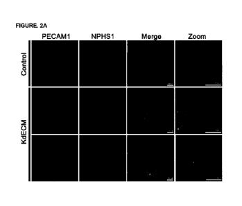
FIGS. 2A to 2F are views illustrating the up-regulation and enhanced
vascularization of podocyte and tubular epithelial markers when kidney
organoids
are cultured in an in vitro kidney dECM: (FIGS. 2A and 2B) Results of
observing
vasculature marker PCAM1-positive cells and vascular network formation after
culture with a kidney dECM for 1 week; (FIGS. 2C to 2F) Real-time quantitative
PCR results showing that maturation markers including vascular progenitors,
PCAM1, and MCAM and vascular endothelial-cadherin (VE-cadherin) are
upregulated when cultured in a kidney dECM.
FIGS. 3A and 3B are views illustrating the enhancement in cell viability and
maturity of kidney organoids when cultured in an in vitro kidney dECM: (FIG.
3A)
Representative confocal microscopy images of live/dead staining; (FIG. 3B)
Representative confocal microscopy images of podocytes and proximal tubular
cells.
FIGS. 4A to 4F are views illustrating the vascular network formation of
kidney organoids in vivo and maturation of glomerulus-like structures when
transplanted with a kidney dECM: (FIG. 4A) Representative images of CD31
immunohistochemical staining in transplanted grafts; (FIG. 4B) Representative
confocal microscopy images of MECA32 in transplanted grafts; (FIG. 4C)
Representative confocal microscopy images of MECA32 and collagen IV in
transplanted grafts; (FIG. 4D) Representative confocal microscopy images
comparing the expression of VEGF; (FIG. 4E) Representative microscopic images
comparing podocyte structure and alignment with the glomerular basement
membrane; (FIG. 4F) Microscopic images showing that podocytes and endothelial
cells are aligned to the glomerular basement membrane.
6
CA 03174384 2022- 9- 30
[Modes of the Invention]
Human pluripotent stem cell (hPSC)-derived kidney organoids have
segmental structures including nephron-like arrangements of podocytes, and
proximal and distal tubules. However, the limited vascularization of nephron-
like
structures and the resulting immaturity of blood vessels still remain a
challenge to be
overcome.
An extracellular matrix (ECM) provides the mechanical support and
biochemical microenvironment for cell growth and differentiation. The present
inventors developed a culture system for hPSC-derived kidney organoids
including
kidney decellularized extracellular matrix (dECM) hydrogels, and confirmed
that the
culture system can induce the up-regulation of gene expression for maturation
of
podocytes and tubular epithelial cells to enhance the angiogenesis of kidney
organoids, thereby completing the present invention.
Therefore, the present invention may provide a method for producing a
kidney organoid having a nephron-like structure, the method including
culturing a
kidney organoid in a collagenous three-dimensional matrix including a
decellularized
kidney extracellular matrix.
As another aspect of the present invention, the present invention may provide
a method for producing a kidney organoid having a nephron-like structure, the
method including transplanting a collagenous three-dimensional matrix
including a
decellularized kidney extracellular matrix; and a kidney organoid cultured in
the
collagenous three-dimensional matrix into the kidneys of animals other than
humans.
As used herein, the term "decellularization" refers to the removal of other
cellular components, for example, nuclei, cell membranes, nucleic acids, and
the like,
except for the extracellular matrix from cells or tissues. The term
"decellularized
7
CA 03174384 2022- 9- 30
extracellular matrix" refers to an extracellular matrix remaining after
cellular
components such as nuclei, cell membranes, and nucleic acids have been removed
from tissues or cells.
As used herein, the term "organoid" refers to an organ-specific cell aggregate
made by aggregating and recombining cells isolated from stem cells or organ-
derived
cells by a three-dimensional culture method, and is an organ analogue of an
organ-
specific cell type that self-organizes (or self-patterns) through cellular
classification
and spatially limited lineage commitment in a manner analogous to the in vivo
state.
Thus, organoids exhibit the native physiology of cells, and have an anatomical
structure that mimics the native state of a cell mixture (including not only
limited cell
types, but also residual stem cells, and proximal physiological niches). Stem
cells
may be isolated from tissue or organoid fragments. Cells, in which organoids
are
produced, differentiate in vivo to form organ-like tissues that exhibit
multiple cell
types that are self-organized to form structures very similar to those of
organs.
Therefore, the organoid is an excellent model for studying human organs and
human
organ development in a system that is very similar to in vivo development.
As used herein, the term "nephron" plays a central role in urine production as
a basic unit that constitutes the structure and function of the kidneys, and
is also
referred to as a renal unit. The nephron consists of the renal corpuscle
(glomerulus
and glomerular capsule), a proximal tubule (proximal convoluted tubule),
Henle's
loop, a distal tubule (distal convoluted tubule), and a collecting duct.
Normally,
1,000,000 to 1,500,000 nephrons are present in one kidney. Kidney failure
occurs
when nephron function is paralyzed by an infectious disease such as nephritis,
or
when the number of nephrons decreases due to other diseases.
In an exemplary embodiment of the present invention, kidney organoids
8
CA 03174384 2022- 9- 30
derived from human induced pluripotent stem cells (iPSCs) were inserted into a
kidney dECM and cultured in vitro (see Example 2). In another exemplary
embodiment of the present invention, kidney organoids having a dECM were
transplanted into mouse kidneys, and their vascularization and maturation were
concentrated (see Examples 3 and 4).
As a result, in a kidney organoid cultured with a kidney dECM, the
expression of a tubular epithelial transporter, aquaporin 1 (AQP1), a distal
tubule cell
marker, E-cadherin, a cilium gene, 3-phosphoinositide-dependent protein kinase-
1
(PKD1), a vascular endothelial growth factor (VEGF), a podocyte marker,
nephrin
(NPHS1), synaptopodin (SYNPO) and a podocyte adult transcription factor (WT1)
was up-regulated, and accordingly, it was confirmed that the nephron structure
of the
kidneys matured because vascularization was remarkably promoted.
In the present invention, the kidney organoid may be produced by
differentiation from human pluripotent stem cells, but is not limited thereto.
As used herein, the term "stem cell" is a cell capable of differentiating into
various cells that make up a biological tissue, and refers to undifferentiated
cells
capable of being regenerated unlimitedly to form specialized cells of tissues
and
organs. Stem cells are totipotent or multipotent cells which can be developed,
and
can divide into two daughter stem cells, or one daughter stem cell and one
derived
(transit) cell, and then proliferate into cells in a mature and intact form of
tissue.
As used herein, the term "pluripotent stem cells" refers to stem cells that
are
completely capable of differentiating into cells constituting the endoderm,
mesenchyme, and ectoderm as cells in a state in which cells are developed more
than
a fertilized egg. According to a specific exemplary embodiment of the present
invention, the pluripotent stem cells used in the present invention are
embryonic stem
9
CA 03174384 2022- 9- 30
cells, embryonic germ cells, embryonic carcinoma cells or induced pluripotent
stem
cells, and more specifically embryonic stem cells or induced pluripotent stem
cells
(iPSCs).
In the present invention, the decellularized kidney extracellular matrix may
be produced from kidney tissue of animals other than humans. In an exemplary
embodiment of the present invention, kidney tissue obtained from a pig was
used, but
is not limited thereto.
The decellularized kidney extracellular matrix may be produced by a method
including the following steps, but is not limited thereto.
(a) cutting the kidney tissue of an animal other than humans into sections
having a thickness of 0.01 to 1 mm;
(b) treating the kidney tissue with Triton X-100 dissolved in sodium chloride
(NaC1) for 10 to 24 hours;
(c) treating the kidney tissue with DNase for 2 to 10 hours;
(d) sterilizing the kidney tissue; and
(e) lyophilizing the kidney tissue.
In the present invention, the decellularized kidney extracellular matrix may
promote the angiogenesis or vascular maturation of the kidney organoid. In an
exemplary embodiment of the present invention, it was confirmed that the
vascularization of the kidney organoid is promoted and the nephron structure
of the
kidneys is matured by the decellularized kidney extracellular matrix.
In the present invention, the collagenous three-dimensional matrix may be a
hydrogel, but is not limited thereto.
As used herein, the term "hydrogel" may be used interchangeably with the
term "hydrated gel," is a hydrophilic polymer network forming a three-
dimensional
CA 03174384 2022- 9- 30
cross-linkage, and exhibits a protein composition almost similar to native
tissue due
to its high moisture content. In addition, since the hydrogel is not dissolved
in an
aqueous environment and is made from various polymers, it has various chemical
compositions and physical properties.
Furthermore, the hydrogel is easily
processed, and thus, may be transformed into various shapes depending on the
application. The hydrogel has high biocompatibility due to its high water
content
and physicochemical similarity to the extracellular matrix.
The decellularized extracellular matrix hydrogel of the kidney according to
the present invention may include an extracellular matrix protein including
collagen-
IV, laminin, heparan sulfate proteoglycan and isoforms thereof.
In the present invention, the transplantation may be performed in the renal
subcapsular space of an animal, but is not limited to a specific site in the
kidney.
In the present invention, the transplanted kidney organoid may recruit
endothelial cells from the kidney of a host animal. Further, the blood vessels
of the
transplanted kidney organoid may be connected to the blood vessels of a host
animal.
In addition, in the present invention, the transplantation may be performed by
embedding a kidney organoid in a collagenous three-dimensional matrix, and the
kidney organoid embedded in the collagenous three-dimensional matrix may
include
at least one or more, for example, 5 or more and 30 or less, kidney organoids
(or cell
aggregates), but is not limited thereto.
As another aspect of the present invention, the present invention may provide
a kidney organoid having a nephron-like structure produced by the method
according
to the present invention.
As still another aspect of the present invention, the present invention may
provide a collagenous three-dimensional matrix for producing a kidney organoid
11
CA 03174384 2022- 9- 30
having a nephron-like structure, including a decellularized kidney
extracellular
matrix.
Terms or words used in the specification and the claims should not be
interpreted as being limited to typical or dictionary meanings and should be
interpreted with a meaning and a concept that are consistent with the
technical spirit
of the present invention based on the principle that an inventor can
appropriately
define a concept of a term in order to describe his/her own invention in the
best way.
Hereinafter, preferred examples for helping with understanding of the present
invention will be suggested. However, the following examples are provided only
so
that the present invention may be more easily understood, and the content of
the
present invention is not limited by the following examples.
[Examples]
Experimental Example. Experimental materials and methods
1. Decellularization of kidney tissue and production of decellularized
extracellular matrix (dECM) hydrogel
1.1. Decellularization of kidney tissue
Kidney tissue obtained from a pig was sliced into slices having a thickness of
0.1 to 0.3 mm and washed three times with distilled water for 30 minutes.
Next, the
slices were treated with 0.5% Triton X-100 (Sigma-Aldrich, USA) in 1 M NaCl
(Samchun Chemical Co., Ltd., Korea) for 16 hours. Thereafter, the slices were
again washed three times for 1 hour. Remaining cellular components were
removed
by treatment with DNase at 37 C for 6 to 7 hours. Subsequently, the tissue
slices
treated with DNase were washed with phosphate-buffered saline (PBS) for 12
hours,
12
CA 03174384 2022- 9- 30
then sterilized with a 0.1% peracetic acid solution for 1 hour, and washed
again using
distilled water. A decellularized tissue was lyophilized at -80 C, and then
used for
biochemical characterization and production of kidney dECM hydrogels.
1.2. Production of kidney dECM hydrogel
A kidney dECM hydrogel was produced by dissolving the previously
decellularized kidney tissue in an acetic acid solution. The acetic acid
solution
included decellularized kidney tissue and pepsin at a mass ratio of 10:1 and
was
stirred for 72 to 96 hours depending on the concentration of decellularized
tissue in
the solution. After the dissolution was completed, the acetic acid solution
was
neutralized using sodium hydroxide and diluted using distilled water to
finally make
a kidney dECM hydrogel at a required concentration.
2. Biochemical characterization of kidney dECM
To quantify double-stranded DNA (dsDNA), a kidney dECM was digested at
60 C for 16 hours using 1 ml of a papain solution (125 g/m1 papain in 0.1 M
sodium phosphate containing 5 mM Na2-EDTA and 5 mM cysteine-HC1 at a pH of
6.5). Then, dsDNA was isolated from the digested sample using a GeneJET
genomic DNA purification kit (Thermo Scientific, USA). 1 I of the digested
sample was loaded into a NanoDrop (Thermo Scientific) and the amount of its
contents was determined.
For immunohistochemical analysis, native kidney and decellularized tissues
were fixed in 10% formalin, embedded in paraffin, and then a section was made
using a microtome. Sectioned samples were stained with hematoxylin and eosin
(H&E), alcian blue, Masson's trichrome and anti-fibronectin. Subsequently, the
13
CA 03174384 2022- 9- 30
stained samples were observed under an optical microscope.
3. Differentiation of kidney organoid
A CMC11 iPSC cell line was obtained from The Catholic University of Korea
(male donor). The differentiation of kidney organoids was performed according
to
a conventional method using cells with a passage number between 30 and 60
(Freedman et al., 2015). Briefly, hPSCs were plated at a density of 5,000
cells/well
along with an mTeSR1 medium (Stem Cell Technologies, USA) containing 10 M
Y27632(LC Laboratories, USA) in a 24-well glass plate (LabTek, Australia)
coated
with 3% GelTrexTm (Thermo Fisher Scientific, USA) (day -3).
The medium was exchanged with mTeSR1 including 1.5% GelTrex on day -
2, with mTeSR1 on day -1, with RPMI (Thermo Fisher Scientific) containing 12
M
CHIR99021 (Tocris, UK) on day 0, and with RPMI (Thermo Fisher Scientific)
containing a B27 supplement on day 1.5, respectively. Thereafter, a RPMI
(Thermo
Fisher Scientific) medium containing a B27 supplement was supplied every 2 and
3
days to promote the differentiation of the kidney organoid.
On day 18, the organoid attached to the 24-well plate were microdissected
using a 23-gauge injection needle under an inverted phase-contrast microscope.
Then, the acquired kidney organoid was placed on an 8-well chamber slide
(ibidi,
Germany) coated with 0.1% kidney dECM, and RPMI containing a B27 supplement
was supplied every 2 and 3 days. On day 25, the kidney organoid was fixed.
4. Immunofluorescence and immunohistochemical analysis
For immunofluorescence analysis, the organoid was fixed on day 18 unless
otherwise stated. For fixation, equal volumes of PBS (Thermo Fisher
Scientific)
14
CA 03174384 2022- 9- 30
and 8% paraformaldehyde (Electron Microscopy Sciences, USA) were added to the
medium for 15 minutes, and then the samples were washed three times with PBS.
Fixed organoid cultures were blocked with 5% donkey serum (Millipore, USA)
containing 0.3% Triton-X-100/PBS, cultured with a primary antibody in PBS
containing 3% bovine serum albumin (Sigma-Aldrich) overnight, and then washed.
Thereafter, the organoid cultures were treated with an AlexaFluor secondary
antibody
(Invitrogen), cultured, washed, stained with DAPI, or mounted using
Vectashield H-
1000.
After embedding, for single immunohistochemical (IHC) staining, kidneys
and kidney organoids were fixed, then embedded in wax and cut transversely
into a
thickness of 4 gm using a microtome. Some kidney sections and kidney organoid
sections were processed and stained with an H&E stain or Masson's trichrome
stain.
The other sections were treated for inu-nunohistochemical analysis after
embedding.
These tissue sections were hydrated with graded ethanol and rinsed with tap
water.
After dewaxing, the sections were microwave-incubated with a retrieval
solution for
10 minutes. The sections were washed with tap water and incubated with
methanolic H202 for 30 minutes for endogenous peroxidase blocking. Next, the
sections were cultured with a 0.5% Triton X-100/PBS solution for 15 minutes
and
rinsed with PBS. Non-specific binding sites were blocked with normal donkey
serum (diluted 1:10 in PBS) for 1 hour followed by overnight incubation with a
primary antibody at 4 C. The next day, after being rinsed with PBS, the
sections
were incubated in peroxidase-conjugated donkey anti-mouse or anti-rabbit
immunoglobulin G (IgG; Jackson ImmunoResearchLab, USA) for 2 hours, and then
washed again with a 0.05 M Tris buffer (pH 7). For detection, the sections
were
treated with 0.05% 3,3'-diaminobenzidine (DAB) and 0.01% 11202. Thereafter,
the
CA 03174384 2022- 9- 30
sections were washed with distilled water, dehydrated with ethanol and xylene,
and
then mounted on Canada balsam and observed under an optical microscope.
After embedding, for multiplex immunohistochemical (INC) staining, tissue
and organoid sections were stained with DAB and then treated with methanolic
H202
for 30 minutes to remove the peroxidase remaining from the first staining.
Subsequently, the sections were incubated with other primary antibodies. After
washing once with PBS, the sections were cultured with peroxidase-conjugated
donkey anti-rabbit IgG (Jackson Immuno Research Lab) for 2 hours. For the
detection of peroxidase, Vector SG (Vector Laboratories, USA) was used as a
chromogen to produce a gray-blue color, which is easily distinguished from a
brown
stain produced by DAB. The sections were washed with distilled water,
dehydrated
with graded ethanol and xylene, then mounted on Canada balsam and observed
under
an optical microscope. The following antibodies were used as primary
antibodies:
anti-LTL (Vector Labs FL-1321, 1:500 dilution), anti-ZO-1 (Invitrogen 339100,
1:100), anti-NPHS1 (R&D AF4269, 1:500), anti-ECAD (Abeam, ab11512, 1:100),
anti-THP (MP Bio, 55140; 1:200), anti-Claudin 1 (Abeam an15098, 1:100), anti-
WT1 (Abeam ab89901, 1:100), anti-CD31 (R&D Systems AF3628, 1:200), anti-
laminin (Sigma-Aldrich L9393. 1:200), anti-human nuclear antibody (HNA) (Merck
Millipore MAB1281, 1:100) and anti-WT1 (Santa Cruz sc-192, 1:100).
5. Electron microscopy analysis
Adult mouse kidney block samples, transplanted kidney organoids and in
vitro kidney organoid samples were fixed at 4 C overnight using 4%
paraformaldehyde and 2.5% glutaraldehyde in a 0.1 M phosphate buffer. After
washing with a 0.1M phosphate buffer, the samples were post-fixed with 1%
osmium
16
CA 03174384 2022- 9- 30
tetroxide in the same buffer at 4 C for 1 hour. Subsequently, the samples were
dehydrated with a series of graded ethyl alcohol solutions, exchanged through
acetone, and then embedded in Epon 812. Thereafter, an ultrathin section (70
to 80
nm) was obtained by an ultramicrotome (Leica Ultracut UCT, Germany). The
ultrathin section was double-stained with uranyl acetate and lead citrate, and
then
observed with a transmission electron microscope (JEM 1010, Japan) at 60 kV.
For
quantitative analysis, 20 low-magnification (x6,000) fields were randomly
selected
from each section of cortex, and the number of autophagosomes per 100 1.1m2
was
determined.
6. Transplantation of human iPSC-derived kidney organoids
Adherent organoids were microdissected from 24-well plates using a 23-
gauge injection needle on day 18 of differentiation, and then carefully
transferred to
an Eppendorf tube containing RB using a transfer pipette. Harvested kidney
organoids were transplanted with 0.1% kidney dECM into the renal subcapsular
space of 8-week-old immunodeficient male NOD/SCID mice (Jackson Laboratories,
USA).
Briefly, the mice were anesthetized with Zoletil and then the kidneys were
exposed through a lateral incision in the back. After about a 2 mm incision in
the
host kidney capsule with a 23-gauge injection needle, a PESO tube containing
10 to
20 kidney organoids was carefully placed under the kidney capsule. The kidney
organoids and 0.1% kidney dECM were delivered by carefully blowing through the
other side of the PESO tube. Mice were sacrificed 14 days after
transplantation (n=3
per group).
17
CA 03174384 2022- 9- 30
7. Real-time quantitative polymerase chain reaction (RT-qPCR) analysis
Kidney organoid samples were collected and total RNA was isolated from
each sample using an RNAiso plus kit (TAKARA, Japan) according to the
manufacturer's instructions. Complementary DNA was synthesized using a
Maxima First Strand cDNA synthesis kit for RT-qPCR (Thermo Fisher Scientific).
Gene expression was analyzed with a Power SYBR Green PCR master mix (Applied
Biosystems, USA) using a real-time polymerase chain reaction (Applied
Biosystems,
USA).
8. Statistical Analysis
For all quantitative measurements, the entire population was used for
statistical significance calculations, and a mean with an n value of 3 was
used to
calculate standard errors and graphical confidence intervals. Data was then
analyzed using the Mann-Whitney test or the Kruskal-Wallis test to determine
the
significance between groups. Error bars in each graph represent -2 SEM
(standard
error of the mean) with a 95% confidence interval. A single asterisk was used
for a
p-value of <0.05, two asterisks for p <0.01, and three asterisks for p <0.001.
Example 1. Decellularization of kidneys and characteristics of
decellularized kidney ECM hydrogel
Kidneys were decellularized as illustrated in the schematic view of FIG. 1A.
First, it was confirmed that there was no visible cellular component in the
kidney dECM through H&E staining. The remaining fibronectin and collagen
components were visually evaluated by alcian blue, anti-fibronectin and
Masson's
trichrome staining, respectively. As a result, as illustrated in FIG. 1B, it
was
18
CA 03174384 2022- 9- 30
confirmed that fibronectin and collagen, which are major ECM components of the
kidneys, were well preserved in the decellularized kidney tissue.
Further, the cellular components remaining after decellularization were
evaluated. As a result, as illustrated in FIG. 1C, the DNA content of kidney
dECM
remained at a level of 2.29% compared to that of native kidney tissue, and
only 0.64
ng of DNA/mg remained. This indicates that most cellular components were
successfully removed.
Example 2. Culture of kidney organoid on in vitro kidney dECM and its
effect
To confirm the effect of a kidney dECM in the culture of kidney organoids,
the present inventors generated kidney organoids from human iPSCs using an
adherent culture differentiation protocol and then purified the kidney
organoids by
microdissection from the peripheral stroma. The kidney organoids obtained as
described above were inserted into a kidney dECM and cultured.
As a result, as illustrated in FIGS. 2A and 2B, the kidney dECM increased
vasculature marker PCAM1-positive cells and vascular network formation in
kidney
organoids within 1 week. The vascular network appeared to extensively surround
the nephron-like structure. In addition, it was confirmed that the area,
length and
diameter of PCAM1-positive vasculature increased in kidney organoids inserted
into
the kidney dECM compared to the control (FIG. 2B).
Furthermore, it was confirmed through a real-time quantitative polymerase
chain reaction (RT-qPCR) that vascular progenitor and maturation markers,
including
PCAM1 and MCAM, as well as vascular endothelial cadherin (VE-cadherin), were
increased when the kidney organoids were cultured in the kidney dECM (FIG.
2C).
19
CA 03174384 2022- 9- 30
When the kidney organoids were transplanted into mouse kidneys, cultured in
a microfluidic system, or transplanted into chick chorioallantoic membranes,
enhanced vascularization of kidney organoids as well as progressive
morphogenesis
of tubular structures can be observed. The present inventors determined an
enhanced vascularization effect according to the culture with the kidney dECM
in
regard to the maturation of tubular epithelial cells. As a result, as
illustrated in FIG.
2D, it was confirmed that when the kidney organoids were cultured on the
kidney
ECM, the expression of a tubular epithelial transporter, aquaporin 1 (AQP1), a
distal
tubule cell marker, E-cadherin, a cilium gene, and 3-phosphoinositide-
dependent
protein kinase-1 (PKD1) was up-regulated.
In consideration of the fact that glomerular vascularization is essential for
human podocyte development, the present inventors also investigated the effect
of a
kidney dECM on the vascularization of the glomerular compartment. As a result,
in
the kidney organoids inserted into the kidney dECM, the PCAM1-positive
vasculature partially penetrated into NPHS1-positive cells, whereas this
phenomenon
was not observed in the control (FIG. 2A).
In the development of the kidneys, at the s-shape body stage, vascular
endothelial growth factor A (VEGF-A) produced by podocyte progenitors
contributes
to subsequent podocyte maturation by attracting infiltrating endothelial
cells. Thus,
the present inventors analyzed the gene expression of VEGF-A and podocytes. As
a
result, it was confirmed that VEGF was up-regulated in organoids cultured with
the
kidney dECM (FIG. 2E).
When the kidney organoids were cultured in a kidney dECM, a podocyte
marker, nephrin (NPHS1), synaptopodin (SYNPO) and a podocyte adult
transcription factor (WT1) were up-regulated (FIG. 2F). When taken together,
the
CA 03174384 2022- 9- 30
above results indicate that the kidney dECM up-regulates VEGF expression and
induces infiltrating glomerulus-like structures by a PCAM1-positive
vasculature
accompanied by podocyte maturation.
In addition, as illustrated in FIGS. 3A and 3B, it was confirmed that when the
kidney organoids were cultured in a kidney dECM, the survival of cells
constituting
kidney organoids was enhanced (FIG. 3A) and the polarity of proximal tubular
epithelial cells was enhanced.
Example 3. In vivo transplantation of kidney organoids including kidney
dECM and its effect
Transplantation of human kidney organoids into mouse kidneys was known
to enhance the formation of a perfusable vasculature that facilitated the
maturation of
glomerulus-like and tube-like structures in kidney organoids. Thus,
considering
that a kidney dECM up-regulates VEGF expression and enhances the
vascularization
of kidney organoids, the present inventors hypothesized that transplanting
kidney
organoids having a kidney dECM could accelerate vascularization in the
transplanted
graft, through which more advanced morphogenesis could be elicited in nephron-
like
structures of kidney organoids.
To test the above hypothesis, the present inventors transplanted kidney
organoids derived from human iPSCs having a kidney dECM under the kidney
capsule of immunodeficient NOD-SCID mice for engraftment. Blood vessels with
CD31-positive cells were abundantly formed in transplanted grafts for 2 weeks
after
transplantation (FIG. 4A). It was found that the vessel diameter of grafts
transplanted with the kidney dECM was larger than that of grafts lacking a
kidney
dECM and transplanted (FIG. 4A).
21
CA 03174384 2022- 9- 30
Furthermore, mouse endothelial cells (MECA32+) were abundantly observed
within transplanted kidney organoid grafts and glomerulus-like structures
(FIG. 4B).
More abundant MECA32-positive cells were observed in the transplanted kidney
organoids having the kidney dECM compared to the control, suggesting that the
kidney dECM has an effect of recruiting endothelial cells from the mouse
kidneys to
transplanted grafts.
Collagen IV, a major component of the basement membrane, is essential for
vascular integrity, stability and functionality during development. Thus, the
present
inventors investigated the expression of collagen in transplanted kidney
organoids,
considering that collagen IV is the most abundant protein in a kidney dECM.
Confocal fluorescence microscopy revealed that transplanted kidney organoids
having a kidney dECM had greater expression of collagen IV in glomerular
capillaries and peritubular capillaries compared to kidney dECM-free
transplanted
kidney organoids (top of FIG. 4C).
Further, to confirm the integrity of the vasculature formed in kidney
organoids transplanted with a kidney dECM, mice were injected with dextran
labeled
with 500 kDa fluorescein isothiocyanate (FITC) into the tail vein. FITC-
labeled
dextran was present inside the blood vessels and capillaries of the glomerulus-
like
structure in the transplanted kidney organoids (bottom of FIG. 4C), suggesting
that
the vasculature of the transplanted kidney organoids is connected to the
infiltrating
renal vasculature derived from the host mouse to maintain vascular integrity.
When the above results are taken together, the kidney dECM accelerated the
recruitment of endothelial cells from the host mouse kidney, confirming that
by
increasing collagen IV in the basement membrane, a vascular network is formed
and
the integrity of blood vessels is maintained.
22
CA 03174384 2022- 9- 30
Example 4. In vivo transplantation of kidney organoids including kidney
dECM and maturation effect of glomerulus-like structures
Podocytes are cells in the outer layer of the kidney glomerular capillary
loop.
As the first step in forming urine, the glomeruli filter the blood to send
back large
molecules such as proteins and allow small molecules such as water, salts and
sugars
to pass through. Long projections or foot processes of podocytes wrap around
capillaries and rest on the basement membrane of the glomerulus. The foot
processes are connected by a porous structure called the slit diaphragm.
In vitro studies showed that the up-regulation of VEGF expression appeared
in addition to increased glomerular vascularization and podocyte maturation
during
culture with a kidney dECM (FIGS. 2A to 2F). Thus, the present inventors
investigated the degree of maturation of glomerulus-like structures during
transplantation with a kidney dECM in consideration of the enhanced
vascularization
after transplantation of kidney organoids.
As a result of observation by confocal fluorescence microscopy, it was
confirmed that the expression of VEGF was increased more in the glomerulus-
like
structures of transplanted kidney organoids having a kidney dECM compared to
kidney dECM-free transplanted kidney organoids (FIG. 4D).
In addition, in order to determine the ultrastructure of cells, an additional
structural analysis using transmission electron microscopy (TEM) was
performed,
and in vitro kidney organoids, kidney organoids transplanted in vivo and adult
mouse
kidneys were compared with one another.
As a result, as illustrated in FIG. 4E, in in vitro kidney organoids,
podocytes
had an immature structure with apical microvilli and intermittently arranged
along
23
CA 03174384 2022- 9- 30
glomerular basement membrane (GBM)-like tracks. In comparison, in the case of
kidney organoids transplanted alone, erythrocyte fragments were observed in
the
transplanted kidney organoids, indicating that capillaries can be formed (FIG.
4E).
However, transplanted kidney organoids lacked a bona fide foot process with
well-
organized tertiary interdigitation along the GBM. Furthermore, the Bowman's
capsule of the transplanted organoid was structurally similar to the Bowman's
capsule of the adult mouse, but had a substantially thicker capsule layer than
the
Bowman's capsule of the adult mouse. In contrast, the podocytes of kidney
organoids transplanted with a kidney dECM had secondary or tertiary foot
processes
that engaged the GBM similar to those of the adult mouse kidney (FIG. 4E).
Further, when kidney organoids were transplanted with a kidney dECM, the GBM
was well-organized and aligned with podocytes and endothelial cells compared
to the
adult kidneys of a mammal (FIG. 4F). The aforementioned results demonstrate
the
fact that the kidney dECM contributes to the maturation of glomerulus-like
structures
in transplanted kidney organoids.
In summary, when kidney organoids having a kidney dECM were
transplanted under the kidney capsule in immunodeficient mice, the recruitment
of
endothelial cells from the kidney of the host mouse was promoted and the
integrity
of blood vessels was maintained. In addition, in transplanted kidney organoids
having a kidney dECM, slit diaphragm-like structures were more organized
compared to kidney dECM-free slit diaphragm-like structures.
These results suggest the fact that a microenvironment provided from a
kidney dECM hydrogel promotes angiogenesis and maturation of iPSC-derived
kidney organoids, and it is expected that the microenvironment generates a
kidney
with blood vessels on a chip or can be applied to regenerative therapy.
24
CA 03174384 2022- 9- 30
The above-described description of the present invention is provided for
illustrative purposes, and those skilled in the art to which the present
invention
pertains will understand that the present invention can be easily modified
into other
specific forms without changing the technical spirit or essential features of
the
present invention. Therefore, it should be understood that the above-described
embodiments are only exemplary in all aspects and are not restrictive.
[Industrial Applicability]
The present invention induced the vascularization of the kidney organoid,
induced the expression of podocytes, tubular transporters and cilium genes and
formed a more mature nephron-like structure using a kidney organoid culture
system
using kidney dECM hydrogels. Therefore, the kidney organoid produced from
human pluripotent stem cells according to the production method of the present
invention is expected to treat nephron loss by being transplanted to humans or
be
utilized as a kidney on a chip, which is an in vitro kidney model, and thus is
expected
to have great industrial utility value.
CA 03174384 2022- 9- 30