Note: Descriptions are shown in the official language in which they were submitted.
CA 03174443 2022-08-31
WO 2021/178344
PCT/US2021/020373
METHODS TO RE-ENGAGE A FETAL WOUND HEALING PATHWAY
FOR ADULT SKIN REPAIR
CROSS REFERENCE TO RELATED APPLICATIONS
This application claims priority to U.S. Provisional Patent Application No.
62/985,008 filed on March 4, 2020, the disclosure of which is expressly
incorporated
herein.
INCORPORATION BY REFERENCES OF MATERIAL SUBMITTED
ELECTRONICALLY
Incorporated by reference in its entirety is a computer-readable
nucleotide/amino acid sequence listing submitted concurrently herewith and
identified
as follows: 4 kilobytes ACII (Text) file named "333982_5T25.txt," created on
March
1, 2021.
BACKGROUND OF THE DISCLOSURE
Nonhealing chronic wounds are a challenge to the patient, the health care
professional, and the health care system. They significantly impair the
quality of life
for millions of people and impart a burden on society in terms of lost
productivity and
health care dollars.
Fetal wound healing is more efficient and regenerative than adult wound
healing. Fetal skin tissue perfectly executes epidermal regeneration; a
pattern that is
absent during adult wound healing. Prior to the present disclosure it was
unknown if
the drivers of the fetal repair process continued to exist in adult tissue,
and if so,
whether the critical elements of the fetal repair process could be activated
in adult
tissue to improve adult tissue repair outcomes.
The protein nonselenocysteine-containing phospholipid hydroperoxide
glutathione peroxidase (NPGPx) is an oxidant stress sensor protein. NPGPx is
abundantly expressed in normal fetal epidermis, but is not expressed adult
epidermis.
NPGPx is a direct target of the miR-29 family and is variably induced upon
adult
tissue wounding. More particularly, after injury, the abundance of miR-29 is
lowered,
and this lower abundance of miR-29 after injury permits NPGPx transcripts and
protein expression to promptly increase after injury in adult wound-edge
tissue.
The low abundance of miR-29 after injury is due to pre-miRNAs being rapidly
degraded by the protein endoribonuclease monocyte chemoattractant protein-
induced
CA 03174443 2022-08-31
WO 2021/178344
PCT/US2021/020373
protein 1 (MCPIP1). MCPIP1 is induced at the site of the wound by lipids that
are
blood-clot derived post-injury. This favorable physiologic response was
blunted in
non-healing wounds, such as in diabetic mice.
This pathway may be schematically illustrated as:
MCPIP1 ¨> miR-29 ¨> NPGPx.
The MCPIP1 ¨> ¨> NPGPx pathway induced more regenerative
healing in adult wounds by re-engaging expression of numerous developmentally
active coding genes and fetal proteins relevant for tissue formation and
repair.
Accordingly, there is a need for treatments that enhance would repair by
stimulating the fetal wound healing pathway in adult skin repair.
SUMMARY
In accordance with one embodiment of the present disclosure, a
method of promoting wound healing in a subject is provided, the method
comprising
the step of administering a composition that enhances the expression of the
protein
nonselenocysteine-containing phospholipid hydroperoxide glutathione peroxidase
(NPGPx) in wound-edge tissues. In one embodiment the composition comprises an
anti-miR-29 oligonucleotide and a pharmaceutically acceptable carrier, wherein
the
composition is formulated for introduction into the cytosol of wound edge
tissues. In
one embodiment the anti-miR-29 oligonucleotides are provided in a carrier such
as a
viral vector or lipid vessel. In one embodiment the composition is formulated
for
transmission by electroporation. In accordance with one embodiment,
compositions
for enhancing NPGPx concentrations in wound-edge tissues as disclosed herein
are
used in conjunction with known treatments for use on chronic wounds including
in
diabetic patients.
In one embodiment a method to regulate wound healing in adult skin,
optionally the adult skin of a diabetic individual, is provided. In one
embodiment the
method enhances regenerative healing in an adult wound by re-engaging
expression
of developmentally active coding genes and fetal proteins in tissue formation
and
repair. In one embodiment the method comprises augmenting physiologic
expression
of the protein nonselenocysteine-containing phospholipid hydroperoxide
glutathione
peroxidase (NPGPx) in wound-edge tissues. In one embodiment, increased
expression of NPGPx is induced in wound-edge tissues by transfecting the cells
of
wound-edge tissues with nucleic acid sequences that stimulate increased
expression of
2
CA 03174443 2022-08-31
WO 2021/178344
PCT/US2021/020373
NPGPx, optionally by suppressing miR-29, resulting in regulated wound healing
in
adult skin. In one embodiment accelerated wound healing in adult skin is
promoted by
manipulating at least one component of the following pathway: by increasing
endoribonuclease monocyte chemoattractant protein-induced protein (MCPIP1),
decreasing miR-29, reducing levels of miR-29 activity, and increasing the
cellular
concentration of nonselenocysteine-containing phospholipid hydroperoxide
glutathione peroxidase (NPGPx), resulting in accelerated wound healing and
wound
closure of wounds in adult skin.
In one embodiment the method facilitates healing of a wound in a diabetic
patient by augmenting expression of nonselenocysteine-containing phospholipid
hydroperoxide glutathione peroxidase (NPGPx) expression at the wound site. In
one
embodiment the wound is a non-healing/chronic wound. In one embodiment the
method increases the expression of NPGPx and this increased expression of
NPGPx
overcomes a deleterious effect of diabetes on wound closure.
In one embodiment the method comprises topical delivery, to a patient in need
thereof, of anti-miR-29 oligonucleotides by topical tissue nanotransfection
(TNT) to
directly induce nonselenocysteine-containing phospholipid hydroperoxide
glutathione
peroxidase (NPGPx) in skin keratinocytes.
In one embodiment a method to regulate wound healing in adults is provided
wherein the method comprises topically delivering anti-miR-29 oligonucleotides
by
topical tissue nanotransfection (TNT) under conditions sufficient to induce
nonselenocysteine-containing phospholipid hydroperoxide glutathione peroxidase
(NPGPx) in skin keratinocytes and result in wound healing.
In one embodiment a pharmaceutical composition for enhancing wound
closure is provided, wherein said composition comprises an oligonucleotide at
least 6,
7, or 8 nucleotides in length, wherein the oligonucleotide has at least 85%
sequence
identity to a continuous 6-8 nucleotide sequence of a human mature miR-29
sequence
selected from the group consisting of SEQ ID NO: 2, SEQ ID NO: 3, SEQ ID NO:
4,
SEQ ID NO: 5, SEQID NO: 6 and SEQ ID NO: 10, or a complement of any of those
sequences thereof and a pharmaceutically acceptable carrier.
BRIEF DESCRIPTION OF THE DRAWINGS
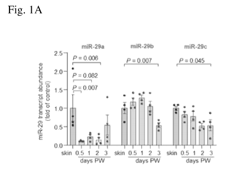
Figs 1A-1H. Fig. 1A shows miR-29a, miR-29b and miR-29c transcript
abundance in skin and wound-edge tissue of adult C57BL/6 mice at different
time
3
CA 03174443 2022-08-31
WO 2021/178344
PCT/US2021/020373
points post-wounding (PW) (n=4). Fig. 1B shows the binding of positions 41-47
of
mNPGPx 3' UTR (SEQ ID NO: 1) to mmu-miR-29a-3p (SEQ ID NO: 2), mmu-miR-
29b-3p (SEQ ID NO: 3) and mmu-miR-29a-3p (SEQ ID NO: 4). Fig. 1C shows
NPGPx transcript abundance in skin and wound-edge tissue in adult C57BL/6 mice
(n=6). Fig. 1D shows Western blot analysis of NPGPx in skin and wound-edge
tissue
in adult C57BL/6 mice; GAPDH was the loading control and independent blots
were
repeated at least three times with similar results. Fig. 1E shows NPGPx
transcript
abundance in laser captured microdissected (LCM) keratinocytes from skin and
day 7
wound-edge tissue of C57BL/6 mice (n=4). Fig. 1F shows Western blot analysis
of
NPGPx in murine fetal skin (E15.5-E18.5) and adult skin; GAPDH was the loading
control and independent blots were repeated at least three times with similar
results.
Fig. 1G shows NPGPx transcript abundance in human fetal and adult skin (n=6,
5).
Fig. 1H shows Western blot analysis of NPGPx in human fetal and adult skin;
each
lane indicates separate biological samples (n=5) and GAPDH was the loading
control.
All data were shown as mean SEM.
Figs. 2A-2F show the role of NPGPx for fetal and adult wound closure. The
NPGPx expressed in human epidermis correlated with the highest level of wound
closure (Fig. 2A). NPGPx suppression impaired adult wound closure (Figs. 2B
and
2D). NPGPx overexpression improved adult wound closure (Figs. 2C and 2E).
NPGPx overexpression accelerated wound re-epithelialization with significant
improvement in skin barrier function (Fig. 2F); a critical functional test of
re-
epithelialization.
Figs. 3A-3K show NPGPx in diabetic db/db mice with mutations of the leptin
receptor that display impaired wound healing and serve as a model for non-
healing
wounds. Figs. 3A and 3B show miR-29a, miR-29b and miR-29c expression in fetal
(E15.5-E-18.5) and adult skin of C57BL/6 mice (Fig. 3A), and fetal and adult
human
skin (Fig. 3B). (Fig. 3A: n=5; Fig. 3B: n=6, 6; 6,6; 5,6). Fig. 3C shows NPGPx
transcript abundance in skin and wound-edge tissue in K14-dicer+/+ and K14-
dicer-/-
mice (n=4). Figs. 3D and 3E show Western blot analysis of HaCaT cells
transfected
with miR-29b or miR-29c inhibitor (Fig. 3D) and miR-29b or miR-29c mimic (Fig.
3E). GAPDH was the loading control. Independent blots were repeated at least
three
times with similar results. Figs. 3F and 3G show miRNA target reporter
luciferase
assay in in HaCaT cells after delivery of miR-29b (Fig. 3F) and miR-29c (Fig.
3G)
mimic. (H: n=6; I: n=6). Fig. 3H shows NPGPx transcript abundance in skin and
day
4
CA 03174443 2022-08-31
WO 2021/178344
PCT/US2021/020373
7 wound-edge tissue of non-diabetic (m+/db, littermate control) and diabetic
db/db
mice (n=4). Western blot analysis of NPGPx from skin and day 7 wound-edge
tissue
from m+/db and db/db mice showed similar results as obtained for transcript
abundance. Independent blots were repeated at least three times with similar
results.
Fig. 31 shows miR-29a, miR-29b and miR-29c expression in day 7 wound-edge
tissue
in m+/db and db/db mice (n=4). Fold change was calculated using respective
skin
(m+/db and db/db) as 1. Fig. 3J shows NPGPx transcript abundance of NPGPx in
day
wound-edge tissue of diabetic db/db mice treated with either control (pLVcon)
or
NPGPx over expressing (pLVNPGPx) lentivirus. Western blot analysis mice showed
10 similar results as obtained for transcript abundance. GAPDH was used as
loading
control. Digital photographs of wounds in db/db mice were taken at day 0, 6
and 10
and wound closure was presented as percentage of initial wound area. (n=4) in
Fig.
3K.
Figs. 4A-4F shows the role of each of MCPIP1, miR-29, and NPGPx in
increased wound healing. Suppression of miR-29b and miR-29c significantly
elevated NPGPx expression (Figs. 4A and 4B). Delivery of miR-29b and miR-29c
mimics significantly suppressed both NPGPx expression (Fig. 4B), as well as
NPGPx
3'-UTR reporter luciferase activity. Fig. 4C gives the sequence of wild-type
NPGPx
3'-UTR (top; SEQ ID NO: 8) and NPGPx 3'-UTR (SEQ ID NO: 9) with the mutation
of predicted binding site (seed region) cloned in the reporter construct
(bottom).
Positions mutated were marked with underlined text. Mutations of the predicted
binding sites (seed sequences) in the 3'-UTR of NPGPx (Fig. 4C) abolished miR-
29b
and miR-29c dependent translational repression. Fig. 4D shows MCPIP1
transcript
abundance in skin and wound-edge tissue at different time points PW in adult
C57BL/6 mice, (n=5, 4). Digital photographs of wounds in K14-MCPIP1 and K14-
MCPIP1-/- mice were taken and wound closure presented as percentage of initial
wound area (n=6); scale, 2 mm (Fig. 4E). Fig. 4F shows transepidermal water
loss
(TEWL) at d12 post-wounding is lower in K14-MCPIP1' and higher in K14-
MCPIP1-/- mice suggesting poor barrier function, (n=6).
Figs 5A- 5F show use of topical tissue nanotransfection (TNT) chip 2.0 to
deliver agents to demonstrate increased NPGPx and decreased miR-29b and miR-
29c
result in increased wound healing. Wound closure occurred significantly more
quickly
(Fig. 5A) by induction of NPGPx expression in the skin following miR-29b and
miR-
29c suppression (Fig. 5B). Figs. 5C and 5D show expression of miR-29b (Fig.
5C)
5
CA 03174443 2022-08-31
WO 2021/178344
PCT/US2021/020373
and miR-29c (Fig. 5D) in laser-captured epidermis of C57BL/6 mice 24h post-TNT
of
LNA-control, LNA-anti-miR-29b, and LNA-anti-miR-29c mice (n=4). Fig. 5E
presents wound closure data (Fig. 5F) at day 12 wounds in K14-MCPIP1-/- mice
based on percent wound closure, showing accelerated re-epithelialization in
wounds
treated with LNA-anti-miR-29b, and LNA-anti-miR-29c relative to LNA-control.
All
data were shown as mean SEM.
Figs 6A- 6C provide data showing that MCPIP1 plays a role in adult wound
healing and that MCPIP1 is induced post-wounding independent of MCP1. Fig. 6A
shows Western blot analysis and Fig. 6B shows quantification of MCPIP1 from
skin
and wound-edge tissue collected at 6 h post-wounding (PW) from wild type (WT)
and
MCP-1 knockout mice. GAPDH was a loading control. Independent blots were
repeated at least three times with similar results. Data are expressed as mean
SEM
(n=3). Fig. 6C shows the role of serum in the induction of MCPIP1 and the
expression of MCPIP1 in primary human keratinocytes after removal of bioactive
phospholipids from serum by charcoal stripping method (n=4). As shown by the
data,
the presence of a blood clot and MCP-1 increases cellular concentrations of
MCPIP1.
DETAILED DESCRIPTION
DEFINITIONS
In describing and claiming the invention, the following terminology will be
used in
accordance with the definitions set forth below.
The term "about" as used herein means greater or lesser than the value or
range of values stated by 10 percent but is not intended to limit any value or
range of
values to only this broader definition. Each value or range of values preceded
by the
term "about" is also intended to encompass the embodiment of the stated
absolute
value or range of values.
As used herein, the term "purified" and like terms relate to the isolation of
a
molecule or compound in a form that is substantially free of contaminants
normally
associated with the molecule or compound in a native or natural environment.
As
used herein, the term "purified" does not require absolute purity; rather, it
is intended
as a relative definition. The term "purified polypeptide" is used herein to
describe a
polypeptide which has been separated from other compounds including, but not
limited to nucleic acid molecules, lipids and carbohydrates.
6
CA 03174443 2022-08-31
WO 2021/178344
PCT/US2021/020373
The term "isolated" requires that the referenced material be removed from its
original environment (e.g., the natural environment if it is naturally
occurring). For
example, a naturally-occurring polynucleotide present in a living animal is
not
isolated, but the same polynucleotide, separated from some or all of the
coexisting
materials in the natural system, is isolated.
Tissue nanotransfection (TNT) is an electroporation-based technique capable
of delivering nucleic acid sequences and proteins into the cytosol of cells at
nanoscale. More particularly, TNT uses a highly intense and focused electric
field
through arrayed nanochannels, which benignly nanoporates the juxtaposing
tissue cell
members, and electrophoretically drives cargo (e.g., nucleic acids or
proteins) into the
cells.
As used herein a "control element" or "regulatory sequence" are non-translated
regions of a functional gene, including enhancers, promoters, 5 and 3'
untranslated
regions, which interact with host cellular proteins to carry out transcription
and
translation. Such elements may vary in their strength and specificity.
"Eukaryotic
regulatory sequences" are non-translated regions of a functional gene,
including
enhancers, promoters, 5' and 3' untranslated regions, which interact with host
cellular
proteins of a eukaryotic cell to carry out transcription and translation in a
eukaryotic
cell including mammalian cells.
As used herein a "promoter" is a sequence or sequences of DNA that function
when in a relatively fixed location in regard to the transcription start site
of a gene. A
"promoter" contains core elements required for basic interaction of RNA
polymerase
and transcription factors and can contain upstream elements and response
elements.
As used herein an "enhancer" is a sequence of DNA that functions
independent of distance from the transcription start site and can be either 5'
or 3' to the
transcription unit. Furthermore, enhancers can be within an intron as well as
within
the coding sequence itself. They are usually between 10 and 300 bp in length,
and
they function in cis. Enhancers function to increase transcription from nearby
promoters. Enhancers, like promoters, also often contain response elements
that
mediate the regulation of transcription. Enhancers often determine the
regulation of
expression.
An "endogenous" enhancer/promoter is one which is naturally linked with a
given gene in the genome. An "exogenous" or "heterologous" enhancer/promoter
is
one which is placed in juxtaposition to a gene by means of genetic
manipulation (i.e.,
7
CA 03174443 2022-08-31
WO 2021/178344
PCT/US2021/020373
molecular biological techniques) such that transcription of that gene is
directed by the
linked enhancer/promoter. As used herein an exogenous sequence in reference to
a
cell is a sequence that has been introduced into the cell from a source
external to the
cell.
As used herein the term "non-coded (non-canonical) amino acid" encompasses
any amino acid that is not an L-isomer of any of the following 20 amino acids:
Ala,
Cys, Asp, Glu, Phe, Gly, His, Ile, Lys, Leu, Met, Asn, Pro, Gln, Arg, Ser,
Thr, Val,
Trp, Tyr.
The term "identity" as used herein relates to the similarity between two or
more sequences. Identity is measured by dividing the number of identical
residues by
the total number of residues and multiplying the product by 100 to achieve a
percentage. Thus, two copies of exactly the same sequence have 100% identity,
whereas two sequences that have amino acid deletions, additions, or
substitutions
relative to one another have a lower degree of identity. Those skilled in the
art will
recognize that several computer programs, such as those that employ algorithms
such
as BLAST (Basic Local Alignment Search Tool, Altschul et al. (1993) J. Mol.
Biol.
215:403-410) are available for determining sequence identity.
The term "stringent hybridization conditions" as used herein mean that
hybridization will generally occur if there is at least 95% and preferably at
least 97%
sequence identity between the probe and the target sequence. Examples of
stringent
hybridization conditions are overnight incubation in a solution comprising 50%
formamide, 5X SSC (150 mM NaCI, 15 mM trisodium citrate), 50 mM sodium
phosphate (pH 7.6), 5X Denhardt's solution, 10% dextran sulfate, and 20 pg/ml
denatured, sheared carrier DNA such as salmon sperm DNA, followed by washing
the
hybridization support in 0.1 X SSC at approximately 65 C. Other hybridization
and
wash conditions are well known and are exemplified in Sambrook et al,
Molecular
Cloning: A Laboratory Manual, Second Edition, Cold Spring Harbor, N.Y. (1989),
particularly chapter 11.
As used herein, the term "pharmaceutically acceptable carrier" includes any of
the standard pharmaceutical carriers, such as a phosphate buffered saline
solution,
water, emulsions such as an oil/water or water/oil emulsion, and various types
of
wetting agents. The term also encompasses any of the agents approved by a
regulatory agency of the US Federal government or listed in the US
Pharmacopeia for
use in animals, including humans.
8
CA 03174443 2022-08-31
WO 2021/178344
PCT/US2021/020373
As used herein, the term "phosphate buffered saline" or "PBS" refers to
aqueous solution comprising sodium chloride and sodium phosphate. Different
formulations of PBS are known to those skilled in the art but for purposes of
this
invention the phrase "standard PBS" refers to a solution having have a final
concentration of 137 mM NaCl, 10 mM Phosphate, 2.7 mM KC1, and a pH of 7.2-
7.4.
As used herein, the term "treating" includes prophylaxis of the specific
disorder or condition, or alleviation of the symptoms associated with a
specific
disorder or condition and/or preventing or eliminating said symptoms.
As used herein an "effective" amount or a "therapeutically effective amount"
.. of a drug refers to a nontoxic but enough of the drug to provide the
desired effect.
The amount that is "effective" will vary from subject to subject or even
within a
subject overtime, depending on the age and general condition of the
individual, mode
of administration, and the like. Thus, it is not always possible to specify an
exact
"effective amount." However, an appropriate "effective" amount in any
individual
.. case may be determined by one of ordinary skill in the art using routine
experimentation.
As used herein an amino acid "substitution" refers to the replacement of one
amino acid residue by a different amino acid residue.
As used herein, the term "conservative amino acid substitution" is defined
herein as exchanges within one of the following five groups:
I. Small aliphatic, nonpolar or slightly polar residues:
Ala, Ser, Thr, Pro, Gly;
II. Polar, negatively charged residues and their amides:
Asp, Asn, Glu, Gln;
III. Polar, positively charged residues:
His, Arg, Lys; Ornithine (Om)
IV. Large, aliphatic, nonpolar residues:
Met, Leu, Ile, Val, Cys, Norleucine (Nle), homocysteine (hCys)
V. Large, aromatic residues:
Phe, Tyr, Trp, acetyl phenylalanine, napthylalanine (Nal)
As used herein the term "patient" without further designation is intended to
encompass any warm blooded vertebrate domesticated animal (including for
example,
9
CA 03174443 2022-08-31
WO 2021/178344
PCT/US2021/020373
but not limited to livestock, horses, cats, dogs and other pets) and humans
and
includes individuals not under the direct care of a physician.
The term "carrier" means a compound, composition, substance, or structure
that, when in combination with a compound or composition, aids or facilitates
preparation, storage, administration, delivery, effectiveness, selectivity, or
any other
feature of the compound or composition for its intended use or purpose. For
example,
a carrier can be selected to minimize any degradation of the active ingredient
and to
minimize any adverse side effects in the subject.
The term "inhibit" refers to a decrease in an activity, response, condition,
disease, or other biological parameter. This can include but is not limited to
the
complete ablation of the activity, response, condition, or disease. This may
also
include, for example, a 10% reduction in the activity, response, condition, or
disease
as compared to the native or control level. Thus, the reduction can be a 10,
20, 30, 40,
50, 60, 70, 80, 90, 100%, or any amount of reduction in between as compared to
native or control levels.
The term "polypeptide" refers to amino acids joined to each other by peptide
bonds or modified peptide bonds, e.g., peptide isosteres, etc. and may contain
modified amino acids other than the 20 gene-encoded amino acids. The
polypeptides
can be modified by either natural processes, such as post-translational
processing, or
by chemical modification techniques which are well known in the art.
Modifications
can occur anywhere in the polypeptide, including the peptide backbone, the
amino
acid side-chains and the amino or carboxyl termini.
The term "amino acid sequence" refers to a series of two or more amino acids
linked together via peptide bonds wherein the order of the amino acids
linkages is
designated by a list of abbreviations, letters, characters or words
representing amino
acid residues. The amino acid abbreviations used herein are conventional one
letter
codes for the amino acids and are expressed as follows: A, alanine; B,
asparagine or
aspartic acid; C, cysteine; D aspartic acid; E, glutamate, glutamic acid; F,
phenylalanine; G, glycine; H histidine; I isoleucine; K, lysine; L, leucine;
M,
methionine; N, asparagine; P, proline; Q, glutamine; R, arginine; S, serine;
T,
threonine; V, valine; W, tryptophan; Y, tyrosine; Z, glutamine or glutamic
acid.
The phrase "nucleic acid" as used herein refers to a naturally occurring or
synthetic oligonucleotide or polynucleotide, whether DNA or RNA or DNA-RNA
hybrid, single-stranded or double-stranded, sense or antisense, which is
capable of
CA 03174443 2022-08-31
WO 2021/178344
PCT/US2021/020373
hybridization to a complementary nucleic acid by Watson-Crick base-pairing.
Nucleic
acids can also include nucleotide analogs (e.g. , BrdU), and non-
phosphodiester
internucleoside linkages (e.g. , peptide nucleic acid (PNA) or thiodiester
linkages) . In
particular, nucleic acids can include, without limitation, DNA, RNA, cDNA,
gDNA,
ssDNA, dsDNA or any combination thereof.
"Nucleotide" as used herein is a molecule that contains a base moiety, a sugar
moiety, and a phosphate moiety. Nucleotides can be linked together through
their
phosphate moieties and sugar moieties creating an intemucleoside linkage. The
term
"oligonucleotide" is sometimes used to refer to a molecule that contains two
or more
nucleotides linked together. The base moiety of a nucleotide can be adenine-9-
y1 (A),
cytosine-1 -yl (C) , guanine-9-y1 (G), uracil- 1 -yl (U), and thymin-1 -yl
(T). The
sugar moiety of a nucleotide is a ribose or a deoxyribose. The phosphate
moiety of a
nucleotide is pentavalent phosphate. A non-limiting example of a nucleotide
would be
3'-AMP (3'-adenosine monophosphate) or 5'-GMP (5'-guanosine monophosphate).
A nucleotide analog is a nucleotide that contains some type of modification to
the
base, sugar, and/or phosphate moieties. Modifications to nucleotides are well
known
in the art and would include, for example, 5-methylcytosine (5-me-C), 5
hydroxymethyl cytosine, xanthine, hypoxanthine, and 2-aminoadenine as well as
modifications at the sugar or phosphate moieties.
Nucleotide substitutes are molecules having similar functional properties to
nucleotides, but which do not contain a phosphate moiety, such as peptide
nucleic
acid (PNA). Nucleotide substitutes are molecules that will recognize nucleic
acids in a
Watson-Crick or Hoogsteen manner, but are linked together through a moiety
other
than a phosphate moiety. Nucleotide substitutes are able to conform to a
double helix
type structure when interacting with the appropriate target nucleic acid.
The term "vector" or "construct" designates a nucleic acid sequence capable of
transporting into a cell another nucleic acid to which the vector sequence has
been
linked. The term "expression vector" includes any vector, (e.g., a plasmid,
cosmid or
phage chromosome) containing a gene construct in a form suitable for
expression by a
cell (e.g., linked to a transcriptional control element). "Plasmid" and
"vector" are used
interchangeably, as a plasmid is a commonly used form of vector. Moreover, the
invention is intended to include other vectors which serve equivalent
functions.
The term "operably linked to refers to the functional relationship of a
nucleic
acid with another nucleic acid sequence. Promoters, enhancers, transcriptional
and
11
CA 03174443 2022-08-31
WO 2021/178344
PCT/US2021/020373
translational stop sites, and other signal sequences are examples of nucleic
acid
sequences that can operably linked to other sequences. For example, operable
linkage
of DNA to a transcriptional control element refers to the physical and
functional
relationship between the DNA and promoter such that the transcription of such
DNA
is initiated from the promoter by an RNA polymerase that specifically
recognizes,
binds to and transcribes the DNA.
As used herein "Interfering RNA" is any RNA involved in post-transcriptional
gene silencing, which definition includes, but is not limited to, double
stranded RNA
(dsRNA), small interfering RNA (siRNA), and microRNA (miRNA) that are
comprised of sense and antisense strands.
As used herein a "locked nucleic acid" (LNA), is a modified RNA nucleotide
in which the ribose moiety is modified with an extra bridge connecting the 2
oxygen
and 4' carbon. For example, a locked nucleic acid sequence comprises a
nucleotide of
0¨ Base
0
0
the structure:
As used herein the term "vasculogenesis" is defined as the differentiation of
precursor cells (angioblasts) into endothelial cells and the de novo formation
of a
primitive vascular network.
As defined herein "wound healing" defines a process wherein a living
organism replaces destroyed or damaged tissue by newly produced tissue. The
process includes three phases blood clotting, tissue growth (cell
proliferation), and
tissue remodeling. Accelerated wound healing includes a shorten length of time
required to complete any of three phases, including for example the closure of
an
open wound due to tissue growth.
As disclosed herein a generic reference to miR-29 is intended to include all
known variants of mammalian miR-29 including for example human mature forms
miR-29a, miR-29b and miR-29c of SEQ ID NO: 2, SEQ ID NO: 3 and SEQ ID NO:
4, respectively.
12
CA 03174443 2022-08-31
WO 2021/178344
PCT/US2021/020373
EMBODIMENTS
Major differences exist in the processes of fetal tissue healing and adult
tissue
healing, including different pathways, growth factors, cytokines,
interleukins, matrix
metalloproteinases, extracellular matrix molecules (ECM), inflammatory cells,
and
cell surface molecules utilized in the two responses. Fetal repair processes
engage
specific combinations of these pathways to permit efficient restitution of all
cellular
elements and appendages (hair follicles and sweat glands) upon skin wounding,
with
appropriate remodeled ECM components to provide proper developmental barrier
and
biomechanical properties. Most of these regenerative processes are
extinguished prior
to birth. As disclosed herein methods are provided for activating components
of the
fetal wound healing process in adult cell on a temporary basis to enhance
and/or
accelerate wound healing, including wound closure.
Numerous microRNAs (miRNAs) are temporally and spatially muted in fetal
tissues, presumably to enable fetal tissue to execute rapid developmental
processes.
While this difference is a key contrast between fetal tissue and adult tissue,
it is poorly
understood. For example, low abundance of the miR-29 family during fetal
development has been reported in skin across several species. After acute skin
injury,
the miR-29 family is suppressed over the first 7 days commencing within 48-72
h of
the perturbation (Fig. 1A).
The miR-29 family is predicted to have conserved binding sites on the 3'-
untranslated regions (3'-UTRs) of twenty collagen genes independent of
sequence
homology. In adult tissue, overexpression of the miR-29 family members
suppresses
ECM genes, resulting in abnormal tissue repair. In silico analyses predicted
that, in
.. addition to these known ECM proteins, miR-29 might contain potential
binding site(s)
for the 3'-UTRs (Fig. 1B) of the oxygen stress sensor protein
nonselenocysteine-
containing phospholipid hydroperoxide glutathione peroxidase (NPGPx), that is
conserved among all vertebrates. Applicant have demonstrated NPGPx is wound-
inducible as a response to the reactive oxidant stress, based on work
demonstrating
the key role of injury-induced generation of NOX-dependent reactive oxygen
species
at the skin wound-edge. Alterations in NPGPx expression cause systemic
evidence of
excessive oxidative stress, cardiovascular disease, obesity, autoimmunity,
increased
risk for carcinogenesis, and shortened lifespan in mice. However, the
biological
significance of NPGPx as a key element in tissue injury and repair remains an
enigma
13
CA 03174443 2022-08-31
WO 2021/178344
PCT/US2021/020373
because a critical selenocysteine residue at its catalytic center is absent,
rendering
NPGPx catalytically inactive as a GPx.
In accordance with one embodiment a method of accelerating wound healing
and/or wound closure in adult skin of a subject is provided. In one embodiment
the
wound is a chronic or non-healing wound, optionally wherein the patient is a
diabetic.
The method comprising the step of increasing the concentration of the protein
nonselenocysteine-containing phospholipid hydroperoxide glutathione peroxidase
(NPGPx) in the cells of wound-edge tissue. In accordance with one embodiment
the
cells of wound-edge tissue are transfected with nucleic acid sequences, using
any
transfection technique known to the skilled practitioner, that result in
increased
cellular concentration of NPGPx. The nucleic acid sequences introduced into
the
cells can be gene encoding sequences or interference oligonucleotides.
Increased
expression of NPGPx, as demonstrated herein can be accomplished by increasing
cellular concentrations of endoribonuclease monocyte chemoattractant protein-
induced protein (MCPIP1) or Monocyte chemoattractant protein-1 (MCP-1) or by
decreasing active miR-29 cellular concentrations, including reducing miR-29b
or
miR-29c cellular concentrations.
In one embodiment a method of enhancing or accelerating wound healing is
provided wherein wound-edge tissue is transfected with a modifier of miR-29
activity
in an amount effective to lower miR-29 activity and increase NPGPx expression.
In
one embodiment the modifier the of miR-29 activity is a gene encoding the
protein
endoribonuclease monocyte chemoattractant protein-induced protein 1 (MCPIP1).
In
one embodiment the modifier of miR-29 activity is an oligonucleotide at least
6, 7, 8,
9 or 10 nucleotides in length, wherein the oligonucleotide has at least 85%,
90%,
95%, 99% sequence identity to a continuous nucleotide complementary sequence
of
SEQ ID NO: 10. In one embodiment the modifier of miR-29 activity is an
oligonucleotide at least 6, 7, 8, 9 or 10 nucleotides in length, wherein the
oligonucleotide has at least 85%, 90%, 95%, 99% sequence identity to a
continuous
nucleotide sequence of SEQ ID NO: 11. In one embodiment the modifier of miR-29
activity is an oligonucleotide at least 6, 7, 8, 9 or 10 nucleotides in
length, wherein the
oligonucleotide has 100% sequence identity to a continuous nucleotide sequence
of
SEQ ID NO: 11. In one embodiment the modifier of miR-29 activity is an
oligonucleotide at least 8 nucleotides in length, wherein the oligonucleotide
has at
least 85% sequence identity to a continuous 8 nucleotide sequence of human
mature
14
CA 03174443 2022-08-31
WO 2021/178344
PCT/US2021/020373
miR-29a (UAGCACCAUCUGAAAUCGGUUA SEQ ID NO: 2) or a complement
thereof. In one embodiment the modifier of miR-29 activity is an
oligonucleotide at
least 8 nucleotides in length, wherein the oligonucleotide has at least 85%
sequence
identity to a continuous 8 nucleotide sequence of human mature miR-29b
(UAGCACCAUUUGAAAUCAUGUU; SEQ ID NO: 3) or miR-29c
(UAGCACCAUUUGAAAUCGGUUA; SEQ ID NO: 4 or a complements thereof. In
one embodiment the modifier of miR-29 activity comprises an oligonucleotide
comprising SEQ ID NO: 5 or SEQ ID NO: 6.
In one embodiment the interference RNA comprises a locked nucleic acid. In
one embodiment the locked nucleic acid is located at i) the N-terminus; ii)
the C-
terminus; or iii) at both the N-terminus and the C-terminus of the
oligonucleotide.
In one embodiment an anti-miR-29 oligonucleotide is directed for delivery
into the cytosol of human keratinocyte cells. In one embodiment the
oligonucleotide
is delivered into the cytosol of cells via skin electroporation or tissue
nanotransfection. In one embodiment the oligonucleotide is delivered into the
cytosol
of cells via a viral vector.
In accordance with one embodiment, a method is provided for promoting
wound healing in a subject by administering a therapeutic agent that reduces
miR-29
activity. In one embodiment a miR-29 inhibitor is brought in contact with a
wound on
subject, in an amount effective to reduce the function or activity of miR-29,
thereby
promoting wound healing. In one embodiment miR-29 inhibitor is delivered
locally
to the wound by physical contact of a topical formulation, or by injection of
an miR-
29 inhibitor into wound-edge tissue. In one embodiment, the miR-29 inhibitor
is
administered by skin electroporation or tissue nanotransfection. In one
embodiment,
the miR-29 inhibitor is an oligonucleotide, including for example an
oligonucleotide
comprising a locked nucleic acid (LNA) conjugated antisense miR-29
oligonucleotide, optionally wherein the antisense miR-29 oligonucleotide is at
least 6
nucleotides in length and shares at least 95, 99 or 100% sequence identity
with a
sequence selected from the group consisting of SEQ ID NO: 2, SEQ ID NO: 3, SEQ
ID NO: 4, SEQ ID NO: 5, SEQ ID NO: 6 and SEQ ID NO: 10, or a complement of
any of those sequences.
The miR-29 inhibitor oligonucleotides disclosed herein may comprise one or
more locked nucleic acid (LNAs) residues, or "locked nucleotides." The
oligonucleotides of the present invention may comprise one or more nucleotides
CA 03174443 2022-08-31
WO 2021/178344
PCT/US2021/020373
containing other sugar or base modifications. The terms "locked nucleotide,"
"locked
nucleic acid unit," "locked nucleic acid residue," "LNA" or "LNA unit" may be
used
interchangeably throughout the disclosure and refer to a bicyclic nucleoside
analogue.
For instance, suitable oligonucleotide inhibitors can be comprised of one or
more
"conformationally constrained" or bicyclic sugar nucleoside modifications
(BSN) that
confer enhanced stability to complexes formed between the oligonucleotide
containing BSN and their complementary target strand.
In one embodiment the miR-29 inhibitory oligonucleotide may comprise,
consist essentially of, or consist of, an interference RNA or antisense
sequence to
miR-29a (SEQ ID NO: 2), miR-29b (SEQ ID NO: 2), or miR-29c (SEQ ID NO: 4). In
one embodiment, the oligonucleotide comprises an antisense sequence directed
to
miR-29b or miR29c. For example, the oligonucleotide can comprise a sequence of
at
least 8 nucleotides that has at least about 75%, 76%, 77%, 78%, 79%, 80%, 81%,
82%, 83%, 84%, 85%, 86%, 87%, 88%, 89%, 90%, 91%, 92%, 93%, 94%, 95%,
96%, 97%, 98%, or 99% sequence identity to a continuous 8 nucleotide sequence
of
human mature miR-29a (SEQ ID NO: 2), miR-29b (SEQ ID NO: 3) or miR-29c (SEQ
ID NO: 4). In one embodiment the miR-29 inhibitor is an oligonucleotide at
least 6, 7,
or 8 nucleotides in length, wherein the 6, 7, or 8 nucleotides of the
oligonucleotide has
100% sequence identity to a continuous 6, 7 or 8 nucleotide sequence of human
miR-
29a sequence (SEQ ID NO: 2), miR-29b (SEQ ID NO: 3) or miR-29c (SEQ ID NO:
4) or any complement of those sequences. In one embodiment, the
oligonucleotide
inhibitor as provided herein comprises a sequence that has at least 95%
sequence
identity to SEQ ID NO: 5 or SEQ ID NO: 6. In one embodiment, the
oligonucleotide
inhibitor as provided herein comprises a sequence that has 100% sequence
identity
(i.e., fully complementary) with a contiguous sequence found within the mature
miR-
29a, miR-29b, or miR-29c sequence, or a complement thereof. It is understood
that
the sequence of the oligonucleotide inhibitor is considered to be
complementary to
miR-29a, miR-29b, or miR-29c even if the oligonucleotide inhibitor sequence
includes a modified nucleotide instead of a naturally-occurring nucleotide.
In one embodiment the oligonucleotide miR-29 inhibitor is an RNA 6-15
nucleotide in length and comprising a sequence that has at least 80, 85, 90,
95 or 99%
sequence identity with a contiguous sequence found in an miR-29a (SEQ ID NO:
2),
miR-29b (SEQ ID NO: 3), or miR-29c (SEQ ID NO: 4) sequence or a complement
thereof, respectively. In one embodiment the oligonucleotide miR-29 inhibitor
is an
16
CA 03174443 2022-08-31
WO 2021/178344
PCT/US2021/020373
RNA comprising the sequence of SEQ ID NO: 5 or SEQ ID NO: 6, or a complement
thereof, or the corresponding DNA or its complement. In one embodiment any of
the
oligonucleotide miR-29 inhibitors disclosed herein further comprises a locked
nucleic
acid. In one embodiment the oligonucleotide comprises two or more locked
nucleic
acids. In one embodiment the oligonucleotide miR-29 inhibitor is an RNA
comprising
i) a single locked nucleic acid at its 5' terminus;
ii) a single locked nucleic acid at its 3' terminus; or
iii) a locked nucleic acid at its 5' and 3' terminus.
In one embodiment the oligonucleotide miR-29 inhibitor is an RNA comprising
the
sequence of SEQ ID NO: 5 or SEQ ID NO: 6 and an additional locked nucleic
acid,
located at its 5' terminus or 3' terminus or at both the 5' terminus and the
3' terminus.
The wound to be treated in accordance with the present disclosure may be a
surgical wound, a chronic wound, or an acute wound. In addition, the wound may
be
an incision, a pressure ulcer, a venous ulcer, an arterial ulcer, a diabetic
lower
extremity ulcer, a laceration, an abrasion, a puncture, a contusion, an
avulsion, a
cavity, a burns, or any combination thereof. The wound may be a wound edge, a
wound bed, and/or a pen-wound.
In one embodiment, a method of promoting wound healing in a subject
comprises administering to the subject a miR-29 inhibitor, such as an
oligonucleotide
disclosed herein. In some embodiments, the subject suffers from diabetes. In
some
embodiments, healing of a chronic wound, diabetic foot ulcer, venous stasis
leg ulcer
or pressure sore is promoted by administration of a miR-29 inhibitor. In one
embodiment the method comprises transfecting the cells of wound-edge tissue
with
one or more oligonucleotides having a length of at least 6, 7 or 8
nucleotides, wherein
the oligonucleotides are selected from oligonucleotides having at least 80%,
85, 90%,
95% or 99% sequence identity to a continuous 6, 7 or 8 nucleotide sequence of
human
mature miR-29a sequence (SEQ ID NO: 2), miR-29b (SEQ ID NO: 3), miR-29c
sequence (SEQ ID NO: 4), SEQ ID NO: 5, SEQ ID NO: 6 or any complement of said
sequences.
In one embodiment, administration of a miR-29 inhibitor as provided herein
provides at least about 5%, 10%, 20%, 30%, 40%, 50%, 60%, 70%, 80%, or 90%
improvement in wound re-epithelialization or wound closure as compared to a
wound
not administered the miR-29 inhibitor relative to time. In some embodiments,
17
CA 03174443 2022-08-31
WO 2021/178344
PCT/US2021/020373
administration of a miR-29 inhibitor as provided herein provides at least
about 5%,
10%, 20%, 30%, 40%, 50%, 60%, 70%, 80%, or 90% more granulation tissue
formation or neovascularization as compared to a wound not administered the
miR-29
inhibitor.
In one embodiment, administration of a miR-29 inhibitor as provided herein
provides at least about 5%, 10%, 20%, 30%, 40%, 50%, 60%, 70%, 80%, or 90%
improvement in wound re-epithelialization or wound closure as compared to a
wound
administered an agent known in the art for treating wounds relative to time.
In some
embodiments, administration of a miR-29 inhibitor as provided herein provides
at
least about 5%, 10%, 20%, 30%, 40%, 50%, 60%, 70%, 80%, or 90% more
granulation tissue formation or neovascularization as compared to a wound
administered an agent known in the art for treating wounds relative to time.
In accordance with the present invention nucleic acids and/or proteins are
introduced into the cytosol of cells of wound-edge tissue, including for
example
.. dermal fibroblasts or keratinocytes, to decrease the concentration of
function miR-29
in the target cells. Any of the standard techniques for introducing
macromolecules
into cells can be used in accordance with the present disclosure. Known
delivery
methods can be broadly classified into two types. In the first type, a
membrane-
disruption-based method involving mechanical, thermal or electrical means can
be
used to disrupt the continuity of the cell membrane with enhanced
permeabilization
for direct penetration of desired macromolecules. In the second type, a
carrier-based
method, using various viruses, exosomes, vesicles and nanoparticle capsules,
allows
uptake of the carrier through endocytosis and fusion processes of cells for
delivery of
the carrier payload.
In one embodiment intracellular delivery is via a viral vector, or other
delivery
vehicle capable of interacting with a cell membrane to deliver its contents
into a cell.
In one embodiment intracellular delivery is via three-dimensional nanochannel
electroporation, delivery by a tissue nanotransfection device, or delivery by
a deep-
topical tissue nanoelectroinjection device. In one embodiment the miR-29
inhibitor is
delivered into the cytosol of cells of wound-edge tissues in vivo through
tissue
nanotransfection (TNT) using a silicon hollow needle array.
Among the methods of permeabilization-based disruption delivery,
electroporation has already been established as a universal tool. High
efficiency
delivery can be achieved with minimum cell toxicity by careful control of the
electric
18
CA 03174443 2022-08-31
WO 2021/178344
PCT/US2021/020373
field distribution. In accordance with one embodiment nucleic acid sequences
are
delivered to the cytosol of somatic cells through the use of tissue
nanotransfection
(TNT). Tissue nanotransfection (TNT) is an electromotive gene transfer
technology
that delivers plasmids, RNA and oligonucleotides to live tissue causing direct
conversion of tissue function in vivo under immune surveillance without the
need for
any laboratory procedures. Unlike viral gene transfer commonly used for in
vivo
tissue reprogramming, TNT obviates the need for a viral vector and thus
minimizes
the risk of genomic integration or cell transformation.
Current methods can involve transfecting cells in vivo or in vitro followed by
implantation. Although one embodiment of the present invention entails in
vitro
transfection of cells followed by transplantation, cell implants are often met
with low
survival and poor tissue integration. Additionally, transfecting cells in
vitro involves
additional regulatory and laboratory hurdles.
In accordance with one embodiment the cells of wound-edge tissue are
transfected in vivo with an miR-29 interference oligonucleotide comprising
composition as disclosed herein. Common methods for bulk in vivo transfection
are
delivery of viral vectors or electroporation. Although viral vectors can be
used in
accordance with the present disclosure for delivery of a oligonucleotides,
viral vectors
suffer the drawback of potentially initiating undesired immune reactions. In
addition,
many viral vectors cause long term expression of gene, which is useful for
some
applications of gene therapy, but for applications where sustained gene
expression is
unnecessary or even undesired, transient transfection is a viable option.
Viral vectors
also involve insertional mutagenesis and genomic integration that can have
undesired
side effects. However, in accordance with one embodiment certain non-viral
carriers,
such as liposomes or exosomes can be used to deliver a miR-29 interference
oligonucleotide to somatic cells in vivo.
TNT provides a method for localized gene delivery that causes direct
transfection of tissues in vivo under immune surveillance without the need for
any
laboratory procedures. By using TNT with oligonucleotides or plasmids, it is
possible
to temporally and spatially control overexpression of a gene or inhibit
expression of a
target gene. Spatial control with TNT allows for transfection of a target area
such as a
portion of skin tissue without transfection of other tissues. Details
regarding TNT
devices have been described in US published patent application nos.
20190329014
and 20200115425, the disclosures of which are expressly incorporated by
reference.
19
CA 03174443 2022-08-31
WO 2021/178344
PCT/US2021/020373
Tissue nanotransfection allows for direct cytosolic delivery of cargo (e.g. ,
interference oligonucleotides or genes) into cells by applying a highly
intense and
focused electric field through arrayed nanochannels, which benignly
nanoporates the
juxtaposing tissue cell members, and electrophoretically drives cargo into the
cells.
In accordance with one embodiment a pharmaceutical composition for
enhancing wound closure is provided. In one embodiment the composition
comprises
an oligonucleotide at least 8 nucleotides in length, wherein the
oligonucleotide has at
least 80%, 85, 90%, 95% or 99% sequence identity to a continuous 8 nucleotide
sequence of human mature miR-29a sequence (SEQ ID NO: 2), miR-29b (SEQ ID
NO: 3), miR-29c sequence (SEQ ID NO: 4), SEQ ID NO: 5, SEQ ID NO: 6 or any
complement of said sequences, and a pharmaceutically acceptable carrier. In
one
embodiment the oligonucleotide is an RNA comprising a locked nucleic acid. In
one
embodiment the siRNA of any of the embodiments disclosed herein comprise a
locked nucleic acid at the N-terminal and/or C-terminal nucleotide in said
oligonucleotide. In one embodiment the pharmaceutical compositions disclosed
herein are used to promote wound healing in a subject, wherein an miR-29
inhibitor is
transfected into the wound-edge tissue to reduce the function or activity of
miR-29b
and/or miR-29c, and thereby promoting wound healing.
EXAMPLE 1
Wound Healing Pathway in Adult Skin Repair
Materials and Methods
Cell cultures used immortalized human keratinocytes (HaCaT) grown in
Dulbecco's low-glucose modified Eagle's medium (Life Technologies,
Gaithersburg
MD). Human dermal microvascular endothelial cells (HMECs) were cultured in
MCDB-131 medium (Life Technologies). Human skin fibroblast BJ cells (ATCC
CRL-2522) were cultured in Eagle's Minimum Essential Medium (catalog no. 30-
2003) per instructions. Cells were maintained in a standard culture incubator
with
humidified air containing 5% CO2 and 10% FBS at 37 C. Unless otherwise noted,
regular FBS was used for all experiments.
For transfection of miRNA mimics or miRNA inhibitors, DharmaFECTTm 1
transfection reagent was used to transfect HaCaT cells with miRIDIAN miR-29a,
miR-29b and miR-29c mimic (50 nM), miR-29a, miR-29b and miR-29c hairpin
inhibitor (100 nM) (Dharmacon) as described (Mol Ther 23, 1201-1210 (2015).
Non-
CA 03174443 2022-08-31
WO 2021/178344
PCT/US2021/020373
targeting miRNA mimic and inhibitors were transfected in the cells to serve as
negative controls respectively. Cells were collected 48-72 h after
transfection for
further analysis as indicated.
miR-target 3'-UTR luciferase reporter assay was performed using HaCaT cells
transfected with 100 ng pLuc-NPGPx-3'UTR plasmid (Origene) or a mutant (Fig.
S4K) construct using Lipofectamine LTX/Plus reagent per manufacturer's
protocol
(28). The pLuc-NPGPx-3'UTR plasmid was designed based on the sequence of miR-
29 binding sites and a total of 646 bp were cloned in the 3' UTR of the pLuc-
plasmid
(NPGPx-3'UTR (SEQ ID NO: 8); mutated NPGPx-3'UTR (SEQ ID NO: 9). Data
normalization was achieved by co-transfecting cells with Renilla plasmid.
Cells were
lysed after 24 h, and luciferase activity was determined using dual-luciferase
reporter
assay system (Promega). Data are presented as ratio of firefly to Renilla
luciferase.
To prepare lipid-depleted serum, blood collected from human subjects was
allowed to clot by addition of 250 IU thrombin (bovine origin) and 2.5 pl
1.25M
CaCl2 per 1 ml blood. The serum was collected, and lipid depletion was
performed.
Briefly, 1 ml serum was incubated overnight with 100 mg activated charcoal
(Sigma)
at 4 C. After centrifugation at 1200 g for 20 minutes, the supernatant was
filtered
(0.22 pm filter) and stored at ¨20 C.
Human wound biopsy samples were obtained from chronic wound patients at
Ohio State University's Comprehensive Wound Center clinic. Blood was obtained
from healthy adult consented human subject. All human studies were approved by
OSU's Institutional Review Board (IRB). Declaration of Helsinki protocols was
followed, and patients gave written informed consent. Experiments with human
fetal
skin samples were performed in Prof. Nilanjana Maulik's laboratory at the
University
of Connecticut Health, Farmington CT. Fetal skin samples were purchased from
Advanced Bioscience Resources, Inc. California.
Male C57BL/6 mice (aged 8-10 weeks) were obtained from Harlan
Laboratory. Mice homozygous (BKS.Cg-m / Leprd"' or db/db; stock no 000642)
for
spontaneous mutation of the leptin receptor (Leprd)) or their respective non-
diabetic
lean control littermates m /db (aged 10-12 weeks) that is an established model
for
impaired healing were obtained from Jackson Laboratory. Mutant mice carrying
foxed Dicerl (Dicern allele was a gift by Dr. Fuchs. Keratinocytes specific
Dicer-
ablated mouse (K14-Dicer-/-) was generated by crossing Dicer'' mouse with
mouse
having Cre recombinase protein fused to estrogen-receptor ligand binding
domain
21
CA 03174443 2022-08-31
WO 2021/178344
PCT/US2021/020373
under keratin 14 promoter (STOCK Tg(KRT14-cre/ERT)20Efu/J; stock no:005107).
The method for conditional deletion of dicer from keratinocytes was described
previously (Mol Ther 23, 1201-1210 (2015). MCP/P/'' mice were a gift from
Prof.
Kolattukudy. Keratinocytes specific MCPIP1-ablated mouse (K14-MCPIP1-/-) was
generated by crossing MCP/P/' mouse with mouse having Cre recombinase protein
fused to estrogen-receptor ligand binding domain under keratin 14 promoter
(STOCK
Tg(KRT14-cre/ERT)20Efu/J; stock no:005107). Mice homozygous (B 6.129S4-
Cc/Tma; stock no 004434) for spontaneous mutation of the MCP-1 were from
Jackson Laboratory. Humanized CD34+ engrafted NOD.Cg-PrkdcscidIl2rgtm1Wil
(NSG) mice (age 24-28 weeks) from Jackson Laboratory (Bar Harbor, Maine) with
stable engrafted human skin were used to test delivery efficiency of TNT2.0
using
FAM-DNA. Female Yorkshire pig was used to test delivery efficiency of TNT2.0
in
porcine skin.
All animal studies (mouse and pig) were performed in accord with protocols
approved by OSU' s Laboratory Animal Care and Use Committee and Indiana
University's Laboratory Animal Resource Center. No statistical methods were
used
to predetermine sample size. Power analysis were not necessary. The animals
were
tagged and grouped randomly using a computer-based algorithm (www.random.org).
No mice with the appropriate genotype were excluded.
For wounding, two 8x16 mm full-thickness excisional wounds were created
on the dorsal skin, equidistant from the midline and adjacent to the 4 limbs.
For
lentivirus delivery, two 6-mm diameter full-thickness excisional wounds were
developed on the dorsal skin of mice with a 6-mm disposable biopsy punch and
splinted with a silicon sheet to prevent contraction thereby allowing wounds
to heal
through granulation and re-epithelialization. During the wounding procedure,
mice
were anesthetized by low-dose isoflurane (1.5%-2%) inhalation per standard
recommendation. Each wound was digitally photographed at the time point
indicated.
Wound size was calculated by the ImageJ software.
Full-thickness dorsal fetal wounds were generated at E15.5 or E18.5 in FVB
.. pregnant mice. After wounding, 1 pl phosphate-buffered saline containing
10% India
ink was injected subcutaneously at the wound site. Skin from age-matched
unwounded animals served as controls. All animal studies were approved by OSU'
s
Institutional Animal Care and Use Committee (IACUC).
22
CA 03174443 2022-08-31
WO 2021/178344
PCT/US2021/020373
Animals were euthanized at the indicated time and wound edges were
collected for analyses. For wound-edge harvest, 1-1.5 mm of tissue from the
leading
edge of the wounded skin was excised around the entire wound. Tissue was snap
frozen and collected either in 4% paraformaldehyde or in optimal cutting
temperature
(OCT) compound.
Trans-epidermal water loss (TEWL) is as a reliable index to evaluate skin
barrier function in vivo. TEWL was measured from the skin and wounds using
DermaLab TEWL Probe (cyberDERM, Broomall PA). Data were expressed in
-2 -1
g.m .n .
In vivo dermal delivery of lentivirus was by intradermal injection of either
ilenti (suppressing) or plenti (overexpressing) virus having different
backbone.
Briefly, ilenti-NPGPx (iLV-NPGPx) or plenti-NPGPx (pLV-NPGPx) with their
respective control constructs (Applied Biological Materials) at titer 1x10⁷
cfu/mL (50
pL per wound) was intradermally injected into the skin 1 mm away from the
wound
.. edge 2 days before inducing wounds as described above. The injection
procedure was
repeated on the day of wounding and at day 3 post-wounding.
Tissue nanotransfection 2.0 in vivo TNT was performed as described
previously with a modification in the chip design (Nat Nanotechnol 12, 974-979
(2017). The hollow microneedle array was fabricated on a double side polished
silicon wafer using a standard semiconductor process in a cleanroom
environment.
First, the Si wafer was wet oxidized in a furnace at 1150 C to grow 4 pm
thermal
oxide on both sides that served as a hard mask during the deep silicon
etching. A 10
pm thick, positive photoresist of AZ 9260 was spin coated on one side of the
silicon
wafer followed by a prebake at 110 C for 10 min. A direct laser writing
system was
used to expose a layout of 25 pm circle arrays followed by development in a
diluted
AZ400K solution to remove the exposed area. The 4 pm oxide was removed by a
plasma etcher using CHF3 chemistry. The wafer was then transferred to another
plasma etching system to perform a deep Si etching called Bosch process, a
common
semiconductor process to achieve a vertical etching profile with a high-aspect
ratio.
.. After silicon etching of about 350 - 450 pm in depth to form the reservoir
arrays, the
wafer was flipped for the next step to etch the hollow microneedle arrays. A
donut-
shaped pattern was exposed onto the resist and the pattern was transferred to
the oxide
using the same set of steps as above. The wafer was then etched by the Bosch
process
until the hollow microneedles were connected to the reservoirs so that the
cargo or the
23
CA 03174443 2022-08-31
WO 2021/178344
PCT/US2021/020373
plasmid DNA fluid could freely flow from the reservoir to the hollow
microchannel.
SEM images showed the fabricated silicon hollow microneedle array having 170
pm
length, 50 pm outer diameter, and 4 pm hollow diameter. When an electric pulse
was
applied between the TNT chip and tissue, the negatively charged plasma DNA
traveled from the reservoir to nearby target cells by electrophoresis and
entered them
by electroporation. To test TNT2.0 delivery efficiency, FAM-DNA (5'/56-
FAM/TACCGCTGCGACCCTCT-3'; SEQ ID NO: 7) was used in murine skin,
porcine skin and human skin in humanized mouse.
Laser capture microdissection (LCM) used the PALM Technologies laser
microdissection system (Bernreid, Germany). For epidermal LCM captures,
sections
were stained with hematoxylin for 30 s, subsequently washed with DEPC-H20 and
dehydrated in ethanol. Epidermal fractions, identified based on histology,
were
typically cut and captured under a 20x ocular lens. Samples were catapulted
into 25
pl of cell direct lysis extraction buffer (Invitrogen). About 15,00,000 pm2 of
tissue
area was captured into each cap and the lysate was then stored at ¨80 C for
further
processing.
RNA from cells or murine wound edge tissue samples was extracted using
miRVana miRNA isolation kit (Ambion) per the manufacture's protocol. Specific
TaqMan assays for miRs and the TaqMan miRNA reverse transcription kit were
used
to determine miR expression, followed by real time polymerase chain reaction
(PCR)
using the Universal PCR Master Mix (Applied Biosystems, Foster City CA). mRNA
was quantified by real-time or quantitative (Q) PCR assay using the double-
stranded
DNA binding dye SYBR Green-I.
RNA samples (10 pg each) were used to detect the pre-miRNA-29c using
.. miRNA Northern Blot Assay kit (Signosis, NB-0001) following the
manufacturer's
instructions. Northern blots were hybridized with biotin-labeled miR-29c probe
(MF-
0529) and pre- and mature miR-29c was detected based on difference in size.
Western blots was performed using antibodies against NPGPx (GeneTex;
GTX108578, 1: 2,000), MCPIP1 (GeneTex; GTX110807, 1:1,000), and signals were
visualized using corresponding HRP-conjugated secondary antibody (Amersham,
1:3,000) and ECL PlusTM Western Blotting Detection Reagents (Amersham).
GAPDH (Sigma-Aldrich; G9295, 1: 15,000) served as loading control.
Immunohistochemistry (IHC) was performed as described previously (Mol
Ther 25, 2502-2512 (2017). Immunostaining of NPGPx (GeneTex; GTX108578, 1:
24
CA 03174443 2022-08-31
WO 2021/178344
PCT/US2021/020373
400; GTX105683, 1:100 for human samples), MCPIP1 (GeneTex; GTX110807,
1:200), Collagen 3 (Abcam; ab7778, 1:100), Chondroitin sulfate (CS-56) (Abcam;
ab11570, 1:200), HAS 3 (Novus Biologicals; NBP1-86328, 1:100), and Keratin14
(Covance; PRB-155P, 1:400) was performed on cryosections of wound sample using
specific antibodies as described previously (The Journal of biological
chemistry 282,
23482-23490 (2007)). Briefly, OCT embedded tissue were cryosectioned at 10 pm
thick, fixed with cold acetone, blocked with 10% normal goat serum and
incubated
with specific antibodies against NPGPx (1:400), MCPIP1 (1:400), Keratin14
(1:400),
overnight at 4 C. The signal was visualized by subsequent incubation with
fluorescence-tagged appropriate secondary antibodies (FITC-tagged a-rat,
1:200;
Alexa 488-tagged a-rabbit, 1:200; Alexa 568-tagged a-rabbit, 1:200) and
counter
stained with DAPI. Images were captured by microscope and the fluorescent
intensity
of images were quantified by software AxioVision Rd 4.6 (Carl Zeiss
Microimaging). Paraffin-embedded sections were processed for picrosirius red
and
.. Masson trichome staining.
Statistical analysis used GraphPad Prism (GraphPad Software) v8Ø No
statistical methods were used to predetermine sample size. The AACt value was
used
for statistical analysis of all RT-qPCR data. Statistical analysis between
multiple
groups were performed using one-way analysis of variance with the post hoc
Sidak or
.. Bonferroni multiple comparison test. Statistical analysis between two
groups were
performed using unpaired Student's two-sided t tests. P < 0.05 was considered
statistically significant. Significance levels and exact P values are
indicated in all
relevant figures. Data were assumed to be normally distributed for all
analyses
conducted. Data for independent experiments were presented as means SEM
unless
otherwise stated.
Results
Fig. 1 collectively demonstrates that NPGPx is wound inducible and abundant
in developing fetal skin. Excisional wounding (8 x 16mm) followed by tissue
harvesting at time points 12h, dl, d2, d3, d5, d7, d9, dll and d14 was
conducted in
C57BL/6 (wild type) mice. Fig. 1A shows miR-29a, miR-29b and miR-29c
transcript
abundance in skin and wound-edge tissue of adult C57BL/6 mice at different
time
points post-wounding (PW) (n=4). Fig. 1C shows NPGPx transcript abundance in
skin and wound-edge tissue in adult C57BL/6 mice (n=6). Fig. 1D shows Western
CA 03174443 2022-08-31
WO 2021/178344
PCT/US2021/020373
blot analysis of NPGPx in skin and wound-edge tissue in adult C57BL/6 mice;
GAPDH was the loading control and independent blots were repeated at least
three
times with similar results. Fig. 1E shows NPGPx transcript abundance in laser
captured microdissected (LCM) keratinocytes from skin and day 7 wound-edge
tissue
of C57BL/6 mice (n=4). Fig. 1F shows Western blot analysis of NPGPx in murine
fetal skin (E15.5-E18.5) and adult skin; GAPDH was the loading control and
independent blots were repeated at least three times with similar results.
Fig. 1G
shows NPGPx transcript abundance in human fetal and adult skin (n=6, 5). Fig.
1H
shows Western blot analysis of NPGPx in human fetal and adult skin; each lane
indicates separate biological samples (n=5) and GAPDH was the loading control.
All
data were shown as mean SEM. Data in Figs. 1A and 1C were analyzed by one-
way analysis of variance with the post-hoc Bonferroni multiple comparison
test. Data
in Figs. 1E and 1G were analyzed by two-tailed unpaired Student's t test.
Figs. 1A, 1C and 1D demonstrate the kinetics of NPGPx expression following
wounding. In adult mice, excisional wounding (Fig. 1A), rapidly and
transiently
induced NPGPx expression at the wound-edge tissue (Figs. 1C and 1D). Wound-
inducible NPGPx was localized in the epidermis.
NPGPx expression was evaluated in murine fetal skin, since successful
cutaneous wound healing recapitulates embryonic skin development in numerous
aspects. There was copious expression of NPGPx in murine fetal skin on
embryonic
day (E) E15.5 (Fig. 1F). However, by E18.5, when murine fetal epidermal and
dermal
regenerative healing capacity is known to be lost, NPGPx expression was
minimized
and approached NPGPx levels in adult skin (Fig. 1F). In human fetal skin,
NPGPx
was also localized predominantly in epidermis and was more abundant than in
adult
epidermis. These data support that NPGPx is developmentally expressed in fetal
keratinocytes, declines in expression in adult skin, but may be re-engaged
following
injury.
NPGPx was critical for fetal and adult wound closure and healing.
If NPGPx is involved in keratinocyte regeneration, then impairing NPGPx
should impair wound re-epithelization. Indeed, in-utero delivery of sh-NPGPx
lentiviral particles suppressing NPGPx (iLV-NPGPx) in E15.5 fetal skin,
strikingly
halted fetal wound healing with keratinocyte migration stalled at the wound
edge.
After in utero delivery of NPGPx suppressing lentivirus (iLVNpGpx) or control
lentivirus (iLVeor,) in FVB mouse at E15.5, wound closure in the healing and
non-
26
CA 03174443 2022-08-31
WO 2021/178344
PCT/US2021/020373
healing wounds was measured on the day of presentation (d0) to wound clinic
and as
measured 30 days post-presentation (d30). Wounds with the greatest closure
displayed highest NPGPx abundance (Fig. 2A). Digital photographs of excisional
stented punch wound (6 mm) at different days and quantification by digital
planimetry
were taken following delivery of either iLV. or iLVNpGpx (Fig. 2B) or NPGPx
overexpressing lentivirus (pLVNPcPx) with its respective control (pLV.) (Fig.
2C) in
C57BL/6 mice. (D: n=10; E: n=6). Delivery of pLVNPcPx showed faster re-
epithelialization compared to its respective control (pLVeon). All data were
shown as
mean SEM.
NPGPx is critical for fetal and adult wound healing. Figs. 2D and 2E show
NPGPx transcript abundance at day 10 wound-edge tissue after either lentiviral
suppression using iLVNpGpx with its respective control iLVeor, (Fig. 2D) or
lentiviral
overexpression using pLVNpGpx with its respective control pLVeor, (Fig. 2E)
(Fig. 2D:
n = 5; Fig. 2E: n = 5,4). Western blot analysis of NPGPx at day 10 wound-edge
tissue
after either lentiviral suppression using iLVNpGpx with its respective control
iLVeor, or
lentiviral overexpression using pLVNpGpx with its respective control pLVeor,
showed
similar results as the transcript abundance analysis.
Trans-epidermal water loss (TEWL) is as a reliable index to evaluate skin
barrier function in vivo. Fig. 2F shows transepidermal water loss (TEWL) at
d10
post-wounding in C57BL/6 mice treated with either pLVcon or pLVNPGPx (n=10).
These data demonstrate that NPGPx is pivotal in driving fetal wound
regenerative re-epithelialization. NPGPx expression in healing and non-healing
human skin wounds was examined. Wound biopsies from healing human cutaneous
wounds had significantly more NPGPx expresses, compared to NPGPx expressed in
either non-healing subjects (Fig. 2A) or in normal adult skin (Figs. 1H). The
NPGPx
expressed in human epidermis correlated with the highest level of wound
closure (Fig.
2A).
The significance of modulating epithelial NPGPx expression in adult murine
wound closure was evaluated. Lentiviral particles were constructed to either
suppress
NPGPx abundance (iLV-NPGPx), or to overexpress NPGPx (pLV-NPGPx). These
lentiviral particles were delivered intra-dermally into skin two days before
cutaneous
wounding. NPGPx suppression impaired adult wound closure (Figs. 2B and 2D).
NPGPx overexpression improved adult wound closure (Figs. 2C and 2E). NPGPx
overexpression accelerated wound re-epithelialization with significant
improvement
27
CA 03174443 2022-08-31
WO 2021/178344
PCT/US2021/020373
in skin barrier function (Fig. 2F); a critical functional test of re-
epithelialization.
NPGPx overexpression enhanced wound collagen deposition with increased levels
of
desirable collagen III, chondroitin sulfate (CS-56) and hyaluronic acid
synthase 3;
elevated expression of these three proteins is an established hallmark of
regenerative
fetal wound healing.
Augmenting NPGPx expression by gene delivery re-engaged elements of the robust
re-epithelialization and regenerative tissue phenotype typical of fetal wound
healing,
to adult cutaneous wound healing.
Post-transcriptional regulation of epithelial NPGPx.
Figs. 3A and 3B show miR-29a, miR-29b and miR-29c expression in fetal
(E15.5-E-18.5) and adult skin of C57BL/6 mice (Fig. 3A), and fetal and adult
human
skin (Fig. 3B). (Fig. 3A: n=5; Fig. 3B: n=6, 6; 6,6; 5,6). Fig. 3C shows NPGPx
transcript abundance in skin and wound-edge tissue in K14-dicer / and
K14-dicer-/- mice (n=4).
Quantification of NPGPx intensity in wound-edge tissue (24h post-wounding)
in K14-dicer / and K14-dicer-/- mice (n=3).
Figs. 3D and 3E show Western blot analysis of HaCaT cells transfected with
miR-29b or miR-29c inhibitor (Fig. 3D) and miR-29b or miR-29c mimic (Fig. 3E).
GAPDH was the loading control. Independent blots were repeated at least three
times
with similar results. Figs. 3F and 3G show miRNA target reporter luciferase
assay in
in HaCaT cells after delivery of miR-29b (Fig. 3F) and miR-29c (Fig. 3G)
mimic. (H:
n=6; I: n=6). Data in Figs. 3A, 3C, 3F and 3G were analyzed by one-way
analysis of
variance with the post-hoc Bonferroni multiple comparison test. Data in Fig.
3B were
analyzed by two-tailed unpaired Student's t test
NPGPx gene delivery improved diabetic adult wound healing. Fig. 3H shows
NPGPx transcript abundance in skin and day 7 wound-edge tissue of non-diabetic
(m+/db, littermate control) and diabetic db/db mice (n=4). Western blot
analysis of
NPGPx from skin and day 7 wound-edge tissue from m+/db and db/db mice showed
similar results as obtained for transcript abundance. Independent blots were
repeated
at least three times with similar results. Fig. 31 shows miR-29a, miR-29b and
miR-
29c expression in day 7 wound-edge tissue in m /db and db/db mice (n=4). Fold
change was calculated using respective skin (m+/db and db/db) as 1. Fig. 3J
shows
NPGPx transcript abundance of NPGPx in day 10 wound-edge tissue of diabetic
db/db mice treated with either control (pLVeon) or NPGPx over expressing
(pLVNpopx)
28
CA 03174443 2022-08-31
WO 2021/178344
PCT/US2021/020373
lentivirus. Western blot analysis mice showed similar results as obtained for
transcript
abundance. GAPDH was used as loading control. Digital photographs of wounds in
db/db mice were taken at day 0, 6 and 10 and wound closure was presented as
percentage of initial wound area. (n=4) in Fig. 3K. All data were shown as
mean SEM. Data in Figs. 3H were analyzed by one-way analysis of variance
with
the post-hoc Sidak multiple comparison test. Data in Figs. 31 and 3J were
analyzed
by two-tailed unpaired Student's t test.
Diabetes impairs skin wound closure.
Diabetic db/db mice with mutations of the leptin receptor display impaired
wound healing and serve as a model for non-healing wounds. The wound-edge
tissue
of db/db mice was deficient in NPGPx expression (Fig. 3H), with concomitantly
elevated transcript abundance of miR-29a, miR-29b and miR-29c at day 7 post-
wounding (Fig. 31). Correcting the NPGPx deficiency by overexpressing NPGPx in
the wound edge tissue with a lentivirus (pLV-NPGPx) injected intra-dermally
(Fig.
3J) markedly accelerated wound closure in db/db mice. Thus, augmenting NPGPx
expression at a wound site may overcome the deleterious effects of diabetes on
wound
closure, with associated blunting of NPGPx expression in keratinocytes (Fig.
3K).
There is post-transcriptional regulation of epithelial NPGPx.
In murine fetal skin, the expression pattern of the miR-29 family was low from
E15.5 until E17.5 (Fig. 3A), after which it gradually increased. This
expression
pattern was an exact inverse of the NPGPx expression pattern in the epidermis
(Figs.
1F and 1G). In human fetal skin, there was a similar expression pattern (Fig.
3B).
If miR-29 targets NPGPx transcripts for degradation, then there should be
significantly increased NPGPx expression under conditions of low miR
transcript
abundance following tissue injury.
Excisional wounds were developed in K14 (keratinocyte restricted)
conditional dicer-ablated mice; animals in which all miR would be diminished
in
keratinocytes. In these mice, wound inducible NPGPx transcripts in
keratinocytes
appeared earlier at 12 h (Fig. 3C). NPGPx protein expression was also elevated
at 24
h post-wounding. These results are consistent with miR-29 regulating NPGPx.
The
specific role of miR-29 family members in regulating NPGPx expression was
determined to demonstrate a direct role for miR-29 in regulating NPGPx
expression.
The miR-29 family members share the same sequence homology at the predicted
binding site (seed region) that decides the fate of coding gene miRNA
abundance.
29
CA 03174443 2022-08-31
WO 2021/178344
PCT/US2021/020373
Unlike miR-29b and miR-29c, transfection of miR-29a mimic and inhibitor to
adult
human keratinocytes did not change expression of NPGPx.
miR-29b and miR-29c was evaluated determine whether NPGPx was a direct
target for the miR-29 family members. Adult human keratinocytes were
transfected
with mimics and inhibitors of miR-29b and miR-29c. Suppression of miR-29b and
miR-29c significantly elevated NPGPx expression (Fig. 4B). Delivery of miR-29b
and miR-29c mimics significantly suppressed both NPGPx expression (Fig. 4B),
as
well as NPGPx 3'-UTR reporter luciferase activity. Fig. 4C gives the sequence
of
wild-type NPGPx 3'-UTR (top; SEQ ID NO: 8) and NPGPx 3'-UTR (SEQ ID NO: 9)
with the mutation of predicted binding site (seed region) cloned in the
reporter
construct (bottom). The top 10 predicted miR-29a-c binding sites were marked
with
bold text. Positions mutated were marked with underlined text. Mutations of
the
predicted binding sites (seed sequences) in the 3'-UTR of NPGPx (Fig. 4C)
abolished
miR-29b and miR-29c dependent translational repression. This established that
miR-
29b and miR-29c specifically bind to the predicted sites to enact post-
transcriptional
gene silencing of NPGPx. Thus, NPGPx was established as a direct target of miR-
29b
and miR-29c. Elevated NPGPx abundance in E15.5-17.5 fetal skin was reconciled
with low miR-29b and miR-29c abundance and established a causal relationship
between low miR-29b and miR-29c abundance in a tissue with concomitant up-
-- regulation of NPGPx expression.
Lentiviral particles delivered anti-miR-29b and anti-miR29c oligonucleotides
into the wound edge tissue of diabetic db/db mice, known to display low NPGPx
but
high miR-29b and miR-29c abundance at wound edge tissue (Figs. 3H and 31).
Wound closure occurred significantly more quickly (Fig. 5A) by induction of
NPGPx
-- expression in the skin following miR-29b and miR-29c suppression (Fig. 5B).
Thus,
augmenting NPGPx expression in the epidermis mediates wound closure in
diabetic
mice and provides a mechanism to induce NPGPx expression by miR-29b and miR-
29c suppression.
Wound inducible endoribonuclease MCPIP1 causes miR 29c suppression.
Fig. 4D shows MCPIP1 transcript abundance in skin and wound-edge tissue at
different time points PW in adult C57BL/6 mice, (n=5, 4). Digital photographs
of
wounds in K14-MCPIP1 and K14-MCPIP1-/- mice were taken and wound closure
presented as percentage of initial wound area (n=6); scale, 2 mm (Fig. 4E).
Fig. 4F
shows transepidermal water loss (TEWL) at d12 post-wounding is lower in K14-
CA 03174443 2022-08-31
WO 2021/178344
PCT/US2021/020373
MCPIP1 / and higher in K14-MCPIP1-/- mice suggesting poor barrier function,
(n=6).
Expression of miR-29b and miR-29c was elevated in wound-edge laser captured
keratinocytes from K14-MCPIP1 and K14-MCPIP1-/- mice. (n=5, 6). In summary,
the data is consistent with increased MCPIP1 concentrations resulting in
decreased
pre-miRNA concentrations and decreased miR-29b and miR-29c levels which result
in increased concentrations of NPGPx concentrations which result in faster
wound
healing.
All reporter construct were cloned in pMirTarget vector by Origene. All data
were shown as mean SEM. Data were analyzed by two-tailed unpaired Student's
t
.. test.
Use of topical tissue nanotransfection (TNT) chip 2.0 to deliver agents to
demonstrate increased NPGPx and decreased miR-29b and miR-29c result in
increased wound healing.
TNT of LNA-control, LNA-anti-miR-29b, and LNA-anti-miR-29c was
.. performed in K14-MCPIP1-/- mice at day 1 post-wounding. Wound closure is
presented as percentage of initial wound area. Scale, 2mm. (n=6,7,7). Figs. 5C
and
5D show expression of miR-29b (Fig. 5C) and miR-29c (Fig. 5D) in laser-
captured
epidermis of C57BL/6 mice 24h post-TNT of LNA-control, LNA-anti-miR-29b, and
LNA-anti-miR-29c mice (n=4). Fig. 5E presents wound closure data (Fig. 5F) at
day
.. 12 wounds in K14-MCPIP1-/- mice based on percent wound closure, showing
accelerated re-epithelialization in wounds treated with LNA-anti-miR-29b, and
LNA-
anti-miR-29c relative to LNA-control. All data were shown as mean SEM. Data
in
Fig. 5C and Fig. 5D were analyzed by two-tailed unpaired Student's t test.
Data in
Fig. 5E was analyzed by one-way analysis of variance with the post-hoc
Bonferroni
.. multiple comparison test.
Suppression of miR-29b and miR-29c by lentivirus (iLVmiR-29b and
iLVmiR-29c) delivery in db/db mice induced NPGPx expression and improved
diabetic adult wound healing. Diabetic db/db mice treated with control
(iLVeor,), miR-
29b and miR-29c suppressing lentivirus (iLVmat-29b and iLVmat-29e) at day12
show
.. accelerated re-epithelialization in wounds relative to control (Fig 5F).
Wound closure
is presented as percentage of initial wound area. (n=8,8,6). Scale, 2mm. Fig.
5G
shows the expression of NPGPx in db/db mice treated with either iLVcon, iLVmat-
29b or
iiNmi12-29c at day 12 post wounding. (n=4). The concentrations of detected
NPGPx
correlated with the speed of wound closure as shown in Fig. 5F. All data were
shown
31
CA 03174443 2022-08-31
WO 2021/178344
PCT/US2021/020373
as mean SEM. Data were analyzed by one-way analysis of variance with the
post-
hoc Bonferroni multiple comparison test.
Cutaneous delivery of reprogramming molecules by TNT was efficient and
effective in directly reprogramming dermal fibroblast cells into a variety of
functionally distinct lineages. Modified TNT silicon chips (TNT2.0) have
longer
needle height of 170 ittm and pore diameter of 4 mn. Modifying the electrical
charge
applied to the chip and skin led to step-wise increases in the depth of
transfection of
fluorescein amidite (FAM)-labeled DNA in mice and dermal delivery in pig skin
and
human skin engrafted in NSG mice. Delivery of LNA-anti-miR-29b and anti-miR-
29c by TNT to the skin of wound healing deficient K14-MCPIP1-/- mice led to a
significant increase in wound closure (Figs. 5A) with a robust increase in
expression
of NPGPx in wound edge keratinocytes as expected following miR-29b and miR-29c
suppression post-TNT application (Figs. 5C and 5D).
MCPIP1 is induced post-wounding independent of MCP1.
Fig. 6A shows Western blot analysis and Fig. 6B shows quantification of
MCPIP1 from skin and wound-edge tissue collected at 6 h post-wounding (PW)
from
wild type (WT) and MCP-1 knockout mice. GAPDH was a loading control.
Independent blots were repeated at least three times with similar results.
Data are
expressed as mean SEM (n=3). Fig. 6C shows the role of serum in the
induction of
MCPIP1 andthe expression of MCPIP1 in primary human keratinocytes after
removal
of bioactive phospholipids from serum by charcoal stripping method (n=4). As
shown by the data, the presence of a blood clot and MCP-1 increases cellular
concentrations of MCPIP1. Wound-edge tissue analysis identified a transient
downregulation of pre-miR-29c transcripts at 12 h post-wounding, followed by
lowering of mature miR-29c levels 1-2 days later. This identified a plausible
mechanism to rapidly diminish the abundance of miR-29c in post-wound tissue.
Wound-induced changes in endoribonuclease expression could degrade pre-miR-29c
abundance.
MCPIP1 (Zc3h12a) is an early inducible endoribonuclease that could degrade
pre-miR-29c. MCPIP1 cleaves the terminal loops of pre-miRNAs and counteracts
Dicer, leading to de novo miRNA biosynthesis inhibition. MCPIP1 was induced in
wound-edge keratinocytes and in cells within the blood clot at 6 h post-
wounding
(Fig. 4D) to levels higher than those in fetal skin post-wounding. This may
account
32
CA 03174443 2022-08-31
WO 2021/178344
PCT/US2021/020373
for degradation of wound-edge pre-miR-29c at 12 h post wounding. MCPIP1 was
not
expressed in E15.5 or E18.5 fetal skin or wound edge tissue or in normal human
skin,
implying that adult cutaneous wounding activates a specific wound inducing
pathway.
A keratinocyte-specific tamoxifen inducible transgenic mouse, to delete
MCPIP1 expression in a restricted fashion, was generated to examine the role
of
MCPIP1 in regulating miR-29b and miR-29c abundance in keratinocytes. Wound
closure was markedly stalled in K14-MCPIP1-/- mice lacking MCPIP1 expression
in
keratinocytes (Fig. 4E). K14-MCPIP1-/- mice displayed diminished NPGPx
expression but concomitantly elevated abundance of miR-29b and miR-29c in the
-- wound edge tissue. This impairment in wound healing was evident by poorer
barrier
function (Fig. 4F).
These data establish a critical role of MCPIP1 expression in keratinocytes in
regulating the abundance of miR-29b and miR-29c that directly modulates NPGPx
abundance and wound re-epithelialization.
Monocyte chemoattractant protein-1 (MCP-1) is the classical inducer of
MCPIP1 and may be a wound-inducible regulator of MCPIP1 abundance. Wound-
inducible MCPIP1 was evident, though at a lower level of abundance, in MCP-1
null
mice (Figs. 6A and 6B). Other inducers of MCPIP1 may be relevant to the adult
wound microenvironment.
In cell cultures, experimental human blood clots induced MCPIP1 in
keratinocytes, but not in endothelial or fibroblast cells. Post-clot human
serum was
active in inducing MCPIP1 in keratinocytes. Removal of all serum lipids by
charcoal
stripping abrogated the induction of MCPIP1 to control serum albumin levels
(Fig.
6C). These results suggested that serum lipid components within the blood clot
may
-- play a role, along with MCP1 in inducing MCPIP1 expression at the wound
edge
post-injury.
Suppression of miR-29b and miR-29c rescues wound healing in K14-MCPIP1
knock out mice.
Given the role of miR-29b and miR-29c abundance in fetal wound
-- regenerative healing and in post-injury wound healing in adult skin,
topical tissue
nanotransfection (TNT) to deliver locked nucleic acid (LNA) anti-miRs to the
wound
edge in K14-MCPIP1-/- mice may rescue the delayed wound healing typically
displayed by these animals. Topical delivery of anti-miR oligonucleotides by
TNT is
33
CA 03174443 2022-08-31
WO 2021/178344
PCT/US2021/020373
a direct approach to inducing NPGPx in skin keratinocytes that may be
beneficial in
human subjects whose non-healing wounds lack NPGPx.
Fetal murine and fetal human skin tissue is rich in NPGPx. Adult tissue is not
rich in NPGPx. Fetal murine skin re-epithelialization post-injury requires
NPGPx.
Adult human cutaneous wounds that heal express high levels of NPGPx. Adult
human cutaneous wounds that do not heal do not express NPGPx. Diabetes in
adult
mice blunts NPGPx expression at the edge of induced wounds and the wounds heal
slowly.
Augmenting NPGPx expression plays a direct role in accelerating wound
-- closure in diabetic adult mice cutaneous wounds by enhanced re-
epithelialization.
NPGPx has an important role in fetal and adult skin biology: NPGPx is a
catalytically
silent GPx family member that augments cutaneous wound re-epithelialization
and
closure. Results from murine studies are anticipated to translate to benefit
humans,
particularly diabetic patients. The rescue of diabetic murine wound healing by
-- modulating the pathway:
ImiR29c¨>1 NPGPx
permits therapeutic intervention by replenishing key missing links of the
healing
process.
Key elements of the fetal repair path exist in the adult wound tissue. These
-- elements are inherently purposed to promote adult repair by a pathway
required for
fetal skin repair. Execution of this physiologic wound repair mechanism is
conflicted
by adult conditions, resulting in poorer quality repair in healthy adults and
impaired
repair in diabetics.
Therapeutic bolstering of the NPGPx pathway in adult wounds, including the
-- diabetic state, will more successfully engage the fetal regenerative repair
pathway
delivering improved healing outcomes. Use of a topical TNT approach to augment
the physiological mechanisms for re-engaging NPGPx expression in wounds
presents
an imminently translational (feasibility to deliver through pig and human skin
demonstrated), non-viral, minimally invasive, and widely applicable device for
-- therapeutic intervention.
34