Note: Descriptions are shown in the official language in which they were submitted.
WO 2021/215927
PCT/NL2021/050268
Title: Structural variation detection in chromosomal proximity experiments
Field of the invention
The present invention relates to the field of molecular biology and more in
particular to
DNA technology. The invention relates to strategies for assessing structural
integrity of DNA
sequences of a gcnomic region of interest, which has clinical applications in
diagnostics and
personalized cancer therapy.
In particular a method of detecting a chromosomal rearrangement for DNA reads
and a
genomic region of interest is provided. An observed proximity score is
assigned (101) to genomic
fragments. An expected proximity score is assigned (102) to each of at least
one genomic
fragment of the plurality of genomic fragments, based on the observed
proximity scores of the
plurality of genomic fragments, wherein the expected proximity score is an
expected value of the
proximity score of the at least one of the plurality of genomic fragments. An
indication is
generated (104) of a likelihood that said at least one genomic fragment of the
plurality of genomic
fragments is involved in a chromosomal rearrangement, based on the observed
proximity score
of said at least one genomic fragment of the plurality of genomic fragments
and the expected
proximity score of said at least one genomic fragment of the plurality of
genomic fragments.
Background
There are a series of techniques (3C, 4C, 5C, Hi-C, ChIA-PET, HiChIP, Targeted
Locus
Amplification (TLA), capture-C, promoter-capture HiC, to name a few (see
Denker & de Laat,
Genes & Development 2016) that are based on proximity-ligation in 3D space of
the nucleus: the
fragmentation and subsequent re-ligation of DNA inside the cell nucleus (in
situ). In most
proximity-ligation assays, prior to fragmentation chromatin is first
crosslinked to help preserving
the original 3D conformation, but there are also crosslinking-free in situ
fragmentation and
proximity ligation technologies (e.g. Brant et al., Mol Sys Biol 2016). These
procedures give
ligation products between spatially proximal (i.e. interacting) DNA fragments
and as such can
be used to analyze chromosome folding inside the cell nucleus. In addition to
proximity ligation
methods there are other nuclear proximity methods such as SPRITE (split-pool
recognition of
interactions by tag extension) (Quinodoz et at, Cell 2018) that depend on
crosslinking but not on
ligation to identify nuclear proximal DNA sequences. However, the dominant
signal contributing
to proximity in the nuclear (cellular) space is linear proximity. Linearly
adjacent DNA fragments
on a chromosome will inevitably be physically proximal which in turn increases
their likelihood
to be found together in proximity-ligatcd products or other nuclear proximity
assays. In general,
this propensity decays exponentially with increased linear distance between
pairs of fragments
on the chromosome.
This feature enables nuclear proximity methods, including the proximity
ligation assays
to sensitively detect chromosomal rearrangements that cause changes in the
linear structure of
the chromosomes. For example, performing such a proximity ligation assay and
analyzing
ligation products formed with DNA fragments near a translocation site (close
to where two
different chromosomes are fused) would give very frequent ligation products
between the two
fused partners.
CA 03174973 2022- 10-6
WO 2021/215927
PCT/NL2021/050268
2
De Laat and Grosveld disclosed in W02008084405 that rearrangements can be
detected
based on (a) 'the difference in interaction frequency between the DNA
sequences of diseased
cells and non-diseased cells' and/or (b) 'a transition from low to high
interaction frequencies'.
Summary of the invention
In one aspect, thc disclosure provides a method for confirming the presence of
a
chromosomal breakpoint junction, fusing a candidate rearrangement partner to a
position within
a genomic region of interest, said method comprising:
a. performing a proximity assay on a DNA comprising sample to generate a
plurality of proximity
linked products;
b. enriching for proximity linked products that comprise genomic fragments
comprising
sequences flanking the 5' end of the genomic region of interest,
wherein said proximity linked products further comprise genomic fragments
being in proximity
to said genomic fragments comprising sequences flanking the 5' end of the
genomic region of
interest;
sequencing said proximity linked products to produce sequencing reads,
mapping to a reference sequence the sequences of the genomic fragments that
are in proximity
to said genomic fragments comprising sequences flanking the 5" end of the
genomic region of
interest;
c. enriching for proximity linked products that comprise genomic fragments
comprising
sequences flanking the 3' end of the genomic region of interest,
wherein said proximity linked products further comprise genomic fragments
being in proximity
to said genomic fragments comprising sequences flanking the 3' end of the
genomic region of
interest;
sequencing said proximity linked products to produce sequencing reads,
mapping to a reference sequence the sequences of the genomic fragments that
are in proximity
to said genomic fragments comprising sequences flanking the 3' end of the
genomic region of
interest;
d. identifying, as a candidate rearrangement partner, at least one genomic
fragment based on the
proximity frequency of said genomic fragment with the genomic region of
interest or genomic
fragments comprising sequences flanking the genomic region of interest,
e. determining whether genomic fragments of the candidate rearrangement
partner that are in
proximity to said genomic fragments comprising sequences flanking the 5' end
of the genomic
region of interest and genomic fragments of the candidate rearrangement
partner that are in
proximity to said genomic fragments comprising sequences flanking the 3' end
of the genomic
region of interest are overlapping or linearly separated,
wherein linear separation of said candidate rearrangement partner genomic
fragments is
indicative of a chromosomal breakpoint junction within the genomic region of
interest.
Preferably, the proximity assay is a proximity ligation assay that generates a
plurality of
proximity ligated products.
Preferably, step d) comprises assigning (101) an observed proximity score to
each of a plurality
of genomic fragments of a genome, the observed proximity score of each genomic
fragment
being indicative of a presence in the dataset of at least one sequencing read
in proximity to the
genomic region of interest and comprising a sequence corresponding to the
genomic fragment;
CA 03174973 2022- 10-6
WO 2021/215927
PCT/NL2021/050268
3
assigning (102) an expected proximity score to each of at least one genomic
fragment of the
plurality of genomic fragments, based on the observed proximity scores of the
plurality of
genomic fragments, wherein the expected proximity score comprises an expected
value of the
proximity score of the at least one of the plurality of genomic fragments; and
generating (103)
an indication of a likelihood that said at least one genomic fragment of the
plurality of genomic
fragments is involved in a chromosomal rearrangement, bascd on the observed
proximity score
of said at least one genomic fragment of the plurality of genomic fragments
and the expected
proximity score of said at least one genomic fragment of the plurality of
genomic fragments and
identifying said genomic fragment as a candidate rearrangement partner.
Preferred embodiments
of step d) are described further herein as embodiments of PL1ER.
Preferably, step b) comprises performing oligonucleotide probe hybridization
or primer-based
amplification Lo enrich for proximity linked products that comprise genomic
fragments
comprising sequences flanking the 5' end of the genomic region of interest
and/or step c)
comprises performing oligonucleotide probe hybridization or primer-based
amplification to
enrich for proximity linked products that comprise genomic fragments
comprising sequences
flanking the 3' end of the genomic region of interest.
Preferably, step b) comprises providing at least one oligonucleotide probe or
primer that is at
least partly complementary to sequences flanking the 5' region of the genomic
region of interest,
and/or step c) comprises providing at least one oligonucleotide probe or
primer that is at least
partly complementary to sequences flanking the 3' region of the genomic region
of interest.
Preferably, the method comprises determining the position of the chromosomal
breakpoint
junction fusing the candidate rearrangement partner to a position within the
genomic region of
interest, said method comprising:
enriching for proximity linked products that comprise i) at least part of the
genomic
region of interest and ii) genomic fragments being in proximity to the genomic
region of interest
sequencing said proximity linked products and mapping the chromosomal
breakpoint, wherein
the mapping comprises detecting I) proximity linked products comprising at
least a first part of
the genomic region of interest and genomic fragments of a rearrangement
partner and II)
proximity linked products comprising at least a second part of the genomic
region of interest and
genomic fragments of a rearrangement partner, wherein the rearrangement
partner genomic
fragments from I) and II) are linearly separated.
Preferably, the method comprises performing oligonucleotide probe
hybridization or primer-
based amplification to enrich for proximity linked products that comprise i)
at least part of the
genomic region of interest and ii) genomic fragments being in proximity to the
genomic region
of interest.
Preferably, the method comprises generating a matrix for at least a subset
ofthe sequencing reads,
wherein one axis of the matrix represents the sequence location of the genomic
region of interest
and/or the region flanking the genomic region of interest and the other axis
represent the sequence
location of the candidate rearrangement partner, wherein the matrix is
generated by
superimposing the sequencing reads over the matrix such that each element
within the matrix
represents the frequency of a proximity linked product identified that
comprises a genomic
fragment of the genomic region of interest or flanking the region of interest
and a genomic
fragment from the rearrangement partner. Preferably, the matrix is a butterfly
plot.
Preferably, the method comprises determining the sequence of a genomic region
spanning the
breakpoint, said method comprising identifying proximity linked products
comprising i)
CA 03174973 2022- 10-6
WO 2021/215927
PCT/NL2021/050268
4
breakpoint-proximal genomic fragments of the genomic region of interest and
ii) rearrangement
partner genomic fragments.
Preferably, step d) comprises assigning (101) an observed proximity score to
each of a plurality
of genomic fragments of a genome, the observed proximity score of each genomic
fragment
being indicative of a presence in the da.taset of at least one sequencing read
in proximity to the
genomic region of interest and comprising a sequence corresponding to thc
genomic fragment;
assigning (102) an expected proximity score to each of at least one genomic
fragment of the
plurality of genomic fragments, based on the observed proximity scores of the
plurality of
genomic fragments, wherein the expected proximity score comprises an expected
value of the
proximity scorc of the at least one of the plurality of genomic fragments; and
generating (103) an indication of a likelihood that said at least one genomic
fragment of the
plurality of genomic fragments is involved in a chromosomal rearrangement,
based on the
observed proximity score of said at least one genomic fragment of the
plurality of genomic
fragments and the expected proximity score of said at least one genomic
fragment of the plurality
of genomic fragments and identifying said genomic fragment as a candidate
rearrangement
partner. Preferred features from step d) are described further herein. For
example, in some
embodiments, the assigning (102) the expected proximity score to said at least
one genomic
fragment comprises:
determining (303) a plurality of related proximity scores based on the
observed proximity scores
of a plurality of related genomic fragments, wherein the related genomic
fragments are related to
said at least one gcnomic fragment according to a set of selection criteria;
and
determining (304) the expected proximity score of said at least one genomic
fragment based on
the plurality of related proximity scores. Preferably, wherein the determining
(303) the plurality
of related proximity scores comprises:
generating (401) a plurality of permutations of the observed proximity scores,
thereby identifying
a corresponding plurality of permuted observed proximity scores of each of the
genomic
fragments, wherein generating a permutation comprises swapping the observed
proximity scores
of randomly chosen genomic fragments that are related to each other according
to the set of
selection criteria. Preferably, wherein
determining (303) each related proximity score of said at least one genomic
fragment further
comprises aggregating (402) the permuted observed proximity scores of a
permutation by
aggregating the permuted observed proximity scores of the genomic fragments in
a genomic
neighborhood of said at least one genomic fragment within the permutation to
obtain an
aggregated permuted observed proximity score of the genomic fragment for each
permutation.
further comprising aggregating (101a) the observed proximity scores of the
genomic fragments
in the genomic neighborhood of said at least one genomic fragment, to obtain
an aggregated
observed proximity score of said at least one genomic fragment,
wherein the generating (103) the indication of whether said at least one
genomic fragment of the
plurality of genomic fragments is involved in a chromosomal rearrangement is
performed based
on the aggregated observed proximity score of the at least one genomic
fragment and the expected
proximity score of the at least one genomic fragment. Preferably, further
comprising aggregating
(101a) the observed proximity scores of the genomic fragments in the genomic
neighborhood of
each genomic fragment, to obtain an aggregated observed proximity score of
each genomic
fragment, wherein the permutations are generated (401) based on the aggregated
observed
proximity score of each genomic fragment, and wherein the generating (103) the
indication of
CA 03174973 2022- 10-6
WO 2021/215927
PCT/NL2021/050268
whether said at least one genomic fragment of the plurality of genomic
fragments is involved in
a chromosomal rearrangement is performed based on the aggregated observed
proximity score
of the at least one genomic fragment and the expected proximity score of the
at least one genomic
fragment. Preferably, the steps of aggregating the proximity scores (101a),
assigning (102) the
5 expected proximity score, and generating (103) the indication of a
likelihood that said at least
one genomic fragment of the plurality of gcnomic fragments is involved in a
chromosomal
rearrangement are iterated (502) for a plurality of different scales (501),
wherein in each iteration
(101a', 102', 103') a size of the genomic neighborhood is based on the scale.
Preferably,
determining (304) the expected proximity score of said at least one genomic
fragment comprises
combining the plurality of related proximity scores of said at least one
genomic fragment to
determine for example an average and/or a standard deviation. Preferably, the
assigning (101)
the observed proximity score to each of the plurality of genomic fragments
comprises:
assigning (201) an observed proximity frequency to a plurality of genomic
fragments of a
genome, the observed proximity frequency being indicative of a presence in the
dataset of at least
one DNA read of the corresponding genomic fragment; and
computing (202) each observed proximity score by combining the observed
proximity
frequencies in a genomic neighborhood of each genomic fragment, for example by
binning the
observed proximity frequencies. Preferably, the observed proximity frequency
comprises a
binary value indicating whether the DNA read corresponding to the genomic
fragment is present
in the dataset or a value indicative of a number of DNA reads corresponding to
the genomic
fragment in the dataset.
In some embodiments a method is provided for confirming the presence of a
chromosomal
breakpoint junction, fusing a candidate rearrangement partner to a position
within a genomic
region of interest, said method comprising:
- defining a genomic region of interest;
- performing a proximity assay on a DNA comprising sample to generate a
plurality of proximity
linked products;
-enriching for proximity linked products that comprise genomic fragments
comprising sequences
flanking the 5' end of the genomic region of interest,
wherein said proximity linked products further comprise genomic fragments
being in proximity
to said genomic fragments comprising sequences flanking the 5' end of the
genomic region of
interest;
sequencing said proximity linked products to produce sequencing reads,
mapping to a reference sequence the sequences of the genomic fragments that
are in proximity
to said genomic fragments comprising sequences flanking the 5' end of the
genomic region of
interest;
- enriching for proximity linked products that comprise genomic fragments
comprising sequences
flanking the 3- end of the genomic region of interest,
wherein said proximity linked products further comprise genomic fragments
being in proximity
to said genomic fragments comprising sequences flanking the 3' end of the
genomic region of
interest;
sequencing said proximity linked products to produce sequencing reads,
mapping to a reference sequence the sequences of the genomic fragments that
are in proximity
to said genomic fragments comprising sequences flanking the 3' end of the
genomic region of
interest;
CA 03174973 2022- 10-6
WO 2021/215927
PCT/NL2021/050268
6
-enriching for proximity linked products that comprise i) at least part of the
genomic region of
interest and ii) genomic fragments being in proximity to the genomic region of
interest;
sequencing said proximity linked products to produce sequencing reads,
mapping to a reference sequence the sequences of the genomic fragments that
are in proximity
to the genomic region of interest;
- identifying, as a candidatc rearrangement partner, at least one genomic
fragment based on the
proximity frequency of said genomic fragment with the genomic region of
interest or genomic
fragments comprising sequences flanking the genomic region of interest,
(preferred embodiments
of this step are described further herein as embodiments of PLIER),
- determining whether genomic fragments of the candidate rearrangement partner
that are in
proximity to said genomic fragments comprising sequences flanking the 5' end
of the genomic
region of interest and genomic fragments of the candidate rearrangement
partner that are in
proximity to said genomic fragments comprising sequences flanking the 3' end
of the genomic
region of interest are overlapping or linearly separated,
wherein linear separation of said candidate rearrangement partner genomic
fragments is
indicative of a chromosomal breakpoint junction within the genomic region of
interest;
- mapping the location of the chromosomal breakpoint, comprising detecting
I) proximity linked
products comprising at least a first part of the genomic region of interest
and genomic fragments
of a rearrangement partner and II) proximity linked products comprising at
least a second part of
the genomic region of interest and genomic fragments of a rearrangement
partner, wherein the
rearrangement partner genomic fragments from 1) and 11) are linearly
separated.
In some embodiments a computer program product is provided for detecting a
chromosomal
breakpoint fusing a rearrangement partner to a position within a genomic
region of interest, said
computer program product comprising computer-readable instructions that, when
executed by a
processor system, cause the processor system to:
-generate a matrix for at least a subset of sequencing reads, wherein the
sequencing reads
correspond to the sequences of proximity linked products, said products
comprising genomic
fragments from the genomic region of interest or flanking the region of
interest and wherein at
least a subset of proximity linked products comprises a genomic fragment of a
candidate
rearrangement partner,
wherein one axis of the matrix represents the sequence location of the genomic
region of interest
and/or region flanking the genomic region of interest and the other axis
represent the sequence
location of the candidate rearrangement partner, wherein the matrix is
generated by
superimposing the sequencing reads over the matrix such that each element
within the matrix
represents the frequency of a proximity linked product that comprises a
genomic segment of the
genomic region of interest or flanking the region of interest and a genomic
segment from the
rearrangement partner, and
-search the matrix to detect one or more coordinates on the axis representing
the sequence
location of the genomic region of interest and/or region flanking the genomic
region of interest
that shows a transition in proximity frequency of the genomic segments from
the candidate
rearrangement partner.
In some embodiments, the processor system searches the matrix to detect one or
more elements
that divides at least a part of the matrix into four quadrants, such that the
differences in frequency
between adjacent quadrants is maximized and the differences between opposing
quadrants is
minimized. Preferably, the processor system
CA 03174973 2022- 10-6
WO 2021/215927
PCT/NL2021/050268
7
- compares the four quadrants identified and
- classifies the chromosomal breakpoint as resulting in a reciprocal
rearrangement when two
opposing quadrants exhibit minimal difference in frequency and the adjacent
quadrants exhibit
maximal differences in frequency or classifies the chromosomal breakpoint as
resulting in a non-
reciprocal rearrangement when a single quadrant exhibits the maximal
difference in frequency
compared to the other three quadrants.
Preferably, the computer program product is used in any of the methods
disclosed herein.
It would be advantageous to be able to detect chromosomal rearrangements more
accurately. To better address this concern, a method of detecting a
chromosomal rearrangement
involving a genomic region of interest is provided. This method, also referred
to herein as
"PLIER" (Proximity Ligation-based IdEntification of Rearrangements),
comprises:
providing a dataset of DNA reads, obtained from a proximity assay (e.g., a
nuclear
proximity assay), the dataset comprising DNA reads representing genomic
fragments being in
proximity (e.g., nuclear/linear/chromosomal proximity) to the genomic region
of interest;
assigning an observed proximity score to each of a plurality of genomic
fragments of a
genome, the observed proximity score of each genomic fragment being indicative
of a presence
in the dataset of at least one DNA read in nuclear proximity to the genomic
region of interest and
comprising a sequence corresponding to the genomic fragment;
assigning an expected proximity score to each of at least one genomic fragment
of the
plurality of genomic fragments, based on the observed proximity scores of the
plurality of
genomic fragments, wherein the expected proximity score comprises an expected
value of the
proximity score of the at least one of the plurality of genomic fragments; and
generating an indication of a likelihood that said at least one genomic
fragment of the
plurality of genomic fragments is involved in a chromosomal rearrangement,
based on the
observed proximity score of said at least one genomic fragment of the
plurality of genomic
fragments and the expected proximity score of said at least one genomic
fragment of the plurality
of genomic fragments.
This method and the preferred embodiments described below are useful for
identifying,
as a candidate rearrangement partner, at least one genomic fragment based on
the proximity
frequency of said genomic fragment with the genomic region of interest or
genomic fragments
comprising sequences flanking the genomic region of interest, as described
further herein.
The expected proximity score forms a particularly suitable comparison material
to
compare to the observed proximity score to identify rearrangements.
The assigning the expected proximity score to said at least one genomic
fragment may
comprise determining a plurality of related proximity scores based on the
observed proximity
scores of a plurality of related genomic fragments, wherein the related
genomic fragments are
related to said at least one genomic fragment according to a set of selection
criteria; and
determining the expected proximity score of said at least one genomic fragment
based on the
plurality of related proximity scores. This allows for a context-specific
expected proximity score,
which may be better suited to detect chromosomal rearrangements.
The determining the plurality of related proximity scores may comprise
generating a
plurality of permutations of the observed proximity scores, thereby
identifying a corresponding
plurality of permuted observed proximity scores of each of the genomic
fragments, wherein
generating a permutation comprises swapping the observed proximity scores of
randomly chosen
CA 03174973 2022- 10-6
WO 2021/215927
PCT/NL2021/050268
8
genomic fragments that are related to each other according to the set of
selection criteria. The
permutations may provide an improved accuracy of the determined expected
proximity score.
The determining each related proximity score of said at least one genomic
fragment may
comprise aggregating the permuted observed proximity scores of a permutation
by aggregating
the permuted observed proximity scores of the genomic fragments in a genomic
neighborhood
of said at least one genomic fragment within the permutation to obtain an
aggrcgatcd permuted
observed proximity score of the genomic fragment for each permutation. This
helps to make the
permuted proximity scores more realistic by reducing outliers. In addition, or
alternatively, it
allows to determine the expected proximity scores at a certain genomic length
scale.
The method may comprise aggregating the observed proximity scores of the
genomic
fragments in the genomic neighborhood of said at least one genomic fragment,
to obtain an
aggregated observed proximity score of said at least one genomic fragment,
wherein the
generating the indication of whether said at least one genomic fragment of the
plurality of
genomic fragments is involved in a chromosomal rearrangement is performed
based on the
aggregated observed proximity score of the at least one genomic fragment and
the expected
proximity score of the at least one genomic fragment. This may help to improve
the accuracy of
the detection. In addition, or alternatively, it allows to determine the
observed proximity scores
at a certain genomic length scale, which may be the same genomic length scale
used to aggregate
the permuted observed proximity scores.
Alternatively, the method may comprise comprising aggregating the observed
proximity
scores of the genomic fragments in the gcnomic neighborhood of each genomic
fragment, to
obtain an aggregated observed proximity score of each genomic fragment, and
wherein the
permutations are generated based on the aggregated observed proximity score of
each genomic
fragment, and wherein the generating the indication of whether said at least
one genomic
fragment of the plurality of genomic fragments is involved in a chromosomal
rearrangement is
performed based on the aggregated observed proximity score of the at least one
genomic
fragment and the expected proximity score of the at least one genomic
fragment. This is another
approach to improve accuracy of the detection and/or determine observed and
permuted
proximity scores at a certain genomic length scale.
The aggregating the observed proximity scores may be performed according to a
length
scale, and the aggregating the permuted observed proximity scores may be
performed according
to the same length scale. This allows to determine the significance score
indicative of the
rearrangement on a particular length scale.
The steps of aggregating the proximity scores, assigning the expected
proximity score,
and generating the indication of a likelihood that said at least one genomic
fragment of the
plurality of genomic fragments is involved in a chromosomal rearrangement may
be iterated for
a plurality of different scales, wherein in each iteration a size of the
genomic neighborhood is
based on the scale. This way, a multi-scale approach may be provided, to
identify a chromosomal
rearrangement across multiple scales.
The determining the expected proximity score of said at least one genomic
fragment may
comprise combining the plurality of related proximity scores of said at least
one genomic
fragment to determine for example an average and/or a standard deviation. This
may provide a
value for the expected proximity score that allows to provide a reliable
significance score for the
rearrangement detection.
CA 03174973 2022- 10-6
WO 2021/215927
PCT/NL2021/050268
9
The assigning the observed proximity score to each of the plurality of genomic
fragments
may comprise assigning an observed proximity frequency to a plurality of
genomic fragments of
a genome, the observed proximity frequency being indicative of a presence in
the dataset of at
least one DNA read of the corresponding genomic fragment; and computing each
observed
proximity score by combining the observed proximity frequencies in a genomic
neighborhood of
each gcnomic fragment, for example by binning the observed proximity
frequencies. This can
improve the result by, for example, averaging out noise in the raw proximity
frequency data, such
as raw ligation frequency data.
The proximity frequency of a genomic fragment may comprise a binary value
indicating
whether the DNA read corresponding to the gcnomic fragment is present in the
dataset. This
allows for example independently ligated fragments.
The proximity frequency of a genomic fragment may comprise a value indicative
of a
number of DNA reads corresponding to the genomic fragment in the dataset. This
allows for
example use of untargeted assays.
The providing the dataset of DNA reads may comprise determining a genomic
region of
interest in the reference genome; performing a proximity assay to generate a
plurality of
proximity ligated/linked fragments (also referred to as proximity linked
products); sequencing
the proximity linked products; mapping the sequenced proximity linked products
to a reference
genome; selecting a plurality of the sequenced proximity linked products that
include a genomic
fragment that is mapped to the genomic region of interest; and detecting
genomic fragments that
are ligated to the gcnomic region of interest in at least one of the selected
sequenced proximity
linked products. Preferably, the providing the dataset of DNA reads may
comprise determining
a genomic region of interest in the reference genome; performing a proximity
ligation assay to
generate a plurality of proximity ligated fragments; sequencing the proximity
ligated fragments;
mapping the sequenced proximity ligated fragments to a reference genome;
selecting a plurality
of the sequenced proximity ligated fragments that include a genomic fragment
that is mapped to
the genomic region of interest; and detecting genomic fragments that are
ligated to the genomic
region of interest in at least one of the selected sequenced proximity ligated
fragments. These are
suitable ways to provide the DNA reads. As described further herein, the
proximity assay may
comprise enriching for proximity linked products that comprise genomic
fragments comprising
sequences flanking the 5' end of the genomic region of interest and enriching
for proximity linked
products that comprise genomic fragments comprising sequences flanking the 3'
end of the
genomic region of interest.
The set of selection criteria for identifying the plurality of related genomic
fragments that
are related to the genomic fragment may comprise at least one of: whether a
candidate related
genomic fragment localizes in the reference genome in cis to the same
chromosome that also
harbors the genomic region of interest; whether the candidate related genomic
fragment localizes
in the reference genome in cis to a specific part of the same chromosome that
also harbors the
genomic region of interest; and whether the candidate related genomic fragment
localizes in the
reference genome in trans to a chromosome that does not harbor the genomic
region of interest.
These criteria may help to improve the quality of the expected proximity
score.
The set of selection criteria for identifying the plurality of related genomic
fragments that
are related to the genomic fragment may comprise at least one of: whether the
candidate related
genomic fragment localizes to a genomic part of a same or similar three-
dimensional nuclear
compartment as the genomic region of interest; whether the candidate related
genomic fragment
CA 03174973 2022- 10-6
WO 2021/215927
PCT/NL2021/050268
localizes to a genomic part that has a same or a similar epigenetic chromatin
profile as the
genomic region of interest; whether the candidate related genomic fragment
localizes to a
genomic part that has a similar transcriptional activity as the genomic region
of interest; whether
the candidate related genomic fragment localizes to a genomic part that has a
similar replication
5 timing as the genomic region of interest; whether the candidate related
genomic fragment
localizes to a gcnomic part that has a related density of experimentally
created fragments as the
genomic region of interest; and whether the candidate related genomic fragment
localizes to a
genomic part that has a related density of non-mappable fragments or fragment
ends as the
genomic region of interest. This helps to make the expected proximity score
more context-aware.
10 In all these examples, -same or similar" may be assessed based on a set
of predetermined
matching criteria; for example, a 'cost function' or 'error function' that is
larger for less similar
situations, and smaller (closer to zero) for more similar situations.
The set of selection criteria for identifying the plurality of related genomic
fragments
may comprise a requirement that the proximity score of the candidate related
genomic fragment
has a value indicative of a non-zero number of DNA reads. This may improve the
quality of the
significance score indicative of a rearrangement.
The generating the indication of the likelihood that said at least one genomic
fragment is
related to a chromosomal rearrangement may comprise generating a first
indication of the
likelihood that said at least one genomic fragment is related to a chromosomal
rearrangement
using a set of selection criteria excluding the requirement that the proximity
score of the
candidate related genomic fragment has a value indicative of a non-zero number
of DNA reads;
generating a second indication of the likelihood that said at least one
genomic fragment is related
to a chromosomal rearrangement using the set of selection criteria including
the requirement that
the proximity score of the candidate related genomic fragment has a value
indicative of a non-
zero number of DNA reads; and generating a third indication of the likelihood
that said at least
one genomic fragment is related to a chromosomal rearrangement, based on the
first indication
and the second indication. This combination may allow to derive a more
reliable likelihood as
compared to performing either one of the proposed methods in isolation.
According to another aspect of the invention, a computer program product may
be
provided, which may be stored on an intangible computer readable media. The
computer program
comprises computer-readable instructions that, when executed by a processor
system, cause the
processor system to:
assign an observed proximity score to each of a plurality of genomic fragments
of a
genome, the observed proximity score of a genomic fragment being indicative of
a presence in a
dataset of at least one DNA read corresponding to the genomic fragment,
wherein the dataset
comprises DNA reads, obtained from a proximity assay (e.g., a nuclear
proximity assay), the
DNA reads representing genomic fragments being in proximity (e.g.,
nuclear/linear/chromosomal proximity) to a genomic region of interest;
assign an expected proximity score to each of at least one genomic fragment of
the
plurality of genomic fragments, based on the observed proximity scores of the
plurality of
genomic fragments, wherein the expected proximity score is an expected value
of the proximity
score of the at least one of the plurality of genomic fragments; and
generate an indication of a likelihood that said at least one genomic fragment
of the
plurality of genomic fragments is involved in a chromosomal rearrangement,
based on the
observed proximity score of said at least one genomic fragment of the
plurality of genomic
CA 03174973 2022- 10-6
WO 2021/215927
PCT/NL2021/050268
11
fragments and the expected proximity score of said at least one genomic
fragment of the plurality
of genomic fragments.
The methods and computer program described above are preferably applied in a
method
for confirming the presence of a chromosomal breakpoint junction in order to
identify candidate
rearrangement partners, as described herein.
The person skilled in the art will understand that the features described
above may be
combined in any way deemed useful. Moreover, modifications and variations
described in respect
of the method may likewise be applied to an apparatus or to the computer
program product.
Brief description of the drawings
In the following, aspects of the invention will be elucidated by means of
examples, with
reference to the drawings. The drawings are diagrammatic and may not be drawn
to scale.
Throughout the drawings, similar items may be marked with the same reference
numerals.
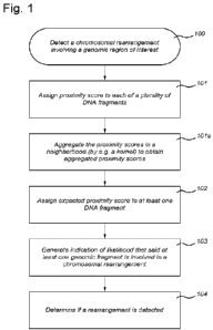
Fig. I shows a flowchart illustrating a method of detecting a chromosomal
rearrangement.
Fig. 2 shows a flowchart illustrating a method to determine a proximity score
for a
plurality of DNA fragments.
Fig. 3 shows a flowchart illustrating a method of determining an expected
proximity
score for at least one DNA fragment.
Fig. 4 shows a flowchart illustrating a method of determining a plurality of
related
proximity scores for specific genomic fragments.
Fig. 5 shows a flowchart illustrating a method of scale-invariant detection of
a
chromosomal rearrangement.
Fig. 6 shows an illustrative example of detecting a chromosomal rearrangement
using an
embodiment of FLIER. A. In a given FFPE-TLC dataset that contains mapped
fragments (i.e.
proximity-ligation products), B. FLIER initially splits the reference genome
into equally spaced
genomic intervals and then C. calculates for every interval a "proximity
frequency" that is
defined by the number of segments within that genomic interval that are
covered by at least a
fragment (or a proximity-ligation product). D. By Gaussian smoothing of
proximity frequencies
across each chromosome, E. observed "proximity scores" are calculated to
remove very local
and abrupt increase (or decrease) in proximity frequencies that are most
likely spurious. F. An
expected (or average) proximity score and a corresponding standard deviation
are estimated for
genomic intervals with similar properties (e.g. genomic intervals present on
trans chromosomes)
by in silico shuffling of observed proximity frequencies across the genome
followed by a
Gaussian smoothing across each chromosome. H. Finally, a Z-Score is calculated
for every
genomic interval using its observed proximity score and the related expected
proximity scores
and standard deviation thereof. Taken together, FLIER objectively searches for
genomic intervals
with significantly increased concentrations of captured fragments and
considers them as prime
can di dates for rearrangements.
Fig. 7 shows a block diagram of an apparatus for detecting a chromosomal
rearrangement.
Fig. 8 A shows a schematic overview of the FFPE-TLC workflow. (1) Through
sample
fixation, spatially proximal sequences (red) are preferentially crosslinked.
Next, paraffin is
removed and the sample section is permeabilized to allow enzymes to access the
DNA. (2) The
DNA is fragmented using NlaIII and then (3) ligated, which results in
concatenates of co-
CA 03174973 2022- 10-6
WO 2021/215927
PCT/NL2021/050268
12
localizing DNA fragments. (4) After crosslink reversal and DNA purification,
(5) the DNA is
subjected to next-generation sequencing library preparation. (6) Sequences of
interest are
enriched using hybrid capture probes. (7) The prepared library is paired-end
Illumina sequenced.
B. Genome -wide coverage of fragments retrieved from a typical FFPE-TLC
experiment targeting
MYC, BCL2 and BCL6. Shown in blue is the coverage seen at the (+/- 5 Mb)
genomic intervals
targeted by the capturc probes. Thc rearranged region to the MYC gene (in
green) is identified
by the concentration of fragments clustered around the GRHPR gene (chr9:31mb-
42mb), shown
in red. C. The probe sets used in FFPE-TLC not only retrieve the probe-
complementary genomic
sequences (in blue), but also mega bases of its flanking sequences (i.e. the
proximity-ligation
products), shown for MYC (pink), BCL2 (brown) and BCL6 (orange). In case of a
rearrangement
(MYC-GRHPR in this case), the corresponding capture probes also retrieve
fragments
originating from the rearrangement partner (GRHPR, in red). This is not the
case for regions that
do not harbor any rearrangement (e.g. BCL2 in brown or BCL6 in orange), as
shown for the
GRHPR locus.
Fig 9. A. Overview of structural variant identification by PLIER. B. Schematic
explanation of how butterfly plots of proximity-ligation products (green
arches on top of
chromosomes) between the target gene and the FLIER-identified rearrangement
partner can help
distinguish true target rearrangements (breakpoints 1-3, inside the probe
targeted region) from
non-target rearrangements (breakpoint 4, outside the probe targeted region).
In a reciprocal
rearrangement inside the target locus, the locus should reveal a 5' part
(section a) that
preferentially forms proximity-ligation products with one side of the partner
locus and that
separates from a 3' part (section b) that preferentially contacts and ligates
the other part of the
partner locus. If a breakpoint is present in cis outside the probe-targeted
region (breakpoint 4), a
5' (a) and 3' (b) part of the target gene cannot be distinguished. C. Three
examples of reciprocal
rearrangements uncovered by butterfly plots, involving MYC, BCL2 and BCL6,
respectively. D.
Rearrangements can be non-reciprocal, such that only one part of a target
locus fuses to a partner,
as exemplified using butterfly plots of MYC, BCL2 and BCL6. E. An example of
identified
amplification events. Such events are apparent from the elevated number of
ligation products that
are captured by all target genes (shown for MYC, BCL2 and BCL6 genes).
Fig 10. A. Circos plots showing the rearrangement partners identified in this
study, for
translocations with MYC (pink), BCL2 (brown) and BCL6 (orange). Partners found
by more
than one target gene are indicated in bold. The frequency at which a given
partner is found in our
study is indicated in parentheses. Additionally, over the circumference of
each Circos plot
(highlighted in light blue), dots indicate the target genes (i.e. MYC with
pink dots, BCL2 with
brown dots, BCL6 with orange dots) that are found to be rearranged with each
partner in our
study. B. Example of a non-reciprocal translocation event that fused the
different parts of BLC6
to different genomic partners (chr3 and chr5). C. Example of a complex, three-
way
rearrangement involving IGH, MYC, BCL2 as well as regions on chr8 and chr10,
shown in
butterfly plots as well as schematically. D. An example in which both alleles
of BCL6 are
independently involved in rearrangements. E. Overview of breakpoint positions
identified in the
MYC locus in our study. Such breakpoints are discerned in base pair resolution
by mapping
fusion-reads captured by FFPE-TLC.
Fig 11. A. Overview of PLIER identified rearrangements in diluted samples.
Green
check marks indicate successful identification of translocations by PLIER
without any false-
positive calls across the genome. Red crosses indicate failure of PLIER in
detecting the
CA 03174973 2022- 10-6
WO 2021/215927
PCT/NL2021/050268
13
rearrangement, either by missing the rearrangement or because of false-
positive calls on other
regions B. Visualization of ligation products as well as PLIER-computed
enrichment scores
across dilutions for sample F46 that harbors a BCL2-IGH rearrangement. C.
Butterfly
visualization of F16 and F221 that were negative for breaks in MYC by FISH.
FFPE-TLC
revealed that they in fact harbor a MYC rearrangement within the same
chromosome. D.
Butterfly visualization of three BCL6 rearrangements (F38, F40, F49) that were
missed by FISH.
In two instances (F38, F40), FISH failed to identify the rearrangements as the
percentages of
cells with breaks were below threshold. E. In F49, FFPE-TLC revealed that a
1.35 Mb section of
the TBL1XR1 locus was inserted into the BCL6 locus. F. BCL6 FISH image of F46
showing no
breaks at initial inspection. With hindsight, the zoomed-in view (orange
boxes) reveals some split
signals (white arrows) that indicate the existence of a translocation, as
detected by FFPE-TLC.
Fig 12. A. Comparison of FISH, Capture-NGS and FFPE-TLC results showing
rearrangements identified in MYC, BCL2 and BCL6 genes across 19 samples. Each
circle is a
sample that is analyzed for rearrangements in a particular gene. Filled-in
circles indicate
correspondence with FISH diagnosis and empty (red) circles indicate
discordance with FISH
diagnosis. B. Example of false-negative call by Capture-NGS. As the region
around the
breakpoint (red arrowhead) lacked capture probes and therefore NGS reads, the
breakpoint could
not be identified for sample F190. SV identification by FFPE-TLC and FLIER is
fusion read
independent and correctly called the translocation (z-score of 82.4). C. FFPE-
TLC capabilities
in detecting translocations even if breakpoints occur far away from the probed
regions. Each plot
demonstrates this ability for a particular gene for two samples, from left to
right: BCL2-1GH
(shown for F46 and F73). BCL6-IGL (shown for F37 and F45) and MYC-IGH (shown
for F50
and F59). The X-axis in each plot indicates the minimum distance between the
last probe and the
breakpoint position. The Y-axis shows enrichment scores that are computed by
FLIER. In all
tested cases, FLIER confidently identifies the translocation. even when the
probes are located 50
kb away from the breakpoint. D. Diagram showing the fraction of breakpoint
sequences from
this study that cannot be mapped uniquely on the reference sequence at varying
mapping lengths.
E. Example of false positive call by Capture-NGS. Breakpoint spanning reads
linking the MYC
locus to the X chromosome were found, but no translocation peak was called by
PLIER for
sample F189. PCR using primers on chrX and sequencing confirmed the
integration of a 240 bp
fragment from chr8, as shown schematically.
Fig 13. Comparison between FISH diagnoses and FFPE-TLC results. Quantitative
overview of samples with FISH diagnosis horizontally and FFPE-TLC calls (using
FLIER)
vertically. Note that 'inconclusive' FISH results refer to samples carrying an
unusual or uneven
number of FISH signals.
Fig 14. Schematic view of read structure in FFPE-TLC samples. FFPE-TLC samples
were Illumina sequenced in paired-end mode. Probed fragments (shown in light
green) may be
represented on one read-end only, or on both reads-ends. Apart from such
fragments, proximity-
ligation fragments (shown in blue) can be present. Such fragments are
recognizable through a
restriction site recognition sequence (shown as a vertical line in orange)
that links them to the
probed fragments. Proximity-ligation fragments may originate from the
surroundings of the
probed area, or from the neighborhood of the rearranged partner if a
rearrangement is present
either inside the probed area or in its vicinity. If a rearrangement is
present, FFPE-TLC reads can
also carry fragments that are produced through fusion of probed (or proximity-
ligation) fragments
to sequences from the rearranged partner (shown in red). Such reads can depict
the rearrangement
CA 03174973 2022- 10-6
WO 2021/215927
PCT/NL2021/050268
14
event in base pair resolution and therefore provide even further detail about
the occurred
structural variant.
Fig 15. Example of PLIER calls that are later identified as not relevant using
butterfly
plots. A. In sample F209 when looking from BLC 6, PLIER identified a
significant increase of
enrichment score around chrl 0:91mb near the PTEN gene (top plot). However,
when looking
from PTEN, no reciprocal peak at BCL6 was seen, but ¨4.5 Mb away from BCL6.
This
observation confirms that the rearrangement did not occur within the region of
interest (BCL6 in
this case). B. The existence of not relevant cases can be further validated in
a butterfly
visualization of the same case (i.e. F209 looking from BCL6) that is depicted
in the left most
butterfly plot. As shown, no transition (or breakpoint) of coverage can be
seen. Instead a vertical
pattern of coverage is visible. We observed two more cases with similar
characteristics. One case
was seen in F262 when looking from BCL6 and was very similar to the already
described case in
F209. The other case was in F233 and also looking from BCL6, but this time the
increased vertical
coverage was seen around chrl 0:104. All three cases were therefore considered
as not relevant
calls of PLIER.
Fig 16. Overview of breakpoints found in BCL2, BCL6 and IGH using captured
fusion-
reads in FFPE-TLC.
Fusion-reads in FFPE-TLC can map the occurred breakpoints of rearrangements at
base
pair resolution. This plot visualizes the identified breakpoints seen from
BCL2, BCL6 and IGH
MYC? locus, across all samples in our study.
Fig 17. Dilutions coverage vs. enrichment score
Fig 18. Probe details
Detailed description of embodiments
Certain exemplary embodiments will be described in greater detail hereinafter,
with
reference to the accompanying drawings. The matters disclosed in this
description and drawing,
such as detailed construction and elements, are provided to assist in a
comprehensive
understanding of the exemplary embodiments. Accordingly, it is apparent that
the exemplary
embodiments can be carried out without those specifically defined matters.
Also, well-known
operations or structures are not described in detail, since they would obscure
the description with
unnecessary detail.
Definitions
In the following description and examples, a number of terms are used. In
order to
provide a clear and consistent understanding of the specification and claims,
including the scope
to be given by such terms, the following definitions are provided. Unless
otherwise defined
herein, all technical and scientific terms used have the same meaning as
commonly understood
by one of ordinary skill in the art to which this invention belongs. The
disclosures of all
publications, patent applications, patents and other references mentioned in
this specification are
incorporated herein in their entirety by reference.
Methods of carrying out the conventional techniques that may be used in
methods of the
invention will be evident to the skilled worker. The practice of conventional
techniques in
molecular biology, biochemistry, computational chemistry, cell culture,
recombinant DNA,
bioinformatics, genomics, sequencing and related fields are well-known to
those of skill in the
art and are discussed, for example, in the following literature references:
Sambrook et al.,
CA 03174973 2022- 10-6
WO 2021/215927
PCT/NL2021/050268
Molecular Cloning. A Laboratory Manual, 2nd Edition, Cold Spring Harbor
Laboratory Press,
Cold Spring Harbor, N. Y., 1989; Ausubel et al., Current Protocols in
Molecular Biology, John
Wiley & Sons, New York, 1987 and periodic updates; and the series Methods in
Enzymology,
Academic Press, San Diego.
5 As
used herein, the singular forms "a," "an" and "the" include plural referents
unless the
context clearly dictates otherwise. For example, a method for isolating "a"
DNA molecule, as
used above, includes isolating a plurality of molecules (e.g. 10's, 100's,
1000's, 10's of thousands,
100's of thousands, millions, or more molecules).
The expression "genomic region of interest", as used herein, refers to a DNA
sequence
10 of a
chromosome of an organism of which it is desirable to assess (at least part
of) its structural
integrity. For instance, a genomic region which is suspected of comprising a
translocation
associated with a disease can be defined as a genomic region of interest. A
genomic region of
interest may be a single DNA fragment, a gene, a genomic locus containing a
gene, a part of a
chromosome, etc.
15 In
some embodiments, the genomic region of interest corresponds to a
"Topologically
associating domain" (TAD). TADs are defined by DNA-DNA interaction frequencies
and their
boundaries are regions across which relatively few DNA-DNA interactions occur.
TADs average
0.8 Mb and may contain several protein-coding genes. The TAD boundaries are
generally shared
by the different cell types of an organism and are enriched for the insulator
binding protein CTCF.
The expression of genes within a TAD is somewhat correlated, and thus some
TADs tend to have
active genes and others tend to have repressed genes (see, e.g., Dixon et al.
Nature. 2012 May
17; 485(7398): 376-380).
The term 'gene', as used herein, refers to an open reading frame and all
genetic elements
associated with this open reading frame. These genetic elements may include
introns, exons, start
codons, stop codons, 5' untranslated regions, 3' untranslated regions,
terminators, enhancer sites,
silencer sites, promoters, alternative promoters, TATA boxes and/or CAAT
boxes. In prokaryotic
contexts, 'gene' may also refer to an operon and may comprise multiple open
reading frames. In
some embodiments, the genomic region of interest refers to the sequences of a
gene starting at
the 5' untranslated region (5'UTR) and ending at the 3' UTR. Methods for
predicting open
reading frames as well the genetic elements referred to above are well-known
to a skilled person.
These methods, also referred to as structural annotation, may utilize a number
of different
databases and computer algorithms reviewed in Ejigu and Jung (Biology 2020,
9(9), 295;
https: //doi .org/10.3390/biology9090295).
The expression 'open reading frame', as used herein, refers to the genetic
elements
between and including a start codon and a stop codon.
The expression 'breakpoint cluster region', as used herein, also referred to
as 'breakpoint
clustering region', refers to a subsequence of an open reading frame or gene
from which it is
known by the person skilled in the art that chromosomal rearrangements occur
or have occurred
in a significant number of patients, organisms or specimens. As is known to a
skilled person,
some genomic regions comprise several breakpoint cluster regions which may be
further defined
as major breakpoint cluster regions and minor breakpoint cluster regions.
As used herein, the term "allele(s)" means any of one or more alternative
forms of a gene
at a particular locus. In a diploid cell of an organism, alleles of a given
gene are located at a
specific location, or locus (loci plural) on a chromosome. One allele is
present on each
CA 03174973 2022- 10-6
WO 2021/215927
PCT/NL2021/050268
16
chromosome of the pair of homologous chromosomes. Thus, in a diploid cell, two
alleles and
thus two separate (different) genomic regions of interest may exist.
The expression "nucleic acid", as used herein, may refer to any polymer or
oligomer of
pyrimidine and purine bases, preferably cytosine, thymine, and uracil, and
adenine and guanine,
respectively (See Albert L. Lehninger, Principles of Biochemistry, at 793-800,
Worth Pub. 1982).
The present invention contemplates any deoxyribonucleotide, ribonucleotide or
peptide nucleic
acid component, and any chemical variants thereof, such as methylated, hydroxy
methylated or
glycosylated forms of these bases, and the like. The polymers or oligomers may
be heterogeneous
or homogenous in composition and may be isolated from naturally occurring
sources or may be
artificially or synthetically produced. In addition, the nucleic acids may be
DNA or RNA, or a
mixture thereof, and may exist permanently or transitionally in single-
stranded or double-
stranded form, including homoduplex, heleroduplex, and hybrid slates.
The expression "sample DNA", as used herein, refers to a sample that is
obtained from
an organism or from a tissue of an organism, or from tissue and/or cell
culture, which comprises
genomic DNA. Genomic DNA encodes the genome of an organism that is the
biological
information of heredity which is passed from one generation of an organism to
the next. A sample
DNA from an organism may be obtained from any type of organism, e.g. micro-
organisms,
viruses, plants, fungi, animals, humans and bacteria, or combinations thereof.
For example, a
tissue sample from a human patient suspected of a bacterial and/or viral
infection may comprise
human cells, but also viruses and/or bacteria. The sample may comprise cells
and/or cell nuclei.
The sample DNA may be from a patient or a subject who may be at risk or
suspected of having
a particular disease, for example cancer or any other condition which warrants
the investigation
of the DNA of the organism.
The expression "crosslinking", as used herein, refers to reacting DNA at two
different
positions, such that these two different positions connect to each other as a
covalent bond between
DNA strands. Two DNA strands may be crosslinked directly using UV-irradiation,
forming
covalent bonds directly between DNA strands. The connection between the two
different
positions may be indirect, via an agent, e.g. a crosslinker molecule. A first
DNA section may be
covalently connected to a first reactive group of a crosslinker molecule
comprising two reactive
groups, that second reactive group of the crosslinker molecule may be
covalently connected to a
second DNA section, thereby crosslinking the first and second DNA section
indirectly via the
crosslinker molecule. A crosslink may also be formed indirectly between two
DNA strands via
more than one molecule. For example, a typical crosslinker molecule that may
be used is
formaldehyde. Formaldehyde induces covalent protein-protein and DNA-protein
crosslinks.
Formaldehyde thus may crosslink different DNA strands to each other via their
associated
proteins. For example, formaldehyde can react with a protein and DNA,
covalently connecting a
protein and DNA via the crosslinker molecule. Hence, two DNA sections may be
crosslinked
using formaldehyde forming a connection between a first DNA section and a
protein, the protein
may form a second connection with another fomialdehyde molecule that connects
to a second
DNA section, thus forming a crosslink which may be depicted as DNA1-
crosslinker-protein-
crosslinker-DNA2. In any case, it is understood that crosslinking according to
the invention may
comprise forming covalent connections (directly or indirectly) between strands
of DNA that are
in physical proximity of each other. DNA strands may be in physical proximity
of each other in
the cell, as DNA is highly organized, while being separated from a sequence
point of view e.g.
CA 03174973 2022- 10-6
WO 2021/215927
PCT/NL2021/050268
17
by 100kb. As long as the crosslinking method is compatible with subsequent
fragmenting and
ligation steps, such crosslinking may be contemplated.
The expression "sample of crosslinked DNA", as used herein, refers to a sample
DNA
which has been subjected to crosslinking. Crosslinking the sample DNA has the
effect that the
three-dimensional state of the genomic DNA within the sample remains largely
intact. This way,
DNA strands that arc in physical proximity of cach other remain in each
other's vicinity. A
µ`sample of crosslinked DNA" may be formalin fixed and paraffin embedded: it
may be a tissue
or tumor section or biopsy that is preserved and stored as formalin fixed
paraffin embedded
(FFPE) material. A "sample of crosslinked DNA" may be a an FFPE sample or
tumor sample as
routinely collected for pathological studies. A -sample of crosslinked DNA"
may also be
reconstituted chromatin that has been crosslinked, wherein genomic DNA that
has been isolated
from a cell (e.g. a tissue sample or a DNA sample) is subjected to chromatin
reconstitution or
otherwise packaged or coated by proteins or molecules that facilitate
crosslinking, and
subsequent crosslinking. A sample of crosslinked DNA comprises genomic DNA.
The sample
may be a derived from cells or tissue samples. In some embodiments, the
crosslinked DNA is
from crosslinked chromatin from a cell, tissue, or nuclei sample. While in a
preferred
embodiment the sample is from a human patient, DNA from other organisms may
also be used.
The expression -Reversing crosslinking", as used herein, comprises breaking
the
crosslinks such that the DNA that has been crosslinked is no longer
crosslinked and is suitable
for subsequent steps such as ligation, amplification and/or sequencing steps.
For example,
performing a protease K treatment on a sample DNA that has been crosslinked
with
formaldehyde will digest the protein present in the sample. Because the
crosslinked DNA is
connected indirectly via protein, the protease treatment in itself may reverse
the crosslinking
between the DNA. The protein fragments that remain connected to the DNA may
hamper
subsequent sequencing and/or amplification. Hence, reversing the connections
between the DNA
and the amino acids in the protein may also result in "reversing
crosslinking". The DNA-
crosslinker-protein connection may be reversed through a heating step for
example by incubating
at 70 C. As in a crosslinked DNA large amounts of protein can be present, it
is often desirable
to digest the protein with a protease in addition. Hence, any "reversing
crosslinking" method may
be contemplated wherein the DNA strands that are connected in a crosslinked
sample no longer
are connected and become suitable for sequencing and/or amplification.
The expression "Fragmenting DNA", as used herein, refers to any technique
that, when
applied to DNA (which may be crosslinked DNA or not), results in DNA
"fragments". Well
known techniques to fragment the DNA are sonication, shearing and/or enzymatic
restriction,
but other techniques can also be envisaged.
The expression -restriction endonucleasc" or -restriction enzyme'', as used
herein, may
be an enzyme that recognizes a specific nucleotide sequence (recognition site)
in a double-
stranded DNA molecule, and will cleave both strands of the DNA molecule at or
near every
recognition site, leaving a blunt or a 3'- or 5'-overhanging end. The specific
nucleotide sequence
which is recognized may determine the frequency of cleaving, e.g. a nucleotide
sequence of 6
nucleotides occurs on average every 4096 nucleotides, whereas a nucleotide
sequence of 4
nucleotides occurs much more frequently, on average every 256 nucleotides.
The expression "Ligating", as used herein, involves the concatenation of
separate DNA
fragments. The DNA fragments may be blunt ended or may have compatible
overhangs (sticky
overhangs) such that the overhangs can hybridize with each other. The ligation
of the DNA
CA 03174973 2022- 10-6
WO 2021/215927
PCT/NL2021/050268
18
fragments may be enzymatic, with a ligase enzyme (i.e. DNA ligase). However, a
non-enzymatic
ligation may also be used, as long as DNA fragments are concatenated, i.e.
forming a covalent
bond. Typically, a phosphodiester bond between the hydroxyl and phosphate
group of the
separate strands is formed.
The expression "Oligonucleotide primers" or "primers" in general, as used
herein, refer
to strands of nucleotides which can primc thc synthesis of DNA. DNA polymcrasc
cannot
synthesize DNA de novo without primers. A primer hybridizes to the DNA, i.e.
base pairs are
formed. Nucleotides that can form base pairs, that are complementary to one
another, are e.g.
cytosine and guanine, thymine and adenine, adenine and uracil, guanine and
uracil. The
complementarity between the primer and the existing DNA strand does not have
to be 100%, i.e.
not all bases of a primer need to base pair with the existing DNA strand. From
the 3'-end of a
primer hybridized with the existing DNA strand, nucleotides are incorporated
using the existing
strand as a template (template directed DNA synthesis). We may refer to the
synthetic
oligonucleotide molecules which are used in an amplification reaction as
"primers".
The expression "oligonucleotide probes- or "probes- in general, as used
herein, refers to
strands of (modified) RNA and/or (modified) DNA nucleotides, which are
complementary to
and can hybridize, pulldown and extract the sequences of a genomic region of
interest
ligated/linked to the fragments that were in proximity in the nucleus to the
sequences of a
genomic region of interest, as done for example in capture-C, promoter-capture
C, Targeted
Chromatin Capture (T2C), Tiled-C and promoter-capture Hi-C methods (Hughes et
al., 2014;
Kolovos et al., 2014; Cairns et al., 2016; Martin et al., 2015; Javicrre et
al., 2(116; Dao et al.,
2017; Choy et al., 2018; Mifsud et al., 2015; Montefiori et al., 2018; Jager
et al., 2015; Orlando
et al., 2018; Chesi et al., 2019; Oudelaar et al., 2019). Modified probes
include, e.g., xGen
Lockdown Probes (5 '-bi oti nyl ated ol i go s) .
The term "hybridization" as used herein refers to the binding of two nucleic
acid strands
through base pairing. Nucleic acid sequences such as from probes and primers
preferably have a
contiguous sequence (e.g. between 15-100 bp) that is at least 90, 95, or 100%
identical to their
target sequence. As is known to a skilled person selective or specific
hybridization is dependent
on, e.g., salt and temperature conditions. Preferably stringent hybridization
conditions are used
such that a probe or primer binds only to its target sequence.
The expression "primer-based amplification", as used herein, refers to a
polynucleotide
amplification reaction, namely, a population of polynucleotides that are
replicated from one or
more starting sequences, i.e. a primer. A suitable primer may have a sequence
length of, for
example, 15-30 nucleotides. Amplifying may refer to a variety of amplification
reactions,
including but not limited to polymerase chain reaction (PCR), linear
polymerase reactions,
nucleic acid sequence-based amplification, rolling circle amplification,
isothermal amplification,
and the like. Suitable primer-based amplification methods further include
Region-Specific
Extraction (RSE) (Dapprich et al. BMC Genomics. 2016; 17: 486), molecular
inversion probe
circularization (Porreca et al. at Methods 2007 Nov;4(11):931-6.) and loop
mediated isothermal
amplification (LAMP) (see, e.g., Notomi et al. Nucleic Acids Res 2000 Jun
15;28(12):E63)
The expression "sequencing", as used herein, refers to determining the order
of
nucleotides (base sequences) in a nucleic acid sample, e.g. DNA or RNA. Many
techniques are
available such as Sanger sequencing and "High throughput sequencing"
technologies, also
referred to in the art as next generation sequencing, such as have been
offered by Roche, Illumina
and Applied Biosystems, or also referred to in the art as third generation
sequencing, as described
CA 03174973 2022- 10-6
WO 2021/215927
PCT/NL2021/050268
19
by David J Munroe & Timothy J R Harris in Nature Biotechnology 28, 426-428
(2010) and such
as have been offered by Pacific Biosciences and Oxford Nanopore Technologies,
may also be
used. Such technologies allow from one sample DNA multiple sequence reads in a
single run.
For example, the number of sequence reads may range from several hundred up to
billions of
reads in a single run of a high throughput sequence technology. High
throughput sequencing
technologies may bc performed according to the manufacturer's instructions (as
have been
provided by e.g. Roche, Illumina or Applied Biosystems). Both long-read and
short-read
sequencing methods are contemplated herein. The technology may involve the
preparation of
DNA before carrying out a sequencing run. Such preparation may include
ligation of adaptors to
DNA. Adaptors may include identifier sequences to distinguish between samples.
Depending on
the size of DNA that is suitable or compatible with the high throughput
sequencing technology
used, the DNA that is to be sequenced may be subjected to a fragmenting step.
An "adapter" is a
short double-stranded oligonucleotide molecule with a limited number of base
pairs, e.g. about
10 to about 30 base pairs in length, which are designed such that they can be
ligated to the ends
of fragments. Adaptors are generally composed of two synthetic
oligonucleotides which have
nucleotide sequences which are partially complementary to each other. Such
adapters may be
used in combination with PCR based enrichment strategies and/or for the
sequencing of
proximity ligated molecules.
The expression "sequencing reads", as used herein, refers to the piece of DNA
that is
sequenced ("read") by a nucleic acid sequencer, such as a massively parallel
array sequencer
(e.g., Illumina or Pacific Biosciences of California). A sequencing read may
include a portion of
a genomic fragment or proximity ligated molecule. The sequencing reads may be
mapped to a
reference sequence and/or combined in silico through, for example, alignment,
to yield a
contiguous sequence. In sonic embodiments, the methods produce at least 1,000,
at least 5,000,
or at least 10,000 sequencing reads. The number of sequencing reads may refer
to the number of
sequencing reads corresponding to proximity ligated molecules comprising
sequences flanking
the 5' end of the genomic region of interest; proximity ligated molecules
comprising sequences
flanking the 3' end of the genomic region of interest; or both proximity
ligated molecules
comprising sequences flanking the 5' end and the 3' end of the genomic region
of interest. The
number of sequencing reads may also refer to proximity ligated molecules
comprising fragments
of the genomic region of interest. As is clear to a skilled person, the
mapping of such extensive
sequencing reads requires the use of computer programs, which are known in the
art.
With the terms "aligning" and "alignment", as used herein, is meant the
comparison of
two or more nucleotide sequences based on the presence of short or long
stretches of identical or
similar nucleotides. Methods and computer programs for alignment are well
known in the art.
One computer program which may be used or adapted for aligning is "Align 2",
authored by
Genentech, Inc., which was filed with user documentation in the United States
Copyright Office,
Washington, D.C. 20559, on Dec. 10, 1991.
The expression "reference genome" (also known as a reference assembly), as
used
herein, refers to a digital nucleic acid sequence database, assembled by e.g.
scientists as a
representative example of a species' set of genes. As they are often assembled
from the
sequencing of DNA from a number of donors, reference genomes do not accurately
represent the
set of genes of any single person. Instead a reference provides a haploid
mosaic of different DNA
sequences from each donor. For example, GRCh37, the Genome Reference
Consortium human
genome (build 37) is derived from thirteen anonymous volunteers from Buffalo,
New York.
CA 03174973 2022- 10-6
WO 2021/215927
PCT/NL2021/050268
Other examples of a reference genome include GRCh19 and CRCh38. As will be
appreciated by
a skilled person, a reference sequence may also be used in the methods
described herein. Suitable
reference sequences include a reference genome as well as a subset of
sequences from a reference
genome.
5 The
expression "independently ligated DNA fragment", as used herein, refers to a
DNA
fragment that is ligated to a fragmcnt originating from the gcnomic region of
interest of a given
allele of a given cell. In proximity-ligation assays an independently ligated
fragment may be PCR
amplified prior to sequencing and may therefore be sequenced multiple times.
Also, in some
proximity ligation methods the proximity ligated products obtained after
crosslinking (optional),
10
fragmentation and ligation, may be further fragmented, for example for the
purpose of efficient
PCR amplification, oligonucleotide bait capture pulldown and/or sequencing, in
which case
different parts of the same independently ligated fragment may be sequenced.
In all such
instances that an independently ligated fragment contributes multiple reads to
the sequencing
dataset, filtering may be performed to generate a dataset that most optimally
represents the
15 collection of independently ligated fragments.
The expression "chromosomal rearrangements" or "structural variation", as used
herein,
refers to the set of hereditary and somatic genetic aberrations comprising
chromosomal deletions,
chromosomal inversions, chromosomal duplications and chromosomal
translocations, wherein
chromosomal deletions and inversions occur within the same chromosome (in
cis), chromosomal
20
duplications occur within the same chromosome (in cis) or between two or more
different
chromosomes (in trans) or result in extra-chromosomal copies of a locus, and
wherein
translocations occur between two different chromosomes (in trans). Chromosomal
rearrangement
also includes rearrangements resulting from insertions of foreign DNA, such as
transgenes and
transposons. In some embodiments, the rearrangement partner is foreign DNA.
The expression 'reciprocal rearrangement', as used herein, may refer to an
exchange of
parts of nonhomologous chromosomes, wherein no genetic elements are lost and
wherein genetic
elements of one chromosome end up being fused to a second chromosome while
genetic elements
of the second chromosome end up being fused to the first chromosome, and
wherein each
chromosome involved in the rearrangement has one breakpoint per rearrangement
event.
'Reciprocal rearrangement' may alternatively refer to the product as a result
of an exchange of
parts of nonhomologous chromosomes, wherein no genetic elements are lost and
wherein genetic
elements of one chromosome end up being fused to a second chromosome while
genetic elements
of the second chromosome end up being fused to the first chromosome, and
wherein each
chromosome involved in the rearrangement has at least one breakpoint per
rearrangement event.
Reciprocal rearrangement may be the result of a natural or artificial process
and can be identified
in a matrix wherein elements of the matrix represent the proximity frequency
of a gcnomic
segment in the genomic region of interest and its rearrangement partner.
The expression 'non-reciprocal rearrangement', as used herein, refers to the
transfer of
genetic elements from one chromosome to another, nonhomologous, chromosome,
wherein no
genetic elements from the second chromosome are transferred to the first
chromosome. 'Non-
reciprocal rearrangement' may alternatively refer to the product as a result
of the transfer of
genetic elements from one chromosome to another, nonhomologous, chromosome,
wherein no
genetic elements from the second chromosome are transferred to the first
chromosome. 'Non-
reciprocal rearrangement' may also refer to the insertion of foreign DNA. Non-
reciprocal
rearrangement may be the result of a natural or artificial process and can be
identified in a matrix
CA 03174973 2022- 10-6
WO 2021/215927
PCT/NL2021/050268
21
wherein elements of the matrix represent the proximity frequency of a genomic
segment in the
genomic region of interest and its rearrangement partner.
The expression "cis-chromosome", as used herein, refers to the chromosome that
according to the reference genome contains the genomic region of interest.
Typically, in
proximity-ligation technologies, the independently ligated fragments are most
likely to come
from the cis-chromosome. In turn, the independently ligated fragments
originating from the cis-
chromosome are more likely sequences located in close linear proximity to the
genomic region
of interest than sequences that locate at larger distances from the genomic
region of interest.
The expression "trans-chromosome", as used herein, refers to any chromosome in
the
organism of interest that is not cis-chromosome.
The term 'cis-interaction', as used herein, refers to the close physical
proximity of a
genetic element originating from a cis-chromosome with respect to the target
element. The term
'trans-interaction', as used herein, refers to the close physical proximity of
a genetic element
originating from a trans-chromosome with respect to the target element.
The expressions -ligation frequency-, "linkage frequency-, "interaction
frequency-, and
"proximity frequency" of a DNA fragment, as used herein, may refer to the
number of
ligated/linked fragments of that DNA fragment and a genomic region of
interest, or, alternatively,
to the number of independently ligated/linked fragments of that DNA fragment
and the genomic
region of interest. The "ligation frequency", "linkage", "interaction
frequency", and proximity
frequency", may refer to the number of cis- and/or trans-interactions of DNA
fragments with a
given DNA segment that originate from practical or theoretical restriction
digestion of DNA, or
may alternatively refer to a value that is an indication of the number of cis-
and/or trans-
interactions of DNA fragments with a given DNA segment that originate from
practical or
theoretical restriction digestion of DNA. It may also refer to the number of
segments originating
from practical or theoretical restriction digestion of DNA, within a given
genomic interval, that
are covered by at least a ligation product, or to a value representing the
number of segments
originating from practical or theoretical restriction digestion of DNA, within
a given genomic
interval, that are covered by at least a linkage product. Typically, in
proximity-linkage/ligation
technologies, the interaction frequency from cis-interactions is higher than
the interaction
frequency from trans-interactions. The "ligation frequency", "linkage
frequency", "interaction
frequency", and "proximity frequency" may also refer to a value that is
inherently related to
either the number of ligated/linked fragments or the number of independently
ligated/linked
fragments. For example, a p-value representing the probability that the DNA
fragment is ligated
to the genomic region of interest may also be considered to be a ligation
frequency. Such a p-
value may, for example, be calculated using a binomial test. The frequency may
be a normalized
value of the number of interactions detected. Such normalization may include
normalizing for
differences between samples, including sample quality; as well as normalizing
for GC content,
mappability, and restriction site frequency.
The expression "Genomic bin" or "bin", as used herein, refers to a chromosomal
interval,
in size typically between 5kb and 1Mb and preferably between 10kb and 200kb,
that can replace
the DNA fragment as the unit to which ligation frequencies are assigned. The
assignment of a
ligation frequency to a given bin relies on an operator (summation, mean,
median, minimum,
maximum, standard deviation, triangular kernels, Gaussian kernels, half-
Gaussian kernels or any
other type of weighted and parameterized operators) that aggregates the
ligation frequency of the
DNA fragments contained within that bin.
CA 03174973 2022- 10-6
WO 2021/215927
PCT/NL2021/050268
22
The expression "Genomic neighborhood" of a fragment or of a bin, as used
herein, refers
to a defined linear chromosomal interval surrounding the given fragment or bin
in the reference
genome. The genomic neighborhood of a fragment or a bin can be between 10
kilobases and 5
mega bases and preferably is between 200 kilobases and 3 mega bases. The
genomic
neighborhood can also be defined based on the number of fragments surrounding
the fragment
or bin of interest, in which it typically spans between 50-15k fragments.
The expression "Observed aggregated ligation score", as used herein, refers to
a score
that is given to each fragment or bin according to its own ligation frequency
and the ligation
frequencies of fragments or bins residing in its genomic neighborhood.
The expression -Expected aggregated ligation score", as used herein, refers to
a dual
score (i.e. mean and standard deviation) that is given to each fragment or bin
according to a
background modelled by in silico permutation and aggregation of the ligation
frequencies from
the same experiment, to represent for each fragment or bin the most probable
observed
aggregated ligation score (mean) as well as the corresponding variation
(standard deviation).
The expressions "related fragments-, "related bins-, "comparable fragments-,
and
µ`comparable bins", as used herein, refer to fragments or bins that are
related according to certain
matching criteria. These matching criteria may be predetermined and may depend
on the
experiment at hand. For example, related fragments of a given fragment may be
fragments or
bins originating from trans chromosomes, the same trans chromosome, the cis
chromosome, or
to fragments (or bins with fragments) of similar length, or to fragments (or
bins with fragments)
of similar crosslinking efficiency, digestion efficiency, ligation efficiency,
and/or mapping
efficiency, or to fragments or bins with similar epigenetic marks, or to
fragments or bins with
similar GC content or nucleotide composition or degree of conservation, or to
fragments or bins
residing in the same spatial nuclear compartment (as determined for example by
Hi-C methods),
or combinations hereof
The expression "context-aware expected aggregated ligation score", as used
herein,
refers to an expected aggregated ligation score generated by permuting related
fragments or
related bins.
The expression "significance score", as used herein, refers to a score that
may be
calculated by comparing the observed aggregated ligation score for each
fragment or bin to either
the expected aggregated ligation score or the context-aware expected
aggregated ligation score.
The expression "nuclear proximity assay", as used herein, refers to any method
that
enables identifying the DNA fragments that in the nucleus are in proximity to
a genomic region
of interest. Examples of nuclear proximity assays are "proximity ligation
assays" and nuclear
proximity assays that do not rely on proximity ligation. Nuclear proximity may
also be referred
to as chromosomal proximity or physical proximity. In particular, proximity
refers to linear
proximity, i.e., proximity along the cis-chromosome.
The expression "proximity ligation assay", as used herein, refers to an assay
that relies
on ligation of proximal DNA fragments to identify the DNA fragments that in
the nucleus are in
proximity to a genomic region of interest. Proximity ligation assays are also
known in the field,
and may be used herein, as chromosome conformation capture assays and include
methods like
circular chromosome conformation capture or chromosome conformation capture
combined with
sequencing (4C) technology (Simonis et al., 2006; van de Werken et al., 2012)
and variants of
4C technology (e.g. UMI-4C (Schwartzman et al., 2016) and MC-4C (Allahyar et
al., 2018)), Hi-
C (Lieberman-Aiden et al., 2009), in situ Hi-C (Rao et al., 2014) and targeted
locus amplification
CA 03174973 2022- 10-6
WO 2021/215927
PCT/NL2021/050268
23
(TLA) (de Vree et al., 2014). Proximity ligation methods as referred to herein
may also include
methods that use complementary oligonucleotide probes (composed of (modified)
RNA and/or
(modified) DNA nucleotides) for hybridization, pulldown and enrichment of
sequences of a
genomic region of interest ligated to the fragments that were in proximity in
the nucleus to the
sequences of a genomic region of interest, as done for example in capture-C,
promoter-capture
C and promoter-capture Hi-C methods (Hughes et al., 2014; Cairns et al., 2016;
Martin et al.,
2015; Javierre et al., 2016; Dao et al., 2017; Choy et al., 2018; Mifsud et
al., 2015; Montefiori et
at., 2018; Jager et al., 2015; Orlando et al., 2018; Chesi et al., 2019).
Proximity ligation methods
further include methods that use immunoprecipitation, or other protein- or RNA-
directed
strategies to pulldown and enrich for sequences of interest proximity ligated
to a genomic region
of interest carrying or associated with that particular protein or RNA
molecule, such as ChIA-
PET (Li et al., 2012) and Hi-ChIP (Mumbach et al., 2017). Examples of
proximity ligated assays
and chromosome conformation methods are given in (Denker and de Laat, 2016).
Proximity
ligation assays can be performed with and without crosslinking prior to
ligation (Brant et al.,
2016).
Nuclear proximity assays (chromosomal/physical proximity assays) to identify
the DNA
fragments that in the nucleus are in proximity to a genomic region of interest
can also be
performed without relying on ligation of proximal DNA fragments to a genomic
region of
interest: an example of a nuclear proximity assay that is not dependent on
ligation but that
identifies DNA fragments that in the nucleus are in proximity to a genomic
region of interest is
SPRITE (split-pool recognition of interactions by tag extension) (Quinodoz et
al., 2018).
The term "proximity linked products" as used herein, refers to two or more
genomic
fragments, in proximity to each other, which are linked. Genomic fragments may
be linked
directly or indirectly. For example, said genomic fragments may be cross-
linked and linkage may
be determined based on, e.g., barcodes or tags (e.g., SPRITE). In addition,
said genomic
fragments may be ligated to each other (e.g., as the result of a proximity
ligation assay). Such
proximity linked products are referred to herein as proximity ligated
products. A skilled person
will appreciate that the term proximity ligated products as used herein may
also generally include
proximity linked products, unless specified otherwise.
The expression "contact profile of the genomic region of interest", as used
herein, refers
to the genomic map that visualizes the DNA fragments identified as being in
nuclear proximity
to the genomic region of interest, plotted on a reference genome.
The expression 'chromosomal breakpoint junction' and the term 'breakpoint', as
used
herein, refer to the location on a chromosome or on a chromosomal sequence,
where two parts
of a chromosome and/or DNA product have been fused together as a result of a
natural or artificial
process. Particularly relevant breakpoint junctions in the present disclosure
are those which do
not normally occur in healthy or typical patients, organisms or specimens.
The term 'matrix', as used herein, refers to a table of numbers, values or
expressions,
comprised of two axes. The numbers, values or expressions may be represented
by a variety of
elements, such as colors or grayscale tones.
The expression 'butterfly plot', as used herein, refers to a matrix that
displays the
distribution of a variable for two populations. For example, one axis of the
matrix may represent
the sequence location of the genomic region of interest and/or the region
flanking the genomic
region of interest and the other axis represent the sequence location of a
candidate rearrangement
partner.
CA 03174973 2022- 10-6
WO 2021/215927
PCT/NL2021/050268
24
Embodiments
Fig. 1 illustrates a method 100 of detecting a chromosomal rearrangement
involving a
genomic region of interest. To that end, the method 100 contains a number of
steps to analyze a
dataset of DNA reads, which may be obtained from a nuclear proximity assay,
the dataset
comprising DNA reads representing genomic fragments being in nuclear proximity
to the
genomic region of interest.
The method 100 starts in step 101 with determining a proximity score for each
of a
plurality of DNA fragments. The proximity score may represent an indication of
a likelihood that
a DNA fragment is in genomic proximity to a particular genomic region of
interest. For example,
the proximity score may be related to a collection of DNA reads of fragments
that are
ligated/linked to a particular genomic region of interest. More generally, the
reads are a plurality
of reads mapped to DNA fragments that were detected by a detection method to
be in close
proximity to the genetic region of interest. The proximity score of a DNA
fragment indicates the
likelihood of that DNA fragment to be in close proximity of region of interest
within the nucleus.
For example, the proximity score comprises a proximity frequency indicative of
a number of
reads of that DNA fragment among the reads. Alternatively, the proximity score
comprises an
indication of whether at least one read of that DNA fragment is present among
the reads. Yet
alternatively, the proximity score comprises an indication of a likelihood
that at least one read of
that DNA fragment is present among the reads. For example, the proximity
scores can be
determined by accessing a database comprising the proximity scores. Moreover,
the proximity
frequencies may be subjected to a processing step, such as binning, so that
the proximity scores
relate to bins of genomic fragments.
In aggregation step 101a, the proximity scores of step 101 may be aggregated
as another
optional step, to obtain aggregated proximity scores. For example, the
proximity scores of step
202 may be subjected to a moving average or a weighted moving average along
the genome. A
weighted moving average may be implemented by convoluting the proximity scores
of a genome
with a suitable kernel, such as a Gaussian kernel (e.g. sampled Gaussian
kernel or discrete
Gaussian kernel). This is also called a sliding window approach, which may
alternatively involve,
for example, sliding Gaussian windows or kernels, half-Gaussian windows or
kernels, triangular
windows or kernels, rectangular windows or kernels, or other kinds of windows
or kernels. The
result of the aggregation step 101a may be used as the proximity score of the
DNA fragments in
step 103. In case the aggregation step 101a is omitted, the proximity score of
step 202 may be
used, for example.
In step 102, an expected proximity score for at least one DNA fragment is
determined.
This expected proximity score inay be calculated based on the observed
proximity scores of the
other DNA fragments in the database. For example, an average and standard
deviation of all the
DNA fragments in the database relating to a particular experiment and/or
chromosome may be
computed to determine the expected proximity score. Alternatively, a random
selection of DNA
fragments may be averaged. Yet alternatively, a set of related DNA fragments
may be
determined, and the proximity scores of only those related fragments may be
averaged. The
related fragments may be selected based, for example, on their proximity to
the genomic region
of interest, or on other similarity criteria. Examples of such similarity
criteria are disclosed
elsewhere in this description.
In step 103, the proximity score of at least one DNA fragment determined in
step 101 is
compared with the expected proximity score for that at least one DNA fragment.
For example,
CA 03174973 2022- 10-6
WO 2021/215927
PCT/NL2021/050268
the proximity score of the DNA fragment is compared with the expected
proximity score
determined in step 102. This results in an indication of a likelihood that the
at least one DNA
fragment is involved in a chromosomal rearrangement. This indication may be in
form of a
significance score, for example. In certain implementations, a standard
deviation determined in
5 step 102 may be involved in the comparison to determine a statistical
significance of any
deviation of the observed proximity score versus the expected proximity score.
In ease a
significant deviation is found, a chromosomal rearrangement may be considered
to have been
detected. The statistical significance may be expressed as a significance
score. It will be
understood that this significance score may be calculated for each genomic
fragment for which
10 both the observed proximity score and the expected proximity score are
available.
In step 104, it is decided if a rearrangement is detected. This may be a
Boolean decision,
i.e., the available significance scores may be evaluated to come at a yes/no
decision for each
genomic fragment, or the decision may be a soft decision that includes a
probability or a
likelihood, or a certainty that the genomic fragment is involved in a
rearrangement with the
15 genomic region of interest. This decision may be based on the
significance score computed in
step 103. In certain embodiments, the significance score of step 103 is equal
to the soft decision
output in step 104.
However, in certain other embodiments, more input variables are taken into
account in
making the decision, to generate an enhanced significance score indicative of
a possible
20 rearrangement. For example, the density of non-mappable experimentally
created fragments in
the genomic neighbourhood of a mapped target-proximity ligated/linked fragment
may be
determined. The decision in step 104 may be further based on this density,
wherein preferably
the enhanced significance score scales positively with the density of non-
mappable
experimentally created fragments in the genomic neighbourhood of the mapped
target-proximity
25 ligated/linked fragment. Moreover, the density of mappable
experimentally created fragments in
the genomic neighbourhood of a mapped target-proximity ligated/linked fragment
may be
determined. The decision in step 104 may be further based on this density,
wherein preferably
the enhanced significance score scales negatively with the expected aggregated
proximity score
of the given fragment.
After it has been detected at step 104 that there may be a genomic
rearrangement
involving the particular genomic region of interest and another particular
genomic fragment, then
the presence of this rearrangement may, optionally, be further verified by
performing the whole
procedure 100 from the start, using the other particular genomic fragment as -
the particular
genomic region of interest". If that procedure confirms the genomic
rearrangement, it is even
II1Ole certain that the rearrangement is real.
Fig. 2 illustrates a possible method to determine the proximity scores of a
plurality of
DNA fragments, as performed in step 101 of method 100.
In step 201, a proximity frequency is determined for each of a plurality of
DNA
fragments. Preferably, a large number of consecutive DNA fragments in the
genome is used for
this, to facilitate aggregation later on. For example, the proximity frequency
of a DNA fragment
may be the number of reads of that DNA fragment. Depending on the assay it may
be preferable
to perform a binarization of the proximity frequency, for example by setting
the proximity
frequency to 1 if the DNA fragment is found among the reads and 0 if the DNA
fragment is not
found among the reads.
CA 03174973 2022- 10-6
WO 2021/215927
PCT/NL2021/050268
26
In step 202, the proximity frequencies of step 201 may be combined as an
optional step
to generate proximity scores. If step 202 is not performed, the proximity
frequencies themselves
may be the proximity scores, for example. Step 202 may involve, for example,
binning of the
proximity frequencies of step 201. For example, bins of a number of
consecutive bases each may
be defined, and the proximity frequencies may be combined within each bin. The
bin size may
be chosen, for example, between 5 kilobases and 1 mega base, preferably
between 10 kilobases
and 200 kilobases. The bins may, for example, have a size of 25 kilobases,
although any suitable
size of bins may be chosen. The proximity frequencies within each bin may be
combined by
summing or averaging them, for example. Alternatively, a binomial test may be
performed
resulting in, for example, a likelihood that the genomic fragments within the
bin occur among
the reads in the database. Such a binomial test may be particularly suitable
in case of binarized
proximity frequencies. After binning, the resulting proximity score may be
said to be relating to
a larger genomic fragment covering the genomic fragments included in the bin.
It will be understood that in certain embodiments, only one aggregation step
may be
performed (i.e., either the step 202 or the aggregation step 101a, possibly in
conjunction with
step 402) or no aggregation step at all. However, it may be advantageous to
include both
aggregation steps. Moreover, in an alternative implementation it is possible
to use a kernel filter
for the step 202 and binning for the aggregation step 101a.
Fig. 3 illustrates an embodiment of a method implementing step 102 of
determining an
expected proximity score for at least one DNA fragment. For example, the
analysis may be
limited to one DNA fragment, or to a certain region within the genome, or to
an entire
chromosome. Alternatively, the analysis may be performed for the entire
genome.
In step 303, a plurality of related proximity scores is generated for each
genomic
fragment that is to be analyzed. The proximity scores may be the scores
resulting from step 101.
In this respect, it is noted that a genomic fragment may be considered to be a
-bin" of genomic
fragments, in case binning is performed in the combining step 202.
In this disclosure, related proximity scores may be proximity scores of
genomic
fragments that are related to the genomic fragment for which the expected
proximity score is
being determined. In this regard, genomic fragments may be related to each
other when they
satisfy certain matching criteria. For example, fragments on the same
chromosome may be
considered to be related to each other, or fragments within a certain distance
on the genome, or
fragments known to contribute to a certain function or protein, or fragments
that are otherwise
comparable. Other matching criteria are disclosed elsewhere in this
description. In certain
implementations, all the genomic fragments obtained in an experiment are set
to be related
fragments.
The plurality of related proximity scores may comprise all the proximity
scores of the
related genomic fragments. Alternatively, for computational efficiency, the
collection of related
proximity scores may be built up of a random selection of the available
related proximity scores.
For example, the proximity scores of 1000 (or any other predetermined number
of) randomly
selected related genomic fragments may be collected.
In step 304, the plurality of related proximity scores are subjected to
statistical
calculations, so that for example an average and standard deviation are
computed as an expected
proximity score. Alternatively, for example the median of the related
proximity scores may be
determined instead of the average, or the variance may be determined instead
of the standard
CA 03174973 2022- 10-6
WO 2021/215927
PCT/NL2021/050268
27
deviation. Other statistical methods may be used to calculate an expected
proximity score or for
example parameters of a probability density function for the proximity score.
This expected proximity score may be calculated for each genomic fragment, as
desired.
Fig. 4 illustrates an embodiment of a method implementing step 303 of
determining a
plurality of related proximity scores corresponding to the plurality of
related DNA fragments. As
observed hereinabove in respect of step 303, the proximity scores determined
in step 101 may be
used as the starting point for this method.
In step 401, the observed proximity scores of related genomic fragments are
permuted.
As described above, genomic fragments may be considered to be "related" with
each other when
they satisfy certain matching criteria. Therefore, in this step, the proximity
score of a first
fragment may be swapped with the proximity score of a second fragment that is
related to the
first fragment according to the matching criteria. Each of the proximity
scores may thus be
swapped with another proximity score. The particular genomic fragments that
are swapped may
be selected randomly. To create a random permutation, each genomic fragment
may be swapped
with another randomly chosen related genomic fragment. Alternatively, any
number (for
example, a fixed number) of swaps between randomly chosen pairs of related
genomic fragments
may be performed. This step provides permuted proximity scores.
In step 402, the permuted proximity scores of step 401 may be aggregated.
Preferably,
this aggregation step involves the same operations as the aggregation step
101a that is performed
on the observed proximity scores. That way, it is easy to compare the
aggregated observed
proximity scores to the expected aggregated proximity scores. For example, as
discussed above
at step 101a, a moving average or a discrete Gaussian kernel may be applied.
This step provides
aggregated permuted proximity scores.
In step 403, the aggregated permuted proximity scores of step 402 may be
collected in a
collection associated with a specific DNA fragment, so that later on the
expected proximity score
may be calculated in step 304. Alternatively, certain statistics corresponding
to the specific DNA
fragment may be updated based on the aggregated permuted proximity scores of
step 402. As
illustrated at steps 404 and 405, the aggregated permuted proximity scores of
any desired
genomic fragments may be collected. This way, the genomic rearrangements /
discontinuities
may be detected for any number of genomic fragments. In many cases, it may be
most useful to
collect the aggregated permuted proximity scores of all the genomic fragments
on the genome
under study.
In step 406, it is decided whether the collection(s) of aggregated permuted
proximity
scores are sufficiently large. This step may be implemented by an iteration
counter, for example.
This step may ensure that the expected proximity score will have sufficient
statistical relevance.
For example, a predetermined number of permutations may be performed; such as
1000
permutations or 100.000 permutations.
If more permutations are needed to enlarge the collections of permuted
proximity scores
up to the desired number of permutations, in step 406, the process continues
from step 401.
Otherwise, the collections of related proximity scores are complete at step
407.
It will be understood that in certain embodiments, it is not necessary to
store the actual
values of the permuted proximity scores in a collection. Instead, it is
possible to combine steps
403 and 304 in one step, by updating certain parameters. For example, if only
the mean it and
standard deviation a of the estimated proximity score are desired, it is
sufficient to update the
sum of the permuted proximity scores E xi and the sum of squares of permuted
proximity scores
CA 03174973 2022- 10-6
WO 2021/215927
PCT/NL2021/050268
28
E oci2, and the number n of permuted proximity scores. After updating these
parameters in step
403, the actual values oci of the permuted proximity scores may be discarded.
The mean may be
calculated afterwards in step 304 as:
E Xi
p=
and the standard deviation may be calculated as
= jE xi2 (E xi)2.
n )
In certain embodiments, the aggregation steps may implement a length scale.
For
example, the second aggregation step 101a of the observed proximity scores and
the aggregation
step 402 of the permuted proximity scores may be used to compare the observed
proximity scores
to the expected proximity scores at a certain scale. The scale may be
considered, for example,
the standard deviation of a Gaussian kernel filter, when the aggregation step
is implemented by
means of a Gaussian filter. Other kinds of filters may have a similar notion
of a scale. For
example, the window size of a sliding window approach may vary according to
the scale. The
whole procedure of Figs. Ito 4 may be performed a number of times, using
different scales. This
may lead to different significance findings for different scales. The results
for different scales
may be combined to obtain a scale-invariant result. For example, the maximum,
minimum, or
mean of the significance scores obtained from the different scales is used as
the final, scale-
invariant significance score. Similarly, in certain embodiments, the first
aggregation step 202
may be performed at different scales. For example, in case of binning,
different bin sizes may be
used.
In certain embodiments, the step 101a of aggregating the observed proximity
scores in a
neighborhood to obtain aggregated proximity scores, and the step 402, of
aggregating the
permutation of proximity scores, may be performed by processing each DNA
fragment as
follows. A plurality of neighbor DNA fragments of the DNA fragment is
identified. The
(observed or permuted) proximity scores of the DNA fragment and the neighbor
DNA fragments
are selected. The selected proximity scores are combined using an aggregator
operator such as a
moving average, for example a weighted moving average, for example a Gaussian
weighted
moving average, or another type of operator along the genome, to produce the
aggregated
proximity score for the DNA fragment. In certain embodiments, the neighbor DNA
fragments
may be identified as follows. A distance measure may be chosen to identify
neighbor DNA
fragments. A first example of a distance measure is a genomic distance. In
that case, DNA
fragments are selected that are close in terms of genomic length scales, that
is, all the fragments
less than a certain number of bases (e.g. 200 kilobases or 750 kilobases) away
from the DNA
fragment may be the neighbor DNA fragments. A second example of a distance
measure is the
number of DNA fragments along the genome. In that case, the K closest DNA
fragments to the
DNA fragment may be the neighbor DNA fragments. For example, K = 31 or K=51.
Fig. 5 shows a flowchart of such a scale-invariant detection method of a
chromosomal
rearrangement involving a genomic region of interest. In Fig. 5, the steps
that are similar to the
steps of Fig. 1 have been given the same reference numeral as in Fig. 1,
provided with an
apostrophe. The scale-invariant detection method contains an iteration 502 to
determine the
significance score in step 103' at different scales, wherein the scale is set
in each iteration in step
CA 03174973 2022- 10-6
WO 2021/215927
PCT/NL2021/050268
29
501. The final determination of a rearrangement can be made in step 104' using
the significance
scores given for the respective scales.
In greater detail, the method starts at step 101 with assigning a proximity
score to each
of a plurality of DNA fragments in the database with e.g. reads generated by
an assay. This step
can be identical to step 101 of Fig. 1. An example implementation is shown in
Fig. 2.
Next, in stcp 501, a scale is set. For example, the scale may be expressed as
a number of
bases. However, this is not a limitation. The scale may be a parameter of an
aggregation function
that aggregates proximity scores of DNA fragments in a genomic neighborhood.
The width of
the neighborhood may be determined by the scale. In case the aggregation
function is a Gaussian
kernel, the scale may be the standard deviation of the Gaussian function used
for the Gaussian
kernel. The tails of the Gaussian kernel may be optionally cut off at a
suitable point. In case the
aggregation function is a sliding window, the scale may be the window width of
the sliding
window. For example, a predetermined set of scales may be selected for the
analysis, one scale
in each iteration 502. The set of scales can have any number of scales. An
example of a set of
scales to be used (as e.g. standard deviation or window width) is: 1 1
kilobase, 1 megabase, 1000
megabases 1.
In step 101a', using the selected scale, the proximity scores are aggregated,
using the
selected scale as set forth hereinabove. This way, the aggregated proximity
scores are obtained.
A suitable process for this aggregation step is outlined hereinabove in
respect of step 101a.
In step 102', the expected proximity score for at least one DNA fragment is
determined,
based on the selected scale. The expected proximity score is assigned to said
at least one DNA
fragment. The expected proximity score may be assigned to one DNA fragment,
for a particular
subset of DNA fragments, such as a genomic region, or to the DNA fragments of
a whole
chromosome or a whole genome. The method to compute the expected proximity
score may be
implemented, for example, as disclosed hereinabove with reference to Fig. 3
and Fig. 4. In step
402, the permutation of proximity scores may be aggregated using the selected
scale. For
example, the same aggregation algorithm and aggregation parameters may be used
as in step
101a'.
In step 103', the indication of the likelihood that said at least one genomic
fragment is
involved in a chromosomal rearrangement, for example a significance score, is
determined using
the aggregated proximity scores according to the scale of step 101a' and the
expected proximity
score according to the scale of step 102'. This way, for each selected scale,
a different indication
of the likelihood of a chromosomal rearrangement may be obtained.
In step 502, it is verified if all desired scales have been applied. If the
calculations are
desired for 11101C scales, the process is repeated from step 501, wherein
another scale is selected.
For example, this process is iterated until all scales of the predetermined
set of scales have been
selected.
If the process has been performed for all desired scales, the process proceeds
in step 104'
to determine if a rearrangement is detected, based on the indications
(significance scores)
determined in step 103' for all the selected scales. The indications
(significance scores) for the
different scales can be combined in one of many possible ways, for example the
maximum value,
mean value, median value, or minimum value of the available significance
scores for the at least
one DNA fragment may be determined. A threshold may thereafter be optionally
be applied to
arrive at a binary determination. After that, the process terminates.
CA 03174973 2022- 10-6
WO 2021/215927
PCT/NL2021/050268
It will be understood that the method described hereinabove with reference to
Figs. 1 to
5 may be implemented as a computer program or as a suitably programmed
computer system.
The dataset created by means of the proximity assay may serve as the input of
such a computer
program, and the output may be an indication of a detected rearrangement.
5
Throughout this disclosure, it may be understood that a ligation frequency is
an example
of the proximity frequency, and ligation score is an example of proximity
score. Although several
techniques are illustrated and explained throughout this document using the
ligation frequency
and ligation score as an example, it will be understood that in general the
techniques disclosed
herein may be carried out using any proximity frequency and/or proximity
score. For example, a
10
nuclear proximity assay may be used that does not rely on "proximity
ligation", such as the
SPRITE method, to identify DNA fragments proximal to a genomic region of
interest. Therefore,
throughout this disclosure, the terms ligation and proximity may be used
interchangeably.
Specifically, the terms ligation frequency and proximity frequency may be used
interchangeably.
Similarly, the terms ligation score and proximity score may be used
interchangeably.
15 Fig.
6 shows an illustrative example of applying the method set forth herein. As an
example, the proximity frequencies can be obtained as a 4C profile or another
assay technique.
Such an assay may result in a proximity ligation dataset. Fig. 6 shows a graph
600 of the observed
proximity frequency (vertical axis) of DNA fragments along a chromosome (shown
partially on
the horizontal axis). A detail of the graph 600, covering a small portion of
the chromosome, is
20 shown
in graph 601. The profile is binned using bins having a width of for example
25 kilobases,
to obtain a score profile of observed proximity scores. A detail of the score
profile is shown in
graph 602, and the full score profile is shown in graph 603. The score profile
603 is aggregated
using, in this example, a Gaussian kernel 605 to obtain an aggregated, or
smoothed, score profile
of observed aggregated proximity scores, shown in graph 606. The score profile
603 is permuted
25 to
obtain a randomly permuted profile 604, which is also smoothed using the
Gaussian kernel
605. The permuting and smoothing are repeated for N times, wherein N is an
integer, for example
1000. From all these permuted smoothed profiles, an expected profile of
expected aggregated
proximity scores is derived, as shown in graph 607. The smoothed profile 606
is compared with
the expected profile 607, for example by subtraction (or e.g. by squared
difference), to obtain a
30
difference profile, shown in graph 608. A significance threshold 609 is also
derived from the
permuted smoothed profiles and/or the expected profile. Alternatively, the
significance threshold
609 may be set to a configurable value. At a fragment where the comparison
profile 608 exceeds
the significance threshold 609, as indicated at fragment 610, an indication of
a possible
rearrangement may be triggered.
Fig. 7 shows a block diagram of an apparatus for detecting a chromosomal
rearrangement. The apparatus may be implemented as a computer system that is
configured to
perform any method disclosed herein. For example, the steps after having
obtained the DNA
reads may be performed by the apparatus 700. In particular, the computational
steps necessary to
detect a chromosomal rearrangement may be performed by the apparatus. For
example, the
apparatus 700 may comprise a processor 701 that can execute instructions. The
processor 701
may comprise a plurality of (sub-)processors that are configured to work
cooperatively. The
apparatus 700 may further comprise a memory 702, which can be any data storage
means, such
as a flash memory or a random-access memory, or both. The memory 702 can
comprise a non-
transitory computer readable media. The memory 702 can store instructions
causing the
processor 701, when executing the instructions, to perform a method set forth
herein. These
CA 03174973 2022- 10-6
WO 2021/215927
PCT/NL2021/050268
31
instructions may collectively form a computer program. The computer program
can alternatively
be stored on a separate non-transitory computer readable media, such as an
optical disc. Further,
the memory 702 may be configured to store data relating to an assay, for
example a database with
DNA reads. The data, such as DNA reads, may be received via a transceiver 703,
which may be
for example a universal serial bus (USB) or a wireless communication device.
Also, the result of
the method, for example significance scores indicative of any rearrangement,
may be output
through the transceiver 703. Peripheral devices may be connected by means of
the transceiver
703. Optionally, the apparatus 700 comprises user interface components (not
illustrated), such as
a display and/or a user input device such as mouse, keyboard, or touch panel.
Such user interface
components may alternatively be connected via the transceiver 703. Moreover,
such user
interface components may be used to control the operation of the apparatus
and/or output a result
of the calculations. The transceiver 703 can also communicate with an external
memory, for
example. Finally, the apparatus 700 may alternatively be implemented as a
distributed computer
system that performs a part of the computations or data storage on a cloud
server and another
part on a client device.
In certain embodiments, nuclear proximity assays known as proximity ligation
assays
may be used. Moreover, technical and biological biases and variation within
and between
samples of (crosslinked) DNA may be taken into account to computationally
identify structural
variation occurring in a genomic region of interest.
In certain embodiments, a method of identifying structural variation occurring
in a
genomic region of interest may comprise the steps of:
- Performing a proximity ligation assay to produce a dataset of
independently ligated
fragments that are in nuclear proximity to a genomic region of interest.
- Using the dataset to assign an observed aggregated ligation score to each
fragment.
- Using the same dataset to compute a context-aware expected aggregated
ligation score
for each fragment.
- Comparing across different chromosomal length scales the fragment's
observed vs.
context-aware expected aggregated ligation score and identify per chromosomal
length-
scale fragments with significantly increased aggregated ligation scores
compared to the
context-aware expected aggregated ligation score.
In certain embodiments, use is made of a nuclear proximity assay that does not
rely on
"proximity ligation", such as the 'SPRITE' method, to identify DNA fragments
proximal to a
genomic region of interest and takes into account technical and biological
biases and variation
within and between samples of (crosslinked) DNA to computationally identify
structural
variation occurred in a genomic region of interest, comprising the steps of:
- Performing a nuclear proximity assay to produce a dataset of DNA
fragments that are
in nuclear proximity to a genomic region of interest.
- Using the dataset to assign an observed aggregated proximity score to
each fragment.
- Using the same dataset to compute a context-aware expected aggregated
proximity
score for each fragment.
- Comparing across different chromosomal length scales the fragment's
observed vs.
context-aware expected aggregated proximity score and identify per chromosomal
length-scale fragments with significantly increased aggregated proximity
scores.
The techniques disclosed herein are based on the realization that it is
desirable to detect
chromosomal rearrangements more accurately. This is mainly because in
comparison of two
CA 03174973 2022- 10-6
WO 2021/215927
PCT/NL2021/050268
32
given samples (e.g. a diseased and a healthy cell) many differences between
proximity-ligation
products can be detected that are not instigated by actual structural
variations. Furthermore, many
transitions from low to high interaction frequencies that can be seen in any
proximity-ligation
dataset are not caused by structural variations. It is therefore an aspect of
the present invention
to remedy these shortcomings, to identify genomic structural variations in the
genome while
accounting for intrinsic technical biascs observed in the same dataset
Translocations (chromosomal rearrangements) underlie different forms of cancer
(Schram et al., 2017). They may result in the ovierexpression of oncogenes or
the production of
fusion proteins having dysregulated expression or kinase activity. The
molecular typing of
translocations is routinely performed in the clinic for diagnosis (tumor
classification), prognosis,
and increasingly also for treatment decisions. For example, non-small cell
lung carcinoma's
(NSCLC) harboring a translocation in the protein kinase genes ALK and ROS1 are
targetable by
FDA-approved protein kinase inhibitors (Kwak et al., 2010; Shaw et al., 2014),
while potent
inhibitors of RET are promising precision medicine drugs for patients with RET
translocations
(Plenker et al., 2017). Molecular typing of NSCLC tumors (Pisapia et al.,
2017) is therefore
highly useful to select the optimal treatment and obligatory for stage IV
(metastatic) lung cancers
in the Netherlands (1000s per year). Translocation analysis is also performed,
among others, for
the ¨1500 patients that are annually diagnosed with diffuse large B-cell
lymphoma (DLBCL) and
many of the annual ¨700 patients haying various forms of sarcomas in the
Netherlands.
Already for decades, routine clinical procedure is to store surgically removed
tumor
biopsies as formalin fixed paraffin embedded (FFPE) specimens. However, DNA or
RNA
rearrangement detection in FFPE samples is compromised due to the fact that
DNA and RNA is
crosslinked and fragmented. RNA and DNA-based PCR strategies for rearrangement
detection
exist but are complicated. First, the breakpoint positions and rearrangement
partners of
recurrently rearranged genes often differ between patients, which makes it
difficult to design
PCR primer sets that detect all possible rearrangements. Novel fusion partners
are often missed,
in which case a conclusive remark regarding rearrangements cannot be formed
when a negative
result is obtained. Some RNA-based PCR strategies like Archer FusionPlex are
agnostic for the
rearrangement partner, but again not finding a rearrangement in a
heterogeneous tumor biopsy
does not rule out its presence. Also, there may be too little RNA or the RNA
in FFPE samples
can be of too low quality for subsequent analysis of cDNA PCR products.
Finally, the so-called
position effect rearrangements, that do not create fusions but cause the
upregulation of otherwise
unaltered oncogenes, are per definition undetectable at the RNA level.
For these reasons, fluorescence in situ hybridization (FISH) is often still
the preferred
diagnostic method for detecting fusions in FFPE biopsies. FISH however is
labor intensive, only
partially informative and not always conclusive. Each gene needs to be tested
separately in an
independent FISH experiment. If the gene of interest promiscuously rearranges
with different
chromosomal partners, which may be often the case, break-apart FISH (or split-
FISH) is used.
Split-FISH entails the hybridization of differently colored probes on each
side of the target gene:
if they break-apart (splif ), i.e. if they are separated over a larger than
expected distance in a
given number of cells, the gene is considered to be involved in a
translocation, but the
rearrangement partner remains obscure. Furthermore, depending on the sample
quality and tumor
load, FISH may give unclear result. A robust, single, all-in-one, assay that
can simultaneously
detect rearrangements in all genes of interest, irrespective of their
breakpoint location and their
CA 03174973 2022- 10-6
WO 2021/215927
PCT/NL2021/050268
33
translocation partner, is therefore highly desired. Such an assay may be made
possible using the
rearrangement detection methods disclosed herein.
Methodology for rearrangement detection in DNA samples or crosslinked DNA
samples
would preferably meet any one or more, ideally all, of the following criteria:
(1) an all-in-one method that enables simultaneous monitoring for
rearrangements in all
genes relevant for thc given disease,
(2) a method that is agnostic for the exact breakpoint position and the
rearrangement
partner and hence able to find known and new translocations partners,
(3) a method that is sufficiently sensitive to also pick up rearrangements in
small (for
example, less than 5%) subpopulations of cells, and
(4) a method that provides unbiased detection of rearrangements.
Nuclear proximity assays, such as proximity ligation assays, may be able to
meet the first
three criteria, as was first illustrated by 4C technology. 4C technology was
originally developed
by the inventors to study the three-dimensional folding of the genome (Simonis
et al., 2006). The
method is a variant of 3C technology (Dekker et al, 2002) and allows unbiased
genome-wide
mapping of all chromosomal segments that are in close proximity to a selected
genomic site of
interest (the 'viewpoint sequence'). The technique involves formaldehyde-
mediated fixation of
cells, which results in crosslinks between physically proximal DNA sequences
inside each cell
nucleus. Crosslinked DNA is subsequently digested with a restriction enzyme
and re-ligated
under conditions that favor proximity ligation between crosslinked DNA
fragments. Hence, 3C
strategies create ligation products between DNA sequences that originally were
close together in
the nuclear space. In 4C technology, inverse PCR with viewpoint-specific
primers is performed
on circular ligation products, which results in the amplification of its
captured ligation partners;
these may subsequently be Illumina sequenced and mapped to the genome to
uncover the
viewpoint's contact profile.
As expected from polymer physics, irrespective of the 3D conformation, the
great
majority of 4C captured fragments always originates from the sequences that
immediately
neighbor the viewpoint on the linear chromosome template. Based on this
realization the
inventors hypothesized and demonstrated in the past that 4C technology is
highly suitable for the
detection of chromosomal rearrangements, including translocations, as such
chromosomal
aberrations disturb the linear chromosome scaffold (Simonis et al., 2009;
Homminga et al., 2011).
Thus, when a 4C viewpoint is in the vicinity of a rearrangement breakpoint, it
will identify the
rearrangement and the rearrangement partner based on an altered contact
profile of the genomic
region of interest (Simonis et al., 2009). The sensitivity of the assay (i.e.
its ability to detect
translocations also in small subpopulations of cells) increases though when
viewpoint and
breakpoint arc closer together: with viewpoints within 100 kb from the
breakpoint, translocations
may be readily found even if they are present in less than 5% of the cells
(Simonis et al., Nat
Methods 2009, and unpublished data). The latter is critical for oncogenetic
diagnostics as cancer
biopsies are often mixtures of healthy and different clonal cancer cell
populations. In summary,
4C offers a sensitive method to investigate whether a candidate gene (e.g. a
gene that one would
want to monitor for rearrangements in the clinic) is involved in a
rearrangement and to identify
its rearrangement partner. A further advantage of 4C, as published (Simonis et
al., 2009), is that
the 4C PCR reaction can easily be multiplexed, implying that the assay can
simultaneously
monitor multiple genes for rearrangements in each patient sample.
CA 03174973 2022- 10-6
WO 2021/215927
PCT/NL2021/050268
34
Besides 4C technology, we now know that there are many other proximity
ligation
methods that based on the same principles can also identify chromosomal
rearrangements with a
genomic region of interest. Examples of such methodologies are targeted locus
amplification
(TLA), capture-C or capture-HiC methods, Hi-C and in situ Hi-C. ChIA-PET and
Hi-ChIP. In
principle, all methods that perform proximity ligation to identify the DNA
fragments that are in
proximity in the nucleus to a gcnomic region of interest, enable thc detection
of chromosomal
rearrangements and translocations.
Proximity ligation methods can be used to identify chromosomal rearrangements.
State
of the art methods aiming to identify structural variations based on proximity
ligation methods
often rely on visual inspection of the contact profile of the genomic region
of interest, to find
elsewhere on the genome clustering (or absence of clustering) of DNA fragments
proximity
ligated to the genomic region of interest in a lest sample (e.g. a sample from
a patient with a
disease) that is clearly different from the clustering of proximity ligated
DNA fragments seen at
that same genomic locus in a control sample (e.g. a sample from a healthy
individual). Examples
of translocations and other chromosomal rearrangements found upon such visual
inspection of
the contact profile of the genomic region of interest are given in (Simonis et
al., 2009; de Vree
et al., 2014; Harewood et al. 2017 and W02008084405). In other current
experimental designs,
nuclear proximity dataset obtained in the test sample produced from disease
(e.g. cancer) cells
are computationally compared to control nuclear proximity datasets produced
from normal
(healthy) cells to identify abnormal genomic distributions of nuclear
proximity DNA fragments
indicative of chromosomal rearrangements (Diaz et al. 2018). Dixon et al. 2018
utilizes an
extensive control dataset by combining nuclear proximity datasets produced
from nine
karyotypically normal cell lines to estimate expected inter-chromosomal
interaction frequencies
that accounts for elevated interactions of fragments originating from
chromosome-ends or small
chromosomes. The disadvantage of such a test sample versus control sample
correction approach
is that it cannot account for sample-specific biases that can easily occur in
nuclear-proximity
assays such as proximity-ligation assays. For example, the purity, the
crosslink-ability, the
fragmentation efficiency and (in proximity ligation assays) the ligation
efficiency of the sample
under study can have a substantial impact on how well the fragments located in
the 3D proximity
of the genomic region of interest are represented in the produced nuclear
proximity dataset.
Therefore, correcting these hidden experiment-specific biases is a major
obstacle in utilizing
nuclear proximity technologies for appraising the structural integrity of
susceptible loci, and
hence for using these methodologies for clinical applications.
Hence, the current inventors devised strategies for identification of
structural variations
in the region of interest by accounting for dataset-specific technical as well
as experimental
biases. These strategics may include building a background model that is
computed from the
proximity-ligation dataset under investigation (for example, from the test
sample obtained from
a tumor of a patient) and then utilizing the background model to assess
significance of clustering
of ligated DNA fragments across the genome of that same test sample. In this
data-intrinsic
analysis procedure, it may be unnecessary to use a control sample dataset.
The inventors have realized, that fragments that are involved in a structural
variation
(such as chromosomal rearrangement or translocation) with the region of
interest will show
higher numbers of independently ligated DNA fragments than would be expected
by chance.
CA 03174973 2022- 10-6
WO 2021/215927
PCT/NL2021/050268
Based on the above premise, the involvement of a genomic region of interest in
a
chromosomal rearrangement may be assessed by means of the method, apparatus,
and computer
program techniques disclosed herein.
In certain embodiments the involvement of a genomic region of interest in a
5 chromosomal rearrangement may be assessed by:
a. Performing a proximity ligation assay that creates a dataset of
independently ligated
DNA fragments with a genomic region of interest (also referred to herein as
proximity ligated/linked products).
b. Aggregating, for example by summation, the ligation frequency in the
genomic
10
neighborhood of each fragment, to assign an "observed aggregated ligation
score" to
each fragment.
c. Permuting (swapping) the ligation frequency of each DNA fragment
(including the
DNA fragments with observed ligation frequency equal to zero) by another
randomly
chosen DNA fragment.
15 d.
Aggregating the permuted ligation frequency of each fragment and its neighbor
fragments to compute a randomized aggregated ligation score for each fragment.
e. Repeating step c-d many times (typically n=1000) to form an "expected
aggregated
ligation score" for each fragment in the dataset.
f. Optionally, set the observed aggregated ligation score of fragments
residing nearby
20 the
region of interest as zero. These fragments can be located in a chromosomal
interval that extends, for example, maximally 10Mb away from the genomic
region
of interest. This step f effectively excludes the observed aggregated ligation
scores
of the genomic region flanking the genomic region of interest, which may
likely have
a high significance score not because of an involvement in rearrangement but
due to
25 linear adjacency to the region of interest in the un-rearranged
genome.
g. Comparing the observed aggregated ligation score of each DNA fragment to
the
expected aggregated ligation score, to identify DNA fragments of high
significance
(i.e. with significantly larger observed aggregated ligation score than the
expected
aggregated ligation scores).
30 In
certain embodiments, a process is provided to assess the involvement of the
genomic
region of interest in a cis-chromosomal rearrangement (e.g. an intra-
chromosomal deletion,
inversion, or insertion) and a context-aware expected aggregated ligation
score is used to account
for differences between the expected ligation frequency of fragments
originating from cis- vs.
trans-chromosomes, by
35 a.
Performing a proximity ligation assay that creates a dataset of independently
ligated
DNA fragments with a genomic region of interest (also referred to herein as
proximity ligated/linked products).
b. Aggregating the ligation frequency of fragments residing in the
neighborhood of
each fragment in the dataset to form an observed "aggregated ligation score"
for each
fragment.
c. Permuting the ligation frequency of each fragment originating from cis-
chromosome
(including the DNA fragments in cis with observed ligation frequency equal to
zero)
by another randomly chosen fragment originating from the cis-chromosome.
CA 03174973 2022- 10-6
WO 2021/215927
PCT/NL2021/050268
36
d. Aggregating the permuted ligation frequency of each fragment and its
neighbor
fragments originating from the cis-chromosome to compute a randomized
aggregated ligation score for each fragment originating from cis-chromosome.
e. Repeating steps b-d many times (typically n=1000) to form an expected
aggregated
ligation score for each fragment in the dataset.
f. Optionally, set the observed aggrcgatcd ligation score of fragments
rcsiding nearby
the region of interest as zero.
g. Comparing the observed aggregated ligation score of each fragment
originating from
the cis-chromosome to the expected aggregated ligation score, to identify
fragments
in the cis-chromosome containing the genomic region of interest with high
significance (i.e. with significantly increased observed aggregated ligation
scores).
In another embodiment, a process is provided to assess the involvement of the
genomic
region of interest in an inter-chromosomal rearrangement (i.e. a translocation
between
chromosomes) while using a context-aware expected aggregated ligation score to
account for
differences between the expected ligation frequency of fragments originating
from cis- vs. trans-
chromosomes by
a. Performing a proximity ligation assay that creates a dataset of
independently ligated
DNA fragments with a genomic region of interest (also referred to herein as
proximity ligated/linked products).
b. Aggregating the ligation frequency of fragments residing in the
neighborhood of
each fragment in the dataset to form an observed -aggregated ligation score"
for
each fragment.
c. Permuting the ligation frequency of each fragment originating from trans-
chromosomes (including the DNA fragments in trans with observed ligation
frequency equal to zero) by another randomly chosen fragment originating from
a
trans-chromosome.
d. Aggregating the permuted ligation frequency of each fragment and its
neighbor
fragments originating from the same trans-chromosome to compute a randomized
aggregated ligation score for each fragment originating from a trans-
chromosome.
e. Repeating steps b-d many times (typically n=1000) to form an expected
aggregated
ligation score for each trans DNA fragment in the dataset.
f. Comparing the observed aggregated ligation score of each fragment
originating
from a trans-chromosome to the expected aggregated ligation score, to identify
fragments in the trans-chromosomes with high significance (i.e. with
significantly
increased observed aggregated ligation scores).
The aggregation of the proximity frequency of neighbor DNA fragments may
comprise
summation, rolling-mean, rolling-median, minimum, maximum, standard deviation,
triangular
kernels, Gaussian kernels, half-Gaussian kernels, or any other type of
weighted sum, or any other
aggregation method, such as average of squared frequency values within a
window of DNA
fragments around a particular DNA fragment in the genome.
Chromosomal amplifications may typically show relative uniform proximity
frequencies
across an amplified chromosomal segment. However, rearrangement partners may
typically have
the highest proximity frequencies near the breakpoint that fuses the partner
to the genomic region
of interest. Moreover, such rearrangement partners may typically show a
smaller proximity
frequency for fragments further away from the breakpoint.
CA 03174973 2022- 10-6
WO 2021/215927
PCT/NL2021/050268
37
In certain embodiments, chromosomal amplifications may be discerned from
rearrangement partners by permuting the proximity frequency (e.g. in step c.
or step 401)
exclusively between fragments that are ligated to the genomic region of
interest. That is, only the
DNA fragments with a proximity frequency higher than zero are permuted when
calculating an
expected aggregated proximity score.
In certain embodiments, several of thc different calculation methods, as
disclosed herein,
are performed, to detect a chromosomal rearrangement. To increase the
detection accuracy, the
outcome of these different calculation methods may be combined. For example,
the expected
aggregated proximity frequencies may be calculated by using either
permutations of DNA
fragments including DNA fragments with observed proximity frequency equal to
zero, or
permutations of exclusively DNA fragments with non-zero observed proximity
frequency.
However, it is also possible to calculate two versions of the expected
aggregated proximity
frequency, using both methods, and determine the significance of any deviation
from both
expected aggregated proximity frequencies, and combine the outcome of both
methods. For
example, only if both methods lead to a significant deviation, a chromosomal
rearrangement may
be decided. Alternatively, a likelihood of a chromosomal rearrangement may be
determined from
both methods, and a final likelihood of a chromosomal rearrangement may be
determined by
combining the likelihoods of the different applied methods. Such a combined
method may be
performed, for example, when detecting an inter-chromosomal rearrangement, as
disclosed
hereinabove.
In certain embodiments, the DNA fragments along the genomc may be binned, so
that a
proximity frequency is detected for a bin of closely related DNA fragments
rather than for each
DNA fragment individually. In such a case, the permutations may be
permutations of bins rather
than permutations of individual DNA fragments.
In certain embodiments, the significance score of observed aggregated
proximity
frequencies of DNA fragments or bins may be computed by comparing the observed
aggregated
proximity frequency of each DNA fragment or bin to the expected aggregated
proximity
frequency in view of all DNA fragments or bins considered in the experiment.
Such procedure
may help mitigating the number of false positive calls.
In certain embodiments, the expected aggregated proximity scores may be
context-
aware. For example, the permutations of the proximity frequencies of DNA
fragments may be
restricted to swaps between DNA fragments (or bins) that are related,
according to certain
criteria. "related fragments" and "related bins" may for example be fragments
or bins originating
from the same trans chromosome, or be fragments or bins originating from a cis-
chromosomal
segment that locates at a defined linear distance from the genomic region of
interest, or be
fragments (or bins with fragments) of similar length, or be fragments (or bins
with fragments) of
similar crosslinking -, digestion-, ligation and/or mapping efficiency, or be
fragments (or bins
with fragments) from chromosomal segments with similar crosslinking -,
digestion-, ligation
and/or mapping efficiency, or be fragments or bins from chromosomal segments
with similar
epigenetic profiles or similar transcriptional activity or similar replication
timing (in the cell type
under investigation), or be fragments or bins with similar GC content or
nucleotide composition
or degree of conservation, or be fragments or bins residing in the same
spatial nuclear
compartment (for example A and B compartments, as determined for example by Hi-
C methods),
or combinations hereof. In these criteria, -similar" may be implemented, for
example, by setting
CA 03174973 2022- 10-6
WO 2021/215927
PCT/NL2021/050268
38
a maximal difference between the values of the relevant quantity in the two
DNA fragments (or
bins) that are swapped.
In certain embodiments, different genomic length scales are considered to
identify
chromosomal rearrangements involving the genomic region of interest, by for
example by
considering multiple sizes for neighborhood aggregation. For example, the
analysis can compute
significance scores for three different gcnomic length scales across genomic
neighborhoods that
are 200kb, 750kb and 3mb in size. For example, aggregation can involve
averaging the proximity
frequency of the N nearest DNA fragments, wherein N is an integer
corresponding to the genomic
length scale. Alternatively, aggregation can involve a weighted sum of the
proximity frequencies
of neighboring DNA fragments, by applying a kernel. For example, a kernel may
correspond to
a Gaussian distribution with a standard deviation, wherein the standard
deviation corresponds to
the genomic length scale. Similarly, other parameterized kernels may be used,
wherein the
parameter of the kernel may correspond to the genomic length scale.
In certain embodiments, significance scores computed for a plurality of
different length
scales of genomic neighborhoods may be combined, to produce a "scale-invariant-
significance
score. The typical operators for significance score combination are minimum
and mean, but other
operators can be utilized as well.
In certain embodiments, the proximity frequency may be corrected for density
of DNA
fragments with at least one read mapped to it (k) in the neighborhood of each
DNA fragment in
a sparse dataset by employing a binomial test that accounts for the total
number of fragments in
the genome (N), and the chance of a DNA fragment to have at least one read
mapped to it (p =
¨m where M is total number of DNA fragments having at least one read mapped to
it in the dataset).
The resulting p-value is then considered as proximity frequency of each
fragment (see Eq.1). The
proximity frequency of neighbor fragments is combined into aggregated
proximity score.
B(k,N,p) = Pr(k; N,p) = (i p (1 p) =
n ¨
Eq .1
i=1
In certain embodiments, the expected proximity score may be corrected for
differences
between expected proximity frequency of fragments in cis- vs. trans-
chromosomes by employing
two independent binomial tests. One of the binomial tests accounts for the
total number of cis-
fragments in the dataset, and the total number of cis-fragments that are
covered by at least one
read. The other binomial test accounts for the total number of trans-fragments
in the dataset, and
the total number of trans-fragments that are covered by at least one read.
Example of chromosomal tran,slocation detection in the region of interest
using Circularized
Chromosome ConfOrmation Capture (4C) data
In this example, a region of interest is selected. The region of interest
often encloses an
oncogene or tumor suppressor gene and the region is commonly found to be
rearranged in a
particular type of cancer. Next, a 4C experiment is performed in the region of
interest using
primers that are designed to flank at least one site that is frequently
translocated (Krimer et al.
2019). Optionally, a Unique Molecule Identifier (UMI) can be attached to
primers to make sure
the ligations are independently captured (Schwartzman et al. 2016). Without
the use of UMIs in
a 4C(-like) experiment involving PCR amplification of ligation products, the
ligation frequencies
of fragments are preferably first filtered to remove PCR duplicates, which for
example can be
CA 03174973 2022- 10-6
WO 2021/215927
PCT/NL2021/050268
39
done by data binarization in the downstream analysis (i.e. to only distinguish
between captured
(1) and not captured (0) fragments). Thus, once the produced reads are mapped
to the reference
genome, the ligation frequency of each fragment can be computed according to
the number of
reads that are mapped to each fragment. If UMI' s are not used, the ligation
frequency of
fragments that are covered by at least one read are set to one, and the rest
are set to zero (i.e.
binarization to only considcr independently- ligatcd fragments).
The ligation frequency of neighbor fragments may be aggregated, for example by
a
Gaussian kernel centered on each fragment, to form the observed aggregated
ligation score. The
neighborhood parameter can be set to 200kb, 750kb and 3mb, or any other
suitable value. Herein,
kb denotes kilobascs and mb denotes mcga bases.
Next, the ligation frequency of each fragment originating from cis-chromosome
is
swapped with another randomly chosen fragment originating from cis-chromosome.
In other
words, the ligation frequency of a first fragment originating from cis-
chromosome is assigned to
a second, randomly chosen, fragment originating from cis-chromosome, and the
ligation
frequency of the second fragment is assigned to the first fragment. By this
action, the original
ligation frequency of the first fragment and second fragment are overwritten
by the ligation
frequency of the second fragment and first fragment, respectively.
Similarly, the ligation frequency of each fragment originating from trans-
chromosome is
swapped with another randomly chosen fragment originating from trans-
chromosome.
The swapped ligation frequency of each fragment and its neighbors are
aggregated by a
Gaussian kernel centered on each fragment to compute a randomized aggregated
ligation score
for each fragment. The swapping procedure is repeated many times (typically
n=1000) to form a
collection of expected aggregated ligation scores for each fragment in the
dataset. From this
collection, a mean and standard deviation for the expected aggregated ligation
score can be
calculated for each fragment. Finally, the observed aggregated ligation score
of each fragment is
compared to the mean and standard deviation of the expected aggregated
ligation score of the
corresponding fragment to calculate a z-score (or a p-value if preferred) for
each fragment. The
z-score (or p-value) identifies fragments with significantly increased
observed aggregated
ligation score.
In certain embodiments, a structural variation detection experiment in the
region of
interest can for example be carried out as follows:
1. Select a region of interest that needs to undergo a structural integrity
test.
2. Perform a 4C experiment in the region of interest using primers that are
designed to flank
site(s) that is/are frequently translocated (Kruger et al. 2019).
3. Optionally, attach UMI to primers to discern independently ligated
fragments
(Schwartzman et al. 2016).
4. Map the captured reads to the reference genome.
5. Compute the ligation frequency of each fragment according to number of
reads that are
mapped to each fragment.
6. If UMI's are not used, set the ligation frequency of fragments that are
covered by at least
one read to one, and the rest of the fragments to zero (i.e. binarization).
7. Aggregate the ligation frequency of neighbor fragments using a
Gaussian kernel centered
on each fragment to form observed aggregated ligation score. The neighborhood
parameter can be set, for example, to 200kb, 750kb and 3mb. However, any
desired
neighborhood parameter can be considered.
CA 03174973 2022- 10-6
WO 2021/215927
PCT/NL2021/050268
8. Swap the ligation frequency of each fragment originating from cis-
chromosome with
another randomly chosen fragment originating from cis-chromosome.
9. Swap the ligation frequency of each fragment originating from trans-
chromosome with
another randomly chosen fragment originating from trans-chromosome.
5 10.
Aggregate the swapped ligation frequency of each fragment and its neighbors
using a
Gaussian kernel centered on each fragment to compute a randomized aggregated
ligation
score for each fragment.
11. Repeat the swapping procedure many times (typically n=1000) to form a
collection of
aggregated ligation scores for each fragment in the dataset.
10 12.
Optionally, set the observed aggregated ligation score of fragments residing
nearby the
region of interest as zero. The area can be, for example, +/- 10mb away from
the region
of interest. However, any size of the area may be chosen as desired. This step
may be
used to exclude the observed aggregated ligation scores that are likely to
have high
significance scores due to linear adjacency to the region of interest from the
analysis.
15 13.
Compute the mean and standard deviation of expected aggregated ligation score
for each
fragment in the dataset, using the collection of aggregated ligation scores
for each
fragment in the dataset.
14. Compare the observed aggregated ligation score of each fragment to its
mean and
standard deviation of expected aggregated ligation score, to calculate a z-
score (and/or
20 p-value if preferred).
15. Fragments with z-score above a certain threshold, for example 7, may be
considered to
be involved in genomic rearrangement with the region of interest. Similarly,
fragments
with a p-value below a certain threshold, for example 0.1, may be considered
to be
involved in genomic rearrangement with the region of interest.
25
Example of chromosomal translocation detection in the region of interest using
Targeted Locus
Amplification (TLA) data
In this example, a region of interest may be selected. The region of interest
often encloses
an oncogene suppressor or tumor suppressor gene and the region may be commonly
found to be
rearranged in a particular type of cancer. Next, a TLA experiment is performed
in the region of
30
interest using primers that are designed to flank a site that is frequently
translocated, or a plurality
of sites that are frequently translocated (Hottentot et al. 2017). Once the
captured reads are
mapped to the reference genome, the ligation frequency of each fragment can be
computed
according to the number of reads that are mapped to each fragment. The
ligation frequency of
fragments that are covered by at least one read may be set to one, and the
rest may be set to zero
35 (i.e. binarization).
The ligation frequency of neighboring fragments may be aggregated by a
Gaussian
kernel centered on each fragment to form the observed aggregated ligation
score. The
neighborhood parameter can be set to 200kb, 750kb, 3mb, or any other value.
Next, the aggregated or unaggregated ligation frequency of a plurality of
fragments
40
originating from cis-chromosome is swapped with another randomly chosen
fragment originating
from cis-chromosome. Similarly, the ligation frequency of a plurality of
fragments originating
from trans-chromosome is swapped with another randomly chosen fragment
originating from
trans-chromosome. The swapped ligation frequency of each fragment and its
neighbors are
aggregated, for example by applying a Gaussian kernel centered on each
fragment to compute a
randomized aggregated ligation score for each fragment. The swapping procedure
is repeated
CA 03174973 2022- 10-6
WO 2021/215927
PCT/NL2021/050268
41
many times (typically n=1000) to form a collection of possible aggregated
ligation scores for
each fragment in the dataset. From this collection, a mean and standard
deviation for the expected
aggregated ligation score can be calculated. Finally, the observed aggregated
ligation score of
each fragment is compared to its respective mean and standard deviation of
expected aggregated
ligation scores to calculate a z-score (or a p-value if preferred) for each
fragment. The z-score (or
p-value) identifies fragmcnts with significantly incrcascd observed aggrcgatcd
ligation score.
In certain embodiments, a structural variation detection experiment in the
region of
interest can for example be carried out as follows:
1. Select a region of interest that needs to undergo a
structural integrity test.
2. Perform a TLA experiment in the region of interest using primers that arc
designed to
flank at least one site that is frequently translocated (Hottentot et al.
2017).
3. Map the captured reads to the reference genome.
4. Set the ligation frequency of fragments that are covered by at least one
read to one, and
the rest of the fragments to zero (i.e. binarization).
5. Aggregate the ligation frequency of neighbor fragments by means of a
Gaussian kernel
centered on each fragment to form observed aggregated ligation scores. The
neighborhood parameter can be set to 200kb, 750kb, 3mb, or any other value.
6. Swap the ligation frequency of each fragment originating from cis-
chromosome with
another randomly chosen fragment originating from cis-chromosome.
7. Swap the ligation frequency of each fragment originating from trans-
chromosome with
another randomly chosen fragment originating from trans-chromosome.
8. Aggregate the swapped ligation frequency of each fragment and its neighbors
by a
Gaussian kernel centered on each fragment to compute a randomized aggregated
ligation
score for each fragment.
9. Repeat the swapping procedure many times (typically n=1000) to form an
expected
aggregated ligation score for each fragment in the dataset.
10. Compute the mean and standard deviation of expected aggregated ligation
score for each
fragment in the dataset.
11. Set the observed aggregated ligation score of fragments residing nearby
the region of
interest as zero. The area is typically +/- 10mb away from the region of
interest. This
excludes the observed aggregated ligation scores that are likely to be
elevated due to
linear adjacency to the region of interest.
12. Compare the observed aggregated ligation score of each fragment to its
mean and
standard deviation of expected aggregated ligation score, to calculate a z-
score (and p-
value if preferred).
13. Fragments with z-score above a certain threshold, for example 7, may be
considered to
be involved in genomic rearrangement with the region of interest.
CA 03174973 2022- 10-6
WO 2021/215927
PCT/NL2021/050268
42
Example of chromosomal translocation detection in the region of interest using
Hi-C data
Hi-C data provides a genome-wide view of the chromatin interactome in the
population
of cells (Lieberman-Aiden et al. 2009). Instead of depicting 3D interactions
that occur between
a selected fragment representing the region of interest (the so called
"viewpoint") and any other
fragments in the genome (as done in 4C or TLA, also known as one vs. all
strategies), Hi-C data
represents interactions between each fragment in the genome and any other
fragments in the
genome (also known as all vs. all). Therefore, Hi-C data could be broken into
many regions of
interests each of which can be independently analyzed for structural integrity
using the
techniques disclosed herein. To this end, the Hi-C obtained sequenced reads
may be initially
mapped to the reference genome. Next, reads that arc found to be ligated to
the selected region
of interest may be selected. Next, using the selected reads, the ligation
frequency of each
fragment may be computed according to the number of selected reads that are
mapped to each
fragment.
The ligation frequency of neighbor fragments may be aggregated, for example by
a
Gaussian kernel centered on each fragment, to form the observed aggregated
ligation score. The
neighborhood parameter (i.e. the length scale) can be set to 200kb, 750kb and
3mb, but other
sizes can also be considered.
Next, the ligation frequency of each fragment originating from cis-chromosome
may be
swapped by another randomly chosen fragment originating from cis-chromosome.
Similarly, the
ligation frequency of each fragment originating from trans-chromosome may be
swapped by
another randomly chosen fragment originating from trans-chromosome. The
swapped ligation
frequency of each fragment and its neighbors may be aggregated, for example by
a Gaussian
kernel centered on each fragment, to compute a randomized aggregated ligation
score for each
fragment.
The above swapping procedure may be repeated many times (typically about
n=1000
times) to form a collection of aggregated ligation scores for each fragment in
the dataset. From
this collection, a mean and standard deviation for the expected aggregated
ligation score can be
calculated for each fragment. Finally, the observed aggregated ligation score
of each fragment is
compared to its respective mean and standard deviation of expected aggregated
ligation scores
to calculate a score, for example a z-score or a p-value, for each fragment.
The score identifies
fragments with significantly increased observed aggregated ligation score.
In certain embodiments, a structural variation detection experiment in the
region of
interest can for example be carried out as follows:
1.
Perform a Hi-C experiment on cells/tissue of interest (Lieberman-Aiden et
al. 2009).
2. Map the sequenced reads to the reference genome.
3. Define the gcnomic region of interest which is sought to undergo a
structural integrity
test.
4. Select reads that are found to be ligated to the region of interest.
5. Aggregate the ligation frequency of neighbor fragments, for example by a
Gaussian
kernel centered on each fragment, to form observed aggregated ligation score.
The
neighborhood parameter can be set to 200kb, 750kb and 3mb but other similar
sizes can
also be considered.
6. Swap the ligation frequency of each fragment originating from cis-
chromosome is by
another randomly chosen fragment originating from cis-chromosome.
CA 03174973 2022- 10-6
WO 2021/215927
PCT/NL2021/050268
43
7. Swap the ligation frequency of each fragment originating from trans-
chromosome by
another randomly chosen fragment originating from trans-chromosome.
8. Aggregate the swapped ligation frequency of each fragment and its
neighbors, for
example by a Gaussian kernel centered on each fragment, to compute a
randomized
aggregated ligation score for each fragment.
9. Repeat the swapping procedure many times (typically n=1000) to form an
expected
aggregated ligation score for each fragment in the dataset.
10. Compute the mean and standard deviation of expected aggregated ligation
score for each
fragment in the dataset.
11. Set the observed aggregated ligation score of fragments residing nearby
the region of
interest to zero. For example, this applies to a genomic area of typically +/-
10mb away
from the region of interest. This optional step may be performed to exclude
the observed
aggregated ligation scores that are likely to be elevated due to linear
adjacency to the
region of interest.
12. Compare the observed aggregated ligation score of each fragment to its
mean and
standard deviation of expected aggregated ligation score, to calculate a
score, for
example a z-score (and/or p-value if preferred).
Fragments with a score above a certain threshold, for example a z-score above
7, may be
considered to be involved in genomic rearrangement with the region of
interest.
Example of- genome-wide chromosonial translocation detection using Hi-C data
Hi-C data provides a genome-wide view of the chromatin interactome in the
population
of cells (Lieberman-Aiden et al. 2009). Instead of depicting 3D interactions
that occur between
a selected fragment representing the region of interest (the so called
"viewpoint") and any other
fragments in the genome (as done in 4C or TLA, also known as one vs. all
strategies), Hi-C data
represents interactions between each fragment in the genome and any other
fragments in the
genome (also known as all vs. all). Therefore, by modifying the described
methods and minor
modification, the Hi-C data can be exploited to deliver a complete picture of
structural integrity
of the entire genome. To this end, the Hi-C obtained sequenced reads may be
initially mapped to
the reference genome. Next, pairs of ligated fragments can be selected. Next,
using the selected
fragment pairs, the ligation frequency of each fragment pairs may be computed.
This essentially
forms a matrix that holds frequency of observing a pair of DNA fragment
ligated to each other
for every combination of DNA fragment pairs in the genome.
The ligation frequency of neighbor fragments pairs may be aggregated, for
example by
a 2D Gaussian kernel centered on each fragment pairs, to form the observed
aggregated ligation
score. The neighborhood parameter (i.e. the length scale) can be set to 200kb,
750kb and 3mb,
but other sizes can also be considered.
Next, the ligation frequency of each fragment pair may be swapped by another
randomly
chosen relevant (see Fig. 4) fragment pair. The swapped ligation frequency of
each fragment pair
and its neighbors may be aggregated, for example by a Gaussian kernel centered
on each
fragment pair, to compute a randomized aggregated ligation score for each
fragment pair.
The above swapping procedure may be repeated many times (typically about
n=1000
times) to form a collection of aggregated ligation scores for each fragment
pair in the dataset.
From this collection, a mean and standard deviation for the expected
aggregated ligation score
can be calculated for each fragment pair. Finally, the observed aggregated
ligation score of each
CA 03174973 2022- 10-6
WO 2021/215927
PCT/NL2021/050268
44
fragment pair is compared to its respective mean and standard deviation of
expected aggregated
ligation scores to calculate a score, for example a z-score or a p-value, for
each fragment pair.
The score identifies fragment pairs with significantly increased observed
aggregated ligation
score.
In certain embodiments, a structural variation detection experiment can for
example be
carried out as follows:
1. Perform a Hi-C experiment on cells/tissue of interest (Lieberman-Aiden
et al. 2009).
2. Map the sequenced reads to the reference genome.
3. Select ligated fragment pairs.
4. Aggregate the ligation frequency of neighbor fragment pairs, for example by
a Gaussian
kernel centered on each fragment pair, to form observed aggregated ligation
score. The
neighborhood parameter can be set to 200kb, 750kb and 3mb but other similar
sizes can
also be considered.
5. Swap the ligation frequency of each fragment pair with another randomly
chosen
relevant DNA fragment pair.
6. Aggregate the swapped ligation frequency of each fragment pairs and its
neighbors, for
example by a 2D Gaussian kernel centered on each fragment pair, to compute a
randomized aggregated ligation score for each fragment pair.
7. Repeat the swapping procedure many times (typically n=1000) to form an
expected
aggregated ligation score for each fragment pair in the dataset.
8. Compute the mean and standard deviation of expected aggregated ligation
score for each
fragment pair in the dataset.
9. Set the observed aggregated ligation score of fragment pairs residing
nearby the region
of interest to zero. For example, this applies to a genomic area of typically
+/- 10mb
away from the region of interest. This optional step may be performed to
exclude the
observed aggregated ligation scores that are likely to be elevated due to
linear adjacency
to the region of interest.
10. Compare the observed aggregated ligation score of each fragment pair to
its mean and
standard deviation of expected aggregated ligation score, to calculate a
score, for
example a z-score (and/or p-value if preferred).
11. Fragment pairs with a score above a certain threshold, for example a z-
score above 7,
may be considered to be involved in genomic rearrangement with the region of
interest.
Example of chromosomal translocation detection in the region of interest using
Capture Hi-C
data
One can employ a Capture Hi-C experiment (Dryden et al. 2014) or a similar
experiment
employing capture probes to pulldown and extract the sequences of a genomic
region of interest
(e.g. spanning an entire gene locus, or the gene locus subdivided into
multiple parts) ligated to
the fragments that were in proximity in the nucleus to the sequences of a
genomic region of
interest, to help identifying the probable rearrangement partner and the
breakpoint in the genomic
region of interest. For example, a reciprocal translocation involving a
genomic region of interest
will have one part of the region fused to the one derivative chromosome and
the other part of the
genomic region of interest fused to the other derivate chromosome. As a
consequence, the part
of the genomic region of interest that is at one side of the rearrangement
breakpoint will show
significantly increased ligation frequencies at the breakpoint and towards the
one side of the
CA 03174973 2022- 10-6
WO 2021/215927
PCT/NL2021/050268
fused trans chromosome, while the part of the genomic region of interest that
is at the other side
of the rearrangement breakpoint will show significantly increased ligation
frequencies from the
breakpoint towards the other side of the fused trans chromosome. By
selectively analyzing, using
the techniques disclosed herein, the ligation products of different parts of
the genomic region of
5 interest, one can estimate or even determine the breakpoint positions in
both rearranged loci.
Once the captured reads arc mapped to the rcfcrcncc genome, the ligation
frequency of
each fragment can be computed according to the number of reads that are mapped
to each
fragment. If the paired-end sequencing is performed, the sequenced reads can
be split into
multiple datasets according to the ligated genomic part (or fragments) in the
region of interest.
10 The ligation frequency of neighbor fragments may be aggregated, for
example by a
Gaussian kernel centered on each fragment, to form the observed aggregated
ligation score. The
neighborhood parameter can be set to 200kb, 750kb and 3mb, but other sizes can
also be
considered.
Next, the ligation frequency of each fragment originating from cis-chromosome
may be
15 swapped with another randomly chosen fragment originating from cis-
chromosome. Similarly,
the ligation frequency of each fragment originating from trans-chromosome may
be swapped
with another randomly chosen fragment originating from trans-chromosome. The
swapped
ligation frequency of each fragment and its neighbors may be aggregated, for
example by a
Gaussian kernel centered on each fragment, to compute a randomized aggregated
ligation score
20 for each fragment.
The swapping procedure may be repeated many times (for example n=1000 times)
to
form a collection of permuted aggregated ligation scores for each fragment in
the dataset. From
this collection, a mean and standard deviation for the expected aggregated
ligation score can be
calculated.
25 Finally, the observed aggregated ligation score of each fragment may
be compared to its
respective mean and standard deviation of expected aggregated ligation scores
to calculate a
score, such as a z-score or a p-value, for each fragment. This score may
identify fragments with
significantly increased observed aggregated ligation score.
In certain embodiments, a structural variation detection experiment in the
region of
30 interest can for example be carried out as follows:
1. Select a region of interest that needs to undergo structural integrity
test.
2. Perform a Capture HiC experiment in the region of interest using set of
probes that
are designed to cover at least one genomic site that is frequently
translocated (Dryden
et al. 2014).
35 3. Map the captured reads to the reference genuine.
4. Possibly (in case of paired-end sequencing) split the mapped reads into
multiple
datasets according to the genomic site of interest they ligated to. Perform
the
following steps with the dataset of fragments that are ligated to the selected
region
of interest.
40 5. Optionally, set the ligation frequency of fragments that are
covered by at least one
read to one, and the rest of the fragments to zero (i.e. binarization).
6. Aggregate the ligation frequency of neighbor fragments,
for example by a Gaussian
kernel centered on each fragment, to form an observed aggregated ligation
score.
The neighborhood parameter can be set to 200kb, 750kb and 3mb but other sizes
can
45 also be considered.
CA 03174973 2022- 10-6
WO 2021/215927
PCT/NL2021/050268
46
7. Swap the ligation frequency of each fragment originating from cis-
chromosome with
another randomly chosen fragment originating from cis-chromosome.
8. Swap the ligation frequency of each fragment originating from trans-
chromosome
with another randomly chosen fragment originating from trans-chromosome.
9. Aggregate the swapped ligation frequency of each fragment and its
neighbors, for
example by a Gaussian kernel centered on each fragment, to compute a
randomized
aggregated ligation score for each fragment.
10. Repeat the swapping procedure many times (typically n=1000) to form a
collection
of aggregated permuted ligation scores for each fragment in the dataset.
11. Compute the mean and standard deviation of expected aggregated ligation
score for
each fragment in the dataset from the collection of aggregated permuted
ligation
scores.
12. Set the observed aggregated ligation score of fragments residing nearby
the region
of interest as zero. The area may be, for example, +/- 10mb away from the
region of
interest. This excludes the observed aggregated ligation scores that are
likely to be
elevated due to linear adjacency to the region of interest.
13. Compare the observed aggregated ligation score of each fragment to its
mean and
standard deviation of the expected aggregated ligation score, to calculate a
score,
such as a z-score and/or p-value if preferred.
14. Fragments with a score above a certain threshold, for example a z-score
above 7,
may be considered to be involved in genomic rearrangement with the region of
interest.
15. In case multiple datasets were created in step 4 (using varied regions of
interest),
repeat steps 5-14 for at least some of the other datasets with the genomic
region of
interest that applies to that dataset. Combine the outcome of the different
datasets to
obtain more detailed information about the rearrangement location.
In the present disclosure, a method is described to process data from a
proximity ligation
assay in order to detect abnormalities, such as chromosomal rearrangements.
The data that is
used as the starting point for this analysis method may be a dataset obtained
by performing a
proximity ligation assay, sequencing the proximity ligated fragments of that
proximity ligation
assay, and mapping the sequenced proximity ligated fragments to a reference
genome.
The starting point for the analysis may thus be a dataset that comprises a
plurality of
sequenced proximity ligated fragments, mapped to the reference genome.
Moreover, a genomic
region of interest may be selected according to the application at hand or
according to any
hypothesis that the user would like to assess.
In certain embodiments, the relationship between proximity score of cis DNA
fragments
and their linear chromosomal distance to the region of interest in the
reference genome is taken
into account to more rigorously estimate the expected aggregated ligation
score of DNA
fragments in the cis chromosome and search for cis-chromosomal rearrangements
such as
deletions or inversions or insertions, as further detailed below. To this end,
for each DNA
fragment originating from the cis chromosome, related DNA fragments are
probabilistically
defined based on their similar linear distance to the region of interest, or
based on a non-linear
distance function that decreases for further away DNA fragments from the
region of interest
(Geeven et al. 2018). During the permutation, related DNA fragments are chosen
randomly to
estimate the expected aggregated ligation score for each DNA fragment in the
cis chromosome.
CA 03174973 2022- 10-6
WO 2021/215927
PCT/NL2021/050268
47
In certain embodiments, genomic insertion into the genomic region of interest
(or into
sequences proximal to the genomic region of interest) of a DNA sequence
originating from
elsewhere on the cis chromosome or from a trans-chromosome is detected by
searching for DNA
fragments from elsewhere on the cis chromosome or from a trans-chromosome with
a proximity
significance score above a certain threshold.
In certain embodiments, gcnomic deletion of a DNA sequence involving the
gcnomic
region of interest (or sequences proximal to the genomic region of interest)
is recognized by
initially correcting for the expected aggregated proximity score of DNA
fragments in the cis
chromosome, and then searching for genomic DNA fragments with a negative
significance score
below a certain threshold which is indicative for these DNA fragments being
deleted.
Alternatively, or in addition, the genomic deletion is recognized by searching
for genomic DNA
fragments with a significance score above a certain threshold, which is
indicative for these DNA
fragments being located on the opposite side of the deleted part on the cis-
chromosome as
compared to the genomic region of interest and as a consequence of the
deletion brought in closer
proximity to the genomic region of interest.
Similarly, genomic inversion of a DNA sequence involving part of the region of
interest
and sequences proximal to the genomic region of interest is recognized by
initially correcting for
the expected aggregated ligation score of DNA fragments in the cis chromosome,
and then
searching for genomic DNA fragments in the cis chromosome of the genomic
region of interest
that have a positive significance score above a certain threshold that
represents the distal end of
the inverted gcnomic region, and gcnomic DNA fragments in the cis chromosome
of the gcnomic
region of interest that have a negative significance score below a certain
threshold that represents
the proximal end of the inverted genomic region.
In certain embodiments, in order to independently confirm detected structural
variations,
the estimated significance score of a structural variation on a particular DNA
fragment can
facilitate the identification of additional evidence for the existence of
structural variation, notably
by facilitating the finding in the proximity (ligation) dataset of reads that
represent at base-pair
resolution the fusion of two sequences not neighboring each other in the
reference genome.
In certain embodiments, haplotype-specific structural variations can be
detected by
linking the DNA fragments in the region of interest according to the co-
occurring single
nucleotide changes within the ligated DNA fragments originating from the
region of interest.
Using these links, haplotype-specific proximity ligation datasets are formed.
Each dataset, is then
processed following the disclosed techniques to identify haplotype -specific
structural variations.
In certain embodiments, haplotype-specific structural variations can be
detected by
analyzing the pairs of reads containing the DNA fragments scored as being
involved in a
structural variation and the DNA fragments from the gcnomic region of interest
they were found
proximal to, each for allele-distinguishing genetic variation such that the
structural variation can
be haplotype resolved.
Some or all aspects of the invention may be suitable for being implemented in
form of
software, in particular a computer program product. The computer program
product may
comprise a computer program stored on a non-transitory computer-readable
media. Also, the
computer program may be represented by a signal, such as an optic signal or an
electro-magnetic
signal, carried by a transmission medium such as an optic fiber cable or the
air. The computer
program may partly or entirely have the form of source code, object code, or
pseudo code,
CA 03174973 2022- 10-6
WO 2021/215927
PCT/NL2021/050268
48
suitable for being executed by a computer system. For example, the code may be
executable by
one or more processors.
As described herein, proximity assays. such as proximity ligation assays, are
suitable for
identifying rearrangements and candidate rearrangement partners. The inventors
have realized
that the detection of a rearrangement with such assays does not always
indicate though that the
rearrangement occurs within the genomic region of interest. As a skilled
person will appreciate,
rearrangements outside of the genomic region of interest are likely not to
have functional
consequences in regards to the genomic region of interest. As discussed
further herein, the
inventors realized that the enrichment of proximity linked products comprising
genomic
fragments flanking the 5' end and fragments flanking the 3' end of the gcnomic
region of interest
improved the accuracy of identifying chromosomal rearrangements involving
breakpoints within
the genomic region of interest. Specifically, enrichment strategies may be
designed with the aim
of minimizing intrinsic noise which in turn supports the downstream analyses
to better discern
genuine chromosomal rearrangements within the genomic region of interest
("true positive
calls-) from chromosomal rearrangements outside the region of interest (-false
positive calls.).
More importantly, enrichment strategies should be designed such that one can
best discern
chromosomal rearrangements having the chromosomal breakpoint inside the
genomic region of
interest from chromosomal rearrangements having the chromosomal breakpoint in
cis (on the
same chromosome) but outside the genomic region of interest allowing to
discern between
relevant and non-relevant events.
False positive calls for chromosomal rearrangements can occur for various
reasons, one
reason being occasional undesired probe or primer hybridization to off-target
sequences
elsewhere in the genome. As a consequence, off-target proximity ligation
products will be
enriched, sequenced and mapped and therefore can show accumulation of
proximity ligation
products on the chromosomal segment carrying the off-target hybridization
sequence. Such
accumulation of signal may falsely be recognized as having a chromosomal
rearrangement (false
positive call).
Multiple strategies have been developed to account for this undesired effect.
One strategy
is to use control individuals that are not expected to carry a rearrangement
involving the
chromosomal region of interest. Identification of same chromosomal
rearrangements in control
samples is a sufficient evidence to recognize these calls as false positive.
In such instances, the
corresponding chromosomal segments covering the rearrangement can be
blacklisted. Another
strategy to prevent false positive calls for rearrangements arising from off-
target probe or primer
hybridization and consequent enrichment of off-target chromosomal proximity
products is to
identify the individual probes or primers that are responsible for off-target
hybridization and
exclude them, physically or in silico, from the probe or primer panels
targeting the chromosomal
region of interest.
Another source of false positive calls arises from copy number variations that
are present
in the genome of the sample under study. Although the underlying biological
reason is different
from off-target probe or primer hybridization, the genomic segments of the
genome that
underwent increased copy number variation are likely to show accumulation of
proximity linked
products. Again, such accumulation of signal may falsely be recognized as a
relevant
chromosomal rearrangement (false positive call). To remedy this, one can
analyze proximity
linked datasets from other regions of interest defined on the same sample. To
this end, presence
CA 03174973 2022- 10-6
WO 2021/215927
PCT/NL2021/050268
49
of copy number variation can be recognized by querying if the same chromosomal
rearrangement
is identified from different regions of interest in in the same sample, but is
not always sufficient.
As described above, proximity assays can readily detect a chromosomal
rearrangement.
However, the examples described herein demonstrate that such assays do not
always discriminate
between events with breakpoint junctions inside the genomic region of interest
(relevant) and
chromosomal breakpoint junctions outside the genomic region of interest (not
relevant).
Surprisingly, for many cases where the chromosomal breakpoint is located
outside the genomic
region of interest, significantly higher than expected nuclear proximity
products accumulating
on the fused genomic partner were identified leading to the event being
detected and called
'positive'. The examples further demonstrate that such false positive calls
can even occur when
breakpoints are mega bases away in cis (on the same chromosome) from the
region of interest.
For many applications, it is crucial to make a distinction between these two
scenarios.
There are a large number of genes well-known to the skilled person that when
mutated,
e.g., as a result of rearrangements, are associated with a disorder, such as
cancer. In order for a
medical practitioner to accurately diagnose or prognose said disorder, it is
important to know
where the rearrangement occurs in relation to the genomic region of interest.
For example, when
searching for fusion genes creating oncogenic fusion gene products, it is
preferred to map the
chromosomal breakpoint to a location inside the gene. As another example, when
searching for
a chromosomal rearrangement that may place a proto-oncogene under the
influence of novel
transcription regulatory DNA sequences that alter its expression level to
oncogenic activity
levels, it is preferred to map the chromosomal rearrangement breakpoint to a
chromosomal
location sufficiently close to the proto-oncogene to expect its altered
transcriptional regulation.
The inventors have realized that the prior art methods can be improved to
provide
increased reliability regarding the calling of true "positives". One aspect of
the present disclosure
thus provides methods useful for confirming whether a sample (in particular a
patient sample,
such as a tumor cell sample) comprises a clinically relevant chromosomal
rearrangement. The
disclosure further provides methods for identifying chromosomal rearrangements
that are
indicative of a particular disease, prognosis, or predict response to
treatment.
The disclosure provides methods for confirming the presence of a chromosomal
breakpoint junction that fuses a candidate rearrangement partner to a position
within a genomic
region of interest. As used herein, confirming the presence of a chromosomal
breakpoint junction
also refers to detecting the presence of a chromosomal breakpoint junction
that fuses a candidate
rearrangement partner to a position within a genomic region of interest.
Preferably, the methods
comprise determining the genomic region of interest in a reference genome. In
some
embodiments, the genomic region of interest is between 100bp to 1Mb, such as
from lkb to
10,00kb.
In a preferred embodiment, the genomic region of interest refers to the DNA
sequences
encoding the open reading frame of a gene. A skilled person will readily
appreciate that
breakpoint fusions residing within an open reading frame are likely to affect
the function of said
gene. Depending on the nature of the rearrangement, the rearrangements may
lead to, e.g.,
premature truncations of the protein encoded by the genomic region of
interest, fusion proteins
comprising a part of the protein encoded by the genomic region of interest and
part of a protein
encoded by the rearrangement partner, as well as novel proteins comprising at
least a part of the
protein encoded by the genomic region of interest together with out-of-frame
sequences from the
rearrangement partner that now code for "neo"-protein sequences.
CA 03174973 2022- 10-6
WO 2021/215927
PCT/NL2021/050268
In a preferred embodiment, the genomic region of interest refers to a gene. A
skilled
person will readily appreciate that breakpoint fusions residing within a gene
sequence are likely
to affect the function of said gene. In addition to the effects described
above in regards to
rearrangements occurring in the open reading frame, rearrangements can also
affect, e.g., the
5
expression and/or transcription of mRNA. For example, a chromosomal
rearrangement may
bring a gene under thc influence of novel transcription regulatory DNA
sequences that may alter
the gene's expression level. The genomic interval spanning sequences with
transcription
regulatory potential will differ in size per gene. Considering the structural
domain, or
topologically associating domain (TAD) containing the target gene, as detected
by chromosome
10
conformation studies, preferably in the tissue or cell-type of interest may
improve efficiency of
the assay in detecting relevant chromosomal rearrangements. Structural domains
or TADs are
chromosomal segments within which sequences preferentially contact each other
and they are
flanked by boundaries that insulate genes from contacting and being regulated
by transcription
regulatory sequences outside the domains. Chromosomal breakpoints located
outside structural
15
domains are therefore unlikely to impact expression of the target gene. If
structural domains or
TADs are undefined, one can define the genomic region of interest, e.g., as
the one mega base
upstream and the one mega base downstream of the target gene's promoter, since
very few
transcription regulatory sequences can act over distances further than one
mega base. A skilled
person is also aware that transcription regulatory sequences may be further
away from a gene
20 when
in the context of a gene desert (i.e., a genomic interval with no or very few
genes
surrounding the target gene). Gene deserts typically contain transcription
regulatory sequences
that can act over large distances on linearly isolated genes.
Preferably, a genomic region of interest is a subsequence of a gene or open
reading frame
in which rearrangements are known to occur to the person skilled in the art.
For example, the
25
genomic region of interest preferably refers to a breakpoint cluster region.
Such clusters are well-
known in the art. In particular, a skilled person is aware of potential
breakpoint clusters associated
with a particular disorder. In some embodiments, the methods are suitable for
determining
whether a rearrangement occurs within breakpoint clusters associated with a
particular disorder.
An example of a breakpoint cluster region is the 175 bp-long 3' most exon in
the region encoding
30 the
3' UTR of the BCL2 gene on chromosome 18 in humans, which accounts for 50% of
all
breaks at the BCL2 gene (Tsai & Lieber, BMC genomics (2010) 11:1). Another
example of a
breakpoint cluster region is the 7466 bp-long chromosomal region between and
including exon
9 and exon 13 of the MLL gene on chromosome 11 in humans (Burmeister et al.,
Leukemia
(2006) 20, 451-457).
35 The
method comprises performing a proximity assay to generate a plurality of
proximity
linked products. In some embodiments, the assay is a proximity ligation assay
to generate a
plurality of proximity ligated molecules (see, e.g., Figure 1). Such proximity
ligation assays are
described further herein. In an exemplary proximity ligation assay,
crosslinked DNA (e.g.,
formaldehyde crosslinked) is digested with a restriction enzyme and re-ligated
under conditions
40 that
favor proximity ligation between crosslinked DNA fragments in order to
generate proximity
ligated molecules. The crosslinking is preferably reversed after ligation.
In some embodiments the proximity ligation assay comprises
a) providing a sample of cross-linked DNA;
b) fragmenting the crosslinked DNA;
45 c) ligating the fragmented crosslinked DNA to obtain proximity
ligated molecules;
CA 03174973 2022- 10-6
WO 2021/215927
PCT/NL2021/050268
51
d) reversing the crosslinking;
e) optionally fragmenting the DNA of step d), (e.g., with a restriction enzyme
or
sonication). In some embodiments, the method further comprises
f) ligating the fragmented DNA of step d) or e) to at least one adaptor and
g) amplifying the ligated DNA fragments of step d) ore) comprising the target
nucleotide
sequence using at least one primer which hybridizes to the target nucleotide
sequence, or
amplifying the ligated DNA fragments of step f) using at least one primer
which hybridizes to
the target nucleotide sequence and at least one primer which hybridizes to the
at least one adaptor.
Preferably, the method comprises providing a sample of crosslinked DNA for the
proximity assay.
In some embodiments, the method comprises enriching for proximity linked
products
thai comprise genomic fragments comprising the genomic region of interest or
sequences
flanking the genomic region of interest. The skilled person is aware of a
number of various
targeted DNA enrichment strategies. Generally, such methods rely on the
hybridization of an
oligonucleotide (such as a probe or primer) to the sequence of interest.
In one embodiment, the method comprises enriching for proximity linked
products that
comprise genomic fragments comprising sequences flanking the 5' end of the
genomic region of
interest and enriching for proximity linked products that comprise genomic
fragments comprising
sequences flanking the 3' end of the genomic region of interest. The proximity
linked products
may be sequenced to produce sequencing reads the sequences of the genomic
fragments that are
in proximity to said gcnomic fragments comprising sequences flanking the 5' or
3'end of the
genomic region of interest may be mapped to a reference sequence. "Flanking
sequences" refers
to sequences which are adjacent to the region of interest. Flanking sequences
may be directly or
indirectly adjacent to the region of interest.
In one embodiment, the method comprises providing at least one oligonucleotide
probe
or primer that is at least partly complementary to sequences flanking the 5'
region of the genomic
region of interest, and/or providing at least one oligonucleotide probe or
primer that is at least
partly complementary to sequences flanking the 3' region of the genomic region
of interest. In
some embodiments, the probes and primers are complementary to unique target
sequences in
order to prevent hybridization to repetitive DNA. The oligonucleotide probes
can be attached to
a solid surface or contain a tag such as biotin that allows capture on a solid
surface such as
streptavidin beads. In some embodiments, adapter sequences may be ligated to
the fragmented
DNA. PCR amplification may then be used with one primer complementary to a
sequence
flanking the genomic region of interest and the other primer complementary to
the adapter
sequence. Alternatively, or in addition to, the adapter sequences may be used
for generating
sequencing reads. Probe and primer design is well known to a skilled person.
Preferably,
oligonucleotide probes and primers are complementary to a sequence between lbp
to 1Mbp
upstream or downstream from the genomic region of interest. Alternatively,
flanking may refer
to genomic regions or sequences distant by 0.5% of the length of the
chromosome at issue or
less. In some embodiments, a panel of probes/primers flanking the genomic
region of interest
may be used.
The methods further comprise identifying, as a candidate rearrangement
partner, at least
one genomic fragment based on the proximity frequency of said genomic fragment
with the
genomic region of interest or sequences flanking the genomic region of
interest. As described
further herein, the methods may comprise enriching for proximity linked
products that comprise
CA 03174973 2022- 10-6
WO 2021/215927
PCT/NL2021/050268
52
i) at least part of the genomic region of interest and ii) genomic fragments
being in proximity to
the genomic region of interest. Preferably, the method enriches for at least
one part of the
genomic region of interest. While the presence of the breakpoint junction
within the genomic
region of interest is confirmed by enriching for proximity ligated molecules
comprising
sequences flanking the genomic region of interest, the identification of
candidate rearrangement
partners can be performed based on sequencing reads comprising either the
genomic region of
interest or sequences flanking the genomic region on interest.
In exemplary embodiments, proximity assays may be targeted to specific genomic
regions of interest by the use of complementary oligonucleotide probes for the
pulldown and
enrichment of nuclear proximity products involving the genomic region of
interest. Alternatively,
chromosomal proximity assays may be targeted to specific genomic regions of
interest by the use
of complementary oligonucleotide primers @rimers) for the linear or
exponential amplification
and enrichment of chromosomal proximity products involving the genomic region
of interest.
Following enrichment, proximity products are sequenced and the sequence reads
are mapped to
a reference genome. Chromosomal rearrangements are found based on the
identification of
genomic segments elsewhere in the genome showing a significantly higher than
expected
accumulation of nuclear proximity products involving the genomic region of
interest.
Suitable methods for identifying a candidate rearrangement partner based on
proximity
frequency are known in the art and are described herein. For example, visual
inspection of the
contact profile of the genomic region of interest may be used (see, e.g.,
Simonis et al., 2009; de
Vrce et al., 2014; and W02008084405). Sec, e.g., Harcwood et al. for a method
based on
selecting the top 1% highly interacting intra-chromosomal regions (Genome
Biology 2017 18:
125). See also the methods described in Diaz et al. 2018 and Dixon et al.
2018, described herein.
Other methods include SALSA, GOTHiC, HiCcompare, HiFT, V4C, LACHESIS, HiNT,
bin3C.
Mifsud describes a model (GOTHiC) to identify true interactions from proximity-
ligation data
and also reviews other well-known models for identifying rearrangements
partners (PLOS ONE
2017 12(4): e0174744).
A preferred method for identifying candidate rearrangement partners is
exemplified in
Fig. 1-6 and is referred to herein as PLIER. In some embodiments, the method
of identifying one
or more candidate rearrangement partners includes
selecting a plurality of sequenced proximity linked DNA molecules that include
a
sequence that is mapped to the genomic region of interest;
assigning (101) an observed proximity score to each of a plurality of genomic
fragments
of a genome, the observed proximity score of each genomic fragment being
indicative of a
presence in the dataset of at least one sequencing read iii proximity to the
genomic region of
interest and comprising a sequence corresponding to the genomic fragment;
assigning (102) an expected proximity score to each of at least one genomic
fragment of
the plurality of genomic fragments, based on the observed proximity scores of
the plurality of
genomic fragments, wherein the expected proximity score comprises an expected
value of the
proximity score of the at least one of the plurality of genomic fragments;
generating (103) an indication of a likelihood that said at least one genomic
fragment of
the plurality of genomic fragments is involved in a chromosomal rearrangement,
based on the
observed proximity score of said at least one genomic fragment of the
plurality of genomic
fragments and the expected proximity score of said at least one genomic
fragment of the plurality
of genomic fragments and identifying said genomic fragment as a candidate
rearrangement
CA 03174973 2022- 10-6
WO 2021/215927
PCT/NL2021/050268
53
partner. Preferred embodiments of this method are described further herein and
Fig. 6 provides
a particularly preferred embodiment of this method.
Once candidate rearrangement partners have been identified, the method
comprising
determining whether genomic fragments of the candidate rearrangement partner
that are in
proximity to said genomic fragments comprising sequences flanking the 5' end
of the genomic
region of interest and gcnomic fragments of thc candidate rearrangement
partner that arc in
proximity to said genomic fragments comprising sequences flanking the 3' end
of the genomic
region of interest are overlapping or linearly separated.
Genomic fragments in proximity to a first part of genomic region of interest
or region
flanking the region of interest will show either an -intermingled" or -
divided" accumulation with
genomic fragments in proximity to a second part of genomic region of interest
or region flanking
the region of interesl. Fragments demonstrating intermingled accumulation are
referred to herein
as "overlapping" and fragments demonstrating divided accumulation are referred
to as "linearly
separated". Preferably, the methods comprise determining whether genomic
fragments of a
candidate rearrangement partner in proximity to a first part of genomic region
of interest or region
flanking the region of interest and genomic fragments of a candidate
rearrangement partner in
proximity to a second part of genomic region of interest or region flanking
the region of interest
are, when mapped to a reference sequence of the candidate rearrangement
partner, overlapping
or linearly separated.
For example, the proximity products originating from the upstream and
downstream
sequences flanking the genomic region of interest can be analyzed to determine
the distribution
across the rearrangement partner. If the flanking genomic sequences show an
overlapping
(intermingled) accumulation of linked products on the linear reference
template of the
rearrangement partner, this indicates that the breakpoint is not located
inside the genomic region
of interest. If the flanking genomic sequences on the linear reference
template of the
rearrangement partner show a divided accumulation (also referred to herein as
a "transition" or
"linearly separated"), this indicates that the breakpoint is located inside
the genomic region of
interest. In regards to the rearrangement partner, the chromosomal breakpoint
is positioned at the
genomic segment that marks the transition of accumulation from proximity
products originating
from the upstream sequences flanking the genomic region of interest to
proximity products
originating from the downstream sequences flanking the genomic region of
interest. If only one
of the flanking regions (i.e., only 5' flanking sequences or only 3' flanking
sequences) contributes
proximity products to the rearrangement partner, this indicates an unbalanced
chromosomal
rearrangement or a complex chromosomal rearrangement having a breakpoint
inside the genomic
region of interest and either a deletion of the other flanking sequences or
its fusion to another
partner in the gcnomc (see, e.g., Figure 9), as well as the insertion of
foreign DNA.
In a preferred embodiment, the sequence location of genomic fragments (e.g.,
corresponding to a candidate rearrangement partner) in proximity to genomic
fragments
comprising sequences flanking the 3' end of the genomic region of interest is
compared to the
sequence location of genomic fragments (e.g., corresponding to a candidate
rearrangement
partner) in proximity to genomic fragments comprising sequences flanking the
5' end of the
genomic region of interest. Linear separation of said candidate rearrangement
partner genomic
fragments is indicative of a chromosomal breakpoint junction within the
genomic region of
interest. In some embodiments, the method comprises analyzing whether enriched
proximity
linked products formed between the rearrangement partner and the targeted 5'
and 3' sequences
CA 03174973 2022- 10-6
WO 2021/215927
PCT/NL2021/050268
54
flanking the gene of interest, respectively, are separated on the linear
chromosome template
containing the rearrangement partner. Such linear separation is evidence for a
chromosomal
breakpoint inside the gene of interest.
One way to visualize overlapping and linear separation is to generate a matrix
from
sequence reads corresponding to genomic fragments where one axis represents
the sequence
location of gcnomic fragments corresponding to the genomic region of interest
or sequences
flanking the genomic region of interest and the other axis represents the
sequence location of
genomic fragments linked to the genomic region of interest or sequences
flanking the genomic
region of interest (e.g. a candidate rean-angement partner). The linked
proximity products can be
superimposed over the matrix such that each element within the matrix
represents the number of
times a linked product is found that comprises the corresponding genomic
segment within or
flanking the region of inlerest and a genomic segment linked to said
corresponding genomic
segment within or flanking the region of interest. See, e.g., Fig. 9B
depicting rearrangement at
position 4. The sequence of the candidate rearrangement partner overlaps at
both positions "a"
and "13- of the genomic region of interest. As is clear to a skilled person,
overlapping candidate
rearrangement partner sequences does not require that proximity ligated
molecules comprising
"a" and proximity ligated molecules comprising "b" must also include identical
or physically
overlapping rearrangement partner sequences. Rather a skilled person
understands that there is
an intermingling of such sequences. Compare this to linear separation
described below.
As described above, one way to visualize linear separation is to generate a
matrix. Linear
separation is indicated if one or more coordinates on the axis representing
the sequence location
of the genomic region of interest and/or region flanking the genomic region of
interest shows a
transition in proximity frequency of the genomic segments from the candidate
rearrangement
partner. In particular, the proximity frequency of genomic segments from the
candidate
rearrangement partner in proximity to genomic fragments from the genomic
region of interest
and/or region flanking the genomic region of interest, which were enriched
using the proximity
assay disclosed herein, are compared.
In some embodiments, proximity linked products comprising the genomic region
of
interest are also enriched. Preferably, probes/primers are used to cover a
significant portion of
the genomic region of interest, such that proximity data is available for a
significant portion of
the genomic region of interest. If the matrix can be divided into four
quadrants at a particular
position based on maximal differences in frequencies between adjacent
quadrants and minimal
differences in frequencies within a quadrant, it indicates linear separation,
which denotes a
chromosomal break point. See, e.g., Fig. 9B depicting rearrangement at
positions 1, 2, and 3 as
well as the examples in Fig. 9C. These examples depict a likely reciprocal
rearrangement.
Linear separation is also present when genomic fragments (e.g. corresponding
to a
candidate rearrangement partner) are in proximity to, e.g., sequences flanking
the 5'region of the
genomic region of interest but not to sequences flanking the 3'genomic region
of interest (or
vice-versa). This form of linear separation can be visualized in a matrix by
identifying one or
more coordinates on the axis representing the sequence location of the genomic
region of interest
and/or region flanking the genomic region of interest that shows a transition
in proximity
frequency of the genomic segments from the candidate rearrangement partner. In
the case of a
non-reciprocal rearrangement, the transition is from a particular proximity
frequency of the
genomic segments from the candidate rearrangement partner to a (statistically
significant)
absence of candidate rearrangement partner sequences. In an exemplary
embodiment, this form
CA 03174973 2022- 10-6
WO 2021/215927
PCT/NL2021/050268
of linear separation can be visualized in a butterfly plot matrix by the
presence of genomic
fragments (e.g., corresponding to a candidate rearrangement partner) in a
single quadrant and the
(statistically significant) absence of candidate rearrangement partner
sequences in the other three
quadrants. See, e.g., the examples depicted in Fig. 9D.
5 In
some embodiments, the method comprises assigning a score to the degree of
intermingling (i.c., overlapping) of the proximity linked products. In some
embodiments, the
assigned score indicates whether the rearrangement is a reciprocal or non-
reciprocal
chromosomal rearrangement.
As demonstrated in the examples, enriching for proximity linked products that
comprise
10
genomic fragments comprising sequences flanking the 5' end of the genomic
region of interest
and proximity linked products that comprise genomic fragments comprising
sequences flanking
the 3' end of the genomic region of interest surprisingly allows for the
confirmation of
rearrangements resulting in breakpoint junctions within the genomic region of
interest and
reduces "false positives" (see Figure 9A).
15 As
described above, the methods may further comprise enriching for proximity
linked
products that comprise i) at least part of the genomic region of interest and
ii) genomic fragments
being in proximity to the genomic region of interest. In some embodiments, the
method
comprises providing a plurality of probes or primers that is at least partly
complementary to the
genomic region of interest. Each of the plurality of oligonucleotide
probes/primers may be
20
directed to a different or overlapping subsequence of the genomic region of
interest. In some
embodiments, the panel of probes/primers is designed to target the genomic
region at intervals
of at least one probe/primer every 100kb, every 10kb, or every lkb. Such
methods are useful for
determining the position of the chromosomal breakpoint junction fusing the
candidate
rearrangement partner to a position within the genomic region of interest, or
rather for "fine-
25 mapping" the breakpoint junction.
In such embodiments the methods further comprise sequencing said proximity
linked
DNA molecules comprising i) at least part of the genomic region of interest
and ii) genomic
fragments being in proximity to the genomic region of interest to produce
genomic region of
interest sequencing reads.
30 The
methods may further comprise mapping the chromosomal breakpoint, wherein the
mapping comprises detecting proximity ligated DNA molecules comprising at
least part of the
genomic region of interest and having linear separation of the rearrangement
partner sequences.
As is clear to a skilled person, the methods may include identifying proximity
ligated molecules
comprising genomic region of interest fragments that are closest in linear
sequence to one another
35 and
haying linear separation of the rearrangement partner sequences. This can be
done by, for
example, organizing proximity linked products (comprising at least part of the
gcnomic region
of interest and genomic fragments being in proximity to the genomic region of
interest, e.g., a
candidate rearrangement partner) according to their position of origin on the
linear template of
the genomic region of interest and analyzing, by means of for example sliding
window
40
approaches, how linear organization on the genomic region of interest is
related to the linear
location of their proximity linked products mapped to the rearrangement
partner. The location
that upon sliding across the genomic region of interest marks the transition
from proximity linked
products intermingling (i.e., overlapping) on the linear template of the
rearrangement partner, to
proximity linked products separated on the linear template of the
rearrangement partner,
45 demarcates the chromosomal breakpoint position inside the genomic region
of interest.
CA 03174973 2022- 10-6
WO 2021/215927
PCT/NL2021/050268
56
In some embodiments, the mapping of the chromosomal breakpoint comprises
generating a matrix for at least a subset of the sequencing reads, wherein one
axis of the matrix
represents the sequence location of the genomic region of interest and/or
sequences flanking the
genomic region of interest and the other axis represent the sequence location
of the candidate
rearrangement partner, wherein the matrix is generated by superimposing the
sequencing reads
over the matrix such that each clement within the matrix represents the
frequency of a proximity
linked DNA molecule that comprises a genomic fragment of the genomic region of
interest and
a genomic fragment from the rearrangement partner. A preferred matrix is a
butterfly plot. See,
Fig. 9 for the mapping of breakpoint junctions in the BCL2 and MYC genes.
In some embodiments, the method comprising determining the sequence of the
genomic
region spanning the breakpoint, said method comprising identifying proximity
ligated DNA
molecules comprising i) breakpoint-proximal sequences of the genomic region of
interest and ii)
rearrangement partner sequences. One advantage of the methods described herein
relates to the
ability to filter 'real' fusion reads from 'noise' reads present in the
sequencing data. Standard
next-generation sequencing methods allow filtering steps primarily on
differences in frequency
(between real and noise) and/or prior knowledge on fusion partners. In some
aspects of the
disclosure, 'real' fusion reads can be separated from noise by first applying
the PLIER algorithm
that locates candidate rearrangement partners. Alternatively, or in addition
to the PLIER
algorithm, methods are provided using a plurality of probes/primer in order to
further fine-map
the location of the breakpoint. The creation of a matrix, such as a butterfly
plot, assists in
identifying the position of the breakpoint. The disclosed methods thus
identify the proximity
ligated molecules with the highest likelihood of comprising the genomic
sequence comprising
the breakpoint junction. This greatly reduces the background noise level. The
identification of
real fusion reads is also improved by discarding proximity ligated products
that are fused at a
restriction enzyme recognition site in the genome (+/- I base pair), or rather
at the restriction site
used for fragmenting during the proximity ligation assay.
In some embodiments, the method further comprises determining the mutation (or
rather
sequence of a mutation) resulting from the chromosomal rearrangement.
The disclosure further provides a computer program product for detecting a
chromosomal breakpoint fusing a rearrangement partner to a position within a
genomic region
of interest, said computer program product comprising computer-readable
instructions that, when
executed by a processor system, cause the processor system to:
-generate a matrix for at least a subset of sequencing reads, wherein the
sequencing reads
correspond to the sequences of proximity linked products, said products
comprising genomic
fragments from the genomic region of interest or flanking the region of
interest and wherein at
least a subset of proximity linked products comprises a genomic fragment of a
candidate
rearrangement partner,
wherein one axis of the matrix represents the sequence location of the genomic
region of
interest and/or region flanking the genomic region of interest and the other
axis represent the
sequence location of the candidate rearrangement partner, wherein the matrix
is generated by
superimposing the sequencing reads over the matrix such that each element
within the matrix
represents the frequency of a proximity linked product that comprises a
genomic segment of the
genomic region of interest or flanking the region of interest and a genomic
segment from the
rearrangement partner, and
CA 03174973 2022- 10-6
WO 2021/215927
PCT/NL2021/050268
57
-search the matrix to detect one or more coordinates on the axis representing
the sequence
location of the genomic region of interest and/or region flanking the genomic
region of interest
that shows a transition in proximity frequency of the genomic segments from
the candidate
rearrangement partner.
In some embodiments, the processor system searches the matrix to detect one or
more
elements that divides at least a part of the matrix into four quadrants, such
that the differences in
frequency between adjacent quadrants is maximized and the differences between
opposing
quadrants is minimized. Such embodiments are particularly useful in
embodiments that also
enrich for a plurality of proximity linked products that comprise different
parts of the genomic
region of interest. In some embodiments of the computer program product, the
processor system
compares the four quadrants identified and classifies the chromosomal
breakpoint as resulting in
a reciprocal rearrangement when two opposing quadrants exhibit minimal
difference in
frequency and the adjacent quadrants exhibit maximal differences in frequency
or classifies the
chromosomal breakpoint as resulting in a non-reciprocal rearrangement when a
single quadrant
exhibits the maximal difference in frequency compared to the other three
quadrants. The
computer program products described herein are useful for performing the
methods as described
herein.
In some embodiments, a computational method is used in the computer program
product
of methods described herein to automatically detect the breakpoint position.
Standard template
matching strategies in computer vision field (such as Kernel Search) are used
to estimate the
most likely position for splitting the matrix. In addition, by exploiting
permutation strategies (1.c.
shuffling ligation products across the matrix), the computation method
estimates the significance
of the detected pattern to reduce the error-rate of detected patterns. This
approach is further
enhanced if the computational method combines permutation strategies with
smoothing
strategies (such as Gaussian kernels) as well as scale-space modeling to
reduce the intrinsic noise
of pattern matching and significance estimation specially using a matrix that
is often sparsely
populated with observed proximity linked products.
References
Allahyar, A., Vermeulen, C., Bouwman, B.A.M., Krij ger, P.H.L., Verstegen,
M.J.A.M.,
Gee v en, G., van Kranenburg, M., Pieterse, M., Strayer, R., Haarhuis, J.H.I.,
et al. (2018).
Enhancer hubs and loop collisions identified from single-allele topologies.
Nat. Genet. 50, 1151-
1160.
Brant L, Georgomanolis T, Nikolic M, Brackley CA, Kolovos P. van Ijcken W,
Grosveld
FG, Marenduzzo D, Papantonis A. Exploiting native forces to capture chromosome
conformation
in mammalian cell nucici.Mol Syst Biol. 2016 Dec 9;12(12):891.
Cairns, J., Freire-Pritchett, P., Wingett, S.W., Varnai, C., Dimond, A.,
Plagnol, V.,
Zerbino, D., Schoenfelder, S., Javierre, B.M., Osborne, C., et al. (2016).
CHiCAGO: Robust
detection of DNA looping interactions in Capture Hi-C data. Genome Biol. Jun 1
5; 17(1):127
Chesi, A., Wagley, Y., Johnson, M.E., Manduchi, E., Su, C., Lu, S., Leonard,
M.E.,
Hodge, KM., Pippin, J.A., Hankenson, K.D., et al. (2019). Genome-scale Capture
C promoter
interactions implicate effector genes at GWAS loci for bone mineral density.
Nat. Commun. 10,
1260.
Choy, M.K., Javierre, B.M., Williams, S.G., Baross, S.L., Liu, Y., Wingett,
S.W.,
Akbarov, A., Wallace, C., Freire-Pritchett, P., Rugg-Gunn, P.J., et al.
(2018b). Promoter
CA 03174973 2022- 10-6
WO 2021/215927
PCT/NL2021/050268
58
interactome of human embryonic stem cell-derived cardiomyocytes connects GWAS
regions to
cardiac gene networks. Nat. Commun. Jun 28;9(1):2526.
Dao, L.T.M., Galindo-Albarran, A.O., Castro-Mondragon, J.A., Andrieu-Soler,
C.,
Medina-Rivera, A., Souaid, C., Charbonnier, G., Griffon, A., Vanhille, L.,
Stephen, T., et al.
(2017). Genome-wide characterization of mammalian promoters with distal
enhancer functions.
Nat. Genet. 49, 1073-1081.
Dekker, J., Rippe, K., Dekker, M., and Kleckner, N. (2002) Capturingchromosome
conformation. Science. 295, 1306-1311
Denker A, de Laat W. (2016). The second decade of 3C technologies: detailed
insights
into nuclear organization. Genes Dev. 30:1357-82.
de Vree PJP, de Wit E, Yilmaz M, van de Heijning M, Klous P. Verstegen MJAM,
Wan
Y, Teunissen H, Krijger PHL, Geeven G, Eijk PP, Sic D, Ylstra B, Hulsman LOM,
van Dooren
MF, van Zutven LJCM, van den Ouweland A, Verbeek S. van Dijk KW, Comelissen M,
Das
AT, Berkhout B, Sikkema-Raddatz B, van den Berg E, van der Vlies P. Weening D,
den Dunnen
JT, Matusiak M, Lamkanfi M, Ligtenberg MJL, ter Brugge P, Jonkers J, Foekens
JA, Martens
JW, van der Luijt R, Ploos van Amstel HK, van Min M, Splinter E, de Laat W
(2014). Targeted
sequencing by proximity ligation for comprehensive variant detection and local
haplotyping.
Nature Biotechnology. Oct; 32(10):1019-25.
Dryden, N. H., Broome, L. R., Dudbridge, F., Johnson, N., On, N.,
Schoenfelder, S., ...
& Assiotis, I. (2014). Unbiased analysis of potential targets of breast cancer
susceptibility loci
by Capture Hi-C. Genome research, 24(11), 1854-1868.
Homminga, I., Pieters, R., Langerak, A.W., de Rooi, J.J., Stubbs, A.,
Verstegen, M.,
Vuerhard, M., Buijs-Gladdines, J., Kooi, C., Klous, P., van Vlierberghe, P.,
Ferrando, A.A.,
Cayuela, J.M., Verhaaf, B., Beverloo, H.B., Horstmann, M., de Haas, V.,
Wiekmeijer, A.S., Pike-
Overzet, K., Staal, F.J., de Laat, W., Soulier, J., Sigaux, F., and Meijerink,
J.P. (2011) Integrated
transcript and genome analyses reveal NKX2-1 and MEF2C as potential oncogenes
in T cell
acute lymphoblastic leukemia. Cancer cell. 19, 484-497.
Hottentot QP, van Min M, Splinter E, White SJ. Targeted Locus Amplification
and Next-
Generation Sequencing. Methods Mol Biol. 2017;1492:185-196.
Hughes, JR., Roberts, N., McGowan, S., Hay, D., Giannoulatou, E., Lynch, M.,
De
Gobbi, M., Taylor, S., Gibbons, R., and Higgs, D.R. (2014). Analysis of
hundreds of cis-
regulatory landscapes at high resolution in a single, high-throughput
experiment. Nat. Genet. 46,
205-212.
Jager, R., Migliorini, G., Henrion, M., Kandaswamy, R., Speedy, H.E., Heindl,
A.,
Whiffin, N., Carnicer, M.J., BIOOMe, L., Dryden, N., et al. (2015). Capture Hi-
C identifies the
chromatin intcractome of colorectal cancer risk loci. Nat. Commun. Feb
19;6:6178
Javierre, B.M., Sewitz, S., Cairns, J., Wingett, SM., Varnai, C., Thiecke,
M.J., Freire-
Pritchett, P., Spivakov, M., Fraser, P., Burren, 0.S., et al. (2016). Lineage-
Specific Genome
Architecture Links Enhancers and Non-coding Disease Variants to Target Gene
Promoters. Cell.
Nov 17; 167(5) : 1369-1384
Kwak, EL., Bang, Y.J., Camidge, DR., Shaw, A.T., Solomon, B., Maki, R.G., Ou,
S.H.,
Dezube, B.J., Janne, P.A., Costa, D .B., Varella-Garcia, M., Kim, W.H., Lynch,
T.J., Fidias, P.,
Stubbs, H., Engelman, J.A., Sequist, L.V., Tan, W., Gandhi, L., MinoKenudson,
M., Wei, G.C.,
Shreeve, S.M., Ratain, M.J., Settleman, J., Christensen, J.Ci., Haber, D.A.,
Wilner, K., Salgia, R.,
CA 03174973 2022- 10-6
WO 2021/215927
PCT/NL2021/050268
59
Shapiro, G.I., Clark, J.W., and Iafrate, A.J. (2010) Anaplastic lymphoma
kinase inhibition in
non-small-cell lung cancer. The New England journal of medicine. 363,1693-
1703.
Li, G., Ruan, X., Auerbach, R.K., Sandhu, KS., Zheng, M., Wang, P., Poh, H.M.,
Gob,
Y., Lim, J., Zhang, J., et al. (2012). Extensive Promoter-Centered Chromatin
Interactions Provide
a Topological Basis for Transcription Regulation. Cell 148, 84-98.
Lieberman-Aiden, E., van Bcrkum, N.L., Williams, L., Imakaev, M., Ragoczy, T.,
Telling, A., Amit, I., Lajoie, B.R., Sabo, P.J., Dorschner, M.O., et al.
(2009). Comprehensive
mapping of long-range interactions reveals folding principles of the human
genome. Science 326,
289-293.
Martin, P., McGovern, A., Orozco, G., Duffus, K., Yarwood, A., Schoenfelder,
S.,
Cooper, N.J., Barton, A., Wallace, C., Fraser, P., et al. (2015). Capture Hi-C
reveals novel
candidate genes and complex long-range interactions with related autoimmune
risk loci. Nat.
Commun. Nov 30;6:10069.
Mifsud, B., Tavares-Cadete, F., Young, AN., Sugar, R., Schoenfelder, S.,
Ferreira, L.,
Wingett, S.W., Andrews, S., Grey, W., Ewels, P.A., et al. (2015). Mapping long-
range promoter
contacts in human cells with high-resolution capture Hi-C. Nat. Genet. 47, 598-
606.
Montefiori, L.E., Sobreira, DR., Sakabe, N.J., Aneas, 1., Joslin, A.C.,
Hansen, G.T.,
Bozek, G., Moskowitz, I.P., McNally, EM., and NObrega, M.A. (2018). A promoter
interaction
map for cardiovascular disease genetics. Elife. Jul 10;7. pii: e35788
Mumbach, MR., Satpathy, A.T., Boyle, E.A., Dai, C., Gowen, B.G., Cho, S.W.,
Nguyen,
ML., Rubin, A.J., Granja, J.M., Kazanc, KR., et al. (2017). Enhancer
conncctomc in primary
human cells identifies target genes of disease-associated DNA elements. Nat.
Genet. 49, 1602-
1612 .
Orlando, G., Law, P.J., Cornish, A.J., Dobbins, S.E., Chubb, D., Broderick,
P.,
Litchfield, K., Hariri, F., Pastinen, T., Osborne, CS., et al. (2018).
Promoter capture Hi-C-based
identification of recurrent noncoding mutations in colorectal cancer. Nat.
Genet.
Nov;49(11):1602-1612.
Pisapia, P., Lozano, M.D., Vigliar, E., Bellevicine, C., Pepe, F., Malapelle,
U.,
andTroncone, G. (2017) ALK and ROS1 testing on lung cancer cytologic samples:
Perspectives.
Cancer. 125,817-830.
Plenker, D., Riedel, M., Bragelmann, J., Dammert, M.A., Chauhan, R., Knowles,
P.P.,
Lorenz, C., Keul, M., Buhrmann, M., Pagel, 0., Tischler, V., Scheel, A.H.,
Schutte, D., Song,
Y., Stark, J., Mrugalla, F., Alber, Y., Richters, A., Engel, J., Leenders, F.,
Heuckmann, J.M.,
Wolf, J., Diebold, J., Pall, G., Peifer, M., Aerts, M., Gevaert, K., Zahedi,
R.P., Buettner, R.,
Shokat, KM., McDonald, N.Q., Kast, S.M., Gautschi, 0., Thomas, R.K., and Sos,
M.L. (2017)
Drugging the catalytically inactive state of RET kinasc in RET-rearranged
tumors. Sci Transl
Med. 9, Jun 14;9(394).
Quinodoz, S. A. et al. Higher-Order Inter-chromosomal Hubs Shape 3D Genome
Organization in the Nucleus. Cell 174,744-757.e24 (2018).
Rao, S.S.P., Huntley, M.H., Durand, N.C., Stamenova, E.K., Bochkov, I.D.,
Robinson,
J.T., Sanborn, AL., Machol, I., Omer, A.D., Lander, ES., et al. (2014). A 3D
Map of the Human
Genome at Kilobase Resolution Reveals Principles of Chromatin Looping. Cell
159, 1665-1680.
Schram, A.M., Chang, MT., Jonsson, P., and Drilon, A. (2017) Fusions in
solidtumours:
diagnostic strategies, targeted therapy, and acquired resistance. Nature
reviews. Clinical
oncology. 14,735-748
CA 03174973 2022- 10-6
WO 2021/215927
PCT/NL2021/050268
Schwartzman 0, Mukamel Z, Oded-Elkayam N, Olivares-Chauvet P. Lubling Y,
Landan
G, Izraeli S. Tanay A. UMI-4C for quantitative and targeted chromosomal
contact profiling. Nat
Methods. 2016 Aug;13(8):685-91.
Shaw, A.T., and Engelman, J.A. (2014) Ceritinib in ALK-rearranged non-
smallcell lung
5 cancer. The New England journal of medicine. 370, 2537-2539.
Shaw, A.T., Ou, S.H., Bang, Y.J., Camidgc, DR., Solomon, B.J., Salgia, R.,
Riely, G.J.,
Varella-Garcia, M., Shapiro, G.I., Costa, D.B., Doebele, R.C., Le, L.P.,
Zheng, Z., Tan, W.,
Stephenson, P., Shreeve, S.M., Tye, L.M., Christensen, J.G., Wilner, K.D.,
Clark, J.W., and
Iafrate, A.J. (2014) Crizotinib in ROS1-rearranged non-small-cell lung cancer.
The New England
10 journal of medicine. 371, 1963-1971.
Simonis, M., Klous, P., Splinter, E., Moshkin, Y., Willemsen, R., de Wit, E.,
van
Steensel, B., and de La:al, W. (2006). Nuclear organization of active and
inactive chromatin
domains uncovered by chromosome conformation capture-on-chip (4C). Nat. Genet.
38, 1348-
1354.
15 van de Werken H, Landan G, Holwerda S, Hoichman M, Klous P, Chachik R,
Splinter
E, Valdes Quezada C, Oz Y, Bouwman B, Verstegen M, de Wit E, Tanay A, de Laat
W. (2012).
Robust 4C-seq data analysis to screen for regulatory DNA interactions. Nature
Methods, 9:969-
72.
20 The examples and embodiments described herein serve to illustrate
rather than limit the
invention. The person skilled in the art will be able to design alternative
embodiments without
departing from the spirit and scope of the present disclosure, as defined by
the appended claims
and their equivalents. Reference signs placed in parentheses in the claims
shall not be interpreted
to limit the scope of the claims. Items described as separate entities in the
claims or the
25 description may be implemented as a single hardware or software item
combining the features of
the items described.
Examples :
Structural variation (SV) in the genome is a recurring hallmark of cancer.
Translocations
(gcnomic rearrangements between chromosomes) in particular are found as
recurrent drivers in
30 many types of hematolymphoid malignancies. They are also increasingly
appreciated in various
types of solid tumors, such as lung- and prostate cancer and soft tissue
sarcomas, serving as
diagnostic, prognostic and even predictive parameters to guide treatment
choice. Translocation
analysis of specific sets of target genes is therefore increasingly
implemented in routine
diagnostic workflows for these malignancies. Diagnostic pathology practice is
highly dependent
35 on fannalin-fixation and paraffin embedding (FFPE) procedures. The
resulting FFPE specimen
blocks provide a long-term preservation method and are particularly suitable
for morphological
assessment, including immunohistochemistry and in situ hybridization
techniques (ISH).
Currently, fluorescence in situ hybridization (FISH) is the "gold standard-
for translocation
detection in lymphoma FFPE samples. Although this method is commonly applied
worldwide
40 and successful in many instances, it has various limitations. FISH
assessment is reliant on
sufficient morphology. Therefore, crushing artifacts, poor fixation, extensive
necrosis and
apoptosis, that frequently impair morphology, often preclude reliable
interpretation.
Furthermore, even though FISH assays can be routinely performed in an
automated fashion
identical to immunohistochemistry, the analysis of the results and
rearrangement detection is
45 largely performed manually, which is labor intensive, error prone and
expensive. Moreover,
CA 03174973 2022- 10-6
WO 2021/215927
PCT/NL2021/050268
61
FISH assessment may be difficult, equivocal or subjective in case of uncommon
breakpoints,
polysomies or deletions that result in complex patterns of fluorescent signals
1'2. The routinely
used break-apart FISH method fails to identify translocation partners, whereas
fusion-FISH is
only applicable in specific situations where the translocation partner is
known, such as the
MYC-
IGH translocation. Knowing the exact composition of the rearrangement is
imperative
information that often delineates tumor progression behavior and its
subclassification'. Finally,
FISH analyses cannot be multiplexed.
More recently, Next-Generation Sequencing (NGS) DNA capture methods have been
introduced for rearrangement detection in selected gene panels in FFPE
samples, which makes it
possible to detect breakpoints at base pair resolution and identify
translocation partner genes
However, such methods rely on capturing unambiguous fusion-reads, which can be
challenging
when non-unique sequences flank the breakpoint g. This is a common situation,
especially in
translocations in malignant lymphoma that typically involve immunoglobulin and
T-cell receptor
genes as translocation partners to oncogenes 9. RNA-based detection methods
are another
approach for rearrangement detection in FFPE material and currently introduced
in daily practice
for those rearrangements that result in a chimeric or altered RNA product, as
is typical for soft
tissue tumors 10-12. RNA is less stable than DNA, which sometimes could affect
performance of
RNA-based diagnostic methods in FFPE specimens 13. Furthermore, RNA-based
detection
methods cannot detect rearrangements in non-coding sequences that drive cancer
through
regulatory displacement effects. This is most often the case in malignant
lymphoma, in which
immunoglobulin- and T-cell receptor enhancer sequences mediate overexpression
of further
unaltered oncogenes. Taken together, there is still a clear need in daily
diagnostic pathology
practice for methodologies that more robustly detect and precisely
characterize translocations in
FFPE specimens.
Importantly, the formalin fixation and (unscheduled) DNA fragmentation in
pathological
tissue processing are obligatory steps in proximity-ligation (or 'chromosome
conformation
capture') methods. Originally invented to study chromosome folding 14,
proximity-ligation
methods use formaldehyde-mediated fixation followed by in situ DNA
fragmentation and
ligation, to fuse DNA fragments that are most proximal within the cell
nucleus. Then NGS and
quantitative analyses of ligation products can provide a relative estimate for
contact frequencies
between pairs of sequences in the cell population and thereby enable the
analysis of recurrent
chromosome folding patterns. The most dominant factor that determines the
contact frequency
between a pair of DNA sequences is their linear adjacency on the same
chromosome, whereby
such contact frequency decays exponentially with increased linear separation
between the two
DNA sequences. Intriguingly, genomic rearrangements change the linear sequence
of
chromosomes and thereby alter DNA contact patterns that are generated in
proximity-ligation
methods. Based on this understanding, variants of proximity-ligation methods
have been
introduced as powerful technologies for the identification of genomic
rearrangements'''. Proof-
of-concept that proximity-ligation methods can also detect SVs in FFPE
material was recently
provided in a non-blind study that applied a Hi-C protocol (i.e. a genome-wide
variant of
proximity-ligation assays) to 15 FFPE tumor samples. In most cases, this
method (called "Fix-
C") gave visually appreciable altered contact frequencies in genes previously
scored to harbor
rearrangement by FISH 21. While potentially relevant to identify novel
rearranged genes, such a
genome-wide analysis requires expensive deep sequencing that is less relevant
to clinical settings
CA 03174973 2022- 10-6
WO 2021/215927
PCT/NL2021/050268
62
where the identification of rearrangements in selected genes with known
clinical significance is
required.
Here, we present FFPE-Targeted Locus Capture (FFPE-TLC), which uses in situ
ligation
of crosslinked DNA fragments, combined with oligonucleotide probe sets to
selectively pull
down, sequence and analyze the proximity-ligation products of genes with known
clinical
significance. FFPE-TLC was blindly applied to 149 lymphoma and control FFPE
samples,
obtained by resections or needle biopsies. Rearrangements were automatically
scored using
'PLIER' (Proximity-Ligation based IdEntification of Rearrangements), a
dedicated
computational and statistical framework that processes FFPE-TLC sequenced
datasets and
identifies rearrangement partners of target genes based on their significantly
enriched proximity-
ligation products. Comparison of FISH, targeted NGS-capture and FFPE-TLC
results shows that
FFPE-TLC outperforms both methods in specificity, sensitivity and details
provided on the
detected rearrangements. Therefore, FFPE-TLC is a powerful new tool for SV
detection in FFPE
samples in malignant lymphoma and other translocation-mediated malignancies.
Briefly, in FFPE-TLC an FFPE scroll of a representative tumor sample is
deparaffinized
and mildly de-crosslinked to enable in situ DNA digestion by a restriction
enzyme (NlaIII) that
creates fragments with a median size of 141bp. After in situ ligation and
reverse crosslinking,
standard protocols for (probe-based) hybridization capturing are followed (see
Methods for
details) and resulting libraries are sequenced in an Illumina sequencing
machine (Figure SA and
Figure 13). In our current probe panel for lymphoma, we targeted the BCL2,
BCL6, MYC genes
and immunoglobulin loci IGH, IGK, IGL as well as other loci implicated in
hcmatolymphoid
malignancies. We applied FFPE-TLC to 129 lymphoma tumor samples selected for
the presence
or absence of rearrangements involving MYC, BCL2 or BCL6, as originally
detected by FISH
(Figure 13). Additionally, 20 FFPE samples from reactive lymph nodes (mostly
from breast
cancer patients) were included that were not analyzed by FISH but were
expected to be devoid
of rearrangements in the six target genes. Samples were provided by five
different medical
centers in the Netherlands and differed in tissue block age, degree of DNA
fragmentation and the
presence of necrosis and/or crushing damage (data not shown). All 149 samples
were
anonymizcd and therefore, the presence or absence of rearrangements in any of
the target genes
were hidden from us in this (blind) study. To illustrate results, Figure 8B
shows a genome-wide
coverage of sequences retrieved from a typical FFPE-TLC experiment. A closer
inspection of
sequences captured at and around the probe-targeted loci ofil/IYC,BCL2 or BCL6
(Figure 8C)
highlights the added value of combining NGS capture with proximity-ligation
for rearrangement
detection: not only are the probe-complementary genomic sequences (in blue)
retrieved
efficiently by FFPE-TLC, it also strongly enriches mega bases of the flanking
sequences (i.e. the
proximity-ligation products, shown in Figure 8C for MYC (pink), BCL2 (brown)
and BCL6
(orange)). Since rearrangements with target loci juxtapose them to new
flanking sequences,
rearranged partner loci show an increased density of proximity-ligation
sequences in FFPE-TLC
and therefore can be uncovered. This phenomenon is depicted in Fig. 8B where
WC (in green)
forms an unusually large number of proximity-ligation products with a locus
containing the
GRHPR gene (in red), indicative of tumor cells carrying this translocation 22.
To objectively identify rearrangement partner genes in FFPE-TLC datasets in an
automated fashion we developed a computational pipeline called PLIER
(Proximity-Ligation
based IdEntification of Rearrangements). In brief, PLIER initially
demultiplexes sequenced
FFPE-TLC samples into multiple FFPE-TLC datasets where each dataset consists
of proximity-
CA 03174973 2022- 10-6
WO 2021/215927
PCT/NL2021/050268
63
ligation products that are captured by a specific targeted gene (e.g. MYC).
Then, for a given
FFPE-TLC dataset (of a target gene), PLIER evaluates the density of proximity-
ligation products
across the genome to assign and compare an observed and expected proximity
score to genomic
intervals and calculate an enrichment score (see Methods and Figure 15 for
details). Genomic
intervals with significantly elevated enrichment score are prime candidate
rearrangement
partners of the targeted gcnc. We initially identified the optimal parameters
for PLIER through a
comprehensive optimization procedure (see Methods for details on the
optimization procedure).
We then applied PLIER to all 149 samples to search for rearrangements
involving the three
clinically relevant targeted genes MYC, BCL2 and BCL6. An overview of the
identified
rearrangements and their comparison with FISH diagnostics is provided in
Figure 13. Across 20
control samples, FFPE-TLC detected no rearrangements, demonstrating the robust
capability of
PLIER in masking the intrinsic topological and methodological noise that
inevitably is present
in (FFPE) proximity-ligation datasets, while able to detect rearrangements
involving MYC, BCL2
and BCL6 across the lymphoma samples.
In total, PLIER identified 137 rearrangements involving MYC, BCL2 and BLC6: 56
MYC
rearrangements (in 49 lymphoma samples), 39 BCL2 rearrangements (in 34
samples) and 42
BCL6 rearrangements (in 40 samples) (Fig. 9A). To unambiguously assess whether
PH-ER-
identified genomic regions were true rearrangements of the interrogated target
genes, we closely
inspected the distributions of their proximity-ligation products along the
linear sequences of each
presumed partner, in so-called butterfly plots 23. If engaged in a reciprocal
translocation, each
locus should reveal a -breakpoint" location separating its upstream sequences
that preferentially
form proximity-ligation products with one side of the partner locus, from its
downstream
sequences that preferentially contact and ligate the other part of the partner
locus (Fig. 9B).
Figure 9C shows three examples of reciprocal rearrangements uncovered by
butterfly plots,
involving MYC, BCL2 and BCL6, respectively. Rearrangements can also be non-
reciprocal, such
that only one part of a target locus fuses to a given partner. Figure 9D shows
butterfly plots of
these more complex rearrangements of MYC, BCL2 and BCL6. Across all analyzed
samples,
MYC was found to be involved in 41 reciprocal translocations (26 with IGH and
15 with non-IG
loci) and 15 more complex rearrangements (4 with IGH), BCL2 in 34 reciprocal
translocations
(33 with IGH and 1 with IGK) and 5 more complex rearrangements, and BCL6 in 37
reciprocal
translocations (16 with IGH, 5 with IGL and 16 with non-IG loci) and 5 more
complex
rearrangements.
In addition to the 137 rearrangements with breakpoints in the MYC, BCL2 or
BLC6 locus,
PLIER was expected to also detect two bystander categories of genomic
rearrangements that also
can yield significant enrichment in proximity-ligation products. The first
were amplified genomic
regions (copy number variations); they could be distinguished from true
positive rearrangements
since PLIER scored them with all target genes (Fig. 9E). PLIER discovered 23
amplifications
throughout the genome across all analyzed lymphoma samples. The second
bystander category
scored by PLIER were genomic rearrangements involving the chromosome that
contained the
target gene, but with breakpoints outside the probe-targeted region. As a
consequence, such
rearrangement showed no linear transition in proximity-ligation signals
between the identified
rearrangement and the target locus in butterfly plots (see Fig. 9B). Six of
these rearrangements
were found and for two cases (F209 and F262) we confirmed a rearrangement
involving
chromosome 3 but with a breakpoint mega bases away from the BCL6 locus (Figure
16).
CA 03174973 2022- 10-6
WO 2021/215927
PCT/NL2021/050268
64
Bystander rearrangements scored by FLIER were considered irrelevant for the
gene of interest
and were therefore classified as negative.
Figure 10A provides a graphical overview of the rearrangement partners
identified in
this study using Circos plots 24. In our collection of samples, we found 3
samples positive for a
translocation in MYC and BCL2 and BCL6 (i.e. triple hit), 19 samples positive
for a translocation
in both MYC and BCL2 or BCL6 (double hit), and 8 samples carrying a
rearrangement in both
BCL2 and BCL6. In 5 tumors, MYC was either directly fused to the BCL6 (F72,
F190, F194)
locus, or involved in a complex 3-way fusion with IGH and BLC2 (F197, F274).
Apart from the
immunoglobulin loci, we found several other recurrent rearrangement partners,
including the
KYNUITLX41 locus (F67, F188, with BCL6 and F201 with MYC), TBLIXR1 (F49, F273,
F329,
with BCL6), IKZF1 (F210, F281, with BCL6) and the TOX locus (F74, F271, with
MYC).
Strikingly, GRHPR was found 5 times as a rearrangemeni partner of BCL6 (F77,
F199) and MYC
(F202, F209, F269) (Figure 10A). In cases such as F197 (MYC) and F331 (BCL6)
we found
strong indications for a non-reciprocal translocation event that fuses the
different parts of the
target locus to different genomic partners (Figure 10B). In other instances,
there was evidence
for allelic three-way rearrangements, often involving the IGH locus, MYC (F50,
F212, F274),
BCL2 (F193, F274, F282) or BCL6 (F77) and a thi rd partner (Figure 10C, for
examples). Further,
in rare cases such as F67 (BCL6) (Figure 10D), F202 (MYC) and F197 (BCL2) both
alleles of
the targeted locus independently appeared to be involved in rearrangements.
Using FFPE-TLC and FLIER, we were readily able to retrieve 90 breakpoint-
spanning
fusion-reads for the 137 identified SVs involving BCL2, BCL6 orMYC. Mapping
the breakpoints
to the target genes as well as to the IGH locus allowed inspection of
recurrent breakpoint clusters
in MYC, BLC2, BCL6 and IGH, as described previously 5,25 (Figure 10E and
Figure 15).
Even though probe design at TO- loci was not optimal (as probes centered only
on the
enhancer regions), FLIER identified most (79 out of 91) rearrangements with
MYC, BCL2 and
BCL6 also reciprocally, when targeting the JO genes. Additionally, many
rearrangements were
found joining the JO loci with other genes, most of which have been described
as rearrangement
partners: IGH-PAX5/GRHPR (F21) 22, 26 IGH-FOXP1 (F41) ", IGH-PRDM6 (F43), IGH-
CPTIA (F58) 28, IGL-BACH2 (F223) 29 and IGH-ACSF3 (F278) 3 . Such cases
warrant further
investigation, particularly since they were found in samples not carrying
other known drivers of
lymphoma.
For validation and to explore an alternative proximity-ligation method, we
processed 47
FFPE samples with 4C-seq 31. In 4C-seq, inverse PCR instead of hybridization
capture is used to
enrich proximity-ligation products that are formed with selected sites of
interest 32. For this study,
a multiplex 4C PCR was used with 14 primer sets distributed over the MYC, BCL2
and BCL6
locus and 7 primer sets targeting the IGH, IGL and IGK loci (total 21 primer
sets). A modified
version of PLIER was used to support the FFPE-4C type of data and score
rearrangement partners
(see Methods). Across all tested samples results were concordant between FFPE-
TLC and FFPE-
4C, with two exceptions (F54 and F67) where FFPE-4C failed to detect the
rearrangement. Both
were older samples, dating from 2007 and 2009, respectively, with severe DNA
fragmentation.
This suggested that FFPE-TLC is more tolerant to poor sample quality than FFPE-
4C, which
could be expected given that 4C additionally requires the circularization of
(small) proximity-
ligation products.
A major aim of our studies was to compare FFPE-TLC to FISH as a diagnostic
method
for rearrangement detection in FFPE specimens. Given background scoring
results in negative
CA 03174973 2022- 10-6
WO 2021/215927
PCT/NL2021/050268
control tissue, FISH is generally considered negative in diagnostic practice
if aberrant signals
occur in less than 10-20% of cells (the exact cut-off can differ per gene and
per diagnostic center).
The sensitivity of FFPE-TLC relies on PLIER's ability to identify candidate
rearrangement
partners. To more systematically investigate PLIER performance and
sensitivity, we took six
5 FFPE samples carrying FISH-validated rearrangements in MYC (2x), BCL2
(2x) and BCL6 (2x)
with known percentages of FISH-positive cells, and diluted each sample (prior
to probe
pulldown) with control material not carrying the rearrangement, to percentages
of 5%, 1% and
0.2%. We found that PLIER made no false-positive calls in any of the samples
and confidently
scored the actual rearrangement partner in all samples having 5% or more
positive cells (see
10 Fig.11A-B and Figure 17). This suggested that FFPE-TLC offers superior
sensitivity as
compared to FISH. However, the clinical implications of low translocation
percentages caused
by low tumor cell percentage or by tumor heterogeneity needs to be determined.
We compared the original FISH results to our FFPE-TLC results. Out of the 49
samples
scored MYC positive by FFPE-TLC, 47 samples were also classified as such by
FISH (Figure
15 13). The MYC rearrangements missed by FISH were both in cis, with
partners on the same
chromosome 8 (F16 and F221: here FISH detected multiple signals) (Figure 11C).
For BCL2,
31 out of the 34 samples that we scored positive had also previously been
reported by FISH: the
three newly identified rearrangements, each carrying a BCL2-IGH translocation,
had not been
analyzed by FISH. For BCL6, 29 out of the 40 tumors with a BCL6 rearrangement
had also been
20 scored as such by FISH. Three BCL6 rearrangements (F38, F40, F49) were
not detected by FISH
(Figure 11D), in two of instances because of below threshold percentages of
cells with a
rearrangement (10% (F38) and 6% (F40)). In the third case (F49), FFPE-TLC
detected a 1.35
Mb insertion of the TBL1XR1 locus into the BCL6 locus (Figure 11E). With
hindsight, some
split of signals could be observed in the FISH image (Figure 11F) that
originally was considered
25 irrelevant. Two FFPE-TLC identified BCL6 rearrangements (one of which
with IGH) were
previously considered inconclusive by FISH because of single fluorescent
signals (F25, F261).
Six newly identified BCL6 rearrangements (2x IGH, 2x IGL) had not been
analyzed by FISH
(Figure 13). Vice versa, all rearrangements scored by FISH were confirmed by
FFPE-TLC,
except for two (F217 and F322, both described as having a complex karyotypc).
Whether FFPE-
30 TLC or FISH was wrong here could not be determined, unfortunately. In
summary, all 149
samples analyzed FFPE-TLC showed very high concordance with FISH. It missed
two
rearrangements apparently scored by FISH but also identified and characterized
two MYC
rearrangements and five BCL6 rearrangements that were not scored by FISH.
Moreover, FFPE-
TLC 's capacity to analyze multiple genes in parallel for their involvement in
rearrangements,
35 enabled discovering 9 cases of BCL2 and BCL6 rearrangements in samples
that had not been
tested for these rearrangements by FISH. In four cases, this discovery changed
the original tumor
classification of the samples. Sample F16 was reclassified from "no hit" to -
double-hit" (DH)
for MYC and BCL2 rearrangements, sample F67 from single (MYC) hit to a MYC-
BCL6 DH
tumor (with partners TGH and IGO, sample F194 from single (MYC) hit to MYC-
BCL2-BCL6
40 triple hit (TH, although MYC and BCL6 fused together) and sample F209
from DH to TH.
We also wished to compare FFPE-TLC to the targeted DNA capture-based
sequencing
methods (Capture-NGS) for the detection and analysis of structural variants in
FFPE specimens
'. For this, we compared Capture-NGS and FFPE-TLC performance on 19 FFPE
samples that
were part of a larger cohort of >200 FFPE samples previously analyzed by
Capture-NCiS. The
45 selected samples included a subset in which the Capture-NGS results were
discordant with the
CA 03174973 2022- 10-6
WO 2021/215927
PCT/NL2021/050268
66
original FISH diagnoses. Figure 12A shows the outcome of this comparison. Six
out of six FFPE
lymphoma samples in which Capture-NGS had failed to identify a total of seven
FISH-reported
translocations were confirmed by FFPE-TLC to carry the seven reported
translocations (samples
F190 (MYC and BCL6), F197 and F198 (MYC), F193 (BCL2), F188, F191, F192 (all
BCL6)). In
an effort to uncover the underlying reasons for which Capture-NGS had missed
these
rearrangements, we found that in three cases thc actual breakpoint was outside
the Capturc-NGS
probe targeted regions (F188, F197, F192). In one case (F190) FFPE-TLC
demonstrated that the
MYC and BCL6 rearrangements identified by FISH were actually a single MYC-BCL6
translocation. Capture-NGS failed to find a breakpoint fusion-read and
therefore missed this
rearrangement because the BCL6 breakpoint located outside the probe targeted
region while the
MYC breakpoint located in a repetitive sequence that could not be covered by
probes (Figure
12B). Thus, in cases where breakpoints occurred outside the probe-covered
region, Capture-NGS
failed to identify the rearrangement, whereas FFPE-TLC, as discussed, has no
problem detecting
such rearrangements. To illustrate this further, we reanalyzed datasets of six
samples carrying a
FISH-confirmed rearrangement with either BCL2 (2x), BCL6 (2x) or MYC (2x), but
filtered the
reads to exclusively consider captures made by a 50 kb interval placed at
increasing distance
from the mapped breakpoint: in all instances PLIER found the rearrangement
with a very high
confidence (Figure 12C). In three other cases (F191, F192, F198) capture-NGS
was not able to
identify the rearrangement partner as it broke and fused at a non-unique
sequence. To further
assess the difficulty that NGS strategies may have in identifying
rearrangements based on
breakpoint fusion-read mapping, we analyzed the mappability of all breakpoint-
flanking
sequences found in this study, across different read lengths. Figure 12D shows
that around 5%
of identified rearrangements would not be uniquely mappable and therefore
missed even when
reading 50 nucleotides into the partner sequence. Oppositely, there was one
case for which
capture NGS identified fusion-reads suggesting a MYC translocation, which was
unconfirmed
by FISH and by MYC immunohistochemistry, and where FFPE-TLC also not scored
the
translocation (F189). Detailed further analysis by PCR and sequencing revealed
that this was a
small insertion placing 240 base pair of chromosome 8 into chromosome X, but
not affecting the
MYC locus (Figure 12E).
In conclusion, FFPE-TLC outperforms regular capture -NGS methods in the
detection of
chromosomal rearrangements. Capture-NGS relies on breakpoint fusion-read
identification for
the detection of rearrangements, which is severely hampered when breaks occur
outside the
probe-covered region and/or in repetitive DNA. FFPE-TLC, as we show,
accurately finds these
rearrangements because it analyzes the proximity-ligation pairs between a
target gene and its
rearrangement partner.
Discussion
We present here FFPE-TLC, a proximity-ligation based method for targeted
identification of chromosomal rearrangements in clinically relevant genes in
FFPE tumor
samples. As an assay to be applied in the diagnostic setting, FFPE-TLC offers
important
advantages over FISH, the current gold standard for targeted rearrangement
detection in
lymphoma FFPE samples. Firstly, unlike FFPE-TLC, FISH is highly dependent on
good quality
tissue and cell morphology, which may be negatively impacted by necrosis,
apoptosis and crush
artifacts in resection specimens and by very limited material from core needle
biopsy samples.
We included core needle biopsy samples in this study, which showed that even
very small
samples yielded good quality FFPE-TLC results. Secondly, FISH results may give
inconclusive
CA 03174973 2022- 10-6
WO 2021/215927
PCT/NL2021/050268
67
results or lead to subjective interpretation in cases where aberrant numbers
of FISH signals are
seen per cell; FFPE-TLC offers the great benefit of objectively scoring
rearrangements involving
the selected target gene loci, based on a data analysis algorithm, PLIER.
Thirdly, FFPE-TLC
results provide much more detailed information on the rearrangement: not only
does the method
score whether or not the clinically relevant genes are intact or rearranged,
as does FISH; it
additionally identifies the rearrangement partner, thc position of the breaks
in relation to the
genes involved, and, often, the fusion-read that describes the rearrangement
at base pair
resolution. Collecting this detailed information in relation to disease
progression and treatment
response is anticipated to improve diagnosis, prognosis and treatment of
cancer patients.
Translocation information at base pair level also provides an individualized
tumor marker to
enable the design of tumor-specific personalized assays for minimal residual
disease testing.
FFPE-TLC is more sensitive: to avoid false positive calling, FISH assessment
generally
uses a 10-20% cut point of aberrant signals as set by a normal control
reference and caused by
"cutting off- signals from 10-20 gm diameter tumor cells in 3-5 ttm sections.
FFPE-TLC reliably
detects rearrangements even if present in only 5% of the cells, which makes it
also an interesting
method to apply to fusion gene detection in solid tumors.
Regular NGS-capture methods are also used to identify SVs, find fusion
partners and
provide detailed information on the rearrangement breakpoint, but also
compared to these
methods FFPE-TLC offers important advantages, particularly because it is not
strictly reliant on
successful pulldown and recognition of fusion reads. Rather, FFPE-TLC measures
accumulated
proximity-ligation events between chromosomal intervals flanking the
breakpoint to identify a
rearrangement. This, as we show, enables robust detection of rearrangements
missed by regular
NGS-capture methods, for example in cases when probes are not positioned close
enough to the
breakpoint for pulling down the fusion read, or when non-unique sequences
flanking the
breakpoint compromise fusion-read recognition.
A critical aspect of our study was the development of PLIER, our
computational/statistical pipeline to objectively interrogate a FFPE-TLC
dataset for
rearrangement partners. Currently utilized fusion-read finders that process
data produced from
targeted NGS approaches often require a certain level of manual data curation,
precluding fully
automated and parallel data processing. In FFPE-TLC, PLIER enables automated
identification
of chromosomal rearrangements, from processing of sequenced FFPE-TLC libraries
to the
delivery of simple tables that include identified rearrangements. PLIER
searches within each test
sample for chromosomal intervals with significantly enriched densities of
independently ligated
fragments, without the need for comparison to a reference (or control)
dataset. It thereby accounts
for differences in the intrinsic signal to noise levels across samples, which
is essential given the
relatively large range of DNA quality from FFPE samples from different
tissues, different
hospitals and different archival storage times and conditions. Initially
trained on a curated dataset
of 6 samples and then applied to the full dataset of all samples, PLIER
demonstrates to be very
robust against varied levels of noise, and at the same time sensitive in
detecting rearrangements
across all 149 samples in our study.
The large number of rearrangements in malignant lymphomas that were uncovered
in
this study warrant consideration in light of the World Health Organization
(WHO) classification
of lymphomas. Currently, aggressive B-cell lymphomas with a combinedMYC- and
BCL2 and/or
BCL6 translocations (so-called double-hit or triple-hit, DH/TH lymphomas) are
classified as a
separate entity, irrespective of morphological features. The rationale for
this is not only found in
CA 03174973 2022- 10-6
WO 2021/215927
PCT/NL2021/050268
68
the aim for "biologically meaningful classification", but also in the
characteristic poor clinical
outcome that justifies a more intensified first-line treatment. More recently,
in a very large series
of such lymphomas, the Lunenburg Lymphoma Biomarker Consortium could show that
this poor
outcome is actually restricted to DH/TH lymphomas with an IG-partner to the
MYC
rearrangement, while all other contexts (MYC-single hit, non-IG partners) have
a similar
outcome to DLBCL without a MYC rearrangement. As a consequence, in the near
future
pathologists will be required to provide translocation status in aggressive B-
cell lymphomas at
this level of detail to support treatment decisions. Using FISH, 4 separate
assays (BCL2,-BA
(break-apart), BCL6-BA, MYC-BA, MYCJGH-F(fiision)) are needed to diagnose
DH/TH
lymphomas, while still missing those cases that carry a MYC-IGL translocation
since no
commercial probes are available for MYC-IGL fusion FISH. Using FFPE-TLC, also
this
translocation context is diagnosed reliably in a single assay, which obviously
improves time- and
cost effectiveness. We identified 4 cases with MYC-IGL and one with MYC-IGK,
of which one
DH case (F264) in which clinical consequences would be immediate. We noted
three cases of
MYC-BCL6 fusion (F072, F190, F194) and two cases fusing _WC, BCL2 and IGH
(F197, F274)
that by FISH would not be identified as such and interpreted as a DH context
in four cases and
TH context in one. it is unknown, however, if a single translocation event
activates both
translocation partner genes and results in similar biological impact as two
separate events.
Similarly, both MYC and BCL6 are frequently translocated to genes with a
likely biological
impact on malignant B-cell behavior (e.g. TBL1XR1, CIITA, IKZF1 , MEF2C,
TCLI).
Nevertheless, until now the impact of such fusion partners could not be
studied in clinical
settings.
In conclusion, FFPE-TLC combined with PLIER for objective rearrangement
calling
offers clear advantages over regular NGS-capture approaches and over FISH for
the molecular
diagnosis of lymphoma FFPE specimens. Future prospective studies should
demonstrate how
FFPE-TLC performs for other cancer types, like soft tissue sarcoma, prostate
cancer and non-
small cell lung carcinoma (NSCLC), which also frequently carry clinically
relevant chromosomal
rearrangements.
References
1. Mufioz-
Marmol, A.M. et al. MYC status determination in aggressive B-cell lymphoma:
the impact of FISH probe selection. Histopathology 63, 418-424 (2013).
2. Scott,
D.W. et al. High-grade B-cell lymphoma with MYC and BCL2 and/or BCL6
rearrangements with diffuse large B-cell lymphoma morphology. Blood 131, 2060-
2064
(2018).
3. Copie-
Bergman, C. et al. MYC-IG rearrangements are negative predictors of survival
in
DLBCL patients treated with immunochemotherapy: a GELA/LYSA study. Blood 126,
2466-2474 (2015).
4. Cassidy, D.P. et al. Comparison Between Integrated Genomic DNA/RNA
Profiling and
Fluorescence In Situ Hybridization in the Detection of MYC, BCL-2, and BCL-6
Gene
Rearrangements in Large B-Cell Lymphomas. American journal of clinical
pathology
153, 353-359 (2020).
5. Chong, L.C. et al. High-resolution architecture and partner genes of MYC
rearrangements in lymphoma with DLBCL morphology. Blood advances 2, 2755-2765
(2018).
CA 03174973 2022- 10-6
WO 2021/215927
PCT/NL2021/050268
69
6. McConnell, L. et al. A novel next generation sequencing approach to
improve sarcoma
diagnosis. Modern Pathology 33, 1350-1359 (2020).
7. Mendeville, M. et al. Aggressive genomic features in clinically indolent
primary HI-1V8-
negative effusion-based lymphoma. Blood 133, 377-380 (2019).
8. Lawson, A.R. et al. RAF gene fusion breakpoints in pediatric brain
tumors are
characterized by significant enrichment of sequence microhomology. Genome
research
21, 505-514 (2011).
9. Hasty, P. & Montagna, C. Chromosomal Rearrangements in
Cancer: Detection and
potential causal mechanisms. Mol Cell Oncol 1 (2014).
10. Solomon, J.P., Benayed, R., Hcchtman, J.F. & Ladanyi, M. Identifying
patients with
NTRK fusion cancer. Annals of oncology: official journal of the European
Society lbr
Medical Oncology 30, viii16-viii22 (2019).
11. Tachon, G. et al. Targeted RNA-sequencing assays: a step
forward compared to FISH
and IHC techniques? Cancer medicine 8, 7556-7566 (2019).
12. Zhu, G. et al. Diagnosis of known sarcoma fusions and novel fusion
partners by targeted
RNA sequencing with identification of a recurrent ACTB-FOSB fusion in
pseudomyogenic hemangioendothelioma. Modern pathology: an official journal of
the
United States and Canadian Academy of Pathology, Inc 32, 609-620 (2019).
13. Pruis, M.A. et al. Highly accurate DNA-based detection and treatment
results of
<em>MET</em> exon 14 skipping mutations in lung cancer. Lung Cancer 140, 46-54
(2020).
14. Dekker, J., Rippe, K., Dekker, M. & Kleckner, N. Capturing chromosome
conformation.
Science (New York, N.Y.) 295, 1306-1311 (2002).
15. Chakraborty, A. & Ay, F. Identification of copy number variations and
tran sl ocati on s in
cancer cells from Hi-C data. Bioinformcitics (Oxford, England) 34, 338-345
(2018).
16. de Vree, P.J. et al. Targeted sequencing by proximity ligation for
comprehensive variant
detection and local haplotyping. Nature biotechnology 32, 1019-1025 (2014).
17. Diaz, N. et al. Chromatin conformation analysis of primary patient
tissue using a low
input Hi-C method. Nature communications 9, 4938 (2018).
18. Dixon, J.R. et al. Integrative detection and analysis of structural
variation in cancer
genomes. Nature genetics 50, 1388-1398 (2018).
19. Harevvood, L. et al. Hi-C as a tool for precise detection
and characterisation of
chromosomal rearrangements and copy number variation in human tumours. Genome
biology 18, 125 (2017).
20. Simonis, M. et al. High-resolution identification of balanced and
complex chromosomal
rearrangements by 4C technology. Nature methods 6, 837-842 (2009).
21. Troll, C.J. et al. Structural Variation Detection by
Proximity Ligation from Formalin-
Fixed, Paraffin-Embedded Tumor Tissue. The Journal of molecular diagnostics :
JMD
21, 375-383 (2019).
22. Akasaka, T., Lossos, I.S. & Levy, R. BCL6 gene translocation in
follicular lymphoma:
a harbinger of eventual transformation to diffuse aggressive lymphoma. Blood
102,
1443-1448 (2003).
23. Wang, S. et al. HiNT: a computational method for
detecting copy number variations and
translocations from Hi-C data. Genome biology 21, 73 (2020).
CA 03174973 2022- 10-6
WO 2021/215927
PCT/NL2021/050268
24. Krzywinski, M.I. et al. Circos: An information aesthetic for
comparative genomics.
Genome research (2009).
25. Joos, S. et al. Variable breakpoints in Burkitt lymphoma cells with
chromosomal t(8; 14)
translocation separate c-myc and the IgH locus up to several hundred kb. Human
5 Molecular Genetics 1, 625-632 (1992).
26. Ohno, H. et al. Diffuse large B-cell lymphoma carrying
t(9;14)(p13;q32)/PAX5-
immunoglobulin heavy chain gene is characterized by nuclear positivity of MUM1
and
PAX5 by immunohistochemistry. Hematological Oncology 38, 171-180 (2020).
27. Gascoyne, D.M. & Banham, A.H. The significance of FO1XP1 in diffuse
large B-cell
10 lymphoma. Leukemia & Lymphoma 58, 1037-1051 (2017).
28. Shi, J. et al. High Expression of <em>CPT1A</em> Predicts Adverse
Outcomes: A
Potential Therapeutic Target for Acute Myeloid Leukemia. EBiuMeclicine 14, 55-
64
(2016).
29. Ichikawa, S. et al. Association between BACH2 expression and clinical
prognosis in
15 diffuse large B-cell lymphoma. Cancer Science 105, 437-444 (2014).
30. Salaverria, I. et al. The CBFA2T3/ACSF3 locus is recurrently involved
in IGH
chromosomal translocation t(14;16)(q32;q24) in pediatric B-cell lymphoma with
germinal center phenotype. Genes, Chromosomes and Cancer 51, 338-343 (2012).
31. van de Werken, H.J.G. et al. Robust 4C-seq data analysis to screen for
regulatory DNA
20 interactions. Nature methods 9, 969-972 (2012).
32. Krtjger, P.H.L., Geeven, G., Bianchi, V., Hilvering, C.R.E. & de Laat,
W. 4C-seq from
beginning to end: A detailed protocol for sample preparation and data
analysis. Methods
170, 17-32 (2020).
33. Li, H. Aligning sequence reads, clone sequences and assembly contigs
with BWA-MEM.
25 arXiv preprint arXiv: 1303.3997 (2013).
34. Geeven, G., Teunissen, H., de Laat, W. & de Wit, E. peakC: a flexible,
non-parametric
peak calling package for 4C and Capture-C data. Nucleic Acids Research 46, e91-
e91
(2018).
35. Collette, A. Python and HDF5: unlocking scientific data. (" O'Reilly
Media, Inc.", 2013).
30 36. de Ridder, J., Uren, A., Kool, J., Reinders, M. & Wessels, L.
Detecting Statistically
Significant Common Insertion Sites in Retroviral Insertional Mutagenesis
Screens.
PLUS Computational Biology 2, e166 (2006).
Materials and Methods
Patient samples: This retrospective study used a set of 129 archival B-cell
Non-Hodgkin
35 lymphoma tissue samples, which were selected by the respective sites,
and may therefore not
represent an entirely random selection of samples in the respective sites. The
corresponding
lymphoma patients had been diagnosed between 2007 and 2019 at the University
Medical Centre
Utrecht, Amsterdam University Medical Centre ¨ location VUMC, Laboratorium
Pathologic
Oost-Nederland, Leiden University Medical Centre and University Medical Centre
Groningen
40 and their affiliated hospitals. They had been mostly diagnosed as DLBCL,
but also Burkitt,
follicular and marginal zone lymphomas and some other diagnoses were included.
20 Non-
lymphoma control samples were also analyzed, mostly reactive lymph node
samples and
tonsillectomy specimens. Formalin-fixed and paraffin-embedded (FFPE) tissue
samples were
obtained using standard diagnostic procedures. Per patient, 1 or more 10 p.m
scrolls or 4 gm
45 unstained sections of the FFPE tissue blocks were provided for FFPE-TLC
analysis in tubes or
CA 03174973 2022- 10-6
WO 2021/215927
PCT/NL2021/050268
71
on slides. The study was performed in accordance with the local institutional
board requirements
and all relevant ethical and privacy regulations were followed during this
study.
Molecular analysis: All patient samples had been analyzed with routine FISH
with break-
apart probes and fusion-probes in selected cases, in the majority of cases for
all 3 genes BCL2
(Cytocell LPS028; Vysis Abbott 05N51-020; IGH/BCL2 Dual Fusion Vysis Abbott
05J71-001)
, BCL6 (Cytocell LPH 035; Vysis Abbott 01N23-020 ) and MYC (Cytocell LPS 027;
Vysis
Abbott 05J91-001; IGH/MYC/CEP 8 Dual Fusion Vysis Abbott 04N10-020), . A
subset of 19
samples had also been analyzed with a Capture-NGS method as developed by the
Amsterdam
University Medical Centre - location VUMC team. A detailed description of this
approach is
provided in the Supplementary Materials & Methods.
FFPE-TLC library preparation: In brief, single FFPE sections were supplied by
the
medical centers in this study as scrolls in 1.5 ml vials or on slides. If a
slide was provided, the
contained material in the slide was scraped and transferred to a 1.5 ml vial.
Excessive paraffin
was removed by a 3-minute 80 C heat treatment, followed by a centrifugation
step after which
the tissue was disrupted and homogenized by sonication using a M220 Focused-
ultrasonicator
(Covaris). Samples were primed for enzymatic digestion through incubation with
0.3% SDS for
2 hours at 80 C, then digested with NlaIII (a 4 base pair cutter restriction
enzyme; NEB) at 37 C
for 1 hour, and finally ligated at room temperature for 2 hours with T4 DNA
ligase (Roche).
Next, a complete reverse crosslinking was done by an overnight incubation at
80 C and the DNA
was purified using isopropanol precipitation and magnetic bead separation.
Following elution,
100 ng of the prepared material was fragmented to 200-300 bp (M220 Focused-
ultrasonicator,
Covaris) and subjected to NGS library prep (Roche Kapa Hyperprep, Kapa Unique
Dual indexed
adapter kit). A total of 16-20 independently prepared libraries were equimolar
pooled with a total
mass of 2 lug and subjected to hybridization with the capture probe pool, wash
steps and PCR
amplification using the Roche Hypercap reagents and workflow according to the
manufacturer's
instructions. Paired-end sequencing was done on an Illumina Novaseq 6000
sequencing machine.
All proximity-ligation libraries were sequenced deeper than deemed necessary.
The samples
with lowest coverage were sequenced to a read depth of around 20M, which
invariably was
sufficient for rearrangement detection.
FFPE-TLC data processing: Sequenced reads from individual samples (i.e.
patients)
were mapped to the human genome (hg19) using BWA-MEM (settings: -SP -k12 -A2 -
B3) in
paired-end mode 1.1. BWA-MEM aligner allowed "split-mapping" in which a single
read can be
mapped into multiple fragments (i.e. separate regions) in the genome. This was
essential to map
FFPE-TLC data, as each sequenced read in FFPE-TLC may contain multiple
fragments mapping
to varied locations in the genuine (see Figure 14). Any fragments with mapping
quality (MQ)
above zero were considered as mapped, as is commonly done for proximity-
ligation data
processing 32,34. Reads were assigned to their related target gene or
"viewpoint" (i.e. a probe set
such as MYC, BCL2, etc.) based on their fragment's overlap with the
viewpoint's coordinates
(Figure 18 for probe set coordinates). A read was discarded if it did not
overlap with any
viewpoint. In cases with fragments within a read that had overlap with
multiple viewpoints, the
read was assigned to the viewpoint with the largest overlap. As a result of
this procedure, for
each combination of sample and viewpoint, an independent FFPE-TLC alignment
file (BAM)
was produced.
The reference genome was split in silico into -segments" based on the
recognition
sequence of NlaIII restriction enzyme (CATG) where each segment starts and
ends with an NlaIII
CA 03174973 2022- 10-6
WO 2021/215927
PCT/NL2021/050268
72
recognition site. Mapped fragments were then overlaid on the segments. Due to
rare alignment
errors, more than one fragment within a read can overlap a segment. In such a
case, only one
fragment was counted for that particular segment and extra overlapping
fragments on that read
were ignored. We used HDF5 format 35 to store FFPE-TLC datasets which is a
cross-platform
and cross-language file storage standard and therefore delivers convenience to
future users of
FFPE-TLC.
Rearrangement identification: See de Ridder et al. 36, which aims to identify
more than
expected enrichment of a signal (i.e. coverage) across the genome. In a given
FFPE-TLC dataset,
PLIER initially splits the reference genome into equally spaced genomic
intervals (e.g. 5kb or
75kb bins) and then calculates for every interval a -proximity frequency" that
is defined by the
number of segments within that genomic interval that are covered by at least
one fragment (i.e.
a proximity-ligation product), see Figure 6 for a schematic overview on the
entire procedure.
"Proximity scores" are then calculated by Gaussian smoothing of proximity
frequencies across
each chromosome to remove very local and abrupt increase (or decrease) in
proximity
frequencies that are most likely spurious. Next, an expected (or average)
proximity score and a
corresponding standard deviation are estimated for genomic intervals with
similar properties (e.g.
genomic intervals present on trans chromosomes) by in silico shuffling of
observed proximity
frequencies across the genome followed by a Gaussian smoothing across each
chromosome.
Finally, a z-score is calculated for every genomic interval using its observed
proximity score and
the related expected and standard deviation of proximity scores. Finally, by
combining z-scores
calculated from multiple scales (i.e. interval widths such as 5kb and 75kb), a
scale-invariant
enrichment score is calculated (see Enrichment score estimation and Parameter
optimization
for PLIER sections for details). This scale-invariant enrichment score is used
to recognize
genomic intervals with elevated clustering of observed ligation products.
For genomic intervals present on cis chromosomes, we first corrected the known
elevated
proximity frequencies of genomic intervals adjacent to the targeted loci. To
this end, for a given
FFPE-TLC dataset we initially excluded the probed area as well as the
surrounding +/- 250kb
area. Then, we performed a Gaussian smoothing (c=0.75, span=31 intervals) on
proximity
frequencies on both sides of the probed area until the chromosome ends. Next,
inspired by peakC
v,we performed an Isotonic-regression on the smoothed proximity frequencies.
For each cis-
interval we considered the difference between its smoothed proximity frequency
and the
corresponding Isotonic-regression prediction value as its proximity score.
This procedure ensures
that the known elevation of proximity scores in genomie intervals adjacent to
the targeted (or
probed) loci is accounted for. Finally, enrichment scores for cis intervals
were calculated
following a shuffling procedure similar to trans intervals (described above).
We discarded cis-
rearrangements identified in the +/- 3mb region around the viewpoint (i.e.
closer than 3mb to the
viewpoint measured across the linear chromosome) to make sure the true 3D
interactions between
the viewpoint and its vicinity is not considered as rearrangement.
It is worth noting that the above statistical approach works well when a FFPE-
TLC
dataset is not sparse and is at least minimally populated with independent
ligation products (i.e.
coverage on diverse genomic segments in the genome). However, a sparse FFPE-
TLC can arise
from a library prepared with poor sample (tissue) quality, DNA extraction, low
digestion or
ligation efficiency or other difficulties in library preparation. In such
cases, only a minimal
number of genomic intervals in the genome will have a proximity score above
zero. As a result,
the utilized permutation strategy (i.e. random shuffling of intervals) will
underestimate the true
CA 03174973 2022- 10-6
WO 2021/215927
PCT/NL2021/050268
73
expected proximity score and therefore many intervals with proximity score
above zero will be
falsely considered as enriched. To remedy this issue, we considered a
complementary
permutation approach in which we only swapped the genomic intervals with
proximity frequency
above zero (instead of random shuffling of all intervals) and then calculated
the corresponding
z-scores by comparing the observed and expected proximity scores that are
calculated using the
swapping permutation strategy. For each genomic interval we took the minimum z-
score between
the shuffling and swapping permutations as the final z-score for that
particular genomic interval.
This addition limited the number of false-positive calls even in a sparse FFPE-
TLC dataset and
makes PLIER suitable for FFPE-4C experiments as well. In all permutations, we
repeated the
shuffling or swapping 1000 times to estimate the corresponding expected and
standard deviation
of proximity scores.
It is important to note that in this approach, we do not correct for known
biases such as
GC content, mappability, segment or restriction site density (i.e. number of
restriction sites per
interval) or a number of other known factors that could influence captured
proximity frequencies.
Owing to PLIERS flexibility, these parameters can be considered in the
background estimation
by only swapping (or shuffling) intervals that have similar chromatin
compartment, GC content,
restriction site density, etc. Nonetheless, our preliminary analyses did not
show a considerable
improvement when these parameters were corrected for in the background
estimation and
therefore, we opted for simplicity of the model which in turn reduces the
computational demand
of PLIER. This decision was especially important because we aimed to produce a
light-weight
pipeline that is suitable to be implemented in a clinical setting with minimal
computational
requirements. PLIER's source code will be available for download from Github
at
haps: //github .com/deLaatLab/PLIER.
Enrichment score estimation: For a given sample (e.g. a patient) and viewpoint
(e.g.
BCL2) and genomic interval width (e.g. 5kb), we initially selected genomic
intervals that showed
z-score above 5.0 and merged the neighbor selected intervals if they were
closer than lmb. We
took the 90-percentile z-score values of the merged intervals as their
integrated z-score. To
estimate the "scale-invariant" enrichment score from multiple interval widths
(e.g. 5kb and
75kb), we grouped merged intervals that were closer than 10mb and took the z-
scorc value of the
intervals with largest scale (75kb in this case) as the final enrichment
score. Each collection of
merged intervals across scales is referred to as a "call" in this study.
Parameter optimization for PLIER (i.e. training phase): To identify PLIER's
optimal
parameters, we used a collection of six FFPE-TLC samples, three lymphoma
("positive") and
three control ("negative") samples. Specifically, three lymphoma samples (i.e.
F73, F37 and F50)
Were included which, based on FISH (the gold standard), Were expected to have
a single
rearrangement in BCL2, BCL6 or MYC, respectively while lacking rearrangement
in the other
two genes. The other three "negative" datasets (i.e. F29, F30 and F33) were
control datasets for
which no rearrangements were expected in any of the three genes. We limited
the optimization
to BC[2,BCL6 and MYC genes as we only had clinical/diagnosis FISH data for
these genes. We
also included dilution (i.e. 5%, 1% and 0.2%) experiments of the three
lymphoma samples (i.e.
F73, F37 and F50) in the optimization procedure. Taken together, we had 12
positive cases (the
3 original patients, plus 3 additional dilution samples for each patient) for
which PLIER should
identify a rearrangement (i.e. "true positives" set) and 33 negative cases (3
control samples each
with three genes, plus the two non-rearranged genes in 12 lymphoma samples)
for which PLIER
should not identify any rearrangement across the genome (i.e. "true negative"
set). Apart from
CA 03174973 2022- 10-6
WO 2021/215927
PCT/NL2021/050268
74
the correctly identified rearrangements, any extra rearrangement found in the
positive cases
across the genome were also considered as "false-positive- rearrangements. As
performance
measure we used Area Under Precision Recall (AUC-PR) instead of Area Under the
Curve as
we potentially had more negative cases than positive cases (i.e. unbalanced
class frequencies).
For an effective performance of PLIER's statistical framework, several
parameters need
to bc optimally defined. We performed a massive parameter sweep using High
Performance
Computing (HPC) of University Medical Center Utrecht to identify the optimal
parameters for
PLIER. These parameters include: Gaussian smoothing degree (G=0.1, 0.25, 0.5,
0.75, 1.0, 1.5,
2.0, 2.5, 3.0, 3.5, 4.0), number of genomic intervals that Gaussian kernel
spans (#step=11, 21,
31, 41, 51, 61) and genomic interval widths (width=5kb, 10kb, 25kb, 50kb,
62kb, 75kb, 100kb).
For interval widths, we also tested if combining multiple interval widths
(i.e. scale-invariant
enrichment scores) would perform better. Additionally, to identify how the z-
score of merged
intervals (i.e. the intervals within lmb neighborhood of each other) should be
integrated, we
considered experimenting with maximum, 90 percentile and median operators.
After the parameter sweep, we identified the followings as optimal parameters
of PLIER:
Gaussian smoothing G=0.75, Gaussian kernel span #step=31, interval
widths=5kb+75kb (i.e.
both z-score should be above 5.0) and 90 percentile of z-scores of neighbor
(<1mb) intervals
being merged as their final z-score. Finally, a significance threshold needed
to be estimated to
consider a call to be significantly enriched. By setting the maximum False
Discovery Rate (FDR)
as 1%, we reached significance of 8.0 as the optimal significance threshold
for enrichment scores
of trans-intervals. Due to computational constraints and limited availability
of diagnostic data,
we only optimized PLIER parameters for trans-intervals of BCL2, BCL6 and MYC .
We then used
these parameters (without further optimization) for trans-intervals of other
genes in the study (i.e.
IGH, IGL and IGK). For cis-intervals of' all genes in our study, we again used
the aforementioned
parameters, with the exception of the significance threshold. For these calls
we took a
conservative approach of much higher significance threshold (i.e. > 16.0).
Each output call from
PLIER consisted of two genomic coordinates that indicate the boundary in which
the scale-
invariant enrichment score was above the significance threshold.
Amplification detection: Although FFPE-TLC is not designed to identify
amplifications,
repeated rearrangements identified by PLIER from different probe sets but in
the same sample
and region can be indications of amplification events in that region. To
leverage this prospect,
we focused on the three primary genes in our study (i.e. IllYC, BCL2 and BCL6)
for which
relatively large areas were probed (see Figure 18 for details). For each
sample, we asked if a
particular rearrangement (i.e. in the same region) is reported from more than
one gene. An
example of such amplification identified by PLIER is depicted in Figure 9E. Of
note, lymphoma
samples could potentially harbor double hit rearrangements (e.g. BCL2 and
IllYC) specifically to
the IGH area. To avoid calling such a rearrangement as amplification events,
we excluded calls
to the IGH area from amplification detection analysis.
Blacklisted areas: We noted that our IGL and IGK probe sets tend to repeatedly
identify
specific regions in the genome. We observed such calls even in our control
samples for which no
rearrangements were expected to be present. Specifically, our IGL probe set
frequently identified
chr9:131.5-132.5mb and our IGK probe set frequently identified chr22:22-24mb
region of the
human(hg19) genome. It is worth noting that the chr22:22-24mb area harbors the
IGL gene and
therefore such calls could potentially be interesting to investigate further.
However, we noted
that the corresponding IGL viewpoints did not identify IGK reciprocally.
Consequently, we
CA 03174973 2022- 10-6
WO 2021/215927
PCT/NL2021/050268
considered the elevation of enrichment scores to be due to a high sequence
similarity between
IGL and /GKthat is likely to cause misalignments during the mapping procedure.
Taken together,
we considered both areas as off-target bindings of IGK and IGL probes.
respectively and ignored
any rearrangements identified by these two probe sets in these areas.
5 Fusion-read identification: To identify fusion-reads in a given FFPE-
TLC dataset (e.g.
MYC), we collected split-alignments (i.e. individual read sequences that
mapped to multiple arcas
in the genome). Then, the split-alignments that referred to enzymatic
digestion in FFPE-TLC
were filtered out by discarding the split-alignments that fused at a
restriction enzyme recognition
site in the genome ( 1- 1 base pair). The split-alignments that occurred at
the rearranged
10 coordinates (identified by FLIER) were manually checked in 1GV to
confirm the existence of
read-fusions.
Fusion-read mappability: The identified breakpoint coordinates from the fusion
reads
were used in the mappability analysis to extract the corresponding sequences
from the reference
genome. In total 347 sequences of 151 bp (equal to the sequencing read length)
upstream and
15 downstream of the breakpoints were extracted from the reference genome.
These 347 sequences
were aligned using blastn (settings: -perc identity 80 -dust no -evalue 0.1)
at different sequence
lengths from 20 to 151, using a step size of 1 bp. The blast results were
parsed to count the
sequences with exact hits at each length; if exactly one hit, the sequence is
considered unique, if
multiple hits the sequence is considered non-unique. The fraction of non-
unique sequences was
20 plotted in a bar graph.
Confirmation of the 240 bp chr8 insertion into chrX in sample F189: A 2x 20
cycles
nested PCR was performed on control DNA and DNA isolated from sample F189
(Nebnext Q5
mix, NEB) using two primers for the initial PCR flanking the insertion on chrX
(Fwd:
A TTTTGA TCGGCTTA GA C C A Rev: GGTTGA TCA A A GCCA GTC) and 2 primers for the
25 nested PCR (Fwd: GTCCAGCTTTGTCCTGTATT, Rey: GTCATGGCTGGTCAAGATAG.
PCR products were separated on agarose gel, showing the expected sized product
with insertion
had been formed only for sample F189 (data not shown). For further
confirmation the primary
PCR products were amplified in the same nested PCR but now including Illumina
sequencing
adapters and an index sequence
(Fwd:
30 GTGACTGGAGTTCAGACGTGTGCTCTTC CGATCTGTCCAGCTTTGTCCTGTATT, Rev:
ACACTCTTTCCCTACACGACGCTCTTCCGATCTGTCATGGCTGGTCAAGATAG) and
subjected to sequencing (Illumina MiniSeq).
Data availability: All sequencing data used in this study were mapped to the
reference genome
(hg19) and are available through the European Genome-phenome Archive.
35 Supplementary Materials & Methods: Capture-NGS
DNA isolation, library preparation and sequencing: DNA was extracted from 3-10
x 10
min FFPE sections using the QIAamp DNA FFPE Tissue Kit (Qiagen, Hilden,
Germany)
according to manufacturer's protocol. Peripheral blood DNA was extracted using
the QIAamp
Blood Mini Kit (Qiagen, Hilden, Germany) according to the manufacturer's spill
protocol.
40 Isolated DNA was quantified using a Qubit 2.0 Fluorometer using the
QubitBR kit (Thermo
Fisher Scientific, Carlsbad CA, USA) and 250-800 ng in a total volume of 130
IA was fragmented
with a Covaris S2 or ME220 (Covaris Inc, Woburn MA, USA) for 6 minutes at 200
cycles per
burst to an average size of 180-220 bp for the Covaris S2 and for 3 minutes at
1000 cycles per
burst to an average size of 250-300 bp. DNA concentrations and the
fragmentation profile/size
45 distribution were determined with a 2100 bioanalyzer using the Agilent
DNA 1000 kit (Agilent
CA 03174973 2022- 10-6
WO 2021/215927
PCT/NL2021/050268
76
Technologies, Santa Clara, CA). 250 ng of 180-220 or 250-300 bp fragmented DNA
was used to
create NGS libraries with the KAPA library preparation kit (KAPA Biosystems,
Wilmington
MA, USA). In short, the DNA ends were repaired (20 C for 30 minutes) and
single A-tails were
ligated (30 C for 30 minutes). Subsequently, uniquely indexed adapters (Roche
Nimblegen,
Madison WI, USA; IDT, Coralville IA, USA) were ligated overnight (16 C) after
which size
selection was performed to retain fragmcnts between 250-450 bp. DNA was
amplified for seven
polymerase chain reaction (PCR) cycles. An aliquot of the created DNA
libraries was subjected
to targeted capture. A capture panel was designed with NimbleGen design
software (Roche). The
capture panel covers exons of ¨350 genes (-1.5 Mb) for mutation analysis and
multiple
chromosomal regions (including genes, introns and intergenic regions; ¨1.5 Mb)
for translocation
analysis (Roche order ID 0200204534, ID 43712, and ID 1000002633). Capture was
performed
according to NimbleGen EZ SeqCap library protocol V5.1 (Roche Nimblegen,
Madison WI,
USA). Per capture, DNA of eight libraries were equimolarly pooled together in
one tube to a total
of 1 jig DNA. Probe hybridization was performed overnight at 47 C. Pools were
amplified for
14 PCR cycles. Three pools were equimolar pooled and loaded together on one
sequence lane
and sequenced 125 bp or 150 bp paired-end on a HiSeq 2500 or 4000
respectively.
Alignment of sequence reads: NGS reads were de-multiplexed with Bc12fastq
(Illumina).
Adapters and poor quality bases were trimmed with SeqPurge (-min len 20 ; v0.1-
104) . Reads
were aligned against the human reference genome (hg19) with BWA mem (-M -R;
v0.7.12)
(Hens 2013). Read realignment with ABRA (v0.96)(Mose et al. 2014) was used to
improve
alignment accuracy. The aligned bamfiles were sorted on query name with
Sambamba (v0.5.6),
and duplicate reads were flagged with Picardtools MarkDuplicates (v2.4.1),
using the setting
ASSUME SORT ORDER=queryname. This setting is required to mark duplicate
secondary
alignments in addition to duplicate primary alignments. (Tarasov et al. 2015;
'Picard tools').
Next, reads were sorted by coordinate (Sambamba) for compatibility with the
rest of the data
analysis pipeline.
Structural variant analysis: The part of the pipeline for structural variant
analysis,
including translocations, inversions, deletions, insertions and duplications,
was created in the
workflow management system Snakemake (Koster and Rahmann 2012) To obtain high
sensitivity and specificity 4 translocation detection algorithms were
combined: BreaKmer
(vØ0.4)(Abo et al. 2015), GRIDSS (v.1.4.2)(Cameron et al. 2017), NovoBreak
(v.1.1.3)(Chong
et al. 2017) and Wham (v.1.7.0)(Kronenberg et al. 2015). These were selected
based upon the
following criteria. 1. Possibility to detect translocations 2. Works with
paired end Illumina
sequencing data with short insert size. 3. Usable on targeted sequencing data
4. Documentation
available S. Maintained till at least 2017. BreaKmer, GRIDSS and novoBreak
were executed
with default settings. Wham was executed with mapping quality of 10 (-p) and
base quality of 5
(-q). For compatibility with BreaKmer, chromosome-prefixes were removed from
the bamfile.
BreaKmer requires a target bed file containing the regions of interest for
translocation detection,
to reduce assembly time and to obtain higher accuracy, the translocation
targets were divided in
regions of 5 kb in the target bed file.
To be able to combine the output of these tools, the output was converted in R
(v.3.4.1)
to be comparable between tools, and gene annotation was added. To remove
noise, filters were
applied. In subsequent order the following SVs were removed from the data:
SVs with both breakpoints off-target, further than 300 bp outside the capture
probe location.
Duplicate SVs with exactly the same breakpoints detected with the same tool.
CA 03174973 2022- 10-6
WO 2021/215927
PCT/NL2021/050268
77
SVs not meeting set thresholds for the tool. For BreaKmer, at least 4 split
reads and 3 discordant
reads, for Wham at least 8 reads (sum of discordant and split reads), for
GRIDSS a quality score
above 450 and for novoBreak an average coverage of at least 4 high mapping
quality
translocation reads.
SV output of the four tools were combined and SVs detected by only one tool
were removed.
Hence, only SVs recognized by at least 2 tools were included. Therefore,
breakpoints that lie
within a 10 bp margin were considered to be the same SV.
Blacklist: Examination of the results showed multiple often recurrent SVs.
Manual
inspection of these events in the integrative genome viewer (IGV) taught us
that those SVs were
artifacts of different origins. Part of the artifactual SVs were a consequence
of highly repetitive
regions in the genome, others were introduced by partly homologous regions.
Furthermore, some
common germ-line SVs, especially small indels, were detected in the data. To
remove those
problematic regions from the output, a blacklist was created based on a panel
of 25 non-tumor
samples, (12 blood samples, 4 FFPE hyperplasia lymph node, 6 FFPE reactive
lymph node and
3 FFPE epithelial tissues). For these 25 samples SV detection was performed
following the exact
same DNA, isolation, preparation and sequencing as well as the four selected
detection tools with
the same settings. Common breakpoint locations detected in at least 2 non-
tumor samples within
a margin of 10 bp were added to the blacklist using Bed-tools multi-inter
(v0.2.17). Blacklisted
areas less than 50 bp apart were merged to one region with Bedtools merge. SVs
with one of the
breakpoints within the blacklisted regions were removed from the SV detection
output.
Remaining S Vs were manually inspected in 1GV.
CA 03174973 2022- 10-6