Note: Descriptions are shown in the official language in which they were submitted.
- l -
APPARATUS FOR SUBRETINAL ADMINISTRATION OF TI-IERAPEMIC AGENT VIA A
CURVED NEEDLE
100011
BACKGROUND
100021 The human eye comprises several layers. The white outer layer is
the sclera,
which surrounds the choroid layer. The retina is interior to the choroid
layer. The sclera
contains collagen and elastic fiber, providing protection to the choroid and
retina The
choroid layer includes vasculature providing oxygen and nourishment to the
retina. The
retina comprises light sensitive tissue, including rods and cones. The macula
is located at
the center of the retina at the back of the eye, generally centered on an axis
passing
through the centers of the lens and cornea of the eye (i.e., the optic axis).
The macula
provides central vision, particularly through cone cells.
100031 Macular degeneration is a medical condition that affects the
macula, such that
people suffering from macular degeneration may experience lost or degraded
central
vision while retaining some degree of peripheral vision Macular degeneration
may he
caused by various factors such as age (also known as "AMD") and genetics.
Macular
degeneration may occur in a "dry" (nonexudative) form, where cellular debris
known as
drusen accumulates between the retina and the choroid, resulting in an area of
geographic
atrophy, Macular degeneration may also OMIT in a "wer (exudative) form, where
blood
vessels grow up from the choroid behind the retina. Even though people having
macular
degeneration may retain some degree of peripheral vision, the loss of central
vision may
have a significant negative impact on the quality 01' life Moreover, the
quality of the
Date Recue/Date Received 2022-09-27
- 2 -
remaining peripheral vision may be degraded and in some cases may disappear as
well.
It may therefore be desirable to provide treatment for macular degeneration in
order to
prevent or reverse the loss of vision caused by macular degeneration. In some
cases it
may be desirable to provide such treatment in a highly localized fashion, such
as by
delivering a therapeutic substance in the subretinal layer (under the
neurosensory layer of
the retina and above the retinal pigment epithelium) directly adjacent to the
area of
geographic atrophy, near the macula. However, since the macula is at. the back
of the eye
and underneath the delicate layer of the retina, it may be difficult to access
the macula in
a practical fashion.
100041 While a variety of surgical methods and instruments have been
made and used to
treat an eye, it is believed that no one prior to the inventors has made or
used the
invention described in the appended
BRIEF DESCRIPTION OF THE DRAWINGS
100051 While the specification concludes with claims which particularly
point out and
distinctly claim this technology, it is believed this technology will be
better understood
from the following description of certain examples taken in conjunction with
the
accompanying drawing, in which like reference numerals identify the same
elements and
in which
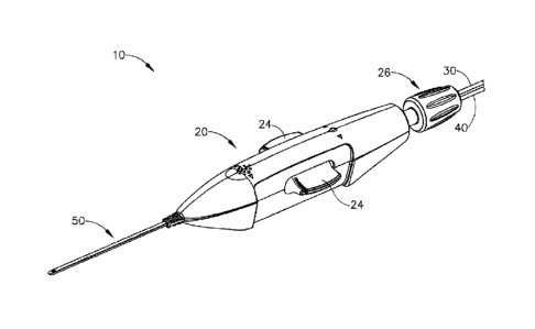
100061 FIG. I depicts a perspective view of an exemplary instrument for
subretinal
administration of a therapeutic agent from a suprachoroidal approach.,
100071 FIG. 2 depicts a perspective view of the distal end of an
exemplary cannula that
may be incorporated into the instrument of FIG. I;
100081 FIG. 3A depicts a cross-sectional side view of the cannula of
FIG. 2, with the
cross-section taken along line 3-3 of FIG. 2, with a needle in a first
longitudinal position;
100091 FIG. 38 depicts a cross-sectional side view of the cannula of
FIG. 2, with the
cross-section taken along line 3-3 of FIG. 2, with the needle in a second
longitudinal
position;
Date Recue/Date Received 2022-09-27
-3-
1000101 FIG. 4A depicts a cross-sectional view of an eye of a patient,
with a chandelier
installed in the eye;
[000111 FIG. 4B depicts a cross-sectional view of the eye of FIG. 4A,
with a suture loop
attached to the eye, and with a sclerotomy being performed;
1000121 FIC.i. 4C depicts a cross-sectional view of the eye of FIG. 4A,
with the instrument
of FIG. l being inserted through the sclerotomy opening and in between the
sclera and
choroid of the eye;
[00013] FIG. 4D depicts a cross-sectional view of the eye of FIG. 4A,
with the instrument
of FIG. I under direct visualization at the back of the eye, between the
sclera and
choroid;
1000] 4] FIG. 4E depicts a cross-sectional view of the eye of FIG. 4A,
with the needle of
the instrument of FIG. I being advanced under direct visualization at the back
of the eye,
pressing against the outer surface of the choroid causing the choroid to
"tent";
1000151 FIG. 4F depicts a cross-sectional view of the eye of FIG. 4A,
with the needle
dispensing a leading bleb under direct visualization at the back of the eye,
the needle
between the sclera and choroid, and the leading bleb in die sub retinal space
between the
choroid and a retina;
[000] 6] FIG. 4G depicts a cross-sectional view of the eye of FIG. 4A,
with the needle
dispensing a therapeutic agent to the eye at the back of the eye, between the
sclera and
choroid;
1000171 FIG, 5A depicts a detailed cross-sectional view of the eye of
FIG. 4A depicted in
the state shown in FIG. 4E;
1000181 FIG. 5B depicts a detailed cross-sectional view of the eye of
FIG. 4A depicted in
the slate shown in FIG. 4F;
1000191 FIG. 5C depicts a detailed cross-sectional view of the eye of
FIG. 4A depicted in
the state shown in FIG. 4G;
1000201 FIG. 6 depicts a cross-sectional view of the eye of FIG. 4A, with
the instrument of
Date Recue/Date Received 2022-09-27
- 4 -
FIG. I at the back of the eye, between the sclera and choroid, with the
cannula of the
instrument providing substantial separation between the sclera and the
choroid,
1000211 FIG. 7 depicts an enlarged view of the distal end of the cannula
of the instrument
of FIG. 1 at the back of the eye, between the sclera and choroid, with the
cannula of the
instrument providing substantial separation between the sclera and the
choroid, with the
needle of the instrument advanced to a distal position;
1000221 FIG. 8 depicts a side elevational view of the distal end of an
exemplary alternative
needle that may be incorporated into the instrument of FIG.
1000231 FIG. 9A depicts a cross-sectional side view of the cannula of FIG.
2, with the
cross-section taken along line 3-3 of FIG. 2. with the needle of FIG. 8 in a
first
longitudinal position;
1000241 FIG. 9B depicts a cross-sectional side view of the cannula of FIG.
2, with the
cross-section taken Mona line 3-3 of FIG. 2, with the needle of FIG. 8 in a
second
longitudinal position;
1000251 FIG. 9C depicts a cross-sectional side view of the cannula of FIG.
2, with the
cross-section taken along line 3-3 of FIG 2, with the needle of FIG 8 in a
third
longitudinal position;
1000261 FIG. 10 depicts an enlarged view of the distal end of the cannula
of the instrument
of FIG. 1 at the back of the eye, between the sclera and choroid, with the
needle of FIG. 8
disposed in the cannula, with the cannula of the instrument providing
substantial
separation between the sclera and the chorea and with the needle of FIG. 8
advanced to
a distal position;
1000271 FIG. 11A depicts a cross-sectional side view of the needle of FIG.
8 disposed in
an exemplary alternative cannula that may be incorporated into the instrument
of FIG. I.
with the needle in a proximal position; and
1000281 FIG. 11B depicts a cross-sectional side view of the needle of FIG.
8 disposed in
the cannula of MG. 11A, with the needle in a distal position.
Date Recue/Date Received 2022-09-27
- 5 -
1000291 The drawings are not intended to be limiting in any way, and it is
contemplated
that various embodiments of the technology may be carried out in a variety of
other ways,
including those not necessarily depicted in the drawings. The accompanying
drawings
incorporated, in and forming a part of the specification illustrate several
aspects of the
present technology, and together with the description serve to explain the
principles of
the technology; it being understood, however, that this technology is not
limited to the
precise arrangements shown.
DETAILED DESCRIPTION
1000301 The following description of certain examples of the technology
should not be
used to limit its scope. Other examples, features, aspects, embodiments, and
advantages
of the technology will become apparent to those skilled in the art from the
following
description, which is by way of illustration, one of the best modes
contemplated for
carrying out the technology. As will be realized, the technology described
herein is
capable of other different and obvious aspects, all without departing from the
technology.
Accordingly, the drawings and descriptions should be regarded as illustrative
in nature
and not restrictive.
1000311 It is further understood that any one or more of the teachings,
expressions,
embodiments, examples, etc. described herein may be combined with any one or
more of
the other teachings, expressions, embodiments, examples, etc. that are.
described herein
The following-described teachinp, expressions, embodiments, examples, etc.
should
therefore not be viewed in isolation relative to each other. Various suitable
ways in
which the teachings herein may be combined ilI be readily apparent to those of
ordinary
skill in the art in view of the teachings herein. Such modifications and
variations are
intended to be included within the scope of the claims.
[000321 For clarity of disclosure, the terms "proximal' and "distal" are
defined herein
relative to a surgeon or other operator grasping a surgical instrument having
a distal
surgical end effector. The term "proximal" refers the position elan element
closer to the
surgeon or other operator and the term "distal" refers to the position of an
element closer
Date Recue/Date Received 2022-09-27
--6 -
to the surgical end effector of the Surgicalinstrumentind iiirtheraWay from
the surgeon.
or other operator
1000331 1. Exemplary. :lnsfrunent.for..Subretiiial A dm ni strati On -
of Therapeutic Agent
1000341 FIG; 1. shows an exemplary -instrument (10) that is Configired for
use in a
procedure fix the subretinal. administration ola therapeutic, agent to an eye
of a 'patient
from a. suprach.otoidal approach, Instrument (.1(J).com prises:a body (20) and
a flexible
cannula (50)extendi ng: di stall y from.body :(20). Cannul a :(50)-.of ih e =
p resent- example has
-
a generally- rectangular cross' section, though any other suitable.-tros.s-
sectional profile
elliptical, etc.) may be used: .Carittula= (50) is generally configured to
support a
needle (100). that is slidable.Withiri cannula (50), as will be.destribed in
greater detail
be ow.
1000351 In the present example,.- cannula. (50) comprises a flexible
material. such as.
Polyether.. block aniide. (PE.BA), which may be manufactured under the trade.
name
PEUIA>t. Of course; .any other suitable-material or combination amaterials
maybe:used.
Also. in the present -exainple,, = canribla 1.50)= has a cross-sectional
profile dimension of
approximately 2.0 min by:0.8 = atm, -with. a.1 ength olapproxi mately 80 mm
Alternatively,
any other suitable dimensions may be used. As will be:describedin greater
detail below,
cannula (50)-ris: flexible .enough to ctxrfoon to "specific structures
and.contours of *the
patient's eye,- yetcan.nula (50) has .sufficient column strength
topermitadvancement = of
cannula (50) between the-sclera and choroid of patient's eye Without buckling,
By 'way
of 'example- only, cannula-00). may be configured and operable-in accordance
with at
least some of the teachings = of U.S. Pub. No.. 2015/0223977, entitled 'Method
and.
Apparatus fix Subretinal Administration of Therapeutic Agent," published
August I `j,
2015.
1000361 As can beSeen in .FIGS: 2-3 B and 6.,.-carinul a (50)com.prises-
a.body-(52.), a closed
distal =end (54), anda lateral opening (5.6) that is located proximal to
distal end. (5.4);
the presentexample,.distal. end (54) has a .rounded configuration. it should
be understood.
that distal.-end-(54). may haveany. suitable kind of curvature. it .should
also b.e-uaderstood
that distal end :(54) may have-any other 'suitable kind Of configuration
(e.g.., beveled,:etei
Date Recue/Date Received 2022-09-27
In the present example, -distal end (54.) is configured to -provide
.Separation between the
sclera and ch.oroid layers: o enable cannula (50) to be. advanced between
.such layers
while not inflicting trauma...to:the -sclera or choroid layers.. Also in the
present example,
the-region, of body (52). that defines:lateral opening(56) s bevel ed, as test
seen inflGS_
3A-34.1. Alternatively, the edge of lateral opening (56) may have any other
suitable
configurati on.
1000371 As best Seen in'FIGS. 3A-..3B,. a needle pide(60):is.disposed
within the hollow-
interior of Cannula. (50). By-Way. of example only, needle guide (60) may be
securth
within tannula (.50) by a. press -or interference fit, by adhesives by.
mechanical 'locking
mechanisms, :andfor it any -other suitable.fashion.. Needle. Ode (60) includes
a .curved
distal end (62)-thatitadstalateral -.opening (56) ofeannula (50), such that 'a
lurnen(64)-Of
needlegui de.(60) (6sta:11y-terminates At 1 atera I opening (S6) The .porti on
of: need! e guide
(6Q)Thatis.prodniál to .distal end (62) is shstanlialiy straight Needle gni
de.(60) -may he
Rimed of plastic, stainless steel, and/or any 'other suitable biocompatible
inaterial(s).
1000381 Nee4ie ioctoy of the. present -example has a sharp distal tip
(102) and defines a
1 m101.(t04). Distal tip = (1 02) -of thepres ent . exam ple has :a:lancet
cenh.gurati on, In some
other .veision.s,..distal .ti .p (102) has a tri-bevel configuration . or any
other configuration as
described in...U.S. Pub: No. 26.15/0223917; entitled-"Method And Apparatus for
Subretinal
AdministratiOn of Therapeutic: Agent," published August 13.1, 2015.
Still other suitable.forins.that distal tip (1.02)
may take will be apparent. to:-those of ordinary skill in the -art in view of
the teachings
herein. Needle 41.00 of The present example comprises a stainless ..steel
hypodermic
needle .6'ot:is-sized:to:deliver -the therapeutic agent while being small
enough-torniniinize
incidental trauma as needle (100) penetrates -tissue structures of the
patient's eye, as will
be .described:in greater detail.beloW. While stainless 'steel is used in
the.present example,
itshould be understood that any other suitablerriaterial(s) may .he used.,
including but not
limited to nitind, etc.
1066391 14 Way *of -example- only, needle (:100) may. be 35 gat*e.With a:
100 ,unt inner
diameter, although *other 'suitable- sizes ...may .beused. -For instance,. the
. outer. diameter of
needle. (100) 'may-fall within the range of 27 gauge to 45 gauge; Or more
particularly
Date Recue/Date Received 2022-09-27
- 8 -
within the range of 30 gauge to 42 gauge, or more particularly within the
range of 32
gauge to 39 gauge. As another merely illustrative example, the inner diameter
of needle
(100) may fall within the range of approximately 50 gm to approximately 200
gm; or
more particularly within the range of approximately 50 gm to approximately 150
gm; or
more particularly within the range of approximately 75 pm to approximately 125
gm.
1000401 Needle (100) is slidably disposed within lumen (64) of needle
guide (60). Needle
guide (60) is generally configured to direct needle (100) upwardly along an
exit axis (EA)
that is obliquely oriented relative to the longitudinal axis (LA) of cannula
(50) through
lateral opening (56) of cannula (50). This is shown in the sequence depicted
in FIGS.
3A-3B, in which FIG. 3A shows needle (100) in a proximal position (where
distal tip
(102) of needle (100) is fully contained in lumen (64) of needle guide (60));
and FIG. 3B
shows needle (100) in a distal position (where distal tip (102) of needle
(100) is outside
of needle guide (60)). While needle (100) is flexible, needle (100) of the
present example
is resiliently biased to assume a straight configuration. Thus, as shown in
FIG. 3B, the
portion of needle (100) that extends outside of cannula (50) and needle guide
(60) is
substantially straight, extending along exit axis (EA). In particular, at
least a substantial
length of the portion of needle (WO) that extends outside of cannula (50) and
needle
guide (60) is coaxially aligned with exit axis (EA).
1000411 It should be understood that the depiction of exit axis (EA) in
FIGS. 3A-3B may
be somewhat exaggerated, for illustrative purposes only. In some versions,
curved distal
end (62) is configured to direct needle (100) along an exit axis (EA) that
extends distally
from cannula (50) at an angle of approximately 7 to approximately 9 relative
to the
longitudinal axis (LA) of cannula (50). It should be understood that, such an
angle may
be desirable to deflect needle (100) in a direction to ensure penetration of
needle into the
choroid and to minimize the possibility of needle (10(Y) continuing beneath
the choroid
through the suprachoroidal space (as opposed to penetrating through the
choroid) and the
possibility of retinal perforation. By way of further example only, curved
distal portion
(88) may urge needle (100) to exit cannula (50) along an exit axis (EA) that
is oriented at
an angle within the range of approximately 50 to approximately 30 relative to
the
longitudinal axis (LA) of cannula (50); or more particularly within the range
of
Date Recue/Date Received 2022-09-27
- 9.-
approximately. 5 to approximately 200 'relative to the longitudinal axis (LA)
of cannula
(50);. or more particularly Within the range ocapprocimaiely 50 to
approximaidy = 10
relative to the lontucknal axis (LA) of cannula (50).
(0004211 At -
shown in FIG. it nStrument- (10) of the present example further comprises an
aettiatian knob (26) located at the prOxini al end ...Of body (.20), Actüation
knob :.(26)
rOtatable.relative to :body .20) to :thereby selectively translate nee. dle-
(100) longitudinally
relative to cannula :=(50). In:particular, actuation knob.- (26).:i.S.-
rotatable in 'a firstangular
di rection4o .drive needle (1.00) distally relative to candula (50);,.--and
ina second angular
direction to. drive needle .(.1.00).proximally '-relatime -to.'.=cannula (50).-
-14- way of example
iitiStrument. (10). May prorvi de- such functionality throtigh n
accordance
with at least some of the -teaching *of :U.S.. Pub: No..241.5/0223 r 7
entitled "Method-and
Apparatus for SUbreti nal Adininistration. Of Therapeutic Agent," published
August 1.3,
2015.
Alternatively,
any other = suitable kind of actuation feature(s) may be used ,to drive needle
( 00) longitudinally .relative to .cannula (56).:
[000431 In
the-present example,- knob =M) is -rotatablethrctigh -acanfplefe range of
motion
that corresponds to -advancement of =needle. (100) to a position relative to
tannula1(.50):ioi
predetermined amount of penetration within. an eye of a patient_ lit ..cither
*words.
instrument (10) is configured such ...that an operator rotates knob (26).Untit
knob :(26) :can
no longer rotate or until knob .(2.6) begins to slipor "finewheel" in a clutch-
atsembly,...to:
properly position needle (100). within = an. eye of a patient. hi some
exaMples; the
predetennined amount of *advancement of needle: (100.). relative to cannula
(50) is
between- approximately:0..25.111mA app.roxiniately1.0 Min; or-more
particularly within
the.range of approximately 0.1.mm:1o...approximately 1.0 inm or more
particularly within
the range of approximately = 2 mrn to approximately 6 min; or more
particularly to.
approximately. 4 mm.
[00044! In
addition or in the =alternative, instrument (10) may .be.eQuipped. with.
certai ri
tactile feedback features to indicate to an operator when needle.(1.00) has
been _advanced-
to certain predetermined distances relati veto .canntila (-.50). Accordingly,
an operator may -
determine the desired depth of penetration of needle (160)-into a -patient'S
*eye based. on
Date Recue/Date Received 2022-09-27
- to-
direct visualization of indicia on instrument andlor based on tactile feedback
from
instrument (10) Of cairse, such tactile feedback features may be combined with
the
present example, as will be apparent to thime of ordinary skill in the art in
view of the
teachings herein.
1000451 As
also shown in FIG. 1, a pair 'of suppiy tubes (30, 40) extend proximally fi-om
actuator knob (24 In the-present example, first supply tube '00) is configured
to couple
with a source of bleb fluid (340).(eg.,- BSS);.. while-second .sUpply tube
r40); .s configured
to couple with a source of therapeutic agent
It:should be understood that each
fluid supply tube (30, 40) may include a conventional bier feature and.'or
other structures
perniitting fluid -supplylubes o, :40 to be coupled with respective fluid
sources. Fluid
supply tubes (30, 40) lead to a valve assembly that. includes actuation arms.
(24).
Acttotion arms (24) are pivotable .10: selectively change the slate of the
:Valve assembly,
Based on the pivotal pmition of actuation arms (24). the valve assembly is
operable to
selectively pinch or otherwise open/close the supply of fluid ft om fluid
supply tubes (30,
40) to lumen (104) of needle (100). Thus, actuation .arnis (24) are operable
to selectiVely
control the delivery of bleb. fluid (340) and therapeutic agent (341) via
=needle (100) By
way of .example only, the valve assembly may be configured and operable in
accordance
with .at least SCOW Of the Ieachints of U.S. Pub. No. 2015/0223977, entitled
"Method and
Apparatus for Subretinal Administration of Therapeutic Agent," published
August 13,
2015.
Other suitable features
and configurations that may he used to = oontrol fluid delivery via needle
(100) Will be
apparent to those of ordinary skill in the art in view of the teachings
herein.
(000461 -ft
should be .understood that the features and operability of instrument (10) may
be varied in. numerous Ways. By way of example only, needle (100) may be
replaced
with needle (200) as described in :greater detail below.. In addition, cannula
(50) may be
replaced with cannula (400) as will be descii bed in greater detail below. In
addition,
instrument -(10) .may be modified in .accordance with at least some of the
teachings of
U.S, Pub. No. 2015;13223977, entitled "Method and Apparatus for Subretinal
Administration of. Therapeutic =Agent,' published August. 13.. 2015;
U.S. Pub. No. 2015/035195h, entitled
Date Recue/Date Received 2022-09-27
-
TherapeUtic 'Agent Delivery Device with Convergent Lumen," published December
10,
2015; U.SPubNo. 201/035i959, entitled "Sub-Retinal Tangential Needle-
Catheter. Guide and introducer" published December 10,. 2015', US... Pub. No.
-
2016/0674212, entitled "Method and Apparatus. for. Sensing-Position
Between..Ltiyers
of an Eye," published March 17, 2016; US: Pub. -..Nct 20-1610074217,- entitled
'Motorized
SupraChoroidal Injection of Therapeutic Agent,' published March 11, .201;
U.:S. Pub, No. 2016/007421.1, entitled "Therapeutic Agent:Delivery. De-Vice-
With
Advanceabje Cannula and Needle published March 1:7; .206;41nd/or U.S. Pub. No.
2016/00131849, entitled '"rherveutic Agent Delivery Device," published March-
24,
20-16c Other-Suitable modifications will be -apparent to those of ordinary
skill in the art-
in eoi of the teachings herein. =
1000471 II. Exemplary Procedurefor Subreti Mil Administration of
Therapeutic Agent =
1000481 FIGS. 4A-.5C show an exempt arrprocedure for subretinal delivery
of therapeutic
agent from a s.upichoroidal approach using instrument 0 %described above. By
=way Of
example only, the method described, herein may be employed to treat macular
degeneration and/Or-other-ocular conditions.. .Although-the procedure
described. herein
discussed in the context of the treatment of age-related macular
degeneration,itshould be
understood that oasuch limitation-Is:intended. or implied. For instance,:in
some merely:
exemplary-alternative procedures, the same techniques described herein -may.
be used to=
treat retinitis pigntentOsa, .diabetic retinopathy., and/or other ocular.
conditions..
Additionally, it should be understood that the procedure-described herein may
be used.to
:treat either dry.or wet. age-related macular degeneration.
1000491 in the .present exampl eõ the . procedure begins by an
operatorimmobilizing tissue
surrounding a 'patient's eye (30I.)-.(eg,: the. eyelids) using a speculiantõ
and/or any 'other
instrument suitable for immobilization. While :iniinobilization described
herein With
reference tO iiistre:Surrounding eye (301), if- should be Understood that. eye
001y-itself
Date Recue/Date Received 2022-09-27
12 - -
may remain free to move. Once the tissue surrounding eye (301) has been
immobilized,
an eye chandelier port (314) is inserted into eye (301), as shown in FIG. 4A,
to provide
intraocular illumination when the interior of eye (301) is viewed through the
pupil. In the
present example, eye chandelier port (314) is positioned in the inferior
medial quadrant
such that a superior temporal quadrant sclerotomy may be preformed. Eye
chandelier
port (314) is positioned to direct light onto the interior of eye (301) to
illuminate at least a
portion of the retina (e.g, including at least a portion of the macula). As
will be
understood, such illumination corresponds to an area of eye (301) that is
being targeted
for delivery of therapeutic agent.
1000501 In the present example, only chandelier port (314) is inserted at
the stage shown
in FIG. 4A, without yet inserting an optical fiber (315) into port (314). In
some other
versions, an optical fiber (313) may be inserted into chandelier port (314) at
this stage In
either case, a microscope may optionally be utilized to visually inspect the
eye to confirm
proper positioning of eye chandelier port (314) relative to the target site.
Although HG
4A shows a particular positioning of eye chandelier port (314), it should be
understood
that eye chandelier port (314) may have any other positioning as will be
apparent to those
of ordinary skill in the art in view of the teachings herein.
1000511 Once eye chandelier port (314) has been positioned, the sclera
(304) may be
accessed by dissecting the conjunctiva by incising a flap in the conjunctiva
and pulling
the flap posteriorly. After such a. dissection is completed, the exposed
surface (305) of the
sclera (304) may optionally be blanched using a cautery- tool to minimize
bleeding. Once
conjunctiva dissection is complete, the exposed surface (305) of the sclera
(304) may
optionally be dried using a WECK-CEL. or other suitable absorbent device. A
template
may then be used to mark eye (301), as described in U.S. Pub. No.
2015/0223977,
entitled "Method and Apparatus for Subretinal Administration of Therapeutic
Agent,"
published August 13, 2015.
An operator may then use a visual wide created using the template to attach
suture loop
assembly (332) and to perform a sclerotorny, as shown in FIG. 48, using a
conventional
scalpel (313) or other suitable cutting instrument The sclerotomv procedure
forms a
small incision through sclera (304) of eye (301). The sclerotany is preformed
with
Date Recue/Date Received 2022-09-27
- 13 -
particular care to avoid penetration of the choroid (306). Thus, the
seleratomy procedure
provides access to the space between sclera (304) and choroid (306). Once the
incision is
made in eye (301), a blunt dissection may optionally be pertbimed to locally
separate
sclera (304) from choroid (306). Such a dissection may be performed using a
small blunt
elongate instrument, as will he apparent to those of ordinary skill in the art
in view of the
teachings herein.
1000521 With the sclerotomy procedure performed, an operator may insert
cannula (50) of
instrument (10) through incision (316) and into the space between sclera (304)
and
thoroid (306). As can be seen in FIG. 4C, cannula (50) is directed through
suture loop
assembly (332) and into the incision. Suture loop assembly (332) may stabilize
cannula
(50) during insertion. Additionally, suture loop assembly (332) maintains
cannula (50) in
a generally tangential orientation relative to the incision_ Such tangential
orientation may
reduce trauma as cannula (50) is guided through the incision. As cannula (50)
is inserted
into the incision through suture loop assembly (332), an operator may use
fonceps or
other instruments to further guide cannula (50) along an atraurnatic path. Of
course, use
of forceps or other instruments is merely optional, and may be omitted in some
examples.
1000531 Although not shown, it should be understood that in some examples
cannula (50)
may include one or more markers on the surface of cannula (50) to indicate
various
depths of insertion. While merely optional, such markers may be desirable to
aid an
operator in identifying the proper depth of insertion as cannula ($0) is
guided along an
atratmiatic path. For instance, the operator may visually observe the position
of such
markers in relation to suture loop assembly (332) and/or in relation to the
incision in the
sclera (304) as an indication of the depth to which cannula (50) is inserted
in eye (301).
By way of example only, one such marker may correspond to an approximately 6
mm
depth of insertion of cannula (50).
1000541 As shown in FIG. 41), once cannula (50) is at least partially
inserted into eye
(301), an operator may insert an optical fiber (315) into eye chandelier port
(314) if the
fiber (315) had not yet been inserted at this stage. With eye chandelier port
(314) in place
and assembled with optical fiber (315), an operator may activate eye
chandelier port
Date Recue/Date Received 2022-09-27
- 14 -
(314) by directing light through optical fiber (315) to provide illumination
of eye (301)
and thereby visualize the interior of eye (301). Further adjustments to the
positioning of
cannula (50) may optionally be made at this point to ensure proper positioning
relative to
the area of geographic atrophy of retina (308). In some instances, the
operator may wish
to rotate the eye (301), such as by pulling on suture loop assembly (332), to
direct the
pupil of the eye (301) toward the operator in order to optimize visualization
of the
interior of the eye (301) via the pupil.
[000551 FIGS. 4C-413 show cannula (50) as it is guided between sclera
(304) and choroid
(306) to the delivery site for the therapeutic agent. In. the present example,
the delivery
site corresponds to a generally posterior region of eye (301) adjacent to an
area of
geographic atrophy of retina (308). In particular, the delivery site of the
present example
is superior to the macula, in the potential space between the neurosensory
retina and the
retinal pigment. epithelium layer. By way of example only, the operator may
rely on
direct visualization through a microscope directed through the pupil of eye
(301) as
cannula (50) is being advanced through the range of motion shown in FIGS. 4C-
4D, with
illumination provided through fiber (315) and port (314). Cannula (50) may be
at least
partially visible through a retina (308) and choroid (306) of eye (301).
Visual tracking
may be enhanced in versions where an optical fiber is used to emit visible
light through
the distal end of cannula (50).
1000561 Once cannula (50) has been advanced to the delivery site as shown
in FIG. 4D, an
operator may advance needle (100) of instrument (10) as described above by
actuating
knob (26). As can be seen in FIGS. 4E and 5A, needle (100) is advanced
relative to
cannula (50) such that needle (100) pierces through choroid (306) without
penetrating
retina (308). Immediately prior to penetrating choroid (306), needle (100) may
appear
under direct visualization as "tenting" the surface of chotoid (306). In other
words,
needle (100) may deform choroid (306) by pushing upwardly on choroid (306),
providing
an appearance similar to a tent pole deforming the roof of a tent. Such a
visual
phenomenon may be used by an operator to identify whether choroid (306) is
about to be
pierced and the location of any eventual piercing. The particular amount of
needle (100)
advancement sufficient to initiate "tenting" and subsequent piercing of
choroid (306) may
Date Recue/Date Received 2022-09-27
- 15 -
be of any suitable amount as may he determined by a number of factors such as,
but not
limited to, general patient anatomy, local patient anatomy, operator
preference, and/or
other factors. As described above, a merely exemplaiy range of needle (100)
advancement may be between approximately 0.25 mm and approximately 10 rum or
more particularly between approximately 2 mm and approximately 6 mm.
1000571 in the present example, after the operator has confirmed that
needle (100) has
been properly advanced by visualizing the tenting effect described above, the
operator
infuses a balanced salt solution (1355) or other similar solution as needle
(100) is
advanced relative to cannula (50). Such a BSS may form a leading bleb (340)
ahead of
needle (100) as needle (100) is advanced, through choroid (306). Leading bleb
(340) may
be desirable for two reasons. First, as shown in FIGS. 4F and 513, leading
bleb (340) may
provide a further visual indicator to an operator to indicate when needle
(100) is properly
positioned at the delivery site Second, leading bleb (340) may provide a
barrier between
needle (100) and retina (308) once needle (100) has penetrated choroid (306).
Such a
barrier may push the retinal wall outwardly, thereby minimizing the iisk of
retinal
perforation as needle (100) is advanced to the delivery site. In some
versions, a foot
pedal is actuated in order to drive leading bleb (340) out from needle (100).
Alternatively, other suitable features that may be used to drive leading bleb
(340) out
from needle (100) will be apparent to those of ordinary skill in the art in
view of the
teachings herein.
100053) Once the opeTator visualizes leading bleb (340), the operator may
cease innasion
of BSS, leaving a pocket of fluid as can be seen in FIGS. 4F and 5B. Next, a
therapeutic
agent (341) may be infused by actuating a syringe or other fluid delivery
device as
described in various references cited. herein. The particular therapeutic
agent (341)
delivered may be any suitable therapeutic agent configured to treat an ocular
condition.
Some merely exemplary suitable therapeutic agents may include, but are not
necessarily
limited to, drugs having smaller or large molecules, therapeutic cell
solutions, certain
gene therapy solutions, tissue plasminogen activators, and/or any other
suitable
therapeutic agent as will be apparent to those of ordinary skill in the art in
view of the
teachings herein. By way of example only, the therapeutic agent (341) may be
provided
Date Recue/Date Received 2022-09-27
- 16
in accordance with at least some of the teachings of U.S. Patent No.
7,413.734,entided
"Treamient of Retinitia Pignnentosa with Human Umbilical Cord Cells," issued
August
19, 2008.. In
addifion to, or as
an alternative to, being used to deliver a therapeutic agent (341), instrument
(10) and
variations thereof may be used to provide. drain age and:or perform other
operations.
1000591 In
the present example, the amount of therapeutic agent (341) that is ultimately
delivered to the:delivery site is approximately 50pL, although any other
suitable amount
may be delivered. In some versions, a foot pedal is actuated in order to drive
agent (341)
out from needle (100). Alternatively, other suitable features that may be used
to drive
agent (341) out from needle (100) will be apparent to those of ordinary skill
in the art in
view of the teachines herein. Delivery of therapeutic agent (341) may be
visualized by
an expansion of the pocket of fluid as can be seen in FIGS. 4G and 5C. As
shown,
therapeutic agent (341) essentially mixes with the fluid of leading bleb (340)
as
therapeutic agent (341) is injected intothe surprachoroidal. subretinal spice.
1000601
Once delivery is complete, needle (100) may be retracted by rotating knob
(2(i) in
a direction opposite to that used to advance needle (100); and cannula (50)
may 'then be
withdrawn from eye (301). Ii should be understood that because of the size of
needle
( IOU), the site where needle (100) penetrated through choroid (306) is self
sealing, such
that no further steps need be taken to seal the delivery site through choroid
(306). Suture
loop assembly (332) and chandelier (314) may be removed, and the incision in
the sclera
(304) may be closed using any suitable conventional techniques.
1000611 As
noted above the foregoing procedure may be carried out to treat a patient
having macular degeneration. In some such instances, the therapeutic agent
(341) that is
delivered by needle (100) may comprise cells that are derived from postpartum
umbilicus
and placenta. As noted above, and by way of example only, the therapeutic
agent (341)
may be provided in accordance with at least. some of the teachings of U.S.
Patent No
7,413,734, entitled -Treatment of Retinitis Piwnentosa with Human Umbilical
Cord
Cells," issued Augast. 19, 2008.
Alternatively, needle (100) may be used to deliver any other suitable
substance or
Date Recue/Date Received 2022-09-27
- 17 -
substance's, in addition to or in lieu of those described in ..11S. Patent No.
7,413,734:
ancliorelseWhere herein.. By Way of example only, thempeutit .actent .(341)
may. comprise
various kinds of drugs including- but not limited to .small molecules, .large.
molectiles,
cells, and/or gene-therapies, it should also-be understood. that macular
degeneration .is
just one -merely illustrative example of a. condition that =may. be *treated
through the
proceduredesCribed herein: OtherbiolOgical.*condifions that may be-addressed
.using The
instruments and procedures described herein Will be:apparent:to those
dfordinary skill in
the au.
1000621 it.. should -also be-understood that the procedure described above
maybe carried
out. in -accordance with any of the teachings .:of U.S. Pub.. No.
201.5/0223977, entitled
"Method and. Apparatus for Subretinal Administration a Therapeutic .Agent"
published
August 13, 2015; US. Pub. .No. . 201.5/03:5195.8; entitled . f.!Therapeutic
Agent.
Delivery Device with Convergent Lumen," published = December 10, 2015;- U.S.
Pub. No. 201510351959, entitied:"Sub-ftetinal. Tangential Needle Catheter
Guide and
intnoducer," published. December 10, 201.5; U.S. Pub. No. .1016/00742:12,
entitled
"Method. and Apparatus for. Sensing Position Between Layers 'of an Eye;"
published
March. 17; 2046;-. US. Ptib.. No. 2016/0074217, entitled 'Motorized
8uprachotoidat.
Injection of. Therapeutic Agent," published March* .17, .2016;
Pub. No.
20.16/0074211, entitled "Therapeutic -Agent.:Delivery Device with AdVanceable
Cannula
and Needle:* published March 17, 20.16; and/or US. Pub.. No.. 201610081849;
entitled "Therapeutic Agent Delivery Device,' published March 24, 2016_
1000631 In_ Exemplary Alternative Needle forlastn.iment
1000641 Several variables may affect th.e relationship. between. the
*.exit -.attgle..(EA). of
needle 100) and the .choroid-(306) of any given patient. It shotild be
understood-that the
Date Recue/Date Received 2022-09-27
- 18 -
choroid (306) and the retina (308) are very thin and have relatively little
structural
integrity. Thus, even when a very flexible cannula (50) is used, cannula (50)
may tend to
provide substantial separation between the choroid (306) and the sclera (304)
as cannula
(SO) is inserted between the choroid (306) and the sclera (304). The degree of
separation
may vary from patient to patient (e.g., based on normal anatomical variation
and/or based
on the patient's disease state, etc.). In cases where the separation is truly
substantial, the
exit angle (EA) of needle (100) may be insufficient to result in distal lip
(102) passing
fully through the choroid (306). In other words, needle (100) may continue
through the
suprachoroidal space without fully penetrating the choroid (306).
1000651 FM. 6 shows an exemplary scenario where cannula (50) has elevated
the choroid
(306) and retina (308) away from the sclera (304) to the point where a
substantial gap
(305) is defined between the sclera (304) and the choroid (306) As also shown
in FIG. 6,
the exit angle (EA) is oriented such that needle (100) would not penetrate the
choroid
(306); and further such that needle (100) would eventually engage the sclera
(304). FIG.
7 shows needle (100) advanced distally along this exit angle (EA). As shown,
needle
(100) passes tangentially along the choroid (306) without ever breaching the
choroid
(306). In some other instances, needle (100) may pass partially through the
choroid (306)
and immediately exit the choroid (306) without ever reaching the subretinal
space
between the choroid (306) and the retina (308).
100066I If the operator determines (e.g., based on the absence of a
choroidal "tenting"
observation as described above) that needle (100) has not fully penetrated the
choroid
(306) despite needle (100) being advanced fully distally, the operator may
retract needle
(100) proximally, slightly reposition cannula (50) and/or another portion of
instrument
(10) in order to provide a better orientation for the exit angle (EA), and
then try
advancing needle (100) distally again. Even with such efforts, it may still be
very
difficult OT even impossible in some cases to successfully penetrate the
choroid (306)
with needle (100). Even in cases where efforts to reposition are successful,
the success
rate may he highly dependent on the skill of the operator, and the
repositioning efforts
will add time to the procedure. Moreover, the repositioning may increase the
risk of
tissue trauma, increase the risk of bleb collapse, and/or increase the risk of
cell egress
Date Recue/Date Received 2022-09-27
- 19-.
into the suprachoroidal space.
1000671 It may semi apparent to address the above-noted issues
by simply modifying
needle guide (60) to provide a steeper exit angle (EA). However, this kind of
modification may be unsuitable for many patients. In particular, increasing
the exit angle
(EA) by providing a more pronounced bend in distal end (62) of needle guide
(60) may
increase the risk of needle (100) perforating the retina (308) in some
patients, particularly
in those where the pp (305) created by ca.nnula (50) between the sclera (304)
and the
choroid (306) is less pronounced than the gap (305) shown in PEGS. 6-7;
including cases
where the gap (305) is non-existent. It may therefore be desirable to provide
a more
nuanced solution that provides greater consistency in penetration of the
choroid (306)
without substantially increasing the risk of penetration of the retina (308).
Such a
solution may provide better accommodation of anatomical variations across
patients;
accommodate variation in operator technique and expertise; and minimize the
level of
operator training required.
1000681 FIG. S shows an exemplary alternative needle (200) that
may be incorporated into
instrument (10) in place of needle (100). In some instances, needle (200) may
be
PAGE 21/66 * RCVD AT 9/27/2022 10:33:16 AM [Eastern Daylight Time] *
SVR:OTT2350,FAX01/19* DNIS:3905* CSID:MLT Aikins Winnipeg *ANI:2049570840*
DURATION (mm-ss):34-1
Date Recue/Date Received 2022-09-27
- '70 -
of curvature between approximately 7 mm and approximately 12 mm; a constant
radius
of curvature between approximately 8 mm and approximately 11 mm; or a constant
radius of curvature between approximately 9 mm and approximately 10 mm, In
some
versions, bent portion (214) has a radius of curvature of approximately 10.5
mm. In
some other versions, bent portion (214) has a radius of curvature of
approximately 100
mm. In some other versions, bent portion (214) has a radius of curvature of
approximately 9.5 mm. It should be understood that the radius of curvature
must be
carefully selected because if the radius is too small, there may be an
increased risk of
perforating the retina (308); and if the radius is too large, the needle (200)
may still fail to
fully penetrate the choroid (306).
1000701 While the radius of curvature of bent portion (214) is constant in
the present
example, in some other versions the radius of curvature may be vati able. For
instance,
some variations of needle (200) may provide a larger radius of curvature in a
region of
needle (200) that remains disposed in cannula (50), even when needle (200) is
in a
distally extended position; with a smaller radius of curvature in a region of
needle (200)
that extends distally from cannula (50) when needle (200) is in a distally
extended
position. This kind of configuration may impart a slight ptvcurvature to
cannula (50),
which may further assist in cannula (50) conforming to the curved inner wall
of sclera
(304), which may in turn reduce the occurrence (or magnitude) of gap (305).
100071I As shown in FIGS. 9A-9C, needle (200)13 slidably disposed in
needle guide (60)
within cannula (50). While FIG. 9A shows needle (200) in a partially advanced
state, it
should be understood that needle (200) may be retracted further proximally in
needle
guide (60) such that distal tip (202) does not protrude through lateral
opening (56). As
shown in FIG. 9A, as needle (200) bens to exit cannula (50) via lateral
opening (56),
the distally protruding portion of needle (200) is oriented along a first exit
axis (EA.1). At
this stage, bent portion (214) and part of distal portion (212) are still
contained within
needle guide (60), such that needle guide (60) prevents needle (200) front
reaching the
configuration shown in FIG. 8.
1'000721 As the operator continues to advance needle GOO) distally relative
to cannula
Date Recue/Date Received 2022-09-27
_
(50), more of needle (200) protrudes distally from lateral opening (56), as
shown in FIG.
911 Due to the resilient bias of needle (200), the now longer protruding
portion of needle
(200) is oriented along a second exit axis (EA2). Second exit axis (EA2)
defines an angle
with the longitudinal axis (LA) that is larger than the ande defined between
first exit axis
(EA1) and the longitudinal axis (LA). As the operator continues to advance
needle (200)
fiirther distally relative to cannula (SO), even more of needle (200)
protrudes distally from
lateral opening (56), as shown in FIG. 9C Due to the resilient bias of needle
(200), the
now longer protruding portion of needle (200) is oriented along a third exit
axis (EA3).
Third exit axis (EA.;) defines an angle with the longitudinal axis (LA) that
is larger than
the angle defined between second exit axis (FA3) and the longitudinal axis
(LA). Thus,
the further needle (200) is advanced, the larger the angle defined between the
exit axis
(EA) and the longitudinal axis (LA). It should be understood that the
depictions of exit
axes (EA1, EA2, EA3) in FIGS. 9A-9C may be somewhat exaggerated, for
illustrative
purposes only.
1000731 As shown in FIG. 10, needle (200) may be particularly useful in
cases where
cannula creates a substantial gap (305) between the sclera (304) and the
choroid (306). It
should be understood that the gap (305) in FIG 10 is substantially the same as
the gap
(305) in FIG. 7_ As noted above, due to the gap (305) in FIG_ 7 and the
associated
relationships between the anatomical structures and the instrument (10)
structures, needle
(100) is unable to penetrate choroid (306). However, as shown in FIG. 10, the
curvature
of needle (200) allows needle (200) to penetrate choroid (306) despite the
presence of gap
(305) and the associated relationships between the anatomical structures and
the
instrument (10) structures.
[000741 As noted above, the exit allele (EA) of needle (200) varies based
on the extent to
which needle (200) is extended from cannula. (50). It should be understood
that this
variation in the exit angle (EA) will allow the operator to control the
optimal exit angle
(EA) by controlling the amount of needle (200) extension. This may allow for
shallower
angles (less extension) for some patients and steeper angles (more extension)
for other
patients, to more consistently be able to achieve penetration of the choroid
(306) in a
relatively safe and efficient manner, eliminating the need for other
mitigations or
Date Recue/Date Received 2022-09-27
- -
workarounds that would otherwise be required from the scenario depicted in
FIG. 7.
1000751 IV. Exemplary Cannula Needle for Instrument
1000761 As noted above, cannula (50) includes a closed distal end (54) and
a lateral
opening (56) that is located proximal to distal end (54). In some instances,
it may be
desirable to provide an alternative cannula that has an open distal end,
without a lateral
opening By way of example only, this may provide simplified manufacturing
processes.
Since it may still be desirable to have a needle exit the cannula at such that
the distal tip
of the needle is oriented along an axis that is oblique to the longitudinal
axis of the
cannula, it may be desirable to use a needle with a preformed curve in
versions where the
cannula has an open distal end.
1000771 FIG. I I A shows an exemplary alternative cannula (400) that may
be readily
incorporated, into instrument (10) in place of cannula (SO). Cannula (400) of
this example
has a flexible body (402) and a distal opening (406). Distal opening (406) is
coaxi ally
positioned on the longitudinal axis of cannula (400) in this example. In some
other
versions, distal opening (406) is offset from the longitudinal axis of cannula
(400). By
way of example only, cannula (400) may be formed of Polyether block amide
(PEBA)
and/or any other suitable kind(s) of material(s). Like cannula (50), cannula
(400) of the
present example has sufficient column strength to be advanced distally between
the sclera
(306) and choroid (308) of patient's eye without buckling.
1000781 An insert (408) is positioned within cannula (400). Insert (408)
may be secured
within canaille (4(X)) by a press or interference fit, by adhesives, by
mechanical locking
mechanisms, and/or in any other suitable fashion_ In the present example,
insert (408) is
formed of a polyimide material, though it should be understood that any other
suitable
biocompatibie material(s) may be used. Insert (408) of the present example is
substantially straight yet may bend with cannula (400). Needle (200) is
slidably disposed
in a lumen (410) defined by insert (408). When needle (200) is in a proximal
position as
shown in FIG. I IA, distal tip (202) of needle (200) is fully contained within
Lumen (410).
At this stage, insert (408) constrains needle (200) such that needle (200) is
held under
stress in a substantially straight configuration. When needle (200) is in a
distal position
Date Recue/Date Received 2022-09-27
e3 _
as shown in FIG. 11B, distal tip (202) of needle is positioned distally of
cannula (400).
At this stage, curved portion (214) is exposed such that the distal portion
(212) of needle
(200) is oriented along an exit axis that is oblique to the longitudinal axis
of cannula
(400). It should be understood that this configuration and orientation may
position distal
tip (202) at the subretinal space (Le., between the choroid (306) and the
retina (308)).
1000791 V. Exemplary Cocchi nati on s
1000801 The following examples relate to various non-exhaustive ways in
which the
teachings herein may be combined or applied. It should be understood that the
following
examples are not intended to restrict the coverage of any claims that may be
presented at
any time in this application or in subsequent filings of this application. No
disclaimer is
intended. The following examples are being provided for nothing more than
merely
illustrative purposes. ft is contemplated that the various teachings herein
may he
arranged and applied in IMITICTOUS other ways. It is also contemplated that.
some
variations may omit certain features referred to in the below examples.
Therefore, none
of the aspects or features referred to below should be deemed critical unless
otherwise
explicitly indicated as such at a later date by the inventors or by a
successor in interest to
the inventors. If any claims are presented in this application or in
subsequent filings
related to this application that include additional features beyond those
referred to below,
those additional features shall not be presumed to have been added for any
reason relating
to patentability.
1000811 Example 1
100082i An apparatus, comprising: (a) a body; (b) a cannula extending
distally from the
body, wherein the cannula is flexible; and (c) a needle slidably disposed in
the cannula,
wherein the needle includes: (i) a sharp distal tip, wherein the needle is
configured to
translate relative to the cannula between a proximal position and. a distal
position,
wherein the diem] tip is configured to be positioned inside the cannula when
the. needle is
in the proximal position, wherein the distal tip is configured to be
positioned outside the
cannula when the needle is in the distal position, and (ii) a curved portion,
wherein the
needle is resiliently biased to extend along a curve through the curved
portion.
Date Recue/Date Received 2022-09-27
-24-
1000831 Example 2
[000841 The apparatus of Example 1, wherein the cannula includes: (i) a
closed distal end,
and (ii) a lateral opening located proximal to the closed distal end.
1000851 Example 3
1000841 The apparatus of Example 2, wherein the cannula further includes
a ramp feature,
wherein the ramp feature extends from an interior region of the cannula 10 the
lateral
opening.
1000871 Example 4
1000881 The apparatus of any one or more of Examples 1 -through 3,
wherein the curved
portion is resiliently biased to define a constant radius of curvature.
1000891 Example 5
1000901 The apparatus of Example 4, wherein the radius of curvature is
between
approximately 7 mm and approximately 12 mm.
1000911 Example 6
1000921 The apparatus of Example 4, wherein the radius of curvature is
between
approximately 4 mm and approximately 15 mm.
1000931 Example 7
1000941 The apparatus of Example 4, wherein the radius of curvature is
between
approximately 9 mm and approximately 10 mm.
1.000951 Example 8
1000961 The apparatus of any one or more of Examples 1 through 7, wherein
the curved
portion is configured to position the distal tip at a progressively increasing
exit angle
relative to a longitudinal axis of the cannula, based on a distance to which
the needle is
advanced distally relative to the cannula,
[000971 Example 9
Date Recue/Date Received 2022-09-27
,75 _
1000981 The apparatus of any one or more of Examples I through 8, wherein
the curved
portion comprises a first curved region and a second curved region, wherein
the first
curved region is located near a distal portion of the needle, wherein the
second. curved
region is located proximal to the first curved region.
1000991 Example 10
10001001 The apparatus of Example 9, wherein the first curved region has a
first radius of
curvature, wherein the second curved region has a second radius of curvature,
wherein
the second radius of curvature is greater than the first radius of curvature.
[000101] Example 11
1000102] The apparatus of any one or more of Examples 9 through 10, wherein
the first
curved region is configured to not impart a curvature to the cannula, wherein
the second
curved region is configured to impart a curvature to the cannula.
10001031 Example 12
10001041 The apparatus of any one or more of Examples 1 through 11, wherein
the needle
further includes a straight proximal portion and a straight distal portion,
wherein the
curved portion is longitudinally positioned between the straight proximal
portion and the
straight. distal portion.
10001051 Example 13
10001061 The apparatus of any one or more of Examples 1 through 12, wherein
the cannula
defines an open distal end.
10001071 Example 14
10001081 The apparatus of Example 13, wherein the needle is configured to
protrude from
the open distal end of the cannula when the needle is in the distal position.
10001091 Example 15
10001101 The apparatus of any one or more of Examples 1 through 14, further
comprising a
source of liquid therapeutic agent, wherein the needle is operable to deliver
the liquid
Date Recue/Date Received 2022-09-27
- -
therapeutic agent.
10001111 Example 16
1000112j The apparatus of Example IS, wherein the body includes: (i) a
needle actuator,
wherein the actuator is operable to drive the needle longitudinally relative
to the cannula,
and (ii) a valve member, wherein the valve member is operable to selectively
provide
fluid communication from the source of liquid therapeutic agent to the needle.
[000113] Example 17
1000114J An apparatus, comprising: (a) a body; (b) a cannula extending
distally from the
body, wherein the cannula is flexible, wherein the cannula includes: (i) a
closed distal
end, and (ii) a lateral opening located proximal to the closed distal end; and
(c) a needle
slidably disposed in the cannula, wherein the needle includes: (i) a sharp
distal tip,
wherein the needle is configured to translate relative to the cannula between
a proximal
position and a distal position, wherein the distal tip is configured to be
positioned inside
the cannula when the needle is in the proximal position, wherein the distal
tip is
configured extend past the lateral opening when the needle is in the distal
position, and
(ii) a curved portion, wherein the curved portion is configured to provide an
oblique exit
angle to a portion of the needle extending past the lateral opening when the
needle is in
the distal position.
[000115] Example 18
10001161 The apparatus of Example 17, wherein the curved portion is
resiliently biased to
assume a curved configuration, wherein the curved portion is further
configured to
deform to a substantially straight configuration within the cannula when the
needle is in
the proximal position.
10001171 Example 19
[0001181 A method of administering a therapeutic agent to an eye of a
patient, wherein the
eye includes a sclera, a choroid, and a retina, the method comprising: (a)
inserting a
flexible cannula between the sclera and the choroid; (b) advancing a needle
relative to the
cannula, thereby penetrating the choroid with a distal tip of the needle,
wherein the
Date Recue/Date Received 2022-09-27
- 27 -
needle includes a preformed curve, wherein the curve guides the needle toward
a targeted
region of the choroid; and (c) administering the therapeutic agent to a region
between the
choroid and the retina via the needle.
1(1001191 Example 20
10001201 The method of Example 19, wherein the act of advancing the needle
includes: (0
advancing the needle to a first longitudinal position relative to the cannula,
wherein the
needle defines a first exit angle relative to the cannula at the first
longitudinal position,
and (ii) advancing the needle further distally to a second longitudinal
position relative to
the cannulaõ wherein the needle defines a second exit angle relative to the
cannula at the
second longitudinal position, wherein the second exit angle is greater than
the first exit
angle.
1000121] VI. Miscellaneous
10001221 It should be understood that any of the versions of the
instruments described
herein may include various other features in addition to or in lieu of those
described
above. By way of example only, any of the devices herein may also include one
or more
of the various features disclosed in any of the various references
10001231 it should be understood that any one or more of the teachings,
expressions,
embodiments, examples, etc. described herein may be combined with any one or
more of
the other teachings, expressions, embodiments, examples, etc. that are
described herein.
The above-described teachings, expressions, embodiments, examples, etc. should
therefore not be viewed in isolation relative to each other. Various suitable
ways in
which the teachings herein may be combined will be readily apparent to those
of ordinary
skill in the art in view of the teachings herein. Such modifications and
variations are
intended to be included within the scope of the claims.
10001241
Date Regue/Date Received 2022-09-27
- 2.8 -
1001251
Versions described above may be designed to be disposed of after a single use,
or
they can be designed to be used multiple times, Versions may, in either or
both cases, be
reconditioned for reuse after at least one use. Reconditioning may include any
combination of the steps of disassembly of the device, f011owed by cleaning or
replacement of particular pieces, and subsequent reassembly. In
particular, some
versions of the device may be disassembled, and any number of the particular
pieces or
parts of the device may be selectively replaced or removed in any combination.
Upon
cleaning and/or replacement of particular parts, some versions of the device
may be
reassembled for subsequent use either at a reconditioning facility, or by an
operator
immediately prior to a procedure.
Those skilled in the art will appreciate that
reconditioning of a device may utilize a variety of techniques for
disassemblyõ
cleaning/replacement, and reassembly. Use of such techniques, and the
resulting
reconditioned device, are all within the scope of the present application.
IOt0F21 By
way of example only, versions described herein may he sterilized before
and/or after a procedure_ In one sterilization technique, the device is placed
in a closed
and sealed container, such as a plastic or TYVEK bag: The container and device
may
then be placed in a field of radiation that can penetrate the container, such
as gamma
radiation, x-rays, or high-energy electrons. The radiation may kill bacteria
on the device
and in the container. The sterilized device may then he stored in the sterile
container for
later use. A. device may also be sterilized using any other technique known in
the art,
including but not limited to beta or gamma radiation, ethylene oxide, or
steam.
[000127]
Having shown and described various embodiments of the present invention,
further adaptations of the methods and systems described herein may be
accomplished by
appropriate modifications by one of ordinary skill in the art without
departing from the
Date Regue/Date Received 2022-09-27
-,79 -
scope of the present invention. Several of such potential modifications have
been
mentioned, and others will be apparent to those skilled in the art. For
instance, the
examples, embodiments, geometries, materials, dimensions, ratios, steps, and
the like
discussed above are illustrative and are not required. Accordingly, the scope
of the
present invention should be considered in terms of the following claims and is
understood
not to be limited to the details of structure and operation shown and
described in the
specification and drawings
Date Recue/Date Received 2022-09-27