Note: Descriptions are shown in the official language in which they were submitted.
CA 03176719 2022-09-21
WO 2022/009000
PCT/IB2021/055293
1
IMPROVEMENTS RELATING TO RESPIRATORY SUPPORT
FIELD OF THE INVENTION
The present disclosure relates to apparatus, systems and/or methods for using
a respiratory
index to determine aspects of respiration, including but not limited to
respiratory index
respiratory status and/or changes in respiratory support (e.g. based on
respiratory index
and/or status).
.. BACKGROUND TO INVENTION
High flow respiratory support has become popular and is commonly used. It has
become a
front-line therapy for patients with respiratory distress. High flow
respiratory support is also
used to assist patients whose respiratory systems are compromised (including
patients with
conditions such as COPD, pulmonary fibrosis, asthma etc).
High flow respiratory support can be an oxygenation tool for patients with
respiratory
distress or failure. Further, it can increase the amount of 02 delivered due
to the fact that
high flows can prevent the entrainment of room air. However, high 02 fraction
(high Fi02)
can potentially mask deterioration of the patient and delay escalation of
care.
Patients can be escalated to e.g. mechanical ventilation or non-invasive
ventilation. The risk
of invasive mechanical ventilation is well understood, although delaying
ventilation can
result in a lengthened hospital stay and increased mortality.
Therefore, it is desirable to determine when to escalate respiratory support.
It can further
be desirable to determine a patient's respiratory status to enable a clinician
to make better
decisions for patients.
SUMMARY OF INVENTION
In one aspect the present disclosure may be said to comprise a method of
assessing a
patient receiving NHF respiratory support, and where necessary changing the
respiratory
support based on the assessment, comprising: receiving from one or more
sensors over one
or more patient parameters for a patient, comprising at least an oxygenation
parameter,
determining in a controller a respiratory index at a plurality of times, and
determining from
a trend in respiratory index over time whether a change in respiratory support
is required,
and if so, making a change in respiratory support.
CA 03176719 2022-09-21
WO 2022/009000
PCT/IB2021/055293
2
In another aspect the present disclosure may be said to comprise a method of
treating a
patient with a respiratory support apparatus comprising receiving from one or
more sensors
over one or more patient parameters for a patient, comprising at least an
oxygenation
parameter, determining in a controller a respiratory index at a plurality of
times, and
determining from a trend in respiratory index over time whether a change in
respiratory
support is required, and if so, making a change in respiratory support.
Optionally the respiratory index is ROX index.
Optionally the respiratory index is determined from one or more lung mechanics
parameters
and one or more oxygenation parameters.
Optionally a lung mechanics parameter is a parameter that indicates lung
mechanics, such
as respiratory rate, expiratory time, minute ventilation).
Optionally a oxygenation parameter/oxygenation exchange parameter is a
parameter that
indicates oxygenation, such as Sp02, Fi02, Fd02, 02 fraction... While
different, Fi02, Fd02
and 02 fraction can be approximate proxy measurements for each other and can
be used
interchangeably where appropriate.
Optionally the assessment phase can comprise one or more of:
= assessing the respiratory status, and determining whether it is normal,
abnormal,
deteriorating, stable, improving or the like,
= assessing whether a change in respiratory support is required (as a result
of
assessing the respiratory status),
= if the changes required, assessing what change in respiratory support is
required
(e.g. escalation, de-escalation, increasing or decreasing high flow therapy,
escalating
to NIV or invasive ventilation, deescalating from NIV or invasive ventilation
or the
like)
Optionally the change in respiratory support phase can comprise:
= indicating any of the above outcomes of the assessment phase, e.g. by
alerts,
alarms, messages or other indicators, and/or
= making any of the changes determined in the assessment phase.
Optionally the assessment phase could be implemented:
CA 03176719 2022-09-21
WO 2022/009000
PCT/IB2021/055293
3
= by a clinician alone,
= one or more assessment, therapy and/or other apparatus without a
clinician,
= or both the clinician and one or more apparatus.
Optionally the respiratory support phase could be implemented:
= by a clinician alone,
= one or more assessment, therapy and/or other apparatus without a
clinician,
= or both the clinician and one or more apparatus.
In another aspect the present disclosure may be said to comprise a method of
assessing a
patient receiving respiratory support during a session to determine a
respiratory status
comprising: receiving from one or more sensors, for a plurality of time
points, one or more
patient parameters for a patient, comprising at least one respiratory
parameter,
determining in a controller: for each time point, a respiratory index and/or
one or more
component parameters, from the one or more patient parameters, and a change in
respiratory index and/or one or more component parameters over time, and
determining,
from the change in respiratory index and/or one or more component parameters
over time,
a patient respiratory status.
Optionally the patient is receiving respiratory support, and optionally the
respiratory support
is: high flow respiratory support, non-invasive pressure respiratory support.
In one aspect, the present disclosure may be said to comprise a method
according to any
preceding claim wherein a clinician determines a patient respiratory status as
"at risk but
improving" if: ROX index is below a threshold but the ROX index change
indicator showing a
trend towards lower risk.
Optionally, if a clinician determines a patient respiratory status as "at risk
but improving"
then an assessment apparatus provides an indication, such as an initial alarm
and display
message that indicates the patient is at risk but improving.
Optionally a clinician determines a patient respiratory status as "at risk and
deteriorating"
if: ROX index is below a threshold and the ROX index change indicator is
showing a trend
toward higher risk.
CA 03176719 2022-09-21
WO 2022/009000
PCT/IB2021/055293
4
Optionally if a clinician determines a patient respiratory status as "at risk
and deteriorating"
then an assessment apparatus provides an indication, such as an alarm and
display
message that indicates the patient is at risk and deteriorating.
Optionally a clinician determines a patient respiratory status as "not at risk
but
deteriorating" if:
ROX index is above a threshold but the ROX index change indicator is showing a
trend toward higher risk.
Optionally if a clinician determines a patient respiratory status as "not at
risk but
deteriorating" then an assessment apparatus provides an indication, such as an
alarm
quietly, and then alarms loudly if/when the ROX index drops below the
threshold.
Optionally:
a clinician determines a patient respiratory status as "stable" if:
respiratory rate is trending upwards (by more than threshold slope or other
change
indicator) but Sp02 is stable, and
a message is displayed on screen.
Optionally:
a clinician determines a patient respiratory status as "deteriorating" if:
respiratory rate is trending upwards (by more than threshold slope or other
change
indicator) and Sp02 is trending downwards, and
an alarm is activated.
Optionally, a clinician determines a patient respiratory status from ROX index
compared to
one or more thresholds.
Optionally, a clinician determines a patient respiratory status from:
respiratory rate,
Sp02, and/or
Fi02
based on one or more thresholds.
Optionally, a clinician determines a patient respiratory status from a change
over time of:
respiratory index, and/or
CA 03176719 2022-09-21
WO 2022/009000
PCT/IB2021/055293
patient parameter, such as respiratory rate, Sp02, and/or Fi02 .
Optionally, a clinician determines a patient respiratory status from a change
indicator such
as slope, magnitude and/or angle between respiratory index at a plurality of
time points.
5
Optionally, a clinician determines a patient respiratory status from a change
indicator such
as slope, magnitude and/or angle between a patient parameter, such as
respiratory rate,
Sp02, and/or Fi02, at a plurality of time points.
Optionally, a clinician determines a patient respiratory status from the
length of time a
respiratory index and/or change indicator takes to change and/or the magnitude
of the
change over a threshold time.
Optionally, a clinician determines a patient respiratory status from the time
taken to for a
respiratory index and/or change indicator by a threshold amount.
Optionally the assessment apparatus and/or respiratory support apparatus have
a user
interface, such as a display.
In another aspect the present disclosure may be said to comprise a method of
assessing a
patient receiving respiratory support during a session to determine a
respiratory status
comprising: receiving from one or more sensors, for a plurality of time
points, one or more
patient parameters for a patient, comprising at least one respiratory
parameter,
determining in a controller: for each time point, a respiratory index from the
one or more
patient parameters, and a change in respiratory index over time, and
determining, from the
change in respiratory index over time, a patient respiratory status.
Optionally the patient is receiving respiratory support, and optionally the
respiratory support
is:
high flow respiratory support,
Optionally the session:
is a treatment session,
a day or part thereof,
a night or part thereof,
sub-sessions,
a length of time.
CA 03176719 2022-09-21
WO 2022/009000
PCT/IB2021/055293
6
Optionally the one or more patient parameters are one or more lung mechanics
parameters
and one or more oxygenation parameters.
Optionally a lung mechanics parameter can be one or more of:
= Respiratory rate
= expiratory time,
= minute ventilation.
Optionally a oxygenation parameter can be one or more of:
= Fi02
= Fd02
= 02 fraction
= Sp02
Optionally the respiratory index is ROX index.
Optionally components of the ROX index are: respiratory rate,
Sp02, and/or
Fi02, Fd02, and/or 02 fraction.
Optionally a respiratory rate is determined by the controller from one or more
patient
parameters received from the one or more sensors.
Optionally further comprising indicating and/or making a change in respiratory
support
based on the respiratory status and/or respiratory index.
Optionally displaying ROX index numerically and/or graphically.
Optionally determining a change in respiratory index over time comprises:
for a plurality of time points, determining a change in respiratory index over
time for
each of the plurality of time points.
Optionally further comprising displaying, for the plurality of time points,
the change in
respiratory index over time for each of the plurality of time points.
CA 03176719 2022-09-21
WO 2022/009000
PCT/IB2021/055293
7
Optionally determining a patient respiratory status from the change in
respiratory index
over time comprises monitoring a change, over the plurality of points in time,
of the change
in respiratory index over time.
Optionally monitoring a change, over the plurality of time points, of the
change in
respiratory index over time comprises, for the plurality of time points:
viewing the
displayed change, over the plurality of points in time, of the change in
respiratory index
over time ,and/or calculating and comparing the change to relationship
information
Optionally further comprising displaying a respiratory index threshold and/or
change
indicator threshold.
Optionally determining a change in respiratory index over time comprises
determining a
trend in the respiratory index.
Optionally the trend comprises a plurality of instantaneous trends, and
determining a trend
comprises determining a plurality of instantaneous trends over time.
Optionally a trend or an instantaneous trend is represented with a trend
parameter
comprising magnitude and a direction, and optionally could be in the form of:
a vector, or a
slope and magnitude.
Optionally further comprising communicating the determined change in
respiratory support
to: a clinician, for example in the form of a message, alarm, respiratory
status, respiratory
index, and/or a respiratory support apparatus.
Optionally the one or more sensors comprise: one or more sensors arranged to
sense a flow
path of a respiratory support apparatus, and/or one or more sensors arranged
to sense
parameters of a patient, and the controller receives the one or more patient
parameters
from the one or more sensors.
Optionally further comprising displaying on an interface, either on a
respiratory apparatus,
mobile device and/or other assessment apparatus one or more of: the
respiratory index
versus time, either graphically and/or numerically, one or more components of
the
respiratory index (e.g. respiratory rate, Sp02, Fi02 or the like), alone,
combined and/or
options versus time, either graphically and/or numerically, and/or one or more
vectors,
slopes, angles, magnitudes, differences and/or other change indicators
indicating change
CA 03176719 2022-09-21
WO 2022/009000
PCT/IB2021/055293
8
between two or more respiratory indexes and/or components thereof, over time
or
otherwise.
Optionally further comprising receiving input (e.g. user input) to revise the
display and re-
displaying information based on the user input, comprising one or more of:
receiving input
to display one or more components of the respiratory index, and displaying the
one or more
components of the respiratory index alone, combined and/or options versus
time, either
graphically and/or numerically, and/or receiving input to display, zoom and/or
move the
display, and displaying, or redisplaying a zoomed and/or moved version of:
respiratory
index (e.g. ROX index) versus time, either graphically and/or numerically, one
or more
components of the respiratory index, alone, combined and/or options versus
time, either
graphically and/or numerically, and/or one or more vectors, slopes, angles,
magnitudes,
differences and/or other change indicators indicating change between two or
more
respiratory indexes and/or components thereof, over time or otherwise.
Optionally a clinician determines a patient respiratory status from a change
in respiratory
index over time by viewing the respiratory index (e.g. ROX index) versus time,
either
graphically and/or numerically, one or more components of the respiratory
index (e.g.
respiratory rate, Sp02, Fi02 or the like), alone, combined and/or options
versus time, either
graphically and/or numerically, and/or one or more vectors, slopes, angles,
magnitudes,
differences and/or other change indicators indicating change between two or
more
respiratory indexes and/or components thereof, over time or otherwise.
Optionally a clinician determines a patient respiratory status from a change
in respiratory
index over time by any one or a combination of the following: comparing one or
more
respiratory index(es) and/or a change in respiratory index, relative to one or
more
threshold(s), comparing one or more change indicators relative to one or more
threshold(s),
comparing one or more respiratory index(es) and/or a change in respiratory
index, relative
to one or more other respiratory index(es) and/or a change in respiratory
index and/or
relative to one or more other one or more change indicators, comparing one or
more
change indicators relative to one or more other change indicators and/or one
or more
respiratory index(es) and/or a change in respiratory index, considering one or
more:
respiratory indexes, change in respiratory indexes over time, change in,
change in
respiratory indexes over time, and/or change indicators.
Optionally upon determining a patient respiratory status, one or more of the
following can
occur to indicate respiratory status: an alarm is sounded and/or a message
is
CA 03176719 2022-09-21
WO 2022/009000
PCT/IB2021/055293
9
displayed, which indicate the respiratory status, alert the clinician and/or
indicate action
required, and/or a change in therapy is actioned, automatically and/or
manually.
In one aspect, the present disclosure may be said to comprise an apparatus for
assessing a
patient receiving respiratory support during a session to determine a
respiratory status
comprising: one or more sensors, or inputs for one or more sensors for
receiving from for a
plurality of time points, one or more patient parameters for a patient,
comprising at least
one respiratory parameter, a controller for determining: for each time point,
a respiratory
index from the one or more patient parameters, and determining, from the
change in
respiratory index over time, a patient respiratory status, and/or displaying
on a display a
change in respiratory index over time for a user to determine a patient
respiratory status.
Optionally the patient is receiving respiratory support, and optionally the
respiratory support
is: high flow respiratory support.
Optionally: the assessment apparatus provides the respiratory support, or the
assessment
apparatus is separate to a respiratory support apparatus.
Optionally the session:
is a treatment session,
a day or part thereof,
a night or part thereof,
sub-sessions,
a length of time.
Optionally the one or more patient parameters are one or more lung mechanics
parameters
and one or more oxygenation parameters.
Optionally a lung mechanics parameter can be one or more of:
= Respiratory rate
= expiratory time,
= minute ventilation
Optionally an oxygenation parameter can be one or more of:
= Fi02
= Fd02
= 02 fraction
CA 03176719 2022-09-21
WO 2022/009000
PCT/IB2021/055293
= Sp02
Optionally the respiratory index is ROX index.
5 Optionally components of the ROX index are: respiratory rate,
Sp02, and/or
Fi02, Fd02, and/or 02 fraction.
Optionally a respiratory rate is determined by the controller from one or more
patient
10 parameters received from the one or more sensors.
Optionally the respiratory index is ROX index, determined from respiratory
rate, Fi02 and/or
Sp02 .
Optionally comprising displaying ROX index numerically and/or graphically on
the display.
Optionally determining a change in respiratory index over time comprises:
for a plurality of time points, determining a change in respiratory index over
time for
each of the plurality of time points.
Optionally comprising displaying, for the plurality of time points, the change
in respiratory
index over time for each of the plurality of time points.
Optionally determining from the change in respiratory index over time,
comprises the
controller calculating and comparing the change to relationship information.
Optionally further comprising the controller displaying:
a respiratory index threshold and/or change indicator threshold.
Optionally further comprising communicating the determined change in
respiratory support
to: a clinician, for example in the form of a message, alarm, respiratory
status, respiratory
index, and/or a respiratory support apparatus.
Optionally the one or more sensors comprise: one or more sensors arranged to
sense a flow
path of a respiratory support apparatus, and/or one or more sensors arranged
to sense
CA 03176719 2022-09-21
WO 2022/009000
PCT/IB2021/055293
11
parameters of a patient, and the controller receives the one or more patient
parameters
from the one or more sensors.
Optionally the apparatus is one or more of a:
respiratory apparatus,
mobile device,
server,
either alone or integrated.
Optionally comprising the sensors.
In another aspect the present disclosure may be said to comprise system for
assessing a
patient receiving respiratory support during a session to determine a
respiratory status
comprising:
An apparatus according to any statement herein carrying out a method according
to
any statement herein
Optionally the apparatus is configured to determine a patient respiratory
status from the
change in respiratory index over time by monitoring a change, over the
plurality of points in
time, of the change in respiratory index over time.
Optionally the at least one patient parameter is patient Fi02.
Optionally the respiratory parameters are:
respiratory rate, and/or
Sp02.
Optionally the apparatus is configured to determine a change in respiratory
index over time
comprises determining a trend in the respiratory index.
Optionally trend comprises a plurality of instantaneous trends, and
determining a trend
comprises determining a plurality of instantaneous trends over time.
Optionally the change indicator could be in the form of:
CA 03176719 2022-09-21
WO 2022/009000
PCT/IB2021/055293
12
a vector, or
a slope and magnitude.
Optionally the apparatus is further configured to display on an interface,
either on a
respiratory apparatus, mobile device and/or other assessment apparatus one or
more of:
the respiratory index versus time, either graphically and/or numerically, one
or more
components of the respiratory index (e.g. respiratory rate, Sp02, Fi02 or the
like), alone,
combined and/or options versus time, either graphically and/or numerically,
and/or one or
more vectors, slopes, angles, magnitudes, differences and/or other change
indicators
indicating change between two or more respiratory indexes and/or components
thereof,
over time or otherwise.
Optionally the apparatus is further configured to receive input (e.g. user
input) to revise the
display and re-displaying information based on the user input, comprising one
or more of:
receive input to display one or more components of the respiratory index, and
displaying the one or more components of the respiratory index alone, combined
and/or
options versus time, either graphically and/or numerically, and/or
receive input to display, zoom and/or move the display, and displaying, or
redisplaying a zoomed and/or moved version of:
respiratory index (e.g. ROX index) versus time, either graphically and/or
numerically.
one or more components of the respiratory index, alone, combined and/or
options versus time, either graphically and/or numerically, and/or
one or more vectors, slopes, angles, magnitudes, differences and/or other
change indicators indicating change between two or more respiratory indexes
and/or
components thereof, over time or otherwise.
Optionally the apparatus is further configured to allows a clinician to
determine a patient
respiratory status from a change in respiratory index over time by viewing
the respiratory index (e.g. ROX index) versus time, either graphically and/or
numerically,
one or more components of the respiratory index (e.g. respiratory rate, Sp02,
Fi02
or the like), alone, combined and/or options versus time, either graphically
and/or
numerically, and/or
one or more vectors, slopes, angles, magnitudes, differences and/or other
change
indicators indicating change between two or more respiratory indexes and/or
components
thereof, over time or otherwise.
CA 03176719 2022-09-21
WO 2022/009000
PCT/IB2021/055293
13
Optionally the apparatus is further configured to allow a clinician to
determine a patient
respiratory status from a change in respiratory index over time by any one or
a combination
of the following:
compare one or more respiratory index(es) and/or a change in respiratory
index,
relative to one or more threshold(s),
compare one or more change indicators relative to one or more threshold(s),
compare one or more respiratory index(es) and/or a change in respiratory
index,
relative to one or more other respiratory index(es) and/or a change in
respiratory index
.. and/or relative to one or more other one or more change indicators.
compare one or more change indicators relative to one or more other change
indicators and/or one or more respiratory index(es) and/or a change in
respiratory index.
consider one or more:
respiratory indexes,
change in respiratory indexes over time,
change in, change in respiratory indexes over time, and/or
change indicators.
Optionally the apparatus is further configured to upon determining a patient
respiratory
status, provide one or more of the following can occur to indicate respiratory
status:
an alarm is sounded and/or a message is displayed, which indicate the
respiratory
status, alert the clinician and/or indicate action required, and/or
a change in therapy is actioned, automatically and/or manually.
Optionally a method or apparatus as described wherein the respiratory index is
ROX index.
Optionally a method or apparatus as described wherein components of the
respiratory index
are respiratory rate, Sp02, and/or Fi02
In another aspect, the present disclosure may be said to comprise a method of
assessing a
patient receiving respiratory support during a session to determine a
respiratory status
comprising: receiving from one or more sensors, for a plurality of time
points, one or more
patient parameters for a patient, comprising at least one respiratory
parameter,
determining in a controller for each time point, a respiratory index and/or
one or more
component parameters, from the one or more patient parameters, and a change in
respiratory index and/or one or more component parameters over time, and
determining,
CA 03176719 2022-09-21
WO 2022/009000
PCT/IB2021/055293
14
from the change in respiratory index and/or one or more component parameters
over time,
a patient respiratory status.
In another aspect, the present disclosure may be said to comprise a method of
assessing a
patient to determine a change in respiratory support comprising: Receiving,
for a plurality of
time points, one or more patient parameters from a patient, comprising at
least one
respiratory parameter, determining, for each time point, a respiratory index
from the one or
more patient parameters, from the respiratory index, determining a patient
respiratory
status, and/or a change in respiratory support based on the trend of the
respiratory index.
In another aspect, the present disclosure may be said to comprise a method of
assessing a
patient to determine a change in respiratory support comprising: receiving,
for a plurality of
time points, one or more patient parameters from a patient, comprising at
least one
respiratory parameter, determining, for each time point, a respiratory index
from the one or
more patient parameters, determining a change in respiratory index over time,
from the
change in respiratory index, determining a patient respiratory status, and/or
a change in
respiratory support.
Optionally wherein determining a change in respiratory index over time
comprises
determining a trend in the respiratory index.
Optionally the trend comprises a plurality of instantaneous trends, and
determining a trend
comprises determining a plurality of instantaneous trends over time.
Optionally wherein a trend or an instantaneous trend is represented with a
trend parameter
comprising a magnitude and a direction, and optionally could be in the form
of:
a vector, or
a slope (i.e. gradient) and magnitude.
Optionally the method further comprising communicating the determined change
in
respiratory support to:
a clinician, for example in the form of a message, alarm, respiratory status,
respiratory index, and/or
a respiratory support apparatus.
Optionally the method further comprising controlling a respiratory support
apparatus based
on the determined change in respiratory support.
CA 03176719 2022-09-21
WO 2022/009000
PCT/IB2021/055293
Optionally the method further comprising determining, from the respiratory
index, a patient
status and/or change in patient status, and optionally communicating the
patient status to a
clinician, for example in the form of a message, alarm, and/or status.
5
Optionally the change in respiratory index, determining a patient respiratory
status, and/or
a change in respiratory support based on the trend of the respiratory index
comprises,
comparing one or more of:
= A respiratory index or change in respiratory index
10 = Trend or plurality of instantaneous trends
= Trend parameter or plurality of trend parameters
= Patient respiratory status or change in respiratory status
against relationship information,
wherein optionally the relationship information comprises:
15 at least one threshold, and/or
a time over which the threshold is met, exceeded or not exceeded.
Optionally the method further comprising communicating one or more of:
= A respiratory index or change in respiratory index
= Trend or plurality of instantaneous trends
= Trend parameter or plurality of trend parameters
= Patient respiratory status or change in respiratory status
= relationship information.
Optionally the respiratory index is ROX index, and the trend parameter is a
vector that
indicates the change in ROX index and the relationship information is a
threshold that
indicates risk of respiratory failure.
Optionally the change in respiratory support is an escalation or de-escalation
of respiratory
support.
Optionally escalating respiratory support comprises:
= providing high flow respiratory support at a higher level. Optionally by
increasing or
providing flow, 02 concentration, humidification, flow oscillation and/or
other high
flow parameters
= transferring the patient to a more invasive respiratory support such as:
o NIV pressure respiratory support
CA 03176719 2022-09-21
WO 2022/009000
PCT/IB2021/055293
16
o Mechanical ventilator respiratory support via intubation
Optionally escalation comprises:
Controlling an apparatus to escalate respiratory support, and/or
Communicating, optionally in the form of message, status, alarm, to a
clinician to
escalate, or consider escalating, respiratory support.
Optionally the change in respiratory support improves the patient's
respiratory status
and/or respiratory index.
In another aspect the present disclosure may be said to comprise a method of
assessing a
patient to determine a change in respiratory support comprising: receiving,
for a plurality of
time points, one or more patient parameters from a patient, comprising at
least one
respiratory parameter, determining, for each time point, a respiratory index
from the one or
more patient parameters, determining at least one vector with magnitude and
direction
indicating a change of respiratory index over time, determining a change in
respiratory
support based on the vector.
In another aspect the present disclosure may be said to comprise an apparatus
to
determine a change in respiratory support comprising: a controller, the
controller configured
to: receive, for a plurality of time points, one or more patient parameters
from a patient,
comprising at least one respiratory parameter, determine, for each time point,
a respiratory
index from the one or more patient parameters, determine a change in
respiratory index
over time, from the change in respiratory index, determine a patient
respiratory status,
and/or a change in respiratory support, and an I/O interface to communicate
one or more
of the: respiratory index and/or change in respiratory index, patient
respiratory status,
change in respiratory support.
Optionally the apparatus is a respiratory apparatus, comprising a flow
generator and a
humidifier.
Optionally the flow generator and humidifier are integrated in a housing.
Optionally the apparatus further comprising or configured to couple to one or
more of:
a sensor to determine 02 concentration of a gas,
a sensor for determining respiratory rate of a patient.
CA 03176719 2022-09-21
WO 2022/009000
PCT/IB2021/055293
17
Optionally the apparatus further comprising a wireless communications
transceiver.
Optionally the apparatus is a mobile device with an 10 interface and receives
patient
parameters using one or more of:
Mobile telecommunications
BluetoothTm
NFC.
Optionally the mobile device communicates the:
respiratory index and/or change in respiratory index,
patient respiratory status, and/or
change in respiratory support
to a respiratory apparatus for control of the respiratory apparatus and/or
communication on
a 10 interface of the respiratory apparatus.
In another aspect the present disclosure may be said to comprise a method of
controlling a
respiratory apparatus comprising: determining a change in respiratory index
over time from
patient parameters, from the change in respiratory index, determining a
patient respiratory
status, and/or a change in respiratory support, and communicating to a
clinician how to
change respiratory support, and/or controlling to a respiratory support
apparatus to change
respiratory support.
Optionally the change in respiratory support improves the patient's
respiratory status
and/or respiratory index.
In another aspect the present disclosure may be said to comprise a method of
determining
one or more trend parameters for a respiratory index: receiving, for a
plurality of time
points, one or more patient parameters from a patient, comprising at least one
respiratory
parameter, determining, for each time point, a respiratory index from the one
or more
patient parameters, determining one or more trend parameters representing a
change in
the respiratory index over time.
Optionally the trend parameter comprises magnitude and a direction, and
optionally could
be in the form of:
a vector, or
a slope (i.e. gradient) and magnitude.
CA 03176719 2022-09-21
WO 2022/009000
PCT/IB2021/055293
18
In another aspect the present disclosure may be said to comprise a system to
determine a
change in respiratory support comprising: a mobile device, with a controller,
10 interface
and a wireless communications transceiver, and a respiratory apparatus, with a
controller,
flow generator and a humidifier, wherein one or both of the controllers are
configured to do
some or all of: receive, for a plurality of time points, one or more patient
parameters from a
patient, comprising at least one respiratory parameter, determine, for each
time point, a
respiratory index from the one or more patient parameters, determine a change
in
respiratory index over time, from the change in respiratory index, determine a
patient
respiratory status, and/or a change in respiratory support.
In another aspect the present disclosure may be said to comprise in an
apparatus to
determine a change in respiratory support comprising: a mobile device, with a
controller, 10
interface and a wireless communications transceiver, to receive, for a
plurality of time
points, one or more patient parameters from a patient, comprising at least one
respiratory
parameter, determine, for each time point, a respiratory index from the one or
more patient
parameters, determine a change in respiratory index over time, from the change
in
respiratory index, convey information on the 10 interface, and/or determine a
patient
respiratory status, and/or a change or suggested change in respiratory
support.
Optionally the mobile device receives patient parameters via the wireless
communications
transceiver using one or more of:
Mobile telecommunications
BluetoothTm
NFC,
WiFi.
Optionally the mobile device receives patient parameters via a WAN, LAN or
wireless
network.
Optionally the mobile device communicates the:
respiratory index and/or change in respiratory index,
patient respiratory status, and/or
change in respiratory support
to the respiratory apparatus for control of the respiratory apparatus and/or
communication
on an 10 interface of the respiratory apparatus.
CA 03176719 2022-09-21
WO 2022/009000
PCT/IB2021/055293
19
Optionally the mobile device and/or respiratory apparatus conveys one or more
of the
following in a graph, message, display, information, and/or audibly or
otherwise in the 10
interface:
= A respiratory index or change in respiratory index
= Trend or plurality of instantaneous trends
= Trend parameter or plurality of trend parameters
= Patient respiratory status or change in respiratory status
= Relationship information.
In another aspect the present disclosure may be said to comprise a mobile
device and/or a
mobile device programmed to carry out a method comprising:
receiving, for a plurality of time points, one or more patient parameters from
a
patient, comprising at least one respiratory parameter,
determining, for each time point, a respiratory index from the one or more
patient
parameters,
determining a change in respiratory index over time,
from the change in respiratory index, determining
a patient respiratory status, and/or
a change in respiratory support.
In another aspect the present disclosure may be said to comprise a method
implemented by
a mobile device and/or a mobile device programmed to carry out a method
further
comprising:
conveying one or more of the following in a graph, message, display,
information, and/or
audibly or otherwise in an 10 interface:
= A respiratory index or change in respiratory index
= Trend or plurality of instantaneous trends
= Trend parameter (e.g. vector, including magnitude and/or direction) or
plurality of
trend parameters
= Patient respiratory status or change in respiratory status
= Relationship information.
Optionally the system or method may be configured to determine a change in
flow rate
provided by the respiratory support apparatus required to improve the
respiratory index,
and may be configured to present instructions on the mobile device to change
the flow rate
or another parameter of the respiratory support apparatus, optionally wherein
the change in
flow rate or another parameter is one or more of:
CA 03176719 2022-09-21
WO 2022/009000
PCT/IB2021/055293
the flow rate is increased to improve the respiratory index
the flow rate is changed based on or relative to the change in the respiratory
index
the flow rate is changed based on or relative to the change in trend or trend
pa ra meter
5 Fi02 is be changed relative to respiratory rate change or relative to
change in
respiratory index.
a gas valve is be controlled to either increase Fi02 or maintain Fi02 while
flow rate is
changed relative to the index change.
10 Optionally a system or method wherein the respiratory index is ROX
index, which is based
on Sp02, Fi02 and respiratory rate, and optionally the system comprises or is
configured to
connect to one or more sensors from which Sp02, Fi02 and/or respiratory rate
can be
determined, and optionally wherein respiratory rate is calculated in the
controller based on
frequency response of an respiratory rate sensor.
Optionally a mobile device captures/receives respiratory rate and Fi02 using
an NFC
protocol.
Optionally further comprising one or more of:
a respiratory rate sensor
an oxygen concentration sensor
flow sensor (optionally inline)
pressure sensor
temperature sensor
ultrasonic sensor.
and optionally wherein the controller receives signals from one or more of the
sensors
and/or from manual input and calculates respiratory rate and Fi02 based on
received
signals.
Optionally a system or method according to any preceding claim wherein: the
controller is
configured to calculate or the method comprises calculating a trend of a
respiratory index
over a set period of time based on respiratory rate and Fi02 measurements
taken within the
set time period, and/or the controller is configured to increase flow from a
base flow rate if
the trend (or change) in respiratory index indicates respiratory status
deterioration, and/or
the controller is configured to reduce flow towards a base flow rate if the
respiratory index
indicates respiratory status improvement.
CA 03176719 2022-09-21
WO 2022/009000
PCT/IB2021/055293
21
In another aspect the present disclosure may be said to comprise a method of
providing
respiratory support comprising: determining a respiratory index of patient
respiration at one
or more time points, determining a change in respiratory index over time, from
the change
in respiratory index, determining a patient respiratory status, and/or a
suitable respiratory
support, and providing the determined respiratory support to the
patient.
In one implementation, the embodiment comprises a mobile device receiving
information
from wearable sensors. The information is used as above and the information
conveyed to
the clinician and also a respiratory support device to control the device,
e.g. through wired
or wires transmission, including NFC. The mobile device may communicate with a
respiratory support device using Bluetooth or Infra-red or another suitable
wireless
communication protocol. The mobile device may receive information from the
respiratory
device and sensors within the respiratory device. The mobile device may
automatically ping
(i.e. interrogate) the respiratory device at regular time intervals to receive
data from
sensors onboard the respiratory support device. Alternatively, the respiratory
support
device may periodically transmit data to the mobile device. In one example NFC
communication is advantageous because a user of the mobile device e.g. a
clinician can
initiate when sensor data from the respiratory support device is received at
the mobile
device. The mobile device may determine effectiveness of respiratory support
using a
method as described herein.
In one aspect, the present disclosure may comprise an apparatus for providing
respiratory
support comprising: a housing,
a flow generator (e.g. a blower) within the housing,
a supplementary gases inlet, a valve in fluid communication with the
supplementary
gases inlet and configured to control the amount of supplementary gases
introduced into
the apparatus, an outlet located within or on the housing, a gases path
extending from the
gases inlet to the outlet, through the housing, wherein the flow generator is
configured to
receive supplementary gases from the supplementary gases inlet and generate a
flow of
gases, the flow of gases travelling through the gases path, a plurality of
sensors, a
controller in electronic communication with the one or more sensors and
receive signals
from the sensors, wherein the sensors are non-invasive sensors, the controller
configured
to: determine a lung mechanics parameter and an oxygenation parameter from the
sensor
signals, determine a respiratory index based on the lung mechanics parameter
and
oxygenation parameter, determine a change in the respiratory index over time,
change
respiratory support based on the change in the respiratory index over time.
CA 03176719 2022-09-21
WO 2022/009000
PCT/IB2021/055293
22
The apparatus optionally comprises a humidifier. The humidifier is positioned
downstream of
the flow generator, and the humidifier is configured to humidify gases flow.
Optionally, change in respiratory index comprises a trend or a rate of change
or a second
derivative of the rate of change.
Optionally, the respiratory apparatus may comprise a communication interface
that is
configured to transmit information to a mobile device (e.g. smartphone or
tablet) associated
with a clinician or healthcare professional and/or transmit information to a
remote patient
monitoring system. The remote patient monitoring system may comprise one or
more
servers, memory units, databases and other components that allow management of
patient
information, generation of reports of patient's health status and allow alerts
to be sent to
the patient and/or clinician. The change in respiratory index may be
transmitted to the
mobile device and/or to the remote patient monitoring system.
The respiratory index measurements and change in respiratory index may be
incorporated
into a patient report that includes measured patient parameters e.g. Sp02,
flow rate,
humidity set point and usage hours and the change in respiratory index and
respiratory
index measured values over time. The
The change in respiratory index allows a clinician to assess if the current
therapy being
provided is being effective and also allows a clinician to make a change in
the therapy
provided. In one example the operational parameters of the respiratory support
apparatus
(e.g. prescription settings) may be remotely updated based on the change in
the respiratory
index.
In another aspect the present disclosure may be said to comprise a monitoring
system comprising:
respiratory support apparatus, (such as a high flow respiratory support
apparatus,
e.g. a nasal high flow respiratory support apparatus),
a remote monitoring apparatus for a clinician to monitor a patient being
supported
by the respiratory apparatus,
and one or more controllers in the respiratory apparatus, remote monitoring
apparatus and/or other apparatus in the system configured to:
receive from one or more sensors, for a plurality of time points, one or more
patient parameters for a patient, comprising at least one respiratory
parameter,
determine in the one or more controllers,
CA 03176719 2022-09-21
WO 2022/009000
PCT/IB2021/055293
23
for each time point, a respiratory index from the one or more patient
parameters, and
a change in respiratory index over time,
and
provide information (numerically, graphically or otherwise) to the remote
monitoring
apparatus, the information being one or more of:
respiratory index, and/or
change in respiratory index over time.
Optionally one or more of the following could also be provided
- patient respiratory status,
change in respiratory status over time,
- patient parameter versus time
- change patient parameter versus time
- respiratory index threshold,
- change indicator threshold,
- suggestions of respiratory support.
It is intended that reference to a range of numbers disclosed herein (for
example, 1 to 10)
also incorporates reference to all rational numbers within that range (for
example, 1, 1.1, 2,
3, 3.9, 4, 5, 6, 6.5, 7, 8, 9 and 10) and also any range of rational numbers
within that
range (for example, 2 to 8, 1.5 to 5.5 and 3.1 to 4.7) and, therefore, all sub-
ranges of all
ranges expressly disclosed herein are hereby expressly disclosed. These are
only examples
of what is specifically intended and all possible combinations of numerical
values between
the lowest value and the highest value enumerated are to be considered to be
expressly
stated in this application in a similar manner.
The term "comprising" as used in this specification means "consisting at least
in part of".
When interpreting each statement in this specification that includes the term
"comprising",
features other than that or those prefaced by the term may also be present.
Related terms
such as "comprise" and "comprises" are to be interpreted in the same manner.
Unless the
context clearly requires otherwise, throughout the description and the claims,
the words
"comprise", "comprising", and the like, are to be construed in an inclusive
sense as opposed
to an exclusive or exhaustive sense, that is to say, in the sense of
"including, but not limited
to".
CA 03176719 2022-09-21
WO 2022/009000
PCT/IB2021/055293
24
In this specification where reference has been made to patent specifications,
other external
documents, or other sources of information, this is generally for the purpose
of providing a
context for discussing the features of the disclosure. Unless specifically
stated otherwise,
reference to such external documents is not to be construed as an admission
that such
documents, or such sources of information, in any jurisdiction, are prior art,
or form part of
the common general knowledge in the art.
The disclosure may also be said broadly to comprise in the parts, elements and
features
referred to or indicated in the specification of the application, individually
or collectively, in
any or all combinations of two or more of said parts, elements or features.
Where, in the
foregoing description reference has been made to integers or components having
known
equivalents thereof, those integers are herein incorporated as if individually
set forth.
To those skilled in the art to which the disclosure relates, many changes in
construction and
widely differing embodiments and applications of the disclosure will suggest
themselves
without departing from the scope of the disclosure as defined in the appended
claims. The
disclosures and the descriptions herein are purely illustrative and are not
intended to be in
any sense limiting. Where specific integers are mentioned herein which have
known
equivalents in the art to which this disclosure relates, such known
equivalents are deemed
to be incorporated herein as if individually set forth. The disclosure
comprises the foregoing
and also envisages constructions of which the following gives examples only.
BRIEF DESCRIPTION OF DRAWINGS
Embodiments of the will now be described with reference to the following
drawings, of
which:
Figure 1 shows a flow diagram of an assessment phase and respiratory support
phase of the
present disclosure for determining respiratory support requirements of a
patient.
Figure 2 shows a graph of a respiratory index versus time with respect to a
threshold
relating to patient respiratory status.
Figure 3 shows a system for implementing the assessment phase and respiratory
support
phase.
Figure 4 shows a graph of ROX index versus time with respect to a threshold
relating to
patient respiratory status.
Figure 5 shows a graph of Respiratory rate versus Fi02 with vectors of ROX
index over time
with respect to a threshold relating to patient respiratory status.
Figure 6 shows a respiratory support apparatus.
CA 03176719 2022-09-21
WO 2022/009000
PCT/IB2021/055293
Figure 7 shows a mobile device and screen displaying information for assessing
patient
respiration.
Figures 8 to 10 show use cases of the method and apparatus described.
Figures 11A to 11E show displayed information for example use cases.
5
DETAILED DESCRIPTION OF EMBODIMENTS
Terms
Breathing assistance apparatus, respiratory apparatus, respiratory support
apparatus,
breathing apparatus can all be interchangeably used to define the same
apparatus.
Respiratory index - an indicator of a patient's respiration, for example, and
an indicator of
respiration and/or gases exchange in the patient. Respiratory index is a
parameter from
which respiratory status and/or decisions about respiratory support provided
to the patient
can be determined. For example, a respiratory index might indicate increasing
severity of
respiratory distress to allow a clinician to escalate therapy to a more severe
therapy (e.g.
NIV or intubation). A respiratory index could be determined from/is a function
of: one or
more lung mechanics parameters (such as respiratory rate, expiratory time,
minute
ventilation), and one or more oxygenation parameters (such as Sp02, Fi02,
Fd02, 02
fraction,...).
In an alternative characterisation, the respiratory index may be considered a
unitless figure
that is characterised as a function f(x) of one or more:
= Patient parameters, which might comprise among other things:
o physiological parameters, (which can comprise respiratory
parameters), and
o therapy parameters (therapy delivered to the patient);
and
= respiratory apparatus parameters (which can comprise operational
parameters).
Lung mechanics parameter ¨ this is a parameter that indicates lung mechanics,
such as
respiratory rate, expiratory time, minute ventilation).
Oxygenation parameter/oxygenation exchange parameter ¨ this a parameter that
indicates oxygenation, such as Sp02, Fi02, Fd02, 02 fraction. While different,
Fi02, Fd02
and 02 fraction can be approximate proxy measurements for each other and can
be used
interchangeably where appropriate.
CA 03176719 2022-09-21
WO 2022/009000
PCT/IB2021/055293
26
02 fraction - is the fraction of oxygen in a gases stream.
Fi02 ¨ the fraction of inspired oxygen by a patient
Fd02 ¨ the fraction of oxygen delivered to a patient
Sp02 - is blood oxygen concentration in a patient.
Respiratory state ¨ the current state of a patient's respiration. The status
could indicate
normal respiration, or respiratory distress. It can be an indicator and/or a
result of
respiration and gases exchange. Respiration status will be affected by lung
mechanics (such
as respiratory rate) and gases exchange (that is, blood gases exchange ¨ such
as indicated
by Fi02 requirements). Respiratory status can change over time
Respiratory status ¨ a indicator of a patient's current and/or possible future
respiratory
state. It comprises the respiratory state, but also any past or future changes
or trends in
the state that indicate the overall wellbeing of patient both now and possible
well-being in
the future. This can be used to predict the likely course of patient well-
being and decide
what action, if any, is needed.
Respiratory distress ¨ when a patient's respiration is not normal. For
example,
respiratory distress might be e.g. hypoxemic respiratory distress, acute
respiratory distress
syndrome, hypercapnic respiratory distress, dyspnoea, or respiratory
compromise.
Respiratory distress can be on a scale from mild to severe (e.g. respiratory
failure).
Respiratory distress might, for example, range from mild to severe and may
present itself
as one or more of:
patient difficulty breathing,
patient has increased respiratory rate,
deterioration of breathing towards respiratory failure,
onset of respiratory failure,
occurrence of respiratory failure,
an increase in 02 requirement e.g. increased Fi02 to maintain a level of Sp02,
abnormality of patient blood gases level,
Dyspnoea,
Low Sp02,
High PaCO2,
CA 03176719 2022-09-21
WO 2022/009000
PCT/IB2021/055293
27
high likelihood of decompensation.
Respiratory distress happens first. Respiratory failure may follow on after
respiratory
distress.
Respiratory failure - develops when a patient's lungs cannot get enough oxygen
into their
blood, and may manifest as an abnormality in patient blood gases and/or
abnormality in
breathing. The degree of respiratory failure can be indicated by respiratory
rate and level of
blood oxygen. The sicker the patient the more 02 required and/or higher the
respiratory
rate. For example, respiratory failure can manifest as and/or be indicated by
an increase in
respiratory rate beyond resting respiratory rate - e.g. double the resting
respiratory rate.
Risk of respiratory failure - indicates the risk of onset of respiratory
failure
Change indicator - indicates a change in respiratory index (or other
parameter) over time
(or over any other parameter change). It could be a slope, vector, angle,
magnitude,
difference, or the like - be it numerical or graphical. A reference to any
particular change
indicator, e.g. slope, is generally used by way of example only and it will be
appreciated
that other change indicators could convey the same or similar information and
generally a
reference to a particular change indicator can be considered to be
interchangeable with
another change indicator.
High flow respiratory support - In general terms, this provides a high flow of
gas to
support respiration of a patient. For example, this can be supplied by a nasal
cannula in
nasal high flow respiratory support (nasal high flow respiratory support
(NHF)) , or by a
trachea interface (e.g. tracheostomy adapter) in tracheal high flow
respiratory support. the
term "high flow respiratory support" can be taken to mean one or more, without
limitation,
of the following terms and types of respiratory support used by those skilled
in the art.
Note, some of these are the similar terms used for the same type of
respiratory support:
= high flow
= high flow oxygen
= humidified high flow
= high flow nasal oxygen
= nasal high flow
= tracheal high flow
= high flow delivery
= high flow therapy
CA 03176719 2022-09-21
WO 2022/009000
PCT/IB2021/055293
28
= humidified high flow nasal cannula
High flow respiratory support can be useful for respiratory distress and
respiratory failure.
Non-invasive (NIV) pressure respiratory support ¨ This is ventilatory support
for a
patient. It controls ventilation by providing Bi-Level pressure therapy. This
therapy is a non-
invasive pressure therapy. For example, Bi-Level pressure therapy, where
higher pressure is
provided on inspiration and lower pressure on expiration. This allows control
of tidal volume
and PEEP at least. NIV is ventilatory support and controls ventilation. NIV is
administered
with a sealed interface. The terms NIV, NIV pressure respiratory support and
Bi-level
pressure support can be used interchangeably.
Invasive respiratory support - in general terms this is mechanical ventilation
provided to
an intubated patient.
Base respiratory support - this is the initial respiratory support provided by
a clinician,
typically via nasal high flow or tracheal high flow respiratory support.
High flow - (for example with respect to high flow respiratory support) means,
without
limitation, any gas flow with a flow rate that is higher than usual/normal,
such as higher
than the normal inspiration flow rate of a healthy patient. It can be provided
by a non-
sealing respiratory system with substantial leak happening at the entrance of
the patient's
airways due to non-sealing patient interface, such as a nasal cannula. High
flow is provided
as part of high flow respiratory support as defined above, such as in nasal
high flow or
tracheal high flow. It is also provided with humidification to improve patient
comfort,
compliance, and safety. Alternatively or additionally, it can be higher than
some other
threshold flow rate that is relevant to the context ¨ for example, where
providing a gas flow
to a patient at a flow rate to meet or exceed inspiratory demand, that flow
rate might be
deemed "high flow" as it is higher than a nominal flow rate that might have
otherwise been
provided. "High flow" is therefore context dependent, and what constitutes
"high flow"
depends on many factors such as the health state of the patient, type of
procedure/therapy/support being provided, the nature of the patient (big,
small, adult,
child) and the like. Those skilled in the art know from context what
constitutes "high flow".
It is a magnitude of flow rate that is over and above a flow rate that might
otherwise be
provided.
But, without limitation, some indicative values of high flow can be as
follows.
CA 03176719 2022-09-21
WO 2022/009000
PCT/IB2021/055293
29
= In some configurations, delivery of gases to a patient at a flow rate of
greater than
or equal to about 5 or 10 litres per minute (5 or 10 LPM or L/min).
= In some configurations, delivery of gases to a patient at a flow rate of
about 5 or 10
LPM to about 150 LPM, or about 15 LPM to about 95 LPM, or about 20 LPM to
about
90 LPM, or about 25 LPM to about 85 LPM, or about 30 LPM to about 80 LPM, or
about 35 LPM to about 75 LPM, or about 40 LPM to about 70 LPM, or about 45 LPM
to
about 65 LPM, or about 50 LPM to about 60 LPM. For example, according to those
various embodiments and configurations described herein, a flow rate of gases
supplied or provided to an interface via a system or from a flow source, may
comprise, but is not limited to, flows of at least about 5, 10, 20, 30, 40,
50, 60, 70,
80, 90, 100, 110, 120, 130, 140, 150 LPM, or more, and useful ranges may be
selected to be any of these values (for example, about 20 LPM to about 90 LPM,
about 40 LPM to about 70 LPM, about 40 LPM to about 80 LPM, about 50 LPM to
about 80 LPM, about 60 LPM to about 80 LPM, about 70 LPM to about 100 LPM,
about 70 LPM to about 80 LPM).
= In some configurations typical flow rates for adults often range from,
but are not
limited to, about fifteen litres per minute (LPM) to about seventy litres per
minute or
greater. Typical flow rates for paediatric patients (such as neonates,
infants, and
children) often range from, but are not limited to, about one litre per minute
per
kilogram of patient weight to about three litres per minute per kilogram of
patient
weight or greater. High flow can also optionally include gas mixture
compositions
including supplemental oxygen and/or administration of therapeutic
medicaments.
The flow rates used to achieve "high flow" may be any of the flow rates listed
below.
For example, in some configurations, for an adult patient 'high flow
respiratory
support' may refer to the delivery of gases to a patient at a flow rate of
greater than
or equal to about 10 litres per minute (10 LPM), such as between about 10 LPM
and
about 100 LPM, or between about 15 LPM and about 95 LPM, or between about 20
LPM and about 90 LPM, or between 25 LPM and 75 LPM, or between about 25 LPM
and about 85 LPM, or between about 30 LPM and about 80 LPM, or between about
35
LPM and about 75 LPM, or between about 40 LPM and about 70 LPM, or between
about 45 LPM and about 65 LPM, or between about 50 LPM and about 60 LPM. In
some configurations, for a neonatal, infant, or child patient 'high flow
respiratory
support' may refer to the delivery of gases to a patient at a flow rate of
greater than
1 LPM, such as between about 1 LPM and about 25 LPM, or between about 2 LPM
and
CA 03176719 2022-09-21
WO 2022/009000
PCT/IB2021/055293
about 25 LPM, or between about 2 LPM and about 5 LPM, or between about 5 LPM
and about 25 LPM, or between about 5 LPM and about 10 LPM, or between about 10
LPM and about 25 LPM, or between about 10 LPM and about 20 LPM, or between
about 10 LPM and 15 LPM, or between about 20 LPM and 25 LPM. A high flow
5 respiratory support apparatus with an adult patient, a neonatal, infant,
or child
patient, may deliver gases to the patient at a flow rate of between about 1
LPM and
about 100 LPM, or at a flow rate in any of the sub-ranges outlined above.
= The flow therapy apparatus 10 can deliver any concentration of oxygen
(e.g.,
10 Fd02), up to 100%, at any flowrate between about 1 LPM and about 100
LPM. In
some configurations, any of the flowrates can be in combination with oxygen
concentrations (Fd02s) of about 20%-30%, 21%-30%, 21%-40%, 30%-40%, 40%-
50%, 50%-60%, 60%-70%, 70%-80%, 80%-90%, and 90%400%. In some
combinations, the flow rate can be between about 25 LPM and 75 LPM in
combination
15 with an oxygen concentration (Fd02) of about 20%-30%, 21%-30%, 21%-40%,
30%-40%, 40%-50%, 50%-60%, 60%-70%, 70%-80%, 80%-90%, and 90%-
100%. In some configurations, the flow therapy apparatus 10 may include safety
thresholds when operating in manual mode that prevent a user from delivering
to
much oxygen to the patient.
= Flow rates for "High flow" for premature/infants/paediatrics (with body
mass in the
range of about 1 to about 30 kg) can be different. The therapeutic flow can be
set to
0.4-8 L/min/kg with a minimum of about 0.5 L/min and a maximum of about 25
L/min. For patients under 2 kg maximum flow is set to 8 L/min. The oscillating
flow
is set to 0.05-2 L/min/kg with a preferred range of 0.1-1 L/min/kg and another
preferred range of 0.2-0.8 L/min/kg.
In "high flow" the gas delivered will be chosen depending on for example the
intended use
of a therapy, of which some examples are above. Gases delivered may comprise a
percentage of oxygen. In some configurations, the percentage of oxygen in the
gases
delivered may be about 15% to about 100%, 20% to about 100%, or about 30% to
about
100%, or about 40% to about 100%, or about 50% to about 100%, or about 60% to
about
100%, or about 70% to about 100%, or about 80% to about 100%, or about 90% to
about
100%, or about 100%, or 100%.
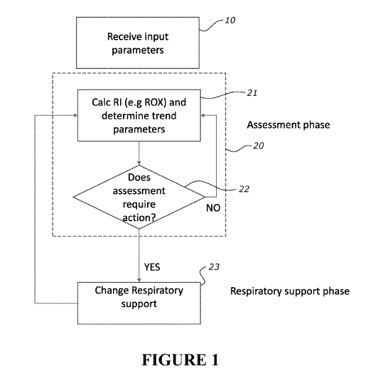
1. Overview
CA 03176719 2022-09-21
WO 2022/009000
PCT/IB2021/055293
31
Embodiments described herein provide apparatus, systems, and methods for
assessing a
patient's respiratory status (e.g. normal, distressed, deteriorating, or
improving, stable)
("assessment phase" or "diagnostic phase"); and based on that assessment
optionally
taking appropriate respiratory support action ("respiratory support phase").
For example, if
a patient is in respiratory distress, or is progressing to or in respiratory
failure in a
respiratory support phase, then escalation of respiratory support might be
made to reduce
further deterioration of respiratory status. The assessment is preferably made
when the
patient is receiving respiratory support, e.g. high flow respiratory support,
NIV, invasive
ventilation or the like.
In the assessment phase, a respiratory index can be determined. The
respiratory index is an
indicator of patient respiration, and the respiratory index (and/or change in
respiratory
index) can be used to determine a patient's (current) respiratory state and/or
change in
respiratory state - therefore leading to a determination of respiratory
status.
A respiratory status can be "normal", or "respiratory distress". Respiratory
distress can
range from mild to severe, as will be described later. The respiratory index
can indicate if a
patient is trending towards or experiencing: onset of respiratory distress or
is in respiratory
distress, and/or is experiencing or is trending towards deteriorating
respiratory distress.
(Deteriorating respiratory distress can lead to risk of or actual respiratory
failure, which is a
severe type of respiratory distress).
From the patient respiration, respiratory index and/or respiratory status, it
can be
determined if it is desirable to escalate respiratory support to improve the
patient's
respiratory status/relieve respiratory distress (in a respiratory support
phase). This might
be to prevent further deterioration of respiratory distress (stabilisation) or
to move the
patient out of respiratory distress into normal breathing. For example, if the
patient is in
mild respiratory distress, escalation of respiratory support might be used to
reduce the
chances that the patient deteriorates to a more severe level of respiratory
distress, such as
risk of respiratory failure, or actual respiratory failure. Or, if a patient
is already at risk of
respiratory failure (or has experienced respiratory failure) escalation of
respiratory support
reduces the risks of respiratory failure occurring (or continuing) and/or the
negative health
outcomes of respiratory failure.
However, escalating respiratory support (such as increasing high flow
respiratory support,
providing NIV pressure respiratory support, or providing invasive respiratory
support) has
its own risk, so it is undesirable to escalate respiratory support
unnecessarily. Likewise,
CA 03176719 2022-09-21
WO 2022/009000
PCT/IB2021/055293
32
when respiratory distress has lessened, it is often desirable to de-escalate
respiratory
support so as to remove the risk encountered due to the escalated respiratory
support.
Among other things, a reason for the assessment phase is to identify early
enough if a
patient's respiratory status is deteriorating so that action can be taken pre-
emptively
without doing so unnecessarily early. Escalating respiratory support early
improves health
outcomes, whereas delay can risk negative health outcomes.
For example, a patient might be receiving base respiratory support, in the
form of high flow
respiratory support e.g. nasal high flow respiratory support or tracheal high
flow respiratory
support. The assessment phase can be used to determine if escalating
respiratory support
could benefit the patient and should be implemented.
In one example, escalation of respiratory support can comprise escalating high
flow
respiratory support. This might take the form of increasing the high flow
respiratory support
parameters (e.g. flow rate, 02 concentration, humidification, or the like) of
respiratory
support, while de-escalating respiratory support can comprise reducing the
support
parameters. Such escalation might occur, for example, when it is determined
from the
respiratory index that the patient is in respiratory distress and is
deteriorating, but is not
yet at high risk of respiratory failure. Escalating high flow respiratory
support might stabilise
or even improve the patient respiratory status, meaning risk of respiratory
failure (and
hence even more invasive escalation) is avoided.
As another example, escalation of respiratory support can comprise moving to a
more
invasive respiratory support. This might be an escalation to NIV respiratory
support, or
invasive respiratory support. This might happen immediately or after
escalation of high flow
respiratory support. In the case of nasal/tracheal high flow respiratory
support, escalation of
respiratory support can also comprise going from nasal high flow to invasive
ventilation,
such as providing mechanical ventilation to an intubated patient. De-
escalation can
comprise:
if it is being used, removing invasive respiratory support and returning to
the base
respiratory support (for example, NIV respiratory support or nasal high flow),
or
if it is being used, removing NIV respiratory support and returning to the
base
respiratory support (for example, nasal high flow), or
if it is being used, de-escalating nasal high flow).
Alternatively, as another example of more invasive respiratory support,
escalation of
respiratory support can comprise transferring the patient to non-invasive
("NIV") pressure
CA 03176719 2022-09-21
WO 2022/009000
PCT/IB2021/055293
33
respiratory support. This might happen immediately or after escalation of high
flow
respiratory support. De-escalation can comprise removing NIV pressure
respiratory support
and returning to the base respiratory support (for example, nasal high flow).
Respiratory support can comprise using humidification also, for example with
nasal high
flow, tracheal high flow and/or NIV pressure respiratory support. As another
example,
escalation of respiratory support can comprise providing, or escalating,
humidification. This
can be for therapy and/or comfort, and might instead of or be in addition to
any one of the
other escalations described. De-escalation can comprise removing or reducing
humidification.
Therefore, the present embodiments utilise the change in respiratory index
(such as a trend
in respiratory index) to help ascertain when it is appropriate to escalate
and/or de-escalate
respiratory support. Using the respiratory index trend helps to improve the
timing of the
escalation of therapy as required to help improve stabilisation of the patient
(and improve
respiratory index, status etc.).
This provides improved health outcomes (including improved respiratory index,
status etc.)
as escalated respiratory support is provided when it is more likely that the
benefits of
providing escalated support outweigh the risk of providing escalated support.
Typically, the embodiments described relate to base respiratory support in the
form of nasal
and/or tracheal high flow respiratory support, which is high flow of gas
provided to a patient
to support respiratory function. Preferably, the high flow respiratory support
comprises
humidification for patient comfort.
In general terms, the present apparatus and methods comprise one or more of:
an assessment method,
carried out by an assessment apparatus, and
optionally a method and/or apparatus for providing respiratory support based
on the
assessment method outcome.
This allows a clinician and/or apparatus to determine a suitable change in
respiratory
status/state, e.g. changes in respiratory distress or deterioration toward
respiratory failure,
and allows a clinician and/or respiratory apparatus to escalate respiratory
support sooner.
For example, a patient can be intubated and provided with mechanical
ventilation earlier
than under current diagnostic methods. The methods described herein allow for
early
CA 03176719 2022-09-21
WO 2022/009000
PCT/IB2021/055293
34
detection of deterioration of a patient respiratory state e.g. increasing
respiratory distress
or deterioration that is indicative of potential respiratory distress (be that
mild or severe).
This allows a clinician/respiratory apparatus to escalate respiratory support
earlier, which
can increase chances of recovery and increase chances of survival.
Referring to Figure 1, in general terms, the apparatus and/or methods in
combination can:
a) in an assessment phase determine:
a respiratory index, and
determining from the change in respiratory index the respiratory status of the
patient and/or if respiratory support changes are required,
and
b) in a respiratory support phase, based on the assessment phase,
implement (by either the clinician and/or respiratory apparatus) respiratory
support
changes using a respiratory apparatus.
It is not necessary to have both a) and b). For example, just an assessment
stage a) might
be implemented.
The assessment phase can comprise one or more of:
= assessing the respiratory status, and determining whether it is normal,
abnormal,
deteriorating, stable, improving or the like,
= assessing whether a change in respiratory support is required (as a
result of
assessing the respiratory status),
= if the changes required, assessing what change in respiratory support is
required
(e.g. escalation, de-escalation, increasing or decreasing high flow therapy,
escalating
to NIV or invasive ventilation, deescalating from NIV or invasive ventilation
or the
like)
The implementation and the respiratory support phase can comprise:
= indicating any of the above outcomes of the assessment phase, e.g. by
alerts,
alarms, messages or other indicators, and/or
= making any of the changes determined in the assessment phase.
The assessment phase could be implemented:
= by a clinician alone,
= one or more assessment, therapy and/or other apparatus without a
clinician,
= or both the clinician and one or more apparatus.
CA 03176719 2022-09-21
WO 2022/009000
PCT/IB2021/055293
Likewise, the respiratory support phase could be implemented:
= by a clinician alone,
= one or more assessment, therapy and/or other apparatus without a
clinician,
5 = or both the clinician and one or more apparatus.
A respiratory index ("RI") is determined, and then optionally an assessment of
the patient
respiratory status is made from the change in respiratory index. Then, based
on either the
change in respiratory index and/or the patient respiratory status (which e.g.
might be
10 displayed to a clinician), optionally a decision is made whether a
change (e.g. escalation or
de-escalation) of respiratory support is required, and if so, what change.
Optionally a
respiratory apparatus is configured to provide the change in support, either
automatically or
by intervention by a clinician.
15 A respiratory index could be determined from/is a function of: one or
more lung mechanics
parameters (such as respiratory rate, expiratory time, minute ventilation),
and one or more
oxygenation parameters (such as Sp02, Fi02, Fd02, 02 fraction). These
parameters may
be calculated by a controller of the respiratory apparatus based on
signals/measurements
from the one or more sensors associated with the respiratory apparatus.
Alternatively the respiratory index could be characterised as follows: it is
may be a unitless
figure that is a function f(x) of one or more:
= Patient parameters, which might comprise among other things:
o physiological parameters, (which can comprise respiratory
parameters), and
o therapy parameters (therapy delivered to the patient);
and
= respiratory apparatus parameters (which can comprise operational
parameters).
Physiological parameters can comprise lung mechanics and/or oxygenation
parameters.
Typically, a respiratory index may use one or more of Sp02 (patient
physiological
parameter), Fi02 (patient therapy parameter), and respiratory rate (patient
physiological
parameter). Note, Fd02 could be used as a proxy for Fi02. Fd02 is the fraction
of delivered
oxygen, which is the 02 concentration of the gases stream and Fi02 is the
fraction of
inspired oxygen. They are related and similar in value, but not the same
¨although if the
gas flow is high enough and a patient does not entrain ambient air, Fd02 will
be
CA 03176719 2022-09-21
WO 2022/009000
PCT/IB2021/055293
36
substantially equivalent to Fi02 so Fd02 can be used as a proxy for Fi02. Fd02
is a
respiratory apparatus parameter. With respiratory apparatus that provide
oxygen, the
oxygen concentration (an operational parameter) provided by the apparatus can
be closely
related to Fi02 and can be used as a proxy of Fi02. But other parameters can
be used also.
The respiratory index can be a function of any set of parameters that provides
an indication
of respiration, from which a determination of a patient respiratory status
(such as the risk of
respiratory failure) can be made.
For example, the respiratory index RI could be defined as in general a
function of:
= one parameter representative of lung mechanics and one parameter
representative
of oxygen exchange, that RR = f(LM, 0), or
= respiratory rate (RR) - e.g. RI= f (RR), or,
= a function of respiratory rate and Fi02 - e.g. RI= f (Fi02, RR), or
= a function of respiratory rate, Fi02, and Sp02 - e.g. RI= f (Sp02, Fi02,
RR).
=
In one example, the respiratory index can be ROX which is defined as:
ROX = (Sp02/Fi02) / RR
where
Sp02 is the setpoint saturation of oxygen (%) in the blood of a patient, or
alternatively the actual saturation of oxygen (%) in the blood of a patient,
Fi02 is the fraction of inspired oxygen (%) by the patient (Fd02 could be used
as a
proxy), and
Respiratory rate is the breathing rate in breaths per minute.
A change in the respiratory index RI, e.g. over time, may be used to determine
patient
respiratory status. From the respiratory status optionally a determination can
be made
about whether the respiratory support needs changing. Or, from the from the
change in
respiratory index itself, optionally a determination can be made about whether
the
respiratory support needs changing.
For example, the change in respiratory index itself might provide an
indication of whether a
change in respiratory support is required. Or, for example, some relationship
between the
change in respiratory index and some other information ("relationship
information"); for
.. example, a parameter (e.g. threshold ¨ predetermined or otherwise) can
provide that
determination. Multiple values of the respiratory index might be used to make
a
determination (e.g. multiple values of the respiratory index might be
determined over time,
CA 03176719 2022-09-21
WO 2022/009000
PCT/IB2021/055293
37
and from that a trend determined which can be determined with a respiratory
changes
required). The relationship between multiple values of the respiratory index,
or some
information such as a parameter that generalises the multiple values and
another
parameter could be used. There are various options for determining whether
respiratory
support needs changing, which are underpinned by the respiratory index but
optionally in
addition to other information.
Looking at the change in respiratory index over time could comprise looking at
multiple time
instances of the respiratory index changing over time. For example, for each
of a plurality
of points in time, a (e.g. instantaneous) change in respiratory index over
time (at that point
in time) could be determined. This change (over the plurality of points of
time) of the (e.g.
instantaneous) change in respiratory index (over time) can be used to
determine a patient
respiratory status. That is, for example, a derivative of the respiratory
index over time could
be found at multiple times, and those multiple time instances of the
derivative of respiratory
index over time could also be differentiated over time to give a second
derivative of
respiratory index over time. For example, this could be an acceleration in the
change of:
respiratory index, patient parameter, patient state, and/or patient status.
The first and/or
second derivatives could be viewed or compared to relationship information
(e.g. slope
threshold other change indicator threshold) to assess patient respiratory
status. For
example, the derivative of respiratory index over time and/or the second
derivative of
respiratory index over time could be displayed in numerical and/or graphical
form.
Monitoring a change, over the plurality of points in time, of the change in
respiratory index
over time comprises, for the plurality of points in time: viewing the
displayed change in
respiratory index over time and/or calculating and comparing the change to
relationship
information. The change in respiratory index over time can be indicated by a
change
indicator.
As an example, a desirable option would be to look at a respiratory index over
time and
determine the trend of whether the respiratory index increases or decreases or
changes in
some other manner, or otherwise has some relationship change with respect to
relationship
data, such as a threshold (for example as shown in Figure 2). From any of the
above, an
indication of respiratory status can be determined and from that respiratory
support
changes made. Or optionally the respiratory index and its trend (such as its
magnitude and
direction) relative to a threshold can be used to directly determine if
respiratory support
.. changes required. The assessment using the respiratory index can provide a
trigger for
respiratory support changes.
CA 03176719 2022-09-21
WO 2022/009000
PCT/IB2021/055293
38
A threshold or other relationship information might be determined through
experimental
data, clinical research, user input, calculation, and/or otherwise. The
threshold or other
relationship might be predetermined or determined in real-time or input or
otherwise
provided. The threshold or other relationship might be fixed or varying
depending on other
parameters (for example it may change over time, or may change depending on
other
parameters used in the respiratory index or alternatively not used in the
respiratory index).
Non-limiting examples of a threshold could be a respiratory index threshold,
which
delineates normal v distressed respiratory status, and/or a slope threshold
(or other
"change indicator" threshold, depending on the indicator being used that
indicates a change
in respiratory index), which might delineate between a trend toward
improvement and
deterioration of respiratory index. There could be one or more respiratory
index or slope
thresholds.
As some examples, the respiratory index and/or change in respiratory index can
be
assessed using one or a combination of the following:
= Comparing the respiratory index (e.g. ROX) v one or more respiratory
index
threshold(s).
= Comparing the change in respiratory index over time v one or more
respiratory index
threshold(s)
= Comparing a change indicator (e.g. vector, slope or other measure of
magnitude
and/or direction) of change in respiratory index over time v a threshold (e.g.
one or
more slope threshold(s))
= Comparing respiratory index v patient parameter(s), or some function of
patient
parameters(s) (such as Sp02, Fi02 or the like), such as in graphical and/or
numerical form that can be displayed and monitored
= Comparing the change in magnitude (drop or climb) of the respiratory
index over
time to a threshold(s). e.g. a slow (that is, small slope) but significant
difference
(that is large delta in ROX) in respiratory index at time=1 v respiratory
index at time
=2 might indicate deterioration. Likewise, a large but slow positive change in
magnitude could be an indication of improvement.
Comparing the change of any of the above versus time as a first derivative to
some
relationship data, and/or comparing that change in time versus time
(acceleration of
change) as a second derivative to some relationship
The examples above refer to the use of a respiratory index to determine the
respiratory
status of a patient.
CA 03176719 2022-09-21
WO 2022/009000
PCT/IB2021/055293
39
In an alternative, it is possible to use respiratory index and patient
parameters (which could
be constituent components of the respiratory index) to make the assessment.
Therefore, in
more general terms, one or more respiratory parameter(s) can be used to assess
the
patient status along with a respiratory index. This can be particularly useful
when a clinician
is making the assessment and wants to drill down deeper into what is causing
the
respiratory status. The respiratory index itself can indicate the respiratory
status (e.g. a
change in respiratory state) but may not indicate why that change is
happening. In
contrast, patient parameters such as respiratory rate, Fi02 and/or Sp02 might
provide
insights into that. This will be described in further detail later.
Respiratory index, patient
parameters and any other information which relates to patient state or status
to make an
assessment can be termed "assessment information". Assessment criteria can be
used to
help with the assessment.
The above is not limiting. The respiratory status assessment can be more
generally carried
out by:
displaying assessment information, assessment criteria and/or relationship
information as numbers, plots and/or or other graphical indications on a
display (either
static or animated and/or in 2 or 3 dimensions - for example with time on one
axis), and a
person viewing that information, and/or
an apparatus comparing the above to relationship information.
The optional determination of whether respiratory support changes are required
based on
using the respiratory index can be termed a "diagnostic stage" or
alternatively a "trigger".
Alternatively, the assessment can be considered the diagnostic stage,
irrespective of
whether a course of action is determined.
In many cases, it is the clinician that will assess the respiratory status
based on information
displayed on the assessment apparatus. By displaying various combinations of
graphical and
numerical representations of parameters and respiratory indexes, it is
possible for a trained
clinician to interpret these to gain an indication of respiratory status, and
in particular the
direction of respiratory state and what interventions might be required. This
enables a
clinician to make decisions quickly in situations where there might be many
patients that
are being monitored simultaneously - e.g. in an Emergency Department. These
assessments might be assisted by indications (alarms, messages and the like)
from the
assessment apparatus and/or automated decisions made by an assessment
apparatus.
Similarly this same advantage can be achieved for homecare patients that is, a
clinician
CA 03176719 2022-09-21
WO 2022/009000
PCT/IB2021/055293
monitoring multiple patients remotely can quickly assess a patient that is
deteriorating or
potentially going to suffer respiratory failure.
Traditionally, a clinician has had to use subjective measures based on limited
information to
5 assess a patient's respiratory status. Additionally they have had to use
invasive testing
methods. The present embodiments improve on this. In general terms, the
assessment can
be made by displaying graphically and/or numerically a respiratory index, a
change
indicator, patient parameters and/or thresholds etc. in various combinations
to provide the
supporting information for a clinician to make the decision. Numerical
information, two or
10 three-dimensional plots, animations, moving plots, zooming in and
drilling down into
information and contextual information can also be used to make an assessment.
Providing
visual, contextual and/or accessible information will assist a clinician to
make quick
decisions in an objective manner, as opposed to having to use solely
subjective
considerations. This also enables treatment by exception ¨ addressing those
who need help
15 most first. Examples will be described herein later.
The assessments (determination) are carried out during a session. A session
might be
defined by a treatment session, a day or part thereof, a night or part
thereof, sub-sessions,
or a length of time (e.g. 5 hours) for example. Generally, the comparisons are
made
20 relative to thresholds and/or previous respiratory index parameters
determined in the same
session. That is, the assessments are based on what is happening in real-time.
Assessments
might be made on a time-period-by-time-period basis, where each time period is
a part of a
session. E.g. assessments might be made on a minute-by-minute basis, ten
minute-by-ten
minute basis, hour-by-hour basis or the like. Over this time frame, assessment
can
25 comprise considering the change between respiratory index and/or
constituent parameters
from time period to time period. Such assessments can be based on the change
in
respiratory index and/or constituent parameters either alone and/or compared
to
relationship information also. Assessment (determination) can also
alternatively be made
within and/or across multiple sessions. For example, over multiple sessions,
assessment can
30 comprise considering the change between respiratory index and/or
constituent parameters
from session to session (and/or within a session). Such assessments can be
based on the
change in respiratory index and/or constituent parameters either alone and/or
compared to
relationship information also.
35 In a home environment, the clinician will most likely not be situated
with the patient. In that
case, patient respiratory index/status can be monitored remotely. For example
this could be
by way of an assessment apparatus that provides assessment information
remotely to a
CA 03176719 2022-09-21
WO 2022/009000
PCT/IB2021/055293
41
clinician and/or might be by way of the clinician possessing the assessment
apparatus,
which remotely communicates with the respiratory support apparatus and/or any
other
apparatus (such as sensors) that are required to make the assessment. The
respiratory
support apparatus and/or assessment apparatus will have communications
functionality
55B, 55A to enable the clinician to monitor the patient remotely. For example,
a remote
assessment apparatus may calculate the respiratory index and/or changes in the
respiratory
index based on measurements from the respiratory support apparatus as with the
patient.
The respiratory index may be calculated in the respiratory index values
provided to the
clinician. The processing may take place on the respiratory apparatus and the
processed
information provided to the clinician, and/or alternatively, the clinician as
an assessment
apparatus that receives the raw data and makes the assessment. There might be
a remote
patient monitoring system 57, e.g. comprising at least a remote server 57,
which receives
information from the assessment apparatus and/or respiratory support
apparatus, via a
network 56 which the clinician can access. For example, the clinician might
access the
information via a web browser/web server. The assessment apparatus might be
the server,
respiratory apparatus, mobile device and/or any other assessment apparatus.
The remote patient monitoring system 57 further comprises one or more
databases, a
reporting engine to generate patient reports and other suitable components
that allow
monitoring of a patient, generation of patient reports (e.g. that include
usage of a
respiratory support apparatus, therapy settings etc.). The remote patient
monitoring system
57 allows a clinician to manage multiple patients remotely e.g. allow
management of
patient's while the patients are at home (i.e. outside hospital).
The respiratory index data may be provided to the remote patient monitoring
system 57.
The respiratory index measurements and change in respiratory index may be
incorporated
into a patient report that includes measured patient parameters e.g. Sp02,
flow rate,
humidity set point and usage hours and the change in respiratory index and
respiratory
index measured values over time.
The change in respiratory index allows a clinician to assess if the current
therapy being
provided is being effective and also allows a clinician to make a change in
the therapy
provided. In one example the operational parameters of the respiratory support
apparatus
(e.g. prescription settings) may be remotely updated based on the change in
the respiratory
index. The change in the respiratory index can be used by the remote
monitoring system to
generate alerts or messages to clinicians and patients. The remote patient
monitoring
system 57 may automatically change therapy settings e.g. change high flow
therapy
CA 03176719 2022-09-21
WO 2022/009000
PCT/IB2021/055293
42
settings such as oxygen fraction (i.e. % oxygen in gases) and/or flow rate
and/or humidity
(e.g. dew point or relative humidity or absolute humidity) remotely, based on
the changes
in the respiratory index. Examples of how therapy settings (i.e. therapy
parameters) are
changed are described later. The therapy settings, specifically for example
high flow
respiratory support apparatus settings may be changed and transmitted as a new
prescription to the respiratory support apparatus 10 via the network 56. The
respiratory
support apparatus 10 may incorporate these changes and begin to operate based
on the
new prescription (i.e. updated therapy settings i.e. updated therapy
parameters).
Alternatively a clinician or healthcare professional may make a change to the
therapy
settings at the remote monitoring system 57 based on the changes in the
respiratory index.
These clinician changes are transmitted to the respiratory support apparatus
10 via the
network 56. The new settings are transmitted as a new prescription. The
respiratory
support apparatus makes changes defined in the new prescription (i.e. updates
its therapy
settings) to operate as per the new settings.
The process of monitoring a respiratory index can continue. This remote
monitoring
provides an effective out of hospital monitoring of the patient. It allows
clinicians to assess
patient respiratory status and determine if high flow therapy is assisting the
patient by
checking the change in the respiratory index. The change in respiratory index
is indicative of
a change in the respiratory status of the patient. The disclosure provides an
effective out of
hospital monitoring of the patient. It further allows remotely changing
therapy settings a
respiratory support apparatus associated with a patient based on the assessed
change in
respiratory index.
Once a determination has been made (trigger), the respiratory support changes
are
determined. The support the respiratory support changes could be determined by
a clinician
and/or by the assessment apparatus.
This might result in one or more changes to use of the respiratory support
apparatus, be
that:
operational changes (either automatic or clinician instigated) such as
increasing flow
or oxygen fraction provided to a patient;
use changes (such as intubating a patient instead of providing high flow
through an
unsealed cannula), including providing entirely different therapy - e.g.
ventilation,
provision of indications (e.g. alerts, alarms and/or instructions) to a
clinician that:
a change needs to be made,
how and/or when to change therapy, and/or
configurational changes.
CA 03176719 2022-09-21
WO 2022/009000
PCT/IB2021/055293
43
These are not limiting of the possible respiratory support change options.
As part of the assessment phase, irrespective of whether it is carried out by
a person and/or
an apparatus, an indication and/or change in therapy may be used to indicate
that there
has been a determination made and/or there has been a change in therapy and/or
there
should be a change in therapy and/or what a change in therapy should be. For
example:
= Audible alarm - this may be made to indicate a determination has been
made, what
the determination is (e.g. worsening) and/or that some therapy change will or
may
have occurred. The alarm may be made on the therapy device or a separate
device
(e.g. a doctor's phone). Different sounds may indicate different patient
states (or
different changes to a patient's state).
= Visual alarm - this may be made to indicate a determination has been made,
what
the determination is (e.g. worsening) and/or that some therapy change will or
may
have occurred. The alarm may be on the therapy device or a separate device.
Different visual outputs may indicate different patient states (or different
changes to
a patient's state).
= Message - this may be made to indicate a determination has been made,
what the
determination is (e.g. worsening) and/or that some therapy change will or may
have
occurred. The message may be sent to one or more recipients/devices (each
recipient/device may receive a different message or the same message). This
message may include a warning and/or suggest a change to one or more device
set
points. The message may include a suggestion for the specific value(s) that
the set
point(s) should be changed to. The calculation of these specific values could
be
performed on the therapy device or on a separate device.
= Automatic change to device set point(s) - after a determination (e.g.
worsening
patient status) a controller associated with the device may change one or more
set
points if the index value/slope suggests that the patient needs different
support, for
example, more flow. The calculation of the suitable change(s) could be
performed on
the therapy device or on a separate device.
= Device turns off - after a determination (e.g. improving patient status)
a controller
associated with the device may turn the therapy/device off if the index
value/slope
CA 03176719 2022-09-21
WO 2022/009000
PCT/IB2021/055293
44
suggests that the patient no longer needs the therapy (i.e. if their index
value/slope
is on the low-risk side of a threshold and/or is moving in a good direction).
The above could be provided on the apparatus itself or remotely to another
apparatus,
.. where a clinician might be based remotely.
This list is not exhaustive.
The assessment apparatus might be in the vicinity or possession of the
clinician. This might
.. be in a hospital environment, for example. Alternatively, the assessment
apparatus might
be remote from the clinician. It would have communications functionality to
enable
communication to a remote clinician. This might be in a home environment, for
example,
where the clinician is not situated with the patient/respiratory apparatus
and/or assessment
apparatus. In another alternative, the respiratory apparatus might be with the
patient, but
the assessment apparatus remote with a remote clinician elsewhere.
As noted above, the embodiments described will typically be related to use of
(initially at
least) a nasal or tracheal high flow respiratory support apparatus 10,
generally shown in
Figure 3. But this is not limiting, and for example, the embodiments could be
used in
relation to NIV or other respiratory apparatus. There is the apparatus 10
which provides
.. the respiratory support and the apparatus 20 which determines the
diagnostics. These may
be one and the same apparatus (e.g. respiratory support apparatus) or
different apparatus
(e.g. respiratory support apparatus and a mobile device). If separate
apparatus (as in
Figure 3), together they can form a respiratory assessment and support system
1.
For example, referring to Figure 3, there could be a respiratory support
apparatus that has
a controller (could also be referred to as a processor) 19, I/O interface 54,
flow generator
50 and humidifier 52. The respiratory support apparatus could be configured to
receive air
and/or supplementary gas such as oxygen. It could provide a flow of gas to a
patient
through a breathing conduit 5 and patient interface 51, e.g. an unsealed
interface (such as
but not limited to a nasal cannula) or tracheal interface. The controller 19
can operate the
device to provide the required flow rate and/or pressure, temperature,
humidity, oxygen or
other gas fraction and the like, based on input from sensors 11 in the system
or to which
the system is connected. They could be non-invasive sensors.
The apparatus also has (integrated within it) and/or connects to sensors 12
that provide:
= physiological parameters, (which can comprise respiratory parameters),
and
CA 03176719 2022-09-21
WO 2022/009000
PCT/IB2021/055293
= respiratory apparatus parameters (which can comprise operational
parameters)
to obtain parameters for operation of the apparatus (such as temperature,
humidity,
pressure, flow sensors) and parameters to determine the respiratory index
(such as Sp02,
5 Fi02 (or Fd02 ¨ fraction of delivered oxygen to a patient), and
respiratory rate). Any
reference to Fi02 could be replaced with a reference to Fd02, 02 fraction, or
any other
oxygenation parameter and vice versa ¨ the variations can be used
interchangeably.
Physiological parameters can comprise lung mechanics and/or oxygenation
parameters.
10 As possible examples:
Note, any of these could be wearable (e.g. see 54E in Figure 6).
For Sp02 the apparatus could connect to physiological sensors such as pulse
oximeter or
other blood oxygen sensor. This may be a wearable device e.g. see 54E in
Figure 6. There
15 .. could be a wireless pulse oximeter (Sp02 sensor). Wireless pulse
oximeter could
communicate via Bluetooth or infrared or other communications protocol.
Wireless Sp02
allows patient mobility e.g. patient can walk around while still using Sp02
and the
assessment apparatus still receives measurements. The Sp02 sensor connects to
the
respiratory or assessment apparatus. The respiratory apparatus is a non-
invasive sensing
20 unit that also provides respiratory support. The respiratory apparatus
can process the
sensor signals and calculate the index/change in respiratory index.
Alternatively change in respiratory index could be done at a remote monitoring
system.
25 For respiratory rate, this can be measured/determined using a pressure
sensor or a flow
sensor or any other respiratory rate sensor from which respiratory rate can be
determined.
For example, respiratory rate can be calculated using a flow sensor, and then
applying a
mathematical process to the flow sensor readings to determine respiratory
rate. In one
example the respiratory support apparatus is configured to use the flow
sensor, process the
30 flow sensor to filter out the flow signals from the blower. A frequency
response of the
filtered flow signal is determined (e.g. a Fast Fourier Transform or other
frequency
response) to calculate a peak frequency. The peak frequency corresponds to the
respiratory
rate. Other frequency analysis techniques may be applied to the flow signal to
determine a
respiratory rate. For example, the frequency analysis can determine one or
more local
35 maxima, and identify the highest magnitude maxima as the respiratory
rate. Alternatively, a
motor speed signal is processed (e.g. filtered) from the flow signal and
frequency analysis is
conducted, where the local maxima and frequency with the highest magnitude is
respiratory
CA 03176719 2022-09-21
WO 2022/009000
PCT/IB2021/055293
46
rate. Alternatively, a frequency analysis using Geortzel algorithm/Geortzel
analysis might be
done.
Alternatively, respiratory rate measurement can be achieved by having a
temperature
sensor at the cannula e.g. incorporated into the cannula and determining
temperature
differential. Difference in temperature is indicative of respiratory rate. A
similar
measurement can be achieved by pressure sensor or flow sensor at the cannula
and
changes in pressure or flow can be indicative of respiratory rate.
Alternatively respiratory rate may be determined from other sensors such as
(but not
limited to), a pulse oximeter, a respiratory rate sensor, a pressure sensor in
the device; or a
pressure sensor or flow sensor in the tube or patient interface; or chest band
sensors or any
combination thereof. One or more of these could optionally be a wearable
device or an
actigraphy device (e.g. see 54E in Figure 6) that is configured to measure
respiratory rate.
The wearable device may be, e.g., a wrist-worn device that is in wireless
communication
with the flow generator or smartphone.
For example, the respiratory rate is calculated by based on pressure signals.
The pressure
signal can be measured by a pressure sensor at the patient interface or using
a pressure
pathway, the sensor can be in the device and the pressure values can be
communicated
back to the device. The differences in pressure can be processed and the
respiratory rate
can be calculated by measuring the number of 0 crossings. A similar approach
can be used
using flow readings. The cyclic nature of the P or F signal can be processed
to get a
respiratory rate.
Alternatively, a suitable sensor e.g. a respirometer can be used to measure
the gases
exchange (e.g. CO2 exchange) at the interface to determine respiratory rate.
Alternatively,
a photoplethysmogram or an electrocardiogram can be used to measure.
Alternatively, any sensor, such as an accelerometer can be placed on the chest
or an
abdominal wall to measure respiratory rate. The sensor can be separate from
the device
with wireless communication.
Alternatively, respiratory rate could be determined as described in
US202101133796 which
is incorporated herein in its entirety.
CA 03176719 2022-09-21
WO 2022/009000
PCT/IB2021/055293
47
In one example expiratory time may be calculated from flow sensor signals or
pressure
signals or a combination of flow and pressure sensor signals. Below is an
example of
expiratory time calculation for an unsealed system that provides high flow
respiratory
support via an unsealed cannula e.g. apparatus 10 disclosed herein. The method
is
executed by the controller. The controller of the apparatus receives a flow
signal
representative of a flow rate of the gases. The controller pre-process and
filters the flow
rate signal. The controller then determines a primary breathing parameter
ratio e.g. a ratio
between inspiratory time and total respiratory time, and/or expiratory time
and total
respiratory time for a patient's breathing cycle. Determine or receive
respiratory rate.
Respiratory rate may be determined as per above or received manually at the
respiratory
support apparatus. The apparatus uses the breathing parameter ratio and the
respiratory
rate to determine an expiratory time. Further minute ventilation may be
determined from
the respiratory rate and the measured flow rate. In a further example
expiratory time may
be calculated as described in US provisional application US 63/146,184 filed
on 5 February
2021, the contents of which are incorporated herein in its entirety.
The measured or calculated respiratory rate is stored in the NHF therapy
device. The Fi02
readings are also stored in the NHF device over a period of time.
Alternatively, or
additionally, these and/or any other readings might be transmitted to and
stored in another
.. device, such as a smart phone.
Optionally, respiratory rate may be manually inputted via user interface.
Other options are possible also.
Fi02 can be found by measuring Fd02 or another measure of concentration of 02
in the gas
flow using any suitable sensor e.g. an ultrasonic sensor or other gas
concentration sensor.
For example, this could be done with a flow sensor and 02 concentration
sensor, where
optionally the flow sensor is an inline flow sensor. This sensor could be
positioned
.. downstream of a mixer of ambient air and 02.
An ultrasound sensor could be used to allow fast measurement of Fi02 that is,
Fd02. This
allows for fast response and control of oxygen in the gases stream. As
mentioned earlier
Fd02 can be a proxy for Fi02. The measured Fd02 is reported and presented on a
screen of
.. the apparatus as Fi02. During high flow respiratory support the flow rate
is sufficiently high
such that ambient air is not entrained at the nasal cannula when the patient
is inspiring.
Therefore the delivered oxygen fraction Fd02 is denoted as equivalent to Fi02
(inspired
CA 03176719 2022-09-21
WO 2022/009000
PCT/IB2021/055293
48
oxygen fraction). The ultrasound (i.e. ultrasonic) sensor allows for fast
response i.e. quick
measurement and therefore fast response. A valve on the oxygen inlet can be
controlled to
change the oxygen fraction in the gases stream (thereby affecting Fd02).
Any of the parameters above could be manually input instead and respiratory
index
calculated from that.
The assessment apparatus and/or respiratory support apparatus might also have
respective
communications functionality. This could be a modem or other transceiver. This
enables
the assessment apparatus and the respiratory support apparatus to communicate
with each
other, whether remote or in the same location, and/or also communicate to a
remote
clinician. This would enabler clinician to remotely monitor patient and their
respiratory
support and/or change as required. There might be a remote server, which
receives
information from the assessment apparatus and/or respiratory support
apparatus, which the
clinician can access. For example, the clinician might access the information
via a web
browser/web server. The assessment apparatus might be the server, respiratory
apparatus,
mobile device and/or any other assessment apparatus
A high flow respiratory apparatus 10 in general is described with reference to
e.g. Figure 3.
In general terms, the apparatus comprises a main housing 10 that contains a
flow generator
50 in the form of a motor/impeller arrangement, an optional humidifier 52, a
controller 19,
and a user I/O interface (comprising, for example, a display and input
device(s) such as
button(s), a touch screen, or the like). An input for supplementary oxygen or
other
supplementary gas can be provided. A valve (e.g. proportional valve) can be
provided in
fluid communication with the supplementary gases inlet and configured to
control the
amount of supplementary gases introduced into the apparatus. The screen may be
a
detachable screen. The controller 19 is configured or programmed to control
the
components of the apparatus, including: operating the flow generator to create
a flow of
gas (gas flow) for delivery to a patient, operating the humidifier (if
present) to humidify
and/or heat the generated gas flow, receive user input from the I/O interface
for
reconfiguration and/or user-defined operation of the apparatus, and output
information (for
example on the display) to the user. The user could be a patient, healthcare
professional, or
anyone else interested in using the apparatus. A patient breathing conduit is
coupled to a
gas flow output in the housing of the flow therapy apparatus, and is coupled
to a patient
interface 51 such as a nasal cannula with a manifold and nasal prongs. The
patient
breathing conduit can have a heater wire 5 to heat gas flow passing through to
the patient.
CA 03176719 2022-09-21
WO 2022/009000
PCT/IB2021/055293
49
Some examples of high flow respiratory apparatus are disclosed in
International Application
No. PCT/NZ2016/050193, titled "Flow Path Sensing for Flow Therapy Apparatus",
filed on
December 2, 2016, and International Application No. PCT/IB2016/053761, titled
"Breathing
Assistance Apparatus", filed on June 24, 2016, which are hereby incorporated
by reference
in their entireties. Examples of configurations of high flow respiratory
apparatus that can be
used with aspects of the present disclosure are also discussed in further
detail below.
The respiratory support apparatus might determine the respiratory index and/or
change in
respiratory index and make the assessment leading to any changes in
respiratory support.
.. Alternatively, the relevant information may be communicated to a separate
assessment
device where the determination of the respiratory index, change in respiratory
index,
patient respiratory status and/or any changes in respiratory support can be
made.
Information regarding change in respiratory support can then be communicated
back to a
clinician and/or the respiratory support apparatus for the appropriate action.
Additionally, or
.. alternatively, information might be transferred to a physician server 100
or a remote server.
For example, the clinician might access the information via a web browser/web
server. The
assessment apparatus might be the server, respiratory apparatus, mobile device
and/or any
other assessment apparatus. There can be wired and/or wireless communication
between
the respiratory support apparatus and clinician device, such as a smartphone.
The respiratory support apparatus may comprise a controller configured to
control the
blower to provide Bi-level pressure therapy. The respiratory support apparatus
may be
coupled to a sealed interface e.g. a full face mask. Optionally an NIV tube
may be coupled
that has a lower resistance to flow than the high flow tube (as shown in
figures). A user
may manually select an NIV mode (e.g. Bi-level pressure therapy or CPAP
therapy) mode
and the controller is configured to operate in the selected mode.
Alternatively the
respiratory support apparatus could be configured to detect the connection of
a sealed
interface and/or a connection of an NIV tube and automatically change control.
.. In a further alternative a patient may be physically coupled to a suitable
pressure support
device e.g. a NIV device.
In general terms, there are various embodiments of a respiratory index and how
a change
in respiratory index is used to assess patient respiratory status and/or a
change in
respiratory support; and any of these embodiments can be used in combination
with any of
various embodiments of respiratory support apparatus and (where applicable) a
separate
assessment device to carry out:
the assessment phase, and optionally
CA 03176719 2022-09-21
WO 2022/009000
PCT/IB2021/055293
the respiratory support phase (information on and/or change in respiratory
support)
2. Assessment method
2.1 Overview of assessment method
5 Referring to Figure 1, a change in respiratory index is used to determine
an appropriate
respiratory support action. As an interim step, a patient respiratory status
might be
determined from the change in respiratory index, but that is not essential.
A respiratory index could be determined from/is a function of: one or more
lung mechanics
10 parameters (such as respiratory rate, expiratory time, minute
ventilation) and one or more
oxygenation parameters (such as Sp02, Fi02, Fd02, 02 fraction).
The respiratory index may be a unitless figure that is a function f(x) of one
or more patient
parameters being:
15 = physiological parameters, (which can comprise respiratory
parameters), and
= respiratory apparatus parameters (which can comprise operational
parameters)
Physiological parameters can comprise lung mechanics and/or oxygenation
parameters.
20 A respiratory index can be calculated based on sensor inputs and/or user
inputs that provide
the above data. A respiratory index as an example is based on respiratory rate
(respiratory
rate) and Fi02 (oxygen concentration delivered to patient). The index is
preferably related
to 1/RR and/or 1/Fi02. In one example the index = A/B (RR*Fi02), where A and B
could be
constants or other values.
One example of a respiratory index is a ROX index.
A ROX index takes the form of a function
ROX(x) = f(F102, Sp02, RR)
and is calculated as follows:
ROX = (Sp02/Fi02) / RR
Where:
Sp02 is the setpoint saturation of oxygen (%) in the blood of a patient, or
alternatively the actual saturation of oxygen (%) in the blood of a patient,
CA 03176719 2022-09-21
WO 2022/009000
PCT/IB2021/055293
51
Fi02 is the fraction of inspired oxygen (%) by the patient (Fd02 could be used
as a
proxy), and
respiratory rate is the breathing rate in breaths per minute.
The above are "input parameters", received via sensors and/or a user via a I/O
interface or
preconfigured, step 10.
The Sp02 is received for example from a sensor, such as a pulse oximeter, or
from
apparatus Sp02 set point. Respiratory rate is, for example, received from a
sensor such as
described previously. Fi02 can be measured for example via a sensor or
inferred from the
oxygen concentration (e.g. Fd02) provided by the apparatus to a patient. These
are
examples only. More details of how the information is obtained is described
with respect to
the apparatus embodiments.
Where ROX index is used, in a preferred embodiment the ROX index threshold for
respiratory failure is 4.88, which has been determined by experimental data.
In that case, if
the ROX index is greater than or equal to about 4.88 the patient is deemed to
be at low risk
of respiratory failure. However, if the ROX index is below about 4.88 then the
patient is
deemed to be at high risk of respiratory failure. It should be noted that a
clinically
insignificant deviation from 4.88 as the threshold is possible and should not
be excluded
from scope of the embodiment. Clearly also, the threshold could be a different
value, where
appropriate.
However, having simply a single instance in time of ROX index relative to the
threshold in
itself may not be sufficient information to determine whether the current
therapy settings
are resulting in effective treatment for the patient. The trend over time of
the ROX index (or
more generally respiratory index) and/or it's relation to the threshold may
also be useful.
In an assessment phase step 20, a respiratory index (e.g. ROX index) can be
calculated,
step 21, and then a change in respiratory index over time can be determined
and used to
determine success of high flow respiratory support and whether change is
required, step 22.
For ROX index, if the Fi02 and respiratory rate increase, the ROX index begins
to drop in
value. An increase in Fi02 and respiratory rate is indicative of the patient
deteriorating. The
continuous monitoring of the ROX index (resulting in a change of ROX index) is
helpful when
the patient is an unstable condition.
CA 03176719 2022-09-21
WO 2022/009000
PCT/IB2021/055293
52
As an example, referring to Figure 1, the apparatus receives input parameters,
step 10, and
determines the respiratory index (e.g. ROX index) and the change in
respiratory index over
time ¨ for example through calculation, step 21. Optionally, an assessment is
then made as
to the respiratory status of the patient based on the change of respiratory
index over time.
This can be done by determining the relationship of the change in respiratory
index over
time to other information. For example, the change in respiratory index over
time is
compared to a threshold to determine if the patient respiratory status is
"respiratory
distress" or even further a level of distress such as "severe" (e.g. the
patient has a high risk
of respiratory failure).
An assessment of a change in respiratory support can optionally be determined,
step 22,
based on the patient respiratory status (or directly from the ROX index or
other respiratory
index as it changes over time). (Because the patient respiratory status can be
assessed
based on the relationship of the respiratory index change over time to a
threshold, then an
assessment of the change in respiratory support, step 22, 23, can
alternatively be
considered to be determined based on the relationship of the respiratory index
change over
time relative to the threshold). Using the ROX index over time and the
threshold, the
change in respiratory support, step 23, can be an escalation of respiratory
support, or a de-
escalation of respiratory support. Details of a change in respiratory support
(either
escalation, de-escalation or otherwise) is described later with respect to the
apparatus
embodiments.
The example above refers to the use of a respiratory index to determine
respiratory status
of a patient. As noted earlier, in an alternative, it is possible to use
patient parameters
(which could be constituent components of the respiratory index) to make the
assessment.
Therefore, in more general terms, one or more respiratory parameter(s) can be
used to
assess the patient status instead of and/or as well as a respiratory index.
In many cases, it is the clinician that will assess the respiratory status
based on information
displayed on the assessment apparatus. Displaying various combinations of
graphical and
numerical representations of parameters and respiratory indexes makes it
possible for a
clinician to gain an indication of respiratory status, and in particular the
direction of
respiratory state and what interventions might be required.
In the case of clinician assessment, the display of information assists the
clinician, both in
terms of the information displayed in the manner that can be reviewed and
accessed.
CA 03176719 2022-09-21
WO 2022/009000
PCT/IB2021/055293
53
In general terms, the assessment can be made by displaying graphically and/or
numerically
respiratory index, a change indicator, patient parameters and/or thresholds
etc. in various
combinations to provide the supporting information for a clinician to make the
decision.
Examples will be described herein later.
Any one or more of the following parameters could be displayed numerically
and/or
graphically in any suitable combination to assist clinician assessment. Any
particular
parameter in itself might not warrant action, but when considered in
combination with other
information may indicate that action is required. For example, a ROX index on
its own may
not flag any concerns, but a worsening change in the ROX index over time might
do so. Or,
even a worsening change in ROX index over time might not be cause for concern,
unless it
is also accompanied by a concerning change in some other parameter, such as
respiratory
rate, Sp02 or Fi02 for example. Also, absolute or relative changes in any of
the above
might not be any cause for concern, unless they exceed a certain threshold. By
providing
access to various combinations of such information, a clinician gets a richer
set of
information from which to assess respiratory status.
Some of the following parameters (assessment information, assessment criteria
and/or
relationship information) that could be used alone or in combination are:
= respiratory index
= change in respiratory index over time
= patient parameter (e.g. respiratory rate, 5p02, Fi02)
= change in patient parameter over time
= change indicator (such as slope, vector, magnitude, difference, angle)
that indicates
the change (of any parameter herein) over time or over another measure
= respiratory index threshold
= change indicator threshold.
= These could be displayed graphically (that is as a 2D and/or 3D
plot/graph or other
suitable visual output) or numerically, for example as some of the following:
= respiratory index versus time
= change in respiratory index this is time
= patient parameter versus time
= change patient parameter versus time
= respiratory index threshold
= change indicator threshold.
CA 03176719 2022-09-21
WO 2022/009000
PCT/IB2021/055293
54
= Input can be received from the user to manipulate graphical information
to get more
insights. For example, this could comprise:
= zooming in and/or moving a graph
= drilling into a graph to get further graphs on underlying parameters,
e.g. selecting a
respiratory index versus time graph and then getting patient parameter versus
time
plots.
the above is not limiting, and various examples will be described later.
Possible examples now follow, but should not be considered limiting to the
above more
general embodiment.
2.2 Embodiment 1 ¨ assessment method ¨ respiratory index trend
In this embodiment, referring to Figure 1, a respiratory index is determined
over time, and
the change in respiratory index over time is used to assess respiratory status
and/or
changes in respiratory support.
The respiratory index could be any as previously described such as a function
of:
= one parameter representative of lung mechanics and one parameter
representative
of oxygen exchange, or
= respiratory rate - e.g. RI(RR), or,
= a function of respiratory rate and Fi02 - e.g. RI (Fi02, RR), or
= a function of respiratory rate, Fi02 and Sp02 - e.g. RI(Sp02,Fi02, RR)
The respiratory index change over time is assessed, step 20. Optionally, the
patient's
respiratory status is assessed and for example, in effect, a trend in the
change in
respiratory index is used to indicate patient respiratory status. Referring to
Figure 2 or 5, if
the respiratory status trends towards a better value (e.g. goes towards,
crosses or goes
beyond a threshold value) and/or trends towards a better value at or above a
threshold
rate, then an assessment can be made that the patient respiratory status is
good or is
improving. In contrast, if the respiratory status trends towards a worse value
(e.g. moves
away from or crosses or goes beyond a threshold value in the other direction)
and/or trends
towards a worse value at or above a threshold rate, an assessment can be made
that the
patient respiratory status is bad or is deteriorating. From this, decisions
about any change
in respiratory support can be made. It should be noted that respiratory status
does not
actually need to be determined. Decisions about change in respiratory support,
step 22, 23,
can be made from the change in respiratory index over time without explicitly
determining
CA 03176719 2022-09-21
WO 2022/009000
PCT/IB2021/055293
respiratory status. But the relationship between respiratory index and
respiratory status can
be used to predetermine suitable respiratory support actions when a particular
respiratory
index or change in respiratory index (e.g. trend) occurs.
5 The trend can be characterised in any suitable way, in the form of a
respiratory index
change (trend) parameter. This characterises the trend/change both in terms of
magnitude
and direction. As an example, the trend could be in the form of a vector
showing the
change in respiratory index (over e.g. time but it could be with respect to
another suitable
parameter). In addition, information about a threshold value which indicates
crossing
10 between a lower risk value and a higher risk value for the respiratory
index can be utilised
to assess the trend. The vector might be found, for example, from a derivative
of the
respiratory index versus time plot, which gives a slope and therefore
direction and
magnitude of the change. It may not just be the direction of the change that
is important,
but also the magnitude (that is, rate of change) which indicates how quickly
the change is
15 occurring. A second derivate wrt to time could be used to see the
acceleration of change.
The second derivative can be displayed numerically and/or graphically.
Going a step further, a change in trend over time could also be considered.
For example, a
derivative of respiratory index v time is assessed over time, and optionally a
second
20 derivative of respiratory index (or some other measure of change in
slope/trend) over time
is assessed. The first derivative is a trend, and the second derivative is a
change of trend
over time. The second derivative of respiratory index v time can be assessed
against slope
and/or magnitude relationship information. It also not necessary to actually
obtain derivates
versus time, and it is the change over time, however obtained, that is a
consideration.
A change in respiratory index over time comprises determining a trend in the
respiratory
index. Furthermore, the trend could comprise a plurality of trends each
themselves
changing over time. Each trend becomes an instantaneous trend, specifying the
trend
(change over time) of the respiratory index at that point in time, and
determining a trend
could comprise determining a plurality of instantaneous trends over time..
Each trend or
instantaneous trend can be represented with a trend parameter comprising
magnitude
(itself indicating rate of change over time of the respiratory index) and a
direction (of the
change), and optionally could be in the form of:
a vector, or
a slope (and optionally a magnitude).
CA 03176719 2022-09-21
WO 2022/009000
PCT/IB2021/055293
56
A generic respiratory index versus time plot is shown in Figure 2 along with a
generic
respiratory index threshold. If the respiratory index goes above the
threshold, this indicates
a good respiratory status (normal respiration, or only mild distress and/or
low risk of
respiratory failure), whereas if the respiratory index goes below the
threshold, this indicates
.. a bad respiratory status ( e.g. deteriorating respiration, respiratory
distress and/or thigh-
risk of respiratory failure).
Referring to Figure 2, it can be seen that the patient starts at "A" with a
patient respiratory
status with a low respiratory index, which on its own indicates respiratory
distress, such as
a high risk of respiratory failure. However, referring to part "A" of the
graph, as can be seen
from the slope and direction of vectors (trend parameters) of the plot, and
from the
direction of the plot generally, the respiratory index is trending up towards
the threshold,
which means the patient is improving. Therefore, in that situation a patient
that is on
escalated respiratory support (because they are in a high-risk patient
respiratory status)
.. may be brought off and de-escalated sooner, or the clinician may at least
prepare for de-
escalating respiratory support - even though they are still in a high-risk
patient respiratory
status. Also, in the situation where the patient is not on escalated
respiratory support, the
clinician may decide not to escalate the respiratory support, even though the
patient is
below the threshold, on the basis that they are trending towards the
threshold, that is,
respiratory status is improving.
In contrast, referring to part B of the graph, as can be seen from the slope
and direction of
the vectors of the plot, and from the direction of the plot generally, the
respiratory index is
trending down towards the threshold, which means the patient is deteriorating.
Therefore,
.. in that situation patient that is on de-escalated respiratory support
(because they are in the
low-risk patient status) may be put on escalated respiratory support sooner,
or the clinician
may at least prepare for escalating respiratory support (even though they are
still in low-
risk patient respiratory status). Also, in the situation where the patient is
on escalated
respiratory support, the clinician may decide not to de-escalate the
respiratory support,
.. even though the patient is above the threshold, on the basis that they are
trending
downwards towards the threshold.
Various other trends and thresholds could be used in such an analysis, for
assessing patient
respiratory status and whether to change respiratory support. Using trends
provides a
.. better level of information, including some predictive information that
assists in reacting and
providing the appropriate changes in respiratory support before it is actually
needed.
CA 03176719 2022-09-21
WO 2022/009000
PCT/IB2021/055293
57
Information relating to respiratory index the change in respiratory index over
time,
respiratory status and/or changes in respiratory support can all be conveyed
in a suitable
manner through an 10 interface to enable a clinician to assess and determine
respiratory
support required. The information can also be transferred as required to
devices, respiratory
support apparatus, and/or servers 100 or the like as required.
2.3 Embodiment 2 ¨ assessment method ¨ ROX index trend
Referring to Figures 1, 4, 5, in a possible embodiment of using a respiratory
index over
time, a change in ROX index over time is used to assess patient respiratory
status.
The ROX index is determined as previously described, for example from the
calculation of
the following.
ROX = (Sp02/fi02) / RR
As noted previously, ROX index can be calculated and used to determine success
of
respiratory support
The benefit of this approach over the single value approach can be
demonstrated from the
table below
CA 03176719 2022-09-21
WO 2022/009000
PCT/IB2021/055293
58
Patient I
No Dat elTime
SpO OW F102 RR (rn n- 1) ROX
1 inkia ti on 95 0, 70 34 4.0
.2 295 0. (.50 32 5.0
3 6 95 040 326.0
12 95
30 7,0
,
Patient 2
No DateV77tne. 5.002(%) RR (triFn -
I) ROX
in illation 95 0.75 32 4,0
2 2 95 0.80 34 3,5
3 59C 0.85 37 3,0
4
Table 1
For example, two patients begin NHF treatment and both have a ROX value of
4Ø Because
this is only the start of the therapy, the ROX value can be monitored to see
whether the
index improves. During the first 6 hours, the first patient has a decrease in
respiratory rate
and the Fi02 has been lowered, patient 2 has an increase in respiratory rate
and the Fi02
has been increased. As a result, the ROX value at 6 hours for patient 1 is 6.0
and patient 2
is 3Ø Patient 1 has a high likelihood of NHF therapy success and can be
maintained on
NHF. However, patient 2 has a trending decline and low ROX; therefore
escalation of care
should be considered.
At the starting point, both patient 1 and patient 2 have a ROX index of 4Ø
This is below
4.88 and therefore an indication of risk of respiratory failure. In both those
cases, under a
single value ROX index assessment, the assessment would be that the patient's
respiratory
status indicates a high risk of respiratory failure, and an escalation of
respiratory support is
required. However, it can be seen patient one actually improves soon
afterwards ¨ with the
ROX index improving to 5.0, 6.0 and 7.0 ¨ well above the 4.88 threshold. As
such, any
escalation of respiratory support would be premature, and may unnecessarily
risk the health
CA 03176719 2022-09-21
WO 2022/009000
PCT/IB2021/055293
59
of the patient for no benefit. Conversely, patient two deteriorates as their
ROX index
dropped to 3.5, 3.0 and then goes into respiratory failure. If the Fi02 and
respiratory rate
increase, the ROX begins to drop in value. An increase in Fi02 and respiratory
rate is
indicative of the patient deteriorating. The continuous monitoring of the ROX
index will be
helpful when the patient is an unstable condition. Clearly, an escalation of
respiratory
support is justified in the situation. A single value of ROX index assessment
would not
capture this dynamic situation.
Therefore, rather than comparing a single ROX index to a threshold, the change
of ROX
index over time (trend parameter) can be determined and from the change of the
ROX
index over time, preferably with respect to a threshold, an assessment is made
as to the
patient respiratory status, and from that an assessment made as to the
required change in
respiratory support.
For example, the change in ROX index over time could show a trend of the ROX
index
improving, which might lead to a de-escalation of respiratory support; or
alternatively
deteriorating, which might lead to an escalation of respiratory support.
This can be demonstrated with respect to Figures 4 and 5 which show a plot of
ROX index
versus time, and respiratory rate versus Fi02 respectively for each of the two
patients ¨
patient 1 (40), patient 2 (41). Each graph shows the threshold relating to a
ROX index of
4.88 which delineates between a patient respiratory status that indicates a
high risk of
respiratory failure and one that indicates a low risk of respiratory failure.
ROX (or other
respiratory index) assessment is also demonstrated with respect to Figure 1
which shows a
flow diagram of the assessment method.
Sp02, Fi02 and respiratory rate are obtained in the usual way, e.g. from
sensors, and the
ROX index calculated from that, e.g. in a controller. As previously noted, the
apparatus for
carrying out the assessment method will be described in more detail below. The
ROX index
is calculated over time. It might be calculated continuously, or periodically
in a suitable
manner, and the value stored by the controller.
Furthermore, the change of the ROX index over time is determined in a suitable
manner.
This can be by way of a trend parameter in the form of e.g. a vector showing
the change of
ROX index over time, referring to Figure 4, or alternatively by plotting the
change of
respiratory rate versus Fi02 at various time points, as shown in Figure 5 both
these give a
measure of magnitude and direction of change. Figure 4 gives it in the form of
a slope,
CA 03176719 2022-09-21
WO 2022/009000
PCT/IB2021/055293
while Figure 5 gives it in the form of a vector. Note, these Figures might be
provided on a
mobile device and/or respiratory apparatus, as described in embodiments below.
Going a step further, a change in trend over time could also be considered.
For example, a
5 derivative of respiratory index v time is assessed over time, and
optionally a second
derivative of respiratory index over time is assessed. The first derivative is
a trend, and the
second derivative is a change of trend over time. The second derivative of
respiratory index
v time can be assessed against slope and/or magnitude relationship
information.
For example, the trend could comprise a plurality of instantaneous trends, and
determining
10 .. a trend could comprise determining a plurality of instantaneous trends
over time. Each
trend or instantaneous trend can be represented for example as a vector
comprising
magnitude and a direction. The instantaneous trend parameters provide an
updated trend
over time. Figure 5 shows vectors for patient 1 (40) and patient 2 (41) at
different times,
taken from the data in the table above.
Any other suitable depiction or characterisation of the change in ROX index
could be made
instead, and these two graphs are by way of example only. The graphs and
Figures 5 and 6
are for exemplary purposes only to demonstrate the concept. It may not be
necessary for
the controller to actually determine and/or display the graphs as above.
Rather, the
.. assessment could be made by processing of the ROX index values in a
suitable way to gain
the same information.
The information relating to ROX index the change in ROX index over time,
respiratory status
and/or changes in respiratory support can all be conveyed (and/or stored) in a
suitable
manner through an 10 interface to enable a clinician to assess and determine
respiratory
support required. The information can also be transferred as required to
devices, respiratory
support apparatus and/or servers 100 or the like as required, e.g. through
wired or wires
transmission, including NFC.
3. Respiratory support apparatus and control method
The respiratory index (and optionally respiratory status) assessment method as
described in
the embodiments herein can be used to determine how best to provide
respiratory support.
In a preferred embodiment, the respiratory support is in the form of
nasal/tracheal high
flow respiratory support using an appropriate apparatus. Therefore, the
present
embodiments also relate to a method of providing and changing respiratory
support based
.. on a respiratory status assessment method, and a respiratory apparatus for
providing
respiratory support - where the respiratory support is provided based on the
assessment
method.
CA 03176719 2022-09-21
WO 2022/009000
PCT/IB2021/055293
61
Based on the assessment phase, one or more of the following respiratory
support changes
can take place. The changes in respiratory support mentioned herein can be
implemented
by using the respiratory support apparatus differently.
These can be generally split into uses that provide escalation and de-
escalation of
respiratory support.
Escalation:
= The high flow respiratory support is continued, but at a higher or lower
level. For
example, flow, 02 concentration, humidification, flow oscillation and/or other
parameters are increased or decreased.
= The patient is transferred to a more invasive respiratory support such
as:
o NIV pressure respiratory support
o Mechanical (invasive) ventilator respiratory support via intubation
De-escalation:
= if it is being used, removing invasive respiratory support and returning
to the base
respiratory support (for example, NIV respiratory support or nasal high flow),
or
= if it is being used, removing NIV respiratory support and returning to
the base
respiratory support (for example, nasal high flow), or
= if it is being used, de-escalating nasal high flow).
Escalation and/or de-escalation can be trigger automatically and/or via
messages, alerts or
other indicators to a clinician to indicate a change should be made to
respiratory support
based on an assessment of respiratory index indicating the patient status is
getting better
or worse (such as going into or coming out of respiratory distress and/or
failure)
In one example, flow rate is changed based on or relative to the change in the
respiratory
index or to the change in trend.
Where the respiratory apparatus controls change in respiratory support, it
might receive
instructions or information (e.g. respiratory index, respiratory status, trend
parameter or
the like) from an assessment device to make the change, or determine the
change required
then make the change. Where the clinician controls change in respiratory
support, they
might receive instructions or information (e.g. respiratory index, respiratory
status, trend
parameter or the like) from an assessment device to make the change, or
determine the
change required from that information to make the change.
CA 03176719 2022-09-21
WO 2022/009000
PCT/IB2021/055293
62
Referring to Figure 6, a high flow therapy respiratory support apparatus that
can be used to
provide the respiratory support based on the assessment, and/or can implement
the
assessment method will now be described. Where escalation or de-escalation
comprises
altering the high flow respiratory apparatus, this can be arranged by the
clinician manually
and/or the apparatus can be configured to automatically make the change. Where
escalation requires mechanical ventilation or NIV pressure respiratory
support, the clinician
will provide that using an appropriate apparatus.
Figure 6 shows a respiratory support apparatus 10 for providing high flow
respiratory
support to a patient. The apparatus is configured for delivering apparatus gas
flow
comprising air and auxiliary gas such as oxygen at a particular fraction. The
apparatus 10
could be an integrated or a separate-component-based arrangement, generally
shown in
the dotted box in Figure 6. In some configurations, the apparatus could be a
modular
arrangement of components. As such, the apparatus could be referred to as a
"system", but
the terms can be used interchangeably without limitation. Hereinafter it will
be referred to
as an apparatus, but this should not be considered limiting. The apparatus is
shown as a
nasal high flow respiratory apparatus 10, but could become a tracheal high
flow respiratory
apparatus with a trachea user interface.
The apparatus comprises a flow source 50 for providing a high flow gas 31 such
as oxygen
or air, or a mix of air and oxygen, and/or one or more other gases.
Alternatively, the
apparatus can have a connection for coupling to a flow source. As such, the
flow source
might be considered to form part of the apparatus or be separate to it,
depending on
context, or even part of the flow source forms part of the apparatus, and part
of the flow
source fall outside the apparatus.
The flow source could be an in-wall supply of oxygen, a tank of oxygen 50A, a
tank of other
gas and/or a high flow therapy apparatus with a blower/flow generator 50B.
Figure 6 shows
a flow source 50 with a flow generator 50B, with an optional air inlet 50C and
optional
connection to an 02 source (such as tank or 02 generator) 50A via a shut off
valve and/or
regulator and/or other gas flow control 50D, but this is just one option. A
flow source inlet
could be termed a supplementary gases inlet. The description from here can
refer to either
embodiment. The flow source could be one or a combination of a flow generator,
02 source,
air source as described. The flow source 50 is shown as part of the apparatus
10, although
in the case of an external oxygen tank or in-wall source, it may be considered
a separate
component, in which case the apparatus has a connection port to connect to
such flow
CA 03176719 2022-09-21
WO 2022/009000
PCT/IB2021/055293
63
source. The flow source provides a (preferably high) flow of gas that can be
delivered to a
patient via a delivery conduit, and patient interface 51. Depending on the end-
use, the
patient interface 51 may be an unsealed (also termed "non-sealing") interface
(for example
when used in high flow therapy) such as a nasal interface (cannula), or a
sealed interface
.. (for example when used in CPAP) such as a nasal mask, full face mask, or
nasal pillows. The
apparatus can also be used with a trachea interface for tracheal high flow to
the patient.
The patient interface 51 is preferably a non-sealing patient interface which
would for
example help to prevent barotrauma (e.g. tissue damage to the lungs or other
organs of the
respiratory apparatus due to difference in pressure relative to the
atmosphere). The patient
interface may be a nasal interface (cannula) with a manifold and nasal prongs,
and/or a
face mask, and/or a nasal pillows mask, and/or a nasal mask, and/or a
tracheostomy
interface, or any other suitable type of patient interface. The flow source
could provide a
therapeutic gas flow rate of between, e.g. about 0.5 litres/min and about 375
litres/min, or
any range within that range, or even ranges with higher or lower limits.
Possibly flow ranges
are described in more detail in the definition of terms section above.
The flow rate may comprise a therapeutic flow rate component, wherein the
therapeutic
flow rate is about 375 litres/min to about 0 litres/min, or about 150
litres/min to about 0
litres/min, or is preferably about 120 litres/min to about 15 litres/min, or
is more preferably
about 90 litres/min to about 30 litres/min.
A humidifier 52 can optionally be provided between the flow source 50 and the
patient to
provide humidification of the delivered gas. This humidifier can comprise, for
example, a
heater plate, a region to receive a humidifier chamber (tub), and a humidifier
chamber for
holding water. This could be a humidifier integrated with the flow source 10
to form an
integrated apparatus 59 (see dotted lines) or separate but attachable to the
flow source 10.
Alternatively, the humidifier 52 could be a standalone humidifier with a
chamber and base,
where the humidifier is coupled to the flow source 10 via conduits or other
suitable means.
One or more sensors 53A, 53B, 53C, 53D such as flow rate, oxygen fraction or
other gas
fraction, full or partial pressure, humidity, temperature or other sensors can
be placed
throughout the apparatus and/or at, on or near the patient 16. Alternatively,
or
additionally, sensors from which such parameters can be derived could be used.
In
addition, or alternatively, the sensors 53A-53D can be one or more
physiological sensors for
sensing patient physiological parameters such as, heart rate, oxygen
saturation (e.g. pulse
oximeter sensor 54E), partial pressure of oxygen in the blood, respiratory
rate, Fi02, partial
pressure of 02 and/or CO2 in the blood. Alternatively, or additionally,
sensors from which
such parameters can be derived could be used. Other patient sensors could
comprise EEG
sensors, torso bands to detect breathing, and any other suitable sensors. The
sensors could
CA 03176719 2022-09-21
WO 2022/009000
PCT/IB2021/055293
64
be considered part of the apparatus or separate to it, depending on where they
are
positioned. E.g., and without limitation, the physiological sensors might be
considered
separate to the apparatus, whereas the sensors for measuring parameters of the
apparatus
might be considered part of it. In some configurations the humidifier may be
optional, or it
may be preferred due to the advantages of humidified gases helping to maintain
the
condition of the airways. Humidification is preferably used with high flow gas
flows to
increase patient comfort, compliance, support and and/or safety. One or more
of the
sensors might form part of the apparatus, or be external thereto, with the
apparatus having
inputs for any external sensors. Sensors could be non-invasive.
In some configurations, there is at least one ultrasonic sensor. This is
advantageous
because it is a fast acting sensor that provides fast reading of 02, for Fi02
or Fd02
readings. An ultrasonic sensor allows better and more accurate control of
fraction of 02 due
to speed to response.
In some configurations, the 5p02 sensor could be wireless.
The output from the sensors is sent to a controller to assist control of the
apparatus,
including among other things, to vary gas flow and/or oxygen fraction. This
enables change
of use of the apparatus in accordance with the assessment. The controller is
coupled to the
flow source, humidifier and sensors. It controls these and other aspects of
the apparatus to
be described below. The controller can operate the flow source to provide the
delivered flow
of gas. It can also operate the gas flow modulator(s) (including the flow
source) to control
the flow rate, pressure, volume, 02 fraction and/or other parameters of gas
provided by the
flow source based on feedback from sensors, or optionally without feedback
(e.g. using
default settings). The controller can also control any other suitable
parameters of the flow
source to meet or increase oxygenation requirements and/or CO2 removal. The
controller
19 can also control the humidifier 52 based on feed-back from the sensors 53A-
53D, 14.
Using input from the sensors, the controller can determine oxygenation
requirements and
provide information to a medical professional (who may control the components
of the
respiratory apparatus to provide the desired therapy, e.g. flow rate, 02
fraction, humidity,
etc.) and/or control parameters of the flow source, gas flow modulator(s)
and/or humidifier
as required. Alternatively, the embodiments could be provided as a standalone
monitoring
apparatus, independent of a respiratory apparatus that provides information to
a medical
professional and/or communicate and control components of the respiratory
apparatus to
provide a desired therapy. The medical professional can then control the
respiratory
apparatus to provide the desired therapy. Accordingly, the controller may not
always
determine oxygenation requirements and control parameters of the apparatus.
CA 03176719 2022-09-21
WO 2022/009000
PCT/IB2021/055293
The controller 19 is also configured to operate the apparatus so that the
apparatus gas flow
has flow rate that provides a flow rate as described. It can also operate the
flow source to
control the flow, pressure, volume and/or other parameters of gas provided by
the flow
source based on feedback from sensors, or optionally without feedback (e.g.
using default
5 settings). The controller can also control any other suitable parameters
of the flow source to
meet oxygenation requirements.
The controller 19 is also configured to operate the apparatus so that the
apparatus gas flow
has a gas proportion (such as 02 fraction or other gas fraction) that provides
gas proportion
(such as gas fraction and/or gas partial pressure) as described. It can do
this through any
10 suitable means such as controlling a proportional valve coupled to an 02
source 50A or any
other means previously described. In an embodiment , and single proportional
valve is
used prior to an impeller, which controls a 02 fraction into the inlet of the
impeller along
with the ambient air, and the impeller controls the flow rate. The controller
19 can control
the proportional valves to operate as required to achieve the gas proportion
as described
15 herein.
An input/output interface 54 (such as a display and/or input device) is
provided. The 10
interface is for receiving information from a user (e.g. clinician or patient)
that can be used
for determining oxygenation requirements. The 10 interface can comprise, for
example, a
20 .. display and input device(s) such as button(s), a touch screen, or the
like. The screen may
be a detachable screen. It can display numerical and/or graphical information
(such as
2D/3D plots).
The controller can also be configured to determine and implement the
assessment method
as previously described based on input from e.g. sensors (from the apparatus
itself and/or a
25 .. patient) and other information, such as preconfigured information and/or
information
entered via the I/O interface.
The nasal high flow respiratory support apparatus is controlled in the usual
manner known
to those in the art to provide a flow rate to the patient and/or control
oxygen fraction,
30 among providing other operational parameters to provide respiratory
support.
The nasal high flow therapy respiratory support apparatus is configured to try
to control to
the predetermined concentration of peripheral arterial oxyhaemoglobin i.e. 95%
Sp02.
Typically Sp02 in patients with hypoxemic respiratory failure is controlled to
92 -96%. In
35 patients with hypercapnia Sp02 is typically controlled to 88 ¨ 92% The
NHF device is
CA 03176719 2022-09-21
WO 2022/009000
PCT/IB2021/055293
66
configured to control the oxygen fraction Fi02 to try and achieve the set
Sp02. The device
measures the Fi02 delivered and measures respiratory rate (RR) of the patient.
In addition, the nasal high flow respiratory support apparatus can be operated
to change
the respiratory support when it is determined by the assessment method (as per
the
embodiments above) that a change in respiratory support is required. This will
now be
described.
The respiratory support apparatus can have a communication module for
communication to
a separate assessment apparatus, where applicable - see below. The
communication
module can include WiFi module, a Bluetooth module, mobile telecommunications
module
(such as GSM module) and/or an NFC communication module. The NFC communication
module comprises a coil and associated processor that is configured to allow
NFC
communication of data.
The respiratory apparatus has and/or connects to sensors that provide
= physiological parameters, (which can comprise respiratory parameters),
and
= respiratory apparatus parameters (which can comprise operational
parameters)
to obtain parameters for operation of the apparatus (such as temperature,
humidity,
pressure, flow sensors) and parameters to determine the respiratory index
(such as Sp02,
Fi02 (or Fd02) Respiratory Rate).
To provide change in respiratory support, the apparatus can control itself or
be controlled
by a clinician to continue high flow respiratory support, but at a higher or
lower level. For
example, flow, 02 concentration, humidification, flow oscillation and/or other
parameters
are increased or decreased. Any of the respiratory support changes described
herein in any
of the embodiments relevant to the respiratory apparatus can be made.
4. Assessment apparatus
Embodiments of the apparatus for implementing the assessment method will now
be
described.
4.1 Embodiment 1 ¨ assessment apparatus part of respiratory support
apparatus
CA 03176719 2022-09-21
WO 2022/009000
PCT/IB2021/055293
67
In one embodiment, the respiratory support apparatus implements any assessment
method
described herein. Preferably, the controller of the respiratory support
apparatus as shown in
Figure 6 is used. It is preconfigured with any data and/or instructions
required to carry out
the method, and can receive the required input from sensors, the user input,
and any other
sources. This provides the advantage of integrated sensors or inputs for
sensors which
enables non-invasive monitoring and have a single device that can measure the
various
parameters and provide/change respiratory support required.
The controller is programmed to calculate the respiratory index using a
suitable equation
.. such as one of those above, the preferred one being ROX index. The
equations for
respiratory index including ROX index have been stated earlier.
Alternative means of determining the respiratory index could be used, such as
a lookup
table, database, or similar which correlates input data to the appropriate
respiratory index.
From this point onwards, the apparatus will be described with reference to
calculation of an
ROX index using the equation above, but this should not be considered limiting
and any of
the description below could be equally applied to determination of another
respiratory
index, be it calculated or otherwise determined, based on receiving the
appropriate input
parameters.
Reference will be made to Figure 1 which shows the operation of the controller
and
apparatus. This has parallels to the flow diagrams above describing the
assessment method,
but the present diagrams are focused on the actual actions taken by the
controller.
Referring to the flow diagram in Figure 1 the controller receives the various
physiological
and/or operational parameters, such as respiratory rate, Sp02, Fi02 from the
appropriate
sensors.
Next, the controller calculates the ROX index and ROX index change over time
using these
input parameters. In the case of the dynamic assessment, the ROX index is
calculated
continuously or periodically to provide a sequence of values that can be used
for trend
assessment as shown in Figure 4. Where appropriate, as described in the
embodiments
above, the apparatus could determine the respiratory index change over time to
get a trend
parameter or a plurality of trend parameters.
The patient respiratory status is then optionally assessed, by processing the
ROX index
values change over time and/or trend parameters. Assessing the patient
respiratory status
might not be a step in itself, but rather the outcome of a range of steps. The
outcome might
CA 03176719 2022-09-21
WO 2022/009000
PCT/IB2021/055293
68
be an actual determination of patient respiratory status, or simply some
information which
could relate to a patient respiratory status but is not actually determined.
But the
information can be used to assess the change in respiratory support required
which is
consistent with improving the patient outcome based on what the patient
respiratory status
would be if it were actually specifically determined. However, for explanatory
purposes
reference to assessing a patient respiratory status will be made.
Alternatively, the
information could be obtained by processing the ROX index values change over
time and/or
trend parameters directly.
In the case of a dynamic assessment of a continuously or periodically
determined
respiratory index, the controller determines a trend parameter (or plurality
of trend
parameters) of the respiratory index over time to provide magnitude (amount of
rate of
change) and direction of change. For example, time series measurements are
taken and a
trend is determined based on the change in subsequent index calculations. That
along with
threshold information can be used to provide an assessment of respiratory
status, e.g.
distress and/or a change in respiratory state or distress and/or a trend in
the respiratory
state or distress (e.g. deterioration) For example, the high or low risk of
respiratory failure
could be determined, as previously described.
In general terms, where there is respiratory distress and/or deterioration of
patient
respiratory status (such as a risk of respiratory failure), an escalation of
respiratory support
is provided, and where there is no respiratory distress and/or improvement of
respiratory
status (such as no of respiratory failure) de-escalation is provided. However,
this was all
explained further in detail with respect to the assessment methods described
earlier. The
action taken should not be limited to specific respiratory status/trends, and
these are
examples only. Determined actions based on respiratory index or respiratory
index
change/trend (which might correspond to particular respiratory statuses or
trends) are
used.
If it is determined that an escalation of respiratory support is required, the
controller may
also determine what type of escalation, which can be one or more of:
= The high flow respiratory support is continued, but at a higher level.
For example,
flow, 02 concentration, humidification, flow oscillation and/or other
parameters are
increased or decreased
= The patient is moved to a more invasive respiratory support such as:
o NIV pressure respiratory support
o Mechanical ventilator respiratory support via intubation.
CA 03176719 2022-09-21
WO 2022/009000
PCT/IB2021/055293
69
Depending on which of the escalation methods are required, the apparatus can
do one or
more of the following:
= controller controls apparatus to escalate respiratory support, and/or
= the apparatus conveys information, in the form of instructions, status,
alarms or the
like, advising the clinician to escalate respiratory support.
Information can also be provided on the I/O interface e.g. a display screen to
inform and/or
instruct the clinician. For example, the graphs of Figures 5 and 6 might be
displayed. The
screen can be detachable which means the screen can be moved to eye level.
The controller continually repeats the method to continually assess patient
respiratory index
(an/or status) and continually adjust the operation of the respiratory support
apparatus
accordingly and/or continually convey information to the clinician regarding
escalation
and/or de-escalation of respiratory support.
The respiratory support apparatus can have a communication module for
communication to
a separate assessment apparatus, where applicable - see below. The
communication
module can include WiFi module, a Bluetooth module, mobile telecommunications
module
and/or an NFC communication module. The NFC communication module comprises a
coil
and associated processor that is configured to allow NFC communication of
data.
Some additional operational details of the respiratory support apparatus will
now be
described. The apparatus preferably uses a start-up period. The start-up
period allows a
patient to acclimatise to the high flow respiratory support provided to the
patient. Further a
baseline respiratory index value can be established during the start-up
period. This is a
baseline index. The start-up period can be between 30 mins to 3 hours.
Preferably it is
between 1 to 2 hours of initial readings and acclimatising to the therapy.
The apparatus can have an update period. The update period allows the
respiratory support
apparatus to take measurements and calculate a new respiratory index (e.g. ROX
index
value) and calculate a vector (or other trend parameter). The vectors are
calculated
between two consecutive ROX index value calculations. The update period may be
between
5 mins and 30 mins or any other period. Respiratory support apparatus
preferably takes
measurements of respiratory rate and Fi02. These can be transmitted to the
mobile device
or stored in the device. The sampling period can be between 10 seconds to 20
mins.
CA 03176719 2022-09-21
WO 2022/009000
PCT/IB2021/055293
The flow rate changes may be a smooth transition or a continuous change.
Alternatively,
the flow rate changes may be step changes based on the trend of respiratory
index. As ROX
changes, flow is step changed in then update period. ROX is calculated after
the flow rate is
changed during the update period. The flow is changed until a threshold, at
which point it is
5 uncomfortable.
As a further alternative, the respiratory support apparatus may not
automatically change
flow rate. Instructions to the clinician are made from the assessment
apparatus to change
the flow based on the respiratory index. The instructions may include a video
or a series of
10 images that illustrate how to change the flow rate and how much to
change the flow rate
by.
4.2
Embodiment 2 ¨ assessment apparatus separate to respiratory support
apparatus
15 Referring to Figure 3, in an alternative embodiment, the assessment
method is carried out
in an apparatus separate 20 to the respiratory apparatus. The assessment
apparatus is in
communication with the respiratory apparatus, sensors and/or the patient. The
assessment
apparatus can operate in much the same way to make the assessment as described
previously with respect to Figure 1 and the respiratory apparatus, albeit with
some
20 differences. In such an embodiment, the separate assessment apparatus
and respiratory
support apparatus and/or the sensors could form a respiratory assessment and
support
system.
The assessment apparatus has a controller. The controller is programmed to
calculate the
25 respiratory index using a suitable equation such as one of those above,
the preferred one
being ROX index. The respiratory index equations above can be used, or a
lookup table,
database, or similar as described above.
Reference will be made to Figure 1 which shows the operation of the controller
and
30 apparatus. This has parallels to the flow diagrams above describing the
assessment method,
but the present diagrams are focused on the actual actions taken by the
controller.
Referring to the flow diagram in Figure 1 the controller receives the various
physiological
and/or operational parameters, such as respiratory rate, Sp02, Fi02 from the
appropriate
sensors.
Next, the controller calculates the ROX index and ROX index change over time
using these
input parameters. In the case of the dynamic assessment, the ROX index is
calculated
CA 03176719 2022-09-21
WO 2022/009000
PCT/IB2021/055293
71
continuously or periodically to provide a sequence of values that can be used
for
assessment as shown in Figure 1. Where appropriate, as described in the
embodiments
above, the apparatus could determine the respiratory index change over time to
get a trend
parameter or a plurality of trend parameters.
The controller is preconfigured with any data and/or instructions required to
carry out the
method, and can receive the required input from the respiratory apparatus,
sensors, user
input and/or any other sources.
Once the controller has the required information, it can assess the patient
respiratory status
as previous described with reference to Figure 1 in the methods and/or as
described for the
respiratory support apparatus.
Once the assessment of respiratory status and/or the respiratory support
response has been
made the apparatus can display the appropriate actions and/or communicate with
the
respiratory apparatus and/or clinician to provide instructions and/or
information for the
controller to determine and/or action appropriate respiratory support change
buy the
controller and/or clinician
In one particular non-limiting embodiment, the assessment apparatus takes the
form of a
mobile device such as a smart phone 20, tablet or other portable and/or mobile
communication device, such as shown in Figure 7. The mobile device is running
an
assessment app on a controller. It has an 10 interface for presenting
information, including
alarms
The mobile device communicates with the respiratory support apparatus as
described above
and is programmed with an app that carries out the assessment method. In this
embodiment ROX index is used, although it will be appreciated that the same
technology
could be configured to work with any other respiratory index as described
above. The
mobile communication device comprises an app that receives inputs of
respiratory rate,
Fi02 and the Sp02 set point (or alternatively actual measured Sp02) from the
clinician,
and/or received from the respiratory support apparatus and/or a sensors (e.g.
Sp02 sensor
or as otherwise described earlier ¨ wearable or otherwise), e.g. via
BluetoothTM, NFC, or
other wireless or wire communication modes. A healthcare provider with a
portable device
like a phone or a tablet can additionally or alternatively download the data
via NFC using
and an application that allows a quick a secure downloading of the data. The
inputs are
input or prompted at time intervals. The mobile device calculates the ROX
index value at
CA 03176719 2022-09-21
WO 2022/009000
PCT/IB2021/055293
72
that time based on the inputs. Further the mobile communications device app,
calculates
the ROX index trend parameter (e.g. a vector) based on the input from the
user. Further
the mobile device can calculate a plurality of such trend parameter vectors
over time, each
being an instantaneous vector, and each indicating the ROX index change trend
at that
point in time. The vectors are illustrated on a I/O interface of the mobile
device, such as
shown in Figures 5, 7. This shows vectors at various time points, 2 hours, 6
hours and 12
hours showing the change in ROX index.
The app/mobile communications device provides an assessment tool (diagnostic
tool) for
the clinician to quickly determine a change in the patient's condition based
on the change of
ROX index and the trend of the change of the ROX index over time. This visual
plot of the
ROX index vector such as shown in Figure 5 allows a clinician to make an
objective decision
about a patient's condition and allows a clinician to escalate or de-escalate
the patient
respiratory support earlier. The earlier the patient is escalated if ROX index
vector trend is
the direction of deterioration, the better outcome could be resulting in
reduced mortality
due to earlier intervention and escalation to mechanical ventilation.
The earlier the patient can be de-escalated the ROX index trend is in the
direction of
improvement, the less risk patient faces from unnecessarily escalated
respiratory support.
As an alternative, the assessment made by the app can be communicated to the
respiratory
support apparatus and the respiratory support apparatus can make the
appropriate change
and respiratory support.
The clinician can determine the change in respiratory support required.
Alternatively, the
app can also determine the change in respiratory support that is appropriate,
and either
provide this to the respiratory apparatus and/or convey this to the clinician
via the mobile
communications device. As previously noted, this change in respiratory support
could be
any one or more of the following implemented by the clinician and/or the
respiratory
apparatus.
= The high flow respiratory support is continued, but at a higher level.
For example,
flow, 02 concentration, humidification, flow oscillation and/or other
parameters are
increased or decreased
= The patient is moved to a more invasive respiratory support such as:
o NIV pressure respiratory support
o Mechanical ventilator respiratory support via intubation.
CA 03176719 2022-09-21
WO 2022/009000
PCT/IB2021/055293
73
In one example, flow rate is changed based on or relative to the change in the
respiratory
index or to the change in trend. For example, the flow change can be
proportional to the
gradient of the respiratory index change vector.
Depending on which of the escalation methods are required, the apparatus can
do one or
more of the following:
= controller controls the respiratory apparatus to escalate respiratory
support, and/or
= the apparatus conveys information, in the form of instructions, status,
alarms or the
like, advising the clinician to escalate respiratory support.
For example, it might be assessed that the flow rate provided to the patient
via the
respiratory apparatus needs to change. The mobile communications app is
configured to
calculate a required change in the flow rate provided to the patient. The app
is configured to
calculate a new set flow rate based on either the ROX index or a vector of the
ROX index
(i.e. a trend of change of the ROX index), and/or some other parameter or
parameters. The
app provides instructions to the clinician via the I/O interface of the mobile
communications
device with information of the new flow rate or a change in flow rate required
away from an
initial set flow rate. Alternatively, the information can be communicated
directly to the
respiratory apparatus which will The set flow rate data is also transmitted
from the NHF
device to the phone via NFC or Bluetooth as part of the phone interacting with
the device.
The separate assessment device can communication with the respiratory support
apparatus
via the communication module of the respiratory support apparatus. The
communication
interface can include WiFi module, a Bluetooth module and an NFC communication
module.
The NFC communication module comprises a coil and associated processor that is
configured to allow NFC communication of data.
In an alternative some of the assessment method is carried out partially in
the separate
apparatus and partially in the respiratory support apparatus
Alternatively, the plot and ROX vectors can be calculated in the NHF device
and displayed
on a I/O interface of the NHF device.
5. Exemplary embodiment using ROX index and separate assessment device
CA 03176719 2022-09-21
WO 2022/009000
PCT/IB2021/055293
74
The present method and apparatus embodiments described herein can be combined
in any
suitable order to provide an apparatus and/or system to provide respiratory
assessment and
support.
5.1 Exemplary example with use case
Referring to apparatus Figures 3, 6 and 7, and flow diagram of Figure 1 one
non-limiting
exemplary example is described here.
It implements respiratory assessment using a mobile telecommunications device
with an
app utilising a change (trend) in ROX index (preferably a change/trend over
time), and a
nasal high flow respiratory support apparatus which is controlled at least in
part based on
the assessment of ROX index trend. Alternatively, a NIV apparatus could be
used.
The ROX index is calculated and used to determine success of high flow
respiratory support
¨ such as nasal or tracheal high flow respiratory support. The change in ROX
index over
time is used to predict if a patient is going to improve or deteriorate. In
particular, the
disclosure is directed to determining a temporal change in ROX index values or
using a
vector or vectors of ROX index over time to determine how the ROX index is
changing and
providing an indication if a patient is deteriorating or if a patient is
improving. If the Fi02
and respiratory rate increase, the ROX index begins to drop in value. An
increase in Fi02
and respiratory rate is indicative of the patient deteriorating. The
continuous monitoring of
the ROX index will be helpful when the patient is an unstable condition. The
nasal high flow
device is controlled to amend flow rate provided to the patient and/or control
oxygen
fraction. The nasal high flow respiratory support apparatus is configured to
try and control
to the predetermined concentration of peripheral arterial oxyhaemoglobin (i.e.
95% Sp02).
Typically, Sp02 in patients with hypoxemic respiratory failure is controlled
to 92 -96%. In
patients with hypercapnia Sp02 is typically controlled to 88 ¨ 92%. The NHF
device is
configured to control the oxygen fraction Fi02 to try and achieve the set
Sp02. The device
measures the Fi02 delivered and measures respiratory rate (RR) of the patient.
A mobile device. E.g. smartphone or tablet 20, is used by e.g. a clinician ¨
see Figure 7.
The mobile device of Figure 7 has the controller and 10 interface of the
device in Figure 3
and has a 10 interface as a touch screen that displays information and allows
input of
information ¨ e.g. through a touch screen keyboard. It can also have speakers
for
conveying alarms, prompts, spoken messages and the like.
CA 03176719 2022-09-21
WO 2022/009000
PCT/IB2021/055293
The mobile device is configured to wirelessly communicate with the respiratory
support
apparatus such as in Figure 3, 6. This forms a respiratory assessment device
and system,
such as shown in Figure 3. The mobile device receives required information
from sensors as
previous described and calculates the ROX index value at that time based on
the inputs.
5 Further the mobile communications device app, calculates the ROX index
trend parameter
(e.g. a vector) based on the input from the user. Further the mobile device
can calculate a
plurality of such trend parameter vectors over time, each being an
instantaneous vector,
and each indicating the ROX index change trend at that point in time. The
vectors are
illustrated on a I/O interface of the mobile device, such as shown in Figure
5, 7. In one
10 example the mobile device is configured to communicate with the NHF
device using an NFC
protocol. A user can tap the mobile device on the NHF device at a predefined
location that is
the location of the NFC communication module. Other options for communication
are
possible.
15 The mobile communication device comprises an app that is configured to
be activated when
the mobile device is tapped on the NHF device. Tapping the mobile device on
the NHF
device causes RR data, Fi02 data and the pre-set Sp02 set point (or actual
measured
Sp02) to be transmitted to the mobile device. The mobile device is configured
to receive
inputs of respiratory rate, Fi02 and the Sp02 set point (or alternatively
actual measured
20 Sp02) from the clinician, and/or received from the respiratory support
apparatus and/or a
sensor (e.g. Sp02 sensor), e.g. via BluetoothTM, NFC, or other wireless or
wire
communication modes. The inputs are input or prompted at time intervals. The
respiratory
rate, Fi02 data can be measured at predetermined time intervals. The time
interval could
fall between about 1min to about 2 hours, or any other suitable time interval.
In one
25 example measurements are taken every 15 mins. In another example
measurements are
taken every hour or every 2 hours. Alternatively, or additionally, the app may
receive
inputs of respiratory rate, Fi02 and the Sp02 set point from the clinician via
a I/O interface.
The inputs are input or prompted at regular time intervals.
30 The app is configured to calculate a ROX index value from the data
received at each
interval. A vector of the ROX index value is calculated based on the
respiratory rate and
Fi02 at various time intervals. A sum of the ROX index calculations over a
period of time
e.g. 12 hours is calculated in the phone app. The plot is developed. The app
also calculates
a change in the ROX index and a trend of the change of ROX index.
An increase in respiratory rate and Fi02 indicates deterioration of the
patient's condition. A
reduction in respiratory rate and Fi02 indicates an improvement. Further a
reduction in Fi02
CA 03176719 2022-09-21
WO 2022/009000
PCT/IB2021/055293
76
alone is an indication of improvement. A reduction in respiratory rate is an
indication of
improvement of the patient.
The app is configured to create the plot shown in Figure 5 and present it on
the I/O
interface for the user as shown in Figure 7. The ROX index values are plotted
and a vector
of the ROX index indicative of a change of ROX index over time is plotted. The
trend of the
ROX index changes are plotted on the plot of respiratory rate vs Fi02.
Referring to Table 1 earlier, raw measurements for patient 1 and patient 2 are
shown. The
app calculates the ROX index and ROX index vector between various ROX index
values at
various time intervals for each patient and plots this ROX index, and vectors
on a plot. The
plot is shown in Figure 5. This plot would be presented to a clinician on the
mobile device
screen in Figure 7 to allow for quick diagnosis.
A threshold ROX value is plotted as a threshold line, in this example 4.88.
The line indicates
a successful ROX index - that is it delineates between good patient
respiratory status and
bad patient respiratory status. A movement or trend of the ROX index toward
the upper
right is indicative of deterioration of the patient's condition. This movement
of the ROX
index corresponds to a reduction of ROX index value which also relates to a
patient
condition deteriorating.
The Figure 5 plot on the screen of the device in Figure 7 shows for patient 1
(40) 3 vectors,
each showing the instantaneous trend at hours 2, 6 and 12. Each instantaneous
vector
shows a magnitude and direction that shows the patient is trending towards
(i.e. the vector
is pointing towards) a lower ROX value and therefore is improving. Overtime,
each vector
shows the improvement trend is continuing. The first vector shows the patient
is trending
towards the ROX threshold 4.88 (below which the risk of respiratory failure is
gone or at
least significantly less) and by the second 6 hour vector, the patient
respiratory rate is
similar but the ROX index has dropped below the ROX threshold, which means the
risk of
respiratory failure has gone or is at least significantly less.
The Figure 5 plot on the screen of the device in Figure 7 shows for patient 2
(41) 2 vectors,
each showing the instantaneous trend at hours 2 and 6. Each instantaneous
vector shows a
magnitude and direction that shows the patient is trending towards (i.e. the
vector is
pointing towards) a higher ROX value and therefore is deteriorating. Overtime,
each vector
shows the deterioration trend is continuing. The first and second vectors
shows the patient
is trending further away from the ROX threshold 4.88 (below which the risk of
respiratory
CA 03176719 2022-09-21
WO 2022/009000
PCT/IB2021/055293
77
failure is gone or at least significantly less), which means the risk of
respiratory failure is
increasing.
Like in the other embodiments, the device can also display other parameters,
such as the
ROX index, the patient respiratory status, instructions on what to do
(including any
respiratory support changes) and any other information through an 10
interface. It can also
provide audible alarms and/or audible messages conveying the same information.
The
information can also be transferred (and/or stored) as required to devices,
respiratory
support apparatus and/or servers 100 or the like as required, e.g. through
wired or wires
transmission, including NFC.
The app provides a tool for the clinician to quickly determine a change in the
patient's
condition based on the change of ROX index and the trend of the change of the
ROX index
over time. This visual plot of the ROX index vector allows a clinician to make
an objective
decision about a patient's condition and allows a clinician to escalate the
patient earlier. The
earlier the patient is escalated if ROX index vector trend is the direction of
deterioration, the
better outcome could be resulting in reduced mortality due to earlier
intervention and
escalation to mechanical ventilation.
The phone app is configured to calculate a required change in respiratory
support. For
example, it can determine the flow rate increase to be provided to the
patient. The phone
app is configured to calculate a new set flow rate based on either the ROX
index or a vector
of the ROX index (i.e. a trend of change of the ROX index). The phone app
provides
instructions to the patient via the I/O interface of the mobile device with
information of the
new flow rate or a change in flow rate required away from an initial set flow
rate. The set
flow rate data is also transmitted from the NHF device to the phone via NFC or
Bluetooth as
part of the phone interacting with the device.
Alternatively, the plot and ROX vectors can be calculated in the NHF device
and displayed
.. on a I/O interface of the NHF device.
The clinician can determine the change in respiratory support required.
Alternatively, the
app can also determine the change in respiratory support that is appropriate,
and either
provide this to the respiratory apparatus and/or convey this to the clinician
via the mobile
communications device. As previously noted this change in respiratory support
could be any
one or more of the following implemented by the clinician and/or the
respiratory apparatus.
CA 03176719 2022-09-21
WO 2022/009000
PCT/IB2021/055293
78
= The high flow respiratory support is continued, but at a higher level.
For example,
flow (flow rate), 02 concentration, humidification, flow oscillation and/or
other
parameters are increased or decreased
= The patient is moved to a more invasive respiratory support such as:
o NIV pressure respiratory support
o Mechanical ventilator respiratory support via intubation
Depending on which of the escalation methods are required, the apparatus can
do one or
more of the following:
= controller controls apparatus to escalate respiratory support, and/or
= the apparatus conveys information, in the form of instructions, status,
alarms or the
like, advising the clinician to escalate respiratory support.
Change in operation of the respiratory support apparatus based on the
clinician and/or
assessment device is then implemented. The respiratory support apparatus is
controlled or
controls itself so that the flow based on the change i.e. trend i.e. vector of
respiratory index
to improve the respiratory index (and/or respiratory status).
In one implementation, the embodiment comprises a mobile device receiving
information
from wearable sensors. The information is used as above and information
conveyed (and/or
stored) to the clinician and also a respiratory support device to control the
device, e.g.
through wired or wires transmission, including NFC.
In one implementation, and referring to Figure 5, a length of the vector is
the magnitude.
The magnitude determines how big the change of the ROX vector over time is. In
one
example, the maximum value on the plot for an adult could be approximately at
(Fi02 1.0;
respiratory rate 45) and the minimum value at approximately (Fi02 0.21;
respiratory rate
15). A distance between these points is the maximum magnitude of the vector.
The
respiratory apparatus can display magnitude, i.e. from the start of therapy,
in the form of a
bar, dial, colour, %, numbers or the like. If the magnitude is big and the
vector movers in
the lower left corner (direction) then the therapy is very effective. This can
be a simple way
to display the therapy effect of the device. The above relates to calculating
the magnitude
of the ROX index vector i.e. at the instantaneous trend you define the vector
with a
magnitude and direction. The magnitude is calculated based on Fi02 and
respiratory rate.
More specifically magnitude = Square root of (Fi022 + RR2). The magnitude and
direction is
used to define the effectiveness of the therapy. If the magnitude is large and
the direction
of the vector is moving in the right direction, then therapy is very
effective. Magnitude
CA 03176719 2022-09-21
WO 2022/009000
PCT/IB2021/055293
79
relates to how quickly the patient's health is improving. The direction of the
ROX vector may
be colour coded. For example, the vector may be illustrated in a first colour
if the vector
direction illustrates patient respiratory status is improving. The vector may
be illustrated in
a second colour if the vector direction illustrates a patient respiratory
status is deteriorating
i.e. getting worse. Referring to Figure 5, if the vector is presented in a
first colour if the
vector direction is toward the bottom left i.e. an improvement of respiratory
status.
Alternatively, the vector is illustrated in a second colour if the vector
direction is toward the
upper right i.e. a deterioration of the patient respiratory status. Further
the vector may be
presented such that the length corresponds to the magnitude.
The presented information of magnitude and direction is used by a clinician to
determine
whether to continue with high flow, or if a patient needs to be escalated to a
different
therapy. Alternatively, the mobile device may alarm if the magnitude is above
a threshold
and the direction of the vector is trending toward a deterioration of the
patient respiratory
status. Alternatively, the respiratory support device user interface may be
configured to
display the vector. The respiratory support device is configured to alarm if
the magnitude is
above a threshold and the direction of the vector is trending toward a
deterioration of the
patient respiratory status. The respiratory support device may be configured
to
automatically increase the flow rate
Some exemplary examples of respiratory support that might be provided (which
comprises
among other things, although not limited to, changes in respiratory support)
based on
respiratory index/status assessments are as follows.
These can be applied to any of the embodiments described herein.
= The flow rate provided to the patient is varied. The flow rate may be
increased or
decreased by a predetermined amount. Alternatively, the flow rate provided to
the
patient may be varied based on the magnitude of the respiratory index vector.
A flow
rate increase helps to reduce respiratory rate because it increases expiratory
resistance. The additional flow can also increase the amount of oxygen (not an
increase in Fi02), but the greater flow rate may improve flushing and increase
the
02 amount provided to the lungs. This can help with oxygenation of the patient
which can increase the ROX index value.
= The flow (e.g. flow rate) is increased if the respiratory index vector
indicates a
deterioration of the patient. The flow is increased to a limit away from a
base line
flow rate. Further the flow may be reduced as the respiratory index vector
indicates
CA 03176719 2022-09-21
WO 2022/009000
PCT/IB2021/055293
an improvement in patient health. The reduction may be away from a baseline
until
a minimum required flow OR flow is reduced away from an increased flow rate
value.
= In one example, respiratory index is ROX index which is based on a Sp02
set point,
Fi02 and respiratory rate. The respiratory support apparatus may also be
configured
5 to control the flow rate based on the change in ROX index.
= If the ROX index drops below the threshold e.g. 4.88 then the flow rate
can be
increased in order to try and provide additional respiratory support.
= The flow rate may be increased from a base set flow rate to a new flow
rate based
on the change in the ROX index. Alternatively, the rate of change of the flow
rate
10 may be proportional to the rate of change of the ROX index.
= In a further example the NHF device may be configured to control flow
rate provided
by the NHF device (and hence the motor speed of the blower) based on the
change
in respiratory rate. For example, if respiratory rate increases the flow rate
can be
increased to reduce the respiratory rate, and thereby improve ROX index. The
flow
15 rate change may be proportional to the change in respiratory rate.
Alternatively, the
flow rate change may be a function of the change in respiratory rate or a
function of
the respiratory rate. The flow rate is increased if the respiratory rate
increases to
provide additional expiratory pressure i.e. additional resistance to breathing
out such
that the user's respiratory rate is slowed.
20 = However, if the ROX index vector trends toward the safe level and
above the safe
level i.e. toward a threshold that indicates deterioration, the NHF device may
alarm
to indicate the patient needs to be escalated.
= The respiratory support apparatus may also change the flow rate based on
the
change in Fi02 requirements. If the Fi02 requirements to meet the required
Sp02
25 increases, the flow rate may be increased. The increase in flow rate is
proportional to
the increase in Fi02 or may be a function of the increase in Fi02. The
increased flow
rate works to reduce respiratory rate and increases the overall amount of 02
delivered. This can help to reduce the requirements of Fi02 and cause a change
in
the ROX vector toward a safe direction. The flow rate increase may be in
increments
30 of 2L/min to 10L/min from a base flow rate of 30 L/min. If a max flow
limit is
reached, then the respiratory support apparatus may alarm.
= Further to the above flow control, the respiratory support apparatus may
provide
breath synchronized flow. The flow rate delivered during expiration is lower
than the
flow rate during inspiration. The patient's breathing phase is detected using
a
35 pressure sensor, or flow sensor or a combination thereof. One example is
an
integrated pressure sensor in the patient interface. Another example is a flow
sensor
integrated into the patient interface. A further example is calculating a
change in
CA 03176719 2022-09-21
WO 2022/009000
PCT/IB2021/055293
81
flow or a resistance to flow based on a pressure sensor of flow sensor
(integrated
into the respiratory support apparatus).
= In one example the inspiratory flow rate and expiratory flow rate
provided to the
patient are predefined e.g. by the clinician or predefined by a patient who
selects the
level of "comfort". The level of comfort defines the difference between the
expiratory
flow rate and inspiratory flow rate. E.g. comfort level 1 = exp flow rate is -
10L/min
from inspiratory flow rate. Comfort level 2 = exp flow rate is -20 L/min etc.
= The flow controller preferably uses feedback control to control the flow
rate and
switch flow rate between the inspiratory flow rate value and expiratory flow
rate
value.
= If the ROX index (i.e. respiratory index) trend (vector) is trending to
indicate
deterioration of the patient, the expiratory flow rate (i.e. flow rate during
expiration)
is increased away from a predefined expiratory flow rate.
= The increased flow rate (e.g. increased constant flow rate or increased
expiratory
flow rate) increases the expiratory impedance. The increased expiratory
impedance
makes it more difficult for a patient to breathe out. This reduces the
respiratory rate
of the patient thereby improving the respiratory index (e.g. ROX index).
= Further increasing the flow rate provided during expiration can also help
to improve
flushing of the upper airways and improve CO2 clearance from the airways. this
can
improve oxygenation of the patient and reduce Fi02 requirement or make
oxygenation more efficient. This can help to improve the respiratory index or
change
it to indicate improvement of the patient's health condition. The increased
flow rate
also ensures the flow rate delivered is greater than or equal to inspiratory
demand,
thereby reducing the entrainment of ambient air. Reducing ambient air
entrainment
ensures Fi02 concentration is more consistent.
= The mobile device is configured to determine a change in flow provided by
the
respiratory apparatus required to improve the respiratory index, and
presenting on
the 10 interface of the mobile device instructions to change flow rate. For
example,
the flow rate is increased to improve the respiratory index.
= An 02 valve of the respiratory apparatus may be controlled to either
increase Fi02 or
maintain Fi02 while flow rate is change relative to the index change.
= Fi02 in the respiratory apparatus may be changed relative to respiratory
rate change
or relative to change in index.
= The mobile device is configured to determine a change in flow provided by
the high
flow respiratory apparatus required to improve the respiratory index, and
presenting
on the UI of the mobile device instructions to change flow rate.
= Flow rate is increased to improve the respiratory index.
CA 03176719 2022-09-21
WO 2022/009000
PCT/IB2021/055293
82
= respiratory rateThe controller is configured to increase flow from a base
flow rate if
the trend (or change) in respiratory index indicates increased respiratory
deterioration
= The controller is configured to reduce flow to a base flow rate if the
respiratory index
indicates reduced respiratory deterioration (or improvement).
= Optionally, any flow rate change is proportional to the change in the
respiratory
index
= Optionally, any flow rate change is a function of the change or function
of the
magnitude of change of the respiratory index.
= Display of respiratory therapy apparatus displays vector of respiratory
index wherein
vector indicates a change and trend over a period of time.
= If NHF is increased and respiratory rate drops without a decrease of Fi02
then this
may indicate on the temporal effect of expiratory resistance only and device
holds
NHF rate or decrease it, for example by 5 L/min. If both Fi02 and respiratory
rate
are decreased the this an indication on the positive dynamic and NHF may start
to
decrease slowly from 60 L/min to 40-35 L/min to make the therapy more
comfortable. Alternatively, in variable NHF the device starts to increase the
expiratory pressure relief.
In an alternative, there is no separate assessment device, and the above
embodiment is
carried out completely on the respiratory support apparatus. The mobile device
alternatively
or additionally could be used as a remote control to control the respiratory
support
apparatus.
5.2 Alternative use cases
Figures 8A to 11E show alternative use cases that could be implemented on the
apparatus
of this section, or any other described or within the scope of this
description.
Referring to Figures 8A to 8C, consider an example case in which the ROX
values for three
patients 80A to 80C are monitored over time. ROX is used by way of example,
and the use
cases could be generalised to any respiratory index.
The first patient's (patient 1) 80A current ROX value at tn 84A is on the high-
risk side of a
ROX value threshold 82 (that is below the threshold), but the ROX slope 83A
(that is, the
derivative of ROX over time tn-1 84A to tn 85A, or the slope between the
current data ROX
point tn 85A and the previous ROX data point 84A tn-1) is on the low-risk side
of a slope
threshold (the slope is zero in this case).
CA 03176719 2022-09-21
WO 2022/009000
PCT/IB2021/055293
83
The second patient's (patient 2) 80B current ROX value 84B and ROX slope 83B
are both on
the high-risk side of their respective thresholds. That is, the ROX is below
the ROX threshold
82, and the derivative of ROX over time tn-1 84B to tn 85B, or the slope
between the current
data ROX point tn 85B and the previous ROX data point 84B tn-1 is negative, so
is trending
worse).
The third patient's (patient 3) 80C current ROX value tn 84C is on the low-
risk side of the
value threshold 82 (below the threshold), but their ROX slope 83C is on the
high-risk side of
the slope threshold. That is, the derivative of ROX over time tn-1 84C to tn
85C, or the slope
between the current data ROX point tn 85C and the previous ROX data point 84C
tn-1 is
negative, so is trending worse).
The ROX (values and slopes) for each of these patients 80A to 80C may indicate
different
states of wellbeing and may require different responses. For example, the
first patient's
ROX 85A may indicate that they are in a state that requires attention, but
also that they
have not recently deteriorated (a doctor may choose to prioritise other
patients based on
this information). The second patient's ROX indicates that their wellbeing is
already in a
high-risk state but may still be deteriorating ¨ they need immediate attention
from a doctor.
The third patient's ROX 85C may indicate that their wellbeing is quickly
deteriorating
towards a high-risk state. This indication may allow a doctor to intervene
early and prevent
the patient's wellbeing from actually reaching a high-risk state (i.e. prevent
their ROX value
from decreasing to a high-risk value).
Referring to Figures 9A, 9B, consider another example case. Two patients
(patient 1 and
patient 2) have current ROX values tn-1 94A, 94B that are on the high-risk
side of a ROX
value threshold 92 (that is, below the threshold).
The first patient's (patient 1 90A) slope 93A indicates that their ROX value
94A is trending
towards a lower-risk value 95A (towards the threshold). The second patient's
(patient 2
90B) slope 93B indicates that their ROX value 94B is trending towards a higher-
risk value
95B. Again, the ROX (values and slopes) for each of these patients indicate
different states
of wellbeing and may require different responses. The first patient 90A
appears to be
improving (trending better/positive 93A), so they may benefit from more
therapy time at
the same settings. The second patient 90B appears to be deteriorating further
(trending
worse/negative 93B), so they may need a change in therapy settings.
CA 03176719 2022-09-21
WO 2022/009000
PCT/IB2021/055293
84
In addition to identifying short-term changes in patient wellbeing, the
present system may
use comparisons to thresholds to identify longer-term (i.e. slower) changes in
patient
wellbeing:
Referring to Figure 10, consider an example case in which a patient's 101 ROX
value 103 to
107 decreases slowly over a number of days (tn-4 to tn). Although this decline
may
eventually result in the patient's ROX value dropping below a value threshold
102, the
present system may be able to identify a decline in patient wellbeing before
this occurs. By
looking over multiple previous ROX-value data points 103 to 107, the present
system may
identify a (slow) decline in the ROX value ¨ even if individual slopes e.g.
108 (between any
two data points e.g. 103 to 104) are on the low-risk side of a slope
threshold. The present
system may then respond before the patient's ROX value actually drops below
the value
threshold ¨ enabling an early (i.e. pre-emptive) intervention.
What this indicates is it is not just the slope/change over time that counts,
but the relative
magnitude of the ROX drop over a time period.
One exemplary embodiment is as follows. The present method and/or apparatus
enable the
determination of a respiratory index and/or respiratory status of a patient
receiving high
flow or other respiratory support. This provides information on a patient's
condition when
they are on respiratory therapy (e.g. high flow respiratory support such as
nasal high flow).
This provides information on whether the patient is stable, getting worse or
getting better.
This indirectly also can act as an indicator of effectiveness of the
respiratory support. The
information can be displayed numerically and/or graphically ¨ e.g. as a
vector. The
information can be e.g.:
= the change over time of the respiratory index, akin to a first derivative
with respect
to time, and/or
= a trend over time of the change in time of the respiratory index - akin
to second
derivative with respect to time.
By reviewing the respiratory index and/or other parameters, and the change in
respiratory
index over time, assessment can be made about whether:
= a change in respiratory support is required, and/or
= what that change might be.
Some action can be taken based on the information, e.g.
CA 03176719 2022-09-21
WO 2022/009000
PCT/IB2021/055293
= the change can be made,.
= instructions/alarm to change therapy, and/or
= apparatus automatically tries to change therapy.
5 Eventually if patient is not improving as based on the index (i.e. repeat
of assessment
phase), then alarms raised to escalate therapy
6. Clinician assessment and displayed information
10 As noted earlier, in many cases, it is the clinician that will assess
the respiratory status
based on information displayed on the assessment apparatus. By displaying
various
combinations of graphical and numerical representations of parameters and
respiratory
indexes, it is possible for a trained clinician to interpret these to gain an
indication of
respiratory status, and in particular the direction of respiratory state and
what interventions
15 might be required.
Various examples will now be discussed. These are not limiting, but rather
provide
indications of the source of information that might be displayed, and how it
is used by
clinician to assist respiratory status. The assessment by a clinician can
include assessment
20 criteria, which are criteria that allow the clinician to assess the
respiratory index, patient
parameters, change indicators and/or other assessment information to determine
patient
respiratory status. Assessment criteria can comprise and/or use relationship
information,
such as thresholds. These assessments can be used with any of the embodiments
described
herein.
To display assessment information, assessment criteria, relationship
information and/or any
other information, a user interface 54 can be used - on the assessment device
(such as
mobile device and/or therapy apparatus) and/or on a therapy apparatus.
Numerical
information, and graphical information can be displayed. Graphical information
could take
the form of graphs/plots in 2D or 3D. In 3D, optionally, one axis can be the
time axis to
show change of assessment information over time. User controls allow
manipulation of the
display. A touch screen might be used.
In a more general case, such as shown in Figure 2, the assessment method and
apparatus
comprise displaying, for the plurality of time points, the change in
respiratory index over
time for each of the plurality of time points. The clinician can then view
this. A respiratory
index threshold might be provided, to help assist determine whether the
respiratory status
CA 03176719 2022-09-21
WO 2022/009000
PCT/IB2021/055293
86
is trending towards improvement or deterioration. Likewise, a change
indicator, in this case
a vector with a slope, can indicate the trend.
Referring to Figure 5, yet further information is provided. In this case
vectors showing the
change in respiratory index are plotted on a respiratory rate versus Fi02
graph. The ROX
index threshold of 4.88 is also plotted on this graph. A positive slope (in
this case) over
time towards the top right hand of the figure indicates the patient is
deteriorating.
Alternatively, a negative slope over time towards the bottom left-hand corner
indicates
improvement (even though the ROX index starts in the "at risk" section, it is
trending
towards and passes the threshold into the improvement section).
In either case, the clinician can monitor change in respiratory index over
time, with
reference to a threshold, and in the case of Figure 5 also with the benefit of
additional drill
down information, where respiratory rate and Fi02 are also shown. This could
be in
addition to a process of calculating and comparing the various change
information to
relationship information (being thresholds, such as ROX threshold and/or
change indicator
thresholds such as a vector slope threshold). The information in Figures 2, 4
and 5 show a
combination of graphical (e.g. plots and vectors etc.) in combination with
numerical
information.
In more general terms information is displayed on an interface, either on a
respiratory
apparatus, mobile device and/or other assessment apparatus, and can comprise:
the
respiratory index (e.g. ROX index) versus time, either graphically and/or
numerically, one
or more components of the respiratory index (.e.g. respiratory rate, Sp02,
Fi02 or the like),
alone, combined and/or options versus time, either graphically and/or
numerically, and/or
one or more vectors, slopes, angles , magnitudes, differences and/or other
change
indicators indicating change between two or more respiratory indexes and/or
components
thereof, over time or otherwise.
In doing so, a clinician can determines a patient respiratory status from a
change in
respiratory index over time by viewing: the respiratory index (e.g. ROX index)
versus time,
either graphically and/or numerically, one or more components of the
respiratory index
(.e.g. respiratory rate, Sp02, Fi02 or the like), alone, combined and/or
options versus time,
either graphically and/or numerically, and/or one or more vectors, slopes,
angles,
magnitudes, differences and/or other change indicators indicating change
between two or
more respiratory indexes and/or components thereof, over time or otherwise.
CA 03176719 2022-09-21
WO 2022/009000
PCT/IB2021/055293
87
To help assess the information, the user might interact and/or manipulate the
graphical
interface to better use the information. This can comprise an apparatus and
method for
receiving input (e.g. user input) to revise the display and re-displaying
information based on
the user input, comprising one or more of: receiving input to display one or
more
components of the respiratory index, and displaying the one or more components
of the
respiratory index (.e.g. respiratory rate, Sp02, Fi02 or the like), alone,
combined and/or
options versus time, either graphically and/or numerically, and/or receiving
input to display,
zoom and/or move the display, and displaying, or redisplaying a zoomed and/or
moved
version of: respiratory index (e.g. ROX index) versus time, either graphically
and/or
numerically, one or more components of the respiratory index, alone, combined
and/or
options versus time, either graphically and/or numerically, and/or one or more
vectors,
slopes, angles , magnitudes, differences and/or other change indicators
indicating change
between two or more respiratory indexes and/or components thereof, over time
or
otherwise.
As an example, the patient might be able to click on the information in Figure
2 to receive
the information of Figure 5. They could then zoom in and/or move Figure 5 to
better view
the information.
A clinician determines a patient respiratory status from a change in
respiratory index over
time by any one or a combination of the following:
comparing one or more respiratory index(es) and/or a change in respiratory
index,
relative to one or more threshold(s) ¨ see e.g. Figure 2, 4, 5
comparing one or more change indicators relative to one or more threshold(s),
comparing one or more respiratory index(es) and/or a change in respiratory
index,
relative to one or more other respiratory index(es) and/or a change in
respiratory index
and/or relative to one or more other one or more change indicators.
comparing one or more change indicators relative to one or more other change
indicators and/or one or more respiratory index(es) and/or a change in
respiratory index.
considering one or more:
respiratory indexes,
change in respiratory indexes over time,
change in, change in respiratory indexes over time, and/or
change indicators.
Particular nonlimiting examples are shown in Figures 11A to 11E
CA 03176719 2022-09-21
WO 2022/009000
PCT/IB2021/055293
88
A clinician determines a patient respiratory status as "at risk but improving"
if: ROX index is
below a threshold but the ROX index change indicator shows a trend towards
lower risk. See
Figure 11A. If determined as such, an assessment apparatus might also provide
an
indication, such as an initial alarm and display message that indicates the
patient is at risk
but improving.
A clinician determines a patient respiratory status as "at risk and
deteriorating" if: ROX
index is below a threshold and the ROX index change indicator is showing a
trend toward
higher risk. See Figure 11B. If determined as such, then an assessment
apparatus provides
an indication, such as an alarm and display message that indicates the patient
is at risk and
deteriorating.
A clinician determines a patient respiratory status as "not at risk (or low
risk) but
deteriorating" if: ROX index is above a threshold but the ROX index change
indicator is
showing a trend toward higher risk. See Figure 11C. If determined as such,
then an
assessment apparatus provides an indication, such as a quiet alarm, and then
alarms loudly
if/when the ROX index drops below the threshold.
A clinician determines a patient respiratory status is not changing
significantly, but still
assesses a potential issue if: respiratory rate is trending upwards (by more
than threshold
slope or other change indicator) even if 5p02 is stable, see Figure 11D. If
determined, then
a suitable message is displayed on screen.
A clinician determines a patient respiratory status as "deteriorating" if:
respiratory rate is trending upwards (by more than a threshold slope or other
change
indicator) and 5p02 is trending downwards. See Figure 11E. If determined, a
suitable
alarm is activated.
A clinician determines a patient respiratory status from ROX index compared to
one or more
thresholds. E.g. see Figure 2.
A clinician determines a patient respiratory status from:
respiratory rate,
5p02, and/or
Fi02
based on one or more thresholds.
CA 03176719 2022-09-21
WO 2022/009000
PCT/IB2021/055293
89
A clinician determines a patient respiratory status from a change over time
of:
respiratory index, and/or patient parameters, such as respiratory rate, Sp02,
and/or Fi02.
See Figure 5.
A clinician determines a patient respiratory status from a change indicator
such as slope,
magnitude, and/or angle between respiratory index values at a plurality of
time points. See
Figure 5.
A clinician determines a patient respiratory status from a change indicator
such as slope,
magnitude, and/or angle between patient parameters, such as respiratory rate,
5p02,
and/or Fi02, at a plurality of time points.
A clinician determines a patient respiratory status from the length of time a
respiratory
index and/or change indicator takes to change and/or the magnitude of the
change over a
threshold time.
A clinician determines a patient respiratory status from the time taken to for
a respiratory
index and/or change indicator by a threshold amount.
The assessment information will be based on information obtained at a
plurality of time
points. These may be continuous time points. They might be non-continuous time
points
(discrete). Likewise, the display of assessment information may be made at
continuous
points, in time and/or on the display. Or, the display of information might be
made non-
continuously, in time and/or on the display. Where the information or display
are non-
continuous in time, the time points might be separated by less than a second,
1 second,
seconds, less than a minute, 1 minute or minutes, or any point between 1 to 59
minutes,
less than an hour, one hour, hours, or any point between 1 to 24 hours, less
than a day,
one day, or days. Thee time points could be regular or irregular.
If the respiratory index has improved by a certain threshold then the flow
rate could be
dropped to a base flow rate or flow rate reduced based on the rate of change
of the
respiratory index. For example, a change in flow may be proportional or may be
defined by
a function that relates flow rate change to the change in the respiratory
index. The function
may be a decay function or log function or a hyperbolic function. When a
clinician makes a
respiratory status/index assessment, they can also determine a suitable
respiratory support
change, if required.
CA 03176719 2022-09-21
WO 2022/009000
PCT/IB2021/055293
If the apparatus makes a respiratory status/index assessment, it might alert
the clinician
via messages, alerts, alarms, information, or other indicators as described
herein. This
alerts the clinician to the requirement of a respiratory support change, but
may not actually
suggest what the change should be. They could manually make the change by
operating a
5 .. respiratory support apparatus. Alternatively, or additionally, the
assessment apparatus
might suggest what the respiratory support change should be. This might be
through any
sort of indicators, such as alerts, alarms, messages information or the like.
Again, the
clinician could make the change. It further, the respiratory support apparatus
might
automatically make the change required.
Optionally, the respiratory apparatus may comprise a communication interface
that is
configured to transmit information to a mobile device (e.g. smartphone or
tablet) associated
with a clinician or healthcare professional and/or transmit information to a
remote patient
monitoring system. The remote patient monitoring system may comprise one or
more
servers, client devises, memory units, databases and/or other components that
allow
management of patient information, generation of reports of patient's health
status and
allow alerts to be sent to and/or accessed by the patient and/or clinician.
The change in
respiratory index may be transmitted to the mobile device and/or to the remote
patient
monitoring system.
The respiratory index measurements and change in respiratory index may be
incorporated
into a patient report that includes measured patient parameters e.g. Sp02,
flow rate,
humidity set point and usage hours and the change in respiratory index and
respiratory
index measured values over time.
The change in respiratory index allows a clinician to assess if the current
therapy being
provided is being effective and also allows a clinician to make a change in
the therapy
provided. In one example the operational parameters of the respiratory support
apparatus
(e.g. prescription settings) may be remotely updated based on the change in
the respiratory
index.
7. Advantages
The one or more of the embodiments above can provide one or more of the
following
advantages:
= Visual tracking of a respiratory trend to enable a clinician to make
decisions about
respiratory support. E.g. embodiments can show a trend that is a negative
trend and/or
a negative trend that is greater than threshold.
CA 03176719 2022-09-21
WO 2022/009000
PCT/IB2021/055293
91
= Provision of an alarm that a patient is trending in the wrong direction
so decisions can
be made about respiratory support.
= Device-led decisions based on respiratory trends that suggest a patient
is likely to have
an adverse health effect and requires a particular level of respiratory
support or a
particular type of respiratory support, including being escalated to a more
invasive
respiratory support. The device controller may automatically change the level
of
respiratory support or generate an alarm.
= Enabling decisions about escalation of respiratory support to be made soon
enough so
that the escalation will provide a good outcome, but not so soon that the
escalation
might be provided unnecessarily early.
= An automated method of determining patient respiratory status and change
in
respiratory state based on the change or rate of change of respiratory index.
This
provides an early warning as compared to more time consuming and/or invasive
diagnostic methods.
= An automated, minimally invasive method of determining a respiratory
index and
assessing respiratory status.
= A respiratory support apparatus that acts as an integrated sensing unit
that can be
used for the assessment phase in hospital or home.
= Improved decision making.
= Faster escalation of respiratory support if respiratory status and/or
respiratory index
worsens.
= Remote monitoring of conditions (e.g. monitoring homecare patients) to help
with
managing therapy. The embodiments can help assess patients that are remote to
clinicians based on the change in the respiratory index.
= The respiratory apparatus functions as a non-invasive sensing
block/device to collate
devices. Respiratory index is calculated in the device. Respiratory index
changes can be
calculated in the respiratory support apparatus too or in a remote patient
monitoring
device.
CA 03176719 2022-09-21
WO 2022/009000
PCT/IB2021/055293
92
= Assessing effectiveness of NHF therapy and also helping in determining if
a patient in a
remote setting may suffer from respiratory failure and provide early warning
of this.
= Knowing where to set a respiratory index threshold value (to distinguish
between a
prediction of NHF success and a prediction of NHF failure under the current
therapy
conditions) requires empirical data for different patient groups. This is
because a
threshold value that effectively distinguishes between predicted success or
failure for
patients with COPD may not effectively distinguish this for patients with
pneumonia, for
example. Using the change in respiratory index value to assess patients
overcomes this
issue ¨ as no threshold respiratory index value is needed.
= A clinician could miss a potentially problematic change in a patient's
condition if they
only monitor the value of the patient's respiratory index (and not real-time
changes in
this value). For example, a potentially problematic increase in a patient's
respiratory
rate may not result in a change in their respiratory index value if Fi02
decreases at the
same time. Displaying a plot of the patient's respiratory rate, against their
Sp02
divided by their Fi02, would allow a clinician to see that the patient's
respiratory rate is
increasing, even if their index value is not. Likewise, an alarm that was
based on the
slope, of one or more vectors joining data points on such a plot, could
activate if such a
change in respiratory rate occurred.