Note: Descriptions are shown in the official language in which they were submitted.
CA 03176884 2022-09-23
WO 2021/195446
PCT/US2021/024254
1-
METHODS AND COMPOSITIONS FOR RESTORING STMN2 LEVELS
RELATED APPLICATIONS
This application claims priority to U.S. Provisional Application No.
63/133,749, filed on January 4, 2021, U.S. Provisional Application No.
63/063,174,
filed on August 7, 2020, and U.S. Provisional Application No. 62/994,797,
filed on
March 25, 2020. The entire teachings of the above applications are
incorporated
herein by reference.
BACKGROUND OF THE INVENTION
Amyotrophic lateral sclerosis (ALS) is a fatal neurodegenerative disease
.. characterized by the selective loss of both upper and lower motor neurons
(1). Patients
with ALS experience progressive paralysis and develop difficulties in
speaking,
swallowing, and eventually breathing (2, 3) and usually succumb to the disease
after
1-5 years from the time of diagnosis. Aside from two FDA approved drugs which
modestly alter disease progression (4), treatment for ALS is limited to
supportive
.. care. ALS is now recognized to be on the same clinical and pathological
spectrum as
frontotemporal dementia (FTD), the most common cause of pre-senile dementia.
FTD is characterized by behavioral changes, language impairment, and loss of
executive functions (5) for which there is no effective treatment. Although
the
etiology of most ALS and FTD cases remains unknown, pathological findings and
family-based linkage studies have demonstrated that there is overlap in
molecular
pathways involved in both diseases (1, 6).
CA 03176884 2022-09-23
WO 2021/195446 -2-
PCT/US2021/024254
SUMMARY OF THE INVENTION
TDP-43 is a predominantly nuclear DNA/RNA-binding protein with
functional roles in transcriptional regulation, splicing, pre-microRNA
processing,
stress granule formation, and messenger RNA transport and stability. TDP-43
has
been found to be a major constituent of inclusions in many sporadic cases of
ALS and
FTD. In response to aberrant expression of TDP-43, a decrease in STMN2 levels
is
seen. STMN2, also known as SCG10, is a regulator of microtubule stability and
has
been shown to encode a protein necessary for normal human motor neuron
outgrowth
and repair. Described herein are methods and compositions for restoring or
increasing
STMN2 levels.
Disclosed herein are antisense oligonucleotides that specifically bind an
STMN2 mRNA, pre-mRNA, or nascent RNA sequence, thereby suppressing or
preventing inclusion of an abortive or altered STMN2 RNA sequence. In some
embodiments the antisense oligonucleotides do not bind to a polyadenylation
site of
the STMN2 RNA sequence. In some embodiments, the abortive or altered STMN2
RNA sequence occurs and increases in abundance when TDP-43 function declines
or
TDP-pathology occurs.
Also disclosed herein are antisense oligonucleotides that specifically bind an
STMN2 mRNA, pre-mRNA, or nascent RNA sequence coding for a cryptic exon,
thereby suppressing or preventing inclusion of a cryptic exon in STMN2 RNA,
wherein the antisense oligonucleotide does not bind to a polyadenylation site
of the
STMN2 mRNA, pre-mRNA, or nascent RNA sequence.
Further disclosed herein are antisense oligonucleotides that specifically bind
an STMN2 mRNA, pre-mRNA, or nascent RNA sequence, wherein the antisense
oligonucleotide increases STMN2 protein expression.
In some embodiments, the antisense oligonucleotide is designed to target a 5'
splice site, a 3' splice site, or a normal TDP-43 binding site. In some
embodiments,
the antisense oligonucleotide targets one or more splice sites. In some
embodiments,
the antisense oligonucleotide is designed to target a single stranded region
located
between the TDP-43 binding site and the polyadenylation site.
In some embodiments, the antisense oligonucleotide does not exhibit platelet
toxicity.
Also disclosed herein are antisense oligonucleotides comprising a sequence
selected from the group consisting of SEQ ID NOS: 37-85. In some aspects, the
CA 03176884 2022-09-23
WO 2021/195446 -3-
PCT/US2021/024254
antisense oligonucleotides comprising a sequence selected from the group
consisting
of SEQ ID NOS: 37-74. In some embodiments, the antisense oligonucleotide
comprises a sequence selected from the group consisting of: SEQ ID NO: 40, SEQ
ID
NO: 47, SEQ ID NO: 48, SEQ ID NO: 49, SEQ ID NO: 50, SEQ ID NO: 52, SEQ ID
NO: 53, SEQ ID NO: 54, SEQ ID NO: 56, and SEQ ID NO: 78, or more specifically
the antisense oligonucleotide may comprise SEQ ID NO: 52. In certain
embodiments,
the antisense oligonucleotide comprises a sequence selected from the group
consisting
of SEQ ID NO: 53, SEQ ID NO: 72, and SEQ ID NO: 73, or more specifically the
antisense oligonucleotide comprises SEQ ID NO: 73 or SEQ ID NO: 53.
Further disclosed herein are pharmaceutical compositions comprising one or
more antisense oligonucleotides comprising a sequence selected from the group
consisting of SEQ ID NOS: 37-85. In some embodiments, the one or more
antisense
oligonucleotides comprise a sequence selected from the group consisting of SEQ
ID
NOS: 37-74. In some embodiments, the one or more antisense oligonucleotides
comprise a sequence selected from the group consisting of: SEQ ID NO: 40, SEQ
ID
NO: 47, SEQ ID NO: 48, SEQ ID NO: 49, SEQ ID NO: 50, SEQ ID NO: 52, SEQ ID
NO: 53, SEQ ID NO: 54, SEQ ID NO: 56, and SEQ ID NO: 78, or more specifically
the one or more antisense oligonucleotides may comprise SEQ ID NO: 52. In
certain
embodiments, the antisense oligonucleotide comprises a sequence selected from
the
group consisting of SEQ ID NO: 53, SEQ ID NO: 72, and SEQ ID NO: 73, or more
specifically the antisense oligonucleotide comprises SEQ ID NO: 73 or SEQ ID
NO:
53.
Disclosed herein are pharmaceutical compositions comprising a multimeric
oligonucleotide. The multimeric oligonucleotide comprises one or more
sequences
selected from the group consisting of SEQ ID NOS: 37-85. In some embodiments,
the multimeric oligonucleotide comprises two or more sequences selected from
the
group consisting of SEQ ID NOS: 37-85. The multimeric oligonucleotide may
comprise multiple copies of a sequence, or alternatively may comprise single
copies
of multiple sequences.
In some embodiments, the antisense oligonucleotide suppresses or prevents
inclusion of a cryptic exon in STMN2 RNA. In some embodiments, the antisense
oligonucleotide specifically binds an STMN2 RNA, pre-mRNA, or nascent RNA
sequence, e.g., coding for a cryptic exon. In some embodiments, the antisense
oligonucleotide prevents or retards the degradation of STMN2 protein. In some
CA 03176884 2022-09-23
WO 2021/195446 -4-
PCT/US2021/024254
embodiments, the antisense oligonucleotide increases STMN2 protein. In some
embodiments, the antisense oligonucleotide is designed to target a 5' splice
site, a 3'
splice site, or a normal TDP-43 binding site. In some embodiments, the
antisense
oligonucleotide is designed to target a single stranded region, e.g., a single
stranded
region located between the TDP-43 binding site and the polyadenylation site.
In some
embodiments, the antisense oligonucleotide is designed to target a site
proximal to a
cryptic splice site, a site proximal to a premature polyadenylation site, or a
site located
between a cryptic splice site and a premature polyadenylation site. In some
embodiments, the antisense oligonucleotide binds to a target region within the
cryptic
exon that is unstructured. In some embodiments, the antisense oligonucleotide
binds
near or adjacent to the 5' splice site regulated by TDP-43. In some
embodiments, the
antisense oligonucleotide targets a region proximal to a predicted TDP-43
binding
site. In some embodiments, the antisense oligonucleotide targets the TDP-43
normal
binding site. In some embodiments, the antisense oligonucleotide targets one
or more
splice sites. In some embodiments, the antisense oligonucleotide suppresses
cryptic
splicing.
In some embodiments, a pharmaceutical composition comprises two or more
antisense oligonucleotides, and in some aspects comprises three or more
antisense
oligonucleotides. In some embodiments, the two or more antisense
oligonucleotides
are covalently linked. In some embodiments, the one or more antisense
oligonucleotides increase STMN2 protein expression.
In some embodiments, a pharmaceutical composition further comprises an
agent for treating a neurodegenerative disease, an agent for treating a
traumatic brain
injury, or an agent for treating a proteasome-inhibitor induced neuropathy. In
some
embodiments, a pharmaceutical composition further comprises STMN2 as a gene
therapy. In some embodiments, a pharmaceutical composition further comprises a
JNK inhibitor.
Also disclosed herein are methods of treating or reducing the likelihood of a
disease or condition associated with a decline in TAR DNA-binding protein 43
(TDP-
43) functionality in neuronal cells in a subject in need thereof. The methods
may
include contacting the neuronal cells with an antisense oligonucleotide that
corrects
reduced levels of STMN2 protein, wherein the agent does not target a
polyadenylation
site of a target transcript.
CA 03176884 2022-09-23
WO 2021/195446 -5-
PCT/US2021/024254
Further disclosed herein are methods of treating or reducing the likelihood of
a
disease or condition associated with a decline in TAR DNA-binding protein 43
(TDP-
43) functionality in neuronal cells in a subject in need thereof. The methods
may
include contacting the neuronal cells with an antisense oligonucleotide that
increases
STMN2 protein expression.
In some embodiments, the antisense oligonucleotide specifically binds an
STMN2 RNA, pre-RNA, or nascent RNA sequence coding for a cryptic exon. In
some embodiments, the antisense oligonucleotide is designed to target a 5'
splice site,
a 3' splice site, or a normal TDP-43 binding site. In some embodiments, the
antisense
oligonucleotide is designed to target a single stranded region, e.g., a single
stranded
region located between the TDP-43 binding site and the polyadenylation site.
In some
embodiments, the antisense oligonucleotide is designed to target a site
proximal to a
cryptic splice site, a site proximal to a premature polyadenylation site, or a
site located
between a cryptic splice site and a premature polyadenylation site. In some
embodiments, the antisense oligonucleotide binds to a target region within the
cryptic
exon that is unstructured. In some embodiments, the antisense oligonucleotide
binds
near or adjacent to the 5' splice site regulated by TDP-43. In some
embodiments, the
antisense oligonucleotide targets a region proximal to a predicted TDP-43
binding
site. In some embodiments, the antisense oligonucleotide is designed to target
one or
more splice sites. In some embodiments, the antisense oligonucleotide restores
normal length or protein coding STMN2 pre-mRNA or mRNA.
In some embodiments, the subject exhibits improved neuronal outgrowth and
repair. In some embodiments, the disease or condition is a neurodegenerative
disease,
e.g., amyotrophic lateral sclerosis (ALS), frontotemporal dementia (FTD),
inclusion
body myositis (IBM), Parkinson's disease, or Alzheimer's disease. In some
embodiments, the disease or condition is a traumatic brain injury. In some
embodiments, the disease or condition is a proteasome-inhibitor induced
neuropathy.
In some embodiments, the disease or condition is associated with mutant or
reduced
levels of TDP-43 in neuronal cells.
In some embodiments, the methods further comprise administering an
effective amount of a second agent to the subject. In some embodiments, a
second
agent is administered to treat a neurodegenerative disease or a traumatic
brain injury.
In some embodiments, the second agent is STMN2, e.g., administered as a gene
therapy.
CA 03176884 2022-09-23
WO 2021/195446 -6-
PCT/US2021/024254
Also disclosed herein are methods of treating or reducing the likelihood of a
disease or condition associated with a decline in TAR DNA-binding protein 43
(TDP-
43) functionality in neuronal cells in a subject in need thereof. The methods
may
include contacting the neuronal cells with an antisense oligonucleotide that
corrects
.. reduced levels of STMN2 protein, wherein the antisense oligonucleotide
comprises a
sequence selected from the group consisting of SEQ ID NOS: 37-85.
In some embodiments, the antisense oligonucleotide comprises a sequence
selected from the group consisting of SEQ ID NOS: 37-74. In some embodiments,
the antisense oligonucleotide comprises a sequence selected from the group
consisting
of SEQ ID NO: 40, SEQ ID NO: 47, SEQ ID NO: 48, SEQ ID NO: 49, SEQ ID NO:
50, SEQ ID NO: 52, SEQ ID NO: 53, SEQ ID NO: 54, SEQ ID NO: 56, and SEQ ID
NO: 78, or more specifically the antisense oligonucleotide may comprise SEQ ID
NO: 52. In certain embodiments, the antisense oligonucleotide comprises a
sequence
selected from the group consisting of SEQ ID NO: 53, SEQ ID NO: 72, and SEQ ID
NO: 73, or more specifically the antisense oligonucleotide comprises SEQ ID
NO: 73
or SEQ ID NO: 53.
Further disclosed herein are methods of reducing the likelihood of a disease
or
condition associated with a decline in TAR DNA-binding protein 43 (TDP-43)
functionality in neuronal cells in a subject in need thereof. The methods may
include
contacting the neuronal cells with one or more antisense oligonucleotides that
suppress or prevents inclusion of a cryptic exon in STMN2 RNA. In some
embodiments, the one or more antisense oligonucleotides comprise a sequence
selected from the group consisting of SEQ ID NOS: 37-85.
In some embodiments, the antisense oligonucleotide comprises a sequence
selected from the group consisting of: SEQ ID NO: 40, SEQ ID NO: 47, SEQ ID
NO:
48, SEQ ID NO: 49, SEQ ID NO: 50, SEQ ID NO: 52, SEQ ID NO: 53, SEQ ID NO:
54, SEQ ID NO: 56, and SEQ ID NO: 78, or more specifically comprises SEQ ID
NO: 52. In certain embodiments, the antisense oligonucleotide comprises a
sequence
selected from the group consisting of SEQ ID NO: 53, SEQ ID NO: 72, and SEQ ID
NO: 73, or more specifically the antisense oligonucleotide comprises SEQ ID
NO: 73
or SEQ ID NO: 53.
In some embodiments, the antisense oligonucleotide specifically binds an
STMN2 RNA, pre-RNA, or nascent RNA sequence coding for a cryptic exon. In
some embodiments, the antisense oligonucleotide is designed to target a 5'
splice site,
CA 03176884 2022-09-23
WO 2021/195446 -7-
PCT/US2021/024254
a 3' splice site, or a normal TDP-43 binding site. In some embodiments, the
antisense
oligonucleotide is designed to target a single stranded region, e.g., a single
stranded
region located between the TDP-43 binding site and the polyadenylation site.
In some
embodiments, the antisense oligonucleotides are designed to target a site
proximal to
a cryptic splice site, a site proximal to a premature polyadenylation site, or
a site
located between a cryptic splice site and a premature polyadenylation site. In
some
embodiments, the antisense oligonucleotides bind to a target region within the
cryptic
exon that is unstructured. In some embodiments, the antisense oligonucleotide
binds
near or adjacent to the 5' splice site regulated by TDP-43. In some
embodiments, the
antisense oligonucleotide targets a region proximal to a predicted TDP-43
binding
site. In some embodiments, the antisense oligonucleotide targets the TDP-43
normal
binding site.
In some embodiments, the disease or condition is selected from the group
consisting of amyotrophic lateral sclerosis (ALS), frontotemporal dementia
(FTD),
inclusion body myositis (IBM), Parkinson's disease, and Alzheimer's disease.
In
some embodiments, the disease or condition is a traumatic brain injury. In
some
embodiments, the disease or condition is a proteasome-inhibitor induced
neuropathy.
In some embodiments, the antisense oligonucleotide suppresses cryptic
splicing. In some embodiments, the antisense oligonucleotide prevents or
retards the
degradation of STMN2 protein. In some embodiments, the subject exhibits
improved
neuronal outgrowth and repair.
In some embodiments, the methods further include administering an effective
amount of a second agent to the subject. In some embodiments, the second agent
is
administered to treat a neurodegenerative disease or a traumatic brain injury.
Further disclosed herein are methods of treating or reducing the likelihood of
a
disease or condition associated with a decline in TAR DNA-binding protein 43
(TDP-
43) functionality in neuronal cells in a subject in need thereof, comprising
contacting
the neuronal cells with a multimeric oligonucleotide that corrects reduced
levels of
STMN2 protein, wherein the multimeric oligonucleotide comprises two or more
antisense oligonucleotides selected from the group consisting of SEQ ID NOS:
37-85.
In some embodiments, the multimeric oligonucleotide comprises two or more
antisense oligonucleotides selected from the group consisting of SEQ ID NOS:
37-74.
Also disclosed herein are antisense oligonucleotides that corrects reduced
levels of STMN2 protein, wherein the antisense oligonucleotide is designed to
target
CA 03176884 2022-09-23
WO 2021/195446 -8-
PCT/US2021/024254
an unstructured region within a cryptic exon. In some embodiments, the
unstructured
region within the cryptic exon is located between a cryptic splice site and a
premature
polyadenylation site.
Also disclosed herein are methods of detecting altered levels of STMN2 or
ELAVL3 protein in a subject. The methods comprise obtaining a sample from the
subject; and detecting whether the STMN2 or ELAVL3 protein levels are altered.
In
some embodiments, the subject has amyotrophic lateral sclerosis. In some
embodiments, the detection of whether the STMN2 or ELAVL3 levels are altered
comprises determining if the STMN2 or ELAVL3 levels are decreased (e.g., using
an
ELISA). In some embodiments, the sample is a biofluid sample (e.g., a CSF
sample).
BRIEF DESCRIPTION OF THE DRAWINGS
The patent or application file contains at least one drawing executed in
color.
Copies of this patent or patent application publication with color drawings
will be
provided by the Office upon request and payment of the necessary fee.
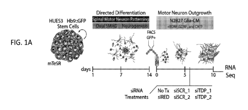
FIGS. 1A-1F demonstrate RNA Sequencing of TDP-43 knockdown in hMNs.
FIG. lA provides a schematic showing hMN differentiation, purification, and
RNAi
strategy for TDP-43 knockdown in cultured MNs. FIG. 1B provides
multidimensional scaling analysis for RNA-Seq data sets obtained from two
biologically independent MN differentiation and siRNA transfection experiments
based on 500 most differentially expressed genes. FIG. 1C provides a volcano
plot
showing statistically misregulated genes in hMNs treated with siTDP-43
compared to
those treated with scrambled controls. Genes identified as significant (B
enjamini-
Hochberg adjusted P value cutoff of 0.05 and a log fold-change ratio cutoff of
0) after
differential expression analysis are highlighted in yellow (for up-
regulated/increased
abundance genes) and in blue (for down-regulated/decreased abundance genes).
FIG.
1D provides a scatter plot comparing TPM values for all genes expressed in MNs
treated with control siRNAs versus the fold change in expression for those
genes in
cells treated with siTDP-43. FIGS. lE and 1F show a subset of 11 genes
initially
identified as 'hits' (significantly up-regulated (FIG. 1E) or down-regulated
(FIG. 1F))
in the TDP43 knockdown experiment were selected for validation by qRT-PCR. A
total of 9 out 11 of these genes (including TDP-43) exhibited the predicted
response
to TDP-43 depletion when their expression was assayed by qRT-PCR (Unpaired t
test, P value < 0.05).
CA 03176884 2022-09-23
WO 2021/195446 -9-
PCT/US2021/024254
FIGS. 2A-2J Demonstrate a familial ALS model. FIG. 2A provides a
schematic of a strategy for assessing gene expression in iPS cell-derived hMNs
expressing mutant TDP-43. FIG. 2B provides micrographs showing the morphology
of neurons cultured for 10 days derived from the iPS cells of healthy controls
(11a,
18a, 20b, 17a) and patients with mutations in TARDP (+/Q343R, +/G2985,
+/A315T,
and +/M337V). FIGS. 2C-2H provide qRT-PCR analysis of the genes consistently
downregulated (FIGS. 2D-2F) or upregulated (FIG. 2C) after TDP-43 knockdown in
neurons differentiated from the controls or TDP-43 patients. (Unpaired t test,
P value
<0.05). FIG. 21 provides representative micrographs of control and patient
neurons
immunostained for TDP-43 (red), 13-III tubulin (green) and counterstained with
DAPI
(blue). Scale bar, 100 pm. FIG. 2J provides Pearson's correlation analysis for
TDP-
43 immunostaining and DAPI fluorescence comparing control neurons to neurons
with TDP-43 mutations. Dots represent individual cells. (Unpaired t test, P
value <
0.05).
FIGS. 3A-3I demonstrate STMN2 regulation and localization. FIG. 3A
provides qRT-PCR analysis for the STMN2 transcript in independent experiments
using two different sets of primer pairs. (Unpaired t test, P value <0.05).
FIG. 3B
provides immunoblot analysis for TDP-43 and STMN2 protein levels following
partial depletion of TDP-43 by siRNA knockdown. Protein levels were normalized
to
GAPDH and are expressed relative to the levels in MNs treated with the siRED
control. FIG. 3C provides qRT-PCR analysis for STMN2 transcript analysis in
Hb9::GFP+ MNs treated with siRNAs targeting three ALS-linked genes (TDP-43,
FUS, and C90RF72). (Dunnett's multiple comparison test, Alpha value < 0.05).
FIGS. 3D-3F show formaldehyde RNA immunoprecipitation was used to identify
transcripts bound to TDP-43. After TDP-43 immunoprecipitation (FIG. 3D), qRT-
PCR analysis was used to test for enrichment of TDP-43 transcripts (FIG. 3E)
and
STMN2 transcripts (FIG. 3F) relative to the sample input. FIG. 3G provides
micrographs of Hb9::GFP+ MNs immunostained for TDP-43 (red), 13411 tubulin
(green) and counterstained with DAPI (blue). FIG. 3H provides micrographs of
Hb9::GFP+ MNs co-cultured on glia immunostained for STMN2 (red) and MAP2
green and GOLGIN97 (green). FIG. 31 provides a micrograph of Hb9::GFP+ MNs
day 3 after sorting immunostained for STMN2 (red), MAP2 (green) and
counterstained with F-actin-binding protein phalloidin (white). Scale bar, 5
pm.
CA 03176884 2022-09-23
WO 2021/195446 -10-
PCT/US2021/024254
FIGS. 4A-4K demonstrate STMN2 Knockout. FIG. 4A provides a schematic
of the knockout strategy using guide RNAs (gRNAs) targeting two constitutive
exons,
Exon 2 and 4, of the human STMN2 gene. The intervening DNA segment (-18Kb) is
targeted and deleted as a result of NHEJ (Non-homologous end joining) repair
of the
two double strand breaks (DSBs) introduced by the Cas9/gRNA nuclease complex.
FIGS. 4B-4D show STMN2 knockout was confirmed in the HUES3 Hb9: :GFP line
by RT-PCR analysis of genomic DNA (FIG 4B), by immunoblot analysis (FIG. 4C),
and by immunofluorescence (FIG. 4D). FIG 4E provides an experimental strategy
used to assess the cellular effect of lacking STMN2 in hMNs. FIGS. 4F-4H show
Sholl analysis of hMNs with and without STMN2 and in the absence (FIG. 4G) or
presence (FIG. 4H) of a ROCK inhibitor (Y-27632, 10 11M) to stimulate neurite
outgrowth. (Unpaired t test, P value < 0.05). FIG. 41 provides an experimental
strategy used to assess the cellular effect of lacking STMN2 in hMNs after
axonal
injury. FIGS. 4J-4K show axonal regrowth after injury. Representative
micrographs
of hMNs in the microfluidics device prior to and after axotomy (FIG. 4J).
Measurements of axonal regeneration after axotomy. (Unpaired t test, P value
<0.05).
FIGS. 5A-5G demonstrate a sporadic ALS model. FIG. 5A provides an
experimental strategy used to assess the effect of proteasome inhibition on
TDP-43
localization in human motor neurons. FIG. 5B shows Pearson's correlation
analysis
for TDP-43 immunostaining and DAPI fluorescence of cells treated with MG-132
(1
p,M). (Dunnett's multiple comparison test, Alpha value <0.05). FIG. 5C
provides
micrographs of HUES3 motor neurons untreated or treated with MG-132 and
immunostained for TDP-43 (red), 13-III tubulin (green) and counterstained with
DAPI
(blue). Scale bar, 100 pm. FIG. 5D provides immunoblot analysis of TDP-43 in
detergent soluble (RIPA) and detergent-insoluble (UREA) fractions in neurons
treated
with MG-132 (Unpaired t test, P value < 0.05). FIG. 5E provides qRT-PCR
analysis
of STMN2 expression for motor neurons treated with MG-132 at the indicated
concentrations and durations relative to DMSO control (Unpaired t test, P
value <
0.05). FIG. 5F provides a diagram of RT-PCR detection strategy for STMN2
cryptic
exon. FIG. 5G provides a tapestation analysis for the STMN2 cryptic exon in
hMNs
control cells treated with MG-132 (1 p,M).
FIGS. 6A-6H demonstrates ALS patient data. FIGS. 6A-6C provides
histologic analysis of human adult lumbar spinal cord from post-mortem samples
collected from a subject with no evidence of spinal cord disease (control)
(FIG. 6A)
CA 03176884 2022-09-23
WO 2021/195446 -11-
PCT/US2021/024254
or two patients diagnosed with sporadic ALS (FIGS. 6B-6C). Immunoreactivity to
STMN2 was detected in the perinuclear region (indicated by arrows) of spinal
motor
neurons but not in the surrounding glial cells. STMN2 immunoreactivity in
lumbar
spinal motor neurons from control and ALS cases was scored as 'strong' [as
indicated
by arrows in control (FIG. 6A) and sporadic ALS (FIG. 6B)] or as 'absent' [as
indicated by arrowheads in sporadic ALS (FIG. 6C)]. Scale bars, 50 pm. FIG. 6D
show the percentage of lumbar spinal motor neurons with strong STMN2
immunoreactivity was significantly lower in ALS tissue samples (n= 3 controls
and 3
ALS cases; approximately 40 MNs were scored for each subject; Two-tailed t-
test, P
.. value < 0.05). FIGS. 6E-6G show gene expression analysis for STMN2 from
previously published data sets, Rabin et al 2009 (FIG. 6E), Highley et al 2014
(FIG.
6F), and D'Erchia et al. 2017 (Two-tailed t-test, P value < 0.05). FIG. 6H
provides a
molecular model of ALS pathogenesis.
FIGS. 7A-7I demonstrate production of differentiated human motor neurons.
FIG. 7A shows hMN differentiation, purification, and culture strategy. FIG. 7B
provides flow-cytometric analysis of differentiated HUES3 Hb9:GFP cells. Cells
not
treated with the RA and SHH pathway agonist were used as negative control for
the
gating of GFP expression. FIGS. 7C-7F provides micrographs and quantification
of
purified Hb9::GFP+ cells immunostained for HB9 and counterstained with DAPI
(FIG. 7C) (Scale bar = 10 pm) or immunostained for ISL1 and the neuronal
markers
13-III tubulin and MAP2 (FIG. 7E) (Scale bar = 20 pm). FIGS. 7G-7J show
differentiated MNs are electrophysiologically active as determined by whole-
cell
patch-clamp recordings. FIG. 7G show upon depolarization in voltage-clamp
mode,
cells exhibited fast inward currents followed slow outward currents,
indicating the
expression and opening of voltage-activated sodium and potassium channels,
respectively. FIG. 7H shows in current-clamp mode, depolarization elicited
repetitive
action potential firing. FIG. 71 shows response to Kainate is consistent with
the
expression of functional receptors for excitatory glutamatergic transmitters.
FIGS. 8A-8E demonstrate TDP-43 knockdown in cultured hMNs. FIG. 8A
provides RNAi strategy for TDP-43 knockdown in cultured MNs. FIG. 8B shows
phase and red fluorescence micrographs of cultured hMNs 4 days after treatment
with
different siRNAs including scrambled siRNA conjugated to Alexa Fluor 555. FIG.
8C provides flow-cytometric analysis of hMNs after treatment with different
siRNAs.
FIG. 8D shows relative levels of TDP-43 mRNA in MNs exposed to different
siRNAs
CA 03176884 2022-09-23
WO 2021/195446 -12-
PCT/US2021/024254
for 2, 4 or 6 days. Levels for each sample were normalized to GAPDH and
expressed
relative to the no transfection control. FIG. 8E provides immunoblot analysis
of
hMNs after RNAi treated with the indicated siRNAs. Each sample was normalized
using GAPDH, and TDP-43 protein levels were calculated relative to the
siSCR_555-
treated control sample.
FIGS. 9A-9C demonstrate motor neuron RNA-Seq. FIG. 9A shows global
transcriptional analysis of motor neurons treated as indicated represented as
a heat
map. Unsupervised clustering of expression profiles revealed that the samples
segregated based on the batch on motor neuron production and analysis. FIG. 9B
provides analysis of TDP-43 transcript abundance after RNA-Sequencing
validated
the knockdown (Benjamini-Hochberg adjusted P value cutoff of 0.05). FIG. 9C
shows alteration in the splicing pattern of the POLDIP3 gene was detected as
result of
TDP-43 knockdown, with siTDP43-treated cells showing significant reduction of
isoform 1 and increased levels of spliced variant 2 (which lacks Exon3) (false
discovery rate `FDR' >0.05).
FIG. 10 demonstrates pluripotent stem cell genotyping sequencing
chromatograms of Exon6 of TARDBP in the indicated iPS cell lines to confirm
the
heterozygous mutations in the patient lines.
FIGS. 11A-11F demonstrate neuronal cell sorting. FIG. 11A shows using a
cell surface marker screen, antibodies enriched on GFP+ motor neurons
(Quadrant 1)
and GFP- cells (Quadrant 3) were identified. FIG. 11B shows after sorting for
NCAM+ and EpCAM- cells, high content imaging was used to determine if the
sorting method can deplete the cultures of mitotic cells (EdU+) and
significantly
enrich for motor neurons (Is11+) and neurons (MAP2+). N= 6 different iPS cell
lines.
Statistical analysis was performed using a two-tailed Student's t test. FIGS.
11C-11D
provides qRT-PCR analysis of cultures after sorting for the motor neuron
marker
ISL1 (FIG. 11C) and the neuronal marker 13111-tubulin (FIG. 11D) revealed
enrichment and more homogenous cultures compared to unsorted cultures. FIG.
11E
provides flow-cytometric analysis with phycoerythrin (PE)-conjugated
antibodies to
EpCAM (anti-epCAM¨PE) and Alexa Fluor 700¨conjugated antibodies to NCAM
(anti-NCAM¨AF700) of cultures differentiated from the indicated healthy
controls
(grey) and TDP-43 mutant lines (red). FIG. 11F shows the percentage of NCAM+
cells for the indicated lines from 4-6 independent differentiations. No
significant
difference was observed between mutant and control lines in terms of their
ability to
CA 03176884 2022-09-23
WO 2021/195446 -13-
PCT/US2021/024254
generate NCAM+ cells. Statistical analysis was performed using a two-tailed
Student's t test, P value < 0.05.
FIGS. 12A-12G demonstrate TDP-43 and STMN2 connections. FIGS. 12A-
12C provide qRT-PCR validation of the downregulation of ALS genes upon siRNA
treatments. Expression of TDP-43 (FIG. 12A), FUS (FIG. 12B), and C90RF72 (FIG.
12C) was assessed for all the controls and each siRNA used (Unpaired t test, P
value
<0.05). FIG. 12D provides a western blot analysis of STMN2 protein in
different cell
types along the motor neuron differentiation. FIG. 12E shows RNA-Seq
expression
levels for the Stathmin family in motor neurons treated with either siSCR (-)
or
siTDP-43 (+) oligos. Only STMN2 levels were altered after TDP-43 knockdown.
FIGS. 12F-12G shows TDP-43 binding sites within the Stathmin family of genes
(FIG. 12F) normalized to gene length (FIG. 12G). STMN2 has the greatest number
of
binding motifs.
FIGS. 13A-13H demonstrate STMN2 regulates neuronal outgrowth. CRISPR-
mediated STMN2 knockout in the WA01 line was confirmed by RT-PCR analysis of
genomic DNA (FIG. 13A), by immunoblot analysis (FIG. 13B), and by
immunofluorescence (FIG. 13C). FIGS. 13D-13F provide Sholl analysis of hMNs
with and without STMN2 and in the presence of a Y-27632 (10 [tM), a ROCK
inhibitor (FIG. 13F) (Unpaired t test, P value < 0.05). FIGS. 13G-13H shows
axonal
regrowth after injury. Representative micrographs of hMNs in the microfluidics
device prior to and after axotomy (FIG. 13G). Analysis of axonal regrowth
after
axotomy (Unpaired t test, P value < 0.05) (FIG. 13H).
FIGS. 14A-14E demonstrate cell survival and proteasome activity assays.
FIGS. 14A-14C shows Cell Titer Glo uses ATP from metabolically active cells to
generate light. (FIG. 14A) shows a direct relationship exists between
luminescence
and the number of cells in culture over several orders of magnitude. FIG. 14B
shows
the assay can detect differences in neuronal survival in the absence of growth
factors.
N= 6 separate wells of neurons. (Unpaired t test, P value <0.05). FIG. 14C
shows
MG-132 neuronal survival experimental outline. FIG. 14D shows dose response
curve for motor neurons cultured with indicated concentrations of MG-132 for
the
indicated times. N= triplicate wells. Cells are viable after 1 day of
treatment at all the
concentrations tested and lower concentrations are tolerated for more extended
periods of time. FIG. 14E shows following cleavage by the proteasome, the
substrate
for luciferase is liberated, which allows for quantitative measurement of
proteasome
CA 03176884 2022-09-23
WO 2021/195446 -14-
PCT/US2021/024254
activity. Neurons treated with MG-132 show significantly decreased proteasome
activity. N= 4 separate wells of neurons (Unpaired t test, P value < 0.05).
FIGS. 15A-15E demonstrate TDP-43 regulates cryptic exon splicing in hMNs
(FIGS. 15A-15C). Visualization of the cryptic exons for PFKP (FIG. 15A),
ELAVL3
(FIG. 15B), and STMN2 (FIG. 15C) for the cells treated with scrambled siRNAs
or
siRNAs targeting TDP-43 transcript. Read coverage and splice junctions are
shown
for alignment to the human HG19 genome. FIGS. 15D-15E provides diagram of RT-
PCR detection strategy for STMN2 cryptic exon (FIG. 15D), and Sanger
sequencing
of the PCR product confirmed the splicing of STMN2 Exon 1 with the cryptic
exon
(FIG. 15E).
FIGS. 16A-16P provide cryptic STMN2 transcript qPCR data from patient
cerebral spinal fluid (CSF) samples. FIGS. 16A-16D provide graphs summarizing
the
patient sample data of normalized cryptic STMN2 relative to healthy controls.
FIGS.
16E-16M provide graphs providing details regarding individual patient samples.
FIG.
16N provides a graph demonstrating survival duration following diagnosis. FIG.
160
provides a graph demonstrating age at death. FIG. 16P provides a graph
demonstrating vital capacity.
FIGS. 17A-17C demonstrate an STMN2 multiplexed qPCR Assay. FIG. 17A
shows Q-RT PCT assay for STMN2 in fluids. Experimental schemes are provided
and STMN2 multiplexed TaqMan assay is shown to simultaneously detect cryptic
STMN2, normal STMN2 transcript, and the housekeeping gene RNA18S5. RNA can
be collected from CSF-derived exosomes and then converted into cDNA to assay
for
full and cryptic STMN2 transcripts, as well as control RNAs for normalization.
FIG.
17B shows in vitro validation of the multiplexed assay in cells where TDP-43
levels
were reduced using either an ASO or using siRNA. FIG. 17C shows the STMN2
multiplexed qPCR assay was used to probe cryptic STMN2 transcript levels in
the
cDNA samples generated from the MGH CSF samples. STMN2 cryptic splicing is
significantly induced in ALS patients.
FIGS. 18A-18D demonstrate a sandwich ELISA for detecting STMN2 protein.
FIG. 18A provides a schematic of the STMN2 sandwich ELISA. FIG. 18B
demonstrates the sensitivity of the STMN2 ELISA to picogram quantities. FIG.
18C
shows the sandwich ELISA was validated using recombinant STMN2 protein and is
capable of detecting picogram levels of STMN2. FIG. 18D shows STMN2 levels are
CA 03176884 2022-09-23
WO 2021/195446 -15-
PCT/US2021/024254
reduced in patient cerebral spinal fluid (CSF) when assessed using the STMN2
ELISA.
FIG. 19 provides a chart demonstrating the genetics of ALS, with each gene
being plotted against the year it was discovered. See Alsultan et al.
Degenerative
Neurological and Neuromuscular Disease. 2016, 6, 49-64.
FIG. 20 demonstrates that TDP-43 is a multifunctional nucleic acid-binding
protein. TDP-43 has been shown to play a role in various functions including
RNA
splicing, miRNA processing, autoregulation of its own transcript, RNA
transport and
stability, and stress granule formation. The transcripts TDP-43 regulates are
highly
species and cell type dependent. See Buratti and Baralle Trends in Biochem.
Sci..
2012, 6, 237-247.
FIG. 21 provides a strategy for measuring transcriptional effects of TDP-43
depletion. The schematic demonstrates hMN differentiation, purification, and
culture
strategy. The strategy uses small molecules that mimic early development to
convert
stem cells into postmitotic neurons in 2 weeks. Various methods were developed
to
sort and study the neurons. siRNA technology combined with RNA sequencing was
used to identify transcripts regulated by TDP-43.
FIG. 22 demonstrates TDP-43 binds to STMN2. ALS patient spinal cords
were stained for STMN2 and decreased STMN2 protein in ALS patients was
observed based on fold enrichment relative to PGK1 (fRIP). See Klim et al.
Nature
Neuroscience vol. 22, pages 167-179 (2019).
FIG. 23 shows splicing alterations after TDP-43 depletion. Differential exon
usage analysis was performed on RNA-seq samples from motor neurons treated
with
siTDP. Splicing changes were observed in STMN2.
FIG. 24 demonstrates TDP-43 suppresses a cryptic exon in STMN2. The
integrated genome viewer was used to look at where RNA seq reads were mapped
to
the human genome (top graph # of reads) and how the reads reconnected between
the
exons (splice track). The graphs show the number of reads mapped to areas of a
gene.
FIG. 25 provides a STMN2 splicing defect summary. Under normal
conditions STMN2 is transcribed with all 5 exons leading to an mRNA that is
translated into a 20 kDa STMN2 protein. After TDP-43 perturbations, the
cryptic
exon intercepts the transcript so that only a 17 amino acid polypeptide could
be
translated.
CA 03176884 2022-09-23
WO 2021/195446 -16-
PCT/US2021/024254
FIG. 26 shows STMN2 is consistently decreased. The overlap of decreased
transcripts down in 3 human RNA seq data sets (ALS patient data sets and
siTDP43
stem cell motor neuron data set) were compared and STMN2 is the only
transcript
down in all three data sets.
FIG. 27 shows the STMN2 cryptic exon is present in ALS patient spinal cords.
Read coverage and splice junctions are shown for alignment to the human HG19
genome. The reads mapped to the human genome in ALS patients was observed, and
for 5 out of 6 patients reads mapped to and splicing went into the cryptic
exon and
none of the controls.
FIG. 28 shows TDP-43 depletion leads to neurite outgrowth and axonal
regrowth defects. Representative micrographs of hMNs treated with indicated
siRNAs
and immunostained for 13-III tubulin to perform Sholl analysis are provided. A
Sholl
analysis of hMNs after siRNA treatment is provided. Lines represent sample
means
and shading represents the s.e.m. with unpaired t-test between siTDP43 and
siSCR,
two sided, P<0.05.
FIG. 29 shows microfluidic devices for investigating axon regeneration. The
microfluidic device includes a soma compartment (left panel) and axon
compartment
(right panel).
FIGS. 30A-30B demonstrate TDP-43 depletion leads to neurite outgrowth and
axonal regrowth defects. FIG. 30A provides representative micrographs of hMNs
in
the microfluidics device after axotomy. Scale bars, 150 i.i.M. FIG. 30B
provides
measurements of axonal regrowth and regeneration after axotomy (Unpaired t
test,
two sided, P value <0.05 18h <0.0001, 24h <0.0001, 48<0.0001 and 72<0.0001).
FIG. 31 demonstrates STMN2 is a c-Jun N-terminal kinase (JNK) target in the
axonal degeneration pathway. JNK1 is shown to bind to and phosphorylate STMN2,
and phosphorylated STMN2 is rapidly degraded. See J. Eun Shin et al. PNAS
2012,
109, E3696-3705.
FIG. 32 provides a strategy to determine if JNKi can rescue siTDP43
phenotypes. See Klim et al. Nature Neuroscience vol. 22, pages 167-179 (2019).
FIG. 33 shows a JNK inhibitor (5P600125) boosts STMN2 levels. STMN2
protein levels increased in neurons treated with JNKi and lower levels
observed in
cells treated with siTDP43 could be rescued.
FIG. 34 shows JNKi (5P600125) increases neurite outgrowth. Cells treated
with JNKi exhibited increased neurite branching.
CA 03176884 2022-09-23
WO 2021/195446 -17-
PCT/US2021/024254
FIG. 35 shows JNKi (SP600125) increases neurite outgrowth. Sholl analysis
confirmed that under all conditions JNKi increased neurite branching and
regrowth
following injury.
FIG. 36 shows JNKi increases axon regeneration. Microfluidic devices
confirmed that under all conditions JNKi increased neurite branching and
regrowth
following injury.
FIG. 37 provides a model for proteasome inhibition. Disruptions to protein
homeostasis lead to TDP-43 mislocalization and altered STMN2 levels, which
disrupts axon biology.
FIGS. 38A-38B shows TDP-43 localization. TDP-43 is normally nuclear
(FIG. 38A), but after compound washout, a loss of distinct nuclear TDP-43
staining
was observed (FIG. 38B). No cytoplasmic aggregation was observed, only loss of
nuclear TDP-43.
FIG. 39 shows TDP-43 mislocalization is reversible.
FIG. 40 shows STMN2 transcripts decreased after TDP-43 mislocalization.
The decrease for STMN2 was even more pronounced than in cells expressing
mutant
TDP-43.
FIG. 41 provides a table summarizing recent ALS genes with their relative
mutation frequencies in different ALS and FTD cohorts and associated pathways.
Advances in WGS and WES have led to identification of genes carrying rare
causal
variants: TBK1, CHCHD10, TUBA4A, MATR3, CCNF, NEK1, C21orf2, ANXA11,
and TIAL TBK1 is shown as having the highest mutation frequencies of ALS-FTD
(3-4%) in different cohorts. See Nguyen, et al., Trends in Genetics, 2018.
FIG. 42 shows Atg7 and TBK1 act at distinct times in autophagy. See
Hansen, et, al,. Nature Reviews Molecular Cell Biology. 2018
FIG. 43 shows eliminating TBK1 shares similarities with, but is distinct from,
blocking autophagy initiation.
FIG. 44 shows TBK1 knock out decreases functional TDP-43 and STMN2
levels while eliminating ATG7 has no effect. Loss of TBK1 induces TDP-43
pathology in motor neurons through autophagy-independent mechanisms.
FIG. 45 shows loss of TBK1 shows impaired axon regeneration after axon
injury.
FIG. 46 shows proteasome inhibition induced TDP-43 mislocalization in
TBK1 mutant motor neurons.
CA 03176884 2022-09-23
WO 2021/195446 -18-
PCT/US2021/024254
FIGS. 47A-47C demonstrate targeting STMN2 intron using CRISPR. A
CRISPR strategy for targeting STMN2 is provided, as well as genotyping for
STMN2
(FIGS. 47A-47B). FIG. 47C provides a table summarizing the CRISPR targeting
strategy and genotyping for STMN2.
FIG. 48 demonstrates STMN2 mice are significantly smaller than Rosa26
control mice and show deficiencies in motor performance tasks with no signs of
progression of these deficits over time.
FIG. 49 demonstrates STMN2 mice are significantly smaller than Rosa26
control mice and show deficiencies in motor performance tasks with no signs of
progression of these deficits over time.
FIG. 50 demonstrates behavioral outcomes, as well as the total distance
traveled in open field assays, appear to be similar between two mice cohorts.
FIG. 51 demonstrates STMN2 transcript levels are significantly reduced or no
transcript is present in brain tissue from mutant cohort.
FIG. 52 provides Western Blot of brain tissue validating loss or significant
reduction of STMN2 protein in mutant mice cohort.
FIG. 53 demonstrates STMN2 primarily localizes to ChAT+ motor neurons in
the ventral horn of adult mice spinal cords.
FIG. 54 demonstrates a STMN2 cohort exhibits a significant decrease in the
number of STMN2+/ChAT+ motor neurons on the ventral horn of the spinal cord.
FIG. 55 provides graphs showing the difference in organ or muscle weight
between control and STMN2 mice. It is demonstrated that lower limb muscles are
lighter in STMN2 mice (see two boxed graphs).
FIG. 56 provides pre- and post-synaptic staining of STMN2 gastrocnemius
(GA) muscle and Rosa26 control gastrocnemius (GA) muscle. The staining
suggests
de-innervation in STMN2 -/- animals.
FIG. 57 demonstrates pre-and post-synaptic staining of STMN2 gastrocnemius
(GA) muscle and Rosa26 control gastrocnemius (GA) muscle suggests de-
innervation
in STMN2 -/- animals.
FIG. 58 demonstrates neuromuscular junction (NMJ) morphology supports
active de-innervation in gastrocnemius muscle of STMN2 mutants.
FIG. 59 demonstrates mutant TDP-43 does not display pathological
mislocalization. Stains of control and ALS patient neurons for TDP-43 show
that for
both the control and ALS patient neurons TDP-43 was primarily nuclear.
CA 03176884 2022-09-23
WO 2021/195446 -19-
PCT/US2021/024254
FIG. 60 identifies different classes of proteasome inhibitors and provides
their
chemical structures.
FIG. 61 shows decreased expression of full length STMN2 in hMNs upon
treatment with structurally distinct proteasome inhibitors.
FIG. 62 shows a PCR assay of hMNs treated with MG-132 or Bortezomib.
Full length STMN2 was detected in all samples as a control. The presence of
transcripts containing the STMN2 cryptic exon were specific to those cells
treated
with the proteasome inhibitors.
FIGS. 63A-63B demonstrate in vitro assay for TDP-43 binding to STMN2
RNA. Using genomic DNA, RNA containing the TDP-43 binding sites from the
cryptic exon region of STMN2 was in vitro transcribed (FIG. 63A). The RNA was
used to assess whether it could pull down IP TDP-43 protein from human
neuronal
protein lysates. The in vitro assay shows transcripts containing the cryptic
exon
region pulled down TDP-43 (FIG. 63B).
FIG. 64 shows an in vitro assay for TDP-43 binding to STMN2 RNA. RNA
containing the 5' and 3' TDP-43 binding regions were in vitro transcribed
similar that
described in FIG. 63. Although both 5' and 3' transcripts can pull down some
TDP-
43, the enrichment is not as strong as the full cryptic exon.
FIG. 65 shows design of gRNAs for generation of targeted mutant cell line
with no cryptic exon. A strategy was prepared to delete 105 nucleotides within
the
cryptic exon within STMN2 intron between exons 1 and 2. The deletion will
eliminate the TDP-43 binding motif, but not affect the predicted poly-
adenylation site.
FIG. 66 provides a confirmation of mutational status. TIDE analysis was used
to analyze the mutational status of the clones and checked the sequence
alignment to
control cells to obtain a more precise view of the size and location of the
deletions.
One cell line contained a homozygous 105 nt deletion, which was consistent
with the
gel electrophoresis. The deletion eliminated the TDP-43 binding motif, but did
not
affect the predicted poly-adenylation site.
FIG. 67 shows TDP-43 binding site is a potential negative regulator of
STMN2 expression. Three cell lines, HUES3, IG2 (5tmn2 KO), and CN7 (cryptic
exon deletion) were treated with normal media or media + 1 uM MG132 for 24
hours
to stress the cells. In HUES3 cells, the stressed condition had 52% STMN2 mRNA
expression compared to the unstressed condition. In IG2 (5tmn2 KO) condition,
unstressed cells had 13% expression, and when stressed, expression increased
to 42%.
CA 03176884 2022-09-23
WO 2021/195446 -20-
PCT/US2021/024254
The expression levels in the CN7 (Cryptic Exon Deletion) cell line were
significantly
higher than the other two cell lines, with unstressed having 729% and stressed
having
473% expression. It was shown that if several exons are knocked out the
expression
goes down, but if the TDP-43 binding site is removed, expression goes way up.
FIGS. 68A-68B demonstrate deletion of putative TDP-43 binding site leads to
increased STMN2 protein levels. Consistent with the gene expression data,
deletion of
the TDP-43 binding region within the STMN2 cryptic exon causes increased
protein
expression.
FIGS. 69A-69B demonstrate the conservation of the STMN2 gene locus. FIG.
69A shows human STMN2 is located on long arm of chromosome 8 and is
transcribed
as several isoforms generally including 5 canonical exons. The location of the
cryptic
exon is highlighted in orange. Conservation amongst 100 vertebrates along the
locus
reveals strong conservation at exons as well as some intronic regions. FIG.
69B shows
a higher resolution genomic view at the STMN2 cryptic exon (orange) with
nucleotide
resolution combined with multiple sequence alignment for 12 primates and 2
rodents.
Salient features of the human gene and the extent of their conservation down
the list
of species are underlined including the splice acceptor site (teal), the
putative coding
region (yellow), the stop codon (red), the TDP-43 binding motifs (blue), and
the poly-
A signal (purple).
FIG. 70 demonstrates a multiplexed assay for detecting cryptic STMN2.
FIGS. 71A-71C demonstrate siTDP-43 and TDP-43 ASO induce STMN2
reduction and cryptic exon induction. Relative expression levels are shown for
TARDBP (FIG. 71A), STMN2 Exons 3-4 (FIG. 71B), and Cryptic STMN2 (FIG.
71C) when treated with SCR ASO, TDP ASO or siTDP.
FIGS. 72A-72C show relative mRNA levels for TARDP (FIG. 72A), STMN2
(FIG. 72B), and cryptic STMN2 (FIG. 72C) after treatment with a scrambled ASO,
TDP-43 ASO or SOD1 ASO over a time course of 6 days.
FIG. 73 demonstrates cryptic STMN2 expression. mRNA levels of cryptic
STMN2 expression is shown after treatment with Scrambled ASO, TDP-43 ASO,
SOD1 ASO, siTDP-43, and siRED. Each treatment was applied using NeuroPorter5,
NeuroPorterl, RNAiMAX, or LipoFecamine, with RNAimax being the most
effective.
FIG. 74 provides a schematic showing the strategy for testing STMN2 splice
switching ASOs.
CA 03176884 2022-09-23
WO 2021/195446 -21-
PCT/US2021/024254
FIGS. 75A-75D provide schematics of ASO screening set up plate 1 (FIG.
75A), plate 2 (FIG. 75B), plate 3 (FIG. 75C), and plate 4 (FIG. 75D).
FIG. 76 provides results from ASO screening with comparable cDNA for all
wells. The ASOs screened are STMN2 intron targeting ASOs.
FIG. 77 provides results from ASO screening showing ASOs near the splice
junction suppress cryptic exon inclusion.
FIG. 78 provides the best hits from the ASO screen showing dose dependence
or suppression to lowest concentration.
FIGS. 79A-79B demonstrate TDP-43 protein structure, pathogenic mutations,
and function. FIG. 79A shows TDP-43 comprises six domains: an N-terminal
region
(aa 1-102) with a nuclear localization signal (NLS, aa 82-98); two RNA
recognition
motifs: RRM1 (aa 104-176) and RRM2 (aa 192-262); a nuclear export signal (NES,
aa 239-250); a C-terminal region (aa 274-414), encompassing a prion-like
glutamine/asparagine-rich (Q/N) domain (aa 345-366); and a glycine-rich region
(aa
366-414). Forty-six dominant mutations have been identified in TDP-43 in
sporadic
and familial ALS patients and in rare FTLD patients, mostly lying in the C-
terminal
glycine-rich region. FIG. 79B shows salient TDP-43 functions are strongly
implicated
in disease pathogenesis. The most common motif identified for TDP-43 is (TG)n,
which corresponds to the (UG)n RNA binding motif. Interaction with RNA allows
TDP-43 to regulate pre-mRNA splicing to inhibit the inclusion of cryptic exons
as
well as influence polyadenylation site selection. Cytosolic roles for TDP-43
include
transport of RNA along neuronal processes and response to stresses including
those
affecting proteostasis that can trigger TDP-43 nuclear efflux and localization
to stress
granules. A multitude of these basic molecular functions contribute to TDP-43
autoregulation including splicing and polyadenylation.
FIGS. 80A-80B demonstrate STMN2 protein structure and function. FIG.
80A shows STMN2 comprises two domains that can be further subdivided: 1) an N-
terminal domain containing a conserved Golgi-specifying sequence and two
palmitoylation sites enabling membrane insertion, and 2) a Stathmin-like
domain
containing two tubulin binding repeats (TBR1 and TBR2) that each bind tubulin,
a
proline rich domain (PRD) harboring two phosphorylation sites that can be
modulated
by JNK to potentially modulate the ability of STMN2 to interact with tubulin
and
promote STMN2 degradation, and a stathmin N-terminal domain (SLDN), which
contain a peptide that inhibits tubulin polymerization. Identified
posttranslational
CA 03176884 2022-09-23
WO 2021/195446 -22-
PCT/US2021/024254
modifications (PTMs) according to PhosphositePlus are marked along the protein
structure. FIG. 80B shows the reported subcellular localization of STMN2
protein.
STMN2 localizes to the golgi apparatus and is found in vesicles trafficked
throughout
dendrites and axons, and concentrates within growth cones of developing
neurons as
well as in regenerating axon tips after injury.
FIG. 81 provides a proposed model for TDP-43 regulation of STMN2. A
pathological hallmark of ALS is the nuclear loss of TDP-43 and its
aggregation. We
propose a model of TDP-43 regulation of STMN2 where it binds to STMN2 pre-
mRNA upon the intron between exons 1 and 2. Either reduction of TDP-43 levels
or
nuclear egress leads to early polyadenylation and splicing of a cryptic exon
leading to
a truncated STMN2 mRNA transcript. The blunted transcript encodes for a
putative
17 amino acid polypeptide thus leading to reduced levels of STMN2 protein.
Loss of
STMN2 leads to reduced neurite outgrowth and axonal repair after injury.
FIG. 82 shows antisense oligonucleotides and their location in relation to the
STMN2 sequence. The sequence, chemistry and alignment of ASOs to STMN2 locus
is indicated. Salient features of the human gene highlighted including the
splice
acceptor site (teal), the putative coding region (yellow), the stop codon
(red), the
TDP-43 binding motifs (orange), and the poly-A signal (purple). ASOs
highlighted in
yellow had locked nucleic acid chemistry.
FIGS. 83A-83C examine the cryptic exon-containing region of STMN2 pre-
mRNA. FIG. 83A provides the sequence of the cryptic exon-containing region of
STMN2 pre-mRNA, with various salient features highlighted. FIGS. 83B-83C
provide predicted RNA structures of the cryptic exon-containing region of
STMN2
pre-mRNA, showing that the green highlighted region is partially unstructured
and
can adopt different binding interactions with similar energies.
FIGS. 84A-84D demonstrate patient specific induced pluripotent stem cell
characterization. FIG. 84A provides a micrograph showing the undifferentiated
patient iPS cells. FIG. 84B provides sequencing chromatogram of PCR product
amplified from exon 8 of TBK1 in the indicated iPS cell line confirming the
heterozygous L3061 non-pathological variant of no significance in the patient
line.
FIGS. 84C-84D provide micrographs showing the motor neurons differentiated
from
the patient iPS cells.
FIGS. 85A-85B demonstrate decreased nuclear TDP-43 observed in patient
neurons. FIG. 85A provides representative micrographs of control and patient
CA 03176884 2022-09-23
WO 2021/195446 -23-
PCT/US2021/024254
neurons immunostained for TDP-43 (red), f3-III tubulin (green) and
counterstained
with DAPI (blue) marking the nucleus. Scale bar, 100 Ilm. FIG. 85B provides
Pearson's correlation analysis for TDP-43 immunostaining and DAPI fluorescence
comparing control neurons to the patients. Dots represent individual cells and
are
displayed as mean with s.d. for at least 25 cells from n= 4 control and 1
patient lines
(unpaired t test, two-sided, P< 0.05).
FIGS. 86A-86C demonstrate patient motor neurons produce truncated STMN2
in response to TDP-43 depletion. RNA levels analyzed by qRT-PCR analysis after
TDP-43 knockdown by siTARDBP in motor neurons differentiated from patients iPS
cells. FIG. 86A shows RNA levels of TDP-43. FIG. 86B shows RNA levels of full-
length STMN2. FIG. 86C shows RNA levels of cryptic STMN2 compared to control
(siCTRL).
FIGS. 87A-87C demonstrate patient STMN2 locus sequencing. FIG. 87A
shows the sequencing results of PCR product amplified from the first intron of
STMN2 in the patient iPS cell line aligned to the reference sequence. FIG. 87B
identifies one mismatch between the patient and the reference sequence
consisting of
a common single nucleotide variant (SNP). FIG. 87C provides a sequencing
chromatogram of PCR product-amplified from the ASO-targeted region of first
intron
of STMN2 confirms no heterozygous at this locus and highlights the match for
the
ASOs.
FIGS. 88A-88B demonstrate levels of cryptic and full length STMN2 RNA
with SJ+94 ASO (SEQ ID NO: 73) in patient motor neurons. FIG. 88A shows
cryptic
STMN2 RNA levels. FIG. 88B shows full-length STMN2 RNA levels after TDP-43
reduction by siTARDP in patient's motor neurons. Neurons were cultured from
left
to right with 30, 3, 0.3, or 0.03 nM of the STMN2-targeting ASO (SJ+94) or a
non-
targeting control ASO (NTC).
FIG. 89 demonstrates full length STMN2 RNA is increased by ASO SJ+94
after its suppression due to nuclear depletion of TDP43 in patient's motor
neurons.
qRT-PCR analysis of full-length STMN2 after proteasome inhibition with MG-132
(1
iiM) in patient's neurons, which induces nuclear depletion of the TDP-43,
leads to
decreased STMN2 expression. Full length STMN2 RNA is increased by ASO SJ+94
under these conditions when compared to those treated with a non-targeting
control
ASO (NTC).
CA 03176884 2022-09-23
WO 2021/195446 -24-
PCT/US2021/024254
FIGS. 90 demonstrates immunoblot analysis for STMN2 protein levels
following reduction of TDP-43 by siRNA. Protein input was normalized by BCA
and
STMN2 levels are expressed relative to the levels in hMNs treated with control
siRNAs. Data are displayed as mean with s.d. of technical replicates from n =
3
independent experiments (unpaired t test, two-sided, P<0.05).
FIGS. 91A-91E demonstrate outgrowth deficits following TDP-43 depletion
can be rescued by STMN2 ASO SJ +94 in patient's motor neurons. FIG. 91A
outlines the experimental strategy used to assess the cellular effect of STMN2
restoration in hMNs after axonal injury. FIG. 91B provides representative
micrographs of patient's motor neurons in the microfluidics devices 18 hours
after
axotomy. Fields highlighted by red rectangles from NTC and SJ +94 are enlarged
in
the images (i) and (ii) respectively. FIG. 91C shows length of individual
neurites
displayed as dots along with the mean and standard deviation. (unpaired t
test, two-
sided). FIG. 91D provides representative micrographs of patient's motor
neurons in
the microfluidics devices 18 hours after axotomy. Fields highlighted by red
rectangles
from NTC and SJ-1 are enlarged in the images (i) and (ii) respectively. FIG.
91C
shows lengths of individual neurites displayed as dots along with the mean and
standard deviation. (unpaired t test, two-sided).
FIG. 92 demonstrates neurite outgrowth deficits following TDP-43 depletion
can be rescued by STMN2 ASOs SJ-1, SJ+94, and SJ+101. Individual neurites are
displayed as dots.
FIG. 93 demonstrates STMN2 can be restored in TDP-43 depleted neurons by
STMN2 ASOs SJ-1, SJ+94, and SJ+101.
FIG. 94 demonstrates cry STMN2 can be reduced in TDP-43 depleted neurons
.. by STMN2 ASOs SJ-1, SJ+94, and SJ+101.
FIGS. 95A-95B demonstrate levels of cryptic and full length STMN2 RNA
with SJ-1 ASO in patient motor neurons. FIG. 95A shows cryptic STMN2 RNA
levels. FIG. 95B shows full-length STMN2 RNA levels after TDP-43 reduction by
siTARDBP (siTDP-43) in patient's motor neurons. Neurons were cultured from
left to
.. right with 30, 3,0.3, or 0.03 nM of the STMN2-targeting ASO (SJ -1) or a
non-
targeting control ASO (NTC).
FIG. 96 demonstrates full length STMN2 RNA is increased by ASO SJ-1 after
its suppression due to nuclear mis-localization of TDP3 in patient's motor
neurons:
qRT-PCR analysis of full-length STMN2 after proteasome inhibition with MG-132
(1
CA 03176884 2022-09-23
WO 2021/195446 -25-
PCT/US2021/024254
[tM) in patient's neurons, which induces nuclear mis-localization of TDP-43,
leads to
decreased STMN2 expression. Full-length STMN2 RNA is increased by ASO SJ-1
under these conditions when compared to those treated with a non-targeting
control
ASO (NTC).
FIG. 97 demonstrates STMN2 protein levels measured by Western Blot in
patient's motor neurons following reduction of TDP-43 by siRNA. Protein
loading
was normalized by total protein content and STMN2 levels are expressed
relative to
the levels in hMNs treated with control siCTRLs. Data are displayed as mean
with
s.d. of technical replicates from n = 3 independent experiments. The p values
for the
increase in STMN2 levels induced by SJ-1, SJ+94 and SJ+101 as compared to the
non-targetting controls (NTC) are indicated above each result. The increase is
significant in each case (unpaired t test, two-sided, P < 0.05).
DETAILED DESCRIPTION OF THE INVENTION
Mislocalization or depletion of the RNA-binding protein TDP-43 results in
decreased expression of STMN2, which encodes a microtubule regulator. STMN2 is
essential for normal axonal outgrowth and regeneration. Decreased TDP-43
function
causes an abortive or altered STMN2 RNA sequence which results in reduced
STMN2 protein expression. STMN2 may be a promising therapeutic target and
biomarker of disease risk (e.g., neurodegenerative diseases).
Work described herein relates to compositions and methods for suppressing or
preventing the inclusion of a cryptic exon in STMN2 mRNA. The inclusion of a
cryptic exon in STMN2 mRNA may lead to a truncated transcript and protein. In
some aspects the inclusion of the cryptic exon leads to early polyadenylation.
STMN2 expression may be restored through suppression of a cryptic splicing
form of
STMN2 that occurs when TDP-43 becomes sequestered or is reduced in
functionality,
such as by blocking the occurrence or accumulation of the cryptic form and
converting it back to or restoring functional STMN2 RNA (e.g., by
administration of
an antisense oligonucleotide). In addition, work described herein relates to
compositions and methods for increasing protein synthesis of STMN2, i.e.,
increasing
STMN2 protein expression.
Agents and Pharmaceutical Compositions
CA 03176884 2022-09-23
WO 2021/195446 -26-
PCT/US2021/024254
The disclosure contemplates agents (e.g., antisense oligonucleotides) that
specifically bind an STMN2 mRNA, pre-mRNA, or nascent RNA sequence that
occurs and increases in abundance when TDP-43 function declines or TDP-
pathology
occurs, thereby suppressing or preventing inclusion of an abortive or altered
STMN2
.. RNA sequence. In some aspects, agents prevent degradation of STMN2 protein.
In
some aspects, agents restore STMN2 protein levels. In some aspects, an agent
suppresses or prevents inclusion of a cryptic exon in STMN2 RNA. In certain
aspects
an agent specifically binds an STMN2 mRNA, pre-mRNA, or nascent RNA sequence
coding for a cryptic exon.
In some aspects, the disclosure further contemplates agents (e.g., antisense
oligonucleotides) that specifically bind an ELAVL3 mRNA, pre-mRNA, or nascent
RNA sequence. ELAVL3 may be downregulated when TDP-43 function declines or
TDP-pathology occurs. In some aspects, an agent suppresses or prevents cryptic
splicing of ELAVL3.
In some embodiments, the agent (e.g., an antisense oligonucleotide) binds to
an STMN2 RNA sequence (e.g., an abortive or altered STMN2 RNA sequence). In
some aspects the binding of an agent to a short abortive or altered STMN2 RNA
sequence results in continued production by the RNA polymerase. For example,
the
agent may directly suppress premature transcriptional termination at the
polyadenylation site of the cryptic exon or may mimic the activity of TDP-43
binding
at its target site, thereby altering transcriptional termination at the
cryptic exon. In
some aspects, the agent suppresses or prevents inclusion of a cryptic exon in
STMN2
RNA. In some aspects the agent prevents degradation of STMN2 protein. In some
aspects the agent increases STMN2 levels (e.g., through exon skipping). In
some
.. aspects the agent restores normal length or protein coding STMN2 RNA (e.g.,
pre-
mRNA or mRNA). In some aspects the agent increases the amount or activity of
STMN2 RNA. In some aspects the agent increases protein expression of STMN2.
The terms "increased" or "increase" are used herein to generally mean an
increase by a statically significant amount; for the avoidance of any doubt,
the terms
"increased", or "increase" means an increase of at least 10% as compared to a
reference level, for example an increase of at least about 20%, or at least
about 30%,
or at least about 40%, or at least about 50%, or at least about 60%, or at
least about
70%, or at least about 80%, or at least about 90%, or up to and including a
100%
increase or any increase between 10-100% as compared to a reference level, or
at
CA 03176884 2022-09-23
WO 2021/195446 -27-
PCT/US2021/024254
least about a 2-fold, or at least about a 3-fold, or at least about a 4-fold,
or at least
about a 5-fold, or at least about a 10-fold increase, or any increase between
2-fold and
10-fold or greater as compared to a reference level.
In some aspects the agent increases the amount or activity of STMN2 RNA by
at least about 2-fold, at least about 3-fold, at least about 4-fold, at least
about 5-fold, at
least about 6-fold, at least about 7-fold, at least about 8-fold, at least
about 9-fold, or
at least about 10-fold. In some aspects the agent increases STMN2 protein
expression
by at least about 2-fold, at least about 3-fold, at least about 4-fold, at
least about 5-
fold, at least about 6-fold, at least about 7-fold, at least about 8-fold, at
least about 9-
fold, or at least about 10-fold.
In some embodiments an agent (e.g., an antisense oligonucleotide) targets one
or more sites, for example, a 5' splice site, a 3' splice site, a normal
binding site,
and/or a polyadenylation site of the STMN2 transcript. In some aspects an
agent
targets one or more sites for example a site proximal to a 5' splice site, a
site proximal
to a 3' splice site, a site proximal to a normal binding site, and/or a site
proximal to a
polyadenylation of the STMN2 transcript. In certain embodiments an agent
targets
one or more sites including a 5' splice site regulated by TDP-43, a TDP-43
normal
binding site, and/or a cryptic polyadenylation site. In some embodiments, an
agent
targets a single stranded site. In certain embodiments, an agent targets a
single
stranded region located between the TDP-43 binding site and the
polyadenylation site. In
some embodiments, the agent targets a site proximal to a cryptic splice site.
In some
embodiments, the agent targets a site proximal to a premature polyadenylation
site. In
some embodiments, the agent targets a region located between the cryptic
splice site
and the premature polyadenylation site. In some embodiments the agent does not
.. target or bind to the polyadenylation site. In some embodiments the agent
does not
target or bind to the polyadenylation site of the STMN2 transcript. In some
embodiments the agent does not target or bind to the cryptic polyadenylation
site. In
some aspects an agent targets and promotes the splicing of STMN2 Exon 2 to
Exon 1.
STMN2 Exon 1 may have a sequence of:
AGCTCCTAGGAAGCTTCAGGGCTTAAAGCTCCACTCTACTTGGACTGTACT
ATCAGGCCCCCAAAATGGGGGGAGCCGACAGGGAAGGACTGATTTCCATT
TCAAACTGCATTCTGGTACTTTGTACTCCAGCACCATTGGCCGATCAATAT
TTAATGCTTGGAGATTCTGACTCTGCGGGAGTCATGTCAGGGGACCTTGG
GAGCCAATCTGCTTGAGCTTCTGAGTGATAATTATTCATGGGCTCCTGCCT
CA 03176884 2022-09-23
WO 2021/195446 -28-
PCT/US2021/024254
CTTGCTCTTTCTCTAGCACGGTCCCACTCTGCAGACTCAGTGCCTTATTCA
GTCTTCTCTCTCGCTCTCTCCGCTGCTGTAGCCGGACCCTTTGCCTTCGCCA
CTGCTCAGCGTCTGCACATCCCTACAATGGCTAAAACAGCAATGGGACTC
GGCAGAAGACCTTCGAGAGAAAGGTAGAAAATAAGAATTTGGCTCTCTGT
GTGAGCATGTGTGCGTGTGTGCGAGAGAGAGAGACAGACAGCCTGCCTAA
GAAGAAATGAATGTGAATGCGGCTTGTGGCACAGTTGACAAGGATGATAA
ATCAATAATGCAAGCTTACTATCATTTATGAATAGC (SEQ ID NO: 1).
STMN2 Exon 2 may have a sequence of:
CCTACAAGGAAAAAATGAAGGAGCTGTCCATGCTGTCACTGATCTGCTCT
TGCTTTTACCCGGAACCTCGCAACATCAACATCTATACTTACGATGG (SEQ
ID NO: 2).
A cryptic exon may have a sequence of:
GACTCGGCAGAAGACCTTCGAGAGAAAGGTAGAAAATAAGAATTTGGCT
CTCTGTGTGAGCATGTGTGCGTGTGTGCGAGAGAGAGAGACAGACAGCCT
GCCTAAGAAGAAATGAATGTGAATGCGGCTTGTGGCACAGTTGACAAGGA
TGATAAATCAATAATGCAAGCTTACTATCATTTATGAATAGC (SEQ ID NO:
3).
Exemplary types of agents that can be used include small organic or inorganic
molecules; saccharines; oligosaccharides; polysaccharides; a biological
macromolecule selected from the group consisting of peptides, proteins,
peptide
analogs and derivatives; peptidomimetics; nucleic acids selected from the
group
consisting of siRNAs, shRNAs, antisense RNAs, ribozymes, and aptamers; an
extract
made from biological materials selected from the group consisting of bacteria,
plants,
fungi, animal cells, and animal tissues; naturally occurring or synthetic
compositions;
antibodies; and any combination thereof.
In some embodiments the agent is an oligonucleotide, protein, or a small
molecule. In some embodiments the agent comprises one or more
oligonucleotides.
In some aspects the oligonucleotide is a splice-switching oligonucleotide. In
certain
aspects the oligonucleotide is an antisense oligonucleotide (ASO). In some
embodiments the agent is not an antisense oligonucleotide. In some embodiments
the
agent is a small molecule (e.g., Branaplam (Novartis) or Risdiplam (Roche))
capable
of binding to the target site (e.g., the STMN2 transcript) and shifting the
metabolism
of the target.
CA 03176884 2022-09-23
WO 2021/195446 -29-
PCT/US2021/024254
In some embodiments the agent is an oligonucleotide, protein, or a small
molecule. In some embodiments the agent comprises one or more
oligonucleotides.
Agents comprising multiple oligonucleotides may be considered multimeric
compounds. In some aspects the agent comprises one or more copies of an
oligonucleotide. In some aspects the agent comprises one or more copies of
multiple
oligonucleotides. In some aspects, multiple oligonucleotides may be covalently
linked. In some aspects the oligonucleotide is a splice-switching
oligonucleotide. In
certain aspects the oligonucleotide is an antisense oligonucleotide (ASO). In
some
embodiments the agent is a small molecule (e.g., Branaplam (Novartis) or
Risdiplam
(Roche)) capable of binding to the target site (e.g., the STMN2 transcript)
and shifting
the metabolism of the target. In some aspects the agent does not exhibit
toxicity, e.g.,
platelet toxicity.
An agent may target one or more of a 5' splice site, a 3' splice site, a
normal
binding site, or a polyadenylation site. In some aspects an agent targets one
or more
of a site proximal to a 5' splice site, a site proximal to a 3' splice site, a
site proximal
to a normal binding site, and/or a site proximal to a polyadenylation of the
STMN2
transcript. In some embodiments, the agent targets a site proximal to a
cryptic splice
site. In some embodiments, the agent targets a site proximal to a premature
polyadenylation site. In some embodiments, the agent targets a single stranded
region
of the STMN2 transcript. In some embodiments, the agent targets a single
stranded
region located between the TDP-43 binding site and the polyadenylation site.
In some
embodiments, the agent targets a region located between the cryptic splice
site and the
premature polyadenylation site. In some aspects the polyadenylation site is
the
polyadenylation site of the STMN2 transcript. In some aspects the
polyadenylation
site is the polyadenylation site of the cryptic exon (e.g., is a cryptic
polyadenylation
site). In some embodiments an agent does not target a 5' splice site (e.g., a
TDP-43 5'
splice site). In some embodiments an agent does not target a normal binding
site
(e.g., a normal TDP-43 binding site). In some embodiments an agent does not
target a
polyadenylation site (e.g., a cryptic polyadenylation site). In some aspects,
a
In certain embodiments an antisense oligonucleotide may target one or more
of a 5' splice site, a 3' splice site, a normal binding site, or a
polyadenylation site. In
some embodiments an antisense oligonucleotide does not target a 5' splice site
(e.g., a
TDP-43 5' splice site). In certain aspects an antisense oligonucleotide
targets one or
more of a site proximal to a 5' splice site, a site proximal to a 3' splice
site, a site
CA 03176884 2022-09-23
WO 2021/195446 -30-
PCT/US2021/024254
proximal to a normal binding site, and/or a site proximal to a polyadenylation
of the
STMN2 transcript. In some embodiments an antisense oligonucleotide targets a
single stranded region of the STMN2 transcript. In certain embodiments, the
antisense
oligonucleotide targets a single stranded region located between the TDP-43
binding site
and the polyadenylation site. In some embodiments, the antisense
oligonucleotide
targets a site proximal to a cryptic splice site, e.g., targets a site -1 of a
cryptic splice
site. In some embodiments, the antisense oligonucleotide targets a site
proximal to a
premature polyadenylation site. In some embodiments, the antisense
oligonucleotide
targets a region located between the cryptic splice site and the premature
polyadenylation site. In some aspects, the antisense oligonucleotide targets a
region
+90 to +105, or more specifically +94 or +101, relative to a cryptic splice
junction. In
some embodiments an antisense oligonucleotide does not target a normal binding
site
(e.g., a normal TDP-43 binding site). In some embodiments an antisense
oligonucleotide does not target a polyadenylation site (e.g., a cryptic
polyadenylation
site).
In certain embodiments an antisense oligonucleotide comprises a sequence
selected from the group consisting of SEQ ID NOS: 37-85. In some embodiments
an
antisense oligonucleotide comprises a sequence selected from the group
consisting of
SEQ ID NOS: 37-74. In some aspects, the antisense oligonucleotide comprises a
sequence selected from the group consisting of: SEQ ID NO: 40, SEQ ID NO: 47,
SEQ ID NO: 48, SEQ ID NO: 49, SEQ ID NO:50, SEQ ID NO: 52, SEQ ID NO: 53,
SEQ ID NO: 54, SEQ ID NO: 56, and SEQ ID NO: 78. In certain aspects, the
antisense oligonucleotide comprises SEQ ID NO: 52. In some embodiments, the
antisense oligonucleotide comprises a sequence selected from the group
consisting of
SEQ ID NO: 53, SEQ ID NO: 72, and SEQ ID NO: 73. In one embodiment, the
antisense oligonucleotide comprises SEQ ID NO: 73. In one embodiment, the
antisense oligonucleotide comprises SEQ ID NO: 53. In one embodiment, the
antisense oligonucleotide comprises SEQ ID NO: 72.
Table 1 provides a listing of exemplary antisense oligonucleotides, and in
some instances, the corresponding target site within the STMN2 intron. The
underlined bases within SEQ ID NOS: 93-108 represent bases flanking the
cryptic
splice site. The underlined bases within SEQ ID NOS: 112-114 represent the
binding
site of TDP-43 protein. The oligonucleotides described herein were synthesized
with
multiple chemical modifications. For example, the antisense oligonucleotides
of SEQ
CA 03176884 2022-09-23
WO 2021/195446 -31-
PCT/US2021/024254
ID NOS: 37-74 were made fully modified with MOE sugars having the following
structure:
AINNJVVV
-......p.... Base
0 0
0 = 0
0
and phosphorothioate linkages. Additional modifications may also be tested.
Table 1: Oligonucleotides
Name Oligo sequence Target site
TATGAATATAATTTTAAA TTTAAAATTATATTCATA
SJ-24 (SEQ ID NO: 37) (SEQ ID NO: 91)
GCAATATGAATATAATTT AAATTATATTCATATTGC
SJ-20 (SEQ ID NO: 38) (SEQ ID NO: 92)
CTGCAATATGAATATAAT ATTATATTCATATTGCAG
SJ-18 (SEQ ID NO: 39) (SEQ ID NO: 93)
TC CTGCAATATGAATATA TATATTCATATTGCAG GA
SJ-16 (SEQ ID NO: 40) (SEQ ID NO: 94)
AGTC CTGCAATATGAATA TATTCATATTGCAG GACT
SJ-14 (SEQ ID NO: 41) (SEQ ID NO: 95)
GAGTC CTGCAATATGAAT ATTCATATTGCAG GACTC
SJ-13 (SEQ ID NO: 42) (SEQ ID NO: 96)
CGAGTC CTGCAATATGAA TTCATATTGCAG GACTCG
SJ-12 (SEQ ID NO: 43) (SEQ ID NO: 97)
GCCGAGTC CTGCAATATG CATATTGCAG GACTCGGC
SJ-10 (SEQ ID NO: 44) (SEQ ID NO: 98)
TGCCGAGTC CTGCAATAT ATATTGCAG GACTCGGCA
SJ-9 (SEQ ID NO: 45) (SEQ ID NO: 99)
CTGCCGAGTC CTGCAATA TATTGCAG GACTCGGCAG
SJ-8 (SEQ ID NO: 46) (SEQ ID NO: 100)
TCTGCCGAGTC CTGCAAT ATTGCAG GACTCGGCAGA
SJ-7 (SEQ ID NO: 47) (SEQ ID NO: 101)
TTCTGCCGAGTC CTGCAA TTGCAG GACTCGGCAGAA
SJ-6 (SEQ ID NO: 48) (SEQ ID NO: 102)
CTTCTGCCGAGTC CTGCA TGCAG GACTCGGCAGAAG
SJ-5 (SEQ ID NO: 49) (SEQ ID NO: 103)
TCTTCTGCCGAGTC CTGC GCAG GACTCGGCAGAAGA
SJ-4 (SEQ ID NO: 50) (SEQ ID NO: 104)
GTCTTCTGCCGAGTC CTG CAG GACTCGGCAGAAGAC
SJ-3 (SEQ ID NO: 51) (SEQ ID NO: 105)
GGTCTTCTGCCGAGTC CT AG GACTCGGCAGAAGACC
SJ-2 (SEQ ID NO: 52) (SEQ ID NO: 106)
AGGTCTTCTGCCGAGTC C G GACTCGGCAGAAGACCT
SJ-1 (SEQ ID NO: 53) (SEQ ID NO: 107)
SJ+1 AAGGTCTTCTGCCGAGTC GACTCGGCAGAAGACCTT
CA 03176884 2022-09-23
WO 2021/195446 -32- PCT/US2021/024254
(SEQ ID NO: 54) (SEQ ID NO: 108)
CGAAGGTCTTCTGCCGAG CTCGGCAGAAGACCTTCG
SJ+3 (SEQ ID NO: 55) (SEQ ID NO: 109)
TCTCGAAGGTCTTCTGCC GGCAGAAGACCTTCGAGA
SJ+6 (SEQ ID NO: 56) (SEQ ID NO: 110)
ATTCTTATTTTCTACCTTT AAAGGTAGAAAATAAGAAT
SJ+25 (SEQ ID NO: 57) (SEQ ID NO: 111)
CATGCTCACACAGAGAGCCA TGGCTCTCTGTGTGAGCATG
SJ+45 (SEQ ID NO: 58) (SEQ ID NO: 112)
CACATGCTCACACAGAGAGC GCTCTCTGTGTGAGCATGTG
SJ+47 (SEQ ID NO: 59) (SEQ ID NO: 113)
CACACACGCACACATGCTCACACA TGTGTGAGCATGTGTGCGTGTGTG
SJ+53 (SEQ ID NO: 60) (SEQ ID NO: 114)
GAAGGTCTTCTGCCGAGT
SJ+2 (SEQ ID NO: 61)
TCGAAGGTCTTCTGCCGA
SJ+4 (SEQ ID NO: 62)
CTCGAAGGTCTTCTGCCG
SJ+5 (SEQ ID NO: 63)
GTCTTCTGCCGAGTCCT
SJ-2 (17) (SEQ ID NO: 64)
AGGTCTTCTGCCGAGTCCT
SJ-2 (19) (SEQ ID NO: 65)
AAGGTCTTCTGCCGAGTCCT
SJ-2 (20) (SEQ ID NO: 66)
TTTAATTTCTTCAGTATTGC (SEQ ID
SJ+189 NO: 67)
TATTCATAAATGATAGTAAGC (SEQ ID
SJ+168 NO: 68)
TTTAATTTCTTCAGTATTGCTATTC
SJ+184 (SEQ ID NO: 69)
ATAAATGATAGTAAGCTTGCATTAT
SJ+159 (SEQ ID NO: 70)
GAGACAGCAATCTTTTGTTTT (SEQ ID
SJ+206 NO: 71)
TTCACATTCATTTCTTCTTAG (SEQ ID
SJ+101 NO: 72)
SJ+94 CATTTCTTCTTAGGCAGGCT (SEQ ID
(20) NO: 73)
SJ+94 TTCACATTCATTTCTTCTTAGGCAGGCT
(28) (SEQ ID NO: 74)
LNA-SJ- T+CCT+GCA+ATA+TGA+ATA+TA
16 (SEQ ID NO: 75)
LNA-SJ- G+AGT+CCT+GCA+ATA+TGA+AT
13 (SEQ ID NO: 76)
LNA-SJ- G+CCG+AGT+CCT+GCA+ATA+TG
(SEQ ID NO: 77)
C+TGC+CGA+GTC+CTG+CAA+TA
LNA-SJ-8 (SEQ ID NO: 78)
CA 03176884 2022-09-23
WO 2021/195446 -33-
PCT/US2021/024254
T+TCT+GCC+GAG+TCC+TGC+AA
LNA-SJ-6 (SEQ ID NO: 79)
T+CTT+CTG+CCG+AGT+CCT+GC
LNA-SJ-4 (SEQ ID NO: 80)
G+GTC+TTC+TGC+CGA+GTC+CT
LNA-SJ-2 (SEQ ID NO: 81)
LNA- T+CTC+GAA+GGT+CTT+CTG+CC
SJ+6 (SEQ ID NO: 82)
LNA +T+TTAAT+TTCTTCAG+TAT+TG+C
SJ+189 (SEQ ID NO: 83)
LNA +TA+TTCATAAA+TGA+TAG+TAAG+C
SJ+168 (SEQ ID NO: 84)
LNA GAGA+CAG+CAAT+CTT+TTGTTT+T
SJ+206 (SEQ ID NO: 85)
TCACTTTCATAATGCTGG
nusinersen (SEQ ID NO: 86)
CCTATAGGACTATCCAGGAA
NTC (SEQ ID NO: 87)
CAGGATACATTTCTACAGCT
tofersen (SEQ ID NO: 88)
TDP-43 AAGGCTTCATATTGTACTTT
ASO (SEQ ID NO: 89)
GCGACTATACGCGCAATATG
NC5 (SEQ ID NO: 90)
Oligonucleotides (e.g., antisense oligonucleotides) may be designed to bind
mRNA regions that prevent ribosomal assembly at the 5' cap, prevent
polyadenylation during mRNA maturation, or affect splicing events (Bennett and
Swayze, Annu. Rev. Pharnacol. Toxicol., 2010; Watts and Corey, J. Pathol.,
2012;
Kole et al., Nat. Rev. Drug Discov., 2012; Saleh et al, In Exon Skipping:
Methods and
Protocols, 2012, each incorporated herein by reference). In some aspects, an
oligonucleotide (e.g., an antisense oligonucleotide) is designed to target one
or more
sites including, for example, the 5' TDP-3 splice site or the TDP-43 normal
binding
site. In some aspects, the oligonucleotide targets one or more splice sites.
In some
aspects, the oligonucleotide targets one or more of the 5' splice site
regulated by
TDP-43 or the TDP-43 normal binding site. In some aspects, an antisense
oligonucleotide is designed to not target a polyadenylation site (e.g., a
cryptic
polyadenylation site). In some aspects, the oligonucleotide targets an
unstructured
region located between the cryptic splice site and the polyadenylation site
(see FIG.
83).
CA 03176884 2022-09-23
WO 2021/195446 -34-
PCT/US2021/024254
Antisense oligonucleotides are small sequences of DNA (e.g., about 8-50 base
pairs in length) able to target RNA transcripts by Watson-Crick base pairing,
resulting
in reduced or modified protein expression. Oligonucleotides are composed of a
phosphate backbone and sugar rings. In some embodiments oligonucleotides are
unmodified. In other embodiments oligonucleotides include one or more
modifications, e.g., to improve solubility, binding, potency, and/or stability
of the
antisense oligonucleotide. Modified oligonucleotides may comprise at least one
modification relative to unmodified RNA or DNA. In some embodiments,
oligonucleotides are modified to include internucleoside linkage
modifications, sugar
modifications, and/or nucleobase modifications. Examples of such modifications
are
known to those of skill in the art.
In some embodiments the oligonucleotide is modified by the substitution of at
least one nucleotide with a modified nucleotide, such that in vivo stability
is enhanced
as compared to a corresponding unmodified oligonucleotide. In some aspects,
the
modified nucleotide is a sugar-modified nucleotide. In another aspect, the
modified
nucleotide is a nucleobase-modified nucleotide.
In some embodiments, oligonucleotides, may contain at least one modified
nucleotide analogue. The nucleotide analogues may be located at positions
where the
target-specific activity, e.g., the splice site selection modulating activity
is not
substantially affected, e.g., in a region at the 5'-end and/or the 3'-end of
the
oligonucleotide molecule. In some aspects, the ends may be stabilized by
incorporating modified nucleotide analogues.
In some aspects preferred nucleotide analogues include sugar- and/or
backbone-modified ribonucleotides (i.e., include modifications to the
phosphate-sugar
backbone). For example, the phosphodiester linkages of a ribonucleotide may be
modified to include at least one of a nitrogen or sulfur heteroatom. In
preferred
backbone-modified ribonucleotides the phosphoester group connecting to
adjacent
ribonucleotides is replaced by a modified group, e.g., of phosphothioate
group. In
preferred sugar-modified ribonucleotides, the 2' OH-group is replaced by a
group
selected from H, OR, R, halo, SH, SR, NH2, NHR, NR2 or ON, wherein R is C1-C6
alkyl, alkenyl or alkynyl and halo is F, Cl, Br or I.
In some embodiments, modified oligonucleotides comprise one or more
modified nucleosides comprising a modified sugar moiety. In some embodiments,
modified oligonucleotides comprise one or more modified nucleosides comprising
a
CA 03176884 2022-09-23
WO 2021/195446 -35-
PCT/US2021/024254
modified nucleobase. In some embodiments, modified oligonucleotides comprise
one
or more modified internucleoside linkages. In certain embodiments, modified
oligonucleotides comprise at least two of: one or more modified nucleosides
comprising a modified sugar moiety, one or more modified nucleosides comprise
a
modified nucleobase, and one or more modified internucleoside linkages. In
certain
embodiments, modified oligonucleotides comprise one or more modified
nucleosides
comprising a modified sugar moiety, one or more modified nucleosides comprise
a
modified nucleobase, and one or more modified internucleoside linkages.
Sugar modifications
In some embodiments, modified sugar moieties are non-bicyclic modified
sugar moieties. In some embodiments, modified sugar moieties are bicyclic or
tricyclic sugar moieties. In some embodiments, modified sugar moieties are
sugar
surrogates. Such sugar surrogates may comprise one or more substitutions
corresponding to those of other types of modified sugar moieties.
In some embodiments, modified sugar moieties are non-bicyclic modified
sugar moieties comprising a furanosyl ring with one or more substituent groups
none
of which bridges two atoms of the furanosyl ring to form a bicyclic structure.
Such
non bridging substituents may be at any position of the furanosyl, including
but not
limited to substituents at the 2', 4', and/or 5' positions. In certain
embodiments one or
more non-bridging substituent of non-bicyclic modified sugar moieties is
branched.
In some embodiments, modified sugar moieties comprise a substituent that
bridges two atoms of the furanosyl ring to form a second ring, resulting in a
bicyclic
sugar moiety. In some aspects the bicyclic sugar moiety comprises a bridge
between
the 4' and 2' furanose ring atoms.
In some aspects bicyclic sugar moieties and nucleosides incorporating such
bicyclic sugar moieties are further defined by isomeric configurations. In
some
embodiments, an LNA nucleoside is in the a-L configuration. In some
embodiments,
an LNA nucleoside is in the f3-D configuration.
In some embodiments an oligonucleotide modification includes Locked
Nucleic Acids (LNAs) in which the 2'-hydroxyl group is linked to the 3' or 4'
carbon
atom of the sugar ring thereby forming a bicyclic sugar moiety. The linkage is
preferably a methelyne (¨CH2¨)n group bridging the 2' oxygen atom and the 4'
carbon atom wherein n is 1 or 2. LNAs and preparation thereof are described in
WO
CA 03176884 2022-09-23
WO 2021/195446 -36-
PCT/US2021/024254
98/39352 and WO 99/14226, the entire contents of which are incorporated by
reference herein.
In some embodiments, modified sugar moieties comprise one or more non-
bridging sugar substituent and one or more bridging sugar substituent (e.g.,
5'-
.. substituted and 4'-2' bridged sugars).
In some embodiments, modified sugar moieties are sugar surrogates. In some
aspects the oxygen atom of the sugar moiety is replaced, e.g., with a sulfur,
carbon, or
nitrogen atom. In some aspects such modified sugar moieties also comprise
bridging
and/or non-bridging substituents as described herein. In some aspects sugar
surrogates
comprise rings having other than 5 atoms. In certain aspects a sugar surrogate
comprises a six-membered tetrahydropyran (THP). In some aspects sugar
surrogates
comprise acyclic moieties.
Nucleobase modifications
Modified oligonucleotides may comprise one or more nucleosides comprising
an unmodified nucleobase. In some embodiments modified oligonucleotides
comprise
one or more nucleosides comprising a modified nucleobase. In some embodiments,
modified oligonucleotides comprise one or more nucleosides that does not
comprise a
nucleobase.
In certain embodiments, modified nucleobases are selected from: 5-substituted
pyrimidines, 6-azapyrimidines, alkyl or alkynyl substituted pyrimidines, alkyl
substituted purines, and N-2, N-6 and 0-6 substituted purines. In certain
embodiments,
modified nucleobases are selected from: 2-aminopropyladenine, 5 -hydroxymethyl
cytosine, xanthine, hypoxanthine, 2-aminoadenine, 6-N-methylguanine, 6-N-
methyladenine, 2-propyladenine , 2-thiouracil, 2-thiothymine and 2-
thiocytosine, 5-
propynyl (-C C-C]3/4) uracil, 5-propynylcytosine, 6-azouracil, 6-azocytosine,
6-
azothymine, 5-ribosyluracil (pseudouracil), 4-thiouracil, 8-halo, 8-amino, 8-
thiol, 8-
thioalkyl, 8-hydroxyl, 8-aza and other 8-substituted purines, 5-halo,
particularly 5-
bromo, 5-trifluoromethyl, 5-halouracil, and 5-halocytosine, 7-methylguanine, 7-
methyladenine, 2-F-adenine, 2-aminoadenine, 7-deazaguanine, 7-deazaadenine, 3-
deazaguanine, 3-deazaadenine, 6-N-benzoyladenine, 2-N-isobutyrylguanine, 4-N-
benzoylcytosine, 4-N-benzoyluracil, 5-methyl 4-N-benzoylcytosine, 5-methyl 4-N-
benzoyluracil, universal bases, hydrophobic bases, promiscuous bases, size-
expanded
bases, and fluorinated bases. Further modified nucleobases include tricyclic
pyrimidines, such as 1,3-diazaphenoxazine-2-one,1,3-diazaphenothiazine-2-one
and 9-
CA 03176884 2022-09-23
WO 2021/195446 -37-
PCT/US2021/024254
(2-aminoethoxy)-1,3-diazaphenoxazine-2-one (G-clamp). Modified nucleobases may
also include those in which the purine or pyrimidine base is replaced with
other
heterocycles, for example 7-deaza-adenine, 7-deazaguanosine, 2-aminopyridine
and
2-pyridone.
Also preferred are nucleobase-modified ribonucleotides, i.e., ribonucleotides,
containing at least one non-naturally occurring nucleobase instead of a
naturally
occurring nucleobase. Examples of modified nucleobases include, but are not
limited
to, uridine and/or cytidine modifications at the 5-position, e.g., 5-(2-
amino)propyl
uridine, 5-bromo uridine; adenosine and/or guanosines modified at the 8
position, e.g.,
8-bromo guanosine; deaza nucleotides, e.g., 7-deaza-adenosine; 0- and N-
alkylated
nucleotides, e.g., N6-methyl adenosine. Oligonucleotide reagents of the
invention also
may be modified with chemical moieties that improve the in vivo
pharmacological
properties of the oligonucleotide reagents.
Internucleoside modifications
In some embodiments, nucleosides of modified oligonucleotides are linked
together using any internucleoside linkage. The two main classes of
internucleoside
linking groups are defined by the presence or absence of a phosphorous atom.
Representative phosphorus-containing internucleoside linkages include but are
not
limited to phosphates, which contain a phosphodiester bond ("P=0") (also
referred to
as unmodified or naturally occurring linkages), phosphotriesters,
methylphosphonates,
phosphoramidates, and phosphorothioates ("P=S"), and phosphorodithioates ("HS-
P=S"). Representative non-phosphorus containing internucleoside linking groups
include but are not limited to methylenemethylimino (-CH2-N(CH3)-0-CH2-),
thiodiester, thionocarbamate (-0-C(=0)(NH)-S-); siloxane (-0-SiH2-0-); and
N,N'-
dimethylhydrazine (-CH2-N(CH3)-N(CH3)-). Modified internucleoside linkages,
compared to naturally occurring phosphate linkages, can be used to alter,
typically
increase, nuclease resistance of the oligonucleotide. In certain embodiments,
internucleoside linkages having a chiral atom can be prepared as a racemic
mixture, or
as separate enantiomers. Methods of preparation of phosphorous-containing and
non-
phosphorous-containing internucleoside linkages are well known to those
skilled in
the art.
Additional modifications are known by those of skill in the art and examples
can be found in WO 2019/241648, US 10,307,434, US 9,045,518, and US
10,266,822, each of which is incorporated herein by reference.
CA 03176884 2022-09-23
WO 2021/195446 -38-
PCT/US2021/024254
Oligonucleotides may be of any size and/or chemical composition sufficient to
target the abortive or altered STMN2 RNA. In some embodiments, an
oligonucleotide
is between about 5-300 nucleotides or modified nucleotides. In some aspects an
oligonucleotide is between about 10-100, 15-85, 20-70, 25-55, or 30-40
nucleotides or
modified nucleotides. In certain aspects an oligonucleotide is between about
15-35,
15-20, 20-25, 25-30, or 30-35 nucleotides or modified nucleotides.
In some embodiments, an oligonucleotide and the target RNA sequence (e.g.,
the abortive or altered STMN2 RNA) have 100% sequence complementarity. In some
aspects an oligonucleotide may comprise sequence variations, e.g., insertions,
deletions, and single point mutations, relative to the target sequence. In
some
embodiments, an oligonucleotide has at least 70% sequence identity or
complementarity to the target RNA (e.g., STMN2 mRNA, pre-mRNA, or nascent
RNA). In certain embodiments, an oligonucleotide has at least 70%, 75%, 80%,
85%,
90%, 95%, 97%, 99%, or 100% sequence identity to the target sequence.
An antisense oligonucleotide targeting the abortive or altered STMN2 RNA
sequence (e.g., STMN2 mRNA, pre-mRNA, or nascent RNA sequence) may be
designed by any methods known to those of skill in the art. In certain aspects
one or
more oligonucleotides are synthesized.
In some embodiments, STMN2 is administered as a gene therapy. In some
embodiments STMN2 is administered in combination with an agent described
herein.
In some embodiments an agent is an inhibitor of c-Jun N-terminal kinase
(JNK). In some aspects a JNK inhibitor is selected from the group consisting
of small
organic or inorganic molecules; saccharines; oligosaccharides;
polysaccharides; a
biological macromolecule selected from the group consisting of peptides,
proteins,
peptide analogs and derivatives; peptidomimetics; nucleic acids selected from
the
group consisting of siRNAs, shRNAs, antisense RNAs, ribozymes, and aptamers;
an
extract made from biological materials selected from the group consisting of
bacteria,
plants, fungi, animal cells, and animal tissues; naturally occurring or
synthetic
compositions; antibodies; and any combination thereof. In certain aspects the
agent is
a small molecule inhibitor, an oligonucleotide (e.g., designed to reduce
expression of
JNK), or a gene therapy (e.g., designed to inhibit JNK). In some aspects
inhibition of
JNK restores or increases STMN2 protein levels. In certain embodiments the
agent is
an oligonucleotide (e.g., an antisense oligonucleotide) targeting JNK.
CA 03176884 2022-09-23
WO 2021/195446 -39-
PCT/US2021/024254
The disclosure further contemplates pharmaceutical compositions comprising
the agent (e.g., the antisense oligonucleotide) that binds an abortive or
altered STMN2
RNA sequence. In some embodiments, the pharmaceutical composition comprises
the agent that binds an STMN2 mRNA, pre-mRNA, or nascent RNA sequence coding
for a cryptic exon. In some embodiments pharmaceutical compositions comprise
the
agent that prevents degradation of an STMN2 protein. In some embodiments
pharmaceutical compositions comprise the agent that increases expression of
STMN2
protein, e.g., activates STMN2 protein expression. In some aspects the
composition
comprises an oligonucleotide, protein, or small molecule. In some embodiments
the
composition comprises an oligonucleotide (e.g., an antisense oligonucleotide),
wherein the oligonucleotide specifically binds an STMN2 mRNA, pre-mRNA, or
nascent RNA sequence coding for a cryptic exon. In some aspects the agent
(e.g., the
antisense oligonucleotide) suppresses or prevents inclusion of a cryptic exon
in
STMN2 RNA. In some aspects the agent suppresses cryptic splicing.
In some embodiments, a pharmaceutical composition comprises an agent (e.g.,
an antisense oligonucleotide) that targets one or more sites, e.g., one or
more splice
sites, binding sites, or polyadenylation sites. In some embodiments, a
pharmaceutical
composition comprises an agent that targets one or more splice sites (e.g., 5'
splice
site regulated by TDP-43). In some embodiments, a pharmaceutical composition
comprises an agent that targets a normal binding site (e.g., a TDP-43 normal
binding
site). In some embodiments, a pharmaceutical composition comprises an agent
that
targets a polyadenylation site (e.g., a cryptic polyadenylation site). In some
embodiments, a pharmaceutical composition comprises an agent that targets a
site
proximal to a cryptic splice site or a site proximal to a polyadenylation site
(e.g., a
premature polyadenylation site). In some embodiments, a pharmaceutical
composition comprises an agent that targets a site located between a cryptic
splice site
and a polyadenylation site. In some embodiments, a pharmaceutical composition
comprises an agent that does not target one or more splice sites (e.g., 5'
splice site
regulated by TDP-43). In some embodiments, a pharmaceutical composition
comprises an agent that does not target a normal binding site (e.g., a TDP-43
normal
binding site). In some embodiments, a pharmaceutical composition comprises an
agent that does not target a polyadenylation site (e.g., a cryptic
polyadenylation site).
In some aspects a pharmaceutical composition comprises a multimeric
compound, e.g., a compound comprising two or more antisense oligonucleotides.
The
CA 03176884 2022-09-23
WO 2021/195446 -40-
PCT/US2021/024254
two or more antisense oligonucleotides may comprise two or more antisense
oligonucleotides having the same sequence, or alternatively, may comprise two
or
more antisense oligonucleotides having different sequences. In some aspects,
the two
or more antisense oligonucleotides are covalently linked. In some aspects, a
pharmaceutical composition comprises two or more antisense oligonucleotides.
The
two more antisense oligonucleotides may comprise a combination of multiple
copies
of the same antisense oligonucleotide and/or individual copies of multiple
different
antisense oligonucleotides.
In certain embodiments a pharmaceutical composition comprises an antisense
oligonucleotide comprising a sequence selected from the group consisting of
SEQ ID
NOS: 37-85. In some embodiments, a pharmaceutical composition comprises an
antisense oligonucleotide comprises a sequence selected from the group
consisting of
SEQ ID NOS: 37-74. In some aspects, the pharmaceutical composition comprises
an
antisense oligonucleotide comprising a sequence selected from the group
consisting
of: SEQ ID NO: 40, SEQ ID NO: 47, SEQ ID NO: 48, SEQ ID NO: 49, SEQ ID NO:
50, SEQ ID NO: 52, SEQ ID NO: 53, SEQ ID NO: 54, SEQ ID NO: 56, and SEQ ID
NO: 78. In certain aspects, the pharmaceutical composition comprises antisense
oligonucleotide comprising SEQ ID NO: 52. In some embodiments, the
pharmaceutical composition comprises an antisense oligonucleotide comprising a
sequence selected from the group consisting of SEQ ID NO: 53, SEQ ID NO: 72,
and
SEQ ID NO: 73. In certain embodiments, the pharmaceutical composition
comprises
an antisense oligonucleotide comprising SEQ ID NO: 73.
In some embodiments a pharmaceutical composition comprises an effective
amount of an agent (e.g., an antisense oligonucleotide) that binds an STMN2
mRNA
sequence coding for a cryptic exon and an effective amount of a second agent.
In
some aspects the second agent is an agent that treats or inhibits a
neurodegenerative
disorder. In some aspects the second agent is an agent that treats or inhibits
a
traumatic brain injury. In some aspects the second agent is an agent that
treats or
inhibits a proteasome inhibitor induced neuropathy.
In some embodiments a pharmaceutical composition comprises an effective
amount of an agent (e.g., an antisense oligonucleotide) that binds to an
abortive or
altered STMN2 RNA sequence and an effective amount of STMN2 (e.g.,
administered as a gene therapy).
CA 03176884 2022-09-23
WO 2021/195446 -41-
PCT/US2021/024254
In some embodiments a pharmaceutical composition comprises an effective
amount of a first agent (e.g., an antisense oligonucleotide) that binds to an
abortive or
altered STMN2 RNA sequence and a second agent that inhibits JNK.
In some embodiments a pharmaceutical composition comprises an effective
amount of an agent (e.g., an antisense oligonucleotide) that binds an STMN2
mRNA,
pre-mRNA, or nascent RNA sequence coding for a cryptic exon, an effective
amount
of a second agent, and a pharmaceutically acceptable carrier, diluent, or
excipient.
The compositions comprising the agent (e.g., the antisense oligonucleotide)
that binds to an abortive or altered STMN2 RNA sequence can be used for
treating a
disease or condition associated with a decline in TDP-43 function or a TDP-
pathology. In some aspects the compositions comprising the agent (e.g., the
antisense
oligonucleotide) that binds to an abortive or altered STMN2 RNA sequence can
be
used for treating a disease or condition associated with mutant or reduced
levels of
STMN2 protein (e.g., in neuronal cells) as described herein.
Methods of Treatment
The disclosure contemplates various methods of treatment utilizing
compositions comprising an agent (e.g., antisense oligonucleotide) that
restores
normal length or protein coding STMN2 RNA. In some aspects, an agent (e.g., an
antisense oligonucleotide) specifically binds a STMN2 mRNA, pre-mRNA, or
nascent RNA sequence that occurs and increases in abundance when TDP-43
function
declines or TDP-pathology occurs, thereby suppressing or preventing inclusion
of an
abortive or altered STMN2 RNA sequence. In some aspects, the agent restores
expression of a normal full-length or protein coding STMN2 RNA. In some
aspects
an agent suppresses or prevents inclusion of a cryptic exon in STMN2 RNA. In
some
aspects, an agent activates protein expression of STMN2.
In some aspects, the disclosure contemplates the treatment of any disease or
condition in which the disease is associated with a decline in TDP-43 function
or a
TDP-pathology. In some embodiments, the inventions disclosed herein relate to
methods of treating mutant or reduced levels of TDP-43 in neuronal cells
(e.g., a
disease or condition having a TDP-43 associated pathology). In some
embodiments,
the inventions disclosed herein relate to methods of treating TDP-43
associated
dementias (e.g., ALS, FTD, Alzheimer's, Parkinson's, or TBI).
CA 03176884 2022-09-23
WO 2021/195446 -42-
PCT/US2021/024254
In some embodiments, the inventions disclosed herein relate to methods of
treating a disease or condition associated with mutant, increased, or reduced
levels of
TDP-43. In some embodiments, the inventions disclosed herein relate to methods
of
treating a disease or condition associated with mislocalized TDP-43. In some
embodiments the inventions disclosed herein relate to methods of treating a
disease or
condition associated with mutant or reduced levels of STMN2 protein and/or
mislocalization of STMN2 protein. In some embodiments, the inventions
disclosed
herein relate to methods of treating a disease or condition associated with
proteasome-
inhibitor induced neuropathies (e.g., neuropathies occurring as a result of
reduced
amounts of functional nuclear TDP-43). In some embodiments, the inventions
disclosed herein relate to methods of treating neurodegenerative disorders. In
some
embodiments, the inventions disclosed herein relate to methods of treating
disorders
or conditions associated with or occurring as a result of a TBI (e.g., a
concussion).
In some aspects mutant or reduced levels of TDP-43 (e.g., nuclear TDP-43)
results in mutant or reduced levels of STMN2 protein. Mislocalization of TDP-
43
may result in increased levels of TDP-43 in the cytosol, but decreased levels
of
nuclear TDP-43. In addition, STMN2 levels may be decreased as a result of
mutations in TDP-43. In some aspects mutant or increased levels of TDP-43
(e.g.,
nuclear TDP-43) results in mutant or reduced levels of STMN2 protein.
In some aspects methods of treatment comprise increasing levels of and/or
preventing degradation or retardation of STMN2 protein. In some aspects
methods of
treatment comprise correcting mutant or reduced levels of STMN2 protein. In
some
aspects methods of treating comprise increasing the amount or activity of
STMN2
RNA. In some aspects methods of treating comprise increasing the amount of
STMN2
protein, e.g., increasing activation of protein expression. In some aspects
methods of
treatment comprise suppressing or preventing inclusion of a cryptic exon in
STMN2
RNA (e.g., STMN2 mRNA). In some aspects methods of treatment comprise rescuing
neurite outgrowth and axon regeneration.
In some embodiments methods of treatment comprise administering an
effective amount of an agent (e.g., an antisense oligonucleotide) to a
subject, wherein
the agent prevents degradation of STMN2 protein. In some embodiments methods
of
treatment comprise administering an effective amount of an agent to a subject,
wherein the agent restores normal length or protein coding STMN2 RNA. In some
embodiments methods of treatment comprise administering an effective amount of
an
CA 03176884 2022-09-23
WO 2021/195446 -43-
PCT/US2021/024254
agent to a subject, wherein the agent binds to an abortive or altered STMN2
RNA
sequence. In some embodiments methods of treatment comprise administering an
effective amount of an agent to a subject, wherein the agent suppresses or
prevents
inclusion of a cryptic exon in STMN2 RNA (e.g., in neuronal cells). In some
aspects
the agent increases STMN2 levels through exon skipping. In some aspects the
agent
is an oligonucleotide, protein, or small molecule. For example, the agent may
be an
oligonucleotide (e.g., an antisense oligonucleotide) that specifically binds
an STMN2
mRNA, pre-mRNA or nascent RNA sequence coding for the cryptic exon.
In certain embodiments, methods of treatment comprise administering an
effective amount an antisense oligonucleotide to a subject, wherein the
antisense
oligonucleotide comprises a sequence selected from the group consisting of SEQ
ID
NOs: 37-85. In some aspects, methods of treatment comprise administering an
effective amount an antisense oligonucleotide to a subject, wherein the
antisense
oligonucleotide comprises a sequence selected from the group consisting of SEQ
ID
NOs: 37-74. In some embodiments, methods of treatment comprise administering
an
effective amount of an antisense oligonucleotide to a subject, wherein the
antisense
oligonucleotide comprises a sequence selected from the group consisting of SEQ
ID
NO: 40, SEQ ID NO: 47, SEQ ID NO: 48, SEQ ID NO: 49, SEQ ID NO: 50, SEQ ID
NO: 52, SEQ ID NO: 53, SEQ ID NO: 54, SEQ ID NO: 56, and SEQ ID NO: 78. In
some embodiments, methods of treatment comprise administering an effective
amount
of an antisense oligonucleotide to a subject, wherein the antisense
oligonucleotide
comprises SEQ ID NO: 52. In some embodiments, methods of treatment comprise
administering an effective amount of an antisense oligonucleotide to a
subject,
wherein the antisense oligonucleotide comprises a sequence selected from the
group
consisting of SEQ ID NO: 53, SEQ ID NO: 72, and SEQ ID NO: 73. In some
embodiments, methods of treatment comprise administering an effective amount
of an
antisense oligonucleotide to a subject, wherein the antisense oligonucleotide
comprises SEQ ID NO: 73. In some embodiments, methods of treating a
neurodegenerative disease or disorder (e.g., ALS, FTD, Alzheimer's,
Parkinson's, or
TBI) comprises administering to a subject an antisense oligonucleotide
comprising a
sequence selected from the group consisting of SEQ ID NOS: 37-85, or
alternatively
from the group consisting of SEQ ID NOS: 37-74. In some embodiments, methods
of
treating a neurodegenerative disease or disorder (e.g., ALS, FTD, Alzheimer's,
Parkinson's, or TBI) comprises administering to a subject an antisense
CA 03176884 2022-09-23
WO 2021/195446 -44-
PCT/US2021/024254
oligonucleotide comprising a sequence selected from the group consisting of
SEQ ID
NO: 40, SEQ ID NO: 47, SEQ ID NO: 48, SEQ ID NO: 49, SEQ ID NO: 50, SEQ ID
NO: 52, SEQ ID NO: 53, SEQ ID NO: 54, SEQ ID NO: 56, and SEQ ID NO: 78. In
some embodiments, methods of treating a neurodegenerative disease or disorder
(e.g.,
ALS, FTD, Alzheimer's, Parkinson's, or TBI) comprises administering to a
subject an
antisense oligonucleotide comprising SEQ ID NO: 52. In some embodiments,
methods of treating a neurodegenerative disease or disorder (e.g., ALS, FTD,
Alzheimer's, Parkinson's, or TBI) comprises administering to a subject an
antisense
oligonucleotide comprising a sequence selected from the group consisting of
SEQ ID
NO: 53, SEQ ID NO: 72, and SEQ ID NO: 73. In some embodiments, methods of
treating a neurodegenerative disease or disorder (e.g., ALS, FTD, Alzheimer's,
Parkinson's, or TBI) comprises administering to a subject an antisense
oligonucleotide comprising SEQ ID NO: 73. In some embodiments, the methods of
treatment include administering a second agent.
In some embodiments an agent (e.g., an antisense oligonucleotide) is
administered (e.g., in vitro or in vivo) in an amount effective for increasing
and/or
restoring STMN2 protein levels.
In some aspects the agent (e.g., the antisense oligonucleotide) suppresses
cryptic splicing. In some embodiments a subject treated with an agent that
suppresses
or prevents inclusion of a cryptic exon in STMN2 RNA exhibits improved
neuronal
(e.g., motor axon) outgrowth and/or repair. In some aspects the agent prevents
degradation of STMN2 protein. In some aspects an agent improves symptoms of a
neurodegenerative disease including ataxia, neuropathy, synaptic dysfunction,
deficit
in cognition, and/or decreased longevity.
In some embodiments inclusion of a cryptic exon in STMN2 RNA is
suppressed or prevented using genome editing (e.g., CRISPR/Cas).
As used herein, "treat," "treatment," "treating," or "amelioration" when used
in reference to a disease, disorder or medical condition, refers to
therapeutic
treatments for a condition, wherein the object is to reverse, alleviate,
ameliorate,
inhibit, slow down or stop the progression or severity of a symptom or
condition. The
term "treating" includes reducing or alleviating at least one adverse effect
or symptom
of a condition. Treatment is generally "effective" if one or more symptoms or
clinical
markers are reduced. Alternatively, treatment is "effective" if the
progression of a
condition is reduced or halted. That is, "treatment" includes not just the
improvement
CA 03176884 2022-09-23
WO 2021/195446 -45-
PCT/US2021/024254
of symptoms or markers, but also a cessation or at least slowing of progress
or
worsening of symptoms that would be expected in the absence of treatment.
Beneficial or desired clinical results include, but are not limited to,
alleviation of one
or more symptom(s), diminishment of extent of the deficit, stabilized (i.e.,
not
worsening) state of, for example, a neurodegenerative disorder, delay or
slowing
progression of a neurodegenerative disorder, and an increased lifespan as
compared to
that expected in the absence of treatment.
"Neurodegenerative disorder" refers to a disease condition involving neural
loss mediated or characterized at least partially by at least one of
deterioration of
neural stem cells and/or progenitor cells. Non-limiting examples of
neurodegenerative disorders include polyglutamine expansion disorders (e.g.,
HD,
dentatorubropallidoluysian atrophy, Kennedy's disease (also referred to as
spinobulbar
muscular atrophy), and spinocerebellar ataxia (e.g., type 1, type 2, type 3
(also
referred to as Machado-Joseph disease), type 6, type 7, and type 17)), other
trinucleotide repeat expansion disorders (e.g., fragile X syndrome, fragile XE
mental
retardation, Friedreich's ataxia, myotonic dystrophy, spinocerebellar ataxia
type 8, and
spinocerebellar ataxia type 12), Alexander disease, Alper's disease, Alzheimer
disease, amyotrophic lateral sclerosis (ALS), ataxia telangiectasia, Batten
disease
(also referred to as Spielmeyer-Vogt-Sjogren-Batten disease), Canavan disease,
Cockayne syndrome, corticobasal degeneration, Creutzfeldt-Jakob disease,
Guillain-
Barre syndrome, ischemia stroke, Krabbe disease, kuru, Lewy body dementia,
multiple sclerosis, multiple system atrophy, non-Huntingtonian type of Chorea,
Parkinson's disease, Pelizaeus-Merzbacher disease, Pick's disease, primary
lateral
sclerosis, progressive supranuclear palsy, Refsum's disease, Sandhoff disease,
Schilder's disease, spinal cord injury, spinal muscular atrophy (SMA),
SteeleRichardson-Olszewski disease, frontotemperal dementia (FTD), and Tabes
dorsalis. In some contexts neurodegenerative disorders encompass neurological
injuries or damages to the CNS or PNS associated with physical injury (e.g.,
head
trauma, mild to severe traumatic brain injury (TBI), diffuse axonal injury,
cerebral
contusion, acute brain swelling, and the like).
In some embodiments the neurodegenerative disorder is a disorder that is
associated with mutant or reduced levels of TDP-43 in neuronal cells. In some
embodiments the neurodegenerative disorder is a disorder that is associated
with
mutant or reduced levels of STMN2 protein and/or mislocalization of STMN2
CA 03176884 2022-09-23
WO 2021/195446 -46-
PCT/US2021/024254
protein. In some embodiments the neurodegenerative disorder is selected from
the
group consisting of amyotrophic lateral sclerosis (ALS), frontotemporal
dementia
(FTD), frontotemporal lobar degeneration (FTLD), Alzheimer's disease,
Parkinson's
disease, Inclusion Body Myositis (IBM) and combinations thereof. In some
aspects
the neurodegenerative disorder is ALS. In some aspects the neurodegenerative
disorder is ALS in combination with FTD and/or FTLD. In some aspects the
neurodegenerative disorder is Alzheimer's. In some aspects the
neurodegenerative
disorder is Parkinson's.
"Proteasome-inhibitor induced neuropathy" is used herein to refer to a
disorder or condition that occurs as a result of a reduced amount of
functional nuclear
TDP-43. The nuclear TDP-43 may be decreased in overall levels, or the
decreased
levels may occur as a result of an increase in cytoplasmic aggregation of TDP-
43,
which induces evacuation of nuclear TDP-43. In some aspects, proteasome
inhibition
leads to decreased expression of STMN2.
"Traumatic brain injury" or "TBI" refers to an intracranial injury that occurs
when an external force injures the brain. TBIs may be classified based on
their
severity (e.g., mild, moderate, or severe), mechanism (e.g., closed or
penetrating head
injury), or other features (e.g., location). A TBI can result in physical,
cognitive,
social, emotional, and behavioral symptoms. Conditions associated with TBI
include
concussions. TBIs and conditions associated with a TBI have been associated
with
TDP-43 pathology. In some aspects, alterations in STMN2 occur in a TBI or a
condition associated therewith.
In some embodiments the traumatic brain injury is, or results in, a disorder
that is associated with mutant levels of TDP-43 in neuronal cells. In some
embodiments the traumatic brain injury is, or results in, a disorder that is
associated
with mutant or reduced levels of STMN2 protein and/or mislocalization of STMN2
protein. In some embodiments the severity of a traumatic brain injury is
measured
based on the decrease of functional TDP-43 in neuronal cells. In some
embodiments
the severity of a concussion is measured based on the decrease of functional
TDP-43
in neuronal cells.
For administration to a subject, the agents disclosed herein can be provided
in
pharmaceutically acceptable compositions. These pharmaceutically acceptable
compositions comprise a therapeutically-effective amount of one or more of the
agents, formulated together with one or more pharmaceutically acceptable
carriers
CA 03176884 2022-09-23
WO 2021/195446 -47-
PCT/US2021/024254
(additives) and/or diluents. The pharmaceutical compositions of the present
invention
can be specially formulated for administration in solid or liquid form,
including those
adapted for the following: (1) oral administration, for example, drenches
(aqueous or
non-aqueous solutions or suspensions), gavages, lozenges, dragees, capsules,
pills,
tablets (e.g., those targeted for buccal, sublingual, and systemic
absorption), boluses,
powders, granules, pastes for application to the tongue; (2) parenteral
administration,
for example, by subcutaneous, intramuscular, intrathecal, intercranially,
intravenous
or epidural injection as, for example, a sterile solution or suspension, or
sustained-
release formulation; (3) topical application, for example, as a cream,
ointment, or a
controlled-release patch or spray applied to the skin; (4) intravaginally or
intrarectally,
for example, as a pessary, cream or foam; (5) sublingually; (6) ocularly; (7)
transdermally; (8) transmucosally; or (9) nasally. Additionally, agents can be
implanted into a patient or injected using a drug delivery system. (See, for
example,
Urquhart, et al., Ann. Rev. Pharmacol. Toxicol. 24: 199-236 (1984); Lewis, ed.
"Controlled Release of Pesticides and Pharmaceuticals" (Plenum Press, New
York,
1981); U.S. Pat. No. 3,773,919; and U.S. Pat. No. 35 3,270,960, content of all
of
which is herein incorporated by reference.)
As used herein, the term "pharmaceutically acceptable" refers to those agents,
materials, compositions, and/or dosage forms which are, within the scope of
sound
medical judgment, suitable for use in contact with the tissues of human beings
and
animals without excessive toxicity, irritation, allergic response, or other
problem or
complication, commensurate with a reasonable benefit/risk ratio.
As used herein, the term "pharmaceutically-acceptable carrier" means a
pharmaceutically-acceptable material, composition or vehicle, such as a liquid
or solid
filler, diluent, excipient, manufacturing aid (e.g., lubricant, talc
magnesium, calcium
or zinc stearate, or steric acid), or solvent encapsulating material, involved
in carrying
or transporting the subject agent from one organ, or portion of the body, to
another
organ, or portion of the body. Each carrier must be "acceptable" in the sense
of being
compatible with the other ingredients of the formulation and not injurious to
the
subject. Some examples of materials which can serve as pharmaceutically-
acceptable
carriers include: (1) sugars, such as lactose, glucose and sucrose; (2)
starches, such as
corn starch and potato starch; (3) cellulose, and its derivatives, such as
sodium
carboxymethyl cellulose, methylcellulose, ethyl cellulose, microcrystalline
cellulose
and cellulose acetate; (4) powdered tragacanth; (5) malt; (6) gelatin; (7)
lubricating
CA 03176884 2022-09-23
WO 2021/195446 -48-
PCT/US2021/024254
agents, such as magnesium stearate, sodium lauryl sulfate and talc; (8)
excipients,
such as cocoa butter and suppository waxes; (9) oils, such as peanut oil,
cottonseed
oil, safflower oil, sesame oil, olive oil, corn oil and soybean oil; (10)
glycols, such as
propylene glycol; (11) polyols, such as glycerin, sorbitol, mannitol and
polyethylene
glycol (PEG); (12) esters, such as ethyl oleate and ethyl laurate; (13) agar;
(14)
buffering agents, such as magnesium hydroxide and aluminum hydroxide; (15)
alginic
acid; (16) pyrogen-free water; (17) isotonic saline; (18) Ringer's solution;
(19) ethyl
alcohol; (20) pH buffered solutions; (21) polyesters, polycarbonates and/or
polyanhydrides; (22) bulking agents, such as polypeptides and amino acids (23)
serum
component, such as serum albumin, HDL and LDL; (22) C2-C12 alcohols, such as
ethanol; and (23) other non-toxic compatible substances employed in
pharmaceutical
formulations. Wetting agents, coloring agents, release agents, coating agents,
sweetening agents, flavoring agents, perfuming agents, preservative and
antioxidants
can also be present in the formulation. The terms such as "excipient",
"carrier",
"pharmaceutically acceptable carrier" or the like are used interchangeably
herein.
The phrase "therapeutically-effective amount" as used herein means that
amount of an agent, material, or composition comprising an agent described
herein
which is effective for producing some desired therapeutic effect in at least a
sub-
population of cells in an animal at a reasonable benefit/risk ratio applicable
to any
medical treatment. For example, an amount of an agent administered to a
subject that
is sufficient to produce a statistically significant, measurable increase in
TDP-43
function.
The determination of a therapeutically effective amount of the agents and
compositions disclosed herein is well within the capability of those skilled
in the art.
Generally, a therapeutically effective amount can vary with the subject's
history, age,
condition, sex, and the administration of other pharmaceutically active
agents.
As used herein, the term "administer" refers to the placement of an agent or
composition into a subject (e.g., a subject in need) by a method or route
which results
in at least partial localization of the agent or composition at a desired site
such that
desired effect is produced. Routes of administration suitable for the methods
of the
invention include both local and systemic routes of administration. Generally,
local
administration results in more of the administered agents being delivered to a
specific
location as compared to the entire body of the subject, whereas, systemic
CA 03176884 2022-09-23
WO 2021/195446 -49-
PCT/US2021/024254
administration results in delivery of the agents to essentially the entire
body of the
subject.
The compositions and agents disclosed herein can be administered by any
appropriate route known in the art including, but not limited to, oral or
parenteral
routes, including intravenous, intramuscular, subcutaneous, transdermal,
airway
(aerosol), pulmonary, nasal, rectal, and topical (including buccal and
sublingual)
administration. Exemplary modes of administration include, but are not limited
to,
injection, infusion, instillation, inhalation, or ingestion. "Injection"
includes, without
limitation, intravenous, intramuscular, intraarterial, intrathecal,
intraventricular,
intracranial, intracapsular, intraorbital, intracardiac, intradermal,
intraperitoneal,
transtracheal, subcutaneous, subcuticular, intraarticular, sub capsular,
subarachnoid,
intraspinal, intracerebro spinal, and intrasternal injection and infusion. In
preferred
embodiments of the aspects described herein, the compositions are administered
by
intravenous infusion or injection.
As used herein, a "subject" means a human or animal (e.g., a mammal).
Usually the animal is a vertebrate such as a primate, rodent, domestic animal
or game
animal. Primates include chimpanzees, cynomologous monkeys, spider monkeys,
and
macaques, e.g., Rhesus. Domestic and game animals include cows, horses, pigs,
deer,
bison, buffalo, feline species, e.g., domestic cat, canine species, e.g., dog,
fox, wolf,
avian species, e.g., chicken, emu, ostrich, and fish, e.g., trout, catfish and
salmon.
Patient or subject includes any subset of the foregoing, e.g., all of the
above, but
excluding one or more groups or species such as humans, primates or rodents.
In
certain embodiments of the aspects described herein, the subject is a mammal,
e.g., a
primate, e.g., a human. The terms, "patient" and "subject" are used
interchangeably
herein. A subject can be male or female. In some embodiments the subject
suffers
from a disease or condition associated with mutant or reduced levels of TDP-43
(e.g.,
in neuronal cells).
Screening Methods
The disclosure contemplates methods of screening one or more test agents
(e.g., one or more antisense oligonucleotides) to identify candidate agents
for treating
or reducing the likelihood of a disease or condition associated with a TDP-
pathology.
In some aspects, a disease or condition is associated with mutant or reduced
levels of
TDP-43 (e.g., in neuronal cells). The disclosure further contemplates methods
of
CA 03176884 2022-09-23
WO 2021/195446 -50-
PCT/US2021/024254
screening one or more test agents to identify candidate agents for treating or
reducing
the likelihood of a disease or condition associated with either mutant or
reduced levels
of STMN2 protein.
In some embodiments the method comprises providing a neuronal cell having
reduced TDP-43 levels; contacting the cell with the one or more test agents;
determining if the contacted cell has an increased level of STMN2 protein; and
identifying the test agent as a candidate agent if the contacted cell has an
increased
level of STMN2 protein. In some aspects the step of determining if the
contacted cell
has increased level of STMN2 protein comprises measuring STMN2 protein levels
in
the contacted cell. In some aspects STMN2 protein level is measured using an
ELISA
(e.g., a sandwich ELISA), dot blot, and/or Western blot. In some aspects the
step of
determining if the contacted cell has increased level of STMN2 protein
comprises
assessing the morphology or function of the contacted cell. For example,
neurons
lacking STMN2 may have an altered morphology from that of neurons having
STMN2. In some aspects the morphology or function of the contacted cell is
assessed
using immunoblotting and/or immunocytochemistry. In some aspects the contacted
cell may further be assessed to determine if it expresses full-length STMN2
RNA.
STMN2 RNA expression may be measured using qRT-PCR.
In some embodiments the method comprises providing a neuronal cell having
mutant TDP-43 levels; contacting the cell with the one or more test agents;
determining if the contacted cell has an increased level of STMN2 protein; and
identifying the test agent as a candidate agent if the contacted cell has an
increased
level of STMN2 protein. In some aspects the step of determining if the
contacted cell
has increased level of STMN2 protein comprises measuring STMN2 protein levels
in
the contacted cell. In some aspects STMN2 protein level is measured using an
ELISA, dot blot, and/or Western blot. In some aspects the step of determining
if the
contacted cell has increased level of STMN2 protein comprises assessing the
morphology or function of the contacted cell. For example, neurons lacking
STMN2
or having a reduced amount of STMN2 may have an altered morphology from that
of
neurons having normal levels of STMN2 (i.e., levels of STMN2 from a control
sample). In some aspects the morphology or function of the contacted cell is
assessed
using immunoblotting and/or immunocytochemistry. In some aspects the contacted
cell may further be assessed to determine if it expresses full-length STMN2
RNA.
STMN2 RNA expression may be measured using qRT-PCR.
CA 03176884 2022-09-23
WO 2021/195446 -51-
PCT/US2021/024254
In some embodiments the method comprises providing a neuronal cell having
reduced TDP-43 levels; contacting the cell with the one or more test agents;
and
determining if the contacted cell has cryptic exons in STMN2 RNA. The
contacted
cell may be assessed using FISH RNA, or RT-PCT, qPCR, qRT-PCR, or RNA
sequencing to identify whether there is a cryptic exon in the STMN2 RNA. In
some
embodiments the method comprises providing a neuronal cell having reduced TDP-
43
levels; contacting the cell with the one or more test agents; and determining
if the
contacted cell expresses full length STMN2 RNA. The contacted cell may be
assessed using RNA FISH or RT-PCT, qPCR, qRT-PCR, or RNA sequencing.
In some embodiments the method comprises providing a neuronal cell having
mutant TDP-43 levels; contacting the cell with the one or more test agents;
and
determining if the contacted cell has cryptic exons in STMN2 RNA. The
contacted
cell may be assessed using FISH RNA or RT-PCT, qPCR or RNA sequencing to
identify whether there is a cryptic exon in the STMN2 RNA. In some embodiments
.. the method comprises providing a neuronal cell having mutant TDP-43 levels;
contacting the cell with the one or more test agents; and determining if the
contacted
cell expresses full length STMN2 RNA. The contacted cell may be assessed using
RNA FISH or RT-PCT, qPCR, qRT-PCR, or RNA sequencing.
Biornarkers
In some aspects the disclosure contemplates the use of STMN2 and/or
ELAVL3 as a biomarker for a disease or condition associated with a decline in
TDP-
43 functionality (e.g., a disease or condition having a substantial TDP-43-
associated
pathology). In some aspects STMN2 and/or ELAVL3 may act as a biomarker for the
presence of a disease or condition. In other aspects STMN2 and/or ELAVL3 may
act
as a biomarker for monitoring the progression of a disease or condition. In
some
aspects STMN2 and/or ELAVL3 protein levels are assessed. In some aspects
STMN2 and/or ELAVL3 transcript levels are assessed.
In some embodiments, a disease or condition is associated with mutant or
reduced levels of TDP-43 in neuronal cells. In some embodiments, a disease or
condition is associated with mutant or increased levels of TDP-43 in neuronal
cells.
In some embodiments the disease or condition is a neurodegenerative disease
(e.g.,
amyotrophic lateral sclerosis (ALS), Alzheimer's disease, Parkinson's disease,
or
CA 03176884 2022-09-23
WO 2021/195446 -52-
PCT/US2021/024254
frontotemperal dementia (FTD)). In some embodiments the disease or condition
is
associated with or occurs as a result of a traumatic brain injury.
In some aspects a method for detecting a disease or condition associated with
a decline in TDP-43 functionality comprises obtaining a sample from a subject
and
assessing the sample to determine if it exhibits either mutant or reduced
levels of
STMN2 and/or ELAVL3 protein. In some embodiments the STMN2 and/or ELAVL3
protein levels are measured using any method known to those of skill in the
art,
including immunoblot, immunocytochemistry, dot blot, and/or ELISA. In certain
aspects STMN2 and/or ELAVL3 protein levels are measured using ELISA. In some
aspects a method for detecting a disease or condition associated with a
decline in
TDP-43 functionality comprises obtaining a sample from a subject and assessing
the
sample to determine if it exhibits reduced levels of STMN2 and/or ELAVL3
transcript. In some embodiments the STMN2 and/or ELAVL3 transcript levels are
measured using any method known to those of skill in the art, including RNA
FISH,
RT-PCR, qPCR, or RNA sequencing. In certain aspects STMN2 and/or ELAVL3
transcript levels are measured using qRT-PCR. Reduced levels of STMN2 and/or
ELAVL3 protein and/or transcript may be an indication of a decline in TDP-43
functionality as a result of a disease or disorder. In some aspects the
progression of a
disease or condition associated with a decline in TDP-43 functionality is
assessed by
analyzing multiple samples from a subject over an extended period of time to
monitor
the levels of STMN2 and/or ELAVL3 protein and/or transcript (e.g., in response
to a
treatment protocol).
In some aspects a method for detecting a neurodegenerative disease (e.g.,
ALS, FTD, Parkinson's, Alzheimer's) in a subject comprises obtaining a sample
(e.g.,
a biofluid sample) from the subject suffering, and determining if the sample
contains
altered levels of STMN2 and/or ELAVL3 protein. In certain aspects the
determination is made using ELISA. In some aspects a method for detecting a
neurodegenerative disease (e.g., ALS, FTD, Parkinson's, Alzheimer's) in a
subject
comprises obtaining a sample (e.g., a biofluid sample) from the subject
suffering, and
determining if the sample contains reduced levels of STMN2 and/or ELAVL3
transcript. The screening of the sample may be performed using RNA FISH, RT-
PCR, qPCR, or RNA sequencing. In certain aspects STMN2 and/or ELAVL3
transcript levels are measured using qRT-PCR. Reduced levels of STMN2 and/or
CA 03176884 2022-09-23
WO 2021/195446 -53-
PCT/US2021/024254
ELAVL3 protein and/or transcript may be an indication of a decline in TDP-43
functionality as a result of a neurodegenerative disease or disorder.
In some aspects a method for detecting a traumatic brain injury (TBI) in a
subject comprises obtaining a sample (e.g., a biofluid sample) from the
subject, and
determining if the sample contains altered levels of STMN2 and/or ELAVL3
protein.
In certain aspects the determination is made using ELISA. In some aspects a
method
for detecting a traumatic brain injury (TBI) in a subject comprises obtaining
a sample
(e.g., a biofluid sample) from the subject, and screening the sample for
reduced levels
of STMN2 and/or ELAVL3 transcript. The screening of the sample may be
performed using RNA FISH, RT-PCR, qPCR, or RNA sequencing. In certain aspects
STMN2 and/or ELAVL3 transcript levels are measured using qRT-PCR. Reduced
levels of STMN2 and/or ELAVL3 protein and/or transcript may be an indication
of a
decline in TDP-43 functionality as a result of a TBI.
In some aspects the disclosure contemplates the use of cryptic variants of
STMN2 as a biomarker for a disease or condition associated with a decline in
TDP-43
functionality (e.g., a disease or condition having a substantial TDP-43-
associated
pathology). In some embodiments the disease or condition is a
neurodegenerative
disease (e.g., ALS, FTD, Alzheimer's, Parkinson's). In some embodiments the
disease or condition is associated with or is a result of a traumatic brain
injury.
In some aspects a method for detecting a disease or condition associated with
a decline in TDP-43 functionality comprises obtaining a sample from a subject
and
assessing the sample to determine if it includes a cryptic variant of STMN2.
In some
embodiments the STMN2 transcript is assessed using RNA FISH, RT-PCR, qPCR, or
RNA sequencing. In certain aspects an STMN2 transcript is measured using qRT-
PCR. The presence of a cryptic variant of STMN2 may be an indication of a
decline
in TDP-43 functionality.
In some aspects a method for detecting a neurodegenerative disease comprises
obtaining a sample (e.g., a biofluid sample) from the subject, and screening
the
sample for a cryptic variant of STMN2. The screening of the sample may be
performed using PCR. The presence of a cryptic variant of STMN2 may be an
indication of a decline in TDP-43 functionality as a result of a
neurodegenerative
disease or disorder.
In some aspects a method for detecting a TBI comprises obtaining a sample
(e.g., a biofluid sample) from the subject, and screening the sample for a
cryptic
CA 03176884 2022-09-23
WO 2021/195446 -54-
PCT/US2021/024254
variant of STMN2. The screening of the sample may be performed using PCR. The
presence of a cryptic variant of STMN2 may be an indication of a decline in
TDP-43
functionality as a result of a traumatic brain injury.
EXAMPLES:
Example 1:
In a landmark finding, TDP-43 (TAR DNA-binding protein 43) was
discovered to be a major constituent of ubiquitin-positive inclusions in many
sporadic
cases of ALS and a substantial subset of FTD (7). TDP-43 is a predominantly
nuclear
DNA/RNA binding protein (8) with functional roles in transcriptional
regulation (9),
splicing (10, 11), pre-miRNA processing (12), stress granule formation (13,
14), and
mRNA transport and stability (15, 16). Subsequently, autosomal-dominant,
apparently causative mutations in TARDBP were identified in both ALS and FTD
families, linking genetics and pathology with neurodegeneration (17-21). Thus,
elucidating the role that TDP-43 mislocalization and mutation play in disease
is
essential to understanding both sporadic and familial ALS.
Whether neurodegeneration associated with TDP-43 pathology is the result of
loss-of-function mechanisms, toxic gain-of-function mechanisms, or a
combination of
both, remains unclear (22). Early studies showed that overexpression of both
wildtype
and mutant TDP-43 led to its aggregation and loss of nuclear localization
(22). While
these studies along with the autosomal dominant inheritance pattern of TARDBP
mutations would seemingly support a gain-of-function view, the loss of nuclear
TDP-
43, generally associated with its aggregation, suggests its normal functions
might also
be impaired. Subsequent findings revealed that TDP-43 depletion in the
developing
embryo or post-mitotic motor neurons can have profound consequences (23-27).
Given the myriad roles TDP-43 plays in neuronal RNA metabolism, a key
question has become: what are the RNA substrates that are misregulated upon
TDP-
43 mislocalization, and how do they contribute to motor neuropathy? Early
efforts to
answer this question utilized cross-linking and immunoprecipitation with RNA
sequencing (RNA-seq) of whole brain homogenates from either patients or mice
subjected to TARDBP knockdown (11, 28). These resulting discoveries led to a
general understanding that many transcripts are regulated by TDP-43 with a
preference towards lengthy RNAs containing UG repeats and long introns;
however,
the prominence of glial RNAs in the brain homogenates sequenced in these
CA 03176884 2022-09-23
WO 2021/195446 -55-
PCT/US2021/024254
experiments limited insights into the specific neuronal targets of TDP-43. As
a result,
few clear connections between the TDP-43 target RNAs and mechanisms of motor
neuron degeneration could be forged.
To identify substrates that when misregulated contribute to neuronal
degeneration, the identity of RNAs regulated by TDP-43 in purified human motor
neurons was sought. Because the vulnerable motor neurons in living ALS
patients are
fundamentally inaccessible for isolation and experimental perturbation,
directed
differentiation approaches have been developed for guiding human pluripotent
stem
cells into motor neurons (hMNs) to study ALS and other neurodegenerative
conditions in vitro (29-31). Here, RNA-seq of hMNs was performed after TDP-43
knockdown to identify transcripts whose abundance are positively or negatively
regulated by TDP-43's deficit. In total, 885 transcripts were identified for
which TDP-
43 is needed to maintain normal RNA levels. Although misregulation of any
number
of these targets may play subtle roles in motor neuron degeneration, it was
noted that
one of the most abundant transcripts in motor neurons, encoding STMN2, was
particularly sensitive to a decline in TARDBP, but not FUS or C90RF72
activities.
Additionally, it was determined that STMN2 levels were also decreased in hMNs
expressing mutant TDP-43 and in hMNs whose proteasomes were pharmacologically
inhibited, which has been shown to induce cytoplasmic accumulation and
aggregation
of TDP-43 in rodent neurons (32). It was further shown that STMN2, a known
regulator of microtubule stability, encodes a protein that is necessary for
normal
human motor neuron outgrowth and repair. Importantly, loss of STMN2 function
as a
result of loss of TDP-43 activity is likely to be of functional relevance to
people with
ALS as its expression was also found to be reproducibly decreased in the motor
neurons of ALS patients.
Results
Differentiation and purification of human motor neurons (hMNs)
In order to produce hMNs, the human embryonic stem cell line HUES3
Hb9::GFP (33, 34) was differentiated into GFP+ hMNs under adherent culture
conditions (35, 36) using a modified 14-day strategy (FIG. 7A). This approach
relies
on neural induction through small molecule inhibition of SMAD signaling,
accelerated neural differentiation through FGF and NOTCH signaling inhibition,
and
MN patterning through the activation of retinoic acid (RA) and Sonic Hedgehog
CA 03176884 2022-09-23
WO 2021/195446 -56-
PCT/US2021/024254
signaling pathways (FIG. 7A). On day 14 of differentiation, cultures
comprising -18-
20% GFP+ cells were routinely obtained (FIG. 7B). 2 days following fluorescent
activated cell sorting (FACS), >95% of the resulting cells expressed the
transcription
factors HB9 (FIGS. 7C-7D). After another 8 days, cultures were composed of
neurons
expressing the transcription factor Islet-1(80%) as well as the pan-neuronal
cytoskeletal proteins b-III tubulin (97%) and microtubule associated protein 2
(MAP2) (90%) (FIGS. 7E-7F). Whole-cell patch-clamp recordings following FACS
and 10 days of culture in glia-conditioned medium supplemented with
neurotrophic
factors revealed that these purified hMNs were electrophysiologically active
(FIGS
7G-7I). Upon depolarization, hMNs exhibited initial fast inward currents
followed by
slow outward currents, consistent with the expression of functional voltage-
activated
sodium and potassium channels, respectively (FIG. 7G). In addition, hMNs fired
repetitive action potentials (FIG. 7H), and responded to Kainate, an
excitatory
neurotransmitter (FIG. 71). Taken together, these data demonstrated these
purified
hMN cultures had expected functional properties.
RNA-Seq of hMNs with reduced levels of TDP-43
Reduced nuclear TDP-43 observed in ALS is emerging as potential cellular
mechanism that may contribute to downstream neurodegenerative events (7, 37).
It
was therefore desired to identify the specific RNAs regulated by TDP-43 in
purified
hMN populations through a combination of knock-down and RNA-Seq approaches.
Using a short interfering RNA conjugated to Alexa Fluor 555, transfection
conditions
were first validated to achieve high levels of siRNA delivery (-94.6%) into
the hMNs
(FIGS. 8A-8C). TDP-43 RNAi was then carried out in purified hMNs using two
distinct siRNAs targeting the TDP-43 transcript (siTDP43), two control siRNAs
with
scrambled sequences that do not target any specific gene (siSCR and
siSCR_555), and
at three different time points after siRNA delivery (2, 4 and 6 days) (FIG.
8A). After
siRNA transfection, total RNA and protein were isolated from the neurons. qRT-
PCR
assays validated the downregulation of TDP-43 mRNA levels at all the time
points for
MNs treated with siTDP43s, but not in those with the scrambled controls, with
maximum knockdown occurring 4 days after siRNA transfection (FIG. 8D).
Furthermore, depletion of TDP-43 was also confirmed at the protein level by
immunoblot assays, with siTDP43-treated MNs showing a 54-65% reduction in TDP-
43 levels (FIG. 8E).
CA 03176884 2022-09-23
WO 2021/195446 -57-
PCT/US2021/024254
To capture global changes in gene expression in response to partial loss of
TDP-43 in hMNs, RNA-Seq libraries were prepared from siRNA treated cells (FIG.
1A). After next-generation sequencing, expression data was obtained for each
gene
annotated as the number of transcripts per million (TPMs). Initial
unsupervised
hierarchical clustering revealed a transcriptional effect based on the batch
of MN
production (Experiment 1 vs. Experiment 2). (FIG. 9A) Subsequent principle
component analyses of the RNA-Seq samples focused on the 500 most
differentially
expressed genes then segregated the samples based on siTDP-43 treatment (pcl),
indicating that reduction of TDP-43 levels resulted in reliable
transcriptional
differences, followed by the batch of MN production (pc2) (FIG. 1B) Inspection
of
TPM values for TDP-43 transcripts confirmed that its abundance was
significantly
reduced only in MNs treated with siTDP43 (FIG. 9B). Differential gene
expression
analysis was then performed using DESeq2 suite of bioinformatics tools (38),
which
at a false discovery rate (FDR) of 5%, identified a total of 885 statistically
differentially expressed genes in hMNs after TDP-43 knockdown (FIGS. 1C-1D).
In
these cells, TPM values were significantly higher for 392 genes
('upregulated'), and
significantly lower for 493 genes ('downregulated') compared to those values
in MNs
treated with the scrambled sequence siRNA controls (FIGS. 1C-1D).
In addition to altering total transcriptional levels of hundreds of genes in
the
mammalian CNS (11), reduced levels of TDP-43 can also influence gene splicing
(11,
39-42). Although global analysis of splicing variants traditionally involves
splicing-
sensitive exon arrays (11, 39), the development of computational approaches
for
isoform deconvolution of RNA-Seq reads is rapidly evolving (43-45). A limited
examination of the data with the bioinformatics algorithm `Cuffdiff 2' (45)
was
indeed able to detect the POLDIP3 gene as the top candidate for differential
splicing
with two significant isoform-switching events (FIG. 9C), which has previously
been
associated with deficits in TDP-43 function both in vitro and in vivo (42,46).
Of the 885 genes identified as significantly misregulated after TDP-43
knockdown, a candidate subset was selected for further validation. First,
genes with
enriched neuronal expression (STMN2 (47,48), ELAVL3 (49)), and association
with
neurodevelopment and neurological disorders (RCAN1 (50), NAT8L (Si)) were
considered. In addition, genes with reasonable expression levels (TPM > 5) and
high
fold changes as 'positive controls' (SELPLG, NAT8L) were considered, as it was
hypothesized that these candidates would be more robust and likely to
validate. RNA
CA 03176884 2022-09-23
WO 2021/195446 -58-
PCT/US2021/024254
was then obtained from independent biological replicates after TDP-43
knockdown
and the relative expression levels for 11 candidate genes, including TARDBP,
was
determined by qRT-PCR. Notably, differential gene expression for 9/11 of these
genes was confirmed in cells treated with either siTDP-43 relative to those
treated
with scrambled control (FIGS. 1E-1F). These results indicate reproducible
expression
differences among the genes selected and validate the findings from RNA-Seq
analysis.
STMN2 levels are downregulated in hMNs expressing mutant TDP-43
It was next asked if any of the RNAs with altered abundances after TDP-43
depletion were also perturbed by expression of mutant forms of TDP-43 that
cause
ALS. To this end, the putative TDP-43 target RNAs that displayed reproducibly
altered expression after TDP-43 knockdown in patient iPS cell-derived motor
neurons
harboring pathogenic mutations in TARDBP were investigated (FIG. 10). Based on
previous experience with pluripotent stem cells, it was known that directed
differentiation approaches tend to yield heterogeneous cultures making
quantitative,
comparative analyses challenging (52). Furthermore, the presence of mitotic
progenitor cells is especially troublesome because they can overtake the
cultures and
skew results. To overcome these barriers, an unbiased FACS-based
immunoprofiling
analysis was performed (53) on the differentiated HUES3 Hb9::GFP cell line
using
242 antibodies against cell surface markers to identify signatures enriched on
the
GFP+ and GFPcells (FIG. 11A). By sorting for NCAM+/EpCAM- cells, it was
determined that the cultures could be rid of proliferating, Edu+ cells and
normalize the
number of MAP2+/Islet-1+ neurons across a large number of induced pluripotent
stem cell differentiations (FIGS. 11B-11D). Using this cell surface signature,
5
control iPSC lines (11a, 15b, 17a, 18a, and 20b) and 4 iPSC lines with
distinct TDP-
43 mutations (36a (Q343R), 47d (G2985), CS (M337V), and RB20 (A325T)) were
differentiated and the resulting MNs were FACS purified. As anticipated, each
iPS
cell line exhibited its own propensity to differentiate into NCAM+ MNs (FIGS.
11E-
11F). After sorting, however, homogenous neuronal cultures for all iPSC lines
were
obtained (FIG. 2B).
After 10 days of further neuronal culture, total RNA from these FACS-
purified MNs were collected and qRT-PCR was performed to investigate levels of
the
gene products most reproducibly impacted by TDP-43 depletion (ALOX5AP, STMN2,
CA 03176884 2022-09-23
WO 2021/195446 -59-
PCT/US2021/024254
ELAVL3, and RCAN1). For two of the genes (STMN2 and ELAVL3), a significant
decrease in transcript levels was observed (FIGS. 2C-2F). Consistent with the
TDP-43
depletion experiments, significant changes to the abundance of the closely
related
STMN1 RNA were not observed, suggesting a specific relationship between TDP-43
and STMN2 (FIG. 2H, FIG. 12E). Additionally, significant differences in TDP-43
transcript levels between mutant and control neurons were not observed (FIG.
2G).
Together, these data imply that the presence of pathogenic point mutations in
TDP-43
can alter STMN2 and ELAVL3 mRNA levels without affecting its own levels.
How ALS-associated mutations might hamper TDP-43's ability to regulate
target transcripts was subsequently explored. Previous studies have reported
that
hMNs derived from iPSC lines expressing mutant TDP-43 recapitulate some
aspects
of TDP-43 pathology including its accumulation in both soluble and insoluble
cell
protein extracts (54, 55) as well as cytoplasmic mislocalization (56). Because
decreased nuclear TDP-43 in mutant neurons could mimic the partial loss
induced by
the siRNAs, signs of TDP-43 mislocalization were tested for using
immunofluorescence. In both control and mutant neurons, however, primarily
nuclear
staining for TDP-43 was observed (FIG. 21). Pearson's correlation coefficient
analysis
supported these observations and revealed a strong correlation between TDP-43
immunostaining and DNA counterstain for both mutant and control neurons (FIG.
2J).
These results are consistent with some TDP-43 iPS disease modeling studies
(56), yet
inconsistent with others (54), and raises the possibility that additional
cellular
perturbations could be required to induce TDP-43 mislocalization (57).
Collectively,
the data suggest that a subset of genes affected after TDP-43 depletion are
also altered
in neurons expressing mutant TDP-43, and that these changes precede the
hallmark
cytoplasmic aggregation of TDP-43. Thus, at least through the lens of these
limited
number of transcripts, the data suggest that mutations in TDP-43 can
contribute in
part to a loss-of-function transcriptional phenotype.
STMN2 levels are regulated by TDP-43 in hMNs
It was intriguing to see that transcripts for Stathmin-like 2 (STMN2) were
decreased in both neurons expressing mutant TDP-43 and after TDP-43 depletion.
STMN2 is one of four proteins (STMN1, STMN2, SCLIP/STMN3, and
RB3/STMN4) belonging to the Stathmin family of microtubule-binding proteins
with
functional roles in neuronal cytoskeletal regulation and axonal regeneration
pathways
CA 03176884 2022-09-23
WO 2021/195446 -60-
PCT/US2021/024254
(47,48,58-62). In humans, STMN1 and STMN3 genes exhibit ubiquitous expression,
whereas STMN2 and STMN4 are enriched in CNS tissues (63). Considering the
growing evidence for the relevance of cytoskeletal pathways in ALS (64-66) and
its
enrichment within the CNS, it was decided to focus on further characterizing
the
relationship between STMN2 and TDP-43.
First, it was examined if the significant downregulation of the STMN2
transcripts also resulted in reduced levels of STMN2 protein. In independent
RNAi
experiments, qRT-PCR was performed with two different sets of primer pairs
binding
the STMN2 mRNA and found significant downregulation (-50-60%) in siTDP43-
treated hMNs relative to controls (FIG. 3A). Immunoblot assays were then
carried out
on hMN protein lysates and found that STMN2 protein levels were also reduced
in
siTDP-43-treated hMNs (FIG. 3B).
It was then considered whether downregulation of two other ALS-linked
genes, FUS or C90RF72 (5,67), would also change STMN2 levels in hMNs. FUS
protein, structurally similar to TDP-43, is also involved in RNA metabolism
(68), and
FUS variants have been detected in familial ALS and FTD cases (69). The
function of
C90RF72 is an active area of research, but large repeat expansions in the
intronic
regions of C90RF72 are responsible for a substantial number of familial and
sporadic
ALS and FTD cases (70-72). Following induction of RNAi targeting TDP-43, FUS,
or C90RF72, significant downregulation of the respective siRNA-targeted genes
by
qRT-PCR was found. (FIGS. 12A-12C). Downregulation of TDP-43 did not alter
expression levels of FUS or C90RF72, and reduced expression of either FUS or
C90RF72 showed no effect on the other ALS-linked genes (FIGS. 12A-12C).
Although knockdown of TDP-43 again reduced levels of STMN2, it was not the
case
for FUS or C90RF72 (FIG. 3C). Importantly, these results demonstrate that
STMN2
downregulation is not a consequence of RNAi induction, but instead a specific
molecular mechanism in response to partial loss of TDP-43.
Through highly conserved RNA recognition motifs (73), TDP-43 can bind to
RNA molecules to regulate them. To determine whether TDP-43 associates
directly
with STMN2 RNA, which has many canonical TDP-43 binding motifs (FIGS. 12F-
12G), conditions for TDP-43 immunoprecipitation were developed (FIG. 3D) and
subsequently formaldehyde RNA immunoprecipitation (fRIP) was performed. After
reversing the cross-linking, quantitative qRT-PCR was performed to detect
bound
RNA molecules. Amplification from TDP-43 RNA transcripts was looked for,
CA 03176884 2022-09-23
WO 2021/195446 -61-
PCT/US2021/024254
because this auto-regulation is well established (11), as well as STMN2
transcripts. In
both cases, enrichment after TDP-43 pull down was observed, but not for an IgG
control or when a different ALS-associated protein, SOD-1, was pulled down
(FIGS.
3E-3F). Together, the results indicate that TDP-43 associates directly with
STMN2
mRNA, and that reduced TDP-43 levels lead to reduced STMN2 levels.
STMN2 function in hMNs
The function of STMN2 in hMNs was explored next. First, expression of
STMN2 was examined across the differentiation process that yields MNs (FIG.
12D).
.. Supporting previous expression studies (62, 63, 74), it was found that
STMN2 protein
is selectively expressed in differentiated neurons, as it could not be
detected in stem
cells or in neuronal progenitors (FIG. 12D). Immunocytochemistry was then used
to
probe the subcellular localization of STMN2 and found that it localized to
discrete
cytoplasmic puncta present at neurite tips with particular enrichment in the
perinuclear region (FIG. 3G). It was determined that this region corresponds
to the
Golgi apparatus using a human-specific antibody against the Golgi-associated
protein
GOLGIN97, (FIG. 3H), substantiating the prediction of STMN2 N-terminus as the
target of palmitoylation for vesicle trafficking and membrane binding (75).
STMN2 is
also predicted to function at the growth cone during neurite extension and
injury (47).
When hMNs were stained just after differentiation and sorting, strong staining
of
STMN2 was observed at the interface between microtubules and F-actin bundles,
components defining the growth cone (FIG. 31). These findings support a role
for
STMN2 microtubule dynamics at the growth cone. Together, the data indicate
that
STMN2 could function in cytoskeletal defects and altered axonal transport
pathways
implicated in ALS pathogenesis (76).
To explore the cellular consequences of decreased STMN2 levels in hMNs,
STMN2 knock-out stem cells were generated. Specifically, a CRISPR/Cas9-
mediated
genome editing strategy was used (FIG. 4A) to generate a large deletion in the
human
STMN2 locus in two hES cell lines (WA01 and HUES3 Hb9::GFP). After carrying
out a primary PCR screen to identify clones harboring the 18kb deletion in the
STMN2 gene (FIG. 4B), protein knockout in differentiated hMNs was confirmed by
both immunoblotting and immunocytochemistry (FIGS. 4C-4D). As expected, it was
found that when compared to the parental STMN2+/+ lines, the hMNs derived from
the candidate deletion clones exhibited the complete absence of STMN2
staining.
CA 03176884 2022-09-23
WO 2021/195446 -62-
PCT/US2021/024254
Given the reported role of STMN2 in regulating axonal growth by promoting
the dynamic instability of microtubules (77), phenotypic assays were carried
out
characterizing neurite outgrowth in the STMN2-/- hMNs. After 7 days in
culture,
sorted hMNs were fixed and stained for P-III-tubulin to label the neuronal
processes
(FIG. 4E). Sholl analysis, which quantifies the number of intersections at a
given
interval from the center of the soma (78), revealed significantly reduced
neurite
extension in the STMN2-7- lines compared to the STMN2' (FIGS. 4F-4G).
Separately, neurons were cultured in the presence of a ROCK inhibitor, Y-
27632,
which has been shown to increase neurite extension. The difference in neurite
outgrowth was even more striking in these experiments with the molecule
enhancing
the outgrowth of the STMN / line but not the STMN-7- line, which suggests a
role for
STMN2 in this signaling cascade (FIG. 4H). Similar results were observed for
the
WA01 cell line (FIG. 13).
It was next asked if STMN2 functions not only in neuronal outgrowth, but also
in neuronal repair after injury. To test these hypothesis, sorted hMNs were
plated into
a microfluidic device that permits the independent culture of axons from
neuronal cell
bodies (79) (FIG. 41). Cells cultured for 7 days in the soma compartment of
the device
extended axons through the microchannels into the axon chamber (FIG. 4J).
Repeated
vacuum aspiration and reperfusion of the axon chamber was performed until
axons
were cut effectively without disturbing cell bodies in the soma compartment.
Neurite
length was then measured from the microchannel across a time course to assess
axonal repair after injury. The analysis revealed significantly reduced
regrowth in the
STMN2-7- lines compared to the STMN2' for all time points measured (FIG.
4K).
Similar results were observed for the WA01 cell line (FIG. 13). Together,
these data
indicate that reducing levels of STMN2 can have measurable phenotypic effects
on
the growth and complexity of neuronal processes in hMNs as well as repair
after
axotomy.
Proteasome function regulates TDP-43 localization and STMN2 levels
A previous study established that proteasome inhibition in hMNs could trigger
accumulation of mutant SOD-1 (31). It was, therefore, examined whether MG-132-
mediated proteasome inhibition affected TDP-43 localization in hMNs as a
potential
model of sporadic ALS. First, the range and timing of small molecule treatment
that
could inhibit the proteasome without inducing overt cellular toxicity was
established
CA 03176884 2022-09-23
WO 2021/195446 -63-
PCT/US2021/024254
(FIGS. 14A-14D). It was determined that neurons could withstand an overnight
111M
treatment, which decreases proteasome activity to less than 10% of normal
activity
(FIG. 14E). Then a pulse-chase experiment was performed to determine the
consequences of proteasome inhibition on TDP-43 localization (FIG. 5A).
Strikingly,
using the Pearson's correlation coefficient analysis as described above, it
was
observed that TDP-43 staining in the nucleus was greatly diminished after 24
hour 1
11M pulse of MG-132 (FIGS. 5B-5C). Notably, following washout, it was found
that
TDP-43 staining became indistinguishable to unchallenged neurons after 4 days
(FIGS. 5B-5C). Thus, proteasome inhibition in hMNs induces a TDP-43
mislocalization that is reversible. These findings are analogous with stress
condition
studies on primary cortical and hippocampal neurons, where proteasome
inhibition
also caused loss of TDP-43 nuclear staining (32).
To determine what happened to TDP-43 after proteasome inhibition, TDP-43
levels were examined by immunoblot analysis in both the detergent-soluble and
detergent-insoluble fractions. In the soluble lysates obtained from control
neurons
treated with a low dose of MG-132 (FIG. 5A), significantly decreased TDP-43
levels
(FIG. 5D) were found. The UREA, or insoluble, fraction was probed and it was
discovered that proteasome inhibition triggers TDP-43 to become insoluble
(FIG.
5D). Finally, STMN2 levels in neurons treated with either a short-term high
dose or a
long-term low dose of MG-132 were probed. In both cases, significant decreases
were
observed in STMN2 mRNA levels (FIG. 5E). Together, these data connect protein
homeostasis with TDP-43 localization and STMN2 levels.
TDP-43 suppresses appearance of cryptic exons in hMNs
TDP-43 plays an important role in the nucleus regulating RNA splicing, and
recent studies highlight its ability to suppress non-conserved or cryptic
exons to
maintain intron integrity (80). When cryptic exons are included in RNA
transcripts, in
many cases, their inclusion can affect normal levels of the gene product by
disrupting
its translation or by promoting nonsense-mediated decay (80). Interestingly,
no
overlap in the genes regulated by TDP-43 cryptic exon suppression has been
observed
between mouse and man (80). The sequencing data was examined for evidence of
cryptic exons in genes observed to be reproducibly regulated by TDP-43 in
human
cancer cells (81). Reads mapping to cryptic exons in 9 of these 95 genes were
found,
including PFKP, which was consistently down-regulated in the RNA-Seq
experiment
CA 03176884 2022-09-23
WO 2021/195446 -64-
PCT/US2021/024254
(FIG. 15A, FIG. 3C). Based on this observation, the RNA-Seq reads mapping to
the
other genes consistently misregulated in hMNs after TDP-43 depletion were also
scrutinized. Strong evidence was found for the inclusion of cryptic exons in
both
ELAVL3 and STMN2 (FIGS. 15B-15C). It was then asked if cryptic exon inclusion
could be contributing to decreased STMN2 levels in hMNs after proteasome
inhibition. To accomplish this goal, an RT-PCR assay was developed to detect
transcripts containing the cryptic exon (FIG. 5F). Only hMNs treated with the
proteasome inhibitor had detectable levels of the expected PCR product (FIG.
5G),
and Sanger sequencing of the PCR product confirmed the anticipated splice
junction
(FIGS. 15D-15E). Together the data suggest that the mechanism for STMN2 down-
regulation is similar for both TDP-43 depletion and mislocalization.
STMN2 is expressed in human adult primary spinal MNs and is altered in ALS
Finally, it was sought to test if the in vitro findings were relevant to ALS
patient
motor neurons in vivo. To this end, immunohistochemistry was used of human
adult
spinal cord tissues to investigate STMN2 expression in control and ALS
patients. It
was predicted that levels of STMN2 protein would be altered in post-mortem
spinal
MNs from sporadic ALS cases, which typically manifest pathological loss of
nuclear
TDP-43 staining and accumulation of cytoplasmic TDP-43 immunoreactive
inclusions (7, 37). Similar to what was observed in stem cell derived hMNs,
strong
STMN2 immunoreactivity was present in the cytoplasmic region of human adult
lumbar spinal MNs, but absent in the surrounding glial cells (FIGS. 6A-6C).
The
percentage of MNs exhibiting strong STMN2 immunoreactivity in lumbar spinal
cord
tissue sections in 3 control cases (no evidence of spinal cord disease) and in
3 ALS
cases was determined. Consistent with the hypothesis, it was found that the
percentage of lumbar MNs with clear immunoreactivity to the STMN2 antibody was
significantly reduced in tissue samples collected from sporadic ALS cases
(FIG. 6D).
The results are further supported by several independent expression studies of
ALS
postmortem samples. Three studies have performed laser dissection of motor
neuron
from ALS patients to perform expression studies (82-84). This data was
interrogated
and decreased STMN2 transcript levels were observed for the ALS patient
samples
relative to control samples (FIGS. 6E-6F).
Discussion
CA 03176884 2022-09-23
WO 2021/195446 -65-
PCT/US2021/024254
The studies suggest that the abundance of hundreds of transcripts is likely
regulated by TDP-43 in human motor neurons, including several RNAs that have
surfaced previously in the context of studying ALS. For instance, the findings
suggest
that BDNF expression could in part be regulated by TDP-43, which is of note
given
that decreased expression of this neurotrophin has been observed previously
(85).
MMP9 has previously been shown in the SOD] ALS mouse model to define
populations of motor neurons most sensitive to degeneration (86). The studies
suggest
that reduced TDP-43 function might more widely induce expression of this
factor,
which could sensitize motor neurons to degeneration. Further interrogation of
the
transcripts that were identified here may provide insights into how
perturbations to
TDP-43 lead to motor neuron dysfunction.
An important outstanding question has been, what are the mechanistic
consequences of familial mutations in TDP-43 and how do their effects relate
to the
events that occur when TDP-43 becomes pathologically relocalized in patients
with
sporadic disease. The identification of motor neuron transcripts regulated by
TDP-43
provided an opportunity to explore the potential impact of differing
manipulations to
TDP-43 relevant to both familial and sporadic disease. First, it was asked
whether a
subset of the target RNAs identified as reduced after TDP-43 depletion
displayed
significant expression changes in motor neurons produced from patients with
TDP-43
mutations. Interestingly, modest but significant changes were found in the
expression
of the RNA binding protein ELAVL3 and the microtubule regulator STMN2, but not
other putative targets identified. Thus, reduced expression of target RNAs is
considered as a TDP-43 phenotype, patient mutations displayed partial loss-of-
function effects.
Upon over-expression, it has previously been shown that mutant TDP-43 is
prone to aggregation (22). Some studies have also suggested that mutant TDP-43
is
similarly prone to aggregation when expressed at native levels in patient
specific
motor neurons (54, 56, 57). To determine whether aggregation or loss of
nuclear
mutant TDP-43 could be contributing to decreased expression of STMN2 and
ELAVL3
in the experiments, TDP-43 was carefully monitored in these patient motor
neurons,
but no such defect was identified. Although it cannot be ruled out that modest
nuclear
TDP-43 loss or insolubility that were below the range of detection are
responsible for
the observed decline in STMN2 and ELAVL3 expression, the findings are
consistent
with the notion that mutant protein might simply have reduced affinity or
ability to
CA 03176884 2022-09-23
WO 2021/195446 -66-
PCT/US2021/024254
process certain substrates. Further biochemical experiments beyond the scope
of this
study will likely be required to discern these potential hypotheses.
It is believed that if larger scale aggregation, or nuclear loss of mutant TDP-
43
were occurring in familial patient motor neurons it would be detectable. It
was found
that proteasome inhibition induced dramatic nuclear loss of TDP-43, along with
its
insoluble accumulation. The inspiration to perform this manipulation occurred
after
discovering that proteasome inhibition led to an accumulation of insoluble
SOD1 in
motor neurons from SOD1 ALS patient-specific stem cells but not in control
motor
neurons harboring only normal SOD1 (31). Interestingly, and as apparently
observed
by others in distinct contexts (32), proteasome inhibition caused loss of
nuclear TDP-
43 and its insoluble accumulation regardless of whether in a control of
disease
genotype. This result was captivating as it suggested that disrupted
proteostasis
induced by any number of ALS implicated mutations or events could be upstream
of
the most common histopathological finding in sporadic ALS. The findings
further the
thought that TDP-43 re-localization to the cytoplasm may initially provide a
protective and adaptive response to disrupted proteostasis (87). However, it
may be
that the biochemical nature of this response and the liquid crystal conversion
that
these complexes can undergo causes a transient response to become a
pathological
state that chronically depletes motor neurons of important RNAs regulated by
TDP-43
(88). The finding that TDP-43 targets are depleted from motor neurons
following
proteasome inhibition is consistent with that model.
Although it was found that hundreds of RNAs were impacted by TDP-43
depletion, it was noted that not all transcripts seemed to be equally affected
by
alterations in TDP-43, with a modest number, including those encoding STMN2,
ELAVL3 being particularly sensitive. This observation raises an important
question
with substantial therapeutic implications: Are the primary effects of TDP-43
pathology in patients and the role that it might play in motor neuropathy and
degeneration propagated through a small number of target RNAs? If so,
understanding the functions of these key TDP-43 targets, the mechanisms by
which
they become disrupted and whether they can be restored could be significant as
it
might spotlight a pathway downstream of TDP-43 pathology for restoring motor
neuron functionality. Given the established functions of STMN orthologs and
the
magnitude of the effect of TDP-43 depletion on STMN2 levels, it was wondered
if it
might be such a target.
CA 03176884 2022-09-23
WO 2021/195446 -67-
PCT/US2021/024254
The Stathmin family of proteins are recognized regulators of microtubule
stability and have been demonstrated to regulate motor axon biology in the fly
(77).
Gene editing was used to determine if STMN2 has an important function in human
stem cell derived motor neurons and it was found that both motor axon
outgrowth and
repair were significantly impaired in the absence of this protein. Although
hMNs
generated in vitro share many molecular and functional properties with bona
fide
MNs (29), the in vivo validation of discoveries from stem cell-based models of
ALS is
a critical test of their relevance to disease mechanisms and therapeutic
strategies (89).
Human adult spinal cord tissues were therefore used to provide in vivo
evidence
corroborating the finding that STMN2 levels are altered in ALS. The likely
mechanism for reduced expression of STMN2 was the emergence of a cryptic exon.
Properly targeted antisense oligonucleotides may suppress this splicing event
and
restore STMN2 expression.
Materials and Methods
Cell culture and Differentiation of hESCs and hiPSCs into MNs
Pluripotent stem cells were grown with mTeSR1 medium (Stem Cell
Technologies) on tissue culture dishes coated with MatrigelTM (BD
Biosciences), and
maintained in 5% CO2 incubators at 37 C. Stem cells were passaged as small
aggregates of cells after 1 mM EDTA treatment. 10 11M ROCK inhibitor (Sigma, Y-
27632) was added to the cultures for 16-24 hours after dissociation to prevent
cell
death. MN differentiation was carried out using a modified protocol based on
adherent culture conditions in combination with dual inhibition of SMAD
signaling,
inhibition of NOTCH and FGF signaling, and patterning by retinoic acid and SHH
signaling. In brief, ES cells were dissociated to single cells using
accutaseTM (Stem
Cell Technologies) and plated at a density of 80,000 cells/cm2 on matrigel-
coated
culture plates with mTeSR1 medium (Stem Cell Technologies) supplemented with
ROCK inhibitor (1011M Y-27632, Sigma). When cells reached 100% confluency,
medium was changed to differentiation medium (1/2 Neurobasal (Life
TechnologiesTm) 1/2 DMEM-F12 (Life TechnologiesTm) supplemented with lx B-27
supplement (Gibed)), lx N-2 supplement (Gibed)), lx Gibed) GlutaMAX TM (Life
TechnologiesTm) and 10011M non-essential amino-acids (NEAA)). This time point
was defined as day 0 (d0) of motor neuron differentiation. Treatment with
small
molecules was carried out as follows: 1011M SB431542 (Custom Synthesis), 100nM
CA 03176884 2022-09-23
WO 2021/195446 -68-
PCT/US2021/024254
LDN-193189 (Custom Synthesis), 111M retinoic acid (Sigma) and 111M Smoothend
agonist (Custom Synthesis) on d0-d5; 51.4.M DAPT (Custom Synthesis), 41.4.M SU-
5402
(Custom Synthesis), 111M retinoic acid (Sigma) and 111M Smoothend agonist
(Custom
Synthesis) on d6-d14.
Fluorescent Activated Cell Sorting (FACS) of GFP+ MNs
On d14, differentiated cultures were dissociated to single cells using
accutaseTM treatment for 1 hour inside a 5% CO2 / 37 C incubator. Repeated (10-
20
times) but gentle pipetting with a 1000pL Pipetman was used to achieve a
single cell
preparation. Cells were spun down, washed lx with PBS and resuspended in
sorting
buffer (lx cation-free PBS 15mM HEPES at pH 7 (Gibc", 1% BSA (Gibc", lx
penicillin-streptomycin (Gibc", 1 mM EDTA, and DAPI (1m/mL). Cells were
passed through a 451.tm filter immediately before FACS analysis and
purification. The
BD FACS Aria II cell sorter was routinely used to purify Hb9::GFP cells into
collection tubes containing MN medium (Neurobasal (Life TechnologiesTm), lx N-
2
supplement (Gibc", B-27 supplement (Gibc", GlutaMax and NEAA) with 1011M
ROCK inhibitor (Sigma, Y-27632) and lOng/mL of neurotrophic factors GDNF,
BDNF and CNTF (R&D). DAPI signal was used to resolve cell viability, and
differentiated cells not exposed to MN patterning molecules (RA and SAG) were
used
as negative controls to gate for green fluorescence. For lines not containing
the
Hb9::GFP reporter, single cell sunspensions were incubated with antibodies
against
NCAM (BD Bioscience, BDB557919, 1:200) and EpCAM (BD Bioscience,
BDB347198, 1:50) for 25 minutes in sorting buffer, then washed once with PBS
lx
and resuspended in sorting buffer. For RNA-Seq experiments, 200,000 GFP cells
per
well were plated in 24-well tissue culture dishes precoated with matrigel. MN
medium supplemented with 10 ng/mL of each GDNF, BDNF and CNTF (R&D
Systems) was used to feed and mature the purified MNs. RNA-Seq experiments and
most downstream assays were carried with d10 purified MNs (10 days in culture
after
FACS) grown plates coated with 0,1 mg/ml poly-Dlysine (Invitrogen) and 5
1.tg/m1
laminin (Sigma-Aldrich) at a concentration of around 130000 cells/cm2.
RNAi
RNAi in cultures of purified GFP MNs was induced with Silencer Select
siRNAs (Life TechnologiesTm) targeting the TDP-43 mRNA or with a non-targeting
CA 03176884 2022-09-23
WO 2021/195446 -69-
PCT/US2021/024254
siRNA control with scrambled sequence that is not predicted to bind to any
human
transcripts. Lyophilized siRNAs were resuspended in nuclease-free water and
stored
at -20oC as 2011M stocks until ready to use. For transfection, siRNAs were
diluted in
Optimem (Gibco ) and mixed with RNAiMAX (Invitrogen) according to
manufacturer's instructions. After 30 min incubation, the mix was added drop-
wise to
the MN cultures, so that the final siRNA concentration in each well was 60nM
in 1:1
Optimem:MN medium (Neurobasal (Life TechnologiesTM, N2 supplement
(Gibco ), B-27 supplement (Gibco ), GlutaMax and NEAA) and lOng/mL of each
GDNF, BDNF and CNTF (R&D). 12-16 hours posttransfection media was changed.
RNA-Seq experiments and validation assays were carried with material collected
4
days after transfection.
Immunocytochemistry
For immunofluorescence, cells were fixed with ice-cold 4% PFA for 15
minutes at 4 C, permeabilized with 0.2% Triton-X in lx PBS for 45 minutes and
blocked with 10% donkey serum in lx PBS-T (0.1% Tween-20) for 1 hour. Cells
were then incubated overnight at 4 C with primary antibody (diluted in
blocking
solution). At least 4 washes (5 min incubation each) with 1xPBS-T were carried
out,
before incubating the cells with secondary antibodies for 1 hour at room
temperature
(diluted in blocking solution). Nuclei were stained with DAPI. The following
antibodies were used in this study: Hb9 (1:100, DSHB, MNR2 81.5C10-c), TUJ1
(1:1000, Sigma, T2200), MAP2 (1:10000, Abcam ab5392), Ki67 (1:400, Abcam,
ab833), GFP (1:500, Life TechnologiesTm, A10262), Isletl (1:500, Abcam
ab20670),
TDP-43 (1:500, ProteinTech Group), STMN2 (1:4000, Novus), AlexaFluorTM 647-
Phalloidin (1:200,). Secondary antibodies used (488, 555, 594, and 647) were
AlexaFluorTM (1:1000, Life TechnologiesTm) and DyLight (1:500, Jackson
ImmunoResearch Laboratories). Micrographs were analyzed using FIJI software to
determine the correlation coefficient.
Immunoblot Assays
For analysis of TDP-43 and STMN2 protein expression levels, d10 MNs were
lysed in RIPA buffer (150mM Sodium Chloride; 1% Triton X-100; 0.5% sodium
deoxycholate; 0.1% SDS; 50 mM Tris pH 8.0) containing protease and phosphatase
inhibitors (Roche) for 20 min on ice, and centrifuged at high speed. 200pL of
RIPA
CA 03176884 2022-09-23
WO 2021/195446 -70-
PCT/US2021/024254
buffer per well of 24-well culture were routinely used, which yielded -20m of
total
protein as determined by BCA (Thermo Scientific). After two washes with RIPA
buffer, insoluble pellets were resuspended in 200 pi of UREA buffer (Bio-Rad).
For
immunoblot assays 2-3m of total protein were separated by SDS-PAGE (BioRad),
transferred to PDVF membranes (BioRad) and probed with antibodies against TDP-
43 (1:1000, ProteinTech Group), GAPDH (1:1000, Millipore) and STMN2 (1:3000,
Novus). Insoluble pellets were loaded based on protein concentration of
correspondent RIPA-soluble counterparts. The same PDVF membrane was
immunoassayed 2-3 times using RestoreTM PLUS Western Blot Stripping Buffer
(Thermo Scientific). GAPDH levels were used to normalized each sample, and
LiCor
software was used to quantitate protein band signal.
RNA preparation, qRT-PCR and RNA sequencing
Total RNA was isolated from d10 MNs for RNA-Seq experiments and
validation assays using Trizol LS (Invitrogen) according to manufacturer's
instructions. 500pL were added per well of the 24-well cultures. A total of
300-
1000ng of total RNA was used to synthesize cDNA by reverse transcription
according
to the iSCRIPT kit (Bio-rad). Quantitative RT-PCR (qRT-PCR) was then performed
using SYBR green (Bio-Rad) and the iCycler system (Bio-rad). Quantitative
levels for
all genes assayed were normalized using GAPDH expression. Normalized
expression
was displayed relative to the relevant control sample (mostly sired treated
MNs or
cells with lx TDP-43 levels). For comparison between patient line, normalized
expression was displayed relative to the average of pooled data points. All
primer
sequences are available upon request. For next-generation RNA sequencing (RNA-
Seq), at least two technical replicas per siRNA sample or AAVS1-TDP43 genotype
were included in the analyses. After RNA extraction, samples with RNA
integrity
numbers (RIN) above 7.5, determined by a bioAnalyzer, were used for library
preparation. In brief, RNA sequencing libraries were generated from -250ng of
total
RNA using the illumina TruSeq RNA kit v2, according to the manufacturer's
directions. Libraries were sequenced at the Harvard Bauer Core Sequencing
facility
on a HiSeq 2000 platform. All FASTQ files were analyzed using the bcbioRNASeq
workflow and toolchain (90). The FASTQ files were aligned to the GRCh37/hg19
reference genome. Differential expression testing was performed using DESeq2
suite
of bioinformatics tools (38). The Cuffdiff module of Cufflinks was used to
identify
CA 03176884 2022-09-23
WO 2021/195446 -71-
PCT/US2021/024254
differential splicing. Salmon was used to generate the counts and tximport to
load
them at gene level (91,92). All p-values are then corrected for multiple
comparisons
using the method of Benjamini and Hochberg (93).
Electrophysiology recordings
GFP MNs were plated at a density of 5,000 cells/cm2 on poly-D-
lysine/laminin-coated coverslips and cultured for 10 days in MN medium,
conditioned
for 2-3 days by mouse glial cells and supplemented with lOng/mL of each GDNF,
BDNF and CNTF (R&D Systems). Electrophysiology recordings were carried out as
previously reported (31,94). Briefly, whole-cell voltage-clamp or current-
clamp
recordings were made using a Multiclamp 700B (Molecular Devices) at room
temperature (21-23C). Data were digitized with a Digidata 1440A A/D interface
and
recorded using pCLAMP 10_software (Molecular Devices). Data were sampled at 20
kHz and low-pass filtered at 2 kHz. Patch pipettes were pulled from
borosilicate glass
capillaries on a Sutter Instruments P-97 puller and had resistances of 2-4 MW.
The
pipette capacitance was reduced by wrapping the shank with Parafilm and
compensated for using the amplifier circuitry. Series resistance was typically
5-10
MW, always less than 15 MW, and compensated by at least 80%. Linear leakage
currents were digitally subtracted using a P/4 protocol. Voltages were
elicited from a
holding potential of -80 mV to test potentials ranging from -80 mV to 30 mV in
10
mV increments. The intracellular solution was a potassium-based solution and
contained K gluconate, 135; MgCl2, 2; KC1, 6; HEPES, 10; Mg ATP, 5; 0.5 (pH
7.4
with KOH). The extracellular was sodium-based and contained NaCl, 135; KC1, 5;
CaCl2, 2; MgCl2, 1; glucose, 10; HEPES, 10, pH 7.4 with NaOH). Kainate was
purchased from Sigma.
Formaldehyde RNA Immunoprecipitation
1 well of a 6 well plate of hMNs (2 million cells) were crosslinked and
processed according to the MagnaRIP instructions (Millipore). The following
antibodies were used in this study: SOD1 (Cell Signaling Technologies), TDP-43
(FL9, gift of D. Cleveland), and mouse IgG, (cell signaling technology). Each
RIP
RNA fractions' Ct value was normalized to the Input RNA fraction Ct value for
the
same qPCR Assay to account for RNA sample preparation differences. To
calculate
the dCt [normalized RIP], Ct[RIP] ¨ (Ct[Input] ¨ 1og2 (Input Dilution Factor))
was
CA 03176884 2022-09-23
WO 2021/195446 -72-
PCT/US2021/024254
determined, where the dilution factor was 100 or 1%. To determine the fold
enrichment, the ddCt by dCt[normalized RIP] ¨ dCt[normalized IgG] then fold
enrichment = 2^-ddCt was calculated.
STMN2 knockout generation
STMN2 guide RNAs were designed using the following web resources:
CHOPCHOP (chopchop.rc.fas.harvard.edu) from the Schier Lab (95). Guides were
cloned into a vector containing the human U6 promotor (custom synthesis Broad
Institute, Cambridge) followed by the cloning site available by cleavage with
BbsI, as
well as ampicillin resistance. To perform the cloning, all the gRNAs were
modified
before ordering. The following modifications were used in order to generate
overhangs compatible with a BbsI sticky end: if the 5' nucleotide of the sense
strand
was not a G, this nucleotide was removed and substituted with a G; for the
reverse
complement strand, the most 3' nucleotide was removed and substituted with a
C,
while AAAC was added to the 5' end. The resulting modified STMN2 gRNA
sequences were used for Cas9 nuclease genome editing: guide 1: 5'
CACCGTATAGATGTTGATGTTGCG 3' (Exon 2) (SEQ ID NO: 4), guide 2: 5'
CACCTGAAACAATTGGCAGAGAAG 3' (Exon 3) (SEQ ID NO: 5), guide 3:5'
CACCAGTCCTTCAGAAGGCTTTGG 3' (Exon 4) (SEQ ID NO: 6). Cloning was
performed by first annealing and phosphorylating both the gRNAs in PCR tubes.
1 [IL
of both the strands at a concentration of 100 1.tM was added to 1 [IL of T4
PNK (New
England Biolabs), 1 [IL of T4 ligation buffer and 6 [IL of H20. The tubes were
placed
in the thermocycler and incubated at 37 C for 30 mins, followed by 5 mins at
95 C
and a slow ramp down to 25 C at a rate of 5 C/minute. The annealed oligos were
subsequently diluted 1:100 and 2 [IL was added to the ligation reaction
containing 2
[IL of the 100 1.tM pUC6 vector, 2 [IL of NEB buffer 2.1, 1 [IL of 10mM DTT, 1
[IL of
10mM ATP, 1 [IL of BbsI (New England Biolabs), 0.5 [IL of T7 ligase (New
England
Biolabs) and 10.5 [IL of H20. This solution was incubated in a thermocycler
with the
following cycle, 37 C for 5 minutes followed by 21 C for 5 minutes, repeated a
total
of 6 times. The vectors were subsequently cloned in OneShot Top10
(ThermoFisher
Scientific) cells and plated on LB-ampicilin agar plates and incubated
overnight on
37 C. The vectors were isolated using the Qiagen MIDIprep kit (Qiagen) and
measured DNA concentration using the nanodrop. Proper cloning was verified by
sequencing the vectors by Genewiz using the M13F(-21) primer.
CA 03176884 2022-09-23
WO 2021/195446 -73-
PCT/US2021/024254
Stem cell transfection was performed using the Neon Transfection System
(ThermoFisher Scientific) with the 100 [IL kit (ThermoFisher Scientific).
Prior to the
transfection, stem cells were incubated in mTeSR1 containing 1011M Rock
inhibitor
for 1 hour. Cells were subsequently dissociated by adding accutase and
incubating for
5 min at 37 C. Cells were counted using the Countess and resuspended in R
medium
at a concentration of 2,5*106 cells/mL. The cell solution was then added to a
tube
containing 11.tg of each vector containing the guide and 1.5 1.tg of the
pSpCas9n(BB)-
2A-Puro (PX462) V2.0, a gift from Feng Zhang (Addgene). The electroporated
cells
were immediately released in pre-incubated 37 C mTeSR medium containing 1011M
of Rock inhibitor in a 10-cm dish when transfected with the puromycin
resistant
vector. 24 hours after transfection with the Puromycin resistant vector,
selection was
started. Medium was aspirated and replaced with mTESR1 medium containing
different concentrations of Puromycin: lm/pL, 2lig/pL and 4 1.tg/pL. After an
additional 24 hours, the medium was aspirated and replaced with mTeSR1 medium.
Cells were cultured for 10 days before colony picking the cells into a 24-well
plate for
expansion.
Genomic DNA was extracted from puromycin-selected colonies using the
Qiagen DNeasy Blood and Tissue kit (Qiagen) and PCR screened to confirm the
presence of the intended deletion in the STMN2 gene. PCR products were
analyzed
after electrophoresis on a 1% Agarose Gel. In brief, the targeted sequence was
PCR
amplified by a pair of primers external to the deletion, designed to produce a
1100 bp
deletion-band in order to detect deleted clones. Sequences of the primers used
are as
follows: OUT_FWD, 5' GCAAAGGAGTCTACCTGGCA 3' (SEQ ID NO: 7) and
OUT_REV, 5' GGAAGGGTGACTGACTGCTC 3' (SEQ ID NO: 8). Knockout lines
were further confirmed using immunoblot analysis.
Neurite outgrowth assay
Individual Tujl-positive neurons used for Sholl analyses were randomly
selected and imaged using a Nikon Eclipse TE300 with a 40x objective. The
neurites
were traced using the ImageJ (NIH) plugin NeuronJ (78), and Sholl analysis was
performed using the Sholl tool of Fiji (96), quantifying the number of
intersections at
10-1.tm intervals from the cell body. Statistical analysis was performed by
comparing
the number of intersections of KO clones with the parental WT line for each 10-
1.tm
CA 03176884 2022-09-23
WO 2021/195446 -74-
PCT/US2021/024254
interval using Prism 6 (Graph Pad, La Jolla, CA, USA). Significance was
assessed by
a standard Student's t-test, with a p value of p<0.05 considered as
significant.
Axotomy
Sorted motor neurons were cultured in standard neuron microfluidic devices
(SND150, XONA Microfluidics) mounted on glass coverslips coated with 0.1 mg/ml
poly-D-lysine (Sigma-Aldrich) and 5 1.tg/mllaminin (Invitrogen) at a
concentration of
around 250,000 neurons/device. Axotomy was performed at day 7 of culture by
repeated vacuum aspiration and reperfusion of the axon chamber until axons
were cut
.. effectively without disturbing cell bodies in the soma compartment.
TDP-43 and STMN2 immunohistochemical analyses
Post-mortem samples from 3 sporadic ALS cases and 3 controls (no evidence
of spinal cord disease) were gathered from the Massachusetts Alzheimer's
Disease
Research Center (ADRC) in accordance with Partners and Harvard IRB protocols.
Histologic analysis of TDP-43 immunoreactivity (rabbit polyclonal, ProteinTech
Group) was performed to confirm the diagnosis. For STMN2 analyses, sections of
formalin fixed lumbar spinal cord were stained using standard
immunohistochemical
procedure with the exception that citrate buffer antigen retrieval was
performed
before blocking. Briefly, samples were rehydrated, rinsed with water, blocked
in 3%
hydrogen peroxide then normal serum, incubated with primary STMN2 rabbit-
derived
antibody (1:100 dilution, Novus), followed by incubation with the appropriate
secondary antibody (anti-rabbit IgG conjugated to horseradish peroxidase
1:200), and
exposure to ABC Vectastain kit and DAB peroxidase substrate, and briefly
counterstained with hematoxylin before mounting. Multiple levels were examined
for
each sample.
STMN2 splicing analysis
Total RNA was isolated from neurons using RNeasy Mini Kit (Qiagen)
according to manufacturer's instructions. A total of 300-100Ong of total RNA
was
used to synthesize cDNA by reverse transcription according to the iSCRIPT kit
(Bio-
rad). RT-PCR was then performed using one cryptic exon-specific primer and
then
analyzed using the Agilent 2200 Tapestation.
CA 03176884 2022-09-23
WO 2021/195446 -75-
PCT/US2021/024254
Statistical analysis
Statistical significance for qRT-PCR assays and STMN2
immunohistochemical analyses was assessed using a 2-tail unpaired Student's t-
test,
with a p value of *p<0.05 considered as significant. Type II Error was
controlled at
.. the customary level of 0.05.
References
1. Pasinelli, P. & Brown, R. H. Molecular biology of amyotrophic lateral
sclerosis:
insights from genetics. Nat. Rev. Neurosci. 7, 710-723 (2006).
2. Rowland, L. P. & Shneider, N. A. Amyotrophic lateral sclerosis. New England
Journal of Medicine 344, 1688-1700 (2001).
3. Ravits, J. et al. Deciphering amyotrophic lateral sclerosis: What
phenotype,
neuropathology and genetics are telling us about pathogenesis. Arnyotroph
Lateral
Scler Frontoternporal Degener 14, 5-18 (2013).
4. Miller, R. G., Mitchell, J. D. & Moore, D. H. Riluzole for amyotrophic
lateral
sclerosis (ALS)/motor neuron disease (MND). Cochrane Database Syst Rev 3,
CD001447 (2012).
5. Ling, S.-C., Polymenidou, M. & Cleveland, D. W. Converging mechanisms in
ALS
and FTD: disrupted RNA and protein homeostasis. Neuron 79, 416-438 (2013).
6. Dion, P. A., Daoud, H. & Rouleau, G. A. Genetics of motor neuron disorders:
new
insights into pathogenic mechanisms. Nat. Rev. Genet. 10, 769-782 (2009).
7. Neumann, M. et al. Ubiquitinated TDP-43 in frontotemporal lobar
degeneration
and amyotrophic lateral sclerosis. Science 314, 130-133 (2006).
8. Chen-Plotkin, A. S., Lee, V. M.-Y. & Trojanowski, J. Q. TAR DNA-binding
protein 43 in neurodegenerative disease. Nature Reviews Neurology 6, 211-220
(2010).
CA 03176884 2022-09-23
WO 2021/195446 -76-
PCT/US2021/024254
9. Ignatius, S. H., Wu, F., Harrich, D., GarciaMartinez, L. F. & Gaynor, R. B.
Cloning
and Characterization of a Novel Cellular Protein, Tdp-43, That Binds to Human-
Immunodeficiency-Virus Type-1 Tar Dna-Sequence Motifs. J Virol 69,3584-3596
(1995).
10. Buratti, E. et al. Nuclear factor TDP-43 and SR proteins promote in vitro
and in
vivo CFTR exon 9 skipping. EMBO J 20,1774-1784 (2001).
11. Polymenidou, M. et al. Long pre-mRNA depletion and RNA mis splicing
contribute to neuronal vulnerability from loss of TDP-43. Nat Neurosci 14,459-
468
(2011).
12. Kawahara, Y. & Mieda-Sato, A. TDP-43 promotes microRNA biogenesis as a
component of the Drosha and Dicer complexes. P Natl Acad Sci Usa 109,3347-3352
(2012).
13. Liu-Yesucevitz, L. et al. Tar DNA Binding Protein-43 (TDP-43) Associates
with
Stress Granules: Analysis of Cultured Cells and Pathological Brain Tissue.
PLoS ONE
5, (2010).
14. Parker, S. J. et al. Endogenous TDP-43 localized to stress granules can
subsequently form protein aggregates. Neurochern. Int. 60,415-424 (2012).
15. Alami, N. H. et al. Axonal transport of TDP-43 mRNA granules is impaired
by
ALS-causing mutations. Neuron 81,536-543 (2014).
16. Volkening, K., Leystra-Lantz, C., Yang, W., Jaffee, H. & Strong, M. J. Tar
DNA
binding protein of 43 kDa (TDP-43), 14-3-3 proteins and copper/zinc superoxide
dismutase (SOD1) interact to modulate NFL mRNA stability. Implications for
altered
RNA processing in amyotrophic lateral sclerosis (ALS). Brain Research 1305,168-
182 (2009).
17. Benajiba, L. et al. TARDBP mutations in motoneuron disease with
frontotemporal
lobar degeneration. Ann Neurol. 65,470-473 (2009).
CA 03176884 2022-09-23
WO 2021/195446 -77-
PCT/US2021/024254
18. Rutherford, N. J. et al. Novel Mutations in TARDBP(TDP-43) in Patients
with
Familial Amyotrophic Lateral Sclerosis. PLoS Genet 4, (2008).
19. Sreedharan, J. et al. TDP-43 mutations in familial and sporadic
amyotrophic
lateral sclerosis. Science 319,1668-1672 (2008).
20. Van Deerlin, V. M. et al. TARDBP mutations in amyotrophic lateral
sclerosis
with TDP-43 neuropathology: a genetic and histopathological analysis. The
Lancet
Neurology 7,409-416 (2008).
21. Yokoseki, A. et al. TDP-43 mutation in familial amyotrophic lateral
sclerosis. Ann
Neurol. 63,538-542 (2008).
22. Lee, E. B., Lee, V. M.-Y. & Trojanowski, J. Q. Gains or losses: molecular
mechanisms of TDP43-mediated neurodegeneration. Nat. Rev. Neurosci. 13,38-50
(2012).
23. Kraemer, B. C. et al. Loss of murine TDP-43 disrupts motor function and
plays an
essential role in embryogenesis. Acta Neuropathol 119,409-419 (2010).
24. Sephton, C. F. et al. TDP-43 is a developmentally regulated protein
essential for
early embryonic development. J. Biol. Chem. 285,6826-6834 (2010).
25. Wu, L.-S., Cheng, W.-C. & Shen, C.-K. J. Targeted depletion of TDP-43
expression in the spinal cord motor neurons leads to the development of
amyotrophic
lateral sclerosis-like phenotypes in mice. J. Biol. Chem. 287,27335-27344
(2012).
26. Yang, C. et al. Partial loss of TDP-43 function causes phenotypes of
amyotrophic
lateral sclerosis. Proc. Natl. Acad. Sci. U.S.A. 111, E1121-9 (2014).
27. Iguchi, Y. et al. Loss of TDP-43 causes age-dependent progressive motor
neuron
degeneration. Brain 136,1371-1382 (2013).
28. Tollervey, J. R. et al. Characterizing the RNA targets and position-
dependent
splicing regulation by TDP-43. Nat Neurosci 14,452-458 (2011).
CA 03176884 2022-09-23
WO 2021/195446 -78-
PCT/US2021/024254
29. Davis-Dusenbery, B. N., Williams, L. A., Klim, J. R. & Eggan, K. How to
make
spinal motor neurons. Development 141, 491-501 (2014).
30. Han, S. S. W., Williams, L. A. & Eggan, K. C. Constructing and
deconstructing
stem cell models of neurological disease. Neuron 70, 626-644 (2011).
31. Kiskinis, E. et al. Pathways disrupted in human ALS motor neurons
identified
through genetic correction of mutant SOD1. Cell Stem Cell 14, 781-795 (2014).
32. van Eersel, J. et al. Cytoplasmic accumulation and aggregation of TDP-43
upon
proteasome inhibition in cultured neurons. PLoS ONE 6, e22850 (2011).
33. Di Giorgio, F. P., Boulting, G. L., Bobrowicz, S. & Eggan, K. C. Human
Embryonic Stem Cell-Derived Motor Neurons Are Sensitive to the Toxic Effect of
Glial Cells Carrying an ALS-Causing Mutation. Stem Cell 3, 637-648 (2008).
34. Amoroso, M. W. et al. Accelerated high-yield generation of limb-
innervating
motor neurons from human stem cells. J Neurosci 33, 574-586 (2013).
35. Chambers, S. M. et al. Highly efficient neural conversion of human ES and
iPS
cells by dual inhibition of SMAD signaling. Nat Biotechnol 27, 275-280 (2009).
36. Chambers, S. M. et al. Combined small-molecule inhibition accelerates
developmental timing and converts human pluripotent stem cells into
nociceptors. Nat
Biotechnol 30, 715-720 (2012).
37. Mackenzie, I. R. A. et al. Pathological TDP-43 distinguishes sporadic
amyotrophic lateral sclerosis from amyotrophic lateral sclerosis with SOD1
mutations. Ann Neurol. 61, 427-434 (2007).
38. Love, M. I., Huber, W. & Anders, S. Moderated estimation of fold change
and
dispersion for RNA-seq data with DESeq2. Genome Biology 15, 550 (2014).
CA 03176884 2022-09-23
WO 2021/195446 -79-
PCT/US2021/024254
39. Arnold, E. S. et al. ALS-linked TDP-43 mutations produce aberrant RNA
splicing
and adult-onset motor neuron disease without aggregation or loss of nuclear
TDP-43.
Proc. Natl. Acad. Sci. U.S.A. 110, E736-45 (2013).
40. Fiesel, F. C., Weber, S. S., Supper, J., Zell, A. & Kahle, P. J. TDP-43
regulates
global translational yield by splicing of exon junction complex component
SKAR.
Nucleic Acids Res 40,2668-2682 (2012).
41. Prudencio, M. et al. Misregulation of human sortilin splicing leads to the
generation of a nonfunctional progranulin receptor. P Natl Acad Sci Usa
109,21510-
21515 (2012).
42. Yang, C. et al. Partial loss of TDP-43 function causes phenotypes of
amyotrophic
lateral sclerosis. Proc. Natl. Acad. Sci. U.S.A. 111, E1121-9 (2014).
43. Bernard, E., Jacob, L., Mairal, J. & Vert, J.-P. Efficient RNA isoform
identification and quantification from RNA-Seq data with network flows.
Bioinforrnatics 30,2447-2455 (2014).
44. Bryant, D. W., Priest, H. D. & Mockler, T. C. Detection and quantification
of
alternative splicing variants using RNA-seq. Methods Mol Biol 883,97-110
(2012).
45. Trapnell, C. et al. Differential analysis of gene regulation at transcript
resolution
with RNA-seq. Nat Biotechnol 31,46-53 (2013).
46. Shiga, A. et al. Alteration of POLDIP3 Splicing Associated with Loss of
Function
of TDP-43 in Tissues Affected with ALS. PLoS ONE 7, (2012).
47. Grenningloh, G., Soehrman, S., Bondallaz, P., Ruchti, E. & Cadas, H. Role
of the
microtubule destabilizing proteins SCG10 and stathmin in neuronal growth. J.
Neurobiol. 58,60-69 (2004).
48. Shin, J. E., Geisler, S. & DiAntonio, A. Dynamic regulation of SCG10 in
regenerating axons after injury. Exp Neurol 252,1-11 (2014).
CA 03176884 2022-09-23
WO 2021/195446 -80-
PCT/US2021/024254
49. Kasashima, K., Sakashita, E., Saito, K. & Sakamoto, H. Complex formation
of the
neuron-specific ELAV-like hu RNA-binding proteins. Nucleic Acids Res 30, 4519-
4526 (2002).
50. Martin, K. R. et al. Over-expression of RCAN1 causes Down syndrome-like
hippocampal deficits that alter learning and memory. Human Molecular Genetics
21,
3025-3041 (2012).
51. Ariyannur, P. S. et al. Methamphetamine-induced neuronal protein NAT8L is
the
NAA biosynthetic enzyme: implications for specialized acetyl coenzyme A
metabolism in the CNS. Brain Research 1335, 1-13 (2010).
52. Boulting, G. L. et al. A functionally characterized test set of human
induced
pluripotent stem cells. Nat Biotechnol 29, 279-286 (2011).
53. Yuan, S. H. et al. Cell-surface marker signatures for the isolation of
neural stem
cells, glia and neurons derived from human pluripotent stem cells. PLoS ONE 6,
e17540 (2011).
54. Egawa, N. et al. Drug Screening for ALS Using Patient-Specific Induced
Pluripotent Stem Cells. Sci Transl Med 4, 145ra104-145ra104 (2012).
55. Serio, A. et al. Astrocyte pathology and the absence of non-cell autonomy
in an
induced pluripotent stem cell model of TDP-43 proteinopathy. Proc. Natl. Acad.
Sci.
U.S.A. 110, 4697-4702 (2013).
56. Bilican, B. et al. Mutant induced pluripotent stem cell lines recapitulate
aspects of
TDP-43 proteinopathies and reveal cell-specific vulnerability. Proc. Natl.
Acad. Sci.
U.S.A. 109, 5803-5808 (2012).
57. Zhang, Z. et al. Downregulation of MicroRNA-9 in iPSC-Derived Neurons of
FTD/ALS Patients with TDP-43 Mutations. PLoS ONE 8, e76055 (2013).
CA 03176884 2022-09-23
WO 2021/195446 -81-
PCT/US2021/024254
58. Mason, M., Lieberman, A. R., Grenningloh, G. & Anderson, P. N.
Transcriptional
upregulation of SCG10 and CAP-23 is correlated with regeneration of the axons
of
peripheral and central neurons in vivo. Mol Cell Neurosci 20, 595-615 (2002).
59. Morii, H., Shiraishi-Yamaguchi, Y. & Mori, N. SCG10, a microtubule
destabilizing factor, stimulates the neurite outgrowth by modulating
microtubule
dynamics in rat hippocampal primary cultured neurons. J. Neurobiol. 66, 1101-
1114
(2006).
60. Shin, J. E. et al. SCG10 is a JNK target in the axonal degeneration
pathway. P
Natl Acad Sci Usa 109, E3696¨E3705 (2012).
61. Sobczak, A. et al. Calmyrinl binds to SCG10 protein (stathmin2) to
modulate
neurite outgrowth. Biochirn Biophys Acta 1813, 1025-1037 (2011).
62. Stein, R., Mori, N., Matthews, K., Lo, L. C. & Anderson, D. J. The NGF-
inducible SCG10 mRNA encodes a novel membrane-bound protein present in growth
cones and abundant in developing neurons. Neuron 1, 463-476 (1988).
63. Bieche, I. et al. Expression of stathmin family genes in human tissues:
non-neural-
restricted expression for SCLIP. Genornics 81, 400-410 (2003).
64. Smith, B. N. et al. Exome-wide rare variant analysis identifies TUBA4A
mutations associated with familial ALS. Neuron 84, 324-331(2014).
65. Wu, C.-H. et al. Mutations in the profilin 1 gene cause familial
amyotrophic
lateral sclerosis. Nature 488, 499-503 (2012).
66. Nicolas, A. et al. Genome-wide Analyses Identify KIF5A as a Novel ALS
Gene.
Neuron 97, 1268-1283.e6 (2018).
67. Guerreiro, R., Bras, J. & Hardy, J. SnapShot: Genetics of ALS and FTD.
Cell 160,
798¨U485 (2015).
CA 03176884 2022-09-23
WO 2021/195446 -82-
PCT/US2021/024254
68. Lagier-Tourenne, C. et al. Divergent roles of ALS-linked proteins FUS/TLS
and
TDP-43 intersect in processing long pre-mRNAs. Nat Neurosci 15, 1488-1497
(2012).
.. 69. Lattante, S., Rouleau, G. A. & Kabashi, E. TARDBP and FUS Mutations
Associated with Amyotrophic Lateral Sclerosis: Summary and Update. Hum. MutaL
34, 812-826 (2013).
70. DeJesus-Hernandez, M. et al. Expanded GGGGCC hexanucleotide repeat in
noncoding region of C90RF72 causes chromosome 9p-linked FTD and ALS. Neuron
72, 245-256 (2011).
71. Renton, A. E., Chia, A. & Traynor, B. J. State of play in amyotrophic
lateral
sclerosis genetics. Nat Neurosci 17, 17-23 (2014).
72. Renton, A. E. et al. A Hexanucleotide Repeat Expansion in C90RF72 Is the
Cause of Chromosome 9p21-Linked ALS-FTD. Neuron 72, 257-268 (2011).
73. Buratti, E. & Baralle, F. E. Multiple roles of TDP-43 in gene expression,
splicing
regulation, and human disease. Front Biosci 13, 867-878 (2008).
74. Sugiura, Y. & Mori, N. SCG10 expresses growth-associated manner in
developing
rat brain, but shows a different pattern to p19/stathmin or GAP-43. Brain Res.
Dev.
Brain Res. 90, 73-91 (1995).
75. Levy, A. D. et al. Subcellular Golgi localization of stathmin family
proteins is
promoted by a specific set of DHHC palmitoyl transferases. Mol Biol Cell 22,
1930-
1942 (2011).
.. 76. Taylor, J. P., Brown, R. H. & Cleveland, D. W. Decoding ALS: from genes
to
mechanism. Nature 539, 197-206 (2016).
CA 03176884 2022-09-23
WO 2021/195446 -83-
PCT/US2021/024254
77. Chauvin, S. & Sobel, A. Neuronal stathmins: A family of phosphoproteins
cooperating for neuronal development, plasticity and regeneration. Progress in
Neurobiology 126, 1-18 (2015).
78. Meijering, E. et al. Design and validation of a tool for neurite tracing
and analysis
in fluorescence microscopy images. Cytornetry Part A 58A, 167-176 (2004).
79. Taylor, A. M. et al. A microfluidic culture platform for CNS axonal
injury,
regeneration and transport. Nat Methods 2, 599-605 (2005).
80. Ling, J. P., Pletnikova, 0., Troncoso, J. C. & Wong, P. C. TDP-43
repression of
nonconserved cryptic exons is compromised in ALS-FTD. Science 349, 650-655
(2015).
81. Humphrey, J., Emmett, W., Fratta, P., Isaacs, A. M. & Plagnol, V.
Quantitative
analysis of cryptic splicing associated with TDP-43 depletion. BMC Med
Genornics
10, 38 (2017).
82. Rabin, S. J. et al. Sporadic ALS has compartment-specific aberrant exon
splicing
and altered cell¨matrix adhesion biology. Human Molecular Genetics 19, 313-328
(2009).
83. Highley, J. R. et al. Loss of nuclear TDP-43 in amyotrophic lateral
sclerosis
(ALS) causes altered expression of splicing machinery and widespread
dysregulation
of RNA splicing in motor neurones. Neuropathology and Applied Neurobiology 40,
670-685 (2014).
84. D'Erchia, A. M. et al. Massive transcriptome sequencing of human spinal
cord
tissues provides new insights into motor neuron degeneration in ALS. Sci Rep
7,
10046 (2017).
85. Henriques, A., Pitzer, C. & Schneider, A. Neurotrophic growth factors for
the
treatment of amyotrophic lateral sclerosis: where do we stand? Front Neurosci
4, 32
(2010).
CA 03176884 2022-09-23
WO 2021/195446 -84-
PCT/US2021/024254
86. Kaplan, A. et al. Neuronal Matrix Metalloproteinase-9 Is a Determinant of
Selective Neurodegeneration. Neuron 81, 333-348 (2014).
87. Markmiller, S. et al. Context-Dependent and Disease-Specific Diversity in
Protein
Interactions within Stress Granules. Cell 172, 590-604.e13 (2018).
88. Taylor, J. P., Brown, R. H. & Cleveland, D. W. Decoding ALS: from genes to
mechanism. Nature 539, 197-206 (2016).
89. de Boer, A. S. et al. Genetic validation of a therapeutic target in a
mouse model of
ALS. Sci Transl Med 6, 248ra104-248ra104 (2014).
90. Steinbaugh, M. J. et al. bcbioRNASeq: R package for bcbio RNA-seq
analysis.
F1000Research 6, 1976 (2017).
91. Patro, R., Duggal, G., Love, M. I., Irizarry, R. A. & Kingsford, C. Salmon
provides fast and bias-aware quantification of transcript expression. Nat
Methods 14,
417-419 (2017).
92. Soneson, C., Love, M. I. & Robinson, M. D. Differential analyses for RNA-
seq:
transcript-level estimates improve gene-level inferences. F1000Research 4,
1521
(2016).
93. Benjamini, Y. & Hochberg, Y. Controlling the False Discovery Rate: A
Practical
and Powerful Approach to Multiple Testing. http://www.jstor.org/stable/2346101
1,
289-300 (1995).
94. Son, E. Y. et al. Conversion of mouse and human fibroblasts into
functional spinal
motor neurons. Cell Stern Cell 9, 205-218 (2011).
95. Labun, K., Montague, T. G., Gagnon, J. A., Thyme, S. B. & Valen, E.
CHOPCHOP v2: a web tool for the next generation of CRISPR genome engineering.
Nucleic Acids Res 44, W272¨W276 (2016).
CA 03176884 2022-09-23
WO 2021/195446 -85-
PCT/US2021/024254
96. Ferreira, T. A. et al. Neuronal morphometry directly from bitmap images.
Nat
Methods 11, 982-984 (2014).
Example 2:
Recently the identity of mRNA transcripts regulated by the RNA binding
protein TDP-43 in human motor neurons was reported. See Klim, J.R., et al.,
ALS-
implicated protein TDP-43 sustains levels of STMN2, a mediator of motor neuron
growth and repair. Nat Neurosci, 2019. 22(2): p. 167-179. Although TDP-43
regulates hundreds of transcripts in human motor neurons, one of the
transcripts most
affected by TDP-43 depletion was STMN2. STMN2 is a protein involved in
microtubule assembly and is one of the most abundant transcripts expressed by
a
neuron. In depth analysis of the data revealed that TDP-43 suppresses a
cryptic exon
in the STMN2 transcript. The inclusion of this cryptic exon prevents the full-
length
form from being expressed leading to drastically decreased levels of STMN2
protein.
Knockdown of TDP-43 in cell culture, as well as post-mortem tissue from
patients
exhibiting TDP-43 pathology, display altered STMN2 splicing. The cryptic exon-
containing transcript contains its own stop and start sites and therefore
potentially
encodes for a 17 amino acid peptide. This change in human models was validated
in
RNA sequencing data from post-mortem spinal cord. Therefore, it was considered
whether the cryptic STMN2 transcript or the peptide it encodes could serve as
a
CSF/fluid biomarker for people developing or with ALS or other patients
exhibiting
TDP-43 proteinopathies (e.g., Parkinson's, traumatic brain injury,
Alzheimer's).
FIGS. 17A-17C show RNA can be readily collected from CSF-derived
.. exosomes and then converted into cDNA to assay for full and cryptic STMN2
transcripts as well as control RNAs for normalization (FIG. 17A). The TaqMan Q-
RT-PCR assay was validated to show that it simultaneously detects both the
full and
cryptic STMN2 transcripts using TDP-43 knockdown approaches in human neurons.
STMN2 transcripts are normalized to the house keeping ribosomal subunit
RNA1855.
TDP-43 levels were reduced in cultured human neurons using either an antisense
oligonucleotide (AS 0) to deplete cells of TDP-43 or an siRNAs to induce TDP-
43
knockdown. In both conditions, a strong induction of the cryptic exon relative
to a
control was identified (FIG. 17B). Using the validated multiplexed qPCR assay,
next
RNA was isolated from CSF-derived exosomes using 300u1 patient samples to
CA 03176884 2022-09-23
WO 2021/195446 -86-
PCT/US2021/024254
determine the levels of cryptic STMN2 (n=7 healthy controls, n = 2 disease
mimics
and n = 9 ALS patients). Relative to control samples, most ALS samples
demonstrated above average levels of the STMN2 cryptic exon, with several
samples
showing levels orders of magnitude higher (FIG. 17C). Note that even in this
modest
set of samples that the increase in cryptic exon expression in ALS patients
was highly
significant (P<0.005). It is further notable that the two individuals who had
non-ALS
motor neuron disease (mimics) showed control levels of splicing. Finally,
there is an
interesting "texture" to the patient data with some patients showing high
levels of
expression and others more normal levels. It is hypothesized that patients
with lower
levels may either be earlier in their disease or have non-TDP-43 disease.
The most common pathological hallmark in ALS is the cytoplasmic
accumulation and nuclear clearance of TDP-43. Many groups and companies are
interested in developing therapeutics that rescue these changes in TDP-43
localization
and function. However, to date, there are no biomarkers that could be used in
a living
person to monitor TDP-43 dysfunction or its rescue. The assay described here
could
be used in exactly this way. Furthermore, there is interest in STMN2 and its
cryptic
splicing itself as a target in ALS. The assay will allow for target engagement
to be
directly measured in patients during clinical studies.
Example 3
Background on the Patient
The patient is currently a 40 year old male whose ALS symptoms first began
in April 2017 with weakness in the left hand. The weakness progressively
worsened
and spread to involve bilateral hand and arm atrophy. Around May 2018, the
patient
developed progressively worsening leg spasticity, weakness and atrophy, and
dysarthria. The diagnosis of ALS was established clinically in November 2017,
and
confirmed by EMG studies in March 2018. There is no family history of ALS;
comprehensive exome and genome scans have not disclosed any mutations
documented to cause ALS such as mutations in the C90RF72 or SOD1 genes.
The patient takes three FDA-approved ALS therapies: riluzole, edaravone, and
Nuedexta. Additionally, the patient was treated with autologous mesenchymal
stem
cells in South Korea in June and November 2019. Despite the foregoing
therapies,
the patient's clinical course and the ALSFRS trajectory have accelerated.
CA 03176884 2022-09-23
WO 2021/195446 -87-
PCT/US2021/024254
Project Rationale
Stathmin2 (STMN2) is a 179 amino acid protein expressed exclusively in the
CNS (and particularly prominently in spinal motor neurons) that controls
stability of
microtubules. Studied for years as SCG10 (superior cervical ganglion 10),
STMN2 is
essential for axon regrowth after injury. Strikingly, in 2019 two important
papers
independently documented that the function of stathmin2 is suppressed in many
cases
of sporadic ALS, as well as in ALS arising from mutations in genes encoding
TDP43
and C90RF72 (1, 2). These findings were recently independently confirmed by a
third lab (3).
Importantly, these studies identified STMN2, one of the most abundant
transcripts in human motor neurons, as a central TDP-43 interacting RNA. They
also
each provided support for a mechanism in sporadic ALS in which disruptions to
protein homeostasis resulting from aging, environmental exposure, injury or
ALS/FTD-causing mutations leading to TDP43 mislocalization, aggregation, and
altered RNA metabolism ¨ a pathology that is present in nearly all sporadic
ALS
cases. While the abundance of many transcripts changes due to loss of TDP-43
function, the precipitous loss of STMN2 after TDP-43 knockdown or loss of
function
provides compelling evidence linking STMN2 to TDP-43 pathology and the
disruption of mechanisms protecting the axon and preventing neuropathy.
In light of this impressive recent literature, tissue was sampled from the
patient, and culture conditions were developed for modeling impacts on his
motor
neurons. A series of studies directed at this pathway and the patient's cells
were
undertaken to study the mechanism of TDP-43 regulation of STMN2 in which TDP-
43 binds to STMN2 pre-mRNA on the intron between exons 1 and 2. Either
reduction of TDP-43 levels or nuclear egress leads to the same outcome for
STMN2:
early polyadenylation and splicing of a cryptic exon leading to a truncated
STMN2
mRNA transcript at the cost of full-length transcript (FIG. 81). It thus
appears that
TDP-43 regulation of STMN2 has the potential to serve as a disease biomarker
or
even a therapeutic target for splice-switching antisense oligonucleotides
given the
success of nusinersen for spinal muscular atrophy.
After extensive screening, a panel of three ASOs were identified, with one
(SJ+94) that: (i) effectively corrects TDP-43-induced STMN2 mis-splicing in
the
patient's motor neurons and (ii) is non-toxic. Further analysis was also
performed of
the other two ASOs in the panel.
CA 03176884 2022-09-23
WO 2021/195446 -88-
PCT/US2021/024254
Patient's motor neurons have less nuclear TDP-43 when compared to healthy
individuals
The scientific discoveries that ultimately led to the ASOs in the panel,
including SJ+94 and SJ-1, are that (1) sporadic ALS patients have mis-
localization of
TDP-43, i.e., less nuclear TDP-43 when compared to healthy individual, and (2)
this
mis-localization of TDP-43 causes mis-splicing of STMN2, leading to truncated,
cryptic STMN2 in sporadic ALS patients which is a driver of the progression of
their
disease.
Cells were reprogramed from cells donated by the patient to generate induced
pluripotent stem cells (iPSC) MGH 138 (FIG. 84A). Using sequence analysis, the
genotype of the stem cell line (MGH 138) was confirmed to be the patient's
(FIG.
84B). With this confirmation stem cell-derived motor neurons (hMNs) were
generated
from the patient's iPS cells (FIGS. 84C-84D). The patient's motor neurons were
then
used for all the in vitro proof of concept tests described herein.
Once the patient's motor neurons were generated, it was determined to
ascertain whether there is any difference in the nuclear TDP-43 in the
patient's motor
neurons versus healthy controls. As discussed above, loss of nuclear TDP-43,
which
can manifest as cytoplasmic mis-localization, is a pathological hallmark of
sporadic
.. ALS based on multiple analyses of post-mortem CNS tissues. Though far more
difficult to detect in motor neurons than post-mortem tissue, at least one
previous
study has reported that iPSC-derived neurons from ALS patients can
recapitulate
TDP-43 pathology, including its cytoplasmic mis-localization.
Neurons were isolated from the patient's iPS cells as well as five healthy
.. control iPSC lines. Immunocytochemistry was used to probe the subcellular
localization of TDP-43 in the neurons (FIG. 85A). In the control neurons,
primarily
nuclear TDP-43 staining was observed using Pearson's coefficient analysis,
which
revealed a strong correlation between TDP-43 immunostaining and the DNA
counterstain (FIG. 85B). In contrast, the patient's iPS cell-derived neurons
displayed
a diminished correlation between TDP-43 and the nuclear stain indicating lower
levels of nuclear TDP-43 in the patient's motor neurons compared to control
confirming TDP-43 pathology in the patient (FIG. 85B).
Patient specific in vitro model
CA 03176884 2022-09-23
WO 2021/195446 -89-
PCT/US2021/024254
Over the last two years three independent published studies have shown that
depletion of nuclear TDP-43 in sporadic ALS patients causes truncation of
STMN2.
These studies however have involved post-mortem tissue of sporadic ALS
patients.
Thus, the patient's motor neurons were studied to see if the patient's STMN2
is
similarly regulated by TDP-43. Therefore, while it has been demonstrated that
the
nuclear TDP-43 level in the patient's motor neurons was reduced when compared
to
non-ALS controls, it was then further reduced in an in vitro cell assay in
order to
more clearly assess the efficacy, if any, of the potential ASO' s in
suppressing cryptic
STMN2 in the patient's motor neurons. This approach was required because
definitive corroboration that TDP-43 and STMN2 are dysfunctional requires
detailed
analysis and dissection of CNS tissue, which is not an option for any living
ALS
patient. Moreover, this in vitro approach is fully consistent with the in vivo
TDP-43
pathology (loss of functional TDP-43) in the patient with sporadic ALS.
To test whether the patient's STMN2 is regulated by TDP-43 the patient's
motor neurons were treated with siTARDBP RNA, to reduce TDP-43 levels.
Quantitative reverse transcription¨polymerase chain reaction (qRT-PCR) was
performed to measure TDP-43 mRNA levels and it was confirmed that TDP-43
mRNA levels had been reduced in the patient's motor neurons relative to those
exposed to a nontargeting siRNA (siCTRL) (FIG. 86A). It was further confirmed
that
the TDP-43 depletion in the patient's motor neurons caused a decrease in STMN2
full
length transcript and strong induction of the truncated (mis-spliced) form of
STMN2
RNA (FIGS. 86B-86C).
These results thus confirmed that the patient's STMN2 is regulated by TDP-
43. Moreover, it was established that depletion of TDP-43 levels in the
patient's
motor neurons directly causes mis-splicing of STMN2 leading to truncated,
cryptic
STMN2 mRNA transcript at the cost of full-length transcript. With these
results, it
was then assessed whether the pathological effects in the patient's motor
neurons
would be amenable to therapeutic modulation using antisense
oligonucleotides¨the
pharmacological approach used for nusinersen, eteplirsen, mipomersen, milasen
and
jacifusen.
Design and screen of ASOs
To ensure that ASOs that were designed matched the patient's genetic
signature, the region around the STMN2 cryptic exon ¨ the intronic region that
is
CA 03176884 2022-09-23
WO 2021/195446 -90-
PCT/US2021/024254
retained upon TDP-43 dysfunction - was PCR-amplified from genomic DNA
extracted from the patient's iPS cells. The region was focused on as it was
hypothesized that defects in STMN2 transcription could be rescued by targeting
ASOs to the RNA region from the cryptic splice site to the cryptic
polyadenylation
site, and which includes the TDP-43 binding site. The PCR product was then
Sanger
sequenced and confirmed that the targeted region was a perfect match between
the
patient's sequence and the reference genome (FIG. 87A, FIG. 87C).
ASOs targeting this region were designed and synthesized in order to attempt
to correct the splicing defects observed in STMN2 transcript of the patient's
motor
neurons. In particular, several ASOs were designed to be complementary to a
region
of the pre-mRNA that is predicted to be unstructured and thus potentially
accessible
for ASO binding (this region is from bases 94 to 121 after the cryptic splice
site).
These ASOs were synthesized using two different chemistries (2'-0-methoxyethyl
RNA (MOE), as well as chimeras of MOE with locked nucleic acid; all sequences
contained phosphorothioate linkages) and were tiled along the intron ranging
from
just 5' of the cryptic exon to the 3' polyadenylation site (FIG. 82). Because
the
compounds do not contain DNA, it was expected that these targeted ASOs would
bind
to the transcript and act sterically to promote proper STMN2 splicing.
In total, 51 ASOs were screened in the patient's motor neurons for their
ability
to (1) suppress the generation of truncated STMN2 transcript as well as (2)
restore the
full-length STMN2 transcript. ASO SJ+94 and ASO SJ-1 were selected as
candidates
after iterative screening experiments described below based upon their ability
to both
suppress cryptic splicing of STMN2 and restore full length STMN2 RNA in the
patient's motor neurons (the latter in two different experiments), boost STMN2
protein levels in the patient's motor neurons and promote axonal regrowth in
the
patient's motor neurons¨creating the potential for a real clinical benefit.
In the first experiment the patient's motor neurons were treated with
siTARDBP, the patient's motor neurons were then cultured with the various ASOs
over a range of concentrations (ranging from 30nM to 0.03nM) before extracting
total
RNA. Extracted RNA was used to synthesize cDNA by reverse transcription. qRT-
PCR was used to assess levels for both truncated and full-length STMN2 RNAs
normalized using RNA18S5 expression. While a number of ASOs showed promising
results, ASO SJ+94's results stood out as it was able in a dose dependent
manner to
both (i) suppress cryptic splicing (FIG. 88A) and (ii) restore full length
STMN2 RNA
CA 03176884 2022-09-23
WO 2021/195446 -91-
PCT/US2021/024254
relative to a non-targeting control ASO-NTC (FIG. 88B) in the patient's motor
neurons. In addition, ASO SJ-1's results was both effective and safe in (i)
suppressing cryptic splicing (FIG. 95A) and (ii) restoring full length STMN2
RNA
relative to a non-targeting control ASO-NTC (FIG. 95B) in the patient's motor
.. neurons.
Summary of efficacy of ASOs
It was established that ASO (SJ+94) and ASO (SJ-1) suppressed cryptic
splicing of STMN2 and restored full length STMN2 RNA in the patient's motor
neurons when there is a reduction in nuclear TDP-43. It was then assessed to
see if it
would prove efficacious in a different experimental paradigm when TDP-43 was
mis-
localized. Post-mortem tissue studies have shown that TDP-43 mis-localization
and
its aggregation in cytoplasm is a hallmark of sporadic ALS. Several groups
have
reported cytoplasmic aggregation of TDP-43 akin to that observed in post-
mortem
tissue of sporadic ALS patients occurs in response to pharmacological
inhibition of
the proteasome (1, 4). This mis-localization of TDP-43 has also been shown to
cause
altered expression of its transcripts including STMN2.
Proteasome inhibition (MG-132 (1 uM)) in the patient's neurons, which
induces nuclear depletion of TDP-43, led to decreased STMN2 expression (FIG.
89).
Indeed, the patient's motor neurons treated with ASO SJ+94 maintained
significantly
higher levels of full length STMN2 RNA (p value 0.0024) than those treated
with a
non-targeting control ASO (NTC) (FIG. 89). In addition, the patient's motor
neurons
treated with ASO SJ-1 maintained significantly higher levels of full-length
STMN2
RNA (30% higher) than those treated with a non-targeting control ASO (NTC)--
which translates to a p value of 0.0003 (FIG. 96).
After establishing and validating that the STMN2 ASOs could affect transcript
levels, it was sought to determine if they could also rescue diminished
protein levels
observed after TDP-43 reduction. The patient's motor neurons were treated with
siRNAs and either a nontargeting ASO (NTC) or one of the lead compounds from
the
screen (FIG. 90, FIG. 97). As a positive control, the patient's motor neurons
were
cultured with SP600125, an established JNK inhibitor (JNKi) that has
previously been
demonstrated to boost STMN2 protein levels (1, 5). Subsequent immunoblot
analysis
showed STMN2 protein levels were decreased after the loss of nuclear TDP-43 by
siTDP and increased after JNK inhibition (FIG. 90). Unlike the cells treated
with the
CA 03176884 2022-09-23
WO 2021/195446 -92-
PCT/US2021/024254
non-targeting control ASO (NTC), restoration of STMN2 to the levels of the
siRNA
controls for the lead candidates was observed. These collective results
demonstrated
that the ASOs tested prevent processing of the nascent STMN2 RNA transcript
into
the truncated form in favor of the full-length transcript to restore protein
levels back
to normal.
Summary of efficacy of ASOs on axonal regeneration
It was previously demonstrated that TDP-43 depletion leads to reduced axonal
regrowth after injury (1). A similar phenotype was observed in hMNs with
reduced
levels of STMN2 or completely lacking STMN2, which could be rescued through
restoration of STMN2 or post-translational stabilization of STMN2 (1, 2).
These
results strongly implicate STMN2 in the motor neuropathy observed in ALS.
To test if ASO SJ+94 could rescue axonal regrowth after TDP-43 depletion and
injury, the patient's motor neurons were cultured in microfluidic devices that
permitted axon growth into a chamber distinct from the neuronal cell bodies
(FIG.
91A). Neurons cultured for 7 days in the soma compartment of the device
extended
axons through the microchannels into the axon chamber. Neurons were treated
with
siTARDBP and ASO SJ+94 before severing axons without disturbing cell bodies in
the soma compartment. The axon extension was then measured from the
microchannel to assess regrowth after injury (FIG. 91B, FIG. 91D). The
analysis
revealed significantly increased regrowth with ASO SJ+94 relative to the non-
targeting control ASO (FIG. 91C). The analysis additionally revealed
significantly
increased regrowth with ASO SJ-1 relative to the non-targeting control ASO
(FIG.
91E) with mean values of 243um and 176um respectively (p value 0.0014).
CA 03176884 2022-09-23
WO 2021/195446 -93-
PCT/US2021/024254
References
1. Klim JR, Williams LA, Limone F, Guerra San Juan I, Davis-Dusenbery BN,
Mordes DA, Burberry A, Steinbaugh MJ, Gamage KK, Kirchner R, Moccia R, Cassel
SH, Chen K, Wainger BJ, Woolf CJ, Eggan K. ALS-implicated protein TDP-43
sustains levels of STMN2, a mediator of motor neuron growth and repair. Nat
Neurosci. 2019;22(2):167-79. Epub 2019/01/16. doi: 10.1038/s41593-018-0300-4.
PubMed PMID: 30643292.
2. Melamed Z, Lopez-Erauskin J, Baughn MW, Zhang 0, Drenner K, Sun Y,
Freyermuth F, McMahon MA, Beccari MS, Artates JW, Ohkubo T, Rodriguez M, Lin
N, Wu D, Bennett CF, Rigo F, Da Cruz S, Ravits J, Lagier-Tourenne C, Cleveland
DW. Premature polyadenylation-mediated loss of stathmin-2 is a hallmark of TDP-
43-dependent neurodegeneration. Nat Neurosci. 2019;22(2):180-90. Epub
2019/01/16. doi: 10.1038/s41593-018-0293-z. PubMed PMID: 30643298; PMCID:
PMC6348009.
3. Prudencio M, Humphrey J, Pickles S, Brown AL, Hill SE, Kachergus J, Shi
J, Heckman M, Spiegel M, Cook C, Song Y, Yue M, Daughrity L, Carlomagno Y,
Jansen-West K, Fernandez De Castro C, DeTure M, Koga S, Wang YC, Sivakumar P,
Bodo C, Candalija A, Talbot K, Selvaraj BT, Burr K, Chandran S, Newcombe J,
Lashley T, Hubbard I, Catalano D, Kim D, Propp N, Fennessey S, Fagegaltier D,
Phatnani H, Secrier M, Fisher EM, Oskarsson B, van Blitterswijk M, Rademakers
R,
Graff-Radford NR, Boeve B, Knopman DS, Petersen R, Josephs K, Thompson EA,
Raj T, Ward ME, Dickson D, Gendron TF, Fratta P, Petrucelli L. Truncated
stathmin-
2 is a marker of TDP-43 pathology in frontotemporal dementia. J Clin Invest.
2020.
Epub 2020/08/14. doi: 10.1172/JCI139741. PubMed PMID: 32790644.
4. van Eersel J, Ke YD, Gladbach A, Bi M, Gotz J, Kril JJ, Ittner LM.
Cytoplasmic accumulation and aggregation of TDP-43 upon proteasome inhibition
in
cultured neurons. PLoS One. 2011;6(7):e22850. Epub 2011/08/11. doi:
10.1371/journal.pone.0022850. PubMed PMID: 21829535; PMCID: PMC3146516.
5. Shin JE, Miller BR, Babetto E, Cho Y, Sasaki Y, Qayum S, Russler EV,
Cavalli V, Milbrandt J, DiAntonio A. SCG10 is a JNK target in the axonal
degeneration pathway. Proc Natl Acad Sci U S A. 2012;109(52):E3696-705. Epub
2012/11/29. doi: 10.1073/pnas.1216204109. PubMed PMID: 23188802; PMCID:
PMC3535671.
***
CA 03176884 2022-09-23
WO 2021/195446 -94-
PCT/US2021/024254
Those skilled in the art will recognize, or be able to ascertain using no more
than routine experimentation, many equivalents to the specific embodiments
described herein. The scope of the present invention is not intended to be
limited to
the Description or the details set forth therein. Articles such as "a", "an"
and "the"
may mean one or more than one unless indicated to the contrary or otherwise
evident
from the context. Claims or descriptions that include "or" or "and/or" between
one or
more members of a group are considered satisfied if one, more than one, or all
of the
group members are present in, employed in, or otherwise relevant to a given
product
or process unless indicated to the contrary or otherwise evident from the
context. The
invention includes embodiments in which exactly one member of the group is
present
in, employed in, or otherwise relevant to a given product or process. The
invention
includes embodiments in which more than one, or all of the group members are
present in, employed in, or otherwise relevant to a given product or process.
Furthermore, it is to be understood that the invention encompasses all
variations,
combinations, and permutations in which one or more limitations, elements,
clauses,
descriptive terms, etc., from one or more of the claims (whether original or
subsequently added claims) is introduced into another claim (whether original
or
subsequently added). For example, any claim that is dependent on another claim
can
be modified to include one or more element(s), feature(s), or limitation(s)
found in
any other claim, e.g., any other claim that is dependent on the same base
claim. Any
one or more claims can be modified to explicitly exclude any one or more
embodiment(s), element(s), feature(s), etc. For example, any particular
sideroflexin,
sideroflexin modulator, cell type, cancer type, etc., can be excluded from any
one or
more claims.
It should be understood that (i) any method of classification, prediction,
treatment selection, treatment, etc., can include a step of providing a
sample, e.g., a
sample obtained from a subject in need of classification, prediction,
treatment
selection, treatment, for cancer, e.g., a cancer sample obtained from the
subject; (ii)
any method of classification, prediction, treatment selection, treatmentõ
etc., can
include a step of providing a subject in need of such classification,
prediction,
treatment selection, treatment, or treatment for cancer.
Where the claims recite a method, certain aspects of the invention provide a
product, e.g., a kit, agent, or composition, suitable for performing the
method.
CA 03176884 2022-09-23
WO 2021/195446 -95-
PCT/US2021/024254
Where elements are presented as lists, e.g., in Markush group format, each
subgroup of the elements is also disclosed, and any element(s) can be removed
from
the group. For purposes of conciseness only some of these embodiments have
been
specifically recited herein, but the present disclosure encompasses all such
embodiments. It should also be understood that, in general, where the
invention, or
aspects of the invention, is/are referred to as comprising particular
elements, features,
etc., certain embodiments of the invention or aspects of the invention
consist, or
consist essentially of, such elements, features, etc.
Where numerical ranges are mentioned herein, the invention includes
embodiments in which the endpoints are included, embodiments in which both
endpoints are excluded, and embodiments in which one endpoint is included and
the
other is excluded. It should be assumed that both endpoints are included
unless
indicated otherwise. Furthermore, unless otherwise indicated or otherwise
evident
from the context and understanding of one of ordinary skill in the art, values
that are
expressed as ranges can assume any specific value or subrange within the
stated
ranges in different embodiments of the invention, to the tenth of the unit of
the lower
limit of the range, unless the context clearly dictates otherwise. Where
phrases such
as "less than X", "greater than X", or "at least X" is used (where X is a
number or
percentage), it should be understood that any reasonable value can be selected
as the
lower or upper limit of the range. It is also understood that where a list of
numerical
values is stated herein (whether or not prefaced by "at least"), the invention
includes
embodiments that relate to any intervening value or range defined by any two
values
in the list, and that the lowest value may be taken as a minimum and the
greatest value
may be taken as a maximum. Furthermore, where a list of numbers, e.g.,
percentages,
is prefaced by "at least", the term applies to each number in the list. For
any
embodiment of the invention in which a numerical value is prefaced by "about"
or
"approximately", the invention includes an embodiment in which the exact value
is
recited. For any embodiment of the invention in which a numerical value is not
prefaced by "about" or "approximately", the invention includes an embodiment
in
which the value is prefaced by "about" or "approximately". "Approximately" or
"about" generally includes numbers that fall within a range of 1% or in some
embodiments 5% or in some embodiments 10% of a number in either direction
(greater than or less than the number) unless otherwise stated or otherwise
evident
CA 03176884 2022-09-23
WO 2021/195446 -96-
PCT/US2021/024254
from the context (e.g., where such number would impermissibly exceed 100% of a
possible value).
It should be understood that, unless clearly indicated to the contrary, in any
methods claimed herein that include more than one act, the order of the acts
of the
method is not necessarily limited to the order in which the acts of the method
are
recited, but the disclosure encompasses embodiments in which the order is so
limited.
In some embodiments a method may be performed by an individual or entity. In
some embodiments steps of a method may be performed by two or more individuals
or entities such that a method is collectively performed. In some embodiments
a
method may be performed at least in part by requesting or authorizing another
individual or entity to perform one, more than one, or all steps of a method.
In some
embodiments a method comprises requesting two or more entities or individuals
to
each perform at least one step of a method. In some embodiments performance of
two or more steps is coordinated so that a method is collectively performed.
It should
also be understood that unless otherwise indicated or evident from the
context, any
product or composition described herein may be considered "isolated". It
should also
be understood that, where applicable, unless otherwise indicated or evident
from the
context, any method or step of a method that may be amenable to being
performed
mentally or as a mental step or using a writing implement such as a pen or
pencil, and
a surface suitable for writing on, such as paper, may be expressly indicated
as being
performed at least in part, substantially, or entirely, by a machine, e.g., a
computer,
device (apparatus), or system, which may, in some embodiments, be specially
adapted
or designed to be capable of performing such method or step or a portion
thereof.
Section headings used herein are not to be construed as limiting in any way.
It
is expressly contemplated that subject matter presented under any section
heading
may be applicable to any aspect or embodiment described herein.
Embodiments or aspects herein may be directed to any agent, composition,
article, kit, and/or method described herein. It is contemplated that any one
or more
embodiments or aspects can be freely combined with any one or more other
embodiments or aspects whenever appropriate. For example, any combination of
two
or more agents, compositions, articles, kits, and/or methods that are not
mutually
inconsistent, is provided. It will be understood that any description or
exemplification
of a term anywhere herein may be applied wherever such term appears herein
(e.g., in
CA 03176884 2022-09-23
WO 2021/195446 -97-
PCT/US2021/024254
any aspect or embodiment in which such term is relevant) unless indicated or
clearly
evident otherwise.