Note: Descriptions are shown in the official language in which they were submitted.
WO 2021/222157
PCT/US2021/029276
DEVICES AND METHODS FOR ACCESSING THE INTRADURAL
COMPARTMENT AND TREATING INTRACRANIAL HEMATOMA
PRIORITY CLAIM
This application claims benefit from U.S. Provisional Patent Application
Number 63/016,613, filed April 28, 2020, which is hereby incorporated by
reference.
BACKGROUND
1. Technical Field
This disclosure relates to devices and methods for treating intracranial
hematoma and access the intradural compartment from a trans-vascular approach.
For
example, this disclosure relates to devices and methods for embolization of
the middle
meningeal artery and subdural hematoma drainage in a single endovascular
intervention using multimodal catheter-based technology.
2. Background Information
A subdural hematoma (SDH) is a collection of blood outside the brain
generally resulting from head trauma and frequently associated with blood
thinners.
SDH complicates approximately 11% of mild to severe head injuries that require
hospitalization and approximately 20% of severe traumatic brain injuries. If
not
surgically drained, SDH may cause an increase in the pressure inside the
skull,
damage the delicate brain tissue, and become life-threatening. Initially,
acute SDH
(aSDH) are mostly formed by stiff clots, but in the subsequent days, the clots
progressively liquefy into a viscous subacute SDH (saSDH), which tends to
perpetuate and expand into a chronic SDH (cSDH). The latter condition is
becoming a
public health problem in aging populations as it is associated with brain
atrophy in
elderly patients and anti-coagulation with the use of blood thinners.
Annually, there
are approximately 17-20 per 100000 Americans affected by cSDH. To date, cSDH
remains a disabling and deadly disease, with in-hospital mortality of 16.7%, 1-
year
3o mortality of 32%, and only 21.1% of patients admitted returning home.
The standard treatment for symptomatic SDH is surgical evacuation.
Generally, cSDH are relatively thin and can be drained with two burr holes,
while
saSDH and aSDH are formed by viscous fluid and/or clots and their evacuation
utilizes large bone "windows" called craniotomies. Craniotomies are used in
acute-on-
CA 03176947 2022- 10- 26
WO 2021/222157
PCT/ITS2021/029276
chronic SDH (acSDH), which affect >10% of patients with cSDH and are formed by
encapsulated liquefied hematoma mixed with solid subdural clots. Despite the
effectiveness of initial surgical evacuation, it has been fraught for a
failure rate of up
to 37%. Even when treatment fails once and patients undergo a second surgical
treatment, further recurrences are common; recurrence for cSDH can reach as
high as
46%.
Surgical evacuation is commonly combined with the introduction of drains in
the subdural space, which remain in place for 2-3 days. Although this strategy
was
reported to reduce the recurrence rate and the 6-month mortality rate by
approximately 50% when compared to surgery with no drains, drains can lead to
complications such as brain injury, further hemorrhage from neomembranes,
infection
without changing the rate of recurrence, and/or clinical outcome.
Open surgical intervention utilizes reversal or discontinuation of
anticoagulation and antiplatelet medications, increasing the risk of
cardiovascular
perioperative risks. Craniotomies can entail general anesthesia, which can be
particularly hazardous to elderly patients with other comorbidities. The
morbidity and
mortality rates associated with craniotomy for SDH continues to be high and
has been
reported to be as high as 25% and 11%, respectively.
Endovascular middle meningeal artery (MMA) embolization is an emerging
endovascular procedure used to reduce postoperative recurrence. Following the
injection of embolic agents, the hematoma is then slowly reabsorbed, reducing
the
mass effect on the brain over a period of weeks to months. A meta-analysis of
MMA
embolization case series reported a lower recurrence rate for cSDH after
embolization
compared with conventional management (2.1% vs. 27.7%, OR 087, 95% CI 0.026 to
0.292, P<0.001). MMA embolization is a promising approach for treating cSDH
and
preventing recurrence in high-risk patients with aSDH, saSDH, and acSDH (i.e.,
coagulopathy or requiring blood thinners).
Although "Two-step- management is effective (surgical evacuation for rapid
brain decompression with endovascular MMA as a preoperative or postoperative
adjunct), this strategy still carries all the aforementioned risks and
discomfort of
surgery and requires two different procedures. This is inconvenient for
patients,
prolongs the length of hospital stay and recovery time; thus, increasing
healthcare
costs. A fully endovascular procedure capable of embolizing the MMA to prevent
2
CA 03176947 2022- 10- 26
WO 2021/222157
PCT/ITS2021/029276
SDH expansion and evacuate SDH to provide immediate relief of brain
compression
is an urgent unmet clinical need.
An integrated endovascular approach to treat chronic SDH requires concurrent
MMA embolization and drainage of the fluid. Based on the teachings described
herein
that includes the anatomy of the MMA, the location and viscosity of cSDH, and
the
strength of the arterial wall of the MMA and the underlying dura, MMA
embolization
and trans-arterial cSDH is feasible by the devices and methods herein
disclosed.
An endovascular approach to evacuate acute, subacute and acute on chronic
typically SDH requires catheters with lumen larger than the ones that could
accommodate the MMA. In addition, embolization of the MMA is of a lesser
importance compared to cSDH. Based on the teaching here described that
includes the
anatomy and strength of the superior sagittal sinus, the transverse-sigmoid
complex
and the superior petrosal sinus, trans-venous SDH evacuation is feasible by
the
devices and methods herein disclosed.
The devices and methods for trans-arterial and trans-venous access to the
intracranial compartment will enable delivery of therapeutics drugs and
devices
within the intracranial compartment.
SUMMARY
This disclosure describes devices and methods for accessing the intradural
compartment and treating subdural hematomas For example, this disclosure
describes devices and methods for embolization of the middle meningeal artery
and
chronic subdural hematoma drainage in a single endovascular intervention using
multimodal catheter-based technology.
Described herein are devices and methods to navigate intracranial venous
sinuses from a peripheral approach and access the intradural compartment to
evacuate
subdural hematomas. Also described herein are devices and methods for
transvascular
access to the supratentorial intradural compartment. The intradural
compartment is
composed of the subdural space, the subarachnoid space with their expansions
(e.g.,
cisterns), the brain tissue, and the brain ventricles (e.g., fluid filled
cavities inside the
brain). The supratentorial compartment is considered the intracranial space
above the
tentorium. The devices and methods disclosed herein also describe access into
the
epidural space from a transvascular approach.
3
CA 03176947 2022- 10- 26
WO 2021/222157
PCT/ITS2021/029276
In one aspect, this disclosure is directed to a system for drainage of
intracranial extravasculiu- fluid, thrombus or particulate matter. The system
includes:
(i) a suction catheter defining a first lumen and having a distal tip portion
configured
for insertion into a vascular channel; (ii) a shaft defining a second lumen
and slidably
disposable within the first lumen, a distal end portion of the shaft having a
curved
shape when unconstrained and being flexible so as to have a linear shape when
radially constrained within the first lumen; and (iii) a stylet slidably
disposable within
the second lumen and having a beveled tip configured for penetration of a wall
of a
middle meningeal artery.
Such a system for drainage of intracranial extravascular fluid, thrombus or
particulate matter may optionally include one or more of the following
features. In
some embodiments, the system also includes system a micro-catheter slidably
disposable within the first lumen and having a distal tip portion configured
for
insertion into a branch vessel of the middle meningeal artery.
In another aspect, this disclosure is directed to a method for trans-arterial
drainage of a subdural hematoma of a patient. The method includes: (a)
advancing a
suction catheter within the vasculature of the patient until a distal tip of
the suction
catheter is located within a middle meningeal artery of the patient, the
suction catheter
defining a first lumen; (b) advancing a shaft defining a second lumen and a
stylet
within the second lumen through the first lumen of the suction catheter; (c)
advancing
the stylet distally beyond outlets of the first and second lumens so that a
beveled distal
tip of the stylet creates a puncture through a wall of the middle meningeal
artery and
dura; (d) advancing the shaft over the stylet and through the puncture so that
a distal
tip portion of the shaft takes on a natural curved shape; (e) advancing the
suction
catheter through the puncture; (f) advancing the shaft with the distal tip
portion
having the curved shape and the suction catheter toward the subdural hematoma
until
an open distal end portion of the suction catheter is in the subdural
hematoma; and (g)
draining fluid from the subdural hematoma using the suction catheter with
vacuum
and possibly other thrombectomy enhancement methods. Such a method for
drainage
of a subdural hematoma of a patient may optionally include one or more of the
following features. The method may also include: (h) advancing a micro-
catheter
within the first lumen until a distal tip of the micro-catheter is located
within the
middle meningeal artery; and (i) injecting an embolic material via the micro-
catheter
to occlude the middle meningeal artery, which is generally performed before
arterial
4
CA 03176947 2022- 10- 26
WO 2021/222157
PCT/ITS2021/029276
perforation. In some embodiments, the method may also include withdrawing the
suction catheter, shaft, and stylet from extending through the puncture; and
delivering
a plug, coil or particles through the first lumen to block the puncture.
The devices and methods described herein provide access to the intracranial
compartment enabling the permanent or temporary delivery of therapeutics and
devices and performance of multiple interventions and can include features to:
a)
penetrate the dura to remain in the subdural space; 2) penetrate the dura to
transverse
the subdural space into subarachnoid space, the brain tissue and brain
ventricles.
In another aspect, this disclosure is directed to a method for trans-venous
drainage of a subdural hematoma of a patient. The method includes: (a)
advancing a
suction catheter within the vasculature of the patient until a distal tip of
the suction
catheter is located within an intracranial vein (like a superior cerebral
vein) or dural
venous sinus, the suction catheter defining a first lumen; (b) advancing a
shaft
defining a second lumen and a stylet within the second lumen through the first
lumen
of the suction catheter; (c) advancing the stylet distally beyond outlets of
the first and
second lumens so that a beveled distal tip of the stylet creates a puncture
through a
wall of the vein and/or sinus; (d) advancing the shaft over the stylet and
through the
puncture so that a distal tip portion of the shaft takes on a natural curved
shape; (e)
advancing the suction catheter through the puncture; (1) advancing the shaft
with the
distal tip portion having the curved shape and the suction catheter toward the
subdural
hematoma until an open distal end portion of the suction catheter is in the
subdural
hematoma and (g) draining fluid from the subdural hematoma using the suction
catheter.
Particular embodiments of the subject matter described in this document can
be implemented to realize one or more of the following advantages. First, the
devices
and methods described herein for embolization of the middle meningeal artery
provide an effective strategy to reduce chronic subdural hematoma recurrence
after
surgery and as primary treatment of chronic subdural hematoma, especially
useful in
patients in whom anticoagulation or antiplatelet therapy cannot be stopped. In
the
endovascular procedure described herein, the middle meningeal artery is
embolized to
decrease the blood supply to the "leaky" membranes; and the chronic subdural
hematoma is drained, reducing the mass effect to the brain without the need of
opening the skull through surgery.
Second, devices and methods described herein advantageously combine
5
CA 03176947 2022- 10- 26
WO 2021/222157
PCT/ITS2021/029276
middle meningeal artery embolization and subdural hematoma drainage procedures
in
a single minimally invasive endo vascular intervention procedure.
Third, the new techniques and apparatuses described herein circumvent open
cranial surgery and all the related discomfort and complications while
providing
immediate brain decompression and prevention of hematoma recurrence.
Fourth, because the endovascular procedure described herein does not require
reversal or discontinuation of anticoagulation, pen-operative risks and
complications
are decreased, leading to improved clinical outcome.
Fifth, the minimally invasive intervention described can be performed under
conscious sedation (and potentially as outpatient) and with minimal
discomfort,
significantly shortening hospitalization time and accelerating the recovery
time of
patients.
Sixth, the devices and methods described herein advantageously uses the
larger sizes of veins and dural venous sinuses to access the intradural
compartment
closer or directly on top of the SDH or with larger bore catheters. This may
be needed
to drain fluid with high viscosity, ingests clots, or delivery therapeutic
matter and
implants that would otherwise not fit through the arteries of the dura.
Seventh, the devices and methods described herein provide access to the
intracranial compartment enabling the performance of multiple interventions
including drainage of hematoma or other fluid collections, dmg and cell
delivery,
implantation of electrodes or tubes, biopsies.
Unless otherwise defined, all technical and scientific terms used herein have
the same meaning as commonly understood by one of ordinary skill in the art to
which this invention pertains. In addition to treatment of subdural hematomas
in its
liquid, gel or solid form (or a combination), the methods and materials herein
described can be used in the treatment of other intracranial collections such
as
epidural hematomas, cysts, hygromas, infection or any other fluid in any other
location of the body. Although methods and materials similar or equivalent to
those
described herein can be used to practice the invention, suitable methods and
materials
are described herein. All publications, patent applications, patents, and
other
references mentioned herein are incorporated by reference in their entirety.
In case of
conflict, the present specification, including definitions, will control. In
addition, the
materials, methods, and examples are illustrative only and not intended to be
limiting.
6
CA 03176947 2022- 10- 26
WO 2021/222157
PCT/ITS2021/029276
The details of one or more embodiments of the invention are set forth in the
accompanying drawings and the description herein. Experimental findings to
support
embodiments are herein disclosed. Other features, objects, and advantages of
the
invention will be apparent from the description and drawings, and from the
claims.
DESCRIPTION OF THE DRAWINGS
FIG. lA is a computer generated images depicting areas of a human brain in
which subdural hematomas commonly occur.
FIG. 1B shows a diagram of an example subdural hematoma.
FIG. IC is an image depicting a number of example pathways of subdural
hematoma drainage.
FIG. ID is an image depicting the transverse sinus, sigmoid sinus, and
superior petrosal sinus and a typical location of a subdural hematoma.
FIG. 2 is a schematic illustration of a subdural hematoma being formed from a
bleeding branch vessel of the middle meningeal artery.
FIG. 3 is a schematic illustration of an example micro-catheter that is
injecting embolic material to stop the hemorrhage from the branch vessel.
FIG. 4 is a schematic illustration of an example stylet piercing a perforation
in
a wall of the middle meningeal artery.
FIG. 5 is a schematic illustration of an example guidewire being advanced
through the wall perforation and toward the subdural hematoma.
FIG. 6 is a schematic illustration of an example suction catheter being
advanced over the guidewire, through the wall perforation, and toward the
subdural
hematoma.
FIG. 7 is a schematic illustration showing the distal tip portion of the
suction
catheter in the subdural hematoma to drain the subdural hematoma.
FIG. 8 is a schematic illustration of the suction catheter delivering a
collagen
plug to occlude the wall perforation and middle meningeal artery branch
vessels.
FIG. 9 shows the collagen plug occluding the wall perforation and middle
meningeal artery branch vessels in an example final configuration after
removal of the
suction catheter.
FIG. 10 is a schematic illustration of another example shaft that has a distal
end portion with a double "J- configuration.
FIG. 11 shows the shaft of FIG. 10 in a suction catheter.
7
CA 03176947 2022- 10- 26
WO 2021/222157
PCT/US2021/029276
FIG. 12 shows the shaft of FIG. 10 puncturing the wall of the middle
meningeal artery.
FIG. 13 shows the advancement of the suction catheter and shaft of FIG. 12.
FIG. 14 shows another example embodiment of a suction catheter and shaft.
FIGS. 15 and 16 show the advancement of the suction catheter and shaft of
FIG. 14.
FIG. 17 shows another example embodiment of a suction catheter and shaft.
The suction catheter has a lateral opening through which the shaft is
advanced.
FIG. 18 shows the advancement of the shaft of FIG. 17.
FIG. 19 is a box plot the cutting force to penetrate the MMA and the dura of
the middle cranial fossa with a needle.
FIG. 20 is a box plot representing the cutting force to penetrate the MMA and
the dura underlaying the frontal and parietal bones (i.e. convexity) with a
needle.
FIG. 21 is a box plot representing the cutting force to penetrate the MMA and
the dura of the frontal and parietal regions (convexity) vs the middle cranial
fossa
(skull base) with a needle.
FIG. 22 is a box plot representing the cutting force to penetrate the lateral
wall
of the anterior, middle and posterior third of the superior sagittal sinus
with needle.
FIG. 23 is a logarithmic line chart comparing viscosity and shear rate for
multiple samples of chronic subdural hematomas from different patients.
FIG. 24 is a line chart comparing logarithmic flow rate of chronic subdural
hematoma fluid to catheter ID.
FIGS. 25A-25C are schematic illustrations of an embodiment of the device.
FIGS. 26A-26F are schematic illustrations of a subcomponent including an
RF energy element to drain an SDH.
FIGS. 27A-27K are schematic illustrations of a subcomponent including an
anchoring element to performate a dura, drain an SDH, and close the
arteriotomy.
FIGS. 28A-28C are schematic illustrations of a catheter including two
actuators for directing the distal catheter end.
FIGS. 29A and 29B are schematic illustrations of a catheter including one
actuator for directing the distal catheter end.
FIGS. 30A-30C are schematic illustrations of a catheter including an
aperature and a secondary catheter for annex wire anchoring.
8
CA 03176947 2022- 10- 26
WO 2021/222157
PCT/ITS2021/029276
FIGS. 31A-31D are schematic illustrations of a device including an
intravascular ultrasound element for imaging an artery and surrounding
tissues.
FIGS. 32A-32I are schematic illustrations of various embodiments of the
shaft including unclogging elements.
FIG. 33A is a schematic illustration of a catheter embodiment to increase
suction force and flow.
FIG. 33B is a schematic illustration of a catheter embodiment to disrupt
particulate matter plugging the distal end of the catheter.
FIGS. 34A-34E are schematic illustrations of accessing the subdural space
though the wall of the SSS and draining an SDH.
FIGS. 35A-35C are schematic illustrations of anchoring the device with an
annex wire in a secondary vascular branch such as the SPS.
FIGS. 36A and 36B are schematic illustrations of a sheath including an annex
that can be advanced over a wire in the SPS.
FIGS. 37A-37C are schematic illustrations of a catheter including an anchor
element and protective sheath including a rail system.
FIGS. 38A-38D are schematic illustrations of a device including a balloon
element connected to the shaft.
FIG. 39 is a line chart comparing the permittivity of blood and cranial tissue
at different radiofrequencies.
Like reference numbers represent corresponding parts throughout.
DETAILED DESCRIPTION
The device and method here disclosed is an entirely new class of platform to
enter the intradural compartment from a vascular lumen. This system would
enable
trans-vascular neurosurgery including drainage of SDH without opening the
skull. A
minimally invasive procedure offering immediate relief of brain compression
and
prevention of subdural hematoma re-accumulation, ideally done with a single
approach and without the need of stopping anticoagulation, is an unmet
clinical need.
Accordingly, this disclosure describes devices and methods for treating
subdural
hematoma in such a fashion.
This disclosure describes devices and methods that include middle meningeal
artery embolization and subdural hematoma drainage in an endovascular
intervention
9
CA 03176947 2022- 10- 26
WO 2021/222157
PCT/ITS2021/029276
by a catheter-based technology for trans-arterial hematoma drainage. The
device
navigates into the MMA from peripheral arterial access and enables the
delivery of
embolizing agent; provides access to the subdural space from the intra-
vascular
compartment; reduces blood extravasation while the passageway is patent;
enables
navigation within the intracranial compartment without brain perforation or
damage;
allows drainage of subdural collections; and facilitates arteriotomy (e.g.,
perforation
of the arterial wall and/or dura) closure and artery occlusion upon the
removal of the
catheter system.
This disclosure also describes devices and methods for navigation into the
dural sinuses, including the superior sagittal sinus and the superior petrosal
sinus,
from a peripheral venous approach and perforation into the subdural space for
hematoma drainage. The device includes trans-venous use which navigates into
the
dural sinus from peripheral venous access; provides access to the subdural
space from
the intra-vascular compartment; prevents blood extravasation while the
passageway is
patent; enables navigation within the intracranial compai intent without
brain
perforation or damage; allows drainage of subdural collections; facilitates
durotomy
closure upon the removal of the catheter system.
Referring to FIG. 1A-D, a subdural hematoma (SDH) is a type of bleeding in
which a collection of blood¨usually associated with a traumatic brain injury-
gathers between the inner layer of the dura mater and the arachnoid mater of
the
meninges surrounding the brain. It usually results from tears in bridging
veins that
cross the subdural space. Subdural hematomas may cause an increase in the
pressure
inside the skull, which in turn can cause compression of and damage to
delicate brain
tissue. SDH are located between the brain and the dura mater (e.g., the dura)
and
typically facing the convexity of the cerebral hemisphere and in proximity to
the
vascular structures of the dura, including the MMA, the superior sagittal
sinus (SSS),
inferior sagittal sinus (ISS), the superior petrosal sinus (SPS), the
transverse-sigmoid
junction or the transverse sinus (TS).
Distribution of SDH was computed by analyzing CT scans from 71 patients
with each case including scan at 9 different levels: 1) axial plane: lcm from
the
vertex, top of corpus callosum, foramen of Monroe, mid-brain, 2) coronal
plane:
sphenoid wing, forum rotundum, 4th ventricle, tentorial notch, and torcula.
The
possibilities of SDH presence at the surface of the brain on each 2D plane
were then
calculated and used to compute the probability of SDH presence along the
peripheral
CA 03176947 2022- 10- 26
WO 2021/222157
PCT/ITS2021/029276
of the 3D brain with spatial interpolation. The results are shown in FIG. 1A
which is
a contour map of the probability of SDH presence at different locations of
human
brain in 3D isometric view (left), lateral view (top right), and superior view
(bottom
right). The color legend bar denotes the probability on a scale from 0 to 1 in
0.1 units.
The units of the x- and z-axes is millimeters (rum).
FIG. 1B-D show example access routes of the devices and methods disclosed
herein to access the subdural space and drain subdural hematomas from a trans-
arterial and trans-venous route.
Referring now to FIG. 1B, the MMA 20 is typically the third branch of the
first portion of the maxillary artery, one of the two terminal branches of the
external
carotid artery. FIG. 1B is an image showing a coronal section of the head
including
the skull 1, brain 2, the dura 4, and with a left-sided subdural hematoma 10
and the
relationship to the middle meningeal artery (MMA 20) and superior sagittal
sinus
(SSS 5).
FIG. 1C is an image showing a posterolateral view of the skull 1 with the
vasculature of the dura 4. Dotted lines represents pathway of SDH drainage
through
the middle meningeal artery (MMA 20), the superior sagittal sinus (SSS 5), the
inferior sagittal sinus (IPS 6), the superior petrosal sinus (SPS 7).
There is one MMA 20 in each side of the head. After branching off the
maxillary artery in the infratemporal fossa, it runs through a bony canal
called
foramen spinosum to enter the intracranial compartment which can measure 03cm
to
2.8cm. Upon entering the intracranial compartment, the MMA 20 is deflected
anteriorly and laterally at an angle of 60 to 120 degrees from the
longitudinal axis of
the foramen spinosum and runs on the epidural side of the dura 4 (between the
dura 4
and the skull 1).
The artery runs in a bony groove of the internal surface of the calvaria which
typically surrounds the artery in < 180 degrees of its circumference. The main
trunk
of the MMA 20 measures 24 mm 10 mm, and then bifurcates into a frontal and a
parietal branch. Other minor branches are present. The mean diameter of the
main
trunk of the MMA 20s is 0.9 nam + 0.3 mm, but it is generally larger in cases
of cSDH
with a mean diameter of the 1.48 mm 0.48 mm.
The SSS 5 is a midline vein without valves that courses along the falx cerebri
from the vicinity of the crista galli to the confluence of sinuses at the
posterior
cranium. The superior sagittal sinus faces both cerebral hemispheres for a
typical
11
CA 03176947 2022- 10- 26
WO 2021/222157
PCT/ITS2021/029276
length of 31 cm to 38 cm, and receives 12 to 20 venous tributaries from each
side (left
and right cerebral hemispheres). The sinus has a triangular shape with a
typical width
of 3 mm to 18 mm and a height of 3 mm to 14 mm. The typical cross-sectional
area of
the SSS 5 ranges 15 mm2 to 90 mm2, and the angle between the sinus wall and a
midline typically ranges 25 to 65 . The structural analysis of the SSS 5 and
subdural
space (SDS) in non-contrasted head CTs of 100 patients undergoing surgical
evacuation of SDH 10 showed that the SSS 5 has a typical width of 9.6 mm (SD
2.4),
a typical height of 5.6 mm (SD 1.6), and a typical area of 34.5 mm2(SD 13.8).
The
minimal width of the parasagittal subdural space (i.e., between the SSS 5
medially
and the SDH 10 laterally) was 5.3 mm (SD 3.31), and the distance between the
SSS 5
and the SDH 10 via the subdural space was 19.8 mm (SD 14.1).
Typically, the sinus is larger closer to the confluence of the sinuses on the
back of the head. At the confluence of the sinuses, the lumen of the SSS 5
continues
into the transverse and sigmoid sinus, and then drains into the jugular vein.
The SSS 5
is surrounded by dura 4 mater and separated from the brain by the arachnoid
and
subarachnoid space filled with cerebrospinal fluid. In elderly patients the
brain
undergoes atrophy resulting in widened spaces between the sinus and the brain.
In a
cohort of 90 patients with chronic SDH 10, we found that the space between the
surface of the brain and the dura 4 covering the skull 1 in a parasagittal
location is 1
mm to 20 mm, typically 2 mm to 8 mm. The distance between the SSS 5 wall and
the
SDH 10 ranged between 0 mm and 60 mm, with >90% of the patients within 40 mm,
and >75% of patients within 20 mm.
FIG. ID is an image showing an oblique view of the left middle and posterior
cranial fossa. The transverse sinus 11, the sigmoid sinus 12, and the superior
petrosal
sinus 13 are highlighted along with the typical location of the subdural
hematoma 10
is highlighted in red. The angles between each sinus, 11, 12, and 13 are
marked with
black dotted lines. The angles of the trajectory for transvascular perforation
from the
junction of the three sinuses, 11, 12, and 13, is marked in gray dotted
arrows.
The superior petrosal sinus (SPS 13) is part of the dural venous system that
typically receives blood from the cavernous sinus and superior petrosal venous
complex and drains into the transverse sinus 11. The SPS 13 connects with both
the
cavernous sinus and transverse-sigmoid junction in 60% of cases, only
laterally with
the transverse-sigmoid junction without connecting with the cavernous in 37%,
and
only the cavernous sinus without connecting with the transverse-sigmoid
junction in
12
CA 03176947 2022- 10- 26
WO 2021/222157
PCT/ITS2021/029276
3%. In addition, the SPS 13 is a bilateral structure. Therefore, in 97% of
cases it is
possible to access the SPS 13 though a transjugular approach (the transverse
sinus 11
drains into the sigmoid sinus 12, which in turn continues and jugular bulb and
then
internal jugular vein and the skull 1 base). The sinus runs in a groove in the
temporal
bone called superior petrosal sulcus, and the tentorium cerebelli is attached
to the
edges of the SPS 13. The connection of the SPS 13 to the transverse sinus 11
or
transverse-sigmoid junction occurs in the most posterior and lateral part of
the
superior petrosal sulcus, and it is an anatomically strategic point for
perforation (from
the proximal SPS 13, transverse sinus 11 or transverse sigmoid junction) into
the
subdural space to reach the cerebral convexities. The angle between the SPS 13
and
the transverse sinus 11 is generally 80 degrees to 120 degree in an axial
plane. The
angle between the SPS 13 and the sigmoid sinus 12 is typically 30 to 80
degrees. The
SPS sinus is typically 1 mm to 5 mm in diameter. Most of SDH 10 over the
cerebral
convexity will be accessed if the transvascular perforation and subdural space
navigation is done at an angle of 80 degrees to 180 degrees from the
longitudinal
main axis of the SPS 13.
Referring to FIGS. 19-22, needle penetration tests were performed and the
results displayed in box-and-whisker plots with penetration force on the y-
axes and
sample identification on the x-axes. The median value is the central line, the
box
encompasses the first and third quartiles, and dotted lines extend to the
minimum, and
maximum values respectively.
The needle penetration tests were performed through the wall of the MMA 20
and dura 4 was conducted with a stainless-steel beveled needle (distal bevel
21
degrees, proximal bevel 14 degrees) with an outer diameter of 0.014" and with
an
angle of attack of 10-15 degrees. For example, a 20G needle with a 1-1/4"
length
manufactured by Jelco such as an IV Catheter Radio-opaque, REF 4056, MOD11.
FIG. 19 represents the cutting force to penetrate the MMA and the dura of the
middle cranial fossa with a needle.
FIG. 20 represents the cutting force to penetrate the MMA and the dura
underlaying the frontal and parietal bones (i.e. convexity) with a needle.
FIG. 21 represents the cutting force to penetrate the MMA and the dura of the
frontal and parietal regions (convexity) vs the middle cranial fossa (skull
base) with a
needle.
13
CA 03176947 2022- 10- 26
WO 2021/222157
PCT/ITS2021/029276
FIG. 22 represents the cutting force to penetrate the lateral wall of the
anterior, middle and posterior third of the superior sagittal sinus with
needle.
As shown in FIGS. 19-22, the required cutting forces to penetrate from the
arterial lumen to the subdural space are as follows: 1) MMA 20/dura 4 overall:
0.75 N
(standard deviation (SD) 0.33N); 2): MMA 20/Non-calcified dura 4: 0.68 N(SD
0.24N); 3) MMA 20/Calcified dura 4: 1.29 N(SD 0.48N). The MMA 20/dura 4 of the
middle cranial fossa required a cutting force of 0.39N (SD 0.12N).
Penetration of MMA 20 wall and non-calcified dura 4 with same needle and
an outer beveled shaft (distal bevel 23 degrees, proximal bevel 9 degrees)
with an
outer diameter of 0.028" required a cutting force of 1.8 N to 2.2 N.
Penetration of
MMA 20 wall and non-calcified dura 4 with same needle and an outer shaft as
above
and a non-tapered catheter with an outer diameter of 0.045" required a cutting
force of
2 N to 8 N. Penetration of MMA 20 wall and non-calcified dura 4 with same
needle
and an outer shaft as above and a tapered catheter (inner diameter at the
taper of 032"
to final outer diameter of 0.045-) required a cutting force of 1.5 N to 2.5 N.
Needle penetration test (with the needle of 20G 1-1/4" Jelco IV Catheter
Radio-opaque, REF 4056, MOD11) through the wall of the SSS (including dura 4)
with an angle of attack of 10-15 degrees required the following cutting
forces: 1) SSS
overall: 0.57 N(SD 0.25N); 2) anterior third of the sinus: 0.53 N(SD 0.22N);
middle
third of the sinus: 0.56 N(SD 0.28N); 3) posterior third of the sinus: 0.61
N(SD
0.24N). Using a tri-axial telescoping perforating system formed by a needle
(0.042")
mounted on a trocar (OD 0.083") and a catheter (ID 0.088"/ OD 0.106"), 6N of
force
was required to perforate though the SSS into the subdural space.
Referring to FIG. 23, ten cSDH were collected during evacuation surgery in
ten patients and were tested on a reo meter (DHR-1 Hybrid, TA Instruments) to
evaluate the visocisty under different shear rates (e.g., y = 10-2, 10-1, 1,
10, 100, and
1000 s-1) at 37 C. FIG. 23 is a logarithmic line chart with viscosity on the
y-axis in
Pa s, and shear rate in s' on the x-axis. A sample key is inset in the upper
right and a
curve fit is shown as a dashed line having a negative slope and the viscosity
of water
is shown as a horizontal dashed line at 10-3 Pa. s (labeled "water-).
A non-Newtonian shear-thinning behavior was observed. A power-law was
used to estimate (e.g., fit) the viscosity as p = where At is the
visocity, 2 is the
shear rate, and K and n are material constants and equal to 0.113 Pass and
0.410,
repectively. The fit is shown as the dashed line correlated with the sample
points.
14
CA 03176947 2022- 10- 26
WO 2021/222157
PCT/ITS2021/029276
Referring to FIG. 24, for non-Newtonia fluid going through a tube under a
71)3 9F'D \I in
pressure gradient, the flow rate Q is calculated by (
lQ , where D
is the
8/n+24 µALK-
catheter ID, AP is the pressure difference between the arterial pressure at
the catheter
distal end and the vacuum pressure at the promixal end, L is the catheter
length. Fig
24 is a line chart comparing logarithmic flow rate in mL/min on the y-axis to
catheter
ID in inches along the x-axis. The aspirational flow rate of SDH fluid is
shown as a
curved line as a function of catheter ID, assuming -45 kPa vacuum pressure is
generated by manual pull of a syringe and the catheter is 1.4 m in length.
From the equation, the flow rate is in proportion to D544, therefore, the ID
to should be selected as large as possible while maintaining catheter
access to the MMA
20. A catheter with .027" ID can generate an aspirational flow rate of 18
mL/min. We
found that cSDH can be aspirated at a clinically relevant rate by a syringe
though a
150cm long catheter with an ID 0.027". Catheters with 0.027" ID can be
navigated
into the MMA 20.
FIG. 2 is a schematic illustration of a subdural hematoma 10 being formed
from a bleeding membrane and/or branch vessel 22 of the middle meningeal
artery 20.
After branching off the maxillary artery in the infratemporal fossa, it runs
through the
foramen spinosum to supply the dura 4, the outer meningeal layer, and the
calvaria. In
cases of cSDH, the MMA 20 also supplies blood to pathological membranes
responsible to expand and perpetuate the collection.
In order to identify bleeding vessels such as the bleeding branch vessel 22,
an
imaging procedure can be performed. For example, in some embodiments an x-ray
(fluoroscopy) and/or computed tomography (CT) imaging procedure can be
performed to identify bleeding vessels that are contributing to the subdural
hematoma
10. In such a case, a contrast material (e.g., iodine-based contrast
materials) can be
injected intravenously and used to enhance the x-ray and/or CT images.
FIG. 3 is a schematic illustration of an example micro-catheter 110 that is
injecting embolic material (e.g., liquid embolic agents, micro particles,
etc.) to
embolize the membranes and stop or prevent the hemorrhage from the branch
vessel
22 and the membranes that the branches irrigate. The micro-catheter 110 is
advanced
via a suction catheter 100 that can be installed into the patient's
vasculature through
an access points such as the femoral artery (groin) or the radial artery
(wrist) or any
other suitable vascular access point. Typically, the suction catheter 100 will
be
CA 03176947 2022- 10- 26
WO 2021/222157
PCT/ITS2021/029276
delivered into the internal maxillary artery through the lumen of a guide
catheter
(typically 5 French or 6 French, e.g., 5F or 6F) that is introduced into a
sheath placed
in the peripheral arterial vasculature of the patient, typically the femoral
and radial
artery; less commonly, the brachial artery and carotid artery.
Based on the results herein disclosed, the following design specification can
be considered a preferred embodiment: the suction catheter 100 has a distal OD
less
than or equal to 0.060- to navigate MMA 20, and a distal ID greater than or
equal to
0.020" to drain the SDH 10, a working length of greater than or equal to 125
cm to
enable transfemoral and transradial interventions, The suction catheter 100 is
able to
advance through a minimal curve angle of 70 without kinking to enter the
intracranial compartment through the foramen spinosum. The suction catheter
100 has
sufficient column strength to generate greater than 1 N forward load without
kinking,
ovalizing, or herniating into the parent lumen or branching artery to
perforate the
MMA 20/dura, and generates aspiration force > 20 inHg without collapsing to
aspirate the SDH 10 with a syringe while not collapsing or kinking at the
perforation
site through the MMA 20 wall and dura.
FIG. 4 is a schematic illustration of an example stylet 130 piercing a
perforation in a wall of the MMA 20 in a direction toward the subdural
hematoma 10.
The stylet 130 can have a beveled distal tip portion to assist with the
penetration of
the wall of the MMA 20. The stylet 130 is advanced through a lumen of a shaft
120
that is, in turn, advanced through a lumen of the suction catheter 100.
The shaft 120 is compatible with (e.g., ID greater than) 0.014" microwires
which are advanced over a wire into the MMA 20. The shaft 120 ID is greater
than
0.012" to inject PVA particles sized 150-250 and has a distal OD less
than 0.006"
smaller than the catheter's ID to avoid the catheter's edge to catch the dura
4. The
shaft 120 includes a uni-directional deflection to direct stylet towards
subdural space
The stylet 130 includes a distal OD of less than 0.003" smaller than the shaft
120 ID to avoid the shaft 120 edge catching on the dura 4. The stylet 130
advances
through the shaft 120 with a minimal curve angle of 70 to enter the
intracranial
compartment through the foramen spinosum. The stylet 130 includes a sharp
beveled
needle at the distal end for trans-arterial perforation of less than 1 N
cutting forces,
and a closure device having a diameter compatible with delivery through the
catheter
with a minimal curve angle of 70 . This allows the stylet 130 to enter the
intracranial
compartment through the foramen spinosum via pushing or detaching. All
16
CA 03176947 2022- 10- 26
WO 2021/222157
PCT/ITS2021/029276
subcomponents are radio-opaque (e.g., provide sufficient x-ray attenuation to
be
visualized on conventional fluoroscopy), or have at least one or more radio-
opaque
region. Alternatively, the components can include one or more fluoroscopic
markers,
such as gold, platinum, platinum iridium, tantalum, bismuth, and tungsten-
filled
polymers.
In some embodiments, markers are applied in the back end of the perforating
elements to indicate the relative location at the front end. Markers can
display
rotational orientation or relative depth of each element of the device.
In some embodiments, the back end of the stylet 130 and shaft 120 are
coupled by an assembly that enables adjustments of the relative length of
these
elements. Examples of the assembly can include a threaded screw operated by
knob or
a wheel. This assembly beneficially retracts the stylet 130 into the distal
shaft 120 to
prevent the beveled tip of the stylet 130 to damage, e.g., scratch and/or
catch, the
inner surface of the suction catheter 100 during advancement especially at the
angulation of the foramen spinosum. This assembly exposes the cutting bevel of
the
stylet 130 to a set distance distal to the shaft 120 for depth-controlled
penetration.
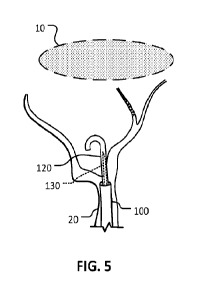
FIG. 5 is a schematic illustration of the shaft 120 being advanced through the
wall perforation of the MMA 20 and toward the subdural hematoma 10. The shaft
120 is advanced over the stylet 130 and has a suitably high pushability (e.g.,
column
strength). The flexible distal tip portion of the shaft 120 has a natural,
unconstrained
curved shape (e.g., J-shape) so that the shaft 120 can be advanced
atraumatically
toward the subdural hematoma 10. Based on advancement of microcatheters 110
with
an OD of 0.040" over a tip of a 0.014" microwire though the subdural space in
cadaveric human heads, subdural navigation is feasible and does not result in
macroscopic brain damage, e.g., atraumatically. During advancement in the
subdural
space, the J or U shape is parallel to the brain surface. This decreases the
risk of
unwanted brain penetration. If the advancement of the shaft 120 disposes the
shaft
120 between the brain surface and the SDH, the shaft 120 is rotated to direct
the J or
U shape towards the SDH and then advanced.
Although the shaft 120 has an unconstrained J or U shape, while the distal tip
portion of the shaft 120 is over the stylet 130, the rigidity of the stylet
130 causes the
distal tip portion of the shaft 120 to be straightened (e.g., as depicted in
FIG. 4). In
some embodiments, the shaft 120 is a coiled-wire reinforced micro-catheter. By
17
CA 03176947 2022- 10- 26
WO 2021/222157
PCT/ITS2021/029276
changing the relative position of the stylet 130 over the shaft 120, the
stiffness and
shape of the shaft 120 are modified.
FIG. 6 is a schematic illustration of the suction catheter 100 being advanced
over the shaft 120, through the wall perforation of the MMA 20, and toward the
subdural hematoma 10. The suction catheter 100 can be a wire-reinforced tube.
The
suction catheter 100 will be advanced until the distal tip portion of the
suction catheter
100, with its open port, is positioned in the subdural hematoma 10. The
advancement
of the catheter 100 can be facilitated by irrigation of solution into the
subdural space.
The shaft 120 and catheter 100 can be coated by lubricious substances like
Teflon or
similar.
FIG. 7 is a schematic illustration showing the distal tip portion of the
suction
catheter 100 positioned in the subdural hematoma 10. From here, suction can be
applied using the suction catheter 100 to aspirate blood and/or other fluids
to at least
partially drain the subdural hematoma 10. The wire reinforcement of the
suction
catheter 100 can help to prevent the suction catheter 100 from collapsing
while
vacuum is being applied to aspirate the subdural hematoma 10. The applied
vacuum
can be continuous, dynamic, cyclical, and pulsatile, at low and/or high
frequency.
Pulsatile pressure induces clot fatigue and fracture facilitating aspiration
removal.
Fluid drainage can occur spontaneously by a pressure gradient between the
intracranial compartment and the atmosphere. Vacuum can be applied either
using
syringes or pump.
FIG. 8 is a schematic illustration of the suction catheter 100 delivering a
hemostatic element 1R0 (e.g., a collagen material that can be used, for
example, to
cause rapid hemostasis in a puncture site, or another type of plug
material/device) to
occlude the wall perforation of the MMA 20 and to occlude branch vessels of
the
MMA 20. Thereafter, the suction catheter 100 can be withdrawn, as depicted in
FIG.
9, to complete the procedure described in FIGS. 2-9 for draining the subdural
hematoma 10.
FIGS. 2-9 describe a platform of devices, also referred to herein as an Extra-
Vascular Access Catheter (EVAC). As described above, the EVAC devices includes
at least the suction catheter 100 and a perforating element that can be formed
by the
micro-catheter 110, and the shaft 120. However, two of these components could
be
merged into a single component by design to achieve similar procedural steps,
or
more components could be added.
18
CA 03176947 2022- 10- 26
WO 2021/222157
PCT/ITS2021/029276
The EVAC device is a platform that provides access to the subdural space
from the intra-vascular compartment, prevents blood extravasation while the
passageway is patent, enables navigation within the intracranial compartment
without
brain perforation or damage, allows drainage of subdural collections, and
ensures
passageway closure (and artery occlusion if needed) upon the removal of the
EVAC
device.
Referring to FIGS. 25A-25E, an embodiment of the EVAC device 2500 is
shown. The EVAC device 2500 shown in FIG. 25A is, in some embodiments, the
suction catheter 2510 subcomponents (e.g., a micro-catheter, shaft 2520, or
stylet
2530) herein described are actuated directly by hand, or at least partially by
a handle
assembly 2540. In one embodiment of the handle assembly 2540, the suction
catheter
2510 is attached, at an end proximal to the user, to a handle housing 2550
including a
port 2560 connected to a vacuum source for SDH 10 suction.
Inside the handle housing 2550 and co-axially with the suction catheter 2510,
the shaft 2520 is assembled at the proximal end to a slider 2570, which when
operated
by pushing or pulling the slider 2570 slides along a slot 2572 on the handle
housing
2550 and creates corresponding co-axial translational movement of the shaft
2520
within the suction catheter 2510. The shaft 2520 distal end is positioned
relative to the
suction catheter 2510 distal end using the slider knob 2570. The slider 2570
is
rotatably tightened against the handle housing 2550 in a setscrew manner
(e.g., using
a setscrew, or rotated into the handle housing to freeze the shaft 2520
translation
motion) to maintain the shaft 2520 position within the suction catheter 2510.
In some
embodiments, the shaft 2520 has a beveled cutting tip with a hollow lumen to
dispose
an atraumatic microwire.
The handle housing 2550 proximal end receives a stylet knob 2580 to which
the stylet 2530 is assembled. FIG. 25B shows a cross-section view of the
components
of the handle assembly 2540 including the nested assembly of the suction
catheter
2510, shaft 2520, and stylet 2530. The stylet 2530 terminates and affixes
within the
stylet knob 2580 at the proximal end. The stylet knob 2580 controls the
relative
position of the stylet 2530 through rotation driving the stylet knob 2580 into
or out of
the handle housing 2550.
A ring seal 2590 is arranged at the proximal ends of the suction catheter 2510
and the shaft 2520 which reduces vacuum leakage and the introduction of air
into the
system. In one embodiment, each of the suction catheter 2500, shaft 2520, and
stylet
19
CA 03176947 2022- 10- 26
WO 2021/222157
PCT/ITS2021/029276
2530 has a liquid port to infuse fluid such as saline lubrication and dilution
of SDH.
In an alternative embodiment, these ports can also be connected to a vacuum
source to
enhance SDH aspiration, such as port 2560. In various alternative embodiments,
discrete markers or rulers are labeled on the slider 2570 and/or stylet knob
2580 to
indicate the relative positions of the distal ends of the shaft 2520, the
suction catheter
2500, and/or the stylet 2530. In another embodiment. a spacer with inner
threads, such
as a nut, can assembled between the stylet knob 2580 and slider 2570 to reduce
the
maximum protrusion of stylet 2530 from the shaft 2520 distal end and provides
a hard
stop.
FIG. 25C is a transverse cross-section view of the handle assembly 2540
through the handle housing 2550 and slider 2570 and perpendicular to the
longitudinal
axis of the handle housing 2550, stylet 2530, and shaft 2520. The rotatable
connection
of the slider 2570 is shown on the left. The slider 2570 is rotated until the
slider 2570
contacts the handle housing 2550 and maintains the shaft 2520 positioning with
respect to the handle housing 2550.
The EVAC device 2500 is navigated into the MMA from a peripheral access
and is used to deliver intra-arterial embolization material for
microvasculature
occlusion of membranes. Then, the EVAC device 2500 is used to intentionally
perforate the arterial wall, advance through the subdural space (e.g., between
the
brain, the dura, and the skull), and drain the chronic subdural hematomas
(cSDH).
During access, a hemostatic element is deployed, delivered or injected across
the
arterial perforation and or the lumen of the MMA closing the extravascular
passageway.
It should be understood that the devices, systems, and methods described
herein are not exclusively for drainage of fluid, clots and particulate matter
from the
subdural space. Instead, the methods and systems described herein can be
adapted to
obtain safe access and drain fluid and clots in the epidural space, for
example, such as
for evacuation of acute epidural hematomas, cystic fluid and pus. In these
cases, the
system can also include elements to macerate clots (e.g., rotating elements,
vibrating
elements, fluid jet, etc.), disposed inside, outside, or both of the
evacuation catheter.
In addition, the methods and systems described herein can be adapted to obtain
access
to any intracranial target in the intradural compartment, including the
subarachnoid
space, the cisterns, the brain tissue and the brain ventricles.
CA 03176947 2022- 10- 26
WO 2021/222157
PCT/ITS2021/029276
It should be understood that the devices, systems, and methods described
herein are not exclusively for drainage of fluid or particulate matter through
the
arteries. Instead, the methods and systems described herein can be adapted and
used
to obtain safe access to the subdural or epidural space and drain fluid,
particulate
matter and clots though veins, the dural venous sinuses and any other natural
corridor.
It should be understood that the devices, systems, and methods described
herein can be used to obtain safe and stable transvascular access to any
extravascular
space and then close the arteriotomy or venotomv site.
Variations and Other Embodiments and/or Features
In some embodiments, the shaft 120 has diathermy, electrocautery or any
other electrical feature to facilitate arterial wall penetration and entry
into the subdural
hematoma. Diathermy, laser and electrocautery can also be used to cut and or
coagulate the membranes surrounding the subdural hematoma, the septations
inside
the hematoma, or any bleeding source. Diathermy, laser and electrocautery can
also
be used to close the transvascular passageway and the vascular lumen such as
the
MMA. A monopolar or bipolar cautery can be used as a separate component or
integrated into the suction catheter 100 and/or shaft 120. In some
embodiments, the
shaft 120 can act as a monopolar (at least a segment of the shaft, generally
the tip) and
the shaft 120 and the catheter 100 (at least a segment, generally at the
distal end) can
act as a bipolar during coupled action.
In some embodiments, the shaft 120 or the suction catheter 100 can be coupled
with thermoablation.
Referring now to FIGS. 26A-26F, in some embodiments, at least one of the
penetrating elements (stylet 2630, shaft 2620, and suction catheter 2600) has
radiofrequency (RF) ablation tip. RF energy can be applied to rapidly increase
tissue
temperature to convert fluid to steam (i.e. vaporization) resulting in focal
tissue
disruption and void. Vaporization may result in a fenestration from the
vascular
lumen to the intradural compartment. This is beneficial to decrease the
cutting force
for perforation and requiring reduced column strength compared to mechanical
needles including cutting edges. The resulting tissue void would also reduce
the
likelihood of edge catching along the trans-vascular passageway. In addition,
atraumatic RF needles tip would be less likely to damage the shaft and suction
catheter.
21
CA 03176947 2022- 10- 26
WO 2021/222157
PCT/ITS2021/029276
In some embodiments, RF energy is used to facilitate ingress into the SDH
through the surrounding membranes, perforation of septations associated with
mixed-
aged SDH and chronic SDH, and to unclog the apertures of the draining tubular
element (e.g. suction catheter).
In some embodiments, RF ablation energy can also be delivered by the same
or additional RF element to coagulate tissues at the penetration site and
arterial
closure if needed at the conclusion of the intervention. In some embodiment,
the RF
element tip includes two or more electrodes with connecting wires extending
from the
distal end to the proximal end of the RF element and connected by an
electrical joint
within a hub to a RF generator. These wires are typically made of conductive
metals
such as stainless steel, copper, and silver and are insulated with plastic
layers such as
PTFE or by embedding inside the wall of the shaft or the suction catheter. The
electrodes are uninsulated and are made of or coated with conductive and
biocompatible metal with high radiopacity such as stainless steel, silver,
gold or
platinum. In some embodiments, one or more electrodes are connected
individually to
a RF generator to work in parallel in a monopolar manner and share the same
grounding pad. In another embodiment with two electrodes, one of the
electrodes is
connected to the RF generator while the other one of the electrodes is
connected to the
ground to work in a bipolar manner. In another embodiment, a single or a
plurality
(>2) of electrodes can be assembled to the RF element and configured to work
in
monopolar or bipolar manner thereof In a bipolar system, the current is
preferentially
concentrated between the two electrodes.
In some embodiments, a bipolar configuration can be obtained by an electrode
in the suction catheter and one electrode in the perforating element. The
perforating
element may acquire a shape upon emergence from the suction catheter to direct
the
tip to the arterial wall and dura. The penetrating element and the suction
catheter can
be concurrently advanced maintaining the distance between electrodes and
delivery of
current to the tissue, or the perforating element can be advanced while
maintaining the
suction catheter stationary resulting in an increased distance between
electrode with a
drop in tissue disruption and decreased likelihood of brain penetration.
In some embodiments, the electrode of the energy delivery device has one of
the following shapes: bullet, cone, truncated cone, cylinder, sphere, dome,
ring, semi-
annular, ellipse, bevel, and arrowhead. The shapes can be at least partially
electrically
insulated for preferential current delivery and directional perforation. The
electrically
22
CA 03176947 2022- 10- 26
WO 2021/222157
PCT/ITS2021/029276
exposed area of the electrode is no greater than 16mm2, and typically in a
range from
2 mm2 to 10mm2.
In some embodiments, the RF perforating element consists of a substantially
tubular member made from an electrically conductive material including
stainless
steel, copper, titanium and nickel-titanium alloys. The tubular element is
proximally
coupled to the RF generator and has an electrical insulator disposed thereon
to deliver
energy to an uninsulated segment or electrode at the distal region with
minimal
dissipation. The distal end of the tubular element can be open or closed. The
tubular
element can be tapered, coupled to a hand-held actuator, and can be scored to
increase
flexibility as described herein. In some embodiments, two or more tubular
elements
can be coupled to form the RF perforating element.
FIGS. 26A-26F depict the RF ablation device and steps to remove an SDH
2610 using such device. FIG. 26A is a schematic illustration of an example RF
stylet
2630 positioned near a wall of the MMA 2620 in a direction toward the subdura1
hematoma 2610.
Radiofrequency energy is generated by a generator and delivered by one (in a
monopolar arrangement) or more (e.g., a plurality) of electrodes (e.g., two
electrodes
in a bipolar array) attached to the distal end of the penetrating member. The
electrodes
are connected to an electrical wave generator via conductive wires embedded
inside
or attached to the wall of the penetrating member extending from the distal
end to the
proximal end of the penetrating member. The conductive wires are electrically
insulated along the whole length except at the very tip. The electrical wave
generator generates a high frequency electrical waveform in a range from 300-
600
kHz (e.g., 400 kHz to 600 kHz, 500 kHz to 600 kHz, 300 kHz to 500 kHz, or 300
kHz
to 400 kHz) and in a range from 120-220 V (e.g., 140 V to 220 V, 160 V to 220
V,
180 V to 220 V, 200 V to 220 V, 120 V to 200 V, 120 V to 180 V, 120 V to 160
V, or
120 V to 140 V).
In some embodiments, the penetrating member if formed by a stylet 2630
with RF capacity and an atraumatic blunt or rounded distal end, as shown in
FIG 26A.
In FIG. 26B, the stylet 2630 is shown penetrating through the MMA 20 while
delivering RF energy 2632 and is followed by coaxial advancement of a shaft
2620
and then a suction catheter 2600 (FIG. 26B). The stylet 2630 can be used to
then
advance atraumatically through the subdural space, or be exchanged by a wire.
23
CA 03176947 2022- 10- 26
WO 2021/222157
PCT/US2021/029276
In some embodiments, as shown in FIG. 26C, the RF stylet 2630 acquires a
curve or a pigtail shape upon emergence from the shaft 2620 or the suction
catheter
2600 (FIG 26D). This may be beneficial to prevent unintentional pullback into
the
vascular lumen and to prevent brain perforation during device advancement into
the
subdural space.
In other embodiments, the shaft 2620 has RF capacity. The shaft 2620 is
advanced trans-arterially under fluoroscopic guidance to the perforation
point, and is
then pushed through the arterial wall and dura into the subdural space while
delivering RF energy 2632. Then, a wire can be pushed through the shaft 2620
into
the subdural space followed by advancement of the suction catheter 2600.
FIG. 26D is a schematic illustration showing the distal tip portion of the
suction catheter 2600 positioned in the subdural hematoma 2610. From here,
suction
can be applied using the suction catheter 2600 to aspirate blood and/or other
fluids to
at least partially drain the subdural hematoma 2610, as described above.
FIG. 26E is a schematic illustration showing the suction catheter 2600
retracted through the MMA 20 wall and RF energy 2632 applied. In some
embodiments, RF energy 2632 is applied to the opening on the MMA 20 wall via
the
electrode to induce thermal coagulation (e.g., clotting) to close the
perforation.
FIG. 26F is a schematic illustration showing the MMA 20 including a clot
2640 which forms from the applied RF energy 2632 disrupting the walls of the
MMA
20.
In some embodiments, the penetrating system includes one or more apertures I
the distal segment fluidly coupled to channels to inject contrast through the
injection
port to confirm the perforation of the targeted tissue, saline solution to
cool the
surrounding tissue to reduce the thermally affected zone during radiofrequency
perforation, or saline solution to increase the lubricity and width of the
subdural
space.
In some embodiment, monopolar electrode is used, and another grounding pad
is attached to the peripheral of the patient head in a direction along the
vector pointing
from the distal end of the penetrating member to the targeted tissue under the
guidance of fluoroscopy. This is beneficial to enable directional
radiofrequency
perforation using minimum energy and creating minimum thermal injury to the
surrounding tissues.
24
CA 03176947 2022- 10- 26
WO 2021/222157
PCT/ITS2021/029276
In another embodiment, the RF energy 2632 can be applied continuously or in
pulses.
In another embodiment, one or more thermocouples are attached near the
distal end of the penetrating member (e.g., near RF stylet 2630) to monitor
the tissue
temperature at and/or near the targeted site. The temperature signal is
transmitted to
the electrical wave generator and the waveform parameters, such as duration
and duty
cycle of pulsed RF energy 2632, is modulated via by the electrical wave
generator.
For example, the electrical wave generator includes an algorithm to modulate
the
waveform parameters based upon at least the temperature signal from the one or
more
thermocouples, such as proportional-integral-derivative (PID) algorithm and/or
Kalman filtering algorithm.
In another embodiment, the penetrating member includes one or more sharp
edges, such as a bevel, and perforates the vessels and dura of the MMA 20
which
reduces the RF energy 2632 to perform the penetration. In alternative
embodiments,
the RF stylet 2630 has an atraumatic non-cutting tip.
In some embodiments, the RF element, which can be the stylet 2630, shaft
2620, catheter 2600, or the combinations thereof, includes a combination of
one or
more electrode, temperature sensor, and/or pressure sensor. In one embodiment,
two
pressure sensors are placed at the distal end of the RF element to sense the
contact
pressure between the tip of the RF element and the tissue (e.g., first
pressure sensor)
and/or the pressure of fluid surrounding the RF element such as the blood or
SDH
fluid (e.g., second pressure sensor).
The first pressure sensor is placed at the tip of the RF element and the
second
pressure sensor is placed 0.2-2 mm proximal to the first pressure sensor. Such
distance is selected to distinguish different stages in the perforation
process. When the
RF element is navigating to the vascular perforation point, both the first and
second
pressure sensor measure the nominal blood pressure.
When the RF element is advanced to push against the target tissue with good
wall apposition, the contact pressure is high, reflected by a high reading
from the first
pressure sensor. Meanwhile, the second pressure sensor is not in contact with
the
tissue and only measuring the nominal fluid pressure.
During tissue perforation, the contact pressure between the first pressure
sensor and the tissue is reduced from high to nominal while the contact
pressure
between the second pressure sensor and the tissue is increased from nominal to
high.
CA 03176947 2022- 10- 26
WO 2021/222157
PCT/ITS2021/029276
After the tissue is perforated, both the first and second pressure sensors
measure the
nominal fluid pressure in the subdural space. In another embodiment, multiple
pressure sensors are placed in a circumferential manner and the average
pressure
measurement is used to reduce the bias due to the non-perpendicular contact
angle
between the RF element tip and tissue.
In another embodiment, such pressure measurement thereof is used to activate
and/or terminate the application of RF energy 2632. In a typical perforating
procedure, the device is first advanced to the vascular perforation point, and
the RF
element position and deflection angle is then adjusted until good wall
apposition of
the RF element against the tissue is confirmed by the pressure measurement.
The RF
energy 2632 is activated, and the RF element starts to perforate the tissue
until the
tissue is perforated and confirmed by the pressure measurement. In another
embodiment, such pressure measurement thereof is used to give signals (in a
form of
light, sound, or other signal) to the operating clinicians to inform the
progress.
In another embodiment, one or more temperature sensors are placed at the
distal end of the RF element to monitor a temperature value during RF
activation and
feed the temperature value to the RF generator to provide a signal
corresponding to a
high temperature, or a low temperature. The RF generator can receive the
temperature
measurement and regulate the RF energy 2632 by tuning the device impedance,
voltage, duty cycle, pulse width, and/or a combination thereof using control
functions
such as proportional-integral-derivative (PID) algorithm, and/or Kalman
filtering
algorithm, to terminate the RF energy 2632 for safety.
In some embodiments, the shaft 120 can have one or more mechanism to
straighten the tip and one or more mechanism to increase the stiffness of at
least one
segment of the shaft 120 (like pulling micro-wires inside, or a coil pull
system).
In some embodiments, at least a segment of the suction catheter 100 and/or the
shaft is deflectable and/or steerable. Deflection (e.g., steering) refers to
the movement
of the distal catheter segment (e.g., the end) independent of the rest of the
catheter.
Steerability refers to the ability to rotate the distal catheter segment
(e.g., clockwise
and/or counterclockwise with respect to the rest of the catheter) by torque
transmission along the length of the device.
The torque causing the deflection can be transmitted by one or more shafts
connected to a pull or anchor ring near the device tip. The distal catheter
segment
rotates one or more directions (e.g., rotational, or flexing within a plane)
upon
26
CA 03176947 2022- 10- 26
WO 2021/222157
PCT/US2021/029276
actuation and return to the original shape (e.g., linear). The deflection can
be
symmetrical, asymmetrical, loop curves, or compound. Deflection can occur in
one or
more planes and be on plane and off planes.
FIGS. 27A-27K are schematic illustrations depicting the use of a deflectable
catheter. The skull 271, brain 272, dura 274, and the MMA 276 are shown with
an
SDH 2710 between the dura 274 and brain 272.
For example, in some embodiments, the suction catheter 2700 includes one or
more pull wires slidably positioned in a wall of the suction catheter 2700. By
pulling
on the wires, the distal end segment of the suction catheter 2700 can be
laterally
deflected. In some embodiments, the distal end segment includes the terminal
0.5 cm
or more of the catheter 2700 (e.g., 1 cm, 1.5 cm, or 2 cm). In addition to
using the
deflecting capability to steer the suction catheter 2700, the deflecting
capability can
also be actuated to anchor (e.g., maintain the position of) the suction
catheter 2700
against an internal wall of a vessel, such as the MMA 276.
Anchoring the suction catheter 2700 against an internal wall of the MMA 276
enhances the pushability of the shaft 2720 and/or stylet 2730 and decreases
the
kickback (e.g., the likelihood of pushing) the suction catheter 2700 out of
the MMA
276. While the suction catheter 2700 is anchored against an internal wall of
the
MMA 276, the ability to push the shaft 2720 and/or stylet 2730 within the
lumen of
the suction catheter 2700 is enhanced.
In such embodiments, the suction catheter 2700 can be advanced into the
intracranial MMA 276 over the shaft 2720 which was advanced over a wire 2705
as
shown in FIG. 27A and FIG. 27B. After removing the microwire 2705 and
embolizing the MMA 276 by injection of embolization agent though the shaft
2720
(FIG. 27C), the stylet 2730 is advanced to the distal end of the shaft 2720
(FIG.
27D).
Stylet 2730 advancement can be facilitated by retracting the shaft 2720
proximal to the foramen spinosum and then advancing the shaft 2720 and the
stylet
2730 concurrently to the distal MMA 276 lumen. The rotatable suction catheter
2700
is oriented (e.g., by rotation or deflection) by visualizing a radio-opaque
fluorscopic
element with fluoroscopy.
The orientation of the catheter 27 with respect to the dura 274 and subdural
space is determined and the pull microwire is actuated resulting in deflection
of the
distal end segment of the suction catheter (FIG. 27D). Altering the
orientation of the
27
CA 03176947 2022- 10- 26
WO 2021/222157
PCT/ITS2021/029276
catheter 2700 and anchoring within the MMA 276 lumen maintains the catheter
2700
position within the MMA 276.
The stylet 2730 and shaft 2720 are advanced (concurrently or subsequently)
through the catheter 2700 and penetrate the subdural space, as shown in FIG.
27E,
and advance into the subdural space, as shown in FIG. 27F. The stylet 2730 can
be
exchanged for a microwire 2705 with an atraumatic tip and, in some
embodiments,
can include a shape, such as a J shape as described above. The stylet 2730
advances
into the subdural space and into the subdural hematoma 2710 (FIG. 27G).
The shaft 2720 advances over the microwire 2705. The wire 2705 can be less
stiff (e.g., flexible) in distal regions and more stiff in proximal regions to
provide
stability and support to the advancement of the shaft 2720 and suction
catheter 2700.
Then, the suction catheter 2700 is advanced through the arterial wall over the
shaft
2720 and microwire 2705 (or stylet 2730 if not exchanged) into the subdural
space
and advanced into the SDH 2710 (FIG. 27H). The microwire 2705 is then removed
(FIG. 271) and the SDH 2710 evacuated by aspiration (FIG. 27J). The
arteriotomy is
closed using a hemostatic element, such as a hemostatic coil 278 (FIG. 27K).
In some embodiment, the shaft 2720 has cutting features at the distal tip
which
includes a bevel, and a lumen to dispose a wire 2705. In this embodiment, the
shaft
2720 is advanced distal to the catheter 2700 distal end and pushed to
penetrate the
dura 274, followed by coaxial advancement of the microwire through the
subdural
space. In some embodiments, the suction catheter slides over the shaft 2720 to
navigate into the subdural space. In other embodiments, the shaft 2720 is
advanced
over the microwire though the subdural space followed by coaxial advancement
of the
suction catheter.
FIGS. 28A through 28C are schematic illustrations depicting a suction
catheter 2800 including two actuators 2850 (e.g., a pull wire attached to an
anchor),
including actuator 2850a and actuator 2850b, respectively. The pull wires of
FIGS.
28A through 28C are shown as dashed lines and the anchors as dark bands
transverse
the catheter 2800 axis. The catheter 2800 is shown advanced into the 1VIMA 286
lumen adjacent the dura 284. In some embodiments, the actuators 2850 are pull
wires
attached to a ring anchor arranged circumferentially around the inner lumen
the
catheter 2800. In some embodiments, the actuators 2850 are anchored
circumferentially to the outer surface of the catheter 2800. Including two
actuators
2850 at two positions within the catheter 2800 provides a means to create a
torque
28
CA 03176947 2022- 10- 26
WO 2021/222157
PCT/ITS2021/029276
between the two actuators 2850 and create higher angle deflections than using
a
singular actuator 2850.
FIG. 28A is a schematic illustration of a catheter 2800 within the lumen of an
MMA 286 adjacent the dura 284. In some embodiments, a first actuator, e.g.,
actuator
2850a, is anchored more proximally (e.g., further from the distal end of)
along the
catheter 2800 than a second actuator, e.g., actuator 2850b, which is anchored
more
distally than the first. The pull wire connecting the first actuator 2850a is
slidably
disposed along a surface of the catheter 2800 along a first route and the
second
actuator 2850b pull wire is slidably disposed along the surface along a second
route.
The first actuator 2850a is actuated which deflects the suction catheter 2800
in the
MMA 286 laterally at the location of the ring anchor of actuator 2850a, which
in FIG.
28B is next to the foramen spinosum. As shown in FIG. 28C, the second actuator
2850b pull wire is actuated to deflect the suction catheter 2800 tip medially
towards
the dura 284, for example, in a second, different direction than the first
actuator 2850a
was deflected.
FIG. 29A and 29B are schematic illustrations of depicting a single actuator
2950 (e.g., a pull wire attached to an anchor) deflecting the catheter 2900
distal tip.
The pull wire of FIGS. 29A and 29B is shown as a dashed line and the anchor as
a
dark band transverse the catheter 2900 axis. In some embodiments, one or more
actuators 2950 can be attached to the catheter 2900 following a pathway, e.g.,
a linear,
curved, "s" shaped, or spiral pathway, along the suction catheter 2900 wall
leading to
one or more articulation points and/or deflections in one or more directions.
This
configuration can actively assist in spatially arranging the suction catheter
2900 in an
advantageous manner.
For example, the actuator 2950 articulates the suction catheter 2900 to form a
curve with a concavity centered at the foramen spinosum (entry point of the
MMA
296 to the intracranial compartment). The actuator 2950 articulates the
suction
catheter 2900 medially and directs the catheter 2900 outlet towards the dura
294 and
the subdural space. For example, a spirally-aligned actuator 2950 capable of
articulating the suction catheter 2900 to curve the distal segment both
medially and
laterally will accommodate catheter 2900 insertion and traversing the MMA 296
for
directable penetration into the subdural space by following the anatomy and
fixation
points at the skull base and dura 294.
29
CA 03176947 2022- 10- 26
WO 2021/222157
PCT/ITS2021/029276
In some embodiment, articulation of the suction catheter 2900 can be achieved
with one or more actuators which can provide xittators 2950 or 2850, such as
pull
wires, notches, preset curves, shapes, and/or any other mechanism obvious by
the
ones skilled in the art. In some embodiments, the actuators, such as the pull
wires or
other articulating elements, are present in the shaft.
In some embodiments, one or more passive (e.g., non-articulable) wires can be
used to stabilize and provide direction to the suction catheter 2900.
In some embodiments, insufflating a balloon, deploying a stent, or other
expandable element disposed in a distal vascular segment and mechanically
connected
to one or more of the telescoping elements can enhanced pushability of the
perforating element and decrease kickback.
In some embodiments, enhanced pushability of the shaft 120 and/or stylet 130,
decrease the kickback of the suction catheter 100 and directionality can be
achieved
by advancing over a wire a delivery sheath with a branching annex into an
arterial
bifurcation. FIGS. 30A through 30E are schematically illustrations depicting a
suction catheter 3000 including a second catheter 3002 affixed to the exterior
surface
at an arterial bifurcation, such as a bifurcation in the MMA 306. The second
catheter
3002 has a smaller inner lumen then the first catheter 3000.
The MMA 306 typically is located in the epidural side of the dura and that
typically has a bifurcation in a plane parallel to the dura. A catheter 3000
including a
second catheter 3002 can include an annex to hold the MMA 306 bifurcation and
stabilize the catheter 3000 position provides 3-dimensional orientation to
perforate the
MMA 306 wall and dura in a perpendicular medialized trajectory.
FIG. 30B depicts the annex 3010 as a wire having a J-shaped distal end,
though the annex 3010 can be a hypotube, catheter, stent, balloon, or other
deployable
element sufficient to provide an arresting tension to the catheter 3000 and
second
catheter 3002. In embodiments in which the annex 3010 is a wire, it can travel
in the
main catheter 3000 lumen or in the second catheter 3002 lumen arranged or
affixed to
the main catheter 3000 wall. The annex 3010 is disposed as an independent
component or as a deployable appendage from the delivery catheter 3002.
The catheter 3000 has a pre-made orifice 3004 that provides off-axis opening
orientable towards the subdural space allowing access to the perforating
element
3012. In some embodiments, the perforating element 3012 is a needle that is
advanced
though the catheter 3000 to penetrate into the subdural space (FIG. 30C). The
shape
CA 03176947 2022- 10- 26
WO 2021/222157
PCT/ITS2021/029276
of the perforating element 3012 can be permanent or composed of a malleable
material (e.g., nitinol).
The shape can be also provided by a shaped inner element within the
perforating element 3012. The distance that the perforating element 3012 is
advanced
beyond the suction catheter 3000 or delivery sheath is called the perforation
distance
(dp). This distance, dm can be fixed, temporarily fixed, or adjustable by the
handle as
described herein.
In some embodiments, the perforating element 3012 is hollow and a contrast
agent can be injected into the subdural space though the perforating element
3012
lumen. FIG. 30C depicts a perforating element 3012 injecting a contrast agent
3015
into the subdural space. The perforating element 3012 tip position can then be
visualized in comparison to the contrast agent to ensure an intradural needle
tip
location. Alternatively, other fluids can be injected through the perforating
element
3012 lumen. For example, injection of fluid, such as water, to increase the
size and
lubricity of the subdural space.
FIG. 30D depicts a microwire 3014 advanced co-axially through the
perforating element 3012 into the subdural space, for example, into an SDH. In
some
embodiments, a third suction catheter 3016 can be advanced over the
perforating
element 3012 though the dura and over the microwire 3014, such as to the
subdural
location of the SDH, as shown in FIG. 30E. The risk of losing access to the
subdural
space after creation of the transarterial passageway decreases by advancing a
wire into
the subdural space.
In some embodiments, the suction catheter distal end segment includes a
beveled tip, e.g., the opening to the suction catheter includes an ovalized
opening and
the tip plane can be at an angle with respect to the transverse plane of the
suction
catheter. In some embodiments, the beveled tip includes a fluorscopic element
for
orientation.
The suction catheter beveled tip facilitates MMA wall penetration into the
subdural space. The suction catheter penetration capabilities are increased
when
combined with a cutting edge. The beveled tip improves apposition between
catheter
lumen and penetrating surface. The beveled tip provides improved penetration
directionality and facilitates evacuation of an SDH.
As an example, a suction catheter is positioned within the MMA and rotated
under fluoroscopic observation (e.g., fluoroscopy) until the beveled tip faces
the
31
CA 03176947 2022- 10- 26
WO 2021/222157
PCT/ITS2021/029276
MMA wall. Penetration is accomplished by any of the embodiments disclosed
herein,
including but not limited to advancing a stylet and/or a shaft into the
subdural space
and penetration with the suction catheter, or, alternatively, penetration with
a pre-
shaped curved needle followed by wire advancement in the subdural space.
In some embodiments, the suction catheter has an actuator (e.g., a pull wire)
to
position the beveled tip against the arterial wall and orient the tip towards
the subdural
space which facilitates directional penetration while providing more column
strength
to minimize kickback. In some embodiments, the depth of penetration of the
shaft and
stylet can be maintained and adjusted with a handle. A typical depth of
penetration
includes a range from 1 mm to 10 mm (e.g., from 2 mm to 10 mm, 4 rum to 10 mm,
60 mm to 10 mm, 8 mm to 10 mm, from 1 mm to 8 mm, from 1 mm to 6 mm, from 1
mm to 4 mm, or from 1 mm to 2 mm).
The depth of penetration is adjustable based on, in some examples, images
obtained before or during the intervention, such as CT scan image and/or brain
MRI
image. The stylet and/or shaft advances into the subdural space into a final
position
and the handle operated to lock the shaft position. The stylet is removed and
contrast
injected into the subdural space. The area is imaged using a method described
herein
to confirm the subdural location of the shaft distal end. A wire is advanced
coaxially
into the shaft into the subdural space.
In some embodiments, the perforation can be created with a curved (e.g., "S"
shaped) perforation element (e.g., a needle) that first medially penetrates
the MMA
wall and dura and curves laterally away from (e.g., along the surface of) the
brain to
direct the bevel facing the subdural surface of the dura. The curvature of the
perforation element distal segment results in a proximal segment parallel to
the MMA
lumen, an intermediate segment transitioning at an oblique angle though the
MMA
wall and dura, and a distal segment within the subdural space that is parallel
to the
dura and the brain. This embodiment reduces the risk of unwanted brain
perforation
and, in the case of brain perforation, ensures that wire advancement will
follow a
trajectory to re-enter the subdural space.
In some embodiments, the actuator articulates the suction catheter to deflect
towards the apex of the bevel followed by perforation with any of the elements
previously described including needle and shaft.
In some embodiments, the perforation element can be the stylet or a suction
catheter. In some embodiments, in cases when the perforation is directed into
the
32
CA 03176947 2022- 10- 26
WO 2021/222157
PCT/US2021/029276
SDH, the needle can be connected to a vacuum source to drain the SDH, or the
perforation element can be retracted resulting in a passageway between the SDH
and
the catheter lumen. In some embodiments, the telescoping elements (e.g., the
suction
catheter, the stylet, the sheath, and/or the microcatheter) includes one or
more
structural elements (e.g., rails) to increase the element stiffness, minimize
unwanted
rotation, and maintain the orientation along the plane.
In some embodiments, the device includes an imaging component, such as a
tomography component. For example, the device can include an optical coherence
tomography (OCT) or an intravascular ultrasound (IVUS) tomography component.
In
embodiments using IVUS, the following echogenicity appearances for biological
components provide orientation. The dura and dural appendages are hyperechoic.
The
cerebrospinal fluid is hypoechoic. The brain superficial pia mater is a well-
defined
hyperechoic layer overlying the hypoechoic cortical gray matter, which
overlies the
hyperechoic white matter. The subarachnoid space contains numerous vessels on
Doppler mode of the ultrasound. The subdural collection includes rare or no
vessels
on Doppler, can have hyperechoic membranes, and can be hyperechoic,
hypoechoic,
or a combination.
FIGS. 31A through 31D are schematic illustrations of a device including an
image component arranged within an MMA 316. FIG. 31A depicts a delivery sheath
3100 anchored by a microwire 3110 and a secondary annex microwire 3111 within
the MMA 316. The perforation element 3112 is shown penetrating the MMA 316
wall
and the can optionally include a delivery sheath with one or more lumens that
is
advanced over microwire 3110 into the distal MMA.
The delivery sheath 3100 be rotated to direct an opening in the distal end to
the penetration point. Typically, the delivery sheath 3100 has higher inner
and outer
diameter proximally and tapers (e.g., lower inner and outer diameter)
distally. This
increases delivery sheath 3100 flexibility aiding in entering the foramen
spinosum and
the intracranial MMA 316.
The opening can be at the distal end of the delivery sheath 3100 or in the
lateral wall (e.g., such as opening 3004) proximal (e.g., within 5 cm of) to
the distal
end. Leveraging the bifurcation of the MMA 316 in the outer surface of the
dura, the
delivery sheath 3100 advances in the dominant branch (e.g., the right branch
of FIGS.
31A) and the annex microwire 3111 is advanced into the second branch of the
MMA
316 bifurcation (e.g., the left branch of FIG. 31A). This orients the delivery
sheath
33
CA 03176947 2022- 10- 26
WO 2021/222157
PCT/ITS2021/029276
3100 in space and disposes an opening towards the perforation point
perpendicular the
plane of the bifurcation and medially (e.g., towards the brain). T
The orientation of the delivery sheath 3100 can be guided by rotating the
device to follow radiopaque markers and/or fluoroscopy. Alternatively, the
imaging
component 3160 (e.g., the OCT or IVUS component) can be used to image the MMA
316 branch lumen and rotating the device to orient the delivery sheath 3100
and
microwire 3110 with corresponding angular markers, such as detected biological
markers within the MMA 316.
One or more penetrating elements (perforating element 3112, stylet, shaft, or
suction catheter) are advanced towards the opening and the MMA 316 wall and
dura
perforated. The perforating element 3112 can be made of a memory material
(e.g.,
nitinol) which curves upon aperture egress, or the aperture can be coupled to
an
angled surface to provide an angle of attack to the dura greater than 1 degree
(e.g.,
greater than 2 degrees, greater than 5 degrees, or greater than 10 degrees).
The
perforating element 3112 can be pre-shaped from a memory material (e.g.
nitinol) and
advanced to penetrate into the subdural space and enable co-axial advancement
of a
microwire to the SDH.
A suction catheter can be advanced over the perforating element 3112 though
the dura and then over the wire though the subdural space to the location of
the SDH.
The perforating element 3112 can be alternatively be exchanged for a suction
catheter
which will be advanced over a wire from the intravascular space to the SDH. In
some
embodiments, the device perforates the dura on top of the SDH. In these cases,
after
advancement of the perforating element 3112 and piercing of MMA 316 wall and
dura, the perforating element 3112 will enable drainage of SDH.
In some embodiments, the perforating element 3112 is reinforced by a stylet
for penetration though the dura. After penetration, the stylet can be removed
providing a lumen to the perforating element 3112 to drain the SDH. In some
embodiments, after penetration with the perforating element 3112 into the SDH,
a
length of exchange wire can be advanced (into the subdural space though the
perforating element 3112 and the device can be removed and exchanged over the
wire
for a suction catheter. The exchange wire length is in a range from 250 cm to
300 cm.
The suction catheter advances over the wire though the arteriotomy site into
the
subdural space and accesses the SHD. The wire is removed and the SDH drained.
34
CA 03176947 2022- 10- 26
WO 2021/222157
PCT/ITS2021/029276
Referring now to FIG. 31D, an embodiment which maximizes the size of the
suction catheter is shown. The suction catheter is delivered though the lumen
of the
delivery sheath 3100. The delivery sheath 3100 advances into the MMA 316
bifurcation and is oriented by use of a wire in each branch of the MMA 316.
The delivery sheath 3100 has an opening in the distal end which enables the
advancement of a perforating element towards the MMA 316 wall and dura. In
some
embodiments, the direction of the perforating element can be provided by using
a pre-
shaped perforating element 3112 with a curved orientation and is oriented with
fluoroscopy to perforate the MMA 316 wall medially and perpendicularly.
Orientation can be also provided by a rail system connected to the inner or
outer surface of the delivery sheath 3100 and/or the perforating element 3112.
In
alternative embodiments, the perforating element 3112 advances towards a
deflecting
surface fixed in the ID of the delivery sheath 3100 resulting in advancing of
the
perforating element 3112 towards the opening and perforating point.
In some embodiments, the perforating elements herein described is used to
perforate through membranes and septations associated with mixed-aged SDH and
chronic SDH.
In some embodiments, the perforating element 3112 is fluidly connected with
one or more apertures at or near the distal end which allows injecting of
fluid,
including a contrast agent, or saline.
In some embodiments, the perforation process and ingress in the subdural
space monitored by including one or more sensors in the device, including
pressure
sensors, e.g., potentiometric pressure sensors, inductive pressure sensors,
capacitive
pressure sensors, strain gauge pressure sensors, fiber optic pressure sensor,
variable
reluctance pressure sensors, microelectromechanical system pressure, and
piezoelectric pressure sensors.
A piezoelectric pressure sensor includes piezoelectric film disposed in the OD
of the perforating element 3112 in proximity to the distal end to capture a
pressure
signal, the signal including a pressure peak associated with penetration
through the
MMA 316 wall and by pressure drop upon accessing the subdural space. Pressure
sensors can also sense the pressure waveform (e.g., arterial, intracranial, or
venous
perforation waveforms) upon penetration from the vascular lumen into the
intracranial
compartment.
CA 03176947 2022- 10- 26
WO 2021/222157
PCT/US2021/029276
In some embodiments, the perforation process and ingress in the subdural
space is monitored by measuring spectroscopy values of tissues.
In some embodiments, the perforation process and ingress in the subdural
space is monitored by measuring an impedance value and/or a permittivity value
of
tissue and fluids. FIG. 39 is a line chart comparing the permittivity value on
the y-
axis to the frequency in kHz of the electrical signal (e.g., the RF energy) on
the x-axis.
FIG. 39 includes four example tissues (e.g., blood vessel wall, dura, or brain
surface)
or fluid (e.g., blood) represented by lines of FIG. 39. The individual lines
represent
the permittivity of the example tissue or fluid with respect to the applied
energy
frequency. according to the key inset in the upper right. Blood (solid line)
has the
highest permittivity across the depicted frequencies, with a brain surface
(dotted line)
having lower permittivity than blood, the blood vessel wall (dashed line)
having lower
permittivity than the brain surface, and the dura (dash-dotted line) having
the lowest
permittivity up to approximately 720 kHz and the blood vessel wall having the
lowest
permittivity above approximately 720 kHz.
Permittivity is an intensive (e.g. independent from the volume or mass of
object or tissue) electrical property of tissue. Tissue with higher
permittivity has lower
impedance when transmitting alternating current, or in the invention described
here,
RF current. Blood has higher permittivity compared to the brain surface,
followed by
the blood vessel wall and dura in the RF frequency range (e.g., in a range
from 100 to
1000 kHz).
In a typical operating RF energy frequency range (e.g., 300-600 kHz), the
permittivity of blood is 4 times higher than that of the brain surface and
over 10 times
higher than that of the dura and blood vessel wall. Such large difference in
permittivity can be used to detect the location of the RF element. During RF
penetration, the RF element is sensing a low permittivity (or high impedance)
from
the dura and the vessel wall. Once the dura and vessel wall is perforated, an
increase
in permittivity (or decrease in impedance) is measured as the RF element goes
into the
subdural space and contacts the brain or SDH. Such impedance signal is fed to
the RF
generator to automatically reduce or shutoff the RF power and/or notify the
operating
clinicians in a manner of sound, light, vibration or a combination thereof.
After
completing the intradural intervention, the apparatus including the RF element
is
pulled back into the vascular lumen and again surrounded by blood. This
results in an
increase of permittivity (or decrease in impedance). Such permittivity and/or
36
CA 03176947 2022- 10- 26
WO 2021/222157
PCT/US2021/029276
impedance signal is fed to the RF generator to modify (e.g., raise, lower,
initiate, or
terminate) the RF power to a coagulation mode, and/or provide a notification
to the
operating clinicians in a manner of sound or light or a combination thereof.
In some embodiments, the perforation process and ingress in the subdural
space is monitored by including a force sensor at the proximal end of the
perforating
element to measure a thrust force associated with penetration through the MMA
316
wall.
In some embodiments, the perforation process is followed by measuring
pressures at the tip of the perforating element by channel fluidly coupled to
a pressure
transducer which can be outside the patient.
In some embodiments, the perforation process is followed by recording
electroencephalographic (EEG) activity with an electrode (e.g., EEG measuring
device) disposed at or within 20mm from the front end of the penetrating
element and
electrically coupled to an EEG recorder which can be outside (e.g., external
to) the
patient. In some embodiments, the EEG measuring device can function as the RF
energy delivery device, such as RF stylet 2630. In this embodiment, an EEG
interface
device operating as a splitter enables concurrent connection of the device to
an RF
generator and an EEG recorder. This embodiment facilitates continuous EEG
recording concurrently with RF energy 2632 delivery.
The construction of the styl et, shaft or catheter to achieve these features
comprises one longitudinal element selected from the group consisting of a
hypotube,
single solid rod, multiple roads, bundle, tubing (with one or more lumens),
shaft
strands, cable (two or more wires running side by side, bonded, twisted or
braided),
coil, braid or combinations thereof
In some embodiments, the device subcomponents can be made of metal or
metal alloy (including but not limited to stainless steel, nitinol, silver,
titanium,
copper, cobalt chromium, nickel chromium, platinum iridium, and others),
polymer
(including but not limited to nylon or other polyamides, fluoropolymers,
polyolefins,
polythetrafluoroethylene, high density polyethyene, polyurethanes and
polyimides),
ceramic, bio-absorbable or dissolvable components, or combinations.
The construction of the device can include inner liners and outer jackets and
the manufacturing techniques are known by those skilled in the art. These
elements
may be necessary to enhance the structural support to the device, facilitate
smooth
telescoping between components, prevent vacuum leaks by sealing holes, and
37
CA 03176947 2022- 10- 26
WO 2021/222157
PCT/ITS2021/029276
enhancing the flow of hematoma to be drained and reduce the likelihood of
clots and
fibrin to clog the suction catheter.
It is beneficial for the elements of the device herein to remain highly
flexible
to navigate into the intracranial compartment and have sufficient column
strength to
perforate and advance into the subdural space. In some embodiments, the
elements of
the device have a tip bending stiffness in a range from 0.0002 lbf in' to
0.005 lbf
with a typical value around 0.001 lbf in' for navigating to/within the MMA. In
some
embodiments, the elements of the device have a bending stiffness of 0.006 lbf
in2 to
0.15 lbf in2 with a typical value of 0.028 lbf in2 for navigating to/within
the SSS. To
this end, the elements have sections of varying stiffness. This is
accomplished by
employing and combining different element construction configurations,
materials,
ration of materials, thicknesses, amount of material, materials with different
durometer, and/or selective reinforcement.
In some embodiments, any of the subcomponents may include a plurality of
scorings to increase element flexibility, for example, to transverse the
curves of
foramen spinosum. The plurality of scorings can take a shape or pattern
including but
not limited to spiral scoring patterns (continuous or interrupted), radial
scoring
patterns, bespoke scoring patterns, radial ring patterns, longitudinal
scoring, oblique
scorings, windows, tabs, or holes. Scorings may be created in the elements by
using
any suitable scoring methods including laser scoring and etching. Scorings can
be in
at least a portion (e.g., a segment) of the subcomponent and in some
embodiments are
preferentially located on one side.
In some embodiments, any of the subcomponents have one of the following
profiles along its length: continuous, tapered in distal direction, tapered in
proximal
direction, multi tapered and combinations thereof
Generally, subcomponents are larger, stiffer, and have higher torque
transmission proximally and will taper distally and have increasing
flexibility to enter
the foramen spinosum and intracranial MMA.
In some embodiments, any subcomponent, including stylet, shaft or catheter is
constructed with two or more layers of high-tensile wire wound at opposing
pitch
angles resulting in flexible elements with high torsional stiffness.
In another embodiment, the subcomponents, including stylet, shaft, catheter,
anchoring element, or protective sheath, can have a railing system along at
least a
portion of the length with one of the following cross-sectional shapes:
circular, oval,
38
CA 03176947 2022- 10- 26
WO 2021/222157
PCT/ITS2021/029276
square, start, diamond, rectangular, flat, or a combination thereof. The
receiving
lumen conforms to the shape of the inner member. The receiving lumen ID
approximately matches (e.g., within 0.002") the OD of the inner member to
restrict
non-longitudinal motion or is shaped to allow a limited preset range of
rotational
motion. In some embodiments, the subcomponents include one or more rail
systems
per subcomponent, and the rail system can include the full subcomponent
length.
Non-circular configurations of the stylet and shaft can limit the relative
rotation between subcomponents while maintaining the capacity to telescope
along
the longitudinal axis (e.g., longitudinally). Such non-circular embodiments
help to
maintain the trajectory of the perforating element over the guide element
towards the
perforation target. As one example, the rail system may be beneficial when
advancing
the penetrating element over a distal anchoring element. In this example, the
anchoring element (balloon or stent or other) can be connected to the device
by a flat
wire. The flat wire can allow element advancement over the wire of the
perforating
element (or protective sheath) to the perforation point. In other embodiments,
radial
alignment between telescoping elements can be maintained by coupling
longitudinal
recesses and fins at the interfacing surfaces.
In some embodiments, deploying a stent or insufflating a balloon disposed in a
distal segment of the shaft 120 in the extravascular space can provide
improved
purchase to push a suction catheter 100 into the extravascular space.
Fluoroscopic elements that are highly visible under fluoroscopy can be located
on any of the components in any locations as desired.
In some embodiments, the fluoroscopic elements construction can include
elements indicating the direction of the deflecting element. The direction of
the
deflecting element indicates the plane to which the marker will direct the
subcomponent, e.g., the plane of deflection. For example, the marker can be a
partial
circle with a notch matching the plane of deflection, or the plane of
deflection can be
marked by the partial radiopaque circle, or by an asymmetry in the marker.
Alternative examples of fluoroscopic elements include an arrow-head, ring, or
band
symbol or structure.
In some embodiments, the fluoroscopic elements provide support to the
perforating element in a circumferential fashion.
39
CA 03176947 2022- 10- 26
WO 2021/222157
PCT/ITS2021/029276
In some embodiments, the fluoroscopic elements decrease the cutting force
required to perforate the MMA and/or dura, such as a fluoroscopic marker
including a
beveled tip, or cutting tip.
In some embodiments, the fluoroscopic elements can be tapered to decrease
the gap with the OD of a telescoping element.
In some embodiments, two or more fluoroscopic elements are present in a
subcomponent. For example, the shaft or the catheter can have one fluoroscopic
elements distally to indicate the distal end of the device, and another
fluoroscopic
elements in a more proximal segment to indicate the detachment of the element
to
close the arteriotomy, including off-the-shelf coils.
In some embodiments, two or more different subcomponents have two or
more fluorscopic elements which indicate longitudinal and or rotational
alignment
between subcomponents. These fluorscopic elements can include symbols (e.g.,
"bullseye"), forming intersecting shapes (e.g., "T", "L","+", or "X"), or
other way
evident by those skilled in the art.
In some embodiments, the distal end portion of the shaft 120 or stylet can
have
elements (e.g., ridges, fins, wedges) or shapes to maintain the tip of the
shaft 120
away from the brain surface. The shape can be permanent, temporary, or
reversible.
For example, compacting a reversible shaped element composed of a memory
material (e.g., nitinol) within a subcomponent and, after emerging from the
subcomponent, the element resumes the original shape; alternatively, the shape
is
resumed after removing a rectifying (e.g., constraining) element.
In some embodiments, the shaft 120 or stylet (or other subcomponent)
includes a shaped inner element and the shape of the distal end portion of the
shaft
120 or stylet (or other subcomponent) corresponds to the shape of the inner
element.
The inner element can be a rigid or semi-rigid element within a portion of the
length
of the subcomponent and be composed of a rigid, or semi-rigid material, such
as a
memory material.
For example, the inner element and corresponding subcomponent shape can
include "S" shapes wherein the distal end portion is directed towards the dura
for
penetration and a proximal end portion is outside the dura, with a connecting
portion
angled with respect to the distal and proximal portions. After a distance of
advancement (e.g., the length of the connecting portion) wherein the distal
end of the
outer coaxial element extends through the dura, for example extends through
the dura
CA 03176947 2022- 10- 26
WO 2021/222157
PCT/ITS2021/029276
by between 1 mm and 5 mm, the shape is formed directing the tip away from the
brain. In some embodiments, the distance of advancement is in a range from 0.1
mm
to 15 mm (e.g., from 1 mm to 15 mm, from 5 mm to 15 mm, from 10 mm to 15 mm,
from 0.1 mm to 10 mm, from 0.1 mm to 5 min, or from 0.1 mm to 1 mm). A second
curve proximal to the distal most "S- shape can help orienting the curves of
the shape
parallel to the brain and dura surface.
In some embodiments, one or more subcomponents include one or more
anchoring features to anchor the subcomponents to the subdural surface of the
dura
after penetration, or to anchor to an intradural surface. Examples of these
anchoring
features include wires (including shaped wires, e.g., pigtail wires), stents,
balloons,
arrowheads, wings, fins, loop, bend, harpoons, spikes, hooks, and/or barbs.
These
anchoring features anchor a subcomponent of the device (such as the stylet, or
shaft)
after penetration and enable more tensional load to be applied to the outer
penetrating
elements during advancement through the MMA wall and dura. These anchoring
features may be compressible (e.g., formed of a low durometer material) to be
recaptured.
In some embodiments, the anchoring features are actuatable to anchor the
subcomponent upon actuation or activation. Alternatively, the anchoring
features are
fixed (e.g., static) to provide anchoring coincident upon ingress into the
extravascular
compartment. In some embodiments, the penetrating element can become the
anchor
upon actuation or advancement. As an example, the stylet can be used to
penetrate
through the vascular wall and dura, and upon emergence from an enclosing
subcomponent acquires a pigtail shape which coincidently anchors the stylet in
the
subdural space. The stylet pigtail shape provides an atraumatic tip upon
advancement
in the subdural space.
In some embodiments, one or more subcomponents have one or more arresting
features which minimize the risk of unintentional subcomponent retreat into
the artery
after perforation of the arterial wall or dura This is advantageous as retreat
may result
in bleeding though the arteriotomy. The arresting features include elements
that
expand and/or deploy resulting in a larger dimeter than the subcomponent
carrying
these elements (or alternatively expand the subcomponent diameter), and
examples of
arresting features include barbs, fins, wires, collar, rib, rim, ribbon, and
baskets. In
some embodiments, the arresting features are recaptured (e.g., retracted).
Other
limiting feature examples can be elements that reduce the subcomponent
diameter
41
CA 03176947 2022- 10- 26
WO 2021/222157
PCT/ITS2021/029276
along a portion of the subcomponent length such reversible bevels,
indentations, or
notches. In some embodiments, the arresting features are manually or
automatically
retractable (e.g., hidable).
In some embodiments, one or more subcomponents include one or more
limiting features to limit the longitudinal advancement. For example, the
limiting
features limit subcomponent penetration depth to within a distance of the dura
perforation. The limiting features include elements that expand and/or deploy
resulting in a larger dimeter than the subcomponent carrying these elements
(or
alternatively expand the subcomponent diameter), and examples of arresting
features
include barbs, fins, wires, collar, rib, rim, ribbon, and baskets. In some
embodiments,
the limiting features are recaptured (e.g., retracted). Other limiting feature
examples
can be elements that reduce the subcomponent diameter along a portion of the
subcomponent length such reversible bevels, indentations, or notches. In some
embodiments, the limiting features are manually or automatically retractable
(e.g.,
hidable).
In some embodiment, the perforating element can have fixed features to limit
the penetration depth. This is beneficial when the perforating element is not
intended
to advance beyond a point but enable an inner element and or an outer element
to be
advanced distally. As an example, a beveled shaft can be the penetrating
element that
pierces the arterial wall and dura until and advances into the subdural space
and is
stopped by a rim. At this point, a microwire is advanced inside the shaft and
distally
into the subdural space. Then, the suction catheter is advanced over the shaft
though
the vessel wall and over the microwire though the subdural hematoma while the
fixed
shaft is providing column support to the remaining advancing elements.
In some embodiments, the perforating element that initially pierces the dura
has features that deploy in the unconstrained subdural space to prevent brain
perforation.
In some embodiments, one or more device subcomponents could have one or
more features to minimize the risk of unintentional pull back into the artery
after
perforation and to limit the longitudinal advancement beyond a certain point.
In some embodiments, the suction catheter can be telescoped through the
subdural space beyond the distal end of the stylet or shaft. In one of these
embodiments, the stylet with a beveled tip can be advanced to penetrate the
arterial
wall and the dura. The shaft can be advanced over the stvlet to penetrate the
arterial
42
CA 03176947 2022- 10- 26
WO 2021/222157
PCT/ITS2021/029276
wall and dura. The stylet can be removed, contrast injected though the shaft
to ensure
subdural location of the tip and to map the intradural target, and an
atraumatic
microwire advanced coaxially into the shaft and then distally into the
subdural space.
The shaft can be advanced over the microwire into the subdural space followed
by
subsequent coaxial advanced of the catheter. After reaching the target with
the distal
tip of the suction catheter, the inner elements are pulled out.
In other embodiments, the shaft is constructed to provide proximal support and
only perforate the arterial wall and dura over the stylet for short distances
(e.g., 1 mm
to 20 mm) to facilitate the perforation of the outer catheter, which will then
be
advanced into the subdural space over the microwire.
In other embodiments, a protective sheath can be added to the telescoping
system and be disposed over the perforating subcomponent. The protective
sheath can
be disposed throughout the length of the perforating subcomponent or be
selectively
disposed to cover the distal segment of the perforating component. In such
cases, the
protective sheath is translated longitudinally by one or more pull or push
wires. The
protective sheath protects the cutting features of the perforating element and
the inner
surface of the delivery catheter during the advancement of the perforating
element to
the target. The protective sheath provides orientation for the inner
perforating element
when the protective sheath is coupled to the anchoring element (balloon,
stent, etc.)
by a rail system as described herein. The protective sheath enables a
perforation
element to acquire a memory shape when unsheathed from the protective sheath.
The
protective sheath can have one or more fluorscopic elements to indicate the
location
of the perforating element.
In other embodiments, the shaft is constructed with perforating elements
described elsewhere herein and actuated to perforate the arterial wall and
dura without
an inner stylet. The shaft can have a lumen to inject a radio-opaque contrast
agent to
ensure that the tip opens to the subdural space. The shaft lumen could also be
used to
infuse saline which would flow with low resistance if injected in the subdural
space,
and at a higher resistance if injected into the brain parenchyma. An
atraumatic stylet
or microwire can be coaxially introduced inside and beyond to the shaft into
the
subdural space. The suction catheter can be advanced over the shaft though the
arterial wall and the dura, and then over the stylet/microwire inside the
subdural
space.
43
CA 03176947 2022- 10- 26
WO 2021/222157
PCT/ITS2021/029276
Any of the elements herein described can be telescoped co-axially and/or over
the wire rapid exchange system.
In some embodiments, any of the elements or subcomponents herein described
can have reinforcements to increase the column strength of that particular
element.
In some embodiments, the distal end of the stylet, shaft, and/or catheter
includes a commercially available hypodermic needle or vascular access
needles. The
length of the needle tip reduces the applied force to penetrate the arterial
wall and
dura and increases the ease of navigation through the curvature at the foramen
spinosum. Longer bevels result in lower puncture forces but higher likelihood
of not
tracking into the intracranial MMA. Bevels with a length less than 1.5 mm
obtained
from commercially available 28 G needles welded at the distal end of a stylet
(e.g.,
rope-like element with diameter 0.015") can be advanced within a shaft having
an ID
of 0.016" and an OD of 0.024" through the foramen spinosum inside a catheter
having
an ID 0.030" and an OD of 0.043" located in the MMA. The needle can include
plurality of cuts to increase flexibility while maintaining column strength as
described
elsewhere.
Generally, arterial navigation is facilitated by maintaining the tip of the
needle
at the front end of the stylet 1 mm distal to the shaft, flush with the shaft,
or within 10
mm of the distal end of the shaft. Needles can be larger, the same size, or
smaller than
the element they are welded on. Minimal gaps (e.g., a diameter difference of
less than
0.2 mm) between the telescoping elements reduce the likelihood of catching at
the
artery/dural wall.
In some embodiments, the distal part of the shaft 120 includes a balloon. The
shaft 120 can be centered or off-centered position and have a parallel or
diagonal
emergence from the suction catheter 100. The inflation of the balloon can be
used to:
(i) provide support to the shaft 120 for advancement through the arterial wall
and/or in
the subdural space, (ii) occlude the arteriotomy of the parent artery to
prevent blood
extravasation, (iii) enlarge the arteriotomy to facilitate passage of the
suction catheter
100 pushed the brain surface insufflated, (iv) provide distal
support/anchorage to
facilitate over the shaft advancement of the suction catheter 100 into the
subdural
space, (v) push the brain surface away from the shaft 120 during advancement,
and
(vi) unclog the catheter 100 of particulate matter.
44
CA 03176947 2022- 10- 26
WO 2021/222157
PCT/ITS2021/029276
In some embodiments, the distal part of the shaft 120 includes a balloon to
create an extradural, intradural of subdural corridor for suction catheter 100
advancement.
In some embodiments, the shelf between the catheter 100 and the shaft 120
can be tapered. In another embodiment, the catheter 100 can have one or more
bevels
to direct advancing forces away from the brain surface and towards the SDH.
Such a
taper design can facilitate advancement through the arterial wall, the
subdural space
and into the SDH.
In some embodiments, the shaft 120 has a larger diameter tip as a "cap." The
diameter of the cap can be smaller or equal to the outer diameter of the
catheter 100.
The cap can be round or conical with the apex of the cone centered or off
centered.
The interface between the cap and the catheter 100 can be perpendicular to the
main
axis, oblique, or a combination. For penetration and advancement modes, the
cap can
be disposed in proximity or touching the distal end of the catheter 100 by a
pull wire
to enhance the pushability of the system and conceal the catheter edges (which
may
facilitate advancement through the arterial wall and the subdural space into
the SDH).
For aspiration mode, the cap can be separated from the tip end of the catheter
100 by a
push wire to open an entry point and aspirate. If the tip of the catheter 100
clogs, the
cap be withdrawn by pull wire to unplug the catheter 100.
In some embodiments, the tip end of the catheter 100 can be closed and/or
have different shapes. The catheter 100 can have side holes/fenestrations to
aspirate
fluid. In some embodiments, the catheter end is tapering and has no opening at
the
tip. This design has no ledge and therefore is highly atraumatic. Some
embodiments
may have an opening at the end to allow coaxial advancement of a shaft
disposed at
least partially inside the catheter 100.
In some embodiments, the distal tip of the catheter 100 can have bevel and
sharp edges to facilitate the entry into the subdural space and element in the
wall to
prevent penetration into the brain. Catheters 100 with an oval lumen rather
than
round can decrease likelihood of brain penetration. Catheters with beveled tip
can
facilitate drainage of fluid and clots.
In some embodiments, catheters can have a distal funnel to enhance drainage
of fluid and clots. The funnel can be open by a balloon system, pull or push
wires, or
have a braided design or slotted hypotube design that can be introduced in a
compressed state and then expand into a funnel after unsheathing.
CA 03176947 2022- 10- 26
WO 2021/222157
PCT/ITS2021/029276
The lumen of the catheter 100 can be configured to enable irrigation of
solutions, drugs, cells, or particles. In some embodiments, the catheter 100
may have
additional hollow channels to enable fluid irrigation before, during and after
subdural
hematoma drainage. The fluid can be directed outside the suction catheter 100,
at the
tip of the suction catheter 100 or inside the main lumen of the suction
catheter 100.
Fluid irrigation can decrease the viscosity of the fluid and enhance the
drainage.
In some embodiments, the fluid can deliver pharmacological agents to the
treatment location, or suspended cells and particles in a solution which is
later
aspirated by the suction catheter.
In some embodiments, the lumen of the suction catheter 100 can be coated by
lubricious substances to facilitate drainage of fluid and particulate matter.
The guidance of the system can be coupled by including components visible
by invasive (US, CTO, Angioscopy) and non-invasive (fluoroscopy, US, CT, MR)
imaging modalities. This could be coupled with image guided interventions. In
some
embodiments, the suction catheter 100 or the penetrating element itself can be
integrated with invasive imaging modalities for structural visualization.
In some embodiments, the suction catheter 100 can include an integrated
camera for endoscopically-assisted transvascular drainage of subdural
collection. The
camera can provide visualization of the advancement of the catheter 100 over
the
shaft 120 in the subdural or epidural space. The endoscope functionality can
be based
in optical fibers, complementary-symmetry metal¨oxide¨semiconductor, scanning
fiber endoscope or any other methods. The optical system can be mounted in the
head
of the catheter (light source and electric wire can be disposed in the
catheter wall),
which can be advanced over a wire (which could be the penetrating element) in
the
extravascular space and into the collection to drain.
In some of these embodiments, the catheter 100 can have drainage
fenestrations and can have a steerable mechanism to deflect the shaft of the
catheter
100. In this embodiment, upon perforation of the arterial wall the catheter
100 is
advanced co-axially.
In some embodiments, saline irrigation can be infused in the subdural space to
facilitate advancement of the catheter (e.g., to increase lubricity of the
surfaces and
create and separate the space) and/or for improving direct visualization with
the
camera. The system can be advanced and used to enter the collection to drain
as
described above.
46
CA 03176947 2022- 10- 26
WO 2021/222157
PCT/ITS2021/029276
In some embodiments, the penetrating element can have one or more orifices
in the distal segment in a range from 0.01 mm to 10 mm from the tip. The
orifice can
be fluidly coupled to one or more lumens of the shaft or catheter and enable
irrigation
upon perforation into the subdural space.
In some embodiments, a coring element can be mounted over the balloon of a
catheter and generate a passageway through the vascular channel wall. The
coring
element can be in continuity with the lumen of the suction catheter enabling a
stable
passageway into the extravascular space. In some embodiments, the balloon
mounted
over a catheter has a channel in continuity to the catheter lumen.
Insufflation of the
balloon stabilized the catheter and approximates the opening of the channel to
the
vascular wall. Extravascular access is accomplished as previously described
herein.
In some embodiments, the MMA wall-penetrating element has a pre-shaped
curvature that it is acquired upon emergence from the delivery catheter 100.
In such a
case, the wall-penetrating element is flexible enough to cause minimal or no
deformation to the delivery catheter 100, but upon emergence into the vascular
lumen
it acquires a curvature that is maintained while penetrating through the
vascular wall.
In some embodiments, the bending radius of the curvature may vary, and can be
selected to aim the system towards the location of the target. This embodiment
would
be beneficial, for example, when the vascular geometry is divergent from the
target
and the penetration to the vascular wall will not lead to the target. This
diverging
curvature can be combined with other curvatures in the shaft that only are
acquired in
the extravascular space. This can be achieved by modifying the bending radius
and
flexibility of each individual curve.
In some embodiments, the penetrating element can have features to facilitate
penetration through the vascular wall and dura including cutting edges,
bevels, cutting
tips, cone shape, coring punch, and corkscrew shapes. Cutting features can be
in the
outer edge of the inner edge. The later reverse cutting edge to the inside
lumen
minimize the gap between two coaxial telescoping elements facilitating
perforation at
lower forces. Features to facilitate penetration can be one or a combination
of multiple
features, and it can be combined in any of the device elements including
stylet, shaft
or catheter. In some embodiments, the rigidity of the catheter 100 and/or
penetrating
element (e.g., shaft or micro-catheter) can be modified by air or fluid
introduction at
variable pressures in accessory channels associated with the wall of these
elements.
47
CA 03176947 2022- 10- 26
WO 2021/222157
PCT/ITS2021/029276
The channels can be disposed in any segment the catheter 100 and/or
penetrating
element (e.g., shaft or micro-catheter) and can be of any length.
For example, the catheter 100 can be made stiff upon saline injection in the
wall channel to enhance the support to the penetrating element (e.g., shaft or
micro-
catheter) to transverse the arterial wall or subdural membrane. Upon
advancement in
the extravascular space, the distal most segment of the catheter 100 can be
made
flexible/soft by removing saline solution.
In another example, the penetrating element (e.g., shaft or micro-catheter)
can
have a multi-durometer construction and be stiff proximally and flexible/soft
distally.
The distal segment can be associated with a channel to modify flexibility. For
penetration through the arterial wall, the distal segment can be stiffened by
fluid
injection in the inner channel. Upon vascular penetration, the inner channel
can be
deflated leading to increase flexibility of the penetrating element and save
advancement. In some embodiments, this inner channel has an opening in the
distal
segment that becomes occluded while pushing against or thorough the arterial
wall.
While occluded, the fluid remains inside the channel conferring maximal
stiffness to
the penetrating element by positive pressure. Upon penetration into the
extravascular
space, the channel opening becomes un-occluded and enables rapid release of
the
fluid of the inner channel leading to a decrease in stiffness. This mechanism
increases
the safety feature of the device as decreases the chance of brain penetration.
The
injection of air and fluid in these channels can be also used to deflect the
catheters or
penetrating elements.
In some embodiments, the catheter 100 and or the penetrating elements (e.g.,
shaft 130 or micro-catheter 110) are combined with methods and mechanism to
sense
the extravascular position of at least of segment of the tube, including
differential
pressure, impedance, and other.
In some embodiment, one or more magnetic component can be added to any
component of the EVAC system for magnetic-based movement.
Upon completion of the extravascular intervention, the arteriotomy is closed
by one or a combination of the following hemostatic mechanisms: balloon, gel
foam,
collagen, thrombin, particles (e.g., polyvinyl alcohol, embospheres), coils
(e.g.,
pushable, injectables, detachable), liquid agents (e.g., glue, ethylene vinyl
alcohol),
sclerosant agents (e.g., sodium teradecyl sulfate, alcohol, algel), plugs
(e.g., including
self-expandable cylindrical or hourglass shape), stitches, electro
coagulation.
48
CA 03176947 2022- 10- 26
WO 2021/222157
PCT/ITS2021/029276
In some embodiments, the hemostatic element has mechanism to prevent
accidental retreat during device retrieval. As a mode of example, these
elements
includes a focal enlargement on the distal segment of the gel foam or collagen
pledget
and flowering elements that radially expand after being unsheathed.
In some embodiments, at least a segment of any device subcomponents can be
detached to close the arteriotomy.
In some embodiments, the hemostatic element (like a coil) can be transected at
the desired length by chemical, mechanical, and or electrical mechanisms. This
is
desirable to deploy the closing element though the arteriotomy and then cut
the
hemostatic element to prevent long segments of arteries to embolize.
In some embodiments, the perforating element can be coupled with a closure
device. The closure device can be released upon removal of the perforating
element at
the perforation point.
In some embodiments, the arteriotomy is very small and would not result is
significant blood extravasation making the hemastatic device not needed.
Referring to FIGS. 10-13, an alternative embodiment of a shaft 140 has a
distal end portion with a double -J" configuration. The distal most "J" 142
can be
performed can define a cutting edge or a bevel and allows to directionally
penetrate
the wall of the MMA 20 or subdural hematoma membrane. Disposed in a proximal
fashion, there is another "J" or "U" 144 that forms after the shaft 140
emerges out
from the catheter 100 and provides an atraumatic shape for advancement into
the
subdural space. The distal J 142 can be sharper and stiffer than the proximal
J
segment 144 to enhanced wall penetration. By being flexible, the proximal J
segment
becomes straightened while radially constrained within the catheter 100. In
this
embodiment, no stylet is needed. Instead, the distal J 142 tip penetrates and
perforates the wall of the MMA 20. The distal preformed J 142 allows a
directional
penetration through the arterial wall by rotating the shaft 140 until it
points towards
and perpendicular to the subdural space. Upon penetration, the shaft 140 can
be
rotated as needed to point parallel and into the subdural space and the shaft
140
advanced outside the catheter 100 until the shaft 140 acquires the shape of
the
proximal J 144.
Referring to FIGS. 14-16, the depicted embodiment includes a suction
catheter 200 and a shaft 210 that is slidably disposed within a lumen defined
by the
catheter 200. The catheter 200 defines drainage fenestrations in its distal
tip portion.
49
CA 03176947 2022- 10- 26
WO 2021/222157
PCT/ITS2021/029276
As shown in FIG. 14, the shaft 210 is used to perforate the wall of the MMA
20. Thereafter, the distal tip portion of the catheter 200 is tapered such
that, while
defining a hole through which the shaft 210 extends, the tip is also
configured for
advancement through the wall of the MMA 20. In addition, a distal end portion
of the
shaft 210 has a natural J or U shape so that (as shown in FIG. 15) the shaft
210
provides an atraumatic tip for advancement toward the subdural hematoma 10.
Upon
entry of the subdural hematoma 10, the shaft 210 can be pulled back and/or
removed
exposing the fenestrations and lumen of the suction catheter 200.
In some embodiments, the shaft 210 can be composed by one, two or more
telescoping elements. This design provides stiffness to penetrate through the
MMA
and through the subdural membranes, support to advance into the subdural
space,
an atraumatic leading shape with a J or U shaft 210, and maximal vacuum
efficacy
when the fenestrations of the catheter 200 are introduced into the subdural
hematoma
10. Referring to FIGS. 32 and 33, in some embodiments, the re-introduction of
a shaft
15 into the catheter 200 can unclog a catheter 200 that is obstructed by
particulate matter.
The matter can be pushed outside the catheter 200 or compacted or macerated at
the
distal end of the catheter 200 by unclogging elements to allow fluid drainage
through
the suction catheter distal end and/or one or a plural of side windows.
In some embodiments (FIGS. 32A-B), the distal end of the shaft has the
20 configuration of a plunger, brush, arrowhead, disk, and balloon to push
the particulate
matter out. In some embodiments, (FIGS. 32C-E), the shaft has a straight or
shaped
configurations, such as -.1", "S", sinusoidal, -T", or other
eccentric shapes and
can move in one or a combination of linear translation, vibration, spinning,
and
orbiting.
In another embodiment (FIGS. 32F), the shaft has an eccentric mass at the
distal end to augment the mechanical energy delivered to agitate the
particulate
matter. In addition, the eccentric configuration generates vibration in the
suction
catheter and can facilitate dislodging the particulate matter which are sticky
to the
inner wall of the suction catheter wall due to friction. The eccentric mass
can also be a
cutting element to macerate the particulate matter.
In alternative embodiments (FIGS. 32G), the shaft distal end has an
expandable element, which can be expanded manually such as inflating a balloon
or
opening a stent, or self-expand due to centripetal force under rotational
motion. This
expandable element can augment the mechanical energy delivered to agitate the
CA 03176947 2022- 10- 26
WO 2021/222157
PCT/ITS2021/029276
particulate matter and can also have a cutting feature to macerate the
particulate
matter. In another embodiment (FIGS. 32H) the shaft is an impeller, auger, or
an
Archimedes screw to facilitate transportation of the fluid or particulate
matter into and
outside the suction catheter.
The unclogging elements can be enclosed within the suction catheter to
remove the particulate matter and fluid that are already pulled into the
suction catheter
by vacuum. In some embodiments, the unclogging elements can be extended to be
flush with or beyond the distal end of the suction catheter. In some
embodiments, the
unclogging elements become larger upon emergence from the distal end of the
suction
catheter. The unclogging elements can be constructed with a memory material
(e.g.,
shape-memory alloy) such as copper-aluminum-nickel and nickel-titanium
(nitinol) or
actuation by centripetal force due to rotation. For the unclogging elements to
be
deployed outside the suction catheter, the cutting element can be configured
to
macerate the clot but not damage the brain or dural surface by having the
cutting
edges enclosed and blinded to the brain or dural surface but allows contact
with the
particulate matter (FIGS. 321).
The shaft with such features are longitudinally extended though the extraction
device including the suction catheter. The unclogging element facilitates
hematoma
evacuation and contains the above features preferentially in the distal end of
the
subcomponent, e.g., within 5 cm from the distal end. The proximal end of the
unclogging elements can be attached to a drive unit including an aspiration
pump and
an electrical motor capable of providing a rotational speed to the unclogging
element
in a range from 50 rpm to 500,000 rpm.
In other embodiments, the unclogging elements can be actuated by hand to
achieve a rotational speed in a range from 1 rpm to 100 rpm. In some
embodiments, a
fitting assembly of the unclogging elements provides movement of the
macerating
element relative to the suction catheter. The fitting assembly can include a
Luer
assembly or a Touhy-Borst valve to maintain a seal around the unclogging
element or
a surrounding hypotube.
In some embodiments, the shaft is tapered to minimize the occupation of the
suctional lumen of the suction catheter and to maximize the suction and
suctional
flow. In some embodiments, drugs are infused through one or a plural of lumens
in
this catheter system to facilitate dissolving the particulate matter. In some
embodiments, drugs are infused through one or a plural of lumens in this
catheter
51
CA 03176947 2022- 10- 26
WO 2021/222157
PCT/ITS2021/029276
system to lubricate the interfaces between the particulate matter and the
suction
catheter and the shaft to help the particulate matters to flow proximal along
the
suction catheter. In some embodiments, fluids are injected to dilute the fluid
to be
drained.
Referring to FIG. 33A, in some embodiments, the distal end of the suction
catheter, e.g., the portion of the suction catheter within 5 cm from the
distal end. wall
is expandable to increase the opening area of the suction catheter. The
suction force is
proportional to the catheter cross-sectional area and the suctional flow is
proportional
to the square of the catheter cross-sectional area. Increasing the opening
area
increases the suction force and flow.
In one embodiment, the distal end of the suction catheter is constructed with
a
polymer jacket with low elasticity and high stretch limit (e.g., such as
thermoplastic
polyurethane, or silicone). After the suction catheter is delivered to the
target, a stent
is delivered to the distal end of the suction catheter and opened to deform
the polymer
jacket.
In an alternative embodiment, the distal end of the suction catheter is made
by
shape memory alloy, such as copper-aluminum-nickel and nickel-titanium
(nitinol)
pre-configured during manufacturing to have the expanded funnel shape. The
shape
memory alloy funnel is attached by one or a plural of pull wires extending
from the
distal end to the proximal end of the suction catheter. Before activating
expansion, the
pull wires are pulled to compress the funnel to a cylindrical shape. After the
suction
catheter reaches target, the pull wires are released to let the shape memory
alloy
bounce back to take the funnel shape.
In some embodiments, the distal end of the suction catheter can be constructed
by materials with high thermal-expansion coefficient. Heat is generated and
conducted to this segment, e.g., using one or more electrical resistance wire
inside the
suction catheter wall or another accessory lumen, to activate expansion. To
deactivate
expansion, cold fluid such as saline is transmitted through the lumen of the
catheter to
cool down the distal end of the suction catheter and restore the cylindrical
shape.
Referring to FIG. 33B, in another embodiment, high-pressure fluid (e.g.,
saline) can be delivered through a lumen inside the suction catheter wall or a
separate
lumen adjacent to the suction catheter from a fluid source and enters the
suction
catheter lumen at or near the distal end through one or a plural of flow
injection ports
on the inner wall of the suction catheter. This high-pressure flow jet is
beneficial as it
52
CA 03176947 2022- 10- 26
WO 2021/222157
PCT/ITS2021/029276
can macerate the particulate matter inside the suction catheter and also push
the
fragments of the particulate matter towards the proximal end of the suction
end. The
flow injection port is oriented so that the flow jet is preferably inclined
towards the
proximal end of the suction catheter or perpendicular to the long axis of the
suction
catheter to prevent high pressure flow entering the subdural space. The flow
injection
pattern can be continuous or pulsed.
In some embodiments, the suction catheter and/or the fitting assembly may
have valves to ensure unidirectional flow of fluid away from the subdural
space.
In some embodiments, the distal segment of the suction catheter 100 can
include wire baskets, balloons, and other elements to prevent particulate
matter to
clog the suction catheter 100. In some embodiments, the suction catheter 100
can be
funnel shaped or equipped with an expandable funnel shaped balloon to enhance
fluid
and thrombus removal. In some embodiments, a funnel can be created by
unsheathing a hypotube with a plurality of cuts or braided stent. In some
embodiments, the funnel skeleton can be covered by polymers to be fluidly
coupled to
the suction catheter.
In some embodiments, the suction catheter 100 can be equipped with a balloon
proximal to the distal end. The insufflation of the balloon after entry to the
subdural
space can be advantageous to prevent bleeding, prevent kickback back into the
artery,
enhance the pushability of the shaft 120, and atraumatically push the brain
expanding
the subdural space. In some embodiments, a shaped balloon at the distal
segment of
the suction catheter 100 could direct the opening to the catheter to be
parallel to the
brain and dura into the subdural space.
Bleeding may occur in the extravascular space from the same vessel harboring
the device or from other vessels. To reduce bleeding or to achieve hemostasis,
in
some embodiments, one or more subcomponents include cauterization devices and
methods including electrocautery, chemical cautery, laser, ultrasonic cautery
and
balloon.
Referring to FIGS. 17 and 18, in some embodiments, the penetrating element
310 (e.g., shaft or micro-catheter) can be diverged laterally by a diverging
surface of
the lumen of the catheter 300. In this embodiment, the diverging surface is
disposed
at or close the outlet of the catheter 300. The angle of the surface in
reference to the
main axis of the lumen of the catheter 300 where the penetrating element 310
is
53
CA 03176947 2022- 10- 26
WO 2021/222157
PCT/ITS2021/029276
disposed will define the angle of which the penetrating element 310 will
project upon
emergence from the delivery catheter 300.
In some embodiments, the suction catheter 100 or venous delivery sheath can
be equipped by balloons or stents to anchor the device to the vascular wall
and
facilitate directional penetration. This would be beneficial when veins and
dural
venous sinuses are used to navigate into the intracranial compartment and
drain fluid,
particular matter or clots.
The invention disclosed herein teaches access into the intradural compartment
and drainage of SDH though the venous system. In particular, the SSS and the
junction of the SPS 13 to the transverse-sigmoid sinus 12 have anatomical
features
that make them suitable to access to the supratentorial subdural space where
SDH are
typically located.
In some embodiments, a venous delivery sheath can be navigated from a
peripheral venous access into the jugular vein, the sigmoid and transverse
sinuses and
into the superior sagittal sinus. At that point, the venous delivery sheath
can be
articulated by any of the embodiments described herein (for example a pull
wire) to
anchor the catheter in the venous system and direct the distal end towards the
lateral
wall of the sinus ipsilateral to the collection to drain. The shape of the SSS
is
triangular with the largest side being the base oriented against the skull.
The
articulation of the delivery sheath self-orients the device at the base of the
triangle
(e.g., the skull) and orients the distal end of the device to the lateral wall
of the sinus
and provides directionality to the perforation elements towards the subdural
space.
In some embodiments, directionality and anchoring can be also achieved by
deployment of a stent or insufflation of a balloon. At this point, the dural
sinus wall
can be penetrated by a shaft 120 or an orifice created by thermoablation,
coring punch
or any embodiment described herein. Then, access to the extravascular space
can be
obtained and devices navigated from the catheter into the extravascular space,
including a suction catheter 100 for drainage of fluid, thrombus and
particulate matter.
After completion of the intervention in the extravascular space, the device is
pulled
back into the vascular channel and the orifice closed by a covered stent or
any of the
embodiments described herein.
In some embodiments, the sheath distal OD is less than 0.118- to navigate into
the SSS in >90% of patients. The length is 130cm to enable trans-femoral vein
approach. The sheath has a flexibility allowing the subcomponents to advance
through
54
CA 03176947 2022- 10- 26
WO 2021/222157
PCT/ITS2021/029276
a curve angle of 90 or more to enter the intracranial compartment through the
jugular
bulb and transverse the transverse-sigmoid junction and the torcula. ,
The sheath flexibility can achieve a uni-directional deflection having curve
angle greater than 90 to direct the inner perforating element to the lateral
wall of the
sinus and provide distal support.
In some embodiments, the catheter has a distal ID less than 0.070- to
facilitate
clot ingestion, and a length of 140cm for trans-femoral use. The catheter is
sufficiently stiff to generate 6N of forward load without kinking and
ovalizing to
perforate SSS/dura and resist aspiration pressures of greater than 20 inHg
without
collapsing to aspirate cSDH with syringe.
In some embodiments, the catheter has sufficient flexibility to achieve
bidirectional deflection to swipe the subdural space and ingest SDH and
capable of
advancing though a minimal curve angle of at least 90 .
In some embodiments, the trocar is compatible with off-the-shelf 0.035" wires
to be advanced over a wire into SSS. The trocar has a distal OD of less than
0.004-
smaller than an enveloping catheter's ID to avoid catching the dura. The
trocar
includes a sharp beveled tip and is capable of generating at least 6N of
forward load
without kinking for SSS wall and dura perforation. The trocar is able to
advance
though minimal curve angle of 90 .
In some embodiments, the plug has a diameter compatible with delivery
through the catheter (e.g., an OD less than the catheter ID) and is capable of
achieving
a minimal curve angle of 90 . The plug is pushable or detachable for durotomy
closure with patency of the SSS. The subcomponents need to be radio-opaque or
have
fluorscopic elements.
FIGS. 34A through 34E are schematic illustrations depicting the use of an
embodiment to access the subdural space though the wall of the SSS and drain
SDH.
Each illustration includes a corona' cross section on the SSS and left
parasagittal
space, and an oblique view of a 3D reconstruction on the left sigmoid sinus.
In FIG.
34A, the delivery sheath distal access sheath and catheter are advanced over a
wire
into the SSS from the femoral vein.
The wire is removed and roadmap venogram is performed (e.g., injecting
contrast in the SSS or from the arterial size if there is a catheter in the
artery
introduced for diagnostic purposes) to select a vein-free segment of the sinus
for
perforation.
CA 03176947 2022- 10- 26
WO 2021/222157
PCT/ITS2021/029276
FIG. 34B shows the trocar is coaxially advanced to the distal end of the
catheter and the sheath is articulated to provide stability, stiffness and
proximal
support to the system, and to direct the perforating element to the lateral
wall of the
sinus. The trocar is then pushed forward to gain access through the sinus wall
and
dura into the parasagittal subdural space.
FIG. 34C shows wire advanced over the trocar for safe subdural navigation
and the catheter is advanced coaxially through the durotomy site into the
subdural
space over and beyond the trocar. The trocar is removed and the catheter is
connected
to a vacuum source and articulated anteriorly or posteriorly to swipe the
subdural
space.
In embodiments in which enhanced evacuation of fluid and clots (e.g., an
SDH) is needed, an auger shaft (or a rotational element, or a vibrational
element, or
any macerating elements described herein) is advanced to the distal end of the
catheter and actuated with concurrent vacuum as shown in FIG. 34D.
FIG. 34E shows the catheter system is removed and a hemostatic closure
element is deployed at the durotomy site after completion of drainage.
FIGS. 35A through 34C are schematic illustrations depicting the use of an
embodiment in which a venous delivery sheath is navigated from a peripheral
venous
access into the jugular vein and then advanced intracranially into the sigmoid-
transverse junction. This can be facilitated by advancing the venous delivery
sheath
over a standard glidewire and a catheter (for example Sofia 5F or 6F,
Microvention).
At that point, the venous delivery sheath can be oriented and stabilized by
leveraging
the anatomy of the region including the SPS.
The venous complex formed by the transverse sigmoid junction and the SPS
provide has features enabling safe and effective perforation. For example,
these
features include but are not limited to an SPS is present and communicates
with the
transverse sinus in 97% of patients, enabling trans-jugular approaches into
the
intracranial space; a highly stable position of the SPS between a bony groove
and the
thick dural tentorium providing ideal place to introduce or deploy an element
to
anchor and orient the device; the SPS connects at or close to roof the
transverse sinus
which faces the supratentorial compartment; most of the SDH will be transected
if the
transvascular perforation and subdural space navigation is done at an angle of
100
degrees from the longitudinal axis of the SPS; the large lumen of the sigmoid-
transverse junction enabling access with large bore device for evacuation of
thicker
56
CA 03176947 2022- 10- 26
WO 2021/222157
PCT/ITS2021/029276
fluid or stiffer clots and for delivery of larger devices to the intradural
compartment;
very close topographic proximity to the cerebral convexity in the
supratentorial
compartment where the SDH are located.
In some embodiments, as shown in FIGS. 35 and 36, the venous delivery
sheath has an annex (extension of the sheath, catheter, wire, stent, and
balloon) that
can be advanced over a wire in the SPS. The annex can orient the venous
delivery
sheath and provide support while preventing kickback during penetration. In
the
embodiments with a balloon, the balloon can be at the tip of a microcatheter
or a wire.
In the embodiments with stents, the stent can be deployed through a
microcatheter and
remained mechanically attached to a wire.
The wire can be round, square, rectangular or any other shape and is attached
to the anchoring element distally which is within the SPS. The perforating
element
can be advanced over the wire to the distal end of the venous delivery sheath.
In some
embodiments, the perforating element is a hollow structure with a cutting
bevel
(needle or catheter) which is advanced over a wire (including in a rapid
exchange
system).
The rail between the wire and the penetrating element is at least in a distal
segment of the perforating element but not at the most distal segment of the
perforating element. This embodiment limits the penetration depth into the
intradural
compartment. The distance between the distal end of the perforating element
and the
distal most aperture of the rail system where the wire enters the perforating
system
minus the wall thickness of the sinus results in the penetration depth. This
system is
advantageous as orients the perforation element, provides distal support (by
providing
tension to rail system by pulling on the anchoring element while pushing
forward the
penetrating element) and limits the depth of perforation.
Referring to FIGS. 37A through 37C, in some embodiments, the rail system
can be formed by the anchor element and wire and a protective sheath. The
protective
sheath may be beneficial to prevent catching of the perforating element during
advancement inside the venous delivery sheath though the normal anatomical
tortuosity of the sigmoid sinus and jugular bulb. Referring to FIG. 37A, in
such
embodiments, the perforating element covered by the protective sheath is
advanced to
the distal end of the venous delivery sheath. Referring to FIG. 37B, the
protective
sheath is pulled back over the anchor wire exposing the penetrating element.
Referring to FIG. 37C, the penetrating element is advanced though the sinus
wall.
57
CA 03176947 2022- 10- 26
WO 2021/222157
PCT/ITS2021/029276
In another embodiment, the protective sheath is advanced to the distal end of
the venous delivery sheath, and the penetrating element pushed forward to
protrude
outside the protective sheath and perforate the sinus wall. In some
embodiments, the
protective sheath is translated longitudinally by pull or push wires actuated
by a knob
or wheel in a handle assembly.
In other embodiments, the venous delivery sheath has one or more balloon
elements to anchor the sheath to the sinus wall. Balloon elements can be
single,
multiple, located in the same subcomponent segment or in multiple subcomponent
segments, and can be occlusive or non-occlusive.
In some embodiments, a balloon can be insufflated at the distal segment of the
venous delivery sheath within the distal sigmoid sinus. A perforating element
can be
advanced under fluoroscopic guidance though the sinus wall.
The lumen of the perforating element provides a channel to inject saline
solution or other lubricious substance in the subdural space, contrast to
confirm the
subdural location of the perforating element, and/or a wire and suction
catheters to
navigate into the subdural space and drain the SDH. The perforating element
provides a path to introduce other devices and diagnostic or therapeutic
matter.
In some embodiments, after gaining extravascular access, one or more implant
elements can be placed, fully or partially, in the extravascular space
temporarily or
permanently (e.g., long-term implants). Implant elements can include
electrodes,
sensors, transmitter, receivers, grids, ports, catheters (associated with
valves and anti-
syphon mechanisms), biopsy needles or punches, implantable chemotherapy wafers
or
radiation seeds.
The perforating element is retracted before, during or after securing
hemo stasis at the perforation point by a hemostatic element, the anchor
element
recaptured by advancing a microcatheter and re-sheathing the stent, of
deflating the
balloon, or pulling back the wire or annex.
The perforation point through the wall of a sinus can be closed by the
hemostatic agents previously described, including but not limited to gel foam,
collagen, plugs, stitches. Plugs include self-expandable nitinol braid with
cylindrical
or hourglass shape, or one or more disks or lobes. Detachment of hemostatic
agents
can be electrochemical, electromechanical, mechanical, rotation of screw
attached. In
most embodiments for closure of perforation at the sinus wall the sinus main
lumen
remains open.
58
CA 03176947 2022- 10- 26
WO 2021/222157
PCT/ITS2021/029276
In some embodiments, the hemostatic element has a mechanism to prevent
accidental (e.g., unintentional) retreat during device retrieval. As an
example, these
elements includes a focal enlargement on the distal segment of the gel foam or
collagen pledget and flowering elements that radially expand after being
unsheathed.
FIGS. 384 through 38D are schematic illustration depicting an embodiment in
which
a plug is delivered though a plug delivery catheter. In FIG. 384. the plug
element is
advanced though the needle into the extravascular space. In FIG. 38B, the
distal plug
element is unsheathed and expands as a disk or a parachute.
Referring to FIG. 38C, the plug element is mechanically connected by a wire
which is pulled to ensure good apposition against the subdural side of the
sinus. In
FIG. 38D, the plug element delivery catheter is unsheathed and deploys the
proximal
plug element, which is then pushed forward to ensure good apposition against
the
intravascular side of the sinus.
While this specification contains many specific implementation details, these
should not be construed as limitations on the scope of any invention or of
what may
be claimed, but rather as descriptions of features that may be specific to
particular
embodiments of particular inventions. Certain features that are described in
this
specification in the context of separate embodiments can also be implemented
in
combination in a single embodiment. Conversely, various features that are
described
in the context of a single embodiment can also be implemented in multiple
embodiments separately or in any suitable subcombination. Moreover, although
features may be described herein as acting in certain combinations and even
initially
claimed as such, one or more features from a claimed combination can in some
cases
be excised from the combination, and the claimed combination may be directed
to a
subcombination or variation of a subcombination.
Similarly, while operations are depicted in the drawings in a particular
order,
this should not be understood as requiring that such operations be performed
in the
particular order shown or in sequential order, or that all illustrated
operations be
performed, to achieve desirable results. In certain circumstances,
multitasking and
parallel processing may be advantageous. Moreover, the separation of various
system
modules and components in the embodiments described herein should not be
understood as requiring such separation in all embodiments, and it should be
understood that the described program components and systems can generally be
integrated together in a single product or packaged into multiple products.
59
CA 03176947 2022- 10- 26
WO 2021/222157
PCT/ITS2021/029276
Particular embodiments of the subject matter have been described. Other
embodiments are within the scope of the following claims. For example, the
actions
recited in the claims can be performed in a different order and still achieve
desirable
results. As one example, the processes depicted in the accompanying figures do
not
necessarily require the particular order shown, or sequential order, to
achieve
desirable results. In certain implementations. multitasking and parallel
processing
may be advantageous. In addition, figures representing embodiments including
variations which facilitate the identification and function of each device
subcomponents.
60
CA 03176947 2022- 10- 26