Note: Descriptions are shown in the official language in which they were submitted.
WO 2022/011446
PCT/CA2020/050976
- 1 -
DEVICES AND METHOD FOR BLOOD VESSEL OCCLUSION
Field of the Invention
[0001] The present disclosure generally relates to the
field of
occlusion devices for the temporary occlusion of a blood vessel. More
specifically, the disclosure relates to Resuscitative Endovascular Balloon
Occlusion of the Aorta (REBOA) via a unique occlusion assembly and method of
employing the same.
Background of the Invention
[0002] Devices utilized in REBOA procedures are
generally occlusion
catheters that are inserted through the groin and advanced into the aorta,
where
the occlusion assembly, such as a balloon, is expanded in order to occlude the
aorta thereby cutting off or reducing blood flow to organs downstream from the
balloon and thereby increasing blood flow above or upstream of the balloon,
specifically to the heart and the brain.
[0003] Preferably, catheters used in REBOA techniques should have as
Iowa profile as possible so as to minimize complications during insertion,
particularly those that are associated with the risk of bleeding when
accessing
arteries. Known REBOA devices have profiles which allow them to be inserted
via relatively larger introducer sheaths of between 7 and 12 French. A lower
profile would allow for easier insertion of the device since a smaller access
hole
in an artery will suffice. In turn, this may reduce or eliminate the need for
large
sheaths to guide entry. Furthermore, removal of a device having a lower
profile
may also reduce the risk of bleeding, since a smaller access hole also leads
to
CA 03176991 2022- 10- 26
WO 2022/011446
PCT/CA2020/050976
- 2 -
reduced bleeding from the access site, which is particularly important in a
battlefield or emergency setting.
[0004] A low profile device with the addition of an
atraumatic tip
eliminates the need to be tracked over an initially placed endovascular guide
wire. This offers other advantages as well, including ease of use with minimal
training, and may dispense with the need of using imaging, such as by
fluoroscopic or X-ray guidance to make sure that the balloon is in place
before
inflation and occlusion of an artery. This is especially beneficial in
emergency
settings, when the expert users and imaging equipment may not be available.
[0005] There remains a need among REBOA devices for an occlusion
assembly that has as reduced a profile as is possible, capable of atraumatic
insertion, and does not require tracking over an initially placed endovascular
wire, that may be utilized in a variety of conditions by personnel ranging
from
trained physicians in a hospital setting, to first responders in an emergency
or
battlefield setting. We have discovered that a REBOA device should be capable
of
smooth transitional inflation and deflation to ensure proper occlusion of the
aorta during use, while providing various degrees of partial occlusion of the
aorta to allow transient flow past the balloon to the ischemic tissues. We
have
also discovered that the ability to overinflate the balloon with a reduced
risk of
balloon or blood vessel rupture is desirable in some instances as it permits
safe
usage and facilitates placement in emergency settings.
[0006] The occlusion assembly disclosed herein, meets all of the needs
mentioned above in a single device.
Summary of the Invention
[0007] In contrast to conventional REBOA occlusion
devices, the
present device may be inserted into a patient via an introducer sheath having
a
lower profile as small as 4 French.
CA 03176991 2022- 10- 26
WO 2022/011446
PCT/CA2020/050976
- 3 -
[0008] The device includes an atraumatic J-tip with a
built in, peel-off,
J-tip straightener that allows the atraumatic tip to be easily inserted into
an
introducer sheath.
[0009] The main components of the device are a single elastomeric
molded balloon that envelopes a portion of the elongate shaft and its central
wire. Proximal of the balloon envelope, the elongate shaft defines a
longitudinal
passage that does double duty as an inflation lumen and wire positioning
lumen.
Distal of the balloon envelope the elongate shaft is adhered to the wire. The
balloon envelope shape is modified by stretching and bonding each end of the
balloon envelope over a mounting region of the elongate shaft, which is itself
constructed of two types of extruded polyether block amide (PEBA) materials.
The elongate shaft has an inflation outlet port within the interior of the
balloon
envelope that is in fluid communication with the central passage. The central
passage extends proximally along the length of the shaft to an inflation inlet
port,
into which inflation fluid for expanding the balloon envelope may be injected,
via
a syringe or other mechanism.
[0010] The balloon envelope has a pre-molded size and shape. This,
along with its elastomeric construction and the manner of it being bonded to
the
mounting region of the elongate shaft, provides the balloon envelope with
several operation modes, or states, of operation other than being limited to
an
unexpanded state and a fully expanded state.
[0011] In contrast to conventional spherical or
rounded occlusion
balloons, the balloon envelope of the type disclosed herein, has a generally
"reverse tear drop" or "ice cream cone" shape. The essential sameness of the
shap of the balloon envelope independent of the inflation volume over a range
of
operational states is referred to herein as a "self-similar shape". This
general
shape is largely maintained over the entire range of inflation states. The
reproducibility of the shape at several inflation volumes allows the balloon
envelope to form a variable valve with the descending aorta in operation. This
attribute, in combination with the narrow profile of the inflation lumen,
allows
CA 03176991 2022- 10- 26
WO 2022/011446
PCT/CA2020/050976
- 4 -
the device to address two important medical concerns. The first is the
reduction
of shock due to a too rapid restoration of flow when the device moves from a
fully inflated state to the minimal, or uninflated, state. Reduction in shock
makes
the device much safer in use than prior devices. Secondly, the ability to
operate
at intermediate inflation values allows for physician control of limited and
controlled perfusion distal to the balloon to support organs, thus extending
the
time that the device may be used to treat patients. This is a benefit in both
emergency and clinical settings, and greatly improves the utility of the
device in
contrast to conventional devices offering only "on" and "off" flow states.
[0012] The balloon envelope of the present device may also be safely
over inflated over its normal "fully inflated" state. This provides further
utility
over conventional REBOA devices. Over inflation of the balloon envelope with
conventional REBOA devices can predispose the balloon envelope to damage
and/or the aorta to rupture. The ability to overinflate the balloon envelope
in the
aorta is an important safety feature of the present device allowing a larger
window of inflation volumes to the user to reduce the overall risk of
inflation.
When over inflated in an upside down Y-shaped vessel bifurcation, for example
the aorto-iliac bifurcation, the balloon will essentially pull itself gently
into the
larger vessel. This reduces the risk of the balloon envelope rupturing the
narrower iliac artery and instead the balloon envelope is gently pulled up
into
the wider aorta greatly facilitating ease of use and safety.
[0013] These and other attributes and embodiments of the present
occlusion device are shown in the accompanying drawings and described in
greater detail below.
Brief Description of the Drawings
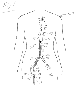
[0014] Fig.1 is an illustration of a region of human
anatomy wherein
an embodiment of the occlusion assembly is shown in use in a REBOA procedure.
[0015] Fig. 2 is a side view of the embodiment of the
occlusion device,
shown before use.
CA 03176991 2022- 10- 26
WO 2022/011446
PCT/CA2020/050976
- 5 -
[0016] Fig. 3 is a cross-sectional view of the
embodiment shown in Fig.
2 taken at line X.
[0017] Fig. 4 is a cross-sectional view of the
embodiment shown in Fig.
2 taken at line Y.
[0018] Fig. 5 is a cross-sectional view of the
embodiment shown in Fig.
2 taken at line Z.
[0019] Fig. 6 is a close-up view of the balloon
envelope and the
adjacent balloon mounting region of the occlusion assembly shown in Fig. 2,
depicted in longitudinal sectional view.
[0020] Fig. 7 is a side view of a distal end portion
of an embodiment of
the occlusion device shown prior to the bonding of the pre-molded balloon
envelope to the elongate shaft.
[0021] Fig. 8 is a longitudinal, sectional view, of
the distal end portion
of the occlusion device shown in Fig. 7 but in an assembled state and the
balloon
envelope inflated to its normal or fully inflated state in an unconfined
environment.
[0022] Fig. 9 is a longitudinal, sectional view, of
the distal end portion
of the embodiment shown in Fig. 8, with the balloon envelope in the fully
inflated
state and occluding the aorta of a patient.
[0023] Fig. 10 is a longitudinal, sectional view, of
the distal end portion
of the embodiment shown in Fig. 9, shown during initial inflation of the
balloon
envelope.
[0024] Fig. 11 is a longitudinal, sectional view, of
the distal end portion
of the embodiment shown in Fig. 10, with the balloon envelope depicted in a
partially inflated state.
[0025] Fig. 12 is a longitudinal, sectional view, of
the distal end portion
of the embodiment shown in Fig. 11 with the balloon envelope in an over
inflated.
CA 03176991 2022- 10- 26
WO 2022/011446
PCT/CA2020/050976
- 6 -
[0026] Fig. 13 is a is a longitudinal, sectional view,
of the distal end
portion of the embodiment shown in Fig. 12 wherein the balloon in the over
inflated state is drawn distally into a side vessel of a bifurcation.
[0027] Fig. 14 is a side view of the embodiment shown
in Fig. 2 shown
in a fully assembled state and equipped with a side-arm shaft assembly, J-tip
straightener and inflation syringe.
[0028] Fig. 15 is a close-up perspective view of the
distal end of the
embodiment shown in Fig. 14.
[0029] Fig. 16 shows the embodiment depicted in Fig.
15, with the J-tip
straightener partially peeled off to illustrate the manner in which the
atraumatic
tip is straightened for ease of insertion into an introducer sheath (not
shown).
[0030] Fig. 17 is a graph produced from experimental
observation and
measurements.
[0031] Fig. 18 is a graph produced from experimental
observation and
measurements.
Detailed Description
[0032] As indicated above, embodiments of the present invention are
directed to an occlusion assembly 10 for use in REBOA procedures. An example
of an embodiment of the occlusion assembly 10 as it may be used in a REBOA
procedure is illustrated in Fig. 1. As is shown, the assembly 10 includes an
elongate shaft 12, which terminates at a distal shaft end 14 in an atraumatic
J-tip
16. Proximal to the J-tip 16 is positioned a balloon envelope 18. The shaft 12
defines a central passage or lumen 22, which is in fluid communication with
the
interior 24 of the balloon envelope 18 and sealed at the distal end of the
balloon.
The lumen 22 extends from the balloon envelope 18 to an inflation port 26
located at the proximal shaft end 28. Through the inflation port 26, inflation
fluid 30 may be injected into the lumen 22, and thus, the balloon envelope 18,
via
a syringe 32 or equivalent mechanism; thereby causing the balloon envelope 18
to expand and occlude the aorta 102.
CA 03176991 2022- 10- 26
WO 2022/011446
PCT/CA2020/050976
- 7 -
[0033] During the REBOA procedure, the occlusion
device 10 is
advanced to a target site within the aorta 102 of a human patient 100. Though
the occlusion assembly 10 may be inserted into the aorta 100 using a variety
of
different arterial pathways, in the embodiment shown, the occlusion assembly
is inserted initially into the femoral artery 104 via a 4 Fr introducer sheath
(not shown), and then advanced into the aorta 102 beyond the aortic
bifurcation
106. Once the balloon envelope 18 is at the target position within the aorta
or
other branch vessel, the balloon envelope 18 is expanded in the manner
described above.
[0034] In at least one embodiment, proper positioning
of the balloon
envelope 18 may be visually estimated by way of one or more visual markers
placed on the external surface of the elongate shaft 12. Such a marker 34,
corresponds to the length of the elongate shaft 12, or the distance that the
elongate shaft must be advanced into the aorta 102, in order to place the
balloon
envelope 18 in a desired anatomical area or zone. For example, in at least one
embodiment, the assembly has a visual marker 34 corresponding to a
length/distance of at least 40 cm from the balloon envelope 18 to the mark 34,
which corresponds to a placement of the balloon envelope above the junction of
the lowest renal artery in most adult patients.
[0035] Embodiments of the assembly 10 may have any number of
visual markers to indicate proper deployment distances for specific anatomical
positioning. In at least one embodiment, the elongate shaft has two visual
markers 34, with one corresponding to a length/distance of 48 cm from the
center of the balloon envelope 18 and the other corresponding to a length of
28
cm from the center of the balloon envelope 18. These marker designations
correspond to Zone 1 of the thoracic aorta and Zone 3 of the infrarenal aorta.
[0036] In at least one embodiment, the visual
marker(s) 34 may be
customized to the assembly 10 based on pre-use examination of the patient.
[0037] Turning to Fig. 2, an embodiment of an occlusion assembly 10
is shown prior to use. In this view, the presence of a support wire 36 is
shown.
CA 03176991 2022- 10- 26
WO 2022/011446
PCT/CA2020/050976
- 8 -
The support wire or wire 36, is a stainless steel, or equivalent material,
wire
having a diameter of 0.03 inch (0.76 mm), or less, that extends the entire
length
of the elongate shaft 12 between the proximal shaft end 28 and the distal
shaft
end 14. In at least one embodiment, the wire has a diameter of 0.028 inch
(0.71
mm). The length of the shaft 12 and the wire 36 is measured from the proximal
shaft end 28 to the distal shaft end 14.
[0038] At the proximal shaft end 28, a hub 15 is engaged to the
elongate shaft 12. The wire 36 is held in place relative to the other
components
of the elongate shaft 12 (said components are identified and discussed in more
detail below) as well as the balloon envelope 18, by way of its proximal end
29
being embedded or other secured to the hub 15. The hub 15 also defines the
inflation port 26, referenced in Fig. 1, which is in fluid communication with
the
inflation lumen 22 of the elongate shaft 12, and to which a syringe 32 or
other
inflation device may interface with the inflation lumen 22.
[0039] At the opposite end of the assembly 10, the
wire 36 terminates
at the atraumatic J-tip 16. The J-tip 16 is a 5 cm coil or J-curve of
approximately
180 to 360 degrees imparted to the wire 36 to ensure that the distal shaft end
14
does not catch or otherwise harm the vessels through which the occlusion
assembly 10 is advanced.
[0040] In at least one embodiment, the length of the
elongate shaft 12
is no greater than 75 cm. In at least one embodiment the length of the
elongate
shaft is no greater than 65 cm. If the access site is at another area, such as
the
radial artery in the wrist, then the length of the elongate shaft is no
greater than
85 cm. In another embodiment for pediatric patients, the elongate shaft is no
longer than 45 cm.
[0041] In Fig. 2, three cross-sectional reference
lines X, Y and Z are
labeled at different points along the length of the elongate shaft 12. These
cross-
sections are depicted in FIGs. 3, 4 and 5 respectively, and illustrate the
manner in
which the balloon envelope 18 is bonded or welded to the materials of the
elongate shaft 12.
CA 03176991 2022- 10- 26
WO 2022/011446
PCT/CA2020/050976
- 9 -
[0042] Note that in the embodiment illustrated in in
Figs. 3-6, the
bonding or welding of the balloon envelope 18 to the elongate shaft 12 is, for
purposes of illustration and description, presented to show the relevant
structures in overlapping engagement, it will be understood by those of
ordinary
skill in the art that the relevant structures may alternatively be bonded or
welded together end to end (i.e. butt welded or joined).
[0043] The reference lines X, V and Z are also useful
for dividing up the
elongate shaft into three component regions that make up the elongate shaft
12.
Extending distally from the proximal shaft end 28 to cross-sectional reference
Z,
the elongate shaft 12 comprises a proximal shaft region 38. Extending distally
from cross-sectional reference X to the distal shaft end 14, the elongate
shaft 12
comprises a distal shaft region 40. Extending between the proximal shaft
region
38 and the distal shaft region 40 (i.e. between cross-sectional reference Z
and
cross-sectional reference X), the elongate shaft 12 comprises a balloon
mounting
region 42, directly visible in Fig. 6, but obscured here by the presence of
the
balloon envelope 18, which is mounted over the balloon mounting region 42.
[0044] Turning now to the cross-sectional views
depicted in Figs. 3, 4,
and 5, in Fig. 3, a section of the elongate shaft 12 is shown which
corresponds to
a distal bonding region 44. The distal bonding region 44 marks the distal end
of
the balloon mounting region 42 and the beginning of the distally extending
distal
shaft region 40. The distal shaft region 40 comprises a layer 46 of polyether
block amide (PEBA) that is extruded on to a distal portion of the wire 36
extending from the distal bonding region 44 to the terminal end 48 of the
atraumatic J-tip 16. A distal end 50 of the balloon envelope 18 is bonded or
welded to the proximal end 52 of the PEBA layer 46 of the distal shaft region
40.
PEBA layer 46 adheres to the wire 36 thus sealing both the distal shaft region
40
and the distal end 50 of the balloon envelope 18 to the wire 36.
[0045] In at least one embodiment, the PEBA layer 46
of the distal
shaft region 40 is a lubricious form of PEBA sold under the trademarked name
VESTAMI DE EVERGLI DE 0 MED by the Polymer Dynamix company.
CA 03176991 2022- 10- 26
WO 2022/011446
PCT/CA2020/050976
- 10 -
[0046] In at least one embodiment the distal shaft
region 40 and the
corresponding layer 46 have a length of no greater than 8 cm as measured from
the distal bonding region 44 to the terminal end 48 of the atraumatic J-tip
16.
[0047] Skipping Fig. 4 for the moment, and looking now
to Fig. 5,
depicted in Fig. 5 a section of the elongate shaft 12 is shown, which
corresponds
to the position of a proximal bonding region 54.
[0048] The proximal bonding region 54 marks the
proximal end of
the balloon mounting region 42 and the beginning of the proximally extending
proximal shaft region 38. The proximal shaft region 38 comprises a tube 56 of
polyether block amide (PEBA) that is disposed about that portion of the wire
36
extending from the proximal bonding region 54 to the proximal shaft end 28.
The
tube 56 defines the inflation lumen 22, which does double duty as a passage
through which the wire 36 extends. A proximal end 58 of the balloon envelope
18 is bonded or welded to the distal end 60 of the tube 56 at the proximal
bonding region 54. The distal end 60 of the tube 56 corresponds with the end
of
the inflation lumen 22, which is in fluid communication with the interior 24
of
the balloon envelope 18.
[0049] In at least one embodiment the tube 56 is manufactured from a
form of PEBA sold under the trademark PEBAX and manufactured by the
Compounding Solutions company.
[0050] Returning now to Fig. 4, the cross-section
depicted in Fig 4 is a
section of the assembly 10 corresponding approximately to the mid-point of the
working portion of the balloon envelope 18. As is shown, the balloon envelope
18 is disposed about the wire 36, with the bare wire 36 passing through the
interior 24 of the balloon envelope 18, between the proximal bonding region 54
and the distal bonding region 44.
[0051] In at least one embodiment, the balloon
envelope is formed
from an elastomeric polymer such as Urethane.
[0052] Turning to Fig. 7 the elongate shaft 12 is shown adjacent to the
balloon envelope 18 prior to the balloon envelope being mounted onto the
CA 03176991 2022- 10- 26
WO 2022/011446
PCT/CA2020/050976
-11 -
elongate shaft 12. The balloon envelope 18 as shown in its as molded state.
The
balloon envelope 18 is a contiguous envelope of elastomeric material which is
molded into form whose working portion has a shape akin to an ice cream cone
or reverse tear drop. This shape has several identifiable portions that are
useful
in describing the balloon and its performance characteristics. Starting from
the
proximal end 58, the balloon envelope 18 includes a proximal neck 62, which
transitions into a conical proximal shoulder section 64; this shoulder section
transitions into a conical proximal taper section 66. From the distal end 50
the
balloon envelope 18 includes a distal neck 68, which transitions into a distal
shoulder section 70; this shoulder section transitions into a truncated
conical
distal blunt section 72.
[0053] As is shown, conical proximal taper section 66 and truncated
conical distal blunt section 72 intersect at a meridian 74, which marks the
area of
the balloon envelope 18 having the largest as molded diameter. In the as
molded
state, the conical proximal taper section 66 has a greater longitudinal length
than
that of the truncated conical distal blunt section 72.
[0054] In at least one embodiment, the balloon
envelope 18, in the
molded state has a total length of approximately 70 mm as measured from the
proximal end 58 to the distal end 50, and an outer diameter of approximately 8
mm at the meridian 74. The proximal neck 62 has a length of approximately 10
cm and the distal neck 68 has an approximate length of 2 cm and both have a
contiguous outer diameter of approximately 1.35 mm.
[0055] As is shown in Fig. 7, the balloon mounting
region 42 has a
length greater than the length of the balloon envelope itself. Thus, the
balloon
envelope 18 must be longitudinally stretched in order to be mounted onto the
elongate shaft 12. In at least one embodiment, the balloon mounting region is
at
least 2.5 cm longer than the balloon envelope 18.
[0056] The dimensions and shape of the balloon envelope 18, in
combination with the unique construction of the elongate shaft 12, not only
allows for the occlusion assembly to be inserted into the patient using an
CA 03176991 2022- 10- 26
WO 2022/011446
PCT/CA2020/050976
- 12 -
introducer sheath as small as 4 Fr, but also allows the balloon envelope 18 to
have multiple useful inflation states and unique inflation characteristics.
[0057] For purposes of standard REBOA use, the balloon envelope 18
has a fully inflated state, such that when the balloon envelope 18 is fully
expanded, the meridian 74 will correspond with that region of the envelope 18
having the greatest diameter, such as in the manner illustrated in Figs. 8 and
9.
This relationship of the meridian's position as the widest section of the
balloon
envelope 18 is constant whether the envelope 18 is expanded to its fully
inflated
state within the confines of the aorta 102 and subject to blood pressure
acting
against it, such as in the depiction of Fig. 9; as well as when the balloon
envelope
18 is expanded to its fully inflated state outside of the body, such as in the
depiction of Fig. 8.
[0058] In at least one embodiment, the fully inflated
state of the
balloon envelope is achieved by injection of between 10-15 ccs of inflation
fluid
(e.g. saline) into the interior of the balloon envelope in the manner
previously
described. When fully expanded the meridian 74 has an outer diameter of
approximately 25-30 mm. When positioned within the aorta 102, and inflated to
the fully inflated state, such as in the manner shown in Fig. 9, the external
surface
76 of the balloon envelope 18 should be in contact with the vessel wall 108
and
providing complete occlusion to blood flow.
[0059] As implied above, the position of the meridian
74 is not
constant in the various inflation states. For example during initial
inflation, i.e. a
low inflation state such as is shown in Fig, 10, or a partial inflation state
such as
is shown in Fig. 11, the meridian 74 has a first diameter and is located at a
first
distance from the distal shaft end 14. But as the balloon envelope 18 reaches
the
fully inflated state shown in Fig. 9, the meridian 74 has a larger diameter
than in
the other states and has transitioned further away from the distal shaft end
14.
[0060] Other aspects of the balloon envelope 18 will
vary during
expansion as well. For example, as the balloon envelope 18 is expanded from
the
partial inflation state of Fig. 11 to the fully inflated state of Fig. 9, the
volume of
CA 03176991 2022- 10- 26
WO 2022/011446
PCT/CA2020/050976
- 13 -
the conical distal shoulder section 70 decreases such that in the fully
expanded
state the conical distal shoulder section volume is less than the conical
distal
shoulder section volume in the partially inflated state. Whereas the opposite
occurs in the conical proximal shoulder section 64. In that section of the
balloon
envelope 18, the volume increases as the balloon envelope is expanded, such
that
in the fully inflated state the conical proximal shoulder section volume is
greater
than the conical proximal shoulder section volume in the partially inflated
state.
[0061] During inflation, as the balloon envelope 18 is
acted upon more
and more by the blood pressure, the balloon envelope 18 migrates downwards
along the wire 36 (in the direction of the proximal shaft end) and then
eventually
catches the aortic wall 108 for full occlusion. When the balloon envelope 18
is
deflated, and blood is allowed to pass around the external surface of the
balloon
76, the balloon envelope 18 will begin to go back to its unmigrated position.
[0062] With the offset nature of the balloon envelope 18 and also
because of the stretch imparted to the balloon envelope 18 when it is mounted
onto the balloon mounting region 42 (2.5 cm in at least one embodiment as
discussed above), the general shape of the balloon envelope 18 is maintained
during the inflation and deflation processes, which allows the fine
adjustments
(titratability) of blood flow. This is in contrast to known spherical balloons
that
are fixed to a catheter shaft and imparted with no stretch. Such balloons are
unable to migrate and therefore the shape of the balloon changes substantially
when acted on by blood pressure. Such balloons act much like an on/off switch
in terms of performance (i.e. no appreciable occlusive effect before full
occlusion
at full inflation) and do not provide for the ability to be adjusted in the
manner of
the present balloon envelope 18 nor have the ability to gradually recirculate
blood flow in a graduated manner during deflation such as the present device
10
provides.
[0063] The ability of the balloon envelope 18, and the
device 10, to
provide gradual and incremental occlusive effects is illustrated in Figs. 17
and
18.
CA 03176991 2022- 10- 26
WO 2022/011446
PCT/CA2020/050976
- 14 -
[0064] Fig. 17 and Fig. 18 should be considered
together. They are
plots of data taken in vivo from a porcine model used in development of the
device 10. As mentioned above, in the prior art the occlusion balloon is
either in
the fully occluded state or effectively in the fully contracted state
permitting full
flow past the device. In contrast to this, the present device 10 provides for
proportionate flow permitted past the balloon envelope 18 prior to and
following full occlusion.
[0065] This is demonstrated by occluding the aorta and measuring the
Mean Arterial Pressure (MAP) distal of the balloon. The measured MAP is seen
on the Y-Axis in both Fig. 17 and Fig. 18. Referring to Fig. 17 note that the
relationship between measured pressure and balloon envelope volume is nearly
linear over the range of slightly less than 1 ml taken from the balloon
envelope to
the point where 4 nil are removed. Procedurally this corresponds to reducing
volume in the balloon envelope from total occlusion to near total deflation.
The
linearity demonstrates that the pressure driving flow in the organ increases
in a
gradual fashion as balloon envelope volume is slowly reduced.
[0066] In Fig. 18 the same information is expressed as
a function of
percent balloon envelope volume reduction. Qualitatively this shows that
inflation volume controls MAP over the full range of values from total
occlusion
to maximal flow. The self-similar shape attribute of the balloon envelope is
maintained over a substantially linear range of inflation volume. This graphed
data demonstrates that the amount of blood flow past the balloon envelope will
be proportional to inflation volume over the operational range defined by the
range of the self-similar shape of the balloon.
[0067] A unique feature of the present assembly 10, is the capacity to
safely inflate the balloon envelope 18 into an over inflated state such as is
shown
in Figs. 12 and 13. When over inflating the balloon 18 within the aorta 102
beyond the fully inflated state, the meridian 74 does not further increase in
diameter from that in the fully inflated state, but rather will increase in
longitudinal length, effectively transitioning the meridian from a narrow band
of
CA 03176991 2022- 10- 26
WO 2022/011446
PCT/CA2020/050976
- 15 -
intersection with the aortic wall 108, to potentially the majority of the
balloon
envelope's external surface 76 being in contact with the wall 108. This is
accomplished as a result of the shoulder sections 64 and 70 growing, rather
than
as a result of further stretching by the conical proximal taper section 66 and
truncated conical distal blunt section 72.
[0068] This longitudinal widening of the meridian 74
is accompanied
by a longitudinal advancement/growth of the balloon envelope 18 such that in
the over inflated state the meridian 74 is closer to the distal shaft end 14
than in
the fully inflated state, the partially inflated state, or the low inflation
state.
[0069] There is an additional benefit of preventing vessel damage at a
bifurcation with the ability to advance the balloon envelope 18 distally via
the
"growth" of the shoulder sections 64 and 70. For example, as shown in Fig. 13,
when the balloon envelope 18 is over inflated in a upside-down Y-shaped vessel
bifurcation (aorto-iliac bifurcation), the balloon will pull itself gently
into the
larger vessel (aorta) and prevent damage to the smaller vessel (iliac artery).
The ice-cream cone shape of the balloon envelope 18 also promotes this growth
into the larger vessel by preferentially inflating the wider ice-cream cone
section
of the truncated conical distal blunt section 72 of the balloon envelope 18,
as
long as this portion of the balloon is above the bifurcation.
[0070] In some embodiments, the balloon envelope 18 may be over
inflated up to 700% by volume over the fully inflated state. A key
characteristic
of the present assembly 10, is that regardless of the degree of over inflation
when properly used in the manner described herein, the balloon envelope will
fail before damaging the aorta.
[0071] In addition to the characteristics discussed
thus far,
embodiments of the occlusion assembly 10 disclosed herein are provided with
several other features that benefit both safety and ease of use during a REBOA
procedure, an example of such an embodiment is shown in Fig. 14.
[0072] In the embodiment shown, the assembly 10, is
provided with a
side-arm shaft assembly 78 which is in fluid communication with the inflation
CA 03176991 2022- 10- 26
WO 2022/011446
PCT/CA2020/050976
- 16 -
lumen 22 of the elongate shaft 12 via a t-valve 80. The side-arm shaft
assembly
78 includes a stop cock valve 82 that may be open and shut to allow inflation
fluid to egress from the lumen 22 and provides the user with greater control
of
the inflation and deflation of the balloon envelope that a syringe 32 may
allow by
itself. The side-arm assembly may also act as an interface for a blood
pressure
monitor.
[0073] In the present embodiment, the assembly 10 is
also provided
with a J-tip straightener 84 that is preloaded over a portion of distal shaft
region
40, between the distal end 50 of the balloon envelope 18 and the atraumatic J-
tip
16.
[0074] The J-tip straightener 84 has a unique
construction and role as
illustrated in more detailed views of Figs. 15 and 16. The J-tip straightener
is
essentially a hollow peel-off shaft or tube 86 that is disposed about a
portion of
distal shaft region 40. When the assembly 10 is ready for use, the user slides
the
J-tip straightener 84 onto and over the coil of the atraumatic J-tip 16, which
will
temporarily straighten as a consequence of the confinement and advancement of
the J-tip straightener 84 over the tip 16. This temporary straightening of the
atraumatic J-tip 16 allows it to be more easily threaded into the 4 Fr
introducer
(not shown) during initial insertion of the assembly during a REBOA procedure.
[0075] For further ease of operation the 1-tip
straightener 84 includes
a user engagement tab or grip 88 that protrudes from the peel-off shaft 86,
and
which user may grasp and pull distally to more easily advance of the J-tip
straightener 84 over the J-tip 16.
[0076] Finally, in at least some embodiments, the
elongate shaft 12 is
provided with at least two radiopaque (RU) markers 90 and 92, such as are
shown in Figs. 6-13. As illustrated, the RO markers 90 and 92 are placed on
the
wire 36 at locations within the balloon mounting region 42 so as to allow the
position of the balloon envelope 18 to be monitored within the patient, via a
visualization mechanism (Fluoroscope, etc.), during the performance of a REBOA
procedure utilizing the assembly 10.
CA 03176991 2022- 10- 26
WO 2022/011446
PCT/CA2020/050976
- 17 -
[0077] The many features and advantages of the invention are
apparent from the above description. Numerous modifications and variations
will readily occur to those skilled in the art. Since such modifications are
possible, the invention is not to be limited to the exact construction and
operation illustrated and described. Rather, the present invention should be
limited only by the following claims.
[0078] As used herein terms such as "about" or "approximately" and
the like when used to describe a measurement value attributed to any aspect of
the occlusion assembly 10, or any of its components, such terms are provided
so
as to reflect the range of tolerances inherent in the production of a given
article
of manufacture or its assembly as understood by one of ordinary skill.
CA 03176991 2022- 10- 26