Note: Descriptions are shown in the official language in which they were submitted.
CA 03177740 2022-09-28
WO 2021/211549 PCT/US2021/027042
KIF18A INHIBITORS FOR TREATMENT OF NEOPLASTIC DISEASES
CROSS REFERENCE TO RELATED APPLICATIONS
[0001] This application claims under 35 U.S.C. 119(e) the benefit of U.S.
Provisional
Application No. 63/009,637, filed April 14, 2020, U.S. Provisional Application
No. 63/055,111,
filed July 22, 2020, and U.S. Provisional Application No. 63/085,607, filed
September 30, 2020.
The entire contents of each application are incorporated herein by reference.
INCORPORATION BY REFERENCE OF MATERIAL SUBMITTED ELECTRONICALLY
[0002] Incorporated by reference in its entirety is a computer-readable
nucleotide/amino acid
sequence listing submitted concurrently herewith and identified as follows:
227,954 byte ASCII
(Text) file named "A-2607-WO-PCT_Seqlisting.txt"; created on March 22, 2021.
BACKGROUND
[0003] Cancer is one of the most widespread diseases afflicting mankind, and a
leading
cause of death worldwide. In the United States alone, cancer is the second
most common
cause of death, only surpassed by heart disease. In an effort to find an
effective treatment or a
cure for one or more of the many different cancers, over the last couple of
decades, numerous
groups have invested a tremendous amount of time, effort and financial
resources. However, to
date, of the available cancer treatments and therapies, only a few offer any
considerable degree
of success.
[0004] Cancer is often characterized by deregulation of normal cellular
processes or
unregulated cell proliferation. Cells that have been transformed to cancerous
cells proliferate in
an uncontrolled and unregulated manner leading to, in some cases, metastasis
or the spread of
the cancer. Damage to one or more genes, responsible for the cellular
pathways, which control
progress of proliferation through the cell cycle and centrosorne cycle, can
cause the loss of
normal regulation of cell proliferation. These deregulated genes can code for
various tumor
suppressor or oncogene proteins, which participate in a cascade of events,
leading to
unchecked cell-cycling progression and cell proliferation. Various kinase and
kinesin proteins
have been identified, which play key roles in cell cycle and mitotic
regulation and progression of
normal dividing cells and cancer cells.
[0005] Cancer pathways, phenotypes, differentiation state associated with
mitotic and
replicative stress may lead to specific vulnerabilities associated with
mitotic entry, mitotic spindle
formation, centrosome integrity and positioning, MT-kinetochore attachment,
sister chromatid
1
CA 03177740 2022-09-28
WO 2021/211549 PCT/US2021/027042
cohesion, and SAC control. Thus, a strategy to improve the clinical potential
of new antimitotic
therapies should exploit a tumor-specific vulnerability while sparing or
reducing the collateral
damage to normal tissues. K1F18A is an emerging and promising anticancer
target as KIF18A
inhibition leads to activation of the SAC in mitosis, induction of
multipolarity and apoptosis, and
inhibits the growth of subset of human cancer cell lines while sparing normal
dividing somatic
cells,
[0006] Mitosis is the process by which a eukaryotic cell segregates its
duplicated
chromosomes into two identical daughter nuclei. It is generally followed
immediately by
cytokinesis, which divides the nuclei, cytoplasm, organelles and cell
membranes into two
daughter cells containing roughly equal shares of these cellular components.
Mitosis and
cytokinesis together define the mitotic (M) phase of the cell cycle¨the
division of the mother cell
into two daughter cells, genetically identical to each other and to their
parent cell. The process
of mitosis is complex and highly regulated. The sequence of events is divided
into distinct
phases, corresponding to the completion of one set of activities and the start
of the next. These
stages are prophase, prometaphase, metaphase, anaphase and telophase. During
the process
of mitosis duplicated chromosomes condense and attach to spindle rnicrotubule
(MT) fibers that
pull the sister chromatids to opposite sides of the cell. Spindle assembly
checkpoint (SAC) is
active until all sister chromatids are properly attached to the spindle
kinetochore fibers and
spindle tension is achieved during metaphase, if no errors are detected the
cells progress to
anaphase. The cell then divides in cytokinesis, to produce two identical
daughter cells.
[0007] Normally, cell-cycle checkpoints are activated if errors are
detected (e.g. DNA
damage, DNA replication fork stall/collapse, centrosome aberrations,
chromosome mis-
segregation, micronuclei formation). If these errors to the genome cannot be
fixed, the cell
normally undergoes cell arrest and apoptosis. However, if the cell is allowed
to move through its
cell-cycle and progress unchecked, then mutations, chromosome rnis-
segregation, centrosome
aberrations can accumulate overtime. These aenelkaryotypelcentrosome
alternations can
accrue and eventually leading cell progeny with pre-malignant or malignant
neoplastic
characteristics (e.g. uncontrolled proliferation) through adaptation,
[0008] Cancers with high intraturnor heterogeneity and chromosomal
instability (ClN) have
complex karyotypes due to continuous chromosomal changes resulting from
numerical (gain or
loss) and structural alternations. The mechanisms believed to contribute to
CIN include defects
in kinetochore MT attachment dynamics, centrosome copy number, mitotic
checkpoint function,
chromosome cohesion, and cell cycle regulation. Chromosome instability (CIN)
is associated
2
CA 03177740 2022-09-28
WO 2021/211549 PCT/US2021/027042
with an increased level of replicative and mitotic stress enriched for genetic
lesions in a subset
of tumor suppressor and oncogenes (examples include but not limited to TP53,
RBI, BROM,
BRCA2, homologous recombination deficient (HRD) genes, FBXVV7, CCNEI, MYC)
that
regulate cell-cycle progression/checkpoints, centrosome-cycle, and DNA repair
(SL Thompson
et al Current Biology. 2010;20:285-95 and R Nagel et al EMBO Reports
2016;17:1516-1531).
[0009] Mitosis, or cell division, is a validated point-of-intervention in
treating cancer.
Approved antimitotic drugs are anti-cancer agents that inhibit the function of
microtubules.
Microtubules are protein polymers formed by a-tubulin and 3-tubulin
heterodimers that play an
important role in the formation of the mitotic spindle apparatus and
cytokinesis at the end of
mitosis. Anti-cancer agents that target microtubules represent a proven
approach for intervening
in the proliferation of cancer cells. Taxanes are the most prominent class of
antimitotic agent
that includes paciitaxei (taxol) and docetaxel (taxotere). The vinca alkaloids
are a class of
microtubule-destabilizing agents that includes vincristine, vinblastine, and
vinorelbine. Other
new tubulin binding anti-cancer drugs include ixabepilone and eribulin. These
antimitotic agents
act to prevent the proliferation of cancer cells by either stabilizing- or
destabilizing-microtubules.
This direct inhibition of microtubules results in cell arrest and death
through apoptosis, mitotic
catastrophe, and lethal multipolar division. Paclitaxel was the first compound
of the taxane
series to be discovered, Docetaxel, a structural analog of paclitaxel, was
later discovered.
Paclitaxel and docetaxel are commonly used to treat a variety of human
malignancies, including
ovarian cancer, breast cancer, head and neck cancer, lung cancer, gastric
cancer, esophageal
cancer, prostate cancer, and AIDS-related Kaposi's sarcoma. The primary side
effect of taxanes
is myelosuppression, primarily neutropenia, while other side effects include
peripheral edema,
and neurotoxicity (peripheral neuropathy).
[0010] Resistance to anti-mitotic agents such as taxanes is a complicating
factor to
successful cancer treatment and is often associated with increased expression
of the MDR-1
encoded gene and its product, the P-glycoprotein (P-gp), Other documented
mechanisms of
acquired resistance to taxanes include tubulin mutations, overexpression,
amplification, and
isotype switching). Mutations in a- or 3-tubulin inhibit the binding of
taxanes to the correct place
on the microtubules; this renders the drug ineffective. Resistance to other
anticancer drug
classes, including, without limitation to chemotherapeutic agents (e.g.
platinum agents,
anthracyclines) and targeted therapies (e.g. TKI, PARP inhibitors) has become
a major
drawback in the effective treatment of cancer and inevitably leads to patient
death.
Consequently, development of drug resistance remains a problem with all
anticancer therapies.
3
CA 03177740 2022-09-28
WO 2021/211549 PCT/US2021/027042
[0011] Precision medicine is aimed at improving cancer patient response
rates by reducing
toxicities to normal tissues and utilizing stratification markers to enrich
for patients most likely to
benefit from therapy treatment or importantly to exclude patients unlikely to
benefit. A
biomarker guided approach has the potential to customizing cancer patient
treatment to achieve
higher response rates and to drive improvement in patient outcomes and quality
of life. There is
a lack of established stratification markers (biomarkers) available for
selecting patients most
likely to benefit from current anti-mitotic therapies.
[0012] Thus, there is a need for biomarkers to identify patients will
benefit from KIF18A
inhibitor treatment.
SUMMARY
[0013] Presented herein for the first time are data evidencing biomarkers
of sensitivity to
KlF1 8A inhibitor treatment. Cancer cells exhibiting an inactivated TP53 gene
and/or at least
one of: (I) an inactivated Rbl gene, (ii) an amplified CCNEI gene, gene copy
number gain of
the CCNEI gene, or overexpression of a CCNEI gene product, (iii) an
inactivated BRCA gene
or (iv) a combination thereof, demonstrated sensitivity to treatment with a
KIF18A inhibitor, Also
provided herein are data demonstrating that KIF18A inhibitor-sensitive cancer
cells exhibit a
reduced or lost sensitivity or resistance to CDK4/6 inhibitors. The data
further support that
CDK4/6 inhibitor-sensitive cancer cells exhibit a reduced or lost sensitivity
or resistance to
KIF18A inhibitors.
[0014] The present disclosure provides methods of determining a treatment for
a subject with
a neoplastic disease (e.g., cancer). In exemplary embodiments, the method
comprises
assaying a sample obtained from the subject for (a) an inactivated TP53 gene
and/or (b) at least
one of: (i) an inactivated Rbl gene, (ii) an amplified CCNEI gene, gene copy
number gain of
the CCNEI gene, or overexpression of a CCNEI gene product, (iii) an
inactivated BRCA gene
or (iv) a combination thereof. In various aspects, the treatment determined
for the subject
comprises of a KIF18A inhibitor, when the sample is positive for an
inactivated TP53 gene
and/or positive for at least one of an inactivated Rh/ gene, (ii) an amplified
CCNEI gene, gene
copy number gain of the CCNEI gene, or overexpression of a CCNEI gene product,
(iii) an
inactivated BRCA gene or (iv) a combination thereof. In exemplary embodiments,
the method
comprises determining sensitivity of the neoplastic disease to treatment with
a CDK4/6 inhibitor.
In various aspects, the treatment for the subject is determined as a treatment
comprising a
KIF18A inhibitor, when the neoplastic disease is insensitive or resistant to
the CDK4/6 inhibitor.
In exemplary embodiments, the method comprises determining sensitivity of the
neoplastic
4
CA 03177740 2022-09-28
WO 2021/211549 PCT/US2021/027042
disease to treatment with a KIF18A inhibitor. In various aspects, the
treatment for the subject is
determined as a treatment comprising a CDK4/6 inhibitor, when the neoplastic
disease is
insensitive or resistant to the KIF18A inhibitor.
[0015] Methods of identifying a subject with a neoplastic disease as
sensitive to treatment
with a KIF18A inhibitor are provided herein. In exemplary embodiments, the
method comprises
assaying a sample obtained from the subject for (a) an inactivated TP53 gene
and/or (b) at least
one of: (i) an inactivated F?b1 gene, (ii) an amplified CCNE1 gene, gene copy
number gain of
the CCNE1 gene, or overexpression of a CCNE1 gene product, (iii) an
inactivated BRCA gene
or (iv) a combination thereof. In various instances, the subject is identified
as sensitive to
treatment with a KIF18A inhibitor, when the sample is positive for an
inactivated TP53 gene
and/or positive for at least one of an inactivated Rbi gene, (ii) an amplified
CCNE1 gene or
overexpression of a CCNE1 gene product, (iii) an inactivated BRCA gene or (iv)
a combination
thereof.
[0016] The present disclosure additionally provides a method of identifying
a subject with a
neoplastic disease as responsive to treatment with a KIF18A inhibitor. In
exemplary
embodiments, the method comprises determining the sensitivity of the
neoplastic disease to
treatment with a KIF18A inhibitor. In various aspects, the subject is
identified as responsive to
treatment with a KIF18A inhibitor, when the cancer cells of the sample are
insensitive to the
0DK4/6 inhibitor.
[0017] Methods of maintaining sensitivity of a neoplastic disease to
treatment with a CDK4/6
inhibitor in a subject are provided herein. In exemplary embodiments, the
method comprises
administering to the subject a KIF18A inhibitor.
[0018] Methods of treating a subject with a neoplastic disease, e.g,,
methods of treating the
neoplastic disease in a subject, are provided herein. In exemplary
embodiments, the method
comprises administering a KIF18A inhibitor to treat the patient. Optionally,
the neoplastic
disease is resistant to treatment with a CDK4/6 inhibitor. In various
instances, the subject is or
has been treated with a CDK4/6 inhibitor, In various aspects, the KIF18A
inhibitor is co-
administered with the 0DK4/6 inhibitor. In various instances, the method
comprises
administering a pharmaceutical combination comprising a CDK4/6 inhibitor and a
K1F18A
inhibitor. Accordingly, a pharmaceutical combination comprising a KIF18A
inhibitor and a
CDK4/6 inhibitor is provided herein.
CA 03177740 2022-09-28
WO 2021/211549 PCT/US2021/027042
[0019] Methods of inducing or increasing tumor regression in a subject with
a tumor are
additionaliy provided herein. In exemplary embodiments, the method comprises
administering
to the subject a KIF18A inhibitor in an amount effective to induce or increase
tumor regression.
The present disclosure also provides methods of reducing tumor growth or
cancer growth in a
subject. In exemplary embodiments, the method comprises administering to the
subject a
KIF18A inhibitor in an amount effective to reduce tumor or cancer growth.
Methods of inducing
or increasing death of tumor cells or cancer cells in a subject are provided
herein, The method
in exemplary embodiments comprises administering to the subject a KIF18A
inhibitor in an
amount effective to induce or increase death of the tumor cells or cancer
cells. In various
aspects, the neoplastic disease is a cancer, optionally, breast cancer,
ovarian cancer, or
prostate cancer. In various instances, the neoplastic disease is triple-
negative breast cancer
(TNBC), non-luminal breast cancer, or high-grade serous ovarian cancer
(HGSOC). In
exemplary aspects, the neoplastic disease is an endometriai cancer,
optionally, serous
endometrial cancer. Optionally, the cancer comprises cells that are positive
for an inactivated
TP53 gene and/or positive for at least one of an inactivated Rb gene, (ii) an
amplified CCNE1
gene or overexpressed CCNE1 gene product, (iii) an inactivated BRCA gene or
(iv) a
combination thereof. In some aspects, the cancer comprises cells that are
positive for a mutant
TP53 gene. In various instances, the cancer comprises cells that are positive
for an amplified
CCNE1 gene, a silenced BRCA1 gene, a deficient Rbl gene, or a combination
thereof.
Optionally, the KIF18A inhibitor is administered for oral administration,
optionally once a day. In
exemplary aspects, the amount of the KIF18A inhibitor is effective to induce
at least 50% or at
least 75% (e.g,, at least 80% or 85% or at least 90% or 95%) tumor regression,
compared to a
control. In various instances, the KIF18A inhibitor selectively treats the
neoplastic disease,
selectively induces or increases tumor regression, selectively reduces tumor
or cancer growth,
and/or selectively induces or increases death of tumor or cancer cells and the
KIF18A inhibitor
is not toxic to normal somatic cells. in various aspects, the KIF18A inhibitor
treats the
neoplastic disease, induces or increases tumor regression, reduces tumor or
cancer growth,
and/or induces or increases death of tumor or cancer cells and the
proliferation of the normal
somatic cells in the subject is substantially the same as the proliferation of
the normal somatic
cells of a control subject. In exemplary instances, the KIF18A inhibitor
treats the neoplastic
disease, induces or increases tumor regression, reduces tumor or cancer
growth, and/or
induces or increases death of tumor or cancer cells and the level of apoptosis
of normal somatic
cells is not increased in the subject, relative to the level of apoptosis of
normal somatic cells of a
6
CA 03177740 2022-09-28
WO 2021/211549 PCT/US2021/027042
control subject, optionally, wherein the level of apoptosis of normal somatic
cells is substantially
the same as the level of apoptosis of the normal somatic cells of a control
subject.
[0020] In various aspects of the present disclosure, the KIF18A inhibitor
is a compound of
formula (I). In exemplary aspects, the KIF18A inhibitor is Compound C1,
Compound C2,
Compound 03, Compound 04, Compound 05, Compound 06, Compound 07, Compound 08,
Compound C9, Compound C10, Compound 011, Compound 012, Compound 013, or
Compound C14, or any pharmaceutically-acceptable salt thereof, as described
herein,
[0021] In alternative aspects, the KIFI8A inhibitor is a large molecule
KIF18A inhibitor, such
as a non-coding RNA, e.g., an siRNA.
BRIEF DESCRIPTION OF THE DRAWINGS
[0022] Figures 1A-1C provide tables referenced in Example 1. Figure 1A
provides Table 1A
which lists tissue culture growth conditions for the indicated cell line
originating from the
indicated tissue. Figure 1B provides Table 1B which lists the seeding density
for the NCA for
each tested cell line. Figure 1C provides Table 1C which lists cancer cell
lines and their
sensitivity to a first KIF18A inhibitor,
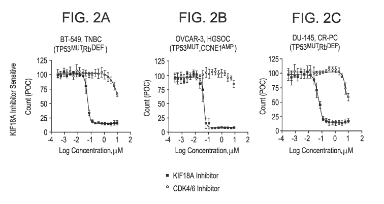
[0023] Figures 2A to 2F are graphs of POC plotted as a function of
concentration of KIF18A
inhibitor (solid squares) or CDK416 inhibitor (open circles). Figures 2A-2C
demonstrate KIF18A
inhibitor-sensitive, CDK416 inhibitor-insensitive cells, cells and Figures 2D-
2F demonstrate
K1F18A inhibitor insensitive cells, CDK4i6 inhibitor-sensitive cells.
[0024] Figure 3A is a graph plotting the POC as a function of concentration
of KlF18A
inhibitor for the OVCAR-8 HGSOC cell line. Figure 3B is a graph plotting the
POC as a function
of concentration of KIF18A inhibitor for the MX-1 TNBC cell line, Figure 30 is
a graph plotting
the POC as a function of concentration of KIF18A inhibitor for the MAX401NL
TNBC cell line.
Figure 3D is a graph plotting the POC as a function of concentration of K1F18A
inhibitor for the
HCC-1937 TNBC cell line. Figure 3E is a graph plotting the POC as a function
of concentration
of KIF18A inhibitor Compound C14, Olaparib, Paclitaxel, Doxorubicin, or
Carboplatin for the
OVCAR-8 cancer cell line,
[0025] Figure 4A provides Table 2 which lists cancer cell lines and their
sensitivity to a
second KIF18A inhibitor. Figure 4B provides Table 3 which lists cancer cell
lines and their
sensitivity to a second KIF18A inhibitor. Figure 4C is a graph plotting the
POC as a function of
concentration of KIF18A inhibitor for breast and ovarian cancer cell lines
which demonstrated
sensitivity to KIF18A inhibitor treatment. Figure 4D is a graph plotting the
POC as a function of
7
CA 03177740 2022-09-28
WO 2021/211549 PCT/US2021/027042
concentration of KIF18A inhibitor for breast and ovarian cancer cell lines
which were insensitive
to KIF18A inhibitor treatment. Figure 4E is a graph plotting the P00 as a
function of
concentration of KIF18A inhibitor for a second group of ovarian and breast
cancer cell lines
which demonstrated sensitivity to KIF18A inhibitor treatment. Figure 4F is a
graph plotting the
P00 as a function of concentration of KIF18A inhibitor fora second group of
ovarian and breast
cancer cell lines which were insensitive to KIF18A inhibitor treatment.
[0026] Figure 5A is a graph of the tumor volume of K1F18A inhibitor-treated
or vehicle-treated
mice plotted as a function of time after cell implantation. Figure 53 is a
graph of the body
weight of KIF18A inhibitor-treated or vehicle-treated mice plotted as a
function of time after cell
implantation.
[0027] Figure 6A is a graph of the tumor volume of KlF18A inhibitor-treated
or vehicle-treated
mice plotted as a function of time after cell implantation. Figure 68 is a
graph of the body
weight of KIF18A inhibitor-treated or vehicle-treated mice plotted as a
function of time after cell
implantation.
[0028] Figure 7A is an image of DMSO-treated cells or KlF18A inhibitor
cells as described
herein. Figure 73 is a pair of graphs demonstrating the % p-Histone or
Pericentrin spots plotted
as a function of KIF18A inhibitor concentration. Figure 70 is a table listing
the E050 of the
KIF18A inhibitor with respect to p-Histone or Pericentrin spots, as described
herein.
[0029] Figure 8A is a series of images of two types of cancer cells stained
for centrin-3,
percentrin, or centrosome markers upon treatment with KIF18A inhibitor or with
DMSO control.
Figure 83 is a Western blot showing the protein levels of ci-PARP in cells
treated with DMSO
control, KIF18A inhibitor, or Eg5 Inhibitor. GADPH is a loading control.
[0030] Figure 9 is a series of Western blots showing the levels of the
indicated proteins (cf-
PARP, Cyclin 81, Mcl-1 Cyclin El, KIF18A, BubR1) in synchronized or
asynchronized cells
treated with DMSO control or KlF18A inhibitor. 8-actin is a loading control.
[0031] Figure 10A is a series of Western blots showing the levels of the
indicated proteins (p-
Histone H3, y-H2A,X, ci-PARP, BubR1, Total HE01, p-Hecl) in cells treated with
DMSO control
or K1F18A inhibitor. GADPH is a loading control, Figure 10B is a pair of
images showing cells
stained for cGAS (green), yll2A,X (red), or DAPI (blue). Cells were treated
with DMSO control
or KIF18A inhibitor.
[0032] Figure 11 is a pair of images showing cells stained for Centrin-3
(green), KIF18A (red),
or DNA (DAPI (blue)), Cells were treated with DMSO control or KIF18A
inhibitor.
8
CA 03177740 2022-09-28
WO 2021/211549 PCT/US2021/027042
[0033] Figure 12 is a graph of p-Histone H3 in cells treated with the
indicated amount of a
KiF18A inhibitor of vehicle control.
[0034] Figure 13A is a graph of the POC plotted as a function of
concentration (log
concentration) of cells treated with KIF18A inhibitor Compound 014 in the
presence (open
circles) or absence (closed circles) of a P-gp inhibitor. Figure 138 is a
graph of the POC plotted
as a function of concentration (log concentration) of cells treated with
paclitaxel (tubulin) in the
presence (open circles) or absence (closed circles) of a P-gp inhibitor.
[0035] Figure 14A is a series of FAGS plots demonstrating DNA content of human
bone
marrow mononuclear cells (HBMNC) treated with DMSO, KIFI8A inhibitor Compound
C9 or
Compound C11, ispinesib, paclitaxel or palbociclib. Figure 148 is a graph
demonstrating %
human bone marrow mononuclear cells (HBMNC) from Donor 37612 or Donor 37534
stained
with anti-BrdU (top graph) or in SubG1 of the cell cycle (bottom graph)
wherein the HBMNC
were treated with DMSO, KIF18A inhibitor Compound 09 or Compound 011,
ispinesib (Eg5),
paclitaxel (tubulin) or palbociclib (CDK416) for 48 hours. Figure 140 is a
graph of the live cell
count (per 1 x 10'3) of cells from Donor 37612 or Donor 37534 after treatment
with DMSO,
KiF18A inhibitor Compound 09 or Compound C11, ispinesib (Eg5), paclitaxel
(tubulin) or
palbociclib CDK416) for 96 hours. Figure 14D is a series of graphs plotting
the % of human
Foreskin Fibroblast (hFSF) cells stained positive for BrdU treated with DMSO
or different doses
of KIF18A inhibitor Compound C11, ispinesib (Eg5), or palbociclib (CDK416) for
48 hours.
Figure 14E is a series of graphs plotting the % of human mammary epithelial
cells (HMEC)
stained positive for BrdU treated with DIVISO or different doses of KIF18A
inhibitor Compound
CI 1, ispinesib (Eg5), or palbociclib (CDK4/6) for 48 hours.
[0036] Figures 15A-15E are heatmaps demonstrating relative total object
count (Figure 15A),
BrdU incorporation (Figure 158), ci-PARP expression (Figure 150), p21 protein
expression
(Figure 15D), and yHH2X expression (Figure 15E) of cells treated with DMSO,
KiF18A inhibitor
Compound 09 or Compound C11, B1-2536 PLK1, paclitaxel (tubulin), ispinesib
(Eg5),
G3K923295 (CENP-E), Nutlin-3A (IVIDM2), or palbociclib (CDK416),
[0037] Figure 16 is a series of Western blots of lysates of normal HMEC
(Left panel) and BT-
549 TNBC cells (right panel) treated with individual KIF18A siRNAs, or non-
targeting control
(NTC) siRNA, or Positive Control (+ Control; HeLa cells treated with
nocodazole (NOC) or
Jurkat cells treated with staurosporine). Western blots for cleaved PARP (cl-
PARP) apoptosis
marker and p-actin to demonstrate equal protein loading in each lane.
9
CA 03177740 2022-09-28
WO 2021/211549 PCT/US2021/027042
[0038] Figure 17A is a series of graphs of the POC of cells treated with
KIF18A siRNA, non-
targeting control (NTC) siRNA siRNAs, or positive control siRNA (Eg5 siRNA),
Dotted-line
indicates 50% cell growth inhibition based on NTC siRNAs control, KIF18A
siRNAs reduced
counts >50% relative to NTC siRNA were considered as KIF18A siRNA sensitive
with p-value <
0.05. Figure 17B is a table providing the legend for the graph of Figure 16A
with cell line
information (tissue origin, tumor subtype, and genetic status), p-values and
cell growth
inhibition KIF18A siRNAs relative to NTC. KIF18A siRNAs reduced counts >50%
relative to NTC
siRNA were considered as KIF18A siRNA sensitive with p-value < 0.05. Figure
170 is a
Western blot of lysates of KIF18A siRNA sensitive or insensitive cells showing
KIF18A and
actin protein expression levels,
[0039] Figure 18 is a graph of the sensitivity to KIF18A inhibitor Compound
09 for each of the
four groups of breast and ovarian cancer cell lines differing by WGD status
and TP53 status.
[0040] Figure 19A provides ADP-Glo concentration-response profiles of KIF18A
motor
activity presented as MT-ATPase luminescence signal relative to DMSO control
(POC), values
represent mean SEM from three independent experiments.
[0041] Tumor efficacy and tolerability analysis of Compounds 09 and 012 in
OVCAR-3
HGSOC (Figure 19B) or CAL-51 TNBC tumor xenografts (Figure 190) Mice with
established
tumors administered IP dose of vehicle alone, Compound 09 at 100 mg/kg, or
Compound 012
at 25 mg/kg daily for 18 consecutive days. Graphs show tumor volume (left,
efficacy) and body
weight (right, tolerability) measurements presented as mean SEM versus time
(days) (n = 10
per group). Treatment groups compared to vehicle alone group, ****p < 0.0001
by RMANOVA
followed by Dunnett test for multiple comparisons.
[0042] Tumor efficacy, tolerability, tumor re-growth analysis of 09 and 012 in
OVCAR-8
HGSOC tumor xenografts. Graphs of Figure 19D show tumor volume (left,
efficacy) and body
weight (right, tolerability) measurements presented as mean SEM versus time
(days) (n = 10
per group). Treatment groups compared to vehicle alone group, ****p < 0.0001
by RMANOVA
followed by Dunnett test for multiple comparisons.
DETAILED DESCRIPTION
[0043] Methods of Determining Treatment, Methods of Identifying Responders to
Treatment,
and Related Methods
[0044] The present disclosure provides methods of determining a treatment for
a subject with
a neoplastic disease (e.g., cancer). In exemplary embodiments, the method
comprises
CA 03177740 2022-09-28
WO 2021/211549 PCT/US2021/027042
assaying a sample obtained from the subject for (a) an inactivated TP53 gene
and/or (b) at least
one of: (i) an inactivated F?b1 gene, (ii) an amplified CCNE1 gene, gene copy
number gain of
the CCNE1 gene, or overexpression of a CCNE1 gene product, (iii) an
inactivated BRCA gene
or (iv) a combination thereof. In various aspects, the treatment determined
for the subject
comprises a KIF18A inhibitor, when the sample is positive for an inactivated
TP53 gene and/or
positive for at least one of an inactivated Rbl gene, (ii) an amplified CCNEI
gene, gene copy
number gain of the CCNE1 gene, or overexpression of a CCNE/ gene product,
(iii) an
inactivated BRCA gene or (iv) a combination thereof.
[0045] In exemplary embodiments, the method comprises determining
sensitivity of the
neoplastic disease to treatment with a CDK4/6 inhibitor. In various aspects,
the treatment for
the subject is determined as a treatment comprising a KIF18A inhibitor, when
the neoplastic
disease is insensitive or resistant to the CDK4i6 inhibitor.
[0046] In exemplary embodiments, the method comprises determining
sensitivity of the
neoplastic disease to treatment with a KIF18A inhibitor. In various aspects,
the treatment for the
subject is determined as a treatment comprising a 0DK4/6 inhibitor, when the
neoplastic
disease is insensitive or resistant to the KlF18A inhibitor.
[0047] Methods of identifying a subject with a neoplastic disease as
sensitive to treatment
with a KIF18A inhibitor are provided herein. In exemplary embodiments, the
method comprises
assaying a sample obtained from the subject for (a) an inactivated TP53 gene
and/or (b) at least
one of: (i) an inactivated Rbl gene, (ii) an amplified CCNE1 gene, gene copy
number gain of
the CCNE1 gene, or overexpression of a CCNE1 gene product, (iii) an
inactivated BRCA gene
or (iv) a combination thereof. In various instances, the subject is identified
as sensitive to
treatment with a KIF18A inhibitor, when the sample is positive for an
inactivated TP53 gene
and/or positive for at least one of an inactivated Rbi gene, (ii) an amplified
CCNE1 gene, gene
copy number gain of the CCNE1 gene, or overexpression of a CCNEI gene product,
(iii) an
inactivated BRCA gene or (iv) a combination thereof.
[0048] The present disclosure additionally provides a method of identifying
a subject with a
neoplastic disease as responsive to treatment with a KIF18A inhibitor. In
exemplary
embodiments, the method comprises determining the sensitivity of the
neoplastic disease to
treatment with a KIF18A inhibitor. In various aspects, the subject is
identified as responsive to
treatment with a KIF18A inhibitor, when the cancer cells of the sample are
insensitive to the
0DK4/6 inhibitor.
11
CA 03177740 2022-09-28
WO 2021/211549 PCT/US2021/027042
[0049] Methods of maintaining sensitivity of a neoplastic disease to
treatment with a CDK4/6
inhibitor in a subject are provided herein. In exemplary embodiments, the
method comprises
administering to the subject a KlF18A inhibitor.
[0050] Methods of determining efficacy of treatment with a KIF18A inhibitor
in a subject are
furthermore provided herein, In exemplary embodiments, the method comprises
assaying a
sample obtained from the subject after commencement of treatment with the
KIF18A inhibitor
for one or more of
a) expression level of p-Histone H3
b) centrosome features (centriole and PCM markers include but not limited to
centrin-1-3, pericentrin, y-tubulin; measure spotting pattern or
fragmentation,
pole-to-pole distance), nsis
c) chromosome features (DNA dyes; measure mitotic chromatin dimensions,
lagging chromosomes/anaphase bridges, micronuclei formation),
d) spindle features (tubulin markers, measure multipolarity and spindle
geometry),
e) KIF18A protein localization (KIF18A marker; measure localization of KIF18A
in
mitosis,
f) KlF18A protein post-translational modification (KIF18A protein doublet
detection
by Western analysis),
g) protein or gene expression modulation (such as cyclin Bl, securin, p-
histone H3
(ser-10), cyclin El, McI-1, BubRi, SAC components_ cl-PARP, ci-caspase-3/-7),
h) markers of apoptosis (such as TUNEL), DNA damage and repair (such as y-
H2AX (Ser-139).
[0051] KIF18A inhibitors
[0052] The present disclosure relates to KIF18A inhibitors. The term
"KIF18A inhibitor"
means any compound useful for modulating KIF18A protein alone or in a bound
complex with
microtubules (MT) for treating KIF18A-mediated conditions and/or diseases,
including
neoplastic diseases (e.g., cancer); inflammation, or ciliopathologies. The
KIF18A inhibitor
compounds disclosed herein have MT-based KIF18A modulatory activity and, in
particular,
KlF18A inhibitory activity. To this end, the present disclosure also provides
the use of these
compounds, as well as pharmaceutically acceptable salts thereof, in the
preparation and
manufacture of a pharmaceutical composition or medicament for therapeutic,
prophylactic,
acute or chronic treatment of KIF18A mediated diseases and disorders,
including without
12
CA 03177740 2022-09-28
WO 2021/211549 PCT/US2021/027042
limitation, cancer. Thus, the compounds of the present disclosure are useful
in the manufacture
of anti-cancer medicaments.
[0053] In various aspects, the term "KIF18A inhibitor" means any compound
or molecule that
targets KIF18A and reduces or inhibits KIF18A activity. KIF18A gene belongs to
Kinesin-8
subfamily and is a plus-end-directed motor. KIF18A is believed to influence
dynamics at the plus
end of kinetochore microtubules to control correct chromosome positioning and
spindle tension.
Depletion of human KIF18A leads to longer spindles, increased chromosome
oscillation at
metaphase; and activation of the mitotic spindle assembly checkpoint in HeLa
cervical cancer
cells (MI Mayr et al, Current Biology 17,488-98,2007). KIF18A is overexpressed
in various
types of cancers, including but not limited to colon, breast, lung, pancreas,
prostate, bladder,
head, neck, cervix, and ovarian cancers: Overexpression of K1F18A dampens
sister chromatid
oscillation resulting in tight metaphase plates. Inactivation of K1F18A motor
function in K1F18A
knockout mice or by mutagenic ethylmethanosulfonate (EMS) treatment in
KIF18Agcd21gcd2 mice
(missense mutation (R308K) in the motor domain) resulting in viable mice with
no gross
abnormalities in major organs except for clear testis atrophy and sterility
(JStumpff et al
Developmental Cell. 2008;14:252-262; J Sturnpff et al Developmental Cell.
2012;22:1017-
1029; XS Liu et al. Genes & Cancer. 2010;1:26-39; CL Fonseca et al J Cell
Biol, 2019;1-16; A
Czechanski et al Developmental Biology. 2015;402:253-262.0 Rath, F Kozielski.
Nature
Reviews Cancer. 2012;12:527-539). Normal human and mouse KIF18A-deficient
somatic cells
were shown to complete cell division with relatively normal mitotic
progression but without
proper chromosome alignment resulting in daughter cells with a normal
karyotype, some defects
in exit from mitosis were noted in a subset of normal cells resulting in
micronuclei formation on
slower proliferation (CL Fonseca et al J Cell Biol. 2019;1-16). These genetic
studies suggest
that normal germ and somatic cells have different dependency on requirements
for
chromosome alignment and indicate that KIF18A may be dispensable in normal
eupioidy
somatic cell division (XS Liu et al Genes & Cancer. 2010;1:26-39; A Czechanski
et al
Developmental Biology. 2015;402:253-262). In normal human tissues, expression
of KIF18A is
elevated in tissues with actively cycling cells, with highest expression in
the testis (GTEx Portal,
GTEx Portal, J Lonsdale et al Nature Genetics. 2013:29;45;580). In various
aspects, the KIF18A
inhibitor inhibits ATPase activity. For example, the KIF18A inhibitor inhibits
MT-ATPase activity
and not basal ATPase activity.
[0054] The reduction or inhibition provided by the KIF18A inhibitor may not be
a 100% or
complete inhibition or abrogation or reduction. Rather, there are varying
degrees of reduction or
13
CA 03177740 2022-09-28
WO 2021/211549 PCT/US2021/027042
inhibition of which one of ordinary skill in the art recognizes as having a
potential benefit or
therapeutic effect, In this regard, the KIF18A inhibitor may inhibit the
K1F18A protein(s) to any
amount or level, In exemplary embodiments, the reduction or inhibition
provided by the KIF18A
inhibitor is at least or about 10% reduction or inhibition (e.g., at least or
about 20% reduction or
inhibition, at least or about 30% reduction or inhibition, at least or about
40% reduction or
inhibition, at least or about 50% reduction or inhibition, at least or about
60% reduction or
inhibition, at least or about 70% reduction or inhibition, at least or about
80% reduction or
inhibition, at least or about 90% reduction or inhibition, at least or about
95% reduction or
inhibition, at least or about 98% reduction or inhibition).
[0055] In exemplary embodiments, the KIF18A inhibitor compounds that can be
used in the
present methods of the present disclosure is a small molecule compound of
formula (I):
R4
X3 X2 Rx
0
R2)µ"X1- X4
kW-
RY
R7 R1
R8 (I);
or any pharmaceutically-acceptable salt thereof, wherein:
X' is N or -CR6;
X2 is N or -CR5;
X3 is N or -CR3;
X4 is N or -CFO;
wherein 0, 1, or 2 of X1, X2, X3 and X4 is N;
R1 is -CN, or a group -Z-R'2 wherein Z is -00.4alk-, -NR"-, -NR"S02-,
-S02NR11-, -NR"-S(=0)(=NH), -S(=0)(=NH)-, -S-, -S(=0)-, -S02-, -
(C=0)-,
-(C=0)NR11-, -C=N(OH)-, or -NR11(C=0); or
the group -Z-R12 is -N=S(=0)-(R12)7, wherein the two R12 pair can
alternatively combine
with the sulfur atom attached to each of them to form a saturated or partially-
saturated 3-, 4-, 5-,
or 6-membered monocyclic ring containing 0, 1 2 or 3 N atoms and 0, 1, or 2
atoms selected
from 0 and 3;
R2 is halo or a group ¨Y-R13, wherein Y is -00.4a1k-, -N(Co.lalk)-Co..4a1k-,
-C(=0)NRaRa(Ci_4alk), -0-00.4a1k-, S, 3=0, S(=0)2, -302NR13, or -S(=0)(=NH)-;
14
CA 03177740 2022-09-28
WO 2021/211549
PCT/US2021/027042
R3 is H, halo, C1_8alk, or Ci4haloalk;
R4 is H, halo, R4" or R.4');
R5 is H, halo, C1_8aik, or C1.4haioalk;
R6 is H, halo; C1.8alk, C1.4haloalk, -0-C1.8alk, or -0-R5a; wherein R6a is a
saturated or
partially-saturated 3-, 4-, 5-, or 6-membered monocyclic ring containing 0, 1,
2 or 3 N atoms and
0; 1, or 2 atoms selected from 0 and S;
RI is H, halo, Ci_salk, or Ci_4haloalk;
R3 is H; halo, C1_8alk, CiAhaloalk, -OH, -0-R63; or-O-R
R9 is H, halo; Cl.salk, or Ci..4haloalk;
R. R'Cla
R101 RlOb RIoit
õõRiob
Ri* Rio; Rioc
Riad
N R' ci
N "--R1.0e
RIM RiE)f
Rx is selected from the group consisting of
=
R10a 101 Rim.)
R = \10
R"j ,Rieb Rlcm
Rl k
IL
RiQe
R1 0(3 Ri 1 Ri f
" 'Ri e R1Og
Riug Ri 1 lqh
R1Of R
, and
Each of Rica, Riob, Rioc, R10d5 R10e5 R1015 R10g, RiCh,
R10, and R10.1 is H, halo, R.10'-, or R10;
or alternatively, each of Rica and Riob pair, Rio,, and Riod pair, Rlue and
Riaf; pair, RI9g and
R10h pair, or R10 and R1 ' pair, independently, can combine with the carbon
atom attached to each
of them to form a saturated or partially-saturated 3-, 4-, 5-, 6-membered
monocyclic ring spiro to
the Rx ring; wherein said 3-, 4-, 5-, 6-membered monocyclic ring contains 0,
1, 2 or 3 N atoms
and 0; 1, or 2 atoms selected from 0 and S, and further wherein said 3-, 4-, 5-
, 6-membered
monocyclic ring is substituted by 0; 1; 2 or 3 group(s) selected from F, Cl,
Br, Ci_ealk,
C1.4haloalk, -OR", -0C1.4.haloalk, CN, -NRaRa, or oxo;
RY is H, C1_4alk, or akahaloalk;
R11 is H, Rile, or R115;
RI2 is H, R123, or Rub;
R13 is Ri3a or R13b;
R48, R8a, Rick; Rlla, R12a, and R13a is independently; at each instance;
selected from the
group consisting of a saturated, partially-saturated or unsaturated 3-, 4-, 5-
, 6-, or 7-membered
monocyclic or 4-, 5-, 6-, 7-, 8-, 9-, 10-, 11-, or 12-membered bicyclic ring
containing 0, 1,2 or 3 N
CA 03177740 2022-09-28
WO 2021/211549 PCT/US2021/027042
atoms and 0, 1, or 2 atoms selected from 0 and S, which is substituted by 0,
1, 2 or 3 group(s)
selected from F, Cl, Br, Ci_ealk, 01_4haloalk, -OR
, -001_4haloalk,
ON, -C(=0)Rb, -0(=0)0R", -C(=0)NRaRa, -O(=NRINR"R , -
00(=0)Rb,
-0C(=0)NRaRa, -002.6alkNR3R3, -0O2.6alkOR , -SR, -S(=0)Rb, -S(=0)2Rb, -
3(=0)2NR3R3,
-NRaRa, -
N(RIO(=0)0R1),-N(Ra)0(=0)NRaRa, -N(R1C(=NRINR2R2,
-N(RIS(=0)2Rb, -N(R1S(=0)2N RaR8, -NR2C2_5alkN RaRa, -NR2C2_5alkOR3, -01.6a
IkN RIR ,
-Ci_6alkN(R9C(=0)Rb, -Ci_salk00(=0)Rb, -01_salkC(=0)NR2R2, -Ci_6alkC(=0)0R2,
R14, and oxo;
R4b, Rah, R10, R"b, Ri2b, and Ri3b is independently, at each instance,
selected from the
group consisting of C1_6alk substituted by 0, 1, 2, 3, 4, or 5 group(s)
selected from F, Cl,
Br, -OR , -001_4ha1oa1k, or ON,
RF4 is independently, at each instance, selected from the group consisting of
a saturated,
partially-saturated or unsaturated 3-, 4-, 5-, 6-, or 7-membered monocyclic or
4-, 5-, 6-, 7-, 8-, 9-,
10-, 11-, or 12-membered bicyclic ring containing 0, 1, 2 or 3 N atoms and 0
or 1 atoms selected
from 0 and S, which is substituted by 0, 1, 2 or 3 group(s) selected from F,
Cl, Br, Calk,
C1Thaloalk, -OR , -0C14haloalk, ON, -O(=0)Rb, -O(=0)0R2, -O(=0)NR2R2, -
O(=NR")NR R ,
-0C(=0)Rb, -0O(=0)NR2R2, -0O2_6alkNR2R2, -0O2_6alkOR3,
-SR, -3(=0)R5,
-S(=0)2Rb, -S(.--0)2N R R , -NRaRa, -N(R3)0(=0)R5, -N(Ra)C(a-0)0R5,-
N(RIC(=0)NR2R2,
-N(Ra)C(=NR )NRaRa, -N(R2)S(=0)2R , -N(R2)3(=0)2NR8R8, -NR202.e,alkNR2R2, -
NR2C2..6alkOR2,
-01.6alkNR2R2, -01_6alkOR2, -C1_6alkN(R8)0(--.0)R", -01_6alk00(=0)Rb, -
C1.6alkO(=0)NR2R2,
-01_6alkC(=0)0R3, and oxo;
Ra is independently; at each instance, H or Rb; and
Rh is independently, at each instance, Chalk, phenyl, or benzyl, wherein the
Cl.salk is
being substituted by 0, 1, 2 or 3 substituents selected from halo,
-OH, -001_4alk, -NH2, -NH01_4alk, -00(=0)01_4alk, or -N(01_4alk)C1_4alk; and
the phenyl or benzyl
is being substituted by 0, 1, 2 or 3 substituents selected from halo,
Oi_tialk, C1_3haloalk,
-OH, -0O1.4alk, -NH2, -NHO1_4alk, -0C(=0)OlAalk, or -N(C1.4alk)C1_4alk,
[0056] Preparation of the compounds of formula (I) can be found in the
previously filed US
provisional patent applications serial nos, 62/783,061 and 62/783,069; each of
which was filed
on 20 Dec 2018; and 62/882,255 and 62/882,268; each of which was filed on 02
Aug 2019,
[0057] In another embodiment, the present invention provides the method of
any one of the
preceding embodiments, wherein the KIF18A inhibitor is a compound of formula
(I), or the
pharmaceutically-acceptable salt thereof, wherein 0 of X1, X2, X3and X4 is N.
16
CA 03177740 2022-09-28
WO 2021/211549 PCT/US2021/027042
[0058] In
another embodiment, the present invention provides the method of any one of
the
preceding embodiments, wherein the KIF18A inhibitor is a compound of formula
(I), or the
pharmaceutically-acceptable salt thereof, wherein 1 of X1, X2, X3 and X4 is N.
[0059] In
another embodiment, the present invention provides the method of any one of
the
preceding embodiments, wherein the KIF18A inhibitor is a compound of formula
(I), or the
pharmaceutically-acceptable salt thereof, wherein 2 of X1, X2, X3 and X4 is N.
[0060] In
another embodiment, the present invention provides the method of any one of
the
preceding embodiments, wherein the KIF18A inhibitor is a compound of formula
(I), or the
pharmaceutically-acceptable salt thereof, wherein each of X1 and X3 is N; X2
is -CR5; and X4 is
-CRG; having the formula (la);
R =
108
R'tht I ,R.1`)1)
R'l -Riad
R2 N
0
R9
N
H
R R1
R8 (la).
[0061] In
another embodiment, the present invention provides the method of any one of
the
preceding embodiment, wherein the KIF18A inhibitor is a compound of formula
(i), or the
pharmaceutically-acceptable salt thereof, wherein X1 is -CR5; X2 is -CR5; X3
is N; and X4 is -CRG;
having the formula (lb):
R10a
R10, Ri'jb
RlOc
R4 RiOd
N
N
I 0
R
R'
õ
-
RI R1
R8 (lb).
17
CA 03177740 2022-09-28
WO 2021/211549
PCT/US2021/027042
[0062] In
another embodiment; the present invention provides the method of any one of
the
preceding embodiment; wherein the KIF18A inhibitor is a compound of formula
(I), or the
pharmaceutically-acceptable salt thereof, wherein X1 is N; X2 is -CR5; X3 is -
CR3; and X4 is -CR9;
having the formula (lc):
R10,9
Rloi ,R1o1)
Rith \---"Zi c
,
R4 Riod
I
`'..
R3 ,..,,',,JR5 .- N...- -
0
R2- 1\1----
NNi'
1
R7 RI
R8 (IC),
[0063] In
another embodiment; the present invention provides the method of any one of
the
preceding embodiment, wherein the KIF18A inhibitor is a compound of formula
(I), or the
pharmaceutically-acceptable salt thereof, wherein X1 is -CR5; X2 is -CR5; X3
is -CR3; and X4 is
-CR9; having the formula (Id):
Rica
Riq ,R1 b
Ri9.1,1...,õ<õ...<ioc
R4 Rlod
.."---...,---"
R3 --,, R5 11
0
2. 0...-<-'''N. ...õ..õ.k.......õ..õ.../ Rg
R
N
R6 I 1
H 1
R7 R
R8 (Id).
[0064] In
another embodiment, the present invention provides the method of any one of
the
preceding embodiment, wherein the KIF18A inhibitor is a compound of formula
(I), or the
pharmaceutically-acceptable salt thereof, wherein X1 is -CR5; X2 is -CR5; X3
is -CR3; and X4 is
-N; having the formula (le):
18
CA 03177740 2022-09-28
WO 2021/211549
PCT/US2021/027042
R
RiCij1 -Rtob
Rioi Riac
R4 Rioo
R3
0
R2
R6 Hi
R' R1
Rh (le).
[0065] In
another embodiment, the present invention provides the method of any one of
the
preceding embodiment, wherein the KIF18A inhibitor is a compound of formula
(I), or the
pharmaceutically-acceptable salt thereof, wherein RY is H or methyl. In
another sub-
embodiment, RY is H.
[0066] In
another embodiment, the present invention provides the method of any one of
the
preceding embodiment, wherein the KIF18A inhibitor is a compound of formula
(I), or the
pharmaceutically-acceptable salt thereof, wherein each of Rioc, R10d, R10e,
WM, R10.g, Rich, R101,
and R101 is H, halo, Ci_saik, or CiAhaloalk; and each of R103 and Rlub pair
combine with the
carbon atom attached to each of them form a saturated 3-, 4-, or 5-membered
monocyclic ring
spiro to the Rx ring; wherein said ring contains 0, 1, 2 or 3 N atoms and 0,
1, or 2 atoms
selected from 0 and S.
[0067] In
another embodiment, the present invention provides the method of any one of
the
preceding embodiment, wherein the KIF 18A inhibitor is a compound of formula
(I), or the
pharmaceutically-acceptable salt thereof, wherein each of Rice, RiOd, RiOe,
R10f, wog, R1011, R10i,
and Rwd is H, methyl, or ethyl; and each of R103 and Rich pair combine with
the carbon atom
attached to each of them form a cyclopropyi, cyclobutyl, or cyclopentyl ring
spiro to the Rx ring.
[0068] In
another embodiment, the present invention provides the method of any one of
the
preceding embodiment, wherein the KIF18A inhibitor is a compound of formula
(I), or the
pharmaceutically-acceptable salt thereof, wherein the KIF1 8A or the
pharmaceutically-
R10a
R10j R1C'b
R10c
R1Oci
R10h-
rni0e
RWg N
R: 1
acceptable salt thereof, wherein the group is L.
19
CA 03177740 2022-09-28
WO 2021/211549
PCT/US2021/027042
[0069] In
another embodiment, the present invention provides the method of any one of
the
preceding embodiment, wherein the KW 18A inhibitor is a compound of formula
(I), or the
pharmaceutically-acceptable salt thereof, wherein R1 is a group ¨Z-Rl2;
wherein Z is
-S(=0)(=NH)-, -NHS02-, -SO2-, -307N1-1-, or -NH-; and R12 is cyclopropyl, -
CH7CH2-0H,
-CH(CH3)01-12-0H, -C(01-13)20H2-0H, rnethyloxetanyl, or tert-butyl.
[0070] In
another embodiment, the present invention provides the method of any one of
the
preceding embodiment, wherein the KW 18A inhibitor is a compound of formula
(I), or the
pharmaceutically-acceptable salt thereof, wherein R1 is a group ¨Z-R12;
wherein Z is -NHS02- or
-NH-; and R12 is -0H20H2-0H, -CH(0H3)CH2-0H, or -C(0H3)20H2-0H.
[0071] In
another embodiment, the present invention provides the method of any one of
the
preceding embodiment, wherein the KIF 18A inhibitor is a compound of formula
(l), or the
pharmaceutically-acceptable salt thereof, wherein R1 is a group ¨Z-R12;
wherein Z is -NHS02-
and R12 is -CH2CH2-0H.
[0072] In
another embodiment, the present invention provides the method of any one of
the
preceding embodiment, wherein the KW 18A inhibitor is a compound of formula
(I), or the
pharmaceutically-acceptable salt thereof, wherein R2 is a group ¨Y-R13;
wherein Y is -00.4alk-,
-0-00.4a1k-, 3, S=0, S(=0)7, or -SO2NH-; and -R13 is 4,4-difluoro-1-
piperidinyl; -0H20H2-0F3,
tert-butyl, cyclopentyl, or 2-methylmorpholinyl.
[0073] In
another embodiment, the present invention provides the method of any one of
the
preceding embodiment, wherein the KW 18A inhibitor is a compound of formula
(I), or the
pharmaceutically-acceptable salt thereof, wherein R2 is piperidinyl or
morpholinyl substituted by
1 2 or 3 group(s) selected from F, Cl, Br, methyl, or CF3; or R2 is -0-CH2CH2-
CF3, -SO2NH-
C(CH3)3, or -302-cyclopentyl.
[0074] In
another embodiment, the present invention provides the method of any one of
the
preceding embodiment, wherein the KIF 18A inhibitor is a compound of formula
(I), or the
pharmaceutically-acceptable salt thereof, wherein R2 is a group ¨Y-R13;
wherein Y is -00.4.alk-;
and -R13 is 4,4-difluoro-1-piperidinyl or 2-methylmorpholinyl.
[0075] In
another embodiment, the present invention provides the method of any one of
the
preceding embodiment, wherein the KIF1 SA inhibitor is a compound of formula
(I), or the
pharmaceutically-acceptable salt thereof, wherein R4 is H or methyl.
CA 03177740 2022-09-28
WO 2021/211549
PCT/US2021/027042
[0076] In
another embodiment, the present invention provides the method of any one of
the
preceding embodiment, wherein the KIF18A inhibitor is a compound of formula
(l), or the
pharmaceutically-acceptable salt thereof, wherein Rb is H.
[0077] In
another embodiment, the present invention provides the method of any one of
the
preceding embodiment, wherein the KIF18A inhibitor is a compound of formula
(I), or the
pharmaceutically-acceptable salt thereof, wherein R6 is H.
[0078] In
another embodiment, the present invention provides the method of any one of
the
preceding embodiment, wherein the KIF18A inhibitor is a compound of formula
(I), or the
pharmaceutically-acceptable salt thereof, wherein R7 is H.
[0079] In
another embodiment, the present invention provides the method of any one of
the
preceding embodiment, wherein the KIF18A inhibitor is a compound of formula
(I), or the
pharmaceutically-acceptable salt thereof, wherein R8 is H.
[0080] In
another embodiment, the present invention provides the method of any one of
the
preceding embodiment, wherein the KIF18A inhibitor is a compound of formula
(I), or the
pharmaceutically-acceptable salt thereof, wherein Rg is H.
[0081] In
another embodiment, the present invention provides the method of any one of
the
preceding embodiment, wherein the KIF18A inhibitor is a compound of formula
(I), or the
pharmaceutically-acceptable salt thereof, wherein said compound is selected
from the group
consisting of:
Ex. #* Chemical Structure Chemical Name
zF
F
0..
H 2-(6-
Azaspiro[2,5]octan-6-yI)-4-(R-
cyclopropylsulfonimidoyI)-N-(2-
C1
methyl-4-pyrimidinyObenzamide
H
21
CA 03177740 2022-09-28
WO 2021/211549 PCT/US2021/027042
Ex. #* Chemical Structure Chemical Name
F
0 ¨N
..' NH 2-(6-azaspiro[2.5]octan-6-y1)-4-
(S-
cyclopropylsulfonimidoy1)-N-(2-
02
11 N9c1 (4,4-difluoro-1-piperidinyl)-6-
methyl-4-pyrimidinyObenzamide
HN, =
---- + ------------------------------------ + --------------------------- i
O H 4-((2-
Hydroxyethyl)sulfonamido)-2-
OA (6-azaspiro[2,5]octan-6-yI)-N-(6-
03 HO' '-'., H
0 F (3.3,3-trifluoropropoxy)pyridin-
2-
N õ....N 0.,õõ.õ,.."..,i< , =
u
F F yl)benzamide
. .
O H F:
+s N . 01 N H N-(6-(4,4-difluoropiperidin-1-
y1)-4-
F
C4
0 . methylpyridin-2-y1)-4-((2-
'',. i 7-. hydroxyethyl)sulfonamido)-2-(6-
0 TT
III(
0 XI'. N (R)-N-(2-(4,4-difluoropiperidin-
1-
H0
J,/
,õ1,, y)-6-methylpyrimidin-4-y1)-4-((2-
05 ,,.. js.191 N N a hydroxy-1 -
,10 40 H
S.., = . . == . methylethyl)sulfonarnido)-2-(6-
11 N = = N
0 id F
azaspiro[2.5]octan-6-yl)benzamide
0 N (S)-N-(2-(4,4-difluoropiperidin-
1-
y)-6-methylpyrimidin-4-y1)-4-((2-
06 :: 0 110 = N N)N.L a
hYdroxy-1-
F. methylethyl)sulfonamido)-2-(6-
// N = = = N
0 F-I F
azaspiro[2.5]octan-6-yl)benzamide
O H F
......s......%,.N . Alitti CA N-(3.--(4,4-difluoropiperidin-1-
y1)-5-
07 HO/.. H
t' OP . N dith. . of-F methylphenyl)-44(2-
hydroxyethyl)sulfonamidc)-2-(6-
0 11, azaspiro[2.5]octan-6-
yl)benzamide
22
CA 03177740 2022-09-28
WO 2021/211549 PCT/US2021/027042
Ex. # * Chemical Structure Chemical Name
0 49
40 N S,,,.. Butyl)sulfamoyl)phenyl)-4-((3-
0 H
C8 0 H 4/ N methyloxetan-3-yl)sulfonyl)-2-
%
(6-azaspiro[2.5]octan-6-
s!0 Ov yl)benzamide
00<
H 0
4-(N-(tert-butyl)sulfarnoy1)-N-(3-
>r NI Ni?:1-\
C9 0 H (N-(tert-
butyl)sulfamoyl)pheny1)-
N µNS'''INII 2-(6-azaspiro[2.5]octan-6-
'() 0 yl)benzamide
0
0 lis
N-(3-(N-(tert-
Butyl)sulfamoyl)phenyl)-64(1 -
C1 0 1-10,.,_õ.V. HN 4/SNN hydroxy-2-methylpropan-2-
H yl)amino)-2-(6-
Na 0
H
N N v
azasplro[2.5]octan-6-
yl)nicotinamide
N-(3-
H (cyclopentylsulfonyl)phenyl)-6-
ci .1 N N.,., 11011 0 f---\
1 HlCri tZt ( (1 - h yd ro xy-2 - met h yl
p ro pa n -2 -
y I) a m i n o )-2 - (6-
failiti SN'CA'"'"/
0 azaspiro[2.5]octan-6-
0 lip Anicotinamide
,
0 n
= . . . _ 1 (R)-44(2-
0 $1 N -N N' Hydroxyethy)sulfonamido)-N-
C12 1-1(:).-.....--"-",sii L....õ0 (6-(2-methylmorpholino)pyridin-
ef:i N'N la
uv 2-y1)-2-(6-azaspiro[2.51octan-
6-
H yl)benzamide
0 nl
1 \ (S)-44(2-
0 N N N'"'"*NIµNµ Hydroxyethy)sulfonamido)-
N-
C13 H -,,,----=s* 401 H
(6-(2-methylmorpholino)pyridin-
# N N 2-y1)-2-(6-azaspiro[2.5]octan-
6-
0 H
yl)benzamide
23
CA 03177740 2022-09-28
WO 2021/211549 PCT/US2021/027042
Ex. #* Chemical Structure Chemical Name
/
N-(2-(4,4-Difluoropiperidin-1-
-
0 rN yl)-6-methyipyrimidin-4-0-4-
C14 I I ((2-hydroxyethyl)sulfonamido)-
o p F N N" ) 2-(6-azaspiro[2.5]octan-6-
H F yl)benzamide
HO ¨
*Ex. # stands for the example no. as well as the KIF18A inhibitor Compound's
short name used herein,
e.g., in EXAMPLES.
[0082] In another embodiment, the present invention provides the method of
any one of the
preceding embodiments, wherein the KIF18A or the pharmaceutically-acceptable
salt thereof,
such as sulfate, HCi, mesylate, tosylate, or besylate salt, wherein said
compound is
0. ¨N
H
HN,
; which is named 2-(6-Azaspiro[2.5]octan-6-0)-4-(R-
cyclopropylsulfonimidoyi)-N-(2-(4,4-difiuoro-1-piperidinyi)-6-methyl-4-
pyrimidinyl)benzarnide. In
a sub-embodiment, the salt is a HCI salt, wherein said compound is named 2-(6-
Azaspiro[2.5]octan-6-0)-4-(R-cyclopropylsulfonimidoyl)-N-(2-(4,4-difluoro-1-
piperidinyl)-6-
methyl-4-pyrimidinyObenzamide hydrochloride.
[0083] In another embodiment, the present invention provides the method of
any one of the
preceding embodiments, wherein the KIF18A or the pharmaceutically-acceptable
salt thereof,
such as sulfate, HCi, mesylate, tosylate, or besylate salt, wherein said
compound is
p ¨N
:i= NH
HN, =
<if NO
; which is named 2-(6-azaspiro[2.5]octan-6-y1)-4-(S-
24
CA 03177740 2022-09-28
WO 2021/211549
PCT/US2021/027042
cyclopropylsulfonimidoyI)-N-(2-(4,4-difluoro-1-piperidinyl)-6-methyl-4-
pyrimidinyl)benzamide. In
a sub-embodiment, the salt is a HCI salt, wherein said compound is named 2-(6-
azaspiro[2.5]octan-6-y1)-4-(S-cyclopropylsulfonimidoyi)-N-(2-(4,4-difiuoro-l-
piperidinyl)-6-
methyl-4-pyrimidinyl)benzamide hydrochloride.
[0084] In
another embodiment, the present invention provides the method of any one of
the
preceding embodiments, wherein the KIF18A or the pharmaceutically-acceptable
salt thereof,
0 H
..iii&b. OA
HO '''''S H
0 11,1
.. . = . . N .,,N
F
=-.., 1
wherein said compound is 0 F;
which is named 4-
((2-Hydroxyethyl)sulfonamido)-2-(6-azaspiro[2.5]octan-6-yl)-N-(6-(3,3,3-
trifluoropropoxy)pyridin-
2-yi)benzamide. In a sub-embodiment, the salt is a HCl salt, wherein said
compound is named
4-((2-Hydroxyethyl)sulfonamido)-2-(6-azaspiro[2.5]octan-6-0-N-(6-(3,3,3-
trifluoropropoxy)pyridin-2-yl)benzamide hydrochloride.
[0085] In
another embodiment, the present invention provides the method of any one of
the
preceding embodiments, wherein the KIF18A or the pharmaceutically-acceptable
salt thereof,
such as sulfate, HCI, mesylate, tosylate, or besylate salt, wherein said
compound is
0 H F
IIIPiiiiiõ
Ho............õ,.s, =
0
V; which is N-(6-(4,4-difluoropiperidin-l-y1)-4-
methylpyridin-2-y1)-4-((2-hydroxyethyl)sulfonamido)-2-(6-azaspiro[2.5]octan-6-
yl)benzamide. In
a sub-embodiment, the salt is a FICI salt, wherein said compound is named N-(6-
(4,4-
difluoropiperidin-l-y1)-4-methylpyridin-2-y1)-4-((2-hydroxyethyl)sulfonarnido)-
2-(6-
azaspiro[2.5]octan-6-yl)benzamide hydrochloride.
[0086] In
another embodiment, the present invention provides the method of any one of
the
preceding embodiments, wherein the KIF18A or the pharmaceutically-acceptable
salt thereof,
such as sulfate, HCI, mesylate, tosylate, or besylate salt, wherein said
compound is
CA 03177740 2022-09-28
WO 2021/211549 PCT/US2021/027042
0
1101 = = = = N N
H 0 0 H
N = = = N
0 H
; which is named (F?)-N-(2-(4,4-
difluoropiperldin-l-y1)-6-methylpyrimidin-4-y1)-4-((2-hydroxy-1-
methylethyl)sulfonamido)-2-(6-
azaspiro[2.5loctan-6-yl)benzamide. In a sub-embodiment, the salt is a HCI
salt, wherein said
compound is named (R)-N-(2-(4,4-difluoropiperidin-l-y1)-6-methylpyrimidin-4-
y1)-4-((2-hydroxy-
1-methylethyl)sulfonamido)-2-(6-azaspiro[2,5]octan-6-y1)benzamide
hydrochloride.
[0087] In another embodiment, the present invention provides the method of
any one of the
preceding embodiments, wherein the KIF18A or the pharmaceutically-acceptable
salt thereof,
such as sulfate, HCI, mesylate, tosylate, or besylate salt, wherein said
compound is
0 XL" N
: HO/? 0 = N
S , = .
N = = = = N
0 H
; which is named (S)-N-(2-(4,4-difluoropiperidin-
1-0)-6-methylpyrimidin-4-y1)-4-((2-hydroxy-1-methylethypsulfonamido)-2-(6-
azaspiro[2.5]octan-
6-yi)benzamicie. in a sub-embodiment, the salt is a HCI salt, wherein said
compound is named
(S)-N-(2-(4,4-difiuoropiperidin-1 -yi)-6-methyipyrimidin-4-y1)-4-((2-hydroxy-1
-
methylethyl)sulfonamido)-2-(6-azaspiro[2.5]octan-6-Abenzamide hydrochloride.
[0088] In another embodiment, the present invention provides the method of
any one of the
preceding embodiments, wherein the KIF1 8A or the pharmaceutically-acceptable
salt thereof,
such as sulfate. HCI, rnesylate, tosylate, or besylate salt, wherein said
compound is
C),,,Q,r41 , =
1-10-"."---µ),:, = H
NcJ
; which is named N-(3-(4,4-difluoropiperidin-1-y1)-5-
rnethylpheny1)-4-((2-hydroxyethyl)sulfonarnido)-2-(6-azaspiro[2.5]octan-6-
yi)benzarnide. in a
sub-embodiment, the salt is a HCl salt, wherein said compound is named N-(3-04-
26
CA 03177740 2022-09-28
WO 2021/211549 PCT/US2021/027042
difluoropiperidin-1-0-5-methylphenyl)-44(2-hydroxyethyl)sulfonamido)-2-(6-
azaspiro[2,5]octan-
6-yi)benzamicie hydrochloride.
[0089] In
another embodiment, the present invention provides the method of any one of
the
preceding embodiments, wherein the KIF18A or the pharmaceutically-acceptable
salt thereof,
such as sulfate, HCI, mesylate, tosylate, or besylate salt, wherein said
compound is
0
0
0 H
= Cy
0
; which is named N-(3-(4-(tert-
Butyl)sulfamoyl)phenyl)-4-((3-methyloxetan-3-y1)sulfonyl)-2-(6-
azaspiro[2.5]octan-6-
yObenzarnide. In a sub-embodiment, the salt is a HCI salt, wherein said
compound is named N-
(3-(N-itert-ButypsulfarnoyDpheny1)-4-((3-methybxetan-3-0suffony1)-2-(6-
azaspiro[2.5]octan-6-
0benzamide hydrochloride.
[0090] In
another embodiment; the present invention provides the method of any one of
the
preceding embodiments, wherein the KIF18A or the pharmaceutically-acceptable
salt thereof,
such as sulfate, HCI, mesylate, tosylate, or besylate salt, wherein said
compound is
H
0 H
= = Ai. = µN
; which is named 4-(N-(tert-butyl)sulfarnoy0-N-(3-(N-
(tert-butyl)sulfamoyl)phenyl)-2-(6-azaspiro[2.5]octan-6-yl)benzamide. In a sub-
embodiment, the
salt is a HCI salt, wherein said compound is named 44N-(tert-butyl)sulfamoy1)-
N-(3-(N-(tert-
butyl)sulfamoyl)phenyl)-2-(6-azaspiro[2.5]octan-6-yObenzamide hydrochloride.
[0091] in
another embodiment, the present invention provides the method of any one of
the
preceding embodiments, wherein the KIF18A or the pharmaceutically-acceptable
salt thereof,
such as sulfate. HCl, mesylate, tosylate, or besylate salt, wherein said
compound is
27
CA 03177740 2022-09-28
WO 2021/211549
PCT/US2021/027042
0
N Sõ,
1 N
0 H
N N Nav
; which is named N-(3-(N-(tert-
Butyl)sulfarnoyl)pheny0-64(1-hydroxy-2-rnethypropan-2-Aarnino)-2-(6-
azaspiro[2.5]octan-6-
Anicotinamide. in a sub-embodiment, the salt is a HCI salt, wherein said
compound is named
N-(3-(N-(tert-Butypsuifamoyl)phenyi)-6-((1 -hydroxy-2-methylpropan-2-yl)amino)-
2-(6-
azaspiro[Z 5loctan-6-yl)n icotinamide hydrochloride.
[0092] In
another embodiment, the present invention provides the method of any one of
the
preceding embodiments, wherein the KIF18A or the pharmaceuticaily-acceptable
salt thereof,
such as sulfate. HCI, mesylate, tosylate, or besylate salt, wherein said
compound is
HOX
0
N
0 ; which is named 4-(N-
(tert-butyl)sulfamoyi)-N-(3-(N-
(tert-butyl)sulfarnoyl)phenyl)-2-(6-azaspiro[2.5]octan-6-y1)benzamide. In a
sub-embodiment, the
salt is a HCI salt, wherein said compound is named N-(3-
(cyclopentylsulfonyl)pheny1)-64(1-
hydroxy-2-methylpropan-2-Aarnino)-2-(6-azaspiro[2.5]octan-6-Anicotinarnide
hydrochloride.
[0093] In
another embodiment, the present invention provides the method of any one of
the
preceding embodiments, wherein the KIF18A or the pharmaceutically-acceptable
salt thereof,
such as sulfate, HCI, mesylate, tosylate, or besylate salt, wherein said
compound is
n
0 N N 'Th".
S,
N N
H
; which is named (R)-44(2-
Hydroxyethyl)sulfonamido)-N-(6-(2-methylmorpholino)pyridin-2-yI)-2-(6-
azaspiro[2.5]octan-6-
yi)benzamide. in a sub-embodiment, the salt is a HCI salt, wherein said
compound is named
(R)-44(2-Hydroxyethyl)sulfonamido)-N-(6-(2-methylmorphoiino)pyridin-2-yi)-2-(6-
azaspiro[25loctan-6-yl)benzamide hydrochloride.
28
CA 03177740 2022-09-28
WO 2021/211549 PCT/US2021/027042
[0094] In another embodiment, the present invention provides the method of
any one of the
preceding embodiments, wherein the K1F18A or the pharmaceutically-acceptable
salt thereof,
such as sulfate, HCl, mesylate, tosylate, or besylate salt, wherein said
compound is
0 n
a 401 = = = = N
HO H
N = = = N
H
; which is named (S)-44(2-
1--lydroxyethyl)sulfonarnido)-N-(6-(2-methylrnorpholino)pyridin-2-y1)-2-(6-
azaspiro[2.5]octan-6-
Abenzamide. in a sub-embodiment, the salt is a 1--ICi salt, wherein said
compound is named
(S)-44(2-Hydroxyethyl)sulfonamido)-N-(6-(2-methylmorpholino)pyridin-2-y1)-2-(6-
azaspiro[2.5]octan-6-yl)benzamide hydrochloride.
[0095] In another embodiment, the present invention provides the method of
any one of the
preceding embodiments, wherein the K1F18A or the pharmaceutically-acceptable
salt thereof,
such as sulfate, HC1, mesylate, tosylate, or besylate salt, wherein said
compound is
0 r''N
HO F
; which is named N-(2-(4,4-Difluoropiperidin-l-y1)-
6-methylpyrimidin-4-y1)-4-((2-hydroxyethypsulfonamido)-2-(6-azaspiro[2.5]octan-
6-
yi)benzamide. In a sub-embodiment, the salt is a HC1 salt, wherein said
compound is named N-
(2-(4,4-Difluoropiperidin-1-y1)-6-methylpyrimidin-4-y1)-44(2-
hydroxyethypsulfonamido)-2-(6-
azaspiro[2,5]octan-6-yl)benzamide hydrochloride.
[0096] It is contemplated that the K1F 18A inhibitor compounds include all
pharmaceutically
acceptable isotopically-labelled compounds of the present invention wherein
one or more atoms
are replaced by atoms having the same atomic number, but an atomic mass or
mass number
different from the atomic mass or mass number which predominates in nature.
[0097] Examples of isotopes suitable for inclusion in the compounds of the
invention include,
but are not limited to, isotopes of hydrogen, such as 2H and 3H, carbon, such
as 11C, 130 and
140, chlorine, such as 38C1, fluorine, such as 18F, iodine, such as 1231 and
1251, nitrogen, such as
29
CA 03177740 2022-09-28
WO 2021/211549 PCT/US2021/027042
13N and 15N, oxygen, such as 150, 170 and 130, phosphorus, such as "P, and
sulphur, such as
35S.
[0098] Certain isotopically-labelled compounds of the present invention,
for example; those
incorporating a radioactive isotope, are useful in drug and/or substrate
tissue distribution
studies. The radioactive isotopes tritium, i.e. 3H, and carbon-14, i.e. 14C,
are particularly useful
for this purpose in view of their ease of incorporation and ready means of
detection,
[0099] Substitution with heavier isotopes such as deuterium, i.e. 2H, may
afford certain
therapeutic advantages resulting from greater metabolic stability, for
example, increased in vivo
half-life or reduced dosage requirements, and hence may be preferred in some
circumstances.
[00100] Substitution with positron emitting isotopes, such as 'IC, 18F, 150
and '3N, can be
useful in Positron Emission Topography (PET) studies for examining substrate
receptor
occupancy.
[00101] Isotopically-labeled compounds of the present invention can
generally be prepared
by conventional techniques known to those skilled in the art or by processes
analogous to those
described in the accompanying Examples and Preparations using an appropriate
isotopically-
labeled reagent in place of the non-labeled reagent previously employed.
[00102] Pharmaceutically acceptable solvates in accordance with the
invention include those
wherein the solvent of crystallization may be isotopically substituted, e.g.
D20, d5-acetone, d5-
DMSO.
[00103] Specific embodiments of the present invention include the compounds
exemplified in
the Examples below and their pharmaceutically acceptable salts, complexes,
solvates,
polymorphs, stereoisomers, metabolites, prodrugs, and other derivatives
thereof,
[00104] Unless otherwise specified, the following definitions apply to
terms found in the
specification and claims:
[00105] "Ca_paik" means an alkyl group comprising a minimum of a. and a
maximum of j3
carbon atoms in a branched or linear relationship or any combination of the
three, wherein rx
and 13 represent integers. The alkyl groups described in this section may also
contain one or
two double or triple bonds. A designation of Coalk indicates a direct bond.
Examples of C1_
ealkyl include, but are not limited to the following:
CA 03177740 2022-09-28
WO 2021/211549 PCT/US2021/027042
.S54 g555. cs.
[00106] The terms "oxo" and "thioxo" represent the groups =0 (as in carbonyl)
and =S (as in
thiocarbonyl), respectively.
[00107] "Halo" or "halogen" means a halogen atom selected from F, Cl Br and
I.
[00108] "C:la1oalk" means an alk group, as described above, wherein any number
--at least
one-- of the hydrogen atoms attached to the alk chain are replaced by F, Cl,
Br or I.
[00109] The group N(Ra)Ra and the like include substituents where the two Ra
groups
together form a ring, optionally including a N, 0 or S atom, and include
groups such as:
Ra ___________________________________
FF-N N 0\ FN
[00110] The group N(Calk) Ca_palk, wherein o and 13 are as defined above,
include
substituents where the two Calk groups together form a ring, optionally
including a N, 0 or S
atom, and include groups such as:
NH NC 1.4alk .NO
[00111] "Bicyclic" structure means a group that features two joined rings.
A bicyclic ring can
be carbocyclic (all of the ring atoms are carbons), or heterocyclic (the rings
atoms consist, for
example, 1, 2 or 3 heteroatorns, such as N, 0, or S, in addition to carbon
atoms). The two rings
can both be aliphatic (e.g. decalin and norbornane), or can be aromatic
(e.g.naphthalene), or a
combination of aliphatic and aromatic (e.g. tetralin). Bicyclic rings include
(a) spirocyclic
compounds, wherein the two rings share only one single atom, the spiro atom,
which is usually
a quaternary carbon. Examples of spirocyclic compound include, but are not
limited to:
pTç Ffy
N N =
31
CA 03177740 2022-09-28
WO 2021/211549 PCT/US2021/027042
[00112] (b) fused bicyclic compounds, wherein two rings share two adjacent
atoms. In other
words, the rings share one covalent bond, i.e. the bridgehead atoms are
directly connected
(e.g. ct-thujene and decalin). Examples of fused bicyclic rings include, but
are not limited to:
--N
-N
)
I
\\N
>
=-õ,"
[00113] ; and (c) bridged bicyclic compounds, wherein the two rings share
three or more
atoms, separating the two bridgehead atoms by a bridge containing at least one
atom. For
example, norbornane, also known as bicyclo[2.2.1Theptane, can be thought of as
a pair
of cyclopentane rings each sharing three of their five carbon atoms. Examples
of bridged
bicyclic rings include, but are not limited to:
No7
y
N. N , or
[00114] "Carbocycle" or "Carbocyclic" means a ring comprising by itself or
in combination
with other terms, represents, unless otherwise stated, cyclic version of
"Clk". Examples of
carbocycle include cyclopentyl, cyclohexyl, 1-cyclohexenyl, 3-cyclohexenyl,
cycloheptyl,
cyclobutylene, cyclohexylene and the like.
[00115] "Pharmaceutically-acceptable salt" means a salt prepared by
conventional means,
and are well known by those skilled in the art. The "pharmacologically
acceptable salts" include
basic salts of inorganic and organic acids, including but not limited to
hydrochloric acid,
hydrobromic acid, sulfuric acid, phosphoric acid, methanesulfonic acid,
ethanesulfonic acid,
mak acid, acetic acid, oxalic acid, tartaric acid, citric acid, lactic acid,
furnaric acid, succinic
acid, maleic acid, salicylic acid, benzoic acid, phenylacetic acid, mandelic
acid and the like.
32
CA 03177740 2022-09-28
WO 2021/211549 PCT/US2021/027042
When compounds of the invention include an acidic function such as a carboxy
group, then
suitable pharmaceutically acceptable cation pairs for the carboxy group are
well known to those
skilled in the art and include alkaline, alkaline earth, ammonium, quaternary
ammonium cations
and the like. For additional examples of "pharmacologically acceptable salts,"
see infra and
Berge et. al., J. Pharim Sci. 66:1 (1977).
[00116] "Saturated, partially-saturated or unsaturated" includes
substituents saturated with
hydrogens, substituents completely unsaturated with hydrogens and substituents
partially
saturated with hydrogens.
[00117] It should be noted that compounds of the invention may contain
groups that may
exist in tautomeric forms, such as cyclic and acyclic amidine and guanidine
groups, heteroatom
substituted heteroaryl groups (Y = 0, 5, NR), and the like, which are
illustrated in the following
examples:
NR' NHR'
NHR'
NHR" 'NR"
RHNNR"
NR' fr NHR'
Cr = _____ -
)PH
OH 0 0 0 0 OH
-------------- ¨
R'
and though one form is named, described, displayed and/or claimed herein, all
the tautomeric
forms are intended to be inherently included in such name, description,
display and/or claim.
[00118] Prodrugs of the compounds of this invention are also contemplated
to be used in the
method of this invention. A prodrug is an active or inactive compound that is
modified
chemically through in vivo physiological action, such as hydrolysis,
metabolism and the like, into
a compound of this invention following administration of the prodrug to a
patient. The suitability
and techniques involved in making and using prodrugs are well known by those
skilled in the
art. For a general discussion of prodrugs involving esters see Svensson and
Tunek Drug
33
CA 03177740 2022-09-28
WO 2021/211549 PCT/US2021/027042
Metabolism Reviews 165 (1988) and Bundgaard Design of Prodrugs, Elsevier
(1985).
Examples of a masked carboxylate anion include a variety of esters, such as
alkyl (for example,
methyl, ethyl), cycloalkyl (for example, cyclohexyl), aralkyl (for example,
benzyl, p-
methoxybenzyl), and alkylcarbonyloxyalkyl (for example, pivaloyloxymethyl).
Amines have been
masked as arylcarbonyloxymethyl substituted derivatives which are cleaved by
esterases in vivo
releasing the free drug and formaldehyde (Bungaard J. Med. Chem. 2503 (1989)).
Also, drugs
containing an acidic NH group, such as imidazole, imide, indole and the like,
have been masked
with N-acyloxymethyl groups (Bundgaard Design of Prodrugs, Elsevier (1985)),
Hydroxy groups
have been masked as esters and ethers. EP 039,051 (Sloan and Little, 4/11/81)
discloses
Mannich-base hydroxamic acid prodrugs, their preparation and use.
[00119] The specification and claims contain listing of species using the
language "selected
from and ..." and "is ... or. ..." (sometimes referred to as Markush
groups). When this
language is used in this application, unless otherwise stated it is meant to
include the group as
a whole, or any single members thereof, or any subgroups thereof. The use of
this language is
merely for shorthand purposes and is not meant in any way to limit the removal
of individual
elements or subgroups as needed,
[00120] In various aspects, the KIF18A inhibitor is a large molecule
compound, e.g., a nucleic
acid, oligonucleotide, polynucleotide, polypeptide, protein. In various
instances, the KIF18A
inhibitor is a molecule that targets and/or binds to a nucleic acid encoding
KIF18A. In various
aspects, the nucleic acid encoding KIF18A is the human KIF18A gene sequence
(provided
herein as SEC) ID NO: 30) or the human KIF18A mRNA sequence (provided herein
as SEQ ID
NO: 31) and the encoded KIF18A protein comprises the amino acid sequence of
SEQ ID NO:
11
[00121] Optionally, the KIF18A inhibitor comprises a nucleic acid that
targets and/or binds to
a nucleic acid encoding K1F18A, optionally, SEQ ID NO: 30 or 31). In exemplary
aspects, the
KIF18A inhibitor comprises a nucleic acid comprising a nucleotide sequence
which is
complementary to a portion of a nucleic acid encoding KIF18A (e.g., SEQ ID NO:
30 or 31).
Optionally, the KIF18A inhibitor comprises a nucleic acid comprising a
nucleotide sequence
which binds to a portion of Exon 3, Exon 4, or Exon 7 of the K1F18A gene.
[00122] By "nucleic acid" as used herein includes "polynucleotide,"
"oligonucleotide," and
"nucleic acid molecule," and generally means a polymer of DNA or RNA, or
modified forms
thereof, which can be single-stranded or double- stranded, synthesized or
obtained (e.g.,
isolated and/or purified) from natural sources, which can contain natural, non-
natural or altered
34
CA 03177740 2022-09-28
WO 2021/211549
PCT/US2021/027042
nucleotides, and which can contain a natural, non-natural or altered inter-
nucleotide linkage,
such as a phosphoroarnidate linkage or a phosphorothioate linkage, instead of
the
phosphodiester found between the nucleotides of an unmodified oligonucleotide.
The nucleic
acid, in various aspects, comprises any nucleotide sequence which targets
and/or binds to a
nucleic acid encoding KIF18A. In some embodiments, the nucleic acid does not
comprise any
insertions, deletions, inversions, and/or substitutions. In other embodiments,
the nucleic acid
comprises one or more insertions, deletions, inversions, and/or substitutions.
The nucleic acids
in some aspects are constructed based on chemical synthesis and/or enzymatic
ligation
reactions using procedures known in the art, See, for example, Sambrook et.
al., supra; and
Ausubel et. al., supra. For example, a nucleic acid can be chemically
synthesized using
naturally occurring nucleotides or variously modified nucleotides designed to
increase the
biological stability of the molecules or to increase the physical stability of
the duplex formed
upon hybridization (e.g., phosphorothioate derivatives and acrldine
substituted nucleotides).
Examples of modified nucleotides that can be used to generate the nucleic
acids include, but
are not limited to, 5-fluorouracil, 5-bromouracil, 5-chlorouracil, 5-
iodouracil, hypoxanthine,
xanthine, hl-acetylcytosine, 5-(carboxyhydroxymethyl) uracil, 5-
carboxyrnethyIarninomethyl-2-
thiouridme, 5-carboxymethylaminomethyluracil, dihydrouracil, beta-D-
galactosylqueosine,
inosine, N6-isopentenyladenine, 1-methylguanine, 1-methylinosine, 2,2-
dimethylguanine, 2-
methyladenine, 2-methylguanine, 3-methylcytosine, 5-methylcytosine, N -
substituted adenine,
7-methylguanine, 5-methylammomethyluracil, 5- methoxyaminomethyl-2-thiouracil,
beta-D-
rnannosyl queosine, 5'- methoxycarboxyrnethyIuracil, 5-methoxyuracil, 2-
methylthio-N6-
isopentenyladenine, uracil- 5-oxyacetic acid (v), wybutosine, pseudouratil,
queuosine, 2-
thiocytosine, 5-methyl-2- thiouracil, 2-thiouracil, 4-thiouracil, 5-
methyluracil, uracil-5-oxyacetic
acid methylester, 3- (3-amino-3-N-2-carboxypropyl) uracil, and 2,6-
diaminopurine. Alternatively,
one or more of the nucleic acids of the present disclosure can be purchased
from companies,
such as Macrornolecular Resources (Fort Collins, CO) and Synthegen (Houston,
TX).
[00123] In
various aspects, the KIF18A inhibitor reduces expression of KIF18A. In various
aspects, the KIF18A inhibitor is a non-coding RNA (ncRNA) which reduces
expression of
KIF18A. In exemplary aspects, the KIF18A inhibitor reduces expression of a
KIF18A gene
and/or a gene product thereof (e.g., KIF18A mRNA, KIF18A protein). The
reduction of
expression of a KIF18A gene and/or a gene product thereof (e.g., KlF18A mRNA,
KIF18A
protein) provided by the KIF18A inhibitor may not be a 100% or complete
reduction or inhibition
or abrogation. Rather, there are varying degrees of reduction of which one of
ordinary skill in
the art recognizes as having a potential benefit or therapeutic effect. In
this regard, the KIF18A
CA 03177740 2022-09-28
WO 2021/211549 PCT/US2021/027042
inhibitor may reduce expression of the KIF18A gene and/or gene product to any
amount or
level. In exemplary embodiments, the reduction provided by the KIF18A
inhibitor is at least or
about 10% reduction (e.g,, at least or about 20% reduction, at least or about
30% reduction, at
least or about 40% reduction, at least or about 50% reduction, at least or
about 60% reduction,
at least or about 70% reduction, at least or about 80% reduction, at least or
about 90%
reduction, at least or about 95% reduction, at least or about 98% reduction).
Suitable methods
of determining expression levels of nucleic acids (e,g., KIF18A genes, KIF18A
RNA, e.g.,
mRNA) are known in the art and include but not limited to, quantitative
polymerase chain
reaction (qPCR) (e.g., quantitative real-time FOR (qRT-FOR)), RNAseq, and
Northern blotting.
Techniques for measuring gene expression include, for example, gene expression
assays with
or without the use of gene chips or gene expression microarrays are described
in Onken et. al.,
J Malec Diag 12(4): 461-468 (2010); and Kirby et, al., Adv Olin Chem 44; 247-
292 (2007).
Affymetrix gene chips and RNA chips and gene expression assay kits (e.g.,
Applied
Biosystems TM TaqMae Gene Expression Assays) are also commercially available
from
companies, such as ThermoFisher Scientific (Waltham, MA). Suitable methods of
determining
expression levels of proteins are known in the art and include immunoassays
(e.g., Western
blotting, an enzyme-linked immunosorbent assay (ELISA), a radioimmunoassay
(RiA),
immunohistochemical assay and imiminohistochemical assay) or bead-based
multiplex assays,
e.g., those described in Djoba Siawaya JF, Roberts T, Babb C, Black G, Golakai
HJ, Stanley K,
et al. (2008) An Evaluation of Commercial Fluorescent Bead-Based Luminex
Cytokine Assays.
PLoS ONE 3(7): e2535,
[00124] In various aspects, the KIF18A inhibitor is an ncRNA which is not
translated into a
protein. In exemplary aspects, the KIF18A inhibitor is a short ncRNA, e.g.,
comprising less than
about 30 nucleotides). In alternative aspects, the KIF18A inhibitor is a long
ncRNA, e.g.,
comprising greater than about 200 nucleotides), including but not limited to a
long non-coding
RNA (IncRNA). Optionally, the short ncRNA is a microRNA (miRNA), short
interfering RNA
(siRNA), or a PIWI-interacting RNA (piRNA). In various aspects, the ncRNA is a
small nucleolar
RNA (snoRNA), small nuclear RNA (snRNA), extracellular RNA (exRNA), or a small
Cajal body-
specific RNA (scaRNA). See, e.g., Esteller, Nature Reviews Genetics 12: 861-
874 (2011).
[00125] In exemplary instances, the KIF18A inhibitor is a molecule which
mediates or triggers
RNA interference (RNAi). In exemplary aspects, the KIF18A inhibitor is an RNAi
trigger. RNAi
is a ubiquitous mechanism of gene regulation in plants and animals in which
target mRNAs may
be degraded in a sequence-specific manner (Setten et. al., Nature Reviews Drug
Discovery 18:
36
CA 03177740 2022-09-28
WO 2021/211549 PCT/US2021/027042
421-446 (2019); Sharp; Genes Day,, 15, 485-490 (2001); Hutvagner et, al., Cum
Opin. Genet,
Dev., 12, 225-232 (2002); Fire et, al., Nature, 391, 806-811 (1998); Zarnore
et, al., Cell, 101, 25-
33 (2000)). The RNA degradation process is mediated by the dsR NA-specific
endonuclease
Dicer; which promotes cleavage of long dsRNA precursors into double-stranded
fragments
between 21 and 25 nucleotides long, termed small interfering RNA (siRNA; also
known as short
interfering RNA) (Zamore, et. al., Cell. 101, 25-33 (2000); Elbashir et. al,,
Genes Dev., 15, 188-
200 (2001); Hammond et. al., Nature, 404, 293-296 (2000); Bernstein et. al..
Nature, 409, 363-
366 (2001)). siRNAs are incorporated into a lame protein complex that
recognizes and cleaves
target mRNAs (Nykanen et. al., Cell, 107, 309-321 (2001). The requirement for
Dicer in
maturation of siRNAs in cells can be bypassed by introducing synthetic 21-
nucleotide siRNA
duplexes, which inhibit expression of transfected and endogenous genes in a
variety of
mammalian cells (Elbashir et. al., Nature, 411: 494-498 (2001)).
[00126] siRNAs may be engineered and/or synthesized to enter a cell through
endocytosis,
and, directly interact with RNAi enzymes, Dicer and TAR RNA-binding protein
(TRBP) in the
cytosol to form the RISC-loading complex (RLC) and undergo strand selection to
produce the
mature RNA-induced silencing complex (RISC). The mature RISC regulates gene
expression
by inhibiting mRNA translation, inducing mRNA sequestration into cytoplasmic
bodies,
promoting mRNA degradation, and directing transcriptional gene silencing.
siRNAs usually
have full complementarity to a single target mRNA to induce potent and
narrowly targeted gene
silencing,
[00127] In exemplary aspects; the KlF18A inhibitor mediates RNAi and in
various instances
is a siRNA molecule specific for inhibiting the expression of the nucleic acid
(e.g., the mRNA)
encoding the KIF18A protein. The term "siRNA" as used herein refers to an RNA
(or RNA
analog) comprising from about 10 to about 50 nucleotides (or nucleotide
analogs) which is
capable of directing or mediating RNAi. in exemplary embodiments, a siRNA
molecule
comprises about 15 to about 30 nucleotides (or nucleotide analogs) or about 18
to about 25
nucleotides (or nucleotide analogs), e.g., 19-21 nucleotides (or nucleotide
analogs). The siRNA
can be double or single stranded.
[00128] In alternative aspects, the K1F18A inhibitor is a short hairpin RNA
(shRNA) molecule
specific for inhibiting the expression of the nucleic acid (e.g., the mRNA)
encoding the KIF18A
protein. The term "shRNA' as used herein refers to a molecule of about 20 or
more base pairs
in which a single-stranded RNA partially contains a palindromic base sequence
and forms a
double-strand structure therein (i.e., a hairpin structure). An shRNA can be a
siRNA (or siRNA
37
CA 03177740 2022-09-28
WO 2021/211549 PCT/US2021/027042
analog) which is folded into a hairpin structure. shRNAs typically comprise
about 45 to about 60
nucleotides, including the approximately 21 nucleotide antisense and sense
portions of the
hairpin, optional overhangs on the non-loop side of about 2 to about 6
nucleotides long, and the
loop portion that can be, e.g., about 3 to 10 nucleotides long. The shRNA can
be chemically
synthesized. Alternatively, the shRNA can be produced by linking sense and
antisense strands
of a DNA sequence in reverse directions and synthesizing RNA in vitro with T7
RNA
polyrnerase using the DNA as a template. Though not wishing to be bound by any
theory or
mechanism, it is believed that after shRNA is introduced into a cell, the
shRNA is degraded into
a length of about 20 bases or more (e.g., representatively 21, 22, 23 bases),
and causes RNAi,
leading to an inhibitory effect. Thus, shRNA elicits RNAi and therefore can be
used as an
effective component of the disclosure, shRNA may preferably have a 3'-
protruding end. The
length of the double-stranded portion is not particularly limited, but is
preferably about 10 or
more nucleotides, and more preferably about 20 or more nucleotides, Here, the
3'-protruding
end may be preferably DNA, more preferably DNA of at least 2 nucleotides in
length, and even
more preferably DNA of 2-4 nucleotides in length,
[00129] In exemplary aspects, the KlF18A inhibitor is a microRNA (rniRNA).
As used herein
the term "microRNA" refers to a small (e.g., 15-22 nucleotides), non-coding
RNA molecule
which base pairs with mRNA molecules to silence gene expression via
translational repression
or target degradation. microRNA and the therapeutic potential thereof are
described in the art.
See, e.g., Mulligan, MicroRNA- Expression. Detection, and Therapeutic
Strategies, Nova
Science Publishers, Inc., Hauppauge, NY, 2011; Bader and Lammers, "The
Therapeutic
Potential of microRNAs" innovations in Pharmaceutical Technology, pages 52-55
(March 2011).
[00130] In various aspects, the KIF18A inhibitor is an RNA trigger which is
a perfectly base-
paired dsRNAs or short hairpin RNAs (shRNAs) ranging from 15 to 30 bp in
overall length. In
various instances, the KIF18A inhibitor is a larger (>21 bp) RNA duplex which
interacts with the
RNAi pathway enzyme Dicer for cleavage and handoff to the RLC. In alternative
aspects, the
KIF18A inhibiter is a shorter (<21 bp) siRNA or an analogue thereof that is
able to bypass Dicer
cleavage and enter the RISC via interactions mediated by the TRBP. This second
pathway may
still function in Dicer's absence, In various aspects, the K1F18A inhibitor is
an ss-siRNA,
sshRNA, hydrophobically-modified siRNA, sisiRNA, siRNA (ESC), siRNN, GaIXC,
DsiRNA, or
shRNA, as described in Setten et, al., supra, 2019. Exemplary KIF18A
inhibitors which mediate
genome editing to cause reduced expression of KIF18A gene or to cause a
complete
38
CA 03177740 2022-09-28
WO 2021/211549 PCT/US2021/027042
elimination of KIF18A gene function, e.g., a gene knock-out, are described
herein. In exemplary
aspects, the KIF18A inhibitor comprises a sequence of SEQ ID NO: 12-18.
[00131] Pharmaceutical Compositions, Dosing, And Routes Of Administration
[00132] In various aspects, the KIF18A inhibitor is provided as part of a
pharmaceutical
composition. Accordingly, pharmaceutical compositions including a compound as
disclosed
herein, together with a pharmaceutically acceptable excipient, such as, for
example, a diluent or
carrier, are provided by the present disclosure. Compounds and pharmaceutical
compositions
suitable for use in the present invention include those wherein the compound
can be
administered in an effective amount to achieve its intended purpose.
Administration of the
compound described in more detail below.
[00133] Suitable pharmaceutical formulations can be determined by the
skilled artisan
depending on the route of administration and the desired dosage. See, e.g.,
Remington's
Pharmaceutical Sciences, 1435-712 (18th ed., Mack Publishing Co, Easton,
Pennsylvania,
1990). Formulations may influence the physical state, stability, rate of in
vivo release and rate
of in vivo clearance of the administered agents. Depending on the route of
administration, a
suitable dose may be calculated according to body weight, body surface areas
or organ size.
Further refinement of the calculations necessary to determine the appropriate
treatment dose is
routinely made by those of ordinary skill in the art without undue
experimentation, especially in
light of the dosage information and assays disclosed herein as well as the
pharmacokinetic data
obtainable through animal or human clinical trials,
[00134] The phrases "pharmaceutically acceptable" or "pharmacologically
acceptable" refer
to molecular entities and compositions that do not produce adverse, allergic,
or other untoward
reactions when administered to an animal or a human. As used herein,
"pharmaceutically
acceptable e" includes any and all solvents, dispersion media, coatings,
antibacterial and
antifungal agents, isotonic and absorption delaying agents and the like. The
use of such
excipients for pharmaceutically active substances is well known in the art.
Except insofar as
any conventional media or agent is incompatible with the therapeutic
compositions, its use in
therapeutic compositions is contemplated. Supplementary active ingredients
also can be
incorporated into the compositions. In exemplary embodiments, the formulation
may comprise
corn syrup solids, high-oleic safflower oil, coconut oil, soy oil, L-leucine,
calcium phosphate
tribasic, L-tyrosine, L-proline, L-lysine acetate, DATEM (an emulsifier), L-
glutamine, L-valine,
potassium phosphate dibasic, L-isoleucine, L-arginine, L-alanine, glycine, L-
asparagine
monohydrate, L-serine, potassium citrate, L-threonine, sodium citrate,
magnesium chloride, L-
39
CA 03177740 2022-09-28
WO 2021/211549 PCT/US2021/027042
histidine, L-methionine, ascorbic acid, calcium carbonate, L-glutamic acid, L-
cystine
dihydrochloride, L-tryptophan, L-aspartic acid, choline chloride, taurine, m-
inositol, ferrous
sulfate, ascorbyl palrnitate, zinc sulfate, L-carnitine, alpha-tocopheryl
acetate, sodium chloride,
niacinamide, mixed tocopherols, calcium pantothenate, cupric sulfate, thiamine
chloride
hydrochloride, vitamin A palmitate, manganese sulfate, riboflavin, pyridoxine
hydrochloride, folic
acid, beta-carotene, potassium iodide, phylloquinone, biotin, sodium selenate,
chromium
chloride, sodium molybdate, vitamin D3 and cyanocobalarnin,
[00135] The compound can be present in a pharmaceutical composition as a
pharmaceutically acceptable salt. As used herein, "pharmaceutically acceptable
salts' include,
for example base addition salts and acid addition salts.
[00136] Pharmaceutically acceptable base addition salts may be formed with
metals or
amines, such as alkali and alkaline earth metals or organic amines.
Pharmaceutically
acceptable salts of compounds may also be prepared with a pharmaceutically
acceptable
cation. Suitable pharmaceutically acceptable cations are well known to those
skilled in the art
and include alkaline, alkaline earth, ammonium and quaternary ammonium
cations. Carbonates
or hydrogen carbonates are also possible. Examples of metals used as cations
are sodium,
potassium, magnesium, ammonium, calcium, or ferric, and the like. Examples of
suitable
amines include isopropylamine, trimethylamine, histidine, N,N'-
dibenzylethylenediamine,
chloroprocaine, choline, diethanolamine, dicyclohexylamine, ethylenediamine, N-
methylglucarnine, and procaine.
[00137] Pharmaceutically acceptable acid addition salts include inorganic
or organic acid
salts. Examples of suitable acid salts include the hydrochlorides, formates,
acetates, citrates,
salicylates, nitrates, phosphates. Other suitable pharmaceutically acceptable
salts are well
known to those skilled in the art and include, for example, formic, acetic,
citric, oxalic, tartaric, or
mandelic acids, hydrochloric acid, hydrobrornic acid, sulfuric acid or
phosphoric acid; with
organic carboxylic, sulfonic, sulfo or phospho acids or N-substituted sulfamic
acids, for example
acetic acid, trifluoroacetic acid (TPA), propionic acid, glycolic acid,
succinic acid, maleic acid,
hydroxymaleic acid, methylmaleic acid, fumaric acid, malic acid, tartaric
acid, lactic acid, oxalic
acid, &conic acid, glucaric acid, glucuronic acid, citric acid, benzoic acid,
cinnamic acid,
mandelic acid, salicylic acid, 4-aminosalicylic acid, 2-phenoxybenzoic acid, 2-
acetoxybenzoic
acid, embonic acid, nicotinic acid or isonicotinic acid; and with amino acids,
such as the 20
alpha amino acids involved in the synthesis of proteins in nature, for example
glutamic acid or
aspartic acid, and also with phenylacetic acid, methanesulfonic acid
(mesylate), toluenesulfonic
CA 03177740 2022-09-28
WO 2021/211549 PCT/US2021/027042
acids (tosylate), ethanesulfonic acid, 2-hydroxyethanesulfonic acid, ethane
1,2-disulfonic acid,
benzenesulfonic acid (besylate), 4-methylbenzenesulfonic acid, naphthalene 2-
sulfonic acid,
naphthalene 1,5-disulfonic acid, 2- or 3-phosphoglycerate, glucose 6-
phosphate, N-
cyclohexylsulfamic acid (with the formation of cyclamates), or with other acid
organic
compounds, such as ascorbic acid.
[00138] Pharmaceutical compositions containing the compounds disclosed
herein can be
manufactured in a conventional manner, e.g,, by conventional mixing,
dissolving, granulating,
dragee-making, levigating, emulsifying, encapsulating, entrapping, or
lyophilizing processes.
Proper formulation is dependent upon the route of administration chosen.
[00139] For oral administration, suitable compositions can be formulated
readily by
combining a compound disclosed herein with pharmaceutically acceptable
excipients such as
carriers well known in the art, Such excipients and carriers enable the
present compounds to
be formulated as tablets, pills, dragees, capsules, liquids, gels, syrups,
slurries, suspensions
and the like, for oral ingestion by a patient to be treated. Pharmaceutical
preparations for oral
use can be obtained by adding a compound as disclosed herein with a solid
excipient, optionally
grinding a resulting mixture, and processing the mixture of granules, after
adding suitable
auxiliaries, if desired, to obtain tablets or dragee cores. Suitable
excipients include, for example,
fillers and cellulose preparations. If desired, disintegrating agents can be
added.
Pharmaceutically acceptable ingredients are well known for the various types
of formulation and
may be for example binders (e.g., natural or synthetic polymers), lubricants,
surfactants,
sweetening and flavoring agents, coating materials, preservatives, dyes,
thickeners, adjuvants,
antimicrobial agents, antioxidants and carriers for the various formulation
types.
[00140] When a therapeutically effective amount of a compound disclosed herein
is
administered orally, the composition typically is in the form of a solid
(e.g., tablet, capsule, pill,
powder, or troche) or a liquid formulation (e.g., aqueous suspension,
solution, elixir, or syrup),
[00141] When administered in tablet form, the composition can additionally
contain a
functional solid and/or solid carrier, such as a gelatin or an adjuvant. The
tablet, capsule, and
powder can contain about 1 to about 95% compound, and preferably from about 15
to about
90% compound.
[00142] When administered in liquid or suspension form, a functional liquid
and/or a liquid
carrier such as water, petroleum, or oils of animal or plant origin can be
added. The liquid form
of the composition can further contain physiological saline solution, sugar
alcohol solutions,
41
CA 03177740 2022-09-28
WO 2021/211549 PCT/US2021/027042
dextrose or other saccharide solutions, or glycols. When administered in
liquid or suspension
form, the composition can contain about 0.5 to about 90% by weight of a
compound disclosed
herein, and preferably about 1 to about 50% of a compound disclosed herein. In
one
embodiment contemplated, the liquid carrier is non-aqueous or substantially
non-aqueous. For
administration in liquid form, the composition may be supplied as a rapidly-
dissolving solid
formulation for dissolution or suspension immediately prior to administration,
[00143] When a therapeutically effective amount of a compound disclosed herein
is
administered by intravenous, cutaneous, or subcutaneous injection, the
composition is in the
form of a pyrogen-free, parenterally acceptable aqueous solution. The
preparation of such
parenterally acceptable solutions, having due regard to pH, isotonicity,
stability, and the like, is
within the skill in the art. A preferred composition for intravenous,
cutaneous, or subcutaneous
injection typically contains, in addition to a compound disclosed herein, an
isotonic vehicle.
Such compositions may be prepared for administration as solutions of free base
or
pharmacologically acceptable salts in water suitably mixed with a surfactant,
such as
hydroxypropylcellulose. Dispersions also can be prepared in glycerol, liquid
polyethylene
glycols, and mixtures thereof and in oils. Linder ordinary conditions of
storage and use, these
preparations can optionally contain a preservative to prevent the growth of
microorganisms.
[00144] Injectable compositions can include sterile aqueous solutions,
suspensions, or
dispersions and sterile powders for the extemporaneous preparation of sterile
injectable
solutions, suspensions, or dispersions. In all embodiments the form must be
sterile and must
be fluid to the extent that easy syringability exists. It must be stable under
the conditions of
manufacture and storage and must resist the contaminating action of
microorganisms, such as
bacteria and fungi, by optional inclusion of a preservative. The carrier can
be a solvent or
dispersion medium containing, for example, water, ethanol, polyol (e.g.,
glycerol, propylene
glycol, and liquid polyethylene glycol, and the like), suitable mixtures
thereof, and vegetable oils.
In one embodiment contemplated, the carrier is non-aqueous or substantially
non-aqueous.
The proper fluidity can be maintained, for example, by the use of a coating,
such as lecithin, by
the maintenance of the required particle size of the compound in the
embodiment of dispersion
and by the use of surfactants. The prevention of the action of microorganisms
can be brought
about by various antibacterial and antifungal agents, for example, parabens,
chlorobutanol,
phenol, sorbic acid, thimerosal, and the like. In many embodiments, it will be
preferable to
include isotonic agents, for example, sugars or sodium chloride. Prolonged
absorption of the
42
CA 03177740 2022-09-28
WO 2021/211549 PCT/US2021/027042
injectable compositions can be brought about by the use in the compositions of
agents delaying
absorption, for example, aluminum monostearate and gelatin.
[00145] Sterile injectable solutions are prepared by incorporating the
active compounds in the
required amount in the appropriate solvent with various of the other
ingredients enumerated
above, as required, followed by filtered sterilization. Generally, dispersions
are prepared by
incorporating the various sterilized active ingredients into a sterile vehicle
which contains the
basic dispersion medium and the required other ingredients from those
enumerated above. In
the embodiment of sterile powders for the preparation of sterile injectable
solutions, the
preferred methods of preparation are vacuum-drying and freeze-drying
techniques which yield a
powder of the active ingredient plus any additional desired ingredient from a
previously sterile-
filtered solution thereof.
[00146] Slow release or sustained release formulations may also be prepared
in order to
achieve a controlled release of the active compound in contact with the body
fluids in the GI
tract, and to provide a substantially constant and effective level of the
active compound in the
blood plasma. For example, release can be controlled by one or more of
dissolution, diffusion,
and ion-exchange. In addition, the slow release approach may enhance
absorption via
saturable or limiting pathways within the GI tract. For example, the compound
may be
embedded for this purpose in a polymer matrix of a biological degradable
polymer, a water-
soluble polymer or a mixture of both, and optionally suitable surfactants.
Embedding can mean
in this context the incorporation of micro-particles in a matrix of polymers.
Controlled release
formulations are also obtained through encapsulation of dispersed micro-
particles or emulsified
micro-droplets via known dispersion or emulsion coating technologies.
[00147] For administration by inhalation, compounds of the present
invention are
conveniently delivered in the form of an aerosol spray presentation from
pressurized packs or a
nebulizer, with the use of a suitable propellant. In the embodiment of a
pressurized aerosol, the
dosage unit can be determined by providing a valve to deliver a metered
amount. Capsules and
cartridges of, e.g.; gelatin, for use in an inhaler or insufflator can be
formulated containing a
powder mix of the compound and a suitable powder base such as lactose or
starch.
[00148] The compounds disclosed herein can be formulated for parenteral
administration by
injection (e.g., by bolus injection or continuous infusion). Formulations for
injection can be
presented in unit dosage form (e.g., in ampules or in multidose containers),
with an added
preservative. The compositions can take such forms as suspensions, solutions;
or emulsions in
43
CA 03177740 2022-09-28
WO 2021/211549 PCT/US2021/027042
oily or aqueous vehicles, and can contain formulatory agents such as
suspending, stabilizing,
and/or dispersing agents.
[00149] Pharmaceutical formulations for parenteral administration include
aqueous solutions
of the compounds in water-soluble form. Additionally, suspensions of the
compounds can be
prepared as appropriate oily injection suspensions. Suitable lipophilic
solvents or vehicles
include fatty oils or synthetic fatty acid esters. Aqueous injection
suspensions can contain
substances which increase the viscosity of the suspension. Optionally, the
suspension also can
contain suitable stabilizers or agents that increase the solubility of the
compounds and allow for
the preparation of highly concentrated solutions. Alternatively, a present
composition can be in
powder form for constitution with a suitable vehicle (e.g,, sterile pyrogen-
free water) before use.
[00150] Compounds disclosed herein also can be formulated in rectal
compositions, such as
suppositories or retention enemas (e.g., containing conventional suppository
bases). In addition
to the formulations described previously, the compounds also can be formulated
as a depot
preparation. Such long-acting formulations can be administered by implantation
(e.g,,
subcutaneously or intramuscularly) or by intramuscular injection. Thus, for
example, the
compounds can be formulated with suitable polymeric or hydrophobic materials
(for example, as
an emulsion in an acceptable oil) or ion exchange resins, or as sparingly
soluble derivatives, for
example, as a sparingly soluble salt.
[00151] In particular, a compound disclosed herein can be administered
orally, buccally, or
sublingually in the form of tablets containing excipients, such as starch or
lactose, or in capsules
or ovules, either alone or in admixture with excipients, or in the form of
elixirs or suspensions
containing flavoring or coloring agents. Such liquid preparations can be
prepared with
pharmaceutically acceptable additives, such as suspending agents. A compound
also can be
injected parenterally, for example, intravenously, intramuscularly,
subcutaneously, or
intracoronarily. For parenteral administration, the compound is best used in
the form of a sterile
aqueous solution which can contain other substances, for example, salts, or
sugar alcohols,
such as mannitol, or glucose, to make the solution isotonic with blood.
[00152] For veterinary use, a compound disclosed herein is administered as
a suitably
acceptable formulation in accordance with normal veterinary practice. The
veterinarian can
readily determine the dosing regimen and route of administration that is most
appropriate for a
particular animal.
44
CA 03177740 2022-09-28
WO 2021/211549 PCT/US2021/027042
[00153] In some embodiments, all the necessary components for the treatment of
KIF18A-
related disorder using a compound as disclosed herein either alone or in
combination with
another agent or intervention traditionally used for the treatment of such
disease may be
packaged into a kit. Specifically, the present invention provides a kit for
use in the therapeutic
intervention of the disease comprising a packaged set of medicaments that
include the
compound disclosed herein as well as buffers and other components for
preparing deliverable
forms of said medicaments, and/or devices for delivering such medicaments,
and/or any agents
that are used in combination therapy with the compound disclosed herein,
and/or instructions for
the treatment of the disease packaged with the medicaments. The instructions
may be fixed in
any tangible medium, such as printed paper, or a computer readable magnetic or
optical
medium, or instructions to reference a remote computer data source such as a
world wide web
page accessible via the internet.
[00154] A "therapeutically effective amount" means an amount effective to
treat or to prevent
development of, or to alleviate the existing symptoms of, the subject being
treated.
Determination of the effective amounts is well within the capability of those
skilled in the art,
especially in light of the detailed disclosure provided herein. Generally, a
"therapeutically
effective dose" refers to that amount of the compound that results in
achieving the desired
effect. For example, in one preferred embodiment, a therapeutically effective
amount of a
compound disclosed herein decreases KIF18A activity by at least 5%, compared
to control, at
least 10%, at least 15%, at least 20%, at least 25%, at least 30%, at least
35%, at least 40%, at
least 45%, at least 50%, at least 55%, at least 60%, at least 65%, at least
70%, at least 75%, at
least 80%, at least 85%, or at least 90%.
[00155] The amount of compound administered can be dependent on the subject
being
treated, on the subject's age, health, sex, and weight, the kind of concurrent
treatment (if any),
severity of the affliction, the nature of the effect desired, the manner and
frequency of treatment,
and the judgment of the prescribing physician. The frequency of dosing also
can be dependent
on pharmacodynamic effects on arterial oxygen pressures. While individual
needs vary,
determination of optimal ranges of effective amounts of the compound is within
the skill of the
art. Such doses may be administered in a single dose or it may be divided into
multiple doses.
[00156] Assaying for Inactivated Genes, Amplified Genes, and Expression Levels
[00157] In various embodiments of the methods of the present disclosure,
the methods
comprise assaying a sample for an inactivated gene (e.g., inactivated TP53
gene, inactivated
Rbl gene, and/or inactivated BRCA). As used herein, the term "inactivated' in
the context of a
CA 03177740 2022-09-28
WO 2021/211549 PCT/US2021/027042
gene refers to a reduction or loss of function of the gene or gene product
encoded by the gene.
The inactivation of a gene may be caused by one or more known mechanisms. For
example,
the inactivation of the gene may be caused by a variation in (including, e.g.,
a loss of) DNA
sequence, RNA sequence or protein sequence, relative to the corresponding wild-
type gene,
RNA, or protein or may be caused by an epigenetic variation that does not
involve any
alterations in the DNA sequence of the gene.
[00158] In various aspects, the assaying step comprises detecting the
presence of a variation
or anomaly in a gene or a gene product encoded by the gene, which variation or
anomaly is
relative to the corresponding wild-type gene or gene product, and which
presence of the
variation leads to or is associated with a silencing of the gene, a reduction
or loss of expression
of the gene or gene product encoded by the gene, a reduction or loss of
function of the gene or
gene product encoded by the gene, or a combination thereof. In various
instances, the gene
product is an RNA transcript or a protein. In various instances, the variation
leads to at least a
reduction or loss of function of the gene or gene product encoded by the gene.
In various
instances, the variation leads to at least a reduction or loss of function of
the TP53 gene or gene
product encoded by the TP53 gene, In various instances, the variation leads to
at least a
reduction or loss of function of the Rbl gene or gene product encoded by the
Rbl gene. In
various instances, the variation leads to at least a reduction or loss of
function of the BRCA
gene or gene product encoded by the BRCA gene.
[00159] The variation in the gene may be present anywhere in the gene,
e.g., within an intron
or exon, within a 5'-untranslated region (5'-UTR), or a 3'-untranslated region
(3'-UTR). The
variation may be present within or at any part of the transcript (e.g., RNA
transcript, primary
transcript, pre-mRNA, mRNA) encoded by the gene, or may be present within or
at any part of
the protein encoded by the gene.
[00160] In various aspects, the variation is a difference in DNA sequence,
RNA sequence or
protein sequence, relative to the corresponding wild-type gene, RNA, or
protein. In various
aspects, the sample is assayed for the inactivated gene by analyzing the
nucleotide sequence
of the gene, analyzing the nucleotide sequence of an RNA encoded by the gene,
or analyzing
the amino acid sequence of the protein encoded by the gene and comparing the
sequence of
gene of the sample to the corresponding wild-type human sequence of the gene,
RNA, or
protein. In exemplary aspects, the variation comprises a deletion, insertion,
or substitution of
one or more nucleotides in the DNA sequence or RNA sequence, a deletion,
insertion, or
substitution of one or more amino acids in the protein sequence, relative to
the corresponding
46
CA 03177740 2022-09-28
WO 2021/211549 PCT/US2021/027042
wild-type gene, RNA, or protein. In exemplary aspects, the variation comprises
a deletion,
insertion, or substitution of one or more nucleotides in the DNA sequence or
RNA sequence, a
deletion, insertion, or substitution of one or more amino acids in the protein
sequence, relative
to the corresponding wild-type gene, RNA, or protein that may result in a gene
copy number
gain or amplification of the DNA, RNA, or protein. In various aspects, the
assaying comprises
detecting the presence of a gene mutation in the gene. In various aspects, the
assaying
comprises detecting the presence of a gene mutation in the gene or loss of
nucleotides in the
gene. In exemplary instances, the gene mutation is a missense mutation,
nonsense mutation,
insertion, deletion, duplication, frameshift mutation, truncation, or a repeat
expansion. In
various instances, the inactivated TP53 gene comprises a mutation, deletion,
or truncation, the
inactivated Rb1 gene comprises a mutation, deletion, or truncation, and/or the
inactivated BRCA
gene comprises a mutation, deletion, or truncation. As used herein, the term
"BRCA gene'
refers to the BRCA1 or the BRCA2 gene. In exemplary instances, the BRCA gene
is BRCAl.
In exemplary aspects, the BRCA gene is BRCA2.
[00161] In various instances, the variation is epigenetic and does not
involve any alterations
in the DNA sequence of the gene. In exemplary aspects, the inactivated gene is
epigenetically
silenced and optionally involves a covalent modification of the DNA or histone
proteins. The
covalent modification of the DNA may be, for example, a cytosine methylation
or
hydroxymethylation. The covalent modification of the histone protein may be,
for example, a
lysine acetylation, lysine or arginine methylation, serine or threonine
phosphorylation, or lysine
ubiquitination or sumoylation. Mechanisms of gene silencing can occur during
transcription or
translation. Exemplary mechanisms of gene silencing include but are not
limited to DNA
methylation, histone modification, and RNA interference (RNAi). In various
aspects, the
inactivated gene is an epigenetically silenced gene having an epigenetically
silenced promoter.
Optionally, the inactivated TP53 gene has an epigenetically silenced TP53
promoter or the
inactivated Rbl gene has an epigenetically silenced Rbi promoter or the
inactivated BRCA
gene has an epigenetically silenced BRCA promoter. Suitable techniques to
assay for
epigenetic silencing include but are not limited to chromatin
imrnunoprecipitation (ChIP-on chip,
ChIP-Seq) fluorescent in situ hybridization (FISH), methylation-sensitive
restriction enzymes,
DNA adenine methyltransferase identification (DamID) and bisulfite sequencing.
See, e.g.,
Verma et. at. Cancer Epidemiology, Biornarkers, and Prevention 23: 223-233
(2014).
[00162] In various aspects, the inactivated gene is inactivated by a virus-
induced gene
silencing (VIGS). In various instances, the inactivated TP53 gene is
inactivated by a viral
47
CA 03177740 2022-09-28
WO 2021/211549 PCT/US2021/027042
protein, e.g., human papillomavirus (HPV) E6 protein. Optionally, the HPV E6
protein interacts
with the p53 protein encoded by the TP53 gene and renders the p53 protein
inactive. In various
instances, the inactivated Rbl gene is inactivated by a viral protein, e.g,,
HPV E7 protein,
Optionally, the HPV E7 protein interacts with the Rb protein encoded by the
Rb1 gene and
renders the Rb protein inactive. Such modes of silencing are known in the art.
See, e.g., Jiang
and Milner, Oncogene 21: 6041-6048 (2002).
[00163] In various embodiments of the methods of the present disclosure,
the methods
comprise assaying a sample for a gene amplification, e.g., CCNE1
amplification, or an increase
in the number of copies of a gene, e.g., a gene copy number gain of the gene.
In various
instances, the sample is assayed for the gain or amplified gene by DNA- or RNA-
based
techniques (gene expression analysis [comparative genornic hybridization, RNA-
based
hybridization], NGS, PCR, or Southern blot) or by molecular cytogenetic
techniques (FISH2 with
gene-specific probes, GISH (chromogenic in situ hybridization). In various
aspects, competitive
or quantitative PCR, genomic hybridization to cDNA microarrays, hybridization
and
quantification of gene probes to RNA are carried out to detect the gene
amplification or gene
copy number gain. See., e.g., Harlow and Stewart, Genorne Res 3: 163-168
(1993); Heiskanen
et. al., Cancer Res 60(4): 799-802 (2000). In various instances, the method
comprises
assaying a sample for a gene copy number gain or amplification of an MDM2 gene
and/or a
gene copy number gain or amplification or mutation of an FBX1,1/7 gene. In
exemplary aspects,
the method comprises assaying a sample for a gene copy number gain or
amplification of an
MDM2 gene and a reduction in p53 protein levels. In exemplary aspects, the
method comprises
assaying a sample for a mutation in an FBXViri gene and an overexpression of a
gene product
encoded by the CCNE1 gene. Next Generation Sequencing (NGS) may also be
employed as a
method by which to detect a gene copy number gain or loss or a gene
amplification whereby
genetic areas are sequenced and sequencing reads are compared to other genes
to deduce
gain or loss of the gene of interest.
[00164] In exemplary aspects, the inactivated TP53 gene (i) comprises a TP53
gene
mutation, deletion, truncation, and/or an epigenetically silenced TP53
promoter, (ii) is
inactivated by a viral protein or via gene amplification of an MDM2 gene, or
(iii) a combination
thereof. Optionally, the viral protein is a Human Papillomavirus (HPV) E6
protein. in exemplary
aspects, the inactivated RI)/ gene (i) comprises an Rbl gene mutation,
deletion, truncation,
and/or an epigeneticaliy silenced Rb i promoter, (ii) is inactivated by a
viral protein or (iii) a
combination thereof. Optionally, the viral protein is a Human Papillomavirus
(HPV) E7 protein.
48
CA 03177740 2022-09-28
WO 2021/211549 PCT/US2021/027042
In exemplary aspects, the inactivated BRCA gene (i) comprises a BRCA gene
mutation,
deletion, truncation, and/or an epigenetically silenced BRCA promoter.
Optionally, the BRCA
gene is a BROM gene. Alternatively, the BRCA gene is a BRCA2 gene.
[00165] In various aspects, the inactivated TP53 gene, inactivated Rbl
gene, CCNE1 gene
copy number gain or amplification and/or inactivated BRCA gene is present in
the germline cells
of the neoplastic disease (e.g., cancer). In various aspects, the inactivated
TP53 gene,
inactivated Rbl gene, CCNE1 gene copy number gain or amplification and/or
inactivated BRCA
gene is present in the germline cells of the neoplastic disease (e.g., cancer)
and absent from
somatic cells of the neoplastic disease (e.g., cancer). Optionally, due to
somatic mutations of
the neoplastic disease, the somatic cells of the neoplastic disease have
reverted back to wild-
type genotype and thus do not exhibit the inactivated TP53 gene, inactivated
Rb1 gene, CCNE1
gene copy number gain or amplification and/or inactivated BRCA gene, though
the germline
cells of the neoplastic disease still demonstrate inactivated TP53 gene,
inactivated Rbl gene,
CCNE1 gene copy number gain or amplification and/or inactivated BRCA gene. For
example,
the neoplastic disease may be a PARP inhibitor-resistant cancer and only the
germline cells of
the cancer have an inactivated BROM gene, whereas the somatic cells of the
cancer exhibit a
restored BRCA1 coding region and function.
[00166] In exemplary instances, the assaying step comprises a cytogenetics
method and/or
molecular method for detecting the presence of an inactivated or amplified
gene or gene copy
number gain, e.g., an inactivated TP53 gene, inactivated F?bl gene, amplified
CCNE1 gene or
inactivated BRCA gene. In exemplary aspects, the assaying step comprises
direct DNA
sequencing, DNA hybridization and/or restriction enzyme digestion. Optionally,
the cytogenetics
method comprises karyotyping, fluorescence in situ hybridization (FISH),
comparative genomic
hybridization (CGH), or a combination thereof. In various instances, the
molecular method
comprises restriction fragment length polymorphism (RFLP), amplification
refractory mutation
system (ARMS), polymerase chain reaction (FOR), multiplex ligation dependent
probe
amplification (MLPA), denaturing gradient gel electrophoresis (DGGE), single
strand
conformational polymorphism (SSCP), heteroduplex analysis, chemical cleavage
of mismatch
(CCM), protein truncation test (PIT), oligonucleotide ligation assay (OLA), or
a combination
thereof. Optionally, the FOR is a multiplex FOR, nested FOR, RT-PCR, or real
time quantitative
FOR. In various aspects, the assaying step comprises assaying expression
levels of RNA or
protein encoded by the TP53 gene, Rbl gene, CCNE1 gene, and/or the BRCA gene.
In various
aspects, the assaying step comprises ARMS, FISH, IHC, or NGS. Such techniques
are
49
CA 03177740 2022-09-28
WO 2021/211549 PCT/US2021/027042
described in Su et al., J Experimental Clin Cancer Research 36: 121 (2017) and
He et al., Blood
127(24): 3004-3014 (2016), in various instances, the assaying step comprises
whole-exorne
sequencing or whole genorne sequencing. In exemplary aspects, the assaying
comprises a
liquid biopsy. Liquid biopsies are described in detail in the art. See, e.g,,
Pouiet et al., Acta
Cytol 63(6): 449-455 (2019), Chen and Zhao, Hum Genomics 13(1): 34 (2019).
[00167] In various aspects, the gene copy number gain or amplification
leads to
overexpressed or increased levels of the gene products (e.g., RNA and/or
protein) encoded by
the gene. Methods of detecting increased levels in RNA and/or protein are
known in the art. In
exemplary aspects, the gene copy number gain or amplification of the CCNEI
gene leads to
overexpressed or increased levels of the gene products encoded by the CCNEI
gene. In
exemplary aspects, the overexpression of the CCNE1 gene product is caused by a
mutation in
an FBX1(1/7 gene. In various aspects, the sample is positive for
overexpression of the CCNE1
gene products and a mutation in an FBX14/7 gene.
[00168] In various instances, the methods of the present disclosure
comprise measuring a
level of expression of a gene, via RNA transcripts, e.g., a messenger RNA
(mRNA), or a
protein, in a sample (e.g., a sample comprising tissue or blood) obtained from
a subject. in
exemplary aspects of the presently disclosed methods, the method comprises
measuring the
level of expression of TP53, Rbl BRCA, CCNEI , or any gene product encoded by
the gene, or
any combination thereof. Suitable methods of determining expression levels of
nucleic acids
(e.g,, genes, RNA, mRNA) are known in the art and include but not limited to,
quantitative
polymerase chain reaction (qPCR) (e.g,, quantitative real-time FOR (qRT-PCR)),
RNAseq,
Nanostring, and Northern blotting. Techniques for measuring gene expression
also include, for
example, gene expression assays with or without the use of gene chips or gene
expression
microarrays are described in Onken et. al., J Molec Diag 12(4); 461-468
(2010); and Kirby et,
al., Adv Olin Chem 44: 247-292 (2007). Affyrnetrix gene chips and RNA chips
and gene
expression assay kits (e.g., Applied Biosystems'm TaqMan i Gene Expression
Assays) are also
commercially available from companies, such as ThermoFisher Scientific
(Waltham, MA), and
Nanostring (Geiss et. al., Nature Biotechnology 26: 317-325 (2008)). Suitable
methods of
determining expression levels of proteins are known in the art and include
immunoassays (e.g,,
Western blotting, an enzyme-linked irnmunosorbent assay (ELiSA), a
radioimmunoassay (R IA),
and immunohistochemical assay) or bead-based multiplex assays, e.g., those
described in
Djoba Siawaya JF, Roberts T, Babb C, Black G, Golakai HJ, Stanley K, et al.
(2008) An
Evaluation of Commercial Fluorescent Bead-Based Luminex Cytokine Assays, PLoS
ONE 3(7):
CA 03177740 2022-09-28
WO 2021/211549
PCT/US2021/027042
e2535. Proteomic analysis which is the systematic identification and
quantification of proteins of
a particular biological system are known. Mass spectrometry is typically the
technique used for
this purpose.
[00169] In exemplary aspects, the method comprises measuring the level of a
complementary DNA (cDNA) based on the RNA encoded by said gene. Briefly, the
method
comprises extracting or isolating RNA from the sample (e.g., from the tumor
cell(s) of the
sample) and synthesizing cDNA based on RNA isolated from the sample.
Alternatively or
additionally, in some aspects, measuring the expression level comprises
isolating RNA from the
sample, producing complementary DNA (cDNA) from the RNA, amplifying the cDNA
and
hybridizing the cDNA to a gene expression microarray. Accordingly, in some
aspects,
measuring the expression level comprises isolating RNA from the sample and
quantifying the
RNA by RNA-Seq. In alternative or additional aspects, the level of expression
is determined via
an immunohistochemical assay. In exemplary aspects, measuring the expression
level
comprises contacting the sample with a binding agent to TP53, Rbl BRCA, or
CCNE1 , or a
gene product thereof, or a combination thereof, In some aspects, the binding
agent is an
antibody, or antigen-binding fragment thereof. In some aspects, the binding
agent is a nucleic
acid probe specific for TP53, Rbl, BRCA, or CCNE1, or an RNA transcript
thereof, or a
complement thereof.
[00170] Once the expression level of TP53, Rbl , BRCA, or CCNEI, or the gene
product
thereof, is measured from the sample obtained from the subject, the measured
expression level
may be compared to a reference level, normalized to a housekeeping gene,
mathematically
transformed. In exemplary instances, the measured expression level of TP53,
Rbl , BRCA, or
CCNE1, or the gene product thereof, is centered and scaled. Suitable
techniques of centering
and scaling biological data are known in the art. See, e.g., van den Berg et.
al., BMC Genomics
7: 142 (2006).
[00171] The wild-type TP53, Rbl , CCNE1, and BRCA genes, as well as the RNA
and
proteins encoded by these genes, are known in the art. Exemplary sequences of
each are
available at the website for the National Center for Biotechnology Information
(NCBI) and
provided in the sequence listing submitted herewith.
TABLE A
Gene name NCBI, HUGO
(abbreviation, SEQ ID NO: SEQ
ID NO:
A mRNA Protein ccession No.
Accession No.
full) Gene ID No,
51
CA 03177740 2022-09-28
WO 2021/211549 PCT/US2021/027042
TP53 7157, 11998 NM 000546.6 1 NP
000537.3 2
RB1 5925, 9884 NM 000321.3 3 NP
000312.2 4
CCNE1 898, 1589 NM 001238.4 5 NP
001229.1 6
-
BRCA1 672, 1100 NM 007294.4 7 NP
009225.1 7
BRCA2 675, 1101 NM 000059.4 9 NP
000050.3 10
[00172] In exemplary embodiments, the methods comprise measuring additional
genes,
RNA, and/or proteins not listed in Table A. In exemplary embodiments, the
methods comprise
measuring the expression level of at least one additional gene, RNA, or
protein. In exemplary
instances, the methods comprise measuring the expression level of at least 2,
3, 4, 5 or more
additional genes, at least 2, 3, 4, 5 or more additional RNA, and/or at least
2, 3, 4, 5 or more
additional proteins in the sample. In exemplary instances, the methods
comprise measuring the
expression level of at least 10, 15, 20 or more additional genes, at least 10,
15, 20 or more
additional RNA, and/or at least 10, 15, 20 or more additional proteins in the
sample. In
exemplary instances, the methods comprise measuring the expression level of at
least 50, 100,
200 or more additional genes, at least 50, 100, 200 or more additional RNA,
and/or at least 50,
100, 200 or more additional proteins in the sample. In exemplary instances,
the methods
comprise measuring the expression level of a plurality of different genes, a
plurality of RNA,
and/or a plurality of proteins, in addition to one or more listed in Table A.
In exemplary aspects,
the methods comprise measuring the expression of one or more homologous
recombination
deficiency (HRD) genes, including but not limited to BRCA1, BRCA2õATM, ATRX,
BARD1,
BLM, BRIP1, CDK12, CHEK1, CHEK2, FANCA, FANCC, FANCD2, FANCE, FANCF, FANCI,
FANCL, FANCIVI, MRE11, NBN, PALB2, RAD50, RAD51, RAD51B, RAD51C, RAD51D,
RAD52, RAD54L, and RPA1 (DR Hodgson et al British Journal of Cancer.
2018;119:1401-9; AL
Heeke et al JC0 Precis Oncoi. 2018;2:1-3). in exemplary aspects, the methods
comprise
measuring the expression of one or more kinesin genes, ABC transporter genes,
SAC genes,
kinetochore genes, EMT genes, PAM50 signature (B Wallden et al BIVIC Medical
Genomics.
2015;8(1):54), genes of the CIN25/70 gene signatures (SL Carter et al Nature
Genetics.
2006;38(9):1043-8), or a combination thereof.
[00173] The assaying step allows for the sample to be identified as
"positive' or "negative" for
(a) an inactivated TP53 gene and/or (b) at least one of: (i) an inactivated Rb
I gene, (ii) an
amplified CCNEI gene, gene copy number gain of the CCNEI gene, or
overexpression of a
52
CA 03177740 2022-09-28
WO 2021/211549 PCT/US2021/027042
CCNE1 gene product, (iii) an inactivated BRCA gene or (iv) a combination
thereof, As used
herein, the term "positive" in the context of a sample means that an
inactivated TP53 gene
and/or (b) at least one of: (i) an inactivated Rbl gene, (ii) an amplified
CCNE1 gene, gene copy
number (lain of the CCNE1 gene, or overexpression of a CCNE1 gene product,
(iii) an
inactivated BRCA gene or (iv) a combination thereof is/are present in the
sample. As used
herein, the term "negative" in the context of a sample means that an
inactivated TP53 gene
and/or (b) at least one of: 0) an inactivated Rbl gene, (ii) an amplified
CCNE1 gene, gene copy
number gain of the CCNE1 gene, or overexpression of a CCNE1 gene product,
(iii) an
inactivated BRCA gene or (iv) a combination thereof is/are absent from the
sample, e.g., the
sample does not have an inactivated TP53 gene and/or (b) at least one of: (i)
an inactivated
Rbl gene, (ii) an amplified CCNEI gene, gene copy number gain of the CCNE1
gene, or
overexpression of a CCNE1 gene product, (iii) an inactivated BRCA gene or (iv)
a combination
thereof is/are present in the sample.
[00174] Responsiveness, Sensitivity and Resistance
[00175] The present disclosure relates to responsiveness, sensitivity
and/or resistance to a
drug, e.g., KIF18A inhibitor, CDK4/6 inhibitor. The present disclosure
provides a method of
identifying a subject with a neoplastic disease as sensitive or responsive to
treatment with a
KIF18A inhibitor is provided herein: A method of determining a treatment for a
subject with a
neoplastic disease comprising determining sensitivity of the neoplastic
disease to a KIF18A
inhibitor or determining sensitivity of the neoplastic disease to a CDK4/6
inhibitor are disclosed
herein. The present disclosure also relates to method of treating a subject
with a neoplastic
disease which is resistant to treatment with a CDK4/6 inhibitor.
[00176] As used herein "sensitivity" refers to the way a neoplastic disease
(e.g., cancer,
tumor) reacts to a drug/compound, e.g., a KIP18A inhibitor, CDK4/6 inhibitor),
In exemplary
aspects, "sensitivity" means "responsive to treatment" and the concepts of
"sensitivity" and
"responsiveness" are positively associated in that a neoplastic disease (e.g.,
tumor or cancer
cell) that is responsive to a drug/compound treatment is said to be sensitive
to that drug.
"Sensitivity" in exemplary instances is defined according to Pelikan, Edward,
Glossary of Terms
and Symbols used in Pharmacology (Pharmacology and Experimental Therapeutics
Department Glossary at Boston University School of Medicine), as the ability
of a population, an
individual or a tissue, relative to the abilities of others, to respond in a
qualitatively normal
fashion to a particular drug dose. The smaller the dose required producing an
effect, the more
sensitive is the responding system. "Sensitivity" may be measured or described
quantitatively in
53
CA 03177740 2022-09-28
WO 2021/211549 PCT/US2021/027042
terms of the point of intersection of a dose-effect curve with the axis of
abscissal values or a line
parallel to it; such a point corresponds to the dose just required to produce
a given degree of
effect. In analogy to this, the "sensitivity" of a measuring system is defined
as the lowest input
(smallest dose) required producing a given degree of output (effect). In
exemplary aspects,
'sensitivity' is opposite to "resistance' and the concept of "resistance' is
negatively associated
with "sensitivity". For example, a tumor that is resistant to a drug treatment
is either not
sensitive nor responsive to that drug or was initially sensitive to the drug
and is no longer
sensitive upon acquiring resistance; that drug is not an effective treatment
for that tumor or
cancer cell.
[00177] The term "responsiveness" as used herein refers to the extent of a
therapeutic
response or responsiveness of a cancer cell or tumor to a drug/compound (e.g,,
a K1F18A
inhibitor, a CDK4/6 inhibitor) or other treatment (e.g., radiation therapy) as
per Response
Evaluation Criteria in Solid Tumors (RECIST) or other like criteria. RECIST is
a set of criteria to
evaluate the progression; stabilization or responsiveness of tumors and/or
cancer cells jointly
created by the National Cancer Institute of the United States, the National
Cancer Institute of
Canada Clinical Trials Group and the European Organisation for Research and
Treatment of
Cancer. According to RECIST, certain tumors are measured in the beginning of
an evaluation
(e.g., a clinical trial), in order to provide a baseline for comparison after
treatment with a drug
(e.g., CDK416 inhibitor). The response assessment and evaluation criteria for
tumors are
published in Eisenhauer at, al., EurJ Cancer 45:228-247 (2009) and Litiere et.
al,, Journal of
Clinical Oncology 37(13): 1102-1110 (2019) DOI: 10.12001JC0.18,01100, Briefly,
Section 4.3
of Eisenhauer et. al., 2009; supra, teaches response criteria to be used to
determine objective
tumor response for target lesions; as follows:
Response Type Signifies that:
Disappearance of all target lesions. Any pathological
Complete Response (CR) lymph nodes (whether target or non-target) must
have
reduction in short axis to <10 mm.
At least a 30% decrease in the sum of diameters of
Partial Response (PR) target lesions, taking as reference the
baseline sum
diameters.
Neither sufficient shrinkage to qualify for PR nor
Stable Disease (SD) sufficient increase to qualify for PD, taking
as
reference the smallest sum diameters while on study
54
CA 03177740 2022-09-28
WO 2021/211549 PCT/US2021/027042
At least a 20% increase in the sum of diameters of
target lesions, taking as reference the smallest sum on
study (this includes the baseline sum if that is the
smallest on study). In addition to the relative increase
Progressive Disease (PD) of 20%, the sum must also demonstrate an
absolute
increase of at least 5 mm. (Note: the appearance of
one or more new lesions is also considered
progression).
[00178] In ideal cases, a drug or other treatment results in CR or PR as
best over-all
response with durable duration of response (DOR). Responses of SD with short
DOR or PD in
some aspects are used to show that a drug is not an effective treatment for
cancer or that a
tumor has stopped responding to treatment.
[00179] In exemplary aspects, responsiveness accounts for or is based on
clinical benefit
rate (CBR) which is defined as the proportion of patients in whom the best
overall response is
determined as complete response (CR), partial response (PR) or stable disease
(SD) > 16
weeks and 24 weeks. Optionally, the CBR relates to proportion of patients in
whom the best
overall response is determined as complete response (CR), partial response
(PR) or stable
disease (SD) > 16 weeks and 24 weeks wherein the patients have refractory or
relapsed breast
cancer or ovarian cancer.
[00180] As recognized by one of ordinary skill in the art, such a tumor or
cancer cell is
understood as one that has lost sensitivity to treatment and/or one that has
become resistant to
treatment.
[00181] Provided herein are methods of identifying a subject with a
neoplastic disease as
sensitive or responsive to treatment with a KIF18A inhibitor. In exemplary
embodiments, the
method comprises assaying a sample obtained from the subject for (a) an
inactivated TP53
gene and/or (b) at least one of: (i) an inactivated Rbl gene, (ii) an
amplified CCNEI gene, gene
copy number gain of the CCNEI gene, or overexpression of a CCNEI gene product,
(iii) an
inactivated BRCA gene or (iv) a combination thereof, wherein the subject is
identified as
sensitive to treatment with a KIF18A inhibitor, when the sample is positive
for an inactivated
TP53 gene and/or positive for at least one of an inactivated Rbl gene, (ii) an
amplified CCNEI
gene, gene copy number gain of the CCNEI gene, or overexpression of a CCNEI
gene
product, (H) an inactivated BRCA gene or (iv) a combination thereof. In
exemplary
embodiments, the method comprises determining the sensitivity of the
neoplastic disease to
treatment with a CDK4/6. In various aspects, when the neoplastic disease is
not sensitive to the
CA 03177740 2022-09-28
WO 2021/211549 PCT/US2021/027042
0DK416 inhibitor, the neoplastic disease is deemed as sensitive to the KIF18A
inhibitor, In
various aspects, methods of identifying a subject with a neoplastic disease as
sensitive or
responsive to treatment with a CDK4/6 inhibitor are provided. In exemplary
aspects, the
method comprises determining the sensitivity of the neoplastic disease to
treatment with a
KIA18A inhibitor. In various aspects, when the neoplastic disease is not
sensitive to the KIF18A
inhibitor, the neoplastic disease is deemed as sensitive to the CDK416
inhibitor, In various
aspects, the methods identify the subject as one who is likely to achieve a
complete response
upon treatment with the KIF18A inhibitor. In various aspects, the methods
identify the subject
as one who is likely to achieve at least a partial response upon treatment
with the KIF18A
inhibitor. In various aspects, the methods identify the subject as one who is
likely to not exhibit
stable disease or progressive disease upon treatment with the KIF18A
inhibitor.
[00182] Without being bound to any particular theory, in exemplary
embodiments, a
neoplastic disease which is sensitive or responsive to a CDK4/6 inhibitor is
not sensitive or
responsive to a KIF18A inhibitor and a neoplastic disease which is sensitive
or responsive to a
KIF18A inhibitor is not sensitive or responsive to a CDK4/6 inhibitor. Thus,
the present
disclosure provides a method of determining a treatment for a subject with a
neoplastic disease
comprising determining the sensitivity of the neoplastic disease to treatment
with a KIF18A
inhibitor or a CDK4/6 inhibitor. In various aspects, when the neoplastic
disease is insensitive to
the CDK4/6 inhibitor, the treatment for the subject is determined as a
treatment comprising a
KIF18A inhibitor and when the neoplastic disease is insensitive to the KIF18A
inhibitor, the
treatment for the subject is determined as a treatment comprising a CDK4/6
inhibitor.
Accordingly, the present disclosure provides methods of treating a subject
with a neoplastic
disease resistant to treatment with a CDK4/6 inhibitor, comprising
administering a KIF18A
inhibitor to treat the patient and methods of treating a neoplastic disease in
a subject who is or
has been treated with a CDK4/6 inhibitor, comprising administering to the
subject a KIF18A
inhibitor, optionally, wherein the KIF18A inhibitor is co-administered with
the CDK418 inhibitor.
Also, the present disclosure provides methods of treating a subject with a
neoplastic disease
resistant to treatment with a KIF18A inhibitor, comprising administering a
CDK416 inhibitor to
treat the patient and methods of treating a neoplastic disease in a subject
who is or has been
treated with a KIF18A inhibitor, comprising administering to the subject a
CDK4/6 inhibitor,
optionally, wherein the CDK4/6 inhibitor is co-administered with the KIF18A
inhibitor.
Pharmaceutical combinations comprising a CDK4/6 inhibitor and a KIF18A
inhibitor are
provided,
56
CA 03177740 2022-09-28
WO 2021/211549 PCT/US2021/027042
[00183] In various instances of the presently disclosed methods, the method
further
comprises determining the sensitivity to a CDK4/6 or determining the
sensitivity to a KIF18A
inhibitor. In various instances, the method comprises assaying for the
sensitivity to a CDK4/6
inhibitor, in various aspects, assaying the sensitivity comprises measuring or
describing
quantitatively in terms of the point of intersection of a dose-effect curve
with the axis of
abscissal values or a line parallel to it; wherein the point corresponds to
the dose just required
to produce a given degree of effect. In various aspects, assaying the
sensitivity comprises
carrying out one or more of a nuclear count assay, a centrosome count assay,
growth assay,
and/or tumor regression assay, such as those described herein. See, e.g.,
Examples 1-4.
[00184] In various instances of the presently disclosed methods, the
sensitivity to a CDK4/6
inhibitor is determined by assaying a sample obtained from the subject for the
absence of (i) an
inactivated Rbl gene, (ii) an amplified CCNEI gene, gene copy number gain of
the CCNE1
gene, or overexpression of a CCNEI gene product, or (iii) a combination
thereof.
[00185] Methods of maintaining sensitivity of a neoplastic disease to
treatment with a CDK4/6
inhibitor in a subject are provided herein. In exemplary embodiments, the
method comprises
administering to the subject a KlF18A inhibitor. In various aspects, at least
50% of the
sensitivity to the treatment is maintained. Optionally, at least or about a
50% increase, at least
or about a 60% increase, at least or about a 70% increase, at least or about a
80% increase, at
least or about a 90% increase, at least or about a 95% increase, or at least
or about a 98%
increase, at least or about a 1000/ increase) of the sensitivity to the
treatment is maintained.
[00186] Additional Steps
[00187] With regard to the methods of the invention, the methods may include
additional
steps. For example, the method may include repeating one or more of the
recited step(s) of the
method. Accordingly, in exemplary aspects, the method comprises assaying a
second sample
obtained from the subject for (a) an inactivated TP53 gene and/or (b) at least
one of: (i) an
inactivated Rbl gene, (ii) an amplified CCNEI gene, gene copy number gain of
the CCNEI
gene, or overexpression of a CCNEI gene product, (H) an inactivated BRCA gene
or (iv) a
combination thereof, wherein the second sample is obtained from the subject at
a different time
point, relative to the time at which the first sample was obtained from the
subject. In exemplary
aspects, the method comprises assaying a sample obtained from the subject
every month,
every 2 months, every 3 months, every 4 months, or every 6 to 12 months,
wherein the
assaying is based on a different sample obtained from the same subject,
57
CA 03177740 2022-09-28
WO 2021/211549 PCT/US2021/027042
[00188] In exemplary aspects, the presently disclosed method further
comprises obtaining a
sample from the subject. In various aspects, a sample is obtained by blood
draw, apheresis,
leukapheresis, biopsy or by collection of urine,
[00189] In exemplary aspects, the method further comprises administering a
KIF18A inhibitor
once the need therefor has been determined. Methods of administering a KIF18A
inhibitor to a
subject may be the same as or similar to any of the presently disclosed
methods of
administering a pharmaceutical combination,
[00190] In various aspects, the method further comprises assaying the
sample for spindle
assembly checkpoint (SAC) activation, centrosome aberrations, multipolar
spindles or a
combination thereof. Suitable methods of assaying the sample for these
characteristics/features are described herein. See, Examples 5-10.
[00191] Any and all possible combinations of the steps described herein are
contemplated for
purposes of the inventive methods
[00192] Pharmaceutical Combinations
[00193] In exemplary embodiments, the KIF18A inhibitor described herein is
administered
alone, and in alternative embodiments, the KIF18A inhibitor described herein
is administered in
combination with another therapeutic agent, e.g., another KIF18A inhibitor but
a different type
(e.g,, structure), or another therapeutic which does not inhibit KIF18A. In
exemplary aspects,
the other therapeutic aims to treat or prevent a neoplastic disease. In
exemplary aspects, the
other therapeutic is CDK4/6 inhibitor. Accordingly, the present disclose
provides
pharmaceutical combinations comprising a KIF18A inhibitor. The pharmaceutical
combination
comprises a KIF18A inhibitor and another active agent. In exemplary instances,
the KIF18A
inhibitor is formulated with the other active agent and the two active agents
are administered
simultaneously. In exemplary instances, the KIF18A inhibitor is not formulated
with the other
active agent and the two active agents may be administered separately or
together. In various
aspects, the two active agents are administered to the subject sequentially.
[00194] In exemplary embodiments, the pharmaceutical combination comprises a
KIF18A
inhibitor and a CDK4/6 inhibitor. In various aspects, the KIF18A inhibitor is
formulated
separately from the CDK416 inhibitor.
[00195] In various aspects, the pharmaceutical combination or KIF18A
inhibitor or CDK4/6
inhibitor is formulated with a pharmaceutically acceptable carrier, diluent,
or excipient, including,
for example, acidifying agents, additives, adsorbents, aerosol propellants,
air displacement
58
CA 03177740 2022-09-28
WO 2021/211549
PCT/US2021/027042
agents, alkalizing agents, anticaking agents, anticoagulants, antimicrobial
preservatives,
antioxidants, antiseptics, bases, binders, buffering agents, chelating agents,
coating agents,
coloring agents, desiccants, detergents, diluents, disinfectants,
disintegrants, dispersing agents,
dissolution enhancing agents, dyes, emollients, emulsifying agents, emulsion
stabilizers, fillers,
film forming agents, flavor enhancers, flavoring agents, flow enhancers,
gelling agents,
granulating agents, humectants, lubricants, mucoadhesives, ointment bases,
ointments,
oleaginous vehicles, organic bases, pastille bases, pigments, plasticizers,
polishing agents,
preservatives, sequestering agents, skin penetrants, solubilizing agents,
solvents, stabilizing
agents, suppository bases, surface active agents, surfactants, suspending
agents, sweetening
agents, therapeutic agents, thickening agents, tonicity agents, toxicity
agents, viscosity-
increasing agents, water-absorbing agents, water-miscible cosolvents, water
softeners, or
wetting agents.
[00196] In
various aspects, the pharmaceutical combination or KIF18A inhibitor or CDK4/6
inhibitor is formulated for oral administration or systemic or parenteral
administration (e.g.,
intravenous, subcutaneous, intramuscular administration). In various aspects,
the KIF18A
inhibitor is formulated for oral administration. In various aspects, the
CDK4/6 inhibitor is
formulated for oral administration.
[00197] CDK4/6 Inhibitors
[00198] As used herein, the term "CDK4/6 inhibitor refers to any compound or
molecule that
targets the cyclin-dependent kinases, CDK4 and CDK6, and reduces or inhibits
their enzyme
activity, e.g., kinase activity. In exemplary aspects, the 0DK4/6 inhibitor
acts on CDK4 and
CDK6 to induce cell-cycle arrest. During cell cycle progression, CDK4 and CDK6
target the
growth-suppressive protein, retinoblastoma protein (Rb), for phosphorylation,
and the Rb protein
is inactivated when phosphorylated. When CDK4 and CDK6 are inhibited by CDK4/6
inhibitors,
Rb is not phosphorylated (or is less phosphorylated) such that Rb is free to
carry out its growth-
suppressive function. In exemplary embodiments, the CDK4/6 inhibitor is a
serine/threonine
kinase inhibitor, a Cytochrome P450 (CYP450) 3A Inhibitor, or both. In various
aspects, the
0DK4/6 inhibitor inhibits the phosphorylation of retinoblastoma (Rb) protein.
In various aspects,
the CDK4/6 inhibitor inhibits the function of CYP4503A,
[00199] The reduction or inhibition provided by the CDK4/6 inhibitor may not
be a 100% or
complete inhibition or abrogation or reduction. Rather, there are varying
degrees of reduction or
inhibition of which one of ordinary skill in the art recognizes as having a
potential benefit or
therapeutic effect. In this regard, the CDK4/6 inhibitor may inhibit the CDK4
and/or CDK6
59
CA 03177740 2022-09-28
WO 2021/211549 PCT/US2021/027042
protein(s) to any amount or level. In exemplary embodiments, the reduction or
inhibition
provided by the CDK4/6 inhibitor is at least or about 10% reduction or
inhibition (e.g., at least or
about 20% reduction or inhibition, at least or about 30% reduction or
inhibition, at least or about
40% reduction or inhibition, at least or about 50% reduction or inhibition, at
least or about 60%
reduction or inhibition, at least or about 70% reduction or inhibition, at
least or about 80%
reduction or inhibition, at least or about 90% reduction or inhibition, at
least or about 95%
reduction or inhibition, at least or about 98% reduction or inhibition).
[00200] In exemplary aspects, the CDK4/6 inhibitor comprises a structure;
N
[Structure I] or [Structure II].
[00201] In various aspects, the CDK4/6 inhibitor comprises a structure of
Structure I or
Structure H and further comprises a structure of A-B, wherein A comprises a
bicyclic structure
and B comprises a monocyclic structure. In exemplary aspects, A-B comprises a
structure of
Structure III or Structure IV or Structure V:
N
B = = .
N
; or
[Structure lll] [Structure IV] [Structure V].
[00202] In exemplary aspects, B of Structure III or IV is a cyclopentane.
In exemplary
aspects, B of Structure V comprises a pyrirnidine.
[00203] In various aspects, the 0DK4/6 inhibitor comprises the structure of
CA 03177740 2022-09-28
WO 2021/211549 PCT/US2021/027042
0
0 N
NH
[palbociclib]
or a pharmaceutically acceptable salt thereof.
In various aspects, the CDK416 inhibitor comprises the structure of
0
y4
N N
N H
[ribociclib]
or a pharmaceutically acceptable salt thereof.
In various instances, the CDK4/6 inhibitor comprises the structure of
N N
[abernaciclib]
or a pharmaceutically acceptable salt thereof,
61
CA 03177740 2022-09-28
WO 2021/211549 PCT/US2021/027042
[00204] Methods of Treatment
[00205] Additionally provided herein are methods of treating a neoplastic
disease in a
subject.
[00206] As used herein, the term "treat, as well as words related thereto, do
not necessarily
imply 100% or complete treatment, Rather, there are varying degrees of
treatment of which one
of ordinary skill in the art recognizes as having a potential benefit or
therapeutic effect. In this
respect, the methods of treating a neoplastic disease of the present
disclosure can provide any
amount or any level of treatment. Furthermore, the treatment provided by the
methods of the
present disclosure can include treatment of one or more conditions or symptoms
or signs of the
neoplastic disease being treated. Also, the treatment provided by the methods
of the present
disclosure can encompass slowing the progression of the neoplastic disease.
For example, the
methods can treat neoplastic disease by virtue of enhancing the T cell
activity or an immune
response against the neoplastic disease, reducing tumor or cancer growth or
tumor burden,
reducing metastasis of tumor cells, increasing cell death of tumor or cancer
cells or increasing
tumor regression, and the like. In accordance with the foregoing, provided
herein are methods
of reducing tumor growth or tumor burden or increasing tumor regression in a
subject. In
exemplary embodiments, the method comprises administering to the subject a
KIF18A inhibitor
optionally in combination with a CDK4/6 inhibitor. In exemplary embodiments,
the subject is or
has been treated with a CDK4/6 inhibitor, and the method comprises
administering to the
subject a KIF18A inhibitor, The terms "treat', "treating" and "treatment" as
used herein refer to
therapy, including without limitation, curative therapy, prophylactic therapy,
and preventative
therapy. Prophylactic treatment generally constitutes either preventing the
onset of disorders
altogether or delaying the onset of a pre-clinically evident stage of
disorders in individuals.
[00207] In various aspects, the methods treat by way of delaying the onset
or recurrence of
the neoplastic disease by at least 1 day, 2 days, 4 days, 6 days, 8 days, 10
days, 15 days, 30
days, two months, 3 months, 4 months, 6 months, 1 year, 2 years, 3 years, 4
years, or more. In
various aspects, the methods treat by way increasing the survival of the
subject. In exemplary
aspects, the methods of the present disclosure provide treatment by way of
delaying the
occurrence or onset of metastasis. In various instances, the methods provide
treatment by way
of delaying the occurrence or onset of a new metastasis. Accordingly, provided
herein are
methods of delaying the occurrence or onset of metastasis in a subject with
cancer. In
exemplary embodiments, the method comprises administering a KIF18A inhibitor
to the subject
optionally in combination with a CDK4/6 inhibitor.
62
CA 03177740 2022-09-28
WO 2021/211549 PCT/US2021/027042
[00208] In exemplary instances, the treatment provided may be described in
terms of or
supported by data obtained from a clinical trial wherein the endpoints of the
trial are
progression-free survival (PFS), overall survival (OS), or time to
deterioration of Eastern
Cooperative Oncology Group (ECOG) performance status. In various aspects, the
present
disclosure provides a method of increasing PFS, OS, or time to deterioration
of ECOG
performance status in a subject with a neoplastic disease. In exemplary
embodiments, the
neoplastic disease is resistant to or with a reduced sensitivity to a CDK4/6
inhibitor, and the
method comprises administering to the subject a KIF18A inhibitor optionally in
combination with
a CDK4/6 inhibitor. As used herein, the term "progression-free survival" or
"PFS' means the
time a treated patient experiences without cancer getting worse (by whatever
measure is being
used to measure worsening). The term "overall survival" means how long the
patient lives after
treatment. ECOG performance status is a grade or score according to a scale
used by doctors
and researchers to assess a patient's disease, e.g., how the disease is
progressing/regressing,
how the disease affects the daily living abilities of the patient, and
determine appropriate
treatment and prognosis. ECOG performance status is determined according to
the following
criteria:
SCORE ECOG
0 Fully active, able to carry on all pre-disease performance
without restriction
1 Restricted in physically strenuous activity but ambulatory and
able to carry
out work of a light or sedentary nature, e.g., light house work, office work
2 Ambulatory and capable of all selfcare but unable to carry out
any work
activities. Up and about more than 50% of waking hours
3 Capable of only limited selfcare, confined to bed or chair more
than 500/ of
waking hours
4 Completely disabled. Cannot carry on any selfcare. Totally
confined to bed
or chair
Dead
Oken et. al., Am. J. Olin, Oncol 5: 649-655 (1982)
[00209] In exemplary embodiments, the method of treating a subject for a
neoplastic disease
comprises administering a KIF18A inhibitor to the subject, wherein the subject
comprises cells
that are positive for (a) an inactivated TP53 gene and/or (b) at least one of:
(i) an inactivated
Rbl gene, (ii) an amplified CCNE1 gene, gene copy number gain of the CCNEI
gene, or
overexpression of a CCNE1 gene product, (iii) an inactivated BRCA gene or (iv)
a combination
thereof, said method comprising administering a KIF18A inhibitor to the
subject.
63
CA 03177740 2022-09-28
WO 2021/211549 PCT/US2021/027042
[00210] In exemplary embodiments, the method of treating a subject with a
neoplastic
disease comprises (A) assaying a sample obtained from the subject for (a) an
inactivated TP53
gene and/or (b) at least one of: (i) an inactivated Rbl gene, (ii) an
amplified CCNEI gene, gene
copy number gain of the CCNEI gene, or overexpression of a CCNEI gene product,
(iii) an
inactivated BRCA gene or (iv) a combination thereof, and (B) administering a
KIF18A inhibitor to
a subject who is positive for an inactivated TP53 gene and/or positive for at
least one of an
inactivated Rbl gene, (ii) an amplified CCNEI gene, gene copy number gain of
the CCNE1
gene, or overexpression of a CCNEI gene product, (iii) an inactivated BRCA
gene or (iv) a
combination thereof.
[00211] In exemplary embodiments, the neoplastic disease is resistant to
treatment with a
CDK4/6 inhibitor and the method of treating a subject with such a neoplastic
disease comprises
administering a KIF18A inhibitor to treat the patient.
[00212] In exemplary embodiments, the subject is or has been treated with a
CDK4/6
inhibitor and the method treating such a subject comprises administering to
the subject a
KIF18A inhibitor, optionally, wherein the KIF18A inhibitor is co-administered
with the CDK4/6
inhibitor.
[00213] In exemplary embodiments, the method of treating a neoplastic
disease in a subject
comprises administering to the subject a presently disclosed pharmaceutical
combination
comprising a KIF18A inhibitor. In exemplary instances, the pharmaceutical
combination
comprises a KIF18A inhibitor and a CDK4/6 inhibitor,
[00214] In various aspects, the cancer comprises cells that are positive
for an inactivated
TP53 gene and/or positive for at least one of an inactivated Rbl gene, (ii) an
amplified CCNEI
gene, gene copy number gain of the CCNEI gene, or overexpression CCNEI gene
product, (iii)
an inactivated BRCA gene or (iv) a combination thereof,
[00215] In exemplary aspects, the K1F18A inhibitor is administered to the
subject daily (1 time
per day, 2 times per day, 3 times per day, 4 times per day, 5 times per day, 6
times per day),
three times a week, twice a week, every two days, every three days, every four
days, every five
days, every six days, weekly, bi-weekly, every three weeks, monthly, or bi-
monthly. In various
instances, the CDK inhibitor is administered once daily to the subject.
Optionally, the KIF18A
inhibitor is administered orally once a day.
[00216] Methods of inducing or increasing tumor regression in a subject
with a tumor are
additionally provided herein. In exemplary embodiments, the method comprises
administering
64
CA 03177740 2022-09-28
WO 2021/211549 PCT/US2021/027042
to the subject a KIF18A inhibitor in an amount effective to induce or increase
tumor regression.
The present disclosure also provides methods of reducing tumor growth or
cancer growth in a
subject. In exemplary embodiments, the method comprises administering to the
subject a
K1F18A inhibitor in an amount effective to reduce tumor or cancer growth.
Methods of inducing
or increasing death of tumor cells or cancer cells in a subject are provided
herein. The method
in exemplary embodiments comprises administering to the subject a KIF18A
inhibitor in an
amount effective to induce or increase death of the tumor cells or cancer
cells. In various
aspects, the neoplastic disease is a cancer, optionally, breast cancer,
ovarian cancer, or
prostate cancer. In various instances, the neoplastic disease is triple-
negative breast cancer
(TNBC), non-luminal breast cancer, or high-grade serous ovarian cancer
(HGSOC). In
exemplary aspects, the neoplastic disease is an endometrial cancer,
optionally, serous
endometrial cancer. Optionally, the cancer comprises cells that are positive
for an
inactivated TP53 gene and/or positive for at least one of an inactivated Ri)
gene, (ii) an amplified
CCNEI gene or overexpressed CCNEI gene product, (Hi) an inactivated BRCA gene
or (iv) a
combination thereof. In some aspects, the cancer comprises cells that are
positive for a mutant
TP53 gene. In various instances, the cancer comprises cells that are positive
for an amplified
CCNE1 gene, a silenced BRCA1 gene, a deficient Rbl gene, or a combination
thereof.
Optionally, the KIF18A inhibitor is administered for oral administration,
optionally once a day. In
exemplary aspects, the amount of the KIF18A inhibitor is effective induce at
least 50% or at
least 75% (e.g,, at least 80% or 85% or at least 90% or 95%) tumor regression,
compared to a
control.
[00217] In exemplary embodiments, the methods of the present disclosure are
advantageously highly specific to cells of the neoplastic disease. In various
aspects, the
KIF18A inhibitor effectively treats the neoplastic disease, induces or
increases tumor
regression, reduces tumor or cancer growth, or induces or increases death of a
tumor or cancer
cell, with little to no toxicity to normal somatic cells in the subject. In
various aspects, the
KIF18A inhibitor is administered in an amount effective to treat the
neoplastic disease, maintain
sensitivity to treatment with a CDK4/6 inhibitor, induce or increase tumor
regression, reduce
tumor or cancer growth, and/or induce or increase death of a tumor or cancer
cell, without a
substantial decrease in the proliferation of normal somatic cells in the
subject. In exemplary
instances, the KlF18A inhibitor is administered in an amount effective to
treat the neoplastic
disease, maintain sensitivity to treatment with a CDK4/6 inhibitor, induce or
increase tumor
regression, reduce tumor or cancer growth, or induce or increase death of a
tumor or cancer
cell, without a substantial increase in the apoptosis of normal somatic cells.
As used herein, the
CA 03177740 2022-09-28
WO 2021/211549 PCT/US2021/027042
term "normal" in reference to cells means cells that are not neoplastic and/or
not diseased. In
various aspects, the normal somatic cells are human bone marrow mononuclear
cells. In
various instances, the normal somatic cells are not genetically characterized
as TP53mul or are
genetically characterized as TP53vvr. In various aspects, the KlF18A inhibitor
causes not more
than a 25% increase in the apoptosis of normal somatic cells. In various
aspects, the KIF18A
inhibitor causes not more than a 25% decrease in the proliferation of normal
somatic cells in the
subject. Optionally, the increase in the apoptosis of normal somatic cells or
the decrease in the
proliferation of normal somatic cells is less than about 20%, less than about
15%, less than
about 10%, less than about 9%, less than about 8%, less than about 7%, less
than about 6%,
less than about 5%, less than about 4%, less than about 3%, less than about
2%, or less than
about 1%. Methods of measuring the proliferation of normal somatic cells
and/or apoptosis of
normal somatic cells are described herein.
[00218] Neoplastic disease
[00219] As used herein, the term "neoplastic disease" refers to any condition
that causes
growth of a tumor. In exemplary aspects, the tumor is a benign tumor. In
exemplary aspects,
the tumor is a malignant tumor. In various aspects, the neoplastic disease is
cancer. The
cancer in various aspects is acute lymphocytic cancer, acute myeloid leukemia,
alveolar
rhabdomyosarcoma, bone cancer, brain cancer, breast cancer, cancer of the
anus, anal canal,
or anorectum, cancer of the eye, cancer of the intrahepatic bile duct, cancer
of the joints, cancer
of the neck, gallbladder, or pleura, cancer of the nose, nasal cavity, or
middle ear, cancer of the
oral cavity, cancer of the vulva, chronic lymphocytic leukemia, chronic
myeloid cancer, colon
cancer, esophageal cancer, cervical cancer, gastrointestinal carcinoid tumor,
Hodgkin
lymphoma, hypopharynx cancer, kidney cancer, larynx cancer, liver cancer, lung
cancer,
malignant mesothelioma, melanoma, multiple myeloma, nasopharynx cancer, non-
Hodgkin
lymphoma, ovarian cancer, pancreatic cancer, peritoneum, ornentum, mesentery
cancer,
pharynx cancer, prostate cancer, rectal cancer, renal cancer (e.g., renal cell
carcinoma (ROC)),
small intestine cancer, soft tissue cancer, stomach cancer, testicular cancer,
thyroid cancer,
ureter cancer, or urinary bladder cancer. In particular aspects, the cancer is
head and neck
cancer, ovarian cancer, cervical cancer, bladder cancer, oesophageal cancer,
pancreatic
cancer, gastrointestinal cancer, gastric cancer, breast cancer, endornetrial
cancer, colorectal
cancer, hepatocellular carcinoma, glioblastoma, bladder cancer, lung cancer,
e.g., non-small
cell lung cancer (NSCLC), or bronchioloalveolar carcinoma. In particular
embodiments, the
tumor is non-small cell lung cancer (NSCLC), head and neck cancer, renal
cancer, triple
66
CA 03177740 2022-09-28
WO 2021/211549 PCT/US2021/027042
negative breast cancer, or gastric cancer. In exemplary aspects, the subject
has a tumor (e.g.,
a solid tumor, a hematological malignancy, or a lymphoid malignancy) and the
pharmaceutical
composition is administered to the subject in an amount effective to treat the
tumor in the
subject. In other exemplary aspects, the tumor is non-small cell lung cancer
(NSCLC), small
cell lung cancer (SCLC), head and neck cancer, renal cancer, breast cancer,
melanoma,
ovarian cancer, liver cancer, pancreatic cancer, colon cancer, prostate
cancer, gastric cancer,
lymphoma or leukemia, and the pharmaceutical composition is administered to
the subject in an
amount effective to treat the tumor in the subject.
[00220] The terms "cancer and "cancerous" when used herein refer to or
describe the
physiological condition in mammals that is typically characterized by
unregulated cell growth.
Examples of cancer include, without limitation, carcinoma, lymphoma, sarcoma,
blastoma and
leukemia. More particular examples of such cancers include squamous cell
carcinoma, lung
cancer, pancreatic cancer, cervical cancer, bladder cancer, hepatoma, breast
cancer, colon
carcinoma, and head and neck cancer, ovarian cancer, and endometrial cancer.
While the term
"cancer" as used herein is not limited to any one specific form of the
disease, it is believed that
the methods of the invention will be particularly effective for cancers which
are found to be
accompanied by unregulated levels of KIF18A or dependent on KIF18A for proper
chromosome
segregation and survival in the mammal.
[00221] In various aspects, the cancer is metastatic, the tumor is
unresectable, or a
combination thereof. In various aspects, the neoplastic disease is positive
for an inactivated
TP53 gene and/or positive for at least one of an inactivated Rbl gene, (ii) an
amplified CCNEI
gene, gene copy number gain of the CCNEI gene, or overexpression of a CCNEI
gene
product, (iii) an inactivated BRCA gene or (iv) a combination thereof. In
various aspects, the
neoplastic disease is triple negative breast cancer (TNBC), nonluminal breast
cancer (e.g.,
basal like mesenchyrnal), or high grade serous ovarian cancer (FIGSOC). In
various aspects,
the neoplastic disease is resistant or not sensitive (insensitive) to
treatment with a CDK4/6
inhibitor. In various aspects, the neoplastic disease is resistant or not
sensitive (insensitive) to
treatment with a 0DK4/6 inhibitor and is Rbl proficient (vs. Rbl deficient).
In various aspects,
the neoplastic disease is resistant to treatment with a KIF18A inhibitor. In
various aspects, the
neoplastic disease is resistant to treatment with a KlF18A inhibitor and Rbi
deficient (vs. Rbl
proficient).
[00222] In exemplary aspects, the neoplastic disease is a breast cancer,
optionally, luminal
breast cancer or TNBC. In various aspects, the breast cancer has been (a)
histologically or
67
CA 03177740 2022-09-28
WO 2021/211549 PCT/US2021/027042
cytologically confirmed metastatic or locally recurrent estrogen receptor (ER)-
negative (e.g.,
<1% by irnrnunohistochernistry [IHC]), (b) progesterone receptor (PR)-negative
(e.g., <1% IHO)
and (c) human epidermal growth factor receptor 2 (Her2)-negative (either
fluorescent in situ
hybridisation [FISH] negative, 0 or 1+ by iHO, or 1H02+ and FISH negative per
ASCOICAP
definition) In exemplary aspects, the neoplastic disease is relapsed and/or
refractory to at least
one line of systemic chemotherapy in the metastatic setting or intolerant of
existing therapy(ies)
known to provide clinical benefit for the neoplastic disease. In exemplary
instances, the cancer
has been treated with an immune checkpoint inhibitor. In various instances,
the breast cancer
is hormone receptor (HR)-positive and/or HER2-negative. In various aspects,
the breast cancer
is advanced breast cancer and/or metastatic breast cancer. In various aspects,
the breast
cancer is HR+IHER2- advanced or metastatic breast cancer that has progressed
after
endocrine therapy, in some aspects, the breast cancer is a hormone receptor-
positive
(HR+)/HER2-negative (HER2-) advanced or metastatic breast cancer previously
treated with
endocrine therapy and chemotherapy after the cancer has spread/metastasized.
In various
instances, the cancer is an HR+/HER2- advanced or metastatic breast cancer
that has not been
treated with hormonal therapy (Arimidex (chemical name: anastrozole),
Arornasin (chemical
name: exemestane), and Femara (chemical name: ietrozole). In various
instances, the breast
cancer is HR+1HER2- advanced or metastatic breast cancer that has grown after
being treated
with hormonal therapy. In various instances, the breast cancer is a HER2-
positive breast
cancer, including but not limited to those that are similar to the HER2-
positive breast cancer
cells of Table 2. Optionally, the breast cancer is a HER2-positive, estrogen
receptor (ER)-
negative breast cancer. In various aspects, the neoplastic disease is ovarian
cancer, optionally,
high grade serous ovarian cancer (HGSOC). Optionally, the ovarian cancer is
platinum-
resistant HGSOC. In exemplary aspects, the ovarian cancer is primary
peritoneal cancer or
fallopian-tube cancer. In various aspects, the neoplastic disease is
metastatic or unresectable
HGSOC, with platinum-resistance defined as progression during or within 6
months of a
platinum-containing regimen. in various aspects, the ovarian cancer has been
or is being
treated with platinum-resistant recurrence therapy. In various aspects, the
neoplastic disease is
serous endometrial cancer. Optionally, the neoplastic disease is metastatic or
recurrent serous
endometrial cancer. In various instances, the endometrial cancer is relapsed
and/or refractory
to at least one line of systemic therapy in the metastatic/recurrent setting
or intolerant of existing
therapy(ies) known to provide clinical benefit for the neoplastic disease. In
various instances,
the neoplastic disease is an advanced or metastatic solid tumor that is
unresectabie and
68
CA 03177740 2022-09-28
WO 2021/211549 PCT/US2021/027042
relapsed and/or refractory to at least one line of systemic chemotherapy or
intolerant.
Optionally, the advanced or metastatic solid tumor is 1P53mui,
[00223] In various instances, the neoplastic disease is resistant to
treatment with one or more
drugs. In various aspects, the neoplastic disease exhibits reduced sensitivity
to treatment with
one or more drugs. Optionally, the neoplastic disease is a multidrug resistant
neoplastic
disease, In exemplary instances, the tumor or cancer cells (e.g,, of the
neoplastic disease) are
muitidrug resistant tumor or cancer cells and/or exhibit increased expression
of the Multidrug
resistance 1 (MDR-1) gene and/or a gene product thereof. In exemplary
instances, the tumor or
cancer cells (e.g., of the neoplastic disease) exhibit increased expression of
a P-glycoprotein
(P-gp) encoded by MDR-1 gene. In various aspects, the neoplastic disease
exhibits reduced
sensitivity or resistance to treatment with an anti-mitotic agent or
anthracycline antibiotic,
optionally, paclitaxel or doxorubicin. In various aspects, the tumor or cancer
cells (e.g., of the
neoplastic disease) exhibit mutations in a tubulin gene, overexpression of
tubulin, tubulin
amplification, and/or isotype switched tubulin expression. In various aspects,
the mutations in
a- or p-tubulin inhibit the binding of taxanes to the correct place on the
microtubules, thereby
rendering the taxane ineffective. In exemplary aspects, the neoplastic disease
exhibits reduced
sensitivity or resistance to treatment with any one or more of a platinum
agent an anthracycline,
a targeted therapy (e.g. TKI, PARP inhibitors).
[00224] In various aspects, the neoplastic disease is a cancer comprising
one or more whole
genome duplication or whole genorne doubling (WGD) events. \NGD in the context
of cancer is
discussed in Lens and Hemdema, Nature Reviews Cancer 19: 32-45 (2019); Ganem
et.
al., Current Opinion in Genetics & Development 17, 157-162, and Davoli et.
al., Annual Review
of Cell and Developmental Biology 27, 585-610.
[00225] Subjects
[00226] In exemplary embodiments of the present disclosure, the subject is
a mammal,
including, but not limited to, mammals of the order Rodentia, such as mice and
hamsters, and
mammals of the order Logomorpha, such as rabbits, mammals from the order
Carnivora,
including Felines (cats) and Canines (dogs), mammals from the order
Artiodactyla, including
Bovines (cows) and Swines (pigs) or of the order Perssodactyia, including
Equines (horses). In
some aspects, the mammals are of the order Primates, Ceboids, or Simoids
(monkeys) or of the
order Anthropoids (humans and apes). In some aspects, the mammal is a human.
In various
aspects, the subject has a neoplastic disease, e.g., any one of those
described herein. The
term "patient", "subject", or "mammal" as used herein refers to any "patient",
"subject", or
69
CA 03177740 2022-09-28
WO 2021/211549 PCT/US2021/027042
"mammal" including humans, cows, horses, dogs and cats. In one embodiment of
the invention,
the mammal is a human.
[00227] In exemplary aspects, the subject has cancer with a metastasis, an
unresectable
tumor, or a combination thereof. In various aspects, the cancer or tumor
exhibits or has
exhibited a resistance or reduced sensitivity to treatment with a CDK4/6
inhibitor. In exemplary
aspects, the subject has breast cancer, optionally, luminal breast cancer or
triple negative
breast cancer (TNBC). In various aspects, the breast cancer has been (a)
histologicalIy or
cytologically confirmed metastatic or locally recurrent estrogen receptor (ER)-
negative (e.g.,
<1% by immunohistochemistry [IHC]), (b) progesterone receptor (PR)-negative
(e.g., <1% 1HO)
and (c) human epidermal growth factor receptor 2 (Her2)-negative (either
fluorescent in situ
hybridisation [FISH] negative, 0 or 1+ by lHC, or IHC2+ and FISH negative per
ASCO/CAP
definition). In exemplary aspects, the subject is relapsed and/or refractory
to at least one line of
systemic chemotherapy in the metastatic setting or intolerant of existing
therapy(ies) known to
provide clinical benefit for their condition. In exemplary instances, the
subject has prior exposure
to an immune checkpoint inhibitor. In various instances, the breast cancer
hormone receptor
(HR)-positive and/or HER2-negative. In various aspects, the breast cancer is
advanced breast
cancer and/or metastatic breast cancer. In various aspects, the subject has
HR+1HER2-
advanced or metastatic breast cancer that has progressed after taking
endocrine therapy. In
some aspects, the subject is a hormone receptor-positive (HR+)/HER2-negative
(HER2-)
advanced or metastatic breast cancer patient previously treated with endocrine
therapy and
chemotherapy after cancer has spread/metastasized. In various instances, the
subject has
HR+/HER2- advanced or metastatic breast cancer that has not been treated with
hormonal
therapy before in postmenopausal women (Arimidex (chemical name: anastrozole),
Aromasin
(chemical name: exemestane), and Femara (chemical name: letrozole). In various
instances,
the subject is a postmenopausal woman with HR+iHER2- advanced or metastatic
breast cancer
that has grown after being treated with hormonal therapy. In certain aspects,
the subject is a
pre/perimenopausal or postmenopausal woman with HR+, human epidermal growth
factor
receptor 2 (HER2)-negative advanced or metastatic breast cancer, and has
received endocrine-
based therapy. Optionally, the subject is a postmenopausal woman with HR+,
HER2- advanced
or metastatic breast cancer, and has received initial endocrine-based therapy
or has disease
progression upon treatment with the endocrine therapy. In various aspects, the
subject has
ovarian cancer, optionally, high grade serous ovarian cancer (HGSOC).
Optionally, the ovarian
cancer is platinum-resistant HGSOC. In exemplary aspects, the subject has
primary peritoneal
cancer and/or fallopian-tube cancer. In various aspects, the subject has a
histologically or
CA 03177740 2022-09-28
WO 2021/211549 PCT/US2021/027042
cytologically confirmed diagnosis of metastatic or unresectable HGSOC, with
platinum-
resistance defined as progression during or within 6 months of a platinum-
containing regimen.
In various aspects, the subject has ovarian cancer and has received or is
receiving platinum-
resistant recurrence therapy. In various aspects, the subject has serous
endometrial cancer.
Optionally, the subject has a histologically or cytologically confirmed
diagnosis of metastatic or
recurrent serous endometrial cancer. In various instances, the subject is
relapsed and/or
refractory to at least one line of systemic therapy in the
metastatic/recurrent setting or intolerant
of existing therapy(ies) known to provide clinical benefit for their
condition. In various instances,
the subject has an advanced or metastatic solid tumor that is unresectable and
relapsed and/or
refractory to at least one line of systemic chemotherapy or intolerant.
Optionally, the advanced
or metastatic solid tumor is TP53mu1.
[00228] In exemplary aspects, the subject does not have any of the
following: (a) active brain
metastases, (b) primary central nervous system (CNS) tumor, hematological
malignancies or
lymphoma, (c) uncontrolled pleural effusions(s), pericardial effusion, or
ascites, (d)
gastrointestinal (GI) tract disease causing the inability to take oral
medication.
[00229] Samples
[00230] With regard to the methods disclosed herein, the sample comprises a
bodily fluid,
including, but not limited to, blood, plasma, serum, lymph, breast milk,
saliva, mucous, semen,
vaginal secretions, cellular extracts, inflammatory fluids, cerebrospinal
fluid, feces, vitreous
humor, bone marrow aspirate, peritoneal cavity fluid (e.g., malignant
ascites), or urine obtained
from the subject. In exemplary aspects, the sample is a composite panel of at
least two of the
foregoing samples. In some aspects, the sample is a composite panel of at
least two of a blood
sample, a plasma sample, a serum sample, and a urine sample. In exemplary
aspects, the
sample comprises blood or a fraction thereof (e.g., plasma, serum, fraction
obtained via
leukopheresis). In various aspects, the sample comprises cancer cells, tumor
cells, non-tumor
cells, blood, blood cells, or plasma. In exemplary instances, the sample
comprises cell-free
DNA (cfDNA). In exemplary instances, the sample comprises germline cells of
the neoplastic
disease (e.g., cancer), In exemplary instances, the sample comprises somatic
cells of the
neoplastic disease (e.g., cancer),
[00231] Controls
[00232] In the methods described herein, the level that is determined may
be the same as a
control level or a cut off level or a threshold level, or may be increased or
decreased relative to
71
CA 03177740 2022-09-28
WO 2021/211549 PCT/US2021/027042
a control level or a cut off level or a threshold level. In some aspects, the
control subject is a
matched control of the same species, gender, ethnicity, age group, smoking
status, BMI, current
therapeutic regimen status, medical history, or a combination thereof, but
differs from the
subject being diagnosed in that the control does not suffer from the disease
in question or is not
at risk for the disease.
[00233] Relative to a control level, the level that is determined may an
increased level. As
used herein, the term "increased" with respect to level (e.g,, expression
level, biological activity
level) refers to any % increase above a control level. The increased level may
be at least or
about a 5% increase, at least or about a 10% increase, at least or about a 15%
increase, at
least or about a 20% increase, at least or about a 25% increase, at least or
about a 30%
increase, at least or about a 35% increase, at least or about a 40% increase,
at least or about a
45% increase, at least or about a 50% increase, at least or about a 55%
increase, at least or
about a 60% increase, at least or about a 65% increase, at least or about a
70% increase, at
least or about a 75% increase, at least or about a 80% increase, at least or
about a 85%
increase, at least or about a 90% increase, at least or about a 95% increase,
relative to a
control level.
[00234] Relative to a control level, the level that is determined may a
decreased level. As
used herein, the term "decreased' with respect to level (e.g., expression
level, biological activity
level) refers to any % decrease below a control level. The decreased level may
be at least or
about a 5% decrease, at least or about a 10% decrease, at least or about a 15%
decrease, at
least or about a 20% decrease, at least or about a 25% decrease, at least or
about a 30%
decrease, at least or about a 35% decrease, at least or about a 40% decrease,
at least or about
a 45% decrease, at least or about a 50% decrease, at least or about a 55%
decrease, at least
or about a 60% decrease, at least or about a 65% decrease, at least or about a
70% decrease,
at least or about a 75% decrease, at least or about a 80% decrease, at least
or about a 85%
decrease, at least or about a 90% decrease, at least or about a 95% decrease,
relative to a
control level.
[00235] Exemplary Embodiments
[00236] Exemplary embodiments of the present invention include but are not
limited to the
following:
El. A method of determining a treatment for a subject having a neoplastic
disease, said
method comprising, consisting essentially, or consisting of assaying a sample
obtained from the
72
CA 03177740 2022-09-28
WO 2021/211549 PCT/US2021/027042
subject for (a) an inactivated TP53 gene and/or (b) at least one of: (i) an
inactivated Rb I gene,
(ii) an amplified CCNE1 gene or overexpression of a CCNE1 gene product, (iii)
an inactivated
BRCA gene or (iv) a combination thereof, wherein the treatment determined for
the subject
comprises, consists essentially of, or consists of a KIF1 8A inhibitor, when
the sample is positive
for an inactivated TP53 gene and/ or positive for at least one of an
inactivated Rbl gene, (ii) an
amplified CCNE1 gene or overexpression of a CCNE1 gene product, (iii) an
inactivated BRCA
gene or (iv) a combination thereof.
E2. A method of treating a subject having a neoplastic disease, said method
comprising,
consisting essentially of, or consisting of (A) assaying a sample obtained
from the subject for (a)
an inactivated TP53 gene and/or (b) at least one of: (i) an inactivated Rbl
gene, (ii) an amplified
CCNE1 gene or overexpression of a CCNE1 gene product, (iii) an inactivated
BRCA gene or
(iv) a combination thereof, and (B) administering a K1F1 8A inhibitor to a
subject that is positive
for an inactivated TP53 gene and/or positive for at least one of an
inactivated Rbl gene, (ii) an
amplified CCNE1 gene or overexpression of a CCNE1 gene product, (iii) an
inactivated BRCA
gene or (iv) a combination thereof, optionally, wherein the method further
comprises obtaining
the sample from the subject.
E3. A method of treating a subject having a neoplastic disease, wherein the
subject
comprises cells that are positive for (a) an inactivated TP53 gene and/or (b)
at least one of: 0)
an inactivated Rbl gene, (ii) an amplified CCNE1 gene or overexpression of a
CCNE1 gene
product, (iii) an inactivated BRCA gene or (iv) a combination thereof, said
method comprising,
consisting essentially of, or consisting of administering a KIF1 8A inhibitor
to the subject.
E4. A method of identifying a subject having a neoplastic disease as
sensitive to treatment
with a KIF18A inhibitor, said method comprising, consisting essentially of, or
consisting of
assaying a sample obtained from the subject for (a) an inactivated TP53 gene
and/or (b) at least
one of: (i) an inactivated Rbl gene, (ii) an amplified CCNE1 gene or
overexpression of a
CCNE1 gene product, (iii) an inactivated BRCA gene or (iv) a combination
thereof, wherein the
subject is identified as sensitive to treatment with a KIF18A inhibitor, when
the sample is
positive for an inactivated TP53 gene and/or positive for at least one of an
inactivated Rb1
gene, (ii) an amplified CCNE1 gene or overexpression of a CCNE1 gene product,
(iii) an
inactivated BRCA gene or (iv) a combination thereof.
73
CA 03177740 2022-09-28
WO 2021/211549 PCT/US2021/027042
E5. The method of any one of the preceding embodiments, wherein the
inactivated TP53
gene 0) comprises a TP53 gene mutation, deletion, truncation, and/or an
epigenetically silenced
TP53 promoter, (ii) is inactivated by a viral protein or via amplification of
an MDM2 gene, or (iii)
a combination thereof.
ES. The method of embodiment E5, wherein the viral protein is a Human
Papillomavirus
(HPV) ES protein.
E7. The method of any one of the preceding embodiments, wherein the
inactivated Rbl
gene 0) comprises an Rbl gene mutation, deletion, truncation, and/or an
epigenetically silenced
Rbl promoter, (ii) is inactivated by a viral protein or (iii) a combination
thereof.
ES. The method of embodiment E7, wherein the viral protein is a Human
Papillomavirus
(HPV) E7 protein.
E9. The method of any one of the preceding embodiments, wherein the
overexpression of
the CCNE1 gene product is caused by a mutation in an FBXw7 gene.
E10. The method of any one of the preceding embodiments, wherein the
inactivated BRCA
gene 0) comprises a BRCA gene mutation, deletion, truncation, and/or an
epigenetically
silenced BRCA promoter.
Ell. The method of embodiment El 0, wherein the BRCA gene is a BRCA1 gene.
E12. The method of embodiment El 0, wherein the BRCA gene is a BRCA2 gene.
El 3. The method of any one of the preceding embodiments, further comprising
determining
the sensitivity to a CDK4/6 inhibitor of cells of the sample, optionally,
assaying for the sensitivity
to a CDK4/6 inhibitor.
E14. The method of any one of the preceding embodiments, wherein the assaying
step
comprises a cytogenetics method and/or molecular method for detecting the
presence of an
inactivated TP53 gene, inactivated Rbl gene, amplified CCNEI gene or
overexpression of a
CCNEI gene product, or inactivated BRCA gene.
E15. The method of any one of the preceding embodiments, wherein the assaying
step
comprises direct sequencing, DNA hybridization and/or restriction enzyme
digestion.
74
CA 03177740 2022-09-28
WO 2021/211549 PCT/US2021/027042
E16. The method of embodiment E14, wherein the cytogenetics method comprises
karyotyping, fluorescence in situ hybridization (FISH), comparative genomic
hybridization
(CGH), or a combination thereof.
E17. The method of embodiment E14, wherein the molecular method comprises
restriction
fragment length polymorphism (RFLP), amplification refractory mutation system
(ARMS),
polymerase chain reaction (FOR), multiplex ligation dependent probe
amplification (MLPA),
denaturing gradient gel electrophoresis (DGGE), single strand conformational
polymorphism
(SSCP), heteroduplex analysis, chemical cleavage of mismatch (CCM), protein
truncation test
(PTT), oligonucleotide ligation assay (OLA), or a combination thereof.
E18. The method of embodiment E17, wherein the FOR is a multiplex FOR, nested
FOR, RT-
FOR, or real time FOR.
E19. The method of any one of the preceding embodiments, wherein the assaying
step
comprises assaying expression levels of RNA or protein encoded by the TP53
gene, Rbl gene,
CCNE1 gene, and/or the BRCA gene.
E20. A method of determining a treatment for a subject having a neoplastic
disease, said
method comprising, consisting essentially of, or consisting of determining
sensitivity of the
neoplastic disease to treatment with a CDK4/6 inhibitor, wherein the treatment
for the subject is
determined as a treatment comprising a KIF18A inhibitor, when the neoplastic
disease is
insensitive to the CDK4/6 inhibitor.
E21. A method of determining a treatment for a subject having a neoplastic
disease, said
method comprising, consisting essentially of, or consisting of determining
sensitivity of the
neoplastic disease to treatment with a KIF18A inhibitor, wherein the treatment
for the subject is
determined as a treatment comprising, consisting essentially of, or consisting
of a CDK4/6
inhibitor, when the neoplastic disease is insensitive to the KIF18A inhibitor.
E22. A method of treating a subject having a neoplastic disease resistant to
treatment with a
CDK4/6 inhibitor, said method comprising, consisting essentially of, or
consisting of
administering a KIF18A inhibitor to treat the patient.
E23. A method of treating a neoplastic disease in a subject who is or has been
treated with a
CDK4/6 inhibitor, said method comprising, consisting essentially of, or
consisting of
CA 03177740 2022-09-28
WO 2021/211549 PCT/US2021/027042
administering to the subject a KIF18A inhibitor, optionally, wherein the
KIF18A inhibitor is co-
administered with the CDK4/6 inhibitor,
E24. A method of maintaining sensitivity of a neoplastic disease to treatment
with a CDK4/6
inhibitor in a subject, said method comprising, consisting essentially of, or
consisting of
administering to the subject a KIF18A inhibitor,
E25. A method of identifying a subject having a cancer as responsive to
treatment with a
KlF18A inhibitor, said method comprising, consisting essentially of, or
consisting of determining
the sensitivity of the neoplastic disease to treatment with a KIF18A
inhibitor, wherein the subject
is identified as responsive to treatment with a KIF18A inhibitor, when the
cancer cells of the
sample are insensitive to the CDK4/6 inhibitor,
E26. The method of any one of embodiments E20-E25 wherein sensitivity to a
CDK4/6
inhibitor is determined by assaying a sample obtained from the subject for the
absence of (I) an
inactivated Rbl gene, (ii) an amplified CCNE1 gene or overexpression of a
CCNE1 gene
product, or (iii) a combination thereof.
E27. The method of any one of the preceding embodiments; wherein the sample
comprises
cancer cells; tumor cells; non-tumor cells, blood, blood cells, or plasma,
optionally, wherein the
sample comprises germline cancer cells or somatic cancer cells,
E28. The method of embodiment E27, wherein the sample comprises cell-free DNA
(cfDNA).
E29. A pharmaceutical combination comprising, consisting essentially of, or
consisting of a
0DK4/6 inhibitor and a KIF18A inhibitor,
E30. The method or pharmaceutical combination of any one of embodiments E20-
E29
wherein the CDK4/6 inhibitor is palbociclib, ribociclib, and/or abemaciclib.
E31. The method of any one of embodiments E23, E27, E28, and E30; wherein the
KIF18A
inhibitor and the CDK4/6 inhibitor are separately administered to the subject,
E32. The method of any one of embodiments E23, E27, E28, E30, and E31, wherein
the
KIF18A inhibitor is formulated and/or packaged separately from the CDK4/6
inhibitor.
E33. The method of any one of the preceding embodiments, wherein the
neoplastic disease is
a cancer, optionally, breast cancer.
76
CA 03177740 2022-09-28
WO 2021/211549 PCT/US2021/027042
E34. The method of embodiment E33, wherein the cancer comprises cells that are
positive for
an inactivated TP53 gene and/or positive for at least one of an inactivated Rb
gene, (ii) an
amplified CCNE1 gene or overexpressed CCNE1 gene product, (iii) an inactivated
BF?CA gene
or (iv) a combination thereof.
E35. The method of any one of the preceding embodiments, wherein the
neoplastic disease is
triple-negative breast cancer (TNBC), non-lurninal breast cancer, or high-
grade serous ovarian
cancer (HGSOC).
E36. The method or pharmaceutical combination of any one of the preceding
embodiments,
wherein the KIF18A inhibitor is administered for oral administration,
optionally once a day.
E37. A method of treating a subject having a neoplastic disease, said method
comprising,
consisting essentially of, or consisting of administering to the subject a
KIF18A inhibitor in an
amount effective to treat the neoplastic disease.
E38. A method of inducing or increasing tumor regression in a subject with a
tumor, said
method comprising, consisting essentially of, or consisting of administering
to the subject a
KlF18A inhibitor in an amount effective to inducing or increasing tumor
regression.
E39. A method of reducing tumor or cancer growth in a subject with a tumor,
said method
comprising, consisting essentially of, or consisting of administering to the
subject a KIF18A
inhibitor in an amount effective to reducing tumor or cancer growth.
E40. A method of inducing or increasing death of tumor or cancer cells in a
subject, said
method comprising, consisting essentially of, or consisting of administering
to the subject a
KIF18A inhibitor in an amount effective to inducing or increasing death of
tumor or cancer cells.
E41. The method of any one of the preceding embodiments, wherein the
neopiastic disease is
a cancer, optionally, breast cancer, ovarian cancer, endometrial cancer, lung
cancer, or prostate
cancer.
E42. The method of embodiment E41, wherein the neoplastic disease is triple-
negative breast
cancer (TNBC), non-luminal breast cancer, or high-grade serous ovarian cancer
(HGSOC).
E43. The method of embodiment E42, wherein the neoplastic disease is TNBC.
77
CA 03177740 2022-09-28
WO 2021/211549 PCT/US2021/027042
E44. The method of embodiment E42, wherein the neoplastic disease is non-
luminal breast
cancer.
E45. The method of embodiment E42, wherein the neoplastic disease is HGSOC.
E46. The method of embodiment E41, wherein the neoplastic disease is an
endometrial
cancer, optionally, serous enclometrial cancer.
E47. The method of any one of embodiments E41 to E46, wherein the cancer
comprises ceils
that are positive for an inactivated TP53 gene and/or positive for at least
one of an inactivated
Rb gene, (ii) an amplified CCNE1 gene or overexpressed CCNE1 gene product,
(iii) an
inactivated BRCA gene or (iv) a combination thereof.
E48. The method of embodiment E47, wherein the cancer comprises cells that are
positive for
a mutant TP53 gene.
E49. The method of embodiment E47 or E48, wherein the cancer comprises cells
that are
positive for an amplified CCNE1 gene, a silenced BRCA1 gene, a deficient Rbl
gene, or a
combination thereof.
E50. The method of any one of the preceding embodiments, wherein the KIF18A
inhibitor is
administered for oral administration, optionally once a day.
E51. The method of any one of the preceding embodiments, wherein the amount of
the
KIF18A inhibitor is effective induce at least 50% tumor regression, compared
to a control.
E52. The method of any one of the preceding embodiments, wherein the amount of
the
K1F18A inhibitor is effective induce at least 75% tumor regression, compared
to a control.
E53. The method of any one of the preceding embodiments, wherein the amount of
the
KIF18A inhibitor is effective induce at least 80% or 85% tumor regression,
compared to a
control.
E54. The method of any one of the preceding embodiments, wherein the amount of
the
KIF18A inhibitor is effective induce at least 90% or 95% tumor regression,
compared to a
control.
78
CA 03177740 2022-09-28
WO 2021/211549 PCT/US2021/027042
E55. The method of any one of the preceding embodiments, wherein the KIF1 8A
inhibitor is a
compound of formula (J):
R4
x2 Rx
R2 X1 Nv X4
R
R8 (1);
or any pharmaceutically-acceptable salt thereof, wherein:
X1 is N or -CR6;
X2 is N or -CR5;
X3 is N or -OR3;
X4 is N or -CR9;
wherein 0, 1, or 2 of X', X2, X3 and X4 is N;
R1 is -ON, or a group -Z-R12 wherein Z is -00_4alk-, -NR"-, -NR11S02-,
-S02NR11-, -NR"-S(=0)(=NH), -S(=0)(=N1-1)-, -S-, -S(=0)-, -SO2-, C0..4alk-0-, -
(C=0)-,
-C=N(OH)-, or -NR"(O.--0); or
the group -Z-R12 is -N=S(--.0)-(R12)2 wherein the two R12 pair can
alternatively combine
with the sulfur atom attached to each of them to form a saturated or partially-
saturated 3-, 4-, 5-,
or 6-membered monocyciic ring containing 0, 1, 2 or 3 N atoms and 0, 1, or 2
atoms selected
from 0 and S;
R2 is halo or a group -Y-R13, wherein Y is -O0_4alk-,
-O(=0)NR"R"(C1_4alk), S, S=0, S(=0)2, -S02NR13, or -S(=0)(=NI-1)-;
R3 is H, halo, C1_8aik, or C1.4haioalk;
R4 is H, halo, R4" or R4b;
R5 is H, halo, C1.8alk, or O1.4haloalk;
R6 is H, halo, C1.8alk, akahaloalk, -0-ak8alk, or -0-R6'1; wherein R68 is a
saturated or
partially-saturated 3-, 4-, 5-, or 6-membered rnonocyclic ring containing 0,
1, 2 or 3 N atoms and
0, 1, or 2 atoms selected from 0 and S;
R7 is H, halo, Cl.salk, or Ci.4haloalk;
R3 is H, halo, C1.8alk, C1.4haloalk, -OH, -0-R6a, or -0-R3b;
R9 is H, halo, C1_8alk, or Ci4haloalk;
79
CA 03177740 2022-09-28
WO 2021/211549 PCT/US2021/027042
R =
F3,1 f Dim) woj ,R1ob
..--"`µ
R1()e
',RIM
N-- \ Riac \---T,R10e
Rx is selected from the group consisting of
ioa
n
i--. Riob
R1
aa R101 / Ricl"
Rfoi _Rim, 1 RlOd
j<s...s.c........
RlOd RiOk
R1 J Rice
I N RlOg
R'''g N \ . , Rioi 1
i Ric' 1 plch
.^--L , and - ;
Each of R108, Rwb, RFac, RlOci, Rise, Riar, Ritig, Rich, Ri0i, and Rm is H,
halo, R10k, or R1 ';
or alternatively, each of R103 and Web pair, Rlac and Ric'd pair, Rl e and
Ririf pair, Rwg and
Wan pair, or R10 and R103 pair, independently, can combine with the carbon
atom attached to each
of them to form a saturated or partially-saturated 3-, 4-, 5-, 6-membered
monocyclic ring spiro to
the Rx ring; wherein said 3-, 4-, 5-, 6-membered monocyclic ring contains 0,
1, 2 or 3 N atoms
and 0, 1, or 2 atoms selected from 0 and S, and further wherein said 3-, 4-, 5-
, 6-membered
monocyclic ring is substituted by 0, 1, 2 or 3 group(s) selected from F, Cl,
Br, O1.6alk,
C1.4haloalk, -0Ra, -001..4ha1oa1k, ON, -NRaRa, or OXO;
RY is H, C1.4alk, or C1.4haloalk;
R" is H, R"a, or R.11');
R12 is H, R12, or R12b;
R13 is R1.33 or Ri3b;
R43, RF3a, Rick, R113, R126, and R133 is independently, at each instance,
selected from the
group consisting of a saturated, partially-saturated or unsaturated 3-, 4-, 5-
, 6-, or 7-membered
monocyclic or 4-, 5-, 6-, 7-, 8-, 9-, 10-, 11-, or 12-membered bicyclic ring
containing 0, 1,2 or 3 N
atoms and 0, 1, or 2 atoms selected from 0 and S. which is substituted by 0,
1, 2 or 3 group(s)
selected from F, Cl, Br, Cl.salk, C1.4haloalk, -
0R3, -0O 1.4 haloalk,
ON, -O(..--0)Rb, -O(=0)0R3, -C(=0)NR3R3, -
O(=NRa)NR"Ra, -0O(=0)Rb,
-0C(=0)NR8R8, -0C2_6alkNR3R3, -0O2_6alkOR3, -SR", -S(=0)R5, -S(=0)2Rb, -
S(=0)2NR3R3,
-NR"R", -N(Ra)O(=0)Rb, -N(R1)O(=0)0R6,-N(R9C(=0)NR3R3, -
N(Ra)C(=NRa)NRaRa,
-N(Ra)S(=0)2Rb, -N(R3)S(=0)2NR3R3, -NR3C2_6alkNR3R3, -NR3C2_6alkOR3, -
Ol_ealkNR"Ra,
-01..6alkOR3, -C1.6alkN(Ra)C(=0)Rb, -Oi..6alk0C(=0)Rb, -O!..6a1kC(--.0)NR3R3, -
C1.6alkO(.--0)0R3,
R14, and oxo;
CA 03177740 2022-09-28
WO 2021/211549 PCT/US2021/027042
R4b, R8b, R10, R"b, Ri2b, and R13b is independently, at each instance,
selected from the
group consisting of C1_6alk substituted by 0, 1, 2, 3, 4, or 5 group(s)
selected from F, Cl,
Br, -0R3, -0C1..4haloalk, or ON,
R14 is independently, at each instance, selected from the group consisting of
a saturated,
partially-saturated or unsaturated 3-, 4-, 5-, 6-, or 7-membered imnocyclic or
4-, 5-, 6-, 7-, 8-, 9-,
10-, 11-, or 12-membered bicyclic ring containing 0, 1, 2 or 3 N atoms and 0
or 1 atoms selected
from 0 and S, which is substituted by 0, 1, 2 or 3 group(s) selected from F,
Cl, Br, Oi_ealk,
Olhaloalk, -0R3, -0C14haloalk, ON, -C(=0)R5, -O(=0)0Ra, -C(=0)NR3R3, -
O(=NR1NRaRa,
-0C(=0)R1, -0C(=0)NR1, -002.6alkNR3R3, -0C2.6alkOR3, -SR,
-S(=0)2NR3R3, -NRaRa, -N(R3)C(--.0)RD, -N(RIC(=0)0R ,-N(R3)C(=0)NRR,
-N(R1O(=NRINR8R8, -N(R8)S(=0)2R", -N(R8)S(=0)2NR3R3, -NR8C2_6alkNR3R3, -
NR3C2_6alkOR8,
-Ci_salkNRaRa, -Oi_ealkORa, -C1_6alkN(R9C(=0)Rb, -Ci_salk0O(=0)Rb, -
C1_6alkO(=0)NR3R3,
-Ci_ealkO(=0)0R3, and oxo;
Ra is independently, at each instance, H or Rh; and
Rb is independently, at each instance; C1_6alk, phenyl; or benzyl, wherein the
Cl.salk is
being substituted by 0, 1, 2 or 3 substituents selected from halo,
-OH, -0O1alk, -NH2, -NHO1_4alk, -0C(..-0)alk, or -N(O1alk)O1_4alk, and the
phenyl or benzyl
is being substituted by 0, 1, 2 or 3 substituents selected from halo, C1.4alk,
C1.3haloalk,
-OH, -0C1.4alk, -NHC1.4a1k, -0C(--.0)C1.4alk, or -N(C1.4alk)C1.4.alk.
E56. The method of any one of the preceding embodiments, wherein the KIF18A
inhibitor is a
compound of formula (l), or the pharmaceutically-acceptable salt thereof,
wherein 0 of X1, X2, X3
and X4 is N.
E57. The method of any one of the preceding embodiments, wherein the KIF18A
inhibitor is a
compound of formula (I), or the pharmaceutically-acceptable salt thereof,
wherein 1 of X1, X2, X3
and X4 is N.
E58. The method of any one of the preceding embodiments, wherein the KIF18A
inhibitor is a
compound of formula (I), or the pharmaceutically-acceptable salt thereof,
wherein 2 of X1, X2, X3
and X4 is N.
E59. The method of any one of the preceding embodiments, wherein the K1F18A
inhibitor is a
compound of formula (I), or the pharmaceutically-acceptable salt thereof,
wherein each of X1
and X3 is N; X2 is -CR5; and X4 is -CR9; haying the formula (la):
81
CA 03177740 2022-09-28
WO 2021/211549 PCT/US2021/027042
IOj
RIc'"
R1L
Fil 6
10c
R4
R'
0
R2 N
H ,
Rl R
E60. The method of any one of the preceding embodiments, wherein the KIF18A
inhibitor is a
compound of formula (I), or the pharmaceutically-acceptable salt thereof,
wherein X1 is -CR6;
X2 is -CR5; X3 is N; and X4 is -CR9; haying the formula (lb):
,Riob
Rioi
,R1Od
R
0
.{`
R6 ,
R'
R8 (lb).
E61. The method of any one of the preceding embodiments, wherein the KIF18A
inhibitor is a
compound of formula (I), or the pharmaceutically-acceptable salt thereof,
wherein X1 is N;
X2 is -CR5; kis -CR3; and X4 is -CR9; having the formula (lc):
R1 3
R10j
R12,
Rtoci
R3 R5
0
R9
R2- NNN
F-1
R7 R'
R8 (lc).
82
CA 03177740 2022-09-28
WO 2021/211549
PCT/US2021/027042
E62. The method of any one of the preceding embodiments, wherein the KIF18A
inhibitor is a
compound of formula (I), or the pharmaceutically-acceptable salt thereof,
wherein X1 is -CR6;
X2 is -CR5; X3 is -CR3; and X4 is -CR.g; having the formula (Id):
R3 R4 R5 .10.1c1:.maRlabR,
Rio; \ , viR:ro:
:
iod
1
I
R5 H
R77NN17 RI
i
R5 (Id),
E63. The method of any one of the preceding embodiments, wherein the KIF18A
inhibitor is a
compound of formula (I), or the pharmaceutically-acceptable salt thereof,
wherein X1 is -CR6;
X2 is -CR5; X3 is -CR3; and X4 is -N; having the formula (le):
R10a
R1Qi 01013
s'"
RiPi ,,Ri0C
R4 RiOd
R3 R5 N
P 1
R2 N,-,- 1 - N
R6
H
R7 R1
R6 (le).
E64. The method of any one of the preceding embodiments, wherein the KIF18A
inhibitor is a
compound of formula (i), or the pharmaceutically-acceptable salt thereof,
wherein RV is H or
methyl.
E65. The method of any one of the preceding embodiments, wherein the KIF18A
inhibitor is a
compound of formula (i), or the pharmaceutically-acceptable salt thereof,
wherein each of RICO,
Riad, R102, R101, R10g, R10h, R10,
and RIO r is H, halo, Ci_salk, or C..4haloalk; and each of R103 and
R105 pair combine with the carbon atom attached to each of them form a
saturated 3-, 4-, or 5-
83
CA 03177740 2022-09-28
WO 2021/211549 PCT/US2021/027042
membered monocyclic ring spiro to the Rx ring; wherein said ring contains 0,
1, 2 or 3 N atoms
and 0, 1, or 2 atoms selected from 0 and S.
E66. The method of any one of the preceding embodiments, wherein the KIF18A
inhibitor is a
compound of formula (I), or the pharmaceutically-acceptable salt thereof,
wherein each of Ric)c,
R10(17 R1007 R10f, R1C1g, R1 11, R10', and R101 is H, methyl, or ethyl; and
each of R103 and R1(31) pair
combine with the carbon atom attached to each of them form a cyclopropyl,
cyclobutyl, or
cyclopentyi ring Spiro to the Rx ring.
E67. The method of any one of the preceding embodiments, wherein the KIF18A
inhibitor is a
compound of formula (I), or the pharmaceutically-acceptable salt thereof,
wherein the group
R Oa
Riljd
RiGh R10e
R N
is .
E68. The method of any one of the preceding embodiments, wherein the KIF18A
inhibitor is a
compound of formula (I), or the pharmaceutically-acceptable salt thereof,
wherein R1 is a group
¨Z-R12; wherein Z is - S(=0)(=NH)-, -NHS02-, -SO2-, -SO2NH-, or -NH-; and R'2
is cycloproPY,
-0H20H2-0H, -CH(CH3)0H2-0H, -C(CH3)20H2-0H, methyloxetanyl, or tert-butyl.
E69. The method of any one of the preceding embodiments, wherein the KIF18A
inhibitor is a
compound of formula (I), or the pharmaceutically-acceptable salt thereof,
wherein R1 is a group
¨Z-R12; wherein Z is -NHS02- or -NH-; and R12 is -CH2CH2-0H, -CH(CH3)CH2-0H,
or
-C(CH3)20H2-0H.
E70. The method of any one of the preceding embodiments, wherein the KIF18A
inhibitor is a
compound of formula (I), or the pharmaceutically-acceptable salt thereof,
wherein R1 is a group
¨Z-R12; wherein Z is -NHS02- and R'2 is -CH2CH2-0H.
E71. The method of any one of the preceding embodiments, wherein the KIF18A
inhibitor is a
compound of formula (I), or the pharmaceutically-acceptable salt thereof,
wherein R2 is a group
¨Y-R13; wherein Y is -00_4alk-, -0-00.4a1k-, S, S=0, S(=0)2, or -SO2NH-; and -
R'3 is 4,4-difluoro-
1-piperidinyi; -0H20H2-0F3, tert-butyl, cyclopentyl, or 2-methylrnorpholinyl,
84
CA 03177740 2022-09-28
WO 2021/211549 PCT/US2021/027042
E72. The method of any one of the preceding embodiments, wherein the KIF18A
inhibitor is a
compound of formula (I), or the pharmaceutically-acceptable salt thereof,
wherein R2 is
piperidinyl or rnorpholinyi substituted by 1, 2 or 3 group(s) selected from F,
Cl, Br, methyl, or
CF3; or R2 is -0-CH2CH2-CF3, -SO7NH-C(CH3)3, or -302-cyclopentyl.
E73. The method of any one of the preceding embodiments, wherein the KIF18A
inhibitor is a
compound of formula (I), or the pharmaceutically-acceptable salt thereof,
wherein R2 is a group
¨Y-R13; wherein V is -00alk-; and -R13 is 4,4-difluoro-1-piperidinyl or 2-
methylmorpholinyi,
E74. The method of any one of the preceding embodiments, wherein the KIF18A
inhibitor is a
compound of formula (I), or the pharmaceutically-acceptable salt thereof,
wherein R4 is H or
methyl.
E75. The method of any one of the preceding embodiments, wherein the KIF18A
inhibitor is a
compound of formula (I), or the pharmaceutically-acceptable salt thereof,
wherein R5 is H.
E76. The method of any one of the preceding embodiments, wherein the KlF18A
inhibitor is a
compound of formula (I), or the pharmaceutically-acceptable salt thereof,
wherein R6 is H.
E77. The method of any one of the preceding embodiments, wherein the KIF18A
inhibitor is a
compound of formula (I), or the pharmaceutically-acceptable salt thereof,
wherein Rf is H.
E78. The method of any one of the preceding embodiments, wherein the KiF18A
inhibitor is a
compound of formula (I), or the pharmaceutically-acceptable salt thereof,
wherein R8 is H.
E79. The method of any one of the preceding embodiments, wherein the KIF18A
inhibitor is a
compound of formula (I), or the pharmaceutically-acceptable salt thereof,
wherein R9 is H.
E80. The method of any one of the preceding embodiments, wherein the KiF18A
inhibitor is a
compound of formula (I), or the pharmaceutically-acceptable salt thereof,
wherein said
compound is selected from the group consisting of;
CA 03177740 2022-09-28
WO 2021/211549 PCT/US2021/027042
Ex. # i Chemical Structure Chemical Name i
4- 4
F
0. -N
: = NH 2-(6-Azaspiro[2.5]octan-6-0-4-
(R-
cyclopropylsulfonimidoy1)-N-(2-
Cl =
(4,4-difluord-1-piperidinyi)-6-
methyl-4-pyrimidinyperizamide
HIN,,,,,'
.0;.......,
< .
0
N)¨NT---)<F
______________________________ F
¨N
.=== NH 2-(6-azaspiro[2.5]octan-6-0-4-
(S-
C2 = cyclopropylsulfonimidoy1)-N-(2-
(4,4-difluom-l-piperidiny1)-6-
methyl-4-pyrimidinyperizamide
HN,_.=
'S,..
<I/ ."0
+ ----------------------------------------------------------------------- i
0 H 4-((2-
Hydroxyethyl)sulfonamido)-2-
,N ,N . .0 . GA (6-azaspiro[2.51octan-6-y1)-N-
(6-
03 HO'''----S%=\ H
0 .. . = . .. N ,,,,N 0 F (3,3,3-
trifludropmpoxy)pyridin-2-
..u.,
1: yi)benzamide
F
0 1-1 F
=iN ,N . . rdifri. . 11\101-4 N-(6-(4,4-difluoropiperidin-1-0-4-
_,......-....,,,,,..S*, F
C4 i-k_i o H
mr.NN0-- methylpyridin-2-y1)-4-((2-
. hydroxyethy)suiforiamido)-2-(6-
azaspiro[2.5]octan-6-y1)benzarnide
0 V
_ON (R)-N-(2-(4,4-difiuoropipeddin-
1-
HO
AI .. yi)-6-methylpyrimidin-4-0-4-
((2-
05 ...........1, ii= 0 = = N N..,-LIa hydroxy-
1-
H
F methylethyl)sulfonamido)-2-(6-
,,, ....
1/ N F azaspiro[2.5]pctan-6-yl)benzamide
0 H
86
CA 03177740 2022-09-28
WO 2021/211549 PCT/US2021/027042
Ex. # i Chemical Structure Chemical Name
4- 1
0 ,...&N (S)-N-(2-(4,4-
difluoropiperidin-1-
õ.. , j,..... yi)-6-methylpyrimidin-4-0-4-
((2-
06 0 7 = = = = N -N a hydroxy-1-
1-10 ,fe H
F
Sõ . == . = methylethyl)sulforiamido)-2-(6-
1/ N = = N
0 H F azaspiro[2.5]octan-6-yl)benzamide
CI, . % [N1 = NOA ' F
N-(3-(4,4-difluoropiperidin-1-0-5-
C7 H0 N D H
( . = = == N 01--F methylpheriy1)-4-((2-
N "=,., hydroxyethy)sullonamido)-2-(6-
0 -L,,,,,..--A azaspiro[2.5]octan-6-yl)benzamide
+
0
I õ. 0
.
. ... = = N -..-"...-?'." 4
=
H /deS '' N BN-(3-1}..sNu-1(ftaemrt-
oyl)pheny1)-4.-(0-
08 0 0 H methyloxetan-3-yi)suifonyI)-2-
(6-
00<o .. . . . N azaspiro[2.5]octan-6-
yl)benzamide
. '
H 0
-5. N , js:, . OA 4-(N-yert-butyl)sulfamoy1)-N-
(3-(N-
09 ..- 1 0 H 0 H
N'= ,.N..õ.- (tett-butyl)sulfamoyi)pheny1)-
2-(6-
N.azaspiro[2.5]octan-6-y1)benzamide
= = = = i.µ
0
0
.... = = ... .. 4, j<
.
----. 0 H0
N = = = S., Butyl)sulfamoyl)pheny1)-6-((1-
C10
,,..,,..Y.
0 H hydroxy-2-methylpropan-2-
N N Cv yi)amino)-2-(6-azaspiro[2.5]octan-
H 6-Anicotinamide
H N-(3-(cyclopentylsulfonyl)phenyi)-
C11 HO N N CA
1 64(1-hydroxy-2-methylpropan-2-
ypamino)-2-(6-azaspirc[2.5loctan-
6-yijnicctinamide
0
0 RIP
87
CA 03177740 2022-09-28
WO 2021/211549
PCT/US2021/027042
Ex, # Chemical Structure Chemical Name
n
0 N NNIN N1"4/1111'
Hydroxyethyl)sulfonamido)-N-(6-
012 S (2-methylmorpholino)pyridin-2-
y)-
,N N 2-(6-azaspiro[2.5]octan-6-
0 H yi)benzamide
0
''µµN\ 1(-S1)(-14ro-(x( o 101 N
2e-th I sulfonamido -N-(6-
Y Y .Y ) )
013 (2-methylmorpholino)pyridin-2-
y1)-
1/ N N 2-(6-azaspiro[2.5]octan-6-
u H Abenzamide
L. N-(2-(4,4-Difluoropiperidin-1-
0-6-
014
N Q N methylpyrimidin-4-0-4-((2-
!
hydroxyethyDsullonamido)-2-(6-
0,9 N " azaspiro[2.5]octan-8-yl)benzamide
HOSN F
*Ex. # stands for the example no. as well as the KIF18A Inhibitor Compound's
short name used herein,
e.g., in EXAMPLES.
E81. The method of any one of the preceding embodiments, wherein the KIF18A
inhibitor is a
compound of formula (I), or the pharmaceutically-acceptable salt thereof,
wherein said
compound is any one of compounds Cl, 02, 03, 04, 05, 06, 07, 08, 09, C10, 011,
012, 013,
or 014, or the pharmaceutically-acceptable salt thereof.
E82. The method of embodiment E81 wherein said salt is sulfate, HCI, mesylate,
tosylate, or
besylate salt.
E83. The method of embodiment E82 wherein said salt is HCI salt.
E84. The method of any one of the preceding embodiments, wherein the KIF18A
inhibitor
selectively treats the neoplastic disease, selectively induces or increases
tumor regression,
selectively reduces tumor or cancer growth, and/or selectively induces or
increases death of
tumor or cancer cells and the KIF18A inhibitor is not toxic to normal somatic
cells.
E85. The method of any one of the preceding embodiments, wherein the KIF18A
inhibitor
treats the neoplastic disease, induces or increases tumor regression, reduces
tumor or cancer
88
CA 03177740 2022-09-28
WO 2021/211549 PCT/US2021/027042
growth, and/or induces or increases death of tumor or cancer cells and the
proliferation of the
normal somatic cells in the subject is substantially the same as the
proliferation of the normal
somatic cells of a control subject,
E86. The method of any one of the preceding embodiments, wherein the KIF18A
inhibitor
treats the neoplastic disease, induces or increases tumor regression, reduces
tumor or cancer
growth, and/or induces or increases death of tumor or cancer cells and the
level of apoptosis of
normal somatic cells is not increased in the subject, relative to the level of
apoptosis of normal
somatic cells of a control subject, optionally, wherein the level of apoptosis
of normal somatic
cells is substantially the same as the level of apoptosis of the normal
somatic cells of a control
subject,
E87. The method of any one of embodiments E84-E86, wherein the normal somatic
cells are
human bone marrow mononuclear cells, human mammary epithelial cells, or human
foreskin
fibroblast cells.
E88. The method of any one of embodiments E84-E87, wherein the normal somatic
cells are
not TP53muT or wherein the normal somatic cells are TP53vvr.
E89. The method of any one of the preceding embodiments, wherein the
neoplastic disease is
a multidrug resistant neoplastic disease.
E90. The method of any one of the preceding embodiments, wherein the tumor or
cancer
cells are multidrug resistant tumor or cancer cells and/or exhibit increased
expression of the
Multidrug resistance 1 (MDR-1) gene and/or a gene product thereof.
E91. The method of embodiment E99, wherein the tumor or cancer cells exhibit
increased
expression of a P-glycoprotein (P-gp).
E92. The method of any one of the preceding embodiments, wherein the
neoplastic disease is
resistant to treatment with an anti-mitotic agent or anthracycline antibiotic,
optionally, paclitaxel
or doxorubicin.
E93. A method of treating a neoplastic disease in a subject who is or has been
treated with an
anti-mitotic agent or anthracycline antibiotic, said method comprising,
consisting essentially of,
or consisting of administering a KIF18A inhibitor to the subject.
89
CA 03177740 2022-09-28
WO 2021/211549
PCT/US2021/027042
E94. The method of any one of the preceding embodiments, wherein the KIF18A
inhibitor
reduces expression of a KIF18A gene and/or a KIF18A gene product.
E95. The method of embodiment E94, wherein the KIF18A inhibitor is a non-
coding RNA.
E96. The method of embodiments E95, wherein the KIF18A inhibitor mediates
RNAi.
E97. The method of any one of the preceding embodiments, wherein the KIF18A
inhibitor is
an siRNA.
E98. The method of embodiment E97, wherein the siRNA comprises a sequence of
any one
of SEQ ID NOs: 12-18.
E99. The method of any one of the preceding embodiments, comprising,
consisting
essentially of or consisting of assaying for an inactivated TP53 gene.
El 00. The method of any one of the preceding embodiments, comprising,
consisting
essentially of or consisting of assaying for an inactivated Rbi gene.
E101. The method of any one of the preceding embodiments, comprising,
consisting
essentially of or consisting of assaying for an amplified CCNEI gene or
overexpression of a
CCNEI gene product.
E102. The method of any one of the preceding embodiments, comprising,
consisting
essentially of or consisting of assaying for an inactivated BRCA gene.
El 03. The method of any one of the preceding embodiments, comprising,
consisting
essentially of or consisting of assaying for an inactivated TP53 gene and an
inactivated Rbi
gene.
E104. The method of any one of the preceding embodiments, comprising,
consisting
essentially of or consisting of assaying for an inactivated TP53 gene and an
amplified CCNEI
gene or overexpression of a CCNEI gene product.
El 05. The method of any one of the preceding embodiments, comprising,
consisting
essentially of or consisting of assaying for an inactivated TP53 gene and an
inactivated BRCA
gene.
CA 03177740 2022-09-28
WO 2021/211549 PCT/US2021/027042
El 06. The method of any one of the preceding embodiments, comprising,
consisting
essentially of or consisting of assaying for an inactivated Rbl gene and an
amplified CCNE1
gene or overexpression of a CCNE1 gene product.
E107. The method of any one of the preceding embodiments, comprising,
consisting
essentially of or consisting of assaying for an inactivated Rb1 gene and an
inactivated BRCA
gene.
El 08. The method of any one of the preceding embodiments, comprising,
consisting
essentially of or consisting of assaying for an amplified CCNE1 gene or
overexpression of a
CCNE1 gene product and an inactivated BRCA gene.
E109. The method of any one of the preceding embodiments, comprising,
consisting
essentially of or consisting of assaying for an inactivated TP53 gene, an
inactivated Rbl gene,
and an amplified CCNE1 gene or overexpression of a CCNEI gene product.
E110. The method of any one of the preceding embodiments, comprising,
consisting
essentially of or consisting of assaying for an inactivated TP53 gene, an
inactivated Rbl gene,
and an inactivated BRCA gene.
E111. The method of any one of the preceding embodiments, comprising,
consisting
essentially of or consisting of assaying for an inactivated TP53 gene, an
amplified CCNE/ gene
or overexpression of a CCNE1 gene product, and an inactivated BRCA gene.
E112. The method of any one of the preceding embodiments, comprising,
consisting
essentially of or consisting of assaying for an inactivated Rh/ gene, an
amplified CCNE1 gene
or overexpression of a CCNE/ gene product, and an inactivated BRCA gene.
El 13. The method of any one of the preceding embodiments, comprising,
consisting
essentially of or consisting of assaying for an inactivated TP53 gene, an
inactivated Rbl gene,
an amplified CCNE1 gene or overexpression of a CCNE1 gene product, and an
inactivated
BRCA gene.
E114. The method of any one of the preceding embodiments, wherein the
neoplastic disease is
a cancer comprising one or more whole genome duplication or whole genome
doubling (WGD)
events.
91
CA 03177740 2022-09-28
WO 2021/211549 PCT/US2021/027042
[00237] The following examples are given merely to illustrate the present
invention and not in
any way to limit its scope,
EXAMPLES
EXAMPLE 1
[00238] This example describes an analysis of cancer cell lines for
sensitivity to a KIF18A
inhibitor, relative to CDK4/6 inhibitors and an Eg5 inhibitor.
[00239] A KIF18A inhibitor, Compound C14, was evaluated in a 4-day image-based
nuclear
count assay (NCA) using a panel of cancer cell lines including breast cancer
cell lines, ovary
cancer cell lines, and a prostate cancer cell line. Eg5 motor inhibitor
(Ispinesib) was used as a
cytotoxic control. A CDK4/6 inhibitor (Palbociclib) was used as a comparator
agent active in cell
lines with an intact Rb pathway (T VanArsdale et al, Clinical Cancer Research.
2015;1:21;2905-
2910).
[00240] A description of the human cancer cell lines and the cell culture
methods for the cell
lines are provided in Table 1A of Figure 1A. All human cancer cell lines were
obtained from the
American Type Culture Collection (ATCC) (Manassas, VA) unless otherwise
specified. The
breast cancer cell line CAL-51 was obtained from DSMZ (GmbH). National Cancer
Institute
(NCI) Ovarian cancer cell lines OVCAR-8_NCl/ADR-RES expressing P-glycoprotein
(P-gp) and
OVCAR-5 were obtained from Amgen Cell Bank, MAX401NLPDX cell line was obtained
from
Charles River Laboratories, Cell lines used in this report were authenticated
by ATCC using
Short Tandem Repeats (SIR) method except for OVCAR-5 cancer cell line
originally obtained
by ACB from NCI, STR profile obtained from ATCC for OVCAR-5 cells was searched
against
the ExPasy database and showed 100% match with OVCAR-5. All cell line cultures
were
maintained in at 37 C in an atmosphere of 5% CO2.
[00241] Cancer cell lines were seeded in a Corning 96-well Flat Clear Bottom
Black
Polystyrene plate (Corning, NY) in 100 ;LI of appropriate complete media at
the appropriate
density and grown for 24 hours. A description of the cell line seeding
densities used in the NCA
studies are provided in Table 1B of Figure 1B.
[00242] In one set of experiments, in preparation for cell treatment, a 2X
concentration of one
of: Compound C14, Pablociclib (CDK4/6 inhibitor), or lspinesib (Eg5
inhibitor), was serial diluted
into 100 p.L. of complete media using the BRAVO [final 20-point concentration
range of 10 LM to
0.0003 R,M (for Compound 014 or Palbociclib) and 1 RIVI to 0.00003 iM
(Ispinesib), using
92
CA 03177740 2022-09-28
WO 2021/211549 PCT/US2021/027042
staggered dose approach]. The compound was added to the cells with a final
volume of 200
in complete media containing 0,5% DMSO.
[00243] In a second set of experiments, in preparation for cell treatment,
2X concentration of
one of: Compound C14, Olaparib (PARP inhibitor), Paclitaxel (taxane),
Doxorubicin
(anthracycline), and Carboplatin (Platinum) were serial diluted into 100 iL of
complete media
using the BRAVO [final 20-point concentration range of 10 iM to 0.0003 pM
(Compound C14),
100 iM to 0.003 !AM (Olaparib, Carboplatin), 1 pM to 0.00003 pM (Paclitaxel),
and 2 pM to
0.00006 pM (Doxorubicin) using staggered dose approach]. Compound was added to
the cells
with a final volume of 200 iL in complete media containing 0.5% DMSO.
[00244] After 4 days (96 hours) or 6 days (144 hours) of treatment, the cells
were fixed by
removing 100 iL of complete media from each well and replacing it with 100 iL
of
2x formaldehyde (final 4%) and incubating the plates for 15 minutes at room
temperature. After
fixation, the cells were permeabilized and stained in 200 p, L Wash Buffer (1%
BSA, 0.2% Triton
X-100, 1X PBS) containing 2 pgimL Hoechst 33342 DNA dye. The plates were
sealed and
incubated for 1 hour at room temperature in the dark. Cells were stored at 4 C
in the dark until
data acquisition. Imaging data was acquired on Cellomics ArrayScan VT1 HCS
Reader
(5N03090745F, ThermoFisher Scientific) using the Target Activation V4 Assay
protocol (Ve
6.6.0 (Build 8153) with a 10X objective, collected 16-fields per well). The
Valid Object Count
was determined using Hoechst 33342 nuclear object features (area, total and
variable intensity
in Channel 1) that were within 3 SD of the DMSO-treated control. The Total
Valid Object
Count was represented as a Count POC (Percentage of DMSO Control) using the
following
formula:
Count POC = (Total Valid Object Count in treated well) (Total Valid Object
Count in DIMS treated
wells) x 100
[00245] Compound concentration and Count POC values were plotted using
GraphPad
Prism software (V7.0,4) and curve-fitting was performed with 4-parameter
equation (variable
slope), The concentration-response curves and standard deviation represent two
independent
experiments run in duplicate.
[00246] Table 1C (Figure 1C) indicates the Mean Count EC5a value for each cell
line and the
Span (POC Top ¨ Bottom), if the Span did not exceed >50%, the cell line was
considered
93
CA 03177740 2022-09-28
WO 2021/211549 PCT/US2021/027042
insensitive. A Mean Count EC50 value ( - SD) across all sensitive cell lines
was determined for
each test agent.
[00247] Consensus information for Table 1C. Cell line characteristics
(tissue type, tumor
subtype, TP53 mutation status, Rb pathway status) were obtained from the
following online
databases (Cancer DepMap and COLE (httbs://debmap.ord/portalidebmap), Broad
Institute;
Cell Model Passports (https://cellmodelpassoorts.sanoerac.uWpassports), Saner
Institute
IARC (http://p53.iarc.frIOtherResources.aspx), International Agency for
Research on Cancer)
and references (O'Brien et al, 2018; Konecny et al, 2012; Finn et al, 2009;
Dai et al, 2017;
Tilley et al, 1990; Witkiewicz et al, 2018).
[00248] Abbreviations for Table 1C. rnissense mutation (MM), nonsense mutation
(NM),
frame shift deletion (FSD), frame shift insertion (FSi), stop codon ("), frame
shift (fs),
amplification (AMP), proficient (PROF), deficient (DEF), amino acid (A.A.),
positive (POS),
negative (NEG), estrogen receptor (ER), and androgen receptor (AR).
Information for NADA-
MB-453 HER-2 amplification status uncertain (?).
[00249] References for Table 1C:
[00250] O'Brien N, Conklin D, Beckmann R, Luo T, Chau K, Thomas J, et al.
Preclinical
activity of abemaciclib alone or in combination with antimitotic and targeted
therapies in breast
cancer. Molecular Cancer Therapeutics, 2018;17(5):897-907,
[00251] Konecny GE, et al. Expression of p16 and retinoblastoma determines
response to
CDK4/6 inhibition in ovarian cancer. Clinical Cancer Research. 2011 Mar
15:17(6):1591-1602.
[00252] Finn RS et al, PD 0332991, a selective cyclin D kinase 4/6
inhibitor, preferentially
inhibits proliferation of lumina' estrogen receptor-positive human breast
cancer cell lines in vitro.
Breast Cancer Research. 2009;11(5):R77.
[00253] Dai X, Cheng H, Bai Z, Li J. Breast cancer cell line classification
and its relevance
with breast tumor subtyping. Journal of Cancer. 2017;8(16):3131.
[00254] Tilley WD, Wilson CM, Marceili M, McPhaul MJ. Androgen receptor gene
expression
in human prostate carcinoma cell lines. Cancer Research. 1990 ;50(17):5382-
5386.
[00255] Witkiewicz AK, Chung 5, Brough R, Vail P, Franco J, Lord CJ, Knudsen
ES.
Targeting the vulnerability of RB tumor suppressor loss in triple-negative
breast cancer. Cell
reports. 2018;22(5):1185-1199.
94
CA 03177740 2022-09-28
WO 2021/211549 PCT/US2021/027042
[00256] As shown in Table 1C of Figure 10, all cell lines that exhibited
sensitivity to the
KiAl 8A inhibitor were mutant TP53 cancer cell lines. Seven of the twelve cell
lines were scored
as "sensitive" and showed similar 4-day Mean Count E0.50 values (0.05634M with
SD 0.008).
Five Rb-proficient cancer cell lines including four TP53vvi- lines (CAL-51, ZR-
75-1, MCF-7,
OVCAR-5) and one TP53 NULL line (MDA-MB-453) were Palbociclib-sensitive and
KIF18A
inhibitor-insensitive. In all KIF18A inhibitor-sensitive cell lines, except
for one (HOC-1937
BRCA1 mutant), the cells lines had either a CCNE1 amplification or an RbDEF
status. Together,
these data indicate KIF18A inhibitor and Palbociclib have distinct and largely
nonoverlapping
sensitivity profiles suggesting that Rb pathway status may serve as
segmentation biomarker
(e.g. RBI loss and CCE1 amplification). KIF18A inhibitor sensitivity profile
is clearly distinct
from cytotoxic effects of Ispinesib, with a focal sensitivity profile
suggesting only a subset of
cancer cell lines exhibit a heightened mitotic-specific vulnerability and
KIF18A dependence.
[00257] Figures 2A-2F are graphs of the Count POC values for six of the
tested cell lines
plotted as a function of concentration of the KIF18A inhibitor, The six cell
lines of Figure 2A-2F
were BT-549 (a TNBC characterized as TP53muT and RBDLF), OVCAR-3 (an HGSOC
characterized as TP53muT and CONE1 AMP), DU-145, a CR-PC characterized as
TP53muT and
,
RBDEF,) CAL-51, a TNBC characterized as TP53' T and RBPR'')F), OVCAR-5, an
HGSOC
characterized as TP53wi and RB'F'- 1-), and ZR-75, a luminal breast cancer
characterized as
TP53wl and RBPR 1-), The concentration-response fitted-curves of Figure 2A-2F
are
represented as the mean count based on percentage of control (DMSO) with error
bars (SD).
The assay was performed in duplicate in two independent experiments.
Interestingly, those cell
lines that were sensitive to KIF18A inhibitor were not sensitive to the CDK4-6
inhibitor,
palbociclib and vice versa. These data suggest that CDK416 inhibitor cancer
cell sensitivity may
serve as a negative predictor for cancer cell sensitivity to KIF18A
inhibitors, and that Rb
pathway inactivation (e.g. RBI loss and CCE/ amplification) may serve as
potential response
biomarkers to KIF18A inhibitor treatment.
[00258] Figures 3A-3D represents a series of graphs of the Count POC values
for four of the
tested cell lines plotted as a function of concentration of KIF18A inhibitor.
The four cell lines of
Figures 3A-3D were OVCAR-8 (an HGSOC characterized as TP53N'IuT and
BRCAlS"enced), MX-1
(a triple negative breast cancer (TNBC characterized as 1P53muT and BRCAlmuT),
MAX401NL
PDX (a TNBC characterized as TP53muT and BRCA1muT), and HCC-1937 (a TNBC
characterized as TP53mul and BRCAlmui). As shown in each graph of Figures 3A-
3D, the
CA 03177740 2022-09-28
WO 2021/211549 PCT/US2021/027042
TP53mul and BROM-deficient cancer cell lines demonstrated sensitivity to
treatment with the
KIF18A inhibitor with Count EC50 values in 0,051 to 0.082 iM range.
[00259] Figure 3E is a graph of the Count POC values for the OVCAR-8 NCI-ADR
RES
subline plotted as a function of concentration of the KIF18A inhibitor, PARP
inhibitor, paclitaxel,
doxorubicin, or carboplatin. As shown in Figure 3E, the cell line demonstrated
the greatest
sensitivity to the KIF18A inhibitor, OVCAR-8 NCI-ADR RES cells overexpress
drug pump MDR1
or ABCB1 gene (encodes for P-glycoprotein) known to induce multi-drug
resistance to anti-
cancer agents (A Vert et al OncoTargets and Therapy 2018:11;221-37), this data
suggests that
KIF18A inhibitor sensitivity due to drug-efflux is only modestly impacted
relative to olaparib,
paclitaxel, and doxorubicin. Furthermore, multiple ABCB1 transcriptional
fusions have been
reported in chemotherapy resistant recurrent ovarian cancer (EL Christie et al
Nature
Communications. 2019:20;10:1-10).
EXAMPLE 2
[00260] This example demonstrates an analysis of cancer cell lines for
sensitivity to a KIF18A
inhibitor Compound C9.
[00261] An analysis was carried out to identify cancer cell lines
sensitivity profile to the
KIF18A inhibitor Compound C9. In this analysis, a panel of different human
breast and ovarian
cancer cell lines (Table 2 of Figure 4A and Table 3 of Figure 4B), were used
in either a 4-day or
6-day cancer cell line growth assay screen.
[00262] In the 4-day growth assay screen, cancer cell lines were treated in
96-well tissue
culture plates at pre-optimized seeding density. Growth media conditions were
determined by
ChemPartners (Shanghai, China). After 24 hours, cells were treated with KIF18A
inhibitor
(Compound 09; final 10-point concentration range 2.0 1.t.M to 0.0001 p M, 3-
fold dilution) in a 4-
day cell growth assay based on quantitation of ATP as an indicator of viable
cells using
OellTiter-GLO 2,0 readout (CTG, Promega). OTG assay was performed in duplicate
for each
cell line, Detection was performed using luminescence plate reader and
expressed as relative
luminescence units (RLU) based on POC (percentage of DMSO control). Raw data
was
provided to Amgen for curve-fitting analysis with 4-parameter equation
(variable slope) using
GraphPad Prism software (V7.0,4). The concentration-response curves and
standard
deviations were graphed (representative curves shown in Figures 4C and 4D).
Reported values
for EC50 (IP) and Span (difference between Max and Min response for fitted
points) for each
96
CA 03177740 2022-09-28
WO 2021/211549 PCT/US2021/027042
cancer cell line and classified cell lines as sensitive to KIF18A inhibitor
compound when EC50
value <0.1 pM and Span ?. 40.
[00263] An expanded screen was performed using 6-day growth assay, cancer cell
lines
were treated in black 384-well tissue culture plates at 500-1500 cells per
well. Growth media
conditions were determined by Horizon Discovery (Cambridge, United Kingdom).
After 24
hours, cells were treated with KIF18A inhibitor (Compound C9: final 11-point
concentration
range 2.0 p,M to 0.0000339 pM, 3-fold dilution) in a 6-day cell growth assay
based on
quantitation of ATP as an indicator of viable cells using CellTiter-GLO 2.0
readout (Promega).
Luminescence was detected using Envision plate reader (Perkin Elmer) and
expressed as
relative luminescence units (RLU) based on POC (percentage of control). Raw
data was
provided to Amen for curve-fitting analysis with 4-parameter equation
(variable slope) using
GraphPad Prism software (V7Ø4). The concentration-response curves and
standard
deviations were graphed (representative curves shown in Figures 4E-4F). Curve-
fitting analysis
was also performed using Horizon's proprietary software. Horizon Discovery
reported values for
EC50 (IP) and Max Response (Observed) for each cancer cell line and classified
cell lines as
sensitive to KIF18A inhibitor compound when EC50 value <0.1 pM and Max
Response 59.5.
[00264] Additionally, a cell count cell line growth assay screen was
carried out with the
KIF18A inhibitor, as follows: a subset of breast and ovarian cancer cell lines
were screened
with K1F18A inhibitor Compound 09 as described above using imaging-based
Nuclear Count
Assay (NCA). Internal data KIF16A inhibitor either 4- or 6-day NCA. Curve-
fitting was
performed at Amgen using GraphPad Prism 7. Reported values: EC50 value (IP)
and Span
(difference between Max and Min response for fitted points) for each cancer
cell line and
classified cell lines as sensitive to KIF18A inhibitor compound when iC50
value <0.1 pM and
Span 40.
[00265] The results of the cell count assays are shown in Table 2 and Table 3
and Figures
4C-4F. Table 2 is a summary of the analysis for the panel of human breast
cancer cell lines and
Table 3 is a summary of the analysis for the panel of human ovarian cancer
cell lines.
[00266] In Tables 2 and 3,1" means that there were differences in
sensitivity calls between
the first screen and the second screen, and "ND" means not determined. The
screen was
carried out either through a first screen or a second screen. The first screen
(ChemPartner, OF)
was a 4-day CellTiter-Glo0 assay (CTG) assay. Curve-fitting was performed
using GraphPad
Prism 7. The reported values in Tables 2 and 3 are: EC50 value (IP) and Span
or Max
Response (difference between Max and Min response for fitted points). The
first screen
97
CA 03177740 2022-09-28
WO 2021/211549
PCT/US2021/027042
sensitivity scoring [Sensitive cell line group defined as Span ?_40 with EC50
value < 0.1 0].
The second screen (Horizon Discovery, HR) was a 6-day CTG assay. Curve-fitting
was
performed by HR. The reported values in Tables 2 and 3 are: EC50 value OP) and
Max
Response (Observed). The second screen sensitivity scoring [Sensitive cell
line croup defined
as Span ?_59.5 with EC50 value <0.1 A
third screen (Amgen, AM) took place and it was
either a 4-day or 6-day nuclear count assay (NCA). Curve-fitting was performed
using
GraphPad Prism 7. The reported values of Tables 2 and 3 are: EC50 value (IP)
and Span
(difference between Max and Min response for fitted points). The third screen
sensitivity scoring
[Sensitive cell line group defined as Span ?_40 with EC50 value < 0.1 pM].
[00267]
Consensus information for Figures 4A-4F. Cell line characteristics (tissue
type,
tumor subtype, TP53 mutation status, TP53 variant type, p53 protein change)
were obtained
from the following online databases (Cancer DepMap and COLE
(https://depmap.orgiportalidepmapi, Broad Institute; Cell Model Passports
(https://cellmodelpassports.sangerac.uk/passports), Sanger Institute IARC
(httpl1p53.iarc.friOtherResources,aspx), International Agency for Research on
Cancer) and
references (Dai et al Journal of Cancer, 2017; O'Brien et al Mol. Cancer
Ther., 2018; Dorncke et
al Nature Comm., 2013).
[00268] Abbreviations for Table 2 and Table 3. ChemPartner screen (CP),
Horizon
Discovery screen (HR), Amgen screen (AM), not determined (ND), (TNBC), high
grade serous
ovary cancer (HGSOC), estrogen receptor (ER), negative (NEG), positive (POS),
HER2
receptor positive (HER2), luminal A (LumA), luminal B (LumB), mutant (MUT),
wild-type ('NT),
loss of expression (LOE), missense mutation (MM), nonsense mutation (NM),
frame shift
deletion (FSD), frame shift insertion (FSI), stop codon (*), frame shift (FS),
splice site (SS), in
frame deletion (IFD), in frame insertion (IFI), silent (5), amino acid (A.A.),
sensitivity column
differences in calls between CP and HR screens (?), and tumor subtype and TP53
consensus
columns calls uncertain (?).
[00269] Tumor Subtypes: Source XDai et al Journal of Cancer 2017, O'Brien et
al Mol.
Cancer Ther.J2018 (Breast), Domcke et al Nature Comm 2013 (Ovarian Cancer).
TNBC =
Triple negative breast cancer, HGSOC = high grade serous ovary cancer, ER =
Estrogen
Receptor, NEG = Negative, POS = Positive, HER2 = HER2 receptor positive, LumA
= lunimal A,
LumB = luminal B, ? = subtype uncertain.
[00270] TP53 status: Source CCLEISanger/IARC calls: If consensus calls were
uncertain
either looked in the literature or list as uncertain (?). MUT (mutant), \ATT
(wild-type), LOE (loss of
98
CA 03177740 2022-09-28
WO 2021/211549 PCT/US2021/027042
expression). Variant Classification. MM (Missense_Mutation), NM
(Nonsense_Mutation), SS
(Splice _Site), lFD (In_Frame_Del), PSI (Frarne_Shift_lns), FSD
(Frame_Shift_Del), NULL, lFl
(In_Frame_Ins), Silent,
[00271] References for Table 2 and Table 3,
[00272] Dai X, Cheng H, Bai Z, Li J. Breast cancer cell line classification
and its relevance
with breast tumor subtyping. Journal of Cancer, 2017;8(16):3131,
[00273] O'Brien N, Conklin D, Beckmann R, LLIO T, Chau K, Thomas J, et al.
Preclinical
activity of abemaciclib alone or in combination with antirnitotic and targeted
therapies in breast
cancer. Molecular Cancer Therapeutics, 2018,17(5):897-907,
[00274] Domcke S, Sinha R, Levine DA, Sander C. Schultz, N. Evaluating cell
lines as
tumour models by comparison of genornic profiles. Nature Communications, 2013;
4(1): 1-10.
[00275] As shown in Table 2, breast cancer cell lines sensitive to treatment
with the KIF18A
inhibitor were positive for a mutant TP53 gene, many of which expressed a
mutant TP53 protein
due to a rnissense mutation. None of the nine 1P53 wild-type breast cancer
cell lines were
sensitive to KIF18A inhibitor including two TNBC lines (CAL-51, DU4475). All
breast cancer cell
lines that were sensitive to treatment with the KIF18A inhibitor had a
negative estrogen receptor
(ER) status and about three-quarters of these cell lines also had a negative
HER2 status, None
of the lumina! A or luminal B breast cancer cell lines were sensitive to
KIF18A inhibitor,
[00276] As shown in Table 3, ovarian cancer cell lines that were sensitive to
treatment with
the KIF18A inhibitor were positive for a mutant TP53 gene, many of which
expressed a mutant
TP53 protein due to a rnissense mutation. None of the nine TP53 wild-type
ovarian cancer cell
lines were sensitive to KIF18A inhibitor. For most of these cancer cell lines,
the tumor subtype
was "likely' or "possibly" a high grade serous ovarian cancer (HGSOC) based on
molecular
classification (S. Domcke et al Nature Communications 2013:4:1-10).
[00277] Representative concentration-response curves from the first screen
(Figures 4C-4D)
and second screen (Figures 4E-4F) are shown in Figures 4C-4F, respectively. As
shown in
Figures 40-4F, the KIF18A inhibitor sensitivity profiles were grouped into
"sensitive" (Figures
4C, 4E) and Insensitive" (Figures 4D, 4F) for breast and ovarian cancer cell
lines.
99
CA 03177740 2022-09-28
WO 2021/211549 PCT/US2021/027042
EXAMPLE 3
[00278] This example demonstrates K1F18A inhibitor Compound C14 induces tumor
regressions in human OVCAR-3 HGSOC xenograft model (TP53MUT, ccNE /MOP) in
female
athymic nude mice.
[00279] To demonstrate the effect the KIF18A inhibitor has on tumor regression
OVCAR-3
cells were re-selected in vivo and subsequently (OVCAR-3SQ3). Female athyrnic
nude mice
were injected with 5 x 10⁶ cells in 0.1 mL subcutaneously in the right flank.
After tumors were
established (average tumor volume of 150 mm3), animals were randomized into 4
treatment
groups (vehicle alone; KIF18A inhibitor at 10, 30, or 100 mg/kg) with 10
animals per group and
treated orally once per day (PO, QD) starting on day 25 post-tumor
implantation. Tumor
measurement was calculated from the length, width and height of tumors
measured with a
PRO-MAX electronic digital caliper (Japan Micrometer Mfg. Co. LTD). The tumor
volume was
calculated as [L x x H] and expressed in mm3. Tumor volume and animal body
weight
measurements were determined twice per week (study start day 25, study end day
45). A single
mouse was removed from the study (KIF18A inhibitor 30 mg/kg group) due to
abdominal
distention, a finding likely unrelated to compound.
[00280] Tumor Growth Inhibition (TGI) and Tumor Regression formulas.
% TG compared to vehicle contra!:
Arreated - initial Voliana)
WIGI-113a Et:Control - Volume) 1 C3
%.Regmesion compared fin& kimor voitime to initi& tumor vokirne:
(Mtn: Volume)
% Regres.,:cie-nr= illthzivobtaletx 1001
[00281] Data was plotted using GraphPad Prism software (V7Ø4), tumor volume
and body
weight data are expressed as means plus or minus standard error of the mean
and plotted as a
function of time. Statistical significance of observed differences between
growth curves were
calculated using SLACR package (y.1Ø3); Statistical significance for tumor
regressions were
performed by paired Student's t-Test on the initial and final tumor volumes
with significant tumor
regression p-values (***p 0.0001) for all three KIF18A inhibitor treatment
groups. K1F18A
inhibitor dose 10 mg/kg (81% regression, 5 of 10 tumor-free); 30 mg/kg (98%
regression; 8 of 9
tumor-free); and 100 mg/kg (97% regression; 7 of 10 tumor-free). No evidence
of overt toxicity
was observed in the KIF18A inhibitor treated groups as determined by changes
in animal body
weight relative to vehicle treated group.
100
CA 03177740 2022-09-28
WO 2021/211549 PCT/US2021/027042
[00282] The results are shown in Figures 5A-5B. As shown in Figure 5A, oral
daily
administration of K1F18A inhibitor significantly inhibited the growth of OVCAR-
3 HGSOC tumors
(TP53 mutant, CCNE1 amplified) and induced regressions at all three doses.
Assessment of
tumor re-growth potential after cessation of KlF18A inhibitor showed durable
tumor regressions
and cures in 50% of the animals with no evidence of overt toxicity, indicating
KIF18A inhibitor
was well-tolerated.
EXAMPLE 4
[00283] This example demonstrates KIF18A Inhibitor Compound 014 induces tumor
regressions in human OVCAR-8 FIGS00 xenograft model (7-P53muT, BRCA/211 nced)
in female
athymic nude mice.
[00284] Female athymic nude mice were injected with 5 1060VCAR-8 cells in
0.1 rriL
subcutaneously in the right flank. Animals with established tumors were
randomized (average
tumor volumes of ¨134 mm3) into 4 treatment groups (vehicle alone, Compound
014 at 10, 30,
or 100 mg/kg) with 10 animals per group and treated orally once per day (PO,
QD) starting on
day 25 post-tumor implantation. Tumor volume and animal body weight
measurements were
determined twice per week (study start day 25, study end day 47). Tumor
measurements,
formulas, and regression analysis as described above for Figures 5A-5B. Data
was plotted
using GraphPad Prism software (V7Ø4), tumor volume and body weight data are
expressed as
means plus or minus standard error of the mean and plotted as a function of
time. Statistical
significance of observed differences between growth curves were calculated
using SLACR
package (v.1Ø3). Statistical significance for tumor growth inhibition was
determined for
Compound 014 10 mg/kg group (57% TGI, p = 0.003) by RMANOVA with a Dunnett's
comparison relative to vehicle group. Statistical significance for tumor
regressions was
performed by paired Student's t-Test based on the initial and final tumor
volumes with
Compound C14 30 mg/kg (86% regression, p 5 0.0001,4 of 10 tumor-free), and
Compound
014 100 mg/kg (98% regression, p 0.0001, 8 of 10 tumor-free). No evidence of
overt toxicity
was observed in the Compound C14 treated groups as determined by changes in
animal body
weight relative to vehicle treated group.
[00285] The results are shown in Figure 6A-6B. As shown in Figures 6A-6B, oral
daily
administration of KlF18A inhibitor significantly inhibited the growth of OVCAR-
8 HGSOC tumors
(TP53 mutant, BRCA1 silenced) and induced regressions at 30 mg/kg and 100
mg/kg doses with
no evidence of overt toxicity, indicating KIF1 8A inhibitor was well-
tolerated.
101
CA 03177740 2022-09-28
WO 2021/211549 PCT/US2021/027042
EXAMPLE 5
[00286] This example describes a study to analyze the effects of a KlF18A
inhibitor on the
mitotic phenotype of cancer cells.
[00287] To analyze the effects of a KIF18A inhibitor on the mitotic
phenotype of cancer cells,
an imaging-based Centrosome Count Assay (CCA) carried out using a KIF18A
inhibitor was
carried out. In preparation for plating, MDA-MB-157 TNBC cells were
resuspended seven times
through a 10 nild syringe with 18-G needle to create a single-cell suspension.
Cells were seeded
at a density of 30 000 cells per well using the BRAVO Automated Liquid
Handling Platform
(Agilent Technologies, Santa Clara, CA) into Corning 96-well Flat Clear Bottom
Black
Polystyrene plate (Corning, NY) in 100 RL of complete media and grown for 24
hours. In
preparation for cell treatment, a 2X concentration of KlF18A inhibitor
Compound C14 was serial
diluted using the BRAVO (final 20-point concentration range 5.0 rIM to 0.00015
LM, using
staggered dose approach) into 100 RL of complete media and then added to the
100 iL of
complete media containing the plated cells with a final DMSO concentration of
0.5%. After 24
hours of treatment, 100 tL of complete media was removed from each well and
the cells were
fixed by adding 100 p.L. formaldehyde (final 4%) to each well containing
remaining 100 t.IL
complete media and incubated for 20 minutes at room temperature. After
fixation, the liquid
was removed and the cells were washed in 200 RL of Wash Buffer (1% BSA, 0.2%
Triton X-100,
1X PBS). Wash Buffer is replaced with 1001AL per well of Blocking Buffer (2
drops of horse
serum (Vector Labs, Burlingame, CA) per 5 mL Wash Buffer) and incubated
overnight at 4 C.
The next day, cells were washed with 200 RL per well Wash Buffer. Cells are
stained with anti-
p-Histone H3 mouse antibody (0.5 ig/mL, 05-806, aka pH3 or p-HH3, Millipore)
and anti-
pericentrin rabbit antibody (0,5 .i.g/mL, ab4448-100, Abcam) in 100 LL Wash
Buffer for 2 hours
at room temperature. Cells were washed twice with 200 RL Wash Buffer. Cells
were stained
with Invitrogen goat anti-mouse laG-alexa-647 (A21236) and goat anti-rabbit
lgG-alexa-488
(A11034) at 1 RgImL in Wash Buffer containing Hoechst 33342 DNA dye (2
.i.gtmL) for 2 hours
at room temperature, in the dark. Cells were washed twice with 200 RL per well
Wash Buffer.
After the last wash, 150 RL of 1X PBS was added to each well and the plates
were sealed
(Perkin Elmer, Waltham, MA), Imaging data was acquired on Cellornics ArrayScan
VTI HCS
Reader (5N03090745F, ThermoFisher Scientific) using the SpotDetector.V4 Assay
protocol
(Ver 6.6,0 (Build 8153) with a 20X objective, experiment #1 collected 100-
fields per well;
experiment #2 collected 67-fields per well). First, acquired mitotic index
data (percentage of p-
102
CA 03177740 2022-09-28
WO 2021/211549 PCT/US2021/027042
Histone H3 positive objects) was acquired as described above, Next, a virtual
scan was
conducted with a channel swap (using p-Histone H3 positive objects as the
primary object for
segmentation, instead of the nuclear object) to enumerate the number of
pericentrin spots for
each mitotic object. The percentage of mitotic objects with > 2 pericentrin
spots (proxy for
centrosome number) was determined for each well. The minimal number of p-
Histone H3
positive objects was set at 250 objects per well for DIVISO control. The data
outputs include:
(1) Valid Object Count. This represents the total valid nuclear object count
per well
(based on Object.Area.Chl and Object.VarIntensity.Chl were used to set range
for valid
objects, objects outside this range were rejected).
(2) Selected Object Count, pHH3. This represents the total valid p-Histone H3
positive
mitotic object count based on the set fluorescence intensity threshold using
alexa-647
(channel 3).
(3) % Selected Object pHH3. This represents the percentage of p-Histone H3
positive
objects [(Selected Object Count, pHH3 + Valid Object Count) x100].
(4) "AHGE-L_ObjectSpotTotalCountCh2,
[00288] The percentage of p-Histone H3 positive mitotic objects for each
KIF18A inhibitor
concentration was plotted using GraphPad Prism software (V7Ø4) and
concentration-response
curves were fitted using 4-parameter equation (variable slope). The mean EC50
value and
standard deviation were determined from two independent experiments run in
duplicate.
[00289] Representative field-level images of DMS0- and KIF18A inhibitor-
treated cells are
provided in Figure 7A. KIF18A inhibitor concentration-response fitted-curves
showing the mean
percentage of mitotic objects with >2 pericentrin spots or the mean percentage
of p-Histone H3
positive objects are provided in Figure 7B, Error bars (SD) are shown. The
mean EC50 values
for pericentrin spot count and p-Histone H3 are shown in the table of Figure
7C.
[00290] The black objects in Figure 7A represent p-Histone H3 positive
mitotic cells.
Enumerated grey spots (pericentrin positive) for each p-Histone H3 positive
mitotic cell. As
shown in Figures 7A to 70, treatment with KIF18A inhibitor activates the
spindle assembly
checkpoint (SAC) in mitosis, measured by the increase in p-Histone H3 positive
cells with an
EC50 value of 0.0794 M. KIF18A inhibitor induced a concentration-dependent
increase in
pericentrin spotting (>2 spots per mitotic object) with EC50 value 0.0522 M
was similar to p-
Histone H3 assay EC50 value suggesting these mitotic phenotypes are likely
coupled in MDA-
103
CA 03177740 2022-09-28
WO 2021/211549 PCT/US2021/027042
MB-157 cells, Together, these data suggest that inhibition of KIF18A ATPase
motor activity with
KlF18A small molecule inhibitors leads to mitotic cell arrest and excessive
pericentrin spotting.
EXAMPLE 6
[00291] This example describes a study to analyze the effects of a KIF18A
inhibitor on mitotic
centrosome/chromosome features and apoptosis of cancer cells.
[00292] To analyze the effects of a KIF18A inhibitor on mitotic
centrosorneichromosome
features and apoptosis of cancer cells, centrosome features in CAL-51 and MDA-
MB-157 TNBC
cells were analyzed by an immunofluorescence imaging analysis. Cells were
seeded into Cell
Carrier Ultra 96-well Polystyrene plate (Perkin Elmer) in 200 p,L of complete
media and grown.
After 24 hours, cells were treated for 24 hours with DMSO (0,05%) or KIF18A
inhibitor
Compound C11 (0.5 tM). Cells were fixed with 2% formaldehyde for 20 minutes at
room
temperature followed by permeabilization in 1X PBS with 0.1% Triton X-100 for
20 minutes at
room temperature. Cells were washed twice with 200 ill_ Wash Buffer (1X
PBS/0.5% BSA,
Rockland Immunochemicals). Cells are stained with 100 jiL anti-CETN3 mouse
antibody
(1:2000, H00001070-M01, Abnova) and anti-pericentrin rabbit antibody (1:2000,
ab4448-100,
Abcam) in 200 1AL of Wash Buffer and incubated overnight at 4 C. Cells were
washed twice
with 200 iL Wash Buffer. Cells were stained with 100 iL secondary antibodies
[goat anti-mouse
IgG-alexa-488 (1:1000, A11029, Invitrogen) and goat anti-rabbit IgG-alexa-647
(1:1000,
A21244, Invitrogen) in Wash Buffer for 2 hours at room temperature protected
for light. Cells
were washed twice followed by addition of 100 L Wash Buffer containing Hoechst
33342 DNA
dye (2 j.tgimL). Cells were imaged on a PerkinElmer Ultraview Vox dual
spinning disc confocal
microscope using a 60x oil immersion objective with laser excitation
wavelengths of 405nm
(Hoechst), 488nrn (alexa-488), and 647nrn (alexa-647). Representative maximal
projection
images were collected for mitotic objects from each treatment well. The images
are shown in
Figure 8A,
[00293] In a separate experiment, a Western blotting analysis was carried
out. Briefly, CAL-
51 and MDA-MB-157 TNBC cell lines were seeded into 6-well plates at a density
of 125000,
and 150000, per well, respectively, in complete growth media. The next day,
cells were treated
with DMSO, KIF18A inhibitor (0.5 1M), or Eg5 inhibitor lspinesib (0.05 ,L.I.M)
in 3 mL of complete
media at a final DMSO concentration of 0.5%. After 48 hours, cell ysates were
prepared for
each treatment group (combined media and cells from 3 wells). Cells were lysed
and Western
blotting was carried out as essentially described in Example 5, Primary
antibodies included
104
CA 03177740 2022-09-28
WO 2021/211549 PCT/US2021/027042
mouse anti-cleaved-PARP (cl-PARP) (451-900017, BD Pharmingen, 1:500), mouse
anti-cyclin
B1 (554179, BD Pharmingen, 1:500), and rabbit anti-GAPDH (2118, Cell
Signaling, 1:10,000).
The results are shown in Figure 8B.
[00294] As shown in Figures 8A and 8B, treatment of TNBC cell lines with a
KIF18A inhibitor
selectively induced alterations in mitotic cell centrosome features
(pericentriolar material and
centriole numerical changes and fragmentation) and apoptosis only in MDA-MB-
157 TP53
mutant and CCNE1 amplified cells, whereas the CAL-51 1P53 wild-type cells
showed no
changes in centrosome features or apoptosis relative to DMSO treated cells.
Together these
data suggest that TP53 mutant TNBC cells are dependent on KIF18A motor
activity for proper
chromosome alignment and segregation, and that KIF18A inhibition leads to SAC
activation
and/or aberrate centrosome features resulting in multipolar spindles and
apoptosis:
EXAMPLE 7
[00295] This example demonstrates a time course study of cell cycle and
apoptosis protein
expression in HGSOC cells treated with a KlF18A inhibitor,
[00296] OVCAR-3 HGSOC cells were seeded at a density of 1.4 million cells into
100 mm
tissue culture plates in 10 mL of complete growth media. The next day,
complete growth media
containing 2 mM thymidine was added to the cells and incubated for 16 hours,
Cells were
washed thrice in 1X PBS before adding complete growth media for 8 hours,
followed by and
second 2 mM thymidine block for 16 hours: The double thymidine block arrested
cells in Gl/S
phase of the cell cycle. Cells were release from GliS block by first washing
thrice in 1X PBS
before adding complete growth media with DMSO or KIF18A inhibitor (Compound
C11 at 0.5
pM), Cell lysates were prepared at multiple time points (4, 8, 10, 12, 14, and
24 hours), As
controls, asynchronous growing OVCAR-3 cells were treated DMSO or KIF18A
inhibitor
(Compound C11 at 0:5 pM) and lysates were prepared at 24 hours. Primary
antibodies included
mouse anti-cleaved-PARP (cl-PARP) (51-900017, BD Pharmingen, 1:500), mouse
anti-cyclin
BI (554179, BD Pharmingen, 1:500), rabbit anti-Mcl-1 (5453, Cell Signaling,
1:500), mouse
anti-Cyclin El (MS-870-P, HE12, NeoMarkers, 1:2000), mouse anti-BubR1 (612503,
BD
Pharmingen, 1:5000), and rabbit anti-KIF18A (HPA039484, Simga, 1:2000), and
mouse anti-6-
actin (A5441, Simga, 1:5000). The results are shown in Figure 9.
[00297] An image of the blot is shown in Figure 9. As shown in Figure 9, the
KIF18A
inhibitor-treated cells showed an increase in cyclin B1 and cl-PARP protein
levels and a
decrease in Mcl-1 and cyclin El levels. Also, an increase in KIF18A and BubR1
protein levels
105
CA 03177740 2022-09-28
WO 2021/211549
PCT/US2021/027042
are shown. In Figure 9, "FL" refers to the full length Cyclin El protein and
"LMW" refers to the
low molecular weight form of Cyclin El, The BubR1 protein blots as a protein
doublet due to
post translational modification, e.g, phosphorylated, forms and un-modified
forms,
[00298] The results suggest that KIF18A inhibitor treatment of OVCAR-3 HGSOC
cells show
marked changes in proteins that regulate cell cycle and mitotic progression
(cyclin Bl, cyclin El,
BubR1, KIF18A) and apoptosis (McI-1, cl-PARP), these changes could serve as
markers of
target engagement in KIF18A inhibitor sensitive cancers.
EXAMPLE 8
[00299] This example demonstrates the effects on mitosis, DNA damage and
apoptosis in
TNBC cells (TP53 mutant, RB1 deficient) treated with two KIF18A inhibitors.
[00300] In
order to analyze the effects on mitosis, BT-549 TNBC cells were seeded into 6-
well plates at a density of 100,000 per well in 3 replicate wells in 4 mL of
complete growth
media. The next day, cells were treated with DMSO, KIF18A inhibitor #1
(Compound C11, 0.5
4), or KIF18A inhibitor #2 (Compound C9, 0.01 p,M) in 4 aiL of complete media
at a final
DIVISO concentration of 0,5%. After 48 hours, cell lysates were prepared for
each treatment
group (combined media and cells from 3 wells) using MinuteTM Total Protein
Extraction kit (SD-
001, Invent Biotechnologies) lysis conditions according to manufactures
protocol, supplemented
with protease inhibitor cocktail (cOmpletemi, Roche) and phospatase inhibitors
(PhosphoStop,
Roche). Primary antibodies included rabbit anti-p-Histone H3 (serine-10) (06-
570, Millipore, 1:
2000), mouse anti-y1-12A.X (serine-139) (05-636, Millipore, 1:2000), mouse
anti-cleaved-PARP
(cl-PARP) (#51-900017, BD Pharrningen, 1:500), mouse anti-BubR1 (612503, BD
Pharrningen,
1:5000), mouse anti-total HEC1 (ab3613, ABCAM, 1: 1000), rabbit anti-
pHEC1(serine-55)
(GTX70017, Genetex, 1:500), and rabbit anti-GAPDH (2118, Cell Signaling,
1:10,000),An
image of the Western blot is shown in Figure 10A.
[00301] Also, cGAS and yl-I2A.X (serine-139) immunostaining in BT-549 TNBC
cells was
carried out. Cells were seeded into Lab-Tek 2-well chamber slides at a density
of 50,000 cells
per chamber in complete growth media and grown for 2 days to approximately 50%
confluency.
Cells were treated for 48 hours with DMSO (0.1%), KIF18A inhibitor (Compound
09, 0.2 1.IM) in
2 mi.. of complete media. Cells were fixed in 4% formaldehyde for 15 minutes
at room
temperature followed by secondary fixation in ice-cold 90% methanol for 10
minutes at 4 C.
After fixation, cells were washed in 2 rrIL of Wash Buffer (1% BSA, 0.2%
Triton X-100, 1X PBS)
and blocked 1 nil_ of Blocking Buffer (2 drops of horse serum (Vector Labs,
Burlingame, CA) per
106
CA 03177740 2022-09-28
WO 2021/211549 PCT/US2021/027042
mL Wash Buffer). Cells are stained with anti-cGAS rabbit antibody (1:500,
15102, Cell
Signaling) and anti-yH2A.X (serine-139) mouse antibody (1:1000, 05-636,
Millipore) in 1 rni_ of
Wash Buffer for 2 hours at room temperature. Cells were washed twice with 2 mL
Wash Buffer.
Cells were stained with secondary antibodies [goat anti-mouse lgG-alexa-488
(1:2000, A11029,
Invitrogen) and goat anti-rabbit IgG-alexa-568 (1:2000, A11036, Invitrogen) in
Wash Buffer
containing DAP! DNA dye (1:5000, 268298, Millipore) for 1 hour at room
temperature protected
for light. Cells were washed twice with 2 mL and chambers were removed and a
drop of
Pro Long Anti-Fade (P36934, Invitrogen) was added before mounting coverslips
onto glass
slides. Wide-field images were captured using 40x objective on upright Nikon
Eclipse NI-E epi-
fluorescence microscope running Elements software. Representative images taken
with a 40x
wide field objective are provided in Figure 10B.
[00302] As shown in Figure 10A, treatment of TP53 mutant and RBI loss TNBC
cells with a
KIF18A inhibitors leads to increased expression of mitotic markers (p-Histone
H3, p-HEC1,
BubR1 doublet), DNA damage marker (yH2A.X), and apoptosis marker (cl-PARP),
suggesting
KIF18A inhibition leads to SAC activation resulting in an increase in DNA
damage and
apoptosis, these changes could serve as markers of target engagement in KIF18A
inhibitor
sensitive cancers.
[00303] As shown in Figure 10B, cytoplasmic micronuclei stained positive for
cGAS and/or
yH2A.X suggesting that KIF18A inhibitor treatment leads to increase in DNA
damage and cGAS
positive micronuclei which may serve as a source of irnrnunostimulatory
cytoplasmic DNA.
EXAMPLE 9
[00304] This example demonstrates an experiment to analyze the effect of
KIF18A protein
localization in mitosis in cancer cells treated with a KIF18A inhibitor.
[00305] To analyze the effect of KIF18A protein localization in cancer
cells treated with a
KlF18A inhibitor, KIF18A and Centrin-3 immunostaining in HeLa cells (Figure
11). Cells were
seeded into Lab-Tek 2-well chamber slides at a density of 100,000 cells per
chamber in
complete growth media and grown for 2 days to approximately 80% confluency.
Cells were
treated for 6 hours with DMSO, KIF18A inhibitor (Compound C9, 0.05 1.IM) in 2
mL of complete
media. Cells were fixed in 4% formaldehyde for 15 minutes at room temperature
followed by
secondary fixation in ice-cold 90% methanol for 10 minutes at 4 C. After
fixation, cells were
washed in 2 mL of Wash Buffer (1% BSA, 0.2% Triton X-100, 1X PBS) and blocked
1 mL of
Blocking Buffer (2 drops of horse serum (Vector Labs, Burlingame, CA) per 5 mL
Wash Buffer),
107
CA 03177740 2022-09-28
WO 2021/211549 PCT/US2021/027042
Cells are stained with anti-KIF18A rabbit antibody (1:3000, A301-080A 05-806,
Bethyl) and anti-
CETN3 mouse antibody (1:1000, H00001070-M01, Abnova) in 1 mL of Wash Buffer
for 2 hours
at room temperature. Cells were washed twice with 2 mL Wash Buffer. Cells were
stained with
secondary antibodies [goat anti-mouse lgG-alexa-488 (1:2000, A11029,
Invitrogen) and goat
anti-rabbit IgG-alexa-568 (1:2000, A11036, lnvitrogen) in Wash Buffer
containing DAP! DNA
dye (1:5000, 268298, Millipore) for 1 hour at room temperature protected for
light. Cells were
washed twice with 2 mL and chambers were removed and a drop of ProLong Anti-
Fade
(P36934, lnvitrogen) was added before mounting coverslips onto glass slides.
Wide-field
images were captured using 40x objective on upright Nikon Eclipse NI-E epi-
fluorescence
microscope running Elements software. Representative images of immunostained
mitotic cells
are provided in Figure 11. KIF18A is shown in red and centrin-3 is shown in
green.
[00306] As shown in Figure 11 KIF18A inhibitor treatment results in KIF18A
protein mis-
localization in mitosis, these changes in KIF18A protein localization could
serve as marker of
target engagement in cancers and surrogate proliferating normal tissues.
EXAMPLE 10
[00307] This example demonstrates Compound 014 induces p-Histone H3 mitotic
marker in
Human OVCAR-3 HGSOC tumor xenograft model (TP53muT, CCNE/AmP) in Female
Athymic
Nude Mice.
[00308] Female athymic nude mice were injected with 5 1060VCAR-3 cells in
0.1 rriL
subcutaneously in the right flank. After tumors were established (average
tumor volume of 450
to 750 mm3), animals were randomized into 5 treatment groups (vehicle alone,
K1F18A inhibitor
at 3, 10, 30, or 100 mg/kg) with 3 animals per group. Tumor and blood plasma
were collected
24 hours post-treatment for pharmacokinetic analysis and tumor for
pharmacodynamic analysis.
Preparations of tumor protein lysates from snap frozen, pulverized, and lysed
and processed
using EpiQuik Total Histone Extraction Kit (OP-0006 Epigentek, Farmingdale,
NY) protocol.
Protein lysate concentrations were determined using a BOA Protein Assay Kit
(23227, Pierce,
Rockford, IL). Lysates normalized for total protein per well were loaded onto
Meso Scale
Discovery (MSD) electrochemiluminescent immunoassay plates at 30 pghvell in
lysis buffer a
processed of pHH3 MSD analysis according to the manufacturers protocol for the
single-plex
MSD assays using phospho-Histone H3 (serine-10) antibody (p11113) and analyzed
on a Sector
Imager 316000 luminescence detection reader (MSD, Gaithersburg, MD). Lysates
were
normalized for total protein and pHH3 fold induction represents the group
average raw NASD
value divided by the average raw MSD value of the vehicle treated group. Data
was plotted
108
CA 03177740 2022-09-28
WO 2021/211549 PCT/US2021/027042
using GraphPad Prism software (V7Ø4), the column graph shows the level of
pHH3 signal
represented as mean RU for each treatment group with standard error of the
mean (SEM). The
fold induction in pHH3 is noted for each KIF18A inhibitor treatment group
relative to mean
baseline pHH3 signal for vehicle group. The micromolar concentration of KIF18A
inhibitor in
plasma and tumor are indicated on the right vertical axis. Statistical
significance was determined
by one-way ANOVA followed by Dunnett's post hoc analysis (***p = 0.0002, ****p
0.0001).
[00309] A graph of the luminescence representing p-Histone H3 level was
plotted for each
dose of KIF18A inhibitor (or vehicle control) is shown in Figure 12. The graph
shows the level of
p-Histone H3 signal represented as mean RUs for each treatment group with SEM.
The fold
induction in p-Histone H3 is noted for each KIF18A inhibitor treatment group
relative to mean
baseline p-Histone signal for vehicle group. The concentration of KIF18A
inhibitor (p..M) in
plasma ( A ) and tumor (o) are indicated on the right vertical axis.
Statistical significance was
determined by one-way ANOVA followed by Dunnett's post hoc analysis (***p =
0.0002,
****p <0.0001).
[00310] As shown in Figure 12, KIF18A inhibitor induced a dose-dependent
increase in p-
Histone H3 mitotic marker levels in the OVCAR-3 HGSOC (TP53muT, CCNE/AmP)
tumor
xenografts in mice. These data suggest that increased levels of p-Histone H3
was dose- and
exposure-dependent indicating p-Histone H3 is a suitable pharrnacodynarnic
marker, where a
4,6-fold induction of p-Histone H3 signal correlated with tumor regressions at
KIF18A inhibitor
doses 10 mg/kg.
EXAMPLE 11
[00311] This example describes exemplary steps for making exemplified KIF18A
inhibitors
that can be used in the methods of the invention,
[00312] The following abbreviations may be used throughout this example:
AcOH acetic acid
aq or aq. aqueous
BOO or Boc tert-butyloxycarbonyl
DOE 1,2-dich10r0ethane
DOM DCM
DMAP 4-dimethylarninopyridine
DIVIF N,N-dimethylformamide
DMSO dimethyl sulfoxide
ESl or ES electrospray ionization
Et ethyl
Et20 diethyl ether
109
CA 03177740 2022-09-28
WO 2021/211549 PCT/US2021/027042
Et0H ethyl alcohol
Et0Ac Et0Ac
grams
hour
HPLC high pressure liquid chromatography
iPr isopropyl
iPr2NEt or DIPEA N-ethyl diisopropylamine (Hunig's base) --
KflAc potassium acetate
LAH lithium aluminum hydride
LDA lithium diisopropylamicie
LC MS, LCMS, LC-MS or LC/MS liquid chromatography mass spectroscopy
miz mass divided by charge
Me methyl
Me0H methanol
Mg milligrams
Min minutes
mL milliliters
MS mass spectra
NMR nuclear magnetic resonance
RT or rt room temperature
sat. or satd. saturated
SFC supercritical fluid chromatography
TEA or Et3N trimethylamine
TFA trifluoroacetic acid
THF tetrahydrofuran
Xantphos 4,5-bi5(dipheny!phosphino)-9,9-
dimethylxanthene
[00313] Unless otherwise noted, all materials were obtained from commercial
suppliers and
used without further purification. All parts are by weight and temperatures
are in degrees
centigrade unless otherwise indicated. All microwave assisted reactions were
conducted with a
Smith SynthesizerTM from BiotageTM. All compounds showed NMR spectra
consistent with their
assigned structures. Melting points were determined on a Buchi apparatus and
are
uncorrected. Mass spectral data was determined by electrospray ionization
technique. All
examples were purified to >90% purity as determined by high-performance liquid
chromatography. Unless otherwise stated, reactions were run at room
temperature.
[00314] In synthesizing compounds of the present invention, it may be
desirable to use
certain leaving groups. The term "leaving groups" ("LG") generally refer to
groups that are
displaceable by a nucleophile. Such leaving groups are known in the art.
Examples of leaving
groups include, but are not limited to, halides (e.g,, I, Br, F, Cl),
sulfonates (e.g., rnesylate,
tosylate), sulfides (e.g., SCH3), N-hydroxysuccinimide, N-
hydroxybenzotriazole, and the like.
Examples of nucleophiles include, but are not limited to, amines, thiols,
alcohols, Grignard
reagents, anionic species (e.g., alkoxides, amides, carbanions) and the like.
110
CA 03177740 2022-09-28
WO 2021/211549 PCT/US2021/027042
[00315] The examples presented below illustrate specific embodiments of the
present
invention. These examples are meant to be representative and are not intended
to limit the
scope of the claims in any manner.
[00316] It is noted that when a percent (%) is used with regard to a
liquid, it is a percent by
volume with respect to the solution. When used with a solid, it is the percent
with regard to the
solid composition. Materials obtained from commercial suppliers were typically
used without
further purification. Reactions involving air or moisture sensitive reagents
were typically
performed under a nitrogen or argon atmosphere. Purity was measured using high
performance
liquid chromatography (HPLC) system with UV detection at 254 nm and 215 nm
(System A:
Agilent Zorbax Eclipse XDB-08 4.6 x 150 mm, 5 pm, 5 to 100% CH3CN in H20 with
0.1% TFA
for 15 min at 1.5 mUrnin; System B: Zorbax SB-C8, 4,6 x 75 mm, 10 to 90% CH3CN
in H20
with 0,1% formic acid for 12 min at 1,0 mLimin) (Agilent Technologies, Santa
Clara, CA). Silica
gel chromatography was generally performed with prepacked silica gel cartidges
(Biotage,
Uppsala, Sweden or Teledyne-Isco, Lincoln, NE). 1H NMR spectra were recorded
on a Bruker
AV-400 (400 MHz) spectrometer (Bruker Corporation, Madison, WI) or a Varian
(Agilent
Technologies, Santa Clara, CA) 400 MHz spectrometer at ambient temperature.
All observed
protons are reported as parts per million (ppm) downfield from
tetramethylsilane (TIVIS) or other
internal reference in the appropriate solvent indicated. Data are reported as
follows: chemical
shift, multiplicity (s = singlet, d = doublet, t = triplet, q = quartet, br =
broad, m = multiplet),
coupling constants, and number of protons. Low-resolution mass spectral (MS)
data were
determined on an Agilent 1100 Series (Agilent Technologies, Santa Clara, CA)
LC/MS with UV
detection at 254 nm and 215 nm and a low resonance electrospray mode (ESI).
[00317] PREPARATION OF INTERMEDIATE COMPOUNDS INTERMEDIATES 1-13:
[00318] Intermediate 1: 2-(4,4-Difluoropiperidin-1-yI)-6-methylpyrirnidin-4-
arnine
X
F
HCI
F ________________
DIPEA, NMP, Ni
180 ''C, 30 h
N. NH:,
CI' N NH2
intermediate 1
[00319] A mixture of 2-chloro-6-rnethylpyrimidin-4-amine (46 g, 320 mmol,
Combi-Blocks,
San Diego, CA), 4,4-difiuoropiperidine hydrochloride (76 g, 481 mmoi, Combi-
Blocks, San
Diego, CA) and DIPEA (166 mL, 961 mmol) in NMP (460 mt.., 10.00 mL/g) was
taken in an
111
CA 03177740 2022-09-28
WO 2021/211549 PCT/US2021/027042
autoclave (1 L) and heated at 180 00 for 30 h. The reaction mixture was cooled
to room
temperature and quenched with water (500 mL), extracted with ethyl acetate (2
x 1000 mL). The
organic layer was washed with brine (500 mL), dried (Na2SO4), filtered and
concentrated under
reduced pressure. The crude material was adsorbed onto a plug of silica gel
and purified by
column chromatography over silica gel (60-120 mesh), eluting with 50% to 100%
En hexanes
as an eluent to give the product. This was re-dissolved in ethyl acetate (500
mL), washed with
water (2 x 500 mL). The organic layer was dried (Na2SO4), filtered and
concentrated under
reduced pressure. The yellow solid was once again suspended in hexanes (400
mL) and stirred
for 30 min. The slurry was filtered, washed with hexanes (100 mL), dried under
vacuum to
provide the title compound (58 g, 79% yield) as a pale yellow solid. 1H NMR
(400 MHz, DMSO-
d6) 6 ppm 6.33 (s, 2H), 5.63 (s, 1H), 3.80 ¨ 3.78 (dd, J= 6.8, 4.7 Hz, 4H),
2.06 (s, 3H), 1.95 ¨
1.85 (tt, J= 14.2, 5,7 Hz, 4H), m/2: (ESl): 229.2 (M+Hr.
[00320] Intermediate 2: 6-(3,3,3-Trifluoropropoxy)pyridin-2-amine
NaH, Dioxane, F
FOH 90 00, 2 h
Ni" NH2
Intermediate 2
[00321] To a solution of 6-fluoropyridin-2-amine (50 a, 450 mmol, Cornbi-
Blocks) in 1,4-
dioxane (500 rnL) was added 3,3,3-trifluoropropan-1-ol (102 g, 892 mmol,
Apollo) under
nitrogen atmosphere and the reaction was cooled to 0 C. NaH (60% in mineral
oil, 42.8 g, 1780
mmol) was added to the reaction mixture at 0 C and the resulting mixture was
stirred at 90 00
for 2 h. The reaction mixture was quenched with cold water (500 mL) and
extracted with ethyl
acetate (2 x 1000 mL). The combined organic extracts were dried (Na2SO4),
filtered, and
concentrated under reduced pressure. The crude residue was purified by column
chromatography over silica gel (60-120 mesh) using 10% ethyl acetate in
hexanes to give the
title compound (45 g, 50 % yield) as a pale brown oil. 1H NMR (400 MHz, DM50-
d6): 57.30 ¨
7.26 (t, J = 7.8 Hz, 1H), 6.02 ¨ 6.00 (dd, J = 7.8, 0.8 Hz, 1H), 5.89 ¨ 5.86
(m, 3H), 4.36 ¨ 4.33 (t,
J = 6.2 Hz, 2H), 2.79 2.67 (qt, J = 11.5, 6.2 Hz, 2H). mit (ESI): 207.1
(M+H)+.
[00322] Intermediate 3: 6-(4,4-Difluoropiperidin-1-yI)-4-methylpyridin-2-
amine
112
CA 03177740 2022-09-28
WO 2021/211549 PCT/US2021/027042
F'CNH HCl
PMB-NE12, Pd(0.402,
DIPEA, NMP. BINAP, Cs2CO3.
autoclave, 180 00, 24 h 1,4-dioxane, 100 00
Cl N Cl Step 1 Step 2
JL.TFA, Anisole DCM,
=----"N""k'N` N rt, 12 h, 60
"0, 4 h NH2
Fi
F Step 3
0
Intermediate 3
[00323] Step 1: To an autoclave was added 2,6-dichloro-4-methylpyridine (80
g, 490 mmol),
4,4-difluoropiperidine hydrochloride (86 g, 540 rnmol), and DIPEA (342 rnL,
1980 mmol) in NMP
(800 mL). The reaction mixture was heated at 180 CC for 24 h. The reaction
mixture was cooled
to room temperature and basified to pH-9 using 10 % aqueous NaHCO3 solution.
The reaction
mixture was extracted with ethyl acetate (2 x 1500 mL), washed with water
(1500 mL), dried
(Na2SO4), filtered, and concentrated under reduced pressure. The crude
material was purified
by column chromatography over silica gel (60-120 mesh) using 5-10 % ethyl
acetate in hexanes
to give the mixture of 2,6-dichloro-4-methylpyridine and 2-chloro-6-(4,4-
difluoropiperidin-1-0-4-
methylpyridine in 1:3 ratio (102 g) as a pale brown oil. This mixture (102 g)
was further purified
by reverse phase chromatography using 60% acetonitrile in water as an eluent
to give 2-chloro-
6-(4,4-difiuoropiperidin-1-y0-4-rnethyipyridine (70 g, 58% yield) as a pale
brown liquid. 1H NMR
(400 MHz, DMSO-de): ö 6.76 (s, 1H), 6.57 (s, 1H), 3.66 (t, j= 5,6 Hz, 4H),
2.22 (s, 3H), 2.03 -
1.91 Om 4H). rrbiz (ESI): 247.1 (M+H)t
[00324] Step 2: To a solution of 2-chloro-6-(4,4-difluoropiperidin-l-yI)-4-
methylpyridine (30.0
g, 122 mmol) in 1,4-dioxane (300 rnle) were added (4-methoxyphenyl)methanamine
(23.8 mL,
182 mmol) and Cs2003 (79 g, 240 mmoi). The reaction mixture was degassed and
purged with
nitrogen for 30 min. BINAP (7.57g. 12.2 mrnol) and palladium(II)acetate (2.73
g, 12.2 mrnol),
were added to the reaction mixture and stirred at 100 CC for 16 h. The
reaction mixture was
cooled to room temperature, filtered through a CELITE0 bed, and washed with
ethyl acetate
(100 mL). The filtrate was concentrated under reduced pressure. The residue
was extracted
with Et0Ac (2 x 500 mL), washed with water (500 mi..) followed by brine (500
mL). The
combined organic extracts were dried (Na2SO4.), filtered, and concentrated
under reduced
pressure. The crude residue was purified by column chromatography over silica
gel (60-120
113
CA 03177740 2022-09-28
WO 2021/211549 PCT/US2021/027042
mesh) using 5-8% ethyl acetate in hexanes to give 6-(4,4-difluoropiperidin-1-
y1)-N-(4-
methoxybenzyl)-4-methylpyridin-2-amine (48 g, 76% yield) as a yellow oil. FH
NMR (400 MHz,
DM50-d6): ö 7.22 (d, J = 7.2 Hz, 2H), 6,85 (d, J = 7.2 Hz, 2H), 6.64 (t, J =
6.0 Hz, 1H), 5.84 (s,
1H), 5.68 (s, 1H), 4.31 (d, J = 6.0 Hz, 2H), 3.71 (s, 3H), 3.56 (t, J = 5.6
Hz, 4H), 2,05 (s, 3H),
1.90 ¨ 1.80 (m, 4H). miz (ES1): 348.1 (M+H)+,
[00325] Step 3: To a solution of 6-(4,4-difluoropiperidin-1-y1)-N-(4-
methoxybenzy1)-4-
methylpyridin-2-amine (48.0 g, 138 mmol) in dry dichloromethane (480 mL) were
added anisole
(30.2 mL, 276 mmol) and TEA (240 mL, 3120 mmol). The reaction mixture was
stirred at 55 C
for 4 h and concentrated under reduced pressure. The residue was dissolved in
water (200 mL)
and basified with 10% aqueous sodium bicarbonate solution to pH-8 and
extracted with ethyl
acetate (2 x 500 ITIL), The combined organic layers were washed with water
(200 mL) followed
by brine (200 mL), dried (Na2SO4), filtered, and concentrated under reduced
pressure. The
crude residue was purified by column chromatography over silica gel using 25 %
to 35 % ethyl
acetate in hexanes to give 6-(4,4-difluoropiperidin-1-yI)-4-methylpyridin-2-
amine (LCMS ¨85 %)
as a brown oil. This material was further purified by reverse phase
chromatography using 50-60
% acetonitrile in water to give 6-(4,4-difluoropiperidin-1-y1)-4-methylpyridin-
2-amine (16.5 g, 72
mmol, 53% yield) as a brown oil. 1H NMR (400 MHz, DMSO-de): ö 5.86 (s, 1H),
5.65 (s, 1H),
5.48 (s, 2H), 3.56 (t, J = 5.2 Hz, 4H), 2.06 (5, 3H), 1.96 ¨ 1.87 (m, 4H). mit
(ESI): 228.2
(M+H)1'.
[00326] Intermediate 4:3-(4,4-Difluoropiperidin-1-y0-5-rnethylaniline
Fe; NH4CI
H2
BrNO Pd2(dba):3,
Et0H, H20, 75 "C
/
-NaOtBu toluene, 100 9C F
Intermediate 4
Step Step 2
[00327] Step 1: A mixture of 1-bromo-3-methyI-5-nitrobenzene (5 g, 23.14
mmol), 4,4-
difluoropiperidine (4.21 g, 34.7 mmol), sodium tert-butoxide (6.67 g, 69,4
mmol), Pd2(dba).3 (2,12
g, 2.31 mmol) and xantphos (1,34 g, 2.31 mmol) in toluene (50 mi..) was
stirred at 100 c'C for 1.5
h. The reaction mixture was diluted with water and extracted with Et0Ac. The
organic extract
was washed with brine, dried over Na2SO4, filtered, concentrated, and purified
by silica gel
column chromatography using 10% Et0Ac in petroleum ether to provide 4,4-
difluoro-1-(3-
methy1-5-nitrophenyl)piperidine (3.70 g, 14.44 mmol, 62% yield) as a grey
solid. 1H NMR (400
114
CA 03177740 2022-09-28
WO 2021/211549 PCT/US2021/027042
MHz, DIVISO-ds): 6 ppm 7.55 (t, J=2,3 Hz, 1 H), 7,45 (s, 1 H), 7.32 (d, J=2.3
Hz, 1 H), 3,46 (t,
J=5.8 Hz, 4 H), 2.38 (s, 3 H), 1,96 ¨ 2, 04 (m, 4 H), ni/z (ESI): 257,1
(M+H)+.
[00328] Step 2: A mixture of 4,4-difluoro-1-(3-methy1-5-
nitrophenyl)piperidine (3.7 a, 14,44
mmol), iron powder (8.06 g, 144 mmol) and ammonium chloride (7.72 g, 144
miTIol) in Et0H (30
mL) and water (7 mi..) was stirred at 75 C for 16 h. The reaction mixture was
filtered through a
CELITE0 pad, washed with methanol, and the filtrate was concentrated. The
residue was
diluted with water and extracted with Et0Ac. The organic extract was washed
with brine, dried
over Na2SO4, filtered, concentrated, and purified by silica gel column
chromatography eluting
with 30-40% Et0Ac in petroleum ether to provide 3-(4,4-difluoropiperidin-1-yI)-
5-methylaniline
(2.6 g, '11,49 mmol, 80% yield) as a brown solid. 1H NMR (400 MHz, DMSO-d6): 6
ppm 6.00 (s,
2 H), 5.89(s, 1 H), 4.81 (s, 2 H), 3,16 ¨ 3.22 (m, 4 H), 2,09 (s, 3 H), 1.94 ¨
2.04 (m, 4 H), mtz
(ESI): 227.1 (M+Hy.
[00329] Intermediate 5: 4-Methyl-6-rnorpholinopyridin-2-amine
0
DIPEA, 150 C
F N NH sealed tube. '18 h NH2
2
Intermediate 5
[00330] To a 250-n-IL pressure tube were added 6-fluoro-4-methylpyridin-2-
amine (10.0 g, 79
mmol, Sibian chemicals, China), morpholine (8,29 g, 95 mmol), and DIPEA (41.5
rriL, 238
mmol). The mixture was heated at '150 C for 18 h. The reaction mixture was
quenched with
water (100 mL) and extracted with Et0Ac (2 x 250 mL), The combined organic
extracts were
washed with brine (200 mL), dried over anhydrous Na2SO4, filtered, and
concentrated under
reduced pressure. The crude residue was absorbed onto a plug of silica gel and
purified by
flash chromatography through a Redi-Sep pre-packed silica gel column (40 g),
eluting with a
gradient of 1 % to 15 % Et0Ac in hexanes, to give the title compound (8,5 g,
44.0 mmol, 56%
yield) as a brown semisolid. 1H NMR (400 MHz, DIVISO-d6) ö 5.75 (s, 1H), 5.67
(s, 1H), 5.44 (s,
2H), 3,65 (t, .1= 8,4 Hz, 4H), 3.30 (t, .1= 8.4 Hz, 4H), 2.06 (s, 3H). rniz
(ESI): 194.2 (M+H) .
[00331] Intermediate 6: 4-lodo-2-(6-azaspiro[2,5]octan-6-yl)benzoic acid
115
CA 03177740 2022-09-28
WO 2021/211549
PCT/US2021/027042
K2CO3 57,)
HO'-'1"1-k
DMSO, 140 C
H HCI -,-.,õ.;=.:--,1
I
Intermediate 6
[00332] To a
solution of 2-fluoro-4-iodobenzoic acid (300 g, 1,13 mol, Combi-Blocks, San
Diego, CA) in DMSO (2.1 L) was added 6-azasbiro[2.5]octane hydrochloride (216
g, 1.47 mol,
Wuxi AppTec) at 20 C, Then K2CO3 (468 g, 3.38 mol) was added and the reaction
solution was
stirred at 140 C for 48 hours under N2. The reaction solution was slowly
poured into ice water
(4.20 L.), then extracted with hexanes (2 L x 3). The water phase was
separated and adjusted
to pH = 6 with HCI (2 M), Solid was precipitated out and collected. The solid
was washed with
water (700 mt.. x 3) and filtered The moist solid was spread out on a large
watch glass and
dried in the air at 25 C for 72 hours. 4-lodo-2-(6-azaspiro[2.5]octan-6-
yl)benzoic acid (280 g,
777 mmol, 69% yield) was obtained as a light yellow solid. 400 MHz DMSO-d66
ppm 8.07 (s,
1H), 7,76 - 7.66 (m, 2H), 3.10 (t, at .-- 5.2 Hz, 4H), 1.55 (br s, 4H), 0.41
(s, 4H).
[00333]
Intermediate 7: 44(3-Methyloxetan-3-Asulfonyl)-2-(6-azaspiro[2,5]octan-6-
Abenzoic
acid
1. K2S205, Pd(0A02, PPh3, X
1,10--phonanthroline,
HCO2Na, TBAB, D h MSO, 3
L7 ..--
0 N 2. 1----CO 0 N
Me0
meo 1 , y.3 Lii-IMDS, Mel
1 -
L._
"11
I Step-1 ___ '
1 ,..---
,S:ss= Step-2 __ .
01
)4(-)
0 1\1' 0 NI'
<,0 LOH a
Ste p3
I
--,-- .,
S, - S).-=\
d ''') 01
Intermediate 7
[00334] Step 1: In
a glass microwave reaction vessel (20 mL) a solution of methyl 4-iodo-2-
(6-azaspiro[2.5]octan-6-yl)benzoate (2.0 g, 5.39 mmol, obtained in a similar
way as described
116
CA 03177740 2022-09-28
WO 2021/211549 PCT/US2021/027042
for Int. 7) in DMSO (15.0 were added potassium metabisulfite (2.40 g, 10.78
mmol), TBAB
(1.91 g, 5,93 mmol), sodium formate (0.81 g, 11.85 mmol), triphenyl phosphine
(0,212 g, 0.81
mmol), 1,10-phenanthroline (0.146 g, 0.81 mmol) and palladium acetate (0.060
g, 0.27 mmol)
under nitrogen atmosphere. The reaction mixture was declassed and purged with
nitrogen for
min. The reaction vessel was sealed and heated at 70 C for 3 h. The reaction
mixture was
cooled to RT and 3-iodooxetane (2.39 g, 12.97 mmol) was added and stirred at
120 C for 4 h.
The reaction mixture was quenched with water (100 mL) and extracted with Et0Ac
(2 x 100
mi..). The combined organic extracts were washed with brine (100 mL), dried
over Na2SO4,
filtered, and concentrated under reduced pressure. The crude residue was
adsorbed onto a plug
of silica gel (60-120 mesh) and purified by silica gel chromatography through
a Redi-Sep pre-
packed silica gel column (40 g), eluting with a gradient of 1-40% Et0Ac in
hexanes to give
methyl 4-(oxetan-3-ylsulfony1)-2-(6-azaspiro[2.5]octan-6-yl)benzoate (360 mg,
15% yield) as a
yellow solid. 1H NMR (400 MHz, Chloroform-d): 57.79 (dd, J= 8.1, 1.6 Hz, 1H),
7.51 (d, J= 1.8
Hz, 1H), 7.38 (dd, J = 8.0, 1.8 Hz, 1H), 4.98 (dd, J= 7.4, 6.2 Hz, 2H), 4.80
(dd, J= 8.4, 7.1 Hz,
2H), 4.45 (tt, J = 8.4, 6.2 Hz, 1H), 3.94 (s, 3H), 3.22 - 3.10 (m, 4H), 1.52
(t, J= 5.2 Hz, 4H),
0,38 (s, 4H), m/2: (ESI): 366.1 [M+1],
[00335] Step 2: To a solution of methyl 4-(oxetan-3-ylsulfonyl)-2-(6-
azaspiro[2.5]octan-6-
yl)benzoate (350 mg, 0.96 mmol) in THF (5 mL) was added LiHMDS (1.0 M solution
in hexanes,
1,92 mL, 1.91 mmol) at -78 'C under nitrogen atmosphere and stirred for 1 h.
lodomethane
(71.9 pL, 1.15 mmol) was added slowly to the reaction mixture and slowly
warmed to RT. The
reaction mixture was quenched with satd. aqueous NH4CI solution (25 mL) and
extracted with
Et0Ac (2 x 50 mL). The combined organic extracts were washed with brine (50
mL), dried over
Na2SO4, filtered, and concentrated under reduced pressure The crude residue
was adsorbed
onto a plug of silica gel (60-120 mesh) and purified by silica gel
chromatography through a
Redi-Sep pre-packed silica gel column (12 g), eluting with a gradient of 1-50%
Et0Ac in
hexanes, to give methyl 4-((3-methyloxetan-3-Asulfonyl)-2-(6-
azaspiro[2.5]octan-6-yObenzoate
(260 mg, 72% yield) as a pale yellow solid. 1H NMR (400 MHz, Chloroform-d): 5
7.82 (d, J = 8.0
Hz, 1H), 7.53 (d, J = 1.6 Hz, 1H), 7.41 (dd, of = 8.0, 1.6 Hz, 1H), 5.20 (d, J
= 6.9 Hz, 2H), 4.43
(d, J = 6.9 Hz, 2H), 3.97 (s, 3H), 3.24 - 3.12 (m, 4H), 1.70 (s, 3H), 1.58 (t,
of = 5.4 Hz, 4H), 0.40
(s, 4H), mtz (ESI): 380.2 [M+1],
[00336] Step 3: To a solution of methyl 4-((3-methyloxetan-3-yl)sulfonyl)-2-
(6-
azaspiro[Z5]octan-6-yl)benzoate (250 mg, 0.66 mmol) in THF (5 mL), water (5
mL) and
methanol (1 mL) was added lithium hydroxide (63 mg, 2.64 mmol) and stirred at
RT for 5 h. The
117
CA 03177740 2022-09-28
WO 2021/211549 PCT/US2021/027042
reaction mixture was acidified with 1,5N HCI pH ¨4. The aqueous layer was
extracted with
Et0Ac (3 x 50 mL), washed with brine (25 mL), dried over Na2SO4, filtered, and
concentrated
under reduced pressure to give 44(3-rnethyloxetan-3-Asulfonyl)-2-(6-
azaspiro[2,5]octan-6-
Abenzoic acid (200 mg, 83% yield) as an off-white solid. 1H NMR (400 MHz, DMSO-
c1,3):
16.01 (s, 1H), 8.05 (d, J.-- 8.1 Hz, 1H), 7.91 (d, J= 1.7 Hz, 1H), 7.72 (dd,
J= 8.0, 1.8 Hz, 1H),
5,01 (d, J= 7.4 Hz, 2H), 4.48 (d, J= 7.4 Hz, 2H), 3.19 (t, J 5.2 Hz, 4H), 1,60
¨ 1,52 (m, 7H),
0.41 (s, 4H). tniz (ESI): 366.2 [M+1].
[00337] Intermediate 8: 6-(4,4-Dimethy1-2-oxooxazolidin-3-0-2-(6-
azaspiro[2.5]octan-6-
yOnicotinic acid.
HN
F 1) SOCl2 0 F 1
,7-0 9 F
HO)CYr-LN 2) SnOH 0 Bn0 s's-N
F 1
Step-I P IBuOK
Step-2
0
0 N 9 N
d/C, H2
_______ B110 P
AY)k'N HO"
Step-3 Step-4
Intermediate 8
[00338] Step 1: 2,6-Difluoronicotinic acid (10.6 g, 66.6 mmol) and thionyl
chloride (35 mL,
480 mmol) were combined under nitrogen and heated to gentle reflux for 2 h.
The solution was
concentrated to dryness under reduced pressure. Toluene (100 mL) was added to
the crude
and lt was evaporated to dryness once more. The crude acid chloride was
dissolved in DCM (50
mL) under nitrogen and cooled in an ice bath. A mixture of triethylamine (25
mL, 180 mmol)
and benzyl alcohol (7.25 mL, 70.1 mmol) in DCM (50 mL) was added dropwise over
10 min, and
the mixture was stirred at rt for 30 min, Then, 0,1 N HCl (100 rnL) was added
and the phases
mixed and separated. The organic phase was taken, dried with magnesium
sulfate, and
evaporated to dryness under reduced pressure to provide benzyl 2,6-
difluoronicotinate which
was used without purification. IT& (ESI): 250.0 (M+H).
[00339] Step 2: 4,4-Dirnethyloxazolidin-2-one (0.80 g, 6.95 mrnol) was
dissolved in THF (15
mL) under nitrogen. Potassium t-butoxide (0.75 g, 6.68 mmol) was added and the
suspension
118
CA 03177740 2022-09-28
WO 2021/211549 PCT/US2021/027042
stirred at RT for 5 min. A solution of benzyl 2,6-difluoronicotinate (1.60 g,
6.42 mmol) in N,N-
dimethylacetamide (40 mL) was added and the mixture was stirred at RT for 10
min, Water (75
mL), Et0Ac (150 mL), and satd ammonium chloride (25 mi.) were added and the
phases mixed
and separated. The organic phase was taken, washed with brine (50 mi.), and
evaporated to
dryness under reduced pressure Purification by silica gel chromatography
(heptane to Et0Ac
gradient) gave benzyl 6-(4,4-dimethy1-2-oxooxazolidin-3-y1)-2-fluoronicotinate
(1.82 g, 5.29
mmol, 82 % yield) as a white solid.
[00340] Step 3: Benzyl 6-(4,4-dimethyl-2-oxooxazolidin-3-y1)-2-
fluoronicotinate (1.81 g, 5.23
mmol) was dissolved in NMP (20 n-11..). Cesium carbonate (2.00 g, 6.14 mmol)
and 6-
azaspiro[2,5]octane (0.60 g, 5,40 mmol) were added and the mixture stirred at
RT for 18 h,
Water (100 mi.) and Et0Ac (150 mi.) were added and the phases mixed and
separated. The
organic phase was taken, washed with brine, and evaporated to dryness under
reduced
pressure. Purification using the silica gel chromatography (0 % to 40 % EtflAc
in heptane) gave
benzyl 6-(4,4-dimethy1-2-oxooxazolidin-3-y1)-2-(6-azaspiro[2.5]octan-6-
yDnicotinate (1.77 g, 4.06
mmol, 78 % yield) as a milky oil, nilz (ES1): 436.1 (M+Fi)'.
[00341] Step 4: Benzyl 6-(4,4-dimethyl-2-oxooxazolidin-3-y1)-2-(6-
azaspiro[2,5]octan-6-
Anicotinate (1.77 a, 4,06 mmol) was dissolved in Et0Ac (30 mL.) and
transferred to a pressure
vessel. Ethanol (60 mL) was added followed by 5% palladium on carbon (dry wt.,
50 A.) water,
0.250 g, 0.117 mmol). The suspension was stirred under 40 psi hydrogen for 15
min. The
mixture was filtered through a pad of CELITE and the solid washed with Et0Ac
(50 mL). The
combined filtrate was evaporated to dryness under reduced pressure to give 6-
(4,4-dimethy1-2-
oxooxazolidin-3-yI)-2-(6-azaspiro[2.5]octan-6-yl)nicotinic acid (1.15 g, 3.33
mmol; 82 % yield) as
a white solid. mit (ES1): 346.0 (M+1-1)+.
[00342] Intermediate 9: N-(2-(4,4-Difluoropiperidin-1-y1)-6-methylpyrimidin-
4-y1)-4-iodo-2-(6-
azaspiro[2,5]octan-6-yl)benzamide
1. SOCl2, cat DMF
N 0 N
NN'NH 2. DIPEA, K31-'04, DCM N N
_ I j H
Intermediate 9
[00343] 4-lodo-2-(6-azaspiro[2.5]octan-6-yObenzoic acid (150.0 g, 420 mmol,
Int. 6) was
suspended in dichloromethane (1000 mi..) under argon. Catalytic DMF (1.0 mi..)
was added
119
CA 03177740 2022-09-28
WO 2021/211549
PCT/US2021/027042
followed by dropwise addition of a solution of thionyl chloride (54.6 g, 28
mL, 459 mmol, Sigma-
Aldrich Corporation) in dichloromethane (500 mL) over 10 minutes. After
stirring at ambient
temperature for 30 minutes, the mixture was evaporated to dryness under
reduced pressure.
The crude was azeotroped with toluene (2 x 300 mL) and suspended in
dichloromethane (300
mL) under argon. Tribasic potassium phosphate (267 g, 1.26 mol, Sigma-Aldrich
Corporation)
was added followed by a solution of 2-(4,4-difluoropiperidin-1-y1)-6-
methylpyrimidin-4-amine
(100 9, 438 mmol, Int, 4) and N,N-diisopropylethylarnine (200 mL, 1.14 rnol,
Sigma-Aldrich
Corporation) in DCM (300 mL, added over 5 minutes). The yellow mixture was
stirred at
ambient temperature for 3 hours then evaporated to dryness under reduced
pressure. The
crude solids were suspended in dichloromethane (1 L) and stirred for 10
minutes. The mixture
was filtered through a frit and the solids washed with additional
dichloromethane (2 x 100 mL).
The solids were discarded, and the filtrate was evaporated to dryness under
reduced pressure.
The crude residue was suspended in acetonitrile (750 mL) and stirred at
ambient temperature
for 15 minutes. The suspension was filtered through a glass frit and the
solids washed with
additional acetonitrile (75 mL). The solids were dried under a stream of
nitrogen to give N-(2-
(4,4-difluoropiperidin-1-y1)-6-methylpyrirnidin-4-y1)-4-iodo-2-(6-
azaspiro[2.5]octan-6-
Abenzamide (186 g, 328 mmol, 78% yield). 1H NMR (400 MHz, DMSO-d6) 6 ppm 13.38
(br s,
1 H) 7,72 - 7.87 (m, 3 H) 7.39 (s, 1 H) 3,91 (br s, 4 H) 2.99 - 3.06 Om 4 H)
2.32 (s, 3 H) 1.92 -
2.07 Om 4 H) 1.62 - 1.85 (m, 4 H) 0.38 (s, 4 H). m/z (ES1): 568.0 (m-Hr.
[00344]
Intermediates 10-13 were prepared following a similar procedure as described
for
intermediate 9.
Int. # Chemical Structure Name
LRMS: (ESI
ve ion) miz
4-iodo-2-(6-azaspiro[2.5]octan-6-y1)-N-
I
N
(6-(3,3,3-trifluoropropoxy)pyridin-2- 546.1
0 N N yl)benzamide
H I
J 4-bromo-N-(6-(4,4-difluoropiperidin-1-
11 o
yl)-4-methylpyridin-2-y1)-2-(6-
519.2 1 521.2
N N azaspiro[2.5]octan-6-yl)benzamide
F
B
120
CA 03177740 2022-09-28
WO 2021/211549
PCT/US2021/027042
Int. # Chemical Structure Name
LRMS: (ESI
+ ye ion) /viz
a 0 CN-e' 4-brorno-N-(3-(4,4-difluoropiperidin-1-
12 yI)-5-rnethyIphenyl)-2-(6-
518.1 I 520.1
H
azaspiro[2.5]octan-6-y)benzamide
F
0 N (R)-4-iodo-N-(6-(2-
13
methylmorpholino)pyridin-2-y1)-2-(6- 533.1
N azaspiro[2.5joctan-6-yl)benzamide
1
[00345]
Examples Cl and C2: 2-(6-Azaspiro12.51octan-6-y1)-4-(R-
cyclopropvlsulfonimidoy1)-
N-(2-(4,4-difluoro-1-piperidinv1)-6-methyl-4-pyrimidinyl)benzamide and 2-(6-
azaspiro[2.51octan-
6-yI)-4-(S-cyclopropylsuIfonimidovI)-N-(2-(4,4-difIuoro-l-piperidinvI)-6-
methyl-4-
avrimidinvI)benzamide
HS
)µ-> .57)
N--'e 0
0 1) P d2(dba)3, '
Xantphos, dioxane
N
N
11110
H 2) (N1-14)2CO3, Ph1(0Ac)2
F
a NNH
[00346] Step 1: lnto a 20 rni_ microwave vessel were placed N-(2-(4,4-
difluoropiperidin-1-yl)-
6-methylpyrimidin-4-0-4-Iodo-2-(6-azaspiro[2.5]octan-6-y1)benzarnide (1.00 g,
1.762 mmol, Int.
19), tris (dibenzylideneacetone) dipalladium (0) (0.161 g, 0.176 mmol) and 4,5-
bis(diphenylphos-phino)-9,9-dimethyl-xanthene (0.102 g, 0.176 mrnol) followed
by 1,4-dioxane
(10 mL). The resulting mixture was stirred and purged with nitrogen for 5 min
before 1,1'-
dimethyltriethylamine (0.616 mL, 3.52 mmol) was added under nitrogen followed
by
cyclopropanethiol (0.142 mL, 1.939 mmol). The vessel was sealed and subjected
to microwave
condition (10 h, 90 C). The crude mixture was directly loaded onto a silica
gel precolumn and
subjected to cornbi-flash column chromatography on a 40-g ISCO gold column
eluting with
Me0H/DCM (5 min at 0% and 25 min from 0 to 6%) twice to give 4-
(cyclopropylthio)-N-(2-(4,4-
difluoropiperidin-1-y1)-6-methylpyrimidin-4-y1)-2-(6-azaspiro[2.5joctan-6-
yl)benzamide (0.92 a,
1.791 mmol, 102% yield) as an off-white solid. 1H NMR (400 MHz,
DICHLOROMETHANE-d2) 6
121
CA 03177740 2022-09-28
WO 2021/211549 PCT/US2021/027042
ppm 13.33 (s, 1H), 8.15 (d, J=8.29 Hz, 1H), 7.48 (s, 1H), 7.22-7.35 (m, 2H),
3.91-4.09 (m, 4H),
3.06 (bit, J=5,18 Hz, 4H), 2.35 (s, 3H), 2.17-2.28 (m, 1H), 1.62-2,10 (m, 6H),
1,52 (s, 2H), 1.13-
1,21 (m, 2H), 0,68-0.76 (m, 2H), 0,40 (s, 4H), mtz (ESI): 514,1 (M+H)+,
[00347] Step 2: To a stirred solution of 4-(cyclopropylthio)-N-(2-(4,4-
difluoropiperidin-1-y1)-6-
methylpyrimidin-4-y1)-2-(6-azaspiro[2,5]octan-6-Abenzamide (0.89 g, 1,733
mmol) and
ammonium carbonate (0.250 g, 2,60 mmol) in MeOH (4.5 mL) and dichloromethane
(9.0 mL)
was added (acetyloxy)(phenyl)-iodanyl acetate (1.284 g, 3.99 mmol) in one
portion as a solid.
The resulting mixture was stirred in open air at it for 18 h. The resulting
mixture was directly
loaded onto silica gel precolumn (25 g) and subjected to combi-flash column
chromatography
on a 40-g !SOO gold column eluting with MeOHIDCM (3 min at 0% and 25 min from
0 to 14%)
to give a racemic mixture of 4-(cyclopropanesulfonirnidoy1)-N-(2-(4,4-
difluoropiperidin-1-y1)-6-
methylpyrimidin-4-yl)-2-(6-azaspiro[2.5]octan-6-y1)benzamide (0.95 g, 1.744
mmol, 101 % yield)
as an off-white solid. The enantiomers were separated via preparative SFC
using a Regis (SS)
Whelk-01 (250 X 21 mm, 5mm) with a mobile phase of 50% Liquid 002 and 50% Me0H
using a
flow rate of 60 mLimin to generate:
[00348] Example Cl: 2-(6-Azaspirof2.51octan-6-yl)-4-(R-
cyclopropvisulfonimidovi)-N-(2-(4,4-
difluoro-1-piperidinv1)-6-methyl-4-pyrimidinvI)benzamide. First eluting peak,
1H NMR (400 MHz,
CHLOROFORM-d) 6 ppm 13.20 (br d, J=3.73 Hz, 1H), 844 (d, J8.29 Hz, 1H), 7.96
(d,
J=1,45 Hz, 1H), 7.87 (dd, J=1,66, 8,29 Hz, 1H), 7.52 (s, 1H), 4.03 (br s, 4H),
3.14 (t, J=5,29 Hz,
4H), 2.53-2.63(m, 1H), 2.44 (br s, 3H), 195-2.10(m. 4H), 1.53-1.89(m, 5H),
1.45 (tdd, J=5.08,
6.92, 1029 Hz, 1H), 1.20-130 (m, 1H), 107-1.17 (m, 1H), 0.93-1.03 (m, 1H),
0.44 (s, 4H). rniz
(ESI): 545.2 (M+H).
[00349] Example 02: 2-(6-Azaspiro[2:5]octan-6-0-4-(R-
cyclopropylsulfonimidoy1)-N-(2-(4,4-
difluoro-1-piperidinv1)-6-methyl-4-pyrimidinyl)benzarnide. Second eluting
peak, 1H NMR (400
MHz, CHLOROFORM-d) 5 ppm 13,20 (br d, J=3.73 Hz, 1H), 8,44 (d, J=8.29 Hz, 1H),
7,96 (d,
J=1:45 Hz, 1H), 7.87 (dd, J=1.66, 8.29 Hz, 1H), 7.52 (s, 1H), 4.03 (br s, 4H),
3.14 (t, J=5:29 Hz,
4H), 2.53-2.83 (m, 1H), 2.44 (br s, 3H), 1:95-2.10 (m, 4H), 1.53-1.89 (m, 5H),
1.45 (tdd, J=5.08,
6.92, 10.29 Hz, 1H), 1:20-1.30 (m, 1H), 1.07-1.17 (m, 1H), 0.93-1,03 (m, 1H),
0.44 (s, 4H). nilz
(ESI): 545,2 (M+H)+, The stereochernistry assignments were arbitrary,
[00350] Example 03: 4-((2-Hydroxvethyl)sulfonamido)-2-(6-azaspirof2.5loctan-
6-v1)-N-(6-
(3,3,3-trifluoropropoxy)pyridin-2-0benzamide
122
CA 03177740 2022-09-28
WO 2021/211549 PCT/US2021/027042
0, ,0
X,...V,,. H2NISC."-OH
0 c 1,4) Cul,KSacr,,,cosine
r -- ".-- " .
- = 0 "1\1"--
F3C0)NN,.-11,... 0 0
DMF, 130 'C, 50 min H I
H 11
-,µ
N'S"-----N0H
H
2-Hydroxyethane-1-sulfonamide (0.741 g, 5.92 mmol, Wuxi AppTec), sarcosine
(0.172 g, 1.93
mmol, Ark Pharm, Inc.), copper(I) iodide (0,241 g, 1.26mmo1, Sigma-Aldrich
Corporation),
potassium carbonate (2.78 g, 20.1 mmol, Thermo Fisher Scientific) and 4-iodo-2-
(6-
azaspiro[2.5]octan-6-y1)-N-(6-(3,3,3-trifluoropropoxy)pyridin-2-yObenzamide
(2.74 g, 5.02 mmol,
Intermediate 38) were combined in degassed dry N,N-dimethylformarnide (5 mL)
under argon
and heated to 13000 for 50 min. The reaction was cooled to ambient
temperature, water (100
mL) and ethyl acetate (150 mL) were added and the phases mixed and separated.
The organic
layer was washed with satd NH4CI: NH4OH:H20 (1:1:8,2 x75 mi..) and evaporated
to dryness
under reduced pressure. The crude product was suspended in toluene (30 mi..)
and heated to
90 "C for 15 min. The mixture was cooled to ambient temperature and the solids
were filtered
off and dried under a stream of nitrogen. The white solids were suspended in
water (100 mL)
and heated to 90 00 for 20 minutes, The mixture was cooled to ambient
temperature and the
solids dried under a stream of nitrogen to give 4-((2-
hydroxyethyl)sulfonamido)-2-(6-
azaspiro[2.5]octan-6-0-N-(6-(3,3,3-trifluoropropoxy)pyridin-2-yObenzarnide
(2.41 g, 4.44 mmol,
88% yield). 1H NMR (500 MHz, DMSO-d6) 61318 (s, 1H), 10.19 (br s, 1H), 8.08
(d, J=8.72 Hz,
1H), 7.91 (d, J=7,80 Hz, 1H), 7.76 (t, J=7.96 Hz, 1H), 7.29 (d, J=1.99 Hz,
1H), 7.14 (dd, J=2,07,
8,64 Hz, 1H), 6.57 (d, J=7.96 Hz, 1H), 4.93 (br s, 1H), 4,52 (t, J=6.12 Hz,
2H), 3.77 (t, J=6.43
Hz, 2H), 3.37 (t, J=6.43 Hz, 2H), 3.00 (br t, J=4.74 Hz, 4H), 2.80-2.87 (m,
2H), 1.74 (br s, 4H),
0.39 (s, 4H). m/z (ESI): 543.2.2 (M+H).
[00351] Example 04: N-(6-(4,4-Difluoropiperidin-1-0-4-methylpyridin-2-y1)-
44(2-
hwiroxvethvI)sulfonamicio)-2-(6-azaspiroi2.5joctan-6-y1)benzarnide
0,,O
+ H 2 N-SOH
Cul, (1R,2R)-N,N'-Dirnethyl
-7)
7--...õ 0 -,N, 1,2-clyclohexanediamine X
H DMF, 90 ct* H
F = -S
Br = N OH
F F H
123
CA 03177740 2022-09-28
WO 2021/211549 PCT/US2021/027042
[00352] A mixture of 4-bromo-N-(6-(4,4-difluoropiperidin-1-y1)-4-
methylpyridin-2-y1)-2-(6-
azaspiro[2,5]octan-6-yl)benzamide (1.0 g, 1.9 mmol, Intermediate 27), methyl 2-
sulfamoylacetate (0.361 g, 2.89 rnmol, Wuxi AppTec), potassium phosphate (1.23
g, 5.78
mmol), (1R,2R)-N1,N2-dimethylcyclohexane-1,2-tharnine (0,137 g, 0.963 mmol)
and copper(I)
iodide (0.183 g, 0.963 mmol) in DMF (20 mL) was heated at 90 C for 16 h. Then
the reaction
mixture was filtered through a plug of CELITE . The filtrate was diluted with
Et0Ac, washed
with water, brine, dried over Na2SO4, filtered, and concentrated. The residue
was purified by
flash column chromatography eluting with a gradient of 0% to 40% Et0Ac in
petroleum ether to
provide N-(6-(4,4-difluoropiperidin-l-y1)-4-methylpyridin-2-y1)-4-((2-
hydroxyethypsulfonamido)-2-
(6-azaspiro[2,5]octan-6-y1)benzamide (0,580 g, 1.02 mmol, 53% yield) as off-
white solid. 1H
NMR (400 MHz, DMSO-d5) 6 ppm 12.85 (s, 1 H), 8.04 (d, J=8.6 Hz, 1 H), 7.51 (s,
1 H), 7.23 (d,
J=2.2 Hz, 1 H), 7.09 (dd, J=8,7, 2.1 Hz, 1 H), 6.56 (s, 1 H), 3.74 (dt,
J=12.5, 6.2 Hz, 6 H), 2,97
(t, J=5.2 Hz, 4 H), 2.26 (s, 3 H), 1,99 (tt, J=13.6, 5.4 Hz, 3 H), 1.79(s, 4
H), 1,60 (br s, 4 H),
0.38 (s, 4 H), (ESI): 564.2 (M+H).
[00353] Examples C5 and 06: (R)-N-(2-(4,4-difluoropiperidin-l-y1)-6-
methylpvrimidin-4-0-4-
((2-hydroxv-1-methylethasulfonamido)-2-(6-azaspirof2,51octan-6-v1)benzamide
and (S)-N-(2-
(4.4-clifiuoropiperidin-1-vi)-6-methylpyrimiclin-4-v1)-4-((2-hydroxv-1-
methylethvi)suifonarnido)-2-
(6-azaspirof2.51octan-6-yl)benzamide
H2Nõ.,,k0Et
0 Cu, Sarcosine 0 (p.7)
N'A'k04 0
H 1
DMF, 100 C, 3 h jts.OEt
step-1
0
, THF/Me0H, rt, 0,5 h õ1õN.,
Na a Ni4 R p
step-2 ,S
OH F
[00354] Step 1: A mixture of ethyl 2-sulfamoylpropanoate (1.44 g, 7.93
mmol, Int. 22),
copper(I) iodide (0.503 g, 2.64 mmol, Strem), sarcosine (0.47 g, 5,29 mmol,
Sigma-Aldrich
Corporation), and potassium phosphate (4.49 g, 21.2 mmol) in DMF (15 mi.) was
placed under
argon atmosphere and warmed to 50 "C for 5 min. N-(2-(4,4-difluoropiperidin-1-
0-6-
124
CA 03177740 2022-09-28
WO 2021/211549 PCT/US2021/027042
methylpyrimidin-4-yI)-4-iodo-2-(6-azaspiro[2.5]octan-6-yl)benzamide (3,0 g,
5.29 mmol, Int. 19)
was added and the mixture was heated to 100 00 for 3 h, then cooled to room
temperature.
Et0Ac (50 rnL), IPA (5 mL) and water (50 mL) were added and the mixture was
stirred
vigorously for 5 min. The resulting biphasic mixture was transferred to a
separatory funnel and
the layers were separated. The aqueous layer was extracted with Et0Ac (2 x 20
mL) and the
combined extracts were then washed with water (2 x 50 mL), 9:1 NH4Cl/NH4OH (1
x 50 mL),
dried over anhydrous Mg304, filtered, and concentrated in vacuo to give an
oil. The crude oil
was purified by silica gel chromatography using a Redi-Sep pre-packed silica
gel column (80 g),
eluting with 0 to 50% Et0Aciheptane gradient, to provide ethyl 2-(N-(4-((2-
(4,4-difluoropiperidin-
1-y1)-6-methylpyrimidin-4-yl)carbamoy1)-3-(6-azaspiro[2.5]octan-6-
yOphenyl)sulfamoyl)propanoate (2,76 g, 4.45 mmol, 84 % yield) as a white
solid. 1H NMR (400
MHz, DM50-d6) 6 ppm 13.35 (s, 1 H) 10.69 (br s, 1 H) 8.07 (d, J=8.71 Hz, 1 H)
7,40 (s, 1 H)
7.31 (d, J=1.87 Hz, 1 H) 7.17 (dd, J= 8.60, 1.97 Hz, 1 H) 4.06 (qd, J=7.08,
4.87 Hz, 2 H) 3.92
(br t, J=5.49 Hz, 4 H) 2.98 (br t, J=4.77 Hz, 4 H) 2.32 (s, 3 H) 1.85 - 2.06
(m, 5 H) 1.73 (br s, 4
H) 1,48 (d, J=6.84 Hz, 3 H) 1.14 (t, J=7.05 Hz, 3 H) 0,39 (s, 4 H). '9F NMR
(376 MHz, DMSO-
d6) 6 ppm -94.75 (s, 1 F). mtz (ESI): 621,2 (m+F)t
[00355] Step 2: To a 250 mL round bottom flask was added ethyl 2-(N-(44(2-(4,4-
difluoropiperidin-l-y1)-6-methylpyrimidin-4-yOcarbamoy1)-3-(6-
azaspiro[2.5]octan-6-
Aphenyl)sulfamoyl)propanoate (10.39 g, 16.74 mmol) and lithium borohydride
solution, (2.0M
in THF,16.7 mi., 33,5 mmol, Sigma-Aldrich Corporation) in THF (100 mL).
Methanol (4.29 mL,
134 mmol) was added slowly over 5 min and the resulting solution was stirred
at room
temperature for 30 min. 1 N HCI (20 mL) was slowly added followed by Et0Ac (20
mL) and the
resulting biphasic mixture was transferred to a separatory funnel and the
phases were
separated. The aqueous layer was extracted with Et0Ac (1 x 25 mL) and the
combined
extracts were washed with saturated NaHCO3(1 x 50 mL), brine (1 x 50 mi.),
dried over
anhydrous MgSO4, filtered, and concentrated to give 8.9 g racemic mixture.
This material was
separated by preparative SFC using a Chiral Tech AD column (250 X 30 mm, 5rnm)
with a
mobile phase of 85% Liquid CO2 and 15% Me0H with 0.2% TEA using a flowrate of
150 mL/min
to give:
[00356] Example C5: (R)-N-(2-(4,4-difluoropiperidin-l-v1)-6-methylpyrimidin-
4-0-4-((2-
hydroxv-1-methylethvl)sulfonamido)-2-(6-azaspiro12.51octan-6-4benzamide. First
eluting peak
(3.50 g, 6.05 mmol, 36,1 % yield, >99%ee). 1H NMR (400 MHz, DMSO-d6) 6 ppm
13.36 (s, 1 H)
8.05 (d, J= 8,50 Hz, 1 H) 7.40 (s, 1 H) 7.31 (d, J= 1.87 Hz, 1 H) 7.17 (dd, J=
8.71, 2.07 Hz,
125
CA 03177740 2022-09-28
WO 2021/211549
PCT/US2021/027042
1H) 3.88 - 3.97 (m, 4 H) 3.84 (dd, J=10,99, 4,35 Hz, 1 H) 3.37- 3,54 (m, 1 H)
3.25- 3,30 (m, 1
H) 2,97 (br t, J= 4,77 Hz, 4 H) 2.32 (s, 3 H) 1.84 -2,06 (m, 4 H) 157- 1.84
(bra, 4 H) 1.30 (d,
J=6,84 Hz, 3 H) 0.39 (s, 4 H), 2 exchangeable protons not observed. 1gF NMR
(376 MHz,
DMSO-d6) 6 ppm -94.74 (a, 1 F). m/z (ESI): 579:2 (M+H)+,
[00357] Example 06: (S)-N-(2-(4,4-difluoropiperidin-1-0-6-methylpyrimidin-4-
0-44(2-
hydroxy-1-methylethvi)sulfonamido)-2-(6-azaspiro12.5loctan-6-Abenzamide.
Second eluting
peak (2.66 g, 4.60 mmol, 27,5 % yield. 98.9%ee), 1H NMR (400 MHz, DMSO-d6) 6
ppm 13.35
(s, 1 H) 8.05 (d, J=8.50 Hz, 1 H) 7.40 (s, 1 H) 7.31 (d, J=2.07 Hz, 1 H) 7.17
(dd, J=8.60, 1.97
Hz, 1H) 3.88 - 3.97 (m, 4 H) 3.84 (dd, J=10.99, 4.35 Hz, 1 H) 3.50 (dd,
J=10.99, 7.46 Hz, 1 H)
3.25 - 3.32 (m, 1 H) 2.97 (br t, J=4;77 Hz, 4 H) 2.31 (s, 3H)1.83 - 2.06 (m, 4
H) 1.73 (br s, 4 H)
1.30 (d, j=6.84 Hz, 3 H) 0,39 (s, 4 H). 2 exchangeable protons not observed.
1gF NMR (376
MHz, DM5046) 6 ppm -94,75 (s, 1 F), mtz (ESI): 579.2 (M+H)+. The
stereochernistry was
arbitrarily assigned.
[00358] Example 07: N-(3-(4,4-difluoropiperidin-1-0-5-methylpheny1)-44(2-
hydroxyethyl)sulfonamido)-2-(6-azaspiro12.51octan-6-vpbenzamide.
,S
H2N N
(1R,2MIT R-Dimethyi
1,2-cyciohex3nedi1mine X
0
0
I ,
Cut, K3PO4, DMF OH, 90 5C r.--
"N'''-`5.--"11),t-- 0
16h H
Br
F
H
[00359] A mixture of 4-bromo-N-(3-(4,4-difluoropiperidin-1-yl)-5-
methylphenyl)-2-(6-
azaspiro[2.5]octan-6-yl)benzamide (0.5 g, 0.96 mmol, Intermediate 20),
potassium phosphate
(0.614 0, 2.89 mmol), 2-hydroxyethane-1-sulfonamide (0.181 g, 1.45 mmol),
(1R,2R)-N1,N2-
dimethylcyclohexane-1,2-diamine (0.069 0, 0.48 mmol) and copper(I) iodide
(0.092 g, 0.48
mmol) in DMF (5 rnL) was stirred at 90 QC for 16 h. The reaction mixture was
quenched with ice
water, filtered through a CELITE bed, and extracted with Et0Ac. The organic
extract was
washed with brine, dried over Na2SO4, filtered, concentrated, and purified by
silica gel column
chromatography using 40% Et0Ac in petroleum ether to provide N-(3-(4,4-
difluoropiperidin-l-
yl)-5-methylphenyl)-4-((2-hydroxyethypsulfonarnido)-2-(6-azaspiro[2.5]octan-6-
y1)benzarnicle
(0,31 g, 0.54 mmol, 56% yield) as an off-white solid. 1H NMR (400 MHz,
DM3046): 6 ppm
11.55 (s, 1 H), 10.09 (s, 1 H), 7.83(d. J=8.5 Hz, 1 H), 7.12 - 7.16 (m, 3 H),
7.03 (dd, J=8.5, 2.1
Hz, 1 H), 6.60 (a, 1 H), 4.97 (br s, 1 H), 3.76 (t, J=6.6 Hz, 2 H), 3.30 -
3.34 (m, 6 H), 2.97 (t,
126
CA 03177740 2022-09-28
WO 2021/211549 PCT/US2021/027042
J=5,3 Hz, 4 H), 2.27 (s, 3 H), 2.00 ¨ 2.10 (m, 4 H), 1.55 (br s, 4 H), 0.36
(s, 4 H), nilz (ES1):
563.2 (M+H)+.
[00360] Example 08: N-(34N-(tert-Butyl)sulfamoyl)phenyl)-4-((3-methyloxetan-
3-0sulfonyl)-
2-(6-azaspiro12.5}octan-6-yl)benzamide
C71 51)
0 NI" -3,-7-- 0
Ii I 0 NH- z R ii
HO Hµ
jr-0 ___________________________________ N
HATU, DIPEA, rt H 0
dt'Nb 0' 0
[00361] To a solution of 44(3-methyloxetan-3-yOsuffony1)-2-(6-
azaspiro[2.5]octan-6-
yObenzoic acid (120 mg, 0.33 3TIMOI; Intermediate 15) in DMF (2 mL) were added
HATU (187
mg, 0.49 rnmol) and D1PEA (143 pL, 0.821 mmol) at RT and stirred for 10 min.
To this reaction
mixture, 3-amino-N-(tert-butyl)benzenesulfonamide (82 mg, 0,36 mmol) was added
and stirred
for 12 h at RT. The reaction mixture was quenched with water (20 mL) and
extracted by Et0Ac
(3 x 25 mL), The combined organic extracts were washed with brine solution (20
mL), dried over
Na2SO4, filtered, and concentrated under reduced pressure. The crude residue
was purified by
silica gel column chromatography using 30% Et0Ac in hexanes to give the title
compound (110
mg, 58% yield) as an off-white solid. 'H NMR (400 MHz, Chloroform-d): 512.33
(s, 1H), 8.47 (d,
J = 8.2 Hz, 1H), 8,31 (d, J = 2.1 Hz, 1H), 8.06 ¨ 7.95 (m, 1H), 7.87 (d, J =
1.8 Hz, 1H), 7.79 (dd,
J= 8.2, 1.7 Hz, 1H), 7.69(d, J= 8.3 Hz, 1H), 754(t, .1= 8,0 Hz, 1H), 5.19 (d,
J= 7.0 Hz, 2H),
4.52 (s, 1H), 4.47 (d, J = 7.0 Hz, 2H), 3.16 (t, J = 5.5 Hz, 4H), 1.73 (s,
3H), 1,70 ¨ 1,60 (b s, 3H),
1.30 (s, 9H), 0.48 (s, 4H). m/z (ES1): 576.2 [M+1].
[00362] Example 09 was prepared analogous to preparation of Example 08 above:
LRMS:
Ex. # Chemical Structure Name
(ES1+ ve ion) m/z
=-"," 0 N) 4-(N-(tert-butyl)sulfamoyI)-N-
09
(3-(N-(tert-
I IL,
butypsulfamoyl)pheny1)-2-(6- 577.2
\b H azaspiro[2.5j0ctan-6-
yl)benzarnide
------------------------- 00
[00363] Example 010: N-(3-(N-(tert-Butypsulfarnoyl)pheny1)-6-((1-hydroxy-2-
methylpropan-2-
0amino)-2-(6-azaspiro12.51octan-6-y1)nicotinamide.
127
CA 03177740 2022-09-28
WO 2021/211549 PCT/US2021/027042
C-7)
C 0 N
0
1. (C0C1)2, DCM
N Q N 0
-11.\
0 ()\\ 0
,s
H
Step-1
0 (L'1\55
NaOH (5 N), Me0H, 70 C
Step-2 -
[00364] Step 1: A 100-mL round-bottomed flask was charged with 6-(4,4-dimethyl-
2-
oxooxazolidin-3-y1)-2-(6-azaspiro[2.5]octan-6-Anicotinic acid (549 mg, 1.59
mrnol, Intermediate
11) and DCM (8 mL). To the reaction mixture at RT was added oxalyl dichloride
(1,43 mL, 2.86
mmol, 2M in DCM) was added followed by a couple of drops of DMF. The mixture
was stirred at
rt for 1 h and the solvent was removed under vacuum. The residue was
redissolved in DCM (10
mL) and treated with 3-amino-N-(tert-butyl)benzenesulfonamide (0.38 rnL, 1.67
mmol), and
DIPEA (1.39 mL, 7.95 mmol), The reaction mixture was stirred at RT for 18 h
before it was
diluted with water and extracted with Et0Ac. The organic extract was washed
with brine, dried
over Na2SO4, filtered and concentrated. The concentrate was purified by flash
column
chromatography eluting with 0 % to 60 % Et0Ac in heptane to N-(3-(N-(tert-
butyl)sulfamoyl)pheny1)-6-(4,4-dimethyl-2-oxooxazolidin-3-0)-2--(6-
azaspiro[25]octan-6--
y1)nicotinamide (703 mg, 1.26 mmol, 80 % yield) as light-yellow solid. MS
(ESI, Positive ion)
mit: 556.1 [M+1].
[00365] Step
2: A Wass vial was charged with N-(3-(N-(tert-butyl)sulfamoyl)phenyl)-6-(4,4-
dirnethyl-2-oxooxazolidin-3-y1)-2-(6-azaspiro[2.5]octan-6-yOnicotinamide (703
mg, 1.26 mrnol),
Me0H (2 mL), and sodium hydroxide (1.26 mL, 6,33 mmol, 5N). Stirred at 70 C
for 1 h, cooled
to RT, and the solvent was removed under vacuum. The residue was partitioned
between half-
saturated NH401 (10 mL) and Et0Ac (10 mL), The aqueous phase was extracted
with Et0Ac (2
x 10 mL). The combined organic extracts were washed with water (20 mL) and
dried over
Na2SO4.The crude material was absorbed onto a plug of silica gel and purified
by
128
CA 03177740 2022-09-28
WO 2021/211549 PCT/US2021/027042
chromatography through a Redi-Sep pre-packed silica gel column, eluting with a
gradient of 0 %
to 60 % EtOAL in heptane, to provide N-(3-(N-(tert-butypsuifamoyl)phenyl)-6-
((1-hydroxy-2-
rnethylpropan-2-yi)amino)-2-(6-azaspiro[2.5]octan-6-Anicotinarnide (485 mg,
0.92 mmoi, 72 %
yield) as white solid. 1H NMR (400 MHz, DMSO-d6) ö ppm 11,21 (s, 1 H), 8.32
(t, J=1.45 Hz, 1
H), 7.84 (dt, J=7,88, 1.45 Hz, 1 H), 7.71 (d, J=8.71 Hz, 1 H), 7.50- 7,57 (m,
2 H), 7.49 (dt,
J=7,88, 1.45 Hz, 1 H), 6.60 (s, 1 H), 6.28 (d, J=8,50 Hz, 1 H), 4,81 (1,
J=5.70 Hz, 1 H), 3.59 (d,
J=5.81 Hz, 2 H), 3.11 - 3.17 (m, 4 H), 1.44- 1.51 (m, 4 H), 1.36 (s, 6 H),
1.12 (s, 9 H), 0.31 (s, 4
H). MS (ESI, Positive ion) m/z: 530.2 [M+1].
[00366] Example C11: was prepared analogous to preparation of Example C10:
LRMS: (ESI ye ion)
Ex. # Chemical Structure Name
m/z
N-(3-
(cyclopentylsulfonyl)ph
enyl)-64(1-hydroxy-2-
C11 fol
methylpropan-2- 527.0
NN yl)amino)-2-(6-
0 0 H azaspirof2.5loctan-6-
yi)nicotinamide
[00367] Examples C12-C13: was prepared analogous to preparation of Examples
C3:
LRMS: (ESI
Ex.
Chemical Structure Name + ye ion)
miz
N-(6-(4,4-Difluoropiperidin-1-yI)-4-
C12 methylpyridin-2-yI)-4-(oxetane-3-
576.2
sulfonamido)-2-(6-
N N oµp
H azaspiro[2.5]octan-6-yl)benzamide
I57`) (R)-4-((2-
N Hydroxyethyl)sulfonamido)-N-(6-(2-
C13 methylmorpholino)pyridin-2-yI)-2-
530.2
N (6-azaspiro[2,5]octan-6-
0---)
yObenzamide
[00368] Example 014: N-(2-(4,4-Difluoropiperidin-1-0-6-methylpyrimidin-4-
y1)-44(2-
hydroxyethyl)sulfonamido)-2-(6-azaspiro[2.5loctan-6-Abenzamide
129
CA 03177740 2022-09-28
WO 2021/211549 PCT/US2021/027042
hi2N-SC)Fi
N "¨". 0 Cul,Sarcosine N'r7N= 9 ThNI"..'
k3PO4
DMF, 100 C, 2 h N p
I-INOH
[00369] A mixture of 2-hydroxyethane-1-sulfonamide (1.28 g, 10,3 mmoi, Wuxi
AppTec),
copper(I) iodide (0,49 g, 2.56 mmol), potassium phosphate tribasic (5,44 g,
25.6 mmol), and
Sarcosine (0.48g. 5.13 mmol) in a 100 mL round bottom flask was placed under
argon
atmosphere. Anhydrous DMF (20 mL) was added and the mixture was warmed to 50
"C for 5
minutes. N-(2-(4,4-difluoropiperidin-1-y1)-6-rnethylpyrimidin-4-yi)-4-iodo-2-
(6-azaspiro[2,5]octan-
6-Mbenzamide (2,91 g, 5.13 mmol, Int. 19) was added as a solid and the mixture
was heated to
1006C and stirred for 2 h, then cooled to room temperature. Et0Ad (20 mL) and
water (20 mL)
were added, the resulting biphasic mixture was separated; and the aqueous
layer was extracted
with Et0Ac (3x), The combined organic extracts were then washed with water
(2x), 9:1
NH4CIINH4OH (aq), brine, dried over anhydrous MgSO4, filtered, and
concentrated in vacuo to
give an oil. The oil was purified by silica gel chromatography, eluting with 0
to 50%
Et0Aciheptane gradient, then 50% Et0Actheptane isocratic elution, to provide
an off-white
solid. This solid was suspended in methanol, filtered; and dried to give a
white solid. This solid
was then suspended in water, stirred for 24 h, filtered, and dried in vacuo to
provide N-(2-(4,4-
difluoropiperidin-1-y1)-6-methylpyrimidin-4-y1)-4-((2-
hydroxyethyl)sulfonamido)-2-(6-
azaspiro[2,5]octan-6-yl)benzamide (1.55 g, 2.75 mmol, 54% yield) as a white
solid. IH NMR
(400 MHz, DMSO-d6) 6 ppm 13,37 (s, 1 H) 10.03- 10,52 (m, 1 H) 8,06 (d, J =
8.71 Hz, 1 H)
7,41 (s, 1 H) 7.28 (d, J = t87 Hz, 1 H) 7,15 (dd, J = 8.71, 1.87 Hz, 1 H) 4,73
- 5.14 (m, 1 H)
3.92 (br t, J = 5,39 Hz, 4 H) 3.77 (t, J = 6.43 Hz, 2 H) 3.34 - 3.40 (m, 2 H)
2.98 (br t, J = 4,56 Hz,
4 H) 2,32 (s, 3 H) 1.93 - 2,07 (m, 4 H) 1,58 - 1,85 (m, 4 H) 0,40 (s, 4 H).
19F NMR (376 MHz,
DMSO-d6) 6 ppm -94.74 (s, 1 F). m/z (ES): 565.2 (M+1-1)+.
[00370] Those skilled in the art understand that they can convert the
compounds of the
invention to their corresponding pharmaceutically acceptable salt thereof by
using conventional
techniques known in the art. For example, to convert the exemplified compounds
C-1 to C-14 to
their corresponding HC I salts, those skilled in the art would understand to
use the proper
equivalent of hydrochloric acid, optionally followed by crystallization step
and drying step to
isolate the HCI salts.
EXAMPLE 12
130
CA 03177740 2022-09-28
WO 2021/211549 PCT/US2021/027042
[00371] The following assays were used in testing the exemplary KIF18A
compounds that
can be used in the methods of the invention. Data for those examples tested in
accordance with
the procedures described below are presented in Table 4 below.
[00372] KIF18A Enzyme Assay: Microtubule-stimulated ATPase activity assay was
used to
measure KIF18A enzyme activity after treatment with compound. Compounds were 2-
fold
serially diluted in DMSO (Sigma Inc) over 22-point concentration range.
Recombinant human
KlF18A (1-467 His-tagged) protein was expressed using a baculovirus system and
purified by
affinity chromatography by Amgen Inc. Concentrations of KIF18A protein,
microtubules (MT),
and ATP in the reaction were optimized for standardized homogenous enzyme
assay using
ADP-GI& M KinaseiATPase Assay Kit (Promega Inc). The assay measures ADP formed
from
the ATPase reaction. Prepare reaction buffer [(15 rriM Tris, pH 7.5 (Teknova
lnc), 10 rriM
MgCl2 (JT Baker Inc), 0.01% Pluronic F-68 (Life Technologies Inc), 1 pM Taxol
(Cytoskeleton
Inc), and 30 pgimL pig microtubules (Cytoskeleton Inc)]. Add compound and
KIF18A protein
(30 nM) to prepared reaction buffer and incubated for 15 minutes at RT, next
add ATP (at Km,
75 pM) to the reaction mixture and incubated for an additional 15 minutes at
RT. Mix 5 pl of
ADPGloTM Reagent and 2,5 pi of the reaction mixture and incubate for 40
minutes at RT. Add
pi ADPGloTM Detection Reagent and incubate for 40 minutes at RT. Read
luminescence
using EnVision microplate reader with ultra-luminescence module (Perkin Elmer
Inc).
Concentration-response curve-fitting and 1050 determination was performed
using Genedata
Screener Software (Standard 15Ø1, Genedata Inc) with a four-parameter
logistic regression fit
model,
[00373] Table 4 provides data for compounds exemplified in the present
application as
representative KIF18A compounds that can be used in the methods of the present
invention, as
follows: compound name and biological data. (10.50 in uM, where available. Ex.
# refers to
Example No.)
TABLE 4: BIOLOGICAL DATA
KIF18A
Ex. # Compound Name ATPase
10.50 (pM)
2-(6-Azaspiro[2.5]octan-6-yl)-4-(R-cyclopropylsulfonimidoy1)-
Cl N-(2-(4,4-difluoro-1-piperidinyl)-6-methyl-4- 0.064
pyrimidinypbenzamide
2-(6-azaspiro[2.5]octan-6-y1)-4-(S-cyclopropylsulfonimidoyl)-
02 N-(2-(4,4-difluoro-1-piperidinyl)-6-methyl-4- 0.057
pyrimidinyl)benzamide
131
CA 03177740 2022-09-28
WO 2021/211549
PCT/US2021/027042
KIF18A
Ex. # Compound Name ATPase
IC.50 (pM)
4-((2-Hydroxyethyl)suifonamido)-2-(6-azaspiro[2.5]octan-6-
C3 0.025
-------- y1)-N-(6-(3,3,3-trifluoropropoxy)pyridin-2-yl)benzarnide
N-(6-(4,4-difluoropiperidin-1-yl)-4-methylpyridin-2-yI)-4-((2-
C4 hydroxyethy)suifonarnido)-2-(6-azaspiro[2.5]octan-6- 0.047
-------- Abenzamide
(R)-N-(2-(4,4-difluoropiperidin-l-y)-6-methylpyrimidin-4-y1)-4-
05 ((2-hydroxy-1-methylethyl)sulfonarnido)-2-(6- 0.062
azaspiro[2.5]octan-6-yl)benzamide
(S)-N-(2-(4,4-difluoropiperidin-l-y1)-6-methylpyrimidin-4-yi)-4-
06 ((2-hydroxy-1-methylethyl)sulfonarnido)-2-(6- 0.070
azaspiro[2.5]octan-6-yl)benzamide
N-(3-(4,4-difluoropiperidin-1-yi)-5-methylphenyl)-4-((2-
07 hydroxyethyl)sulionamido)-2-(6-azaspiro[2,5]octan-6- 0.034
yl)benzamide
N-(3-(N-yert-Butyl)sulfamoyl)phenyl)-44(3-((3-3-
C8 0.061
Asulfonyl)-2-(6-azaspiro[2,5]octan-6-y1)benzamide
4-(N-(tert-butyl)sulfamoyl)-N-(3-(N-(tert-
09 butyl)sulfarnoyl)pheny1)-2-(6-azaspiro[2.5]octan-6- 0.076
yl)benzamide
N-(3-(N-(tert-Butyl)sulfarnoyl)phenyi)-6-((1-hydroxy-2-
010 methylpropan-2-yl)arnino)-2-(6-azaspiro[2.5]octan-6- 0.070
yl)nicotinamide
N-(3-(cyclopentylsulionyl)phenyl)-6-((1-hydroxy-2-
C11 methylpropan-2-yl)amino)-2-(6-azaspiro[2.5]octan-6- 0.041
Anicotinamide
(R)-44(2-Hydroxyethyl)sulfonamiclo)-N-(6-(2-
012 methylmorpholino)pyridin-2-yI)-2-(6-azaspiro[2.5]octan-6- 0.030
yl)benzarnide
(S)-4-((2-Hydroxyethyl)sulfonarnido)-N-(6-(2-
013 methylmorpholino)pyridin-2-y1)-2-(6-azaspiro[2.5]octan-6- 0.046
Abenzamide
N-(2-(4,4-Difluoropiperidin-1-y1)-6-methylpyrimidin-4-0-4-((2-
C14 hydroxyethyl)sulfonamido)-2-(6-azaspiro[2.5]octan-6- 0.071
yl)benzamide
EXAMPLE 13
[00374] This example demonstrates K1F18A inhibitor activity in multidrug
resistant 1P53muT
HGSOC cells.
[00375] Resistance to anti-mitotic agents, such as taxanes, is a
complicating factor to
successful cancer treatment and is often associated with increased expression
of the MDR-1
encoded gene and its product, P-glycoprotein (P-op). As shown in Example 1,
all cell lines that
exhibited sensitivity to a KIA18A inhibitor were mutant TP53 cancer cell
lines. Here, sensitivity
132
CA 03177740 2022-09-28
WO 2021/211549 PCT/US2021/027042
to KIF18A inhibitor treatments evaluated in the presence or absence of
multidrug resistance in
a TP53mu1 cancer cell line.
[00376] A KIF18A inhibitor, Compound C14, was evaluated in a 4-day image-based
nuclear
count assay (NCA) as essentially described in Example 1, except that OVCAR-8
NCl/ADR cells
treated with or without an inhibitor of P-glycoprotein (P-gp), Elacridar
(GF120918), were used.
OVCAR-8 NCI-ADR cells overexpress drug pump MDR1 or ABCB1 gene (encodes for P-
glycoprotein) known to induce multi-drug resistance to anti-cancer agents (A
Vert et al
OncoTargets and Therapy 2018:11;221-37). For comparison purposes, a 4-day
image-based
NCR of paclitaxel in the same OVCAR-8 NCI1ADR cells was carried out alongside
the
Compound C14 NCA. Briefly, OVCAR-8 cells were seeded in duplicate in a Corning
96-well
Flat Clear Bottom Black Polystyrene plates (Corning, NY) in 100 4. of
appropriate complete
media at the appropriate density and grown for 24 hours. In one set of plates,
a concentration of
Compound C14 or paclitaxel alone was serial diluted into 100 L of complete
media and then
added to the cells with a final volume of 200 1L in complete media containing
0.5% DMSO. In a
second set of plates, P-gp inhibitor GF120918 (1 jiM) was added to culture
media along with a
concentration of Compound C14 or paclitaxel alone was serial diluted into 100
j.ila of complete
media and then added to the cells with a final volume of 200 pt in complete
media containing
0.5% DMSO, After 4 days (96 hours) of treatment, the cells were fixed by
removing 100 p.L. of
complete media from each well and replacing it with 100 Ili_ of 2x
formaldehyde (final 4%) and
incubating the plates for 15 minutes at room temperature. After fixation, the
cells were
permeabilized and stained in 200 iL Wash Buffer (1% BSA, 0,2% Triton X-100, 1X
PBS)
containing 2 fAgirni_ Hoechst 33342 DNA dye. The plates were sealed and
incubated for 1 hour
at room temperature in the dark. Cells were stored at 4 C in the dark until
data acquisition.
Imaging data was acquired on Cellomics ArrayScan VTI HCS Reader (SN03090745F,
ThermoFisher Scientific) using the Target Activation V4 Assay protocol (Ve
6.6.0 (Build 8153)
with a 10X objective, collected 16-fields per well). The Valid Object Count
was determined
using Hoechst 33342 nuclear object features (area, total and variable
intensity in Channel 1)
that were within 3 SD of the DMSO-treated control. The Total Valid Object
Count was
represented as a Count POC (Percentage of DMSO Control) using the following
formula:
Count POC = (Total Valid Object Count in treated well) (Total Valid Object
Count in DMSO treated
wells) x 100
133
CA 03177740 2022-09-28
WO 2021/211549 PCT/US2021/027042
[00377] Compound concentration and Count POC values were plotted using
GraphPad
Prism software (V7.0,4) and curve-fitting was performed with 4-parameter
equation (variable
slope), The concentration-response curves and standard deviation represent two
independent
experiments run in duplicate,
[00378] The results of the KIF18A inhibitor NCA and the paclitaxel NCA are
shown in Figures
13A and 13B, respectively. In the absence of the P-gp inhibitor, the E050 of
the KIF18A
inhibitor was about 10-fold higher than the EC50 of the KIF18A inhibitor in
the presence of the
P-gp inhibitor, whereas the EC50 of paclitaxel in the absence of the P-gp
inhibitor was greater
than 1 pM and the EC50 of paclitaxel in the present of the P-gp inhibitor was
much less (0.0017
pM), The fold change in potency of the KIF18A inhibitor Compound 014 in the
presence of the
P-gp inhibitor vs in the absence of the P-gp inhibitor was less than 10,
whereas the fold change
in potency of paclitaxel in the presence of the P-gp inhibitor vs in the
absence of the P-gp
inhibitor was greater than 500. These results suggest that KIF18A inhibitors
are able to
effectively treat cancer cells, even multidrug resistant cancer cells.
EXAMPLE 14
[00379] This example demonstrates that KIF18A inhibitor treatment has minimal
effects on
normal somatic cells.
[00380] The effect KIF18A inhibitor treatment has on proliferation of
normal somatic cells
(e.g., not neoplastic cells) was tested by assaying proliferation of human
bone marrow
mononuclear cells (HBIVINCs), primary human foreskin fibroblast cells (hFSF)
and human
mammary epithelial cells via a 5-bromo-2'-deoxyuridine (BrdU) incorporation
assay in which
BrdU, an analog of the nucleoside thymidine, is used to identify proliferating
cells (Payton et, al.,
Molecular Cancer Therapeutics, 17(12):2575-85 (2018)). Cells were analyzed on
BD
LSRFortessa flow cytometer using BD FACSDiva software (BD Biosciences), and
post-
acquisition data analysis was performed using FSC-Express software (De Novo).
The
percentage of BrdU positive gated events on the stacked DNA content histograms
were
reported.
[00381] Exemplary flow cytometry results are shown in Figure 14A. As shown
in Figure 14A,
the cell cycle DNA content profiles for either Cornpound C9 or Compound C11
are similar to
DIVISO treated cells, in contrast cells treated with lspinesib (Eg5), an anti-
mitotic agent
(paclitaxel), or a CDK 4/6 inhibitor (palbociclib) all showed marked effects
on cell cycle DNA
content profiles including an increase in the Sub-G1 (<2N) population
(indicating cell death),
134
CA 03177740 2022-09-28
WO 2021/211549 PCT/US2021/027042
[00382] To further examine the effects of KIF18A inhibitor treatment on HBMNC,
two normal
donors were assessed using the BrdU coupled cell cycle assay and cell count
assay (96 hours)
was. As shown in Figure 14B, the effect on proliferation seen in cells from
the first donor was
repeated in cells from a second donor. In particular, the amount of BrdU-
stained proliferating
cells treated with Ispinesib, paclitaxel, or a CDK 4/6 inhibitor (palbociclib)
was far less than the
amount of vehicle control-treated BrdU-stained cells. In contrast, the amount
of BrdU
incorporated into cells treated with a KIF18A inhibitor, either Compound 09 or
Compound C11,
was about the same as vehicle control-treated BrdU-stained cells. KiF18A
inhibitor treatment
was also analyzed for impact on live cell count. After 96 hours, live cells
were counted by Vi-
CELL XR Cell Viability Analyzer (Beckman Coulter). As shown in Figure 140,
cells treated with
ispinesib (Eg5), an anti-mitotic agent (paclitaxel), or a CDK 4/6 inhibitor
(palbociclib) led to a
lower cell count, relative to vehicle control treated cells, whereas cells
treated with a KIF18A
inhibitor (Compound 09, Compound 011) had little to no effect on normal cell
counts. As shown
in Figure 14D and 14E, the effect on BrdU incorporation in hFSF and human
mammary
epithelial (HMEC) cells were not impacted at concentrations < 10 pM KIF18A
inhibitor
(Compound 011) relative to DMSO treated cells. In contrast, a decrease in BrdU
incorporation
was observed in cells treated with Ispinesib (Eg5) , or a CDK 4/6 inhibitor
(palbociclib). These
results suggest that, unlike other anti-cancer agents, KIF18A inhibitors do
not impact
proliferation in normal somatic cells at the concentrations effective against
KIF18A inhibitor
sensitive cancer cells,
[00383] Imaging assays were also carried out to determine the effects of
KIF18A inhibitor
treatment on normal somatic cells. Arrayscan VTi multiplex imaging assays were
carried out
with human FSF cells as described below. Briefly, normal human foreskin
fibroblast cells were
seeded at 6000 cells per well in 96-well imaging plates (Corning) and cultured
overnight. The
next day, two replicate 96-well plates were treated with DMSO or panel of test
agents over a 9-
point concentration range using 3-fold dilution with top concentration of 10
pM (KIF18A inhibitor
Compound C11, nutlin-3a), 1 pM (KIF18A inhibitor Compound C9, BI-2536,
ispinesib,
paclitaxel), or 5 pIVI (palbociclib, GSK923295). After 48 hours of treatment,
one plate was
pulsed with BrdU (Invitrogen) for 3 hours before fixation. Both 96-well plates
fixed with 4%
formaldehyde (Thermo Scientific), washed twice with wash buffer [PBS, 1 /0 BSA
(Thermo
Fisher), 0.2% Triton X-100 (Sigma)]. The first 96-well plate was processed for
BrdU epitope
detection using acid wash, blocked overnight at 4 C in wash buffer
supplemented with horse
serum (4 drops serum per 10 mL) (Vector Labs) and stained with anti-BrdU-
AlexaFluor-647
(B35140, Invitrogen, mouse, 3 pg per mL) and anti-p21 (12D1) (2947, Cell
Signaling, rabbit,
135
CA 03177740 2022-09-28
WO 2021/211549
PCT/US2021/027042
1:400) antibodies for two hours at room temperature. Cells were washed twice
in wash buffer
and stained with secondary antibody [anti-rabbit-IgG-AlexaFluor-488 (A11034,
Invitrogen,
1:2000)] and incubated for one hour at room temperature. The second 96-well
plate was
blocked overnight at 4 C in wash buffer supplemented with horse serum (4 drops
serum per 10
mL) (Vector Labs) and stained with anti-cl-PARP (214/215) (44-6986,
Invitrogen, rabbit, 1:1500)
and anti-yH2AX (05-636, Millipore, mouse, 1:1000) antibodies for two hours at
room
temperature. Cells were washed twice in wash buffer and stained with secondary
antibodies
[anti-rabbit-igG-AlexaF]uor-647 (A21245, Invitrogen, 1:2000), anti-mouse-laG-
AlexaFluor-488
(A11029, Invitrogen, 1:2000)1 and incubated for one hour at room temperature.
Both 96-well
plates were washed twice and counterstained with Hoeschst 33342 (Invitrogen)
nuclear dye.
Imaging data was collected by widefield imaging on ArrayScan VTi HCS Reader
(Thermo
Scientific) from 64 fields per well using 20X objective, Valid nuclear object
counts were
determined for each well as well as the percentage of Brd.i, p21, cl-PARP, and
yH2AX positive
objects for each test agent concentration and DMSO control, Concentration-
response
heatmaps were generated using GraphPad Prism software (V7.0,4).
[00384] The results are shown in Figures 15A-14E. As shown in Figures 15A-15B,
cells
treated with a KiF18A inhibitor (Compound C11 or Compound 09) behaved as
vehicle control
treated cells in terms of total object count and BrdU incorporation,
indicating minimal effects on
cell proliferation As shown in Figures 15C-15E, cells treated with a KIF18A
inhibitor (Compound
C11 or Compound 09) showed no induction of apoptosis measured by cl-PARP
expression
(Figure 150), no cell cycle arrest measured by the induction of p21 protein
expression (Figure
15D), or induction of DNA damage measured by increase in yHH2X expression
(Figure 15E). All
the comparator agents induced one or more of these markers relative to the
DMSO control.
These results suggest that, unlike other anti-cancer agents, KIF18A inhibitors
do not impact
proliferation in normal somatic cells.
[00385] Taken
together, these results suggest that the effect of KIF18A inhibitors is cancer
cell-specific, has little to no toxicity in or on normal somatic cells, and
that KIF18A inhibitor
treatment is effective for treating the neoplastic disease, maintaining
sensitivity to treatment with
a CDK4/6 inhibitor, inducing or increasing tumor regression, reducing tumor or
cancer growth,
and/or inducing or increasing death of a tumor or cancer cell, without overt
toxicity to normal
somatic cells, as demonstrated by a lack of a substantial decrease in the
proliferation of normal
somatic cells in the subject and/or lack of a substantial increase in the
apoptosis of normal
somatic cells.
136
CA 03177740 2022-09-28
WO 2021/211549
PCT/US2021/027042
EXAMPLE 15
[00386] This example demonstrates RNA-based KIF18A inhibitors that reduce
KIF18A gene
expression.
[00387] A series of seven KIF18A siRNAs was obtained from three different
vendors
(Qiagen, Dharmacon, Ambion) for use in this study. Non-targeting control (NTC)
siRNAs and
Eg5 (hKIF11) siRNAs were also obtained to serve as negative controls and
positive controls,
respectively. The nucleotide sequences of the siRNAs are listed in Table 5.
TABLE 5
Gene_siRNA _ID Catalog #, Sequence* SEQ ID Count
Mitotic
Custom ID Na Assay Assay
hK1F18A_1 3100140224 ATCCGTCTACAGTAACCTTAA 12 Yes No
hKIF18A_2 3100140238 CAGGTGGAACTAATCTGGTTA 13 Yes Yes
3100140245 CAGGAGGACTTGGACTCTACA 14 Yes Yes
hKIF18A 4 J-006849-05- UAAAUUACCCGAACAAGAA 15 Yes Yes
0005
hKIF18A_5 SI03090941 CTCGAAGTGTAAATTACCCGA 16 Yes Yes
hKIF18A 6 J-006849-08- GGAUAUAAUUGCACAGUAC 17 Yes Yes
0005
hKIF18A_7 118492 GCAGCUGGAUUUCAUAAAGTT 18 Yes No
hEg5 1 S102653770 ' GCCGATAAGATAGAAGATCAA 19 Yes No
(hKIF71-1)
hEg5 2 S103019793 ' CTCGGGAAGCTGGAAATATAA 20 Yes No
(hKIF71-1)
NTC_1 0-001810-01- UGGUUUACAUGUCGACUAA 21 Yes Yes
05
NTC_2 0-001810-02- UGGUUUACAUGUUGUGUGA 22 Yes Yes
05
NTC_3 0-001810-03- UGGUUUACAUGUUUUCUGA 23 Yes Yes
05
NTC_4 0-001810-04- UGGUUUACAUGUUUUCCUA 24 Yes Yes
05
137
CA 03177740 2022-09-28
WO 2021/211549
PCT/US2021/027042
NTC_.5 Custom
AACGCAGAGTTCGACCGTTTA 25 Yes No
Random 2-4
Custom AAGGCGGGTCCGGCAGTTTTT 26 Yes No
Random 9-1
NTC_7 Custom M13-4 AATGCGCTTCCCTGTTTTTAT 27 Yes No
Custom A.ACCACCTTGAACACGTATTT 28 Yes No
Random 10-1
NTCS Custom
AAGGCCACTTGCGTCAGATTT 29 Yes No
Random 7-1
NTC (non-targeting control); *as provided by vendor
[00388] The KIF18A knockdown efficiency of each siRNA was tested by Western
analysis in
31-549, and HMEC cells. Briefly, BT-549, and HMEC cells were seeded in 6-well
plates
(Thermo Scientific) at 2.0 x 10⁵ cells per well and cultured overnight. The
next day, cells were
treated with RNA-lipid complex using RNAiMax Lipofectamine according to
manufactures
protocol (invitrogen) with 10 nM individual KIF18A siRNAs (n = 7) or NTC siRNA
(NTC_2). After
48 hours, celllysates were prepared using RIPA buffer and processed for
Western analysis.
The level of p-actin was assayed to demonstrate equal protein loading in each
lane. HeLa cells
treated with nocodazole overnight were used as a mitotic fraction positive
control and Jurkat
cells treated with staurosporine as apoptosis positive control,
[00389] As shown in Figure 16, each KIF18A siRNA (KIF18A 1 to KIF18A ...7)
effectively
depleted KlF18A protein expression in HMEC and BT-549 cells, whereas cells
transfected with
control siRNA (NTC2) showed baseline KIF18A expression, as expected the HeLa
cell mitotic
fraction exhibited high levels of KIF18A expression, These data show KIF18A
inhibitors, such as
KIF18A siRNAs, induce apoptosis of BT-549 breast cancer cells without evidence
of apoptosis
in normal somatic (non-cancerous) breast epithelial cells.
EXAMPLE 16
[00390] This example explores the effects of RNA-based KIF18A inhibitors on
cancer cells,
[00391] To determine the pattern of sensitivity and phenotypes induced by
siRNA-mediated
KlF18A depletion, a panel of eight cancer cell lines (7 breast, I ovarian) as
well as 1 normal
human mammary epithelial cell line (HMEC) was assembled. The cancer cell lines
were
selected based on tumor subtype and genetic background (TP53, R191, CCNE4
138
CA 03177740 2022-09-28
WO 2021/211549 PCT/US2021/027042
[00392] Using an imaging-based nuclear count assay, the panel was used to
determine the
anti-proliferative effects of individual siRNAs for KIF18A (n = 7), compared
to the effects of non-
targeting controls (NTC, n = 9) and cytotoxic controls (KIF11 (Eg5), n = 2),
on cells treated for
four days. Cells were considered as KIF18A siRNA sensitive when >50%
inhibition of cell
growth was observed. As shown in Figures 17A-17B, KIF18A siRNA sensitivity was
observed in
all three CCNEI amplified lines HCC-1806 (TNBC), MDA-MB-157 (TNBC), OVCAR-3
(HGSOC)
and in RB1-deficient BT-549 TNBC line. KIF18A siRNA insensitive breast cancer
cell lines were
TP53 wild-type (3 of 4), RBI proficient (4 of 4), estrogen receptor status (ER
positive 2 of 4) and
(ER negative 2 of 4). TP53 wild-type CAL-51 TNBC cells with a near normal
karyotype were
insensitive to KIF18A siRNAs as well as normal HMEC line, consistent with
findings with
immortalized human retinal pigment epithelial cell line (hTERT-RPE1), In
contrast, and as
predicted, Eg5 siRNAs were cytotoxic across the cell line panel, demonstrating
its essentiality
for somatic cell division (Figure 17A). Results of the KlF18A siRNAs relative
to NTC controls
are provided in Figure 17A are summarized in the table (Figure 17B), summary
table contains
cell line information, genetic background, and KIF18A vs NTC siRNA groups
statistical
assessment (t-test) and level of decrease in cell growth. K1F18A protein
expression varied
across the panel of cell lines and showed no direct correlation with
sensitivity (Figure 17C).
[00393] Taken together, these results demonstrate that KIF18A siRNAs which
deplete
KIF18A expression demonstrate selective anti-proliferative activity on cancer
cells of a particular
genetic background with respect to TP53, CCNE1, and RB1, which results are
consistent with
earlier observations (e.g., Examples 1-10). These results also support that
KIF18A siRNAs are
able to induce apoptosis of cancer cells and inhibit the growth of cancer
cells.
EXAMPLE 17
[00394] This example describes the materials and methods used in Examples 15
and 16.
[00395] Cell Lines. All human cancer cell lines were obtained ATCC or DSMZ
(GmbH)
unless otherwise specified. Cell lines were authenticated by ATCC using short
tandem repeat
(STR) DNA analysis and referenced against ATCC or ExPasy STR databases. Normal
human
mammary epithelial cell (HMEC) were purchased from Lonza Inc. All cell line
cultures were
maintained in at 37 C in an atmosphere of 5% 002.
[00396] Imaging assays
[00397] ArrayScan VTi nuclear count assay (siRNAs). A panel of cell lines (HCC-
1806,
BT-549, MDA-MB-157, OVCAR-3, MCF-7, CAL-51, MDA-MB-453, ZR-75-1, HMEC) were
139
CA 03177740 2022-09-28
WO 2021/211549 PCT/US2021/027042
seeded in 96-well imaging plates (Corning) at densities individually optimized
for log phase cell
growth. The next day, cells were treated with RNA-lipid complex containing
individual siRNAs
at 10 nM [KIF18A (n = 7), Eg5 (n = 2), NTC (n = 9), siRNA details in (Table 5)
and 0.3 pL
RNAiMax Lipofectamine (Invitrogen) according to manufactures protocol
(Invitrogen). After four
days, cells were fixed with 4% formaldehyde (Thermo Scientific), washed with
PBS (Invitrogen),
and stained with Hoechst 33342 (Invitrogen) nuclear dye in wash buffer [PBS,
1% BSA (Thermo
Fisher), 0.2% Triton X-100 (Sigma)]. Valid nuclear objects (within three SD of
mean nuclear
object area for the control well) were enumerated using Cellomics ArrayScan
VTi HCS Reader
(Thermo Scientific) equipped with 10X objective using Target Activation
BioApplication (Thermo
Scientific), valid nuclear object count data was collected for sixteen image
fields per well.
Graphing and statistical analysis performed using GraphPad Prism 7.04
(GraphPad Software).
Data is represented as mean nuclear count and standard error of the mean (SEM)
from the
aggregated individual siRNA count data from two independent experiments run in
duplicate
[KIF18A (n = 28), Eg5 (n = 8), NTC (n = 36)]. Significance was computed using
unpaired t-test
comparing NTC and KIF18A siRNA groups.
[00398] Western analysis:
[00399] Western analysis methods. Cell lysates were prepared by combining the
non-
adherent and adherent cell fractions using either RIPA Buffer (Sigma) or
MinuteTM Total Protein
Extraction Kit (Invent Biotechnologies), supplemented with a cocktail of
protease and
phosphatase inhibitors (Roche). Total protein concentrations were determined
using Bradford
dye-binding method (Bio-Rad), lysates were stored at -80 C. Proteins were
resolved Tris-
Glycine gel (Invitrogen) based on protein size and transferred to PDVF
membrane (Bio-Rad).
Protein membranes were incubated in 10 mL of blocking buffer [wash buffer
(PBS, 0.5% Tween-
20), 5% dry milk (Albertsons), 3 drops horse serum (for mouse antibodies,
Vector Labs) or goat
serum (for rabbit antibodies, Vector Labs)] for 60 minutes at room temperature
on an orbital
shaker, Primary antibodies were added to blocking buffer and incubated
overnight at 4 C on an
orbital shaker. Membranes were washed thrice (15 minutes each) followed by
secondary
antibody treatment using Vectastain ABC Kit (PK-4002 (mouse), PK-4001
(rabbit), Vector Labs).
Protein detection performed with Western Lighting Chemilurninescence reagent
(Perkin Elmer)
before developing membranes on film (USA Scientific).
[00400] Westerns analysis antibodies. Anti-cleaved-PARP (cl-PARP) (51-900017,
BD
Pharmingen, mouse, 1:500), anti-6-actin (A5441, Sigma, mouse, 1:5000), anti-
KIF18A
(HPA039484, Sigma, rabbit, 1:2000),
140
CA 03177740 2022-09-28
WO 2021/211549 PCT/US2021/027042
[00401] Assessment of baseline KIF18A and Cyan El expression. A panel of cell
lines
were seeded in 6-well plate (Thermo Scientific) at densities individually
optimized for log phase
cell growth. At approximately 80% confluency, cells were harvested, and cell
Iysates were
prepared using RlPA buffer and processed for Western analysis as described
above.
[00402] Assessment of KIF18A siRNA knockdown efficiency. HMEC, and BT-549cells
were seeded in 6-well plates (Thermo Scientific) at 2.0 x 10⁵ cells per well
and cultured
overnight. The next day, cells were treated with RNA-lipid complex using
RNAiMax
Lipofectamine according to manufactures protocol (Invitrogen) with 10 nM
individual KIF18A
siRNAs (n 7) or NTC siRNA (NTC_2). Information on individual siRNAs are shown
in Table 5.
After 48 hours, cell lysates were prepared using RIPA buffer and processed for
Western
analysis as describe above. HeLa cells treated with 0.1 pgimL of nocodazole
(Millipore)
overnight was used as a mitotic fraction positive control. Jurkat cells were
treated with 1 uM of
staurosporine for 24 hours as a cl-PARP positive control.
EXAMPLE 18
[00403] This example demonstrates that TP53MUI human breast and ovarian cancer
cell lines
comprising one or more whole genome doubling (WGD) events correlate with
enrichment to
KIF18A inhibitor treatment
[00404] KIF18A inhibitor Compound C9 was screened using PRISM molecular
barcoded
cancer cell line panel that included 59 breast and ovarian cancer cell lines
(Charming Yu et all,
Nature Biotech 2016 Apr;34(4):419-23, Steven M Corsello et al Nature Cancer
2020
Feb;1(2):235-248). Briefly, pool bar-coded cell lines were treated with
Compound C9 (8-points,
2.5 pM to 0.001 pM) for 5 days. A curve-fitting analysis was performed and an
area under the
curve (AUC) viability value was computed for each cell line. WGD status calls
for each cancer
cell line was obtained from Quinton et al BioRxiv,
2020.06.18.159095; dor https://doi.org/10.1101/2020.0618.159095. WGD scores of
0 ,1, or 2
were assigned for each cell line according to Quinton et al., 2020, supra. In
this correlation
analysis, the cancer cell lines were assigned to one of two groups: WGD
negative (0 WGD
events) or WGD positive (1 or 2 WGD events). TP53 status calls for each cancer
cell line was
obtained from the Broad Institute cancer dependency map (depmap.org, Mutation
DepMap
Consortium 20Q2). A "TP53 Hotspot" status call indicated the cell line
harbored a TP53
mutation and the "TP53 other" status call indicated the cell line had a wild-
type status. Next,
KIF18A inhibitor Compound 09 AUC values for each cell line were graphed into
four groups: (1)
TP53 Other WGD (-), (2) TP53 Other WGD (+), (3) TP53 Hotspot WGD (-), and (4)
TP53
141
CA 03177740 2022-09-28
WO 2021/211549 PCT/US2021/027042
Hotspot WGD (+). The lower the Compound C9 AUC viabty value was the more
sensitive the
cell line was to K1F18A inhibitor treatment. An AUC threshold of < 0.65 was
set to indicate
KlF18A inhibitor sensitivity. Graphing of data and statistical test (unpaired
t-test, TP53 Hotspot
WGD (-) versus WGD (+)) was performed using GraphPad Prism software. Results
are shown
in Figure 18.
[00405] As shown in Figure 18, KIF18A inhibitor sensitivity statistically
significantly correlated
with WGD positive event in TP53mul cancer cells (p-value = 0.00044) suggesting
that K1F18A
inhibitors reduce the growth and/or induce apoptosis of TP53N'IuT cancer cells
comprising one or
more WGD events. These data support that human cancers with TP53 mw plus one
or more
WGD events are likely to respond to KIF18A inhibitor therapy.
EXAMPLE 19
[00406] This example describes the characterization of three K118A
inhibitors.
[00407] A trio of K1F18A inhibitors (Compound C9, Compound C11, and Compound
C12)
were synthesized and tested in vitro for KIF18A inhibitory activity. Figure
19A shows ADP-Glo
concentration-response profiles of K1F18A motor activity (presented as MT-
ATPase
luminescence signal relative to DMSO control (POC)). The values represent mean
SEIVI from
three independent experiments. As shown in Figure 19A, all three KIF18A
inhibitors C9, C11,
and C12 exhibited potent human K1F18A inhibitory activity. The 1050s for C9,
C11 and C12
were 0.180 pM, 0,07 pM, and 0.04 pM, respectively. As in vivo studies in mice
were planned,
the mouse K1F18A inhibitory effects of Compound C9 and C12 were assayed. The
1050s for
Compounds C9 and C12 in mice were 0.232 pM and 0.039 pM, respectively, and
thus
demonstrated that the K1F18A inhibitory effects of C9 and C12 were essentially
equivalent for
mouse and human KIF18A motors.
[00408] K1F18A inhibitor cancer cell line sensitivity profiles were
determined for K1F18A
inhibitor Compounds C9 and C11. Cells of various cancer cell lines were
treated with DMSO or
increasing concentrations of C9 or C11 for 96 h. Exemplary concentration-
response profiles of
cancer cell lines for C9 are provided in Figures 4C-4F. The concentration-
response profiles of
some cancer cell lines (including, e.g., BT-549, OVCAR-3) for C11 were
similar, Mean Count
EC50 values for C9 and C11 in K1F18A inhibitor-sensitive cells (e.g., OVCAR-3
and BT-549)
were 0.021 pM and 0.047 pM, respectively. Cancer cell lines CAL-51, MDA-MG-453
and
OVCAR-5 were insensitive to C9 and C11. Interestingly, the sensitivity
profiles for C9 and C11
were opposite of a CDK 4/6 inhibitor: cancer cell lines that were sensitive to
K1F18A inhibitors
142
CA 03177740 2022-09-28
WO 2021/211549 PCT/US2021/027042
09 and C11 were insensitive to the CDK 4/6 inhibitor, while those cancer cell
lines that were
insensitive to KIF18A inhibitors 09 and C11 were sensitive to the CDK 4/6
inhibitor.
[00409] To investigate whether KlF18A inhibitory activity of Compounds 09,
011, and 012
would translate to a cellular context, pH3 and PCM spot ECso values in MDA-MB-
157 cells were
determined and the results demonstrated a near-perfect potency alignment
between mitotic
endpoints. Indeed, the cell potency was dramatically improved for Compound C11
(>70-fold),
Compound C9 (>450-fold), and Compound C12 (> 120-fold) relative to a control
compound.
[00410] To better understand the durability of response after KIF18A
inhibitor treatment, all
five KIF18A inhibitor-sensitive cancer cell lines were treated with DIVISO or
Compound C11 in a
6-day cell growth assay, where the surviving cells were washed, collected,
counted, and re-
plated in drug-free growth media and cultured for additional 7 to 9 days. MCF-
7 cells treated
with a CDK 4/6 inhibitor was included as a cytostatic comparator. As expected,
treatment with
Compound C11 showed a significant decrease in cell growth and colony
formation, notably, the
cells previously exposed to Compound 011 showed a marked reduction cell growth
potential
relative to DMSO control, and distinct from the CDK 4/6 inhibitor, Note that
HOC-1806 and BT-
549 cells showed greater regrowth potential relative to the other cell lines
after K1F18A inhibitor
withdrawal.
[00411] To investigate whether K1F18A inhibitors could circumvent this
normal cell toxicity
barrier, we examined the effects of Compounds 09 and C11 on a panel of cycling
normal
somatic cell types. First, we treated human bone marrow mononuclear cells
(HBMNC) from two
normal donors with DMSO, Compound 09 and 011 at 1 pM, ispinesib at 0.05 pM,
paclitaxel at
0.05 pM, or palbociclib at 1 pM for 48 11 (cell cycle) or 96 h (cell growth).
Remarkably, cell cycle
analysis revealed KIF18A inhibitor treatment had minimal diminution in BrdU
incorporation (a
direct measure of DNA synthesis) relative to DMSO control, distinct from the
three comparator
agents, ispinesib and paclitaxel were clearly cytotoxic, whereas CDK4/6
inhibitor was largely
cytostatic. Cell growth analysis at 96 h showed comparable cell counts for
both KIF18A
inhibitors and DMSO control, whereas a clear reduction in cell growth was
observed for
ispinesib and paclitaxel (-88% reduction), and to a lesser extent with
palbociclib (68%
reduction).
[00412] To establish whether the observed tumor PD effect with both KIF18A
inhibitors would
result in tumor efficacy, nude mice with OVCAR-3 tumors (130 mm3, n 10) were
dosed IF with
vehicle alone, Compound C9 at 100 mg/kg, or Compound 012 at 25 mg/kg once
daily for 18
consecutive days. Remarkably, both 09 and 012 induced 73% and 46% tumor
regressions,
143
CA 03177740 2022-09-28
WO 2021/211549 PCT/US2021/027042
respectively (p < 0.0001) (Figure 19B). 09 and 012 treatment was well-
tolerated by the mice
with no evidence of body weight loss or changes in blood counts (neutrophil,
reticulocyte,
lymphocyte, rnonocyte, red blood cells, and white blood cells), expected for
decrease on
monocytes with 012 (p = 0.043), Terminal PK analysis revealed 09 and 012
plasma AUC
values of 130 and 53 pM.h, respectively.
[00413] To assess anti-tumor activity of KIF18A inhibitors in near-diploid
tumor model, nude
mice with CAL-51 tumors (140 mm3, n = 10) were dosed iP with vehicle alone, 09
at 100 mg/kg,
or 012 at 25 mg/kg once daily for 18 consecutive days. CAL-51 was a cancer
cell line that was
demonstrated in vitro as insensitive to 09 and C11. Both KIF18A inhibitors
showed no
observable effect on CAL-51 tumor growth relative to vehicle control (Figure
190). Consistent
with OVCAR-3 study, KIF18A inhibitor treatment was well-tolerated by the mice
with no
evidence body weight loss (Figure 19B) and terminal PK analysis showed
comparable plasma
concentration-time profiles between studies
[00414] To further examine KIF18A inhibitors activity in HGSOC in vivo, mice
with OVCAR-8
tumors (145 mm3, n = 10) were dosed IP with vehicle alone, C9 at 50 or 100
mg/kg, or 012 at
25 or 50 mg/kg once daily for 18 consecutive days. As shown in Figure 19D,
treatment with 09
or 012 resulted in 16% and 73% tumor regressions at 50 and 100 mg/kg (p <
0,0001) or 19%
and 75% tumor regressions at 25 and 50 mg/kg (p < 0,0001), respectively. As
before, inhibition
of KIF18A activity was well-tolerated by the mice with no evidence body weight
loss (Figure
19D). At matched doses, the plasma AUC values were 2.8-fold higher for 012 in
OVCAR-8
study relative to the other efficacy studies, whereas the 09 PK profiles were
comparable across
studies
[00415] Collectively, these in vivo data provide the first evidence of anti-
cancer activity with
small molecule inhibitors of KIF18A with a robust tumor PD response and marked
tumor
regressions at well-tolerated doses.
EXAMPLE 20
[00416] The following materials and methods were used in the study of Example
19,
[00417] Cell Lines. All human cell lines were procured from AT00 or DSMZ
(GmbH) unless
otherwise specified. Cell lines were authenticated by ATCC using short tandem
repeat (STR)
DNA analysis and referenced against ATCC or ExPasy Cellosaurus (Robin et al
2020) STR
databases, OVCAR-5, OVCAR-8, and OVCAR-8 NCl/ADR-RES (also known as ADRRE3)
(Vert
et al 2018) cell lines were procured from National Cancer Institute. Human
bone marrow
144
CA 03177740 2022-09-28
WO 2021/211549 PCT/US2021/027042
mononuclear cells (HBMNCs) and human mammary epithelial cells (HMECs) were
procured
from Lonza Inc. Human T-cells were isolated from Leukopaks procured from
HernaCare Inc.
MDA-MB-157 Cas9 cell line was procured from Cellecta Inc. Kyoto HeLa cell line
expressing a-
tubulin-EGFP and H2B-mCherry proteins was procured from Creative Bioarray Inc.
All cell line
cultures were maintained in at 376C in an atmosphere of 5% CO2. OVCAR-3, CAL-
51, and
OVCAR-8 cell lines used for in vivo studies were determined to be free of
contamination with
mycoplasma and a panel of murine vial pathogens. Cell line information for
tumor tissue type,
tumor subtype, TP53 mutation status, RBI and CCNE1 status, and whole genome
doubling
(WGD) status based on cross referenced public data sources (DepMap Consortium,
IARC TP53
database, Sanger Cell Model Passport, Broad Institute CCLE, ATCC, DSMZ) and
published
studies (Domcke et al 2013, Dai at al 2017, Quinton et al 2020).
[00418] Chemistry. The molecular structures have been disclosed for compounds
BI-2536
(Steegmaier et al 2007), BTB-1 (Catarinella et al 2009), doxorubicin (Canialho
et al 2009),
gemcitabine (Pourquier et al 2002), GF120918 (Hyafil et al 1993), G3K923295
and ispinesib
(Rath and Kozielski 2012), nutlin-3a (Vassilev 2004), paclitaxel and docetaxel
(Perez 2009) ,
palbociclib (O'Leary et al 2016), KIF18A inhibitor compounds were synthesized
by Amgen,
[00419] KIF18A inhibitor activity
[00420] KIF18A compounds C9, C11, C12 were 2-fold serially diluted over a
22-point
concentration range in DMSO using 384-well plate (Corning). Recombinant
truncated kinesin
motor proteins (human KIF18A (residues 1-467, 4 nM), mouse KIF18A (residues 1-
467, 4 nM))
were expressed and purified. ADP-Glo luminescence assay (Promega) was used to
measure
the MT-ATPase motor activity. Porcine brain MTs were procured from
Cytoskeleton Inc.
Compounds were pre-incubated for < 30 min in reaction buffer (15 mM Tris, pH
7.5, 10 mM
MgCl2, 0,01% Pluronic F-68, 2% DMSO, 1 pM paclitaxel, 30 pgimL MTs) with motor
proteins at
concentrations indicated above, 30 pM ATP was added to initiate the enzymatic
reaction for 15
min at room temperature (RT). ADP-Glo reagents were added according to
manufactures
protocol, the luminescence intensity proportional to ADP present was measured
using an
EnVision plate reader (Perkin Elmer). Raw luminescence signal data was
normalized to POC
(percentage of positive control) and then Activity (c/o) was computed [POC =
100 x (sample
signal ¨ negative control signal) + (positive control signal ¨ negative
control signal). Activity (%)
= POC 100]. The positive control (enzyme + substrate) and negative control
(substrate alone)
had equivalent concentrations of DMSO. Curve-fitting and IC50 values were
determined by 4-
145
CA 03177740 2022-09-28
WO 2021/211549 PCT/US2021/027042
parameter non-linear regression equation (variable slope) using GraphPad Prism
7,05
(GraphPad Software),
[00421] Cell growth assay. A panel of cell lines (HCC-1806, HCC-1937, BT-549,
MDA-MB-
157, MCF-7, CAL-51, MDA-MB-453, ZR-75-1, OVCAR-3, OVCAR-5) were seeded in 96-
well
plate at densities optimized for 96 h cell growth assay. The next day, cells
were treated with
DMSO or one of the following compounds: KIF18A Inhibitor Compounds 09 and C11,
or
palbociclib, with top concentration of 6 pM (19-point concentration range)
using staggered dose
method. After 96 h, cells were fixed, stained, imaged, and analyzed as
described above. Valid
nuclear object counts were determined for each well and Count POC value was
computed using
the following formula [Count FOC = (valid nuclear object count in treated
well) (valid nuclear
object count in DMSO treated wells) x 100]. Concentration-response curve-
fitting was
performed by 4-parameter non-linear regression equation using GraphPad Prism
7,05. Mean
Count EC50 value was computed for each cell line from two independent
experiments run in
duplicate. If a maximal response of >50% was not reached at top concentration,
cell lines were
assigned Count EC50 value of 6 pM and were considered insensitive. A mean
Count EC50 value
was computed across sensitive cell lines for each test agent.
[00422] BrdU and cell cycle analysis. Human bone marrow mononuclear cells
(HBIVINCs)
from two normal donors (#37612, *37534) were cultured for 8 days in defined
media as
previously described (Payton et al 2018). HBMNCs were seeded in 24-well plate
at 11 x 106
cells per well in duplicate plates. Cells were treated with DMSO, Compound 09
(1 pM),
Compound C11 (1 pM), ispinesib (0,05 pM), paclitaxel (0.1 pM), or palbocic1ib
(1 pM), Cells
were collected at 48 h (cell cycle) and 96 h (cell growth). The first set of
plates were pulsed with
BrdU for 2 11 and processing for BrdU cell cycle analysis as previously
described (Payton et al
2018). Cells were analyzed on BD LSRFortessa flow cytometer running BD
FACSDiva software
and post-acquisition data analysis was performed using FSC-Express software.
Data was
reported for the percentage of BrdU and subG, gated populations. The second
set of plates
were collected, and cells counted using Vi-CELL XR Cell Viability Analyzer
(Beckman Coulter),
Graphing was performed for each donor using GraphPad Prism 7.05,
[00423] OVCAR-3 tumor xenograft efficacy study
[00424] Mice were injected with human OVCAR-3 cells (5,0 x 10⁶) subcutaneously
in the
right flank. Animals with established tumors were randomized into four groups
(n = 10 per
group) with average tumor volume of 130 mm3. Animals were dosed IP with
vehicle alone daily,
09 (100 mg/kg) daily, or 012 (25 mg/kg) daily. Treatment started on day 24
post-tumor
146
CA 03177740 2022-09-28
WO 2021/211549 PCT/US2021/027042
implantation and treatment terminal on day 42. Tumor volumes and body weights
were
recorded twice per week using a digital caliper and analytical lab scale,
respectively. After the
final dose on day 42, complete blood count analysis (n = 6 per treatment
group) was performed
by IDEXX inc. Plasma pharmacokinetic analysis was performed for 09 and 012 at
2, 4, 8, 16
and 24 h (n = 2 per time point) by standard LC-MS/MS methods, The percentage
of tumor
growth inhibition (%TGI) was calculated as c)./0TGI relative to vehicle
control: %TGI = 100 ¨
[(Treated ¨ initial Volume) / (Control - Initial Volume) X 100]. The
percentage of tumor
regression ((ATP) was calculated as e/oTR compared final tumor volume to the
initial tumor
volume: %TR = 100 ¨[(Final Volume)! (Initial Volume) X 100]. Data was graphed
using
GraphPad Prism 7.05, Statistical analysis was performed on treatment groups
for tumor growth
and blood counts using repeated measures ANOVA and one-way ANOVA,
respectively,
followed by Dunnett's multiple comparisons test.
[00425] CAL-51 tumor xenograft efficacy study
[00426] Mice were injected with human CAL-51 cells (5.0 x 10⁶)
subcutaneously in the right
flank. Animals with established tumors were randomized into four groups (n =
10 per group)
with average tumor volume of 140 mms. Animals were dosed IP with vehicle alone
daily, 09
(100 mg/kg) daily, or 012 (25 mg/kg) daily. Treatment started on day 18 post-
tumor
implantation and treatment terminal on day 36. Tumor volume and body weight
assessment
performed as described above. After the final dose on day 36, plasma
pharmacokinetic
analysis was performed as described above. All data analysis was performed as
described
above,
[00427] OVCAR-8 tumor xenograft efficacy study
[00428] Mice were injected with human OVCAR-8 cells (5.0 x 10⁶)
subcutaneously in the
right flank. Animals with established tumors were randomized into four groups
(n = 10 per
group) with average tumor volume of 145 mm3. Animals were dosed lP daily with
vehicle alone,
09 (50 or 100 mg/kg), or 012 (25 or 50 mg/kg). Treatment started on day 28
post-tumor
implantation and treatment terminal on day 46. Tumor volume and body weight
assessment
performed as described above. After the final dose on day 46, plasma
pharmacokinetic
analysis was performed as described above except blood was drawn by retro-
orbital bleed
method,
[00429] References
[00430] The following references are cited in this example:
147
CA 03177740 2022-09-28
WO 2021/211549
PCT/US2021/027042
Robin T, Capes-Davis A, Bairoch A. CLASTR: The Cellosaurus STR similarity
search tool-A
precious help for cell line authentication. International journal of cancer.
2020 Mar 1;146(5):1299-306,
Vert A, Castro J, Rib . M, Vilanova M, Benito A. Transcriptional profiling of
NCl/ADR-RES cells
unveils a complex network of signaling pathways and molecular mechanisms of
drug resistance.
OncoTargets and therapy. 2018;11:221.
Domcke 5, Sinha R, Levine DA, Sander C, Schultz N. Evaluating cell lines as
tumour models by
comparison of genomic profiles. Nature communications. 2013 Jul 9;4(1)1-0,
Dai X, Cheng H, Bai Z, Li J. Breast cancer cell line classification and its
relevance with breast
tumor subtyping, journal of Cancer. 2017;8(16)3131.
Quinton RJ, DiDomizio A, Vittoria MA, KotOkov.a K, Ticas CJ, Patel S. Koga Y,
Vakhshoorzadeh
J. Hermance N, Kuroda TS, Parulekar N. Whole-genome doubling confers unique
genetic
vulnerabilities on tumour cells. Nature. 2021 Jan 27:1-6.
Steegmaier M, Hoffmann M, Baum A, Lenart P, Petronczki M. Krak M, GUrtler U,
Garin-Chesa
P. Lieb S, Quant J. Grauert M. BI 2536, a potent and selective inhibitor of
polo-like kinase 1, inhibits
tumor growth in vivo. Current biology. 2007 Feb 20;17(4):316-22,
Catarinella M, Gruner T, Strittmatter T, Marx A, Mayer TU. BTB-1. a small
molecule inhibitor of
the mitotic motor protein Kif18A. Angewandte Chemie International Edition.
2009 Nov
1648(48):9072-6,
Carvalho C, Santos RX, Cardoso S, Correia 5, Oliveira PJ, Santos MS, Moreira
Pl. Doxorubicin:
the good, the had and the ugly effect. Current medicinal chemistry. 2009 Sep
1;16(25):3267-85,
Pourquier P. Gioffre C, Kohlhagen G, Urasaki Y. Goldwasser F. Hertel LW, Yu 5,
Pon RT,
Gmeiner WH, Pommier Y. Gemcitabine (2", 2'-difluoro-2"-deoxycytidine), an
antimetabolite that
poisons topoisomerase I. Clinical Cancer Research, 2002 Aug 1:8(8):2499-504.
Hyafil F, Vergely C, Du Vignaud P, Grand-Perret T. In vitro and in vivo
reversal of multidiug
resistance by GF120918, an acridonecarboxamide derivative. Cancer research.
1993 Oct
1;53(19):4595-602.
Rath 0, Kozielski F. Kinesins and cancer. Nature reviews cancer. 2012
Aug;12(8):527-39.
Vassilev LT, Vu BT, Graves B, Carvajal D, Podlaski F, Filipovic Z, Kong N,
Kammlott U, Lukacs
C, Klein C, Fotouhi N. In vivo activation of the p53 pathway by small-molecule
antagonists of MDM2.
Science, 2004 Feb 6303(5659):844-8.
Perez EA. Microtubule inhibitors: Differentiating tubulin-inhibiting agents
based on mechanisms of
action, clinical activity, and resistance. Molecular cancer therapeutics. 2009
Aug 1;8(8)2086-95,
O'leary B, Finn RS, Turner NC. Treating cancer with selective CDK4/6
inhibitors. Nature reviews
Clinical oncology. 2016 Jul;13(7)A17-30.
Payton M, Cheung HK, Ninniri MS, Marinaccio C, Wayne WC, Hanestad K, Crispin
JD, Juan G,
Coxon A. Dual targeting of Aurora kinases with AMG 900 exhibits potent
preclinical activity against
acute myeloid leukemia with distinct post-mitotic outcomes. Molecular cancer
therapeutics. 2018 Dec
1;17(12)2575-85.
[00431] All
references, including publications, patent applications, and patents, cited
herein
are hereby incorporated by reference to the same extent as if each reference
were individually
and specifically indicated to be incorporated by reference and were set forth
in its entirety
herein.
148
CA 03177740 2022-09-28
WO 2021/211549 PCT/US2021/027042
[00432] The use of the terms "a" and "an" and "the" and similar referents in
the context of
describing the disclosure (especially in the context of the following claims)
are to be construed
to cover both the singular and the plural, unless otherwise indicated herein
or clearly
contradicted by context. The terms "comprising," "having," "including," and
"containing" are to be
construed as open-ended terms including the indicated component(s) but not
excluding other
elements (i.e., meaning "including, but not limited to,") unless otherwise
noted.
[00433] Recitation of ranges of values herein are merely intended to serve
as a shorthand
method of referring individually to each separate value falling within the
range and each
endpoint, unless otherwise indicated herein, and each separate value and
endpoint is
incorporated into the specification as if it were individually recited herein.
[00434] All methods described herein can be performed in any suitable order
unless
otherwise indicated herein or otherwise clearly contradicted by context. The
use of any and all
examples, or exemplary language (e.g., "such as") provided herein, is intended
merely to better
illuminate the disclosure and does not pose a limitation on the scope of the
disclosure unless
otherwise claimed. No language in the specification should be construed as
indicating any non-
claimed element as essential to the practice of the disclosure,
[00435] Preferred embodiments of this disclosure are described herein,
including the best
mode known to the inventors for carrying out the disclosure. Variations of
those preferred
embodiments may become apparent to those of ordinary skill in the art upon
reading the
foregoing description. The inventors expect skilled artisans to employ such
variations as
appropriate, and the inventors intend for the disclosure to be practiced
otherwise than as
specifically described herein. Accordingly, this disclosure includes all
modifications and
equivalents of the subject matter recited in the claims appended hereto as
permitted by
applicable law. Moreover, any combination of the above-described elements in
all possible
variations thereof is encompassed by the disclosure unless otherwise indicated
herein or
otherwise clearly contradicted by context.
149