Note: Descriptions are shown in the official language in which they were submitted.
WO 2021/226594
PCT/US2021/031617
MATERIAL PROPERTIES FROM TWO-DIMENSIONAL IMAGE
CROSS-REFERENCE TO RELATED APPLICATIONS
[0001] The present application claims benefit of U.S, provisional patent
application
No. 63/021,885 filed May 8, 2020, and entitled "Material Properties from Two-
Dimensional Image" which is incorporated herein in its entirety for all
purposes.
BACKGROUND
[0002] In hydrocarbon production, obtaining accurate subsurface estimates of
petrophysical properties of the rock formations is important for the
assessment of
hydrocarbon volumes contained in the rock formations and for formulating a
strategy
for extracting the hydrocarbons from the rock formation. Traditionally,
samples of the
rock formation, such as from core samples or drilling cuttings, are subjected
to physical
laboratory tests to measure petrophysical properties such as permeability,
porosity,
formation factor, elastic moduli, and the like. Some of these measurements
require
long time periods, extending over several months in some cases, depending on
the
nature of the rock itself. The equipment used to make these measurements can
also
be quite costly.
[0003] Due to the cost and time required to directly measure petrophysical
properties,
the technique of "direct numerical simulation" can be applied to efficiently
estimate
physical properties, such as porosity, absolute permeability, relative
permeability,
formation factor, elastic moduli, and the like of rock samples, including
samples from
difficult rock types such as tight gas sands or carbonates. According to this
approach,
a three-dimensional tomographic image of the rock sample is obtained, for
example
by way of a computer tomographic (CT) scan. Voxels in the three-dimensional
image
volume are "segmented" (e.g., by "thresholding" their brightness values or by
another
approach) to distinguish rock matrix from void space. Direct numerical
simulation of
fluid flow or other physical behavior such as elasticity or electrical
conductivity is then
performed, from which porosity, permeability (absolute and/or relative),
elastic
properties, electrical properties, and the like can be derived. A variety of
numerical
methods may be applied to solve or approximate the physical equations
simulating the
1
CA 03177815 2022- 11- 3
WO 2021/226594
PCT/US2021/031617
appropriate behavior. These methods include the Lattice-Boltzmann, finite
element,
finite difference, finite volume numerical methods and the like.
SUMMARY
[0004] In accordance with at least one example of the disclosure, a method for
analyzing a rock sample includes segmenting a digital image volume
corresponding to
an image of the rock sample, to associate voxels in the digital image volume
with a
plurality of rock fabrics of the rock sample. The method also includes
identifying a set
of digital planes through the digital image volume. The set of digital planes
intersects
with each of the plurality of rock fabrics. The method further includes
machining the
rock sample to expose physical faces that correspond to the identified digital
planes,
performing scanning electron microscope (SEM) imaging of the physical faces to
generate two-dimensional (2D) SEM images of the physical faces, and performing
image processing on the SEM images to determine a material property associated
with each of the rock fabrics.
[0005] In accordance with another example of the disclosure, a system for
analyzing
a rock sample includes a first imaging device configured to produce a digital
image
volume representative of the rock sample, a scanning electron microscope (SEM)
configured to generate two-dimensional (2D) SEM images of physical faces of
the rock
sample, and a computing device coupled to the imaging device and SEM. The
computing device includes a processor and a memory coupled to the processor.
The
memory is configured to store instructions that, when executed by the
processor,
configure the computing device to segment the digital image volume, to
associate
voxels in the digital image volume with a plurality of rock fabrics of the
rock sample.
When executed by the processor, the instructions also configure the computing
device
to identify a set of digital planes through the digital image volume. The set
of digital
planes intersects with each of the plurality of rock fabrics and corresponds
to the
physical faces. When executed by the processor, the instructions further
configure the
computing device to perform image processing on the SEM images to determine a
material property associated with each of the rock fabrics.
[0006] In accordance with yet another example of the disclosure, a non-
transitory,
computer-readable medium is encoded with instructions that, when executed by a
processor, cause the processor to segment a digital image volume corresponding
to
an image of a rock sample, to associate voxels in the digital image volume
with a
2
CA 03177815 2022- 11- 3
WO 2021/226594
PCT/US2021/031617
plurality of rock fabrics of the rock sample. The instructions, when executed
by the
processor, also cause the processor to identify a set of digital planes
through the digital
image volume. The set of digital planes intersects with each of the plurality
of rock
fabrics. The instructions, when executed by the processor, further cause the
processor
to receive two-dimensional (2D) scanning electron microscope (SEM) images of
physical faces of the rock sample that correspond to the identified digital
planes and
perform image processing on the SEM images to determine a material property
associated with each of the rock fabrics.
[0007] Embodiments described herein comprise a combination of features and
characteristics intended to address various shortcomings associated with
certain prior
devices, systems, and methods. The foregoing has outlined rather broadly the
features
and technical characteristics of the disclosed embodiments in order that the
detailed
description that follows may be better understood. The various characteristics
and
features described above, as well as others, will be readily apparent to those
skilled in
the art upon reading the following detailed description, and by referring to
the
accompanying drawings. It should be appreciated that the conception and the
specific
embodiments disclosed may be readily utilized as a basis for modifying or
designing
other structures for carrying out the same purposes as the disclosed
embodiments. It
should also be realized that such equivalent constructions do not depart from
the spirit
and scope of the principles disclosed herein.
BRIEF DESCRIPTION OF THE DRAWINGS
[0008] For a detailed description of exemplary embodiments, reference will now
be
made to the accompanying drawings, which may not be drawn to scale, in which:
[0009] FIG. la is a schematic level diagram that illustrates examples of
sources of
rock samples for a testing system constructed and operating in accordance with
principles disclosed herein;
[0010] FIG. lb shows a block diagram for a testing system for analyzing rock
samples in accordance with principles disclosed herein;
[0011] FIG. 1 c shows a block diagram for a computing device suitable for use
in a
testing system for analyzing rock samples in accordance with principles
disclosed
herein;
[0012] FIG. 2 shows a flow diagram for a method for analyzing rock samples in
accordance with principles disclosed herein;
3
CA 03177815 2022- 11- 3
WO 2021/226594
PCT/US2021/031617
[0013] FIGS. 3a and 3b show a two-dimensional (2D) slice image of a three-
dimensional (3D) image of a rock sample suitable for use with various
embodiments
disclosed herein;
[0014] FIG. 4 shows the rock sample including a plurality of rock fabrics, in
preparation
for machining in accordance with various embodiments disclosed herein;
[0015] FIGS. 5a-5c show scanning electron microscope (SEM) images of a
machined
rock surface at different scales in accordance with various embodiments
disclosed
herein;
[0016] FIG. 5d shows an alternate example of the process of FIGS. 5a-5c in
accordance with various embodiments disclosed herein;
[0017] FIGS. 6a and 6b demonstrate performing a segmenting operation on a SEM
image in accordance with various embodiments disclosed herein;
[0018] FIGS. 7a and 7b demonstrate generating a set of statistically similar
3D models
from a set of segmented SEM images in accordance with various embodiments
disclosed herein;
[0019] FIG. 8 shows a visual representation of a digital numerical simulation
performed
on one of the 3D models from FIG. 7b in accordance with various embodiments
disclosed herein;
[0020] FIG. 9 shows a flow diagram for another method for analyzing rock
samples in
accordance with principles disclosed herein;
[0021] FIG. 10 shows an example of a fractional bounceback parameter (FBP)
determination for use in a grayscale lattice Boltzmann (GSLB) model in
accordance with
principles disclosed herein;
[0022] FIGS. 11a-11d show sets of nomograms that relate values of a material
property of a rock sample, FBP values, and different grid sizes in accordance
with
principles disclosed herein;
[0023] FIG. 12 shows an example of segmenting an image volume using the grid
size
of a nomogram selected from the set of nomograms in FIG. 11;
[0024]
[0025] FIG. 13 shows a comparison of an original digital image and a simulated
flow
field in accordance with principles disclosed herein; and
[0026] FIG. 14 shows a graphical flow chart of another method for analyzing
rock
samples in accordance with principles described herein.
4
CA 03177815 2022- 11- 3
WO 2021/226594
PCT/US2021/031617
NOTATION AND NOMENCLATURE
[0027] In the following discussion and in the claims, the terms "including"
and
"comprising" are used in an open-ended fashion, and thus should be interpreted
to mean
"including, but not limited to ...". Any use of any form of the terms
"connect", "engage",
"couple", "attach", or any other term describing an interaction between
elements is not
meant to limit the interaction to direct interaction between the elements and
may also
include indirect interaction between the elements described. The term
"software"
includes any executable code capable of running on a processor, regardless of
the
media used to store the software. Thus, code stored in memory (e.g., non-
volatile
memory), and sometimes referred to as "embedded firmware," is included within
the
definition of software. The recitation "based on" is intended to mean "based
at least in
part on." Therefore, if X is based on Y, X may be based on Y and any number of
additional factors.
DETAILED DESCRIPTION
[0028] In the drawings and description that follow, like parts are typically
marked
throughout the specification and drawings with the same reference numerals.
The
drawing figures are not necessarily to scale. Certain features of embodiments
may be
shown exaggerated in scale or in somewhat schematic form, and some details of
conventional elements may not be shown in the interest of clarity and
conciseness. The
present disclosure is susceptible to embodiments of different forms. Specific
embodiments are described in detail and are shown in the drawings, with the
understanding that the present disclosure is to be considered an
exemplification of the
principles of the disclosure, and is not intended to limit the disclosure to
that illustrated
and described herein. It is to be fully recognized that the different
teachings and
components of the embodiments discussed below may be employed separately or in
any suitable combination to produce desired results.
[0029] Unconventional shale reservoirs are heterogeneous in nature, and thus
present complications when trying to model the behavior of such shale
reservoirs.
Improved understanding of how nano-microscale fabrics control fluid flow in a
shale
reservoir would be beneficial for optimizing or improving the production of
hydrocarbons. Achieving such understanding has been difficult. Imaging a rock
sample from a shale reservoir to characterize its nano-microscale fabrics, in
terms of
their impact on total hydrocarbon volume and fluid flow, requires high
resolutions.
CA 03177815 2022- 11- 3
WO 2021/226594
PCT/US2021/031617
Currently, a focused ion beam-scanning electron microscope (FIB-SEM) may
provide
3D information at such resolutions. However, FIB-SEM imaging of a rock sample
is
extremely time- and resource-intensive, and thus cannot be performed cost-
effectively, and cannot be performed effectively at the scale necessary for
characterization of nano-microscale fabrics of a rock sample. Further,
existing
attempts to leverage FIB-SEM imaging to model behavior of shale reservoirs do
not
describe the uncertainties of determined material properties, which is
important when
developing a risk profile for a well production plan. For example, FIB-SEM
imaging is
insufficient in this regard because the field of view for a given FIB-SEM
image is
limited. Additionally, physically sampling a large diversity of manifestations
in a textural
family is time-consuming. Further, even if a specific region of a two-
dimensional SEM
image was targeted to acquire statistically representative 3D F1B-SEM samples,
the
initially-available 2D surface information is a poor indicator of what may be
acquired
from the underlying 3D volume.
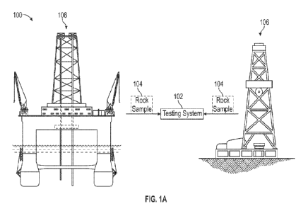
[0030] FIG. 1 a illustrates, at a high level, the acquiring of rock samples
and the
analysis of the rock samples according to principles disclosed herein.
Embodiments
of present disclosure may be especially beneficial in analyzing rock samples
from sub-
surface formations that are important in the production of oil and gas. As
such, FIG.
la illustrates environments 100 from which rock samples 104 to be analyzed by
testing
system 102 can be obtained, according to various implementations. In these
illustrated
examples, rock samples 104 can be obtained from terrestrial drilling system
106 or
from marine (ocean, sea, lake, etc.) drilling system 108, either of which is
utilized to
extract resources such as hydrocarbons (oil, natural gas, etc.), water, and
the like. As
is fundamental in the art, optimization of oil and gas production operations
is largely
influenced by the structure and material properties of the rock formations
into which
terrestrial drilling system 106 or marine drilling system 108 is drilling or
has drilled in
the past.
[0031] The manner in which rock samples 104 are obtained, and the physical
form
of those samples, can vary widely. Examples of rock samples 104 useful in
connection
with embodiments disclosed herein include whole core samples, side wall core
samples, outcrop samples, drill cuttings, and laboratory generated synthetic
rock
samples such as sand packs and cemented packs.
[0032] As illustrated in FIG. la, the environment 100 includes testing system
102
that is configured to analyze images 128 (FIG. 1 b) of rock samples 104 in
order to
6
CA 03177815 2022- 11- 3
WO 2021/226594
PCT/US2021/031617
determine the material properties of the corresponding sub-surface rock, such
properties including petrophysical properties in the context of oil and gas
exploration
and production.
[0033] FIG. lb illustrates, in a generic fashion, the constituent components
of the
testing system 102 that analyzes images 128. In a general sense, testing
system 102
includes imaging device 122 for obtaining 2D or 3D images, as well as other
representations, of rock samples 104, such images and representations
including
details of the internal structure of the rock samples 104. An example of
imaging device
122 is an X-ray computed tomography (CT) scanner, which, as known in the art,
emits
x-ray radiation 124 that interacts with an object and measures the attenuation
of that
x-ray radiation 124 by the object in order to generate an image of its
interior structure
and constituents. The particular type, construction, or other attributes of CT
scanner
122 can correspond to that of any type of x-ray device, such as a micro CT
scanner,
capable of producing an image representative of the internal structure of rock
sample
104. The imaging device 122 generates one or more images 128 of rock sample
104,
and forwards those images 128 to a computing device 120.
[0034] The images 128 produced by imaging device 122 may be in the form of a
three-dimensional (3D) digital image volume (i.e., a digital rock) consisting
of or
generated from a plurality of two-dimensional (2D) sections of rock sample
104. In this
case, each image volume is partitioned into 3D regular elements called volume
elements, or more commonly "voxels". In general, each voxel is a
parallelepiped that
may have different dimensions in the x, y, and z directions. In some examples,
a voxel
may also be cubic, having a side of equal length in the x, y, and z
directions. Digital
image volume 128 itself, on the other hand, may contain different numbers of
voxels
in the x, y, and z directions. Each voxel within a digital volume has an
associated
numeric value, or amplitude, that represents the relative material properties
of the
imaged sample at that location of the medium represented by the digital
volume. The
range of these numeric values, commonly known as the grayscale range, depends
on
the type of digital volume, the granularity of the values (e.g., 8-bit or 16-
bit values),
and the like. For example, 16-bit data values enable the voxels of an x-ray
tomographic
image volume to have amplitudes ranging from 0 to 65,536 with a granularity of
1.
[0035] The testing system 102 may also include a scanning electron microscope
(SEM) 123 for obtaining 2D SEM images of rock samples 104. The SEM 123 is also
coupled to the computing device 120, and thus the 2D SEM images produced by
the
7
CA 03177815 2022- 11- 3
WO 2021/226594
PCT/US2021/031617
SEM 123 are available to (e.g., received by) the computing device 120, which
processes such 2D SEM images as described further below.
[0036] As mentioned above, imaging device 122 forwards images 128 to computing
device 120, which in the example of FIG. lb may be any type of computing
device, for
example, a desktop computer or workstation, a laptop computer, a server
computer, a
tablet computer, and the like. The SEM 123 also forwards 2D SEM images to the
computing device 120. As such computing device 120 will include hardware and
software components typically found in a conventional computing device. As
shown in
FIG. lb, these hardware and software components of computing device 120
include a
testing tool 130 that is configured to analyze images 128 to determine the
petrophysical properties of rock sample 104 under one or more simulated fluid
saturation conditions, including fluid saturation conditions that may be
encountered by
rock formations in the sub-surface. In this regard, the testing tool 130 may
be
implemented as software, hardware, or a combination of both, including the
necessary
and useful logic, instructions, routines, and algorithms for performing the
functionality
and processes described in further detail herein. In a general sense, testing
tool 130
is configured to analyze image volume 128 of rock sample 104 to perform direct
numerical simulation of the petrophysical properties under fluid saturation
conditions
representing subsurface conditions of rock formations, including variation
degrees of
saturation with multiple fluids.
[0037] FIG. lc generically illustrates the architecture of computing device
120 in
testing system 102 according to various embodiments. In this example
architecture,
computing device 120 includes one or more processors 152, which may be of
varying
core configurations and clock frequencies as available in the industry. The
memory
resources of computing device 120 for storing data and/or program instructions
for
execution by the one or more processors 152 include one or more memory devices
154 serving as a main memory during the operation of computing device 120, and
one
or more storage devices 160, for example realized as one or more of non-
volatile solid-
state memory, magnetic or optical disk drives, or random-access memory. One or
more peripheral interfaces 156 are provided for coupling to corresponding
peripheral
devices such as displays, keyboards, mice, touchpads, touchscreens, printers,
and
the like. Network interfaces 158, which may be in the form of Ethernet
adapters,
wireless transceivers, serial network components, etc. are provided to
facilitate
communication between computing device 120 via one or more networks such as
8
CA 03177815 2022- 11- 3
WO 2021/226594
PCT/US2021/031617
Ethernet, wireless Ethernet, Global System for Mobile Communications (GSM),
Enhanced Data rates for GSM Evolution (EDGE), Universal Mobile
Telecommunications System (UMTS), Worldwide Interoperability for Microwave
Access (WiMAX), Long Term Evolution (LTE), and the like. In this example
architecture, processors 152 are shown as coupled to components 154, 156, 158,
and
160 by way of a single bus; of course, a different interconnection
architecture such as
multiple, dedicated, buses and the like may be incorporated within computing
device
120.
[0038] While illustrated as a single computing device, computing device 120
can
include several computing devices cooperating together to provide the
functionality of
a computing device. Likewise, while illustrated as a physical device,
computing device
120 can also represent abstract computing devices such as virtual machines and
"cloud" computing devices.
[0039] As shown in the example implementation of FIG. 1c, the computing device
120 includes software programs 162 including one or more operating systems,
one or
more application programs, and the like. According to embodiments, software
programs 162 include program instructions corresponding to testing tool 130
(FIG. 1b),
implemented as a standalone application program, as a program module that is
part
of another application or program, as the appropriate plug-ins or other
software
components for accessing testing tool software on a remote computer networked
with
computing device 120 via network interfaces 158, or in other forms and
combinations
of the same.
[0040] The program memory storing the executable instructions of software
programs 162 corresponding to the functions of testing tool 130 may physically
reside
within computing device 120 or at other computing resources accessible to
computing
device 120, i.e. within the local memory resources of memory devices 154 and
storage
devices 160, or within a server or other network-accessible memory resources,
or
distributed among multiple locations. In any case, this program memory
constitutes a
non-transitory computer-readable medium that stores executable computer
program
instructions, according to which the operations described in this
specification are
carried out by computing device 120, or by a server or other computer coupled
to
computing device 120 via network interfaces 158 (e.g., in the form of an
interactive
application upon input data communicated from computing device 120, for
display or
output by peripherals coupled to computing device 120). The computer-
executable
9
CA 03177815 2022- 11- 3
WO 2021/226594
PCT/US2021/031617
software instructions corresponding to software programs 162 associated with
testing
tool 130 may have originally been stored on a removable or other non-volatile
computer-readable storage medium (e.g., a DVD disk, flash memory, or the
like), or
downloadable as encoded information on an electromagnetic carrier signal, in
the form
of a software package from which the computer-executable software instructions
were
installed by computing device 120 in the conventional manner for software
installation.
It is contemplated that those skilled in the art will be readily able to
implement the
storage and retrieval of the applicable data, program instructions, and other
information useful in connection with this embodiment, in a suitable manner
for each
particular application, without undue experimentation.
[0041] The particular computer instructions constituting software programs 162
associated with testing tool 130 may be in the form of one or more executable
programs, or in the form of source code or higher-level code from which one or
more
executable programs are derived, assembled, interpreted or compiled. Any of a
number of computer languages or protocols may be used, depending on the manner
in which the desired operations are to be carried out. For example, these
computer
instructions for creating the model according to embodiments may be written in
a
conventional high-level language such as PYTHON, JAVA, FORTRAN, or C++, either
as a conventional linear computer program or arranged for execution in an
object-
oriented manner. These instructions may also be embedded within a higher-level
application. In any case, it is contemplated that those skilled in the art
having reference
to this description will be readily able to realize, without undue
experimentation,
embodiments in a suitable manner for the desired installations.
[0042] The particular functions of testing tool 130, including those
implemented by
way of software programs 162, to analyze rock samples under various saturation
conditions according to embodiments, will now be described with reference to
the FIG.
2 in combination with FIGS. la-1c.
[0043] FIGS. 2A and 2B show a flow diagram for a method 200 for analyzing rock
samples under various saturation conditions in accordance with principles
disclosed
herein. Though depicted sequentially as a matter of convenience, at least some
of the
actions shown can be performed in a different order and/or performed in
parallel.
Additionally, some embodiments may perform only some of the actions shown. In
some
embodiments, at least some of the operations of the method 200, as well as
other
CA 03177815 2022- 11- 3
WO 2021/226594
PCT/US2021/031617
operations described herein, can be implemented as instructions stored in a
computer
readable medium and executed by one or more processors 152.
[0044] In block 202, the testing system 102 acquires rock sample 104 to be
analyzed,
such as from a sub-surface rock formation obtained via terrestrial drilling
system 106
or marine drilling system 108, or from other sources. The specific rock sample
104
may be prepared from a larger volume of the sub-surface rock formation, to be
of a
size, dimension, and configuration that may be imaged by imaging device 122
(e.g., a
CT scanner), for example by drilling or cutting out a portion of the larger
volume of the
rock formation of interest.
[0045] In block 204, imaging device 122 in combination with computing device
120
of testing system 102 generates digital image volume 128 representative of
rock
sample 104, including its interior structure. For example, if the imaging
device 122 is
a CT scanner, then X-ray imaging of rock sample 104 is performed (i.e.,
emitting
radiation directed at rock sample 104 and measuring the attenuation) to
generate
image volumes 128 of or from 2D slice images. Specific conventional techniques
for
acquiring and processing 3D digital image volumes 128 of rock sample 104 in
block
204 include, without limitation, X-ray tomography, X-ray microtomography, X-
ray
nano-tomography, Focused Ion Beam Scanning Electron Microscopy, and Nuclear
Magnetic Resonance Imaging. In some embodiments, the digital image volume 128
may be computationally generated rather than produced by scanning a physical
specimen. In embodiments in which the digital image volume 128 is produced by
scanning a rock specimen, the rock specimen may be a naturally occurring rock
or a
man-made porous material (e.g., a synthetic rock).
[0046] FIG. 3a illustrates an example of one 2D slice image 300 of a 3D image
of a
rock sample, which shows a cross-sectional slice of the structural details of
that rock
sample, including the features of solid material 302, pore or void space 304,
and partial
solid/pore space 306. The image data at this point may be in the form of
grayscale
values representative of the attenuation of the x-ray radiation by the
constituents of
rock sample 104. While FIG. 3a illustrates one 2D slice image 300, 3D digital
image
volume 128 of rock sample 104 is typically composed of multiple 2D slice
images at
locations stepped along one axis of rock sample 104 (e.g., a "stack" of
multiple 2D
slice images), together forming a 3D image of rock sample 104. The combining
of the
2D slice images into 3D digital image volume 128 may be performed by
computational
resources within imaging device 122 itself, or by computing device 120 from
the series
11
CA 03177815 2022- 11- 3
WO 2021/226594
PCT/US2021/031617
of 2D slice images 128 produced by imaging device 122, depending on the
particular
architecture of testing system 102.
[0047] In block 206, the testing system 102 performs segmentation or other
image
enhancement techniques on digital image volume 128 of rock sample 104 to
distinguish and label different components or phases of image volume 128 from
the
grayscale values of the image. More specifically, computing device 120
performs this
segmentation in order to identify components, such as pore space and
mineralogical
components (e.g., clays and quartz). In some embodiments, testing tool 130 is
configured to segment image volume 128 into more than two significant phases,
representing such material constituents as pore space, clay fraction, quartz
fraction,
other various mineral types, organic matter, or composite materials.
[0048] The computing device 120 can utilize any of a number of types of
segmentation algorithms. One approach to segmentation is the application of a
"thresholding" process to image volume 128, in which computing device 120
chooses
a threshold value within the voxel amplitude range. Those voxels having an
amplitude
below the threshold value are assigned one specific numeric value that denotes
pore
space, while those voxels having an amplitude above the threshold are assigned
another numeric value that denotes matrix space (i.e., solid material). In
another
example, there are multiple threshold values that define a number of different
voxel
amplitude ranges. In this approach, thresholding converts a grayscale image
volume
to a segmented volume of voxels having one of two (or more) possible numeric
values,
commonly selected to be 0 and 1. FIG. 3b illustrates an example of the
segmentation
performed on a 2D slice image 310 (e.g., part of 3D digital image volume 128)
via
thresholding with more than two possible numeric values. As illustrated,
segmentation
allows the structural details of a rock sample to be distinguished, in this
example with
the solid material 302 shown in white, and pores or void space 304 shown in
black,
and partial solid/pore space 306 shown in light and dark greys. Further
segmentation
can be applied one or more times to differentiate various features within a
grayscale
image. If simple thresholding is used, multiple threshold values can
distinguish among
different materials exhibiting different x-ray attenuation characteristics,
such as clay,
quartz, feldspar, etc.
[0049] Computing device 120 may alternatively utilize other segmentation
algorithms. An example of such an alternative algorithm is known in the art as
Otsu's
Method, in which a histogram-based thresholding technique selects a threshold
to
12
CA 03177815 2022- 11- 3
WO 2021/226594
PCT/US2021/031617
minimize the combined variance of the lobes of a bimodal distribution of
grayscale
values (i.e., the "intra-class variance"). Otsu's method can be readily
automated, and
may also be extended to repeatedly threshold the image multiple times to
distinguish
additional material components such as quartz, clay, and feldspar. Other
examples of
automated segmentation algorithms of varying complexity may alternatively or
additionally be used by computing device 120 to distinguish different features
of an
image volume, such algorithms including Indicator Kriging, Converging Active
Contours, Watershedding, and the like.
[0050] The computing device 120 may also utilize other image enhancement
techniques to enhance or improve the structure defined in image volume 128 to
further
differentiate among structure, to reduce noise effects, and the like.
Likewise, while
computing device 120 can perform the segmentation or other image enhancement
techniques, it is contemplated that other components of testing system 102,
for
example imaging device 122 itself, may alternatively perform image enhancement
in
whole or in part.
[0051] Segmentation thus associates the voxels in the digital image volume 128
with
the particular material (or pore space, as the case may be) at the
corresponding
physical location within rock sample 104. Each voxel is labeled with one
unique
material identification corresponding to the particular constituent assigned
to a given
x-ray attenuation amplitude. Such constituents including pore space, matrix
material,
mixed pore-clay fraction, individual grains, grain contacts, mineral types,
and the like.
[0052] FIG. 4 shows a representation 400 of digital image volume 128 of the
rock
sample 104. As explained above, the digital image volume 128 may be
constructed
from a series of 2D slice images 300 (or segmented 2D slice images 310). The
representation 400 includes different rock fabrics, for example that
correspond to the
identified rock fabrics from the 2D slice images 300. In some examples, a
fabric refers
to a pattern shared by a "region" or portion of an image. For example, a
region may
be grouped together as a fabric based on sharing underlying attributes such as
porosity, compositional phases and their proportions, image entropy, and the
like.
Certain specific examples of rock fabrics include pore space and various types
of
solids. For example, different types of solid material may correspond to
different rock
fabrics. Thus, a fabric may be considered as a quantification of spatial
patterns of pixel
values (e.g., pixel intensity) within a particular domain or image.
13
CA 03177815 2022- 11- 3
WO 2021/226594
PCT/US2021/031617
[0053] In the example of FIG. 4, the representation 400 includes a first rock
fabric
402, a second rock fabric 404, and a third rock fabric 406, as well as a host
or
depositionally-dominant sedimentary rock fabric 408, which makes up the bulk
of the
rock sample between the rock fabrics 402, 404, 406. At least some of the
voxels that
comprise the representation 400 are associated with or mapped to a physical
coordinate space associated with the rock sarnple 104. This allows for a
mapping of
the imaged rock fabrics 402, 404, 406 to specific physical locations within
the rock
sample 104.
[0054] Embodiments of the present disclosure leverage the location of imaged
rock
fabrics 402, 404, 406 in the representation 400 of the digital image volume
128 to
identify one or more digital planes through the digital image volume 128 that
intersect
with the rock fabrics 402, 404, 406, as shown in block 208 of FIG. 2. In some
examples,
the segmentation described above, performed in block 206 of FIG. 2, provides
the
spatial identification of these fabrics 402, 404, 406. Following segmentation,
and
depending on the complexity and arrangement of rock fabrics 402, 404, 406
within the
digital image volume 128, multiple digital planes may be required to
adequately
intersect all of the different rock fabrics 402, 404, 406. Further, in some
examples,
digital plane(s) are identified that reduce or minimize the number of plane(s)
needed
to adequately intersect all of the different rock fabrics 402, 404, 406.
[0055] For example, to intersect the rock fabrics 402, 404, 406, a first set
of digital
planes includes digital plane 410, which intersects the rock fabrics 402, 406,
and digital
plane 412, which intersects the rock fabric 404. However, it may be
advantageous to
reduce the number of digital planes if possible, to reduce subsequent
machining and
SEM imaging requirements. Thus, in at least one embodiment, a digital plane
414 is
selected that intersects the rock fabrics 402, 404, 406, reducing the required
machining and imaging to adequately image all the rock fabrics 402, 404, 406.
[0056] After the digital plane 414 is identified (e.g., according to block 208
of FIG.
2A), the method 200 continues in block 210 in which the physical rock sample
104 is
machined (or otherwise mechanically prepared) to expose a physical face that
corresponds to the identified set of digital planes (e.g., digital plane 414).
As noted
above, since the digital image volume 128 (e.g., its composite voxels) is
mapped to
the physical coordinate space associated with the rock sample 104, the
identified
digital plane 414 corresponds to a plane in that physical coordinate space,
making it
relatively straightforward to machine the corresponding physical face.
14
CA 03177815 2022- 11- 3
WO 2021/226594
PCT/US2021/031617
[0057] Once the physical rock sample 104 has been machined or otherwise
mechanically prepared to expose one or more physical faces that correspond to
the
identified set of digital planes, the method 200 continues in block 212 with
obtaining a
series of SEM images of the physical face(s). In various embodiments, the SEM
imaging of the physical face(s) of the rock sample 104 is performed at a
variety of
scales (e.g., sequential SEM imaging with increasing amounts of zoom into the
physical face(s) of the rock sample 104). FIGS. 5a-5c demonstrate example SEM
images captured at different (e.g., sequentially zoomed in) scales, which
assist in
defining the spatial characterization of one or more rock fabrics (e.g.,
fabrics 402, 404,
406 in FIG. 4) in the digital plane 414.
[0058] In one example, the regions that are zoomed into may vary depending on
the
circumstances of the imaging being performed. In one example in which organic
porosity is of particular interest for a given project, an emphasis is placed
on acquiring
zoomed-in images of regions that appear to include fabric(s) of that type.
Continuing
this example, regions that do not appear to include fabric(s) demonstrating
organic
porosity are less frequently sampled (i.e., fewer zoomed-in images are
acquired of
regions that do not appear to include volumes of fabric(s) including organic
porosity of
at least a threshold amount, and a level of zoom for those regions not
including at least
a threshold amount of fabric(s) demonstrating organic porosity may also be
lower). In
another example, overall pore connectivity is of particular interest for the
given project.
In this example, all regions may be equally sampled to avoid overlooking image
data
that is relevant to a determination of pore connectivity. One example of equal
sampling
includes acquiring zoomed in images along a rectangular grid path with regular
spacing across the dimensions of an image.
[0059] FIG. 5a shows a SEM image 500, which is approximately 100 microns wide.
A first zoom portion 502 of the SEM image 500 contains multiple rock fabrics,
including
a first fabric 504 and a second fabric 506. For example, the first fabric 504
corresponds
to an inorganic, porosity-rich fabric while the second fabric 506 corresponds
to an
organic, porosity-rich fabric. The zoom portion 502 may be identified as a
result of
containing a particular fabric of interest having finer details not easily
discernable at
lower resolutions. For example, the zoom portion 502 may be selected from the
SEM
image 500 because it indicates the presence of organic rich porosity. The zoom
portion
502 provides greater visual clarity on the structure of the organic pores
compared to
CA 03177815 2022- 11- 3
WO 2021/226594
PCT/US2021/031617
what can be discerned from the SEM image 500. Further zooming-in may sometimes
be useful to discern sufficient structural detail for various regions of
interest.
[0060] FIG. 5b shows the first zoom portion 502 in greater detail. For
example, the
greater detail in FIG. 5b is obtained by subjecting the first zoom portion 502
to SEM
imaging at a higher zoom level, and thus the first zoom portion 502 is
equivalently a
zoomed SEM image 502. In particular, as shown in FIG. 5b, the zoomed SEM image
502 is approximately 50 microns wide. In FIG. 5b, the first and second fabrics
504,
506 are seen as larger, and in greater detail. Further, a second zoom portion
510 is
identified in a manner similar to that in which the first zoom portion 502 was
identified
in FIG. 5a. For example, the second zoom portion 510 may be selected from the
zoomed SEM image 502 as a result of the second zoom portion 510 having a
higher
diversity of represented rock fabrics than other portions of the zoomed SEM
image
502.
[0061] FIG. 5c shows the second zoom portion 510 in greater detail. For
example,
the greater detail in FIG. 5c is obtained by subjecting the second zoom
portion 510 to
SEM imaging at a higher zoom level, and thus the second zoom portion 510 is
equivalently a second zoomed SEM image 510. In particular, as shown in FIG.
5c, the
second zoomed SEM image 510 is approximately 10 microns wide. The first fabric
504
is not present in the second zoomed SEM image 510 because it was not included
in
the second zoomed portion 510 in FIG. 5b. However, the second fabric 506 is
seen as
larger and in greater detail. In the example of FIG. 5c, the identified second
fabric 506
in the central area of the second zoomed SEM image 510 is illustrative of an
organic-
rich pore fabric, which may impact or contribute to the flow, transport, and
storage
descriptions of the larger rock sample 104.
[0062] Although the example of FIGS. 5a-5c includes a first and second zoomed
image 502, 510, other examples of this disclosure may extend to additional or
fewer
iterations of zooming as required to capture a sufficient level of diversity
of represented
rock fabrics. Further, the process or capturing multiple zoomed SEM images may
be
repeated across the physical face(s) of the larger rock sample 104. For
example, a set
of SEM images zoomed to the level of that shown in FIG. 5c may be obtained
over the
area represented by the SEM image 500 of FIG. 5a. Thus, a set of zoomed SEM
images, across multiple scales (e.g., "multi-scale") is produced, which
captures the
finer detail of the physical face(s) of the larger rock sample 104. This set
of multi-scale
SEM images may include at least a threshold number of rock fabrics of the
plurality of
16
CA 03177815 2022- 11- 3
WO 2021/226594
PCT/US2021/031617
rock fabrics contained in the digital image volume 128, described above. A
higher
threshold in this context results in a set of multi-scale SEM images that
captures or
represents a larger percentage (e.g., about 70%, 80%, 90%, or 100% in some
cases)
of the fabrics in the digital image volume 128. A lower threshold in this
context results
in a set of multi-scale SEM images that captures or represents a relatively
lower
percentage (e.g., less than about 70%) of the fabrics in the digital image
volume 128,
for example to reduce computational or processing resources required for
subsequent
steps of the method. Additionally, as will be explained further below,
segmentation
may be performed on the second zoomed SEM image 510 to extract characteristic
quantities (e.g., pore space, organic solid material, and solid inorganic
material), which
may be leveraged for further numerical predictions or determinations related
to the
larger rock sample 104.
[0063] FIG. 5d provides an alternate example of the process shown in FIGS. 5a-
5c
and described above. In particular, in step 3 of FIG. 5d, a probability map is
generated
(e.g., using an unsupervised learning algorithm) based on a segmented, lower-
resolution image obtained in step 2 of FIG. 5d. The probability map of step 3
probabilistically groups similar fabrics. For example, the type 1 probability
map
illustrates the probability of whether a region is a non-porous organic-rich
fabric, while
the type 2 probability map illustrates the probability of whether a region is
a porous,
organic-rich fabric. In some cases, a grouping of similar fabrics indicates or
identifies
a region of interest of the rock sample 104 for higher-resolution imaging. In
the
example of FIG. 5d, this identified region of interest corresponds to the
boxed region
in the lower portion of the type 2 material, which is shown to have a high
probability of
being a similar rock fabric. In one example, the identified region of
interest, or first area
within the SEM image, contains a first number of pixels that are associated
with a
primary fabric type (e.g., porous, organic-rich fabric in the case of the type
2 probability
map) and a second number of pixels associated with fabrics other than the
primary
fabric type. Thus, the region of interest may be identified based on the ratio
of the first
number to the second number being above a threshold, which indicates a certain
percentage (or more) of the primary fabric type in that region. Once the
regions of
interest is identified, additional SEM imaging may be performed at higher
(e.g., finer)
resolutions, as shown in step 4 of FIG. 5d.
[0064] Referring generally to FIGS. 5a-5d, some examples of this description
leverage one or more machine learning algorithms to determine various aspects
of the
17
CA 03177815 2022- 11- 3
WO 2021/226594
PCT/US2021/031617
zoom-and-capture processes described above. For example, the number of rock
fabrics to be sampled (e.g., of a total number of identified fabrics in the
digital image
volume 128) may be determined responsive to application of a machine learning
algorithm. Similarly, the resolution at which to capture certain rock fabrics
(e.g., how
much zooming in is appropriate for a given rock fabric) may be determined
responsive
to application of a machine learning algorithm. In one example, these values
or levels
are determined in an iterative fashion, to produce a data set that is able to
be
processed in a reasonable amount of time (e.g., avoiding a brute force-
sampling of all
possible combinations of numbers of fabrics to capture, and resolutions at
which to
capture those fabrics), while still providing useful data of each of a certain
number of
fabrics. For example, this avoids conditions such as under-zooming a first
fabric, thus
missing out on important detail of that fabric; or over-zooming a second
fabric and thus
wasting valuable time and processing resources (either during capture and/or
subsequent processing of a too-detailed image).
[0065] Referring back to the method 200 of FIG. 2, once a set of SEM images is
obtained at a resolution of approximately 2-4 times the smallest pore or other
material
feature of interest to be analyzed for a given application, the method 200
continues in
block 214 with optionally applying segmentation to the 2D SEM images. For
example,
if characterizing microporosity of a sample (e.g., pore sizes of less than
5nm), the
required resolution may be approximately 2nm. As another example, if
characterizing
pyritic pores of larger than 15nm, the required resolution may be
approximately 5nm,
and thus the level of required zoom may be relatively more relaxed. Segmenting
the
2D SEM images is similar to the segmentation described above with respect to
block
206 and FIGS. 3a and 3b, regarding segmenting the 2D slice image 300. FIG. 6a
shows an exemplary simple, two-constituent (e.g., pore space and organic
matter)
SEM image 600, which may be at a scale similar to the second zoomed SEM image
510 described above with respect to FIG. 5c. In this example, where the SEM
image
600 is relatively simple, segmentation may be performed with a single
threshold to
distinguish between pore space and organic matter. FIG. 6b shows a resulting
segmented SEM image 610 in which pore space is shown as black and organic
matrix
material is shown as white. By segmenting the SEM images obtained in block 212
into
two phases (e.g., pore space and organic matter), the pore space of the larger
rock
sample 104 is captured at a fine level of granularity in two dimensions, which
may then
be leveraged to create a 3D digital model volume that represents the rock
sample 104,
18
CA 03177815 2022- 11- 3
WO 2021/226594
PCT/US2021/031617
but at a finer resolution (e.g., greater level of detail) that that of the
initially captured
digital image volume. In particular, the segmented 2D SEM image 610 in FIG. 6b
may
be used as a training image for a 2D-to-3D volume transformation, which is
explained
in further detail below. In other examples, the SEM images obtained in block
212 may
be segmented into an arbitrary number of phases, representing various rock
fabrics.
[0066] FIG. 7a shows a set 700 of segmented 2D SEM images, similar to the
segmented SEM image 610 explained above. The set of 2D SEM images 700 are
extracted from multiple SEM images that contain differing representations of a
particular
fabric (e.g., 402, 404, 406). Referring back to the method 200, in block 216
the set of
segmented 2D SEM images 700 is used to generate a set of statistically-similar
3D
digital model volumes for a particular rock fabric. In certain examples, one
or more
stochastic algorithms (e.g., a cross-correlation function) are applied to the
set of 2D
SEM images 700 to generate realizations of statistically-similar or
statistically-equivalent
3D pore-organic matrix volumes. In the example of the cross-correlation
function, this
process uses structural information (e.g., data indicative of correlation
between different
parts of the image) in the 2D training images (e.g., the 2D SEM image 610
described
above) to first break up the images into smaller constituent areas and then
recombine
those areas in a stochastic manner to synthetically generate statistically
similar, but non-
identical, versions of the original 2D training image. Subsequently, the
original 2D
training image, as well as its statistically similar versions, are projected
into one or more
imaginary planes (e.g., in 3D). These 3D projections, or "digital model
volumes," may
be used to statistically condition a subsequent iteration of generating
synthetic,
statistically similar images. In one example, such a conditioning process is
useful to
iteratively generate synthetic images that may be overlain or underlain with
previously
generated synthetic images in a manner that appears to more accurately reflect
a
natural look and/or structural continuity of a real-world rock sample.
[0067] FIG. 7b shows an exemplary set 710 of 3D pore-organic matrix model
volumes,
which are statistically-similar to each other. For example, a first digital
model volume is
statistically similar to a second digital model volume when attributes such as
the
variogram of the pore phase of the first model volume and that of the second
model
volume are identical, or within a threshold amount (e.g., based on an
engineering
tolerance). In some cases, statistical equivalence is controlled by an
assumption
regarding ergodicity in the stochastic algorithm. Because the set 710 of 3D
digital model
volumes for a given fabric type is generated computationally from 2D images
taken from
19
CA 03177815 2022- 11- 3
WO 2021/226594
PCT/US2021/031617
a single physical face or a small number of physical faces of the rock sample
104, the
set 710 may be generated much more easily and with relatively less expense
than, for
example, FIB-SEM in which a portion of rock material is continually machined
and
imaged layer-by-layer. In some examples, a numeric value of a voxel in the 3D
digital
model volume(s) of the set 710 is determined based on a spatial distribution
of pixel
values in each of the 2D SEM images used to generate the 3D digital model
volume(s).
In one specific, but non-limiting example, a circular grain in the 2D SEM
images (e.g.,
100 white pixels in an approximate circle surrounded by black pixels) leads to
the
generation of a spherical grain in the 3D digital model volume (e.g., 1000
white voxels
in an approximate sphere surrounded by black voxels).
[0068] Additionally, because the set 710 of 3D digital model volumes for a
given fabric
type may be generated computationally from 2D images taken from multiple
physical
faces of the rock sample 104, such 2D images may be of different axial
orientations of
the rock sample 104. In one specific, but non-limiting example, a first 2D SEM
image is
taken of a physical face of the rock sample 104 along a first axis with
respect to a
position of the rock sample 104, while a second 2D SEM image is taken of a
physical
face of the rock sample 104 along a second axis with respect to the position
of the rock
sample 104. This enables the generated 3D digital model volume(s) to consider
features
that may differ in their axial symmetry. For example, a feature that appears
circular along
one axis might typically be represented as spherical in the resultant 3D
digital model
volume. However, if that same feature appears oblong along another axis, that
feature
may instead be represented as ovoid in the resultant 3D digital model volume.
[0069] Additionally, the process to generate the set 710 of 3D digital model
volumes
for a given fabric type may be repeated for multiple fabric types, which
results in a
plurality of (e.g., N) realizations of each of a plurality of (e.g., M) fabric
types. The N
realizations of 3D digital model volumes for a given fabric type represent a
number of
possibilities of what a 3D version of that fabric type may look like in
nature, which
provides a way to calculate or determine petrophysical properties at a fabric
level and
thus generate a probability distribution of various petrophysical properties
for that
particular fabric type. In some cases, estimates of petrophysical properties
determined
from the 3D digital model volumes are more accurate than estimates of those
same
properties derived from 2D models. As a result, the generation of the 3D
digital model
volumes described herein improves accuracy relative to, for example,
estimating
petrophysical properties based on 2D SEM images.
CA 03177815 2022- 11- 3
WO 2021/226594
PCT/US2021/031617
[0070] Relative to the original digital image volume 128, the 3D digital model
volumes
in the set 710 have a higher (e.g., finer) resolution due to being generated
based on 2D
SEM images. Additionally, each 3D digital model volume is specific to one
fabric type of
the often-multiple fabric types present in the digital image volume 128.
[0071] Once a set of statistically-similar 3D models 710 are generated, the
method
200 continues in block 218 with performing numerical simulation on the 3D
digital model
volumes 710 to determine one or more material or petrophysical properties
associated
with each of the 3D digital model volumes 710. In one example, the 3D digital
model
volumes 710 are used as a modeling grid for one type of rock fabric (e.g., N
realizations
of one fabric type) to determine the desired material property or properties
for that fabric
type. In various examples, the material property may include porosity, pore
size
distributions, permeability, capillary pressure, electric resistivity, and
elastic moduli,
[0072] The following exemplary Table 1 demonstrates representative porosity
and
permeability values as material properties derived from a set of 3D pore-
organic matrix
volumes 710 shown in FIG. 7b. In this example, the 3D pore-organic matrix
volumes
710 are used to calculate porosity and/or permeability using one or more image
analysis
algorithms such as, for example, segmentation (e.g., for porosity) and Lattice-
Boltzmann
simulation (e.g., for permeability). In an example, the set of computed values
that result
from the application of these algorithms represents a full range of possible
porosities
and permeabilities that may be encountered in real-world rock samples that
include that
fabric. Thus, the algorithms produce or provide a probability distribution for
each of
porosity and permeability for a particular fabric type (e.g., porous organic-
matter in this
example). Generating additional realizations, or digital model volumes,
results in the
ability to determine petrophysical properties based on a larger number of
models, and
to thus produce a correspondingly larger dataset of probability distributions.
This results
in a more accurate and/or precise determination of probability distributions
for those
petrophysical properties.
Permeability Porosity
Realization (m2) (% volume)
1 3.29E-19 22.24
2 3.78E-19 23.38
3 3.37E-19 21.28
4 5.08E-19 25.64
21
CA 03177815 2022- 11- 3
WO 2021/226594
PCT/US2021/031617
4.93E-19 25.84
6 3.91E-19 23.39
mean 4.1E-19 23.63
Table 1.
Each realization in Table 1 corresponds to one of the 3D models of the example
set 710
of 3D models. The statistical similarity of the various models is reflected by
the relatively
close grouping of permeability and porosity values for each of the
realizations in Table
1. In some examples, the numerical simulation utilizes proprietary algorithms
from
exemplary direct numerical simulation techniques. For example, a two-phase
lattice
Boltzmann simulation may be utilized to estimate a numeric permeability based
on the
3D digital model volume(s) for a given fabric type, while an object
partitioning and point
counting algorithm may be utilized to estimate a numeric pore size
distribution based on
the 3D digital model volume(s) for a given fabric type.
[0073] In some examples, the determined material property or properties for a
given
rock fabric is associated with voxels in a 3D digital model volume that
correspond to that
given rock fabric. Additionally, locations within the digital model volume(s)
may also be
mapped to the physical coordinate space associated with the rock sample 104.
Thus,
the determined material property or properties can be associated with the
physical
coordinate space associated with the rock sample 104. As described above, the
material property mapped to a particular voxel in the digital model volume(s)
may be
sampled from a distribution of properties measured for the given rock fabric,
which
results in the digital model volume(s) being a composite volumetric grid of
such
material properties.
[0074] FIG. 8 shows an exemplary 3D model (e.g., one of the example sets 710
of 3D
models), in which a flow field (e.g., the determined material property) as a
greyscale
heat map is superimposed on the 3D model. In general, a flow field comprises a
flow
velocity value for multiple voxels (e.g., each voxel) in a 3D digital model
volume. A
permeability tensor is a simplification of a 3D flow field to a representative
value (e.g., a
single value in some cases), and thus may be calculated responsive to a
determined
flow field for a given 3D digital model volume. Although the porosity of the
3D model in
FIG. 8 appears disconnected, the flow field demonstrates that there are flow
pathways
through the 3D volume. The greyscale heat map represents the flow velocity,
with darker
22
CA 03177815 2022- 11- 3
WO 2021/226594
PCT/US2021/031617
tones representing a higher velocity and lighter tones representing a lower
velocity. The
arrow in FIG. 8 represents the overall direction of the flow being modeled
through the
3D volume.
[0075] FIG. 9 shows a flow diagram of a method 950 for analyzing rock samples
in
accordance with principles disclosed herein. FIG. 9 contains certain steps
that are
similar to those described above with respect to FIGS. 2A and 2B. Though
depicted
sequentially as a matter of convenience, at least some of the actions shown
can be
performed in a different order and/or performed in parallel. Additionally,
some
embodiments may perform only some of the actions shown. In some embodiments,
at
least some of the operations of the method 950, as well as other operations
described
herein, can be implemented as instructions stored in a computer readable
medium and
executed by one or more processors 152.
[0076] Blocks 952, 954, and 956 are similar to blocks 202, 204, and 206,
respectively, and description of those blocks is not repeated here for
purposes of
brevity.
[0077] In block 958, a material property is estimated for each of the rock
fabrics
associated in block 956 (and block 206). In the following examples, the
material
property is permeability; however, in other examples, different material
properties for
the rock fabrics may be estimated as described above. As described above with
respect to block 218, numerical simulation may be performed on the 3D models
710
to determine such material properties (e.g., permeability), which are then
associated
with the various rock fabrics (or voxels representing those fabrics) of the
digital image
volume. In various examples, the material property may also include porosity,
pore size
distributions, pern-ieability, capillary pressure, electric resistivity, and
elastic moduli. As
described above, the voxels of the digital image volume (and thus the
associated,
determined material property or properties) may be mapped to a physical
coordinate
space associated with the rock sample 104, improving understanding of the
physical
rock sample 104 and the formation from which it was sampled.
[0078] The method 950 continues in block 960 with selecting from a set of
fractional
bounceback parameter (FBP) nomograms a nomogram having an associated or
effective grid size that associates the material property values determined in
block 956
with an FBP value in a given range. For example, permeability values
determined in
block 956 span a permeability range. Each nomogram of the set of nomograms
associates permeability values with FBP values for a given grid size. In some
23
CA 03177815 2022- 11- 3
WO 2021/226594
PCT/US2021/031617
examples, the selected nomogram is the nomogram for which the permeability
range
is associated with FBP values between a lower FBP threshold and an upper FBP
threshold.
[0079] FIG. 10 shows an example of an FBP determination for use in a grayscale
lattice Boltzmann (GSLB) model or algorithm in accordance with principles
disclosed
herein. In FIG. 10, a grid of voxels is shown both before streaming (e.g.,
grid 1000)
and after streaming (e.g., grid 1020). The streaming step is one step (e.g.,
an
intermediate step) of the GSLB algorithm. "Before streaming" refers to the
state of the
GSLB output prior to mathematical application of the streaming
function/operation.
"After streaming" refers to the state of the GSLB output after application of
the
streaming function/operation. The streaming function/operation is a step at
which the
GSLB algorithm utilizes the FBP information provided in the primary input
(e.g., from
the original digital image/3D digital volume input). These primary inputs are
represented by gridpoints (e.g., illustrated by circles in FIG. 10), as well
as the specific
FBP values associated with each of the gridpoints. In FIG. 10, the FBP values
are
illustrated by the shade of gray color at the gridpoints 1002 and 1004. In
some
examples, each gridpoint has an associated FBP value. In the example of FIG.
10,
colorization of gridpoints is restricted to 1002 and 1004 is for purposes of
simplification.
[0080] The GSLB algorithm is an iterative algorithm, and its output described
above
may be iteratively modified multiple times by multiple functions/operations.
Streaming
is one of the types of intermediate operations of the GSLB algorithm that
performs
such iterative modification. These iterative operations may be performed until
required
criteria are met, at which point the iteration stops (e.g., there are no
additional
iterations of the algorithm). During the iteration of the GSLB algorithm, the
output at
each step is also an input for a subsequent round of iteration. One component
of this
output is a set of fractional streaming values in different directions. For
simplicity, only
two directions: f1 and f2, have been illustrated in FIG. 10. f is the fraction
streaming
from the source gridpoint 1002 towards the target 1004 gridpoint before the
streaming
function/operation is applied. fiT is the fraction streaming from the target
gridpoint 1004
towards a next gridpoint (e.g., to the right of the target gridpoint 1004) for
the next
round of iteration (e.g., after streaming is applied), while f2T is the
fraction streaming
from the target gridpoint 1004 back towards the source gridpoint 1002 after
streaming
is applied. fiT and f2T are calculated by the streaming function/operation and
are part
24
CA 03177815 2022- 11- 3
WO 2021/226594
PCT/US2021/031617
of the inputs for a subsequent round of iteration. as and aT are the decimal
forms of
the FBP at the source 1002 and target 1004 gridpoints, respectively.
[0081] The grid of voxels 1000, 1020 includes a source voxel 1002 and a target
voxel
1004. In this example, a behavior of fluid flow is determined from the source
voxel
1002 to the target voxel 1004. For example, it is previously determined (e.g.,
during a
previous iteration of calculation) that the source voxel 1002 provides a flow
in the
direction of the target voxel 1004 of fis, which may be rewritten as the sum
of
components (1 ¨ as) and as, where as is equal to the FBP divided by 100.
[0082] As shown in the grid of voxels after streaming 1020, fIcorresponds to
the
component 1 ¨ as, which is reflected back toward the source voxel 1002 by the
target
voxel 1004; fiTcorresponds to the component as, which is transmitted through
the
target voxel 1004 and onto another, adjacent voxel.
[0083] In an example in which the target voxel 1004 is complete pore space,
all of
the fluid flow provided to the target voxel 1004 (e.g., As) is transmitted
through the
target voxel 1004, and thus as = 1 and the reflected component (1 ¨ as) is
thus 0. In
this example, the FBP associated with the target voxel 1004 is 100.
[0084] In an example in which the target voxel 1004 is complete solid space,
all of
the fluid flow provided to the target voxel 1004 (e.g., fis) is reflected back
toward the
source voxel 1002 by the target voxel 1004, and thus as = 0 and the reflected
component (1 ¨ as) is thus 1. In this example, the FBP associated with the
target voxel
1004 is O.
[0085] In other examples in which the target voxel 1004 is partial solid/pore
space,
the FBP varies as a function of the amount of provided fluid (g) that is
reflected back
or transmitted by the target voxel 1004. As described further below, the FBP
for a
given voxel may be determined based on its permeability, or another material
property,
which are determined as described above.
[0086] HG. ha shows a set of nomograms 1100 that associates permeability
values
with FBP values for various grid sizes. In this context, grid size refers to
or is related
to voxel size/segmentation volume size. For example, a grid size of one micron
means
that each gridpoint (e.g., described with respect to FIG. 10) is specified to
have a one
micron spacing relative to other gridpoints. In this example, the set of
nomograms
1100 includes a first nomogram 1102 for an exemplary grid size of 1 micron, a
second
nomogram 1104 for an exemplary grid size of 0.1 micron, a third nomogram 1106
for
CA 03177815 2022- 11- 3
WO 2021/226594
PCT/US2021/031617
an exemplary grid size of 0.01 micron, and a fourth nomogram 1108 for an
exemplary
grid size of 0.001 micron. Although not depicted in FIG. 11, in some examples,
one or
more extrapolated nomograms are also included, which is not necessarily part
of the
original set of nomograms 1100 but rather is derived from the nomograms 1102,
1104,
1106, 1108 that are part of the set of nomograms 1100. The generation of the
set of
nomograms 1100 is described further below, with reference to FIG. 11b.
[0087] FIG. llb shows an example set of synthetic samples 1120 used to
generate
the set of nomograms 1100 described above. For each grid size, multiple
synthetic
samples are created, each having a different assigned FBP. A permeability (or
other
material property) value is calculated for one grid square of a particular
synthetic
sample, and a nomogram is generated based on the resulting relationship
between the
grid size for a synthetic sample, the FBP for that synthetic sample, and the
calculated
permeability for one grid square of that synthetic sample.
[0088] For example, a synthetic sample 1122 is created for a grid size of 1
unit (e.g.,
micron) and having an FBP of approximately 10 (for an FBP scale of 0 to 100).
A
permeability value of one grid square of the synthetic sample 1122 is
calculated to be
approximately 100 millidarcy (md), which is plotted as a point in the nomogram
1102 in
FIG. lla as shown. Similarly, synthetic samples having different FBPs are
created for
the grid size of 1 unit, and permeability (or other material property) values
are calculated
for the grid squares of those synthetic samples that, when plotted, result in
the
nomogram 1102 in FIG. 11a. Creating synthetic samples at multiple grid sizes,
across
a range of FBPs, and calculating permeability (or other material property)
values
associated with a grid square of each synthetic sample thus results in the set
of
nomograms 1100 described above.
[0089] In these examples, each of the set of nomograms 1100 associates
permeability values (e.g., determined as described above) with corresponding
FBP
values. For purposes of illustration, it is assumed that the previous blocks
in FIG. 9
identify four rock fabrics that have different permeability values. FIG. 110
shows the
set of nomograms 1100 as a set of nomograms 1150, in which the x-axis and the
y-
axis are reversed, so that permeability is the independent variable in the set
of
nomograms 1150. The four rock fabrics correspond to permeability values as
shown
at 1152. In some examples, FBP values in certain ranges at the end of the FBP
scale
(e.g., <10 and >90 for an FBP scale of 0 to 100) result in numerical
instability during
subsequent modeling, such as using a GSLB algorithm described further below.
In
26
CA 03177815 2022- 11- 3
WO 2021/226594
PCT/US2021/031617
FIG. 11c, the nomogram 1102 corresponding to a grid size of 1 unit (e.g.,
micron) is
selected, which results in FBP values greater than 90 that correspond to the
permeability values 1152 for the four rock fabrics. As a result, if nomogram
1102 is
selected to generate the input to a GSLB engine 1154, the results or output of
the
GSLB engine 1154 may be unusable due to the numerical instability that results
when
the FBP values are in ranges that are instability-prone.
[0090] However, as demonstrated in FIG, 11c, the permeability values for the
four
rock fabrics could intersect more than one nomogram. As described above, it
may be
advantageous to avoid nomograms for which the permeability values of the rock
fabrics are associated with FBP values in ranges that would result in
numerical
instability.
[0091] Thus, in examples of this description, a nomogram is selected that
associates
the material property (e.g., permeability in this example) of each of the four
rock fabrics
with an FBP value in a given range (e.g., greater than a lower FBP threshold
(e.g., 10
for a scale of 0 to 100) and less than an upper FBP threshold (e.g., 90 for a
scale of 0
to 100). Although not shown in the sets of nomograms 1100, 1150, in some
cases, the
set of nomograms 1100 does not initially include a nomogram that satisfies the
FBP
range constraints, and thus an extrapolated nomogram is constructed based on
the
other nomograms in the set of nomograms 1100. Regardless, FIG. lid shows the
set
of nomograms 1150 in which the nomogram 1104 corresponding to a grid size of
0.1
unit (e.g., micron) is selected. Accordingly, the permeability values 1152 for
the four
rock fabrics results in FBP values between 10 and 90 as shown. As a result,
selecting
nomogram 1104 to generate the input to the GSLB engine 1154 for the
permeability
values 1152 enables the GSLB engine 1154 to provide a stable output, such as a
simulated flow field 1156, which can then be mapped to a recovered aggregate
permeability 1158 (e.g., a permeability tensor) associated with the initial
rock sample.
[0092] Referring back to FIG. 9, the method 950 continues in block 962 with
associating each voxel in the digital image volume (e.g., each determined
fabric type)
with the corresponding FBP value indicated by the selected nomogram (or an
extrapolated nomogram), such as the nomogram 1104 in the example of FIG. lid,
described above.
[0093] The method 950 then continues in block 964 with creating a 3D FBP
volume
having similar dimensions as the digital image volume. For example, FIG. 12
shows in
1202 an example digital image volume overlaid by a flow field derived from
applying
27
CA 03177815 2022- 11- 3
WO 2021/226594
PCT/US2021/031617
conventional LB to the full resolution digital image, and in 1204 an example
flow field
derived from applying GSLB to the corresponding coarsened digital image's 3D
FBP
volume. The voxel FBP values of the 3D FBP volume correspond to the grid size
of the
selected (or extrapolated) nomogram described above. For example, using the
nomogram 1104 to determine FBP values for the permeability values 1152 of the
four
rock fabrics results in a grid size of 0.1 microns. As shown in FIG. 12, the
resolution of
the 3D FBP volume 1204 is coarser than that of the digital image volume 1202,
however
the specified grid size and FBP values input for the GSLB engine correspond to
the
selected nomogram 1104 (0.1 microns).
[0094] The method 950 then continues in block 966 with applying a GSLB
algorithm
to the 3D FBP volume 1204 created in block 964. The GSLB algorithm receives as
input
the FBP values determined in blocks 960 and 962, and described above with
reference
to FIGS. 11a-11d. Thereafter, mathematical operations particular to the GSLB
algorithm, such as iterative streaming, collision and fractional-bounceback
operations,
are conducted on voxel locations in the 3D FBP volume 1204 until a stable flow
velocity
is computed for those voxel locations. These flow velocities, referred to in
aggregate as
the "flow field" can be used to compute a permeability tensor estimate for the
digital
image volume.
[0095] FIG. 13 shows a comparison between an original tomogram (e.g., a
captured
digital image) 1302 and a simulated flow field 1304 that results from, for
example,
applying the GSLB algorithm to the 3D FBP volume 1204.
[0096] FIG. 14 is a graphical flow chart of a method 1400 for analyzing a rock
sample,
such as the rock sample 104 described above. At 1402, SEM imaging is performed
on
a physical face or faces of the rock sample 104 as described above, which
generates
multiple 2D SEM images of the rock sample 104 (in some cases from different
axial
orientations as described above). The 2D SEM images at 1402 may be similar to
the
SEM images 500, 502, 510 described above with respect to FIGS. 5a-5c.
[0097] The method 1400 continues at 1404 where the SEM images from 1402 are
optionally segmented, such as described above with respect to FIGS. 6a and 6b.
By
segmenting the SEM images obtained at 1402, fabrics of the larger rock sample
104
are captured at a fine level of granularity in two dimensions, which may then
be
leveraged to create a 3D digital model volume that represents the rock sample
104. In
particular, the segmented 2D SEM image(s) at 1404 may be used as training
image(s)
for a 2D-to-3D volume transformation. The 2D SEM images from 1402, as well as
the
28
CA 03177815 2022- 11- 3
WO 2021/226594
PCT/US2021/031617
segmented training images from 1404, may be from multiple adjacent and/or non-
adjacent views of the rock sample 104.
[0098] The method 1400 continues at 1406, in which a cross-correlation
function is
applied to at least first and second 2D SEM images from 1402 to generate a 3D
digital
model volume. For example, one or more stochastic algorithms (e.g., a cross-
correlation
function) are applied to the 2D SEM images from 1402 (or segmented versions
from
1404) to generate one of multiple realizations of statistically-similar or
statistically-
equivalent 3D pore-organic matrix volumes. In the example of the cross-
correlation
function, this process uses structural information (e.g., data indicative of
correlation
between different parts of the image) in the 2D training images (e.g., the 2D
SEM image
from 1402 or segmented version from 1404) to first break up the images into
smaller
constituent areas and then recombine those areas in a stochastic manner to
synthetically generate statistically similar, but non-identical, versions of
the original 2D
training image. Subsequently, the original 2D training image, as well as its
statistically
similar versions, are projected into one or more imaginary planes (e.g., in
3D). These
3D projections, or "digital model volumes," may be used to statistically
condition a
subsequent iteration of generating synthetic, statistically similar images. In
one
example, such a conditioning process is useful to iteratively generate
synthetic images
that may be overlain or underlain with previously generated synthetic images
in a
manner that appears to more accurately reflect a natural look and/or
structural continuity
of a real-world rock sample.
[0099] The method 1400 continues at 1408, with determining a probability
distribution
of a pore size (e.g., a pore size distribution) of one of the 3D digital model
volumes from
1406. In an example, the pore size distribution is determined based on image
intensity
values of pixels in the 2D SEM images from 1402 that were used to generate the
3D
digital model volume used to determine the pore size distribution at 1408.
[00100] In some examples, determining the pore size distribution includes a
numerical
simulation using algorithms from exemplary direct numerical simulation
techniques. For
example, a two-phase lattice Boltzmann simulation may be utilized to estimate
a
numeric permeability based on the 3D digital model volume(s) for a given
fabric type,
while an object partitioning and point counting algorithm may be utilized to
estimate a
numeric pore size distribution based on the 3D digital model volume(s) for a
given fabric
type.
29
CA 03177815 2022- 11- 3
WO 2021/226594
PCT/US2021/031617
[00101] In the method 1400, aggregating information from multiple 2D SEM
images
(from 1402) or, optionally, segmented training images (from 1404) results in
3D digital
model volume(s) at 1406 that more closely represent the features of the
original rock
sample 104. Additionally, in some examples, multiple 3D model volumes are used
to
determine correspondingly multiple pore size distributions at 1408. For
example, the 3D
digital model volumes are used as a modeling grid for one type of rock fabric
(e.g., N
realizations of one fabric type) to determine the desired material property or
properties
for that fabric type. Because the 3D model volumes represent N realizations
for one
fabric type, the resulting multiple pore size distributions at 1408 may also
be aggregated,
resulting in a final aggregate pore size distribution for that fabric type
that has an
improved accuracy (e.g., relative to a pore size distribution from only one 3D
model
volume realization).
[00102] In another example, because the 3D model volumes represent N
realization for
one fabric type, the 2D SEM images (from 1402) and/or the segmented training
images
(from 1404) may be generated from different rock samples 104, including from
different
geographic regions, provided that the different rock samples 104 include the
same one
fabric type being represented by the 3D model volumes. For example, a first 2D
SEM
image is of a first rock sample from a first geographic region, while another
2D SEM
image is of a second rock sample from a second geographic region. It should be
understood that geographic regions need not be separated by large distances,
but
rather may refer simply to different locations near an exploratory wellsite.
However, the
geographic regions may also be separated by large distances, provided that
each of the
different rock samples 104 includes the type of rock fabric represented by the
3D digital
model volumes generated at 1406.
[00103] The above discussion is meant to be illustrative of various principles
and
embodiments of the present disclosure. While certain embodiments have been
shown
and described, modifications thereof can be made by one skilled in the art
without
departing from the spirit and teachings of the disclosure. The embodiments
described
herein are exemplary only, and are not limiting. Accordingly, the scope of
protection is
not limited by the description set out above, but is only limited by the
claims which follow,
that scope including all equivalents of the subject matter of the claims.
CA 03177815 2022- 11- 3