Note: Descriptions are shown in the official language in which they were submitted.
CA 03178146 2022-09-29
WO 2021/195768
PCT/CA2021/050428
STABILIZING COMPOSITION AND METHOD FOR PRESERVING A BODILY
FLUID
FIELD OF THE INVENTION
[0001] The
present invention pertains to a stabilizing composition and
method for preserving a bodily fluid at ambient temperature.
BACKGROUND
[0002] Urine,
a complex liquid by-product of metabolism in most animals, is
used for a variety of analytical tests. In humans, urine consists primarily of
water, with
organic solutes including urea, creatinine, uric acid, and trace amounts of
enzymes,
carbohydrates, hormones, fatty acids, pigments, mucin and inorganic ions.
Urine, even
from healthy individuals, also contains erythrocytes, leukocytes, urothelial
cells, renal
cells, prostate cells and bacteria. Urine represents a valuable source of
biomarkers for
the study of urological pathologies due to shedding of cellular and cell-free
material
from the urogenital apparatus directly into this sample type. Urine from
pregnant
women is also a useful source of fetal DNA (NBY Tsui, P Jiang, KCK Chow, X Su,
TY
Leung, H Sun, KCA Chan, RWK Chiu and YMD Lo (2012). High resolution size
analysis of fetal DNA in the urine of pregnant women by paired-end massively
parallel
sequencing. PLoS ONE 7(10): e48319) for non-invasive prenatal diagnostic and
prognostic tests.
[0003] Urinary
cell-free DNA (UcfDNA) originates either from cells shedding
into urine from the genitourinary tract, or from cell-free DNA (cfDNA) in
circulation
passing through glomerular filtration. cfDNA exists as fragmented nucleic
acids in
various extracellular bodily fluids, including urine, in both healthy
individuals and
people with diseases (e.g. diabetes, cardiovascular diseases, organ
transplantation,
stroke, epilepsy, autoimmune diseases, sepsis and trauma), serving as an
important
tool of liquid biopsy (R Meddeb, E Pisareva, AR Thierry (2019) Guidelines for
the
preanalytical conditions for analyzing circulating cell-free DNA. Clin Chem
65(5): 623-
633. Doi: 10.1373/clinchem.2018.298323, CM Stewart, PD Kothari, F Mouliere, R
Mair, S Somnay, R Benayed, A Zehir, B Weigelt, S-J Dawson, ME Arcila, MF
Berger,
DVVY Tsui (2018) The value of cell-free DNA for molecular pathology. J Pathol
244(5):
616-627. Doi: 10.1002/path.5048). UcfDNA is believed to have the potential of
being
a useful and ultra-non-invasive tool for cancer screening, diagnosis,
prognosis, and
- 1 -
CA 03178146 2022-09-29
WO 2021/195768
PCT/CA2021/050428
monitoring of cancer progression and therapeutic efficacy (T Lu and J Li
(2017) Clinical
applications of urinary cell-free DNA in cancer: Current insights and
promising future.
Am J Cancer Res 7(11): 2318-2332; S Van Keer, J Pattyn, WAA Tjalma, X Van
Ostade, M leven, P Van Damme, A Vorsters (2017) First-void urine: A potential
biomarker source for triage of high-risk human papillomavirus infected women.
Eur J
Obstetrics & Gynecology and Reproductive Biology 216: 1-11). For example, it
has
recently been reported that first-void urine contains significantly more high
risk-human
papillomavirus (4.8-160 times) and human DNA than the subsequent fraction (A
Vorsters, P Van Damme, G Clifford (2014) Urine testing for HPV: rationale for
using
first void. BMJ 349: g6252).
[0004] Despite
growing interest in cell-free DNA (cfDNA) analysis in various
clinical fields, especially oncology and prenatal diagnosis, few studies on
sample
handling have been reported and no analytical consensus is available.
Nucleated cells
naturally found in urine can release genomic DNA into urine leading to an
increased
DNA background during sample processing and storage. In addition, enzymatic
degradation has the potential to obscure true cfDNA levels, given their
relatively small
molecular weight. Hence, urine specimens need either special treatment, e.g.
processing within a short time of collections (2-4 hours), refrigeration
subsequent to
collection, or preservation using stabilizing compounds. Given the collection
nature of
the sample, a preservative is preferably used to preserve the original
proportion and
integrity of cfDNA in urine post sample collection.
[0005] UcfDNA
holds great potential as a non-invasive form of liquid biopsy.
DNA can be present in both the cellular and cell-free fractions of urine, and
the
procedures used for collection and processing of DNA will greatly impact the
outcome
of biomarker analysis (LK Larsen, GE Lind, P Guldberg, C Dahl (2019) DNA-
methylation-based detection of urological cancer in urine: Overview of
biomarkers and
considerations on biomarker design, source of DNA, and detection technologies.
Int J
Mol Sci 20, 2657). Since cells and DNA in urine are susceptible to degradation
upon
storage (THT Cheng, P Jiang, JCW Tam, X Sun, W-S Lee, SCY Yu, JTC Teoh, PKF
Chiu, C-F Ng, K-M Chow, C-C Szeto, KCA Chan, RWK Chiu, YMD Lo (2017) Whole-
genome bisulfite sequencing reveals the origin and time-dependent
fragmentation of
urinary cfDNA. Clin Biochem 50(9): 496-501. Doi:
10.1016/j.clinbiochem.2017.02.017), proper storage is important when urine
samples
- 2 -
CA 03178146 2022-09-29
WO 2021/195768
PCT/CA2021/050428
are not processed immediately. Human urine is a suitable environment for the
functioning of nucleic acid-hydrolyzing enzymes (nucleases). Specifically,
DNase I is
the major DNA-hydrolyzing enzyme in urine and its activity in urine is more
than 100-
fold higher than its activity in serum (OE Bryzgunova, PP Laktionov (2015)
Extracellular nucleic acids in urine: sources, structure, diagnostic
potential. Acta
Naturae Vol 7(3): 48-54. Doi: 10.32607/20758251-2015-7-3-48-54). The half-life
of
ucfDNA at body temperature is around 2.6-5.1 hours (THT Cheng et al. (2017)
supra).
Presently, none of the registered cancer In vitro Diagnostics (IVDs) are based
purely
on ucfDNA (WJ Locke, D Guanzon, C Ma, YJ Liew, KR Duesing, KYC Fung, JP Ross
(2019) DNA methylation cancer biomarkers: translation to the clinic. Front
Genet 10:
1150. Doi: 10.3389/fgene.2019.01150). One major reason is because the workflow
in
preserving ucfDNA has yet to be standardized. Hence, for effective and
efficient use
of any biochemical and molecular genetic test, the sample collection process,
sample
transport, sample processing and sample storage/stability should be optimized
and
standardized.
[0006] In
healthy individuals, cfDNA originates from apoptosis of nucleated
cells (M Stroun, J Lyautey, C Lederrey, A Olson-Sand, P Anker (2001) About the
possible origin and mechanism of circulating DNA apoptosis and active DNA
release.
Olin Chim Acta 313 (1-2): 139-142). In malignancy, the tumor-derived fraction
of total
cfDNA, termed circulating tumor DNA (ctDNA), can originate from tumor cells by
a
combination of apoptosis, necrosis and active secretion (M Stroun et al.
(2001) supra;
S Jahr, H Hentze, S Englisch, D Hardt, FO Fackelmayer, RD Hesch, R Knippers
(2001) DNA fragments in the blood plasma of cancer patients: quantitations and
evidence for their origin from apoptotic and necrotic cells. Cancer Res 61(4):
1659-
1665; OE Bryzgunova et al. (2015) supra). ctDNA contains tumor-specific
mutations,
variations in copy number and alterations in DNA methylation status (G
Santoni, MB
Morelli, C Amantini, N BetteIli (2018) Urinary markers in bladder cancer: An
update.
Front Oncol 8:362. Doi: 10.3389/fonc.2018.00362). ctDNA levels often increase
with
tumor volume, can be used to predict response to targeted immunotherapies,
monitor
tumor heterogeneity, and reveal expanding drug resistant tumor clones (RJ
Diefenbach, JH Lee, RF Kefford, H Rizos (2018) Evaluation of commercial kits
for
purification of circulating free DNA. Cancer Genetics 228-229: 21-27. Doi:
10.1016/j.cancergen.2018.08.005).
- 3 -
CA 03178146 2022-09-29
WO 2021/195768
PCT/CA2021/050428
[0007] Cancer
diagnostics has begun to move away from a sole
dependence on direct tumor tissue biopsy for cancer detection, diagnosis, and
treatment monitoring. Next-generation sequencing and genomics bioinformatics
analysis have brought forth a new paradigm shift from microscopic levels of
histologic
diagnostics to molecular genomics levels of cancer diagnostics. Novel non-
invasive
cancer diagnostics platforms, such as liquid biopsy from bodily fluids (i.e.,
blood,
plasma, urine, etc.), are used to interrogate ctDNA or circulating tumor
cells,
proteomics, metabolomics, and exosomes, which is used to assay ctDNAs (X Wu, L
Zhu and PC Ma. Next-generation novel non-invasive cancer molecular diagnostics
platforms beyond tissues. Am Soc Clin Oncol Educ Book. 2018 May 23; (38):964-
977.
Doi: 10.1200/EDBK_199767), among other analytes.
[0008]
Molecular biomarkers are extensively investigated and may
contribute to early detection, monitoring and prediction of therapy response
in cancer
patients (L Cerchietti and A Me!nick (2017) DNA methylation-based biomarkers.
J Clin
Oncol 35(7):793-795). These biomarkers represent genetic and epigenetic events
associated with cancer development and progression. DNA hypermethylation is
one
example of an epigenetic process. The detection of hypermethylated DNA in
bodily
fluids, such as urine and blood, are of interest as an oncological biomarker.
An
important development in cancer care is "liquid biopsy", which involves the
analysis of
genetic material of tumor cells shed from primary or metastatic tumors into
bodily
fluids. A liquid biopsy typically involves extraction and analysis of cfDNA,
RNA
(miRNA, IncRNAs and mRNAs), proteins, peptides, exosomes or cells derived from
biofluids such as blood, urine, saliva and cerebrospinal fluid (AD Meo, J
Bartlett, Y
Cheng, MD Pasic, GM Yousef (2017) Liquid biopsy: A step forward towards
precision
medicine in urologic malignancies. Mol Cancer 16: 80. Doi: 10.1186/s12943-017-
0644-5). Among the various liquid biopsy samples, urine and saliva are easily
obtained without needing an expert for sample collection and enable real-time
monitoring of disease through continuous sampling.
[0009] Cell-
free circulating DNA in blood plasma was first observed in 1948
by Mandel and Metais (P Mandel, P Metais (1948) Les acides nucleiques du
plasma
sanguine chez l'homme. C R Aced Sci Paris: 241-243). Increased free DNA levels
have been shown in the serum and plasma of cancer patients (SA Leon, B
Shapiro,
DM Sklaroff, MJ Yaros (1977) Free DNA in the serum of cancer patients and the
effect
- 4 -
CA 03178146 2022-09-29
WO 2021/195768
PCT/CA2021/050428
of therapy. Cancer Res 37: 646-650; S Jahr, et al. (2001) supra). Data from
Jahr et al.
(2001, supra) is consistent with the possibility that apoptotic and necrotic
cells are a
major source of plasma DNA in cancer patients. Characteristics of tumour DNA
have
been found in genetic material extracted from the plasma of cancer patients.
These
features include decreased strand stability and the presence of specific
oncogene,
tumour suppressor gene and microsatellite alterations (P Anker, H Mulcahy, XQ
Chen,
M Stroun (1999) Detection of circulating tumour DNA in the blood
(plasma/serum) of
cancer patients. Cancer and Metastasis Reviews 18: 65-73. Doi.
https://doi.org/10.1023/A:1006260319913). The results obtained in many
different
cancers indicate that plasma DNA, similar to urine DNA, may be a suitable
target for
the development of diagnostic, prognostic and follow-up tests for cancer.
[0010] The
investigation of new biomarkers for renal disease is currently a
pressing issue, with renal disease affecting up to 1 in 10 of the US
population (J
Coresh, E Se!yin, LA Stevens, J Manzi, JW Kusek, P Eggers, F Van Lente, AS
Levey
(2007) Prevalence of chronic kidney disease in the United States. JAMA
298(17):
2038-2047). Urinary extracellular vesicles (UEVs), used in intercellular
communication, represent an ideal platform for biomarker discovery (KC
Miranda, DT
Bond, M McKee, J Skog, TG Paunescu, N Da Silva, D Brown, LM Russo (2010)
Nucleic acids within urinary exosomes/microvesicles are potential biomarkers
for renal
disease. Kidney Int 78(2): 191-199. Doi:10.1038/ki.2010.106). UEVs are small
(20-
1,000 nm) spherical structures loaded with RNA and protein which are
constantly
released by healthy and abnormal cells along the entire urogenital tract (A
Gamez-
Valero, SI Lozano-Ramos, I Bancu, R Lauzurica-Valdemoros, FE Borras (2015)
Urinary extracellular vesicles as source of biomarkers in kidney diseases.
Front
Immunol 6. Doi: http://dx.doi.org/10.3389/fimmu.2015.00006). The term UEVs
refers
to both plasma membrane-derived (e.g. microvesicles, exosome-like vesicles,
ectosomes and retrovirus-like particles) and endosomal-derived vesicles or
exosomes. UEVs appear to mirror the physiological condition of the cells of
their origin
(Gamez-Valero et al. (2015), supra; D Tataruch-Weinert, L Musante, 0 Kretz, H
Holthofer (2016) Urinary extracellular vesicles for RNA extraction:
optimization of a
protocol devoid of prokaryote contamination. J Extracellular Vesicles 5: 30281
¨
http://dx.doi.org/10.3402/jev.v5.30281). Additionally, secreted vesicles
mediate
specific aspects of inter-cellular communication by their miRNA, mRNA and tRNA
- 5 -
CA 03178146 2022-09-29
WO 2021/195768
PCT/CA2021/050428
known as "exosomal shuttle RNA" (H Valadi, K Ekstrom, A Bossios, M Sjostrand,
JJ
Lee, LO Lotvall (2007) Exosome-mediated transfer of mRNAs and microRNAs is a
novel mechanism of genetic exchange between cells. Nature Cell Biology 9: 654-
659).
Depending on the urine collection method, UEVs enrichment and RNA extraction
methods, marked variability has been observed in reported RNA profiles (D
Tataruch-
Weinert et al. (2016), supra).
[0011] Recent
studies suggest that EVs may be the key to timely diagnosis
and monitoring of genito-urological malignancies. Urine exosomes, a subclass
of EVs,
are small vesicles that contain proteins, mRNAs and microRNAs (miRNAs) and are
released by cells in all segments of the nephron and the urogenital tract.
Exosomes
produced by prostate cells travel with prostate secretions via prostate
ejaculatory
ducts that empty directly into the urethra and pass into the urine where they
can be
readily detected (OE Bryzgunova, MM Zaripov, TE Skvortsova, EA Lekchnov, AE
Grigoreva, IA Zaporozhchenko, ES Morozkin, El Ryabchikova, YB Yurchenko, VE
Voitsitskiy, PP Laktionov (2016) Comparative study of extracellular vesicles
from the
urine of healthy individuals and prostate cancer patients. PLoS ONE 11(6):
e0157566.
Doi:10.1371/journal.pone.0157566). Nilsson et al. (J Nilsson, J Skog, A
Nordstrand, V
Baranov, L Mincheva-Nilsson, XO Breakefield, A Widmark (2009) Prostate cancer-
derived urine exosomes: a novel approach to biomarkers for prostate cancer. Br
J
Cancer 100: 1603-1607. Doi: 10.1038/sj.bjc.6605058) were able to detect two
known
prostate cancer m RNA biomarkers, PCA3 and TMPRSS2-ERG, in exosomes isolated
from the urine of prostate cancer patients, showing the potential of EVs for
use in
prostate cancer diagnostics. This and other studies support the use of RNA in
exosomes isolated from urine as diagnostic markers for prostate cancer, and
offers an
alternative, sensitive and unique new type of screening for cancer biomarkers.
[0012] For
urological cancers, urine is in many situations the preferred
liquid biopsy source because it contains exfoliated tumor cells and cell-free
tumor DNA
and can be obtained easily, noninvasively, and repeatedly (LK Larsen, GE Lind,
P
Guldberg, C Dahl (2019) DNA-methylation-based detection of urological cancer
in
urine: Overview of biomarkers and considerations on biomarker design, source
of
DNA, and detection technologies. Int J Mol Sci 20, 2657). Compared to blood,
urine is
thought to be a more sensitive alternative for early detection or monitoring
recurrence
of cancers in the genitourinary tract (SY Lin, JA Linehan, TG Wilson, DSB Hoon
(2017)
- 6 -
CA 03178146 2022-09-29
WO 2021/195768
PCT/CA2021/050428
Emerging utility of urinary cell-free nucleic acid biomarkers for prostate,
bladder, and
renal cancers. Eur Urol Focus 3(2-3): 265-272. Doi:
10.1016/j.euf.2017.03.009). In
addition, there is no need for qualified personnel to obtain the sample which
allows for
collection at home. However, the utilization of urinary hypermethylated DNA in
clinical
practice is constrained by the challenges of preserving urinary nucleic acids.
Hence,
urine needs to be stored and transported in such a way that nucleic acid
preservation
is ensured to allow for downstream analysis (J Bosschieter, S Bach, IV
Bijnsdorp, LI
Segerink, WF Rurup, AP van Splunter, I Bahce, PW Novianti, G Kazemier, RJA van
Moorselaar, RDM Steenbergen, JA Nieuwenhuijzen (2018) A protocol for urine
collection and storage prior to DNA methylation analysis. PLoS ONE 13(8):
e0200906).
[0013] There
is a need for stabilizing compositions for preserving bodily
fluids, such as urine, at ambient temperature.
[0014] This
background information is provided for the purpose of making
known information believed by the applicant to be of possible relevance to the
present
invention. No admission is necessarily intended, nor should be construed, that
any of
the preceding information constitutes prior art against the present invention.
SUMMARY OF THE INVENTION
[0015] While
multiple commercial products for nucleic acid stabilization in
biological samples such as bodily fluids exist, these are largely intended for
stabilizing
either DNA or RNA, but not both simultaneously. A composition to efficiently
stabilize
both cellular and cell-free nucleic acids in bodily fluids, such as urine, has
not yet been
reported. It would be beneficial to provide a collection device and
composition located
therein that prevents the lysis of intact bacterial and human cells, thereby
blocking the
release of unwanted nucleic acids into the biological sample which would
otherwise
contaminate the in vivo urinary signal. The composition would additionally
prevent the
release of membrane vesicles. This is critical as cell-free RNA is
encapsulated in
membrane vesicles, including microvesicles and extracellular vesicles
(including, but
not limited to exosomes). Preferably, the composition would maintain stability
and
integrity of both cell-free and cellular nucleic acids (DNA and RNA) in a
bodily fluid,
such as urine, for a minimum of 7 days at room temperature, preventing both
chemical-
- 7 -
CA 03178146 2022-09-29
WO 2021/195768
PCT/CA2021/050428
and enzymatic-based degradation. The present application discloses such a
composition.
[0016] In one
aspect, there is provided an aqueous stabilizing composition
for preserving a bodily fluid at ambient temperature, the composition
comprising: a
sugar selected from a monosaccharide, a disaccharide, or a combination
thereof; a
buffering agent; a 01-06 alkanol, boric acid, a salt of boric acid, or a
combination
thereof; and a chelating agent; wherein the composition has a pH of from 4.5
to 5.2.
[0017] In
another aspect, there is provided a method for preserving a bodily
fluid, the method comprising: a) obtaining a sample of the bodily fluid; b)
contacting
the bodily fluid with an aqueous stabilizing composition to form a mixture,
the
composition comprising: a sugar selected from a monosaccharide, a
disaccharide, or
a combination thereof; a buffering agent; a 01-06 alkanol, boric acid, a salt
of boric
acid, or a combination thereof; and a chelating agent; wherein the composition
has a
pH of from 4.5 to 5.2; c) mixing the mixture of (b) to form a homogeneous
mixture;
and d) storing the homogeneous mixture at ambient temperature.
[0018] In yet
another aspect, there is provided an aqueous composition
comprising: a sugar selected from a monosaccharide, a disaccharide, or a
combination thereof; a buffering agent; a 01-06 alkanol, boric acid, a salt of
boric acid,
or a combination thereof; a chelating agent; and a bodily fluid.
BRIEF DESCRIPTION OF THE FIGURES
[0019] For a
better understanding of the present invention including the
progression of development to get to the end product, reference is made to the
following description which is to be used in conjunction with the accompanying
drawings, where:
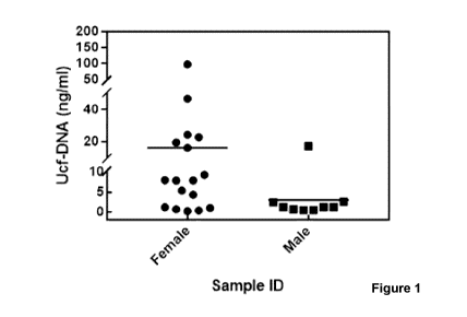
[0020] Figure
1 is a chart illustrating urinary cell-free DNA (UcfDNA) from
female and male donors, which shows that the amount of UcfDNA in urine samples
is
both sample and sex-dependent.
[0021] Figure
2A is a chart illustrating increasing turbidity of a non-stabilized
first morning, first void (FMFV) urine sample due to bacterial growth (as
evidenced
further by Figure 2B).
- 8 -
CA 03178146 2022-09-29
WO 2021/195768
PCT/CA2021/050428
[0022] Figure
2B is a chart illustrating ACt [Ct(r7)_Ct(ro)] determined from
bacterial 16S and [3-globin qPCR assay for the quantification of bacterial and
human
cell-free DNA (cfDNA) content in unstabilized urine samples after 7 days at RT
(room
temperature).
[0023] Figures
20 and 2D illustrate results of Agilent 4200 Tapestation
analysis, showing a massive decline in human cell-free DNA content after 7
days at
room temperature.
[0024] Figures
3A, 3B and 30 are charts illustrating (i) stability and (ii)
neutrality as ACt [Ct(r7)-Ct(ro)] and ACt [Ct(TO Chem)-Ct(TO NA)] ,
respectively, and determined
from [3-globin qPCR assay for the quantification of human cfDNA content in
urine
samples at day 0, as well as after storage at RT for 7 days under various
conditions,
including in admixture with the aqueous stabilizing compositions of the
present
application. a (TO) and a (T7) denotes qPCR cycle threshold at day 0 and day
7,
respectively. Ct(TO Chem) and Ct(TO NA) denotes qPCR cycle threshold for urine
specimen
with chemistry and unpreserved specimen (NA), respectively at day 0.
[0025] Figures
4A and 4B are charts illustrating ACt [Ct(r) - Ct(TO NA)]
determined from [3-globin qPCR assay for the quantification of human cfDNA
content
in urine samples after storage at room temperature for 7 days under various
conditions, including in admixture with the aqueous stabilizing composition of
the
present application. Ct(r) denotes qPCR cycle threshold at day 7. Ct(ro NA)
denotes
qPCR cycle threshold for unpreserved specimen (NA) at day 0
[0026] Figure
5A illustrates (i) stability and (ii) neutrality as ACt [Ctcro-Ct(ro)]
and ACt [Ct(TO Chem)-Ct(TO NA)], respectively, and determined from [3-globin
qPCR assay
for the quantification of human cfDNA content in urine samples at day 0, as
well as
after storage at RT for 7 days under various conditions, including in
admixture with the
aqueous stabilizing composition of the present application. a (To) and a (T7)
denotes
qPCR cycle threshold at day 0 and day 7, respectively. Ct(TO Chem) and Ct(TO
NA) denotes
qPCR cycle threshold for urine specimen with chemistry and unpreserved
specimen
(NA), respectively at day 0. Figure 5B illustrates a representative
Tapestation profile
analysis of these unpreserved and Chemistry F (Chem F) containing urine
samples,
showing that cfDNA is degraded in unpreserved samples and is stabilized in the
aqueous stabilizing composition of the present application. Figures 5C, 5D and
5E are
- 9 -
CA 03178146 2022-09-29
WO 2021/195768
PCT/CA2021/050428
charts illustrating (i) stability and (ii) neutrality as ACt [Ctcp_Ct(ro)] and
ACt [Ct(TO Chem)-
Ct(TO NA)], respectively, and determined from [3-globin qPCR assay for the
quantification
of human cfDNA content in urine samples at day 0, as well as after storage at
RT for
7 or 14 days under various conditions, including in admixture with the aqueous
stabilizing composition of the present application. a (ro) and a (r) denotes
qPCR cycle
threshold at day 0 and day 7 or day 14, respectively. Ct(TO Chem) and Ct(ro
NA) denotes
qPCR cycle threshold for urine specimen with chemistry and unpreserved
specimen
(NA), respectively at day 0.
[0027] Figure
6(i) A & B are charts illustrating ACt [Ct(r)-Ct(roNA)] determined
from [3-globin qPCR assay for the quantification of human cfDNA content in
urine
samples spiked (S) with prostate cancer cells at day 0, as well as after
storage at RT
for 7 days under various conditions, including in admixture with the aqueous
stabilizing
composition of the present application. Ct(r) denotes qPCR cycle threshold at
day 0 or
day 7. Ct(TO NA) denotes qPCR cycle threshold for unpreserved spiked specimen
(NA)
at day 0. Figure 6(i)C illustrates representative Tapestation profile analysis
of the
unpreserved and Chemistry F (Chem F) containing urine samples. Figure 6(ii)A
is a
chart illustrating ACt [Ctcp_Ct(roNA)] determined from [3-globin qPCR assay
for the
quantification of human cfDNA content in urine samples spiked (S) with
prostate
cancer cells at day 0, as well as after storage at RT for 7 days under various
conditions,
including in admixture with the aqueous stabilizing composition of the present
application. Ct(r) denotes qPCR cycle threshold at day 0 or day 7. Ct(TO NA)
denotes
qPCR cycle threshold for unpreserved spiked specimen (NA) at day 0. Figure
6(ii)B is
a chart illustrating the number of copies of 13-globin gene per unit volume
for some of
these samples determined using ddPCR assay. Collectively Figures 6(i) and (ii)
suggest that the aqueous stabilizing composition of the present application
preserves
the integrity of prostate cancer cells in a concentration-dependent manner at
room
temperature for at least 7 days.
[0028] Figure
7 is a chart illustrating ACt [Ctcp_Ct(ro NA)] determined from p-
globin qPCR assay for the quantification of human cfDNA content in urine
samples
spiked (S) with nucleated white blood cells at day 0, as well as after storage
at RT for
7 days under various conditions, including in admixture with the aqueous
stabilizing
composition of the present application, as well as a commercially available
-10-
CA 03178146 2022-09-29
WO 2021/195768
PCT/CA2021/050428
composition from Streck. Ct(r) denotes qPCR cycle threshold at day 0 or day 7.
Ct(ro
NA) denotes qPCR cycle threshold value for unpreserved spiked specimen (NA) at
day
0.
[0029] Figure
8A shows Hpal I and Mspl restriction endonuclease digestion
pattern confirming in vitro plasmid DNA methylation using CpG Methyl
Transferase.
Figure 8B and 80 shows Tapestation results for FOR amplification of methylated
plasmid, suggesting preservation of DNA methylation in the present composition
for 7
days at RT.
[0030] Figure
9A and 9B are charts illustrating ACt [Ctcro-Ct(ro)] determined
from Ampicillin resistance gene (AmpR) and bacterial 16S qPCR assay for the
respective quantification of HPV plasmid DNA and bacterial DNA content in both
the
unpreserved and Chemistry F (Chem F) containing urine samples spiked with
purified
HPV16 plasmid DNA after storage at room temperature for 7 days. Ctcrn denotes
qPCR cycle threshold at day 7. Ct(TO) denotes qPCR cycle threshold at day 0.
[0031] Figure
10A illustrates (i) stability and (ii) neutrality as ACt [Ctcrn_Ct(ro)]
and ACt [Ct(TO Chem)-Ct(TO NA)], respectively, and determined from 8-actin RT-
qPCR assay
for the quantification of human EV RNA content in urine samples at day 0, as
well as
after storage at RT for 7 days under various conditions, including in
admixture with the
aqueous stabilizing composition of the present application. a (To) and a (T7)
denotes
qPCR cycle threshold at day 0 and day 7, respectively. Ct(TO Chem) and Ct(TO
NA) denotes
qPCR cycle threshold for urine specimen with chemistry and unpreserved
specimen
(NA), respectively at day 0. Figure 10B illustrates representative
electropherogram
traces of extracellular vesicles (EV) RNA from both unpreserved and Chemistry
F
(Chem F) containing urine specimens at day 0 and day 7. Figure 100 and 10D
illustrate (i) stability and (ii) neutrality as ACt [Ctcro-Ct(ro)] and ACt
[Ct(TO Chem)-Ct(TO NA)],
respectively, and determined from 8-actin RT-qPCR assay for the quantification
of
human EV RNA content in urine samples at day 0, as well as after storage at RT
for 7
days under various conditions, including in admixture with the aqueous
stabilizing
composition of the present application. a (To) and a (T7) denotes qPCR cycle
threshold
at day 0 and day 7, respectively. Ct(TO Chem) and Ct(TO NA) denotes qPCR cycle
threshold
for urine specimen with chemistry and unpreserved specimen (NA), respectively
at
day 0.
-11 -
CA 03178146 2022-09-29
WO 2021/195768
PCT/CA2021/050428
[0032] Figure 11 is a chart illustrating (i) stability and (ii)
neutrality as ACt
[Ct(T7)-Ct(T0)] and ACt [Ct(TO Chem)-Ct(TO NA)], respectively, and determined
from p-actin RT-
qPCR assay for the quantification of human cell free RNA content in urine
samples at
day 0, as well as after storage at RT for 7 days under various conditions,
including in
admixture with the aqueous stabilizing composition of the present application.
Ct (TO)
and a (T7) denotes qPCR cycle threshold at day 0 and day 7, respectively.
Ct(TO Chem)
and Ct(TO NA) denotes qPCR cycle threshold for urine specimen with chemistry
and
unpreserved specimen (NA), respectively at day 0.
[0033] Figure 12A and 12B are charts illustrating (i) stability and
(ii)
neutrality as ACt [Ct(T7)-Ct(To)] and ACt [Ct(TO Chem)-Ct(TO NA)],
respectively, and determined
from p-actin RT-qPCR assay for the quantification of cellular RNA content in
urine
samples at day 0, as well as after storage at RT for 7 days under various
conditions,
including in admixture with the aqueous stabilizing composition of the present
application. a (TO) and a (T7) denotes qPCR cycle threshold at day 0 and day
7,
respectively. Ct(TO Chem) and Ct(TO NA) denotes qPCR cycle threshold for urine
specimen
with chemistry and unpreserved specimen (NA), respectively at day 0.
[0034] Figure 13A illustrates the Tapestation profile of day 0 and day
7
extracted cellular DNA in both unpreserved (NA) and Chemistry F (Chem F)
containing
urine specimens admixed with the aqueous stabilizing composition of the
present
application. Figure 13B shows Tapestation profile of PCR amplified GAPDH
product.
Figure 13C illustrates % bacterial DNA content determined from bacterial 16S
qPCR
assay.
[0035] Figure 14 illustrates the Tapestation profile of day 0 and day 7
extracted cfDNA in both unpreserved (TE) and Chemistry F containing saliva
specimens admixed with the aqueous stabilizing composition of the present
application. TE stands for 1X Tris-EDTA buffer.
DETAILED DESCRIPTION OF THE INVENTION
[0036] Definitions
- 12-
CA 03178146 2022-09-29
WO 2021/195768
PCT/CA2021/050428
[0037] Unless
defined otherwise, all technical and scientific terms used
herein have the same meaning as commonly understood by one of ordinary skill
in the
art to which this invention belongs.
[0038] As used
in the specification and claims, the singular forms "a", "an"
and "the" include plural references unless the context clearly dictates
otherwise.
[0039] The term
"comprising" as used herein will be understood to mean
that the list following is non-exhaustive and may or may not include any other
additional suitable items, for example one or more further feature(s),
component(s)
ingredient(s) and/or elements(s) as appropriate.
[0040] Terms of
degree such as "substantially", "about" and "approximately"
as used herein mean a reasonable amount of deviation of the modified term such
that
the end result is not significantly changed. These terms of degree should be
construed
as including a deviation of at least 10% of the modified term if this
deviation would
not negate the meaning of the word it modifies.
[0041] The term
"bodily fluid" as used herein will be understood to mean a
naturally occurring fluid from a human or an animal, and includes, but is not
limited to
urine, saliva, sputum, serum, plasma, blood, pharyngeal, nasal/nasal
pharyngeal and
sinus secretions, mucous, gastric juices, pancreatic juices, bone marrow
aspirates,
cerebral spinal fluid, feces, semen, products of lactation or menstruation,
cervical
secretions, vaginal fluid, tears, or lymph. In one embodiment, the bodily
fluid is
selected from urine or saliva. In another embodiment, the bodily fluid is
urine.
[0042] The term
"ambient temperature" as used herein refers to a range of
temperatures that could be encountered by the mixture of a bodily fluid (e.g.
urine
sample) and the aqueous stabilizing composition described herein from the
point of
collection, during transport (which can involve relatively extreme
temperatures, albeit
usually for shorter periods of time (e.g. <5 days)), as well as during
prolonged storage
prior to analysis. In one embodiment, the ambient temperature is ranging from
about
-20 C to about 50 C. In another embodiment, the ambient temperature is room
temperature (RT) and ranges from about 15 C to about 25 C.
[0043] The term
"monosaccharide" as used herein will be understood to
mean a sugar that is not decomposable into simpler sugars by hydrolysis, is
classed
-13-
CA 03178146 2022-09-29
WO 2021/195768
PCT/CA2021/050428
as either an aldose or ketose, and contains one or more hydroxyl groups per
molecule.
In one embodiment, the monosaccharide is selected from fructose, glucose,
mannose,
or galactose. In another embodiment, the monosaccharide is fructose, glucose,
or a
combination thereof.
[0044] The term
"disaccharide" as used herein will be understood to mean
a compound in which two monosaccharide units are joined by a glycosidic
linkage. In
one embodiment, the disaccharide is selected from sucrose, trehalose, and
lactose.
In another embodiment, the disaccharide is sucrose.
[0045] It has
been found that compositions according to the present
application comprising disaccharides can be more difficult to prepare, as such
solutions may have very high viscosities which can lead to improper mixing of
the
components and/or addition to the specimen (i.e. bodily fluid) due to
difficulties in
mixing. Overall, due to workability of the samples, monosaccharides are
preferred
over disaccharides for the compositions and methods of the present
application.
[0046] The term
"chelator" or "chelating agent" as used herein will be
understood to mean a chemical that will form a soluble, stable complex with
certain
metal ions (e.g., Ca2+ and Mg2+), sequestering the ions so that they cannot
normally
react with other components, such as deoxyribonucleases (DNases) or
endonucleases (e.g. type I, ll and III restriction endonucleases) and
exonucleases
(e.g. 3' to 5' exonuclease), enzymes which are abundant in various body fluid
samples.
In the present composition, chelating agent(s) participates in the inhibition
of DNases
and microbial growth in biological samples. A chelator can be, for example,
ethylene
glycol tetraacetic acid (EGTA), (2-hydroxyethyl)ethylenediaminetriacetic acid
(HEDTA), diethylene triamine pentaacetic acid (DTPA), nitrilotriacetic acid
(NTA),
ethylenediaminetriacetic acid (EDTA), 1,2-cyclohexanediaminetetraacetic acid
(CDTA), N,N-bis(carboxymethyl)glycine, triethylenetetraamine (TETA),
tetraazacyclododecanetetraacetic acid (DOTA), desferioximine, citrate
anhydrous,
sodium citrate, calcium citrate, ammonium citrate, ammonium bicitrate, citric
acid,
diammonium citrate, ferric ammonium citrate, and lithium citrate. These
chelating
agents may be used singly or in combination of two or more thereof.
[0047] The term
"01-06 alkanol" as used herein will be understood to mean
straight-chain or branched, such as methanol, ethanol, propanol, isopropanol,
butanol,
- 14-
CA 03178146 2022-09-29
WO 2021/195768
PCT/CA2021/050428
n-butanol, pentanol, hexanol, or any combination thereof. In one embodiment of
the
present composition, the preferred alcohol is ethanol.
[0048] In one
embodiment, there is provided an aqueous stabilizing
composition for preserving a bodily fluid at ambient temperature, the
composition
comprising: a sugar selected from a monosaccharide, a disaccharide, or a
combination thereof; a buffering agent; a 01-06 alkanol, boric acid, a salt of
boric acid,
or a combination thereof; and a chelating agent; wherein the composition has a
pH of
from 4.5 to 5.2.
[0049] In one
embodiment, the aqueous composition comprises boric acid;
a salt of boric acid, such as, for example, dihydrogen borate, hydrogen
borate,
diborate, triborate, tetraborate, metaborate, hydroxoborate, borate salts; or
a
combination thereof. In another embodiment, the aqueous composition comprises
boric acid, sodium borate, or a combination thereof. In yet another
embodiment, the
aqueous composition comprises boric acid. In one embodiment, the boric acid,
the
salt of boric acid or the combination thereof is present in the aqueous
stabilizing
composition in an amount of from about 0.5% to about 5% (wt/vol), or from
about 1%
to about 3% (wt/vol), or from about 2% to about 2.5% (wt/vol), or about 2.2%
(wt/vol).
[0050] In one
embodiment, the sugar is a monosaccharide, such as, for
example, fructose, glucose, mannose, galactose, or a combination thereof. In
another
embodiment, the monosaccharide is fructose, glucose, or a combination thereof.
In
another embodiment, the sugar is a disaccharide, such as, for example,
trehalose,
lactose, or sucrose, or a combination thereof. In
another embodiment, the
disaccharide is sucrose. In one embodiment, the sugar is present in the
aqueous
stabilizing composition in an amount of from about 5% to about 45% (wt/vol),
of from
about 5% to about 40% (wt/vol), or from about 10% to about 30% (wt/vol), or
from
about 18% to about 22% (wt/vol), or about 20% (wt/vol).
[0051] In
general, the pH of the present aqueous stabilizing composition
can be maintained in the desired range using one or more appropriate buffering
agents. In accordance with one embodiment, the composition comprises one, two,
or
more buffering agents (non-limiting examples being acetate buffer and citrate
buffer,
such as sodium acetate, potassium acetate, ammonium acetate, sodium citrate,
and
ammonium citrate) with pKa values, logarithmic acid dissociation constants, at
25 C
-15-
CA 03178146 2022-09-29
WO 2021/195768
PCT/CA2021/050428
ranging from 3 to 6.5 to maintain the pH within the preferred range of 4.5 to
5.2. In
one embodiment, the buffering agent is sodium acetate.
[0052] An acid
dissociation constant, Ka, is a quantitative measure of the
strength of an acid in solution. The larger the Ka value, the more
dissociation of the
molecules in solution and thus the stronger the acid. Due to the many orders
of
magnitude spanned by Ka values, a logarithmic measure of the acid dissociation
constant, pKa, is more commonly used in practice. The larger the value of pKa,
the
smaller the extent of dissociation at any given pH, i.e., the weaker the acid.
In living
organisms, acid-base homeostasis and enzyme kinetics are dependent on the pKa
values of many acids and bases present in the cell and in the body. In
chemistry,
knowledge of pKa values is necessary for the preparation of buffer solutions
and is
also a prerequisite for a quantitative understanding of the interaction
between acids or
bases and metal ions to form complexes. One skilled in the art will understand
that a
given compound/buffer can buffer the pH of a solution only when its
concentration is
sufficient and when the pH of the solution is close (within about one pH unit)
to its pKa.
In one embodiment, the pH of the present composition is in the range of 4.5 to
5.2. In
a preferred embodiment, the pH of the composition is about 5Ø The amount of
buffering agent(s) in the aqueous stabilizing composition can be of from about
150
mM to about 1.75 M, or from about 150 mM to about 1.5 M, or from about 500 mM
to
about 1.2 M, or from about 0.7 M to about 0.8 M, or about 0.75 M, for example.
[0053] In one
embodiment, the 01-06 alkanol in the aqueous stabilizing
composition is selected from methanol or ethanol. In another embodiment, the
01-06
alkanol is ethanol. In yet another embodiment, the 01-06 alkanol is present in
the
aqueous stabilizing composition in an amount of from about 5% to about 50%
(vol/vol),
or from about 10% to about 30% (vol/vol), or from about 20% to about 25%
(vol/vol),
or about 23% (VOI/V01).
[0054] Ethanol
causes dehydration of proteins or a reduction in water
activity, followed by electrostatic attraction between proteins, aggregation
and
insolubilization. While wishing to not be bound by theory, the inventor
believes that
ethanol, at the percentage used, has little to no fixative properties in this
composition;
rather, it is important for overall stability and enhances the functionality
of other
chemical compounds which may be included in the present composition. In
addition,
- 16-
CA 03178146 2022-09-29
WO 2021/195768
PCT/CA2021/050428
for shipping/transport of flammable liquids, it is desirable to keep organic
solvents,
such as ethanol, below 24% by volume in solutions for exemption from Transport
of
Dangerous Goods (TDG) regulations (United Nations (UN) number 1170); otherwise
a solution with >24% ethanol is classified as class 3 (flammable liquids),
special
packaging is mandated, and transport complexity and costs increase. As such,
an
aqueous stabilizing composition comprising about 23% (vol/vol) or lower is
particularly
advantageous.
[0055] In
another embodiment, the chelating agent in the aqueous
stabilizing composition is selected from, for example,
ethylenediaminetriacetic acid
(EDTA), 1,2-cyclohexanediamine tetraacetic acid (CDTA), diethylenetriamine
pentaacetic acid (DTPA),
tetraazacyclododecanetetraacetic acid (DOTA),
tetraazacyclotetradecanetetraacetic acid (TETA), desferioximine, or chelator
analogs
thereof. In another embodiment, the chelating agent is CDTA. In
another
embodiment, the chelating agent is present in the aqueous stabilizing
composition in
an amount of from about 10 mM to about 120 mM, or from about 10 mM to about
100
mM, or from about 30 mM to about 70 mM, or from about 40 mM to about 60 mM, or
about 50 mM.
[0056] In one
embodiment of the aqueous stabilizing composition, the
composition comprises, consists essentially of, or consists of: the sugar
(such as
fructose, glucose, sucrose, or a combination thereof; preferably fructose,
glucose, or
a combination thereof) in an amount of from about 5% to about 45% (wt/vol), of
from
about 5% to about 40% (wt/vol), or from about 10% to about 30% (wt/vol), or
from
about 18% to about 22% (wt/vol), or about 20% (wt/vol), the buffering agent
(such as,
for example, sodium acetate) in an amount of from about 150 mM to about 1.75
M, or
from about 150 mM to about 1.5 M, or from about 500 mM to about 1.2 M, or from
about 0.7 M to about 0.8 M, or about 0.75 NA; the 01-06 alkanol (such as
methanol,
ethanol, or a combination thereof; preferably ethanol) in an amount of from
about 5%
to about 50% (vol/vol), or from about 10% to about 30% (vol/vol), or from
about 20%
to about 25% (vol/vol), or about 23% (vol/vol), the boric acid, the salt of
boric acid or
the combination thereof (preferably boric acid) in an amount of from about
0.5% to
about 5% (wt/vol), or from about 1% to about 3% (wt/vol), or from about 2% to
about
2.5% (wt/vol), or about 2.2% (wt/vol), and the chelating agent (such as CDTA)
in an
amount of from about 10 mM to about 120 mM, or from about 10 mM to about 100
-17-
CA 03178146 2022-09-29
WO 2021/195768
PCT/CA2021/050428
mM, or from about 30 mM to about 70 mM, or from about 40 mM to about 60 mM, or
about 50 mM.
[0057] In one
embodiment, the aqueous stabilizing composition stabilizes
cells (such as cancer cells or nucleated blood cells), extracellular vesicles,
nucleic
acids (e.g. cellular DNA and RNA, such as cell-free DNA (cfDNA), cell-free RNA
(cfRNA), and extracellular vesicle RNA (EV RNA)), and/or microorganisms (such
as
bacteria or viruses) contained in the bodily fluid.
[0058] In
another embodiment, there is provided a method for preserving a
bodily fluid, the method comprising: a) obtaining a sample of the bodily
fluid; b)
contacting the bodily fluid with the aqueous stabilizing composition as
defined above
to form a mixture; c) mixing the mixture of (b) to form a homogeneous mixture;
and d)
storing the homogeneous mixture at ambient temperature. In one embodiment,
preserving the bodily fluid comprises stabilizing cells (such as cancer cells
or
nucleated blood cells), extracellular vesicles, nucleic acids (e.g. DNA and
RNA, such
as cell-free DNA (cfDNA), cell-free RNA (cfRNA), and extracellular vesicle RNA
(EV
RNA)), and/or microorganisms (such as bacteria or viruses) contained in the
bodily
fluid. In another embodiment, the cells, nucleic acids, extracellular
vesicles, and/or
microorganisms contained in the bodily fluid are stabilized for at least 7
days at
ambient temperature. In another embodiment, the cells, nucleic acids,
extracellular
vesicles, and/or microorganisms contained in the bodily fluid are stabilized
for at least
14 days at ambient temperature. In another embodiment, the bodily fluid is
urine or
saliva. In another embodiment, the bodily fluid is urine.
[0059] In yet
another embodiment, there is provided an aqueous
composition comprising: a sugar selected from a monosaccharide, a
disaccharide, or
a combination thereof; a buffering agent; a 01-06 alkanol, boric acid, a salt
of boric
acid, or a combination thereof; a chelating agent; and a bodily fluid. In
one
embodiment, the bodily fluid is urine. In another embodiment, the bodily fluid
is urine
and the pH of the aqueous composition comprising the bodily fluid is between 5
and
5.5. In another embodiment, the sugar is present in an amount of from about
1.5% to
about 15% (wt/vol), or from about 2% to about 10% (wt/vol), or from about 5%
to about
7% (wt/vol), or about 6% (wt/vol), the buffering agent is present in an amount
of from
about 50 mM to about 500 mM, or from about 200 mM to about 400 mM, or from
about
-18-
CA 03178146 2022-09-29
WO 2021/195768
PCT/CA2021/050428
220 mM to about 240 mM, or about 230 mM, or about 225 mM, the 01-06 alkanol is
present in an amount of from about 2% to about 40% (vol/vol), or from about 3%
to
about 20% (vol/vol), or from about 5% to about 10% (vol/vol), or about 6.5%
(vol/vol),
or about 6.9% (vol/vol), the boric acid, the salt of boric acid or the
combination thereof
is present in an amount of from about 0.1% to about 2% (wt/vol), or from about
0.2%
to about 1.5% (wt/vol), or from about 0.5% to about 1.0% (wt/vol), or about
0.7%
(wt/vol), or about 0.6% (wt/vol), and the chelating agent is present in an
amount of
from about 2.5 mM to about 50 mM, or from about 5 mM to about 25 mM, or from
about 10 mM to about 20 mM, or about 16 mM, or about 15 mM.
[0060] In one
embodiment, the bodily fluid is urine and the urine sample is
collected using a device for capturing a predetermined volume of a predefined
portion
of urine (e.g. first void), such as that described in W02014037152 entitled
"LIQUID
SAMPLER, KIT OF PARTS, AND METHOD FOR ASSEMBLY". In one embodiment,
the Colli-Pee First Void Urine Collection Device (Novosanis) can be used. The
aqueous stabilizing composition can be present in the device at the time of
collection,
or the urine can be contacted with the aqueous stabilizing composition
immediately
post-collection. The reservoir containing the urine sample and aqueous
stabilizing
composition can be sealed with an appropriate cap, and the combined sample and
stabilizing composition can be gently mixed, for example by inverting the
tube. Urine
samples can also be collected in standard urine specimen containers (e.g.
VWIR, Cat.
No. 10804-050) and then mixed with the stabilizing composition. Alternatively,
collected urine can be transported to the laboratory on ice packs where it can
be mixed
with the present stabilizing composition.
[0061] In
another embodiment, the bodily fluid is saliva and the saliva
sample is collected using a device such as, for example, those described in
W02007/068094 entitled "CONTAINER SYSTEM FOR RELEASABLY STORING A
SUBSTANCE", W02010/020043 entitled "SAMPLE RECEIVING DEVICE", and
W02010/130055 entitled "CLOSURE, CONTAINING APPARATUS, AND METHOD
OF USING SAME".
[0062] In
another embodiment, the bodily fluid is feces, and the fecal
sample is collected using a device such as that described in W02015172250
entitled
"DEVICE FOR COLLECTING, TRANSPORTING AND STORING BIOMOLECULES
FROM A BIOLOGICAL SAMPLE".
-19-
CA 03178146 2022-09-29
WO 2021/195768
PCT/CA2021/050428
[0063] In
still another embodiment, the sample of the bodily fluid can be
collected in a standard, commercially-available laboratory or transport tube
(e.g. 10
mL round-bottom tube (92 x 15.3 mm), Cat. No. 60.610; Sarstedt, or larger tube
depending on the sample type and size). The tube containing the sample of the
bodily
fluid and aqueous stabilizing composition can be sealed with an appropriate
cap, and
the combined sample and stabilizing composition can be gently mixed, for
example by
inverting the tube.
[0064] Bodily
fluid should preferably be mixed immediately with the
stabilizing composition at the point of collection. Otherwise, samples should
be stored
and/or transported on ice packs or refrigerated before mixing with the
composition.
[0065] As the
skilled worker will appreciate, the aqueous stabilizing
composition ("chemistry") described herein can be combined with the sample of
the
bodily fluid in a variety of ratios. For example, where the bodily fluid is
urine, it is
desirable to avoid overly diluting the sample and thus reducing the analytes
collected;
thus, the ratio of chemistry:urine can range, for instance, from 0.25:1 to
0.75:1 - e.g.
0.25:1, 0.30:1, 0.35:1, 0.40:1, 0.45:1, 0.50:1, 0.55:1, 0.60:1, 0.65:1,
0.70:1, 0r0.75:1.
In one embodiment, the ratio of chemistry:urine is 0.40:1 to 0.45:1.
[0066] For
other bodily fluids, such as feces, in order to ensure sufficient
mixing, higher ratios of chemistry:sample can be used.
[0067] In one
embodiment, following the step of contacting the bodily fluid
with the aqueous stabilizing composition and mixing to form a homogeneous
mixture,
the homogenous mixture then comprises: the sugar (such as fructose, glucose,
sucrose, or a combination thereof; preferably fructose, glucose, or a
combination
thereof) in an amount of from about 1.5% to about 15% (wt/vol), or from about
2% to
about 10% (wt/vol), or from about 5% to about 7% (wt/vol), or about 6%
(wt/vol), the
buffering agent (such as, for example, sodium acetate) in an amount of from
about 50
mM to about 500 mM, or from about 200 mM to about 400 mM, or from about 220 mM
to about 240 mM, or about 230 mM, or about 225 mM, the 01-06 alkanol (such as
methanol, ethanol, or a combination thereof; preferably ethanol) in an amount
of from
about 2% to about 40% (vol/vol), or from about 3% to about 20% (vol/vol), or
from
about 5% to about 10% (vol/vol), or about 6.5% (vol/vol), or about 6.9%
(vol/vol), the
boric acid, the salt of boric acid or the combination thereof (preferably
boric acid) in an
- 20 -
CA 03178146 2022-09-29
WO 2021/195768
PCT/CA2021/050428
amount of from about 0.1% to about 2.2% (wt/vol), or from about 0.2% to about
1.5%
(wt/vol), or from about 0.5% to about 1.0% (wt/vol), or about 0.7% (wt/vol) or
about
0.6% (wt/vol), and the chelating agent (preferably CDTA) in an amount of from
about
2.5 mM to about 50 mM, or from about 5 mM to about 25 mM, or from about 10 mM
to about 20 mM, or about 16 mM, or about 15 mM.
[0068] As
noted above, in one embodiment, the aqueous stabilizing
composition stabilizes cells (such as cancer cells or nucleated blood cells),
extracellular vesicles, nucleic acids (e.g. DNA and RNA, such as cell-free DNA
(cfDNA), cell-free RNA (cfRNA), and extracellular vesicle RNA (EV RNA)),
and/or
microorganisms (such as bacteria or viruses) contained in the bodily fluid. In
one
embodiment, the aqueous stabilizing composition stabilizes such components of
the
bodily fluid for at least 7 days at ambient temperature. In another
embodiment, the
aqueous stabilizing composition stabilizes such components of the bodily fluid
for at
least 14 days at ambient temperature. Such stabilization can be assessed by
methods
known to those skilled in the art, such as via monitoring the degradation of
cell-free
nucleic acids (described further in the Materials and Methods section, and in
the
Examples which follow).
[0069] ACt
corresponds to the relative change in the amount or expression
of a given gene. ACt corresponds to Ctcp-Ct(ro), where Ct(r) stands for cycle
threshold
at day 7 or day 14 while Ct(ro) denotes cycle threshold at day 0. Cycle
threshold
(Ct) value of a reaction is defined as the cycle number when the fluorescence
of a FOR
product can be detected above the background signal. In the present studies,
this ACt
when calculated as Ct(T7 or T14)-Ct(TO) accounts for the change in the
stability of different
analytes in unpreserved and preserved samples after storage at room
temperature for
a specified amount of time. ACt when calculated as Ct(TO Chem)-Ct(TO NA)
accounts for the
neutrality (change in the basal concentration of analytes with the addition of
a given
chemistry in the urine samples relative to the unpreserved urine samples at
the time
of collection, i.e. Day 0). Unchanged ACt values or ACt values close to 0 are
indicative
of stability, as this means that the concentration of analyte is not
significantly changing
over the course of time (and thus is indicative of the stability of the
analyte in the
composition under the testing conditions). For example, in the present cell-
free DNA
studies, ACt value ranged from +2 to +14 in unpreserved samples held at RT for
7
days. This marked increase in ACt value (median value: >+5) is indicative of
-21 -
CA 03178146 2022-09-29
WO 2021/195768
PCT/CA2021/050428
degradation of cell-free DNA in unpreserved samples. On the other hand, ACt
median
value for the detection of cell-free DNA after storage at room temperature in
the
present aqueous stabilizing composition was almost zero, indicating
preservation of
cell-free DNA stability and content and also indirectly accounts for cellular
stability and
integrity. For cell-free RNA, median ACt value of +2.5 in unpreserved samples
is
indicative of cell-free RNA degradation, while relatively lower median ACt
value of 1.3
is indicative of better stability of cell-free RNA content in preserved
samples when
compared to unpreserved samples. For cellular RNA stability, a median ACt
value of
+7.0 in unpreserved samples is indicative of marked degradation of cellular
RNA. On
the other hand, median ACt value of less than 2 in preserved samples indicate
cellular
RNA stability. Similarly, for EV RNA, a median ACt value of more than +3 in
unpreserved samples is indicative of instability and compromised detection of
EV
RNA, while a median ACt value of 0.5 in preserved samples indicates excellent
EV
RNA stability and detection. This is merely one exemplary method of assessing
stabilization of cells, extracellular vesicles, nucleic acids, and/or
microorganisms in
bodily fluids, and other methods of assessing such stabilization are known to
the
skilled worker and/or are outlined in further detail in the Materials and
Methods section
and Examples described below.
[0070] As
described in further detail in Example 7 below, preservative
agents/compositions containing formalin/formaldehyde-based fixatives may be
used
to fix cells in biological samples or specimens and prevent leaking of
cellular nucleic
acids into the extracellular space. Such compositions may contain
formaldehyde, or
alternatively compounds capable of releasing an aldehyde, such as a
formaldehyde
releaser/formaldehyde donor/formaldehyde-releasing preservative which is a
chemical compound that slowly releases formaldehyde. Notably, when compared to
the DNA isolated from frozen tissues, formalin-fixed tissues exhibit a high
frequency
of non-reproducible sequence alteration (Srinivasan M, Sedmak D, Jewell S
(2002)
Effect of fixatives and tissue processing on the content and integrity of
nucleic acids.
Am J Pathol 161(6): 1961-1971). Formaldehyde, a principal ingredient of most
commonly used fixatives, leads to the generation of DNA-protein and RNA-
protein
cross-linkages. Furthermore, the nucleic acids will fragment in situations
where the
fixative solution is not buffered. Both of the above provide challenges for
FOR-based
analyses (Gilbert MTP, Haselkorn T, Bunce M, Sanchez JJ, Lucas SB, Jewell LD,
Van
- 22 -
CA 03178146 2022-09-29
WO 2021/195768
PCT/CA2021/050428
Merck E, Worobey M (2007) The isolation of nucleic acids from fixed, paraffin-
embedded tissues ¨ Which methods are useful when? PLoS ONE 2(6): e537. Doi:
10.1371/joumal.pone.0000537, Wong SQ, Li J, Tan AY-C, Vedururu R, Pang J-MB,
Do H, Ellul J, Doig K, Bell A, MacArthur GA, Fox SB, Thomas DM, Fellowes A,
Parisot
JP, Dobrovic A (2014) Sequence artifacts in a prospective series of formalin-
fixed
tumours tested for mutations in hotspot regions by massively parallel
sequencing.
BMC Medical Genomics 7:23. Doi: 10.1186/1755-8794-7-23). Specifically, this
chemical damage to DNA reduces Taq DNA polymerase fidelity and PCR
amplification
efficiency (Sikorsky JA, Primerano DA, Fenger TW, Denvir J (2007) DNA damage
reduces Taq DNA polymerase fidelity and PCR amplification efficiency. Biochem
Biophys Res Commun 355(2): 431-437). Hence, formalin/formaldehyde-based
fixatives are not ideal for molecular analyses. Thus, an advantage of the
aqueous
stabilizing composition and method for preserving a bodily fluid at ambient
temperature as disclosed herein is that the compositions and methods of the
present
application do not require the use of formaldehyde, or compounds/components
capable of releasing an aldehyde such as formaldehyde releasers, formaldehyde
donors or formaldehyde-releasing preservatives.
[0071] EXAMPLES
[0072] Materials and Methods
[0073] Cell-free nucleic acids extraction:
[0074] Cell-free nucleic acids extraction was performed using QiaAmp
Circulating Nucleic Acid Extraction Kit (Qiagen, Cat. No. 55114) according to
manufacturer's protocol. First morning, First Void (FMFV) human urine, random
mid-
day first void (FV) urine samples and saliva samples were centrifuged down at
3000g-
3800g for 10-20 minutes at room temperature (RT) and the cleared supernatant
(2-4
mL) was used for cell-free nucleic acids extraction. Extracted cell-free
nucleic acids
profile was assessed on 4200 Agilent Tapestation platform using HS D5000 tapes
(Agilent, Cat. No. 5067-5592) and reagents (Agilent, Cat. No.5067-5593)
according to
manufacturer's instructions.
[0075] Urinary extracellular vesicles (EV) RNA extraction:
- 23 -
CA 03178146 2022-09-29
WO 2021/195768
PCT/CA2021/050428
[0076] Urine
EV RNA extraction was performed using exoRNeasy Maxi Kit
(Qiagen, Cat. No. 77164) or Ultrafiltration. Urine Samples were precleared by
centrifugation at 3000xg for 10 minutes at RT, followed by filtration of
supernatant
using 0.80 pm syringe filter (Sartorius0 Minisart NMLO, Cat. No. 16592, or
Millipore
Millex0-AA, Cat. No. SLAA033SB) prior to EV isolation and > 200 nucleotide
(nt) long
RNA extraction according to manufacturer's instructions (Supplemental
Information:
Purification of exosomal RNA, including miRNA, from urine using the exoRNeasy
Serum/Plasma Midi/Maxi Kit). EVs and EV RNA isolation using Ultrafiltration
was
performed using AMICON Ultra-15 centrifugal units with Ultracel-100
regenerated
cellulose membrane (Millipore-Sigma; Cat. No. UF0910024) as follows:
[0077] 1.
Empty Ultracel-100 15 mL columns were washed with 1X PBS
pH 7.4 (Thermo fisher Scientific; Cat. No. 10010023) using centrifugation at
4000 g
for 5 mins at room temperature (RT).
[0078] 2.
Precleared and filtered urine samples were concentrated using
Ultracel-100 columns by performing centrifugation at 4000 g for 10 mins at RT
and the
resulting filtrate was discarded.
[0079] 3.
Ultracel-100 15 mL columns filter with retained concentrated urine
were washed with 1X PBS pH 7.4 (Thermo fisher Scientific; Cat. No. 10010023)
by
centrifugation at 4000 g for 5 mins at RT.
[0080] 3. 700
pL of QIAzol Lysis Reagent Qiagen, Cat. No.79306) was
added directly to the washed Ultracel-100 filter for the lysis of captured EVs
for EV
RNA extraction. The filter columns were transferred to new 50 mL Falcon tubes;
vortexed for 10 sec, incubated at RT for 5 mins followed by centrifugation at
4000 g
for 5 mins at RT.
[0081] 4. The
resulting filtrate and the reminiscent lysate retained on the
filter was collected for EV RNA isolation. Add 100 pL of chloroform and vortex
vigorously. Let stand for 2-5 minutes at RT.
[0082] 5.
Centrifuge at 12,000 x g for 15 minutes at 4 C.Transfer -400 pL
of aqueous phase to a new tube.
- 24 -
CA 03178146 2022-09-29
WO 2021/195768
PCT/CA2021/050428
[0083] 6. Add
400 pL (equal volume) of 70% ethanol and mix properly prior
to transfer of the mixture to Qiagen RNeasy MinElute columns. Centrifuge at
8,000 x
g for 30 seconds at RT. Discard the filtrate
[0084] 7. Add
700 pL of Buffer RVVT (Qiagen) to the columns. Centrifuge at
8,000 x g for 30 seconds at RT. Discard the filtrate.
[0085] 8. Add
500 pL of Buffer RPE (Qiagen) to the columns. Centrifuge at
8,000 x g for 30 seconds at RT. Discard the filtrate.
[0086] 9. Add
500 pL of Buffer RPE (Qiagen) to the columns. Centrifuge at
8,000 x g for 2 minutes at RT. Discard the filtrate and transfer the empty
columns to
new 2 mL collection tubes (Qiagen). Centrifuge the columns with lids open at
maximum speed for 5 mins to dry the membrane.
[0087] 10. Add
20 pL of RNase-free water to the center of the dried spin
columns. Let the columns stand at RT for 1 mins followed by centrifugation at
maximum speed for 1 min at RT.
[0088] 11.
Store the collected RNA samples at -80 C until quantification
and downstream processing.
[0089] 12.
Extracted EV RNA samples were quantified on Agilent 2100
Bioanalyzer using Agilent RNA 6000 Pico Kit (Cat. No. 5067-1513) according to
the
manufacturer's instructions and/or Ribogreen quantification analysis using
Quant-iT
Ribogreen RNA Assay Kit (Thermo Fisher Scientific, Cat. No. R11490) for
downstream
cDNA preparations.
[0090] 16S qPCR assay:
[0091]
Extracted nucleic acids from urine samples were subjected to qPCR
assay for the quantification of bacterial DNA content using 2X iTaq Universal
SYBR
Mastermix (Bio-Rad, Cat. No. 1725121). The primers and qPCR conditions of the
Bacterial 16s rRNA are as follows: BacrRNA173-Forward primer 5'
ATTACCGCGGCTGCTGG 3' (SEQ ID NO: 1), BacrRNA173-Reverse primer 5'
CCTACGGGAGGCAGCAG 3' (SEQ ID NO: 2) (DC Emery, DK Shoemark, TE
Blatstone, CM Waterfall, JA Coghill, TA Cerajewska, M Davies, NX West, SJ
Allen
(2017) 16S rRNA next generation sequencing analysis shows bacteria in
Alzheimer's
post-mortem brain. Frontiers in Aging Neuroscience 9: 195. Doi:
- 25 -
CA 03178146 2022-09-29
WO 2021/195768
PCT/CA2021/050428
10.3389/friagi.2017.00195). The amplification mixture (20 pL) contained: 10 pL
of 2X
iTaq Universal SYBR mastermix, 1 pL each of 10 pM forward and reverse primer,
6
pL of nuclease-free water (NFW from lnvitrogen, Cat. No. 10977023) and 2 pL of
extracted urinary cell-free nucleic acids. E.coli gDNA standards with serial
dilutions (1,
1:10, 1:100 and 1:1000) and a non-template control (2 pL of RNase/DNase-free
water)
were used in each qPCR run. FOR reactions were performed on a Bio-Rad 01000
Touch Thermal Cycler (#1851196) and conditions are as follows: 95 C: 5
minutes,
[95 C: 20 seconds, 56 C: 30 seconds] x45 cycles. Melt curves were obtained by
heating the samples from 65 C to 95 C by increments of 0.5 C and plate read
for 5
seconds at every increment. Bacterial cell-free DNA or cellular DNA
quantification
analysis was performed using "ACC which stands for [Ctcro-Ct(ro)]. "Ctcro" and
"Ct(TO)"
stands for qPCR cycle threshold at day 7 and day 0, respectively.
[0092] Human p-globin qPCR assay:
[0093] Extracted nucleic acids from urine samples were subjected to
qPCR
assay for the quantification of human cell-free DNA content using 2X iTaq
Universal
SYBR Mastermix (Bio-Rad, Cat. No. 1725121). The primers and the FOR conditions
of the human p-globin qPCR assay are described in the literature (M Jung, S
Klotzek,
M Lewandowski, M Fleischhacker, K Jung (2003) Changes in concentration of DNA
in
serum and plasma during storage of blood samples. Clinical Chem 49(6): 1028-
1029)
and are as follows: Forward primer: 5' ACACAACTGTGTTCACTAGC 3' (SEQ ID NO:
3), reverse primer: 5' CAACTTCATCCACGTTCACC 3' (SEQ ID NO: 4). The
amplification mixture (20 pL) contained: 10 pL of 2X iTaq Universal SYBR
mastermix,
1 pL each of 10 pM forward and reverse primer, 6 pL of nuclease-free water
(Invitrogen, Cat. No. 10977023) and 2 pL of extracted urinary cell-free
nucleic acids.
Human gDNA standards with serial dilution (1, 1:10, 1:100, 1:1000) and a non-
template control (2 pL of RNase/DNase-free water) were used in each qPCR run.
FOR
reactions were performed on a Bio-Rad 01000 Touch Thermal Cycler (#1851196)
and
conditions are as follows: 95 C: 5 minutes, [(95 C: 20 seconds, 56 C: 30
seconds)
x45 cycles]. Melt curves were obtained by heating samples from 65 C to 95 C by
increments of 0.5 C and plate read for 5 seconds at every increment. For
stability
assessment: Human cell-free DNA quantification analysis was performed using
"ACC
which stands for [Ctcp-Ct(ro)]. "Ctcp" stands for qPCR cycle threshold at day
7 or day
- 26 -
CA 03178146 2022-09-29
WO 2021/195768
PCT/CA2021/050428
14, while "Ct(ro)" represents qPCR cycle threshold at day 0 for both the
unpreserved
and chemistry containing urine samples. Cell-free DNA quantification relative
to
unpreserved day 0 (NA) sample was quantified using ACt calculations as [CtcD-
Ct(ro
NA)] where Ct(TO NA) represents qPCR cycle threshold for day 0 unpreserved
samples.
Furthermore, to assess neutrality (i.e. change in the basal concentration of
cell-free
DNA with the addition of a given chemistry in the urine samples at the time of
collection), ACt calculations were performed as [Ct(TO Chem)-Ct(TO NA)] where
Ct(TO Chem)
represents qPCR cycle threshold for day 0 urine samples with
chemistry/stabilization
solution.
[0094] In-vitro DNA methylation assay:
[0095] This assay was performed as described in the literature (C
Ernst, PO
McGowan, V Deleva, MJ Meaney, M Szyf, G Turecki (2008) The effects of pH on
DNA
methylation state: In vitro and post-mortem brain studies. J Neurosci Methods
174(1):123-125). pGL3-basic plasmid (Promega, Cat. No. E1751) contains 25 CCGG
sites. 1 pg of plasmid was treated with CpG methyl transferase (New England
Biolabs;
Cat. No. M0226S), an enzyme that methylates all cytosine nucleotides in a CpG
dinucleotide according to the manufacturer's protocol. To confirm the
methylation
status, methylated plasmid (pGL3-CH3) was subjected to restriction
endonuclease
digestions with: HpaII and Mspl. Both of these enzymes recognize the same site
(CCGG). While HpaII is blocked from cutting DNA when the internal C is
methylated;
Mspl is insensitive to the methylation status of the internal C. The in vitro-
methylated
pGL3 plasmid was column purified using Zymo Research's DNA Clean &
Concentrator-5 kit (Cat. No. D4013). An equal amount of the purified plasmid
was
either spiked into lx TE buffer pH 8.0 (positive control) or into male-pooled
and
female-pooled FMFV urine samples containing the composition of the present
invention and the reaction tubes were kept at RT for 7 days. Following
incubation, the
DNA samples under went bisulfite conversion using Qiagen EpiTec Bisulfite Kit
(Cat
No. 59104). Bisulfite treatment will create a sequence difference between un-
methylated plasmid (cytosines to uracil conversion) and methylated plasmid
(methylated cytosines will remain immune to conversion) (Y Li and TO
Tollefsbol
(2011) DNA methylation detection: Bisulfite genomic sequencing analysis.
Methods
Mol Biol 791: 11-21. Doi: 10.1007/978-1-61779-316-5_2). PCR experiment using
- 27 -
CA 03178146 2022-09-29
WO 2021/195768
PCT/CA2021/050428
methylated plasmid-specific primers would generate a 278 bp amplicon. Primers
were
used as described in Ernst et al. (2008) supra (Forward primer: 5'-
AAGATGTTTTTTTGTGATTGGT-3' (SEQ ID NO: 5); Reverse primer: 5'-
TTCCTATTTTTACTCA000AAA-3' (SEQ ID NO: 6)).
[0096] HPV Plasmid spike-in assay:
[0097] E. coil DH5a strain HPV16 plasmid (Human papilloma virus; type
16
clone) (ATCC Cat. No. 45113) was cultured in LB medium for the extraction of
HPV16
plasmid using ZymoPURE II Plasmid Maxi prep Sample Kit (Zymo Research, Cat.
Nos. D4202 & D4203). Extracted/purified plasmid was spiked in female-pooled
and
male-pooled first morning, first void urine samples at concentration (1-10
ng/mL), with
and without the preservative chemistry of the present invention. A 200 pL
aliquot of
each plasmid-spiked urine sample was processed for total DNA extraction using
QiaAmp DNA mini kit on QIAcube Connect. The amount of plasmid DNA in each
reaction tube and at different days (TO and T7) was quantified using a qPCR
assay for
the ampicillin resistance gene (AmpR) found on the HPV16 plasmid backbone. The
AmpR qPCR primers and conditions are as follows: Forward Primer (FP):
5'AGCCATACCAAACGACGAG 3' (SEQ ID
NO: 7); Reverse primer (RP):
5'AGCAATAAACCAGCCAGCC 3' (SEQ ID NO: 8). The amplification mixture (20 pL)
contained 10 pL of 2X iTaq Universal SYBR mastermix, 1 pL each of 10 pM
forward
and reverse primer, 6 pL of nuclease-free water (Invitrogen, Cat. No.
10977023) and
2 pL of extracted urinary nucleic acids. HPV16 plasmid standards with serial
dilution
(1, 1:10, 1:100, 1:1000) and non-template control (2 pL of RNase/DNase-free
water)
was used in each qPCR run. FOR reactions were performed on a Bio-Rad 01000
Touch Thermal Cycler (#1851196) and the conditions are as follows: 95 C: 5
minutes,
[(95 C: 20 seconds, 55 C: 30 seconds) x45 cycles]. Melt curves were obtained
by
heating samples from 65 C to 95 C by increments of 0.5 C and plate read for 5
seconds at every increment. HPV plasmid DNA quantification analysis was
performed
using "ACC which stands for [Ctcro-Ct(ro)]. "Ctcro" and "Ct(ro)" stands for
qPCR cycle
threshold at day 7 and day 0, respectively.
[0098] Urinary cell-free and cellular RNA extraction:
- 28 -
CA 03178146 2022-09-29
WO 2021/195768
PCT/CA2021/050428
[0099] Total
cellular RNA from urine pellets was extracted using 1) Qiagen
RNeasy plus Mini Kit (Cat. No. 74134) and eluted in 30 pL of RNase-free water
according to manufacturer's instructions and/or 2) Trizol LS reagent (Sigma,
Cat. No.
T3934) as described below:
[00100] At each
time point, samples were spun at 3800 x g for 20 minutes.
Pellets were resuspended in 750 pL of TRI Reagent LS (and 250 pL of water) at
each
time point. Samples were allowed to stand for 5 minutes before freezing at -80
C. The
samples were thawed at RT and processed as follows:
1. Add 200 pL of chloroform and vortex vigorously. Let stand for 2-15
minutes at RT.
2. Centrifuge at 12,000 x g for 15 minutes at 4 C (volume of aqueous phase
is about 70% of TRI Reagent volume).Transfer 500 pL of aqueous phase
to a new tube.
3. Add 50 pL of 10x DNase buffer and 1 pL of RNase-free DNase (Lucigen,
Cat. No. D9905K). Incubate at 37 C for 15 minutes.
4. Add 500 pL (equal volume) of acid phenol chloroform and vortex
vigorously. Let stand for 5 minutes followed by centrifuge at 12,000 x g for
minutes at 4 C. Transfer the aqueous phase into a new tube and add 1
pL of 20 pg/pL of glycogen and 500 pL of isopropanol. Let stand for 10
minutes at RT.
5. Centrifuge at 12,000 x g for 8 minutes at 4 C. Remove supernatant and
wash pellet with 1 mL of 75% ethanol. Vortex sample and then centrifuge
at 12,000 x g for 5 minutes. Remove the supernatant and air dry pellet for
5-10 minutes.
6. Resuspend pellet in 30 pL of RNase-free water.
[00101] Total
cell-free nucleic acids were extracted from supernatant using
Qiagen Circulating Nucleic Acids Kit (Cat. No. 55114) and eluted in 30-50 pL
of kit
buffer AVE. RNA Profile Analysis was performed on 2100 Agilent Bioanalyzer
using
Pico6000 RNA assay (Cat. No. 5067-1513). mRNA Target Analysis was performed
using Taqman based RT-qPCR assay for [3 -actin (ACTB: Hs00357333_g1) from
Thermo Fisher Scientific (Cat. No. 4331182). For cell-free RNA quantification
studies,
- 29 -
CA 03178146 2022-09-29
WO 2021/195768
PCT/CA2021/050428
prior to cDNA synthesis, cell-free DNA removal was performed using DNAse I
digestion followed by RNA cleanup using RNeasy MinElute Cleanup Kit (Qiagen,
Cat
No.74204) as per the instructions described in the QIAamp Circulating Nucleic
Acids
Kit (Qiagen, Cat. No. 55114).
[00102] RT-qPCR assay for cellular, cell-free and EV RNA:
[00103] cDNA was prepared with an equal amount (ng) of extracted RNA
from each sample using random hexamers & M-MLV reverse transcriptase (Thermo
Fisher Scientific; Cat. No. 28025-013), according to manufacturer's protocol;
[3-actin
Taqman assay was performed with Taqman Gene Expression Master Mix II with UNG
(Thermo Fisher Scientific; Cat. No. 4440038), according to manufacturer's
protocol,
using 2 pL of cDNA neat and each sample was run in either duplicate or
triplicate.
Initially, the efficiency of ACTB TaqMan assay was tested using serial
dilutions of
cDNA prepared from blood RNA. PCR reaction was performed in a Bio-Rad C1000
Touch Thermal Cycler (Cat. No. 1851196) and the conditions are as follows: 50
C: 2
minutes, 95 C: 10 minutes, [95 C: 15 seconds, 60 C: 1 minute] x45 cycles. RNA
stability quantification was represented as "ACt" which stands for [Ctcro-
Ct(ro)]. "Ctcrn"
and "Ct(ro)" stands for qPCR cycle threshold at day 7 and day 0, respectively.
Furthermore, to assess neutrality (change in the basal concentration of
analytes with
the addition of a given chemistry in the urine samples at the time of
collection), ACt
calculations were performed as [Ct(TO Chem)-Ct(TO NA)] where Ct(TO Chem)
represents qPCR
cycle threshold for day 0 urine samples with chemistry/stabilization solution.
[00104] Droplet digital PCR (ddPCR) analysis of DNA samples for the
target 13-globin gene:
[00105] Individual reactions for ddPCR contained a final primer
concentration of 100 nM with 2x QX200 ddPCR EvaGreen Supermix (Bio-Rad, Cat.
No. 1864034) in a final volume of 23 pL. 20 pL of the reaction mix was
transferred a
DG8 Cartridge (Bio-Rad, Cat. No. 1864008) with 65 pL of Droplet Generation Oil
for
EvaGreen (Bio-Rad, Cat. No.1864006), covered with a DG8 Gasket (Bio-Rad, Cat.
No. 1863009) and converted to droplets with the Bio-Rad QX200 Droplet
Generator.
Droplets were then transferred to a 96-well plate (Bio-Rad, Cat. No. 12001925)
and
heat sealed at 180 C for 6 seconds with Pierce-able Foil Heat Seal (Bio- Rad;
Cat.
- 30 -
CA 03178146 2022-09-29
WO 2021/195768
PCT/CA2021/050428
No. 1814040) using the Bio-Rad PX1 FOR Plate Sealer (Cat. No. 1814000). The
samples were then cycled in a Bio-Rad 01000 Touch Thermal Cycler (Cat. No.
1851196) using a 3-step cycling program: 95 C for 5 minutes, followed by 50
cycles
of 95 C for 30 seconds, annealing temperature set at 58 C for 1 minute and 72
C for
30 seconds, followed by 1 cycle each of 4 C for 5 minutes, 90 C for 5 minutes
and
hold at 12 C. The primers used in the [3-globin ddPCR assay were same as used
in
the above mentioned the [3-globin qPCR assay (Forward primer: 5'
ACACAACTGTGTTCACTAGC 3' (SEQ ID NO: 3), reverse primer: 5'
CAACTTCATCCACGTTCACC 3' (SEQ ID NO: 4)). All ramp rates were set at
2 C/second. The cycled plate was then transferred and read on the QX200
Droplet
Reader (Bio-Rad, Cat. No. 1864003); data was analyzed with the Quanta-Soft
Software (Bio-Rad, Cat. No. 1864011). For the analysis, the abundance was
reported
as concentration (copy number per pL) and the total accepted droplets were
more than
10,000 droplets for a given sample.
[00106] Urinary cellular DNA extraction and quantification:
[00107] Total cellular DNA from urine pellets was extracted using QiaAmp
DNA mini kit (Qiagen, Cat. No. 51306) according to manufacturer's instructions
and
eluted in 50 pL of elution buffer or nuclease-free water (NFW). At each time
point,
samples were spun at 3800 x g for 20 minutes. Urine pellets were kept frozen
at -80 C
until extraction. Pellets were thawed at RT and resuspended in 200 pL of 1X
PBS
followed by total DNA extraction. Total cellular DNA quantification was
performed
using Quant-iTTm PicogreenTM dsDNA Reagent (Thermo Fisher Scientific; Cat. No.
P7581). Total genomic DNA profile was assessed on Agilent 4200 Tapestation
using
Genomic DNA Tape according to the instructions. Targeted amplification of
human
genomic DNA was performed using GAPDH FOR for - 1 Kb amplicon product. The
primers and the FOR conditions of the GAPDH qPCR assay are as follows: Forward
Primer: 5'-GTC AAC GGA TTT GGT CGT ATT G-3' (SEQ ID NO: 9); Reverse Primer:
5'-OTC TOT TOO TOT TGT GOT OTT G-3' (SEQ ID NO: 10). 95 C, 5 minutes, [95 C,
30 seconds; 56 C, 30 seconds; 72 C, 60 seconds] x 25 cycles; 72 C, 10 minutes
4 C,
hold. Each reaction was set up as follows:
- 31 -
CA 03178146 2022-09-29
WO 2021/195768
PCT/CA2021/050428
Final pL Per
Reagent
Concentration Reaction
10X PCR Buffer 1X 2.5
1 mg/mL BSA 100 pg/m L 2.5
50 mM MgCl2 3 mM 1.5
mM dNTPs 0.3 mM 0.75
10 pM Forward
Primer* 0.5 pM 1.25
10 pM Reverse
Primer* 0.5 pM 1.25
5 U/pL Taq 0.1 U/pL 0.5
NFW 12.75
Template DNA (2)
Total 25
[00108] In the
Examples below, percentages of sugar in the compositions
are in wt/vol, percentages of alkanol (e.g. methanol or ethanol) in the
compositions
are in vol/vol, and percentages of boric acid are in wt/vol.
[00109] EXAMPLE
1 - Urinary cell-free DNA content is sample- and sex-
dependent
[00110]
Approximately 20-30 mL of first morning, first void (FMFV) urine was
collected from healthy female and male donors into urine specimen cups;
transported
and stored on ice packs until downstream processing. Within 3 hours of urine
collection, a 4.5 mL aliquot of each specimen was centrifuged at 3,800g for 20
minutes
at room temperature. Cell-free nucleic acids were extracted from each 4.0 mL
of the
resulting supernatant either immediately or from frozen supernatant aliquots
stored at
-80 C using the QIAamp Circulating Nucleic Acids Kit (Qiagen, Catalogue No.
55114;
see Materials and Methods). Subsequently, urinary cell-free DNA (Ucf-DNA)
concentration was measured using a Pico-Green quantification assay. The
average
urinary cell-free DNA concentration for female donors was about 15 ng/mL,
compared
to approximately 3 ng/mL for males (see Figure 1). The presence of higher
amounts
of cell-free DNA in female urine than in male urine has also been reported in
the
literature (Streleckiene G, Reid HM, Arnold N, Bauerschlag D, Forster M.
Quantifying
cell free DNA in urine: comparison between commercial kits, impact of gender
and
inter-individual variation. Biotechniques. 2018, 64(5):225-230).
- 32 -
CA 03178146 2022-09-29
WO 2021/195768
PCT/CA2021/050428
[001 1 1] EXAMPLE
2- Human cell-free DNA degrades in unstabilized urine
stored at room temperature
[00112] In the
absence of a preservative or stabilizing agent (NA), urine
stored at room temperature undergoes both visible and molecular changes. In
this
example, 20-30 mL of first morning, first void (FMFV) urine was collected from
a
healthy female donor and stored at room temperature for 7 days. During this
period,
this representative urine sample became increasingly turbid (Figure 2A), as
measured
by an increase in bacterial count (0D600nm). This observation was further
corroborated
by quantitative bacterial 16S qPCR assay (Figure 2B, see Materials and
Methods),
which showed a dramatic decrease in ACt for bacterial 16S DNA, demonstrating
an
increase in the bacterial cell-free DNA content due to the overgrowth and
lysis of
bacterial cells. In contrast, there was a dramatic increase in ACt for 8-
globin DNA
demonstrating a significant decrease in human cell-free DNA content (Figure 2B-
D),
as measured by a 8-globin cell-free DNA qPCR assay (Figure 213, see Materials
and
Methods) and Agilent 4200 Tapestation analysis (see arrow in Figure 20-D, see
Materials and Methods). Both methods, comparing cell-free DNA extracted from
day
zero and day 7 urine aliquots, clearly show a massive decline in cell-free DNA
content
after 7 days at room temperature (Figure 2B-D).
[00113] EXAMPLE
3 - Different sugars (monosaccharides/disaccharides)
can be used in the present urine stabilization composition for cell-free DNA
[00114] Five
healthy male and female donors provided a 60-70 mL first
morning, first void (FMFV) urine specimen. Specimens were transported to the
laboratory on ice packs where 1) 20 mL of each specimen was stored in the
absence
of a stabilizing composition (unpreserved), and 2) 12 mL of each urine
specimen was
mixed with 4 mL of stock solution [Table 1 (i)] containing different sugars,
i.e. Glucose
(Chem G), Sucrose (Chem S) and Fructose (Chem F), and 4 mL of 95% ethanol. In
this example, final composition of the stabilization solution after mixing
with urine is
described below [see Table 1 (ii)]. Both types of specimens were stored at
room
temperature (23 3 C) for at least 7 days.
[00115] On day
zero and day 7, 4.5 mL aliquot of each unpreserved and
different chemistries containing specimens were centrifuged at 3,800g for 20
minutes
- 33 -
CA 03178146 2022-09-29
WO 2021/195768
PCT/CA2021/050428
at room temperature. 4.0 mL of supernatant was recovered from each specimen
post-
centrifugation and cell-free DNA was extracted using the QIAamp Circulating
Nucleic
Acids Kit (Qiagen, see Materials and Methods). Two microliters of purified
cell-free
DNA from each specimen served as template in 8-globin qPCR analysis (see
Material
and Methods). Figure 3A and 3B illustrates a dramatic increase in ACt for 8-
globin
DNA demonstrating a dramatic decrease in human cell-free DNA content after the
unpreserved specimen was stored for 7 days at room temperature. In contrast,
there
was no significant change in human cell-free DNA levels in specimens with
different
sugars after 7 days at room temperature, as shown by ACt median value of
almost
zero [Figure 3A (i) and 3B (i)]. Moreover, there was no significant change in
the cell
free DNA content in the urine samples upon addition of the chemistries
relative to
unpreserved(NA) samples at the time of collection (day 0) [Figures 3A (ii) and
3B (ii)].
[00116] In
another experimental setting, healthy male and female donors
provided random first void (FV) urine specimen using CoIli-pee device
(Novosanis).
Specimens were transported to the laboratory on ice packs where male and
female
urine samples were pooled to generate male pooled and female pooled specimens,
respectively. An aliquot of each pooled specimen was stored 1) in the absence
of a
stabilizing composition (unpreserved), and 2) mixed with chemistries
containing
different sugars [Table 2 (i)], i.e. Glucose (Chem G), and Fructose (Chem F)
in the
urine: chemistry ratio of 1:0.43 An this example, final composition of the
stabilization
solution after mixing with urine is described below [see Table 2 (ii)]. All
specimens
were stored at room temperature (23 3 C) for at least 7 days.
[00117] On day
zero and day 7, 2.5 mL aliquot of each unpreserved and
different chemistries containing specimen were centrifuged at 3,000g for 10
minutes
at room temperature followed by filtration using 0.8 pm syringe filters
(Sartorius0
Minisart NMLO, Cat. No. 16592, or Millipore Millex0-AA, Cat. No. SLAA033SB).
2.0
mL of precleared supernatant was used for cell-free DNA (cfDNA) extraction
using the
QIAamp Circulating Nucleic Acids Kit (Qiagen, see Materials and Methods).
Figure 3C
(i) illustrates a dramatic increase in ACt (median value:+5.8) for 8-globin
DNA
demonstrating a dramatic decrease in human cell-free DNA content after the
unpreserved specimen was stored for 7 days at room temperature. In contrast,
there
was no significant change in ACt for 8-globin DNA levels suggesting no change
in
- 34 -
CA 03178146 2022-09-29
WO 2021/195768
PCT/CA2021/050428
human cell-free DNA content in Chemistry F (Chem F) and Chemistry G (Chem G)
containing specimens after 7 days at room temperature [Figure 30 (i)].
Moreover,
there was no significant change in the cell free DNA content in the urine
samples upon
addition of the chemistries relative to unpreserved(NA) samples at the time of
collection (day 0) [Figure 30 (ii)].
[00118] The
present composition with the disaccharide sucrose is difficult to
prepare due to very high viscosity of the solution leading to improper mixing
of the
components. High viscosity can further lead to improper addition of
stabilizing solution
to the specimen due to difficulties in mixing. Therefore, to avoid these basic
complications in preparation and testing of stabilizing solutions, it was
decided to focus
on monosaccharide-containing compositions being effective, while still
maintaining
sufficient stabilization of cell-free DNA content (Figure 3B and 30). Overall,
due to
workability of the samples, monosaccharides are preferred over disaccharides
for the
present invention.
[00119] Table
1(i): Compositions of different stock solutions prior to mixing
with urine.
Composition Chemistry G Chemistry S Chemistry F
(Glucose) (Sucrose) (Fructose)
Sodium acetate 1750 mM 1750 mM 1750 mM
Boric acid 5% 5% 5%
CDTA 119 mM 119 mM 119 mM
Sugar 45% 45% 45%
pH 4.7-5.0 4.7-5.0 4.7-5.0
[00120] Table
1(u): Final compositions of stabilizing solution after mixing with
urine.
Composition Chemistry G Chemistry S Chemistry F
(Glucose) (Sucrose) (Fructose)
Sodium acetate 350 mM 350 mM 350 mM
Boric acid 1% 1% 1%
CDTA 23.8 mM 23.8 mM 23.8 mM
Sugar 9% 9% 9%
Ethanol 19% 19% 19%
- 35 -
CA 03178146 2022-09-29
WO 2021/195768
PCT/CA2021/050428
[00121] Table
2(i): Compositions of different stock solutions prior to mixing
with urine.
Composition Chemistry G Chemistry F
(Glucose) (Fructose)
Sodium acetate 750 mM 750 mM
Boric acid 2.2 % 2.2 %
CDTA 50 mM 50 mM
Sugar 20 % 20 %
Ethanol 23 % 23 %
pH 5.0-5.2 5.0-5.2
[00122] Table
2(ui): Final compositions of stabilizing solution after mixing with
urine.
Composition Chemistry G Chemistry F
(Glucose) (Fructose)
Sodium acetate 225 mM 225 mM
Boric acid 0.7 % 0.7 %
CDTA 15 mM 15 mM
Sugar 6% 6%
Ethanol 6.9 % 6.9 %
[00123] Example
4: Presence of sugar, alcohol, buffer and lower pH
modulates the stabilization effect of the present composition.
[00124] Six
healthy female donors provided a 30 mL first morning, first void
urine (FMFV) specimen and their urine samples were pooled together to generate
two
different pooled urine specimens; 1) 15 mL of each pooled specimen was stored
in the
absence of a stabilizing composition (NA), and 2) 11 mL of each pooled urine
specimen was mixed with 3 mL of stock solution (with different iterations of
the present
composition; Table 3 below) and 1 mL of 95% ethanol/methanol as described in
Table
4. The final composition after mixing with the pooled urine is described in
Table 5
below. All specimens were stored at room temperature (23 3 C) for at least 7
days.
For comparison, 25 mL of pooled urine was mixed with 5 mL of Streck's urine
fixative
(reference composition), commercially known as "Cell-free DNA Urine Preserve"
(Cat.
No. 230216), and stored at room temperature for at least 7 days. This
reference
composition comprises the formaldehyde releasing agent imidazolidinyl urea, as
well
as K3EDTA and glycine.
- 36 -
CA 03178146 2022-09-29
WO 2021/195768
PCT/CA2021/050428
[00125] On day
zero and day 7, 4.5 mL aliquot of each unpreserved and
chemistry containing pooled specimen was centrifuged at 3,800g for 20 minutes
at
room temperature. 4.0 mL of supernatant was recovered from each specimen post-
centrifugation and stored at -80 C. To assess the stability of the cell-free
DNA with
and without stabilization composition, frozen supernatant from both
unpreserved and
different chemistries containing day 0 and day 7 urine samples were subjected
to cell-
free DNA extraction using the QIAamp Circulating Nucleic Acids Kit (Qiagen,
see
Materials and Methods). Two microliters of purified cell-free DNA from each
specimen
served as template in 8-globin qPCR analysis (see Material and Methods).
[00126] Figure
4 (A&B) showed a dramatic increase in ACt for 8-globin DNA
demonstrating a dramatic decrease in human cell-free DNA content after the
unpreserved specimen was stored for 7 days at room temperature (NA T7, Figure
4A
& 4B). One of the pooled urine specimens was used to investigate the effect of
different
alcohols (ethanol and methanol) on the stabilization efficiency of the present
composition. Figure 4A suggests that ethanol in the present composition can
also be
substituted with methanol; however, methanol is toxic, at the concentrations
used, and
not ideal for at home collection, compared to ethanol. Moreover, human cell-
free DNA
in urine specimens with the present composition (Chem F, pH 4.7-5.0) was found
to
be similar to the Streck's urine fixative known as "Cell-free DNA Urine
Preserve" after
7 days at room temperature (Figure 4B), suggesting that the present
composition is
as effective as this reference composition as shown by similar ACt values for
both the
present and reference composition. Removal of either ethanol, buffer salt
(e.g. sodium
acetate), sugar, ethanol plus sugar, and ethanol plus salt, as well as
elevated pH
5.5) made the Chemistry F composition less effective in preserving cell-free
DNA
content as indicated by a decrease in ACt values. This decrease in ACt
suggests an
increase in cell-free DNA content in the urine samples kept for 7 days at room
temperature in different chemistry iterations, when compared to the complete
composition of Chemistry F with ethanol (pH 4.7-5.0) (Figure 4B). Finally, the
data
indicates that the ideal pH range for the present composition is 4.7-5.0 (+1-
0.2).
- 37 -
CA 03178146 2022-09-29
WO 2021/195768
PCT/CA2021/050428
[00127] Table 3: Composition of stock solutions prior to the mixture
with
urine.
Composition Chem F Chem F Chem F w/o
w/o Buffer
Fructose
Sodium 1750 mM 1750 mM
acetate
Boric acid 5% 5% 5%
CDTA 119 mM 119 mM 119 mM
Fructose 45% 45%
pH 4.7-8.5 5.0 5.0
[00128] Table 4: Addition of different iterations in the urine sample.
Urine Stock Solution 95% Ethanol/Methanol
(mL) (mL) (mL)
Chem F (pH 4.7-8.5) 11 3 1
Chem F w/o Fructose 11 3 1
Chem F w/o Buffer 11 3 1
Chem F w/o Ethanol 12 3
Chem F w/o Fructose plus 12 3
Ethanol
Chem F w/o Buffer plus 12 3
Ethanol
Chem F with Methanol 11 3 1
[00129] Table 5: Final Composition after mixing with urine.
Composition Chem F Chem F w/o Chem F w/o
Fructose Buffer
Sodium 350 mM 350 mM
acetate
Boric acid 1% 1% 1%
CDTA 23.8 mM 23.8 mM 23.8 mM
Fructose 9% 9%
[00130] Example 5: Stabilizing composition for the preservation of
nucleic
acids in urine at room temperature.
[00131] A total of eleven healthy donors (male and female) provided a 40-
60
mL first morning, first void (FMFV) urine specimen. Specimens were transported
to
the laboratory on ice packs where i) 20 mL of each specimen was stored in the
absence of a stabilizing composition (unpreserved), and 2) 12 mL of each urine
- 38 -
CA 03178146 2022-09-29
WO 2021/195768
PCT/CA2021/050428
specimen was mixed with stabilization solution [4 mL of stock solution; Table
6 (i), and
4 mL of 95% ethanol]. In this example, final composition of the stabilization
solution
after mixing with urine is described below [see Table 6 (ii)]. Both types of
specimens
were stored at room temperature (23 3 C) for at least 7 days. On day zero and
day 7,
4.5 mL aliquot of each unpreserved and specimen with stabilization solution
was
centrifuged at 3,800g for 20 minutes at room temperature. 4.0 mL of
supernatant was
recovered from each specimen post-centrifugation and cell-free DNA (cfDNA) was
extracted using the QIAamp Circulating Nucleic Acids Kit (Qiagen, see
Materials and
Methods). Two microliters of purified cfDNA from each specimen served as
template
in 8-globin qPCR analysis (see Material and Methods). Figure 5A illustrates a
dramatic
increase in ACt for 8-globin DNA demonstrating a dramatic decrease in human
cell-
free DNA content after the unpreserved specimen was stored for 7 days at room
temperature (Figure 5A). In contrast, there was no significant change in ACt
for I3-
globin DNA levels suggesting no change in human cell-free DNA levels in Chem F
containing specimens after 7 days at room temperature [Figure 5A (i)].
Moreover, there
was no significant change in the cell free DNA content in the urine samples
upon
addition of the chemistries relative to unpreserved (NA) samples at the time
of
collection (day 0) [Figure 5A (ii)]. Representative Tapestation profile
analysis (Figure
5B) using HSD5000 tape (Agilent Technologies) showed the presence of cell-free
nucleic acids in unpreserved day zero aliquots, Chemistry F (Chem F) day zero
and
day 7 aliquots; cell-free nucleic acids were degraded in unpreserved day 7
aliquots.
[00132] In
another experimental setting, urine samples from both male and
female healthy donors were pooled to generate male and female pooled urine
specimens. An aliquot of each specimen was stored in the 1) absence of a
stabilizing
composition (unpreserved), 2) mixed with the stock solution [Table 7 (i)] in
1:0.43 ratio
and 3) mixed with Norgen urine collection and preservation tubes (Cat. 18111).
In this
example, final composition of the stabilization solution "Chemistry F (Chem
F)" after
mixing with urine is described below [see Table 7 (ii)]. All specimens were
stored at
room temperature (23 3 C) for at least 7 days. On day zero and day 7, 2.5 mL
aliquot
of each unpreserved and stabilization solution containing urine specimen was
centrifuged at 3,000g for 10 minutes at room temperature followed by
filtration using
0.8 pm syringe filters (Sartorius0 Minisart NMLO, Cat. No. 16592, or Millipore
Millex0-AA, Cat. No. SLAA033SB). 2.0 mL of supernatant was recovered from each
- 39 -
CA 03178146 2022-09-29
WO 2021/195768
PCT/CA2021/050428
specimen post-centrifugation and cell-free DNA (cfDNA) was extracted using the
QIAamp Circulating Nucleic Acids Kit (Qiagen, see Materials and Methods).
Figure 5C
(i) illustrates a dramatic increase in ACt for 8-globin DNA demonstrating a
dramatic
decrease in human cell-free DNA content after the unpreserved specimen was
stored
for 7 days at room temperature [Figure 5C (i)]. In contrast, there was no
significant
change in ACt for 8-globin DNA levels suggesting no change in human cell-free
DNA
levels in Chemistry F (Chem F) containing specimens after 7 days at room
temperature [Figure 5C (i)]. On the other hand, samples containing Norgen
urine
preservative showed a marked increase in ACt for 8-globin DNA demonstrating a
dramatic decrease in human cell-free DNA content after the specimens were
stored
for 7 days at room temperature [Figure 5C (i)]. Moreover, there was no
significant
change in the cell free DNA content in the urine samples upon addition of the
chemistries relative to unpreserved(NA) samples at the time of collection (day
0)
[Figure 5C (ii)].
[00133] In
another experimental setting, first void urine samples from
healthy male and female donors collected using CoIli-pee device (Novosanis)
were
pooled to generate male and female pooled urine specimens, respectively. An
aliquot
of each specimen was stored in the 1) absence of a stabilizing composition
(unpreserved), 2) mixed with the stock solution (Table 7i) in 1:0.43 ratio and
3) mixed
with Norgen urine collection and preservation tubes (Norgen Biotek, Cat.
18111). In
this example, final composition of the stabilization solution after mixing
with urine is
described below (see Table 7ii). All specimens were stored at room temperature
(23 3 C) for at least 14 days. On day zero and day 14, 2.5 mL aliquot of each
unpreserved and stabilization solution containing urine specimen was
centrifuged at
3,000g for 10 minutes at room temperature followed by filtration using 0.8 pm
syringe
filters (Sartorius0 Minisart NMLO, Cat. No. 16592, or Millipore Millex0-AA,
Cat. No.
SLAA033SB). 2.0 mL of supernatant was recovered from each specimen post-
centrifugation and cell-free DNA (cfDNA) was extracted using the QIAamp
Circulating
Nucleic Acids Kit (Qiagen, see Materials and Methods). Figure 5D (i) and 5E
(i)
illustrates a dramatic increase in ACt for 8-globin DNA demonstrating a
dramatic
decrease in human cell-free DNA content after the unpreserved specimen was
stored
for 14 days at room temperature. In contrast, there was no significant change
in ACt
for 8-globin DNA levels suggesting no change in human cell-free DNA levels in
Chem
- 40 -
CA 03178146 2022-09-29
WO 2021/195768
PCT/CA2021/050428
F containing specimens after 14 days at room temperature [Figure 5D(i),
5E(i)]. On
the other hand, samples containing Norgen urine preservative showed either a
decrease in ACt for 8-globin [Figure 5D (i)] or an increase in ACt for 8-
globin DNA
[Figure 5E (i)] demonstrating an increase or decrease in human cell-free DNA
content
in the female and male urine specimens, respectively when stored for 14 days
at room
temperature. Moreover, Norgen urine preservative containing female urine
samples
showed changes in the neutrality when compared to Chem F samples at the time
of
addition (Day 0) [Figure 5D (ii)].
[00134] Table 6(i): Composition of stock solution prior to mixing with
urine.
Composition Stock solution
Sodium acetate 1750 mM
Boric acid 5%
CDTA 119 mM
Fructose 45%
pH 4.7-5.0
[00135] Table 6 (ii): Final compositions of stabilizing solution after
mixing
with urine.
Composition Stabilizing solution (Chem F)
Sodium acetate 350 mM
Boric acid 1%
CDTA 23.8 mM
Fructose 9%
Ethanol 19%
[00136] Table 7(i): Composition of stock solution prior to mixing with
urine.
Composition Stock solution
Sodium acetate 750 mM
Boric acid 2.2%
CDTA 50 mM
Fructose 20%
Ethanol 23%
pH 5.0-5.2
- 41 -
CA 03178146 2022-09-29
WO 2021/195768
PCT/CA2021/050428
[00137] Table 7
(ii): Final compositions of stabilizing solution after mixing
with urine.
Composition Stabilizing solution (Chem F)
Sodium acetate 225 mM
Boric acid 0.7%
CDTA 15 mM
Fructose 6%
Ethanol 6.9%
[00138] Example
6: Stabilizing composition preserves the integrity of
prostate cancer cells for 7 days at room temperature.
[00139] Urine
from male donors may contain exfoliated prostate epithelial
cells as a result of shedding from the prostate gland during normal turnover.
Moreover,
this secretion into urine can also be increased by physical manipulation of
prostate
gland by performing prostatic massage, especially in prostate cancer patients.
Hence,
to test the stability and intactness of cells in the stabilization solution
containing urine
sample, prostate cancer cells were used as one of the cell types of interest.
[00140] Cell-
free DNA content over time was used to measure cellular
integrity in the presence of the stabilizing composition, Chemistry F. In one
experimental setting [Example 6(i)], first morning, first void urine (FMFV)
specimens
were pooled from 3 healthy male and 3 female donors to generate one female-
pooled
(FP) and one male-pooled (MP) urine specimen. Alongside male urine, female
urine
samples were also included in this study to test the stability of cancer cells
in more
concentrated, high biomass-containing urine matrix. The pooled specimens were
centrifuged at 3,000g for 10-20 minutes at room temperature, followed by
filtration of
the resulting supernatant using a 0.2 micron filter. These precleared, cell-
free urine
specimens were aliquoted and then spiked (S) with prostate cancer cells (LNCaP
clone FGC, ATCC CRL-1740Tm).
[00141] To test
the concentration-dependent effect of Chemistry F on the
stability of spiked prostate cancer cells, varying amounts (mL) of stock
solution (see
Table 8) and a fixed amount of 95% ethanol were added to achieve different
final
concentrations of various components in Chemistry F after mixing with
precleared
urine containing spiked prostate cancer cells (see Table 9). The stock
solution and
- 42 -
CA 03178146 2022-09-29
WO 2021/195768
PCT/CA2021/050428
ethanol were mixed with precleared urine containing spiked prostate cancer
cells as
described in Table 10.
[00142] In
another experimental setting [Example (6ii)], first morning, first
void urine (FMFV) specimens were pooled from three healthy females to generate
one
female-pooled (FP) urine specimen. The pooled specimens were centrifuged at
3,000g for 10-20 minutes at room temperature, followed by filtration of the
resulting
supernatant using a 0.2 micron filter. These precleared, cell-free urine
specimens were
aliquoted and then spiked (S) with prostate cancer cells (LNCaP clone FGC,
ATCC
CRL-1740Tm). In this experimental setting, the amount (mL) of 95% ethanol was
also
varied along with variations in the amount of stock solution (mL) (Table 8) to
achieve
different final concentrations of components in Chemistry F after mixing with
precleared urine containing spiked prostate cancer cells as specified in Table
11. The
stock solution and ethanol amounts were mixed with precleared urine containing
spiked prostate cancer cells as described in Table 12.
[00143] In both
the experimental settings, the specimens were incubated
with the present compositions for 30-60 minutes (day 0) or 7 days prior to
cell-free
DNA extraction using QIAamp Circulating Nucleic Acids Kit (Qiagen, see
Materials
and Methods). Extracted human cell-free DNA was quantified using 8-globin qPCR
assay (Figure 6(i) (A&B) and Figure 6(ii) A; see Materials and Methods) and
normalized to day 0 unpreserved urine (NA).
[00144] Figure
6(i) suggests that spiked human prostate cells did not leak
genomic DNA into the supernatant in the presence of Chemistry F with a
relatively
constant amount of ethanol, in a concentration dependent manner. No Chemistry
F
(NA) addition resulted in a significant increase in ACt thus demonstrating
decrease of
cell-free DNA content [Figure 6(i) A&B] in both male- and female-pooled
specimens,
compared to no significant change in 0.5X and 0.8X Chemistry F. On the other
hand,
0.25X concentration showed either a decrease in ACt ( meaning increased cfDNA
content due to the compromised cellular stability leading to genomic DNA
leakage) in
MP urine sample or increase in ACt (meaning decreased cfDNA content due to
compromised chemical stability leading to more degradation of cfDNA) in FP
urine
sample. This difference could be urine matrix dependent. Due to the presence
of high
biomass in the female urine sample, diluted concentration of components at
0.25X
- 43 -
CA 03178146 2022-09-29
WO 2021/195768
PCT/CA2021/050428
strength failed to inhibit the degradation of cell-free DNA from urine DNases
and hence
the rate of degradation is faster than the rate of preservation causing an
overall cfDNA
content loss. On the other hand, as the male urine matrix contains low
biomass,
concentration of chemistry components at 0.25X strength can still inhibit cell-
free DNA
degradation from urine DNases and hence the rate of preservation is faster
than the
rate of degradation leading to an overall increase in the cell-free DNA
content. Overall,
in both the FP and MP urine samples, there is a concentration-dependent effect
of
Chemistry F on cell-free DNA profile and cellular stability. Representative
tapestation
profile analysis of FP specimen (Figure 6(i)C) also showed dramatic
differences in the
cell-free nucleic acids profile in the absence of Chemistry F (NA) and 0.25X
Chemistry
F between day 0 (black trace in Figure 6(i)C) and day 7 (grey trace in Figure
6(i)C),
when compared to 0.8X and 0.5X diluted Chemistry F (Figure 6(i)C).
[00145] Figure
6(ii) A also suggests that spiked prostate cancer cells did not
leak genomic DNA into the supernatant in the presence of Chemistry F with
varying
amounts of ethanol, in a concentration-dependent manner with 1X being most
effective and 0.25X being least effective. 0.25X Chemistry F resulted in an
initial
decrease in ACt meaning increased cfDNA content at day 0, followed by an
increase
in ACt, suggesting decrease in cell-free DNA content at day 7 (Figure 6(ii)A).
8-globin
qPCR assay results (Figure 6(ii)A) were further corroborated by 8-globin
droplet digital
PCR assay (Figure 6(ii)B) which also revealed that 1X Chemistry F solution
preserved
the number of copies of 8-globin gene per unit volume, while 0.25X Chemistry F
resulted in an initial increase at Day 0 followed by significant decrease in
the number
of copies of 8-globin gene per unit volume after 7 days at room temperature in
the
spiked urine specimen (Figure 6(ii)B). Overall, the data suggests that the
composition
of the present invention preserves the integrity of prostate cancer cells in a
concentration-dependent manner at room temperature for at least 7 days.
- 44 -
CA 03178146 2022-09-29
WO 2021/195768 PCT/CA2021/050428
[00146] Table 8: Composition of Stock Solution
Composition Stock solution
Sodium acetate 2800 mM
Boric acid 8%
CDTA 190.4 mM
Fructose 72%
pH 4.7-5.0
[00147] Table 9: Final composition of 0.8X, 0.5X and 0.25X Chemistry F
after
mixing with precleared urine spiked with prostate cancer cells.
Composition 0.8X Chemistry F 0.5X Chemistry F 0.25X Chemistry F
Sodium acetate 280 mM 175 mM 87.5 mM
Boric acid 0.8% 0.5% 0.25%
CDTA 19 mM 11.9 mM 5.95 mM
Fructose 7.2% 4.5% 2.25%
Ethanol 13.2 % 13.7% 14.1%
[00148] Table 10: Amount of Stock Solution and ethanol added to the
precleared urine spiked with prostate cancer cells.
Final conc. Stock 95% Ethanol Precleared Urine Total volume (Urine
+
after Solution (mL) containing spiked Chemistry) (mL)
mixing (mL) cells (mL)
with urine
0.8X 1.4 2 11 14.4
0.5X 0.9 2 11 13.9
0.25X 0.45 2 11 13.45
- 45 -
CA 03178146 2022-09-29
WO 2021/195768
PCT/CA2021/050428
[00149] Table 11: Final composition of 1X, 0.5X and 0.25X Chemistry F after
mixing
with precleared urine spiked with prostate cancer cells.
Composition 1X Chemistry F 0.5X Chemistry F 0.25X Chemistry F
Sodium acetate 350 mM 175 mM 87.5 mM
Boric acid 1% 0.5% 0.25%
CDTA 23.8 mM 11.9 mM 5.95 mM
Fructose 9% 4.5% 2.25%
Ethanol 11.88 % 5.94% 2.97%
[00150] Table 12: Amount of Stock Solution and ethanol added to the precleared
urine spiked with prostate cancer cells.
Final conc. Stock Solution 95% Precleared Urine Total volume
after mixing (mL) Ethanol containing spiked (Urine +
with urine (mL) cells (mL) Chemistry) (mL)
lx 1 1 6 8
0.5X 0.5 0.5 7 8
0.25 X 0.25 0.25 7.5 8
[00151] Example
7: Composition of the present invention maintains the
integrity of nucleated white blood cells spiked into precleared urine
specimens and
stored at room temperature for 7 days.
[00152] Since
bodily fluids (e.g. blood and urine) of most healthy individuals
ordinarily do not contain substantial amounts of cell-free nucleic acids,
elevated
amounts of cell-free nucleic acids are usually indicative of a health issue
(or
pregnancy). However, after a blood sample is collected from a patient, cell
lysis begins
and the nucleic acids from within the blood cells are mixed with the cell-free
nucleic
acids, making it difficult to isolate and distinguish cell-free nucleic acids.
In addition,
these cell-free nucleic acids are susceptible to nuclease-initiated
degradation in vitro.
Consequently, the disease indication capability of cell-free nucleic acids may
be
diminished, as their presence is no longer accurately ascertainable. Ideally,
prevention
of cell lysis and cell-free nucleic acid degradation within the biological
sample would
allow for the cell-free nucleic acids to be accurately measured and the
presence of
any disease risk to be detected.
[00153]
Preservative agents may be used to fix cells in biological samples or
specimens and prevent leaking of cellular nucleic acids into the extracellular
space.
After the cell-free nucleic acids have been isolated, they can be tested to
identify the
- 46 -
CA 03178146 2022-09-29
WO 2021/195768
PCT/CA2021/050428
presence, absence or severity of disease states including, but not limited to,
a
multitude of cancers. Pathology collections around the world represent an
archive of
genetic material to study populations and diseases. However, for preservation
purposes, large portions of these collections have been fixed in
formalin/formaldehyde-containing solutions, a treatment that results in cross-
linking of
biomolecules. A formaldehyde releaser, formaldehyde donor or formaldehyde-
releasing preservative is a chemical compound that slowly releases
formaldehyde.
Notably, when compared to the DNA isolated from frozen tissues, formalin-fixed
tissues exhibit a high frequency of non-reproducible sequence alteration
(Srinivasan
M, Sedmak D, Jewell S (2002) Effect of fixatives and tissue processing on the
content
and integrity of nucleic acids. Am J Pathol 161(6): 1961-1971). Formaldehyde,
a
principal ingredient of most commonly used fixatives, leads to the generation
of DNA-
protein and RNA-protein cross-linkages. Furthermore, the nucleic acids will
fragment
in situations where the fixative solution is not buffered. Both of the above
provide
challenges for PCR-based analyses (Gilbert MTP, Haselkorn T, Bunce M, Sanchez
JJ, Lucas SB, Jewell LD, Van Merck E, Worobey M (2007) The isolation of
nucleic
acids from fixed, paraffin-embedded tissues ¨ Which methods are useful when?
PLoS
ONE 2(6): e537. Doi: 10.1371/journal.pone.0000537, Wong SQ, Li J, Tan AY-C,
Vedururu R, Pang J-MB, Do H, Ellul J, Doig K, Bell A, MacArthur GA, Fox SB,
Thomas
DM, Fellowes A, Parisot JP, Dobrovic A (2014) Sequence artifacts in a
prospective
series of formalin-fixed tumours tested for mutations in hotspot regions by
massively
parallel sequencing. BMC Medical Genomics 7:23. Doi: 10.1186/1755-8794-7-23).
Specifically, this chemical damage to DNA reduces Taq DNA polymerase fidelity
and
PCR amplification efficiency (Sikorsky JA, Primerano DA, Fenger TW, Denvir J
(2007)
DNA damage reduces Taq DNA polymerase fidelity and PCR amplification
efficiency.
Biochem Biophys Res Commun 355(2): 431-437). Hence, formalin/formaldehyde-
based fixatives are not ideal for molecular analyses.
[00154] In this
example, the cellular stability of isolated white blood cells
spiked into urine samples was assessed in the presence of the present
preservative,
compared to the formaldehyde-releasing preservative in Streck's Cell-Free DNA
Urine
Preserve (as described in Example 4). White blood cells were prepared from 1
mL of
whole blood following selective lysis of red blood cells. The pelleted and
washed white
blood cells were spiked into urine samples and cfDNA content was used to
measure
- 47 -
CA 03178146 2022-09-29
WO 2021/195768
PCT/CA2021/050428
the stability/intactness of the white blood cells. FMFV urine samples from
female and
male donors were pooled together to generate two female- and two male-pooled
urine
samples, respectively. The samples were "precleared" by centrifuging at 3,000g
for
10-20 minutes, followed by filtration of the supernatant using a 0.2-micron
filter. The
precleared urine samples were aliquoted and spiked with white blood cells
followed by
the addition of the present chemistry at a final concentration as mentioned in
Table 13
(see below) or Streck's Cell-Free DNA Urine Preserve. The amount of stock
solution
(Table 14) and ethanol added to the precleared urine sample spiked with
nucleated
white blood cells is described in Table 15 (see below). Samples were incubated
at
room temperature for 30-60 minutes (day 0) or 7 days prior to cfDNA extraction
using
the QiaAmp Circulating Nucleic Acid Extraction Kit, according to the
manufacturer's
protocol. The extracted cfDNA was quantified using 8-globin qPCR assay (see
Materials and Methods).
[00155] The
data (see Figure 7) suggests that the spiked white blood cells
did not leak genomic DNA into the supernatant in the presence of the
composition of
the present invention, Chemistry F, after 7 days at room temperature with ACt
median
value of almost zero, suggesting preservation of cfDNA, as well as cellular
stability
and integrity over time. The composition of the present invention is
functionally
equivalent to Streck's formaldehyde-releasing chemistry in terms of
stabilizing cfDNA
at room temperature, without the risk of cross-linking DNA.
[00156] Table
13: Final concentration of the present composition after mixing
with urine spiked with nucleated white blood cells.
Composition Stabilization
solution (Chem F)
Sodium acetate 350 mM
Boric acid 1%
CDTA 23.8 mM
Fructose 9%
Ethanol 11.875%
- 48 -
CA 03178146 2022-09-29
WO 2021/195768 PCT/CA2021/050428
[00157] Table 14: Composition of Stock Solution
Composition Stock solution
Sodium acetate 1750 mM
Boric acid 5%
CDTA 119 mM
Fructose 45%
pH 4.7-5.0
[00158] Table 15: Amount of Stock Solution and ethanol added to the
precleared urine spiked with nucleated white blood cells.
Final conc. Stock 95% Ethanol Precleared Urine Total volume (Urine
+
after mixing Solution (mL) containing spiked Chemistry) (mL)
with urine (mL) cells (mL)
lx 6 3.8 20.2 30
[00159] Example 8: The present composition preserves DNA methylation
status for 7 days at room temperature in both female-pooled and male-pooled
urine
samples.
[00160] DNA methylation, a process by which methyl groups are added to
the DNA molecule, is one of several epigenetic mechanisms that cells use to
control
gene expression. It plays a pivotal role in many biological processes such as
gene
expression, embryonic development, cellular proliferation, differentiation and
chromosome stability. Aberrant DNA methylation is often associated with the
loss of
DNA homeostasis and genomic instability leading to the development of diseases
such as cancer (Y Li, TO Tollefsbol (2011) DNA methylation detection:
Bisulfite
genomic sequencing analysis. Methods Mol Biol 791: 11-21).
[00161] An ideal urine preservative solution must preserve the
methylation
status of DNA in studies involving DNA methylation as an epigenetic biomarker.
Hence, to examine the effect of Chemistry F on DNA methylation status, an in
vitro
DNA methylation assay was performed using pGL3-basic plasmid which contains 25
CCGG sites. The assay involved the following steps as described in the
Materials and
Methods section. 1) In vitro methylation of plasmid followed by confirmation
of
methylation using restriction endonucleases digestion (Figure 8A). 2)
Bisulfite
treatment of methylated plasmid incubated in control 1X TE buffer or in
Chemistry F
(1X) (see Table 16 below), followed by purification and PCR amplification of
- 49 -
CA 03178146 2022-09-29
WO 2021/195768
PCT/CA2021/050428
methylated plasmid using primers as described in the Materials and Methods
section.
The amount of stock solution (Table 17) and 95% ethanol added to the urine
samples
is described in Table 18. The presence of -278 base pair FOR product for
methylated
plasmid incubated in lx TE buffer, as well as in Chemistry F solution (Figure
8B and
80), suggests that methylation status of DNA in both female-pooled (FP) and
male-
pooled (MP) urine samples treated with Chemistry F was preserved for 7 days at
room
temperature.
[00162] Table 16: Final concentration of the composition after mixing
with
urine.
Composition Chemistry F
Sodium acetate 350 mM
Boric acid 1%
CDTA 23.8 mM
Fructose 9%
Ethanol 19%
[00163] Table 17: Stock Solution:
Composition Stock Solution
Sodium acetate 1750 mM
Boric acid 5%
CDTA 119 mM
Fructose 45%
pH 4.7-5.0
[00164] Table 18:
Final concentration Stock Solution 95% ethanol (pL) Urine spiked with
after mixing with (pL) methylated plasmid
urine (pL)
1X Chem F 10 10 30
[00165] Example 9: The composition of the present invention preserves
human papillomavirus (HPV) in first morning, first void urine samples after 7
days
storage at room temperature.
[00166] Cervical cancer is caused by sexually-acquired infection with
certain
types of genital HPV which are classified as high-risk and low-risk depending
on their
association with uterine cervical cancers (Munoz N, Bosch FX, de Sanjose S,
Herrero
R, Castellsaque X, Shah KV, Snijders PJ, Meijer CJ (2003) Epidemiologic
- 50 -
CA 03178146 2022-09-29
WO 2021/195768
PCT/CA2021/050428
classification of human papillomavirus types associated with cervical cancer.
N Engl J
Med 348(6): 518-527. Doi: 10.1056/NEJMoa021641). HPV16, 18, 31, 33, 35, 45,
52,
58, 39, 51, 56, and 59 have been classified as high risk HPV genotypes
(Bouvard V,
Bean R, Straif K, Grosse Y, Secretan B, El Ghissassi F, Benbrahim-Tallaa L,
Guha N,
Freeman C, Galichet L, Cogliano V (2009) A review of human carcinogens- Part
B:
Biological agents. The Lancet Oncology 10: 321-322), out of which two HPV
types (16
and 18) are the major cause (70%) of cervical cancers and pre-cancerous
cervical
lesions according to the WHO.
[00167] Urine
being non-invasive provides a simple and feasible alternative
to HPV detection in cervical specimens based on the literature around HPV
detection
(Vorsters, P Van Damme, G Clifford (2014) Urine testing for HPV: rationale for
using
first void. BMJ 349: g6252, Bernal, S. et al., Comparison of urine and
cervical samples
for detecting human papillomavirus (HPV) with the Cobas 4800 HPV test, Journal
of
Clinical Virology 61(2014) 548-552; Enerly, E. et al., Monitoring human
papillomavirus
prevalence in urine samples: a review, Clinical Epidemiology 2013:5 67-79). In
this
study, the effect of the present composition on the stability of urine spiked
with
exogenous HPV circular DNA (HPV 16) has been evaluated. This system presents
the
most challenging scenario (non-protected circular DNA floating in the urine
space/matrix) as compared to the mixed population of endogenous viral
particles
which would be present in both the protected (particles inside the cervical
cells and/or
covered with host proteins), as well as in non-protected state in HPV16
infected patient
urine samples.
[00168] Healthy
male and female donors provided first morning, first void
(FMFV) urine specimens which were transported to the laboratory on ice packs
and
pooled together to generate two male- and two female-pooled urine samples.
Purified
HPV16 plasmid DNA (see Materials and Methods) was spiked into approximately 1
mL of pooled FMFV urine samples at a concentration of 1-10 ng/mL, with and
without
the composition of the present invention, Chemistry F (pH 4.7-5.0), and stored
at room
temperature for up to 7 days. The final concentration of the components in the
stabilization composition "Chemistry F (Chem F)" after combining with the
urine
sample is described in Table 19 (below). On day 0 and day 7, a 200 pL aliquot
of each
of the HPV16 plasmid-spiked urine sample was processed for total DNA
extraction
using QiaAmp DNA mini kit according to the manufacturer's protocol. The DNA
was
- 51 -
CA 03178146 2022-09-29
WO 2021/195768
PCT/CA2021/050428
eluted using 100 pL of kit elution buffer. The extracted DNA was further
subjected to
a qPCR assay for the HPV16 plasmid DNA quantification using Ampicillin
resistance
gene (AmpR) on the HPV16 plasmid backbone. Bacterial DNA was quantified using
16S qPCR assay (see Materials and Methods).
[00169] After 7
days at room temperature, the composition of the present
invention stabilized exogenous spiked-in HPV16 plasmid DNA in FMFV urine
samples
as shown by ACt median value close to zero in preserved urine specimens,
unlike in
unpreserved specimens which showed marked increase in ACt median value (Figure
9A). In addition, the composition of the present invention prevented an
increase in
bacterial DNA in FMFV urine samples as shown by ACt median value close to zero
(Figure 9B), unlike in unpreserved specimens which showed a marked decrease in
ACt median value suggesting an increase in the bacterial DNA content after
storage
at RT for 7 days. The stability results obtained from spiked HPV16 DNA in
urine
samples can be extrapolated to the stability of endogenous HPV16 particles
present
in the patient samples.
[00170] Table
19: Final concentration of the stabilization composition "Chem
F" in the urine sample.
Composition Stabilizing Solution (Chem F)
Sodium acetate 350 mM
Boric acid 1%
CDTA 23.8 mM
Fructose 9%
Ethanol 6%
[00171] EXAMPLE
10: Stabilizing composition for the preservation of
extracellular vesicles (EV) RNA in urine at room temperature.
[00172] Urine,
being non-invasive as a sample type, has an obvious
advantage over blood when used for liquid biopsy purposes. Urine contains
prostate
secretions and hence represents a potential valuable source for the detection
and
monitoring of prostate cancer. Prostate cancer is the second leading cause of
cancer-
related death in men and the most commonly diagnosed male malignancy
worldwide,
with >1.1 million cases recorded in 2012 (http://www.cancerresearchuk.org/)
(OE
Bryzgunova, MM Zaripov, TE Skvortsova, EA Lekchnov, AE Grigoreva, IA
Zaporozhchenko, EA Morozkin, El Ryabchikova, YB Yurchenko, VE Voitsitskiy, PP
- 52 -
CA 03178146 2022-09-29
WO 2021/195768
PCT/CA2021/050428
Laktionov (2016) Comparative study of extracellular vesicles from the urine of
healthy
individuals and prostate cancer patients. PLoS One 11(6): e0157566. Doi:
10.1371/joumal.pone.0157566).
[00173] The
most well characterized urine biomarker for prostate cancer is
a non-coding EV RNA known as PCA3 (DD3) with an increased expression in
prostate
cancer (MJ Bussemakers, A van Bokhoven, GW Verhaegh, FP Smit, HFM Karthaus,
JA Schalken, FMJ Debruyne, N Ru, WB Isaacs (1999) DD3: a new prostate-specific
gene, highly overexpressed in prostate cancer. Cancer Res 59: 5975-5979; KL
Pellegrini, D Patil, KJS Douglas, G Lee, K Wehrmeyer, M Torlak, J Clark, CS
Cooper,
CS Moreno, MG Sanda (2018) Detection of prostate cancer-specific transcripts
in
extracellular vesicles isolated from post-DRE urine. Prostate 77(9): 990-999.
Doi:
10.1002/pros.23355). ExoDx Prostate test (Exosome Diagnostics) is also based
on
urinary exosome RNA content for the prediction of high-grade prostate cancer
(J
McKieman, MJ Donovan, V O'Neill, S Bentink, M Noerholm, S Belzer, J Skog, MW
Kattan, A Partin, G Andriole, G Brown, JT Wei, IM Thompson, P CVarroll (2016)
A
novel urine exosome gene expression assay to predict high-grade prostate
cancer at
initial biopsy. JAMA Oncol 2(7): 882-889. Doi: 10.1001/jamaonco1.2016.0097).
The
potential for microbial proliferation and the labile nature of host cells and
extracellular
vesicles (EVs) at the point of sample collection and transport to the lab
drive the need
for stabilization of urine samples for home sampling, as multi-site
collections and at-
clinic collections are increasingly prohibitive for large-scale recruitment
and lead to
variability in the time between collections and processing. Therefore,
development of
urine stabilization for home sampling opens up new applications for various
urine
derived biomarkers (e.g. urinary EV RNAs in prostate cancer) to be used in
liquid
biopsy analysis.
[00174] In one
of the experimental settings, first morning first void urine
samples were collected from healthy male and female donors in the standard
urine
collection cup. Specimens were transported to the laboratory on ice packs
where
samples were pooled together to form pooled urine specimens (MP, male-pooled;
FP,
female-pooled). i) 30 mL of pooled urine was stored in the absence of a
stabilizing
composition (unpreserved), and 2) 24 mL of pooled urine specimen was mixed
with
stabilization composition [4 mL of stock solution (Table 20) and 2 mL of 95%
ethanol]
and stored. The composition of the stock solution is described in Table 20.
Both types
- 53 -
CA 03178146 2022-09-29
WO 2021/195768
PCT/CA2021/050428
of specimens were stored at room temperature (23 3 C) for at least 7 days. The
final
composition of the stabilization solution "Chemistry F (Chem F)" after mixing
with urine
is described in Table 21. On day zero and day 7, 10 mL aliquot of each
unpreserved
and Chem F containing urine specimen was centrifuged at 3,000 g for 10 minutes
at
room temperature, followed by 0.8 pM filtration. Precleared supernatant
recovered
from each specimen post-centrifugation and filtration was used for EV RNA
extraction
with the ExoRNeasy maxi kit (Qiagen, see Materials and Methods). The
concentration
of extracted RNA samples was measured using 2100 Agilent Bioanalyzer and/or
Ribogreen quantification. cDNA was prepared using the M-MLV Reverse
Transcription
kit and qPCR was performed using [3-actin (ACTB) TaqMan assay (see Materials
and
Methods). For cDNA synthesis, an equal amount (ng) of total extracted RNA from
the
unpreserved and stabilized condition was used for a given urine sample.
[00175] In
another experimental setting, male and female healthy donors
provided random (mid-day), first void urine sample using the CoIli-Pee First
Void
Urine Collection Device (Novosanis). Specimens were transported to the
laboratory
on ice packs where samples were pooled together to form pooled urine specimens
(MP, male-pooled; FP, female-pooled). i) 40 mL of pooled urine was stored in
the
absence of a stabilizing composition (unpreserved), and 2) 28 mL of pooled
urine
specimen was mixed with 12 mL of Chemistry F (Chem F) stabilizing composition
and
stored. The composition of the stabilization solution is described in Table 22
i. Both
types of specimens were stored at room temperature (23 3 C) for at least 7
days. The
final composition of the stabilization solution "Chem F" after mixing with
urine is
described in Table 22 ii. On day zero and day 7, 17 mL aliquot of each
unpreserved
and Chem F containing specimen was centrifuged at 3,000 g for 10 minutes at
room
temperature, followed by 0.8 pM filtration. 16 mL of precleared supernatant
was
recovered from each specimen post-centrifugation and filtration and EV RNA was
extracted using the ExoRNeasy maxi kit (Qiagen, see Materials and Methods).
The
concentration of extracted RNA samples was measured using 2100 Agilent
Bioanalyzer and/or Ribogreen quantification (see Materials and Methods). The
profile
of the extracted EV RNAs was also determined on 2100 Agilent Bioanalyzer. For
cDNA synthesis, an equal amount (ng) of total extracted RNA from the
unpreserved
and stabilization condition was used for a given urine sample. cDNA was
prepared
using the M-MLV Reverse Transcription kit and qPCR was performed using 8-actin
- 54 -
CA 03178146 2022-09-29
WO 2021/195768
PCT/CA2021/050428
TaqMan assay (see Materials and Methods). 8-actin has been referred to as a
housekeeping gene for exosomal mRNA quantification using qPCR assay (H Jiang,
Z
Li, X Li, J Xia (2015) Intercellular transfer of messenger RNAs in multiorgan
tumorigenesis by tumor cell-derived exosomes. Mol Med Rep 11: 4657-4663. Doi:
10.3892/mmr.2015.3312, KC Miranda, DT Bond, M McKee, J Skog, TG Paunescu, N
Da Silva, D Brown, LM Russo (2010) Nucleic acids within urinary
exosomes/microvesicles are potential biomarkers for renal disease. Kidney Int
78(2):
191-199. Doi: 10.1038/ki.2010.106, S Hague, SR Vaiselbuh (2018) Exosomes
molecular diagnostics: direct conversion of exosomes into the cDNA for gene
amplification by two-step polymerase chain reaction. J Biol Methods 5(3): e96.
Doi:
10.14440/jbm.2018.249, L Dong, W Lin, P Qi, M Xu, Z Wu, S Ni, D Haung, W-
WWeng,
C Tan, W Sheng, X Zhou, X Du (2016) Circulating long RNAs in serum
extracellular
vesicles: their characterization and potential application as biomarkers for
diagnosis
of colorectal cancer. Cancer Epidemiol Biomarkers Prey 25(7) :1158-1166. Doi:
10.1158/1055-9965. EPI-16-0006).
[00176] Overall
data from the total of 7 samples (3 female-pooled and 4
male-pooled urine samples) from both experimental set-ups is combined and
presented in Figure 10A. Figure 10A illustrates ACt which stands for [Ctcro-
Ct(ro)] for I3-
actin (ACTB) RNA content in both unpreserved and stabilization solution
containing
urine specimens after storage for 7 days at RT. An increase in ACt (ACt median
value
of -F2, Figure 10A) for 8-actin (ACTB) RNA demonstrates loss of EV RNA content
in
unpreserved specimens stored for 7 days at room temperature. However, change
in
ACt for 8-actin RNA in the Chem F containing urine specimens was insignificant
(La
median value of almost 0; Figure 10A) demonstrating stabilization of EV RNA
content
after 7 days at room temperature. Moreover, there was no significant change in
the
EV RNA content in the urine samples upon addition of the chemistries relative
to
unpreserved(NA) samples at the time of collection (day 0) [Figure 10A (ii)].
Figure 10B
illustrates representative electropherogram traces of EV RNA from both
unpreserved
and Chem F containing urine specimen at day 0 and day 7. Electropherogram
traces
clearly indicate marked change in the EV RNA profile in unpreserved urine
specimen
at day 7, unlike Chem F containing day 0 and day 7 specimens which showed EV
RNA
profile similar to unpreserved day 0 specimen.
- 55 -
CA 03178146 2022-09-29
WO 2021/195768
PCT/CA2021/050428
[00177] In
another experimental setting, male and female healthy donors
provided random (mid-day), first void urine sample using the CoIli-Pee First
Void
Urine Collection Device (Novosanis). Specimens were transported to the
laboratory
on ice packs where samples were pooled together to form pooled urine specimens
(MP, male-pooled; FP, female-pooled). An aliquot of pooled urine was stored in
the 1)
absence of a stabilizing composition (unpreserved), 2) mixed with stock
solutions
[Table 22 (iii)] containing different sugars in the urine: chemistry ratio of
1:0.43 and
stored. All specimens were stored at room temperature (23 3 C) for at least 7
days.
The final composition of the stabilization solution after mixing with urine is
described
in Table 22 (iv). On day zero and day 7, 8.5 mL aliquot of each unpreserved,
Chem F
and Streck preservative containing urine specimen was centrifuged at 3,000 g
for 10
minutes at room temperature followed by 0.8 pM filtration (Sartorius0 Minisart
NMLO,
Cat. No. 16592, or Millipore Millex0-AA, Cat. No. SLAA033SB). 8 mL of
precleared
supernatant was recovered from each specimen post-centrifugation and
filtration and
EV RNA was extracted using ultrafiltration (see EV RNA extraction in Materials
and
Methods). The concentration of extracted RNA samples was measured using
Ribogreen quantification (see Materials and Methods). For cDNA synthesis, an
equal
amount (ng) of total extracted RNA from the unpreserved and stabilization
condition
was used for a given urine sample. cDNA was prepared using the M-MLV Reverse
Transcription kit and qPCR was performed using 8-actin TaqMan assay (see
Materials
and Methods).
[00178] Figure
10C (i) illustrates ACt which stands for [Ctcro-Ct(ro)] for 8-actin
(ACTB) RNA content in both unpreserved and stabilization solutions containing
urine
specimens after storage for 7 days at RT. An increase in ACt [L,Ct median
value of
>+3.5, Figure 10C (i)] for 8-actin (ACTB) RNA demonstrates loss of EV RNA
content
in unpreserved specimens stored for 7 days at room temperature. Chem F and
Chem
G containing specimens showed median ACt values of 1.5 and 1.1, respectively
for I3-
actin RNA demonstrating efficient stabilization of EV RNA content after 7 days
at room
temperature [Figure 10C (i)]. Moreover, there was no significant change in the
EV RNA
content in the urine samples upon addition of the chemistries relative to
unpreserved(NA) samples at the time of collection (day 0) [Figure 10C (ii)]
- 56 -
CA 03178146 2022-09-29
WO 2021/195768
PCT/CA2021/050428
[00179] In
another experimental setting, male and female healthy donors
provided random (mid-day), first void urine sample using the CoIli-Pee First
Void
Urine Collection Device (Novosanis). Specimens were transported to the
laboratory
on ice packs where samples were pooled together to form pooled urine specimens
(MP, male-pooled; FP, female-pooled). An aliquot of pooled urine was stored in
the 1)
absence of a stabilizing composition (NA, unpreserved), 2) mixed with
Chemistry F
stabilizing composition in the urine: chemistry ratio of 1:0.43 and 3) mixed
with 5 mL
of Streck's urine preservative (Cat. No. 230216) and stored. The composition
of the
stabilization solution is described in Table 23 (i). Both types of specimens
were stored
at room temperature (23 3 C) for at least 7 days. The final composition of the
stabilization solution after mixing with urine is described in Table 23 (ii).
On day zero
and day 7, 11 mL aliquot of each unpreserved, Chem F and Streck preservative
containing urine specimen was centrifuged at 3,000 g for 10 minutes at room
temperature, followed by 0.8 pM filtration. 10 mL of precleared supernatant
was
recovered from each specimen post-centrifugation and filtration and EV RNA was
extracted using the ExoRNeasy Maxi kit (Qiagen, see Materials and Methods).
The
concentration of extracted RNA samples was measured using Ribogreen
quantification (see Materials and Methods). For cDNA synthesis, an equal
amount (ng)
of total extracted RNA from the unpreserved and stabilization condition was
used for
a given urine sample. cDNA was prepared using the M-MLV Reverse Transcription
kit
and qPCR was performed using 8-actin TaqMan assay (see Materials and Methods).
[00180] Figure
10D (i) illustrates ACt which stands for [Ct(T7)-Ct(T0)] for 8-actin
(ACTB) RNA content in both unpreserved and stabilization solutions containing
urine
specimens after storage for 7 days at RT. An increase in ACt [L,Ct median
value of
+3.5, Figure 10C (i)] for 8-actin (ACTB) RNA demonstrates loss of EV RNA
content
in unpreserved specimens stored for 7 days at room temperature. Chem F
containing
specimens showed median ACt value of +1.1 for 8-actin RNA demonstrating
efficient
stabilization of EV RNA content after 7 days at room temperature. On the other
hand,
despite showing a median ACt value of +1.8 for 8-actin RNA for 7 days
stability time
point (T7), Streck preservative containing urine specimens showed significant
loss of
EV RNA at Day 0 time point (median ACt value of +3.3) suggesting overall loss
of EV
RNA stability and content [Figure 10D (ii)].
- 57 -
CA 03178146 2022-09-29
WO 2021/195768
PCT/CA2021/050428
[00181] Table
20: Composition of the present invention prior to mixture with
urine.
Composition Stock solution
Sodium acetate 1750 mM
Boric acid 5%
CDTA 119 mM
Fructose 45%
pH 4.7-5.0
[00182] Table
21: Final composition of the present invention after mixture
with urine.
Composition Stabilizing Solution (Chem F)
Sodium acetate 233.3 mM
Boric acid 0.67%
CDTA 15.9 mM
Fructose 6.0%
Ethanol 6.3%
[00183] Table 22
(i): Composition of the present invention prior to mixture
with urine.
Composition Stock solution
Sodium acetate 771.2 mM
Boric acid 2.2%
CDTA 52.4 mM
Fructose 19.8%
Ethanol 22.4%
pH 5.0
[00184] Table 22
(ii): Final composition of the present invention after mixture
with urine.
Composition Stabilizing Solution (Chem F)
Sodium acetate 231.4 mM
Boric acid 0.67%
CDTA 15.7 mM
Fructose 5.9%
Ethanol 6.7%
- 58 -
CA 03178146 2022-09-29
WO 2021/195768
PCT/CA2021/050428
[00185] Table 22
(iii): Composition of the present invention prior to mixture
with urine.
Composition Stock solution with Stock solution with
Fructose (Chem F) Glucose (Chem G)
Sodium acetate 750 mM 750 mM
Boric acid 2.2% 2.2%
CDTA 50 mM 50 mM
Sugar 20 % 20 %
Ethanol 23 % 23 %
pH 5.0-5.2 5.0-5.2
[00186] Table 22
(iv): Final composition of the present invention after mixture
with urine.
Composition Stabilizing Solution Stabilizing Solution
(Chem F) (Chem G)
Sodium acetate 225 mM 225 mM
Boric acid 0.7% 0.7%
CDTA 15 mM 15 mM
Sugar 6% 6%
Ethanol 6.9% 6.9%
[00187] Table 23
(i): Composition of the present invention prior to mixture
with urine.
Composition Stock solution
Sodium acetate 750 mM
Boric acid 2.2%
CDTA 50 mM
Fructose 20 %
Ethanol 23 %
pH 5.0-5.2
[00188] Table 23
(ii): Final composition of the present invention after mixture
with urine.
Composition Stabilizing Solution (Chem F)
Sodium acetate 225 mM
Boric acid 0.7%
CDTA 15 mM
Fructose 6%
Ethanol 6.9%
- 59 -
CA 03178146 2022-09-29
WO 2021/195768
PCT/CA2021/050428
[00189] EXAMPLE
11: Stabilizing composition for the preservation of cell-
free RNA (cfRNA) in urine at room temperature.
Male and Female healthy donors provided random (mid-day), first void urine
samples
using the CoIli-Pee First Void Urine Collection Device (Novosanis). Specimens
were
transported to the laboratory on ice packs where samples were pooled together
to
form pooled urine specimens (MP, male-pooled; FP, female-pooled). An aliquot
of
pooled urine was stored in the 1) absence of a stabilizing composition
(unpreserved),
2) mixed with Chemistry F stabilizing composition in the urine: chemistry
ratio of 1:0.43
and stored. The composition of the stabilization solution is described in
Table 24. Both
types of specimens were stored at room temperature (23 3 C) for at least 7
days. The
final composition of the stabilization solution after mixing with urine is
described in
Table 25. On day zero and day 7, 2.5 mL aliquot of each unpreserved and Chem F
containing urine specimen was centrifuged at 3,000g for 10 minutes at room
temperature, followed by 0.8 pM filtration. 2 mL of precleared supernatant was
recovered from each specimen post-centrifugation and filtration and cell-free
nucleic
acids were extracted using the QiaAmp circulating nucleic acids extraction kit
(Qiagen,
see Materials and Methods). Extracted nucleic acids were subjected to DNAse
digestion to remove DNA contamination for the efficient purification of total
cell-free
RNA. The concentration of extracted RNA samples was measured using Ribogreen
quantification (see Materials and Methods). For cDNA synthesis, an equal
amount (ng)
of total extracted RNA from the unpreserved and stabilization condition was
used for
a given urine sample. cDNA was prepared using the M-MLV Reverse Transcription
kit
and qPCR was performed using 8-actin TaqMan assay (see Materials and Methods).
[00190] Figure
11(i) illustrates ACt which stands for [Ct(T7)-Ct(T0)] for 8-actin
(ACTB) RNA content in both unpreserved and Chem F containing urine specimens
after storage for 7 days at RT. There was an increase in ACt median value
(+2.5)
suggesting decrease in cell-free RNA content after the unpreserved specimens
were
stored for 7 days at room temperature; however, in contrast, there was less
change in
8-actin cell-free RNA levels in Chem F containing specimens after 7 days at
room
temperature as shown by ACt median value of 1.3 [Figure 11(i)]. Moreover,
there was
no marked change in the cell-free RNA content in the urine samples upon
addition of
- 60 -
CA 03178146 2022-09-29
WO 2021/195768
PCT/CA2021/050428
the chemistries relative to unpreserved(NA) samples at the time of collection
(day 0)
[Figure 11 (ii)].
[00191] Table 24: Composition of the Stock Solution
Composition Stock Solution
Sodium acetate 750 mM
Boric acid 2.2%
CDTA 50 mM
Fructose 20%
Ethanol 23%
pH 4.7-5.0
[00192] Table 25: Final concentration of present composition after
mixing
with urine.
Composition Stabilizing Solution (Chem F)
Sodium acetate 225 mM
Boric acid 0.7%
CDTA 15 mM
Fructose 6%
Ethanol 6.9 %
[00193] EXAMPLE 12: Stabilizing composition for the preservation of
urinary
cellular RNA in urine at room temperature.
[00194] This example is comprised of two separate studies. In the first
study,
mid-day first void urine samples from 8 male and 8 female donors were
collected using
the Colli-Pee First-void Urine Collection Device (Novosanis) and pooled to
form a
total of 4 pooled urine specimens (2 male (MP) and 2 female (FP)). i) 40 mL of
each
specimen was stored in the absence of a stabilizing composition (unpreserved),
and
ii) 28 mL of each urine specimen was mixed with 12 mL of stock solution (Table
26).
Both types of specimens were stored at room temperature (23 3 C) for at least
7 days.
The final composition after mixing with urine is described in Table 27 below.
[00195] In the second study, 4 healthy donors provided a 30 mL first
morning, first void (FMFV) urine specimen and their urine samples were pooled
together to generate two pooled urine specimens. i) 30 mL of each specimen was
stored in the absence of a stabilizing composition (unpreserved, NA), and ii)
24 mL of
each urine specimen was mixed with 4 mL of stock solution (Table 28) and 2 mL
of
- 61 -
CA 03178146 2022-09-29
WO 2021/195768
PCT/CA2021/050428
95% ethanol. Both types of specimens were stored at room temperature (23 3 C)
for
at least 7 days. The final composition of the stabilization solution "Chem F"
after mixing
with urine is described in Table 29 below. For comparison, 25 mL of urine was
mixed
with 5 mL of Streck's urine fixative (reference composition), commercially
known as
"Cell-free DNA Urine Preserve" (Cat. No. 230216), and stored at room
temperature for
at least 7 days. The reference composition comprises the formaldehyde-
releasing
agent imidazolidinyl urea, as well as K3EDTA and glycine.
[00196] On day
zero and day 7, 15-16 mL aliquot of each unpreserved,
Chem F and Streck's preservative containing urine specimen was centrifuged at
3,800g for 20 minutes at room temperature. Total cellular pellet was recovered
from
each specimen post-centrifugation and urinary cellular RNA was extracted using
either
Trizol LS reagent (study I) as described in the Materials and Methods or
Qiagen
RNeasy plus Mini Kit (study II) according to manufacturer's protocol. Targeted
mRNA
analysis on extracted cellular RNA using 8-actin (ACTB) TaqMan based RT-qPCR
experiments was performed as described (see Materials and Methods).
[00197] Overall
data from the total of 4 samples (2 female-pooled and 2
male-pooled urine samples) from first experimental set-up is combined and
presented
in Figure 12A. Figure 12A(i) illustrates ACt which stands for [Ctcro-Ct(ro)]
for 8-actin
(ACTB) RNA content in both unpreserved and Chem F containing urine specimens
after storage for 7 days at RT. There was a dramatic increase in ACt for
cellular 8-actin
(ACTB) RNA content demonstrating drastic loss of cellular RNA content in the
unpreserved specimens stored for 7 days at room temperature. However, ACt for
cellular 8-actin RNA was significantly lower in Chem F containing specimens
when
compared to unpreserved specimens (Figure 12A(i)) which indicates cellular RNA
stability in the urine specimens containing stabilization solution after 7
days at room
temperature. Moreover, there was no major change in the cellular RNA content
in the
urine samples upon addition of the chemistries relative to unpreserved(NA)
samples
at the time of collection (day 0) [Figure 12A (ii)].
[00198] Figure
12B(i) further shows a dramatic increase in ACt for cellular I3-
actin (ACTB) RNA content demonstrating a drastic loss of cellular RNA content
in both
the unpreserved, as well as Streck's urine preservative-containing specimens
stored
for 7 days at room temperature. However, ACt for cellular 8-actin was
significantly
- 62 -
CA 03178146 2022-09-29
WO 2021/195768
PCT/CA2021/050428
lower in Chem F containing specimens when compared to unpreserved and Streck's
preservative containing specimens [Figure 12B(i)]. Moreover, when compared to
Chem F containing specimens, the Streck's preservative containing specimen
showed
more change in the cellular RNA content in urine samples upon addition of the
chemistries relative to unpreserved (NA) samples at the time of collection
(day 0)
[Figure 12B(ii)]. Overall, this data indicates cellular RNA stability in
specimens
containing the present stabilizing composition after 7 days at room
temperature.
[00199] Table
26: Composition of the present invention prior to mixture with
urine.
Composition Stock solution
Sodium acetate 771.2 mM
Boric acid 2.2%
CDTA 52.4 mM
Fructose 19.8%
Ethanol 22.4%
pH 5.0
[00200] Table
27: Final composition of the present invention after mixture
with urine.
Composition Stabilizing Solution (Chem F)
Sodium acetate 231.4 mM
Boric acid 0.67%
CDTA 15.7 mM
Fructose 5.9%
Ethanol 6.7%
[00201] Table
28: Composition of the present invention prior to mixture with
urine.
Composition Stock Solution
Sodium acetate 1750 mM
Boric acid 5%
CDTA 119 mM
Fructose 45%
pH 4.7-5.0
- 63 -
CA 03178146 2022-09-29
WO 2021/195768
PCT/CA2021/050428
[00202] Table
29: Final composition of the present invention after mixture
with urine.
Composition Stabilizing Solution (Chem F)
Sodium acetate 233.33 mM
Boric acid 0.67%
CDTA 15.87 mM
Fructose 6%
Ethanol 6.3%
[00203] EXAMPLE
13: Stabilizing composition for the preservation of urinary
cellular DNA in urine at room temperature.
[00204] In this
study, mid-day first void urine samples from 6 male and 6
female healthy donors were collected using the CoIli-Pee First-void Urine
Collection
Device (Novosanis) and pooled to form a total of 4 pooled urine specimens [2
male-
pooled (MP) and 2 female-pooled (FP)]. i) 30 mL of each specimen was stored in
the
absence of a stabilizing composition (unpreserved), and ii) 21 mL of each
urine
specimen was mixed with 9 mL of stock solution (Table 30). Both types of
specimens
were stored at room temperature (23 3 C) for at least 7 days. The final
composition
after mixing with urine is described in Table 31 below.
[00205] On day
zero and day 7, 15 mL aliquot of each unpreserved and
Chem F containing specimen was centrifuged at 3000g for 10 minutes at room
temperature. Total cellular pellet was recovered from each specimen post-
centrifugation and urinary cellular DNA was extracted using QiaAmp DNA Mini
Kit
(Qiagen) according to manufacturer's protocol. The profile of the extracted
cellular
DNA was assessed on Agilent 4200 Tapestation using Genomic DNA tape. Extracted
DNA was used to amplify -1 Kb PCR product (GAPDH gene) for measuring DNA
stability as described (see Materials and Methods).
[00206] Figure
13A illustrates the Tapestation profile of day 0 and day 7
extracted cellular DNA in both unpreserved (NA) and Chemistry F containing
urine
specimens. In FP samples, there was a consistent dramatic loss of high
molecular
weight genomic DNA in the unpreserved specimens stored for 7 days at room
temperature. In MP unpreserved samples, one pooled sample showed an increase
in
high molecular weight genomic DNA due to bacterial growth, while the second
pooled
- 64 -
CA 03178146 2022-09-29
WO 2021/195768
PCT/CA2021/050428
sample showed a significant decrease in high molecular weight genomic DNA
after 7
days at room temperature. However, the profile of high molecular weight
genomic DNA
was preserved (Figure 13A) in both Chem F containing FP and MP urine specimens
after 7 days at room temperature; thus indicating cellular DNA stability.
Next, targeted
amplification of GAPDH gene for an amplicon size of -1 Kb was performed to
determine the stability of high molecular weight DNA band in both the
unpreserved
and Chem F containing urine specimens at day 0 and day 7 time points.
[00207] Figure
13B shows results of GAPDH FOR amplification. The
presence of -1 Kb product strongly demonstrates human cellular DNA stability
in both
the Chem F containing FP and MP urine specimens after storage for 7 days at
room
temperature. GAPDH FOR amplification failed in the unpreserved specimens
indicating lack of human cellular DNA stability. Bacterial 16S qPCR was
performed
on the DNA extracted from both the FP and MP specimens as described in the
Materials and Methods. Bacterial 16s qPCR showed dramatic increase in the
percentage of bacterial DNA content in both the FP and MP unpreserved urine
samples kept for 7 days at RT, unlike chemistry F (Chem F) containing
specimens
which showed no significant change in the bacterial DNA content at day 7
relative to
day 0 (Figure 130). Overall, the data suggests preservation of human cellular
DNA
and prevention of bacterial growth in the urine specimens containing
stabilization
solution after storage at room temperature for 7 days. On the other hand,
unpreserved
specimens showed complete loss of human cellular DNA and a dramatic increase
in
the bacterial DNA after storage at room temperature for 7 days.
[00208] Table
30: Composition of the present invention prior to mixture with
urine.
Composition Stock Solution
Sodium acetate 771.2 mM
Boric acid 2.2%
CDTA 52.4 mM
Fructose 19.8%
Ethanol 22.4%
pH 5.0
- 65 -
CA 03178146 2022-09-29
WO 2021/195768
PCT/CA2021/050428
[00209] Table
31: Final composition of the present invention after mixture
with urine.
Composition Stabilizing Solution (Chem F)
Sodium acetate 231.4 mM
Boric acid 0.67%
CDTA 15.7 mM
Fructose 5.9%
Ethanol 6.7%
[00210] Example
14: Stabilizing composition for the preservation of cell-free
nucleic acids profile in saliva samples stored at room temperature.
[00211] Like
urine, saliva sampling is easy, safe, inexpensive and ideal for
in-home collection. Saliva is composed of various molecules (e.g. enzymes,
hormones, antibodies, mucins, growth factors, nucleic acids, exosomes, and
antimicrobial constituents) that are filtered, processed and secreted from the
vasculature that nourish the salivary glands. Many of these enter saliva from
blood by
passing through the spaces between cells by transcellular or para-cellular
routes.
Therefore, most compounds found in blood are also present in saliva. Hence,
saliva
shows high potential for monitoring health and disease (Y-H Lee and DT Wong
(2009)
Saliva: an emerging biofluid for early detection of diseases. Am J Dent 22(4):
241-248;
K-A Hyun, H Gwak, J Lee, B Kwak, H-I Jung (2018) Salivary exosome and cell-
free
DNA for cancer detection. Micromachines 9: 340).
[00212] In this
study, raw saliva samples were collected from 6 healthy
individuals and were mixed together to form a pooled saliva sample. i) 7 mL of
pooled
saliva sample was mixed with 3 mL of 1X TE buffer (Thermo Fisher Scientific;
Cat. No.
AM9858) (unpreserved), and ii) 7 mL of pooled saliva was mixed with 3 mL of
stock
solution (see Table 32). TE buffer was added to the unpreserved specimen, to
overcome the mucinous nature of saliva for the efficient separation of
extracellular and
cellular compartments. Both types of specimens were stored at room temperature
(23 3 C) for at least 7 days. The final composition after mixing with saliva
is described
in Table 33 below.
[00213] On day
zero and day 7, 4.5 mL aliquot of each unpreserved and
chemistry F (Chem F) containing specimen was centrifuged at 3,800g for 20
minutes
at room temperature. 4.0 mL of supernatant was recovered from each specimen
post-
- 66 -
CA 03178146 2022-09-29
WO 2021/195768
PCT/CA2021/050428
centrifugation and cell-free nucleic acids were extracted using the QIAamp
Circulating
Nucleic Acids Kit (Qiagen, see Materials and Methods). The profile of the
extracted
cell-free nucleic acids was assessed on Agilent 4200 Tapestation using HS
D5000
tape (Agilent, Cat. No. 5067-5592). Figure 14 illustrates the Tapestation
profile of day
0 and day 7 extracted cell-free DNA in both unpreserved (NA) and preserved
Chem F
containing saliva specimens. Tapestation data (Figure 14) clearly demonstrates
the
preservation of cell-free DNA profile in saliva sample with stabilization
solution after 7
days at room temperature, while there was a dramatic change in the cell-free
DNA
profile in the unpreserved sample after 7 days at room temperature when
compared
to day 0 profile.
[00214] Table
32: Composition of the present invention prior to mixture with
saliva.
Composition Stock Solution
Sodium acetate 771.2 mM
Boric acid 2.2%
CDTA 52.4 mM
Fructose 19.8%
Ethanol 22.4%
pH 5.0
[00215] Table
33: Final composition of the present invention after mixture
with saliva.
Composition Stabilizing Solution (Chem F)
Sodium acetate 231.4 mM
Boric acid 0.67%
CDTA 15.7 mM
Fructose 5.9%
Ethanol 6.7%
[00216] All
publications, patents and patent applications mentioned in this
Specification are indicative of the level of skill of those skilled in the art
to which this
invention pertains and are herein incorporated by reference to the same extent
as if
each individual publication, patent, or patent application was specifically
and
individually indicated to be incorporated by reference.
- 67 -
CA 03178146 2022-09-29
WO 2021/195768
PCT/CA2021/050428
[00217] The
invention being thus described, it will be obvious that the same
may be varied in many ways. Such variations are not to be regarded as a
departure
from the spirit and scope of the invention, and all such modifications as
would be
obvious to one skilled in the art are intended to be included within the scope
of the
following claims. The scope of the claims should not be limited to the
preferred
embodiments set for the description, but should be given the broadest
interpretation
consistent with the description as a whole.
- 68 -