Note: Descriptions are shown in the official language in which they were submitted.
WO 2021/236838
PCT/US2021/033256
DEVICES AND TECHNIQUES FOR TREATING METATARSUS ADDUCTUS
CROSS REFERENCE
[0001] This application claims the benefit of U.S. Provisional Patent
Application No.
63/027,340, filed May 19, 2020, and U.S. Provisional Patent Application No.
63/126,207,
filed December 16, 2020, the entire contents of each of which are incorporated
herein by
reference.
TECHNICAL FIELD
[0002] This disclosure relates to devices and techniques for treating
metatarsus adductus.
BACKGROUND
[0003] Metatarsus adductus (MTA) is a deformity of the foot in which the
metatarsals are
angulated into adduction. MTA is typically characterized by a medial deviation
of the
metatarsals in the transverse plane. For example, MTA is often described as a
structural
deformity occurring at the Lisfranc joint (tarsometatarsal joints), with the
metatarsals being
deviated medially with reference to the lesser tarsus.
[0004] In some patients, MTA presents with hallux valgus, also referred to as
hallux abducto
valgus. Hallux valgus is a complex progressive condition that is characterized
by lateral
deviation (valgus, abduction) of the hallux and medial deviation of the first
metatarsophalangeal joint. Hallux valgus typically results in an increase in
the hallux
adductus angle, which is the angle between the long axes of the first
metatarsal and proximal
phalanx in the transverse plane.
[0005] In some cases, surgical intervention is needed to address MTA and/or
hallux valgus
deformities. Surgical intervention may involve realigning one or more bones of
the foot,
improving patient comfort and increasing patient mobility.
SUMMARY
[0006] In general, this disclosure is directed to devices and techniques for
treating metatarsus
adductus (MTA), either alone or in combination with treatment of hallux
valgus. In some
implementations, a clinician surgically accesses the second and third
tarsometatarsal joints of
the foot to prepare the joints for realignment and fusion. The clinician may
make an incision,
e.g., providing dorsolateral and dorsomedi al access, to the second and third
tarsometatarsal
joints. With the joints exposed, the clinician may prepare the end faces of
the second and
CA 03178704 2022- 11- 14
WO 2021/236838
PCT/US2021/033256
third metatarsals and opposed intermediate and lateral cuneiforms,
respectively. With or
without the use of a cut guide, the clinician may cut an end of at least one
of the bones
forming the second tarsometatarsal joint and also cut an end of at least one
of the bones
forming the third tarsometatarsal joint. The cut may be angled relative to an
end face of the
bone being cut so as to define an opening between the two bones, such as a
wedge expanding
from a narrow end (e.g., apex) to a wider end (e.g., base). Once the bone
slice (e.g., wedge)
is removed from the joint space, a gap (e.g., wedge-shaped gap) may exist
between the end of
the metatarsal and opposed cuneiform. For example, the narrower portion of the
wedge may
be on the medial side of the joint while the wider portion of the wedge may be
on the lateral
side of the joint. The metatarsal can be rotated in at least the transverse
plane, with or
without the use of a bone positioning guide, to close the wedge-shaped gap
formed by cutting
and removing the bone wedge. For example, the metatarsal may be moved in the
transverse
plane, rotated in the frontal plane, and/or moved in the sagittal plane to
realign the metatarsal.
This can help realign the bone to correct the metatarsal adductus deformity
(or other bone
condition being treated).
[0007] In some implementations, the second and third tarsometatarsal joints
are prepared and
the second and third metatarsals independently moved from each other in one or
more planes,
such as the transverse plane. In other implementations, the second and third
tarsometatarsal
joints can be prepared and the second and third metatarsals move together to
address the
angular misalignment of the metatarsals. For example, when accessing and
preparing the
second and third tarsometatarsal joints, the plantar tarsometatarsal ligaments
and the
ligaments between the second and third metatarsals may be preserved (e.g.,
remain uncut or
unbroken). This can maintain the connective tissue between the second and
third metatarsals,
allowing the second and third metatarsals to be manipulated as an
interconnected block or
group during angular realignment.
100081 For instance, in one implementation, the clinician may access the
second and third
tarsometatarsal joints and then prepare the ends of the second and third
metatarsals as well as
the ends of the intermediate and lateral cuneiforms. The clinician may cut an
end of at least
one of the second metatarsal and the intermediate cuneiform, e.g., to define a
wedge-shaped
opening between two bone faces. The clinician may also cut an end of at least
one of the
third metatarsal and the lateral cuneiform, e.g., to define a wedge-shaped
opening between
two bone faces. The clinician can then move the second and third metatarsals
together, e.g.,
by applying a force to the second metatarsal alone, applying a force to the
third metatarsal
alone, or by applying a force to both the second and third metatarsals. In any
case, the distal
2
CA 03178704 2022- 11- 14
WO 2021/236838
PCT/US2021/033256
ends of the second and third metatarsals can move laterally in at least the
transverse plane
while the proximal ends of the second and third metatarsals pivot to close the
opening (e.g.,
wedge-shaped gap) formed during bone preparation. The Lisfranc ligament may
serve as a
tethering point at the base of the second metatarsal around which rotation of
the second and
third metatarsals occurs. In some implementations, a soft tissue release is
performed between
the third and fourth metatarsals to help mobilize the third metatarsal and
allow reorientation.
[0009] In addition to realigning the second and third metatarsals, the fourth
and fifth
metatarsals may also be realigned to help correct the metatarsal adductus. The
distal ends of
the fourth and fifth metatarsals may naturally pivot laterally in the
transverse plane upon
forcible movement of the second and/or third metatarsals. For example, when
the second and
third metatarsals are moved individually or as an interconnected block,
rotation of the
metatarsals may cause natural realignment (e.g., lateral pivoting of the
distal ends) of the
fourth and fifth metatarsals in at least the transverse plane. The force
applied to the second
and third metatarsals may translate through tissue (e.g., one or more
ligaments)
interconnecting the second and third metatarsals with the fourth and fifth
metatarsals. In
different implementations, the fourth and/or fifth tarsometatarsal joints may
or may not be
surgically accessed and prepared for fusion (e.g., by preparing the end of the
fourth and/or
fifth metatarsal and/or preparing the end of the cuboid bone opposite the
metatarsal for
fusion). Realignment of one or more lesser metatarsals also results in
realignment of a
remainder of the digit, e.g., the proximal phalanx and other interconnected
bones.
[0010] With one or more lesser metatarsals realigned in one or more planes
(e.g., at least the
transverse plane), the clinician can fixate the moved position of the one or
more metatarsals.
In some examples, the clinician may provisionally fixate one or more moved
metatarsals
before permanently fixating the moved position. For example, the clinician may
insert a
fixation pin through the second metatarsal into another bone such as the
lateral cuneiform
and/or insert a fixation pin through the third metatarsal into another bone
such as the
intermediate cuneiform. With or without provisional fixation, the clinician
may permanently
fixate a moved bone position, e.g., by applying a fixation device across the
second
tarsometatarsal joint and/or across the third tarsometatarsal joint.
100111 While a surgical technique according to the disclosure may involve
surgically
accessing and preparing multiple lesser tarsometatarsal joints of the foot,
such as the second
and third tarsometatarsal joints as discussed above, in alternative
implementations a
technique can be performed on a single lesser tarsometatarsal joint (e.g., the
second
tarsometatarsal joint, the third tarsometatarsal joint, the fourth
tarsometatarsal joint, and/or
3
CA 03178704 2022- 11- 14
WO 2021/236838
PCT/US2021/033256
the fifth tarsometatarsal joint). This procedure on the single lesser
tarsometatarsal joint may
be performed either alone or in combination with treatment of hallux valgus on
the first
metatarsal. For example, a MTA deformity or other bone deformity may be
corrected by
operating on a single lesser tarsometatarsal joint (e.g., the second
tarsometatarsal joint, the
third tarsometatarsal joint) without operating on other lesser tarsometatarsal
joints, again
optionally with alignment correction of the first metatarsal through a
procedure performed on
the first tarsometatarsal joint.
[0012] For example, the surgeon may access the second tarsometatarsal joint,
the third
tarsometatarsal joint, or yet other lesser tarsometatarsal joint. The surgeon
can prepare the
end of the metatarsal (e.g., second metatarsal, third metatarsal) and/or the
end of the bone on
the other side of the joint (e.g., intermediate cuneiform, lateral cuneiform).
In some
examples, the clinician cuts the end of each of the bones separated by the
tarsometatarsal
joint. The clinician can then apply a force to one or more of the lesser
metatarsals (e.g., the
metatarsal with prepared end, an adjacent metatarsal with unprepared end). The
force may
move the metatarsal in one or more planes, such as the transverse plane and/or
frontal plane,
to realign the metatarsal. In some implementations, the force moves
substantially only the
lesser metatarsal being surgically accessed and operated on to realign the
lesser metatarsal.
In other examples, the force moves the lesser metatarsal being surgically
accessed and
operated on and one or more (e.g., all) other adjacent and/or lesser
metatarsals to realign
multiple bones in the foot.
[0013] In situations where the patient also presents with a first metatarsal
angular deformity
such as hallux valgus, the clinician may also perform a first metatarsal
realignment. The first
metatarsal realignment may be performed before or after realignment of a
lesser metatarsal
(second, third, fourth, and/or fifth metatarsals) or may be performed at least
partially
concurrent with the process of realigning the lesser metatarsal. For example,
the clinician
may realign the lesser metatarsals and, before or after fixating the moved
position of the
realigned lesser metatarsals, realign the first metatarsal in one or more
planes.
[0014] To realign the first metatarsal, the clinician may perform an incision
across the first
tarsometatarsal joint to access the joint. With the joint exposed, the
clinician may prepare the
end of the first metatarsal and also prepare the opposed end of the medial
cuneiform. Before
or after preparing one or both bone ends, the clinician can move the first
metatarsal in one or
more planes. For example the clinician may pivot the distal end of the first
metatarsal in the
transverse plane to close an intermetatarsal angle between the first and
second metatarsals.
Additionally or alternatively, the clinician may rotate the first metatarsal
in the frontal plane
4
CA 03178704 2022- 11- 14
WO 2021/236838
PCT/US2021/033256
and/or adjust the angular alignment of the first metatarsal in the sagittal
plane. With the first
metatarsal suitably realigned, the clinician can fixate the moved position of
the first
metatarsal.
[0015] Independent of the specific surgical technique performed during a
treatment
procedure, a variety of different instruments may be provided to help
facilitate bone
preparation and/or realignment techniques. The instruments may be utilized as
part of a
metatarsal adductus treatment procedure or yet other treatment procedure
(e.g., fusion of an
arthritic joint, realignment of a bone other than a metatarsal). For example,
a bone cutting
guide may be used to help cut an end face of a metatarsal and/or cuneiform to
facilitate
realignment and/or fusion between bones. In general, the bone cutting guide
may be sized
and shaped to be positioned over one or more bones to be cut. The bone cutting
guide may
define at least one guide surface along which a cutting instrument can be
guided to cut a bone
in a plane parallel to the guide surface. For example, the bone cutting guide
may define a
pair of guide surfaces defining a cutting slot there between through which a
cutting
instrument can be inserted.
[0016] In some examples, a bone cutting guide defines a guide surface
configured to be
positioned on a dorsal side of a metatarsal and/or cuneiform (or cuboid) to be
cut. The bone
cutting guide may include a locating feature (e.g., a spacer or pin) that can
be inserted in a
joint space between adjacent bones and/or into a bone, respectively, to help
position the guide
surface over the bone to be cut. The spacer or pin may be fixedly (e.g., non-
movably)
connected to the guide surface or may be movable relative to the guide
surface. For example,
when the spacer or pin is movable relative to the guide surface, the spacer or
pin may be
inserted into a joint space or inserted into a bone and the structure defining
the guide surface
then inserted down over the spacer or pin or otherwise attached to the spacer
or pin (e.g., via
a clamp, pin, screw, or other attachment mechanism). In some configurations,
the guide
surface can rotate about the spacer or pin, for example within a restricted
angular range of
travel, to allow the clinician to adjust the positioning of the guide surface
over the bone to be
cut by rotating the guide surface about the pin or spacer. Once suitably
positioned, one or
more other fixation pins may optionally be used to lock the position of the
cut guide relative
to the bone to be cut.
100171 A bone cutting guide configured for a surgical procedure (e.g.,
metatarsal adductus
procedure) may have a guide surface for guiding cutting of a single bone or
may be
configured to guide a cutting instrument to cut multiple different bones. For
example, the
bone cutting guide may include at least one guide surface (e.g., at least one
cutting slot) to
CA 03178704 2022- 11- 14
WO 2021/236838
PCT/US2021/033256
guide a cutting instrument to cut an end of a metatarsal and at least one
additional guide
surface (e.g., at least one additional cutting slot) to guide a cutting
instrument to cut an end of
an opposed cuneiform. The guide surfaces may be angled relative to each other,
e.g., with the
angle opening toward the lateral side of the foot, when the cutting guide is
installed on the
foot. The angle between the guide surfaces may be fixed or may be adjustable.
When
configured with an adjustable angle, the clinician may adjust the angle
between one guide
surface positionable over a metatarsal to be cut in another guide surface
positionable over an
opposed bone (e.g., cuneiform) to be cut.
[0018] When the intermediate and lateral cuneiforms opposing the second and
third
metatarsals, respectively, are prepared through cutting, the cuneiforms may be
cut
individually or may be cut together. In one implementation, for example, a cut
guide may be
used that has an elongated guide surface configured to extend over both the
intermediate
cuneiform and the lateral cuneiform. The guide surface may be parallel to an
adjacent guide
surface to define a cutting slot. The cutting slot may be positionable on a
dorsal side of the
intermediate and lateral cuneiforms, extending from at least the medial side
of the
intermediate cuneiform to the lateral side of the lateral cuneiform. When so
configured, a
clinician may guide a cutting instrument along the guide surface (e.g.,
through the cutting
slot) to cut both the intermediate cuneiform and the lateral cuneiform. This
can result in the
intermediate cuneiform and the lateral cuneiform having parallel cut end
faces, which can
help realignment to close the metatarsal adductus angle.
[0019] In addition to or in lieu of using a bone cutting guide, a bone
preparation template
may be provided that the surgeon can overlay on one or more bones to be
prepared to mark
locations for preforming a subsequent bone preparation step. The bone
preparation template
may include one or more orienting features relative to one or more underlying
bones (e.g., a
metatarsal, cuneiform, and/or joint line) indicating one or more locations
where the bones
should be cut or otherwise prepared. The surgeon may use the bone preparation
template to
impart indicia on one or more underlying bones where preparation should occur.
The surgeon
may subsequently perform guided and/or freehand bone preparation (optionally
removing the
bone preparation template beforehand) to prepare the one or more bones at the
location
marked using the bone preparation template. The surgeon may move and/or fixate
one or
more bones as discussed in conjunction with the use of a bone cutting guide.
[0020] In one example, a method for treating metatarsus adductus is described.
The method
includes cutting an end of at least one of a second metatarsal and an
intermediate cuneiform
to create a wedge-shaped opening between the end of the second metatarsal and
the
6
CA 03178704 2022- 11- 14
WO 2021/236838
PCT/US2021/033256
intermediate cuneiform. The method also involves preparing an end of the other
of the
second metatarsal and intermediate cuneiform. The method further includes
cutting an end of
at least one of a third metatarsal and a lateral cuneiform to create a wedge-
shaped opening
between the end of the third metatarsal and the lateral cuneiform. The method
also involves
moving the second metatarsal and the third metatarsal in a transverse plane to
close a
metatarsus adductus angle. The method also specifies fixating a moved position
of the
second metatarsal and the third metatarsal.
[0021] In another example, a method for treating metatarsus adductus is
described. The
method includes positioning a cuneiform-side guide surface of a cutting guide
over a dorsal
side of an intermediate cuneiform and over a dorsal side of a lateral
cuneiform and
positioning a metatarsal-side guide surface of the cutting guide over a dorsal
side of a second
metatarsal facing the intermediate cuneiform and over a dorsal side of a third
metatarsal
facing the lateral cuneiform. The method involves using the cuneiform-side
guide surface to
advance a cutting tool in a plane parallel to the cuneiform-side guide surface
to remove a
portion of the intermediate cuneiform and to remove a portion of the lateral
cuneiform and
using the metatarsal-side guide surface to advance the cutting tool in a plane
parallel to the
metatarsal-side guide surface to remove a portion of the second metatarsal and
to remove a
portion of the third metatarsal. The method includes moving the second
metatarsal and the
third metatarsal in a transverse plane to close a metatarsus adductus angle
and fixating the
moved position of the second metatarsal and the third metatarsal.
[0022] The details of one or more examples are set forth in the accompanying
drawings and
the description below. Other features, objects, and advantages will be
apparent from the
description and drawings, and from the claims.
BRIEF DESCRIPTION OF DRAWINGS
100231 FIGS. 1A and 1B are top and front views, respectively, of a foot
showing normal
metatarsal alignment positions.
[0024] FIGS. 2A and 2B are top and front views, respectively, of a foot
showing an example
metatarsal adductus bone misalignment.
100251 FIG. 3A illustrates the different anatomical planes of a foot.
100261 FIG. 3B illustrates the metatarsus adductus of the foot from FIGS. 2A
and 2B
characterized by a metatarsus adductus angle.
[0027] FIG. 4 is a flow diagram illustrating an example technique for
preparing TMT joints
for fusion and realigning multiple metatarsals to treat a metatarsus adductus
deformity.
7
CA 03178704 2022- 11- 14
WO 2021/236838
PCT/US2021/033256
[0028] FIG. 5A is a top view of a foot showing an example cut guide positioned
over the
second and third TMT joints to illustrate example bone wedges that may be cut
during joint
preparation.
[0029] FIGS. 5B-5E illustrate example bone preparation steps that may be
performed on a
foot using an example cutting guide.
[0030] FIG. 6 is a diagram schematically illustrating the ligament structure
of the foot.
[0031] FIG. 7A is a side perspective view of an example bone positioner that
can be used to
move a metatarsal relative to an adjacent bone.
[0032] FIG. 7B is an illustration of an example compressor engaged with a foot
to facilitate
movement of the second and third metatarsals.
[0033] FIG. 8A is a dorsal view of an example radiographic image illustrating
an example
provisional fixation pin arrangement.
100341 FIG. 8B is a dorsal view of an example radiographic image illustrating
another
example provisional fixation pin arrangement.
[0035] FIGS. 9A and 9B are dorsal radiographic images of an example foot
before and after a
treatment procedure, respectively, performed following a surgical technique
discussed with
respect to FIG. 4.
[0036] FIG. 10 is a top view of a foot showing the example cut guide
introduced with respect
to FIG. 5A.
[0037] FIG. 11 is a top view of a foot showing another example configuration
of a cut guide.
[0038] FIGS. 12A and 12B are top views of a foot showing another example
configuration of
a cut guide.
[0039] FIG. 13 is a top view of an example configuration of a cut guide in
which an angle
between a distal-most guide surface and a proximal-most guide surface of the
guide is fixed.
[0040] FIG. 14A is a top view of another example configuration of a cut guide
in which an
angle between a distal-most guide surface of the cut guide and a proximal-most
guide surface
of the guide is variable.
[0041] FIG. 14B illustrates an example of cut guide have separate guide
surfaces for cutting
two metatarsals where the angular position of the guide surfaces are both
adjustable.
100421 FIG. 15 is a perspective view of a foot illustrating an example
locating feature that
can be used with a cut guide.
[0043] FIG. 16 illustrates an example cut guide engaged with and being
advanced plantarly
along a locating feature to help orient the bone guide over one or more bones
to be cut.
8
CA 03178704 2022- 11- 14
WO 2021/236838
PCT/US2021/033256
[0044] FIGS. 17 and 18 illustrate two different configurations of a cut guide
in which the cut
guide is restricted to a limited range of rotational movement relative to a
spacer or pin
insertable into an underlying bone structure.
[0045] FIG. 19 is perspective view of an example cut guide with associated
locating feature.
[0046] FIG. 20 is a front perspective view of a foot showing the cut guide of
FIG. 19
positioned over a dorsal side of one or more bones to be cut.
[0047] FIG. 21 is atop view of the foot with engaged cut guide of FIG. 20.
[0048] FIG. 22 is a perspective view of an example cut guide having two
associated locating
features.
[0049] FIGS. 23A-23I illustrate example target locations on the foot for
inserting one or
more locating features associated with a cut guide.
[0050] FIG. 24 is a perspective view of an example configuration of a cut
guide having at
least one adjustable fixation hole.
[0051] FIG. 25 is a top view of the example cut guide of FIG. 24 showing
example positions
to which adjustable fixation holes can be moved.
[0052] FIGS. 26A and 26B are top images of an example foot showing the cut
guide of FIGS.
24 and 25 positioned on the foot.
[0053] FIGS. 27A and 27B are top view illustrations of an example
configuration of a cut
guide showing an example linkage between two adjustable fixation holes.
[0054] FIGS. 28A and 28B are top view illustrations of an example
configuration of a cut
guide showing example rotational realignment positions for an adjustable
fixation hole.
[0055] FIG. 29 is an image of an example patient's foot showing a distal
offset between a
second TMT joint and a third TMT joint.
[0056] FIG. 30 is a perspective illustration of an example cutting guide that
can be used to
remove a protruding bone portion.
100571 FIG. 31 is top view of a foot showing an example positioning of the
cutting guide of
FIG. 30.
[0058] FIG. 32 is a perspective illustration of a foot showing an example cut
guide and
blocking element, where the blocking element is positioned to limit movement
of a cutting
instrument to help prevent inadvertent cutting of an adjacent metatarsal.
100591 FIG. 33 is a perspective view of an example bone preparation template
that defines
one or more guiding surfaces that can be used to guide a marking instrument.
9
CA 03178704 2022- 11- 14
WO 2021/236838
PCT/US2021/033256
DETAILED DESCRIPTION
[0060] In general, the present disclosure is directed to devices and
techniques for preparing
one or more tarsometatarsal joints ("TMT joint") for fusion and realigning one
or more
metatarsals separated from an opposed bone by the tarsometatarsal joint. While
a technique
according to disclosure can be performed on any TMT joint, in some
implementations, a
surgical technique is performed on at least the second TMT joint and the third
TMT joint.
During the procedure, the clinician may cut an end of one or both of the
second metatarsal
and opposed intermediate cuneiform. Additionally or alternatively, the
clinician may cut an
end of one or both of the third metatarsal and opposed lateral cuneiform. In
some examples
the clinician advances a cutting instrument along a path (e.g., a linear path
and/or a curved
path) to cut one metatarsal end followed by another metatarsal end and/or to
cut one
cuneiform end followed by another cuneiform end. In either case, a bone
portion may be
removed from the TMT joint space, such as between both the second TMT joint
space and
the third TMT joint space. The bone portion and/or space from which the bone
portion is
removed may be shaped to facilitate subsequent repositioning of the metatarsal
relative to the
opposed cuneiform, e.g., by moving the metatarsal to partially or fully close
the space created
upon removal of the bone portion.
[0061] Independent of how one or more TMT joints are prepared, the clinician
can apply a
force to one or more metatarsals, such as the second and/or third metatarsals,
to rotate the one
or more metatarsals in at least one plane (e.g., one or more of the transverse
plane, frontal
plane, and/or sagittal plane). When repositioning both the second and third
metatarsals, the
second and third metatarsals may or may not remain interconnected through
ligamentous
attachments, such as the plantar ligaments and/or second-to-third
intermetatarsal ligaments.
When remaining interconnected, the second and third metatarsals may be pivoted
together as
a block (e.g., in at least one plane, such as the transverse plane). For
example, the second and
third metatarsals may pivot generally about a medial aspect (e.g., side) of
the second TMT
joint in the transverse plane, closing a larger opening on the lateral side of
the joint. In some
implementations, the second and/or third metatarsals may be pivoted in at
least the transverse
plane with the second metatarsal base being attached to the Lisfranc ligament
to serve as a
pivot point about which the bone block can rotate. The clinician can pivot the
second and
third metatarsals by hand and/or with the aid of a bone positioner that
engages with at least
one of the second and third metatarsals and a bone other than that with which
the bone
positioner is engaged.
It)
CA 03178704 2022- 11- 14
WO 2021/236838
PCT/US2021/033256
[0062] The fourth and fifth metatarsals may also be pivot in one or more
planes (e.g., at least
the transverse plane), such as concurrent with the second and/or third
metatarsals being
pivoted in one or more planes. The fourth and fifth metatarsals may realign
without
accessing or preparing the fourth or fifth TMT joints. That being said, in
some examples, the
fourth and/or fifth metatarsals may be surgically accessed and prepared by
prepared an end of
the fourth metatarsal and/or opposed cuboid bone and/or an end of the fifth
metatarsal and/or
opposed cuboid bone. After suitably realigning one or more of the second,
third, fourth
and/or fifth metatarsals, the moved position of the one or more metatarsals
may be fixated. In
some examples, a provisional fixation step is performed in which one or more
temporary
fixation pins are deployed to hold the moved position of one or more
metatarsals (e.g., by
inserting the fixation pin through one or more moved metatarsal and into one
or more
adjacent bones). A permanent fixation device can be used to hold a moved
position of a bone
for subsequent fusion. Example permanent fixation devices include, but are not
limited to,
pins (e.g., intramedullary nail, K-wire, Steinmann pin), plates, screws,
staples, and
combinations.
[0063] Before, after, or concurrent with preparing and moving one or more
lesser metatarsals
(e.g., one or more of the second, third, fourth, and/or fifth metatarsals),
the clinician may
prepare and move the first metatarsal. The clinician may prepare the end of
the first
metatarsal and also prepare the opposed end of the medial cuneiform. Before or
after
preparing one or both bone ends, the clinician can move the first metatarsal
in one or more
planes. For example the clinician may pivot the distal end of the first
metatarsal in the
transverse plane to close an intermetatarsal angle between the first and
second metatarsals.
Additionally or alternatively, the clinician may rotate the first metatarsal
in the frontal plane
and/or adjust the angular alignment of the first metatarsal in the sagittal
plane. With the first
metatarsal suitably realigned, the clinician can fixate the moved position of
the first
metatarsal. Details on example first metatarsal realignment instruments and
techniques that
can be used in conjunction with the present disclosure are described in US
Patent No.
9,622,805, issued April 18, 2017 and entitled "BONE POSITIONING AND PREPARING
GUIDE SYSTEMS AND METHODS," U.S. Patent No. 10,245,088, issued April 2, 2019
and
entitled "BONE PLATING SYSTEM AND METHOD,- US Patent Publication No.
2020/0015856, published January 16, 2020 and entitled -COMPRESSOR-DISTRACTOR
FOR ANGULARLY REALIGNING BONE PORTIONS," and US Patent Publication No.
2020/0015870, published January 16, 2020 and entitled "MULTI-DIAMETER BONE PIN
FOR INSTALLING AND ALIGNING BONE FIXATION PLATE WHILE MINIMIZING
11
CA 03178704 2022- 11- 14
WO 2021/236838
PCT/US2021/033256
BONE DAMAGE." The entire contents of each of these patent documents are
incorporated
herein by reference.
[0064] Preparation and fusion of one or more TMT joints may be performed
according to the
disclosure for a variety of clinical reasons and indications. Preparation and
fusion of a TMT
joint may be performed to treat metatarsus adductus, hallux valgus, and/or
other bone and/or
joint conditions.
[0065] Metatarsus adductus is a deformity of the foot characterized by a
transverse plane
deformity where the metatarsals are adducted at the Lisfranc joint. The extent
of a metatarsus
adductus deformity can be characterized by a metatarsus adductus angle. The
metatarsus
adductus angle can be defined as the angle between the longitudinal axis of
the second
metatarsal (representing the longitudinal axis of the metatarsus) and the
longitudinal axis of
the lesser tarsus. The measurement of the longitudinal axis of the lesser
tarsus can be
characterized by a line perpendicular to the transverse axis of the lesser
tarsus using the
lateral joint of the fourth metatarsal with the cuboid as a reference.
[0066] Hallux valgus, also referred to as hallux abduct() valgus, is a complex
progressive
condition that is characterized by lateral deviation (valgus, abduction) of
the hallux and
medial deviation of the first metatarsophalangeal joint. Hallux valgus
typically results in a
progressive increase in the hallux adductus angle, the angle between the long
axes of the first
metatarsal and proximal phalanx in the transverse plane. An increase in the
hallux adductus
angle may tend to laterally displace the plantar aponeurosis and tendons of
the intrinsic and
extrinsic muscles that cross over the first metatarsophalangeal joint from the
metatarsal to the
hallux. Consequently, the sesamoid bones may also be displaced, e.g.,
laterally relative to the
first metatarsophalangeal joint, resulting in subluxation of the joints
between the sesamoid
bones and the head of the first metatarsal. This can increase the pressure
between the medial
sesamoid and the crista of the first metatarsal head.
100671 While techniques and devices are described herein particularly in
connection with
TMT joints of the foot, the techniques and/or devices may be used on other
similar bones
separated by a joint in the hand or foot. For example, the techniques and
devices may be
performed on the carpometacarpal joints of the hand. As another example, one
or more
techniques and/or devices may be used on a metatarsal and/or phalanx, e.g.,
across a
metatarsophalangeal joint. In various implementations, the devices and/or
techniques can be
used as part of a bone alignment, osteotomy, fusion, fracture repair, and/or
other procedure
where one or more bones are to be prepared and/or moved to a desired position.
12
CA 03178704 2022- 11- 14
WO 2021/236838
PCT/US2021/033256
[0068] Further, while the techniques and devices described herein are
generally discussed in
connection with preparation and fusion of the second and/or third TMT joints,
the devices
and techniques are not limited to these specific anatomical locations or being
performed
together. In various examples, devices and/or techniques of the disclosure may
be utilized to
prepare and promote fusion across a single TMT joint (e.g., the first TMT
joint the second
TMT joint, the third TMT joint, the fourth TMT joint, the fifth TMT joint)
and/or any
combination of TMT joints (e.g., the first and second TMT joints; the second
and third TMT
joints; the first and third TMT joints; the first, second, and third TMT
joints; the first and
fourth TMT joints; the first, second, and fourth TMT joints, etc.).
[0069] To further understand example techniques of the disclosure, the anatomy
of the foot
will first be described with respect to FIGS. 1-3 along with example
misalignments that may
occur and be corrected according to the present disclosure. As noted, a bone
misalignment
may be caused by metatarsus adductus, hallux valgus (bunion), and/or other
condition. The
condition may present with a misalignment of one or more bones in the foot.
[0070] FIGS. lA and 1B are top and front views, respectively, of a foot 10
showing normal
metatarsal alignment positions. Foot 10 is composed of multiple bones
including a first
metatarsal 12, a second metatarsal 14, a third metatarsal 16, a fourth
metatarsal 18, and a fifth
metatarsal 20. First metatarsal 12 is on a medial-most side of the foot while
fifth metatarsal
20 is on a lateral-most side of the foot. The metatarsals are connected
distally to phalanges
22 and, more particularly, each to a respective proximal phalanx. The joint 24
between a
metatarsal and a corresponding opposed proximal phalanx is referred to as a
metatarsophalangeal ("MTP-) joint. The first MTP joint is labeled as joint 24
in FIG. 1A,
although second, third, fourth, and fifth MTP joints are also illustrated in
series adjacent to
the first MTP joint.
[0071] The first metatarsal 12 is connected proximally to a medial cuneiform
26, while the
second metatarsal 14 is connected proximally to an intermediate cuneiform 28,
and the third
metatarsal 16 is connected proximally to lateral cuneiform 30. The fourth and
fifth
metatarsals 18, 20 are connected proximally to the cuboid bone 32. The joint
between a
metatarsal and opposed bone (cuneiform, cuboid) is referred to as the
tarsometatarsal
("TMT-) joint. FIG. 1A designates a first TMT joint 34, a second TMT joint 36,
a third TMT
joint 38, a fourth TMT joint 40, and a fifth TMT joint 42. The angle between
adjacent
metatarsals is referred to as the intermetatarsal angle ("IMA").
[0072] In the example of FIGS. 1A and 1B, foot 10 is illustrates as having
generally normally
aligned metatarsals. Normal metatarsal alignment may be characterized, among
other
13
CA 03178704 2022- 11- 14
WO 2021/236838
PCT/US2021/033256
attributes, by a low intermetatarsal angle (e.g., 9 degrees or less, such as 5
degrees or less)
between the first metatarsal and the second metatarsal. In addition, the
lesser metatarsals
may be generally parallel to a longitudinal axis bisecting the foot proximally
to distally.
[0073] FIG. 3A illustrates the different anatomical planes of foot 10,
including frontal plane
52, transverse plane 54, and sagittal plane 56. The frontal plane 52, which is
also known as
the coronal plane, is generally considered any vertical plane that divides the
body into
anterior and posterior sections. On foot 10, the frontal plane 52 is a plane
that extends
vertically and is perpendicular to an axis extending proximally to distally
along the length of
the foot. The transverse plane 54, which is also known as the horizontal
plane, axial plane, or
transaxial plane, is considered any plane that divides the body into superior
and inferior parts.
On foot 10, the transverse plane 54 is a plane that extends horizontally and
is perpendicular to
an axis extending dorsally to plantarly (top to bottom) across the foot.
Further, the sagittal
plane 56 is a plane parallel to the sagittal suture which divides the body
into right and left
halves. On foot 10, the sagittal plane 56 is a plane that extends vertically
and intersects an
axis extending proximally to distally along the length of the foot
[0074] For patients afflicted with metatarsal adductus, at least one or more
of the lesser
metatarsals (the second through fifth metatarsals) may be deviated medially in
the transverse
plane (e.g., in addition to or in lieu of being rotated in the frontal plane
and/or being deviated
in the sagittal plane relative to clinically defined normal anatomical
alignment for a standard
patient population). FIGS. 2A and 2B are top and front views, respectively, of
foot 10
showing an example metatarsal adductus bone misalignment. As shown in this
example, the
metatarsals are deviated medially relative to an axis bisecting the foot. This
can result in an
abnormal biomechanical structure benefiting from surgical intervention. FIG.
3B illustrates
the metatarsus adductus of foot 10 from FIGS. 2A and 2B being characterized by
a
metatarsus adductus angle 50.
100751 Bone positioning techniques and instruments can be useful to correct a
misalignment
of one or more bones, such as a metatarsal adductus and/or hallux valgus
metatarsal
misalignment. FIG. 4 is a flow diagram illustrating an example technique for
preparing TMT
joints for fusion and realigning one or more (e.g., multiple) metatarsals to
treat at least a
metatarsus adductus deformity. The technique will be described with respect to
the bone
numbering introduced with respect to FIGS. IA and 1B, although may be
performed on other
bones. For purposes of discussion, the technique of FIG. 4 will be discussed
with respect to
different example images, although may be performed without such
instrumentation or with
different instrumentation, as discussed herein.
14
CA 03178704 2022- 11- 14
WO 2021/236838
PCT/US2021/033256
[0076] With reference to FIG. 4, the example technique includes surgically
accessing at least
the second TMT and third TMT joints (100). To surgically access the joints,
the clinician
may make one or more incisions (e.g., on a dorsal side of the foot) exposing
the second and
third TMT joints. The clinician may dissect through the skin, subcutaneous
tissue, and fascia.
The clinician may mobilize the extensor digitorum brevis muscle belly from the
extensor
hallucis brevis and retract the muscle. Soft tissue and/or bone overgrowth may
be removed to
facilitate joint visualization.
[0077] In instances where the clinician is also performing a first metatarsal
correction, the
clinician may also surgically access the first TMT joint. Although the
clinician may make a
single incision spanning the first, second, and third TMT joints, a dual
incision approach can
avoid unnecessary cutting and scarring. With the dual incision approach, the
clinician may
make one incision providing dorsal (e.g., dorsolateral and dorsomedial) access
(and/or, in
other examples, medial access) to the first TMT joint and a second incision
providing dorsal
(e.g., dorsolateral and dorsomedial) to the second and third TMT joints,
resulting in an
intermediate portion of skin between the first and second incisions. When
making a dual
incision, the surgeon may surgically access the first TMT joint before, after,
or concurrent
with surgically accessing the second and third TMT joints.
[0078] In practice, it may be challenging for a clinician to quickly and
accurately locate the
position of one or more TMT joints on the patient's foot, particularly one or
more lesser TMT
joints that may be offset because of a bone deformity. The clinician may
utilize a joint
finding guide (e.g., incision guide) to help identify the location of a TMT
joint, e.g., before
making an incision through the skin and/or after making an incision to help
find the joint
subcutaneously. As one example, the joint finding guide may be an instrument
fabricated at
least partially from a radiopaque material to designate the location of the
TMT joint under
imaging. For example, the joint finding guide may include one or more radio-
identifiable
marking lines that are distinguishable from a remainder of the guide under
imaging. The one
or more radio-identifiable marking lines can be formed from a different
material than a
remainder of the guide, have a different thickness than a remainder of the
guide, and/or
otherwise be distinguishable under imaging from the remainder of the guide. In
either
configuration, the clinician may align a radio-identifiable marking feature
(e.g., line) with a
TMT joint under imaging to designate the location for subsequently accessing a
joint. The
clinician may take a fluoroscopic (e.g. X-ray) image of at least a portion of
foot 10
encompassing the target TMT joint prior to making an incision and/or after
making the
incision. The clinician can use the radio-identifiable marking on the joint
finding guide to
CA 03178704 2022- 11- 14
WO 2021/236838
PCT/US2021/033256
designate the location of the joint, e.g., to subsequently make an incision
over the joint and/or
to release the joint at the designated location.
[0079] As another example, the joint finding guide may take the form of a tool
configured
(e.g., sized and/or shaped) to allow the clinician to physically probe in the
region of the TMT
joint until the tool depresses into the TMT joint. For example, the joint
finding guide may be
a flat-head screw driver, rod, or other instrument. The tool may have a blunt
tip and/or may
be selected to minimize or prohibit bone cutting or other bone removal as the
tool may
contact bone while probing for the TMT joint. The clinician may probe for the
joint using the
tool prior to making an incision and/or after making the incision.
[0080] Thus, in various examples, the clinician may identify a TMT joint space
by visual
and/or tactile inspection and/or through radiographic (e.g., fluoroscopic)
imaging.
Independent of whether the clinician utilizes one or more joint finding guides
to help locate a
TMT joint, the clinician can make an incision to surgically expose the joint.
With the joint
exposed, the clinician may optionally release soft tissue from each accessed
TMT joint (e.g.,
by inserting a cutting instrument in the joint) to help mobilize the joint for
subsequent
realignment.
[0081] With access to the TMT joint spaces, the technique of FIG. 4 involves
preparing the
end faces of the bones forming the second TMT joint 36 in the third TMT joint
38. In
particular, the clinician can prepare the end of the second metatarsal 14
facing the second
TMT joint (102), prepare the end of the third metatarsal 16 facing the third
TMT joint (104),
prepare the end of the intermediate cuneiform 28 facing the second TMT joint
(106), and/or
also prepare the end of the lateral cuneiform 30 facing the third TMT joint
(108). While FIG.
4 schematically illustrates an example order in which the bones defining the
second and third
TMT joints can be prepared, it should be appreciated that the surgical
technique is not limited
to any particular order of preparation. For example, the clinician can prepare
one or both
cuneiforms before preparing one or more metatarsals, can prepare one or both
metatarsals
before preparing one or more cuneiforms, can prepare the ends of one
metatarsal and one
cuneiform defining one TMT joint before preparing the bone ends of the other
TMT joint, or
perform bone preparation in yet another order.
100821 In general, the clinician can prepare the end of each bone forming a
TMT joint so as
to promote fusion of the bone ends across the TMT joint following realignment.
Bone
preparation may involve using a tissue removing instrument, which may also be
referred to as
a cutting instrument, to apply a force to the end face of the bone so as to
create a bleeding
bone face to promote subsequent fusion. Example tissue removing instruments
that can be
16
CA 03178704 2022- 11- 14
WO 2021/236838
PCT/US2021/033256
used include, but are not limited to, a saw, a rotary bur, a rongeur, a
reamer, an osteotome, a
curette, and the like. The tissue removing instrument can be applied to the
end face of the
bone being prepared to remove cartilage and/or bone. For example, the tissue
removing
instrument may be applied to the end face to remove cartilage (e.g., all
cartilage) down to
subchondral bone. Additionally or alternatively, the tissue removing
instrument may be
applied to cut, fenestrate, morselize, and/or otherwise reshape the end face
of the bone and/or
form a bleeding bone face to promote fusion. In instances where a cutting
operation is
performed to remove an end portion of a bone, the cutting may be performed
freehand, with
the aid of a cutting guide having a guide surface positionable over the
portion of bone to be
cut, and/or with the aid of a bone preparation template. When using a cutting
guide, a cutting
instrument can be inserted against the guide surface (e.g., between a slot
defined between two
guide surfaces) to guide the cutting instrument for bone removal. When using a
bone
preparation template, the bone preparation template can be used to mark or
otherwise
designate where on one or more bones a preparation step (e.g., cutting) should
be performed.
The clinician may then preform a freehand bone preparation step (e.g.,
cutting) at a location
indicated through use of the bone preparation template.
[0083] In some examples, the clinician cuts at least one bone defining the
second TMT joint
(e.g., one or both of second metatarsal 14 and intermediate cuneiform 28) and
also cuts at
least one bone defining the third TMT joint (e.g., one or both of third
metatarsal 16 and the
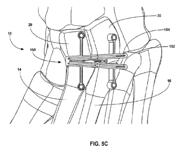
lateral cuneiform 30). The clinician may cut both bones defining the second
TMT joint or
may cut only one bone defining the joint and perform a different preparation
technique on the
other bone. Similarly, the clinician may cut both bones defining the third TMT
joint or may
cut only one bone defining the joint and perform a different preparation
technique on the
other bone.
[0084] Where the clinician cuts at least one bone forming a TMT joint, each
such cut may be
parallel or non-parallel to the end of the bone being cut in one or more of
the frontal,
transverse, and sagittal planes. For example, the cut may be angled in the
transverse plane
relative to the end face of the bone and parallel to the end face of the bone
in the frontal
plane. As other examples, the cut may be curved, arced, spherical, zig-zag, or
may define
other desired cut shape to facilitate realignment and fusion of one bone
relative to another
bone portion. In some examples, the end faces of the two bones defining the
TMT joint are
each prepared by cutting an end portion of each bone to create a shaped
opening between the
end faces. The opening may have a shape that allows the bones to be
repositioned relative to
17
CA 03178704 2022- 11- 14
WO 2021/236838
PCT/US2021/033256
each other (e.g., partially or fully closing the opening created in the
process of realignment)
to facilitate realignment and subsequent fusion.
[0085] In one example, the clinician can cut the end of the bone being
prepared at an angle
relative to the end face in the transverse plane, creating a wedge-shaped
section of bone that
is released from a remainder of the bone being cut. This can create a wedge-
shaped opening
between the newly defined end of the bone being cut and the opposing bone
across the TMT
joint being prepared. The wedge-shaped opening may enlarge moving from the
medial side
of the TMT joint to the lateral side of the TMT joint. For example, the wedge-
shaped bone
portion and corresponding opening may have a generally triangular-shape. The
wedge-
shaped opening can provide a gap across the TMT joint that can be closed by
subsequently
pivoting the metatarsal in the transverse plane. Again, however, other shaped
cuts can be
performed on one or both bones facing the TMT joint without departing from the
scope of the
disclosure. Example bone cutting shapes and configurations that may be used on
one or more
bone ends defining a TMT joint are described in US Patent No. 10,512,470,
dated December
24, 2019 and titled "OSTEOTOMY PROCEDURE FOR CORRECTING BONE
MISALIGNMENT" and US Patent No. 10,582,936, dated March 10, 2020, and titled
"DEVICES AND TECHNIQUES FOR PERFORMING AN OSTEOTOMY PROCEDURE
ON A FIRST METATARSAL TO CORRECT A BONE MISALIGNMENT,- the entire
contents of both of which are incorporated herein by reference.
[0086] FIG. 5A is a top view of foot 10 showing an example cut guide 150
positioned over
the second and third TMT joints to illustrate example bone wedges that may be
cut during
joint preparation. In this example, cut guide 150 is shown defining a first
guide surface 152
(which is illustrated as a cutting slot) positioned over a portion of a second
metatarsal 14 and
a portion of a third metatarsal 16 to be cut. Cut guide 150 is also shown as
defining a second
guide surface 154 (which is illustrated as a cutting slot) positioned over a
portion of an
intermediate cuneiform 28 and a lateral cuneiform 30 to be cut. The clinician
can advance a
cutting instrument parallel to first guide surface 152 to cut an end of second
metatarsal 14 and
also to cut an end of third metatarsal 16. The clinician can also advance the
cutting
instrument parallel to second guide surface 154 to cut an end of intermediate
cuneiform 28
and lateral cuneiform 30. In different implementations, a guide surface of cut
guide 150 may
be linear, curved, and/or define yet other shapes. According, the step of
guiding a cutting
instrument parallel to the guide surface may result in a linear cut, a curved
cut, or yet other
shaped cut across the bone.
18
CA 03178704 2022- 11- 14
WO 2021/236838
PCT/US2021/033256
[0087] In the example of FIG. 5A, first guide surface 152 is illustrated as
being angled in the
transverse plane across the second and third metatarsals 14, 16. The first
guide surface 152 is
illustrated as being angled from a medial-proximal side of second metatarsal
14 toward a
lateral-distal side of third metatarsal 16. The lateral-distal side of third
metatarsal 16 may
still be on the proximal half of the metatarsal, albeit comparatively distal
to the proximal
location on the second metatarsal. By preforming an angled cut relative to the
end faces of
the bones being cut, a wedge-shaped bone portion may be released from the
bone. In FIG.
5A, a wedge-shaped section 156 of second metatarsal 14 is released upon
cutting the second
metatarsal. Further, a wedge-shaped section 158 of third metatarsal 16 is
released upon
cutting the third metatarsal. Each wedge-shaped section of bone removed via
cutting may
have a narrow width (e.g., apex) on a medial side of the bone being cut and a
wider width
(e.g., base) on a lateral side of the bone being cut. The degree of angulation
in the specific
dimensions of the bone wedge formed during cutting may vary depending on the
anatomy of
the patient and the extent of the deformity being corrected. In either case,
the bone wedges
so cut can be removed from the TMT joint spaces to define a wedge-shaped
opening relative
to an opposed bone.
[0088] In the example of FIG. 5A, the clinician can use second guide surface
154 to guide the
cutting instrument to cut an end of intermediate cuneiform 28 and lateral
cuneiform 30 to
promote fusion following realignment of the metatarsals. The cuts performed on
the
intermediate cuneiform 28 and lateral cuneiform 30 may be generally parallel
to the end face
of a bone being cut (e.g., in the transverse plane) or may be angled relative
to an end face of
the bone being cut. In still other examples, the end faces of one or both of
intermediate
cuneiform 28 and lateral cuneiform 30 may not be cut but may be prepared using
a different
technique as discussed above (e.g., fenestrated).
[0089] In some examples in which the second metatarsal 14 and the third
metatarsal 16 are
prepared by cutting, the metatarsals may be cut using a single continuous cut
across both
metatarsals. For example, the clinician may guide a cutting instrument
linearly from a medial
side of the second metatarsal 14 toward the lateral side of the third
metatarsal 16 or from the
lateral side of the third metatarsal to the medial side of the second
metatarsal. In either case,
the clinician may form a continuous cut line transecting the ends of the
second and third
metatarsals. Such a continuous cut across the bases of the second and third
metatarsals may
be useful to promote reliable reduction of the metatarsus adductus angle
during subsequent
bone realignment. In applications where the intermediate cuneiform 28 and the
lateral
cuneiform 30 are cut in addition to or in lieu of the ends of the metatarsals,
the two
19
CA 03178704 2022- 11- 14
WO 2021/236838
PCT/US2021/033256
cuneiforms may or may not be cut using such a continuous cut across the ends
of the two
metatarsals.
[0090] In other applications of the surgical technique, the ends of the second
metatarsal 14
and third metatarsal 16 may be cut independently (e.g., without moving the
cutting
instrument in a continuous cutting line across the two metatarsals). For
example, when the
patient exhibits a significant step off (e.g., distal offset) between the end
of the intermediate
cuneiform 28 and the end of the lateral cuneiform 30, the ends of the opposed
second and
third metatarsals 14, 16 may be prepared independently (e.g., through two
separate cuts) in
lieu of forming a continuous cut across the ends of the two metatarsals. The
ends of the
opposed second and third metatarsals 14, 16 may be prepared independently for
other reasons
as well, such as to provide independent control / adjustability over the cut
angles on the
second and third metatarsals.
100911 While FIG. 5A illustrates one example cutting guide 150 and one example
cutting
arrangement that may be used to prepare the ends of the second and third TMT
joints, it
should be appreciated that a technique in accordance with the disclosure is
not limited to such
example guide or cutting arrangements. For example, a technique according to
disclosure
may be performed freehand (without the use of a cutting guide) or may be
performed with a
cutting guide having a different configuration. In addition to or in lieu of
using a cutting
guide, the clinician may position a bone preparation template over one or more
bone portions
to be subsequently cut. The bone preparation template may be configured (e.g.,
sized and/or
shaped) to indicate where on the underlying bone the bone should be cut or
otherwise
prepared. Positioning the bone preparation template on the underlying bone may
mark or
otherwise indicate on the bone where the bone should be prepared and/or the
clinician may
use the bone preparation template to mark where on the bone the bone should be
prepared.
The clinician may subsequently remove the bone preparation template and
preform a bone
preparation step (e.g., cutting) at the location marked or otherwise indicated
using the
template.
[0092] Further, although FIG. 5A illustrates angled cuts being performed on
the ends of
second metatarsal 14 and third metatarsal 16, such angled cuts are not
required. In general,
any one or both of the metatarsal and cuneiform bones forming the TMT joint
being prepared
may be cut so as to establish an opening for rotating the metatarsal in one or
more planes
(e.g., the transverse plane) and/or facilitating realignment of one or more
bones. The other of
the bones forming the TMT joint may also be prepared by cutting or may be
prepared using a
different bone preparation technique.
CA 03178704 2022- 11- 14
WO 2021/236838
PCT/US2021/033256
[0093] As one example, the clinician may remove a wedge-shaped section 156 of
bone from
the second metatarsal and remove a wedge-shaped section 158 of bone from the
third
metatarsal. The clinician can cut, fenestrate, and/or otherwise prepare the
ends of the
opposed intermediate cuneiform 28 and lateral cuneiform 30. In another
example, the
clinician may remove a wedge-shaped section of bone from the intermediate
cuneiform 28
and/or remove a wedge-shaped section of bone from the lateral cuneiform 30.
The clinician
can cut, fenestrate, and/or otherwise prepare the ends of the opposed second
metatarsal 14
and third metatarsal 16. In still another example, the clinician may remove a
wedge-shaped
section of bone from the cuneiform of one of the second and third TMT joints
and remove a
wedge-shaped section of bone from the metatarsal of the other of the second
and third TMT
joints. The end face of the opposed bone may be cut parallel to the end face
of the bone, at
an angle, and/or otherwise prepared (e.g., with or without cutting). For
example, the clinician
may remove a wedge-shaped section of both from the ends of both bones forming
the TMT
joint. In either case, the opening created between the ends of the bones
defining the TMT
joint may be defined by the cumulative amount of bone removed from both bone
ends. As
noted above, depending on the characteristics of the patient undergoing the
surgical
procedure, in yet other embodiments the clinician may not cut the end faces of
the bones
defining the second and third TMT joints or may perform a bone cut parallel to
the end face
of the bone. Further, while the foregoing examples are described as being
performed by
removing a wedge-shaped section of bone, a bone section having another shape
can be
removed, as described herein.
[0094] In instances where the clinician cuts the end face of the bone, the
clinician may or
may not perform one or more additional preparation steps on the end face prior
to or after
cutting the end face. In some examples, the clinician fenestrates the newly-
formed end face
of the bone after cutting the bone. The clinician may use a drill to
fenestrate the end newly-
formed end face of the bone being cut, which can help promote subsequent
fusion of the bone
following realignment. The clinician may fenestrate a bone face by making
multiple
openings (e.g., drill holes) in the bone face, providing multiple bleeding
points in the end of
the bone face. Each drill hole may be comparatively small relative to the
cross-sectional area
of the end face, such as less than 10% of the cross-sectional area of the end
face, less than 5%
of the cross-sectional area of the end face, or less than 1% of the cross-
sectional area of the
end face. The multiple openings can be arrayed at different locations across
the end face to
provide locations for promoting fusion across the end face. The number of
holes formed
21
CA 03178704 2022- 11- 14
WO 2021/236838
PCT/US2021/033256
during fenestration may vary and, in some examples, is greater than 5, such as
greater than
10.
[0095] As another example of a preparation step that may be performed, the
clinician may
remove one or more protruding bone portions extending into and/or across the
second TMT
and/or third TMT joint line. The protruding bone portions may extend distally
from the
cuneiform into the joint space and/or proximally from the metatarsal into the
joint space. For
instance, as discussed in greater detail with respect to FIG. 29, certain
patients may exhibit
significant step off, or distal offset, between adjacent joint planes (e.g.,
between the plane
defining the second TMT joint and the plane defining the third TMT joint).
This can inhibit
relative movement between the two joints for subsequent realignment and/or
inhibit insertion
of a cutting guide into one or both joint spaces. For these and other reasons,
the clinician
may remove the one or more protruding bone portions, e.g., to create a pocket
or continuous
joint line extending across the second TMT joint and the third TMT joint. The
clinician may
remove the one or more protruding bone portions using a cutting instrument
either freehand
and/or with the aid of a cutting guide, such as cutting guide 292 discussed in
greater detail
with respect to FIG. 30.
[0096] As another example, the clinician may typically visualize the location
of a cutting
guide and/or bone preparation template under radiographic imaging (e.g.,
fluoroscopy), e.g.,
to ensure that one or more guide planes or other guide features are
appropriately positioned
relative to one or more underlying bones. The clinician can adjust the
position of the cutting
guide or bone preparation template under imaging, e.g., until one or more
guide planes or
other alignment features are positioned over a desired portion or region of
underlying bone to
be marked, cut, and/or otherwise prepared.
[0097] FIGS. 5B-5E illustrate example bone preparation steps that may be
performed on foot
using an example configuration of cutting guide 150 according to a technique
of the
disclosure. In particular, FIGS. 5B and 5C are perspective and top (dorsal)
view illustrations
of foot 10 showing cut guide 150 positioned over a dorsal side of the foot.
Specifically, cut
guide 150 is shown with first guide surface 152 positioned over a portion of
second
metatarsal 14 and a portion of third metatarsal 16 to be cut, and second guide
surface 154 is
positioned over a portion of intermediate cuneiform 28 and lateral cuneiform
30 to be cut. In
this example, cut guide 150 defines at least one fixation aperture
positionable over each of
second metatarsal 14, third metatarsal 16, intermediate cuneiform 28, and
lateral cuneiform
30. A clinician can insert fixation pins through one or more (e.g., all) of
the fixation
apertures to secure the cut guide to underlying bone.
22
CA 03178704 2022- 11- 14
WO 2021/236838
PCT/US2021/033256
[0098] In use, the clinician can guide a cutting instrument along first guide
surface 152 to cut
an end of second metatarsal 14 and also to cut an end of third metatarsal 16.
The clinician
can also guide the cutting instrument along second guide surface 154 to cut an
end of
intermediate cuneiform 28 and lateral cuneiform 30. FIG. 5D is a perspective
view of the
foot showing example bone portions that can be removed after cutting,
specifically
illustrating an example wedge-shaped section 156 removed from second
metatarsal 14 and an
example wedge-shaped section 158 removed from third metatarsal 16. Additional
bone
sections may be removed from intermediate cuneiform 28 and lateral cuneiform
30. FIG. 5E
illustrates an example opening 157 formed between second metatarsal 14 and
intermediate
cuneiform 28 upon removal of one or more bone portions and an example opening
159
formed between third metatarsal 16 and lateral cuneiform 30 upon removal of
one or more
bone portions.
100991 With further reference to FIG. 4, the example technique involves moving
the second
metatarsal 14 and the third metatarsal 16 in at least one plane (110). While
FIG. 4
schematically illustrates an example order in which the second and third
metatarsals 14, 16
are moved after preparing the end faces of metatarsals 14, 16 and opposed
intermediate and
lateral cuneiforms 28, 30, other orders of bone preparation and movement may
be performed.
For example, the clinician can move the second and/or third metatarsals 14, 16
before
preparing one or more metatarsals and/or one or more cuneiforms (e.g., before
preparing the
end faces of all of the bones). For instance, the clinician may move the
second and third
metatarsals 14, 16 and then prepare the end faces of metatarsals 14, 16 and
opposed
intermediate and lateral cuneiforms 28, 30. In these implementations, the
clinician may or
may not further move the second and/or third metatarsals 14, 16 after
preparing the end faces
of the metatarsals and cuneiforms. As another example, the clinician may
prepare the end
face of one or more bones (e.g., one or more metatarsals and/or cuneiforms),
move one or
both of second metatarsal 14 and third metatarsal 16, and then prepare the end
face of one or
more other bones (e.g., one or more metatarsals and/or cuneiforms).
[00100] Independent of the order of movement and bone preparation, the
clinician may move
the second and third metatarsals 14, 16 in one or more planes, such as the
transverse plane,
e.g., by pivoting the metatarsals about their proximal ends, causing a distal
end of the
metatarsals to move laterally in the transverse plane. In instances where a
wedge-shaped
opening was formed at the second and/or third TMT j oints during bone
preparation, lateral
rotation of the distal ends of the second and third metatarsals may close the
wedge-shaped
opening(s) (or close another shaped opening, in instances in which a non-wedge-
shaped
23
CA 03178704 2022- 11- 14
WO 2021/236838
PCT/US2021/033256
opening was created). For example, translation of the distal ends of the
second and third
metatarsals 14, 16 laterally in the transverse plane may bring the ends of the
second
metatarsal 14 and opposed intermediate cuneiform 28 as well as the ends of the
third
metatarsal 16 and opposed lateral cuneiform 30 in generally parallel
alignment. The clinician
may move the second and/or third metatarsal in the frontal plane and/or
sagittal plane in
addition to or in lieu of moving one or both bones in the transverse plane.
For example, the
clinician may rotate one or both bones in the frontal plane and/or translate
one or both bones
(e.g., dorsally) in the sagittal plane.
[00101] In general, movement of second metatarsal 14 and third metatarsal 16
in the
transverse plane can close the metatarsus adductus angle. The metatarsus
adductus angle
may be the angular measurement formed between the line bisecting the second
metatarsal and
the longitudinal line bisecting the lesser tarsus on a dorsoplantar
radiograph. In some
examples, the second and third metatarsals 14, 16 are moved until the
metatarsus adductus
angle for each metatarsal is 15 or less, such as 12 or less, 10 or less, 7'
or less, 5' or less,
or 3 or less.
[00102] The second metatarsal 14 and third metatarsal 16 may be moved
individually or
jointly (e.g., as a bone block). Moving the second and third metatarsals 14,
16 as a joined
group may be helpful to achieve a more natural realignment of the metatarsals
and correction
of the metatarsus adductus deformity. To help move the second and third
metatarsals 14, 16
as a joined group, the ligaments between the two metatarsals may be preserved
during
preparation of the second and third TMT joints. For example, the plantar TMT
ligaments and
ligaments between the second and third metatarsals 14, 16 may be preserved
(e.g., remain
uncut or unbroken) during preparation and movement of the second and third
metatarsals.
Preserving the ligament structure can help avoid destabilization of the second
and third TMT
joints during deformity reduction, which may improve the anatomical
realignment of the
bone structure.
[00103] FIG. 6 is a diagram schematically illustrating the ligament structure
of the foot. As
shown, the plantar TMT ligaments include a second plantar tarsometatarsal
ligament between
medial cuneiform 26 and the second and third metatarsals 14, 16. The plantar
TMT
ligaments also include a third plantar tarsometatarsal ligament between
lateral cuneiform 30
and third metatarsal 16. A first plantar intermetatarsal ligament extends
between second
metatarsal 14 and third metatarsal 16. Ligamentous attachments between the
second and
third metatarsals, such as the second and third plantar tarsometatarsal
ligaments and the first
plantar intermetatarsal ligament, may be preserved during preparation and
movement of the
24
CA 03178704 2022- 11- 14
WO 2021/236838
PCT/US2021/033256
second and third metatarsals to correct the metatarsus adductus. This may
allow the two
metatarsals to move together as a joined bone group.
[00104] To move the second and third metatarsals 14, 16, either alone or in
combination, the
bones may be pivoted about their proximal base, causing the distal ends of the
bones to
translate laterally in the transverse plane. When moving the second and third
metatarsals 14,
16 as a group, the clinician may pivot the second and third metatarsal bone
block about the
proximal medial portion of second metatarsal 14. The clinician may move the
second and
third metatarsals 14, 16 as a combined group in the transverse plane, with or
without
simultaneously rotating both bones in the frontal plane and/or adjusting the
sagittal plane
position of the bones. In some implementations, the clinician moves the second
and third
metatarsals 14, 16 as a group about the Lisfranc ligament while the second
metatarsal
remains attached to the Lisfranc ligament. Accordingly, the Lisfranc ligament
may act as a
hinge or pivot point about which the second and third metatarsal bone group
can rotate in the
transverse plane.
[00105] In other examples, the clinician may substantially independently move
the second
and third metatarsals 14, 16 (e.g., by applying a separate movement force to
each metatarsal).
For example, the clinician may apply a force to move third metatarsal 16 in
one or more
planes and subsequently apply a force to move the second metatarsal 14 in one
or more
planes (or, instead, move the second metatarsal 14 followed by the third
metatarsal), such as
in two or more, or all three planes. The clinician may or may not cut or
otherwise release one
or more ligamentous attachments interconnecting the second and third
metatarsals 14, 16 to
help facilitate independent repositioning of the two bones.
[00106] Independent of whether the clinician moves the second and third
metatarsals 14, 16
together or independently, the intermetatarsal angle between second and third
metatarsals
may or may not change during metatarsus adductus correction. In other words,
the
intermetatarsal angle between second metatarsal 14 and third metatarsal 16 may
or may not
compress from a pre-corrected intermetatarsal angle to the intermetatarsal
angle exhibited
after correction. In some implementations, the second and third metatarsals
14, 16 are
pivoted as a group within the transverse plane without substantially changing
the
intermetatarsal angle between the second and third metatarsals. For example,
the
intermetatarsal angle between the second and third metatarsals may change
(e.g., reduce) less
than 5 , such as less than 2 , or less than 10 from the angle exhibited before
metatarsus
adductus correction to the angle exhibited after the correction technique is
performed.
CA 03178704 2022- 11- 14
WO 2021/236838
PCT/US2021/033256
[00107] To help facilitate movement of the second and third metatarsals in the
transverse
plane, the clinician may perform a soft tissue release between third
metatarsal 16 and fourth
metatarsal 18. The soft tissue release may mobilize the third metatarsal
relative to the
adjacent fourth metatarsal, allowing the joined second-third metatarsal bone
block to be
pivoted in the transverse plane.
[00108] In addition to moving the second metatarsal and the third metatarsal
in the transverse
plane, the clinician can also move fourth metatarsal 18 and fifth metatarsal
20 in one or more
planes (e.g., one or more of the transverse plane, the frontal plane, and the
sagittal plane),
e.g., to close the metatarsus adductus angle exhibited by those lesser
metatarsals. In practice,
movement of second metatarsal 14 and third metatarsal 16 in one or more planes
(e.g., the
transverse plane) may cause the fourth and fifth metatarsals to naturally
correct in same one
or more planes (e.g., the transverse plane) without requiring separate
surgical intervention on
the fourth and fifth metatarsals 18, 20. For example, as the clinician rotates
the distal end of
second metatarsal 14 and third metatarsal 16, either alone or in combination,
the distal ends
of fourth metatarsal 18 and fifth metatarsal 20 may also move laterally. The
proximal base of
fourth metatarsal 18 and the proximal base of fifth metatarsal 20 may reorient
relative to the
cuboid bone 32, closing the metatarsus adductus angle of the fourth and fifth
metatarsals.
Without wishing to be bound by any particular theory, it is believed that
force applied to the
second and/or third metatarsal during movement may translate through the
tissue and
ligament structure interconnecting such metatarsal(s) to the fourth and fifth
metatarsals,
pulling the lesser metatarsals into realignment.
[00109] The position of fourth metatarsal 18 and fifth metatarsal 20 may
correct without
surgically accessing and preparing the metatarsal (in response to correction
of second
metatarsal 14 and/or third metatarsal 16). In other applications, however, the
clinician may
surgically access and prepare the bones defining fourth TMT joint 40 and/or
fifth TMT joint
42 in addition to or in lieu of preparing one or more other TMT joints. For
example, before
or after moving the fourth metatarsal 18 and/or fifth metatarsal 20 in one or
more planes (e.g.,
separate from or in combination with movement of the second metatarsal 14
and/or third
metatarsal 16), the clinician can surgically access and prepare the bones
defining fourth TMT
joint 40 and/or fifth TMT joint 42. The clinician may decide whether to access
and prepare
the bones defining fourth TMT joint 40 and/or fifth TMT joint 42 depending,
for example, on
the nature of the deformity being corrected and the perceived need prepare the
joints for bone
realignment and/or fusion
26
CA 03178704 2022- 11- 14
WO 2021/236838
PCT/US2021/033256
[00110] The clinician may prepare the end of fourth and/or fifth metatarsal
18, 20 and/or
prepare the end of cuboid bone 32 opposite the end of the fourth and/or fifth
metatarsal to
facilitate realignment and/or fusion. The clinician can prepare the one or
more bone ends
using any of the bone preparation techniques discussed herein. In various
examples, the
clinician may independently prepare the ends of fourth and fifth metatarsals
18, 20 or may
prepare the ends of the metatarsals together (e.g., such as positioning a
single, continuous
cutting guide plane over both ends for making a continuous cut, as discussed
with respect to
preparation of second metatarsal 14 and third metatarsal 16). Additionally or
alternatively,
the clinician may prepare the portions of the end face of cuboid bone 32
facing fourth and/or
fifth metatarsal 18, 20 together or through separate preparation steps.
[00111] In instances where the clinician accesses and prepares the bones
defining fourth
TMT joint 40 and/or fifth TMT joint 42, one or both of the fourth metatarsal
18 and fifth
metatarsal 20 may be realigned in one or more planes in response to a force
applied to the
second and/or third metatarsal 14, 16 (e.g., by translating the force through
the tissue and
ligament structure interconnecting the metatarsals). Additionally or
alternatively, the
clinician may apply a force to the fourth metatarsal 18 and/or fifth
metatarsal 20 to move one
or both metatarsal in one or more planes, such as two or more, or all three
planes. The fourth
and fifth metatarsals 18, 20 may be moved as joined bone block (e.g., either a
bone block also
connected to the bone block of the second and third metatarsal 14, 16 or
separate therefrom)
and/or may be moved substantially independently from each other (e.g., by
applying a
separate movement force to each metatarsal). Further, the clinician may apply
a force to
move only one of the fourth or fifth metatarsals.
[00112] With typical metatarsus adductus deformities, the metatarsals may
exhibit a
substantially uniplanar misalignment in the transverse plane (although may be
misaligned in
the frontal plane and/or sagittal plane). For this reason, the example
technique of FIG. 4 has
generally been described as correcting the second and third metatarsals 14, 16
(and,
optionally, fourth and fifth metatarsals 18, 20) in the transverse plane. The
clinician may
move the metatarsals in only the transverse plane to correct the generally
uniplanar
misalignment. Alternatively, the clinician may move one or more of the
metatarsals being
realigned (e.g., multiple or all of the metatarsals been realigned) in more
than one plane. For
example, in addition to or in lieu of realigning the metatarsal(s) in the
transverse plane, the
clinician may adjust the rotational angle of the metatarsal(s) in the frontal
plane and/or adjust
the angle of the metatarsal(s) in the sagittal plane.
27
CA 03178704 2022- 11- 14
WO 2021/236838
PCT/US2021/033256
[00113] Where the clinician performs a multi-planar realignment, the clinician
may move one
or more metatarsals in multiple planes simultaneously through a single
movement, e.g., by
moving the metatarsal in an arc or other movement pathway to adjust the
position of the
metatarsal in multiple planes. The clinician may optionally perform further
fine adjustment
of the moved position of the one or more metatarsals, e.g., with the aid of a
bone positioning
device and/or by grasping the metatarsal by hand (e.g., with the aid of a pin
inserted into the
metatarsal) to finalize the position of the metatarsal prior to fixation.
[00114] In other examples, the clinician may perform different movement steps
to move the
one or more metatarsals in different planes. For example, the clinician may
initially move the
one or more metatarsals in one or two planes (e.g., transverse plane, frontal
plane, sagittal
plane) then move the one or more metatarsals in one or two other planes (e.g.,
the other of the
transverse plane, frontal plane, sagittal plane), optionally followed by
movement of the one or
more metatarsals in a third plane. In other words, the clinician may perform
different actions
to move the one or more metatarsals in different planes. Each movement step
may be
performed with the aid of a bone positioning device (which may be the same or
different
device for different movement steps) and/or by grasping the metatarsal by hand
(e.g., with the
aid of a pin inserted into the metatarsal).
[00115] In some examples, the clinician may move one or more of the
metatarsals being
realigned (e.g., second metatarsal 14 and/or third metatarsal 16) proximally
in the transverse
plane toward the opposed bone in addition to or in lieu of moving the
metatarsal(s) laterally.
For example, the clinician may simultaneously move the metatarsal being
realigned (e.g.,
second metatarsal 14 and/or third metatarsal 16) laterally and proximally in
an arc (e.g.,
parabola) to establish a moved position of one or both metatarsals.
[00116] The clinician can move the one or more metatarsals being realigned
(e.g., second
metatarsal 14 and/or third metatarsal 16) by hand and/or with the aid of one
or more
instruments. For example, the clinician can grasp the second and/or third
metatarsal and
advance the distal end of the metatarsal laterally to reduce the metatarsus
adductus angle.
The clinician may insert one or more pins into the metatarsal being moved
(e.g., second
and/or third metatarsal) to provide a joystick or structure that can be
grasped to manipulate
movement of the bones. Additionally or alternatively, the clinician may
utilize a tenaculum
or tong to grasp one or both of the second and third metatarsals to facilitate
realignment.
[00117] In some examples, the clinician may use a bone positioning guide (also
referred to as
a bone positioning device) to help apply a force to a metatarsal (e.g., second
metatarsal 14
and/or third metatarsal 16) to facilitate realignment. The bone positioning
guide may include
28
CA 03178704 2022- 11- 14
WO 2021/236838
PCT/US2021/033256
one end that engages with (e.g., contacts, with or without being provisionally
fixated to) the
metatarsal to which the force is being applied and another end that engages
with (e.g.,
contacts, with or without being provisionally fixated to) a different bone.
For example, the
bone positioning guide may have one end that engages with second metatarsal 14
and/or third
metatarsal 16 and another end that engages with a bone other than the second
and/or third
metatarsals (e.g., a lesser metatarsal, a cuneiform, the cuboid bone). The
bone positioning
guide may have a mechanism that urges the two ends towards each other to
reduce the
metatarsus adductus angle. Example bone positioning guide engagement
mechanisms that
can function to move the two ends of the guide toward each other include a
screw or threaded
rod, a ratchet, a rack and pinion, and/or yet other features that translates a
force applied by the
clinician move the two ends of the bone positioning guide toward each other.
Details on an
example bone positioning guide that may be used are described in US Patent No.
9,936,994,
issued April 10, 2018, and titled "BONE POSITIONING GUIDE,- the entire
contents of
which are incorporated herein by reference.
[00118] FIG 7A is a side perspective view of an example bone positioner 60
(also referred to
as a bone positioning device) that can be used to move a metatarsal relative
to an adjacent
bone. In some implementations, the bone positioning device includes a
metatarsal
engagement member, a tip, and a mechanism to move the metatarsal engagement
member
and the tip relative to each other in one or more planes. For example, the
mechanism may
move the metatarsal engagement member and the tip towards each other (e.g.
moving the
metatarsal engagement member towards the tip, moving the tip towards the
metatarsal
engagement member, or moving both simultaneously). The bone positioning device
may also
include an actuator to actuate the mechanism. When the mechanism engaged, it
can cause a
metatarsal engaged with the metatarsal engagement member to move to correct an
alignment
in at least one plane with respect to a second bone in contact with the tip.
1001191 In the embodiment of FIG. 7A, bone positioning device 60 includes a
main body
member 62, a shaft 64, a metatarsal engagement member 66 connected to the
shaft, and a tip
68 is connected to the main body member. In general, main body member 62 can
be sized
and shaped to clear anatomy or other instrumentation (e.g., pins and guides)
while positioned
on a patient. In the embodiment of FIG. 7A, the main body member 62 is
generally C-
shaped. Although bone positioning device 60 is illustrated as being formed of
two
components, main body member 62 and shaft 64, the guide can be fabricated from
more
components (e.g., three, four, or more) that are joined together to form the
guide.
29
CA 03178704 2022- 11- 14
WO 2021/236838
PCT/US2021/033256
[00120] A shaft 64 can be movably connected to the main body member 62. In
some
embodiments, the shaft 64 includes threads 70 that engage with the main body
member 62
such that rotation of the shaft translates the shaft with respect to the main
body member. In
other embodiments, the shaft can slide within the main body member and can be
secured
thereto at a desired location with a set screw. In yet other embodiments, the
shaft can be
moved with respect to the main body by a ratchet mechanism or yet other
mechanism that
rotates and/or linearly translates metatarsal engagement member 66 relative to
tip 68. In the
embodiment shown, the shaft moves along an axis that intersects the tip. In
other
embodiments, the shaft and/or metatarsal engagement member is offset from the
tip.
[00121] In general, metatarsal engagement member 66 may be configured (e.g.,
sized and/or
shaped) to be positioned in contact, directly or indirectly, with a metatarsal
to be
repositioned. For example, depending on the size and/or shape of metatarsal
engagement
member 66, the metatarsal engagement member may be positioned subcutaneously
in contact
with a metatarsal bone to be realigned or may be positioned in contact with an
external
surface of the skin overlying the metatarsal bone to be realigned. For
instance, in either
configuration, metatarsal engagement member 66 may be positioned on a medial
side of a
metatarsal to be realigned (e.g., medial side of second metatarsal 14, third
metatarsal 16,
fourth metatarsal 18, fifth metatarsal 20) and tip 68 positioned in contact
with another bone
(e.g., with or without being provisionally fixated thereto), such as a
laterally-located bone.
[00122] Metatarsal engagement member 66 may define a concave shape to
generally conform
and/or wrap partially around the underling cylindrical bone. The concave shape
may include
define a continuous radius of curvature, a V-shape, a planer region between
outwardly
extending sidewalls, and/or other shape having a concavity. In still other
examples,
metatarsal engagement member 66 may be planar.
[00123] Tip 68 can be useful for contacting a bone, such as a bone different
than the bone
being moved by bone positioning device 60. For example, if metatarsal
engagement member
66 is positioned over a medial side of one metatarsal, the tip can be
positioned over with a
lateral side of a different metatarsal (e.g., the third, fourth, or fifth
metatarsal), either directly
in contact with the bone or over a lateral side of skin covering such
metatarsal. In different
configurations, tip 68 may be straight or may be tapered to facilitate
percutaneous insertion
and contact with bone. The tip can also include a textured surface, such as
serrated,
roughened, cross-hatched, knurled, etc., to reduce slippage between the tip
and bone. In the
embodiment shown, tip 68 further includes a depth stop 74. Depth stop 74 can
limit a depth
CA 03178704 2022- 11- 14
WO 2021/236838
PCT/US2021/033256
of insertion into an intermetatarsal space (e.g., by contacting a dorsal
surface of the
metatarsal against which tip 68 is intended to be positioned).
[00124] As shown in FIG. 7A, bone positioning device 60 can also include an
actuator (e.g., a
knob or a handle) 76 to actuate the mechanism, in this embodiment associated
with the shaft.
In the embodiment shown, the actuator can be useful for allowing a user to
rotate the shaft
with respect to the main body member 62. Actuator 76, shaft 64, and/or
metatarsal
engagement member 66 may include a cannulation 78 extending therethrough to
allow the
placement of a fixation wire (e.g., K-wire) through these components and into
or through a
bone engaged with the metatarsal engagement member. For example, a fixation
wire can be
placed into the bone engaged with metatarsal engagement member 66 to fix the
position of
the metatarsal engagement member with respect to the bone. In another example,
the fixation
wire can be placed through the bone in contact with the metatarsal engagement
member and
into an adjacent bone to maintain a bone position of the bone in contact with
the metatarsal
engagement member and the adjacent bone. Although shaft 64 and actuator 76 are
illustrated
as projecting away from a side of main body member 62, one or both features
may be
positioned at a different location (e.g., extending dorsally above main body
member 62
through a mechanical linkage) to facilitate positioning of bone positioning
guide 60 (e.g.,
particularly metatarsal engagement member 66) in an intermetatarsal space.
[00125] Embodiments of any instrument described herein (e.g., cutting guide,
bone
preparation template, bone positioning device) may include or be fabricated
from any suitable
materials (e.g., metal, plastic). In certain embodiments, an instrument such
as a bone
positioning device is fabricated at least partially from a radiolucent
material such that it is
relatively penetrable by X-rays and other forms of radiation, such as
thermoplastics and
carbon-fiber materials. Such materials are useful for not obstructing
visualization of bones
using an imaging device when the instrument is positioned on bones.
1001261 Another type of bone positioning guide that may be used to move a
metatarsal in one
or more planes, such as used to move second metatarsal 14 and third metatarsal
16 in at least
the transverse plane, is a compressor instrument. For example, when an opening
(e.g.,
wedge-shaped opening) is created at the second and third TMT joints during
preparation of
the bone ends, a compressor may be attached to the second and/or third
metatarsal and
another bone, such as the intermediate cuneiform and/or lateral cuneiform,
respectively. The
compressor may apply a distal-to-proximal force across the second and/or third
TMT joints,
causing the wedge-shaped opening created across the joint to close. As the
wedge-shaped
opening closes, the distal end of second metatarsal 14 and/or third metatarsal
16 can pivot in
31
CA 03178704 2022- 11- 14
WO 2021/236838
PCT/US2021/033256
the transverse plane. When used, the compressor may also compress the ends of
the bone
faces together, e.g., by compressing intermediate cuneiform 28 and second
metatarsal 14
together and/or compressing lateral cuneiform 30 and third metatarsal 16
together, to
facilitate subsequent fusion.
[00127] FIG. 7B is an illustration of an example compressor instrument 160
engaged with
foot 10 to facilitate movement of the second and third metatarsals. In this
example,
compressor instrument 160 has a first end 162 engaged with the second
metatarsal and a
second end 164 engaged with the intermediate cuneiform. In particular, in the
illustrated
arrangement, first and second ends 162, 164 of compressor instrument 160 are
illustrated as
being pinned to the bones. Compressor instrument 160 can be actuated by the
clinician to
move the first and second ends 162, 164 of the compressor towards each other.
This can
cause the ends of the second metatarsal and intermediate cuneiform to which
the compressor
is attached to draw towards each other, closing a wedge-shaped opening in the
TMT joint
space and causing a distal end of the second metatarsal to translate
laterally. The force
applied across the second TMT joint as illustrated in FIG. 7 can also close a
wedge-shaped
opening in the third TMT joint, and may move the second and third metatarsals
as a bone
group. In turn, this can cause the fourth and fifth metatarsals to also move
in at least the
transverse plane, reducing their metatarsus adductus angle.
[00128] Additional details on example compressor structures that may be used
in accordance
with the disclosure are described in US Patent Publication No. 2020/0015856,
filed July 11,
2019, and titled "COMPRESS OR-DISTRACTOR FOR ANGULARLY REALIGNING
BONE PORTIONS,- the entire contents of which are incorporated herein by
reference.
Further, while the example compressor instrument 160 in FIG. 7B is illustrated
as being
attached across the second TMT joint, the compressor may additionally or
alternatively be
attached across the third TMT joint (e.g., with first end 162 attached to
third metatarsal 16
and second end 164 attached to lateral cuneiform 30) or yet other set of bones
(e.g., across the
fourth TMT joint 40, across the fifth TMT joint 42).
[00129] With additional reference to FIG. 4, the example technique is
illustrated as including
a step of provisionally fixating the moved position of the second metatarsal
and the third
metatarsal (112). For example, after moving the second metatarsal and third
metatarsal into a
desired realigned position in one or more planes, such as the transverse plane
(which may
also involve moving the fourth metatarsal and fifth metatarsal), the clinician
may optionally
provisionally fixate the moved position. Provisional fixation can hold the
moved position of
one or more bones to facilitate subsequent surgical steps, such as application
of one or more
32
CA 03178704 2022- 11- 14
WO 2021/236838
PCT/US2021/033256
permanent fixation devices and/or the performance of additional surgical steps
(e.g., first
metatarsal realignment).
[00130] To provisionally fixate the moved position of the one or more bones,
the clinician
may insert one or more pins into and/or through a moved bone and into an
adjacent bone.
For example, the clinician may insert a pin through the second metatarsal and
into an adjacent
bone (e.g., a cuneiform) and/or insert a pin through the third metatarsal and
into an adjacent
bone. The pin may be in the form of a rod and/or a wire (K-wire), and may or
may not be
configured to apply compression across a joint between the bones in which the
pin is inserted,
e.g., by having an enlarged region of the pin that presses against the outer
surface of the bone
through which the tip of the pin is inserted, thereby applying compression.
[00131] FIG. 8A is a dorsal view of an example radiographic image illustrating
an example
provisional fixation pin arrangement. In this example, a first fixation pin
170 is inserted into
and through the distal base of second metatarsal 14 and into the lateral
cuneiform. A second
fixation pin 172 is inserted into and through the distal base of third
metatarsal 16 and into the
intermediate cuneiform.
[00132] In the illustrated configuration, the first and second pins 170, 172
are shown as
crossing, with the shaft of first fixation pin 170 extending in a proximal
medial to distal
lateral orientation and the shaft of second fixation pin 172 extending in a
proximal lateral to
distal medial orientation. In other cases, first and second pins 170, 172 may
not cross but
may instead be orientated in parallel, such as with the shafts of both pins
extending in a distal
medial to proximal lateral orientation. FIG. 8B is a dorsal view of another
example
radiographic image illustrating a provisional fixation pin arrangement in
which first and
second pins 170, 172 are both positioned extending in a proximal medial to
distal lateral
orientation.
[00133] The clinician may use a different number, configuration, and/or
positioning of
fixation pins. For example, depending on the number of TMT joints being
prepared, the
clinician may insert a provisional fixation pin through the end of one or more
of second
metatarsal 14, third metatarsal 16, fourth metatarsal 18, and/or fifth
metatarsal 20, with the
pin extending into and/or through the end of the metatarsal and also into
another bone, such
as an opposed cuneiform and/or cuboid bone. Additionally or alternatively, the
clinician may
insert a provisional fixation pin through a side of one metatarsal being
fixated (e.g., a medial
side, lateral side) into an adjacent metatarsal).
[00134] Independent of whether the clinician deploys a provisional fixation
device, the
clinician may apply one or more permanent fixation devices to facilitate
fusion of the second
33
CA 03178704 2022- 11- 14
WO 2021/236838
PCT/US2021/033256
and third TMT joints following reduction of the metatarsus adduction angle
(step 114 in FIG.
4). The one or more fixation devices can extend across the second and/or third
TMT joints to
secure and hold opposed bone ends together for fusion (and/or other TMT joint
in instances
in which a different TMT joint is prepared for fusion). For example, the
clinician may apply
a first fixation device across the second TMT joint and apply a second
fixation device across
the third TMT joint.
[00135] A bone fixation device may be any feature or combination of features
that holds two
bone portions in fixed relationship to each other to facilitate fusion of the
bone portions
during subsequent healing. Any one or more bone fixation devices that can be
used include,
but are not limited to, a bone screw (e.g., a compressing bone screw), a bone
plate, a bone
staple, an external fixator, an intramedullary implant, and/or combinations
thereof
Depending on the type of bone fixation device selected, the bone fixation
device may be
attached to external surfaces of the bone portions being fixated or may be
installed as an
intramedullary device internal to the bone portions.
[00136] In one example, the clinician may install a first bone plate across
the second TMT
joint. The first bone plate can be secured on one side to second metatarsal
14, bridge the
second TMT joint, and be secured on an opposite side to intermediate cuneiform
28. The
clinician may install a second bone plate across the third TMT joint. The
second bone plate
can be secured on one side to third metatarsal 16, bridge the third TMT joint,
and be secured
on an opposite side to lateral cuneiform 30. Additionally or alternatively,
the clinician may
apply a U-shaped plate or other shaped plate bridging both the second and
third TMT joint
(e.g., with the U-shaped plate attached to the intermediate and lateral
cuneiforms on the base
of the U-shape and the legs of the U-shape being attached to the metatarsals).
Independent of
the number of plates used, each bone plate may be secured to an underlying
bone using one
or more screws, staples, and/or other securing mechanisms. When using a bone
plate, each
bone plate may be linear or may have a non-linear shape, such as a Y-shape, an
L-shape, a T-
shape, a U-shape, and/or other shape profile. It should be appreciated that,
in this document,
when terms "first" and "second" are used to modify a noun, such use is simply
intended
distinguish one item from another and is not intended to require a sequential
order of
preforming a procedure step unless specifically stated.
1001371 As briefly discussed above, a metatarsus adduction deformity may
present with a
hallux valgus misalignment in some patients. Accordingly, a clinician
performing a
metatarsus adduction correction procedure may also perform a hallux valgus
correction on
the patient undergoing treatment. In the example FIG. 4, the example technique
is illustrated
34
CA 03178704 2022- 11- 14
WO 2021/236838
PCT/US2021/033256
as including a first metatarsal realignment step (116). Although the technique
of FIG. 4
illustrates the first metatarsal realignment being performed after reduction
and permanent
fixation of the second and third TMT joints, a different surgical order may be
performed. For
example, the first metatarsal may be realigned prior to moving a lesser
metatarsal (e.g., the
second and third metatarsals), or may be realigned after moving the lesser
metatarsal (e.g.,
the second and third metatarsals) but prior to permanently fixating the lesser
TMT joint(s).
[00138] While the order of the surgical procedure may vary, in some
applications, it is useful
to correct the alignment of one or more lesser metatarsals (e.g., second
and/or third
metatarsals) prior to correcting the alignment of the first metatarsal. By
initially correcting
the position of the lesser metatarsal, such as the second and third
metatarsals (and, in some
examples, also correcting the position of the fourth and fifth metatarsals),
the clinician may
be able to better anatomically realign the first metatarsal relative to the
aligned lesser
metatarsals. Correction of the alignment of one or more of the lesser
metatarsals may change
the extent of misalignment of the first metatarsal, which can then be further
corrected during
a subsequent first metatarsal realignment step.
[00139] To correct the alignment of first metatarsal 12, the clinician may
surgically access
the first TMT joint as discussed above. Once accessed the clinician may
prepare an end of
first metatarsal 12 and an opposed end of medial cuneiform 26. The clinician
may prepare
the ends of the bones with or without cutting, as discussed above with respect
to preparation
of the ends of second metatarsal 14 and third metatarsal 16 (e.g., using any
preparation
technique discussed herein). In instances in which the clinician prepares one
or more bone
ends using a cutting instrument, the clinician may or may not utilize a cut
guide to guide
controlled cutting of the bone ends and/or a bone preparation template to
indicate where bone
preparation should be performed.
[00140] Either before or after preparing one or both ends of first metatarsal
12 and medial
cuneiform 26, the clinician may move first metatarsal 12 in at least one plane
(e.g., the
transverse plane, the frontal plane) to close an intermetatarsal angle between
the first
metatarsal and second metatarsal 14. In some examples, the clinician moves the
first
metatarsal in multiple planes, such as the transverse plane and/or frontal
plane and/or sagittal
plane. The clinician may or may not utilize a bone positioning guide to
facilitate movement
of the first metatarsal relative to the second metatarsal and/or medial
cuneiform. With the
first metatarsal moved to a desired position, the clinician can optionally
provisionally fixate
the moved position of the first metatarsal and then permanently fixate the
moved position
using one or more bone fixation devices, such as those described above.
Additional details
CA 03178704 2022- 11- 14
WO 2021/236838
PCT/US2021/033256
on example first metatarsal realignment instruments and techniques that can be
used are
described in US Patent No. 9,622,805, issued April 18, 2017 and entitled "BONE
POSITIONING AND PREPARING GUIDE SYSTEMS AND METHODS."
[00141] FIGS. 9A and 9B are dorsal radiographic images of an example foot 10
before and
after a treatment procedure, respectively, performed following an example
surgical technique
discussed above with respect to FIG. 4. FIG. 9A illustrates foot 10 with
metatarsus adductus
and hallux valgus deformities. FIG. 9B illustrates foot 10 after realignment
of the second and
third metatarsals in at least the transverse plane and multiplanar correction
of the first
metatarsal in all three planes. FIG. 9B illustrates three bone plates applied
across the first,
second, and third TMT joints to facilitate fusion across the three joints.
[00142] While the technique of FIG. 4 has generally been described with
reference to
preparation of second TMT joint 36 and third TMT joint 38 and movement of both
second
metatarsal 14 and third metatarsal 16 (optionally in combination with movement
of fourth
metatarsal 18 and fifth metatarsal 20), the techniques and/or devices may be
performed on
single TMT joints and/or different TMT joints without departing from the scope
of the
disclosure. For example, the technique of FIG. 4 may be performed on a single
lesser TMT
joint, such as only the second TMT joint 36, only the third TMT joint 38, only
the fourth
TMT joint 40, or only the fifth TMT joint 42, in each case optionally in
combination with
preparation of the first TMT joint 34 and realignment of the first metatarsal.
Other
combinations of joint preparation are also possible.
[00143] In applications where the clinician prepares only a single lesser TMT
joint for fusion
(again, optionally as part of a procedure that also prepares the first TMT
joint), the clinician
may move the lesser metatarsal associated with that TMT joint in one or more
planes, e.g.,
using devices and/or techniques discussed herein. Repositioning of the
metatarsal associated
with the lesser TMT joint being prepared may or may not also move one or more
adjacent
metatarsals to the lesser metatarsal being moved through ligamentous tissue.
For example, if
the clinician prepares second TMT joint 36 and moves second metatarsal 14, the
repositioning of the second metatarsal may cause realignment of third
metatarsal 16, fourth
metatarsal 18, and/or fifth metatarsal 20.
1001441 As discussed above, a bone realignment technique according to the
disclosure may
involve cutting an end of a cuneiform and/or an end of an opposed metatarsal.
In such
applications, the clinician may perform the cuts freehand or with the aid of
one or more cut
guides (also referred to herein interchangeably as a cutting guide). The use
of a cut guide
may facilitate more accurate and repeatable cuts patient-to-patient, promoting
more
36
CA 03178704 2022- 11- 14
WO 2021/236838
PCT/US2021/033256
consistent clinical outcomes across a range of patients an anatomical
deformities. When a cut
guide is used, the cut guide may generally define at least one guide surface
positionable over
a side of the bone to be cut, such as a dorsal side. The clinician can place a
cutting
instrument adjacent to, and optionally in contact with, the guide surface and
translate the
cutting instrument relative to the guide surface to perform a cut in a plane
parallel to the
guide surface. For example, the clinician may place the cutting instrument in
contact with the
guide surface and then translate the cutting instrument relative to the guide
surface, e.g.,
plantarly into a bone and/or in a medial or lateral direction. The guide
surface may bound
movement of the cutting instrument to a desired direction of cutting.
[00145] FIG. 10 is atop view of foot 10 showing the example cut guide 150
introduced with
respect to FIG. 5A above. Cut guide 150 includes at least one guide surface
positionable over
a dorsal side of a bone to be cut. For example, cut guide 150 includes a guide
surface 152A
positionable over a dorsal side of second metatarsal 14 and third metatarsal
16. Guide
surface 152A can extend straight (e.g., parallel) or an angle in a dorsal to
plantar direction (in
other words, in the sagittal plane) and can guide the cutting tool in a
direction defined by the
guide surface. In use, the clinician can place a cutting tool in abutting
relationship with guide
surface 152A and advance the cutting tool relative to the guide surface to
remove an end of
the metatarsal being cut (e.g., second metatarsal 14 and/or third metatarsal
16).
[00146] In some examples, cut guide 150 defines a single guide surface. In
other examples,
cut guide 150 may include multiple guide surfaces, for example spaced apart
from each other
to define a cutting slot between the guide surfaces. In the illustrated
example, cut guide 150
is shown having first metatarsal-side guide surface 152A and a second
metatarsal-side guide
surface 152B parallel to the first guide surface to define a cutting slot
between the two guide
surfaces. A clinician can insert a cutting tool, such as a saw blade, in the
cutting slot to guide
removal of a portion of the end of second metatarsal 14 and a portion of the
end of third
metatarsal 16.
[00147] As discussed above with respect to FIG. 4, a clinician may
independently prepare
one or more lesser metatarsals (e.g., second metatarsal 14 and third
metatarsal 16) and/or may
prepare the ends of one or more lesser metatarsals together, e.g., by making a
continuous cut
transecting two metatarsals. In applications where the clinician desires to
make a continuous
cut transecting the two metatarsals, cut guide 150 may be configured with a
guide surface
152A (or pair of guide surfaces 152A, 152B as illustrated) extending across
multiple
metatarsals, such as both second metatarsal 14 and third metatarsal 16. For
example, the
guide surface may define a continuous guide surface extending from a medial-
most side of
37
CA 03178704 2022- 11- 14
WO 2021/236838
PCT/US2021/033256
the second metatarsal 14 to a lateral-most side of third metatarsal 16. This
can allow the
clinician to utilize the guide surface to cut through an entirety of the
second and third
metatarsals in the medial to lateral direction. When so configured, the guide
surface (e.g.,
cutting slot) may be sized to terminate at the medial-most side of the second
metatarsal 14
and/or the lateral-most side of third metatarsal 16 or may extend past such
boundary
locations. Oversizing the guide surface may allow cut guide 150 to be used on
broader
patient population set. However, oversizing the guide surface may require
closer clinician
attention when making one or more cuts utilizing the guide surface.
[00148] FIG. 11 is a top view of foot 10 showing another example configuration
of cut guide
150 in which the cut guide is not configured with a continuous guide surface
extending across
multiple metatarsals (e.g., second metatarsal 14 and third metatarsal 16) but
instead has a
discontinuous guide surface, or two guide surfaces, separately positionable
over each of the
metatarsals. When so configured, cut guide 150 may have a guide surface region
positionable over each of two lesser metatarsals (e.g. second metatarsal 14
and third
metatarsal 16) but a discontinuity or break between the guide surface regions
that prevents a
continuous cut from being made that transects both metatarsals. One guide
surface may
extend from a medial to a lateral side of one lesser metatarsal (e.g., second
metatarsal 14),
while another guide surface may extend from a medial to a lateral side of
another lesser
metatarsal (e.g., third metatarsal 16). A parallel and offset guide surface
152B may be
provided to define a cutting slot, e.g., a cut slot over the second metatarsal
and/or third
metatarsal.
[00149] With further reference to FIGS. 10 and 11, cut guide 150 is
illustrated as also having
a guide surface 154A positionable over a dorsal side of intermediate cuneiform
28 and lateral
cuneiform 30. Guide surface 154A can extend straight (e.g., parallel) or an
angle in a dorsal
to plantar direction (in the sagittal plane) and can guide the cutting tool in
a plane parallel to
the guide surface. In use, the clinician can place a cutting tool in abutting
relationship with
guide surface 154A and advance the cutting tool relative to the guide surface
to remove an
end of an opposed cuneiform / cuboid bone, such as intermediate cuneiform 28
and lateral
cuneiform 30.
1001501 As with the metatarsal-side guide surface 152A, the cuneiform-side
guide surface
154A may define a single guide surface or may include multiple guide surfaces,
for example
spaced apart from each other to define a cutting slot between the guide
surfaces. In the
illustrated example, cut guide 150 is shown having first cuneiform-side guide
surface 154A
and a second cuneiform-side guide surface 154B parallel to the first guide
surface to define a
38
CA 03178704 2022- 11- 14
WO 2021/236838
PCT/US2021/033256
cutting slot between the two guide surfaces. A clinician can insert a cutting
tool, such as a
saw blade, in the cutting slot to guide removal of a portion of the end of
intermediate
cuneiform 28 and lateral cuneiform 30.
[00151] In some examples_ the cuneiform-side guide surface 154A (or pair of
guide surfaces
154A, 154B as illustrated) extends across both intermediate cuneiform 28 and
lateral
cuneiform 30. For example, the guide surface may define a continuous guide
surface
extending from a medial-most side of intermediate cuneiform 28 to a lateral-
most side of
lateral cuneiform 30. This can allow the clinician to utilize the guide
surface to perform a
continuous cut to cut an end portion of both the intermediate cuneiform and
the lateral
cuneiform. When so configured, the guide surface (e.g., cutting slot) may be
sized to
terminate at the medial-most side of intermediate cuneiform 28 and the lateral-
most side of
lateral cuneiform 30 or may extend past such boundary locations to be
oversized.
1001521 In other examples, the cut guide is not configured with a continuous
guide surface
extending across intermediate cuneiform 28 and lateral cuneiform 30 but
instead has a
discontinuous guide surface, or two guide surfaces, separately positionable
over each of the
cuneiform and/or cuboid bones. When so configured, cut guide 150 may have a
guide
surface region positionable over each of multiple bones, such as intermediate
cuneiforms 28
and lateral cuneiform 30, but a discontinuity or break between the guide
surface regions that
prevents a continuous cut from being made that transects both cuneiforms. One
guide surface
may extend from a medial to a lateral side of intermediate cuneiform 28, while
another guide
surface may extend from a medial to a lateral side of lateral cuneiform 30. A
parallel and
offset guide surface 154B may be provided to define a cutting slot, e.g., a
cut slot over the
intermediate cuneiform and/or lateral cuneiform.
[00153] While cut guide 150 is illustrated as having both a metatarsal-side
guide surface
152A and a cuneiform-side guide surface 154A, in alternative implementations,
the cut guide
may be configured with a guide surface for only cutting one or more
metatarsals and/or one
or more cuneiform / cuboid bones. One or more separate cut guides may be
utilized to cut
the other of the metatarsal(s) or cuneiform(s). Alternatively, the clinician
may perform
cutting freehand or may perform a bone preparation step that does not involve
cutting the
bone(s).
1001541 As still another example, cut guide may be configured to be positioned
across a
single TMT joint to cut a single metatarsal and/or cuneiform instead of being
configured to
be positioned across multiple metatarsals and/or cuneiforms. FIGS. 12A and 12B
are top
views of foot 10 showing an alternative configuration of cut guide 150 where
the cut guide is
39
CA 03178704 2022- 11- 14
WO 2021/236838
PCT/US2021/033256
configured (e.g., sized and/or shaped) to be positioned across the second TMT
joint and the
third TMT joint, respectively. A cut guide configured to be positioned across
another lesser
TMT joint (the fourth TMT joint, fifth TMT joint) can also be provided.
[00155] As shown in FIG. 12A, cut guide 150 has a metatarsal-side guide
surface 152A
(which is illustrated as a cutting slot) extending from a medial-most side of
second metatarsal
14 to a lateral-most side of the metatarsal. The cut guide also has a
cuneiform-side guide
surface 154A (which is also illustrated as a cutting slot) extending from a
medial-most side of
intermediate cuneiform 28 to a lateral-most side of the cuneiform.
[00156] With reference to FIG. 12B, cut guide 150 is illustrated with a
metatarsal-side guide
surface 152A (which is illustrated as a cutting slot) extending from a medial-
most side of
third metatarsal 16 to a lateral-most side of the metatarsal. The cut guide
also has a
cuneiform-side guide surface 154A (which is also illustrated as a cutting
slot) extending from
a medial-most side of lateral cuneiform 30 to a lateral-most side of the
cuneiform.
[00157] Cut guide 150 in FIGS. 12A and 12B may be the same cut guide that is
moved
between the second TMT joint to the third TMT joint. Alternatively, the
clinician may have
two identical cut guides 150 that are utilized on the different TMT joints. In
still further
applications, two different cut guides 150 may be provided that are configured
differently for
the second TMT joint in the third TMT joint, respectively. The cut guides may
be configured
differently by having different sizes and/or shapes, such as different angular
orientations of
guide surfaces.
[00158] In configurations where cut guide 150 has both a metatarsal-side guide
surface and
an opposed bone-side guide surface (e.g., cuneiform-side guide surface), the
guide surfaces
may be parallel to each other, angled relative to each other (e.g., to define
a wedge-shaped
region), or otherwise oriented relative to each other to achieve desired cut
patterns. When
using an angled guide surface arrangement, the relative angle between the two
guide surfaces
can define the size and shape of bone wedge removed utilizing cut guide 150.
In some
examples, the angle between the metatarsal-side guide surface and the
cuneiform-side guide
surface is fixed. In other words, the angle between the metatarsal-side guide
surface and the
cuneiform-side guide surface is set during the design and manufacturing of the
cut guide and
cannot be varied by the clinician. In these examples, the clinician may be
provided with a
system having a plurality of cut guides 150 (e.g., two, three, four, five, or
more), where each
cut guide defines different angles between guide surfaces. The clinician can
select a cut
guide with desired angle from the system of different guides based on the
needs of the
particular patient undergoing a procedure. In other examples, however, the
angle between the
CA 03178704 2022- 11- 14
WO 2021/236838
PCT/US2021/033256
metatarsal-side guide surface and the cuneiform-side guide surface may be
adjustable. This
can provide the clinician with flexibility to adjust the angular orientation
between the
metatarsal-side guide surface and the cuneiform-side guide surface for patient-
specific
anatomical considerations.
[00159] FIG. 13 is atop view of an example configuration of cut guide 150 in
which an angle
250 between a distal-most guide surface 152A of the cut guide (when positioned
over a
metatarsal) and a proximal-most guide surface 154A of the guide (when
positioned over a
cuneiform or cuboid) is fixed. For many clinical applications, angle 250 may
be less than 75
degrees, such as less than 60 degrees, less than 45 degrees, less than 35
degrees, less than 20
degrees, less than 15 degrees, less than 10 degrees, or less than 5 degrees.
For example,
angle 250 may range from 1 degree to 20 degrees, such as from approximately 5
degrees to
approximately 20 degrees, from approximately 5 degrees to approximately 10
degrees, or
from approximately 6 to approximately 9 degrees. In other examples, angle 250
may be 0
degrees (providing parallel guide surfaces) to allow for reciprocal planing,
e.g., on mild
cases. Bone wedges cut and/or removed according to a surgical technique
according to the
disclosure may define angles within any of the forgoing angular limits (or yet
different
limits), whether or not cut using a cut guide according to the disclosure
(e.g., including when
cut freehand and/or with the aid of a bone preparation template). Further, any
cut guide
described herein having two guide surfaces angled relative to each other can
implement any
of the foregoing angles or angle ranges (or yet different limits).
[00160] FIG. 14A is a top view of another example configuration of cut guide
150 in which
the angle 250 between a distal-most guide surface 152A of the cut guide (when
positioned
over a metatarsal) and a proximal-most guide surface 154A of the guide (when
positioned
over a cuneiform or cuboid) is variable. The two guide surfaces (which, in the
illustrated
example, are shown as guide slots) can be hingedly or otherwise movably
connected together
and allowed to rotate relative to each other about a pivot axis 252. The
opposite end of the
two guide surfaces from pivot axis 252 may or may not be connected together.
In the
illustrated example, the opposite end of the two guide surfaces are movably
connected
together via a sliding connection 254. An adjustable cut guide configuration
can be
adjustable within any of the example ranges of angles discussed above.
1001611 A lock 256 may be provided to lock a desired angular orientation of
the two guide
surfaces relative to each other. Lock 256 may be implemented as a screw or
other moving
feature that bears against a surface to provide frictional engagement for
locking an angular
orientation of the guide surfaces. As another example, lock 256 may be a
projection or recess
41
CA 03178704 2022- 11- 14
WO 2021/236838
PCT/US2021/033256
that engages one of a series of detents to lock the angular orientation of the
guide surfaces.
Other features that provide a locking function can be used without departing
from the scope
of the disclosure. When cut guide 150 is configured without a sliding
connection 254 and/or
lock 256, one or both of the guide surfaces may have an associated pinhole
that allows each
guide surface to be pin to an underlying bone for temporarily fixing the
position of the guide
surface during a surgical procedure. While cut guide 150 in FIG. 14A
illustrates one example
configuration of implementing an adjustable cut guide, other implementations
are possible.
[00162] For example, FIG. 14B illustrates an example of cut guide 150 have
separate guide
surfaces for cutting the second and third metatarsals where the angular
position of the guide
surfaces are both adjustable. As illustrated, the two guide surfaces are
connected by a joining
bar, allowing the angle of the two guide surfaces to be adjusted together
(e.g., so that each
guide surface defines the same angle). In other examples, the guide surfaces
may be
independently adjustable (e.g., by omitting the joining bar).
[00163] To help facilitate positioning of cut guide 150 over one or more bones
to be cut, the
cut guide may include one or more locating features. The locating features may
be insertable
into a bone and/or a joint space between adjacent bones to provide anatomical
reference
locations for orienting cut guide 150 relative to the anatomy of the foot of
the patient
undergoing the clinical procedure. For example, cut guide 150 may include one
or more pins
and/or spacers that are associated with the cut guide and used to help orient
the cut guide
relative to the anatomy of the patient.
[00164] As used in the present disclosure, a locating pin associated with a
cutting guide
generally refers to a feature that is inserted into a bone and can be used to
help position the
cutting guide relative to a bone to be cut. By contrast, a spacer associated
with the cutting
guide generally refers to a feature that is inserted into a joint space
between adjacent bones
and can be used to help position the cutting guide relative to a bone to be
cut. Each feature
described as a locating pin or spacer may have any appropriate size and cross-
sectional shape,
including arcuate shapes (e.g., circular, oval), polygonal shapes (e.g.,
square, rectangular, T-
shaped), and/or combinations of arcuate and polygonal shapes. The term
locating feature
encompasses both a locating pin and/or spacer. Each locating feature may have
a shaft
insertable into a bone and/or joint space.
1001651 When cut guide 150 includes one or more associated pins and/or
spacers, such
features can be integral with (e.g., permanently connected to) the body of the
cut guide or can
be detachable and separable from the cut guide. Configuring cut guide 150 to
be used with at
least one locating feature, e.g., spacer and/or pin that can be separately
installed in a joint
42
CA 03178704 2022- 11- 14
WO 2021/236838
PCT/US2021/033256
space between bones or in a bone, respectively, can be useful. When so
configured, the
spacer and/or pin may be installed independently of the cut guide into a bone
structure and
the cut guide then engaged with the inserted spacer and/or pin. For example,
the cut guide
may be slide down on the locating feature, attached to a side of the locating
feature, or
otherwise operatively connected to the locating feature. Once the cut guide is
installed on the
locating feature, the connection between the cut guide and locating feature
may be fixed (e.g.,
preventing relative movement between the two features) or may be a relatively
movable
connection (e.g., allowing rotation or other relative movement between the two
features). In
either case, the spacer and/or pin can be used to identify an anatomical
landmark for
positioning cut guide 150 and the cut guide then engaged with the spacer
and/or pin.
[00166] FIG. 15 is a perspective view of foot 10 illustrating an example
locating feature 260,
illustrated in the form of a spacer, that can be used with cut guide 150. In
use, the clinician
can insert locating feature 260 in a joint space between adjacent bones and
then engage bone
cutting guide 150 with the locating feature, e.g., by sliding the bone cutting
guide down onto
the spacer. In the specific example of FIG. 15, locating feature 260 is
illustrated as being
inserted into the joint space between the medial cuneiform 26 and intermediate
cuneiform 28.
FIG. 16 illustrates cut guide 150 being engaged with and being advanced
plantarly along
locating feature 260 to help orient the bone guide over one or more bones to
be cut. In
particular, in the example of FIG. 16, cut guide 150 is illustrated as being
oriented over a
dorsal surface of both the second and third metatarsals as well as the
intermediate and lateral
cuneiforms.
[00167] As noted above, cut guide 150 can have one or more associated pins
and/or spacers,
each of which can be permanently affixed to and/or separable from the body of
the cut guide.
In FIG. 16, cut guide 150 is illustrated as including a first locating feature
260 from which the
body of the cut guide is separable as well as a second locating feature 262,
also illustrated in
the form of a spacer, that is permanently affixed to the body of a cut guide.
Second locating
feature 262 is positioned on a different portion of the cut guide,
specifically the lateral half of
the cut guide in the illustrated example. As cut guide 150 is engaged with
first locating
feature 260 (e.g., by being advanced plantarly down on locating feature 260),
the clinician
may rotate the cut guide about locating feature 260 in the transverse plane to
position the
second locating feature 262 over a target insertion location. The target
insertion location may
be the third TMT joint space, as illustrated, or any other joint space and/or
bone insertion
location.
43
CA 03178704 2022- 11- 14
WO 2021/236838
PCT/US2021/033256
[00168] Cut guide 150 according to the disclosure can include any suitable
number of
locating features, which can be permanently affixed to and/or separable from
the body of the
cut guide. For example, cut guide 150 may include a single locating feature or
multiple
locating features (e.g., two, three, or more). When configured with one or
multiple locating
features, the one or more locating features may be arranged at different
locations along the
body of the cut guide.
[00169] For example, one locating feature may be on a medial-most half (e.g.,
medial-most
quarter) of the cut guide and/or one locating feature may be on a lateral-most
half (e.g.,
lateral-most quarter) of the cut guide, when the cut guide is positioned over
bones to be cut.
Additionally or alternatively, one such locating feature may be on a distal
side of the cut
guide (e.g., distal of a TMT joint when the cut guide is positioned over the
joint) and/or one
locating feature may be on a proximal side of the cut guide (e.g., proximal of
a TMT joint
when the cut guide is positioned over the joint). In use, the clinician may
rotate cut guide 150
with an attached locating feature and/or about a locating feature to adjust an
alignment of one
or more guide surfaces relative to one or more bones to be cut. In some
examples, the cut
guide (e.g., guide surface associated therewith) has a length extending from a
first end to a
second end, and a locating feature is positioned at or adjacent an end of the
guide to allow the
guide to rotate thereabout.
[00170] The one or more locating features associated with cut guide 150 can be
positioned in
any desired bones and/or joint spaces suitable for positioning the cut guide
over one or more
target bones to be cut. Correspondingly, cut guide 150 can be configured
(e.g., sized and/or
shaped) to position one or more guide surfaces of the cut guide over one or
more target bones
to be cut, when the one or more locating features are positioned at their
target location and
the cut guide is engaged therewith.
[00171] FIGS. 23A-231 illustrate example target locations on the foot for
inserting one or
more locating features associated with cut guide 150 to position the cut guide
over one or
more bones to be cut. The clinician can insert the shaft of a locating feature
into the indicated
joint space, e.g., with cut guide 150 attached thereto and/or with the cut
guide engageable to
the locating feature after being inserted into the joint space. While each of
FIGS. 23A-23I
illustrate a single joint space location for inserting a locating feature
associated with cut guide
150, any combination of two or more of the illustrated joint space locations
may be utilized in
combination (e.g., for cut guides employing multiple locating features). Each
locating feature
of cut guide 150 may be configured (e.g., sized and/or shaped) to be
positioned in a specific
targeted bone and/or joint space.
44
CA 03178704 2022- 11- 14
WO 2021/236838
PCT/US2021/033256
[00172] FIG. 23A illustrates a medial corner of the second metatarsal proximal
base and
intermediate cuneiform as a target location for a locating feature. FIG. 23B
illustrates the
second TMT joint as a target location for a locating feature. FIG. 23C
illustrates the third
TMT joint as a target location for a locating feature. FIG. 23D illustrates a
combination of
the second and third TMT joints as a target location for a locating feature.
FIG. 23E
illustrates a space between the proximal bases of the second and third
metatarsals as a target
location for a locating feature. FIG. 23F illustrates a space between the
third and fourth
metatarsals as a target location for a locating feature. FIG. 23G illustrates
a space between
the intermediate and lateral cuneiforms as a target location for a locating
feature. FIG. 23H
illustrates a space between the mediate and intermediate cuneiforms as a
target location for a
locating feature. FIG. 231 illustrates the fourth TMT joint as a target
location for a locating
feature.
1001731 With further reference to FIGS. 15 and 16, in some configurations of
cut guide 150,
the cut guide may be freely rotatable about a locating feature (e.g., can
rotate 360' about the
spacer or pin). In other configurations, rotation of the cut guide relative to
the spacer or pin
may be restricted within a limited angular range of movement, such as a range
of 900 or less,
45 or less, or 25 or less. FIGS. 17 and 18 illustrate two different
configurations of cut
guide 150 in which the cut guide is restricted to a limited range of
rotational movement
relative to a locating feature insertable into an underlying bone structure.
[00174] In the example of FIG. 17, a locating feature 260 is shown defining a
slot 270 in
which a portion of the body of cut guide 150 is inserted. Cut guide 150 can
rotate within a
limited range of travel defined by the size of slot 270. In the example of
FIG. 18, a locating
feature 260 is shown inserted into a slot 272 defined by the body of cut guide
150. Again, cut
guide 150 can rotate within a limited range of travel defined by the size of
slot 272. In some
examples, the limited range of rotation movement is a bounded range less than
90 degrees,
such as less than 60 degrees, less than 45 degrees, less than 30 degrees, or
less than 15
degrees.
[00175] With further reference to FIGS. 13-18, cut guide 150 may include one
or more
fixation holes 264 that allow the cut guide to be provisionally fixated to an
underlying bone.
The one or more fixation holes may be configured to receive a fixation pin. In
use, the
clinician can install cut guide 150 over one or more bones to be cut and/or
adjust an
orientation of the one or more guide surfaces of the cut guide until such one
or more guide
surfaces are appropriately positioned relative to the portions of bone to be
cut. For example,
the clinician may rotate cut guide 150 about a locating feature (e.g., a pin
or spacer), such as
CA 03178704 2022- 11- 14
WO 2021/236838
PCT/US2021/033256
locating feature 260, until the rotational position of the cut guide
appropriately aligns the one
or more guide surfaces of the cut guide relative to the bones to be cut.
Depending on the
configuration of cut guide 150, the clinician may further adjust the relative
angle 250
between the guide surfaces. In either case, once cut guide 150 is
appropriately positioned
relative to the bones to be cut, clinician may insert a pin through each of
the one or more
fixation holes 264 into an underlying bone. The one or more fixation pins
installed through
fixation holes 264 can secure and hold cut guide 150 at a desired position for
the clinician to
subsequently utilize the cut guide to guide movement of a cutting instrument.
[00176] In some examples, cut guide 150 includes at least two parallel
fixation holes 264,
such as two holes positioned to be placed on the dorsal side of two different
bones separated
by a joint (e.g., a metatarsal and opposed cuneiform). In use, a clinician can
insert fixation
pins through the two holes to attach the cut guide to the metatarsal and
cuneiform,
respectively. The clinician may remove the cut guide after use while leaving
the parallel pins
in position (e.g., by sliding the cut guide up off the parallel pins). The
clinician may then
insert a second instrument having two parallel fixation holes back down over
the parallel
fixation pins still remaining in the bones. For example, the clinician may
insert a bone
positioner and/or compressor back down over the parallel fixation pins. The
clinician can
then apply a force through the pins using the instrument to move the bones. In
addition to or
in lieu of providing two parallel fixation holes, cut guide 150 may define one
or more fixation
holes that are angled (at a non-zero degree angle) or otherwise skewed
relative to one or more
(e.g., two parallel) fixation holes.
[00177] In some configurations, the position of one or more (optionally all)
of the fixation
holes 264 defined by cut guide 150 are fixedly (e.g., non-movably) located
relative to the
body of the cut guide. In practice, however, the location of patient's bone
surface to a
fixation hole 264 defined by a cut guide may vary depending on the anatomy of
the patient
and extent of the patient's bone deformity. For these and other reasons, cut
guide 150 can be
configured with one or more adjustable fixation holes 264. A fixation hole may
be adjustable
in that the fixation hole may be movable relative to a length and/or width of
the body of cut
guide 150 and/or rotatable to adjust the orientation of the fixation hole
relative to the
orientation of one or more guide surfaces defined by the cut guide.
1001781 FIG. 24 is a perspective view of an example configuration of cut guide
150 having at
least one adjustable fixation hole 264. Cut guide 150 in FIG. 24 is
illustrated as having a
plurality of fixation holes 264A-264D, each of which can receive a fixation
pin. Cut guide
150 includes at least one adjustable fixation hole 264, which is illustrated
as being
46
CA 03178704 2022- 11- 14
WO 2021/236838
PCT/US2021/033256
implemented with two adjustable fixation holes 264A, 264B located on different
sides of the
cut guide. Cut guide may also include at least one non-adjustable fixation
hole, which is
illustrated as being implemented with two non-adjustable fixation holes 264C,
264D located
on different sides of the cut guide.
[00179] Each adjustable fixation hole 264A, 264B on cut guide 150 of FIG. 24
may be
defined by a hole body 180 bounding and defining the fixation hole. Hole body
180 may be
attached to an arm 182 to position hole body 180 offset from a remainder of
the body
defining the cut guide (e.g., a portion of the cut guide defining a guide
surface). Hole body
180 may be operatively and movably connected to the cut guide body, such as
via a rail 184
along which arm 182 can translate. Accordingly, an adjustable fixation hole
264A, 264B can
move relative to the length of the body defining the cut guide to reposition
the hole at
different relative locations along the body.
1001801 FIG. 25 is atop view of the example cut guide 150 of FIG. 24 showing
example
positions to which adjustable fixation holes 264A, 264B can be moved. In
particular, FIG. 25
illustrates a first adjustable fixation hole 264A positioned at a first
location (e.g., medial
location) along a range of travel relative to the length of the cut guide and
a second fixation
hole 264B positioned at a second location (e.g., lateral location) along a
range of travel
relative to the length of the cut guide. Adjustable fixation holes 264A, 264B
can be moved to
different locations and/or can have different lengths of adjustability or
travel than illustrated
without departing from the scope of the disclosure.
[00181] As mentioned, configuring cut guide 150 with one or more adjustable
fixation holes
can be useful to allow the fixation hole to be moved relative to an underlying
bone for
pinning the cut guide to the bone. When cut guide 150 is positioned over one
or more target
bones to be prepared, the position of the fixation holes defined by the cut
guide relative to
underlying bones may vary, e.g., depending on the position of the holes and/or
the anatomy
of the patient undergoing the procedure. By providing one or more adjustable
fixation holes,
a fixation hole may be moved to better align with the anatomy of the patient
before inserting
a pin through the fixation hole.
[00182] For example, FIGS. 26A and 26B are top images of an example foot
showing cut
guide 150 of FIGS. 24 and 25 positioned on the foot. FIG. 26A illustrates an
example
arrangement in which an adjustable fixation hole 264A is offset relative to a
midline along
the length of an underlying bone (third metatarsal 16 in the example). FIG.
26B illustrates
adjustable fixation hole 264A repositioned to be substantially centered on the
midline of the
underlying bone.
47
CA 03178704 2022- 11- 14
WO 2021/236838
PCT/US2021/033256
[00183] Cut guide 150 may be configured with one or more adjustable fixation
holes 264A,
264B. When cut guide 150 include multiple adjustable fixation holes, each of
the adjustable
fixation holes may move independently of each other. Alternatively, at least
two adjustable
fixation holes may be operatively connected to each other and configured to
move together.
For example, FIGS. 27A and 27B are top view illustrations of an example
configuration of
cut guide 150 showing an example linkage between two adjustable fixation holes
264A,
264B. In the illustrated example, a mechanical linkage in the form of a bridge
186 connects
the two adjustable fixation holes 264A, 264B. Bridge 186 may extend outwardly
to a side of
cut guide 150 (e.g., lateral side) and/or above the cut guide (e.g., dorsally
above). In either
case, the bridge may mechanically interconnect the two adjustable fixation
holes 264A, 264B
such that the fixation holes move as a joined pair. Other types of linkages
between fixation
holes can also be used.
1001841 While an adjustable fixation hole associated with cut guide 150 has
generally been
described and illustrated as being translatable along the length (e.g.
parallel to the length) of
the cut guide, an adjustable fixation hole may be adjustable in other
dimensions relative to the
cut guide in addition to or in lieu of being adjustable relative to the
length. As one example,
the adjustable fixation hole may be adjustable relative to the width of the
cut guide (e.g., in
the proximal to distal direction when the cut guide is positioned on a foot).
For example, arm
182 connecting an adjustable fixation hole to the body of the cut guide may
have an
adjustable length and/or the adjustable fixation hole may be mounted on a rail
or other
adjustable feature relative to the width of the cut guide body.
[00185] As another example, in addition to or in lieu of being adjustable
relative to the length
and/or width of the cut guide body, an adjustable fixation hole may be
angularly adjustable
relative to the cut guide body (e.g., rotatable in the frontal plane). For
example, the
adjustable fixation hole may be rotatable about an axis of rotation to adjust
the angle at which
a pin is inserted through the fixation hole into an underlying bone, e.g.,
independent of the
location of the fixation hole relative to the length and/or width of the cut
guide body.
[00186] FIGS. 28A and 28B are top view illustrations of an example
configuration of cut
guide 150 showing example rotational realignment positions for an adjustable
fixation hole.
In particular, FIG. 28A illustrates adjustable fixation hole 264A angularly
orientated to be co-
planar with adjustable fixation hole 264B. FIG. 28B illustrates adjustable
fixation hole 264A
rotationally realigned to position the fixation hole out of plane with
adjustable fixation hole
264B. When configurated to be rotationally adjustable, the adjustable fixation
hole may
rotate 360 degrees or may rotate a lesser degree of rotation in a bounded arc,
such as over a
48
CA 03178704 2022- 11- 14
WO 2021/236838
PCT/US2021/033256
range of 180 degrees or less, such as 120 degrees or less, 90 degrees or less,
or 45 degrees or
less.
[00187] When using one or more adjustable fixation holes, a set screw, series
of detents to
which arm 182 can be moved, and/or other engagement / locking feature may be
used to hold
a position to which an adjustable fixation hole is moved. Accordingly, in use,
a clinician may
position cut guide 150 over one or more bone portions to be cut using one or
more guide
surfaces defined by the cut guide. The clinician may then adjust a position of
one or more
adjustable fixation holes of the cut guide (e.g., in one or more dimensions)
relative to
underlying bones. The clinician may adjust the position of an adjustable
fixation hole so a
pin subsequently inserted therethrough is substantially centered about a
medial line of the
underlying bone. Once adjusted to a desired position, the clinician may lock
the adjusted
position of the fixation hole and then insert a fixation pin through the
adjusted fixation hole
into an underlying bone.
[00188] Cut guide 150 can have a variety of different configurations, as
discussed above. For
example, cut guide 150 can have one or more associated locating features
(e.g., pins and/or
spacers), each of which can be permanently affixed to or separable from the
body of the cut
guide. The pin(s) and/or spacer(s) can function as a locating feature
insertable into a bone
and/or a joint space between adjacent bones, respectively, to provide
anatomical reference
locations for orienting cut guide 150 relative to the anatomy of the foot of
the patient
undergoing the clinical procedure. FIG. 19 is perspective view of another
example
implementation of cut guide 150 with associated locating feature 280, which is
illustrated as a
spacer in the form of a keel. Spacer 280 can be permanently affixed to, or
detachably
couplable to, cut guide 150. Spacer 280 can be configured (e.g., sized and/or
shaped) to be
positioned in one or more joint spaces, such as bridging across multiple joint
spaces of one or
more bones to be cut.
1001891 As discussed above, cut guide 150 can include one or more guide
surfaces
configured to extend across multiple bones to be cut, such as across second
metatarsal 14 and
third metatarsal 16 and/or across intermediate cuneiform 28 and lateral
cuneiform 30.
Accordingly, spacer 280 may be configured to be positionable at least
partially within
multiple joint spaces, such as at least partially within the second
tarsometatarsal joint space
(between second metatarsal 14 and intermediate cuneiform 28) and also at least
partially
within the third tarsometatarsal joint space (between third metatarsal 16 and
lateral cuneiform
30). Spacer 280 can bridge across the intermetatarsal space between second
metatarsal 14
and third metatarsal 16. Configuring spacer 280 to be simultaneously
positionable in two
49
CA 03178704 2022- 11- 14
WO 2021/236838
PCT/US2021/033256
tarsometatarsal joint spaces can be useful to properly align cut guide 150
relative to bones to
be cut on either side of both joint spaces.
[00190] FIG. 20 is a front perspective view of foot 10 showing cut guide 150
positioned over
a dorsal side of one or more bones to be cut with spacer 280 inserted
(plantarly) into two
tarsometatarsal joint spaces. In particular, in the illustrated example,
spacer 280 is positioned
at least partially within the second tarsometatarsal joint space and the third
tarsometatarsal
joint space, with the spacer bridging across the intermetatarsal space between
second
metatarsal 14 and third metatarsal 16. In some examples, spacer 280 is
configured to contact
at least a medial quarter of the end face of second metatarsal 14 and the
opposed end face of
intermediate cuneiform 28, such as at least a medial half, or the full end
face of the second
metatarsal and the intermediate cuneiform. Additionally or alternatively,
spacer 280 can be
configured to contact at least a lateral quarter of the end face of third
metatarsal 16 and the
opposed end face of lateral cuneiform 30, such as at least a lateral half, or
the full end face of
the third metatarsal and the lateral cuneiform. Spacer 280 can bridge across
the
intermetatarsal space between the two tarsometatarsal joint spaces.
[00191] In practice, certain patients may exhibit significant step off, or
distal offset, between
adjacent joint planes (e.g., between the plane defining the second TMT joint
and the plane
defining the third TMT joint). As a result, the patient may exhibit a
protruding bone portion
across the combined joint space that spacer 280 is targeted to be inserted
into. This can make
it challenging for the clinician to insert spacer 280 across the adjacent
joint spaces.
[00192] For example, FIG. 29 is an image of an example patient's foot 10
showing a distal
offset between the second TMT joint 36 and third TMT joint 38. In this
example, protruding
bone portions 290 project at least partially across the region between second
TMT joint 36
and third TMT joint 38 where spacer 280 is targeted for insertion. In
particular, the
illustrated example shows a proximal protruding bone tip 290 on a lateral side
of second
metatarsal 14 and a distal protruding bone tip 290 on a medial side of third
metatarsal 16. To
facilitate insertion of spacer 280, the clinician may remove the one or more
protruding bone
portions 290, e.g., to create a pocket or continuous joint line extending
across the second
TMT joint and the third TMT joint for receiving spacer 280.
1001931 In some examples, the clinician removes the one or more protruding
bone portions
290 freehand (e.g., without the aid of a cutting guide). In other examples,
however, the
clinician may utilize a cut guide to help remove the one or more protruding
bone portions.
FIG. 30 is perspective illustration of an example cut guide 292 that can be
used as a planing
guide to remove a protruding bone portion. FIG. 31 is top view of a foot
showing an example
CA 03178704 2022- 11- 14
WO 2021/236838
PCT/US2021/033256
positioning of the cutting guide of FIG. 30. As shown in this example, cut
guide 292 may
define at least one guide surface 294A along which a cutting instrument can be
guided. For
example, the cut guide may define a pair of guide surfaces 294A, 294B defining
a cutting slot
therebetween through which a cutting instrument can be inserted.
[00194] The at least one guide surface 294A of cut guide 292 can be configured
to extend at
least partially across one or more bones and/or joint spaces for removing
protruding bone.
For example, the cut guide may be sized to extend from a medial side of one or
more bones
(e.g., second metatarsal 14) to a lateral side of one or more bones (e.g.,
third metatarsal 16).
For example, the at least one guide surface 294A may be sized relative to the
size of a
corresponding locating feature 280 of cut guide 150 to be installed after
opening the joint
space using the preliminary cut guide 292. The at least one guide surface 294A
may have a
length (in the medial to lateral direction) at least as long as a width of
spacer 280 (in the
medial to lateral direction).
[00195] In use, the clinician can position cut guide 292 to span at least
partially across a pair
of adjacent joint spaces (e.g., second TMT joint space 36 and third TMT joint
space 38). For
example, the clinician can position cut guide 292 on a dorsal side of the foot
substantially
centered between the two joint spaces (e.g., substantially centered at the
intersection between
the proximal bases of second metatarsal 14 and third metatarsal 16). The
clinician can then
guide a cutting instrument along the at least one guide surface 294A to remove
protruding
bone portion(s). This can create an opening or pocket into which spacer 280
can then be
inserted.
[00196] To help position cut guide 292 spanning over one or more joint spaces,
the cut guide
may include one or more locating features. For example, cut guide 292 may
include a first
locating feature 296A on a medial side of the cut guide and a second locating
feature 296B on
a lateral side of the cut guide. The two locating features may have a
comparatively small
cross-sectional area and be separated by a gap. As a result, first locating
feature 296A can be
positioned on a medial side of second TMT joint space 36 and second locating
feature 296B
can be positioned on a lateral side of third TMT joint space 38, with the gap
spanning any
protruding bone portions to be excised using the cut guide.
1001971 While cut guide 292 may be useful to open a receiving cavity between
adjacent joint
spaces for subsequently inserting a locating feature, the cut guide can be
used for other
purposes as well. For example, cut guide 292 may be used as an axillary
instrument to
remove a portion of a bone end, e.g., as part of a revision procedure or
trimming on a bone
end after removing an initial portion of bone using cut guide 150. As another
example, the
51
CA 03178704 2022- 11- 14
WO 2021/236838
PCT/US2021/033256
clinician may use cut guide 292 to prepare a joint for fusion, e.g., by making
a substantially
planer cut on the end faces of both bones facing the joint space to promote
fusion. This can
be useful, for example, to prepare an arthritic joint for fusion, which may be
done with or
without realignment of a bone defining the joint before or after preparing the
bone end.
[00198] In addition, cut guide 292 is illustrated as having an optional handle
298 extending
upwardly and outwardly away from the at least one guide surface of the cut
guide. Any cut
guide described herein may or may not have a handle, such as handle 298, to
aid clinician
manipulation of the cut guide. When used, the handle can be permanently and
integrally
connected to the remainder of the cut guide or may be detachably connected to
the remainder
of the cut guide (e.g., to the allow the handle to be removed after
positioning the remainder of
the cut guide at a desired location). If desired, the clinician may grasp the
handle to hold the
cut guide in position (e.g., with or without pinning the cut guide to
underlying bones) while
making one or more cuts.
[00199] With further reference to FIG. 19, cut guide 150 includes at least one
guide surface
positionable over a dorsal side of a bone to be cut. In the illustrated
example of FIG. 19, cut
guide 150 includes a first metatarsal-side guide surface 152A and a second
metatarsal-side
guide surface 152B parallel to the first guide surface to define a cutting
slot between the two
guide surfaces. The cut guide also includes a third metatarsal-side guide
surface 152C and a
fourth metatarsal-side guide surface 152D parallel to the third guide surface
to define a
second cutting slot between the two guide surfaces. The second cutting slot is
positioned
distally of the first cutting slot.
[00200] In addition, cut guide 150 in FIG. 19 includes a first cuneiform-side
guide surface
154A and a second cuneiform-side guide surface 154B parallel to the first
guide surface to
define a cutting slot between the two guide surfaces. The cut guide also
includes a third
cuneiform-side guide surface 154C and a fourth cuneiform-side guide surface
154D parallel
to the third guide surface to define a second cutting slot between the two
guide surfaces. The
second cuneiform-side cutting slot is positioned proximally of the first
cuneiform-side cutting
slot. Cut guide 150 can have a different number or arrangement of guide
surfaces, as
discussed above.
1002011 Configuring cut guide 150 with multiple guide surfaces (e.g., cutting
slots) offset
(e.g., proximally or distally) from each other can be useful to provide the
clinician with
flexibility in selecting the amount of bone to remove. The clinician can
select one of multiple
parallel guide surfaces (e.g., two, three, four, or more guide surfaces) based
on the desired
amount of bone to be removed and guide a cutting instrument along the selected
guide
52
CA 03178704 2022- 11- 14
WO 2021/236838
PCT/US2021/033256
surface to remove the desired amount of bone. Configuring cut guide 150 with
multiple
guide surface can also be useful to allow revision cuts. For example, after
the clinician
removes an initial amount of bone using one guide surface, the clinician may
decide that
additional bone removal is appropriate to achieve the desired correction.
Accordingly, the
clinician may reuse the same cut guide, selecting a different guide surface
farther along the
length of the bone to remove an additional portion of bone. Any configuration
of cut guide
150 described herein can include multiple guide surfaces (e.g., cutting slots)
spaced from
each other (e.g., proximally and/or distally), which may or may not be
parallel aligned to
each other, to facilitate removing different amounts of bone depending on the
specific guide
surface selected by the clinician.
[00202] With reference to FIG. 19, cut guide 150 may be configured with a
continuous guide
surface configured to extend across two bones to be cut (e.g., from a medial-
most side of the
second metatarsal 14 to a lateral-most side of third metatarsal 16) or may
have a
discontinuous guide surface with separate portions configured to be positioned
over separate
bones to be cut. In either case, cut guide 150 may define a non-zero degree
angle 282
between the portion of one or more guide surfaces configured to be positioned
over a medial
bone to be cut (e.g., second metatarsal 14, intermediate cuneiform 28) and the
portion of one
or more guide surfaces configured to be positioned over a lateral bone to be
cut (e.g., third
metatarsal 16, lateral cuneiform 30). Angling the medial and lateral portions
of cut guide 150
relative to each other may be useful to orient the guide surface(s) defined by
the guide
relative to the anatomical contour of the foot, e.g., as illustrated in FIG.
20. In some
examples, cut guide 150 defines an angle 282 between a guide surface to be
positioned over a
medial bone and a guide surface to be positioned over an adjacent lateral bone
ranging from
90 to 179 degrees, such as from 110 to 175 degrees, from 125 to 170 degrees,
or from 135 to
165 degrees.
1002031 When cut guide 150 is configured with an angled shape between medial
and lateral
portions of the cut guide, both the plantar side of the cut guide (e.g., bone
contacting surface
of the cut guide) and the dorsal side of the cut guide (e.g., outward facing
side of the cut
guide) may be angled. For example, FIG. 19 illustrates both the bone
contacting side 284 of
cut guide 150 and the outward facing side 286 of the cut guide being angled at
substantially
the same angle 282. This arrangement may be useful so the bone contacting side
284 of cut
guide 150 conforms to the profile of the underlying bones and this profile is
observable to the
clinician through the mirrored profile on the outward facing side 286 of the
cut guide. In
other examples, however, one or both sides 284, 286 of the cut guide may be
straight (e.g.,
53
CA 03178704 2022- 11- 14
WO 2021/236838
PCT/US2021/033256
non-angled) or the bone contacting side 284 may be angled at a different
degree of angulation
than the outward facing side 286 of the cut guide.
[00204] While cut guide 150 may define a sharp transition between the
different planes
defining the bone facing surfaces and/or outward facing surfaces of the cut
guide, in other
examples, the cut guide may define a curved bone facing surface and/or outward
facing
surface to effect the transition between the different planes defined by the
bones. For
example, the bone facing surface 284 of cut guide 150 may define a curved
profile that
positions the bone facing surface in contact with the dorsal surfaces of the
underlying bones.
The outward facing surface 286 may or may not mirror the curved bone facing
surface.
[00205] In practice, angling and/or curving the outward facing surface 286 of
cut guide 150
can be useful so the lateral portion of the cut guide is offset plantarly
relative to the medial
portion of the cut guide. This may help the clinician visualize the sagittal
plane offset
between the second and third metatarsals. For example, the clinician may be
instructed to
move the cutting instrument perpendicular to the outward facing surface of
cutting guide 150,
resulting in an angular reorientation of the cutting instrument as the
instrument moves to the
angled lateral portion of the cutting guide. This can help prevent the
clinician from
inadvertently cutting into the adjacent fourth metatarsal.
[00206] To help guide the clinician's cutting motion and/or to help prevent
inadvertent
cutting of an adjacent metatarsal, a blocking or fencing element may be
positioned on a side
(e.g., lateral side) of the cut guide. FIG. 32 is a perspective illustration
of a foot showing an
example cut guide 150 and blocking element 288, where the blocking element is
positioned
to limit movement of a cutting instrument to help prevent inadvertent cutting
of an adjacent
metatarsal. The blocking element may be a pin, osteotome, or other feature.
Blocking
element 288 can be connected to cut guide 150 or may be a separate feature
from the cut
guide. Blocking element 288 may define a length extending above the uppermost
surface of
cut guide 150. For example, blocking element 288 may extend to a height above
the dorsal
surface of a metatarsal that is at least twice the height to which the upper
surface of cut guide
150 extends. In the illustrated example, blocking element is inserted between
third metatarsal
16 and fourth metatarsal 18. In either case, blocking element 288 may function
as a visual
and/or tactile barrier to limit the lateral motion of a cutting instrument by
a clinician.
1002071 In use, spacer 280 can be positioned at least partially within two
different and
adjacent joint spaces, where each joint space separates two opposed bone ends.
This can
orient the one or more guide surfaces of cutting guide 150 over the dorsal
surfaces of adjacent
bone ends to be cut. FIG. 21 is a top view of foot 10 illustrating an example
configuration of
54
CA 03178704 2022- 11- 14
WO 2021/236838
PCT/US2021/033256
cutting guide 150 positioned over adjacent bone ends to be cut, with spacer
280 inserted into
adjacent joint spaces defined by the bone ends to be cut. Cutting guide 150 in
FIG. 21 is
illustrated as having a single cuneiform-side cutting slot and a single
metatarsal-side cutting
slot, although can have different designs as discussed above.
[00208] In general, spacer 280 may extend from a first end attached to, or
attachable to, cut
guide 150 to a second end insertable plantarly into adjacent joint spaces. In
some examples,
such as the example of FIG. 19, spacer 280 may taper in width (e.g., the
distance the spacer
spans across the adjacent joint spaces) and/or thickness from the first end to
the second end.
In other examples, spacer 280 may have a constant width and/or thickness over
the length of
the spacer.
[00209] In FIG. 19, spacer 280 is illustrated as a block insertable into
adjacent
tarsometatarsal joint spaces, with the spacer block spanning between the two
tarsometatarsal
joint spaces. In other examples, cut guide 150 may be attached to, or
attachable to, two
spacers separately positionable in adjacent joint spaces, with a gap or void
space between the
two spacers. For example, FIG. 22 is a perspective view of another
configuration of cut
guide 150 showing the cut guide associated with two spacers 280A, 280B. Spacer
280A may
be positioned in a first tarsometatarsal joint space (e.g., between second
metatarsal 14 and
intermediate cuneiform 28), and spacer 280B may be positioned in a second
first
tarsometatarsal joint space (e.g., between third metatarsal 16 and lateral
cuneiform 30).
[00210] While the foregoing description of cut guide 150 and associated
locating feature(s)
has generally focused on a configuration for positioning over the second
tarsometatarsal joint
and the third tarsometatarsal joint, the cut guide can be configured to cut
any tarsometatarsal
joint or combination of joints. For example, cut guide 150 and associated
locating feature(s)
(when used) can be configured for positioning one or more guide surfaces over
one or more
bone ends defining the third tarsometatarsal joint and fourth tarsometatarsal
joint, or the
fourth tarsometatarsal joint and fifth tarsometatarsal joint, instead of the
second and third
tarsometatarsal joints. Accordingly, discussion of instruments and techniques
for preparing
an end of second metatarsal 14 and/or and end of intermediate cuneiform 28
(and/or an end of
third metatarsal 16 and/or an end of lateral cuneiform 30) should be
understood to apply
equally to other lesser tarsometatarsal joint spaces and/or other bone ends.
1002111 Further, reference to a metatarsal-side and cuneiform-side for any
device herein (e.g.,
bone positioner, cut guide) is intended to describe relative positions and
orientations of
features where the device crosses a TMT joint with a metatarsal on one side
and a cuneiform
on another side. Where the device is deployed across two different bones, such
as the fourth
CA 03178704 2022- 11- 14
WO 2021/236838
PCT/US2021/033256
metatarsal and the cuboid bone or yet other two bones or bone portions (e.g.,
two bone
portions separated by a joint), the terminology can be changed based on that
anatomy.
[00212] While the foregoing description of techniques and instruments has
included
discussion of example cut guides, it should be appreciated that a portion or
all of one or more
techniques can be performed without the use of a cut guide. For example, a
technique
according to disclosure may be performed freehand (without the use of a
cutting guide) or
with the aid of a bone preparation template in addition to or in lieu of using
a cut guide. In
general, a bone preparation template may be a device that is configured (e.g.,
sized and/or
shaped) to overlay one or more bone portions to be subsequently cut. The bone
preparation
template may be configured to indicate where on the underlying bone the bone
should be cut
or otherwise prepared. Positioning the bone preparation template on the
underlying bone
may mark or otherwise indicate on the bone where the bone should be prepared
and/or the
clinician may use the bone preparation template to mark where on the bone the
bone should
be prepared. The clinician may subsequently remove the bone preparation
template and
preform a bone preparation step (e.g., cutting) at the location marked or
otherwise indicated
using the template.
[00213] As one example, the bone preparation template may have a sharpened
surface, such
as a sharpened surface projecting plantarly on a bone-contacting side of the
bone preparation
template. The sharpened surface can be implemented as chisel, scoring line, or
other feature
that imparts an indicating mark or marks on a surface (e.g., bone surface)
that is contacted by
the feature. The clinician can position the template relative to one or more
bones to be
prepared, for example, using one or more locating features and/or other
orienting features, to
position the template relative to one or more target anatomical locations of
the patient. As
the template contacts one or more bone surfaces (e.g., by the clinician
pressing the template
downwardly against the bone surface and/or sliding the template back and forth
relative to the
bone), the sharpened surface may impart an indicating mark on the bone.
Additionally or
alternatively, the clinician may apply energy (e.g., radio-frequency current,
laser energy)
through and/or adjacent to the template to burn, score, and/or otherwise
generate an
indicating mark on one or more bones. The clinician can subsequently perform
freehand
bone preparation (e.g., cutting) using a tissue removing instrument that
follows or traces the
indicating mark.
[00214] As another example, a bone preparation template may include a light
source (e.g.,
built-in laser light or other light targeting device) that displays a light
template on and/or over
one or more bones. The light source may be positioned relative to one or more
bones to be
56
CA 03178704 2022- 11- 14
WO 2021/236838
PCT/US2021/033256
prepared, for example, using one or more locating features and/or other
orienting features to
position the light source relative to one or more target anatomical locations
of the patient.
The light source can be activated to display one or more light lines or other
indicating light
marks on and/or over one or more bones to be prepared. The clinician can
perform freehand
bone preparation (e.g., cutting) using a tissue removing instrument that
follows or traces the
marks or lines broadcast by the light source.
[00215] As another example, the bone preparation template may be a shaped
structure (e.g.,
fabricated of metal or plastic) that has one or more guide surfaces (e.g.,
optionally without
slots) that can be used to guide a marking source. The guide surface to guide
a marking
instrument can be positioned relative to one or more bones to be prepared, for
example, using
one or more locating features and/or other orienting features to position the
surface relative to
one or more target anatomical locations of the patient. The clinician can then
use the
template to guide a marking source (e.g., a surgical marker pen, scalpel or
other sharp
instrument to scribe or mark a bone surface) to impart one or more indicating
marks on the
bone surface to be prepared. The clinician can subsequently perform freehand
bone
preparation (e.g., cutting) using a tissue removing instrument that follows or
traces the
indicating mark. FIG. 33 is a perspective view of an example bone preparation
template 300
that defines one or more guiding surfaces 302 that can be used to guide a
marking instrument.
[00216] In any configuration of a bone preparation template, the template may
be used to
designation bone preparation locations (e.g., one or more cut lines) that can
be used by the
clinician to prepare one or more bone end. The bone preparation locations can
be those
discussed above with respect to a cut guide, resulting in removed bone
portions and/or joint
openings, e.g., as discussed as being generated with a cut guide. Also, the
locating features
used with the bone preparation template can be those discussed above with
respect to a cut
guide. A bone preparation template as described herein can be used to prepare
any bone or
combination of bones, including a first metatarsal and/or one or more lesser
metatarsals.
[00217] Various examples have been described. These and other examples are
within the
scope of the following claims.
57
CA 03178704 2022- 11- 14