Note: Descriptions are shown in the official language in which they were submitted.
WO 2021/236993
PCT/US2021/033508
METHODS FOR CHARACTERIZING CELL-FREE NUCLEIC ACID FRAGMENTS
CROSS-REFERENCE
[0001] The present application claims the benefit of priority to U.S.
Provisional Application
No. 63/029,328 filed May 22, 2020, which is incorporated herein by reference.
BACKGROUND
[0002] Cell-free nucleic acid (cfNA) may comprise cell-free deoxyribonucleic
acid (cfDNA),
cell-free ribonucleic acid (cfRNA) or some combination thereof and is present
in circulating
plasma, urine, and other bodily fluids of humans. cfDNA comprises both single
and double-
stranded DNA fragments that may be at a low concentration in the circulating
plasma of healthy
individuals. However, cfDNA levels may be increased in patients with chronic
and acute
pathologies and may provide a non-invasive method to quantify or observe
tissue damage, cell
death, or cell turnover. cfDNA derived from tumors may be useful in the non-
invasive detection
of tumor presence, type, or location and may allow for the early detection of
tumors or other
malignancies. cfDNA of fetal origin has been observed in maternal circulation
and may be used
as a non-invasive method of prenatal screening. Donor-derived cfDNA has also
been detected in
the circulating fluids of transplant recipients and may be a biomarker for
acute rejection in these
populations. Given the risks associated with invasive diagnostic procedures in
treating broad
pathologies, it may be important to use non-invasive cfDNA-based diagnostics.
SUMMARY
[0003] In some aspects, the present disclosure provides a method of
characterizing cell-free
nucleic acid (cfNA) fragments derived from a genomic region, comprising:
contacting a
composition comprising cfNA with an oligonucleotide bait comprising a sequence
complementary to a sequence of the genomic region, and characterizing a
fragmentation pattern
of the cfNA fragments that hybridize to the oligonucleotide bait, wherein
characterizing the
fragmentation pattern does not comprise identifying genomic locations or
lengths of the cfNA
fragments. In some embodiments, characterizing the fragmentation pattern of
the cfNA
fragments comprises analyzing abundance of the cfNA fragments that hybridize
to the
oligonucleotide bait. In some embodiments, the characterizing a fragmentation
pattern of the
cfNA fragments comprises analyzing sizes of the cfNA fragments that hybridize
to the
oligonucleotide bait. In some embodiments, characterizing a fragmentation
pattern of the cfNA
fragments comprises calculating a transcriptional activity score (TAS). In
some embodiments,
the analyzing sizes of the cfNA fragments comprises performing an
electrophoretic separation.
In some embodiments, the electrophoretic separation comprises gel or capillary
electrophoresis.
- 1 -
CA 03179853 2022- 11- 22
WO 2021/236993
PCT/US2021/033508
In some embodiments, the electrophoretic separation comprises microfluidic
separation of cfNA
fragments. In some embodiments, the analyzing sizes of the cfNA fragments
comprises
comparing mobilities of the cfNA fragments to a known standard. In some
embodiments,
calculating a TAS comprises determining a fraction of total cfNA having
lengths of at least 230,
255, 270, 285 or 310 nucleotides. In some embodiments, calculating a TAS
comprises
determining a fraction of total cfNA having lengths of 230 ¨ 600 nucleotides.
In some
embodiments, calculating a TAS comprises determining a fraction of total cfNA
that is protected
by a DNA polymerase or transcription factor. In some embodiments, an increased
TAS is
indicative of a medical condition In some embodiments, an increase or decrease
in TAS is
indicative of a medical condition.
100041 In some embodiments, the characterizing cfNA fragments comprises: i)
sequencing
cfNA fragments that hybridize to the oligonucleotide bait, and ii) performing
an alignment-free
sequence comparison of the cfNA fragment nucleotide sequences to a reference
sequence;
wherein the genomic region comprises the reference sequence. In some
embodiments, the
method further comprises quantifying a relative amount of cfNA fragment
sequences aligning to
sequences distal to a first end of the oligonucleotide bait versus cfNA
fragment sequences
aligning to sequences distal to a second end of the oligonucleotide bait. In
some embodiments,
the characterizing a fragmentation pattern of the cfNA fragments comprises: a)
sequencing cfNA
fragments that hybridize to the oligonucleotide bait, b) identifying two or
more subregions
within the genomic region, and c) counting a number of cfNA fragments
comprising a sequence
matching each subregion, wherein the oligonucleotide bait comprises a sequence
complementary
to a sequence of the genomic region. In some embodiments, a cfNA fragment
matches a
subregion if a sequence of the cfNA fragment has no more than one mismatch
over 40
contiguous bases to a sequence of the subregion.
100051 In some embodiments, the method comprises a) contacting the composition
with the
oligonucleotide bait and a second oligonucleotide bait, b) analyzing the cfNA
fragments that
hybridize to the oligonucleotide bait, and c) analyzing the cfNA fragments
that hybridize to the
second oligonucleotide bait, wherein the oligonucleotide bait and the second
oligonucleotide bait
comprise sequences complementary to sequences of the genomic region. In some
embodiments,
the method further comprises comparing the cfNA fragments that hybridize to
the
oligonucleotide bait with cfNA fragments that hybridize to the second
oligonucleotide bait. In
some embodiments, the analyzing the cfNA fragments that hybridize to the
oligonucleotide bait
and the second oligonucleotide bait comprises measuring an amount of cfNA
fragments that
hybridize to the oligonucleotide bait and an amount of cfNA fragments that
hybridize to the
- 2 -
CA 03179853 2022- 11- 22
WO 2021/236993
PCT/US2021/033508
second oligonucleotide bait. In some embodiments, the analyzing the cfNA
fragments that
hybridize to the oligonucleotide bait and the second oligonucleotide bait
comprises analyzing
sizes of the cfNA fragments. In some embodiments, the method further comprises
a) quantifying
a relative amount of cfNA fragment sequences aligning to sequences distal to a
first end of the
oligonucleotide bait versus cfNA fragment sequences aligning to sequences
distal to a second
end of the oligonucleotide bait, b) quantifying a relative amount of cfNA
fragment sequences
aligning to sequences distal to a first end of the second oligonucleotide bait
versus cfNA
fragment sequences aligning to sequences distal to a second end of the second
oligonucleotide
bait In some embodiments, the method further comprises quantifying a relative
amount of cfNA
fragment sequences aligning to sequences distal to an end of the
oligonucleotide bait versus
cfNA fragment sequences aligning to sequences distal to a second end of the
first
oligonucleotide bait.
100061 In some aspects, the present disclosure provides a method of
characterizing cfNA
fragments comprising a sequence of a genomic region, comprising comparing an
amount of the
cfNA fragments from a composition comprising cfNA that comprise a first
portion of the
genomic region with an amount of the cfNA fragments that comprise a second
portion of the
genomic region. In some embodiments, the amounts of cfNA fragments that
comprise the first
portion and the second portion of the genomic region are determined by a
method comprising
amplification of the portions of the genomic region. In some embodiments, the
amplification is
performed by PCR, loop mediated isothermal amplification, nucleic acid
sequence-based
amplification, strand displacement amplification, or multiple displacement
amplification.
100071 In some aspects, the present disclosure provides a method of
characterizing cfNA
fragments comprising a sequence of a genomic region, comprising sequencing the
cfNA
fragments and comparing an amount of cfNA fragment sequences matching a first
set of
reference sequences representing a first fragmentation pattern to an amount of
cfNA fragment
sequences matching a second set of reference sequences representing a second
fragmentation
pattern. In some embodiments, the cfNA fragment sequences matching the first
and second sets
of reference sequences are identified by an alignment-free sequence
comparison.
100081 In some aspects, the present disclosure provides a method of analyzing
a cfNA
fragmentation pattern comprising characterizing cfNA fragments comprising a
sequence of two
or more genomic regions according to the methods disclosed herein. In some
embodiments, the
oligonucleotide bait is conjugated to an affinity tag. In some embodiments,
the affinity tag is
biotin. In some embodiments, the oligonucleotide bait is conjugated to a solid
surface. In some
embodiments, the solid surface is a bead. In some embodiments, the solid
surface is a planar
- 3 -
CA 03179853 2022- 11- 22
WO 2021/236993
PCT/US2021/033508
surface. In some embodiments, the cfNA fragments are cell-free
deoxyribonucleic acid (cfDNA)
fragments. In some embodiments, the cfNA fragments are cell-free ribonucleic
acid (cfRNA)
fragments. In some embodiments, the composition comprising cfNA is plasma,
serum, saliva,
urine, blood components, cerebrospinal fluid, pleural fluid, amniotic fluid,
peritoneal fluid,
ascitic fluid, abdominopelvic washings/lavage, serous effusions,
tracheobronchial or
bronchoalveolar lavage. In some embodiments, the composition comprising cfNA
is plasma. In
some embodiments, the genomic region comprises at least one nucleotide of a
promotor, a
transcriptional start site, a DNase I-hypersensitive site, a Pol II pausing
site, a first exon, or an
intron to exon boundary In some embodiments, the genomic region comprises a
first exon In
some embodiments, the genomic region comprises an active transcriptional start
site.In some
embodiments, the genomic region comprises a start site or first exon of a
steroid responsive
gene. In some embodiments, steroid responsive gene is a glucocorticoid
responsive gene, an anti-
inflammatory gene, or a neutrophil activation signature gene. In some
embodiments, the steroid
responsive gene is DUSP1 or SAEl. In some embodiments, the genomic region
comprises a start
site or first exon of a vascular marker gene. In some embodiments, the
endothelial cell marker
gene is VWF or EPHB4. In some embodiments, the genomic region is selected from
first 5
exons of EPHB4.
100091 In some aspects, the present disclosure provides a method of evaluating
a medical
condition in a subject comprising characterizing a fragmentation pattern of
cfNA fragments
comprising a sequence of a genomic region according to any one of the methods
disclosed
herein.
100101 In some aspects, the present disclosure provides a method of adaptive
immunotherapy
for the treatment of cancer in a subject comprising: a) administering a first
course of a first
immunotherapy compound to the subject; b) acquiring a longitudinal cell-free
DNA
fragmentation profile for one or more genes associated with angiogenesis
and/or vasculogenesis
from the subject; and c) administering a second course of immunotherapy to the
subject; wherein
the second course of immunotherapy comprises: i. a second immunotherapy
compound if the
cell-free DNA fragmentation profile is indicative of an insufficient response
to the first
immunotherapy compound; or ii. a second course of the first immunotherapy
compound if the
cell-free DNA fragmentation profile is not indicative of an insufficient
response to the first
immunotherapy compound.
100111 In some aspects, the present disclosure provides a method of treating a
medical
condition in a subject comprising: administering a course of therapy to the
subject, and acquiring
a longitudinal cell-free DNA fragmentation profile of one or more genomic
regions from the
- 4 -
CA 03179853 2022- 11- 22
WO 2021/236993
PCT/US2021/033508
subject; wherein the a longitudinal cell-free DNA fragmentation profile
indicates that the subject
has responded to the course of therapy.
100121 In some aspects, the present disclosure provides a method of treating a
medical
condition in a subject comprising: acquiring a cell-free DNA fragmentation
profile of one or
more genomic regions from the subject; and administering a course of therapy
to the subject,
wherein the cell-free DNA fragmentation profile indicates that the course of
therapy is indicated
for the subject.
100131 Additional aspects and advantages of the present disclosure will become
readily
apparent to those skilled in this art from the following detailed description,
wherein only
illustrative embodiments of the present disclosure are shown and described. As
will be realized,
the present disclosure is capable of other and different embodiments, and its
several details are
capable of modifications in various obvious respects, all without departing
from the disclosure.
Accordingly, the drawings and description are to be regarded as illustrative
in nature, and not as
restrictive.
INCORPORATION BY REFERENCE
100141 All publications, patents, and patent applications mentioned in this
specification are
herein incorporated by reference in their entirety to the same extent as if
each individual
publication, patent, or patent application was specifically and individually
indicated to be
incorporated by reference. To the extent publications and patents or patent
applications
incorporated by reference contradict the disclosure contained in the
specification, the
specification is intended to supersede and/or take precedence over any such
contradictory
material.
BRIEF DESCRIPTION OF THE DRAWINGS
100151 The novel features of the invention are set forth with particularity in
the appended claims.
A better understanding of the features and advantages of the present invention
will be obtained
by reference to the following detailed description that sets forth
illustrative embodiments, in
which the principles of the invention are utilized, and the accompanying
drawings (also "Figure"
and "FIG." herein), of which:
100161 FIG. 1 illustrates a schematic wherein a female subject received a
single daily dose of 40
mg prednisolone, an effective anti-inflammatory drug used extensively to treat
many diseases,
after administering a blood draw. A second blood draw was performed 16 hours
later.
100171 FIG. 2 illustrates anti-inflammatory glucocorticoid treatments induce
expression of
DUSP1 observed as the relative increase in long read counts at Timepoint 2 vs
Timepoint 1.
- 5 -
CA 03179853 2022- 11- 22
WO 2021/236993
PCT/US2021/033508
100181 FIG. 3 illustrates glucocorticoid treatments induce expression of miR-
708, leading to
suppression of RAP1B expression, observed as a relative drop in "long" reads
counts at
Timepoint 2 vs. Timepoint 1.
100191 FIG. 4 illustrates characterization of the cfDNA fragmentation pattern
of a genomic
region of chromosome 7 (EphB4 locus) as determined from a composite signal
spanning 6 exons
and its use to monitor responses to immunotherapy.
100201 FIG. 5 illustrates characterization of a cfDNA fragmentation pattern
comprising two
genomic regions representative of a vasculature profile.
100211 FIG. 6 illustrates a schematic of a method wherein oligonucleotide
baits target multiple
genomic regions representing clinically relevant functions and cluster them
based on fragment
length to create a composite signal.
100221 FIG. 7 illustrates sequencing-based deconvolution where a custom
reference collection
of sequences of various sizes absent absolute or relative genomic position is
mapped to a library
and the mapped count deconvolution does not involve direct size determination.
100231 FIG. 8 illustrates hybridization capture of cfNA fragments with baits
selective for silent
(short) and active (long) cfDNA fragmentation patterns with discrimination
between patterns
determined by measuring amounts of cfDNA hybridized to each bait.
100241 FIG. 9 illustrates using three probes in a competitive PCR reaction
with subsequent
deconvolution of the underlying fragment counts through accounting for
amplification bias or
directly calibrating against synthetic pools.
100251 FIG. 10 illustrates using four probes in a competitive PCR reaction
with subsequent
deconvoluti on of the underlying counts.
100261 FIG. 11 illustrates the use of cfNA sequencing and custom references
comprising
multiple segments (keywords) to distinguish cfNA fragmentation patterns.
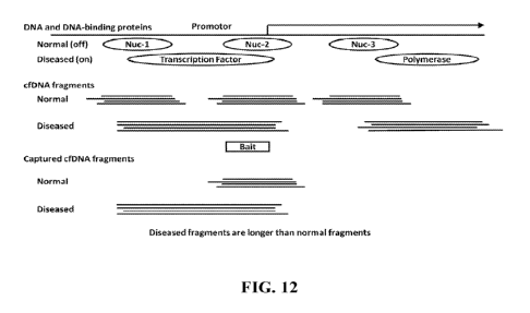
100271 FIGs. 12-18 illustrate various methods of distinguishing between two
cfNA
fragmentation patterns.
100281 FIG. 19 illustrates a Cap Analysis of Gene Expression (CAGE) signal in
the pl promoter
region of DUSP1 and a Transcriptionally Active Locus (TAL) with a cfNA
fragmentation
pattern that differs between transcriptionally silent and active states.
100291 FIG. 20 illustrates various processes used in conjunction with
hybridization capture.
100301 FIG. 21 illustrates the hybridization-based capture where a bait (or
probe) is
complementary to the nucleic acid sequence of a Transcriptionally Active Locus
(TAL).
100311 FIG. 22 illustrates an exemplary bait for capturing cfNA fragments
associated with the
TAL of DUSP1.
- 6 -
CA 03179853 2022- 11- 22
WO 2021/236993
PCT/US2021/033508
100321 FIG. 23 illustrates a simulation wherein cfNA fragments derived from
the TAL of
DUSP1 are captured by the exemplary bait.
100331 FIG. 24 illustrates a cfDNA fragment flanked by sequencing adapters.
The length of
sequence reads can be longer than the length of the cfDNA fragment.
100341 FIG. 25 illustrates a simulation wherein cfNA fragments derived from
the TAL of
DUSP1 are captured by the exemplary bait. The captured cfNA fragments at two
timepoints are
categorized into groups of long and short cfNA fragments. The right panel
illustrates a TAS
calculated from the fraction of long cfNA fragments.
100351 FIG. 26 illustrates Bioanalyzer traces of two cfNA samples Both samples
have a
predominant peak of shorter cfDNA fragments at approximately 167 base pairs,
which is the size
of cfDNA fragments protected by a mononucleosome. The trace in the right has a
higher fraction
of long cfDNA fragments indicative of transcriptional activity.
100361 FIG. 27 illustrates Bioanalyzer traces of cfNA samples captured by the
bait from the
TAL of DUSP1. The mononucleosome peak is shifted right because the cfNA is
flanked by
sequencing adaptors. The fraction of long cfDNA fragments is higher at
Timepoint 2.
100371 FIGs. 28A-B illustrate a method of distinguishing short and long
fragments from a
Bioanalyzer trace. The length of the short fragments is consistent with the
size of DNA protected
by a mononucleosome.
100381 FIG. 29 illustrates transcriptional activation scores determined from
cfDNA fragments
captured by the bait. The increase in measured TAS at Timepoint 2 is
consistent with
expectations from the NGS simulation of FIG. 25.
100391 FIGs. 30A-D illustrates a two-bait system for capturing cfDNA derived
from TALs
associated with two genes involved in glucocorticoid metabolism - DUSP1 and
SAEl.
100401 FIG. 31 compares simulated (NGS-based) and actual two-bait
transcriptional activation
scores using the two-bait system of FIG. 31 to analyze cfDNA isolated from
glucocorticoid
treatment experiment.
100411 FIG. 32 illustrates an N-bait composite read-out system.
100421 FIG. 33 illustrates a simulated example of characterizing cfDNA
fragments that
hybridize the bait by an alignment-free comparison of sequences of the cfDNA
fragments to a
reference sequence.
100431 FIG. 34 illustrates a simulated example of quantifying a relative
amount of cfDNA
fragment sequences aligning to sequences distal to an end of a bait.
- 7 -
CA 03179853 2022- 11- 22
WO 2021/236993
PCT/US2021/033508
100441 FIG. 35 illustrates a simulated example of characterizing cfDNA
fragments that
hybridize to a bait by counting a number of ctIDNA fragments comprising a
sequence matching
each of two or more identified subregions within a transcriptionally active
locus.
100451 FIG. 36 illustrates a simulated example of characterizing cfDNA
fragments that
hybridize to two baits within one transcriptionally active locus.
100461 FIG. 37 illustrates a simulated example of characterizing ctDNA
fragments that
hybridize to two baits within one transcriptionally active locus with
alignment free matching to
reference sequences indicative of long cfDNA fragments.
DETAILED DESCRIPTION
100471 While various embodiments of the invention have been shown and
described herein, it
will be obvious to those skilled in the art that such embodiments are provided
by way of example
only. Numerous variations, changes, and substitutions may occur to those
skilled in the art
without departing from the invention. It should be understood that various
alternatives to the
embodiments of the invention described herein may be employed.
100481 As used herein, the term "nucleic acid," generally refers to a
polymeric form of
nucleotides of any length (e.g., at least 2, 3, 4, 5, 6, 7, 8, 9, 10, 100,
500, 1000 or more
nucleotides), either deoxyribonucleotides or ribonucleotides, or analogs
thereof A nucleic acid
may include one or more subunits selected from adenosine (A), cytosine (C),
guanine (G),
thymine (TO, and uracil (U), or variants thereof A nucleotide can include A,
C, G, T, or U, or
variants thereof. A nucleotide can include any subunit that can be
incorporated into a growing
nucleic acid strand. Such subunit can be A, C, G, T, or U, or any other
subunit that is specific to
one of more complementary A, C, G, T, or U, or complementary to a purine
(e.g., A or G, or
variant thereof) or pyrimidine (e.g., C, T, or U, or variant thereof). In some
examples, a nucleic
acid may be single-stranded or double stranded, in some cases, a nucleic acid
molecule is
circular. Non-limiting examples of nucleic acids include deoxyribonucleic acid
(DNA) and
ribonucleic acid (RNA). Nucleic acids can include coding or non-coding regions
of a gene or
gene fragment, loci (locus) defined from linkage analysis, exons, introns,
messenger RNA
(mRNA), transfer RNA, ribosomal RNA, short interfering RNA (siRNA), short-
hairpin RNA
(shRNA), micro-RNA (miRNA), ribozymes, cDNA, recombinant nucleic acids,
branched
nucleic acids, plasmids, vectors, isolated DNA of any sequence, isolated RNA
of any sequence,
nucleic acid probes, and primers. A nucleic acid molecule may comprise one or
more modified
nucleotides, such as methylated nucleotides and nucleotide analogs.
- 8 -
CA 03179853 2022- 11- 22
WO 2021/236993
PCT/US2021/033508
100491 As used herein, the terms "express" and "expression" mean allowing or
causing the
information in a gene or DNA sequence to become manifest, for example
producing a protein by
activating the cellular functions involved in transcription and translation of
a corresponding gene
or DNA sequence. A DNA sequence is expressed in or by a cell to form an
"expression product"
such as a protein. The expression product itself, e.g. the resulting protein,
may also be the to be
"expressed" by the cell. An expression product can be characterized as
intracellular, extracellular
or transmembrane. The term "intracellular" means something that is inside a
cell. The term
"extracellular" means something that is outside a cell. The term transmembrane
means
something that has an extracellular domain outside the cell, a portion
embedded in the cell
membrane and an intracellular domain inside the cell.
100501 The term "sample", "biological sample", or "patient sample" as used
herein, generally
refers to any sample containing or suspected of containing a nucleic acid
molecule. For example,
a sample can be a biological sample containing one or more nucleic acid
molecules. The
biological sample can be obtained (e.g., extracted or isolated) from or
include blood (e.g., whole
blood), plasma, serum, umbilical cord blood, chorionic villi, amniotic fluid,
lavage fluid (e.g.,
bronchoalveolar, gastric, peritoneal, ductal, ear, arthroscopic), biopsy
sample (e.g., from pre-
implantation embryo), celocentesis sample, fetal nucleated cells or fetal
cellular remnants, bile,
breast milk, urine, saliva, mucosal excretions, sputum, stool, sweat, vaginal
fluid, fluid from a
hydrocele (e.g., of the testis), vaginal flushing fluids, pleural fluid,
ascitic fluid, cerebrospinal
fluid, bronchoalveolar lavage fluid, discharge fluid from the nipple,
aspiration fluid from
different parts of the body (e.g., thyroid, breast), tears, embryonic cells,
or fetal cells (e.g.,
placental cells). In some embodiments, a blood sample is obtained by a heel or
finger prick, from
scalp veins, or by ear lobe puncture. The biological sample can be a fluid or
tissue sample (e.g.,
skin sample). The biological sample can include any tissue or material derived
from a living or
dead subject. A biological sample can be a cell-free sample. A biological
sample can comprise a
nucleic acid (e.g., DNA or RNA) or a fragment thereof.
100511 The term "nucleic acid- can refer to deoxyribonucleic acid (DNA),
ribonucleic acid
(RNA) or any hybrid or fragment thereof. The nucleic acid in the sample can be
a cell-free
nucleic acid. A sample can be a liquid sample or a solid sample (e.g., a cell
or tissue sample). In
some examples, the sample is obtained from a cell-free bodily fluid, such as
whole blood. In
such instance, the sample may include cell-free DNA and/or cell-free RNA. In
some examples,
the majority of DNA in a biological sample that may be enriched for cfDNA
(e.g., a plasma
sample obtained via a centrifugation protocol) can be cell-free (e.g., greater
than 50%, 60%,
70%, 80%, 90%, 95%, or 99% of the DNA can be cell-free). A biological sample
can be treated
- 9 -
CA 03179853 2022- 11- 22
WO 2021/236993
PCT/US2021/033508
to physically disrupt tissue or cell structure (e.g., centrifugation and/or
cell lysis), thus releasing
intracellular components into a solution which can further contain enzymes,
buffers, salts,
detergents, and the like which can be used to prepare the sample for analysis.
In some examples,
the sample can include circulating tumor cells or circulating fetal cells.
100521 The term -whole blood sample", as used herein, generally refers to a
whole blood
sample that has not been fractionated or separated into its component parts.
Whole blood may be
combined with an anticoagulant such as EDTA or ACD during the collection
process but is
generally otherwise unprocessed. "Whole Blood" may refer to a specific
standardized product for
transfusion or further processing, or to any unmodified collected blood
100531 The term "blood fractionation", as used herein, generally refers to the
process of
fractionating whole blood or separating it into its component parts. This may
be done by
centrifuging the blood. The resulting components may be a clear solution of
blood plasma in the
upper phase (which can be separated into its own fractions), a buffy coat,
which is a thin layer of
leukocytes (white blood cells) mixed with platelets in the middle, and
erythrocytes (red blood
cells) at the bottom of a centrifuge tube in the hematocrit faction.
100541 The terms "blood plasma" or "plasma", as used herein, generally refers
to the straw-
colored/pale-yellow liquid component of blood that normally holds the blood
cells in whole
blood in suspension. Blood plasma makes up about 55% of total blood by volume.
It is the
intravascular fluid part of [extracellular fluid] (all body fluid outside of
cells). It is mostly water
(93% by volume), and contains dissolved proteins including albumins,
immunoglobulins, and
fibrinogen, glucose, clotting factors, electrolytes (Nat, Ca', Mg', HCO3 Cl"
etc.), hormones
and carbon dioxide. Blood serum is blood plasma without fibrinogen or the
other clotting factors
(i.e., whole blood minus both the cells and the clotting factors).
100551 As used herein, the term "cell-free deoxyribonucleic acid" (cfDNA), as
used herein,
generally refers to non-encapsulated DNA in bodily fluids, particularly blood.
cfDNA are
nucleic acid fragments that may enter the bloodstream during necrosis or
apoptosis. Fragments
of non-encapsulated DNA may be engulfed by macrophages or other immune cells.
cfDNA
fragments average around 170 base pairs in length and may be present in both
early and late
stage disease. cfDNA may be of fetal origin circulating in a pregnant mother,
derived from
recipient tissues in donated organs or cells, or may be released from
malignancies. cfDNA may
be utilized as a biomarker for the presence or progression of any pathology.
100561 The term "liquid biopsy," as used herein, generally refers to a non-
invasive or
minimally invasive laboratory test or assay (e.g., of a biological sample or
cell-free DNA). In
some instances, a liquid biopsy is performed on a plasma or serum sample
obtained by a simple
- 10 -
CA 03179853 2022- 11- 22
WO 2021/236993
PCT/US2021/033508
needle stick. Blood can be drawn at any time during the course of therapy and
allow for dynamic
monitoring of molecular changes rather than relying on a static time point.
Such "liquid biopsy"
assays may report measurements (e.g., minor allele frequencies, gene
expression, or protein
expression) of one or more pathology associated marker genes.
100571 The term -fragment" (e.g., a cfDNA fragment), as used herein, can refer
to a portion of
a polynucleotide or polypeptide sequence that comprises at least 3 contiguous
nucleotides. A
nucleic acid fragment can retain the biological activity and/or some
characteristics of the parent
polynucleotide. cfDNA may be shed as a fragment with different genetic and
epigenetic profiles
and in various lengths A fragment may be a short (small) fragment or a long
(large) fragment
and the size patterns of fragments, such as cfDNA fragments, may vary in
pathological
conditions.
10058] The term "fragmentation pattern", as used herein, generally refers to a
collection of
fragments, such as cfDNA fragments, present in a subject. The composition of a
fragmentation
pattern may depend upon a tissue of origin, pathological state, or progression
of a disease.
100591 As used herein, the terms "genomic region", "genomic position",
"genomic site", or
"genomic location" generally refer to a physical location on a genome or
chromosome, which
may be associated with a gene or a set of genes, or a portion of a nucleic
acid polymer (e.g., a
chromosome) that is contained within the human genome complement. The term can
relate to a
specific length of DNA. The location of a genome can be defined with respect
to either a
chromosomal band in the human genome or one or more specific nucleotide
positions in the
human genome.
100601 The terms "size profile" and "size distribution", as used herein,
generally relate to the
sizes of DNA fragments in a biological sample. A size profile can be a
histogram that provides a
distribution of an amount of DNA fragments at a variety of sizes. Various
statistical parameters
(also referred to as size parameters or just parameter) can distinguish one
size profile from
another. One parameter can be the percentage of DNA fragment of a size or
range of sizes
relative to all DNA fragments or relative to DNA fragments of another size or
range. A "size
profile" or "size distribution" may represent the size of cfDNA fragments
derived from a specific
locus or specified loci of the genome.
100611 As used herein, the term "exome- generally refers to the subset of the
genome
composed of exons, the sequences which, when transcribed, remain within the
mature RNA after
introns are removed by RNA splicing and contribute to the final protein
product encoded by that
gene.
-11 -
CA 03179853 2022- 11- 22
WO 2021/236993
PCT/US2021/033508
100621 As used herein, the term "nucleosome" generally refers to a section of
DNA that is
wrapped around a core of proteins responsible in part for the compactness of a
chromosome. In
the nucleus, DNA forms a complex with proteins called chromatin which allows
the DNA to be
condensed into a smaller volume. A nucleosome is the fundamental subunit of
chromatin. A
nucleosome is composed of approximately two turns of DNA wrapped around a set
of eight core
histones.
100631 The term "oligonucleotide", as used herein, generally refers to a
nucleic acid molecule
comprising at least one nucleotide that may have various lengths such as
either
deoxyribonucleotides or ribonucleotides or analogs thereof An oligonucleotide
may comprise at
least about 2, 3, 4, 5, 6, 7, 8, 9, 10, 11, 12, 13, 14, 15, 16, 17, 18, 19,
20, 25, 30, 35, 40, 45, 50,
60, 70, 80, 90, 100, 125, 150, 175, 200, 250, 300, 400, 500, 600, 700, 800,
900, 1,000, 5,000,
10,000, 50,000, 100,000 or more nucleotides. An oligonucleotide may comprise
at most about
100,000, 50,000, 10,000, 5,000, 1,000, 900, 800, 700, 600, 500, 400, 300, 250,
200, 175, 150,
125, 100, 90, 80, 70, 60, 50, 45, 40, 35, 30, 25, 20, 19, 18, 17, 16, 15, 14,
13, 12, 11, 10,9, 8, 7,
6, 5, 4, 3, 2, or less nucleotides. An oligonucleotide may be unbound (e.g.,
in solution) or bound
(e.g., chemically bonded to a substrate). Oligonucleotides may include one or
more nonstandard
nucleotide(s), nucleotide analog(s), modified nucleotides, or any combination
thereof.
100641 The term "bait", as used herein, generally refers to a synthetic
oligonucleotide which,
when left to hybridize over a period of time, can capture a nucleic acid
fragment with a
complementary sequence. Baits may be various sizes, may be labeled or
unlabeled, and can
target multiple overlapping and/or non-overlapping genomic regions. Baits may
enable
preferential capture of nucleic acid fragments associated with molecular
functions of interest.
100651 The term "bait pool", as used herein, generally refers to a collection
or panel of baits
with a targeted capture profile. A bait pool may represent an optimized
combination of bait
sequences that target cfNA fragments of interest.
100661 The term "functional typing", as used herein, generally refers to
predicting a
pathological condition probability based on a comparison between the estimated
fractional
representation and a predetermined association of one or more distinct
components with clinical
reference data. Functional typing may be determined through a feature profile
for the designed
bait pools and estimating the fractional representation of one or more pool
components relative
to a combination of other components based on a set of regression
coefficients.
100671 The term "chromatin", as used herein, generally refers to the
nucleoprotein structure
that comprises the cellular genome. Cellular chromatin includes nucleic acid,
primarily DNA,
and protein, including histones and non-histone chromosomal proteins. The
majority of the
- 12 -
CA 03179853 2022- 11- 22
WO 2021/236993
PCT/US2021/033508
eukaryotic cellular chromatin is in the form of nucleosomes, with one
nucleosome core
comprising about 150 DNA base pairs associated with an octamer comprising two
each of the
histones H2A, H2B, H3 and H4. Linker DNA (of variable length depending on the
organism)
extends between nucleosome nuclei. A histone HI protein is generally
associated with the linker
DNA. Cellular chromatin includes both chromosomal and episomal chromatin.
100681 The term "epigenetic", as used herein, generally refers to refers to
information encoded
"on top of' or "in addition to" the traditional genetic basis for inheritance,
i.e. typically does not
include modifications to the underlying sequence (genetic code). An epigenetic
alteration is a
stable alteration in gene expression potential mediated by mechanisms other
than alterations in
the primary nucleotide sequence of a gene. The epigenome is an aggregate of
heritable cellular
markers, such as histone modifications or DNA methylation, that may control
the differential
expression of genes. An epigenetic alteration may be due to environmental
conditions causing
chemical modifications to these heritable cellular markers. These alterations
may be
transgenerational. Assessing or determining an epigenetic profile includes
detecting changes in
the transcriptome and reaction of DNA with bisulfite to modify unmethylated
cysteines.
100691 The term "cell death", as used herein, generally refers to an
irreversible event in which a
cell ceases to carry out its functions. Cell death may occur within a broader
physiological context
such as in embryonic development or tissue renewal, or it may be a pathologic
response to cell
injury or infectious pathogens. Apoptosis is a programmed form of cell death
in multicellular
organisms. Cell death may occur due to autophagy wherein there is
sequestration of cytoplasm
and organelles in double or multimembrane vesicles and delivery to the cells
own lysosomes for
subsequent degradation. Cell death may be due to necrosis, a toxic process,
where the cell
follows an energy independent mode of death and degradative processes that
occur after cell
death. While apoptosis may be accompanied by cell shrinkage, pyknosis, and
karyorrhexis,
oncosis is a process induced by energy depletion that leads to necrosis with
karyolysis
characterized by cell swelling. Cell death may also occur through pyroptosis,
an inflammatory
programmed cell death triggered by pathologic stimuli or inflammatory host
factors which may
form an immune response to such pathological conditions.
100701 The term "cellular debris" or "cell debris", as used herein, generally
refers to the
organic waste left over after a cell dies. Cellular debris may be further
processed and catabolized
by phagocytes.
100711 The term "hybridization," as used herein, generally refers to the
phenomenon in which a
single stranded nucleic acid anneals to a nucleic acid with a complementary
sequence.
- 13 -
CA 03179853 2022- 11- 22
WO 2021/236993
PCT/US2021/033508
100721 The term "cancer" or "malignancy" as used herein, generally refers to
abnormal and
unregulated growth of tissue or cells wherein. A mass of tissue (a tumor) or
uncontrolled cells
can be defined as "benign" or "malignant" depending on the following
characteristics: degree of
cellular differentiation including morphology and functionality, rate of
growth, local invasion
and metastasis. A -benign" mass of tissue or cells can be well differentiated,
have
characteristically slower growth than a malignant mass of tissue or cells and
remain localized to
the site of origin. In addition, in some cases a benign mass of tissue or
cells does not have the
capacity to infiltrate, invade or metastasize to distant sites. A "malignant"
mass of tissue or cells
can be a poorly differentiated (anaplasia), have characteristically rapid
growth accompanied by
progressive infiltration, invasion, and destruction of the surrounding tissue.
Furthermore, a
malignant tumor can have the capacity to metastasize to distant sites.
100731 The term "disease progression" or "level of pathology", as used herein,
may refer to
whether a disease or pathology exists (i.e., a presence or absence), a stage
of a disease, the total
burden of the body, and/or other measure of a severity of a disease. The level
of pathology can
be used in various ways. For example, screening can check if a pathology is
present in someone
who is not known previously to have the pathology. Assessment can investigate
someone who
has been diagnosed with a pathology to monitor the progress of the condition
over time, study
the effectiveness of therapies or to determine the prognosis. Detection can
comprise 'screening'
or can comprise checking if someone, with suggestive features of a pathology
(e.g., symptoms or
other positive tests), has the pathological condition. A "level of pathology"
can refer to level of
pathology associated with a pathogen.
100741 The term "cancer progression" or "level of cancer" can refer to whether
cancer exists
(i.e., presence or absence), a stage of a cancer, a size of tumor, presence or
absence of metastasis,
the total tumor burden of the body, and/or other measure of a severity of a
cancer (e.g.,
recurrence of cancer). The level of cancer can be a number or other indicia,
such as symbols,
alphabet letters, and colors. The level can be zero. The level of cancer can
also include
premalignant or precancerous conditions (states) associated with mutations or
several mutations.
When the cancer is associated with a pathogen, a level of cancer can be a type
of a level of
pathology. The prognosis can be expressed as the chance of a patient dying of
cancer, or the
chance of the cancer progressing after a specific duration or time, or the
chance of cancer
metastasizing.
100751 Whenever the term "at least," "greater than," or "greater than or equal
to" precedes the
first numerical value in a series of two or more numerical values, the term
"at least," "greater
than" or "greater than or equal to" applies to each of the numerical values in
that series of
- 14 -
CA 03179853 2022- 11- 22
WO 2021/236993
PCT/US2021/033508
numerical values. For example, greater than or equal to 1, 2, or 3 is
equivalent to greater than or
equal to 1, greater than or equal to 2, or greater than or equal to 3.
100761 Whenever the term "no more than," "less than," or "less than or equal
to" precedes the
first numerical value in a series of two or more numerical values, the term
"no more than," "less
than," or -less than or equal to" applies to each of the numerical values in
that series of
numerical values. For example, less than or equal to 3, 2, or 1 is equivalent
to less than or equal
to 3, less than or equal to 2, or less than or equal to 1.
100771 The use of the word "a" or "an," when used in conjunction with the term
"comprising"
in the claims and/or the specification may mean "one," but it is also
consistent with the meaning
of "one or more," "at least one," and "one or more than one."
100781 The use of the term "or" in the claims is used to mean "and/or" unless
explicitly
indicated to refer to alternatives only or the alternatives are mutually
exclusive, although the
disclosure supports a definition that refers to only alternatives and "and/or.-
As used herein
"another- may mean at least a second or more.
100791 The term "about" is used to indicate that a value includes the inherent
variation of error
for the device, the method being employed to determine the value, or the
variation that exists
among the study subjects. Unless otherwise specified based upon the above
values, the term
"about" means 5% of the listed value.
100801 The terms "comprise," "have," and "include" are open-ended linking
verbs. Any forms
or tenses of one or more of these verbs, such as "comprises," "comprising,"
"has," "having,"
-includes," and -including," are also open-ended. For example, any method that
-comprises,"
"has," or "includes" one or more steps is not limited to possessing only those
one or more steps
and also covers other unlisted steps.
100811 The term "sequence context" as used herein refers to the nucleic acid
sequence
composition (e.g., DNA sequence composition). DNA sequence composition can be
used to
derive several context metrics that are relevant to underlying transcriptional
status of the locus,
such as individual base composition, GC percentage, number of CpG sites,
number of
informative differentially methylated CpGs (iDMCs), number of dinucleotide
repeat motifs
(DR1V1), occurrence profiles of known TF motifs, etc.
100821 The term "transcriptional activity score- or "TAS- as used herein
refers to a weighted
ratio of longer (non-canonical) fragment counts to a total abundance of
fragments at a locus. It
may also involve different features of a NA fragment length distribution, such
as clusters, gaps,
peaks, and outliers. Anomalies in observed fragment length distribution may
also be summarized
- 15 -
CA 03179853 2022- 11- 22
WO 2021/236993
PCT/US2021/033508
using deep learning that finds anomalous length patterns associated with
transcription. It may
involve learning and training using previously generated data.
100831 We confirmed that distribution of circulating NA fragments length at
transcriptionally
active loci is indicative of transcriptional activity of live cell
prior/during its death. Thus, a
transcriptional activity score can be derived based on the distribution and a
particular fragment
length band and the associated mode of the distribution can be identified as
canonical ¨
chromatin organization of the DNA unperturbed by protein binding, while the
rest of the bands
(or some of the remaining bands) and associated peaks would be labeled as non-
canonical and
represent NA fragments associated with protein binding, indicative of
transcription
100841 The discovery of circulating DNA and RNA in the plasma of healthy
individuals and
patients was made by Mandel and Metais in 1948 (Mandel and Metais, 1948) which
was
furthered in 1966 by Tan et al who observed an anomalous pattern of cell-free
deoxyribonucleic
acid (cfDNA) in patients who suffered from systemic lupus erythematosus (Tan
et al. 1966) ¨ an
autoimmune disease in which the major antigen is nucleosomal self-DNA. Despite
these early
discoveries, the relevance of circulating nucleic acids (CNAs) did not begin
to be explored until
the 1990s when the presence of tumor-derived oncogenic DNA was observed in the
plasma of
patients with cancer (Sorenson et al. 1994) and DNA of fetal origin was
detected in the maternal
circulation (Lo et al. 1997). These findings led to the subsequent
understanding that cfDNA
levels are increased in patients with chronic and acute pathologies, including
autoimmune
diseases, stroke and trauma (Butt and Swaminathan 2008, Wagner 2012),
suggesting the
concentration of cfDNA could serve as a non-invasive blood biomarker to
reflect the rate of
tissue damage, cellular death and turnover.
100851 Circulating cfDNA are relatively short double-stranded DNA fragments,
averaging
approximately 170 base pairs, and present in circulating plasma, urine, and
other bodily fluids. In
the plasma of healthy individuals, cfDNA may be derived primarily from the
apoptosis of cells
of hematopoietic origin, however other tissues may contribute to the
composition of cfDNA in
bodily fluids. While cfDNA has been used in specialties such as reproductive
medicine,
oncology, and transplant medicine, its use as a non-invasive method to screen
for, diagnose,
determine prognosis, and provide guidance in treatment may be applicable to
many other
pathologies and conditions.
100861 cfDNA may be analyzed with regard to the representation and
distribution of specific
sequences and epigenetic features, such as DNA digestion and/or methylation
patterns. In
addition to pathology-associated genetic variants, analysis of cfDNA may
reveal epigenetic
footprints and signatures of phagocytic removal of dying cells, which may
result from an
- 16 -
CA 03179853 2022- 11- 22
WO 2021/236993
PCT/US2021/033508
aggregate nucleosomal occupancy profile of present pathologies as well as
their
microenvironment components, such as tumor malignancies. cfDNA may be released
by various
host cells such as neutrophils, macrophages, eosinophils, as well as tumor
cells and may
accumulate in circulating plasma as a consequence of increased cell death
and/or activation,
impaired clearance of cfDNA, and/or decreases in levels of endogenous DNase
enzymes. cfDNA
circulating in a subject's bloodstream may be packed into membrane-coated
structures such as
apoptotic bodies and may be subsequently analyzed for the effects of these
structures on the
characteristics of cfDNA fragments.
100871 In a cell nucleus, DNA may exist in nucleosomes, structures comprising
a section of
DNA approximately 145 base pairs wrapped around a core histone octamer,
allowing DNA to be
condensed into a smaller volume into a chromatin complex. Electrostatic and
hydrogen-bonding
interactions of DNA and hi stone dimers may result in energetically
unfavorable bending of DNA
over the protein surface. Such bending may be sterically prohibitive to other
DNA-binding
proteins and may serve to regulate access to DNA in a cell nucleus. Nucleosome
positioning in a
cell may fluctuate dynamically over time and across various cell states and
conditions such as
partially unwrapping and rewrapping spontaneously. Since a fragmentation
pattern may reflect
histone-protected DNA fragments that originated from a configuration
influenced by
nucleosomal units, nucleosome stability and dynamics may influence such a
fragmentation
pattern. These nucleosome dynamics may stem from a variety of factors, such as
post-
translational modifications of histones through processes such as acetylation,
methylation,
phosphorylation, or ubiquitination, which may influence chromatin structure.
100881 Chromatin organization may differ depending on factors such as global
cellular identity,
metabolic state, regional regulatory state, local gene activity, cell death,
and mechanisms of
DNA clearance. All of these factors can influence to the manner in which DNA
is fragmented
after cell death, and consequently, the fragmentation pattern. However, cfDNA
fragmentation
patterns may be only partially attributed to the underlying chromatin
architecture of contributing
cells. The fragmentation pattern may also be indicative of the method of
chromatin compaction
during cell death and DNA protection from enzymatic digestion. The genomic
structure of a
given cell type or cell lineage type may only partially contribute to the
heterogeneity of DNA
accessibility due to changes in nucleosome stability, conformation, and
composition at various
stages of cell death or cellular debris trafficking. Additional filtering
mechanisms depending on
factors such as the mode and mechanism of death or cell clearance may
influence cfDNA
clearance and release into circulation, resulting in preferential presence or
absence of specific
cfDNA fragments.
- 17 -
CA 03179853 2022- 11- 22
WO 2021/236993
PCT/US2021/033508
100891 Informative cfDNA fragments may be generated in a cell and released
into blood
circulation or they may form as a consequence of nuclear DNA fragmentation
during processes
such as apoptosis, necrosis, autophagy, karyolysis, or pyroptosis wherein
different nuclease
enzymes act on DNA at difference stages of cell death. The transcriptional
status of a cell before
it dies may have equally important effects on the fragmentation pattern. cfDNA
from all of these
sources is intermingled in circulating blood. The resulting sequence-specific
DNA cleavage
patterns may be analyzed in cfDNA as clinically relevant markers The
intermingled cfDNA
fragments can be classified into distinct components corresponding to the
different states from
which they were derived These components and clearance factors may represent
markers that
can be used to differentiate between different states. A fragmentation pattern
may be analyzed by
identifying specific regions or features where one or more genetic or
epigenetic states, or one or
more clearance mechanisms, are sufficiently different to be used as a marker
indicative of
genetic aberrations or pathological conditions. Genetic aberrations that can
be measured or
inferred by fragmentation pattern analysis may comprise epigenetic variants or
changes which
may allow fragmentation pattern analysis to determine variations in chromatin
organization or
structures, which may be a consequence of genomic aberrations or epigenetic
changes in DNA.
100901 Another way to distinguish these patterns associated with cell function
prior to death
and/or the nature of cell death may be through mapping cfNA fragments to
custom reference
sequences representing different types of fragmentation and quantifying cfNA
fragments
associated with each reference. The cfNA fragments associated with different
custom references
may vary by size.
100911 A collection of synthetic oligonucleotide baits comprising sequences
complementary to
the sequences of specified genomic locations may capture cfNA fragments with
high sequence
homology to the baits. Novel baits may be designed to capture cell-free
fragments that are
specific to a given genomic location and size. Baits may be various sizes,
labeled, unlabeled, and
can represent multiple overlapping and/or non-overlapping genomic regions.
Baits may enable
preferential capture of specific fragments of various sizes associated with
molecular functions of
interest. Baits may be constructed based on a custom reference and target
those subsequences
that are most unique to a given fragmentation. A collection of baits with a
targeted capture
profile, a bait pool, may represent an optimized combination of bait sequences
to target disease-
related cfNA fragments and juxtapose pathological and normal states of
fragment sizes or
abundances. Analysis of fragments captured by a bait pool may enable
functional typing by
estimating the fractional representation of one or more pool components
relative to a
combination of other components. Bait sequence design may be driven by various
factors such as
- 18 -
CA 03179853 2022- 11- 22
WO 2021/236993
PCT/US2021/033508
the expected or empirically observed nucleic acid fragment density in a
targeted region,
sequence-specific thermodynamics with the free energy of the bait with nucleic
acid
hybridization described by a nearest neighbor (NN) model, and baits of various
size to enable
preferential capture of specific fragment lengths associated with a molecular
function of interest.
Different fragmentation patterns may be distinguished by targeting a genomic
region with a
combination of baits configured to capture nucleic acid fragments having
overlapping sequences
but varying in size. For example, custom references (keywords) may be designed
to represent
NA fragments that are abundantly or selectively present in a given
fragmentation pattern in a
genomic region Other sets of custom references may target different sequences
within the same
genomic region representing different fragmentation patterns. The relative
abundance of short
and long fragments derived from a genomic site (region) in a biological sample
can be quantified
by mapping the sequences of captured fragments to these keywords. Fragments
can be capture
by baits with the same or different sequences from keywords. This method does
not require
determining the absolute length of each captured fragment, mapping fragment
sequences to a
reference genome, or identifying the ends of individual fragments. cfNA
fragments derived from
a genomic region may be sequenced and the sequences matched using alignment-
free methods to
the keywords of a Custom References. The percentage of cfNA fragments matching
each
keyword or set of keywords correlates with the relative abundance of different
fragmentation
patterns.
Characterization and analysis of cfNA fragments
100921 Aspects of the present disclosure may extract cell-free nucleic acids
from the
bloodstream for use as a non-invasive method of detecting disease and
monitoring pathological
progression or a response to treatment. cfNA may be utilized as a biomarker
diagnostic
determining the presence of a given disease, prognostic determining the
outcome for a subject
with a disease, or predictive, determining the response of an individual to a
given therapy. Whole
blood may be obtained through a minimally invasive method such as a blood draw
or fingerstick.
Plasma may be isolated from the blood. Circulating nucleic acids may be
extracted from this
plasma through a nucleic acid extraction protocol, a custom workflow may
reduce the
complexity in plasma with no nucleic acid extraction, or nucleic acids may be
directing enriched
from peripheral blood such as through loop-mediated isothermal amplification
(LAMP) or
CRISPR-mediated, amplification-free target enrichment.
100931 Fragmented DNA and the small amounts of DNA common in cfNA
applications, may
be amplified prior to analysis. Nucleic acid amplification may refer to
generating one or more
copies or of a nucleic acid. Nucleic acids may be amplified by the polymerase
chain reaction
- 19 -
CA 03179853 2022- 11- 22
WO 2021/236993
PCT/US2021/033508
(PCR) for DNA or RT-PCR for RNA. Other nucleic amplification methods include
rolling circle
amplification (RCA) (Demidov, 2002), strand displacement amplification (SDA)
(Walker et al.,
1992), helicase-dependent amplification (HDA) (Vincent et al., 2004), nucleic
acid sequence-
based amplification (NASBA) (Deiman et al., 2002), and loop-mediated
amplification (LAMP)
(Notomi et al., 2000a). DNA may be amplified generating several millions of
copies of a specific
segment of DNA from a small amount of starting material, the template. Its
specificity may rely
on sequence hybridization and its sensitivity may rely on enzyme-based
amplification. A PCR
amplification method may comprise a series of temperature cycles repeated
wherein each cycle
denatures DNA duplexes, hybridizes DNA oligonucleotides (primers) flanking the
target
sequence, and elongates those primers by a DNA polymerase. A cfDNA fragment
may be
amplified using only one primer hybridizing to the fragment by ligating a
common primer site to
the ends of every fragment. Additionally, a nucleic acid amplification
technique that utilizes a
polymerase with helicase activity may be employed. The helicase activity may
allow for
amplification of DNA at a constant temperature, isothermal amplification, and
may be facilitated
by primers that form stem-loop DNA structures. Once formed, the stem-loop
structures may
become the template DNA for further amplification.
100941 Reducing the complexity of circulating nucleic acids to a clinically
relevant cell-free
fragment representation may comprise several distinct methods or a combination
thereof such as
selective enrichment, selective capture, or amplicon-based target enrichment.
Selective
enrichment may comprise using collections of synthetic nucleic acid baits
which, when left to
hybridize over a period of time, may capture cell-free NA fragments with high
homology to
these baits, and can represent multiple overlapping and/or non-overlapping
genomic regions. NA
fragments of specific sizes can be enriched by solid phase reversible
immobilization on magnetic
beads. The desired NAs may then be eluted from the beads. Amplicon-based
target enrichment
via PCR-amplification of target regions of interest may comprise using pre-
determined specific
primers.
100951 Nucleic acid thermodynamics may offer a unique approach in liquid
biopsy designs.
Many liquid biopsy designs may involve uniformly tiling a genome with
overlapping baits that
may comprise distinct thermodynamic parameters such as resting temperature.
These
thermodynamic incongruities may result in significant capture bias which may
mask underlying
fragment distributions, indicative of disease conditions. For example,
hybridization in bulk may
be characterized using a model that assumes the process occurs in two steps
_________ the binding of the
end of one strand with the complementary end of the other strand followed by a
"zipping" of the
remaining bases to create a double helix. Such model can be established and
then trained using
- 20 -
CA 03179853 2022- 11- 22
WO 2021/236993
PCT/US2021/033508
specific synthetic baits to produce custom bait panels with targeted capture
profile. Aspects of
the present disclosure may estimate and empirically test thermodynamic
parameters of a given
cell free nucleic acid sequence using approximation of the nearest-neighbor
model of nucleic
duplex formation constrained by observed nucleosomal occupancy. Enrichment
bias may be
minimized by thermodynamically protecting an optimized combination of bait
sequences which
target disease-related cfNA fragments and stabilize the melting temperature of
cfNA to bait
duplexes. These aspects may enable prioritization and targeting of specific
cfNA fragments
associated with cell type as related to a function of a disease of interest
and juxtapose
pathological states of fragment sizes or abundances to normal states Aspects
of the present
disclosure may not preserve or maintain the underlying cell-free fragment
abundance during
enrichment and may distort the underlying cell-free fragment abundance. The
presence of cell-
free fragments alone may be sufficient for an accurate readout.
100961 Aspects of the present disclosure may detect changes in keyword
representation in a set
of cfNA fragments through hybridization capture or amplification of non-
genomic sequences
that may not involve base mapping or positional awareness. A keyword sequence
may be a short
sequence. A keyword sequence may be 10, 20, 30, 40, 50, 60, 70, 80, 90, 100,
150, 200, 250,
300, 350, 400, 450, 500, 1000, 1500, or more than 1500 nucleotides. A keyword
sequence may
be 1500, 1000, 500, 450, 400, 350, 300, 250, 200, 150, 100, 90, 80, 70, 60,
50, 40, 30, 20, 10, or
less than 10 nucleotides. These keyword sequences may be mapped to the human
genome but
need not be. It may be important to know only the sequence of these keywords.
Each keyword in
a list of cfNA fragments may be substring searched and simply counted if it is
found. Some
cfNA fragments may have one keyword hit while some cfNA fragments will have
all keywords
and some cfNA fragments will have no keyword hits. A substring match may
comprise fuzzy
pattern mapping or may be straightforward where an exact subsequence on a cfNA
fragment
sequence is seen. Positionality of a keywording a fragment may be irrelevant
compared to just if
a keyword is found in a fragment. cfNA sequence information may not be
necessary in detecting
changes in keyword representation if baits are designed using these keywords
to capture
fragments using hybridization and homology. Baits may be mapped to the human
genome but
may not be mapped to the human genome as such mapping may not be necessary to
identify
keywords in cfNA fragments. Keywords may be designed to represent NA fragments
that are
most abundantly or selectively present in a given fragmentation pattern in a
genomic region.
Keywords may represent underlying transcriptional states thus enabling the
sorting of keyword
membership in a cfNA fragment and determination of changes in molecular
function between
timepoints and conditions. Targeting genomic regions with keywords may enable
diagnosis of
-21 -
CA 03179853 2022- 11- 22
WO 2021/236993
PCT/US2021/033508
any physiological or pathological condition, or monitor the progression of any
disease or
pathological condition, treated or untreated, including determining the
progression of a cancer,
such as pancreatic cancer, or detecting gene expression changes during a
course of treatment,
such as with steroids by comparing the size or abundance of captured cfNAs
with a
fragmentation pattern correlating with a molecular function of interest.
100971 An aspect of the present disclosure provides systems and methods for
characterizing a
fragmentation pattern of cell-free nucleic acid (cfNA) fragments derived from
genomic origin as
a non-invasive method to screen for, diagnose, determine prognosis, and
provide guidance in
treatment, and may be applicable to a variety of pathologies and conditions
cfNA may be a non-
encapsulated polymeric form of nucleotides of any length (e.g., at least 2, 3,
4, 5, 6, 7, 8, 9, 10,
100, 500, 1000 or more nucleotides), either deoxyribonucleotides or
ribonucleotides, or analogs
thereof. A nucleic acid may include one or more subunits selected from
adenosine (A), cytosine
(C), guanine (G), thymine (TO, and uracil (U), or variants thereof. A
nucleotide can include A,
C, G, T, or U, or variants thereof. A nucleotide can include any subunit that
can be incorporated
into a growing nucleic acid strand. Such subunit can be A, C, G, T, or U, or
any other subunit
that is specific to one of more complementary A, C, G, T, or U, or
complementary to a purine
(e.g., A or G, or variant thereof) or pyrimidine (e.g., C, T, or U, or variant
thereof). In some
examples, a nucleic acid may be single-stranded or double stranded, in some
cases, a nucleic
acid molecule is circular. Non-limiting examples of nucleic acids include
deoxyribonucleic acid
(DNA) and ribonucleic acid (RNA). Nucleic acids can include coding or non-
coding regions of a
gene or gene fragment, loci (locus) defined from linkage analysis, exons,
introns, messenger
RNA (mRNA), transfer RNA, ribosomal RNA, short interfering RNA (siRNA), short-
hairpin
RNA (shRNA), micro-RNA (miRNA), ribozymes, cDNA, recombinant nucleic acids,
branched
nucleic acids, plasmids, vectors, isolated DNA of any sequence, isolated RNA
of any sequence,
nucleic acid probes, and primers. A nucleic acid molecule may comprise one or
more modified
nucleotides, such as methylated nucleotides and nucleotide analogs. cfNA may
comprise a
singular fragment or may comprise a plurality of cfNA fragments. There may be
1, 2, 3, 4, 5, 6,
7, 8, 9, 10, 15, 20, 25, 30, 35, 40, 45, 50, 60, 70, 80, 90, 100, or more than
100 cfNA fragments
in a plurality of cfNA fragments. There may be fewer than about 100, 90, 80,
70, 60, 50, 45, 40,
35, 30, 25, 20, 15, 10, 9, 8, 7, 6, 5, 4, 3, 2, 1 or less than 1 cfNA
fragments in a plurality of cfNA
fragments.
100981 The presence of circulating nucleic acids (DNA and RNA) detectable in
the plasma and
serum of subjects with pathological conditions may be investigated to serve as
markers for
diagnostic or prognostic purposes due to the potential non-invasive nature of
sample acquisition.
- 22 -
CA 03179853 2022- 11- 22
WO 2021/236993
PCT/US2021/033508
For example, in cancer patients, it has been cfNA markers within the plasma
may be identical to
the ones found in the carcinogenic tissue of the patient. Circulating nucleic
acids may comprise
cell-free DNA or cell-free RNA. Circulating RNA may particularly of interest
for use in early
detection cancer screenings due to RNA markers close association with
malignancy.
[0099] cfNA may be derived from a biological sample from a subject. A
biological sample may
be any sample containing or suspected of containing a nucleic acid molecule.
For example, a
sample can be a biological sample containing one or more nucleic acid
molecules. The biological
sample can be obtained (e.g., extracted or isolated) from or include blood
(e.g., whole blood),
plasma, serum, umbilical cord blood, chorionic villi, amniotic fluid, lavage
fluid (e.g.,
bronchoalveolar, gastric, peritoneal, ductal, ear, arthroscopic), biopsy
sample (e.g., from pre-
implantation embryo), celocentesis sample, fetal nucleated cells or fetal
cellular remnants, bile,
breast milk, urine, saliva, mucosa] excretions, sputum, stool, sweat, vaginal
fluid, fluid from a
hydrocele (e.g., of the testis), vaginal flushing fluids, pleural fluid,
ascitic fluid, cerebrospinal
fluid, bronchoalveolar lavage fluid, discharge fluid from the nipple,
aspiration fluid from
different parts of the body (e.g., thyroid, breast), tears, embryonic cells,
or fetal cells (e.g.,
placental cells). The biological sample can be a fluid or tissue sample (e.g.,
skin sample). The
biological sample can include any tissue or material derived from a living or
dead subject. A
biological sample can be a cell-free sample. A biological sample can comprise
a nucleic acid
(e.g., DNA or RNA) or a fragment thereof.
[0100] A sample may be heterogeneous, wherein more than one type of nucleic
acid species
may be present in the sample. For example, heterogeneous nucleic acids can
include, but are not
limited to, (i) fetal derived and maternal derived nucleic acids, (ii) cancer
and non-cancer nucleic
acids, (iii) pathogen and host nucleic acids, and more generally, (iv) mutated
and wild-type
nucleic acids. A sample may be heterogeneous because more than one cell type
is present, such
as a fetal cell and a maternal cell, a cancer and non-cancer cell, or a
pathogenic and host cell. A
minority nucleic acid species and a majority nucleic acid species may be
present.
[0101] Subjects can be humans, non-human primates such as chimpanzees, and
other apes and
monkey species; farm animals such as cattle, horses, sheep, goats, swine;
domestic animals such
as rabbits, dogs, and cats; laboratory animals including rodents, such as
rats, mice and guinea
pigs, and the like. A subject can be of any age. Subjects can be, for example,
elderly adults,
adults, adolescents, pre-adolescents, children, toddlers, infants. A subject
may be a patient with a
disease and/or a lab animal with a condition.
[0102] A composition comprising cfNA may be contacted with an oligonucleotide
bait. An
oligonucleotide bait comprising synthetic nucleic acid bases complementary to
a genomic
- 23 -
CA 03179853 2022- 11- 22
WO 2021/236993
PCT/US2021/033508
location may capture cfNA fragments with high homology to the baits. An
oligonucleotide may
be synthesized, or amplification products may be generated as baits for
genomic targets of
interest and affixed to a capture modality such as biotinylation and bound to
streptavidin coated
magnetic beads for solution-based capture. Bound nucleic acids may serve as
bait for capturing
homologous cfNA fragments. Homologous cfNA fragments from a library that match
the bait
sequence may serve as targets. After purification of the target enriched
library, cfNA fragments
with homology to the baits may be enriched, and non-targeted sequences
removed. An
oligonucleotide bait may be of natural origin. Novel baits may be designed to
capture targeted
cell-free fragments that are specific to a given genomic location and size
Baits may be various
sizes. An oligonucleotide bait may be may comprise at least about 2, 3, 4, 5,
6, 7, 8, 9, 10, 11, 12,
13, 14, 15, 16, 17, 18, 19, 20, 25, 30, 35, 40, 45, 50, 60, 70, 80, 90, 100,
125, 150, 175, 200, 250,
300, 400, 500, 600, 700, 800, 900, 1,000, 5,000, 10,000, 50,000, 100,000 or
more nucleotides.
An oligonucleotide bait may comprise at most about 100,000, 50,000, 10,000,
5,000, 1,000, 900,
800, 700, 600, 500, 400, 300, 250, 200, 175, 150, 125, 100, 90, 80, 70, 60,
50, 45, 40, 35, 30, 25,
20, 19, 18, 17, 16, 15, 14, 13, 12, 11, 10, 9, 8, 7, 6, 5, 4, 3, 2, or less
nucleotides. An
oligonucleotide bait may be unbound (e.g., in solution) or bound (e.g.,
chemically bonded to a
substrate). Oligonucleotide baits may include one or more nonstandard
nucleotide(s), nucleotide
analog(s), modified nucleotides, or any combination thereof. Oligonucleotide
baits may be
labeled or unlabeled and may represent multiple overlapping and/or non-
overlapping genomic
regions. There may be one oligonucleotide bait or a plurality of
oligonucleotide baits. There may
be greater than about 1, 2, 3, 4, 5, 6, 7, 8, 9, 10, 15, 20, 25, 30, 35, 40,
45, 50, 60, 70, 80, 90, 100,
or more than 100 oligonucleotide baits in a plurality of oligonucleotide
baits. There may be
fewer than about 100, 90, 80, 70, 60, 50, 45, 40, 35, 30, 25, 20, 15, 10, 9,
8, 7, 6, 5, 4, or 3
oligonucleotide baits in a plurality of oligonucleotide baits. An
oligonucleotide bait may be
conjugated to an affinity tag. An affinity tag may be but is not limited to
biotin, albumin binding
protein, alkaline phosphatase, horseradish peroxidase, chloramphenicol acetyl
transferase,
maltose binding protein, hexahistidine tag, glutathione-S-transferase, or 13
galactosidase.
[0103] A bait may comprise a plurality of sets of oligonucleotides with each
set specific to a
different molecular function of interest. In a plurality of oligonucleotide
baits each
oligonucleotide bait may comprise a distinct affinity label, such as a
fluorescent label. A labeling
reagent may be a thiol containing fluorophore. A fluorophore may be a xanthene
dye such as a
rhodamine dye, Alexa Fluor dye, an Atto dye, a fluorescent peptide or
protein, or a quantum
dot. Fluorescent methods may employ such fluorescent techniques such as
fluorescence
polarization, fluorimetry and fluorescence microscopy, FOrster resonance
energy transfer
- 24 -
CA 03179853 2022- 11- 22
WO 2021/236993
PCT/US2021/033508
(FRET), or time-resolved fluorescence. Fluorescence microscopy may be used to
determine the
presence of one or more fluorophores.
[0104] Baits of various size may enable preferential capture of specific
fragment length
associated with molecular functions of interest. Baits may be constructed
based on the custom
reference and target those subsequences that are most representative of or
best able to a given
fragmentation state. A bait pool or collection of baits with a targeted
capture profile, a bait pool,
may represent an optimized combination of bait sequences to target disease-
related cfNA
fragments and juxtapose pathological and normal states of fragment sizes or
abundances. cfNA
fragments captured by a bait pool may enable functional typing by estimating
the fractional
representation of one or more pool components relative to a combination of
other components a
fragment may hybridize to.
[0105] An oligonucleotide bait may be conjugated to a solid surface. A solid
surface may be
any suitable material which can be surface modified to incorporate a binding
partner to an
immobilization tag. A solid surface may comprise magnetic beads which
facilitate removal of
bait and captured target of interest. A solid surface may comprise the surface
of resins, gels,
quartz particles, or combinations thereof. In some non-limiting examples, the
methods
contemplate using oligonucleotide baits that have been immobilized on the
support of an
aminosilane modified surface, Tentagel beads, Tentagel resins, or other
similar beads or
resins. The surface used herein may be a hydrogel, such as alginate. The
surface used herein
may be coated with a polymer, such as polyethylene glycol. Fluoropolymers
(Teflon-AF
(Dupont), Cytop (Asahi Glass, Japan)), aromatic polymers (polyxylenes
(Parylene, Kisco,
Calif.), polystyrene, polymethmethylacrytate) and metal surfaces (gold
coating)), coating
schemes (spin-coating, dip-coating, electron beam deposition for metals,
thermal vapor
deposition and plasma enhanced chemical vapor deposition) and
functionalization methodologies
(polyallylamine grafting, use of ammonia gas in PECVD, doping of long chain
end-
functionalized fluorous alkanes etc) may be used in the methods described
herein as a useful
surface. A solid support may be conjugated with different addressable makers.
101061 A solid surface may be a bead. A bead may be a polymer such as a
polystyrene bead or
polystyrene cross-linked with divinylbenzene. The solid support bead may
comprise an iron
oxide core. A bead may comprise a metal salt such as a copper salt, a
magnesium salt, a calcium
salt, or a manganese salt. A bead may be cellulose, cellulose derivatives,
gelatin, acrylic resins,
glass, silica gels, polyvinyl pyrrolidine (PVP), co-polymers of vinyl and
acrylamide,
polyacrylamides, latex gels, dextran, crosslinked dextrans (e.g., SephadexTm),
rubber, silicon,
plastics, nitrocellulose, natural sponges, metal, and agarose gel
(SepharoseTm). The bead
- 25 -
CA 03179853 2022- 11- 22
WO 2021/236993
PCT/US2021/033508
diameter may depend on the density of the oligonucleotide bait or sample
assayed requiring
smaller or larger beads. The bead may have a diameter of at least about 1
micrometer (p.m), 5
gm, 10 gm, 25 gm, 50 gm, 75 gm, 100 gm, 150 gm, 200 gm, 250 gm, 300 gm, 400
gm, 500
gm, 750 gm, 1,000 gm, or more micrometers. The bead may have a diameter of at
most about
1,000 gm, 750 gm, 500 gm, 400 gm, 300 gm, 250 gm, 200 gm, 150 gm, 100 gm, 75
gm, 50
gm, 25 gm, 10 gm, 5 gm, 1 gm, or less than 1 micrometers. A. oligonucleotide
bait may be
coupled to a functional unit on the surface of the bead. A bead may be a
single bead or may be
among a plurality of beads. The plurality of beads may comprise at least 1,000
wells. There may
be 2, 3, 4, 5, 6, 7, g, 9, 10, 15, 20, 25, 30, 35, 40, 45, 50, 60, 70, 80, 90,
100, 200, 300, 400, 500,
1000, 1500, 2000, 3000, 4000, 5000, 10,000, 100,000, 1,000,000 or more than
1,000,000 wells
in a plurality of beads.
101071 A solid support may be a planar surface. A planar surface may be the
interior of a well.
A well may have a dimension of x by y by z, where x, y, and z are each
independently at least
about 0.1 gm, 1 gm, 5 gm, 10 pm, 15 gm, 20 pm, 25 gm, 30 gm, 35 gm, 40 gm, 45
gm, 50 gm,
55 gm, 60 gm, 65 gm, 70 gm, 75 gm, 80 gm, 85 gm, 90 gm, 95 gm, 100 gm, 110 gm,
120 gm,
130 gm, 140 gm, 150 gm, 160 gm, 170 gm, 180 p.m, 190 gm, 200 gm, 250 gm, 300
gm, 400
gm, 500 gm, 600 gm, 700 p.m, 800 gm, 900 gm, 1,000 gm, or more micrometers. A
well may
have a dimension of x by y by z, where x, y, and z are each independently at
most about 1,000
gm, 900 gm, 800 gm, 700 gm, 600 gm, 500 gm, 400 gm, 300 gm, 250 gm, 200 gm,
190 gm,
180 gm, 170 gm, 160 gm, 150 gm, 140 gm, 130 gm, 120 gm, 110 gm, 100 gm, 95 gm,
90 gm,
85 gm, 80 gm, 75 gm, 70 gm, 65 gm, 60 gm, 55 gm, 50 gm, 45 gm, 40 gm, 35 gm,
30 gm, 25
gm, 20 gm, 15 gm, 10 gm, 5 gm, 1 gm, 0.1 gm, or less micrometers. For example,
a well can
have an x dimension of 434 gm, a y dimension of 30 gm, and a z dimension of
510 p.m. In
another example, a well can have an x and y dimension of 16 gm and a z
dimension of 1 gm.
The planar surface may be a well among a plurality of wells. The plurality of
wells may
comprise at least two wells. The plurality of wells may comprise at least
1,000 wells. There may
be 2, 3, 4, 5, 6, 7, 8, 9, 10, 15, 20, 25, 30, 35, 40, 45, 50, 60, 70, 80, 90,
100, 200, 300, 400, 500,
1000, 1500, 2000, 3000, 4000, 5000, 10,000, 100,000, 1,000,000 or more than
1,000,000 wells
in a plurality of wells. A well may comprise a bead or a planar surface may
incorporate a bead.
101081 An oligonucleotide bait may hybridize to and capture cfNA fragments and
the
fragmentation pattern of the captured cfNA fragments may be characterized. The
plurality of
cell-free nucleic acids that hybridize to a bait oligonucleotide may be at
least about 70%, 75%,
80%, 85%, 90%, 95%, 97%, 98%, 99%, or more complementary to an oligonucleotide
bait
sequence or plurality of oligonucleotide bait sequences. The plurality of cell-
free nucleic acid
- 26 -
CA 03179853 2022- 11- 22
WO 2021/236993
PCT/US2021/033508
molecules may be at most about 99%, 98%, 97%, 95%, 90%, 85%, 80% or less
complementary
to an oligonucleotide bait sequence or plurality of oligonucleotide bait
sequences. Non-
hybridized cfNA sequences may be separated from hybridized target sequences,
thereby
uniformly enriching a population of cell-free DNA fragments with high
discrimination potential.
[0109] A fragmentation pattern may detect, diagnose, monitor the progress of a
condition over
time, study the effectiveness of therapies, or determine the prognosis of a
disease or pathological
condition. Non-limiting examples of diseases or conditions that may be
diagnosed or monitored
with a non-invasive cfNA diagnostic may include hematological malignancies,
solid tumor
malignancies, metastatic cancer, benign tumors, HIV/AIDS, autoimmune disease,
hepatitis B,
hepatitis C, rheumatoid arthritis, multiple sclerosis, psoriasis, uveitis,
scleroderma, systemic
lupus erythematosus, diabetes mellitus, eczema, Parkinson's disease,
congenital disease, genetic
abnormalities, or Alzheimer's disease.
[0110] A fragmentation pattern may detect, diagnose, monitor the progress of a
cancer over
time, study the effectiveness of therapies, or determine the prognosis of a
cancer. While
individual malignancies may provide unique genomes of malignant cells,
fragment analysis may
also comprise those of normal non-aberrant cells associated with the presence
of a tumor. Such
fragment analysis may be diagnostic and clinically relevant but may not be
directly of a
cancerous origin as tumor vasculature may comprise normal cells as well as
malignant cells.
Non-limiting examples of cancers that may be diagnosed or monitored with a non-
invasive cfNA
diagnostic include: acute lymphoblastic leukemia, acute myeloid leukemia,
adrenocortical
carcinoma, AIDS-related cancers, AIDS-related lymphoma, anal cancer, appendix
cancer,
astrocytoma, neuroblastoma, basal cell carcinoma, bile duct cancer, bladder
cancer, bone
cancers, brain tumors, such as cerebellar astrocytoma, cerebral
astrocytoma/malignant glioma,
ependymoma, medulloblastoma, supratentorial primitive neuroectodermal tumors,
visual
pathway and hypothalamic glioma, breast cancer, bronchial adenomas, Burkitt
lymphoma,
carcinoma of unknown primary origin, central nervous system lymphoma,
cerebellar
astrocytoma, cervical cancer, childhood cancers, chronic lymphocytic leukemia,
chronic
myelogenous leukemia, chronic myeloproliferative disorders, colon cancer,
cutaneous T-cell
lymphoma, desmoplastic small round cell tumor, endometrial cancer, ependymoma,
esophageal
cancer, Ewing's sarcoma, germ cell tumors, gallbladder cancer, gastric cancer,
gastrointestinal
carcinoid tumor, gastrointestinal stromal tumor, gliomas, hairy cell leukemia,
head and neck
cancer, heart cancer, hepatocellular (liver) cancer, Hodgkin lymphoma,
Hypopharyngeal cancer,
intraocular melanoma, islet cell carcinoma, Kaposi sarcoma, kidney cancer,
laryngeal cancer, lip
and oral cavity cancer, liposarcoma, liver cancer, lung cancers, such as non-
small cell and small
- 27 -
CA 03179853 2022- 11- 22
WO 2021/236993
PCT/US2021/033508
cell lung cancer, lymphomas, leukemias, macroglobulinemia, malignant fibrous
histiocytoma of
bone/osteosarcoma, medulloblastoma, melanomas, mesothelioma, metastatic
squamous neck
cancer with occult primary, mouth cancer, multiple endocrine neoplasia
syndrome,
myelodysplastic syndromes, myeloid leukemia, nasal cavity and paranasal sinus
cancer,
nasopharyngeal carcinoma, neuroblastoma, non-Hodgkin lymphoma, non-small cell
lung cancer,
oral cancer, oropharyngeal cancer, osteosarcoma/malignant fibrous histiocytoma
of bone,
ovarian cancer, ovarian epithelial cancer, ovarian germ cell tumor, pancreatic
cancer, pancreatic
cancer islet cell, paranasal sinus and nasal cavity cancer, parathyroid
cancer, penile cancer,
pharyngeal cancer, pheochromocytoma, pineal astrocytoma, pineal germinoma,
pituitary
adenoma, pleuropulmonary blastoma, plasma cell neoplasia, primary central
nervous system
lymphoma, prostate cancer, rectal cancer, renal cell carcinoma, renal pelvis
and ureter
transitional cell cancer, retinoblastoma, rhabdomyosarcoma, salivary gland
cancer, sarcomas,
skin cancers, skin carcinoma Merkel cell, small intestine cancer, soft tissue
sarcoma, squamous
cell carcinoma, stomach cancer, T-cell lymphoma, throat cancer, thymoma,
thymic carcinoma,
thyroid cancer, trophoblastic tumor (gestational), cancers of unknown primary
site, urethral
cancer, uterine sarcoma, vaginal cancer, vulvar cancer, Waldenstrom
macroglobulinemia, and
Wilms tumor.
101111 Aspects of the present disclosure may comprise a method of
characterizing a
fragmentation pattern of cfNA fragments derived from a genomic region wherein
characterizing
the fragmentation pattern of the cfNA fragments comprises analyzing sizes or
abundance of the
cfNA fragments. Characterizing a fragmentation pattern may comprise
identifying genomic
locations or lengths of cfNA fragments or may not comprise identifying genomic
locations of
lengths of the cfNA fragments. Fragments may be comprised of small/short or
large/long
fragments. The term "small fragment" and the term "short fragment" are
interchangeable. The
term "large fragment" and the tem]. "long fragment" are interchangeable. A
small fragment may
comprise fewer nucleotides than a large fragment. A small fragment may
comprise about 1, 2, 3,
4, 5, 6, 7, 8,9, 10, 11, 12, 13, 14, 15, 20. 25. 30, 35, 40, 45, 50, 60, 70,
80, 90, 100, 150, 200,
230, or more than 230 base pairs. A small fragment may comprise about less
than 230, 200, 150,
100, 90, 80, 70, 60, 50, 45, 40, 35, 30, 25, 20, 15, 10, 9, 8, 7, 6, 5, 4, 3,
2, or 1 base pair. A small
fragment may have a distribution centered around approximately 170 base pairs
and may be
indicative of mononucleosomal protection. A large fragment may comprise about,
50, 55, 60, 65,
70, 75, 80, 85, 90, 100, 150, 200, 250, 300, 350, 400, 450, 500, 600, 700,
800, 900, 1000, or
more than 1000 base pairs. A large fragment may comprise about 1000, 900, 800,
700, 600, 500,
450, 400, 350, 300, 250, 200, 150, 100, 90, 80, 70, 60, 50, or less than 50
base pairs. A large
- 28 -
CA 03179853 2022- 11- 22
WO 2021/236993
PCT/US2021/033508
fragment length may have a size distribution centered around approximately 330
base pairs and
may be indicative of dinucleosomal protection. The determination of a large or
a small cfNA
may be if a fragment is larger or smaller than 230 base pairs in length. An
amount of large cfNA
fragments comprising at least 230 nucleotides may be compared to a small cfNA
fragment
comprising less than 230 nucleotides. A large cfNA fragment may comprise at
least 185, 190,
200, 210, 220, 230, 240, 250, 255, 270, or 310 nucleotides. A small cfNA
fragment may
comprise less than 220, 205, 190, 175, 170, 160, 150, 140, 130, 120, or 110
nucleotides. An
increased abundance of large cfNA fragments may be indicative of a medical
condition. A ratio
of large cfNA fragments to small cfNA fragments of at least 001, 005, 01, 01,
025, 03, 035
or 0.4 may be indicative of a medical condition. A ratio of large cfNA
fragments to small cfNA
fragments may be at least .1, .15, .2, .25, .3, .35, .4, .45, .5, .6, .7, .8,
.9, or 1 and may be
indicative of a medical condition. A ratio of large cfNA to small cfNA
fragments may be less
than 1, .9, .8, .7, .6, .5, .45, .4, .35, .3, .25, .2, .15, .1, or less than
.1.
101121 Large and short cfNA fragments may span one exon or may span multiple
exons. cfNA
fragments may be analyzed with regard to the representation and distribution
of specific
sequences or epigenetic features such as DNA digestion or methylation
patterns. Fragmented
DNAs may be generated in a cell and released as cfDNA into blood circulation
as a result of
nuclear DNA fragmentation during cell processes such as apoptosis and
necrosis. Such
fragmentation may be produced as a result of different nuclease enzymes acting
on DNA in
different stages of cells, resulting in sequence-specific DNA cleavage
patterns which may be
analyzed in cfDNA fragmentation patterns. Classifying such clearance patterns
may be a
clinically relevant marker of cell environments (e.g., tumor
microenvironments, inflammation,
disease states, etc.). Fragmentation patterns may be analyzed by classifying
cfDNA fragments
into distinct components corresponding to the different chromatin states from
which they were
derived. For example, a fragmentation pattern may be expressed as a sum of
components
representing different underlying chromatin states.
101131 A specific genomic region may be identified as profile discriminators
using public
databases and/or empiric experimental data. For example, a subset of designed
baits may exhibit
enrichment bias for cfNA fragments observed in healthy non-diseased cfNA
samples while
another subset of baits may target genomic regions enriched in all or some
pathological
conditions examined during bait design. Cell-free NA targets that are bound to
baits may be
quantified. Methods for analyzing sizes of cfNA fragments or quantifying cfNAs
may include,
but are not limited to, gas chromatography, supercritical fluid
chromatography, liquid
chromatography (including partition chromatography, adsorption chromatography,
ion exchange
- 29 -
CA 03179853 2022- 11- 22
WO 2021/236993
PCT/US2021/033508
chromatography, size exclusion chromatography, thin-layer chromatography, and
affinity
chromatography), electrophoresis (including capillary electrophoresis,
capillary zone
electrophoresis, capillary isoelectric focusing, capillary
electrochromatography, micellar
electrokinetic capillary chromatography, isotachophoresis, transient
isotachophoresis and
capillary gel electrophoresis), comparative genomic hybridization (CGH),
microarrays, bead
arrays, and high-throughput genotyping such as with the use of molecular
inversion probe (MW).
Pathological conditions may be predicted with a pathological condition
probability based on a
comparison between the estimated fractional representation and a predetermined
association of
the one or more distinct components with clinical reference data
101141 cfNA fragments may be separated with microfluidic separation.
Microfluidic separation
may comprise a microfluidic cassette or "chip". A microfluidic chip may
comprise a solid
surface such as a polystyrene which may combine nucleic acid isolation by
solid-phase
extraction; isothermal enzymatic amplification such as Loop-mediated
AMPlification (LAMP),
Nucleic Acid Sequence Based Amplification (NASBA), or Recombinase Polymerase
Amplification (RPA); and real-time optical detection of cfNA analytes. A
microfluidic cassette
may incorporate an embedded nucleic acid binding membrane in an amplification
reaction
chamber. Target nucleic acids extracted from a lysate may be captured on the
membrane and
amplified at a constant incubation temperature. The amplification product may
be labeled with a
fluorophore reporter but need not be. A fluorophore reporter may be excited
with a LED light
source and monitored in situ in real time with a photodiode or a CCD detector.
For whole blood
analysis, a filtration device that separates plasma from whole blood to
provide cell-free samples
may be utilized. A microfluidic chip may utilize a consistent flow design or
an oscillatory flow
design. In a consistent flow design, nucleic acids, droplets, or solution may
be in continuous-
flow. A solution may be viscous or non-viscous. Nucleic acids, droplets, or
solution may be
stationary or semi-stationary. Nucleic acids, droplets, or solution may be in
motion. A
microfluidic chip may utilize oscillating or bidirectional flow. A
microfluidic chip may combine
the cycling flexibility of a stationary chamber-based system and the fast
dynamics of a
continuous flow system. Nucleic acids, droplets, or solution may be
transported back and forth
through a single channel or may be transported in multiple channels or
capillaries. The
channel(s) may span various temperature zones. A microfluidic chip may be
attached to a
pumping system such as but not limited to external pumps and integrated
micropumps. There
may be on board power or an external power source. Centrifugal force and/or
capillary forces
may be used to control the fluid flow. A compact disc format may be used to
house the reaction
chambers or other components. A droplet may serve as a reactor environment
allowing for fast
- 30 -
CA 03179853 2022- 11- 22
WO 2021/236993
PCT/US2021/033508
reagent mixing and minimum surface adsorption. Interfacial chemistry may be
used to create
such a reactor droplet (e.g. an oil-water plug may be flowed through a fluid
capillary to create a
water-in-oil droplet).
101151 To connect specific baits representative of a molecular function, a
deconvolution
analysis may be performed. A molecular function may be assigned to bait
capture products with
a series of calibrating experiments where a benchmark dataset may represent a
known molecular
function state. For example, 50 baits most enriched for molecular functions
can be identified
using moderated t-tests and fold changes. Scores for each molecular function
signature may be
assessed in disease profiles by computing fragment counts in that subtype
relative to all others
and calculating an arithmetic mean of the fragment counts. Scores may be
grouped by
hierarchical clustering according to Euclidian distance. Alternative
deconvolution methods may
be used, such as maximum likelihood/Conjugate gradient, quadratic programming,
non-negative
Matrix Factorization, v-Support Vector Regression, quadratic programming,
Unified Particle
Swarm Optimization, or a Latent Dirichlet Allocation (LDA) model. Using
fragment counts
associated with one of collection of baits, mixture proportions may be
estimated based on
benchmark counting data, or the number of cell/tissues type de novo may be
inferred using the
following approach:
101161 IfS is an n > k bait-specific fragment count matrix that contains k
cell types and
n genes, W may be akxp matrix where each column of W contains the frequencies
of k cell
types in a particular observation, and 0 may be an n >p count matrix that may
contain the
observed bait-specific fragment counts level, where n may represent the number
of genes
and p may be the number of observed tissue samples. The mixing process can be
modeled
through a linear model:
0=SxW (Equation 1)
101171 where S may represent the source signal, W may be the weight matrix for
cell type
frequencies, and 0 may be the observation on tissue samples. In a typical
fragment counts
profiling setting, 0 may be measured through microarray or RNA-seq. Both W, a
cell type
frequency matrix, and S may be unknown, where both S and W may need be
estimated. W may
be estimated using cell type specific markers and a linear model solved using
the estimated W.
101181 As a set of genes may have high fragment counts in a specific cell type
and low counts
in all other cell types, the proportion of each cell type present in a blood
sample may be
predicted using these genes. For example, Xs may be an in k matrix that
contains m cell type
specific genes for k cell types. In each cell type, there may be multiple cell
type specific genes.
As each gene may have higher bait-specific fragment counts in a single cell
type, an average of
- 3 1 -
CA 03179853 2022- 11- 22
WO 2021/236993
PCT/US2021/033508
all the genes that are highly expressed in a single cell type may be
determined and solve the
matrix as VS:
!/11 U_ (I \\
9.21 , ()
0 9,1? õ .
g12 = = = =
. .
0
0 = = = !kik /
y õ 0
g., ...
0 0 ' 0
...
101191 VS may be unknown, however, the corresponding fragment counts for cell
type
specific markers, Os and Os may be measured on the observed mixed samples.
Substituting VS and Os to Equation 1, it may be obtained
Os = Xs X W (Equation 2)
101201 VS may be a diagonal matrix, thus each side of Equation 2 may be
multiplied by
the V¨ 1S and Equation 3 may be obtained:
1 X ¨ ¨ (Equation 3) - W s ¨
101211 As W may be a frequency matrix and each column of W may sum to 1, a
system of
linear questions of k unknown parameters, g1 .. g k, may be formed where:
-,--= =
(Equation 4)
s
ij
1
101221 Where the number of observations on the mixed samples are greater the
number of cell
types involved that is p >k, the system of equations may be solved with k
unknown parameters.
Where g 1...g k may be known, V-1S may be taken into Equation 3 and the cell
type
frequency matrix may be computed.
- 32 -
CA 03179853 2022- 11- 22
WO 2021/236993
PCT/US2021/033508
101231 In digital sorting on blood samples, cfNA fragment count data in blood
samples and a
set of gene symbols known to have high bait-specific fragment counts in a
specific cell type may
be input to obtain a fragment count profile for each of the cell types in a
blood sample. If Wis
not known, W may be estimated using Xs' and Equation 3. If W is known, S may
be estimated
through quadratic programming where 0 may be the fragment counts profile in
blood
samplesõ S may be the bait-specific fragment count profile for pure cell
types, W may be the
weight matrix estimated using the marker genes, and ti and t2 may be the
maximum and
minimum measurable fragment counts level:
min 0¨SW 2
t S t1 and S t2
101241 Aspects of the present disclosure may comprise a method of
characterizing a
fragmentation pattern of cell-free nucleic acid fragments derived from a
genomic region
comprising contacting a composition comprising cfNA with an oligonucleotide
bait or baits and
analyzing abundance of cfNA fragments that hybridize to the oligonucleotide
bait or baits,
wherein the oligonucleotide bait or baits comprise a sequence complementary to
a sequence of
the genomic region and where analyzing the size or abundance of the cfNA
fragments comprises
sequencing of the cfNA fragments and performing alignment-free sequence
comparison of the
cfNA nucleotide sequences to a local reference sequence. A next generation
sequencing library
may be prepared by sequencing cell-free NA fragments and by documenting the
genomic
distribution of the cfNA fragments into a database. The database may be
processed for signal
transformation for some embodiments. A local reference sequence may comprise a
known
genomic region associated with a pathological condition probability based on a
comparison
between the estimated fractional representation and a predetermined
association of the one or
more distinct components with clinical or empirical reference data. A
reference sequence may
comprise data from the human genome or another mammalian genome or may
comprise
individual subject data. Characterizing cfNA fragments derived from a genomic
region may
comprise comparing mobilities of cfNA fragments to a known standard. A known
standard may
comprise the human genome or a mapped animal genome or may comprise clinical
or empirical
data.
101251 An aspect of the present disclosure may comprise a method of
characterizing cfNA
fragments derived from a genomic region, comprising contacting a composition
comprising
cfNA with an oligonucleotide bait, and analyzing sizes of cfNA fragments that
hybridize to the
- 33 -
CA 03179853 2022- 11- 22
WO 2021/236993
PCT/US2021/033508
oligonucleotide bait, wherein the oligonucleotide bait comprises a sequence
complementary to a
sequence of the genomic region and wherein analyzing the sizes of cfNA
fragments may
comprise stretching cfNA fragments, and acquiring an image of the cfNA
fragments. Analyzing
the sizes of cfNA fragments may comprise capturing an end of a cfNA fragment
in an optical
trap or flow-stretching a cfNA fragment. An image of cfNA fragments may be
acquired with
commercially available optical devices such as, light microscopes, confocal
microscopes,
fluorescent microscopes, optical sequencers, or imaging platforms. For
example, a conventional
microscope equipped with total internal reflection illumination and an
intensified charge-couple
device (CCD) detector may be available Imaging with a high sensitivity CCD
camera may allow
the instrument to simultaneously record the fluorescent intensity of multiple
individual (i.e.,
single) peptide molecules distributed across a surface. Image collection may
be performed using
an image splitter that directs light through two band pass filters (one
suitable for each fluorescent
molecule) to be recorded as two side-by-side images on the CCD surface.
101261 An aspect of the present disclosure may comprise a method of
characterizing cfNA
fragments derived from a genomic region, comprising contacting a composition
comprising
cfNA with an oligonucleotide bait, and analyzing sizes of cfNA fragments that
hybridize to the
oligonucleotide bait, wherein the oligonucleotide bait comprises a sequence
complementary to a
sequence of the genomic region and wherein analyzing the sizes of cfNA
fragments may
comprise contacting a cfNA fragment with a dye, separating the cfNA fragments
into droplets,
flowing the droplets past a detector, measuring the fluorescence of each cfNA
fragment, and
calculating a size from the fluorescent intensity, wherein the fluorescence of
the dye is enhanced
by contact with the cfNA fragments. cfNAs, droplets, or solutions in a
microfluidic device, in a
well, attached to a support, or in an array may be incubated, split, and
merged in a microfluidic
device. Droplets may vary in size. Droplets may be at least about 0.5
micrometers (p.m), 1 p.m, 2
pm, 3 pm, 4 pm, 5 pm, 6 pm, 7 pm, 8 Jim, 9 pm, 10 pm, 20 pm, 30 pm, 40 pm, 50
pm, 60 pm,
70 p.m, 80, p.m, 90 pm, 100 p.m, 150 p.m, 200 p.m, 250 p.m, 300 p.m, 350 p.m,
400 m, 450 p.m,
500 p.m, 600 p.m, 700 [tm, 800 p.m, 900 p.m, 1000 p.m or more in diameter.
They may be less
than or greater than these diameters or any value in between. cfNA, droplet,
or solution
formation frequency may be at least about 0.5 Hertz (Hz), 1 Hz, 2 Hz, 3 Hz, 4
Hz, 5 Hz, 6 Hz, 7
Hz, 8 Hz, 9 Hz, 10 Hz, 20 Hz, 30 Hz, 40 Hz, 50 Hz, 60 Hz, 70 Hz, 80 Hz, 90 Hz,
100 Hz, 200
Hz, 300 Hz, 400 Hz, 500 Hz, 600 Hz, 700 Hz, 800 Hz, 900 Hz, 1,000 Hz, 2,000
Hz, 3,000 Hz,
4,000 Hz, 5,000 Hz, 6,000 Hz, 7,000 Hz, 8,000 Hz, 9,000 Hz, 10,000 Hz or more.
The frequency
may be less than or greater than those listed here or any value in between.
- 34 -
CA 03179853 2022- 11- 22
WO 2021/236993
PCT/US2021/033508
101271 A solution comprising the cfNA molecules may be flowed at a flow rate
about 1
microliter (pL)/minute (min) to about 12 pL/min. The solution comprising the
cfNA molecules
may be flowed at a flow rate about 1 [IL/min to about 2 [IL/min, about 1
[IL/min to about 3
[IL/min, about 1 pt/min to about 4 pL/min, about 1 [IL/min to about 5 [IL/min,
about 1 pL/min
to about 6 pL/min, about 1 L/min to about 7 pL/min, about 1 pL/min to about 8
pL/min, about
1 [EL/min to about 9 [EL/min, about 1 pL/min to about 10 [EL/min, about 1
pL/min to about 11
pt/min, about 1 [EL/min to about 12 pt/min, about 2 pt/min to about 3 pt/min,
about 2 pt/min
to about 4 pt/min, about 2 [IL/min to about 5 pt/min, about 2 p.limin to about
6 [IL/min, about
2 [IT/min to about 7 [IT/min, about 2 pL/min to about 8 pL/min, about 2
[IT/min to about 9
[IL/min, about 2 pt/min to about 10 [IL/min, about 2 pL/min to about 11
[IL/min, about 2
pL/min to about 12 pL/min, about 3 pL/min to about 4 pL/min, about 3 pL/min to
about 5
pt/min, about 3 [IL/min to about 6 [IL/min, about 3 pt/min to about 7 pt/min,
about 3 [IL/min
to about 8 [IL/min, about 3 [IL/min to about 9 [IL/min, about 3 pL/min to
about 10 p.L/min,
about 3 pL/min to about 11 [IL/min, about 3 [IL/min to about 12 pt/min, about
4 [IL/min to
about 5 pL/min, about 4 pL/min to about 6 pL/min, about 4 pL/min to about 7
pL/min, about 4
pL/min to about 8 pL/min, about 4 pL/min to about 9 pL/min, about 4 pL/min to
about 10
pL/min, about 4 pL/min to about 11 pL/min, about 4 pL/min to about 12 pL/min,
about 5
[IL/min to about 6 pL/min, about 5 pL/min to about 7 [IL/min, about 5 [IL/min
to about 8
[IL/min, about 5 pt/min to about 9 pL/min, about 5 [IL/min to about 10 pL/min,
about 5 [IL/min
to about 11 [iL/min, about 5 pL/min to about 12 [IL/min, about 6 [IL/min to
about 7 [IL/min,
about 6 pL/min to about 8 pL/min, about 6 pL/min to about 9 it/mm, about 6
pL/min to about
pt/min, about 6 pt/min to about 11 pt/min, about 6 pt/min to about 12 [IL/min,
about 7
[IL/min to about 8 pt/min, about 7 [IL/min to about 9 pt/min, about 7 pt/min
to about 10
pt/min, about 7 pL/min to about 11 [IL/min, about 7 pL/min to about 12
[IL/min, about 8
[iL/min to about 9 p..T/min, about 8 pL/min to about 10 pt/min, about 8 pt/min
to about 11
[IL/min, about 8 pt/min to about 12 [IL/min, about 9 pL/min to about 10
pL/min, about 9
pL/min to about 11 pL/min, about 9 pL/min to about 12 [IL/min, about 10
[IL/min to about 11
pL/min, about 10 pL/min to about 12 [EL/min, or about 11 [EL/min to about 12
[IL/min. The
solution comprising the cfNA molecules may be flowed at about 1 [IL/min, about
2 pL/min,
about 3 pL/min, about 4 pL/min, about 5 pL/min, about 6 pL/min, about 7
pL/min, about 8
pL/min, about 9 p.L/min, about 10 p.L/min, about 11 pL/min, or about 12
pL/min. The solution
comprising the cfNA molecules may be flowed at least about 1 [IL/min, about 2
pL/min, about 3
[IL/min, about 4 pt/min, about 5 [IL/min, about 6 [IL/min, about 7 pL/min,
about 8 [IL/min,
about 9 pL/min, about 10 [IL/min, or about 11 [iL/min. The solution comprising
the polypeptide
- 35 -
CA 03179853 2022- 11- 22
WO 2021/236993
PCT/US2021/033508
molecules may be flowed at most about 2 pL/min, about 3 [IL/min, about 4
[IL/min, about 5
pL/min, about 6 pt/min, about 7 pL/min, about 8 pL/min, about 9 pL/min, about
10 pi/min,
about 11 [IL/min, or about 12 [IL/min.
101281 An aspect of the present disclosure may comprise a method of
characterizing cfNA
fragments derived from a genomic region, comprising: contacting a composition
comprising
cfNA with an oligonucleotide bait, and sequencing cfNA fragments that
hybridize to the
oligonucleotide bait and performing alignment-free sequence comparison of the
cfNA fragment
nucleotide sequences to a local reference sequence, wherein the
oligonucleotide bait may
comprise a sequence complementary to a sequence of the genomic region cfNAs
may be
sequenced with a variety of methods including but not limited to next
generation sequencing,
somatic mutation analysis, amplicon sequencing, massive parallel sequencing,
Maxam-Gilbert
sequencing, Sanger sequencing, deNovo sequencing, shotgun sequencing, short
read sequencing,
long read sequencing, transcriptome profiling, single molecule real time
sequencing, ion
semiconductor sequencing, pyrosequencing, sequencing by synthesis, nanopore
sequencing,
polony sequencing, massively parallel signature sequencing, DNA nanoball
sequencing, or
sequencing by ligation. Alignment-free sequence comparison of cfNA fragment
nucleotide
sequences to a local reference sequence may comprise any method of quantifying
sequence
similarity or dissimilarity that does not use or produce alignment, for
example assignment of
residue¨residue correspondence. Alignment-free methods may not rely on dynamic
programming, may be resistant to shuffling or recombination events, may be
applicable when
low sequence conservation cannot be handled reliably by alignment, and
therefore may be
suitable for whole genome comparisons. Alignment-free methods may comprise
those based on
k-mer/word frequency, length of common substrings, number of word matches,
based on micro-
alignments, based on information theory, or methods based on graphical
representation.
101291 Multiple sequence lengths of a genome of interest may be isolated
without sequencing
Multiple genomes of interest from a biological sample may be simultaneously
isolated without
sequencing. A biological sample may be contacted with a plurality of sets of
pathogen-specific
oligonucleotides. In one example, at least one set of baits may comprise
polyribonucleotides and
at least one set of baits may comprise polydeoxyribonucleotides. Thus, the
biological sample
may be contacted with a plurality of sets of pathogen-specific
polyribonucleotides and a plurality
of sets of pathogen-specific polydeoxyribonucleotides. Each set of pathogen-
specific
oligonucleotides may be provided with a different immobilization tag.
101301 An aspect of the present disclosure may comprise a method of
characterizing cfNA
fragments derived from a genomic region, comprising: contacting a composition
comprising
- 36 -
CA 03179853 2022- 11- 22
WO 2021/236993
PCT/US2021/033508
cfNA with an oligonucleotide bait, and sequencing cfNA fragments that
hybridize to the
oligonucleotide bait and performing alignment-free sequence comparison of the
cfNA fragment
nucleotide sequences to a local reference sequence, wherein the
oligonucleotide bait may
comprise a sequence complementary to a sequence of the genomic region
quantifying a relative
amount of cfNA fragment sequences aligning to sequences distal to an end of
the oligonucleotide
bait versus cfNA fragment sequences aligning to sequences distal to a second
end of the
oligonucleotide bait.
101311 Aspects of the present disclosure may comprise a method of
characterizing cfNA
fragments derived from a genomic region, comprising. contacting a composition
comprising
cfNA with an oligonucleotide bait, and sequencing cfNA fragments that
hybridize to the
oligonucleotide bait and identifying two or more subregions within the genomic
region and
counting a number of cfNA fragments matching each subregion, wherein the
oligonucleotide bait
may comprise a sequence complementary to a sequence of the genomic region. A
subregion
within a genomic region may comprise a group of genomic segments with similar
functional
characteristics such as untranslated regions (UTRs), predicted exon, or
transcription factor
binding sites. A cfNA fragment may match a subregion if a sequence of the
fragment is identical
to the sequence of the subregion or a sequence of the fragment is assigned to
the subregion via
approximate string matching. Once the counts and size distributions for each
reference sequence
match are obtained, a weighted score can be re-defined using parametric (such
as linear
regression), a non-parametric (such as artificial neural networks) models,
e.g. an arbitrary ratio.
Approximate string matching (fuzzy string searching) may comprise a technique
of finding
strings that match a pattern approximately as opposed to exactly. The
closeness of a match may
be measured in terms of the edit distance, the number of primitive operations
necessary to
convert the string into an exact match between the string and the pattern such
as by
characterizing matching insertions, deletions, transpositions, or
substitutions. A cfNA fragment
may match a subregion if a sequence of the fragment is at least about 70%,
75%, 80%, 85%,
90%, 95%, 97%, 98%, 99%, or more complementary to an oligonucleotide bait
sequence or
plurality of oligonucleotide bait sequences.
101321 An aspect of the present disclosure may comprise a method of
characterizing cfNA
fragments derived from a genomic region, comprising: contacting a composition
comprising
cfNA with a first oligonucleotide bait and a second oligonucleotide bait,
analyzing the cfNA
fragments that hybridize to the first oligonucleotide bait, and analyzing the
cfNA fragments that
hybridize to the second oligonucleotide bait, wherein the first
oligonucleotide bait and the
second oligonucleotide bait comprise sequences complementary to sequences of
the genomic
- 37 -
CA 03179853 2022- 11- 22
WO 2021/236993
PCT/US2021/033508
region, and wherein the method does not comprise identifying genomic locations
or lengths of
the cfNA fragments. cil\IA fragments that hybridize to the first bait may be
compared to cfNA
fragments that hybridize to the second bait thus allowing inference or
comparison of distinct
pathological states in a sample. One or a plurality of oligonucleotide baits
may be targeted
toward cfNA fragments derived from a genomic region. There may be 1, 2, 3, 4,
5, 6, 7, 8, 9, 10,
15, 20, 25, 30, 35, 40, 45, 50, 60, 70, 80, 90, 100, or more than 100
oligonucleotide baits in a
plurality of oligonucleotide baits. There may be fewer than about 100, 90, 80,
70, 60, 50, 45, 40,
35, 30, 25, 20, 15, 10, 9, 8, 7, 6, 5, 4, or 3 oligonucleotide baits in a
plurality of oligonucleotide
baits For example, two oligonucleotide baits may be targeted to a cfNA
fragment derived from a
genomic region. A plurality of overlapping oligonucleotide baits spanning a
pathogenic genomic
region of interest may be targeted to a cfNA fragment derived from a genomic
region. Providing
a set of oligonucleotide baits with different immobilization tags specific to
different binding
partners may allow the selective identification of multiple distinct
pathogenic or host genomes as
different immobilization tags may be used. Oligonucleotide baits may comprise
various sizes,
labels, and may represent multiple overlapping and/or non-overlapping genomic
regions. A first
oligonucleotide bait and a second oligonucleotide bait may be conjugated to an
affinity tag
wherein the affinity tag may be but is not limited to biotin, albumin binding
protein, alkaline
phosphatase, horseradish peroxidase, chloramphenicol acetyl transferase,
maltose binding
protein, hexahistidine tag, glutathione-S-transferase, or f3 galactosidase.
[0133] A first oligonucleotide bait and a second oligonucleotide bait may be
conjugated to a
solid surface. A solid surface may comprise magnetic beads which facilitate
removal of bait and
captured target of interest. A solid surface may comprise the surface of
resins, gels, quartz
particles, or combinations thereof. In some non-limiting examples, the methods
contemplate
using oligonucleotide baits that have been immobilized on the support of an
aminosilane
modified surface, Tentagel beads, Tentagel resins, or other similar beads or
resins. The
surface used herein may be a hydrogel, such as alginate. The surface used
herein may be coated
with a polymer, such as polyethylene glycol. Fluoropolymers (Teflon-AF
(Dupont), Cytop0
(Asahi Glass, Japan)), aromatic polymers (polyxylenes (Parylene, Kisco,
Calif), polystyrene,
polymethmethylacrytate) and metal surfaces (gold coating)), coating schemes
(spin-coating, dip-
coating, electron beam deposition for metals, thermal vapor deposition and
plasma enhanced
chemical vapor deposition) and functionalization methodologies (polyallylamine
grafting, use of
ammonia gas in PECVD, doping of long chain end-functionalized fluorous alkanes
etc) may be
used in the methods described herein as a useful surface. A solid support may
be conjugated with
different addressable makers.
- 38 -
CA 03179853 2022- 11- 22
WO 2021/236993
PCT/US2021/033508
101341 A solid surface may be a bead. A bead may be a polymer such as a
polystyrene bead or
polystyrene cross-linked with divinylbenzene. The solid support bead may
comprise an iron
oxide core. A bead may comprise a metal salt such as a copper salt, a
magnesium salt, a calcium
salt, or a manganese salt. A bead may be cellulose, cellulose derivatives,
gelatin, acrylic resins,
glass, silica gels, polyvinyl pyrrolidine (PVP), co-polymers of vinyl and
acrylamide,
polyacrylamides, latex gels, dextran, crosslinked dextrans (e.g., SephadexTm),
rubber, silicon,
plastics, nitrocellulose, natural sponges, metal, and agarose gel
(SepharoseTm). The bead
diameter may depend on the density of the oligonucleotide bait or sample
assayed requiring
smaller or larger beads The bead may have a diameter of at least about 1
micrometer (pm), 5
m, 10 m, 25 m, 50 m, 75 m, 100 pm, 150 m, 200 m, 250 m, 300 pm, 400 m,
500
p.m, 750 p.m, 1,000 p.m, or more micrometers. The bead may have a diameter of
at most about
1,000 gm, 750 gm, 500 gm, 400 gm, 300 gm, 250 tim, 200 gm, 150 pm, 100 p.m, 75
p.m, 50
p.m, 25 p.m, 10 p.m, 5 pm, 1 p.m, or less than 1 micrometers. A.
oligonucleotide bait may be
coupled to a functional unit on the surface of the bead. A bead may be a
single bead or may be
among a plurality of beads. The plurality of beads may comprise at least 1,000
wells. There may
be 2, 3, 4, 5, 6, 7, 8, 9, 10, 15, 20, 25, 30, 35, 40, 45, 50, 60, 70, 80, 90,
100, 200, 300, 400, 500,
1000, 1500, 2000, 3000, 4000, 5000, 10,000, 100,000, 1,000,000 or more than
1,000,000 wells
in a plurality of beads.
101351 A solid support may be a planar surface. A planar surface may be the
interior of a well.
A well may have a dimension of x by y by z, where x, y, and z are each
independently at least
about 0.1 p.m, 1 p.m, 5 p.m, 10 p.m, 15 p.m, 20 p.m, 25 p.m, 30 p.m, 35 p.m,
40 p.m, 45 p.m, 50 p.m,
55 m, 60 m, 65 m, 70 m, 75 m, 80 m, 85 m, 90 m, 95 m, 100 p.m, 110
m, 120 m,
130 jam, 140 jam, 150 m, 160 m, 170 jam, 180 p.m, 190 jam, 200 pm, 250 jam,
300 gm, 400
p.m, 500 p.m, 600 p.m, 700 p.m, 800 p.m, 900 gm, 1,000 p.m, or more
micrometers. A well may
have a dimension of x by y by z, where x, y, and z are each independently at
most about 1,000
p.m, 900 p.m, 800 p.m, 700 p.m, 600 p.m, 500 p.m, 400 p.m, 300 p.m, 250 p.m,
200 p.m, 190 p.m,
180 p.m, 170 p.m, 160 p.m, 150 p.m, 140 p.m, 130 p.m, 120 p.m, 110 gm, 100
p.m, 95 m, 90 p.m,
85 p.m, 80 p.m, 75 p.m, 70 p.m, 65 p.m, 60 p.m, 55 p.m, 50 p.m, 45 p.m, 40
p.m, 35 pm, 30 p.m, 25
p.m, 20 p.m, 15 p.m, 10 p.m, 5 p.m, 1 p.m, 0.1 p.m, or less micrometers. For
example, a well can
have an x dimension of 434 p.m, a y dimension of 30 p.m, and a z dimension of
510 p.m. In
another example, a well can have an x and y dimension of 16 p.m and a z
dimension of 1 pm.
The planar surface may be a well among a plurality of wells. The plurality of
wells may
comprise at least two wells. The plurality of wells may comprise at least
1,000 wells. There may
be 2, 3, 4, 5, 6, 7, 8, 9, 10, 15, 20, 25, 30, 35, 40, 45, 50, 60, 70, 80, 90,
100, 200, 300, 400, 500,
- 39 -
CA 03179853 2022- 11- 22
WO 2021/236993
PCT/US2021/033508
1000, 1500, 2000, 3000, 4000, 5000, 10,000, 100,000, 1,000,000 or more than
1,000,000 wells
in a plurality of wells. A well may comprise a bead or a planar surface may
incorporate a bead. A
cfNA fragment may comprise any non-encapsulated polymeric form of nucleotides
of any length
(e.g., at least 2, 3, 4, 5, 6, 7, 8, 9, 10, 100, 500, 1000 or more
nucleotides), either cell-free
deoxyribonucleotides (cfDNA) or cell-free ribonucleotides (cfRNA), or analogs
thereof.
101361 Analyzing the cfNA fragments that hybridize to a first oligonucleotide
bait and a second
oligonucleotide bait may comprise measuring an amount of cfNA fragments that
hybridize to a
first oligonucleotide bait and an amount of cfNA fragments that hybridize to a
second
oligonucleotide bait may comprise analyzing sizes of the cfNA fragments
Analyzing the size
and abundance of cfNA fragments may allow for the targeted investigation of
specific cfNA
fragments associated with a cell type or function of disease of interest.
Fragment abundance may
be quantified using a method such as real-time polymerase chain reaction
(rtPCR) or quantitative
polymerase chain reaction (qPCR). Nucleic acid amplification may be performed
using a custom
protocol or device or a commercial PCR instrument. A commercial PCR instrument
may
comprise a combination PCR thermal cycler and fluorescence reader and may be
available from
commercial vendors such as Agilent (Mx3000P qPCR System), Bio-Rad Laboratories
(e.g.,
CFX96 TouchTm Real-Time PCR Detection System), Illumina (Eco Real-Time PCR
System),
Life Technologies (e.g., QuantStudioTM 12K Flex System), Qiagen (Rotor-Gene
Q), Roche
Applied Science (LightCyclerg systems), or Thermo Scientific (the PikoReal
Real-Time PCR
System), among others. Amplified cfNA fragments may be quantified with a
platform such as a
NanoDrop which measures UV absorbance or Qubit fluorometer, a DNA
quantification device
based on the fluorescence intensity of fluorescent dye binding to double-
stranded DNA
(dsDNA). Quantification of bait pools may be done en masse using microarrays.
Fragment size
may be quantified using a method such as gel electrophoresis using either a
custom device and
protocol or a commercial system, commercial capillary electrophoresis,
microfluidic separation
of cfNA fragments, modified flow cytometry such as DNA fragment sizing based
on
intercalating dyes which may show a constant ratio of base pairs per dye
molecule resulting in
the fluorescence of a single DNA fragment being proportional to the fragment
length, or a fully
integrated microfluidic instrument that may directly count and size single NA
fragments in flow
with integrated sample volume measurement for concentration determination.
Analyzing sizes of
cfNA fragments may also comprise sequencing the cfNA fragments and performing
alignment-
free sequence comparison of cfNA fragment nucleotide sequences to a local
reference. In
quantifying sizes of cfNA fragments, cfNA fragment mobilities may be compared
to a known
standard. Analyzing the sizes of cfNA fragments may comprise stretching the
cfNA fragments
- 40 -
CA 03179853 2022- 11- 22
WO 2021/236993
PCT/US2021/033508
and acquiring an image of the cfNA fragments. Stretching cfNA fragments may
comprise
capturing an end of a cfNA fragment using a method such as an optical trap or
flow-stretching a
cfNA fragment. An integrated microfluidic instrument may provide a multiplexed
solution for
quantifying both abundance as well as fragment size. Such an instrument may
include a
multiplex PCR amplification in a microfluidic chip. Such a device can be based
on the
hybridization of a custom bait to circulating NAs immobilized on a membrane
and subsequent
chemiluminescent or colorimetric detection In solution, hybridization of the
custom bait may
trigger enzymatic reactions resulting in the production of light that can be
measured using a
luminometer
101371 Analyzing sizes of cfNA fragments may comprise analyzing the sizes of
cfDNA by
contacting cfDNA fragments with a dye, separating cfDNA fragments into
droplets, flowing
droplets past a detector, measuring fluorescence of each cfDNA fragment, and
inferring
fragment size from the fluorescence intensity, wherein fluorescence of the dye
may be enhanced
by contact with the cfDNA fragments. A fluorescent dye may comprise green
fluorescent
protein, fluorescein isothiocyanate, fluorescein, tetramethylrhodamine-5-(and
6)-isothiocyanate,
rhodamine, cyanine, AlexaFluor, DAPI, Hoechst, propidium iodide, acridine
orange, or
tetramethylrosamine.
101381 ciDNA droplets may vary in size. Droplets may be at least about 0.5
micrometers (gm),
1 gm, 2 gm, 3 gm, 4 gm, 5 gm, 6 gm, 7 gm, 8 gm, 9 gm, 10 gm, 20 gm, 30 gm, 40
gm, 50 gm,
60 gm, 70 gm, 80, gm, 90 gm, 100 gm, 150 gm, 200 gm, 250 gm, 300 gm, 350 gm,
400 gm,
450 gm, 500 gm, 600 gm, 700 gm, 800 gm, 900 p.m, 1000 gm or more in diameter.
They may
be less than or greater than these diameters or any value in between. cfDNA,
droplet, or solution
formation frequency may be at least about 0.5 Hertz (Hz), 1 Hz, 2 Hz, 3 Hz, 4
Hz, 5 Hz, 6 Hz, 7
Hz, 8 Hz, 9 Hz, 10 Hz, 20 Hz, 30 Hz, 40 Hz, 50 Hz, 60 Hz, 70 Hz, 80 Hz, 90 Hz,
100 Hz, 200
Hz, 300 Hz, 400 Hz, 500 Hz, 600 Hz, 700 Hz, 800 Hz, 900 Hz, 1,000 Hz, 2,000
Hz, 3,000 Hz,
4,000 Hz, 5,000 Hz, 6,000 Hz, 7,000 Hz, 8,000 Hz, 9,000 Hz, 10,000 Hz or more.
The frequency
may be less than or greater than those listed here or any value in between.
101391 A solution comprising the cfDNA molecules may be flowed past a detector
at a flow
rate about 1 microliter (gL)/minute (min) to about 12 gL/min. The solution
comprising the
cfDNA molecules may be flowed past a detector at a flow rate about 1 gLimin to
about 2
gL/min, about 1 gL/min to about 3 p.L/min, about 1 pL/min to about 4 pL/min,
about 1 gL/min
to about 5 pL/min, about 1 pL/min to about 6 pL/min, about 1 pi/min to about 7
pL/min, about
1 pL/min to about 8 pL/min, about 1 gL/min to about 9 pL/min, about 1 [IL/min
to about 10
gL/min, about 1 gL/min to about 11 gL/min, about 1 gL/min to about 12 gL/min,
about 2
-41 -
CA 03179853 2022- 11- 22
WO 2021/236993
PCT/US2021/033508
L/min to about 3 L/min, about 2 p.L/min to about 4 L/min, about 2 L/min to
about 5
L/min, about 2 L/min to about 6 p.L/min, about 2 L/min to about 7 L/min,
about 2 L/min
to about 8 pL/min, about 2 L/min to about 9 pL/min, about 2 pi/min to about
10 L/min,
about 2 pi/min to about 11 pL/min, about 2 pL/min to about 12 L/min, about 3
pL/min to
about 4 L/min, about 3 L/min to about 5 L/min, about 3 L/min to about 6
L/min, about 3
pL/min to about 7 L/min, about 3 p.L/min to about 8 pL/min, about 3 pL/min to
about 9
L/min, about 3 L/min to about 10 L/min, about 3 [IL/min to about 11 L/min,
about 3
pt/min to about 12 p.L/min, about 4 pt/min to about 5 pt/min, about 4 L/min
to about 6
pL/min, about 4 pL/min to about 7 pL/min, about 4 pL/min to about S pL/min,
about 4 pL/min
to about 9 pL/min, about 4 pL/min to about 10 pL/min, about 4 p.L/min to about
11 pL/min,
about 4 pi/min to about 12 pL/min, about 5 pL/min to about 6 pL/min, about 5
L/min to about
7 L/min, about 5 L/min to about 8 L/min, about 5 L/min to about 9 L/min,
about 5
pL/min to about 10 pi/min, about 5 [IL/min to about 11 pL/min, about 5 pL/min
to about 12
L/min, about 6 L/min to about 7 L/min, about 6 L/min to about 8 L/min,
about 6 L/min
to about 9 L/min, about 6 L/min to about 10 L/min, about 6 L/min to about
11 pL/min,
about 6 L/min to about 12 pL/min, about 7 pL/min to about 8 pL/min, about 7
L/min to about
9 L/min, about 7 L/min to about 10 L/min, about 7 pL/min to about 11
L/min, about 7
L/min to about 12 L/min, about 8 L/min to about 9 L/min, about 8 L/min to
about 10
pL/min, about 8 L/min to about 11 pL/min, about 8 pL/min to about 12 L/min,
about 9
pL/min to about 10 pi/min, about 9 L/min to about 11 pL/min, about 9 L/min
to about 12
L/min, about 10 L/min to about 11 L/min, about 10 L/min to about 12 L/min,
or about 11
L/min to about 12 p.L/min. The solution comprising the cfDNA molecules may be
flowed at
about 1 juL/rnin, about 2 pt/min, about 3 pL/min, about 4 pt/min, about 5
L/min, about 6
pt/min, about 7 [IL/min, about 8 pL/min, about 9 pL/min, about 10 L/min,
about 11 pL/min,
or about 12 [iL/min. The solution comprising the cfDNA molecules may be flowed
past a
detector at least about 1 pL/min, about 2 L/min, about 3 p.L/min, about 4
pL/min, about 5
L/min, about 6 L/min, about 7 L/min, about 8 L/min, about 9 L/min, about
10 pi/min, or
about 11 L/min. The solution comprising the polypeptide molecules may be
flowed past a
detector at most about 2 pL/min, about 3 pL/min, about 4 pL/min, about 5
L/min, about 6
L/min, about 7 L/min, about 8 L/min, about 9 pL/min, about 10 L/min, about
11 pL/min,
or about 12 L/min.
101401 Analyzing a cfNA fragment that hybridize to a first oligonucleotide
bait and a second
oligonucleotide bait may comprise analyzing sizes of cfNA fragments. Analyzing
sizes of cfNA
fragments may comprise comparing an amount of large cfNA fragments comprising
at least
- 42 -
CA 03179853 2022- 11- 22
WO 2021/236993
PCT/US2021/033508
230nuc1eotides to an amount of small cfNA fragments comprising less than 230
nucleotides. A
large cfNA fragment may comprise at least 185, 255, 270, or 310 nucleotides. A
large fragment
may comprise about, 50, 55, 60, 65, 70, 75, 80, 85, 90, 100, 150, 200, 250,
300, 350, 400, 450,
500, 600, 700, 800, 900, 1000, or more than 1000 base pairs. A large fragment
may comprise
about 1000, 900, 800, 700, 600, 500, 450, 400, 350, 300, 250, 200, 150, 100,
90, 80, 70, 60, 50,
or less than 50 base pairs. A small cfNA fragment may comprise less than 220,
205, 190, or 175
nucleotides. A small fragment may comprise about 1, 2, 3, 4, 5, 6, 7, 8, 9,
10, 11, 12, 13, 14, 15,
20. 25. 30, 35, 40, 45, 50, 60, 70, 80, 90, 100, 150, 200, 230, or more than
230 base pairs. A
small fragment may comprise about less than 230, 200, 150, 100, 90, 80, 70,
6C,50, 45, 40, 35,
30, 25, 20, 15, 10, 9, 8, 7, 6, 5, 4, 3, 2, or 1 base pair. A small cfNA
fragment may comprise less
than 220, 205, 190, or 175 nucleotides. An increased abundance of large cfNA
fragments may be
indicative of a medical condition. A ratio of large cfNA fragments to small
cfNA fragments of at
least 0.2, 0.25, 0.3, 0.35 or 0.4 may be indicative of a medical condition. A
ratio of large cfNA
fragments to small cfNA fragments may be at least .1, .15, .2, .25, .3, .35,
.4, .45, .5, .6, .7, .8, .9,
or 1 and may be indicative of a medical condition. A ratio of large cfNA to
small cfNA
fragments may be less than 1, .9, .8, .7, .6, .5, .45, .4, .35, .3, .25, .2,
.15, .1, or less than .1.
101411 Analyzing cfNA fragments that hybridize to a first oligonucleotide bait
and a second
oligonucleotide bait may comprise sequencing cfNA fragments and performing
alignment-free
sequence comparison of the cfNA fragment nucleotide sequences to a local
reference.
Characterizing cfNA fragments derived from a genomic region, may comprise
contacting a
composition comprising cfNA with an oligonucleotide bait, and sequencing cfNA
fragments that
hybridize to the oligonucleotide bait and identifying two or more subregions
within the genomic
region counting a number of cfNA fragments matching each subregion, wherein
the
oligonucleotide bait may comprise a sequence complementary to a sequence of
the genomic
region. Quantifying a relative amount of cfNA fragment sequences may comprise
aligning to
sequences distal to a first end of the first oligonucleotide bait versus cfNA
fragment sequences
aligning to sequences distal to a second end of the first oligonucleotide
bait. Quantifying a
relative amount of cfNA fragment sequences may comprise aligning to sequences
distal to a first
end of the second oligonucleotide bait versus cfNA fragment sequences aligning
to sequences
distal to a second end of the second oligonucleotide bait.
101421 Aspects of the present disclosure may comprise a method of
characterizing cfNA
fragments derived from a genomic region, comprising: collecting a first set of
cfNA fragments
from a biological sample by hybridization capture with a first oligonucleotide
bait comprising a
sequence complementary to a first sequence of the genomic region; collecting a
second set of
- 43 -
CA 03179853 2022- 11- 22
WO 2021/236993
PCT/US2021/033508
cfNA fragments from a biological sample by hybridization capture with a second
oligonucleotide
bait comprising a sequence complementary to a second sequence of the genomic
region; and
comparing the first set of cfNA fragments and second set of cfNA fragments,
wherein
characterizing the fragmentation pattern does not comprise identifying genomic
locations or
lengths of the first set of cfNA fragments or the second set of cfNA
fragments. A first
oligonucleotide bait and a second oligonucleotide bait may be conjugated to an
affinity tag
wherein the affinity tag may comprise biotin. A first oligonucleotide bait and
a second
oligonucleotide bait may be conjugated to a solid surface, A solid surface may
comprise a bead
A solid surface may comprise a planar surface A cfNA fragment may comprise any
non-
encapsulated polymeric form of nucleotides of any length (e.g., at least 2, 3,
4, 5, 6, 7, 8, 9, 10,
100, 500, 1000 or more nucleotides), either cell-free deoxyribonucleotides
(cfDNA) or cell-free
ribonucl eoti des (cfRNA), or analogs thereof. Characterizing the
fragmentation pattern of cfNA
fragments may comprise analyzing sizes or abundances of cfNA fragments.
101431 Each set of molecular function-specific oligonucleotides may facilitate
isolation of a
different fragmentation pattern associated with a target pathology. Each solid
surface may be
provided with a binding partner specific to one immobilization tag present on
only one set of
molecular function-specific or pathology-specific oligonucleotides. Thus,
through binding of
each different immobilization tag to a specific binding partner the different
genomes of interest
can be isolated onto different solid surfaces. For example, if a first
pathogenic genome of interest
is isolated onto a set of magnetic beads and a second pathogenic genome of
interest is isolated
onto a set of polystyrene or glass beads, a simple magnetic separation can
remove the magnetic
beads from the polystyrene or glass beads thereby isolating two different
pathogenic genomes. It
may be possible to isolate multiple different targets on the same solid
surface and subsequently
utilize sequencing and mapping protocols to separate and identify the
different targets.
101441 With multiple pools of enriched nucleic acids, functional typing of
these enriched
nucleic acids may be performed using functional allele information. Every
cell, healthy or with a
pathological condition, may carry a specific molecular signature associated an
organ role or
function. While distinct tissues and cell types may look and operate
differently, they may all be
derived from the same DNA sequence. A molecular signature of a cell may be
governed by
chromatin organization and transcriptional regulation. While DNA itself may
remain the same
throughout distinct cell types, the organization of DNA in the nucleus may
vary due to
nucleosomal organization. Each cell type, including those of specific
pathologies such as
cancers, may have an individual chromatin code defining a nucleosomal location
and spacing
which may govern gene expression. During cell death, the chromatin code may
undergo a
- 44 -
CA 03179853 2022- 11- 22
WO 2021/236993
PCT/US2021/033508
change that may maintain some cell type and function signatures while breaking
others. DNA
fragmentation may take place irrespective of cell death type via macrophage
lysosomal DNaseII
as opposed to caspase-activated DNase in apoptosis. These signatures may be
traced in cfNA
from blood as some cfNA from dead cells may evade or escape phagocytosis and
enter the
bloodstream resulting in cfNAs present in plasma.
101451 Aspects of the present disclosure may comprise a method of
characterizing cfNA
fragments derived from a genomic region, comprising collecting a first set of
cfNA fragments
from a biological sample by hybridization capture with a first oligonucleotide
bait comprising a
sequence complementary to a first sequence of the genomic region; collecting a
second set of
cfNA fragments from a biological sample by hybridization capture with a second
oligonucleotide
bait comprising a sequence complementary to a second sequence of the genomic
region; and
analyzing the abundance of the first set of cfNA fragments and the second set
of cfNA fragments
wherein analyzing abundance of the cfNA fragments may comprise sequencing the
cfNA
fragments and performing alignment-free sequence comparison of the cfNA
fragment nucleotide
sequences to a local reference. Hybridization capture may allow for the
efficient exploitation of
current high-throughput sequencing and larger data sets to be generated for
multiple target loci
as well as for multiple samples in parallel. In hybridization capture a bait
molecule may be used
to select target regions from nucleic acid libraries for sequencing. An
increased abundance of
cfNA fragments may be indicative of a medical condition. Fragment abundance
may be
quantified using a method such as real-time polymerase chain reaction (rtPCR)
or quantitative
polymerase chain reaction (qPCR). An integrated microfluidic instrument may
provide a
multiplexed solution for quantifying both abundance as well as fragment size.
Analyzing cfDNA
size or abundance may comprise a variety of methods such as measuring the
optical density of a
DNA solution at a wavelength of 260 nm using a spectrophotometer or may
comprise gel
electrophoresis that separates DNA fragments and subsequently quantify band
fluorescence from
intercalating dyes to direct quantification of fluorescence from solutions
containing DNA and
intercalating dyes.
101461 Aspects of the present disclosure may comprise a method of
characterizing cfNA
fragments derived from a genomic region, comprising: collecting a first set of
cfNA fragments
from a biological sample by hybridization capture with a first oligonucleotide
bait comprising a
sequence complementary to a first sequence of the genomic region; collecting a
second set of
cfNA fragments from a biological sample by hybridization capture with a second
oligonucleotide
bait comprising a sequence complementary to a second sequence of the genomic
region; and
analyzing sizes of the first set of cfNA fragments and the second set of cfNA
fragments.
- 45 -
CA 03179853 2022- 11- 22
WO 2021/236993
PCT/US2021/033508
Analyzing the sizes of cfNA may comprise an electrophoretic separation wherein
an
electrophoretic separation may comprise gel or capillary electrophoresis.
Electrophoretic
separation may comprise microfluidic separation of cfNA fragments. Analyzing
fragment size of
cfNAs may comprise comparing mobilities of cfNA fragments to a known standard.
A known
standard may be provided from a custom reference library such as one based on
clinical or
empirical data or may be a commercially available molecular weight reference
set. Analyzing the
sizes of cfNA fragments, may comprise stretching cfNA fragments and acquiring
an image of the
cfNA fragments.
101471 Analyzing the sizes of cfNA fragments may comprise capturing an end of
a cfNA
fragment in an optical trap or flow-stretching a cfNA fragments. Analyzing the
sizes of cfNA
fragments may comprise contacting the cfNA fragments with a dye, separating
the cfNA
fragments into droplets, flowing the droplets past a detector, measuring the
fluorescence of each
cfNA fragment, and calculating a size from the fluorescence intensity, wherein
fluorescence of
the dye is enhanced by contact with the cfNA fragments.
101481 Analyzing sizes of cfNA fragments may comprise comparing an amount of
large cfNA
fragments comprising at least 230 nucleotides to an amount of small cfNA
fragments comprising
less than 230 nucleotides for the first set of cfNA fragments and comparing an
amount of large
cfNA fragments comprising at least 230 nucleotides to an amount of small cfNA
fragments
comprising less than 230 nucleotides for the second set of cfNA fragments. A
large cfNA
fragment may comprise at least 185, 255, 270, or 310 nucleotides. A large
fragment may
comprise about, 50, 55, 60, 65, 70, 75, 80, 85, 90, 100, 150, 200, 250, 300,
350, 400, 450, 500,
600, 700, 800, 900, 1000, or more than 1000 base pairs. A large fragment may
comprise about
1000, 900, 800, 700, 600, 500, 450, 400, 350, 300, 250, 200, 150, 100, 90, 80,
70, 60, 50, or less
than 50 base pairs. A small cfNA fragment may comprise less than 220, 205,
190, or 175
nucleotides. A small fragment may comprise about 1, 2, 3, 4, 5, 6, 7, 8,9, 10,
11, 12, 13, 14, 15,
20. 25. 30, 35, 40, 45, 50, 60, 70, 80, 90, 100, 150, 200, 230, or more than
230 base pairs. A
small fragment may comprise about less than 230, 200, 150, 100, 90, 80, 70,
60, 50, 45, 40, 35,
30, 25, 20, 15, 10, 9, 8, 7, 6, 5, 4, 3, 2, or 1 base pair. A small cfNA
fragment may comprise less
than 220, 205, 190, or 175 nucleotides. An increased abundance of large cfNA
fragments in the
first set of cfNA fragments may be indicative of a medical condition. A ratio
of large cfNA
fragments to small cfNA fragments of at least 0.2, 0.25, 0.3, 0.35 or 0.4 in
the first set of cfNA
fragments may be indicative of a medical condition. A ratio of large cfNA
fragments to small
cfNA fragments in the first set of cfNA fragments may be at least .1, .15, .2,
.25, .3, .35, .4, .45,
.5, .6, .7, .8, .9, or land may be indicative of a medical condition. A ratio
of large cfNA to small
- 46 -
CA 03179853 2022- 11- 22
WO 2021/236993
PCT/US2021/033508
cfNA fragments in the first set of cfNA fragments may be less than 1,9, .8,.7,
.6, .5, .45, .4, .35,
.3, .25, .2, .15, .1, or less than .1 and may be indicative of a medical
condition.
101491 Aspects of the present disclosure may comprise a method of
characterizing cfNA
fragments derived from a genomic region, comprising: collecting a first set of
cfNA fragments
from a biological sample by hybridization capture with a first oligonucleotide
bait comprising a
sequence complementary to a first sequence of the genomic region; collecting a
second set of
cfNA fragments from a biological sample by hybridization capture with a second
oligonucleotide
bait comprising a sequence complementary to a second sequence of the genomic
region;
sequencing the first set of cfNA fragments and the second set of cfNA
fragments; and
performing alignment-free sequence comparison of the cfNA fragment nucleotide
sequences to a
local reference sequence. Quantifying a relative amount of cfNA fragments
derived from a
genomic region may comprise collecting a first set of cfNA fragments from a
biological sample
by hybridization capture with a first oligonucleotide bait comprising a
sequence complementary
to a first sequence of the genomic region; collecting a second set of cfNA
fragments from a
biological sample by hybridization capture with a second oligonucleotide bait
comprising a
sequence complementary to a second sequence of the genomic region; sequencing
the first set of
cfNA fragments and the second set of cfNA fragments; identifying two or more
subregions
within the genomic region; and counting a number of cfNA fragments matching
each subregion.
A cfNA fragment may match a subregion if a sequence of the fragment is
identical to the
sequence of the subregion, or a sequence of the fragment is assigned to the
subregion via
approximate string matching or a similar method such as Levenshtein distance,
BK-Trees, or a
Norvig approach.
101501 Aspects of the present disclosure may comprise a method of
characterizing cfNA
fragments derived from a genomic region, comprising comparing an amount of
cfNA fragments
that comprise a first portion of the genomic region with an amount of the cfNA
fragments that
comprise a second portion of the genomic region. Multiple cfNA fragments may
be analyzed
from the same genomic region or from overlapping genomic regions. Amounts of
cfNA
fragments may comprise the first portion and the second portion of the genomic
region may be
determined by a method comprising amplification of the portions of the genomic
region.
Amplification may be performed using a variety of techniques such as a custom
protocol or
device or a commercial PCR instrument. A commercial PCR instrument may
comprise a
combination PCR thermal cycler and fluorescence reader and may be available
from commercial
vendors such as Agilent (Mx3000P qPCR System), Bio-Rad Laboratories (e.g.,
CFX96 TouchTm
Real-Time PCR Detection System), Illumina (Eco Real-Time PCR System), Life
Technologies
- 47 -
CA 03179853 2022- 11- 22
WO 2021/236993
PCT/US2021/033508
(e.g., QuantStudioTM 12K Flex System), Qiagen (Rotor-Gene Q), Roche Applied
Science
(LightCycler systems), or Thermo Scientific (the PikoReal Real-Time PCR
System), among
others. Amplified cfNA fragments may be quantified with a platform such as a
NanoDrop which
measures UV absorbance, Qubit fluorometer, a DNA quantification device based
on the
fluorescence intensity of fluorescent dye binding to double-stranded DNA
(dsDNA), or a method
such as loop-mediated isothermal amplification. Nucleic acid sequence-based
amplification,
strand displacement amplification, or multiple displacement amplification.
101511 A composition comprising cfNA may comprise or include blood (e.g.,
whole blood),
plasma, serum, umbilical cord blood, chorionic villi, amniotic fluid, lavage
fluid (e.g.,
bronchoalveolar, gastric, peritoneal, ductal, ear, arthroscopic),
tracheobronchial lavage, biopsy
sample (e.g., from pre-implantation embryo), celocentesis sample, fetal
nucleated cells or fetal
cellular remnants, bile, breast milk, urine, saliva, mucosal excretions,
sputum, stool, sweat,
vaginal fluid, fluid from a hydrocele (e.g., of the testis), vaginal flushing
fluids, pleural fluid,
ascitic fluid, amniotic fluid, peritoneal fluid, ascitic fluid, abdominopelvic
washings/lavage,
serous effusions, cerebrospinal fluid, bronchoalveolar lavage fluid, discharge
fluid from the
nipple, aspiration fluid from different parts of the body (e.g., thyroid,
breast), tears, embryonic
cells, or fetal cells (e.g., placental cells).
101521 A genomic region may comprise a first exon and/or subsequent exon(s). A
genomic
region may comprise an active transcriptional start site, at least one
nucleotide of a promotor, a
transcriptional start site, a DNase I-hypersensitive site, a Pol II pausing
site, an intron to exon
boundary, or untranslated regions. Expression or post-death fragmentation of
the genomic region
may be altered in a medical condition. Aspects of the present disclosure may
comprise a method
of analyzing a cfNA fragmentation pattern comprising characterizing cfNA
fragments derived
from one, two, or more than two genomic regions.
101531 In some embodiments, a cfNA fragment may be derived from a genomic
region
indirectly. For example, the number of copies of the HER2 gene is expanded in
some breast
cancer tumors by the formation of double minute (DM) chromosomes. In these
tumors, the
HER2 genomic region on the DM chromosome is derived from the HER2 genomic
region on
chromosome 17. A cfNA fragment derived from the HER2 locus of a DM chromosome
could
have the same sequence as a cfNA fragment derived from the HER2 locus of
chromosome 17
and would align to the HER2 locus on chromosome 17 because the cfNA fragment
is derived
from the chromosome 17 locus indirectly. In another embodiment, the cfNA
fragment is a
cfRNA fragment that is derived indirectly from a genomic region by
transcription and RNA
processing.
- 48 -
CA 03179853 2022- 11- 22
WO 2021/236993
PCT/US2021/033508
101541 A genomic region may comprise a start site or first exon of a specific
physiologically
relevant or pathologically relevant condition such as the first exon of a
steroid responsive gene.
Developing biomarkers from such genomic regions may allow the tracking of
pharmacokinetics
or bioavailability of administered drug that would allow for the monitoring of
the magnitude of a
response to treatment, dose optimization, or treatment selection. For example,
the start site or
first exon of a steroid response gene may be used to determine the magnitude
of the immune
response to a treatment with glucocorticoid. The start site or first exon of a
gene with a
molecular function associated with vascul arizati on or angiogenesis may be
used to determine the
response of a malignant tumor to treatment with a chemotherapy. As can be seen
in Table 1, a
molecular function such as a steroid signature, vascular marker, or
angiogenesis may be
associated with a molecular pathway such as vessel stability or endothelial
cell marker genes
through a variety of genes that may be linked to the molecular pathway or
function. These
features may be traced to a specific chromosome, nucleotide sequence,
position, or strand.
- 49 -
CA 03179853 2022- 11- 22
n
>
o
u..
,--
--J
LO
OD
t.ri
us
r,
0
r,
1--.
N, CD = AD Signature Molecular Pathway Gene
Features chr Start End of exon 1 Strand
ar
r-' FKBP5
lot exon, all coons, enhancers, locations of promoter-
proximal pausing and/or any combination of those chr6 35656509
35656692 -
ECHDC3 2nd exon, all exons,
enhancers, locations of promoter-proximal pausing and/or any combination o:
those chr10 11784365 11784745 + 0
= = =
Glucocorticoid signature t.)
J¨t H IL1R2
3rd exon, all coons, enhancers, locations of promoter-
proximal pausing and/or any combination of those chr2 102608306
102608473
)¨t
t,)
CD pp 291816
4th exon, all coons, enhancers, locations of promoter-
proximal pausing and/or any combination of those chr11 113930315
113930604
'ti CIQ DUSP1
5th exon, all coons, enhancers, locations of promoter-
proximal pausing and/or any combination of those chr5 172197589
172198198 - t,)
0 CD Steroid signature
r:N
= `¨' 15C22D3
6th eon, all coons, enhancers, locations of promoter-proximal
pausing and/or any combination of those chrX 106959545 106959775 -
',44
cc) C14 Anti-inf ammatory signature
CD CD I8AK3
7th eon, all coons, enhancers, locations of promoter-proximal
pausing and/or any combination of those chr12 66582659 66583212 +
C=4
PD
= 0 C0163
8th exon, all coons, enhancers, locations of promoter-
proximal pausing and/or any combination of those chr12 7656241
7656489 -
C)4 Neutrophil activation signature
BCL2 9th exon, all coons, enhancers, locations of promoter-
proximal pausing and/or any combination of those chr18 60985315
60987361 -
= 0 (anti-apophatic Mcl-1 pathway) MCL1 10th exon, all exons,
enhancers, locations of promoter-proximal pausing and/or any combination of
those chr1 150551899 150552214 -
8
O CIQ PECAM1
10th exon, all exons, enhancers, locations of promoter-
proximal pausing and/or any combination of those chr17 62,396,775
62,404,856 -
)¨t )-- =
)¨t 0 CDH5
10th exon, all coons, enhancers, locations of promoter-
proximal pausing and/or any combination of those chr16 66,400,525
66,438,689 +
CD Endothelial cell marker genes
(ID cn VWF 10th exon, all exons,
enhancers, locations of promoter-proximal pausing and/or any combination of
those chr12 6,058,040 6,233,936 -
'CS PD
0 EPHB4
10th exon, all exons, enhancers, locations of promoter-
proximal pausing and/or any combination of those 6-7 100,400,187
100,425,143 -
c)) 0-) Vascular markers CSPG4
10th exon, all exons, enhancers, locations of promoter-
proximal pausing and/or any combination of those chr15 75,966,663
76,005,189 -
CD
,¨ 0 Pericyte marker gene
ACTA2 10th exon, all exons, enhancers, locations of
promoter-proximal pausing and/or any combination of those chr10
90,694,831 90,751,147 +
O L<
f¨
)¨t DES 10th exon, all coons,
enhancers, locations of promoter-proximal pausing and/or any combination of
those cly2 220,283,099 220,291,461 +
CD 0 ITGAV
10th exon, all exons, enhancers, locations of promoter-
proximal pausing and/or any combination of those cly2 187,454,790
187,545,628 +
PD " =¨t lntegrins Ct. I1G33
10th exon, all coons, enhancers, locations of promoter-
proximal pausing and/or any combination of those chr17 45,331,208
45,421,658 +
'
vt (4
C co '¨) HIF1AN
10th exon, all coons, enhancers, locations of promoter-
proximal pausing and/or any combination of those chr10 102,288,829
102,319,755 +
=
i =¨t -t VEGFA
10th exon, all coons, enhancers, locations of promoter-
proximal pausing and/or any combination of those clY6 43,737,921
43,754,224 -
= PGF 10th exon, all coons, enhancers, locations of promoter-
proximal pausing and/or any combination of those chr14 75,408,533
75,422,487 -
0
CD FLT1
10th exon, all coons, enhancers, locations of promoter-
proximal pausing and/or any combination of those chr13 28,874,483
29,069,265 -
0
Anglogenesis, vessel de-stabilization KDR 10th exon, all coons,
enhancers, locations of promoter-proximal pausing and/or any combination of
those chr4 55,944,426 55,991,762 +
AD NR4A1
10th exon, all exons, enhancers, locations of promoter-
proximal pausing and/or any combination of those chr12 52,416,616
52,453,291 +
)¨t
'¨) FGF2
10th exon, all exons, enhancers, locations of promoter-
proximal pausing and/or any combination of those 6'4 123,747,863
123,819,391 -
FGFR1 10th exon, all exons,
enhancers, locations of promoter-proximal pausing and/or any combination of
those cly8 38,268,656 38,326,352 -
0
=¨t- FGFR2
10th exon, all exons, enhancers, locations of promoter-
proximal pausing and/or any combination of those chr10 123,237,844
123,357,972 -
0
ANGPT1 10th exon, all exons,
enhancers, locations of promoter-proximal pausing and/or any combination of
those cly8 108,261,710 108,510,283 -
v)
PD Angiogenesis ANGPT2
10th exon, all exons, enhancers, locations of promoter-
proximal pausing and/or any combination of those ch-8 6,357,172
6,420,930 +
v)
It
cf) TEK 10th exon, all exons,
enhancers, locations of promoter-proximal pausing and/or any combination of
those 6-9 27,109,139 27,230,173 - n
o
2 . PDGFB
10th exon, all exons, enhancers, locations of promoter-
proximal pausing and/or any combination of those chr22 39,619,364
39,640,957 -
Vessel stability
PD PDGFRB
10th neon, all coons, enhancers, locations of promoter-
proximal pausing and/or any combination of those ch-5 149,493,400
149,535,435 -
=¨i- CA
CD
SZD. TGFB1
10th exon, all coons, enhancers, locations of promoter-
proximal pausing and/or any combination of those chr19 41,807,492
41,859,831 +
TGFBR1 10th exon, all exons,
enhancers, locations of promoter-proximal pausing and/or any combination of
those ch-9 101,866,320 101,916,474 - It!
ENG 10th exon, all coons, enhancers, locations of promoter-proximal pausing
and/or any combination of those ch-9 130,577,291 130,617,047 -
e- = -
c . 4
v) NOTCH1
10th exon, all owns, enhancers, locations of promoter-
proximal pausing and/or any combination of those ch-9 139,388,896
139,440,314 - rit
=¨t- ):=)
0
)¨t NOTCH3
10th exon, all coons, enhancers, locations of promoter-
proximal pausing and/or any combination of those chr19 15,270,444
15,311,792 + oc
2. Vascular morphogenesis/Notch family
DLL4 10th exon, all coons,
enhancers, locations of promoter-proximal pausing and/or any combination of
those .. chr15 .. 41,221,531 .. 41,231,258 .. -1:).
JAG1 10th exon, all exons,
enhancers, locations of promoter-proximal pausing and/or any combination of
those chr20 10,618,332 10,654,694
WO 2021/236993
PCT/US2021/033508
101551 A steroid responsive gene may comprise a glucocorticoid responsive
gene, an anti-
inflammatory gene, or a neutrophil activation signature gene. A glucocorticoid
responsive gene
may comprise FKBP5, ECHDC3, IL1R2 or ZBTB16 among others. An anti-inflammatory
gene
may comprise DUSP1, TSC22D3, 1RAK3, or CD163 among others. A neutrophil
activation
signature gene may comprise BCL2 or MCL1 among others. A genomic region may
comprise
but is not limited to a start site or first exon of a vascular marker gene,
endothelial cell marker
gene, or an angiogenesis gene. A vascular marker gene may comprise an
endothelial cell marker
gene, or a pericyte marker gene among others An endothelial cell marker gene
may comprise
but is not limited to PECAM1, CDH5, VWF, or EPHB4. A pericyte marker gene may
comprise
but is not limited to CSPG4, ACTA2, or DES. An integrin gene may comprise but
is not limited
to ITGAV or ITGB3. An angiogenesis gene may comprise but is not limited to a
vessel
destability gene, a vessel stability gene, or a notch family gene. A vessel
destability gene may
comprise but is not limited to HIF IAN, VEGFA, PGF, FLT1, KDR, NR4A1, FGF2,
FGFR1, or
FGFR2. A vessel stability gene may comprise but is not limited to ANGPT1,
ANGPT2, TEK,
PDGFB, PDGFRB, TGFB1, TGFBR1, or ENG. The method of claim 100, wherein the
notch
family gene is NOTCH1, NOTCH3, DLL4, or JAG1. A genomic region may be selected
from
but is not limited to the first 5 exons of an EphB4 gene.
101561 Aspects of the present disclosure may comprise a method of evaluating a
medical
condition in a subject comprising characterizing a fragmentation pattern of
cfNA fragments
derived from a genomic region. Characterizing a fragmentation pattern of cfNA
fragments
derived from a genomic region may include non-invasive tests for detecting and
monitoring
presence and progression of one or a plurality of physiological or
pathological conditions.
Interplays of dysregulated cell death between altered epigenetic regulation,
genomic regulation,
and production of autoimmune antibodies in a given disease may cause abnormal
patterns of
circulating NAs such as cfDNAs. Customized enrichment panels or optimal assay
conditions to
delineate biological characteristics of cfNAs in the plasma of a given disease
may allow an
accurate ability to detect any pathology via a blood draw or similar minimally
invasive method.
Combinations of molecular assays, targeted panel designs, and bioinformatics
pipelines may be
associated with design of such methods and downstream interpretation of their
results.
101571 Aspects of the present disclosure may comprise a method of adaptive
immunotherapy
for the treatment of cancer in a subject comprising: administering a first
course of a first
immunotherapy compound to the subject; acquiring a longitudinal cell-free NA
fragmentation
profile for one or more genes associated with a cancer-relevant molecular
function such as
-51 -
CA 03179853 2022- 11- 22
WO 2021/236993
PCT/US2021/033508
angiogenesis and/or vasculogenesis from the subject; and administering a
second course of
immunotherapy to the subject; wherein the second course of immunotherapy may
comprise a
second immunotherapy compound if the cell-free NA fragmentation profile is
indicative of an
insufficient response to the first immunotherapy compound; or a second course
of the first
immunotherapy compound if the cell-free NA fragmentation profile is not
indicative of an
insufficient response to the first immunotherapy compound. A cancer may
comprise but is not
limited to cancers such as is lung cancer, melanoma, gastrointestinal
carcinoid tumor, colorectal
cancer or pancreatic cancer. Acquiring a longitudinal cfNA fragmentation
profile for one or
more genes associated with a cancer-relevant molecular function may allow for
a rapid alteration
of treatment from a first line therapy to a second, third, or subsequent
therapy which may save
time in a treatment course or determine the most effective therapeutic for a
subject rapidly. The
ability to measure cancer progression or response rate to a therapy may reduce
the side effects
and costs associated with ineffective therapy. Availability of tumor
progression markers may
result in improved survival in patients that become available for second line
therapy or eligible
for second line trials.
101581 An adaptive immunotherapy may comprise any immunotherapy, such as a CAR
T-Cell
therapy, which may circumvent or mitigate immune tolerance of cancer cells to
re-establish anti-
tumor immunity. A second and distinct round of immunotherapy or chemotherapy
may be
utilized after a first course of an immunotherapy or chemotherapy if a cfNA
fragmentation
profile in a subject does not indicate a sufficient response to the first
immunotherapy or
chemotherapy compound.
101591 Acquiring a longitudinal cell-free NA fragmentation profile may
comprise acquiring a
first biological sample from the subject at an initial TO time-point prior to
administering a first
dose of the first course of the first immunotherapy compound and acquiring one
or more
biological samples from the subject after administering the first dose of the
first course of the
first immunotherapy compound. One or more biological samples may be acquired
after
administering a first dose of a first course of a first immunotherapy or
chemotherapy compound
and that acquisition may occur on the same day that a dose of the of the first
course of the first
immunotherapy compound is administered.
101601 The cell-free NA fragmentation profile may comprise but is not limited
to sizes of NA
fragments derived from enhancers, promoters, first exons or promoter-proximal
transcriptional
pause sites of the one or more genes associated with a cancer-relevant
molecular function such as
but not limited to angiogenesis and/or vasculogenesis. An increase over time
of large cell-free
NA fragments derived from genomic regions such as but not limited to
enhancers, promoters,
- 52 -
CA 03179853 2022- 11- 22
WO 2021/236993
PCT/US2021/033508
first exons or promoter-proximal transcriptional pause sites of one or more
genes associated with
a molecular function relevant to cancer, such as angiogenesis and/or
vasculogenesis, may be
indicative of an insufficient response to the first immunotherapy or
chemotherapy compound. A
first immunotherapy or chemotherapy compound and a second immunotherapy or
chemotherapy
compound may be selected from a group consisting of but not limited to
pembrolizumab ,
nivolumab, atezolizumab, durvalumab, avelumab, axitinib, ipilimumab,
altretamine,
bendamustine, busulfan, carboplatin, carmustine, chlorambucil, cisplatin, cycl
ophosphami de,
cacarbazine, cfosfami de, lomustine, mechlorethamine, melphalan, oxaliplatin,
temozolomi de,
thiotepa, trabectedin, streptozocin, azacytidine, 5-fluorouracil, 6-
mercaptopurine, capecitabine,
cladribine, clofarabine, cytarabine, decitabine, floxuridine, fludarabine,
gemcitabine,
hydroxyurea, methotrexate, nelarabine, pemetrexed, pentostatin, pralatrexate,
thioguanine,
trifluridine/tipiracil combination, daunorubicin, doxorubicin, epirubicin,
idarubicin, valrubicin,
bleomycin, dactinomycin, mitomycin-C, mitoxantrone, irinotecan, topotecan,
teniposide,
etoposide, cabazitaxel, docetaxel, nab-paclitaxel, paclitaxel, vinblastine,
vincristine, vinorelbine,
arsenic trioxide, asparaginase, eribulin, hydroxyurea, ixabepilone, mitotane,
omacetaxine,
pegaspargase, procarbazine, romidepsin, or vorinostat.
101611 Aspects of the present disclosure may comprise a method of treating a
medical
condition in a subject comprising: administering a course of therapy to the
subject and acquiring
a longitudinal cell-free DNA fragmentation profile for one or more genes from
the subject;
wherein the longitudinal cell-free DNA fragmentation profile may indicate that
the subject has
responded to the course of therapy.
101621 Aspects of the present disclosure may comprise a method of treating a
medical
condition in a subject comprising: acquiring a cell-free NA fragmentation
profile for one or more
genes from the subject; and administering a course of therapy to the subject,
wherein a cell-free
NA fragmentation profile indicates that the course of therapy may be indicated
for the subject.
101631 Aspects of the present disclosure may comprise a kit which may comprise
any
combination of but are not limited to diverse reagent kits, instruments, a
bioinformatic pipeline, a
simple diagnostic kit which may not require laboratory equipment, or a lab-in-
a-box custom
assay handler system.
Multiple-bait systems
101641 The present disclosure provides a multiple-bait system comprising at
least two
oligonucleotide baits for characterizing cfNA fragments derived from at least
two genomic
regions. In some aspects, a method of characterizing cfNA fragments derived
from at least two
- 53 -
CA 03179853 2022- 11- 22
WO 2021/236993
PCT/US2021/033508
genomic regions comprising a first genomic region and a second genomic region
is disclosed
herein, comprising a) contacting a composition comprising cfNA with a first
oligonucleotide bait
comprising a sequence complementary to a sequence of the first genomic region,
b) contacting
the composition with a second oligonucleotide bait comprising a sequence
complementary to a
sequence of the second genomic region, and c) analyzing abundance, size and
sequence context
of the cfNA fragments that hybridize to the at least two oligonucleotide
baits. In some
embodiments, the first oligonucleotide bait enriches a population of short
cfNA fragments from
the composition and the second oligonucleotide bait enriches a population of
long cfNA
fragments from the composition In some embodiments, the method further
comprises contacting
the composition with a third oligonucleotide bait comprising a sequence
complementary to a
sequence of the first genomic region. In some embodiments, the third
oligonucleotide bait
enriches a population of small cfNA fragments from the composition. In some
embodiments, the
third oligonucleotide bait enriches a population of long cfNA fragments from
the composition. In
some embodiments, the method further comprises contacting the composition with
a fourth
oligonucleotide bait comprising a sequence complementary to a sequence of the
second genomic
region. In some embodiments, the fourth oligonucleotide bait enriches a
population of long cfNA
fragments from the composition. In some embodiments, step (a) and step (b)
occur
simultaneously. In some embodiments, the at least two genomic regions further
comprises a third
genomic region; wherein the method further comprises contacting the
composition with a fifth
oligonucleotide bait comprising a sequence complementary to a sequence of the
third genomic
region. In some embodiments, the fifth oligonucleotide bait enriches a
population of short cfNA
fragments from the composition. In some embodiments, the fifth oligonucleotide
bait enriches a
population of long cfNA fragments from the composition. In some embodiments,
the contacting
the composition with the fifth oligonucleotide bait occur simultaneously with
step (a) and step
(b). In some embodiments, the method further comprises contacting the
composition with a sixth
oligonucleotide bait comprising a sequence complementary to a sequence of the
third genome
region. In some embodiments, the sixth oligonucleotide bait enriches a
population of long cfNA
fragments from the composition. In some embodiments, the sixth oligonucleotide
bait enriches a
population of short cfNA fragments from the composition. In some embodiments,
the contacting
the composition with the sixth oligonucleotide bait occur simultaneously with
contacting the
composition with the fifth oligonucleotide bait, step (a), and step (b). In
some embodiments,
wherein the analyzing abundance, size and sequence context of the cfNA
fragments does not
comprise identifying genomic locations or lengths of the cfNA fragments. In
some embodiments,
the first oligonucleotide bait, the second oligonucleotide bait, the third
oligonucleotide bait, the
- 54 -
CA 03179853 2022- 11- 22
WO 2021/236993
PCT/US2021/033508
fourth oligonucleotide bait, the fifth oligonucleotide bait, and/or the sixth
oligonucleotide bait is
conjugated to an affinity tag. In some aspects, the present disclosure
provides a method of
evaluating a medical condition in a subject comprising characterizing a
fragmentation pattern of
cfDNA fragments derived from at least two genomic regions according to any of
the method
disclosed herein.
Examples
Example 1. Glucocorticoid Treatment alters the cfDNA fragmentation pattern of
glucocorticoi
responsive genes
101651 Gene expression is mediated by DNA binding proteins (e.g. transcription
factors and
polymerases) that can protect DNA from cleavage by nucleases, including the
nucleases that
fragment the DNA of dying cells. Changes in gene expression can alter the
fragmentation pattern
of cfDNA derived from the promoters and transcriptional start sites of
differentially expressed
genes.
101661 A healthy female volunteer with previous mild exposure to poison oak
was treated with
a single dose of 40 mg prednisolone, a common anti-inflammatory drug. A first
set of blood
samples was obtained prior to prednisolone administration and a second blood
sample was
obtained 16 hours later (FIG. 1). Blood was collected by venipuncture in a 10
mL EDTA
vacutainer and processed within two hours of the blood draw. The blood samples
were remixed
immediately prior to centrifugation by gently inverting the tube 8 to 10
times. To separate
plasma, whole blood was centrifuged at 1600 x g for 10 minutes at room
temperature. The upper
plasma layer was removed and transferred to a new conical tube. The plasma was
centrifuged at
16000 x g for 10 minutes. Plasma was aliquoted into 1 mL vials as needed and
stored at -80 C to
maintain stability and avoid degradation and contamination of DNA. cfDNA was
extracted from
plasma using a QIAamp Circulating Nucleic Acid kid (Qiagen, 55114) and the
quality of plasma
cfDNA was evaluated on a Bioanalyzer 2100 (Agilent Technologies). Four cfDNA
sequencing
libraries were generated for each timepoint using the Kapa Hyper Prep Kit
(Kapa Biosystems).
Barcoded libraries were quantified, pooled, and paired-end sequenced using an
Illumina
NovaSeq 6000 DNA sequencer. The bioinformatic workflow involved base call
generation by
Illumina's RTA software (v2.12), demultiplexing using bc12fastq and mapping
reads to the
human reference genome Hg19 using BWA v0.7.1, executed on AWS cloud. Duplicate
and low-
quality reads were removed by Picard Tools v1.11 and Samtools v0.1.18 and
processed using a
bioinformatic workflow.
- 55 -
CA 03179853 2022- 11- 22
WO 2021/236993
PCT/US2021/033508
101671 Anti-inflammatory glucocorticoids are known to induce expression of the
DUSP1 gene.
After prednisolone administration (Timepoint 2), there was a relative increase
in the percentage
of long cfDNA fragments derived from the promoter and first exon of the DUSP1
gene,
consistent with the expected increase in transcription factor and RNA
polymerase binding to
DUSP1 (FIG. 2).
101681 Glucocorticoids induce the expression of miR-708 leading to the
suppression of RAP1B
transcription. cfDNA fragments derived from the promoter and first exon of
RAP1B were
compared before and after prednisolone administration to observe the effects
of suppressing
RAP 1B After prednisolone administration, there were relatively fewer long
reads, consistent
with the expected reduction transcription factor and RNA polymerase binding to
the RAP1B
promoter (FIG. 3).
Example 2. Monitoring immunotherapy responses in cfDNA fragmentation patterns
101691 Individuals with end-stage pancreatic cancer were treated with
combination of 1000
mg/m2 gemcitabine and 125 mg/m2 nab-paclitaxel. Plasma samples were collected
at two-month
intervals and stored at -80 C. cfDNA was extracted from plasma samples of the
three patients
who survived to the T3 timepoint and the cfDNA fragments were sequenced on an
Illumina
NovaSeq 6000 DNA sequencer as described in Example 1. The fragmentation
pattern was
interrogated to identify informative correlations between immunotherapy
responsiveness and
cfDNA fragmentation patterns.
101701 A significant correlation was observed at the EphB4 locus. EphB4 is
expressed in
endothelial cells which, aside from white blood cells, are the most prominent
source of cfDNA.
EphB4 functions in vasculogenesis, which creates a blood supply for tumors,
suggesting a
mechanistic connection between EphB4 expression and responsiveness to
immunotherapy. In
particular, the percentage of long cfDNA fragments derived from the first 6
exons of EphB4
increased over time in an individual (PT6) whose cancer responded to
immunotherapy, and
either remained constant or decreased in two individuals (PT11 and PT2) whose
cancer did not
respond to immunotherapy and in the healthy individual (DA) treated with
prednisolone (FIG.
4). Additionally, the final percentage of long fragments in PT6 at the T3 was
most similar to the
percentage of long EphB4 cfDNA fragments observed in the healthy individual
(FIG. 4).
Table 2: cfDNA fragment counts for endothelial cell marker EphB4 in a cohort
of three
pancreatic cancer patients and a cancer-free control subject over a time
course study
- 56 -
CA 03179853 2022- 11- 22
WO 2021/236993
PCT/US2021/033508
Vasculature: Endothelial cell marker: EphB4
Fragment
count
Short Long
DA T1
1225 823
PoC study Cancer-free control
DA T2
1126 768
PT11 T1
85 238
Patient 11 P11112
90 274
P11113 75 269
P12 11
131 218
Clinical feasibility study Patient 2 P12 T2
147 284
P12 13
117 217
P1611 116 240
Patient 6 PT6 T2
95 116
P16 13
167 179
101711 A similar correlation between immunotherapy responsiveness and cfDNA
fragmentation was observed at the vWF locus. The vWF gene also participates in
vasculogenesis. A composite vasculogenesis response index was generated from
the percentage
of long cfDNA fragments derived from the EphB4 gene on chromosome 7 and vWF
gene on
chromosome 12. By including multiple genes in the composite index, it was
possible to focus on
fragments spanning the most informative exon, exon 1, while still considering
enough total
cfDNA fragments to provide a robust signal. The composite index increased over
time in the
responsive individual (P16) and decreased in the non-responders (FIG 5) At T3
(the final
timepoint), P16 had a similar percentage of long EphB4 and vWF fragments as
the healthy
individual. The same broad patterns were observed using two genes to map a
vasculogenesis
molecular function as using only EPHB4.
101721 Early detection of immunotherapy responsiveness through a non-invasive
test of cfDNA
fragmentation patterns can identify responsive patients who should continue
treatment and non-
responsive patients who might benefit from treatment with a different
compound.
101731 Earlier detection may improve with a non-invasive cfDNA diagnostic may
allow for
molecular function profiles, such as vasculature, to quickly analyze treatment
response thus
ruling out ineffective treatments and their lengths of course and giving non-
responders
alternative line therapies earlier on in a treatment cycle. This may save
costs, improve quality of
life, and increase survival.
- 57 -
CA 03179853 2022- 11- 22
WO 2021/236993
PCT/US2021/033508
Example 3. Improved methods of detecting cfNA fragmentation patterns
101741 Targeted methods for analyzing cfNA fragmentation patterns at
clinically relevant
genomic sites can provide a more robust readout at a fraction of the cost of
deep sequencing.
Capturing cfDNA fragments with baits
101751 A collection of baits is designed to target multiple ciDNA fragments
derived from
genomic sites representing clinically relevant functions, as illustrated in
FIG. 6. Each bait
captures a mixture of short (e.g. <230 nt) and long (e.g. >230 nt) cfDNA
fragments. The
abundance of short reads and long reads is compared for distinct timepoints,
conditions, and/or
patient groups. A change in the relative abundance of short and long cfDNA
fragments is
indicative of a change in physiological or pathological conditions according
to the clinically
relevant functions represented by the set of baits.
Inferring fragment sizes from the amounts of DNA captured by two or more baits
101761 A set of baits is designed to distinguish between known cfNA
fragmentation patterns
without determining the sizes of captured fragments, as illustrated in FIG. 8.
A biological sample
with a mixture of short and long cfNA fragments is contacted by two baits
which capture distinct
cfNA fragments and have a preference toward shorter or longer fragments. For
example, bait 1
may capture shorter fragments and bait 2 may capture longer fragments. The
relative amount of
NA fragments hybridized to bait 1 and bait 2 is then used to infer the
relative abundance of short
and long fragments at the targeted genomic region. A set of baits may target
cfNA fragments
derived from one genomic region or multiple genomic regions.
Mapping sequences to references to characterize cfDNA fragmentation
101771 The relative abundance of short and long fragments derived from one or
more genomic
regions can be quantified by mapping the sequences of captured fragments to
custom references
(keywords) as illustrated in FIG. 7. This method does not require determining
the absolute length
of each captured fragment, mapping fragment sequences to a reference genome,
or identifying
the ends of individual fragments. Nucleic acids isolated from a biological
sample with a mixture
of short and long NA fragments are sequenced to determine the nucleotide bases
in a
representative number of NA fragments. Reference 1 and Reference 2 represent
different
portions of the genomic site (e.g. a transcriptionally active locus). In the
example depicted in
FIG. 7, Reference 1 matches both short and long NA fragments, whereas
Reference 2 matches
only the longer fragments. Each sequenced NA fragment is scored for a match to
Reference 1
and Reference 2. An increase in the proportion of NA fragment sequences
matching Reference 2
- 58 -
CA 03179853 2022- 11- 22
WO 2021/236993
PCT/US2021/033508
(or Reference 1 and Reference 2) relative to NA fragment sequence that only
match Reference 1
indicates an increase in the relative abundance of longer NA fragments at the
genomic site.
Use of NA amplification to detect NA fragmentation patterns
101781 Nucleic acid amplification can be used to observe NA fragmentation
patterns without
determining the lengths of individual NA fragments, identifying the ends of
individual NA
fragments, sequencing the fragments, or mapping their sequences to a reference
genome.
101791 As illustrated in FIG. 9, a set of three primers can be designed to
generate a mixture of
short and long amplicons from cfNAs in a biological sample. A forward primer
and one reverse
primer hybridize to both short and long cfNA fragments, and a second reverse
primer only
hybridizes to the longer fragments. The relative abundance of short or long
amplicons correlates
with the abundance of short and long nucleic acid fragments in the biological
sample.
101801 As illustrated in FIG. 10, a set of four primers can be designed to
generate a mixture of
two short amplicons and a long amplicon from a heterogeneous population of NAs
in a
biological sample. A first pair of primers (P1 and P2) produces a short
amplicon representing
one portion of the genomic site and a second pair of primers (P3 and P4)
produces a short
amplicon representing another portion of the genomic site. A longer amplicon
can be generated
from the outer primers (P1 and P4). The relative abundance of the short and
long amplicons
correlates with the abundance of short and long nucleic acid fragments in the
biological sample.
101811 Nucleic acid amplification with the three primers of FIG. 9 or the four
primers of FIG.
can be performed in competitive reactions with all three or four primers, or
in separate
reactions where the amount of each amplicon produced per amplification cycle
is quantified. The
principles for discriminating fragmentation patterns with three or four
primers can be expanded
by adding more primers to produce amplicons of various sizes representing
additional or
overlapping portions of the genomic site. Using multiple probes allows a
plurality of overlapping
polynucleotides to span a genomic region of interest.
Custom References Represent Different Fragmentation Patterns
[0182] Custom references comprising one or more segments from a
transcriptionally active
locus (keywords) are designed to represent the cfNA fragments that are most
abundantly or
selectively present in a given fragmentation pattern in a genomic region. FIG.
11 illustrates two
fragmentations patterns for a genomic region. Most cfNA fragments of
Fragmentation pattern #1
overlap the keywords of Custom reference #1 and most cfNA fragments of
Fragmentation
pattern #2 overlap the keywords of Custom reference #2.
[0183] One method of comparing the relative abundance of the two fragmentation
patterns in a
biological sample is to sequence cfNA fragments derived from the genomic
region and match the
- 59 -
CA 03179853 2022- 11- 22
WO 2021/236993
PCT/US2021/033508
sequences via alignment-free methods to the keywords of Custom references #1
and #2. If
fragmentation pattern #1 predominates, a higher percentage of the cfNA
fragment sequences will
match the keywords of Custom Reference #1. For this method, all cfNA fragments
derived from
a genomic region (e.g. transcriptionally active locus) can be enriched using
tiled baits that cover
the entire region and are not selective for either fragmentation pattern.
[0184] In another method, a comparison is made of the amounts of cfNA
fragments captured
by baits corresponding to the keywords of Custom References #1 and #2. An
increase in the
amount of cfNA fragments captured by Custom Reference #1 baits compared to the
amount of
cfNA fragments captured by Custom Reference #2 baits would indicate an
increase in the
proportion of Fragmentation pattern #1 compared to Fragmentation pattern #2.
Example 4. Comparing A Diseased Patient to a Healthy Control
[0185] Overall, methods of this system comprise extracting cfNAs from plasma,
collecting
cfNA fragments with baits, quantifying large and small cfNA fragments, and
performing
statistical tests to determine the presence or progression of a pathological
or physiological
condition. Biological samples from one or more patients and one or more
healthy volunteers are
collected through a non-invasive procedure such as a blood draw. A plurality
of cfNA fragments
are isolated using baits. In some embodiments, all cfNA fragments isolated
from a biological
sample are analyzed. In other embodiments, targeted cfNA fragments are
captured from a
nucleic acid fraction isolated from a biological sample. Captured cfNA pools
are quantified for
each biological sample. The median abundances of captured cfNA pools in
healthy and diseased
cohorts are identified and baits with the highest variation in sizes and/or
abundances of cfNA
fragments are identified. Variations in sizes and/or abundances can be
compared by any suitable
parametric or non-parametric mathematical relationship. For example, a first
Pearson correlation
is calculated between the cfDNA sizes and/or abundances in identified pools in
a tested sample
as compared to healthy cohorts. A second Pearson correlation is calculated
between the cfDNA
sizes and/or abundances in identified pools in a tested sample as compared to
diseased cohorts. A
disease, disorder, or condition is identified or diagnosed if the second
Pearson correlation is
stronger in magnitude than the first Pearson correlation.
Comparing a Healthy Control with an Induced Pharmacological Response
[0186] Biological samples are collected from one or more patients and one or
more healthy
volunteer that received a targeted drug or treatment through a non-invasive
procedure such as
drawing blood in a blood draw. cfNAs are isolated from the collected
biological samples. A
plurality of cfNA fragments are isolated from fragments using baits that
target genomic regions
that are expected to be altered due to the drug: treatment interaction. The
isolated pools are
- 60 -
CA 03179853 2022- 11- 22
WO 2021/236993
PCT/US2021/033508
quantified for each sample and the median abundances of isolated pools in
healthy and diseased
cohorts are identified. Baits with the highest variation in sizes and/or
abundances of cfNA
fragments are identified. A first Pearson correlation between the cfNA sizes
and/or abundances
in identified pools in a tested sample vs. healthy cohorts is determined and a
second Pearson
correlation between the cfNA sizes or abundances in identified pools in a
tested sample as
compared to diseased cohorts is calculated. A disease, disorder, or condition
is identified or
diagnosed if the second Pearson correlation is stronger in magnitude than the
first Pearson
correlation.
Longitudinal Analysis
101871 Biological samples are collected from one or more patients and one or
more healthy
volunteer that received a targeted drug or treatment through a non-invasive
procedure such as
drawing blood in a blood draw. cfNAs are isolated from the collected
biological samples. A
plurality of cfNA fragments are isolated using baits that target genomic
regions with the
anticipated regulatory changes associated with a disease. The isolated pools
are quantified for
each sample and the abundances and/or sizes of isolated pools at all time
points identified.
Statistical methods for detecting changes in a longitudinal time series for
each pool are
performed. The disease, disorder, or condition is identified or diagnosed if a
statistically-
significant and sustained change is detected via Change Point analysis that
detect distinct
changes in time series data.
Example 5. Methods of distinguishing two fragmentation patterns
101881 FIG. 12-18 present various methods of distinguishing between two cfDNA
fragmentation patterns by enriching long cfDNA fragments. The top part of each
figures
illustrates a genomic region with a promotor and a transcriptional start site
The arrow represents
an mRNA. This exemplary genomic region has two states: an off-state present in
normal cells
and an on-state present in diseased cells. In the Normal (off) state, the
genomic DNA is wrapped
around nucleosomes that consistently bind to the same segments of genomic DNA.
In the
Diseased (on) state, a complex of transcription factors binds to the promoter
and an RNA
polymerase often associates with a segment of DNA at a position where
transcription is delayed
or stalled.
101891 Nucleosomes, transcription factors, polymerases and other DNA-binding
proteins
remain associated with the genomic DNA when a cell dies and protect the DNA
segments that
they bind to from degradation by nucleases acting during apoptosis, necrosis,
or other forms of
cell death. Consequently, cfDNA fragments released from dying cell into
circulating blood have
different fragmentation patterns depending upon the activation state before
cell death. The
- 61 -
CA 03179853 2022- 11- 22
WO 2021/236993
PCT/US2021/033508
Normal cfDNA fragmentation pattern has cfDNA fragments that overlap the three
nucleosome
binding sites and the Diseased ell:WA fragmentation pattern has ciDNA
fragments that overlap
the transcription factor binding site and the polymerase stall site.
101901 The two fragmentation patterns can be distinguished using an
oligonucleotide bait that
captures cfDNA fragments comprising a sequence that overlaps a set of shorter
cfDNA
fragments protected by a nucleosome in normal cells and a set of longer di:WA
fragments
protected by a transcription factor complex in diseased cells, as illustrated
in FIG. 12 The
captured cfDNA fragments are separated according to their size (length) by
electrophoresis. An
increase in the amount of long cfDNA fragments captured by the bait is
indicative of the
Diseased state.
101911 The two fragmentation patterns can be distinguished using an
oligonucleotide bait that
captures cfDNA fragments comprising a sequence that overlaps a set of shorter
cfDNA
fragments protected by a nucleosome in normal cells and a set of longer cfDNA
fragments
protected by a transcription factor complex in diseased cells, as illustrated
in FIG. 13. The
captured cfDNA fragments are sequenced. Each sequence is evaluated for a match
to a reference
sequence representing a portion of the DNA segment protected by the
transcription factor in the
Diseased state. An increase in the percentage of DNA sequences matching the
reference
sequence is indicative of the Diseased state.
101921 The two fragmentation patterns can be distinguished using two
oligonucleotide baits, as
illustrated in FIG. 14. Bait 1 captures long cfDNA fragments protected by the
transcription factor
in the Diseased state. Bait 2 captures shorter cfDNA fragments protected by a
nucleosome in the
Normal state. An increase in the relative amount of cfDNA fragments captured
by Bait 1 is
indicative of the Diseased state.
101931 The two fragmentation patterns can be distinguished using two
oligonucleotide baits, as
illustrated in FIG. 15. Bait 1 captures long cfDNA fragments protected by the
transcription factor
in the Diseased state and shorter cfDNA fragments protected by a nucleosome in
the Normal
state. Bait 2 captures long cfDNA fragments protected by the polymerase in the
Diseased state
and shorter cfDNA fragments protected by a nucleosome in the Normal state. The
sizes of the
two populations of captured cfDNA fragments are compared by electrophoresis.
An increase in
the proportion of long cfDNA fragments associated with Baits 1 and 2 is
indicative of the
Diseased state. Confidence in the conclusion is increased by using two Baits.
101941 The two fragmentation patterns can be distinguished using two
oligonucleotide baits, as
illustrated in FIG. 16. Bait 1 captures long cfDNA fragments protected by the
transcription factor
in the Diseased state and shorter cfDNA fragments protected by a nucleosome in
the Normal
- 62 -
CA 03179853 2022- 11- 22
WO 2021/236993
PCT/US2021/033508
state. Bait 2 captures long cfDNA fragments protected by the polymerase in the
Diseased state
and shorter ctIDNA fragments protected by a nucleosome in the Normal state.
The captured
cfDNA fragments are sequenced. The Diseased state is indicated by an increase
in the number of
captured cfDNA fragments with sequences matching a Reference Sequence 1
representing a
portion of the DNA segment protected by the transcription factor in the
Diseased state and a
decrease in the number of captured ctDNA fragments with sequences matching a
Reference
Sequence 2 representing a portion of the DNA segment protected by a nucleosome
in the Normal
state.
101951 The two fragmentation patterns can be distinguished by amplifying
segments of cfDNA
representing the Diseased and Normal states, as illustrated in FIG. 17. For
example, a cfDNA
segment derived from cfDNA fragments protected by the transcription factor in
the Diseased
state is amplified using the Fl and R1 primers and a cfDNA segment derived
from cfDNA
fragments protected by Nucleosome 3 in the Normal state is amplified using the
F2 and R2
primers. A relative increase in the segment amplified by the first primer pair
compared to the
second primer pair is indicative of the Diseased state.
101961 The two fragmentation patterns can be distinguished by using tiled
baits to capture all cf
DNA fragments derived from the entire genomic region as illustrated in FIG.
18. Sequences of
the captured ctIDNA fragments are matched by alignment-free methods to
Reference Sequence 1
representing the DNA segment protected by the transcription factor in the
Diseased state and
Reference Sequence 2 representing the DNA segment protected by Nucleosome 3 in
the Normal
state. An increase in sequences matching Reference Sequence 1 compared to
Reference
Sequence 2 indicates the Diseased state.
Example 6. Detecting changes in cfDNA fragmentation patterns after prednisone
treatment
101971 The cfDNA samples and whole genome sequencing data of Example 1 were
further
analyzed to determine whether low-dose glucocorticoid treatment induced
changes that can be
detected by the methods disclosed herein. Genomic regions within
transcriptionally active loci
on chromosome 5 (DUSP1) and chromosome 19 (SAE1) were analyzed to provide
exemplary
data. In some examples, transcriptional activation scores (TAS) from
hybridization capture
experiments were compared with simulated results from the paired-end whole-
genome
sequencing (WGS) dataset.
101981 Some processes only involve the base call generation and at least one
reference
sequence. A reference sequence can be a sub string of a TAL that is used in
approximate string
matching of WGS data. Specifically, the sequences obtained in WGS experiment
are subjected to
- 63 -
CA 03179853 2022- 11- 22
WO 2021/236993
PCT/US2021/033508
partial fuzzy matching against the reference sequence(s) to identify which WGS
sequences bare
a significant substring similarity with the reference sequence. The term
"fuzzy" refers to the fact
that the matching algorithm does not look for a perfect, position-by-position
match when
comparing two strings.
101991 Gene regulation is driven by the promoter and the transcription
machinery (Warnmark
et al, Activation functions 1 and 2 of nuclear receptors: molecular strategies
for transcriptional
activation. Molecular Endocrinology. 17 (10): 1901-9). Many genomic regions
have been
identified as drivers of the specific expression of genes and their unique
RNAs Transcription
factors are proteins that are involved in transcribing DNA into RNA.
Transcription factors
include a wide variety of proteins, excluding RNA polymerase, that initiate
and regulate the
transcription of genes. Transcription factors possess DNA-binding domains that
give them the
ability to bind to specific sequences of DNA such as enhancer or promoter
sequences. Some
transcription factors bind to a DNA promoter sequence near the transcription
start site and help
form the transcription initiation complex. Other transcription factors bind to
regulatory
sequences, such as enhancer sequences, and can either stimulate or repress
transcription of the
related gene. These regulatory sequences can be thousands of base pairs
upstream or downstream
from the gene being transcribed.
102001 FIG. 19 shows an example of a genomic region involved in transcription
initiation that
was discovered by the Cap Analysis of Gene Expression (CAGE) technique
(Kanamori-
Katayama et al. Unamplified cap analysis of gene expression on a single-
molecule sequencer.
Genome Res. 2011 Jul; 21(7):1150-9). In the FANTOM5 project, transcription
initiation events
across the human genome were mapped at a single base-pair resolution and their
frequencies
were monitored by CAGE (Noguchi et al. FANTOM5 CAGE profiles of human and
mouse
samples. Sci Data 4, 170112 (2017)). A peak in CAGE signal is observed in the
promoter region
of DUSP1, identified as pl promoter of DUSP1 (FIG. 19).
102011 A Transcriptionally Active Locus (TAL) is a genomic region with an open
and active
chromatin architecture that enable transcription. Such regions mediate
precisely controlled
patterns of gene expression and may include known classes of transcriptional
regulatory
elements, such as core promoters, proximal promoters, distal enhancers,
silencers,
insulators/boundary elements, and locus control regions described in public
databases, e.g.
FANTOM5 or Encyclopedia of DNA Elements (ENCODE). Additional regulatory
elements as
well as novel regions can be identified in independent studies. A TAL may also
represent a
genomic region involved in acceleration, deceleration, backtracking, pausing
and release of the
- 64 -
CA 03179853 2022- 11- 22
WO 2021/236993
PCT/US2021/033508
pol II transcription elongation complex. An example of the TAL for gene DUSP1
is shown in
FIG. 19.
102021 The bloodstreams deliver nutrients and oxygen to tissues, carry out
immunological
surveillance, and remove the waste from dying cells. The latter includes
nucleic acids from
normal cells that died as part of cellular turnover within or outside the
bloodstream. (Hummel et
at. Cell-free DNA release under psychosocial and physical stress conditions.
iransl
Psychiatry 8, 236 (2018)). Some of these cellular debris carry the
transcriptional footprint of
original cell function. (Ulz et at. Inferring expressed genes by whole-genome
sequencing of
plasma DNA Nat Genet 48, 1273-1278 (2016)) The distribution of counts and
lengths of the
cfDNA fragments in the bloodstream can be affected by the presence of DNA-
bound protein
complexes including pol II transcription elongation complexes prior to and
during cell death.
Disclosed herein are methods to capture specific cell-free nucleic acids
within transcriptionally
active loci and enable dynamic surveillance of changes in biological pathways
associated with
disease progression and response to drug or treatment. Some of these methods
involve
hybridization capture and characterization of the capture product.
102031 Hybridization-based capture is one of the most powerful and versatile
tools to allow
rapid and selective target enrichment. Hybridization capture methods typically
involve
denaturing DNA by heating and then contacting the denatured DNA with single-
stranded DNA
or RNA oligonucleotides (called also "probes" or "baits") specific to a region
of interest to allow
the baits to hybridize to the target DNA (Kozarewa et al. (2015). Overview of
target enrichment
strategies. Cum Protoc. Mot. Biol. 112:7.21.1-7.21.23). RNA baits are
preferred in some
embodiments because RNA:DNA duplexes have better hybridization efficiency and
stability
than DNA:DNA hybrids (Lesnik and Freier (1995). Relative thermodynamic
stability of DNA,
RNA, and DNA:RNA hybrid duplexes: relationship with base composition and
structure. Biochemistry 34, 10807-10815). Non-specific unbound molecules are
washed away,
and the enriched DNA is eluted for further analysis.
102041 As shown in FIG. 20, hybridization capture can be carried out in
solution or on a solid
support. In "solid-phase," DNA probes are bound to a solid support, such as a
glass microarray
slide (Albert et al. (2007). Direct selection of human genomic loci by
microarray hybridization.
Nat. Methods 4, 903-905). In a "solution-capture,- free DNA or RNA probes are
biotinylated,
allowing them to isolate the targeted fragment-probe heteroduplexes using
magnetic streptavidin
beads (Gnirke et al. (2009). Solution hybrid selection with ultra-long
oligonucleotides for
massively parallel targeted sequencing. Nat. Biotechnol. 27, 182-189).
Alternatively, the capture
can be carried out on the surface of a plastic tube by immobilized antibodies
specific for RNA-
- 65 -
CA 03179853 2022- 11- 22
WO 2021/236993
PCT/US2021/033508
DNA hybrids (Ferenczy et al. Diagnostic performance of Hybrid Capture human
papillomavirus
deoxyribonucleic acid assay combined with liquid-based cytologic study. Am I
Obstet Gynecol
1996;175:651-656). The unreacted RNA probe is not immobilized on the tube
surface and is
therefore eliminated by washing. Detection of the hybrids is done with an
alkaline phosphatase-
conjugated RNA¨DNA antibody, followed by incubation in a chemiluminescent
substrate.
Alternatively, an ultrashort qPCR can efficiently capture and amplify targeted
ciDNA fragments
(Oreskovic A, Lutz BR (2021) Ultrasensitive hybridization capture: Reliable
detection of <1
copy/mL short cell-free DNA from large-volume urine samples. PLoS ONE 16(2):
e0247851).
102051 Hybridization-based capture requires a bait (or probe) that is
complementary to the
template region of DNA (FIG. 21). Similar to the primer design in PCR, capture
baits should be
optimally designed. Shorter baits produce inaccurate, nonspecific DNA capture
product, and
long baits result in a slower hybridizing rate. The structure of the bait
should be relatively simple
and contain no internal secondary structure to avoid internal folding. Bait
design involves
cfDNA fragment count and length consideration.
102061 FIG. 22 shows an example of a bait for hybridization capture of cfDNA
derived from a
transcriptionally active locus at the DUSP1 promoter. FIG. 23 and FIG. 25 show
all NGS
sequence reads representing cfDNA fragments that could have been captured by
the bait of FIG.
22 from a blood sample collected at timepoint 1 and a blood sample collected
at timepoint 2. In
FIGs. 23 and 25, the sequence reads are stratified based on cfDNA fragment
length. Sequencing
adapters were ligated to either end of the cfDNA, so its apparent length when
measured by a
Bioanalyzer is longer than the length of the isolated cfDNA fragments as shown
in FIG. 24. The
"shorter" reads have insert sizes of 140 ¨ 200 bp and the "longer" reads have
insert sizes of 270-
400 bp. A TAS score was calculated from the fraction of longer peak
concentration to total
cfDNA concentration (sum of concentrations among shorter and longer peaks).
102071 Bioanalyzer, Tape station, Fragment Analyzer or other similar
instruments can be used
to assess the quality, length, and purity of cfDNA. FIG. 26 shows examples of
appropriate
traces. While not all cfDNA samples have identical size distributions, a
typical trace shows a
predominant short (mononucleosomal) cfDNA peak at approximately 167 base
pairs. After the
nucleic acids are separated by electrophoresis, they are normalized to a
ladder and two DNA
markers are then represented as a virtual band. The Bioanalyzer software then
automatically
calculates the size and concentration of each band. In the prednisone
experiment, Agilent 2100
Bioanalyzer and High Sensitivity DNA Kit were used to assess the quantity,
quality and size
distribution of the enrichment products in steroid experiment according to the
manufacturer's
instructions (FIG. 27).
- 66 -
CA 03179853 2022- 11- 22
WO 2021/236993
PCT/US2021/033508
102081 The fragment sizes of cfDNA and the concentrations for each peak in the
electropherogram were determined by were determined with Agilent 2100
Bioanalyzer software.
In some examples, the mononucleosome-protected cfDNA peak is categorized as
short and all
longer fragment cfDNA peaks are categorized as long (or non-canonical) as
illustrated in FIG.
28A. In other examples, the mononucleosome-protected cfDNA peak is categorized
as short and
a specific peak(s) of cfDNA sizes is categorized as long as illustrated in
FIG. 28B. Like the
NGS-based analysis, a TAS score was defined as a fraction of longer peak
concentration to total
cfDNA concentration (sum of concentrations among shorter and longer peaks). In
the steroid
experiment, an NGS-free TAS score was produced for every blood draw (#1, #2,
#3, and #4),
and the measurements were compared between timepoints, resulting in
statistically significant
change between timepoints (FIG. 29) consistent with the one observed in the
simulated TAS
analysis from NGS data (FIG. 25).
102091 Alternative techniques can also be used to determine a cfDNA fragment
length
distribution. Size distributions can be measured by electrophoresis, imaging,
and dye
quantification. It can also be achieved via paired-end NGS sequencing that
determines the insert
size after aligning the reads to a reference. It can be also determined via
single-end sequencing
where the entirety of the NA fragment is base called and the number of the
called nucleotides is
equal to NA fragment length.
102101 Next, a two-bait system, representing two distinct genomic loci mapped
to two genes
involved in glucocorticoid metabolism - DUSP1 and SAE1 - was used to capture
enrichment
product in the same manner described above, see an expanded two-bait schematic
in FIGs. 30A-
11 Since both baits were mixed a single tube, a single NGS-free TAS value was
generated to
characterize the captured cfDNA from two genomic regions. FIG. 31 compares
simulated results
based on NGS data with actual results from hybridization capture experiments.
In both methods,
a significant difference was observed between timepoints. The two-bait system
can be extended
to an N-bait composite read-out system as shown in FIG. 32, where N is more
than two. Also,
hybridization capture using the SAE1 bait alone coupled to bioanalyzer
analysis yielded TAS
results consistent with the results for SUSP1 and the DUSP1/SAE1 pair.
102111 FIG. 33, FIG. 34 and FIG. 35 shows simulated examples where cfDNA
fragments
derived from the DUSP1 promoter region are captured with the DUSP1 bait. The
captured
fragments are sequenced and then alignment free methods are used to identify
fragments
matching references overlapping the bait (FIG. 33), distal to the bait (FIG.
34) or two sequences
representing different fragmentation patterns (FIG. 35). A simulated TAS is
then determined
based on the lengths of the matching fragments. In each case, the simulated
transcriptional
- 67 -
CA 03179853 2022- 11- 22
WO 2021/236993
PCT/US2021/033508
activation score indicates increased transcriptional activity at Timepoint 2.
In some
embodiments, a weighted score can be re-defined using parametric (such as
linear regression), a
non-parametric (such as artificial neural networks) models, e.g. an arbitrary
ratio after the counts
and size distributions for each reference sequence match are obtained.
102121 To be counted as a match, a sequence read must have a continuous
overlap of 40 bases
with up to 1 mismatch permitted. Alternatively, matching can be performed
using an edit
distance, e.g. Levenshtein distance, that accounts for character insertions,
deletions and
substitutions. Edit distance is a string metric. This metric provides a manner
for detecting the
closeness of two strings to one another by identifying the minimum number of
alterations that
must occur to transform one string into the other.
102131 Additional loci having statistically significant changes in TAS between
timepoints were
studied using NGS-free methods. For example, an ultramer RNA oligonucleotide
of 85 bases
(hg19 chr19: 47634187-47634271) was manufactured to target small ubiquitin-
like modifier
(SUMO) activating enzyme subunit 1 (SAE1).
102141 cfDNA hybridizing to the SAE1 bait was captured from the eight cfDNA
libraries
representing the two timepoints in the prednisone experiment by overnight
hybridization. Bait-
target hybrids were bound to streptavidin-coated magnetic beads and
sequestered with a magnet,
while non-target cfDNA was washed away. Agilent 2100 Bioanalyzer and High
Sensitivity DNA
Kit were used to assess the quantity, quality and size distribution of
enrichment products from
the steroid experiment according to the manufacturer's instructions. The
fragment sizes of the
cfIDNAs (+ sequencing adaptors) were determined via Agilent 2100 Bioanalyzer
software.
102151 Transcriptional activation scores (TAS) calculated as previously
described revealed a
statistically significant change between timepoints consistent with the
results for the DUSP1
locus and simulated changes observed by NGS-derived TAS analysis.
102161 While preferred embodiments of the present invention have been shown
and described
herein, it will be obvious to those skilled in the art that such embodiments
are provided by way
of example only. Numerous variations, changes, and substitutions will now
occur to those skilled
in the art without departing from the invention. It should be understood that
various alternatives
to the embodiments of the invention described herein may be employed in
practicing the
invention.
- 68 -
CA 03179853 2022- 11- 22
WO 2021/236993
PCT/US2021/033508
Non-limiting List of Exemplary Embodiments
102171 In addition to the aspects and embodiments described and provided
elsewhere in this
disclosure, the following non-limiting list of particular embodiments are
specifically
contemplated.
1. A method of characterizing cell-free nucleic acid (cfNA) fragments derived
from a genomic
region, comprising:
a) contacting a composition comprising cfNA with an oligonucleotide bait
comprising a
sequence complementary to a sequence of the genomic region, and
b) characterizing a fragmentation pattern of the cfNA fragments that hybridize
to the
oligonucleotide bait,
wherein characterizing the fragmentation pattern does not comprise identifying
genomic
locations of the cfNA fragments or determining fragment lengths from the
genomic locations
of the cfNA fragments.
2. The method of embodiment 1, wherein the oligonucleotide bait is conjugated
to an affinity
tag.
3. The method of embodiment 2, wherein the affinity tag is biotin.
4. The method of embodiment 1, wherein the oligonucleotide bait is conjugated
to a solid
surface.
5. The method of embodiment 4, wherein the solid surface is a bead.
6. The method of embodiment 4, wherein the solid surface is a planar surface.
7. The method of any of embodiments 1-6, wherein the cfNA fragments are cell-
free
deoxyribonucleic acid (cfDNA) fragments
8. The method of any of embodiments 1-7, wherein the cfNA fragments are cell-
free ribonucleic
acid (cfRNA) fragments.
9. The method of any of embodiments 1-8, wherein characterizing the
fragmentation pattern of
the cfNA fragments comprises analyzing sizes or abundances of the cfNA
fragments.
10. A method of characterizing cfNA fragments derived from a
genomic region, comprising:
a) contacting a composition comprising cfNA with an oligonucleotide bait, and
b) analyzing abundance of the cfNA fragments that hybridize to the
oligonucleotide bait,
wherein the oligonucleotide bait comprises a sequence complementary to a
sequence of the
genomic region,
wherein analyzing abundance of the cfNA fragments comprises sequencing the
cfNA fragments
and performing an alignment-free sequence comparison of the cfNA fragment
nucleotide
sequences to a reference sequence, and
- 69 -
CA 03179853 2022- 11- 22
WO 2021/236993
PCT/US2021/033508
wherein the genomic region comprises the reference sequence.
11. A method of characterizing cfNA fragments derived from a
genomic region, comprising:
a) contacting a composition comprising cfNA with an oligonucleotide bait, and
b) analyzing sizes of cfNA fragments that hybridize to the oligonucleotide
bait,
wherein the oligonucleotide bait comprises a sequence complementary to a
sequence of the
genomic region.
12. The method of embodiment 11, wherein analyzing sizes of the
cfNA fragments
comprises performing an el ectrophoretic separation.
13 The method of embodiment 12, wherein the electrophoretic
separation comprises gel or
capillary electrophoresis.
14. The method of embodiment 12, wherein the electrophoretic
separation comprises
mi croflui di c separation of cfNA fragments.
15. The method of any of embodiments 11-14, wherein the method
comprises comparing
mobilities of cfNA fragments to a known standard.
16. The method of embodiment 11, wherein analyzing sizes of the
cfNA fragments
comprises
i. stretching the cfNA fragments, and
ii. acquiring an image of the cfNA fragments.
17. The method of embodiment 11, wherein analyzing sizes of the
cfNA fragments
comprises capturing an end of a cfNA fragment in an optical trap or flow-
stretching a cfNA
fragment.
18. The method of embodiment 11, wherein analyzing sizes of the
cfNA fragments
comprises:
i. contacting the cfNA fragments with a dye,
ii. separating the cfNA fragments into droplets,
iii. flowing the droplets past a detector,
iv. measuring the fluorescence of each cfNA fragment, and
v. calculating a size from the fluorescence intensity,
wherein fluorescence of the dye is enhanced by contact with the cfNA
fragments.
19. The method of any of embodiments 11-18, further comprising
comparing an amount of
long cfNA fragments comprising at least 230 nucleotides to an amount of short
cfNA
fragments comprising less than 230 nucleotides.
20. The method of embodiment 19, wherein the long cfNA fragments
comprise at least 255,
270, 185 or 310 nucleotides.
- 70 -
CA 03179853 2022- 11- 22
WO 2021/236993
PCT/US2021/033508
21. The method of embodiment 19 or embodiment 20, wherein the short cfNA
fragments
comprise less than 220, 205, 190, or 175 nucleotides.
22. The method of any of embodiments 19-21, wherein an increased abundance
of long cfNA
fragments is indicative of a medical condition.
23. The method of embodiment 22, wherein a ratio of long cfNA fragments to
short cfNA
fragments of at least 0.01, 0.05, 0.1, 0.2, 0.25, 0.3, 0.35 or 0.4 is
indicative of a medical
condition.
24. A method of characterizing cfNA fragments derived from a genomic
region, comprising:
a) contacting a composition comprising cfNA with an oligonucleotide bait, and
b) sequencing cfNA fragments that hybridize to the oligonucleotide bait and
c) performing an alignment-free sequence comparison of the cfNA fragment
nucleotide
sequences to a reference sequence,
wherein the oligonucleotide bait comprises a bait sequence complementary to a
sequence of the
genomic region, and
wherein the genomic region comprises the reference sequence.
25. The method of embodiment 23, further comprising:
quantifying a relative amount of cfNA fragment sequences aligning to sequences
distal to a first
end of the oligonucleotide bait versus cfNA fragment sequences aligning to
sequences distal
to a second end of the oligonucleotide bait.
26. A method of characterizing cfNA fragments derived from a genomic
region, comprising:
a) contacting a composition comprising cfNA with an oligonucleotide bait, and
b) sequencing cfNA fragments that hybridize to the oligonucleotide bait and
c) identifying two or more subregions within the genomic region
d) counting a number of cfNA fragments comprising a sequence matching each
subregion,
wherein the oligonucleotide bait comprises a sequence complementary to a
sequence of the
genomic region.
27. The method of embodiment 26, wherein a cfNA fragment matches a
subregion if:
a) a sequence of the fragment is identical to the sequence of the subregion,
or
b) a sequence of the fragment is assigned to the subregion via approximate
string matching.
28. A method of characterizing cfNA fragments derived from a genomic
region, comprising:
a) contacting a composition comprising cfNA with a first oligonucleotide bait
and a second
oligonucleotide bait,
b) analyzing the cfNA fragments that hybridize to the first oligonucleotide
bait, and
c) analyzing the cfNA fragments that hybridize to the second oligonucleotide
bait,
- 71 -
CA 03179853 2022- 11- 22
WO 2021/236993
PCT/US2021/033508
wherein the first oligonucleotide bait and the second oligonucleotide bait
comprise sequences
complementary to sequences of the genomic region, and
wherein the method does not comprise identifying genomic locations or lengths
of the cfNA
fragments.
29. The method of embodiment 28, further comprising comparing the cfNA
fragments that
hybridize to the first bait with cfNA fragments that hybridize to the second
bait.
30. The method of any of embodiment 28 or embodiment 29, wherein the first
oligonucleotide bait and the second oligonucleotide bait are conjugated to an
affinity tag.
The method of embodiment 30 wherein the affinity tag is biotin
32. The method of any of embodiments 28 -31, wherein the first
oligonucleotide bait and the
second oligonucleotide bait are conjugated to a solid surface.
33. The method of embodiment 32, wherein the solid surface is a bead.
34. The method of embodiment 32, wherein the solid surface is a planar
surface.
35. The method of any of embodiments 10-34, wherein the cfNA fragments are
cell-free
deoxyribonucleic acid (cfDNA) fragments.
36. The method of any of embodiments 10-34, wherein the cfNA fragments are
cell-free
ribonucleic acid (cfRNA) fragments.
37. The method of any of embodiments 28-36, wherein analyzing the cfNA
fragments that
hybridize to the first oligonucleotide bait and the second oligonucleotide
bait comprises
measuring an amount of cfNA fragments that hybridize to the first
oligonucleotide bait and an
amount of cfNA fragments that hybridize to the second oligonucleotide bait.
38. The method of any of embodiments 28-36, wherein analyzing the cfNA
fragments that
hybridize to the first oligonucleotide bait and the second oligonucleotide
bait comprises
analyzing sizes of the cfDNA fragments.
39. The method of embodiment 38, wherein analyzing sizes of the cfDNA
fragments
comprises sequencing the cfNA fragments and performing alignment-free sequence
comparison of the cfNA fragment nucleotide sequences to a reference sequence,
wherein the genomic region comprises the reference sequence.
40. The method of embodiment 38, wherein analyzing sizes of the cfDNA
fragments
comprises performing electrophoresis.
41. The method of embodiment 40, wherein the electrophoresis is gel or
capillary
electrophoresis.
42. The method of any of embodiment 37, wherein analyzing sizes of the
cfDNA fragments
comprises microfluidic separation of cfNA fragments.
- 72 -
CA 03179853 2022- 11- 22
WO 2021/236993
PCT/US2021/033508
43. The method of any of embodiments 40-42, wherein the method
further comprises
comparing the mobilities of cfNA fragments to a known standard.
44. The method of any of embodiment 38, wherein analyzing sizes of
the cfNA fragments
comprises
a) stretching the cfNA fragments, and
b) acquiring an image of the cfNA fragments.
45. The method of embodiment 44, wherein stretching the cfNA
fragments comprises
capturing an end of a cfNA fragment in an optical trap or flow-stretching a
cfNA fragment.
46 The method of any of embodiment 38, wherein the cfNA is cfDNA,
and
wherein analyzing sizes of the cfDNA fragments comprises:
a) contacting the cfDNA fragments with a dye,
b) separating the cfDNA fragments into droplets,
c) flowing the droplets past a detector,
d) measuring the fluorescence of each cfDNA fragment, and
e) inferring fragment size from the fluorescence intensity,
wherein fluorescence of the dye is enhanced by contact with the cfDNA
fragments.
47. The method of any of embodiments 38-46, further comprising comparing an
amount of
long cfNA fragments comprising at least 230 nucleotides to an amount of short
cfNA
fragments comprising less than 230 nucleotides.
48. The method of embodiment 47, wherein the long cfNA fragments comprise
at least 255,
270, 185 or 310 nucleotides.
49. The method of embodiment 47 or embodiment 48, wherein the short cfNA
fragments
comprise less than 220, 205, 190, or 175 nucleotides.
50. The method of any of embodiments 47-49, wherein an increased abundance
of long cfNA
fragments is indicative of a medical condition.
51. The method of embodiment 50 wherein a ratio of long cfNA fragments to
short cfNA
fragments of at least 0.2, 0.25, 0.3, 0.35 or 0.4 is indicative of a medical
condition.
52. The method of any of embodiments 28-36, wherein analyzing cfNA
fragments that
hybridize to the first oligonucleotide bait and the second oligonucleotide
bait comprises
sequencing the cfNA fragments and performing alignment-free sequence
comparison of the
cfNA fragment nucleotide sequences to a reference sequence, wherein the
genomic region
comprises the reference sequence.
- 73 -
CA 03179853 2022- 11- 22
WO 2021/236993
PCT/US2021/033508
53. The method of any of embodiments 28-36, further comprising
a) quantifying a relative amount of cfNA fragment sequences aligning to
sequences distal to a
first end of the first oligonucleotide bait versus cfNA fragment sequences
aligning to
sequences distal to a second end of the first oligonucleotide bait
b) quantifying a relative amount of cfNA fragment sequences aligning to
sequences distal to a
first end of the second oligonucleotide bait versus cfNA fragment sequences
aligning to
sequences distal to a second end of the second oligonucleotide bait.
54. A method of characterizing cfNA fragments derived from a genomic
region, comprising:
a) collecting a first set of cfNA fragments from a biological sample by
hybridization capture
with a first oligonucleotide bait comprising a sequence complementary to a
first sequence of
the genomic region;
b) collecting a second set of cfNA fragments from a biological sample by
hybridization capture
with a second oligonucleotide bait comprising a sequence complementary to a
second
sequence of the genomic region; and
c) comparing the first set of cfNA fragments and second set of cfNA fragments,
wherein characterizing the fragmentation pattern does not comprise identifying
genomic
locations or lengths of the first set of cfNA fragments or the second set of
cfNA fragments.
55. The method of embodiment 54, wherein the first oligonucleotide bait and
the second
oligonucleotide bait are conjugated to affinity tags.
56. The method of embodiment 55, wherein the affinity tags are biotin.
57. The method of embodiment 54, wherein the first oligonucleotide bait and
the second
oligonucleotide bait are conjugated to a solid surface.
58. The method of embodiment 57, wherein the solid surface is a bead.
59. The method of embodiment 57, wherein the solid surface is a planar
surface.
60. The method of any of embodiments 54-59, wherein the cfNA fragments are
cfDNA
fragments
61. The method of any of embodiments 54-60, wherein the cfNA fragments are
cfRNA
fragments.
62. The method of any of embodiments 54-61, wherein characterizing the
fragmentation
pattern of the cfNA fragments comprises analyzing sizes or abundances of the
cfNA
fragments.
- 74 -
CA 03179853 2022- 11- 22
WO 2021/236993
PCT/US2021/033508
63. A method of characterizing cfNA fragments derived from a
genomic region, comprising
a) collecting a first set of cfNA fragments from a biological sample by
hybridization capture
with a first oligonucleotide bait comprising a sequence complementary to a
first sequence of
the genomic region;
b) collecting a second set of cfNA fragments from a biological sample by
hybridization capture
with a second oligonucleotide bait comprising a sequence complementary to a
second
sequence of the genomic region; and
c) analyzing abundance of the first set of cfNA fragments and the second set
of cfNA fragments.
64 The method of embodiment 63, wherein analyzing abundance of the
cfNA fragments
comprises sequencing the cfNA fragments and performing alignment-free sequence
comparison of the cfNA fragments nucleotide sequences to a reference sequence,
wherein the
genomic region comprises the reference sequence.
65. A method of characterizing cfNA fragments derived from a
genomic region, comprising:
a) collecting a first set of cfNA fragments from a biological sample by
hybridization capture
with a first oligonucleotide bait comprising a sequence complementary to a
first sequence of
the genomic region;
b) collecting a second set of cfNA fragments from a biological sample by
hybridization capture
with a second oligonucleotide bait comprising a sequence complementary to a
second
sequence of the genomic region; and
c) analyzing sizes of the first set of cfNA fragments and the second set of
cfNA fragments.
66. The method of embodiment 65, wherein analyzing sizes of the
cfNA fragments
comprises performing an el ectrophoretic separation.
67. The method of embodiment 66, wherein the electrophoretic
separation comprises gel or
capillary electrophoresis.
68. The method of embodiment 66, wherein the electrophoretic
separation comprises
microfluidic separation of cfNA fragments.
69. The method of any of embodiments 65-68, wherein the method
comprises comparing
mobilities of cfNA fragments to a known standard.
70. The method of embodiment 65, wherein analyzing sizes of the
cfDNA fragments
comprises
a) stretching the cfNA fragments, and
b) acquiring an image of the cfNA fragments.
- 75 -
CA 03179853 2022- 11- 22
WO 2021/236993
PCT/US2021/033508
71. The method of embodiment 65, wherein analyzing sizes of the cfNA
fragments
comprises capturing an end of a cfNA fragment in an optical trap or flow-
stretching a cfNA
fragment.
72. The method of embodiment 65, wherein analyzing sizes of the cfNA
fragments
comprises:
a) contacting the cfNA fragments with a dye,
b) separating the cfNA fragments into droplets,
c) flowing the droplets past a detector,
d) measuring the fluorescence of each cfNA fragment, and
e) calculating a size from the fluorescence intensity,
f) wherein fluorescence of the dye is enhanced by contact with the cfNA
fragments.
73. The method of any of embodiments 65-72, further comprising
a) comparing an amount of large cfNA fragments comprising at least 230
nucleotides to an
amount of short cfNA fragments comprising less than 230 nucleotides for the
first set of cfNA
fragments; and
b) comparing an amount of long cfNA fragments comprising at least 230
nucleotides to an
amount of short cfNA fragments comprising less than 230 nucleotides for the
second set of
cfNA fragments.
74. The method of embodiment 73, wherein the long cfNA fragments comprise
at least 255,
270, 185 or 310 nucleotides.
75. The method of embodiment 73 or embodiment 74, wherein the short cfNA
fragments
comprise less than 220, 205, 190, or 175 nucleotides.
76. The method of any of embodiments 73-75, wherein an increased abundance
of long cfNA
fragments in the first set of cfDNA fragments is indicative of a medical
condition.
77. The method of embodiment 76 wherein a ratio of long cfNA fragments to
short cfNA
fragments of at least 0.2, 0.25, 0.3, 0.35 or 0.4 the first set of cfNA
fragments is indicative of a
medical condition.
78. A method of characterizing cfNA fragments derived from a genomic
region, comprising:
a) collecting a first set of cfNA fragments from a biological sample by
hybridization capture
with a first oligonucleotide bait comprising a sequence complementary to a
first sequence of
the genomic region;
b) collecting a second set of cfNA fragments from a biological sample by
hybridization capture
with a second oligonucleotide bait comprising a sequence complementary to a
second
sequence of the genomic region;
- 76 -
CA 03179853 2022- 11- 22
WO 2021/236993
PCT/US2021/033508
c) sequencing the first set of cfDNA fragments and the second set of cfDNA
fragments; and
d) performing alignment-free sequence comparison of the cfNA fragment
nucleotide sequences
to a reference sequence, wherein the genomic region comprises the reference
sequence.
79. The method of embodiment 78, further comprising:
quantifying a relative amount of cfNA fragment sequences aligning to sequences
distal to an end
of the first oligonucleotide bait versus cfNA fragment sequences aligning to
sequences distal
to a second end of the first oligonucleotide bait.
80. A method of characterizing cfNA fragments derived from a genomic
region, comprising:
a) collecting a first set of cfNA fragments from a biological sample by
hybridization capture
with a first oligonucleotide bait comprising a sequence complementary to a
first sequence of
the genomic region;
b) collecting a second set of cfNA fragments from a biological sample by
hybridization capture
with a second oligonucleotide bait comprising a sequence complementary to a
second
sequence of the genomic region;
c) sequencing the first set of cfNA fragments and the second set of cfNA
fragments;
d) identifying two or more subregions within the genomic region; and
e) counting a number of cfNA fragments matching each subregion.
81. The method of embodiment 80, wherein a cfNA fragment matches a
subregion if:
a sequence of the fragment is identical to the sequence of the subregion, or
a sequence of the fragment is assigned to the subregion via approximate string
matching.
82. A method of characterizing cfNA fragments derived from a genomic
region, comprising
comparing an amount the cfNA fragments that comprise a first portion of the
genomic region
with an amount of the cfNA fragments that comprise a second portion of the
genomic region.
83. The method of embodiment 82, wherein the amounts of the cfNA fragments
that
comprise the first portion and the second portion of the genomic region are
determined by a
method comprising amplification of the portions of the genomic region.
84. The method of embodiment 83, wherein the amplification is performed by
PCR, loop
mediated isothermal amplification, nucleic acid sequence-based amplification,
strand
displacement amplification, or multiple displacement amplification.
85. A method of characterizing cfNA fragments derived from a genomic
region, comprising:
a) sequencing the cfNA fragments derived from the genomic region; and
b) comparing an amount of cfNA fragment sequences matching a first set of
reference sequences
representing a first fragmentation pattern to an amount of cfNA fragment
sequences matching
a second set of reference sequences representing a second fragmentation
pattern.
- 77 -
CA 03179853 2022- 11- 22
WO 2021/236993
PCT/US2021/033508
86. The method of embodiment 85, wherein the cfNA fragment sequences
matching the first
and second sets of reference sequences are identified by alignment-free
sequence
comparisons.
87. The method of any of embodiments 1-86, wherein the composition
comprising cfNA is
plasma, serum, saliva, urine, blood components, cerebrospinal fluid, pleural
fluid, amniotic
fluid, peritoneal fluid, ascitic fluid, abdominopelvic washings/lavage, serous
effusions,
tracheobronchi al or bronchoalveolarlavage.
88. The method of embodiment 87, wherein the composition comprising cfNA is
plasma.
89 The method of any of embodiments 1-88, wherein the genomic region
comprises at least
one nucleotide of a promotor, a transcriptional start site, a DNase I-
hypersensitive site, a Pol
II pausing site, a first exon, or an intron to exon boundary.
90. The method of embodiment 89, wherein the genomic region
comprises a first exon.
91. The method of embodiment 89, wherein the genomic region
comprises an active
transcriptional start site.
92. The method of any one of embodiments 1-91, wherein expression
or post-cell death
fragmentation of the genomic region is altered in a medical condition.
93. A method of characterizing cfNA fragments comprising:
a) enriching a population of long cfNA fragments from a biological sample, and
b) comparing an amount of the long cfNA fragments to a reference.
94. The method of embodiment 93, wherein the cfNA fragments are
long cfNA fragments.
95. The method of embodiment 93 or embodiment 94, wherein the long
cfNA fragments are
derived from a genomic region.
96. The method of any of embodiments 94-95, the long cfNA fragments
comprise at least
190, at least 200, at least 210, at least 220, at least 230, at least 240, or
at least 250 contiguous
nucleotides.
97. The method of any of embodiments 93-96, wherein the biological
sample is plasma or
serum.
98. The method of any of embodiments 93-97, wherein enriching a
population of long cfNA
fragments from a biological sample comprises contacting the biological sample
with one or
more different oligonucleotide baits to yield captured cfNA fragments.
99. The method of embodiment 98, wherein the one or more different
oligonucleotide baits
comprise an oligonucleotide bait with a sequence complementary to the sequence
of a
genomic region protected by a DNA-binding protein.
100. The method of embodiment 99, wherein the DNA-binding protein is not a
histone.
- 78 -
CA 03179853 2022- 11- 22
WO 2021/236993
PCT/US2021/033508
101. The method of any of embodiments 98-100, wherein enriching a population
of long cfNA
fragments from a biological sample further comprises performing an
electrophoretic
separation on the captured cfNA fragments.
102. The method of embodiment 93, wherein comparing an amount of the long cfNA
fragments to a reference comprises comparing an amount of long cfNA fragments
captured by
the one or more oligonucleotide baits to a total amount of cfNA fragments
captured by the one
or more oligonucl eoti de baits.
103. The method of any one of embodiments 93-102, wherein comparing the amount
of the
long cfNA fragments to a reference comprises measuring an amount of the
captured cfNA
fragments.
104. The method of any one of embodiment 93-103, wherein comparing an amount
of the long
cfNA fragments to a reference comprises sequencing the long cfNA fragments and
matching
the long cfNA fragments to a reference sequence comprising less than 1000
nucleotides.
105. The method of any one of embodiment 93-104, wherein enriching a
population of
long cfNA fragments from a biological sample comprises amplifying a segment of
the long
cfNA fragments.
106. A method of analyzing a cfNA fragmentation pattern comprising
characterizing cfNA
fragments derived from two or more genomic regions according to the methods of
embodiments 1-105.
107. The method of any one of embodiments 1-106, wherein the genomic region
comprises a
start site or first exon of a steroid responsive gene.
108. The method of embodiment 107, wherein the steroid responsive gene is a
glucocorticoid
responsive gene, an anti-inflammatory gene, or a neutrophil activation
signature gene.
109. The method of embodiment 108, wherein the glucocorticoid responsive gene
is FKBP5,
ECHDC3, lL1R2 or ZBTB16.
110. The method of embodiment 108, wherein the anti-inflammatory gene is
DUSP1,
TSC22D3, IRAK3, or CD163.
111. The method of embodiment 108, wherein the neutrophil activation signature
gene is
BCL2 or MCL1.
112. The method of any one of embodiments 1-111, wherein the genomic region
comprises a
start site or first exon of a vascular marker gene or an angiogenesis gene.
113. The method of embodiment 112, wherein the vascular marker gene is an
endothelial cell
marker gene, a pericyte marker gene or an integrin gene.
- 79 -
CA 03179853 2022- 11- 22
WO 2021/236993
PCT/US2021/033508
114. The method of embodiment 113, wherein the endothelial cell marker gene is
PECAML
CDH5, VWF, or EPHB4.
115. The method of embodiment 113, wherein the pericyte marker gene is CSPG4,
ACTA2,
or DES.
116. The method of embodiment 113, wherein the integrin gene is ITGAV or
ITGB3.
117. The method of embodiment 112, wherein the angiogenesis gene is a vessel
destability
gene, a vessel stability gene, or a notch family gene.
118. The method of embodiment 117, wherein the vessel destability gene is HIFI
AN,
VEGFA, PGF, FLT1, KDR, NR4A1, FGF2, FGFR1, or FGFR2
119. The method of embodiment 117, wherein the vessel stability gene is
ANGPT1, ANGPT2,
TEK, PDGFB, PDGFRB, TGFB1, TGFBR1, or ENG.
120. The method of embodiment 117, wherein the notch family gene is NOTCH1,
NOTCH3,
DLL4, or JAG1.
121. The method of any one of embodiments 1-120, wherein the genomic region is
selected
from first 5 exons of EphB4 gene.
122. A method of evaluating a medical condition in a subject comprising
characterizing a
fragmentation pattern of cfNA fragments derived from a genomic region
according to the
method of any one of embodiments 1-121.
123. A method of adaptive immunotherapy for the treatment of cancer in a
subject comprising:
a) administering a first course of a first immunotherapy compound to the
subject;
b) acquiring a longitudinal cell-free DNA fragmentation profile for one or
more genes associated
with angiogenesis and/or vasculogenesis from the subject; and
c) administering a second course of immunotherapy to the subject;
wherein the second course of immunotherapy comprises:
i. a second immunotherapy compound if the cell-free DNA fragmentation
profile is indicative of
an insufficient response to the first immunotherapy compound; or
ii. a second course of the first immunotherapy compound if the cell-free
DNA fragmentation
profile is not indicative of an insufficient response to the first
immunotherapy compound.
124. The method of embodiment 123, wherein the cancer is lung cancer,
melanoma,
gastrointestinal carcinoid tumor, colorectal cancer or pancreatic cancer.
125. The method of embodiment 123 or embodiment 124, wherein acquiring a
longitudinal
cell-free DNA fragmentation profile comprising acquiring a first biological
sample from the
subject at a TO time-point prior to administering a first dose of the first
course of the first
- 80 -
CA 03179853 2022- 11- 22
WO 2021/236993
PCT/US2021/033508
immunotherapy compound and acquiring one or more biological samples from the
subject
after administering the first dose of the first course of the first
immunotherapy compound.
126. The method of embodiment 125, wherein the one or more biological samples
acquired
after administering the first dose of the first course of the first
immunotherapy compound are
acquired on the same day that a dose of the of the first course of the first
immunotherapy
compound is administered.
127. The method of any one of embodiments 123-126, wherein the cell-free DNA
fragmentation profile comprises sizes of DNA fragments derived from enhancers,
promoters,
first exons or promoter-proximal transcriptional pause sites of the one or
more genes
associated with angiogenesis and/or vasculogenesis.
128. The method of embodiment 127, wherein an increase over time of large cell-
free DNA
fragments derived from enhancers, promoters, first exons or promoter-proximal
transcriptional pause sites of the one or more genes associated with
angiogenesis and/or
vasculogenesis is indicative of an insufficient response to the first
immunotherapy compound.
129. The method of any one of embodiments 123-128, wherein the first
immunotherapy
compound and the second immunotherapy compound are selected from the group
consisting
of pembrolizumab , nivolumab, atezolizumab, durvalumab, and avelumab.
130. A method of treating a medical condition in a subject comprising:
administering a course of therapy to the subject, and
acquiring a longitudinal cell-free DNA fragmentation profile of one or more
genomic regions
from the subject;
wherein the a longitudinal cell-free DNA fragmentation profile indicates that
the subject has
responded to the course of therapy.
131. A method of treating a medical condition in a subject comprising:
acquiring a cell-free DNA fragmentation profile of one or more genomic regions
from the
subject; and
administering a course of therapy to the subject,
wherein the cell-free DNA fragmentation profile indicates that the course of
therapy is indicated
for the subject.
132. A method of characterizing cfNA fragments derived from at least two
genomic regions
comprising a first genomic region and a second genomic region, comprising:
a) contacting a composition comprising cfNA with a first oligonucleotide bait
comprising a
sequence complementary to a sequence of the first genomic region,
- 81 -
CA 03179853 2022- 11- 22
WO 2021/236993
PCT/US2021/033508
b) contacting the composition with a second oligonucleotide bait comprising a
sequence
complementary to a sequence of the second genomic region,
and
c) analyzing abundance, size and sequence context of the cfNA fragments that
hybridize to the at
least two oligonucleotide baits.
133. The method of embodiment 132, wherein the first oligonucleotide bait
enriches a
population of small cfNA fragments from the composition and the second
oligonucleotide bait
enriches a population of long cfNA fragments from the composition.
134 The method of embodiment 132 or 133, further comprising
contacting the composition
with a third oligonucleotide bait comprising a sequence complementary to a
sequence of the
first genomic region.
135. The method of embodiment 134, wherein the third oligonucleotide bait
enriches a
population of small cfNA fragments from the composition.
136. The method of any of embodiments 132-135, further comprising contacting
the
composition with a fourth oligonucleotide bait comprising a sequence
complementary to a
sequence of the second genomic region.
137. The method of embodiment 136, wherein the fourth oligonucleotide bait
enriches a
population of long cfNA fragments from the composition.
138. The method of any of embodiments 132-137, wherein step (a) and step (b)
occur
simultaneously.
139. The method of any of embodiments 132-138, wherein the at least two
genomic regions
further comprises a third genomic region; wherein the method further comprises
contacting
the composition with a fifth oligonucleotide bait comprising a sequence
complementary to a
sequence of the third genomic region.
140. The method of embodiment 139, wherein the fifth oligonucleotide bait
enriches a
population of short cfNA fragments from the composition.
141. The method of embodiment 139 or embodiment 140, wherein the contacting
the
composition with the fifth oligonucleotide bait occur simultaneously with step
(a) and step
(b).
142. The method of any of embodiments 139-141, further comprising contacting
the
composition with a sixth oligonucleotide bait comprising a sequence
complementary to a
sequence of the third genome region.
143. The method of embodiment 142, wherein the sixth oligonucleotide bait
enriches a
population of long cfNA fragments from the composition.
- 82 -
CA 03179853 2022- 11- 22
WO 2021/236993
PCT/US2021/033508
144. The method of embodiment 141, wherein the contacting the composition with
the sixth
oligonucleotide bait occur simultaneously with contacting the composition with
the fifth
oligonucleotide bait, step (a), and step (b).
145. The method of any of embodiments 132-144, wherein the analyzing
abundance, size and
sequence context of the cfNA fragments does not comprise identifying genomic
locations or
lengths of the cfNA fragments.
146. The method of any of embodiments 132-145, wherein the first
oligonucleotide bait, the
second oligonucleotide bait, the third oligonucleotide bait, the fourth
oligonucleotide bait, the
fifth oligonucleotide bait, and/or the sixth oligonucleotide bait is
conjugated to an affinity tag
147. The method of embodiment 146, wherein the affinity tag is biotin.
148. The method of any of embodiments 132-147, wherein the oligonucleotide
bait is
conjugated to a solid surface.
149. The method of embodiment 148, wherein the solid surface is a bead.
150. The method of embodiment 149, wherein the solid surface is a planar
surface.
151. The method of any of embodiments 132-150, wherein the cfNA fragments are
cell-free
deoxyribonucleic acid (cfDNA) fragments.
152. The method of any of embodiments 132-150, wherein the cfNA fragments are
cell-free
ribonucleic acid (cfRNA) fragments.
153. The method of any of embodiments 132-152, wherein the analyzing
abundance, size and
sequence context of the cfNA fragments comprises calculating a transcriptional
activity score
(TAS).
154. The method of any of embodiments 132-152, wherein the analyzing
abundance, size and
sequence context of the cfNA fragments comprises performing an electrophoretic
separation
155. The method of embodiment 154, wherein the electrophoretic separation
comprises gel or
capillary electrophoresis.
156. The method of embodiment 155, wherein the electrophoretic separation
comprises
microfluidic separation of cfNA fragments.
157. The method of any of embodiments 132-156, wherein the method comprises
comparing
mobilities of cfNA fragments to a known standard.
158. The method of any of embodiments 132-152, wherein the analyzing
abundance, size and
sequence context of the cfNA fragments comprises
i. stretching the cfNA fragments, and
ii. acquiring an image of the cfNA fragments.
- 83 -
CA 03179853 2022- 11- 22
WO 2021/236993
PCT/US2021/033508
159. The method of any of embodiments 132-152, wherein the analyzing
abundance, size and
sequence context of the cfNA fragments comprises capturing an end of a cfNA
fragment in an
optical trap or flow-stretching a cfNA fragment.
160. The method of any of embodiments 132-152, wherein the analyzing
abundance, size and
sequence context of the cfNA fragments comprises:
i. contacting the cfNA fragments with a dye,
ii. separating the cfNA fragments into droplets,
iii. flowing the droplets past a detector,
iv. measuring the fluorescence of each cfNA fragment, and
v. calculating a size from the fluorescence intensity;
wherein fluorescence of the dye is enhanced by contact with the cfDNA
fragments.
161. The method of any of embodiments 132-160, further comprising comparing an
amount of
long cfNA fragments comprising at least 230 nucleotides to an amount of short
cfNA
fragments comprising less than 230 nucleotides.
162. The method of embodiment 161, wherein the long cfNA fragments comprise at
least 255,
270, 185 or 310 nucleotides.
163. The method of embodiment 161 or embodiment 162, wherein the short cfNA
fragments
comprise less than 220, 205, 190, or 175 nucleotides.
164. The method of any of embodiments 161-163, wherein an increased abundance
of long
cfNA fragments is indicative of a medical condition.
165. The method of embodiment 164, wherein a ratio of long cfNA fragments to
short cfNA
fragments of at least 0.2, 0.25, 0.3, 0.35 or 0.4 is indicative of a medical
condition
166. The method of any of embodiments 132-152, wherein the analyzing
abundance, size and
sequence context of the cfNA fragments comprises sequencing the cfDNA
fragments and
performing alignment-free sequence comparison of the cfNA fragment nucleotide
sequences
to a reference sequence;
wherein the genomic region comprises the reference sequence.
167. The method of embodiment 166, further comprising:
quantifying a relative amount of cfNA fragment sequences aligning to sequences
distal to an end
of the oligonucleotide bait versus cfNA fragment sequences aligning to
sequences distal to a
second end of the oligonucleotide bait.
168. The method of any of embodiments 132-152, wherein the analyzing
abundance, size and
sequence context of the cfNA fragments comprises sequencing the cfNA
fragments,
- 84 -
CA 03179853 2022- 11- 22
WO 2021/236993
PCT/US2021/033508
identifying two or more subregions within the genomic region, and counting a
number of
cfNA fragments comprising a sequence matching each subregion.
169. The method of embodiment 168, wherein a cfNA fragment matches a subregion
if:
a) a sequence of the fragment is identical to the sequence of the subregion,
or
b) a sequence of the fragment is assigned to the subregion via approximate
string matching.
170. A method of characterizing cfNA fragments derived from at least two
genomic regions
comprising a first genomic region and a second genomic region, comprising:
a) collecting a first set of cfNA fragments from a biological sample by
hybridization capture
with a first oligonucleotide bait comprising a sequence complementary to a
sequence of the
first genomic region;
b) collecting a second set of cfNA fragments from the biological sample by
hybridization
capture with a second oligonucleotide bait comprising a sequence complementary
to a
sequence of the second genomic region; and
c) analyzing abundance, size and sequence context of the cfNA fragments that
hybridize to the
at least two oligonucleotide baits.
171. The method of embodiment 170, wherein the first oligonucleotide bait
enriches a
population of small cfNA fragments from the composition and the second
oligonucleotide bait
enriches a population of long cfNA fragments from the biological sample.
172. The method of embodiment 170 or 171, further comprising collecting a
third set of cfNA
fragments from the biological sample by hybridization capture with a third
oligonucleotide
bait comprising a sequence complementary to a sequence of the first genomic
region.
173. The method of embodiment 172, wherein the third oligonucleotide bait
enriches a
population of small cfNA fragments from the composition.
174. The method of any of embodiments 170-173, further comprising collecting a
fourth set of
cfNA fragments from the biological sample by hybridization capture with a
fourth
oligonucleotide bait comprising a sequence complementary to a sequence of the
second
genomic region.
175. The method of embodiment 174, wherein the fourth oligonucleotide bait
enriches a
population of long cfNA fragments from the biological sample.
176. The method of any of embodiments 170-175, wherein step (a) and step (b)
occur
simultaneously.
177. The method of any of embodiments 170-176, wherein the at least two
genomic regions
further comprises a third genomic region; wherein the method further comprises
collecting a
fifth set of cfNA fragments from the biological sample by hybridization
capture with a fifth
- 85 -
CA 03179853 2022- 11- 22
WO 2021/236993
PCT/US2021/033508
oligonucleotide bait comprising a sequence complementary to a sequence of the
third genomic
region.
178. The method of embodiment 177, wherein the fifth oligonucleotide bait
enriches a
population of short cfNA fragments from the biological sample.
179. The method of embodiment 177 or embodiment 178, wherein the collecting a
fifth set of
cfNA fragments from the biological sample occur simultaneously with step (a)
and step (b).
180. The method of any of embodiments 170-179, further comprising collecting a
sixth set of
cfNA fragments from the biological sample by hybridization capture with a
sixth
oligonucleotide bait comprising a sequence complementary to a sequence of the
third genomic
region.
181. The method of embodiment 180, wherein the sixth oligonucleotide bait
enriches a
population of long cfNA fragments from the biological sample.
182. The method of embodiment 180 or embodiment 181, wherein the collecting a
sixth set of
cfNA fragments from the biological sample occur simultaneously with collecting
a fifth set of
cfNA fragments from the biological sample, step (a), and step (b).
183. The method of any of embodiments 170-182, wherein the analyzing
abundance, size and
sequence context of the cfNA fragments does not comprise identifying genomic
locations or
lengths of the cfNA fragments.
184. The method of any of embodiments 170-183, wherein the first
oligonucleotide bait, the
second oligonucleotide bait, the third oligonucleotide bait, the fourth
oligonucleotide bait, the
fifth oligonucleotide bait, and/or the sixth oligonucleotide bait is
conjugated to an affinity tag.
185. The method of embodiment 184, wherein the affinity tag is biotin.
186. The method of any of embodiments 170-185, wherein the oligonucleotide
bait is
conjugated to a solid surface.
187. The method of embodiment 186, wherein the solid surface is a bead.
188. The method of embodiment 186, wherein the solid surface is a planar
surface.
189. The method of any of embodiments 170-188, wherein the cfNA fragments are
cell-free
deoxyribonucleic acid (cfDNA) fragments.
190. The method of any of embodiments 170-189, wherein the cfNA fragments are
cell-free
ribonucleic acid (cfRNA) fragments.
191. The method of any of embodiments 170-190, wherein the analyzing
abundance, size and
sequence context of the cfNA fragments comprises calculating a transcriptional
activity score
(TAS).
- 86 -
CA 03179853 2022- 11- 22
WO 2021/236993
PCT/US2021/033508
192. The method of any of embodiments 170-191, wherein the analyzing
abundance, size and
sequence context of the cfNA fragments comprises performing an electrophoretic
separation.
193. The method of embodiment 192, wherein the electrophoretic separation
comprises gel or
capillary electrophoresis.
194. The method of embodiment 193, wherein the electrophoretic separation
comprises
microfluidic separation of cfNA fragments.
195. The method of any of embodiments 170-194, wherein the method comprises
comparing
mobilities of cfNA fragments to a known standard.
196_ The method of any of embodiments 170-195, wherein the analyzing
abundance, size and
sequence context of the cfNA fragments comprises
i. stretching the cfNA fragments, and
ii. acquiring an image of the cfNA fragments.
197. The method of any of embodiments 170-196, wherein the analyzing
abundance, size and
sequence context of the cfNA fragments comprises capturing an end of a cfNA
fragment in an
optical trap or flow-stretching a cfNA fragment.
198. The method of any of embodiments 170-197, wherein the analyzing
abundance, size and
sequence context of the cfNA fragments comprises:
i. contacting the cfNA fragments with a dye,
ii. separating the cfNA fragments into droplets,
iii. flowing the droplets past a detector,
iv. measuring the fluorescence of each cfNA fragment, and
v. calculating a size from the fluorescence intensity;
wherein fluorescence of the dye is enhanced by contact with the cfNA
fragments.
199. The method of any of embodiments 170-198, further comprising comparing an
amount of
long cfNA fragments comprising at least 230 nucleotides to an amount of short
cfNA
fragments comprising less than 230 nucleotides.
200. The method of embodiment 199, wherein the long cfNA fragments comprise at
least 255,
270, 185 or 310 nucleotides.
201. The method of embodiment 199 or embodiment 200, wherein the short cfNA
fragments
comprise less than 220, 205, 190, or 175 nucleotides.
202. The method of any of embodiments 199-201, wherein an increased abundance
of long
cfNA fragments is indicative of a medical condition.
203. The method of embodiment 202, wherein a ratio of long cfNA fragments to
short cfNA
fragments of at least 0.2, 0.25, 0.3, 0.35 or 0.4 is indicative of a medical
condition.
- 87 -
CA 03179853 2022- 11- 22
WO 2021/236993
PCT/US2021/033508
204. The method of any of embodiments 170-190, wherein the analyzing
abundance, size and
sequence context of the cfNA fragments comprises sequencing the ciDNA
fragments and
performing alignment-free sequence comparison of the cfNA fragment nucleotide
sequences
to a reference sequence;
wherein the genomic region comprises the reference sequence.
205. The method of embodiment 204, further comprising:
quantifying a relative amount of cfNA fragment sequences aligning to sequences
distal to an end
of the oligonucleotide bait versus cfNA fragment sequences aligning to
sequences distal to a
second end of the oligonucleotide bait
206. The method of any of embodiments 170-190, wherein the analyzing
abundance, size and
sequence context of the cfNA fragments comprises sequencing the cfNA
fragments,
identifying two or more subregions within the genomic region, and counting a
number of
cfNA fragments comprising a sequence matching each subregion.
207. The method of embodiment 206, wherein a cfNA fragment matches a subregion
if:
a) a sequence of the fragment is identical to the sequence of the subregion,
or
b) a sequence of the fragment is assigned to the subregion via approximate
string matching.
208. The method of any of embodiments 132-207, wherein the composition or the
biological
sample comprising cfNA is plasma, serum, saliva, urine, blood components,
cerebrospinal
fluid, pleural fluid, amniotic fluid, peritoneal fluid, ascitic fluid,
abdominopelvic
washings/lavage, serous effusions, tracheobronchial or bronchoalveolarlavage.
209. The method of embodiment 208, wherein the composition or the biological
sample
comprising cfNA is plasma.
210. The method of any of embodiments 132-209, wherein the genomic region
comprises at
least one nucleotide of a promotor, a transcriptional start site, a DNase I-
hypersensitive site, a
Pol II pausing site, a first exon, or an intron to exon boundary.
211. The method of embodiment 210, wherein the genomic region comprises a
first exon.
212. The method of embodiment 211, wherein the genomic region comprises an
active
transcriptional start site.
213. The method of any one of embodiments 132-212, wherein expression or post-
cell death
fragmentation of the genomic region is altered in a medical condition.
214. The method of any one of embodiments 132-213, wherein the genomic region
comprises
a start site or first exon of a steroid responsive gene.
215. The method of embodiment 214, wherein the steroid responsive gene is a
glucocorticoid
responsive gene, an anti-inflammatory gene, or a neutrophil activation
signature gene.
- 88 -
CA 03179853 2022- 11- 22
WO 2021/236993
PCT/US2021/033508
216. The method of embodiment 215, wherein the glucocorticoid responsive gene
is FKBP5,
ECHDC3, IL1R2 or ZBTB16.
217. The method of embodiment 215, wherein the anti-inflammatory gene is
DUSP1,
TSC22D3, IRAK3, or CD163.
218. The method of embodiment 215, wherein the neutrophil activation signature
gene is
BCL2 or MCL1.
219. The method of any one of embodiments 132-218, wherein the genomic region
comprises
a start site or first exon of a vascular marker gene, endothelial cell marker
gene, or an
angiogenesis gene
220. The method of embodiment 219, wherein the vascular marker gene is an
endothelial cell
marker gene, a pericyte marker gene, or an integrin gene.
221. The method of embodiment 220, wherein the endothelial cell marker gene is
PECAM1,
CDH5, VWF, or EPHB4.
222. The method of embodiment 220, wherein the pericyte marker gene is CSPG4,
ACTA2,
or DES.
223. The method of embodiment 220, wherein the integrin gene is ITGAV or
ITGB3.
224. The method of embodiment 219, wherein the angiogenesis gene is a vessel
destability
gene, a vessel stability gene, or a notch family gene.
225. The method of embodiment 224, wherein the vessel destability gene is HIF
IAN,
VEGFA, PGF, FLT1, KDR, NR4A1, FGF2, FGFR1, or FGFR2.
226. The method of embodiment 224, wherein the vessel stability gene is
ANGPT1, ANGPT2,
TEK, PDGFB, PDGFRB, TGFB1, TGFBR1, or ENG.
227. The method of embodiment 224, wherein the notch family gene is NOTCH1,
NOTCH3,
DLL4, or JAG1.
228. The method of any one of embodiments 132-227, wherein the genomic region
is selected
from first 5 exons of EphB4 gene.
229. A method of evaluating a medical condition in a subject comprising
characterizing a
fragmentation pattern of cfDNA fragments derived from at least two genomic
regions
according to the method of any one of embodiments 132-228.
230. A method of determining origin of a cell, comprising characterizing a
fragmentation
pattern of cfDNA fragments derived from a genomic region according to the
method of any
one of claims 1-229.
231. A method of adaptive immunotherapy for the treatment of cancer in a
subject comprising:
a) administering a first course of a first immunotherapy compound to the
subject;
- 89 -
CA 03179853 2022- 11- 22
WO 2021/236993
PCT/US2021/033508
b) acquiring a longitudinal cell-free DNA fragmentation profile for one or
more genes
associated with angiogenesis and/or vasculogenesis from the subject; and
c) administering a second course of immunotherapy to the subject;
wherein the second course of immunotherapy comprises:
i. a second immunotherapy compound if the cell-free DNA fragmentation profile
is indicative
of an insufficient response to the first immunotherapy compound; or
ii. a second course of the first immunotherapy compound if the cell-free DNA
fragmentation
profile is not indicative of an insufficient response to the first
immunotherapy compound.
232 The method of embodiment 231, wherein the cancer is lung
cancer, melanoma,
gastrointestinal carcinoid tumor, colorectal cancer or pancreatic cancer.
233. The method of embodiment 231 or embodiment 232, wherein acquiring a
longitudinal cell-free DNA fragmentation profile comprising acquiring a first
biological
sample from the subject at a TO time-point prior to administering a first dose
of the first
course of the first immunotherapy compound and acquiring one or more
biological samples
from the subject after administering the first dose of the first course of the
first
immunotherapy compound.
234. The method of embodiment 233, wherein the one or more biological
samples
acquired after administering the first dose of the first course of the first
immunotherapy
compound are acquired on the same day that a dose of the of the first course
of the first
immunotherapy compound is administered.
235. The method of any one of embodiment s 231-234, wherein the cell-free
DNA
fragmentation profile comprises sizes of DNA fragments derived from enhancers,
promoters,
first exons or promoter-proximal transcriptional pause sites of the one or
more genes
associated with angiogenesis and/or vasculogenesis.
236. The method of embodiment 235, wherein an increase over time of large
cell-free
DNA fragments derived from enhancers, promoters, first exons or promoter-
proximal
transcriptional pause sites of the one or more genes associated with
angiogenesis and/or
vasculogenesis is indicative of an insufficient response to the first
immunotherapy compound.
237. The method of any one of embodiments 231-236, wherein the first
immunotherapy compound and the second immunotherapy compound are selected from
the
group consisting of pembrolizumab , nivolumab, atezolizumab, durvalumab, and
avelumab.
- 90 -
CA 03179853 2022- 11- 22