Note: Descriptions are shown in the official language in which they were submitted.
CA 03179980 2022-10-12
NANOPARTICLES CONTAINING COMPLEXES OF NUCLEIC ACIDS AND CATIONIC
COPOLYMERS, PROCESS FOR PREPARING THEM AND THEIR USE FOR GENE TRANSFER IN
CELLS
Description
Nanoparticles comprising complexes of nucleic acids and cationic copolymers,
methods for
their preparation and their use for gene delivery into cells
The invention relates to the field of production and processing of
nanoparticles comprising
nucleic acid complexes with selected copolymers. These complexes can be
advantageously
used in the transfer of nucleic acids into cells.
EP 1 499 358 B1 describes combinations of nucleic acids with pH-sensitive
polyacrylates.
The production of nanoparticles is not disclosed. The copolymers containing
(meth)acrylic
acid described in this document are anionic depending on the pH. Cationic
polymers are
not disclosed.
WO 2018/ 130 247 Al discloses nanoparticles with a carrier shell consisting of
the
components hydrophobic shell polymer, charged complexing polymer and
hydrophilic
active ingredient, including nucleic acids. The system described in this
document consists
of at least three components, with hydrophobicity and pH dependence divided
between
two different materials. The complexation polymer described in this document
is a water-
soluble linear polymer with amine functionalities, all of which are water-
soluble at
physiological pH values.
For some time the focus has been on cationic polymers as vehicles for gene
delivery.
Cationic groups such as primary, secondary and tertiary amines in the side
chain, such as in
2-(dimethylamino)ethyl methacrylate (DMAEMA) or in the backbone, as found in
linear
polyethylenimine (IPEI), can bind and condense the genetic material through
electrostatic
interactions. This protects the genetic material from degradation by
nucleases, for example,
and at the same time allows it to be transported into cells. By introducing
further
hydrophobic units into cationic, water-soluble polymers, the in vitro
efficiency of gene
delivery can also be increased, inter alia, through improved interaction with
cell
membranes.
1
Date Regue/Date Received 2022-10-12
CA 03179980 2022-10-12
Copolymers comprising amino groups as well as hydrophobic structural units are
known.
One example is the polymer Eudragit E. This copolymer is used to produce
coatings for
orally administrable medicinal products. The coating has the effect of, for
example, masking
the taste of the medicinal product. This is a copolymer derived from 2-
(dimethylamino)ethyl methacrylate (DMAEMA), methyl methacrylate (MMA) and
butyl
methacrylate (BMA), which can also be called dimethylaminoethyl methacrylate
copolymer.
It is further known to use dimethylaminoethyl methacrylate copolymer to
increase the
transfection efficiency of IPEI-DNA complexes (N. Kanthamneini, B. Yung, R.J.
Lee,
Anticancer Research 36: 81-86 (2016)). This paper reported that the
combinations of DNA
complexed with IPEI (= linear polyethylenimine) and dimethylaminoethyl
methacrylate
copolymer as an additive produced a synergistic effect on DNA gene expression
compared
to IPEI alone at low N/P ratios. Nanoparticles consisting of a combination of
IPEI and
(pEGFP)-DNA (= plasmid encoding an enhanced green fluorescent protein) were
produced.
Nanoparticles were also produced in which dimethylaminoethyl methacrylate
copolymer
was added. The proportion of dimethylaminoethyl methacrylate copolymer in the
total
mass of the nanoparticle was relatively low, less than 16% by weight. This
document
reported that it was not possible to prepare nanoparticles consisting only of
dimethylaminoethyl methacrylate copolymer and (pEGFP) DNA. It was also stated
in this
document that the use of dimethylaminoethyl methacrylate copolymer alone as a
cationic
polymer, i.e. without IPEI, does not cause gene expression. Furthermore, this
document
referred to earlier documents describing studies on the use of nanoparticles
of DNA and
DMAEMA homopolymer in transfection experiments. However, the efficiency of
these
systems is inferior to that of IPEL
In Med. Chem. Commun. 2015, 6, 691-701, R. Jain, P. Da ndekar, B. Loretz, M.
Koch and C.M.
Lehr describe nanoparticles of dimethylaminoethyl methacrylate copolymer and
siRNA.
These particles were used to silence a therapeutically relevant gene (gene-
silencing) in
macrophages. The nanoparticles produced in this work have comparatively low
mass
fractions of bound siRNA. The described nanoparticles were produced in several
steps using
organic solvents. The genetic material was added to the finished nanoparticles
and can thus
only be bound to the surface of the nanoparticles, which is illustrated by a
decrease in the
zeta potential and an increase in the particle diameter (z-average) with
increasing amounts
of added genetic material. The siRNA-loaded cationic copolymer was used to
promote the
transfer of siRNA into the cytoplasm of macrophages. The nanoparticles were
produced by
2
Date Regue/Date Received 2022-10-12
CA 03179980 2022-10-12
introducing a solution of the cationic copolymer in the organic solvents
acetone or ethyl
acetate into an aqueous polyvinyl alcohol solution using a high-speed
homogenizer.
Stabilizers are needed to ensure sufficient stability of the nanoparticles.
Instead of the
protective colloid polyvinyl alcohol, surfactants were also used as
stabilizers, e.g. vitamin E-
.. TPGS or poloxamer 407. After evaporation of the solvent, a stabilized
nanoparticle
suspension was obtained, to which pDNA (pUC 18DNA) and then functional siRNA
were
added for stabilization. The nucleic acids can thus be bound to the surface of
the
nanoparticles (electrostatic interaction) and a core-shell structure of the
nanoparticles is
formed. In addition, these polymer-based particles contain protective colloids
or
surfactants for stabilization. However, the use of such stabilizers must be
viewed critically,
as the particle properties are influenced by them, for example the surface
charge (or zeta
potential) of the particles. For example, polyvinyl alcohol as well as other
surfactants have
a cell-damaging effect at high concentrations and consequently entail a
situation whereby
produced particles must subsequently be purified. Gene expression is not
described or
shown in this work, although pDNA was also used.
In Pharmaceutical Research, vol. 26, No. 1, Jan. 2009, 72-81, A. Basarkar and
J. Singh
describe nanoparticles made from a combination of poly(lactide-co-glycolide)
and
dimethylaminoethyl methacrylate copolymer. These particles were loaded with a
plasmid
.. encoding a mouse interleukin-10 gene. The nanoparticles were produced by
introducing a
solution of the polymers in the organic solvent dichloromethane into an
aqueous solution
buffered with phosphate. The proportion of cationic copolymers was up to 50%
by weight
of the total amount of polymers. A w/o emulsion was obtained by sonication
with
ultrasound. The cationic surfactant cetyltrimethylammonium bromide (CTAB) was
added to
this and sonicated again, resulting in a w/o/w emulsion. The organic solvent
was
evaporated, the nanoparticles were separated by centrifugation and excess
surfactant was
removed. The nanoparticles were then freeze-dried. The plasmid was loaded by
suspending
the finished nanoparticles in a plasmid solution. The nucleic acids are
thereby bound to the
surface of the nanoparticles by electrostatic interactions and a core-shell
structure of the
nanoparticles is formed. In addition, these polymer-based particles contain
cationic
surfactants for stabilization, which can induce a positive charge on the
surface of the
particles and thereby contribute to the binding of the genetic material.
In J. Mater. Chem. B 2014, 2, 7123-7131, G. Doerdelmann, D. Kozlova and M.
Epple describe
a pH-sensitive poly(methyl methacrylate) copolymer for use as an effective
agent for drug
and gene delivery across a cell membrane. It describes a system of Ca-
3
Date Regue/Date Received 2022-10-12
CA 03179980 2022-10-12
phosphate/dimethylaminoethyl methacrylate copolymer nanoparticles with
diameters of
less than 200 nm in the form of a water-in-oil-in-water emulsion. These
particles were
produced by making Ca-phosphate nanoparticles loaded with siRNA by mixing
aqueous
solutions of Ca-lactate and of ammonium hydrogen phosphate and adding them to
an
aqueous solution of anti-EGFP siRNA with vigorous stirring. This formed a
dispersion of
nanoparticles from a core of Ca-phosphate coated by the anti-EGFP-siRNA. These
nanoparticles were then encapsulated in the dimethylaminoethyl methacrylate
copolymer
by adding the suspension to a solution of the copolymer in dichloromethane.
After addition
of an aqueous solution of calf serum albumin (BSA), this was sonicated with
ultrasound to
form a primary W/O emulsion. This was poured into water to which polyvinyl
alcohol was
added as a dispersant and again sonicated with ultrasound. After 3 hours of
vigorous
stirring, the dichloromethane had evaporated and nanoparticles had formed with
a core-
shell structure of Ca-Phosphate/a nti-EGFP-siRNA surrounded by a shell of
dimethylaminoethyl methacrylate copolymer.
Known particles comprising combinations of dimethylaminoethyl methacrylate
copolymer
with nucleic acids thus have a core-shell structure, wherein the nucleic acids
are on the
surface of a polymer core or a core of inorganic material, or the nucleic
acids are
surrounded by dimethylaminoethyl methacrylate copolymer, i.e. not bound, or
dimethylaminoethyl methacrylate copolymer is combined with an excess of IPEI.
Often
these known nanoparticles also contain surfactants or protective colloids as
stabilizers,
which can have an influence on the charge and interaction with the genetic
material and
cells.
It has now been surprisingly found that nanoparticles can be produced which
contain
complexes of nucleic acids and selected cationic copolymers, wherein the
nucleic acid is
better complexed and condensed by the cationic copolymer than in complexes
produced
by known methods. This manifests itself, inter alia, in a smaller particle
diameter of the
nanoparticles produced according to the invention compared to conventionally
produced
.. nanoparticles, in particular with a high proportion of genetic material.
One object of the present invention was to provide nanoparticles which have a
compact
and simple structure and a high content of nucleic acid-copolymer complexes
and which
are ideally suited for gene delivery of nucleic acids.
4
Date Regue/Date Received 2022-10-12
CA 03179980 2022-10-12
Another object of the present invention was to provide nucleic acid copolymer
nanoparticles which are preferably free of any stabilizers.
A further object of the present invention was to provide a nanoparticle
production method
that does not require organic solvents, can be performed simply, quickly and
reproducibly
and results in nucleic acid-copolymer complexes with high gene delivery
efficiency.
The present invention relates to nanoparticles comprising complexes formed
from nucleic
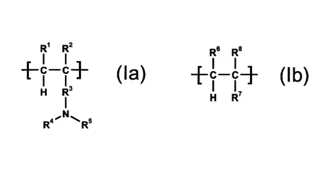
acids and cationic copolymers containing the recurring structural units of the
formulae (la)
and (lb)
R1 R2
C C ( I a )
R6 R8
-FC-CR
II+ (Ib)
R4 40.0 c
R`
wherein
1:11 and 1:16 are, independent of each other, hydrogen, alkyl or -COOR9,
1:12 and R' are, independent of each other, hydrogen or alkyl,
R3 is selected from the group consisting of -0-R1 -, -COO-R1 -, -CONH-R1 - or
1:14 is hydrogen, alkyl, cycloalkyl, aryl, aralkyl or alkylaryl,
R5 is hydrogen, alkyl, cycloalkyl, aryl, aralkyl, alkylaryl or -(alkylene-NH-
)m-alkyl, or
1:14 and R5 together with the nitrogen atom they have in common form a
heterocyclic ring,
R8 is selected from the group consisting of -0-R11, -COO-R11, -CONH-R" or -R11
R9 and R11 are, independent of each other, hydrogen or a monovalent organic
radical,
Ii1 is a bivalent organic radical, and
m is an integer from 1 to 5, with the requirement that
5
Date Regue/Date Received 2022-10-12
CA 03179980 2022-10-12
the nanoparticles have a diameter (z-average), determined by dynamic light
scattering, of
less than or equal to 900 nm, and that the molar ratio of nitrogen atoms in
the copolymer
to the phosphate groups in the nucleic acid is between 1 and 200.
In the nanoparticles according to the invention, DNA and/or RNA and
modifications thereof
can be used as nucleic acids.
Any DNA types can be used. Examples are A-DNA, B-DNA, Z-DNA, mtDNA, antisense
DNA,
bacterial DNA and viral DNA.
Any RNA types can also be used. Examples include hnRNA, mRNA, tRNA, rRNA,
mtRNA,
snRNA, snoRNA, scRNA, siRNA, miRNA, antisense RNA, bacterial RNA and viral
RNA.
Combinations of DNA and RNA can also be used in the complexes according to the
invention.
The cationic copolymers used in the complexes according to the invention are
copolymers
which contain at least one recurring structural unit of the formula (la),
which is derived
from an ethylenically unsaturated monomer containing an amino group, and which
contain
at least one further recurring structural unit of the formula (lb), preferably
at least two
different structural units of the formula (lb), which are derived from
ethylenically
unsaturated monomers comprising a hydrocarbon radical.
The cationic copolymers used in the complexes according to the invention may
contain one
or more different recurring structural units of formula (la). Preferably,
these copolymers
contain only one type of the recurring structural units of formula (la).
The cationic copolymers used in the complexes according to the invention may
contain one
or more different recurring structural units of formula (lb). Preferably,
these copolymers
contain only one or two different types of the recurring structural units of
formula (lb) in
addition to the recurring structural units of formula (la).
6
Date Regue/Date Received 2022-10-12
CA 03179980 2022-10-12
In addition to the recurring structural units of formulae (la) and (lb), the
cationic copolymers
used in the complexes according to the invention may contain further recurring
structural
units of formula (lc)
R12 R13 R13 ' R12
I I
+C¨C¨BG¨C¨C1¨
I 1 I I
H R14 R" H
wherein
R3.2 ,R 13 and RIA are, independent of each other, hydrogen or alkyl,
preferably hydrogen or
Ci-C6 alkyl, particularly preferably hydrogen or methyl, and
BG means a bivalent organic bridging group with ether, ester, amide, sulfide,
phosphate or
disulfide groups.
The presence of structural units of formula (lc) in the copolymer used
according to the
invention confers improved biodegradability upon it.
Examples of polymers from which recurring structural units of formula (lc) are
derived are
polyesters, polyamides, polyethers, phosphates, sulfides or disulfides, each
having two end
groups with ethylenically unsaturated groups, such as vinyl or ally! groups.
The radicals , R2, R4 _ R7 and R12 - R14 can mean alkyl. These are usually
alkyl groups with
one to six carbon atoms, which can be straight-chain or branched. Methyl and
ethyl are
preferred, particularly methyl.
The radicals R4 and R5 can mean cycloalkyl. These are usually cycloalkyl
groups with five to
six ring carbon atoms. Cyclohexyl is particularly preferred.
.. The radicals R4 and R5 can mean aryl. These are usually aromatic
hydrocarbon radicals with
five to ten ring carbon atoms. Phenyl is preferred.
The radicals R4 and R5 can mean alkylaryl. These are usually aryl groups
substituted with
one or two alkyl groups. Tolyl is preferred.
7
Date Regue/Date Received 2022-10-12
CA 03179980 2022-10-12
The radicals R4 and R5 can mean aralkyl. These are usually aryl groups which
are connected
to the rest of the molecule via an alkylene group. Benzyl is preferred.
R5 can also be a radical of the formula -(alkylene-NH-)m-alkyl. The a lkylene
radicals thereby
usually have two to four carbon atoms and can be branched or preferably
straight-chain.
The alkyl group usually has one to four carbon atoms and is preferably ethyl
or methyl,
particularly methyl. The number of recurring units, characterized by the index
m, is 1 to 5,
preferably 1 to 3.
R4 and R5 can also form a heterocyclic ring together with the nitrogen atom
they have in
common. These are usually rings with a total of five to six ring atoms, of
which one or two
ring atoms are heteroatoms and the rest of the ring atoms are carbon atoms.
One of the
ring heteroatoms is a nitrogen atom. An additional ring heteroatom, if
present, is nitrogen,
oxygen or sulfur.
1:19 and R11 can, independent of each other, be monovalent organic radicals.
These are
organic radicals with a covalent bond that establishes the connection to the
rest of the
molecule. The monovalent organic radicals are usually alkyl, cycloalkyl, aryl,
alkylaryl or
a ralkyl.
R11) is a bivalent organic radical. These are organic radicals with two
covalent bonds that
establish the connection to the rest of the molecule. Bivalent organic
radicals are usually
a lkylene, cycloalkylene, arylene, a lkylarylene or a ralkylene.
Preferably, copolymers are used in which fil and R6, independent of each
other, denote
hydrogen or Ci-C6 alkyl, particularly hydrogen or methyl, and very
particularly preferably
hydrogen.
Preferably, copolymers are used in which R2 and 1:17, independent of each
other, denote
hydrogen or Ci-Cg alkyl, particularly hydrogen or Ci-C6 alkyl, particularly
preferably
hydrogen or methyl and particularly preferably methyl.
8
Date Regue/Date Received 2022-10-12
CA 03179980 2022-10-12
Preferably, copolymers are used in which R3 is a bivalent radical of the
formulae -0-1:11 -, -
Coo_Rn_, _CONH-R1 - or -1:11 -, and R11) is selected from the group consisting
of C2-C12
alkylene, C5-C7 cycloalkylene and C6-Cio arylene, particularly C2-C6 alkylene,
and very
particularly preferably ethylene.
Preferably, copolymers are used in which 1:14 and R5, independent of each
other, denote
hydrogen or Ci-C6 alkyl, and particularly hydrogen and methyl and very
particularly
preferably methyl.
Preferably, copolymers are used in which R8 is a monovalent radical of the
formulae -0-R11,
-COO-R11, -CONH-R" or -R11, and R11 is alkyl, alkenyl, cycloalkyl, aryl,
aralkyl or alkylaryl,
particularly Ci-C6 alkyl, vinyl, allyl, phenyl, benzyl or Ci-C6 a lkylphenyl
and very particularly
preferably Ci-C6 alkyl.
Particularly preferably, copolymers are used in which R8 represents -COO-R11
or -CONH-R",
and R11 denotes Ci-C6 alkyl, phenyl, benzyl or Ci-C6 alkylphenyl, very
particularly preferably
Ci-C6 alkyl and extremely preferably methyl, ethyl, propyl and/or butyl.
Preferably, copolymers are used in which 1:19 and R11, independent of each
other, denote
alkyl, cycloalkyl, aryl, aralkyl or alkylaryl, particularly Ci-C6 alkyl,
phenyl, benzyl or Ci-C6
a lkylphenyl and very particularly preferably Ci-C6 alkyl.
Preferably, copolymers are used in which R11) denotes C2-C12 alkylene, C5-C7
cycloalkylene
and C6-Cio a rylene, particularly C2-C6 alkylene, and very particularly
preferably ethylene.
Particularly preferably, copolymers are used containing a recurring structural
unit of the
formula (la) and two different recurring structural units of the formula (lb),
in which lil and
R6 denote hydrogen, R2 and 1:17, independent of each other, are hydrogen or
methyl,
particularly methyl, R3 is -COO-R' _, lil denotes ethylene, 1:14 and R5,
independent of each
other, are Ci-C6 alkyl, particularly methyl, and R8 is -COO-R11, wherein, in a
recurring
structural unit of the formula (lb) R11 is Ci-C3 alkyl, particularly methyl,
and, in another
recurring structural unit of the formula (lb) R11 is C4-C6 alkyl, particularly
n-butyl.
9
Date Regue/Date Received 2022-10-12
CA 03179980 2022-10-12
It is assumed that the compact nanoparticles according to the invention
contain nucleic
acid-copolymer complexes which are distributed over the entire volume of the
nanoparticle. In contrast to previously known nanoparticles with a pronounced
core-shell
structure and with a concentration of the nucleic acid-polymer complexes in
the outer shell,
in the nanoparticles according to the invention nucleic acid-copolymer
complexes are found
both in the interior and in the exterior regions of the nanoparticles. Such
nanoparticles are
also referred to hereinafter as "polyplexes".
The nanoparticles according to the invention are further characterized by a
high content of
nucleic acid. The proportion by weight of cationic copolymer containing the
recurring
structural units of the formula (la) and (lb) in the nanoparticles according
to the invention
is typically 15 to 99%, and preferably 20 to 90% and particularly 30 to 80%,
based on the
total mass of the nanoparticles.
The nanoparticles according to the invention can be characterized by their
particle
diameter. Typical particle diameters (for example z-average) are in the range
of less than
or equal to 900 nm, preferably less than or equal to 500 nm, particularly
preferably between
30 and 500 nm, very particularly preferably between 40 and 250 nm and
especially between
50 and 200 nm. The particle diameters are determined for the purposes of the
present
description by dynamic light scattering (DLS) using a Malvern Zetasizer Nano
ZS (Malvern
Instruments, Worcestershire, United Kingdom). Cumulant analysis of the
correlation
function (IS013321, IS022412) was used to determine the intensity-weighted
mean
diameter (e.g. z-average). For sizing, a refractive index of 1.33 was assumed
for ultrapure
water and 1.59 for the copolymer.
Particle diameters can alternatively be determined by other methods, for
example by
nanosize tracking analysis (NTA), or by electron microscopy, e.g. using a
transmission
electron microscope or a scanning electron microscope.
Particle diameters (z-average) of preferred nanoparticles according to the
invention range
between 50 and 200 nm, determined by dynamic light scattering (DLS).
The cationic copolymers used according to the invention and the nanoparticles
according
to the invention can be further characterized by their polydispersity index
(or PDI). The
polydispersity index D or PDImw of molecular weights is a measure of the
breadth of the
Date Regue/Date Received 2022-10-12
CA 03179980 2022-10-12
molecular weight distribution of a polymer. The polydispersity index D or
PDImw is
calculated from the ratio of the weight average to the number average of the
molecular
weight distribution. The greater 0, the broader the molecular weight
distribution. In the
case of very narrow molecular weight distributions, the value of PDImw tends
towards 1. In
the case of broader molecular weight distributions, the value of PDImw is
significantly
greater than 1.
The polydispersity index of particle size distribution PDITG, on the other
hand, indicates the
breadth of particle size distribution for particles. Values between 0
(monodisperse) and 1
(polydisperse) can thereby be assumed. The PDITG value is determined for the
purpose of
the present description by dynamic light scattering (DLS) using a Malvern
Zetasizer Nano ZS
(Malvern Instruments, Worcestershire, United Kingdom). PDITG was determined by
means
of cumulant analysis of the correlation function.
The PDITG value of the particle size distribution of the nanoparticles
according to the
invention typically ranges between 0.05 and 0.4, preferably between 0.1 and
0.4, and
particularly preferably between 0.1 and 0.3.
The polydispersity index 0 of the molecular weight distribution of the
cationic copolymers
used according to the invention typically ranges between 1.0 and 5.0,
preferably between
1.01 and 2.6.
The nanoparticles according to the invention can also be characterized by
their transfection
efficiency for DNA. For this purpose, nanoparticles comprising a selected
protein-coding
DNA, for example eGFP-coding DNA, are brought into contact with cells for a
predetermined time, for example 1 hour, and incubated. The cells are then
incubated in
growth medium without nanoparticles for 23 hours, after which it is determined
how many
of the cells express the selected protein. The proportion of expressing cells,
specified as a
percentage, is used to represent the transfection efficiency for DNA.
Preferred nanoparticles according to the invention exhibit a transfection
efficiency for DNA
of 15 to 50% (viable fluorescent cells), particularly of 20 to 45%, after 1
hour of incubation.
11
Date Regue/Date Received 2022-10-12
CA 03179980 2022-10-12
The nanoparticles according to the invention can be further characterized by
their N/P
ratio. This is the molar ratio of nitrogen atoms in the copolymer to phosphate
groups in the
nucleic acid.
The N/P ratio in the nanoparticles according to the invention can vary in wide
ranges.
Typically, the N/P ratio in the nanoparticles according to the invention is
between 1 and
200, preferably between 1 and 100, especially between 1 and 50, particularly
preferably
between 1 and 30, very particularly preferably between 2.5 and 100, extremely
preferably
between 5 and 50, and most preferably between 5 and 30.
Preferred nanoparticles according to the invention have diameters (z-average)
determined
by DLS between 40 and 250 nm, particularly between 50 and 200 nm, and a
polydispersity
index of particle diameters between 0.1 and 0.3.
Very particularly preferred nanoparticles according to the invention have
diameters (z-
average) determined by DLS between 40 and 250 nm, particularly between 50 and
200 nm,
and a polydispersity index of particle diameters between 0.1 and 0.3 and an
N/P ratio
between 10 and 30.
The molar proportion of recurring structural units of formula (la) in the
cationic copolymers
used according to the invention is usually between 10 and 75%, preferably
between 15 and
65% and very preferably between 20 and 55%, based on the total cationic
copolymer.
The molar proportion of recurring structural units of formula (lb) in the
cationic copolymers
used according to the invention is usually between 90 and 25%, preferably
between 85 and
45% and very preferably between 80 and 45%, based on the total cationic
copolymer.
The molar proportion of recurring structural units of formula (lc) in the
cationic copolymers
used according to the invention is usually between 0 and 25%, preferably
between 1 and
10% and very preferably between 5 and 10%, based on the total cationic
copolymer.
Preferably, nanoparticles according to the invention are used which do not
contain any
excipients or additives, in particular no protective colloids and/or
surfactants.
12
Date Regue/Date Received 2022-10-12
CA 03179980 2022-10-12
In the event that the nanoparticles according to the invention contain
additional polymers
or additional complexes of nucleic acids with additional polymers, e.g.
complexes of nucleic
acids with IPEI, in addition to the nucleic acid-copolymer complexes described
above, these
further components are present only in small amounts, for example their
proportion by
weight is 15% or less, particularly less than 5%.
Particularly preferably, the nanoparticles according to the invention do not
contain any
further complexes of nucleic acids with other polymers in addition to the
nucleic acid-
copolymer complexes described above.
The terms for "particles" [German: Teilchen/Partikel: particles] are used
synonymously in
the context of the present description.
In the context of the present description, "nanoparticles" are understood to
be particles
whose diameter (z-average) is less than or equal to 900 nm and which may be
composed
of cationic copolymers as well as complexes thereof with nucleic acids or only
of such
complexes. They are generally characterized by a very high surface-to-volume
ratio and
thus offer very high chemical reactivity. Nanoparticles may only consist of
the
aforementioned cationic copolymers and complexes or only of the complexes, or
may also
contain other components in addition to the copolymers and complexes, such as
active
agents or excipients or additives.
In the context of the present description, "copolymers" are understood to be
the above-
mentioned organic compounds which are characterized by the repetition of at
least two
different specific units (monomer units or repetition units). Copolymers are
produced by
the chemical reaction of monomers with the formation of covalent bonds
(polymerization)
and form what is called the polymer backbone by linking the polymerized units.
This can
have side chains on which functional groups can be located. Some of the
copolymers have
hydrophobic properties and, depending on the concentration, can form nanoscale
structures (e.g. nanoparticles, micelles, vesicles) in an aqueous environment.
The
copolymers consist of at least two, preferably three different monomer units,
which can be
arranged statistically, as a gradient or alternately.
In the context of the present description, "surfactants" are understood to be
non-polymeric
substances or mixtures of substances which have water-soluble and water-
insoluble
13
Date Regue/Date Received 2022-10-12
CA 03179980 2022-10-12
properties and which serve to stabilize particles during production and
storage in aqueous
media. They are usually added to the dispersing medium, e.g. the aqueous
phase, during
the production of the particles, but can also be added after their production
to stabilize the
obtained dispersion. For example, cationic surfactants could be added to the
surface of the
.. nanoparticles to shift their surface charge and thus enable nucleic acids
to bind on the
surface, for example by coating with CTAB (cetyltrimethylammonium bromide).
In the context of the present description, "protective colloids" are
understood to be water-
soluble or water-dispersible polymers or polymer mixtures which serve to
stabilize particles
during production and storage in aqueous media. They are usually added to the
dispersing
medium, e.g. the aqueous phase, during the production of the particles, but
can also be
added after their production to stabilize the obtained dispersion.
In the context of the present description, "water-soluble compounds" or "water-
soluble
polymers" are understood to be compounds or polymers that dissolve to at least
1 g/L
water at 25 C and at neutral pH values.
In the context of the present description, "excipients and additives" are
understood to be
substances that are added to a formulation to give it certain additional
properties and/or
to facilitate its processing. Examples of excipients and additives are
tracers, contrast agents,
carriers, fillers, pigments, dyes, perfumes, slip agents, UV stabilizers,
antioxidants or
surfactants. In particular, "excipients and additives" are understood to be
any
pharmacologically acceptable and therapeutically useful substance which is not
a
pharmaceutically active agent but which can be formulated together with a
nucleic acid in
a nucleic acid-copolymer complex in order to influence, in particular improve,
qualitative
properties of the nanoparticle. Preferably, the excipients and/or additives
have no effect or
no significant effect or at least no undesirable effect with regard to the
intended procedure.
In the context of the present description, "gene delivery" is understood to be
the
introduction of nucleic acids into cells and their functional release in the
cells.
The nanoparticles according to the invention may be present in solid form as a
powder or
they may form a dispersion and be dispersed in aqueous solvents, the particles
being
present in the dispersing medium in solid form.
14
Date Regue/Date Received 2022-10-12
CA 03179980 2022-10-12
In a preferred embodiment, the nanoparticles according to the invention form a
disperse
phase in water or in an aqueous buffer solution.
The solubility of the copolymers used according to the invention can be
influenced by co-
polymerization with suitable monomers and by functionalization. Such
techniques are
known to the person skilled in the art.
The proportion of nanoparticles according to the invention in a dispersion can
cover a broad
range. Typically, the proportion by weight of nanoparticles in the dispersion
is between
0.01 and 20%, preferably between 0.05 and 5%.
The organic copolymers used according to the invention can cover a broad range
of molar
masses. Typical molar masses (Me) range from 2,000 to 500,000 g/mol, in
particular from
5,000 to 50,000 g/mol. These molar masses can be determined by 1H-NMR
spectroscopy of
the dissolved copolymer. In particular, an analytical ultracentrifuge or
chromatographic
methods, such as size exclusion chromatography, can be used to determine the
molar
masses.
Preferred organic copolymers have an average molar mass (number average) in
the range
of 5,000 to 40,000 g/mol, determined by 1H-NMR spectroscopy or by using an
analytical
ultracentrifuge.
The cationic copolymers used according to the invention can be produced using
the usual
polymerization methods. Examples are polymerization in substance,
polymerization in
solution, or emulsion or suspension polymerization. These methods are known to
the
person skilled in the art.
The nanoparticles according to the invention can be produced by
nanoprecipitation. For
this purpose, the cationic copolymers used according to the invention, which
are
hydrophilic depending on the pH value due to the presence of polar groups, are
dissolved
in water or in an aqueous buffer solution. The pH of the aqueous solution is
adjusted to a
value between 3 and 6.5, e.g. by using an acetate buffer or another suitable
buffer such as,
for example, citrate buffer, lactate buffer, phosphate buffer and phosphate-
citrate buffer.
In addition, the nucleic acids are dissolved in water, whereby the pH of the
aqueous nucleic
acid solution is preferably adjusted to a value between 6.5 and 8.5,
particularly preferably
Date Regue/Date Received 2022-10-12
CA 03179980 2022-10-12
to a value between 6.8 and 7.5. A buffer solution containing HEPES, TRIS, BIS-
TRIS-propane
or only salts is particularly suitable for this purpose. Both solutions are
combined, whereby
the amounts of nucleic acids and cationic copolymer are chosen in such a way
that a desired
N/P ratio is obtained. After mixing the two solutions, the mixture is
agitated, for example
.. for a short time, such as between 2 and 20 seconds. This may be done by
stirring and/or by
vortexing. Preferably, the resulting nanoparticles are left to stand for some
time, for
example between 5 and 20 minutes, before further use, to allow binding between
the
polymer and the nucleic acids (hereinafter referred to as "incubation"). The
nanoparticles
according to the invention are precipitated in the dispersing medium in finely
dispersed
.. form.
In addition to the cationic copolymer and the nucleic acid, one or more
excipients and
additives may be present during their nanoprecipitation in the dispersing
medium.
Alternatively, these excipients and additives may be added after the nucleic
acid-copolymer
complex has been dispersed in the aqueous phase.
Water is used as the dispersing medium. Buffer substances, salts, sugars or
acids and bases
can be added to this to adjust the desired pH value or osmolarity.
The invention also relates to a method for the production of nanoparticles,
which comprises
the following measures:
i) preparing an aqueous solution of a cationic copolymer containing the
recurring
structural units of formulae (la) and (lb) described above with a pH between 3
and
6.5,
ii) preparing an aqueous solution of a nucleic acid,
iii) mixing both solutions prepared in steps i) and ii) in a chosen
quantity ratio of nucleic
acid and copolymer, such that a desired molar N/P ratio of nitrogen atoms in
the
copolymer to the phosphate groups in the nucleic acid is obtained, i.e. an N/P
ratio
between 1 and 200,
iv) agitating the mixture from step iii), and
v) subsequently incubating the resulting mixture, as appropriate.
The aqueous solution of the cationic copolymer for step i) of the method
according to the
invention preferably contains a buffer, particularly an acetate buffer,
citrate buffer, lactate
buffer, phosphate buffer, phosphate-citrate buffer or mixtures thereof.
16
Date Regue/Date Received 2022-10-12
CA 03179980 2022-10-12
The aqueous solution of the nucleic acid for step ii) of the method according
to the
invention preferably has a pH between 6.5 and 8.5, particularly between 6.8
and 7.5.
The aqueous solution of the nucleic acid for step ii) of the method according
to the
invention preferably contains a buffer, particularly a HBG, HEPES, BIS-TRIS
propane or TRIS
buffer.
The agitation in step iv) of the method according to the invention is
preferably carried out
by stirring or vortexing. The processing duration in this step is usually
between 1 and 60
seconds, particularly between 2 and 20 seconds.
The incubation in step v) of the method according to the invention is usually
carried out by
simply leaving the obtained mixture to stand, for example for a period of 5 to
60 minutes,
preferably from 5 to 20 minutes. The mixture may also be incubated in an oven,
for example
at temperatures between 30 and 80 C.
The separation of the nanoparticles from the aqueous phase can be achieved in
various
ways. Examples are centrifugation, ultrafiltration or dialysis. However, the
dispersion of
nanoparticles can also preferably be used directly after production without
further
processing.
Purification by means of filtration can separate particles, such as
aggregates, but also excess
excipients or impurities from the dispersion. The particle concentration can
thereby
change.
Purification by dialysis can separate dissolved molecules from the dispersion.
This method
is largely independent of the particle size with regard to the dispersed
particles.
Purification by centrifugation can also separate dissolved molecules from the
dispersion.
However, this method also reduces the concentration of the dispersed
particles.
Furthermore, only dispersions with nanoparticles of larger diameter, e.g. of
more than 150
nm, can be treated and the particles may be affected. Furthermore,
redispersing the
particles obtained in this way can cause difficulties.
17
Date Regue/Date Received 2022-10-12
CA 03179980 2022-10-12
The nanoparticles according to the invention are excellently suited for gene
delivery in cells,
i.e. for the introduction of nucleic acids into cells. For this purpose, the
nanoparticles
comprising nucleic acids are added to individual cells, tissues or a cell
culture and taken up
by the cells by endocytosis. Surprisingly, it has been shown that high
contents of nucleic
acids can be transferred into cells by means of the nanoparticles according to
the invention.
Nanoparticles comprising a comparatively low cationic copolymer content can be
used for
this.
The invention therefore also relates to a method for gene delivery into cells,
which
comprises the following steps:
A) Bringing cells into contact with an aqueous suspension comprising the
nanoparticles
described above and
B) Subsequent incubation.
Preferably, the invention relates to a method for gene delivery into cells
comprising the
following steps:
C) Provision of a cell culture in a bioreactor or incubator,
D) Addition of an aqueous suspension comprising the nanoparticles described
above,
E) Distribution of the aqueous suspension in the cell culture, and
F) Subsequent incubation.
The gene delivery method according to the invention can be carried out using
various cells,
for example by using single cells, tissues or cell cultures.
Thus, the nanoparticles according to the invention can be combined with
prokaryotic cells,
with tissues from eukaryotic cells or with cell cultures. These can be plant
cells or preferably
animal cells, including human cells.
The application of the nanoparticles according to the invention can take place
in vivo, for
example under the skin or in the muscle, or the application can also take
place ex vivo, for
example with immune cells, as in CAR-T therapy. It can also be an RNA
vaccination or an
immunization.
18
Date Regue/Date Received 2022-10-12
CA 03179980 2022-10-12
In the context of the present description, "cells" are understood to be the
smallest living
units of organisms. They may be cells of unicellular or multicellular
organisms, which may
originate from prokaryotes or eukaryotes. Cells may be microorganisms or
single cells. Cells
may be of prokaryotic, plant or animal origin or may also originate from
fungi. Preferably,
eukaryotic cells are used, in particular eukaryotic cells which were
originally isolated from
tissue and can be cultivated permanently, i.e. that are immortalized.
In the context of the present description, "tissues" are understood to be
collections of
differentiated cells including their extracellular matrix.
In the context of the present description, "cell cultures" refers to
combinations of cells and
cell culture medium, whereby the cells are cultivated in the cell culture
medium outside the
organism. For this purpose, cell lines are used, i.e. cells of a tissue type
that can divide in
the course of cultivation. Both immortalized (immortal) cell lines and primary
cells (primary
culture) can be cultivated. Primary culture is usually understood to mean a
non-
immortalized cell culture obtained directly from a tissue.
In the context of the present description, "cell culture medium" or "nutrient
medium" are
understood to mean aqueous systems that serve as a platform for the
cultivation of cells.
These systems contain all the substances required for the growth and viability
of the cells.
The cell culture medium may contain serum in addition to the cells and the
required
nutrients. Preferably, the cell culture medium contains sera or proteins and
growth factors.
In the context of this description, "serum" is understood to mean blood serum
or immune
serum. Blood serum is thereby understood to be the liquid portion of the blood
that is
obtained as supernatant when a blood sample is centrifuged. This supernatant
contains all
substances naturally dissolved in the blood fluid except for the coagulation
factors
consumed by coagulation. The blood serum thus corresponds to the blood plasma
minus
the coagulation factors. Immune serum is understood to be a purification of
specific
antibodies obtained from the blood serum of immunized mammals.
Sera in the context of this description usually mean sera from vertebrates,
and in particular
sera from calf, cow, bull, horse or human.
19
Date Regue/Date Received 2022-10-12
CA 03179980 2022-10-12
The cell cultures used according to the invention can be produced and
cultivated according
to standard methods.
For example, primary cultures can be created from various tissues, e.g. from
tissues of
individual organs such as skin, heart, kidney or liver, or from tumor tissue.
The tissue cells
can be isolated by methods as known per se, e.g. by treatment with a protease,
which
degrades the proteins that maintain the cell bond. It may also be appropriate
to specifically
stimulate some cell types to divide by adding growth factors or, in the case
of poorly
growing cell types, to use feeder cells, basement membrane-like matrices or
recombinant
extracellular matrix components. The cells used according to the invention can
also be
genetically modified by introducing a plasmid as a vector.
The cells used according to the invention may have a limited lifespan or they
may be
immortal cell lines with the ability to divide infinitely. These may have been
generated by
random mutation, e.g. in tumor cells, or by targeted modification, for example
by the
artificial expression of the telomerase gene.
The cells used according to the invention can be adherent cells (growing on
surfaces), such
as fibroblasts, endothelial cells or cartilage cells, or they can be
suspension cells that grow
freely floating in the nutrient medium, such as lymphocytes.
Culture conditions and cell culture media are selected depending on the
individual cells
being cultured. The different cell types thereby prefer different nutrient
media, which are
composed specifically. For example, different pH values are established and
the individual
nutrient media can contain various amino acids and/or other nutrients in
various
concentrations.
The cells transfected according to the invention can be used in various
fields, for example
biotechnology, research or medicine. This may involve the production of
(recombinant)
proteins, virus and/or virus particle production, investigation of metabolism,
division and
other cellular processes. Furthermore, the cells transfected according to the
invention can
be used as test systems, for example in the investigation of the effect of
substances on cell
properties, such as signal transduction or toxicity. Further cells preferably
used for the
Date Regue/Date Received 2022-10-12
CA 03179980 2022-10-12
production of the cells transfected according to the invention are stem cells.
These are
known to be body cells that can diversify into various cell types or tissues.
The invention also relates to the use of the nanoparticles described above for
gene delivery
into cells, i.e. for introducing nucleic acids into cells.
The following examples illustrate the invention without limiting it.
The synthesis of polymers by RAFT polymerization, comparable to the
commercially
available product EUDRAGIT E100, is described below. The suitability of the
polymers for
nucleic acid binding was investigated and a novel method of nucleic acid
encapsulation and
formulation is described.
Abbreviations
The following abbreviations are used in the examples:
CPAETC: 4-cyano-4-(phenylcarbonothioylthio)pentanoic acid
nBMA: n-butyl methacrylate
MMA: methyl methacrylate
DMAEMA: 2-(N,N-dimethylamino)ethyl methacrylate
ACVA: 4,4'-azobis-(4-cyanovaleric acid)
DMAc-SEC: Size exclusion chromatography with dimethylacetamide + 0.21 % LiCI
as eluent
CTA: Chain Transfer Agent
pDNA: mEGFP-N1 plasmid (coding for EGFP); pKMyc plasmid (control plasmid)
HEPES: 2-(4-(2-hydroxyethyl)-1 -piperazinyI)-ethanesulfonic acid
HBG: 5% glucose solution buffered with HEPES.
DMEM: Dulbecco's modified Eagle Medium
HBSS: Salt solution according to Hanks
FBS: Foetal calf serum
EGFP: enhanced green fluorescent protein
IPEI: linear polyethylenimine
21
Date Regue/Date Received 2022-10-12
CA 03179980 2022-10-12
PDMAEMA: homopolymeric 2-(N, N-dimethylamino)ethyl methacrylate
PBMD: nBMA-st-MMA-st-DMAEMA copolymer (St = statistically distributed)
PDI: Polydispersity index determined by DLS using a Malvern Zetasizer Nano ZS
(Malvern
Instruments, Worcestershire, United Kingdom) applying cumulant analysis of the
correlation function (1S013321, 15022412).
RFI: relative fluorescence intensity
CTRL: Control in the form of cells treated only with HBG buffer and not with
copolymer.
E100: EUDRAGIT E100 (in powder form)
Materials and methods
The following materials were used in the subsequent experiments:
E100: EUDRAGIT E100 in powder form
pDNA (eGFP, pkmyc): mEGFP-N1 plasmid (coding for EGFP), pKMyc plasmid (control
plasmid, not coding for a fluorescent protein)
Cells: Human embryonic kidney (HEK) cells, in particular the HEK293T cell
line.
Aga rose: Aga rose-HR plus
Molecular weight calculation of the polymers produced during RAFT
polymerization
The monomer conversion (p) was calculated from 1-1-1-NMR data by comparing the
integrals
of the vinyl bands (5.5-6.3 ppm) with an external reference (1,3,5-trioxane,
5.14 ppm)
before (t=0) and after (t=final) polymerization. The theoretical number
average molar mass
(Me, th) was then calculated using the following equation:
Me, th = (([M]o * p * MM) / [CTA]o) + MCTA
wherein [M]o and [CTA]o are the initial concentrations of monomer and CTA,
respectively,
Mrsn and MCTA are the molecular weight of monomer and CTA, respectively, and p
is the
conversion of monomer to CTA.
Production and characterization of polymers and of nanoparticulate polymer
particles or
nanoparticulate DNA-polymer complexes
22
Date Regue/Date Received 2022-10-12
CA 03179980 2022-10-12
Example 1A: Synthesis of (nBMA-st-MMA-st-DMAEMA) copolymer (PBMD) by RAFT
polymerization (st = statistically distributed)
.. CPAETC (130.7 mg, 4.96 x 10⁴ mol), nBMA (3.5265 g, 2.48 x 10⁻² mol), MMA
(2.5218 g, 2.52
x 10-2 mol), DMAEMA (7.8165 g, 4.97 x 10⁻² mol), 1,4-dioxane (6.2113 g), a 1.0
% by weight
ACVA solution in 1,4-dioxane (1.436 g, 14.36 mg ACVA, 5.12 x 10⁻⁵ mol) and
1,3,5-trioxane
(external NMR standard, 23.7 mg) were introduced into a 20 ml microwave vial
equipped
with a magnetic stirrer. The solution was deoxygenated by bubbling with argon
for 10
minutes. The vial was sealed, placed in a 70 C oil bath and stirred for 21
hours, with samples
taken at predetermined times for 1-1-1-NMR and DMAc-SEC analysis. The polymer
was
precipitated three times from THF into cold hexane and dried under reduced
pressure to
give a yellow solid. DMAc-SEC: Mn,sEc = 25.1 kg ma', D = 1.13.
Example 1B: Synthesis of DMAEMA homopolymer by RAFT polymerization
CPAETC (50.0 mg, 1.9 x 10⁻⁴ mol), DMAEMA (4.54 g, 2.88 x 10⁻² mol), 1,4-
dioxane
(2.5 g), 1 % by weight ACVA in 1,4-dioxane (426 mg, 1.5 x 10⁻⁵ mol) and 1,3,5-
trioxane
(external NMR standard, 21 mg) were introduced into a 20 mL microwave vial
with
magnetic stirrer. The vial was sealed and the solution was deoxygenated by
bubbling with
argon for approx. 10 min. The vial was placed in an oil bath set at 70 C and
stirred for 7
hours, with samples taken at set times for 11-1-NMR and CHCI3-SEC analysis.
The polymer
was precipitated three times from THF into cold hexane and dried under reduced
pressure
to give a yellow solid. CHCI3-SEC: Mri,sEc = 14.2 kg mol-1, D = 1.19.
Figure 1A shows the structure of the copolymer Eudragit E100.
Figure 1B shows the structure of the copolymer represented by RAFT
polymerization
according to Example 1A.
Figure 2 shows the molecular weight distribution of the copolymer Eudragit
E100 and the
copolymer represented by RAFT polymerization according to Example 1A,
determined by
size exclusion chromatography (SEC).
23
Date Regue/Date Received 2022-10-12
CA 03179980 2022-10-12
Example V1: Formation of nanoparticles and complexation with pDNA (not
according to
the invention)
For nanoparticle formation, the copolymers produced in Example 1 were
dissolved in 2.5
mL acetone (2 mg mL-1). The polymer solution was manually dropped into 5 mL
ultrapure
water, the resulting nanoparticle suspension was stirred overnight at 800 rpm
to remove
the organic solvent, and stored at 4 C until use. To achieve the respective
N/P ratio, pDNA
was either added in different concentrations during the formation process
during the
acetone phase and subsequent nanoparticle formation or pDNA was added in
different
concentrations to the final particle suspension.
Example 2: Formation of nanoparticulate copolymer-nucleic acid complexes
(according to
the invention)
Stock solutions of the copolymers produced according to Example 1 were
produced by
dissolving in 0.2 M acetate buffer (pH 5.8). pDNA, siRNA or mRNA were
dissolved in
ultra pure water. To produce nucleic acid-polymer complexes, different
dilutions of the
copolymer as well as the nucleic acid were prepared in HBG buffer (20 mM
HEPES, 5%
glucose (w/v), pH 7.4) or in 20 mM HEPES buffer to achieve the respective N/P
ratios (molar
ratio of nitrogen atoms in the copolymer to the phosphate groups in the
nucleic acid). After
mixing the copolymer solution and the nucleic acid solution, the mixtures were
immediately
vortexed for 10 seconds. Before use, the resulting copolymer-nucleic acid
complexes were
incubated at room temperature for at least 15 minutes.
Figure 3 schematically shows the formation of nucleic acid-copolymer complexes
in
nanoparticles from the copolymer according to Example 1A (DMAEMA : BMA: MMA =
2:
1: 1).
The use of the cationic and hydrophobic copolymer results in binding,
stabilization and
protection of the genetic material, formation of stable nanoparticles,
stability against
competing polyanions and causes endosomal release after uptake by the cell.
Example 3: General rule for the characterization of nanoparticles and of
nanoparticulate
copolymer-pDNA complexes
24
Date Regue/Date Received 2022-10-12
CA 03179980 2022-10-12
-
Example 3A: Diameter (z-average) and zeta potential of nanoparticles and pDNA-
copolymer complexes were determined by dynamic or electrophoretic light
scattering (DLS,
Zetasizer Nano ZS, Malvern Instruments, Worcestershire, United Kingdom). For
size
.. determination, a refractive index of 1.33 was assumed for ultrapure water
and 1.59 for the
copolymer. The zeta potential of the nanoparticles produced by precipitation
in the
presence of organic solvents was determined on the same samples.
Example 3B: Gel retardation examination
The pDNA binding ability at different N/P ratios was determined by agarose gel
electrophoresis. Samples were produced as described for the pDNA-polymer
complexes in
Examples V1 and 2, ran at 80 V for 1.5 hours on a 1% agarose gel stained with
ethidium
bromide (EtBr, 0.1 p.g mL-1) and imaged using a gel imager (RedTM Imaging
System, Alpha
Innotech, Kasendorf, Germany).
Example 3C: Ethidium bromide binding assay (EBA) and heparin release assay
(H RA)
.. pDNA complexation and stability of the nanoparticulate copolymer-pDNA
complexes were
investigated by using an ethidium bromide binding assay and a heparin release
assay.
For this purpose, pDNA at a concentration of 15 p.g mL-1 was incubated with
ethidium
bromide for 10 minutes. The polymer stock solutions were diluted in a black 96-
well plate
(Nunc, Thermo Fisher) to adjust N/P ratios from 1 to 50. Then pDNA was added
and the
nanoparticulate copolymer-pDNA complexes were incubated at 37 C for 15
minutes.
Ethidium bromide fluorescence intensity was measured at XEX = 525 nm / XEM =
605 nm.
pDNA without copolymer was defined as 100% free DNA. The release of complexed
DNA
was investigated by gradual addition of heparin and measurement of the
resulting changes
in ethidium bromide fluorescence intensity. The influence of pH on pDNA
binding and pDNA
release was investigated by performing the experiment at different pH values
in the
respective buffers (acetate buffer pH 5 and 5.8 and HBG buffer pH 6.5; 7 and
7.4).
Example 3D: Transfection of HEK293T cells with EGFP pDNA
Date Regue/Date Received 2022-10-12
CA 03179980 2022-10-12
The HEK293T cell line was cultured in Dulbecco's modified Eagle's medium
(DMEM, 1 g Li
glucose, 10% (v/v) FBS, 100 g mL-1 penicillin/streptomycin) at 37 C in a
humidified 5% CO2
atmosphere. For transfection experiments, 0.2*106 cells per mL were seeded
into a 24-well
plate in 500 pi DMEM supplemented with 10 mM HEPES and left to recover for 24
hours.
1 hour before treatment, the treatment medium was replaced with 450 pi fresh
DMEM (10
mM HEPES). Nanoparticulate copolymer-pDNA complexes were freshly prepared as
described in Example 2 using egfp pDNA encoding for EGFP or pkmyc pDNA not
encoding
for a fluorescent protein as negative controls. The cells were treated with 50
pl of a
dispersion of nanoparticulate copolymer-pDNA complexes of the indicated N/P
ratio and
pDNA concentration or with HBG buffer as a control (ctrl) and incubated for 1
or 4 hours.
The supernatant was then removed, cultured by ctrl and incubated. The
supernatant was
then removed, replaced with fresh DMEM (10 mM HEPES) and the cells were
further
incubated for up to 24 hours. After incubation, cells were separated by
trypsin-EDTA,
resuspended in HBSS (2% FBS (v/v), 20 mM HEPES) and fluorescence was measured
using a
flow cytometer (Cytoflex S, Beckmann coulter, CA, U.S.A.). EGFP expression of
viable cells
was analyzed by excitation at 488 nm and measurement of emission at 610 nm
(bandpass
filter 610/20). Fluorescent cells were identified by gating to the negative
control.
Example 3E: Knock-down of GFP in HEK-GFP cells using anti-EGFP siRNA
The HEK-GFP cell line was cultured in Dulbecco's modified Eagle's medium
(DMEM, 1 g Li
glucose, 10% (v/v) FBS, 100 g mL-1 penicillin/streptomycin) at 37 C in a
humidified 5% CO2
atmosphere. For transfection experiments, 0.1*106 cells per mL were seeded
into a 24-well
plate in 500 pi DMEM supplemented with 10 mM HEPES and left to recover for 24
hours.
1 hour before treatment, the treatment medium was replaced with 450 pi fresh
DMEM (10
mM HEPES). Copolymer-siRNA complexes were freshly prepared as described in
Example
2. The cells were treated with 50 pl of the freshly prepared complexes.
Complexes with
siRNA not directed against GFP and HBG buffer were used as negative controls.
72 hours
after treatment with copolymer-siRNA complexes, the supernatant was removed
and the
cells were separated with trypsin-EDTA. After resuspension in HBSS (2% FBS
(v/v), 20 mM
HEPES), the cells were analyzed by flow cytometry at an excitation wavelength
of 488 nm
at 525 nm (bandpass filter 525/40).
Example 3F: Transfection of HEK293T cells with GFP mRNA
26
Date Regue/Date Received 2022-10-12
CA 03179980 2022-10-12
The HEK293T cell line was cultured in Dulbecco's modified Eagle's medium
(DMEM, 1 g L-1
glucose, 10% (v/v) FBS, 100 g mL-1 penicillin/streptomycin) at 37 C in a
humidified 5% CO2
atmosphere. For transfection experiments, 0.2*106 cells per mL were seeded
into a 24-well
plate in 500 pi DMEM supplemented with 10 mM HEPES and left to recover for 24
hours.
1 hour before treatment, the treatment medium was replaced with 450 Afresh
DMEM (10
mM HEPES). Copolymer-mRNA complexes were freshly prepared as described in
Example
2. 50 pi of the copolymer-mRNA complexes were added to the cells and incubated
for 4
hours. The supernatant was then removed and the cells were incubated for a
further 2 or
20 hours. After incubation, the supernatant was removed, the cells were
detached using
trypsin-EDTA and resuspended in HBSS (2% FBS (v/v), 20 mM HEPES).
Subsequently, the
fluorescence of the cells was analyzed by flow cytometry. For this purpose, an
excitation
wavelength of 488 nm was used and the emission was measured at 525 nm
(bandpass filter
525/40).
Example 3G: PrestoBlue test to determine viability
For cytotoxicity assays, HEK293T cells were seeded into 24-well plates at a
density of 0.2
106 cells per mL (HEK293T) in 500 pl DMEM (10 mM HEPES) and incubated for 24
hours to
enable recovery. 50 pl nanoparticulate copolymer-pDNA complexes were added as
described in Example 2 to test a concentration range of 0.25-1.5 g mL-1 pDNA.
The cells were incubated with the nanoparticulate copolymer-pDNA complex for 1
or 4
hours. The supernatant was then removed and replaced with 500 pi fresh DMEM
(10 M
HEPES). 24 hours after treatment, the supernatant was removed and this was
replaced with
a 10% (v/v) solution of PrestoBlue (Invitrogen, CA, U.S.A.) diluted with DMEM
medium.
The cells were incubated for 45 min and the supernatant was transferred to a
96-well plate
(100 pi per well) to determine fluorescence intensity (XEx 560 nm, XEm 590
nm). Cells
treated with the HBG buffer were used as control and viability was calculated
relative to
the buffer control after subtracting the blank (PrestoBlue without cells).
Investigations of nanoparticulate polymer particles and nanoparticulate DNA-
polymer
complexes
The binding of genetic material is a crucial step in the gene delivery
process, as the genetic
material must overcome extracellular and intracellular barriers to reach their
target cells.
27
Date Regue/Date Received 2022-10-12
CA 03179980 2022-10-12
Encapsulation and complexation by cationic polymers, for example, enable
protection from
nucleases in the bloodstream and the crossing of cell membranes (see in this
regard H. Yin,
R. L. Kanasty, A. A. Eltoukhy, A. J. Vegas, J. R. Dorkin, D. G. Anderson, Nat
Rev Genet 2014,
15, 541-555).
Example 4: Experiments on the pDNA-binding ability of polymers and the release
of pDNA
from the complexes formed
Figure 4 describes results of experiments on the DNA-binding ability of
polymers and the
release of pDNA from the complexes formed.
Figure 4A shows results of an ethidium bromide binding assay (EBA) on DNA-
polymer
complexes in HBG buffer at pH 7.4. DNA-IPEI complexes, DNA-PDMAEMA complexes
and
DNA-PBMD complexes were examined.
The binding of the genetic material is crucial for its application in gene
delivery. Therefore,
the pDNA-binding ability of the polymer was investigated by agarose gel
electrophoresis at
different N/P ratios. Figure 4B shows results of agarose gel electrophoresis
tests with DNA-
PBMD complexes at different N/P ratios.
Figures 4C-4E show results of heparin release assays (HRA) for pH-dependent
DNA binding
and DNA release in the pH range from 5 to 7.4. DNA-IPEI complexes, DNA-PDMAEMA
complexes and DNA-PBMD complexes were examined.
The test shown in Figure 4B confirmed the complete binding of pDNA at N/P
ratios above
1. The DNA-polymer interaction can be further characterized by the reversible
intercalation
of ethidium bromide into the DNA. Strong fluorescence can be observed when
this is
intercalated into the pDNA. However, this is reduced by the interaction
between polymer
and genetic material (EBA). The test was carried out at pH 7.4 and N/P ratios
from 1 to 50
in order to investigate the effect of an increasing polymer amount on complex
formation.
In addition to the PBMD polymer, IPEI and a PDMAEMA homopolymer were
investigated
(Figure 4A). The latter is composed of the same number of cationic groups as
the PBMD
polymer in order to evaluate the influence of hydrophobic side chains in the
PBMD polymer.
It was observed that the relative fluorescence intensity (RFI) decreased with
increasing N/P
ratios, reaching a plateau at an N/P ratio of 10 (Figure 4A). IPEI showed the
lowest plateau
28
Date Regue/Date Received 2022-10-12
CA 03179980 2022-10-12
values, followed by PDMAEMA (13% compared to 42%) while PBMD only decreased
the RFI
to 72%. Since the electrophoresis test showed complete DNA binding above N/P
1, these
differences could be due to differences in DNA condensation and thus
differently packed
complexes, which may lead to different levels of EtBr displacement. Therefore,
it can be
assumed that the PBMD polymer forms a less dense DNA-copolymer complex
compared to
IPEI and PDMAEMA.
To investigate the stability of the complexes in the relevant physiological pH
range, the
dissociation of the complexes was examined at pH values from 5 (endosomal) to
7.4
(blood). Preformed DNA-copolymer complexes with an N/P ratio of 20 were
incubated with
increasing amounts of heparin as the competing polyanion, and the fluorescence
intensity
was measured. Dissociation, and thus the release of pDNA from the complex,
caused re-
intercalation of EtBr. For all polymers, it was observed that ambient pH has
an influence on
complex formation prior to the addition of heparin. For IPEI (upper figure
4C), a notable
.. displacement of EtBr and subsequent release of pDNA was observed at
relatively low
heparin concentrations (20 U mL-1). pDNA complexes formed at lower pH values
required
slightly less heparin addition to release the pDNA, but at high heparin
concentrations (100
UN!), pDNA was fully released at all pH values. In general, pDNA release from
PDMAEMA
complexes required higher amounts of heparin (middle figure 4D) and was more
strongly
.. influenced by ambient pH in comparison to IPEI. At low pH values (5 and
5.8), 100 Wm!
heparin resulted in full release of pDNA, whereas at pH values of 6.5 and
above, only 66-
79% was released. The PBMD copolymer (lower figure 4E) showed an even more
pronounced pH-dependent behavior. RFI values before heparin addition range
from 27 to
60% and are lower at pH values of 5 to 5.8. The release of the entirety of
pDNA was only
observed at pH 5 and decreased gradually with increasing pH. At neutral pH, a
slight
decrease in RFI after heparin addition indicates even stronger complexation of
pDNA in the
presence of competing polyanions. In general, IPEI was hardly affected by pH,
while
PDMAEMA and PBMD copolymer showed a weak and the strongest influence of pH,
respectively, on the initial RFI values and the subsequent pDNA release.
These results suggest an influence of the pKa value and the hydrophobicity of
the copolymer
on pDNA binding, arrangement in the complex and subsequent pDNA release. For
IPEI, a
pKa value of 8.5 is reported in the literature, while PDMAEMA has a pKa of
about 7.5. For
PBMD, only an apparent pKa of 6.8 could be determined, as the polymer
precipitated in this
pH range during titration.
29
Date Regue/Date Received 2022-10-12
CA 03179980 2022-10-12
When calculating the percentage of charges from these pKa values, a charge
level of 100 to
90% was calculated for IPEI over the entire pH range tested, while PDMAEMA
showed a
decrease from 100% at pH 5 to 56% at pH 7.4. When calculating the charge level
for PBMD
with the apparent pKa value, the effect is even more pronounced (98% at pH 5
and 20% at
pH 7.4). As the charge level and thus the number of cationic groups in the
polymer
decreases, the ratio of hydrophilic to hydrophobic groups changes, leading to
additional
hydrophobic interactions and to different arrangements in the complex and thus
to altered
release behavior of the DNA. This effect is most clearly illustrated in the
PBMD copolymer,
where the additional hydrophobic side chains of the nBMA and MMA units promote
complex stability, especially at neutral pH values, through strong hydrophobic
interactions
(see in this regard E.J. Adolph, C.E. Nelson, T.A. Werfel, R. Guo, J.M.
Davidson, S.A. Guelcher,
C.L. Duvall, J. Mater Chem B 2014, 2, 8154-8164). Thus, the PBMD copolymer
shows high
potential as a gene delivery vector, as high stability and prevention of
dissociation at neutral
pH in the blood stream is advantageous for systemic application of complexes.
Example 5: Experiments on the complexation of pDNA
Figures 5 and 6 describe results of experiments on the complexation of pDNA in
different
polymers and the formation of nanoparticles.
Figure 5A schematically shows the formation of nanoparticles by
nanoprecipitation with
solvent evaporation technique.
The left side of Figure 5B shows an electron micrograph (scanning electron
microscopy,
SEM) of nanoparticles of Eudragit E100 precipitated from acetone-water
mixtures.
The right side of Figure 5B shows an electron micrograph (scanning electron
microscopy,
SEM) of nanoparticles of PBMD copolymer precipitated from acetone-water
mixtures.
Figure 6 schematically shows the formation of nanoparticulate DNA complexes of
PBMD
with pDNA depending on the formulation method used. The left side of Figure 6
shows the
nanoprecipitation with solvent evaporation techniques combined with different
techniques
of pDNA addition (in-process and post-process addition). The right side of
Figure 6 shows
the water-based solvent-free pH-dependent formulation for complexation of
pDNA. The
obtained nanoparticles were characterized by dynamic light scattering.
Date Regue/Date Received 2022-10-12
CA 03179980 2022-10-12
The upper figure 7A shows the diameter (z-average) of nanoparticles of PBMD
copolymer
and pDNA at different N/P ratios produced by precipitation from acetone or
aqueous
solution. The middle figure 7B shows the PDI values of the diameters of
nanoparticles of
PBMD copolymer and pDNA at different N/P ratios produced by precipitation from
acetone
or from aqueous solution. The lower figure 7C shows the surface charge (zeta
potential) of
nanoparticles of PBMD copolymer and pDNA produced by means of the solvent
evaporation technique from acetone.
The left two figures 7D show electron micrographs (scanning electron
microscopy, SEM) of
nanoparticles of PBMD copolymer produced at two different N/P ratios by
nanoprecipitation from acetone in the presence of pDNA. The right figure 7D
shows an
electron micrograph (cryogenic transmission electron microscopy, cryo-TEM) of
nanoparticles of PBMD copolymer produced by nanoprecipitation from aqueous
solution.
Since the PBMD copolymer showed high potential for pDNA binding and high
complex
stability at the pH of blood, the copolymer was used to develop a stable
formulation for
encapsulating the pDNA. Three different formulation approaches were
investigated. The
commonly used nanoprecipitation with addition of DNA
a) for the purpose of final nanoparticle suspension or
b) during the formulation process was compared with
c) the complexation of the DNA by the polymer dissolved in the acidic buffer
as a pH-
dependent water-based nanoprecipitation.
Nanoprecipitation is a widely used method for the formulation of polymeric
nanoparticles
that allows easy adjustment of nanoparticle size and complexation of a variety
of
components. Nanoprecipitation of the PBMD copolymer without the addition of
pDNA
results in particle diameters of about 125 nm (N/P ratio 0) with a positive
surface charge (+
55 mV) (see Fig. 7C). It was expected that the positive surface charge of the
nanoparticles
should facilitate the binding and complexation of pDNA to the particle
surface. Addition of
pDNA to the pre-formulated positively charged nanoparticles (NP + pDNA)
generally
resulted in increased size and polydispersity of the nanoparticles (Figures 7A
and 7B). Only
at the lowest and highest N/P ratios investigated (N/P 5 and N/P 100) were
nanoparticles
found with sizes below 200 nm and with acceptable PDI values (PDI<0.250).The
surface
charge of the particles decreased with increasing DNA amount and shifted to
negative
31
Date Regue/Date Received 2022-10-12
CA 03179980 2022-10-12
values at N/P 10 (Fig. 7C).These results indicate that pre-formulated
nanoparticles (NP +
pDNA) have limited binding ability for pDNA and do not completely condense the
pDNA
during complexation. Pre-incubation of the DNA with the polymer dissolved in
acetone and
subsequent nanoprecipitation in water (NP(pDNA)) leads to a similar N/P ratio-
dependent
trend of nanoparticle size and homogeneity. Interestingly, the zeta potential
remains in the
positive range compared to the NP + pDNA formulation. In general, particles
obtained by
adding the pDNA in the procedure showed a slightly smaller z-average value
than those
obtained by adding the pDNA afterwards. This could be due to improved
interaction of the
pDNA with the dissolved polymer chains during the nanoprecipitation process.
Overall, this
formulation approach did not significantly improve DNA encapsulation, and the
amount of
complexed pDNA that resulted in stable nanoparticles in the size range below
200 nm with
low polydispersity was limited to relatively high N/P ratios (N/P 100).
The copolymer Eudragit E100 was also used for nanoparticle formation and pDNA
complexation, in addition to the polymers PDMAEMA, IPEI and PBMD copolymer
described
above.
Formulation methods based on polymers and organic solvents for
nanoprecipitation and
related formulation methods are known in principle (see R. Jain, P. Dandekar,
B. Loretz, M.
Koch, C.-M. Lehr, MedChemComm 2015, 6, 691-701; N. Kanthamneni, B. Yung, R.J.
Lee,
Anticancer research 2016, 36, 81-85; M. Gargouri, A. Sapin, S. Bouali, P.
Becuwe, J. Merlin,
P. Maincent, Technology in cancer research & treatment 2009, 8, 433-443).
According to the invention, a new organic solvent-free, water-based
formulation method is
provided, which was inspired by the joint complexation of DNA with water-
soluble
polymers. The Eudragit E100 copolymer used here and the PBMD copolymer
produced by
RAFT are soluble under acidic conditions due to the protonation of the DMAEMA
groups.
To implement the method according to the invention, sodium acetate buffer (pH
5.8) was
used to dissolve the copolymers before mixing with pDNA. This formulation
approach
resulted in nanoparticles in the range of 100 nm with decreasing diameter at
higher N/P
ratios and with PDI values around 0.25 for all N/P ratios tested (see Figures
7A and 7B).
Overall, much lower N/P ratios were obtained with this formulation method,
indicating
better complexation and condensation of the pDNA by the PBMD copolymer. This
could be
due to a higher charge fraction of the polymer when dissolved in the acetate
buffer, and
32
Date Regue/Date Received 2022-10-12
CA 03179980 2022-10-12
thus better complexation and condensation of the pDNA. Low N/P ratios are
preferred for
gene delivery. At lower N/P ratios, lower amounts of copolymer can be used,
resulting in
more efficient gene delivery and lower toxic potential due to reduced
copolymer
concentrations. Since the complex formulation meets these criteria, this
formulation was
tested for in vitro transfection compared to the NP + pDNA formulation. The
NP(pDNA)
formulation was not further investigated as the low N/P ratios tested in the
in vitro
experiments (10 and 20) could not be produced due to failure and strong
aggregation
during the formulation process.
Example 6: Experiments on pDNA transfection efficiency in HEK293T cells
Figures 8, 9 and 10 describe results of experiments on the transfection
efficiency of pDNA-
copolymer complexes in cells.
Figure 8A shows comparisons of formulation methods and their transfection
efficiency
measured by flow cytometry.
Figure 8B describes results obtained by fluorescence microscopy.
Figure 8C describes the results of the optimization of the N/P ratio and the
pDNA
concentration in complexes with PBMD copolymer.
Figure 9 describes results of transfection experiments with complexes of pDNA
with IPEL
Figure 10 describes results of transfection experiments with complexes of pDNA
with PBMD
copolymer as well as with various commercially available polymers such as
Eudragit 100.
The transfection efficiency of the pH-dependent formulation prepared by pDNA
addition
after nanoparticle generation was investigated by transferring EGFP-encoding
pDNA into
HEK293T cells and measuring the EGFP expression after 24 hours. The
transfection
efficiency of the formulation was determined by the addition of pDNA after
nanoparticle
generation. Two different N/P ratios were tested in the non-toxic region
(Figure 8A) of the
copolymer. Complexes were used which had been generated by precipitation of
the
nanoparticles from acetone followed by pDNA addition (right part of figure 8A
and C) and
33
Date Regue/Date Received 2022-10-12
CA 03179980 2022-10-12
which had been generated by precipitation of the nanoparticles from acidic
aqueous
solution in the presence of pDNA (left part of figure 8A). Two different media
containing
different levels of FCS were also used. One medium contained 10% FCS and
another
medium was serum-reduced (Opti-MEM, 2% FCS). The left two columns in each part
of
figure 8A describe the results in the serum-containing medium; the right two
columns in
each part of figure 8A describe the results in serum-free medium.
Figure 8B shows fluorescence microscopy examinations of the transfected cells
after 1+23
incubation time (without sample) or after 4 + 20 hours incubation time
(sample). Complexes
were used which had been generated by precipitation of the nanoparticles from
acetone
followed by addition of pDNA (bottom row of figure 8B) and which had been
generated by
precipitation of the nanoparticles from acidic aqueous solution in the
presence of pDNA
(middle row of figure 8B). N/P ratios of 20 were used in each case.
In addition, the top row of Figure 8B presents results showing cells that
received HBG buffer
instead of nanoparticles or complexes, serving as an internal control within
the experiment
(ctrl).
In general, both formulations, those produced by precipitation from acetone
and those
produced by precipitation from acidic aqueous solution, showed transfection in
HEK293T
cells with higher efficiency under serum-reduced conditions and at higher N/P
ratios. The
pH-dependent formulation showed higher and more consistent transfection
efficiencies
compared to the formulation produced by precipitation from acetone. This could
be due to
aggregates formed by the formulation of pDNA addition after the procedure with
particle
diameters > 500 nm and with higher polydispersity in the DLS measurements
(Figures 7A
and 7B). Therefore, the sedimentation of the different species in the
formulation differs
depending on their size, leading to variations in cellular uptake and
transfection efficiency.
The pH-dependent formulation leads to nanoparticles with less variation in
sizes without
the formation of aggregates. This could lead to more controlled uptake into
cells and more
consistent transfection rates.
Since the pH-dependent formulation yields particles with preferred N/P ratio,
size and
homogeneity, this formulation was further investigated and optimized for
transfection
_
efficiency. The conditions during transfection with PBMD-copolymer complexes
were
optimized for high transfection efficiency while maintaining high cell
viability. Furthermore,
34
Date Regue/Date Received 2022-10-12
CA 03179980 2022-10-12
the minimum amount of copolymer and pDNA was to be identified. Different pDNA
concentrations were investigated, keeping the N/P ratio constant at 20, as
this was found
to be optimal (see left half of Figure 8C). Subsequently, the ideal pDNA
concentration (0.5
mg m1:1) was further optimized by varying the N/P ratio (see right half of
figure 8C).
The results from Figure 8C were compared with complexes formed with p-DNA and
IPE1 as
the gold standard (see Figure 9). Overall, complexes formed with PBMD-
copolymer showed
high transfection efficiencies at lower pDNA concentrations and shorter
incubation times
compared to complexes formed with pDNA and IPEL
Incubation times of 4 hours resulted in higher transfection efficiencies for
complexes of
pDNA and PBMD copolymer compared to complexes of p-DNA and IPE1 over the range
of
concentrations and N/P ratios investigated, but also resulted in reduced cell
viability at
higher polymer concentration (= larger N/P ratio) (see figures 8C and 9). When
the pDNA
concentration was increased (8.59% at 0.25 p.g mL-1 to 64.3% at 1.5 p.g mL-1 ,
4 hours
incubation time), an increase in transfection efficiency was observed (see
left part of figure
8C). Increasing the N/P ratio at constant pDNA concentration resulted in
higher efficiencies
(see right part of figure 8C), but led to increased cell detachment and cell
death. Therefore,
0.5 p.g mL-1 at an N/P ratio of 20 was chosen as the ideal conditions for
effective pDNA
transfection with the PBMD-copolymer complex.
Compared to the gold standard IPE1, the pDNA concentration could be reduced to
0.5 p.g
m1:1, which still resulted in 22.5% fluorescent cells after 1 hour incubation,
and which was
not possible with IPE1 in this short incubation time and in this investigated
pDNA
concentration range (0.5 to 5 p.g m1:1) (see left part of figure 8C with
figure 9). To achieve
higher transfection efficiencies, either the pDNA concentration must be
increased or
incubation times of up to 24 hours are required (see figure 9). PDMAEMA showed
almost
no transfection even with very high pDNA concentrations of 5 p.g m1:1 and
incubation times
of 24 hours. These results support the hypothesis of the influence of
hydrophobic side
chains on the in vitro performance of cationic polymers as gene delivery
vectors.
Figure 10 shows the transfection efficiency of pDNA complexes with PBMD
copolymer, with
PDMAEMA homopolymer and with the commercial polymers Eudragit E100, Viromer
red
and IPEL
35
Date Regue/Date Received 2022-10-12
CA 03179980 2022-10-12
The formulations for PBMD and EUDRAGIT E100 were produced by precipitation
from
acidic aqueous solution in the presence of pDNA. Expression of eGFP in HEK293T
cells after
transfection was measured by flow cytometry. H EK293T cells were transfected
at an N/P
ratio of 20 and a pDNA concentration of 0.5 p.g mL-1.
The pDNA-PBDM complex formulation technique was further applied to the
commercial
polymer Eudragit E100 and investigated under the optimized transfection
conditions. In
addition, the transfection efficiency was compared with the commercial
transfection agent
Viromer Red. Overall, the pH-dependent formulation was applicable to the
commercial
polymer EUDRAGIT E100, which showed comparable transfection efficiencies
after 1 hour
incubation with the pDNA-PBMD-copolymer complex, but showed a slightly lower
efficiency after 4 hours incubation. This could be due to the slightly higher
DMAEMA
content in the PBMD copolymer. A higher cationic content would lead to higher
DNA
binding and increased membrane activity.
Both copolymers are clearly superior in performance to the commercial
transfection agents
Viromer Red and IPEI.
Figure 11 describes results of experiments on the transfection efficiency of
siRNA
complexes in cells.
To determine knock-down efficiency, anti-GFP siRNA was introduced into HEK-GFP
cells
showing stable expression of GFP by copolymer-siRNA complexes produced by
precipitation from aqueous acidic solution. Knock-down efficiency after 72
hours
incubation was compared with the gold standard Lipofectamine. Cells treated
only with
HBG buffer served as control. Both Lipofecta mine and copolymer-siRNA
complexes showed
a reduction in the number of fluorescent cells (GFP positive cells) compared
to the control
(100%). Copolymer-siRNA complexes showed a reduction of GFP positive cells to
42.9% at
an N/P ratio of 10 and a further reduction to 25.2% at an N/P ratio of 5.
Lipofecta mine
resulted in a reduction to 8.6%. Thus, the PBMD copolymer also shows a high
potential for
the introduction of siRNA into cells, which can still be improved by further
optimization of
the transfection conditions such as incubation time, N/P ratio and siRNA
quantity.
Figure 12 describes results of experiments on the transfection efficiency of
mRNA
complexes in cells.
36
Date Regue/Date Received 2022-10-12
CA 03179980 2022-10-12
The precipitation of complexes of genetic material and copolymer was also
applied to the
production of copolymer-mRNA complexes. The transfection efficiency of these
complexes
was determined by introducing GFP mRNA into HEK293T cells. Different N/P
ratios (5, 10,
20) and mRNA concentrations (0.25 to 0.75 p.g mL-1) were tested. The cells
were incubated
for 4 hours with the complexes. 6 and 24 h after the start of treatment, GFP
expression was
determined by fluorescence measurement in flow cytometry. The transfection
efficiency of
the copolymer-mRNA complexes was compared with the commercially available
Viromer
Red, which was developed for the transfection of mRNA and shows high
efficiencies in the
literature. Cells incubated only with HBG buffer or mRNA without copolymer
served as
negative controls. Both Viromer Red and copolymer-mRNA complexes led to mRNA
concentration-dependent GFP expression in HEK293T cells. A higher mRNA
concentration
entailed higher expression levels, while an increase in the N/P ratio in the
copolymer-mRNA
complexes also entailed an increase. Viromer Red showed the highest
transfection
efficiency at an mRNA concentration of 0.75 p.g mL-1 and an incubation time of
24 hours
(55.8%). The highest transfection efficiency was achieved after 24 hours
incubation with
copolymer-mRNA complexes with an N/P ratio of 20 and an mRNA concentration of
0.75
p.g mL-1 (73.1%). In addition to introducing pDNA and siRNA into cells, the
PBMD copolymer
thus also shows a high potential for introducing mRNA into cells.
Figures 13A - 13C describe results for the synthesis and characterization of
different PBMD
copolymers with varying molecular weights.
They show the synthesis route for the production of the PBMD copolymers
(described in
Example 1A, Figure 13A) and the molecular weight distribution and
polydispersity of the
copolymers prepared by RAFT polymerization analogous to Example 1A, as
determined by
size exclusion chromatography (SEC) (Figures 13B and 13C).
Figure 14 shows the transfection efficiency of pDNA complexes with PBMD
copolymers of
different molecular weight in HEK293T cells.
The transfection efficiency of the PBMD polymer library was investigated in
HEK293T cells
under the optimized conditions of PBMD182 copolymer. egfp-pDNA was complexed
by the
copolymers dissolved in acetate buffer and incubated with the cells for either
1 or 4 hours.
Overall, all copolymer-pDNA complexes within the PBMD library showed
transfection
37
Date Regue/Date Received 2022-10-12
CA 03179980 2022-10-12
efficiency in HEK293T cells. The results show a clear influence of molar mass
and molar
content of DMAEMA on transfection efficiency. Copolymer-pDNA complexes with a
molecular weight of >15 kDa and a molar DMAEMA content of 50% show
transfection
efficiencies of 18% and higher depending on the incubation time. The highest
transfection
efficiencies were achieved with the PBMD182 copolymer (the copolymer prepared
according to Example 1A) after 4 hours incubation with the copolymer-pDNA
complexes
(34.6%). The PBMD185(40%) copolymer with a molar content of DMAEMA of 40% and
with
a molar mass > 15 kDa showed comparatively slightly lower transfection
efficiencies (13%
after 1 hour incubation, 21.7% after 4 hours incubation) compared to polymers
with 50%
molar DMAEMA content. The formation of copolymer-pDNA complexes can thus also
be
transferred to polymers of different molar mass.
38
Date Regue/Date Received 2022-10-12