Note: Descriptions are shown in the official language in which they were submitted.
WO 2021/263048
PCT/US2021/038989
PULMONARY ARTERIAL HYPERTENSION CATHETERS
RELATED APPLICATIONS
[0001] This application claims benefit of and priority to U.S.
Provisional Application
Serial No. 63/043,645 filed June 24, 2020 entitled Pulmonary Arterial
Hypertension
Catheters, which is hereby incorporated herein by reference in its entirety.
BACKGROUND OF THE INVENTION
[0002] Pulmonary hypertension is a condition that describes high
blood pressure
in the lungs. There are a variety of causes for the increased pulmonary blood
pressure, including obstruction of the small arteries in the lung, high left-
sided heart
pressures, and chronic lung disease.
[0003] There are many medical conditions that also create high
pulmonary blood
pressure as a secondary condition, including heart failure. In heart failure,
the heart
is unable to meet the demand for blood coming from the body. This often leads
to
increased pressures within the heart that can back up into the lungs causing
pulmonary hypertension at rest or during exercise.
[0004] In almost all cases, this increased pulmonary blood pressure
causes the
right ventricle to work harder to supply the lungs and the left side of the
heart with
blood. Over time, this additional load causes damage to the heart, decreasing
efficiency and limiting the ability to keep up with the demands of the body,
especially
during exercise.
[0005] Reducing pulmonary blood pressure has been the target of
numerous
therapies, especially in patients with pulmonary arterial hypertension where
several
drugs have shown moderate success. However, these drugs are often very
expensive
and burdensome to the patient and over time can lose their effectiveness.
[0006] In this regard, what is needed is an improved treatment
option for reducing
pulmonary blood pressure and other conditions of elevated blood pressure.
¨ 1 -
CA 03181885 2022- 12- 7
WO 2021/263048
PCT/US2021/038989
SUMMARY OF THE INVENTION
[0007] Disclosed herein are improved devices and methods for
creating a shunt
between two vessels or lumens within a patient. While the devices and methods
may
be particularly useful in creating a shunt between a superior vena cava (SVC)
and a
right pulmonary artery (RPA), other shunt locations are also possible.
[0008] One embodiment is directed to a delivery device catheter
configured to
deliver a shunt support structure without a sheath over the delivery device
while
crossing one or more vessel walls. The catheter can include one or more
proximal or
distal cones that cover only a proximal and/or distal end of the support
structure. The
cones can be slidable and biased to a position covering the support structure
or can
be configured to at least partially rip or tear away.
[0009] Another embodiment is directed to a delivery device with a
distal tip having
one or more RF electrodes configured such that the delivery device can pierce
one or
more vessel walls, dilate one or more vessel walls, and then deliver a shunt
support
structure to create a shunt between two vessels.
[0010] Another embodiment is directed to a radiofrequency piercing
guidewire
having a biased outer sheath. The outer sheath covers the distal tip of the
guidewire
in one position and then slides back a predetermined distance to a second
position to
expose the ablative tip of the guidewire. This may limit the length that the
guidewire
tip can penetrate beyond a wall of a vessel.
[0011] Yet another embodiment is directed to a handle for a
radiofrequency
piercing guidewire that includes a mechanism (e.g., a thumbwheel or screw
drive
mechanism) to advance the guidewire out of a sheath a predetermined distance
to
thereby prevent overextension completely through a vessel wall.
[0012] Another embodiment is directed to a snare catheter having an
inflatable
balloon at its distal tip with one or more snare loops positioned within the
balloon,
outside of the balloon, or embedded in the balloon material.
¨ 2 -
CA 03181885 2022- 12- 7
WO 2021/263048
PCT/US2021/038989
[0013] Yet another embodiment is directed to a snare catheter
having a shield
disposed on one side of one or more snare loops. The shield is configured to
prevent
a piercing guidewire from extending through it.
[0014] Another embodiment is directed to a snare catheter having
one or more
balloons and a shield member. The one or more balloons can be configured to
anchor
the distal end of the catheter and or center or brace the distal end of the
catheter in a
desired position. The one or more balloons can be located proximally and/or
distally
of the shield member.
[0015] Yet another embodiment is directed to a snare catheter
having one or more
perfusion passages. The one or more perfusion passages may extend through one
or more balloons or may extend through a body of the catheter.
[0016] Another embodiment is directed to a snare catheter with a
radiofrequency
electrode to help direct radiofrequency current form an RF puncturing
guidewire.
[0017] Yet another embodiment is directed to a snare catheter
having a conductive
coil configured to generate a magnetic field. The magnetic field can be used
by a
puncturing guidewire to sense a position of the conductive coil of the snare
and/or to
magnetically attract the puncturing guidewire via magnetic force.
[0018] Another embodiment is directed to a steerable catheter that
includes one or
more balloons or expandable rings for positioning and/or bracing a distal end
of the
catheter. This may allow a puncturing guidewire to more accurately be deployed
from
the steerable catheter.
[0019] Yet another embodiment includes one or more combinations of
any of the
features of the embodiments of this specification, as well as one or more
combinations
of methods of use of any of the embodiments of this specification.
BRIEF DESCRIPTION OF THE DRAWINGS
[0020] These and other aspects, features and advantages of which
embodiments
of the invention are capable of will be apparent and elucidated from the
following
¨ 3 -
CA 03181885 2022- 12- 7
WO 2021/263048
PCT/US2021/038989
description of embodiments of the present invention, reference being made to
the
accompanying drawings, in which
[0021] Fig. 1 is a view of a heart with a diagnostic catheter.
[0022] Fig. 2 is a view of a heart with a snare catheter.
[0023] Fig. 3 is a view of vessel with a snare catheter within it.
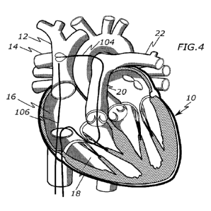
[0024] Fig. 4 is a view of a heart with a snare catheter and a
puncture system.
[0025] Fig. 5 is a view of a vessel with a snare catheter and a
puncture system.
[0026] Fig. 6 is a view of a vessel with a snare catheter and a
puncture system.
[0027] Fig. 7 is a view of a vessel with a snare catheter and a
puncture system.
[0028] Fig. 8 is a view of a vessel with a snare catheter and a
puncture system.
[0029] Fig. 9 is a view of a shunt support structure forming a
stent between two
vessels.
[0030] Fig. 10 is a view of a compressed shunt support structure.
[0031] Fig. 11 is a view of an expanded shunt support structure.
[0032] Fig. 12 is a view of a compressed shunt support structure.
[0033] Fig. 13 is a view of an expanded shunt support structure.
[0034] Fig. 14 is a view of a shunt support structure delivery
catheter.
[0035] Fig. 15 is a view of a shunt support structure delivery
catheter.
[0036] Fig. 16 is a view of a shunt support structure delivery
catheter.
[0037] Fig. 17 is a view of a shunt support structure delivery
catheter.
[0038] Figs. 18, 19, and 20 are views of a puncturing guidewire.
[0039] Figs. 21 and 22 are views of a puncturing guidewire handle.
¨ 4 -
CA 03181885 2022- 12- 7
WO 2021/263048
PCT/US2021/038989
[0040] Fig. 23 is a view of a snare catheter.
[0041] Fig. 24 is a view of a snare catheter.
[0042] Fig. 25 is a view of a snare catheter.
[0043] Fig. 26 is a view of a snare catheter and a puncturing
guidewire.
[0044] Fig. 27 is a view of a snare catheter and a puncturing
guidewire.
[0045] Fig. 28 is a view of a snare catheter and a puncturing
guidewire.
[0046] Fig. 29 is a view of a snare catheter and a puncturing
guidewire.
[0047] Fig. 30 is a view of a snare catheter and a puncturing
guidewire.
[0048] Fig. 31 is a view of a snare catheter and a puncturing
guidewire.
[0049] Fig. 32 is a view of a snare catheter and a puncturing
guidewire.
[0050] Fig. 33 is a view of a steerable or crossing catheter.
[0051] Fig. 34 is a view of a steerable or crossing catheter.
[0052] Fig. 35 is a view of a steerable or crossing catheter.
[0053] Fig. 36 is a view of a steerable or crossing catheter.
[0054] Fig. 37 is a view of steerable or crossing catheter.
[0055] Fig. 38 is a view of steerable or crossing catheter.
[0056] Fig. 39 is a view of catheter with a side aperture.
[0057] Fig. 40 is a view of catheter with a side aperture.
[0058] Fig. 41 is a view of catheter with a side aperture.
[0059] Fig. 42 is a view of catheter with a side aperture.
[0060] Fig. 43 is a view of catheter with a side aperture.
¨ 5 -
CA 03181885 2022- 12- 7
WO 2021/263048
PCT/US2021/038989
[0061] Fig. 44 is a view of catheter with a side aperture.
[0062] Figs. 45, 46, 47, 48, and 49 are views of a catheter with a
side aperture and
radiopaque markers.
[0063] Figs. 50, 51, 52, and 53 are views of a catheter with a side
aperture and a
magnetic connection mechanism.
[0064] Fig. 54 is a view of two catheters with a magnetic
connection mechanism.
[0065] Figs. 55, 56, and 57 are views of a balloon snare catheter.
DESCRIPTION OF EMBODIMENTS
[0066] Specific embodiments of the invention will now be described
with reference
to the accompanying drawings. This invention may, however, be embodied in many
different forms and should not be construed as limited to the embodiments set
forth
herein; rather, these embodiments are provided so that this disclosure will be
thorough
and complete, and will fully convey the scope of the invention to those
skilled in the
art. The terminology used in the detailed description of the embodiments
illustrated in
the accompanying drawings is not intended to be limiting of the invention. In
the
drawings, like numbers refer to like elements. While different embodiments are
described, features of each embodiment can be used interchangeably with other
described embodiments. In other words, any of the features of each of the
embodiments can be mixed and matched with each other, and embodiments should
not necessarily be rigidly interpreted to only include the features shown or
described.
[0067] Disclosed herein are improved devices and methods for
creating a shunt
between two vessels or lumens within a patient. While these devices and
methods
are generally described with regard to treatment of hypertension (e.g.,
pulmonary
arterial hypertension) and/or right heart failure/disfunction, it should be
understood that
they can be used with a variety of different vessels and lumens for other
purposes.
[0068] Shunts can be used to connect several different locations
within a body for
treatment of pulmonary arterial hypertension and/or right heart
failure/disfunction. This
specification will primarily discuss embodiments of the present invention
regarding a
¨ 6 -
CA 03181885 2022- 12- 7
WO 2021/263048
PCT/US2021/038989
shunt connecting a right pulmonary artery (RPA) to a superior vena cava (SVC).
However, these embodiments should not be limited to use solely at this
location, as
use with shunts at other locations is specifically contemplated.
[0069] A general example procedure for creating a shunt between a
right
pulmonary artery and a superior vena cava will be discussed below and further
modifications of this procedure and its equipment will then be discussed. In
this
respect, it is intended that the different embodiments discussed in this
specification
can be mixed and matched in any combination, particularly with shunt procedure
described between a right pulmonary artery and a superior vena cava.
[0070] Figures 1-12 illustrate various aspects of a method and
equipment for
creating a shunt 30 between a right pulmonary artery 14 and a superior vena
cava 12.
While the crossing is performed from the superior vena cava 12 into the right
pulmonary artery 14, the opposite may also be performed, crossing from the
right
pulmonary artery 14 into the superior vena cava 12. The resulting shunt
connection
may decrease the total pulmonary vascular resistance and the afterload of the
right
ventricle. The method generally includes the steps of targeting the right
pulmonary
artery 14, crossing through the superior vena cava 12 to the right pulmonary
artery 14
(or vice versa), positioning a shunt support structure 120 between both
vessels 12, 14,
and removing the delivery system to establish the shunt 30.
[0071] Optionally, pre-implant hemodynamic or blood flow-related
data may first be
acquired from the patient to determine or characterize any abnormalities exist
in the
heart and lungs. For example, a Swan-Ganz catheterization procedure can be
performed, as seen in Figure 1, to allow pressure measurement in the right
atrium,
pulmonary artery, and pulmonary capillaries. The pulmonary artery catheter 102
is
typically advanced into the right atrium 16 and a balloon tip 102A is
inflated, allowing
the balloon tip 102A to be carried into the right ventricle 18, into the
pulmonary trunk
20, and into the left pulmonary artery 22, which lead to the lungs.
[0072] Next, a target and/or grasping device is placed in one of
the vessels followed
by a piercing device can be placed in the other vessel, allowing one device to
pierce
both vessels and the other device to grab or engage the piercing device. For
example,
¨ 7 -
CA 03181885 2022- 12- 7
WO 2021/263048
PCT/ITS2021/038989
the target and/or grasping device can be positioned within the right pulmonary
artery
14 and the piercing device can be placed in the superior vena cava 12, or vice
versa.
[0073] Figures 2 and 3 illustrate placing a target and/or grasping
device in the right
pulmonary artery 14. In this example, the target and/or grasping device is a
snare
catheter 104 that includes one or more loops 104A that can be retracted into
an outer
sheath 104B. Additionally, radiopaque markers may be included on the catheter
104
to help "target" the loops 104A of the snare catheter. The loops 104A can be
composed of a wire, such as a metal or polymer wire.
[0074] The snare catheter 104 can be placed, in one example,
through the inferior
vena cava 24, into the pulmonary trunk 20, and further into the right
pulmonary artery
14. Placement can be achieved with a variety of techniques, including via
floating an
arrow balloon catheter to the desired location and then advancing the snare
catheter
104 within the arrow catheter or other catheter or guidewire advanced to the
location
of the arrow catheter.
[0075] Figures 4 and 5 illustrate introducing the puncture system
106 in the
superior vena cava 12. In one example, the puncture system 106 may include an
outer steerable catheter 110 (e.g., an Agilis catheter), a flexible crossing
catheter 108
positioned within the outer steerable catheter 110, and a puncturing guidewire
112
(e.g., an RF guidewire) positioned within the crossing catheter 108. However,
other
puncturing systems are possible. The puncture system 106 may be introduced by
first
accessing the femoral vein with a 12Fr catheter sheath. Next, a guidewire
(e.g.,
0.035") is advanced to the superior vena cava 12. An outer steerable catheter
110
(Agilis catheter) is tracked over the guidewire into the superior vena cava
12. Finally,
the guidewire is exchanged for the crossing catheter 108 and the inner
puncturing
guidewire 112.
[0076] Figures 6 and 7 illustrate the process of puncturing the
superior vena cava
12 and the right pulmonary artery 14. The tip of the crossing catheter108 can
be
angled or directed towards the desired puncture location (e.g., by directing
or adjusting
the position of the steerable catheter 110) and the tip angle and position can
be
confirmed via fluoroscopy. Next, the puncturing guidewire 112 is advanced into
the
target location via its puncturing method (e.g., by activating radiofrequency
energy) so
¨ 8 -
CA 03181885 2022- 12- 7
WO 2021/263048
PCT/ITS2021/038989
that it passes through the wall of the superior vena cava 12, through the wall
of the
right pulmonary artery 10, and into one of the loops 104A of the snare
catheter. The
position of the puncturing guidewire 112 within one of the loops 104A can be
confirmed
via imaging techniques. As seen in Figure 7, the loops 104 of the snare 104
can be
at least partially withdrawn into the outer sheath 104B to grab or capture the
puncturing
guidewire 112. It also may be necessary to advance the crossing catheter 108
or a
different dilator catheter through the punctures to dilate the openings. In
that respect,
the crossing catheter 108 preferably has a dilating tip.
[0077] Next, as seen in Figure 8, a shunt support structure 120 is
delivered
between the superior vena cava 12 and the right pulmonary artery 14. For
example,
the puncturing guidewire 112 may be exchanged for a delivery guidewire 111 and
the
crossing catheter108 can be removed, allowing a delivery catheter 114 to be
advanced
over the delivery guidewire 111. The distal end of the delivery catheter 114
is
positioned between the superior vena cava 12 and the right pulmonary artery
14. The
delivery catheter 114 can have an outer sheath that is withdrawn to expose the
shunt
support structure 120 and the shunt support structure 120 can be radially
expandable
by either self-expanding, by a balloon being inflated within the structure
120, or a
combination of both methods. The support structure 120 may include a passage
therethrough, which creates the shunt passage 30 between both vessels 12 and
14.
[0078] A variety of different shunt support structures 120 are
possible. For
example, Figures 10 and 11 illustrate one embodiment of a structure 120A in a
radially
compressed and radially expanded configuration, respectively. This structure
120A
has loops or leaflets that self-bend or can be bent (e.g., via balloon
inflation) generally
perpendicularly to engage the tissue of each vessel. Another example shunt
support
structure 120B can be seen in Figures 12 and 13 in a radially compressed and
radially
expanded configuration, respectively. This structure 120B may expand in a
manner
similar to a rivet by decreasing in length and radially increasing in size at
its proximal
and distal ends. Further shunt support structures and details on structures
120A and
120B can be found in U.S. App. Nos. 16/576,704 and 16/785,501, both of which
are
incorporated herein by reference. Additional shunt methods, techniques, and
equipment can also be found in the aforementioned incorporated references.
¨ 9 -
CA 03181885 2022- 12- 7
WO 2021/263048
PCT/US2021/038989
[0079] The following embodiments and methods are discussed in the
context of the
previously described shunt creation technique and equipment. While only
portions of
the previously described equipment and procedures are discussed, it should be
understood that any or all of the previously described equipment and
procedures can
be combined with those described below.
[0080] In the previous discussion of Figures 6-8, the puncturing
guidewire 112 is
advanced through the superior vena cava 12 and the right pulmonary artery 14,
the
crossing catheter 108 is advanced through the superior vena cava 12 and into
the right
pulmonary artery 14, and the delivery catheter 114 is advanced through the
punctures
of the vessels to deliver the shunt support structure 120. This procedure can
be
simplified by using a single catheter to both "steer" and dilate/cross the
opening
created by the puncturing guidewire 112. Such a device may be generally
similar to
the catheter shown in U.S. 10,076,638, herein incorporated by reference, but
may
further include a distal tip shape to dilate tissue openings (e.g., a tapered
distal tip).
[0081] In the previous discussion of Figures 6-8, the puncturing
guidewire 112 is
advanced through the superior vena cava 12 and the right pulmonary artery 14,
the
crossing catheter 108 is advanced through the superior vena cava 12 and into
the right
pulmonary artery 14, and the delivery catheter 114 is advanced through the
punctures
of the vessels to deliver the shunt support structure 120. Figures 14 and 15
illustrate
one embodiment of a delivery catheter 140 that can cross both vessels 12 and
14
without an overlying sheath disposed completely over the shunt support
structure 120
during crossing.
[0082] Typically, delivery catheters for stent-like devices include
an overlying
sheath that completely covers the stent-like device until it is in position
for being
expanded, at which point the sheath is withdrawn. However, when a delivery
device
is positioned through the wall of two vessels (e,g., vessels 12 and 14),
withdrawing the
overlying sheath may pull one or more of the vessel walls, causing the vessels
walls
to reposition relative to the position of the underlying support structure
120. Hence,
minimizing movement against the vessel's walls may help maintain a more
consistent
position of the vessel walls relative to the shunt support structure 120.
¨ 10 -
CA 03181885 2022- 12- 7
WO 2021/263048
PCT/ITS2021/038989
[0083] The delivery catheter 140 may include a proximal sleeve 146A
and/or a
distal sleeve 146B that are each positioned over only the proximal and/or
distal ends
of the shunt support structure 120 (e.g., 1-5 mm on each end) and radially
compressed
on a distal end of the catheter 140. A middle portion of the support structure
remains
uncovered by any protective barrier, such as a sleeve or sheath. This allows
most of
the shunt support structure 120 to pass through the openings of the vessels
"bare".
The sleeves 146A and 146B may be conical in shape, decreasing in diameter away
from the structure 120, and may be composed of a relatively soft polymer
material.
[0084] In one example, the sleeves 146A and 146B are disposed over
the
elongated body 144 of the catheter 140 in a manner that allows them to slide
away
from the support structure 120 prior to or during expansion. The one or more
of the
sleeves 146A and 146B may freely move or slide over the elongated body 144,
may
be biased to positions covering the support structure 120 (e.g., via a spring
or other
compressible item positioned within the sleeves 146A and 146B or at either of
their
free ends), or may have a releasable locking mechanism that releases the
sleeves
146A and 146B from a locked position to an unlocked and slidable position
(e.g., via
a pull wire).
[0085] In the example of Figures 14 and 15, a balloon 142 is
included underneath
the support structure 120. The proximal and distal ends of the balloon 142 can
be
shaped and positioned such that they push the sleeves 146A and 146B away from
the
support structure 120 when inflated, so as to release the sleeves 146A and
146B from
radially retaining the support structure 120. The balloon 142 may also include
a tacky
or adhesive layer on its outer surface to help further retain the support
structure 120
in position on the delivery device 140 during positioning, but while also
allowing the
support structure 120 to be released during expansion.
[0086] Alternately, the proximal sleeve 146A may instead be an
outer sheath or
catheter with a similar distal position that extends back to a proximal end of
the
elongated body 144. This outer sheath functions similar to the proximal sleeve
146A
except that it is longer. Hence, as the balloon 142 inflates, the outer sheath
is
proximally pushed back. A bias mechanism, such as a spring, may be connected
between the proximal ends of both the outer sheath and the elongated body 144
so
¨ 11 -
CA 03181885 2022- 12- 7
WO 2021/263048
PCT/ITS2021/038989
as to keep the outer sheath over at least a proximal end of the support
structure 120.
Additionally, the outer sheath allows the user to manually retract the outer
sheath, if
necessary, since it extends to the proximal end of the elongated body 144. The
distal
sleeve 146B may optionally be present in this embodiment.
[0087] Alternately, the sleeves 146A and 146B may be configured to
remain in
place without sliding, but instead at least partially tear as the balloon 142
expands.
These sleeves 146A and 146B may be composed of a relatively thin material
(e.g.,
urethane) and may include weakened areas or one or more cuts to promote
tearing
during expansion.
[0088] Alternately, the sleeves 146A and 146B may be configured to
remaining in
place without sliding or tearing but are instead configured such that the
support
structure 120 slides out of the sleeves 146A and 146B as the balloon 146A
expands.
The inner surface of the sleeves 146A and 146B may include a coating to reduce
friction and allow slippage. The sleeves 146A and 146B may also be composed of
a
material that stretches as the balloon 142 expands, allowing the support
structure to
pull out of the sleeves 146A and 146B as the balloon expands 142.
[0089] Figures 16 and 17 illustrate another embodiment of a
delivery catheter 150
that can be used to both pierce the vessels walls of two vessels, such as the
superior
vena cava 12 and the right pulmonary artery 14, as well as deliver the support
structure
120 to both vessels 12 and 14. Hence, instead of the need to use a separate
puncturing guidewire or similar device and delivery catheter, only the
delivery catheter
150 is needed for the puncture and support structure 120 delivery.
[0090] The delivery catheter 150 includes an elongated body 152
with a distal tip
156 configured for piercing vessel walls. In one example, the distal tip 156
includes
one or more electrodes 158 that are connected to a power source to supply
radiofrequency energy to create an opening in a vessel (e.g., the one or more
electrodes 158 are electrically connected to an RF power supply via a proximal
end of
the catheter).
[0091] The delivery catheter 150 can also act as a dilator catheter
by having a
conical cone that decreases in diameter in the distal direction. Additionally,
the
¨ 12 -
CA 03181885 2022- 12- 7
WO 2021/263048
PCT/US2021/038989
delivery catheter 150 may have an outer sheath 154, and therefore to help with
dilation, a distal portion 154A of the sheath 154 may be tapered, decreasing
in
thickness in a distal direction (e.g., along about 2-5 mm in length).
[0092] The delivery catheter 150 can also include a support
structure 120 that is
radially compressed over an inflatable balloon 153. An outer sheath 154 can be
withdrawn proximally to expose the support structure 120 and the balloon 153
can be
inflated.
[0093] In operation, the delivery device 150 is advanced with a
vessel, such as the
superior vena cava 12 such that its distal tip 156 is angled towards a target
or snare
catheter in an adjacent vessel, such as a right pulmonary artery 14. The one
or more
electrodes on the distal tip 156 are activate, e.g., applying radiofrequency
energy, to
thereby cause an opening in both vessels 12 and 14. The taper of the distal
tip 156
and the taper of the distal portion 154A allow the catheter 150 to be pushed
through
both openings so that it is positioned in both vessels 12 and 14. Next, the
outer sheath
154 is proximally withdrawn to expose the support structure 120. Finally, the
balloon
153 under the support structure 120 is inflated to expand the support
structure (or
optionally the support structure is self-expanding). In this manner, the
delivery
catheter 120 may take the place of several other catheters with dedicated
purposes.
[0094] As previously discussed in Figures 5-8, a puncturing
guidewire 112, such
as an RF guidewire, can be used to puncture or pass through both the superior
vena
cava 12 and the right pulmonary artery 14. One danger of using such a RF
guidewire
is the risk it will contact an unintended area of either of the two vessels 12
and 14
during a procedure, thereby damaging or even creating another opening in one
of the
vessels 12 and 14. Particularly, there is a danger of extending an RF
guidewire
longitudinally too far through one or more vessels, such that two openings are
created
in a vessel.
[0095] Figures 18-20 illustrates one embodiment of an RF puncturing
guidewire
160 that includes a protective sheath 166 a distal end of an elongated RF wire
body
162 to help protect from unintended lateral contact and unintended
longitudinal
contact. The sheath 166 is configured to maintain a position such that its
distal end is
either even with or extends beyond the distal end of the RF wire body 162, as
seen in
¨ 13 -
CA 03181885 2022- 12- 7
WO 2021/263048
PCT/US2021/038989
Figure 18. The distal end of the RF wire body 162 includes one or more RF
electrodes
that are connected to a power supply, and the sheath 166 thereby prevents
contact
with tissue in that Figure 18 position. Preferably, the sheath 166 has a
tubular shape
for maximum lateral protection, though other configurations are also possible,
such as
a braided tubular shape.
[0096] The sheath 166 is configured to be longitudinally slidable
and biased to the
Figure 18 position. For example, a spring 164 or similar compressible element
may
be fixed to the RF wire body (e.g., at a proximal end of the spring 164) and
to the
sheath 166 (e.g., at a distal end of the sheath 166), causing the sheath 166
to bias
distally. In this respect, the sheath 166 can be configured to longitudinally
move only
a predetermined distance (e.g., about 1 cm), which may prevent it from passing
entirely through the second vessel (e.g., the right pulmonary artery 10).
[0097] As seen in Figure 19, when the distal end of the RF
puncturing guidewire
160 is pushed against tissue (e.g., a vessel wall), the sheath 166 moves
proximally
back only a predetermined distance as the RF wire body 162 pushes against and
through a vessel wall (e.g., a stop). The predetermined distance can be
configured
so as to limit the travel of the RF puncturing guidewire 160, thereby
preventing it from
advancing it too far. As seen in Figure 20, the sheath can also be pushed
through the
first vessel wall to cover the distal end of the RF wire body 162 until it is
pressed
against and through the adjacent vessel.
[0098] Figures 21 and 22 illustrate another embodiment of an RF
guidewire
assembly 170 that is configured to limit and/or control longitudinal movement
of an RF
puncturing guidewire to prevent it from distally extending completely through
a second
vessel (e.g., two walls of a right pulmonary artery 10). Specifically, a
handle portion
172 includes a mechanism configured to move the RF puncturing guidewire 178
relative to its outer tubular sheath 176. In one example, the mechanism
includes a
thumbwheel 174 that engages a toothed track connected to the RF puncturing
guidewire 178 such that rotation of the thumbwheel 174 moves the track and the
guidewire 178 longitudinally. A limit mechanism or stop member can be
positioned
within the handle to prevent movement of the guidewire 178 beyond a
predetermined
distance that would otherwise puncture entirely through a vessel (e.g., 1 cm).
¨ 14 -
CA 03181885 2022- 12- 7
WO 2021/263048
PCT/US2021/038989
Alternate movement mechanisms are possible, such as screw drive mechanisms or
thumb sliders. The handle 172 and the guidewire 178 can be connected to an RF
power source so as to allow the guidewire 178 to apply RF energy to the
patient's
tissue.
[0099] In practice, the user advances the tubular sheath 178 so
that the distal end
is in a desired target location. RF energy can be applied to the guidewire 178
so that
its distal end can apply radiofrequency energy to tissue. The user can rotate
the
thumbwheel 174 to cause the RF puncturing guidewire 178 to contact the wall of
a first
vessel (e.g., superior vena cava 12), pass through its vessel wall, contact a
second
vessel (e.g., right pulmonary artery 10) and then pass through its wall.
[00100] In an alternate embodiment, the handle 172 can move the outer sheath
176
relative to the RF puncturing guidewire 178. This allows the user to advance
the entire
guidewire assembly 170 to be distally advanced until the distal end of the
sheath 178
blocks further advancement.
[00101] Alternately or additionally, the guidewire assembly 170 may include a
switch
or circuit breaker mechanism that interrupts the RF current when the guidewire
178 is
extended from the sheath 176 a predetermined distance (e.g., 1 cm). The switch
or
circuit breaker mechanism may be located within the handle 172 and can be
actuated
when a portion or feature on or connected to the guidewire 178 distally
advances to
the predetermined distance. In another embodiment, the switch may be an
electrolytic
segment of the circuit near or in electrical communication with one of the
electrical
contacts of the puncturing guidewire 112 or snare, such that as the electrical
contacts
of the puncturing guidewire 112 contact the snare catheter (e.g., the shield
or loops),
the electrolytic segment or fuse dissolves, breaking the circuit.
[00102] In another embodiment, any of the piercing guidewires discussed in the
specification may be connected to an RF energy source with a timer configured
to
activate for only a length of time sufficient to pierce through one wall of
the first vessel
(e.g., superior vena cava 12) and/or one wall of the second vessel (e.g.,
right
pulmonary artery 14). For example, the RF energy may be activated for only .5
second, 1 second, 1.5 seconds, or two seconds. In this manner, the RF energy
can
¨ 15 -
CA 03181885 2022- 12- 7
WO 2021/263048
PCT/US2021/038989
be quickly turned off to prevent unwanted damage (e.g., puncturing entirely
through
opposite walls of a vessel).
[00103] As previously discussed with regard to Figures 6-8, a target or snare
catheter 104 can be used to capture a puncturing guidewire 112. One challenge
with
using a snare catheter in this manner is that it may be difficult to maintain
the position
of its loops 104A so that the puncturing guidewire 112 can be threaded
through.
Additionally, once through the loops 104A, the puncturing guidewire 112 may be
accidentally advanced through the opposite side of the vessel it entered
(i.e., entirely
through the vessel). The following embodiments address one or more of these
challenges.
[00104] The snare catheter 180 shown in Figure 23 includes an inflatable
balloon
182 that can be inflated to engage the walls of the vessel (e.g., right
pulmonary artery
14) so that its distal end can be locked into place within the vessel. The
balloon 182
can be located at the distal end of an elongated catheter body 187, which
further
includes one or more apertures 186 in communication with a fluid passage
through
the body 188 that allows inflation of the balloon 182. The catheter body 187
can be
moved into and out of an elongated tubular sheath 188.
[00105] One or more snare loops 184 (e.g., two loops) are positioned at the
distal
end of the elongated catheter body 187. This can be achieved in several ways.
For
example, the loops 184 can be fixed to the elongated catheter body 187 and
positioned
within the balloon 182 such that the balloon 182 inflates around the loops. In
another
example, the loops 184 may be fixed to the elongated catheter body 187 and
positioned outside of the balloon 182 such that the loops remain on an outer
surface
of the balloon 182 when inflated. In another example, the loops may be
positioned
outside of and adjacent to the balloon 182 but are connected to a separate
elongated
body or pusher that allows the loops 184 to move independently of the balloon
182.
In another example, the loops 184 can be embedded, adhered to, or bonded to
the
material of the balloon 182.
[00106] In practice, the distal ends of the sheath 188 and catheter body 187
can be
positioned at a desired location in a vessel (e.g., right pulmonary artery
14), the balloon
182 can be inflated to engage the walls of the vessel, a puncturing guidewire
112 can
¨ 16 -
CA 03181885 2022- 12- 7
WO 2021/263048
PCT/US2021/038989
be advanced through the loops 184 (and optionally through the balloon 182, and
the
loops 184 can be at least partially retracted into the sheath 188 to grab the
puncturing
guidewire 112.
[00107] Optionally, the balloon 182 may be composed of a puncture resistant
material that resists puncture from the puncturing guidewire 112. For example,
only
one side may be composed of a puncture resistance material when the loops 184
are
located within the balloon 184, allowing the puncturing guidewire 112 to pass
through
one side of the balloon 184 but not its opposite side. In embodiments with the
loops
184 being located outside the balloon 182, the entire balloon may be composed
of
puncture resistant material. The puncture resistant material may be a hardened
polymer or flexible material containing one or more metal strands or panels.
[00108] Figures 24 and 25 illustrate an alternate embodiment of a snare
catheter
190 that includes a rear shield 192 that extends behind a plurality of wire
snare loops
194 and blocks a puncturing guidewire 112 from passing entirely through the
vessel it
is deployed in (e.g., right pulmonary artery 14). Both the snare loops 194 and
the
shield 192 may be fixed to the end of an inner elongated catheter body 196
which can
be extended out of and pulled into an outer tubular sheath 198.
[00109] The shield 192 may be composed of a plurality of woven or braided
wires,
textile, a polymer sheet (e.g., polyurethane), silicone, or similar materials.
The shield
192 may also be composed of a shape memory frame (e.g., a Nitinol wire) that
allows
the shield 192 to expand to its desired shape. The shield 192 may also expand
from
a radially compressed configuration to an expanded configuration having a
variety of
different shapes. For example, the shield 192 may expand to an oval, planar
shape.
In another example, the shield 192 may expand to a curved shape across the
axis of
the device to conform to the curvature of the vessel it is deployed in, as
seen in the
end view of Figure 25.
[00110] In one embodiment, the shield 192 can be configured to turn off
radiofrequency energy being supplied to a puncturing guidewire 112 that uses
RF
energy. For example, the shield 192 may be composed of an outer electrically
insulated layer and an inner conductive layer so that when the puncturing
guidewire
112 punctures through, it creates electrical contact with the conductive
layer. The
¨ 17 -
CA 03181885 2022- 12- 7
WO 2021/263048
PCT/US2021/038989
conductive layer and therefore the snare catheter 190 may be connected to an
RE
power supply that is configured to interrupt the RE power to the puncturing
guidewire
112.
[00111] The inner catheter 196 may also include a funnel/cone portion at the
distal
end of its body and proximal of the shield 192 and loops 194 to help radially
compress
these structures as the inner catheter 196 is pulled proximally back into the
outer
sheath 198. For example, the funnel may be composed of one or more coiled
wires,
a braided mesh cone, or a polymer cone.
[00112] Figure 26 illustrates an embodiment of a snare catheter 191 that is
generally similar to the previously described snare catheter 190 but has a
shield 193
forming a circular diameter with a concave interior as opposed to the more
oval shape
of shield 192. In other words, the shield 193 is hemispherical with an
interior space
positioned at least partially around loops 194.
[00113] Figure 27 illustrates an embodiment of a snare catheter 195 that is
generally
similar to the previously described snare catheter 190 but has a generally
planar shield
197. The shield 197 can have a variety of different planar shapes, such as a
square,
rectangle, circle, or oval shape. Optionally, the "plane" of the shield 197
may also
have a slight curve and the axial direction of the catheter 195, thereby
forming a partial
tubular shape.
[00114] Figure 28 illustrates an embodiment of a snare catheter 200 that is
generally
similar to the previously described snare catheter 190 but includes an
anchoring
mechanism to anchor the shield 202 and snare loop 206 in a desired position
within a
vessel (e.g., right pulmonary artery 14).
[00115] In one example, an elongated inner catheter 208 includes one or more
distal
balloons 204A and one or more proximal balloons 204B that are spaced on either
side
of the shield 202 and snare loop 206. The inner catheter 208 includes one or
more
inflation lumens that are configured to connect to a fluid supply, thereby
allowing the
balloons to be inflated. Each of the balloons 204A can be a single balloon
that entirely
expands with in the vessel 14 or can each include a plurality of balloons
(e.g., two,
three, four, or five balloons). By using a plurality of balloons, it may be
possible to
¨ 18 -
CA 03181885 2022- 12- 7
WO 2021/263048
PCT/ITS2021/038989
include spaces or perfusion passages across the balloons to allow for blood
flow
during inflation.
[00116] As in any of the previous embodiments, the snare loop 206 can be fixed
to
the shield 202 or the snare loops 206 can be connected to a separate elongated
wire
or body that allows it to move independently of the shield 202.
[00117] In practice, the distal end of the inner catheter 208 is positioned at
a desired
shunt creation location, outside of the outer sheath 198. Next, the one or
more distal
balloons 204A and one or more proximal balloons 204B are inflated to engage
the
walls of the vessel (e.g., right pulmonary artery 14), distally and proximally
of the
expanded shield 202 and snare loop 206. The puncturing guidewire 112 is then
advanced through another vessel (e.g., superior vena cava 12), into the prior
vessel
(e.g., right pulmonary artery 14), through the snare loop 206, and is
prevented from
further advancement by the shield 202. Finally, balloons 204A and 204B are
deflated
and the inner catheter 208 (or the wire connected to the snare loop 206) is at
least
partially retracted into the outer sheath 198 to grasp the puncturing
guidewire 112.
[00118] Any of the embodiments relating to a target or snare catheter may
include
perfusion features or passages to allow blood to flow around any blockages
that are
created. While these perfusion features may be particularly
desirable for
embodiments with balloons (e.g., snare catheter 180 in Figure 23 or snare
catheter
200 in Figure 28), it may also be desirable in embodiments with a shield as
well, since
these shields may at least partially block blood flow through the vessel.
[00119] As previously discussed for the snare catheter 200, one way to achieve
perfusion passages is to provide two or more balloons at a particular location
that,
when inflated, create gaps or longitudinal passages between themselves.
Another
technique can be seen in the snare catheter 210 in Figure 29 which includes a
proximal
perfusion opening and a distal perfusion opening 212B that both connect to a
perfusion
passage or channel therebetween in the inner catheter 187. This snare catheter
210
is generally similar to the snare catheter 180 in Figure 23, but the perfusion
channel
and openings 212A and 212B can be used on any of the snare catheter
embodiments
described herein, including those with balloons that also have perfusion
passages
between themselves.
¨ 19 -
CA 03181885 2022- 12- 7
WO 2021/263048
PCT/US2021/038989
[00120] As previously discussed, it can be undesirable for radiofrequency
energy
from a puncturing guidewire 112 to damage unwanted areas of the patient.
Figure 30
illustrates one embodiment of a snare catheter 220 that helps maintain the RF
energy
between only the puncturing guidewire 112 and the snare catheter 220 by
including
one or more RF electrodes 222 in the snare catheter 220. For example, the
electrode
222 may be embedded within a balloon 182, a shield, or any component of the
snare
catheter embodiments of this specification. The one or more electrodes 222 may
have
an opposite polarity to the electrodes on the distal end of the puncturing
guidewire 112
and may also be connected to the RF energy source outside of the patient.
Hence,
the RF energy takes the path of least resistance to the electrode 222, thereby
avoiding
other tissue that is not intended to be damaged. The electrodes can be strips
of
conductive material on or embedded in the balloon 182 (or other component) or
can
be a plurality of wires arranged in a pattern (e.g., braided).
[00121] The snare catheter embodiments of this specification may also include
mechanisms for sensing the position of the snare catheter and/or aligning
puncturing
guidewire 112 with the snare catheter. Figures 31 and 32 (side and top views,
respectively) illustrate one example of such a snare catheter 240 that creates
a
magnetic field that can be used for either positioning or self-aligning
purposes. In this
example, the snare catheter 240 is generally similar to the snare catheter 180
in Figure
23 except that one or more coils of conductive wire 242 is located within, on,
or
embedded into the balloon 182. The one or more coils of conductive wire 242
are
connected to a power source via the proximal end of the catheter 240, allowing
current
to selectively pass through the one or more coils 242 and generate a magnetic
field.
[00122] The magnetic field can be used in two possible ways. First, the
puncturing
guidewire 112 may include one or more magnetic sensors that can sense the
magnetic
field, allowing the puncturing guidewire 112 to be better aligned with the
snare catheter
240. For example, the one or more sensors may sense the magnitude of the
magnetic
field on each side of the puncturing guidewire 112 and/or may sense the
polarity of
the magnetic field, thereby providing additional data to achieve a desired
orientation.
Second, the puncturing guidewire 112 may have its own magnets or ferrous
material
that is attracted to the magnetic field generated by the one or more coils of
conductive
wire 242. This may provide physical force and guidance to better align the
puncturing
¨ 20 -
CA 03181885 2022- 12- 7
WO 2021/263048
PCT/US2021/038989
guidewire 112 with the snare catheter 240. Either of these two
sensing/aligning
features or both of these features can be used.
[00123] The coil 242 may also be incorporated into other structures, such as a
shield
or catheter body. Alternately, either a balloon or shield may include one or
more
permanent magnets to provide similar functionality. Alternately, ferrous
material can
be incorporated into the balloon or shield and the puncturing guidewire 112
may
include permanent magnets or an electromagnet (e.g., conductive wire coil).
[00124] As previously discussed, one challenge of a shunt procedure between
vessels, particularly between the superior vena cava 12 and right pulmonary
artery 14,
is directing the puncturing guidewire 112 through the vessel walls at the
desired
location and at the desired angle. Further, as the puncturing guidewire 112 is
advanced out of the outer steerable catheter 110 (or out of the crossing
catheter 108
within the steerable catheter), it may cause the steerable catheter 110 to
deflect from
the intended position and angle.
[00125] One approach to maintaining the position of the steerable catheter 110
during a procedure is to include an expandable member on a side of the
catheter
opposite of which it bends forward so as to brace the distal end of the
catheter 110 in
place. For example, Figures 33 and 34 illustrate a steerable catheter 250
having an
elongated tubular body with an inflatable balloon 254 that is positioned on an
outer
surface of the catheter body 252, opposite of the distal opening of the
catheter body
252. When the steerable catheter 250 is bent in a first direction, the balloon
254 can
be inflated via inflation passage 252A, either prior to or after the bending.
The balloon
254 expands in a direction opposite of the bend and braces the back side of
the
catheter body 252 which allows the puncturing guidewire 112 to be advanced out
in a
predictable direction and location. The steerable catheter 250 generally
comprises an
elongated tubular body that includes mechanisms to allow the distal end of the
catheter to bend via user controls on a proximal end of the catheter.
[00126] Figures 35 and 36 illustrate a similar steerable catheter 255 in which
one or
more balloons 256 inflate on multiple sides of the catheter body 252 to center
the
catheter body 252 within the vessel 12. Again, this helps provide an anchored
position
for the steerable catheter 255 that allows for a more predictable location and
direction
¨21 -
CA 03181885 2022- 12- 7
WO 2021/263048
PCT/US2021/038989
advancement of the puncturing guidewire 112. The one or more balloons 256 can
be
a single balloon that extends entirely or nearly entirely around the
circumference of
the catheter body 252, or can be two or more balloons (e.g., 3, 4, or 5
balloons).
[00127] Figure 37 illustrates another embodiment of a steerable catheter 260
that
includes an expandable wire frame or structure 264 that can expand
perpendicularly
to an axis of the catheter body 262 from a side opposite the bent opening of
the body
262. In one example, the expandable wire structure 264 is composed of a shape
memory material (e.g., Nitinol) and is shape set to expand to the desired
perpendicular
position. The wire structure 262 can be a ring shape (e.g., circular, square,
etc.).
Alternately, the wire structure can be one, two, three, four, or more arms
272, as seen
in the steerable catheter 270 in Figure 38. Each arm can be composed of a
shape
memory material (e.g., Nitinol) that is biased outwards in a direction
generally
perpendicular to the body 262. Each arm 272 can be a single wire (e.g.,
generally
straight or bent) or each arm 272 can be a loop of wire (e.g., circular, oval,
square,
rectangular, etc.).
[00128] While the embodiments of the previously discussed Figures
33 through 37
are contemplated for use on a steerable catheter through which the puncturing
guidewire can be advanced through, other components may also use these
features.
For example, the snare catheter 104 may also include one or more of these
centering
or positioning features.
[00129] Turning to Figure 39, a catheter 280 or elongated catheter body having
a
side aperture 282 can also be used with the shunt creation methods of this
specification. The catheter 280 includes at least one lumen within it that is
in
communication with the aperture 282. The aperture 282 may be located in the
sidewall
of the catheter, just proximal of the distal end of the catheter 280. For
example, the
aperture may be about 1-2 cm from the distal end of the catheter 280. The
aperture
282 may also have a general diameter of about 0.1 -0.5 cm.
[00130] In one embodiment, the catheter 280 is configured to form a curve
through
its distal end to conform to the right pulmonary artery 14 and help brace it
during a
procedure. In one example, about 5 to 15 cm of the distal end has a curve of
about
60 - 90 degrees relative to the remaining proximal portion of the catheter
280.
¨22 -
CA 03181885 2022- 12- 7
WO 2021/263048
PCT/US2021/038989
[00131] In one example use, seen in Figure 39, the catheter 280 can be
advanced
into the right pulmonary artery 14 so that the aperture 282 aligns with the
superior
vena cava 12. Next, a puncturing guidewire 112 is advanced through the lumen
of the
catheter 280, out the aperture 282, and into the superior vena cava 12.
[00132] Optionally, the catheter 280 may include an anchoring device to help
brace
or maintain its position within the right pulmonary artery 14. One such
anchoring
device is a balloon 284 that is positioned at the distal end or tip of the
catheter 280, as
seen in Figure 40. This balloon 284 is configured to be inflated to a size
that engages
the vessels walls (e.g., via an inflation lumen in the catheter 280).
Alternately or
additionally, the catheter 280 may include a balloon, ring, expandable braided
mesh,
or arms extending from the outer surface of the catheter wall, directly behind
the
aperture 282.
[00133] Figure 41 illustrates another anchoring device comprising wire
framework
comprising a wire 286 that is attached to and radially expands from a distal
end of the
catheter 280 to engage the walls of the vessel. The wire may be composed of
shape
memory material (e.g., Nitinol) and shape set to a desired shape. The shape
may
include a helical coil, as seen in the figure, a plurality of loops, a
plurality of arms, or
similar shapes.
[00134] Figure 42 illustrates another anchoring device comprising one or more
centering balloons 285 positioned near or adjacent to the aperture 282 so as
to
position the catheter 280 near a center of the right pulmonary artery 14.
Hence, the
one or more centering balloons may help both anchor and position the catheter
280 to
a position that allows access to the superior vena cava 12. However, the one
or more
centering balloons 285 may include any of the other anchoring devices
previously
discussed, as well.
[00135] In one example, the one or more balloons 285 is a single "C" shaped
balloon
that is positioned around the circumference of the catheter 280 at the
location of the
aperture 285 but leaving the aperture 285 uncovered. In another example, a
plurality
of cylindrical balloons can be used in a similar position to achieve the "C"
shape.
¨ 23 -
CA 03181885 2022- 12- 7
WO 2021/263048
PCT/US2021/038989
[00136] Additionally, radiopaque markers 287 may be included adjacent the
aperture 282 in this embodiment or any of the other embodiments. For example,
a
first marker 287 can be located just distal of the aperture 282 and a second
marker
287 can be located just proximal of the aperture 282. Alternately or
additionally,
markers 287 can be located above or below (i.e., on the same circumference of
the
catheter 280) of the aperture 282.
[00137] As also seen in Figure 42, a snare 104 (or any of the other snare
embodiments of this specification, including those with shields or other
safety
measures that prevent completely passing through a vessel, such as the
embodiment
shown in Fig. 26) can be used in the superior vena cava 12 to snare or capture
the
puncturing guidewire 112. This snare 104 can be used in this manner with any
of the
previous examples/embodiments.
[00138] Again, while the catheter 280 in Figure 42 is shown in the right
pulmonary
artery 14, this catheter may also be used in the superior vena cava 12
instead, as any
of the embodiments of this specification can be reversed in this manner. In
such an
arrangement, any of the target/snare catheters described in this specification
may be
used.
[00139] It may be helpful to provide an additional mechanism to help direct
the
puncturing guidewire 112 out of the aperture 282 in a desired direction. For
example,
the lumen of the catheter 180 may include a curved or ramped surface near the
aperture 282 that is configured to help direct the distal end of the guidewire
112 out of
the aperture 282. In another example, the puncturing guidewire 112 may include
a
balloon, wire loop, or wire arms, extending from one side of its body. In
another
example, a steerable catheter 110 may be advanced through the lumen of the
catheter
280, along with the puncturing guidewire 112, as seen in Figure 43. In this
respect,
the distal end of the steerable catheter 110 can be turned or directed so that
its distal
opening faces or extends out of the aperture 282.
[00140] Alternately, the catheter 280 may be used as a target catheter,
similar to the
previously discussed snare catheter, such that the puncturing guidewire 112 is
advanced from the superior vena cava 12 into the right pulmonary artery 14, as
seen
in Figure 44.
¨24 -
CA 03181885 2022- 12- 7
WO 2021/263048
PCT/US2021/038989
[00141] In such an arrangement, it may be desirable to include radiopaque
markers
on the catheter 280 and on the steerable catheter 110 (or alternately a
crossing
catheter 108). In one example seen best in Figures 45-49, the catheter 280
includes
one or more radiopaque marker 288 that are located proximally adjacent and
distally
adjacent of the aperture 282. For example, the markers 288 may include a first
and
second line extending perpendicular to the axis of the catheter 280.
Additionally or
alternately, the markers 288 may include lines parallel to the axis of the
catheter 280.
The steerable catheter 110 may also include one or more radiopaque markers 289
that allow the user to help line up the distal end of the catheter 110 with
the apertures
288 of catheter 280. In one example, the marker 189 is one or more (e.g., 2 or
4)
radiopaque lines that are aligned with the axis of the steerable catheter 110.
In the
case of 2 markers 289, they can be located at about 180 degrees from each
other and
immediately adjacent to the distal end of the catheter 110. In the case of 4
markers
289, they can be located at about 90 degrees from each other and immediately
adjacent to the distal end of the catheter 110.
[00142] In practice, the user can view both markers 288 and 289 and then align
the
markers 189 of the steerable catheter 110 with those markers 288 of the
catheter 280.
Once aligned (e.g., figures 47-49), the puncturing guidewire 112 can be
advanced out
of the steerable catheter 110 and into the aperture 282 of catheter 280.
[00143] In another embodiment, the catheter 280 may include echogenic markers
in
similar positions as any of the previously discussed radiopaque markers,
either instead
of or in addition to the radiopaque markers. The echogenic markers allow a
physician
to utilize intracardiac echo imaging to monitor and then adjust the position
of either of
the catheters involved in the procedure.
[00144] As previously discussed, the catheter 280 can be connected to with a
steerable catheter 110 or flexible crossing catheter 108 (or a catheter with
both
abilities), via a puncturing guidewire 112 passing from either the superior
vena cava
14 or right pulmonary artery 14. In either method, a magnetic connection
mechanism
can be used to help connect to the aperture 282, as seen in Figures 50-53. For
example, the crossing catheter 108 may include a magnetic ring 290 located at
or near
the distal edge of the catheter 108. The ring 290 may have magnetic material
¨ 25 -
CA 03181885 2022- 12- 7
WO 2021/263048
PCT/US2021/038989
extending entirely around the distal opening of the catheter 108 as seen in
Figure 51
or the ring 290 may have several discrete areas of magnetic material at
locations
around the distal opening of the catheter 108, as seen in Figure 52 (e.g., at
least two
locations 280 degrees apart from each other).
[00145] The catheter 280 may include magnetic material 292 (or ferrous
material)
near or around the aperture 282. For example, the magnetic material 292 may be
two
lines or areas proximally and distally adjacent to the aperture 282.
Preferably, the
magnetic material 292 is spaced apart a similar distance as that of magnetic
material
290 on the crossing catheter 290 and configured to attract each other (e.g.,
opposite
polarities), allowing the two areas of magnetic material 290, 292 to align and
engage
with each other as the tip of the catheter 108 is advanced toward the aperture
282.
[00146] The catheter 280 may also include an elongated tip 280A to help
position
and brace the catheter 280 in a desired position to achieve a magnetic
connection.
[00147] The magnetic material 290, 292 and previous configuration may be
included
on a variety of different catheter configurations, especially those described
in the
present specification. For example, two catheters 291, 108 with openings
directly on
their distal ends can be configured with the magnetic material 290, 192, as
seen in
Figure 54. One of more of the catheters 291 and 108 may be steerable (as well
as
configured for crossing). Hence, the puncturing guidewire 112 can be advanced
through either of the catheters 291, 108 and one of the catheters that is
configured for
crossing/dilating (e.g., crossing catheter 108) can move through the puncture,
causing
the magnetic material 290 to align with magnetic material 292, connecting the
lumens
of the two catheters.
[00148] Figures 55-57 illustrate another embodiment of a target or snare
catheter
system 300 that captures a distal end of a puncturing guidewire 112 via a
plurality of
balloons 304. When the puncturing guidewire 112 is positioned between the
balloons
304 and the balloons 304 are deflated, they at least partially engage or wrap
around
the end of the guidewire 112, allowing the elongated catheter body 302 and
balloons
304 to be withdrawn into the outer sheath 306, thereby capturing the guidewire
112.
¨26 -
CA 03181885 2022- 12- 7
WO 2021/263048
PCT/US2021/038989
[00149] The balloons 304 are positioned at the distal end of an elongated
catheter
body 302 which includes one or more lumens configured to inflate the balloons
304.
The balloons 304 can have a variety of different shapes, including
longitudinal
cylindrical shapes, as seen in the figures. Preferably, the balloons 204 are
positioned
adjacent to each other so that after inflation they contact one another but
also allows
for some space between them so that the guidewire 112 can pass between them
and
into the space. In one example, the balloons 304 may be supported on a
framework
(e.g., of tubes or wires) with no central catheter member within the balloon
group or
alternately, a very small diameter tube/body that allows spacing between it
and the
balloons 304. The catheter system 300 includes at least two balloons, but
three, four,
five, six or more balloons 304 are also possible.
[00150] Figure 56 illustrates the guidewire 112 moving into the central space
between four inflated balloons after puncturing the walls of the vessels. Once
positioned, the balloons 304 are deflated, as seen in Figure 57, which cause
the
balloon material to partially wrap around the guidewire 112. The elongated
catheter
body 302 and balloons 304, along with the captured guidewire 112 are retracted
into
the outer sheath 306 to further lock the position of the guidewire 112.
[00151] This specification primarily discusses embodiments of the present
invention
with regard to a shunt connecting a right pulmonary artery to a superior vena
cava.
However, shunts can be created at other locations for similar purposes.
[00152] In one example, a main pulmonary artery (PA) is shunted to the right
atrium
or atrial appendage (RAA). In this method, a right-to-right shunt from a
region of higher
pressure in the PA is connected to a region of lower pressure in the RAA.
Doing so
utilizes the high compliance of the RAA to "absorb" additional volume received
from
the shunt since the RAA is a naturally compliant reservoir. An additional
benefit may
arise from the fact that the RAA and the main PA are both inside the
pericardium and,
therefore, would contain any leaks resulting as a complication of an
improperly seated
shunt. Another benefit may be that the risk of puncturing the aorta is
minimized.
[00153] In another example, a connection made between a pulmonary artery (PA)
and a pulmonary vein (PV) may be used to treat pulmonary hypertension or right
heart
failure/dysfunction. To reduce the total pulmonary vascular resistance and the
¨ 27 -
CA 03181885 2022- 12- 7
WO 2021/263048
PCT/US2021/038989
afterload of the right ventricle, a shunt is created between a right pulmonary
artery
(RPA) and a right pulmonary vein (RPV). Alternatively, the shunt could be
placed
between a left pulmonary artery ([PA) and a left pulmonary vein (LPV).
[00154] In another example, a connection is created between a pulmonary artery
(PA) and a left atrial appendage (LAA), in order to treat pulmonary
hypertension, right
heart failure/dysfunction, or atrial fibrillation, which reduces the total
pulmonary
vascular resistance and the afterload of the right ventricle. An added benefit
to the
reduced right ventricular afterload is the washout of the LAA in those
patients that are
at risk of stroke.
[00155] In yet another example, a shunt is created between a pulmonary vein
(PV)
and superior vena cava (SVC) to treat heart failure. This may particularly
help treat
elevated left atrial pressures causing fluid to back up in the lungs.
[00156] In yet another example, a plurality of shunts at different locations,
such as
any of the previously discussed locations can be used. For instance, there may
be a
benefit to placing an RPA-SVC shunt as well as an atrial shunt in certain
populations.
The RPA-SVC shunt would help reduce RV afterload and the LA shunt would help
reduce PVR while keeping LA pressure and LV filling pressure low. To the same
effect, there may be a benefit to the combination of the RPA-VC, intra-atrial,
and
arteriovenous peripheral shunt in certain patients.
[00157] Although the invention has been described in terms of particular
embodiments and applications, one of ordinary skill in the art, in light of
this teaching,
can generate additional embodiments and modifications without departing from
the
spirit of or exceeding the scope of the claimed invention. Accordingly, it is
to be
understood that the drawings and descriptions herein are proffered by way of
example
to facilitate comprehension of the invention and should not be construed to
limit the
scope thereof.
¨ 28 -
CA 03181885 2022- 12- 7