Note: Descriptions are shown in the official language in which they were submitted.
CA 03182158 2022-11-02
WO 2021/226486
PCT/US2021/031337
METHODS AND DEVICES FOR TREATMENT OF NEUROPATHY
CROSS REFERENCE TO RELATED APPLICATIONS
[0001] The present application claims priority to U.S. Provisional
Application No.
63/022,258, filed May 8, 2020, the entire contents of which are hereby
incorporated by
reference in its entirety.
BACKGROUND
[0002] Peripheral Neuropathy (PN) refers to a condition that results
when nerves
that carry messages to and from the brain and spinal cord from and to the rest
of the body
are damaged or diseased. An estimated 30 million Americans suffer from this
painful and
debilitating disease, which is marked by a progressive dying back of distal
nerves that starts
in the skin and moves inwards, causing a complex suite of symptoms that
include pain, loss
of sensation, numbness, and even limb amputation. These drastic clinical
outcomes are often
due to subpar diagnostic tools that cannot provide early or sufficiently
sensitive diagnosis of
neuropathy. For certain peripheral neuropathies, early and/or more
sensitive/functional
diagnosis can enable interventions to prevent further decline.
[0003] Currently there is no cure for PN, and treatment is largely
palliative (for
example, analgesics for pain relief). However, early detection and monitoring
affords the
possibility of interventions to halt the progression of neuropathy, and
potentially reverse the
disease with novel treatments that are currently under research. As new
therapies are
available and in use, monitoring nerve recovery and regrowth is also a
clinical need. Thus,
there exists a need for improved diagnostic systems and methods for early
diagnosis,
monitoring, prevention, and treatment of PN.
SUMMARY
[0004] Presented herein are systems, methods, and devices related to
biomedical
devices that can provide, inter alia, early-onset, sensitive, and/or
functional detection of
neuropathy with the goal to better control the disease. Early detection of
neuropathy during
1
CA 03182158 2022-11-02
WO 2021/226486
PCT/US2021/031337
clinical progression (the disease is degenerative with time) enables
clinicians to mitigate the
causes of neuropathy when known (for example, glucose regulation for diabetes,
discontinuation or switching chemotherapy medications, improvement of
environment that
may include neuropathic chemicals, treatment of autoimmune diseases, etc.). In
some
embodiments, systems and methods described herein allow for rapid or faster
detection of
neuropathy that provides clinicians with quicker diagnosis times that can
enable
interventions to prevent further decline. In some embodiments, systems as
described herein,
may be utilized as `theragnostic' (therapeutic and diagnostic) systems, for
example, that not
only provide accurate, early, and/or functional detection of neuropathy
especially for small,
free nerve fibers, but also allow delivery of treatments (e.g. therapeutic
agents, stimulation)
for prevention and treatment of the disease (e.g., via transdermal delivery,
for example,
through at least some needles of arrays as described herein). Furthermore, in
some
embodiments, devices as described herein allow for delivery of treatments, for
example, to
stimulate nerve re-growth and/or control pain. Other treatments targeted to
skin and
underlying tissue layers reached by needles of the arrays described herein,
could also be
delivered for treating other or related medical conditions in the same
patients. In accordance
with various embodiments, provided systems may allow for relatively pain-free
and non-
invasive testing, diagnosis, and even treatment of particular diseases,
disorders or
conditions. In some embodiments, methods, devices, and systems described
herein facilitate
biological sample collection, delivery of treatments, for example therapeutic
agents and
nerve stimulation, and nerve re-growth monitoring. In some embodiments,
methods,
systems, and/or devices as described herein may be used for monitoring nerve
re-growth
following a medical intervention (e.g., following a nerve graft,
administration of a
therapeutic agent, or following nerve stimulation) and/or the progression
and/or regression
of symptoms corresponding to a disease, disorder, or condition of interest. In
the context of
neuropathy, the potential for such diagnosis and/or intervention may improve
the quality of
life for millions of patients, thus saving on healthcare costs for pain
medications, neuropathy
related injuries, and limb amputations. Improved detection and diagnosis also
allows
clinicians to save time and money in patient interactions and testing.
[0005] Among other things, systems, devices, and methods, for example,
as
described herein, identify and address at least some of the challenges of
early and accurate
2
CA 03182158 2022-11-02
WO 2021/226486
PCT/US2021/031337
detection and treatment of various diseases, disorders, or conditions
disclosed herein, for
example, neuropathies. In some embodiments, provided systems and methods are
portable,
easy to use, wireless, and able to be administered in a doctor's office,
clinic or eventually at
home, and would fill a gap in the current market, since an accurate, specific,
non-invasive,
painless, easy to deliver, early onset neuropathy diagnostic and treatment
system currently
does not exist. It is also beneficial for the clinician to remove the
subjective patient self-
reporting, as is common with other neuropathy tests, by providing a functional
and
quantitative unbiased assessment of nerve integrity. This testing platform
further improves
clinical testing by being portable, inexpensive, disposable (e.g. arrays
only), easy to operate,
and quick (e.g. testing time of 5-10 min on average).
[0006] In one aspect, the present disclosure provides methods of
evaluating and/or
treating a subject for neuropathy, and/or evaluating nerve health, nerve
function, and/or
nerve regrowth in a subject. In some embodiments, such methods may comprise:
placing a
device (for example, as disclosed herein) on or below a surface of the skin of
the subject,
inserting one or more needles of the device into the skin and/or subcutaneous
tissue of the
subject, and performing one or more of an electrical measurement, a
stimulation (e.g.
mechanical stimuli, thermal stimuli, etc.), fluid sampling, and administration
of one or more
therapeutic agents using at least one of the one or more inserted needles,
over a period of
time, to evaluate and/or treat a subject. In some embodiments, a method
further comprises
determining, by comparing a performed electrical measurement with a similar
electrical
control measurement, a likelihood that the subject suffers from neuropathy. In
some
embodiments, an electrical control measurement is obtained from healthy
control subjects, a
patient data sample taken earlier in disease progression, or from a healthy
region of tissue on
the subject's body.
[0007] In some embodiments, the step of performing comprises performing
two or
more of an electrical measurement, a stimulation, fluid sampling, and
administration of
agents. In some embodiments, for example, this step is performed using at
least one of the
one or more inserted needles, over a period of time, to evaluate and/or treat
a subject. In
some embodiments, performing an electrical measurement comprises obtaining a
measure of
nerve conductance and/or tissue electrical activity, and/or local field
potentials (e.g.
3
CA 03182158 2022-11-02
WO 2021/226486
PCT/US2021/031337
extracellular recordings). In some embodiments, for example, the one or more
inserted
needles is recording from one section of its length (e.g. the needle's
interior or exterior). In
some embodiments, the one or more inserted needles is recording from its tip
(e.g. if coated
with insulating material). In some embodiments, the one or more inserted
needles is
recording from the entire length of the needle(s). In some embodiments, the
needles may be
at any distance from each other in the array in order to provide a discernable
electrical
signal.
[0008] In some embodiments, the step of performing an electrical
measurement
over the period of time comprises: measuring electrical potential (or current)
via at least one
of the inserted needles over the period of time, wherein the electrical
potential (or current) is
measured versus a reference electrode; and determining a measure of nerve
conductance
associated with the measured electrical potential (or current). In some
embodiments,
performing an electrical measurement comprises: at each of a plurality of
target depths
within the skin or subcutaneous tissue, measuring electrical potential (or
current) of at least
one inserted needle over a period of time; and determining, for each of the
plurality of target
depths, a measure of nerve conductance associated with the measured electrical
potential (or
current) for the target depth. In some embodiments, a plurality of target
depths comprise two
or more depths within the skin and/or subcutaneous tissue associated with
neuropathy.
[0009] In some embodiments, a measure of nerve conductance is determined
based
on a characteristic frequency of potential (or current) maxima measured in a
given period of
time. In some embodiments, a measure of nerve conductance is determined based
on a
characteristic amplitude of current measured in a given period of time. In
some
embodiments, a measure of nerve conductance is determined based on a
characteristic
voltage associated with potential (or current) maxima measured in a given
period of time. In
some embodiments, a measure of nerve conductance is determined based on a
characteristic
voltage level of a measured electrical potential (or current) in a given
period of time. In
some embodiments, a measure of nerve conductance is determined based on an
average of
measured electrical potentials (or currents) acquired from each inserted
needle of the array.
In some embodiments, a measure of nerve conductance is determined based on a
characteristic frequency of potential (or current) minima measured in a given
period of time.
4
CA 03182158 2022-11-02
WO 2021/226486
PCT/US2021/031337
In some embodiments, a measure of nerve conductance is determined based on a
characteristic voltage associated with potential (or current) minima measured
in a given
period of time. In some embodiments, a measure of nerve conductance is
determined based
on the rate of voltage or current pulses (e.g. in a given period of time). In
some
embodiments, a measure of nerve conductance is determined based on amplitude
of voltage
or current pulses (e.g. in a given period of time). In some embodiments, a
measure of nerve
conductance at each of a plurality of target depths comprises a measurement of
nerve die-
back as a function of distance from a skin surface inward. In some
embodiments, a
measurement of nerve die-back provides a measure of delivery of one or more
therapeutics
to one or more of the plurality of target depths. In some embodiments, a
measure of nerve
conductance is determined by a train of electrical spikes (e.g. a spike train)
that fires in a
row over a given period of time (e.g. a temporal signature). In some
embodiments, a
measure of nerve conductance is determined by the duration between consecutive
electrical
spikes (e.g. a spike train) that fires in a row over a given period of time
(e.g. time period of a
temporal signature). In some embodiments, a measure of nerve conductance is
determined
by the average duration between consecutive electrical spikes (e.g. a spike
train) that fires in
a row over a given period of time (e.g. average time period of a temporal
signature). In
some embodiments, a measure of nerve conductance is determined by bursts of
electrical
activity (e.g. electrical spikes, or change in voltage, current, frequency,
etc.) over a given
period of time. In some embodiments, a measure if nerve function is determined
by a
measurement of the local field potential (LFP) over a given period of time.
[0010] In some embodiments, the step of determining a measure of nerve
conductance comprises: determining a characteristic frequency of potential (or
current)
maxima measured in a given period of time; and comparing the characteristic
frequency to a
pre-determined threshold frequency to determine a measure of nerve
conductance. In some
embodiments, a pre-determined threshold frequency is obtained from a healthy
subject. In
some embodiments, the step of determining the measure of nerve conductance
comprises:
determining a characteristic value associated with potential (or current)
maxima measured in
a given period of time; and comparing the characteristic value to a pre-
determined threshold
value in order to determine a measure of nerve conductance. In some
embodiments, a
characteristic value is an average of one or more maxima of measured
electrical potential (or
CA 03182158 2022-11-02
WO 2021/226486
PCT/US2021/031337
current) during a given period of time. In some embodiments, the step of
determining a
measure of nerve conductance comprises: determining a characteristic voltage
(or current)
level of measured electrical potential (or current) in a given period of time;
and comparing
the characteristic voltage (or current) level to a pre-determined threshold
value in order to
determine a measure of nerve conductance. In some embodiments, the step of
determining a
measure of nerve conductance comprises: determining a characteristic amplitude
of the
measured electrical potential (or current) in a given period of time; and
comparing the
characteristic amplitude to a pre-determined threshold value in order to
determine a measure
of nerve conductance. In some embodiments, the step of determining a measure
of nerve
conductance comprises determining a characteristic shape of action potentials,
duration of
compound action potentials, excitatory potentials, or waves of electrical
activity at the
measured electrical potential (or current) in a given period of time. In some
embodiments, a
pre-determined threshold value is obtained from a healthy subject. In some
embodiments,
the step of determining a measure of nerve conductance comprises: determining
a
characteristic pattern of firing of neurons at the measured electrical
potential (or current) in a
given period of time; and comparing the characteristic pattern of firing of
neurons to a pre-
determined threshold pattern in order to determine a measure of nerve
conductance. In some
embodiments, a pre-determined threshold pattern is obtained from a healthy
subject. In some
embodiments, the step of determining a measure of nerve conductance comprises
determining the tissue depth at which electrical activity is recorded.
[0011] In some embodiments, the step of performing comprises: sampling
interstitial fluid of a subject from the skin and/or subcutaneous tissue using
one or more
inserted needles; and determining, based at least in part on properties of the
sampled
interstitial fluid, the status of disease progression in the subject.
[0012] In some embodiments, the step of performing comprises
administering one
or more therapeutic agents using at least one of the one or more inserted
needles, over a
period of time, to evaluate and/or treat a subject. In some embodiments,
administering one
or more therapeutic agents to a subject reverses and/or prevents progression
of disease. In
some embodiments, administering one or more therapeutic agents to a subject
comprises
6
CA 03182158 2022-11-02
WO 2021/226486
PCT/US2021/031337
providing the one or more therapeutic agents to the skin and/or subcutaneous
tissue of the
subject.
[0013] In some embodiments, one or more therapeutic agents is selected
from the
group consisting of a pharmacological inhibitor, a growth factor, a gene
therapy agent, a
drug, a biological, or a combination thereof
[0014] In some embodiments, the step of performing comprises stimulating
(e.g.,
one or more nerves) using at least one of the one or more inserted needles,
over a period of
time, to evaluate and/or treat a subject. In some embodiments, stimulation is
achieved by
electrically stimulating skin or subcutaneous tissue of a subject to prevent
and/or reverse
progression of disease. In some embodiments, stimulation through temperature
regulation
comprises heating or cooling skin and/or subcutaneous tissue of a subject. In
some
embodiments, cooling is performed using ice, a thermoelectric cooler, or the
like. In some
embodiments, heating is performed using heating pad or the like. In some
embodiments,
stimulation is achieved by mechanically stimulating skin and/or subcutaneous
tissue of a
subject. In some embodiments, the step of stimulating is performed using a
means to provide
mechanical vibration thereby mechanically stimulating muscle and/or other
tissues near one
or more inserted needles to stimulate nerve re-growth.
[0015] In another aspect, the present disclosure provides a system for
evaluating,
diagnosing, preventing, and/or treating neuropathy in a subject, comprising: a
device, a
means for providing fluid flow, and one or more processors (e.g. digital
processors) and
associated electronics configured to receive data from the device and to
control the device.
In some embodiments, one or more processors (e.g. digital processors) and
associated
electronics are configured to filter/process data received from the device. In
some
embodiments, a system as disclosed herein further comprises a battery to power
the system.
In some embodiments, a system further comprises a wireless transmission module
to
wirelessly exchange data to and from an operating device, for example, by Wi-
Fi and/or
Bluetooth. In some embodiments, a system further comprises a PC-card and/or an
amplifier.
[0016] In some embodiments, a device as disclosed herein may comprise a
means
for providing fluid flow. In some embodiments, a means for providing fluid
flow is or
comprises a mechanical pump and/or a syringe pump. For example, in some
embodiments, a
7
CA 03182158 2022-11-02
WO 2021/226486
PCT/US2021/031337
means for providing fluid flow is fluidically coupled to at least one hollow
needle of the
device.
[0017] In some embodiments, a processor of a system disclosed herein is
configured
to perform any one or more methods disclosed herein. In some embodiments, a
processor is
configured to, responsive to a determination by the processor that a subject
suffers from
neuropathy, automatically provide a fluid to the skin and/or subcutaneous
tissue of a subject
via a hollow needle of the device. In some embodiments, a fluid comprises one
or more
therapeutic agents.
[0018] In another aspect, the present disclosure provides a device
comprising: a
substrate and an array of needles disposed on or through the substrate. In
some
embodiments, one or more needles of the array is hollow through the entire
length of the
needle. In some embodiments, an array as disclosed herein is configured to
allow fluid
transport through at least one hollow needle of the array. In some
embodiments, an array as
disclosed herein is configured to receive and communicate one or more
electrical signals. In
some embodiments, at least one of the needles of the array (e.g., an inserted
needle) is
capable of transmitting electrical signals to and/or from a subject (e.g.
penetrated tissue of a
subject).
[0019] In some embodiments, least two or more needles are of different
length. In
some embodiments, a length of the needles between 10[tm to 12mm. In some
embodiments,
one or more needles of the array are microneedles. In some embodiments, an
outer diameter
of the needles is 100 p.m or greater. In some embodiments, an inner diameter
of the needles
is 10 p.m or greater. In some embodiments, a length of one or more needles is
configured to
allow electrical measurement. In some embodiments, a length of one or more
needles is
configured to allow therapeutic delivery to a target depth within the skin
and/or
subcutaneous tissue of a subject.
[0020] In some embodiments, a substrate and/or array, as disclosed
herein, are
configured to allow adjustment of depth of penetration of one or more needles
into skin
and/or subcutaneous tissue of a subject, thereby allowing measurement and/or
treatment at a
plurality of target depths within skin, and/or subcutaneous tissue of a
subject. In some
embodiments, depth of penetration of one or more needles is configured using
an adjustable
8
CA 03182158 2022-11-02
WO 2021/226486
PCT/US2021/031337
spacer. In some embodiments, a substrate is or comprises a flexible backing.
In some
embodiments, a substrate is a square, rectangle, or other shape suitable to
accommodate
curvature of various body parts. In some embodiments, a substrate has a
surface area of at
least 1 square inch. In some embodiments, substrate has at least one dimension
of at most 8
inches.
[0021] In some embodiments, one or more needles in an array as disclosed
herein is
or comprises stainless steel, silicon, platinum, gold, silver, copper, any
electrically
conductive metal, or any combination thereof In some embodiments, one or more
needles in
the array further comprises a first coating to penetrate skin to epidermis,
dermis, or further
beneath the skin (e.g., such as to adipose or muscle tissues). In some
embodiments, one or
more needles in the array further comprises a second coating to insulate an
electrical signal.
In some embodiments, one or more needles in the array comprises two or more
coatings. In
some embodiments, one or more needles in the array is coated with an
electrically
conductive material. In some embodiments, one or more needles in the array is
at least
partially filled with an electrically conductive material. In some
embodiments, an
electrically conductive material is selected from the group consisting of
stainless steel,
silicon, platinum, gold, silver, copper, polymers, any metal, or any
combination thereof In
some embodiments, one or more needles comprise an electrically conductive wire
through
the hollow opening. In some embodiments, one or more needles in the array is
or comprises
an electrically non-conductive material. In some embodiments, one or more
needles is at
least partially filled or coated with an electrically non-conductive material.
[0022] In some embodiments, one or more of the needles is a stimulating
electrode.
In some embodiments, one or more needles is a reference electrode or ground
electrode.
[0023] In some embodiments, fluid transport through at least one hollow
needle of
the array may be bidirectional. In some embodiments, fluid transport is
between a device as
disclosed herein and one or more of skin, interstitial fluid, and/or
subcutaneous tissue of a
subject.
[0024] In some embodiments, a device as disclosed herein further
comprises a
heating or cooling mechanism. In some embodiments, a device as disclosed
herein further
comprises a mechanism of stimulation. In some embodiments, a mechanism of
stimulation
9
CA 03182158 2022-11-02
WO 2021/226486
PCT/US2021/031337
is electrical. In some embodiments, electrical stimulation is achieved by
administration of
approximately 20% greater voltage/current than required for maximal stimulus.
In some
embodiments, mechanism of stimulation is mechanical. In some embodiments,
mechanical
stimulation is achieved by vibration. In some embodiments, mechanism of
stimulation is
through temperature regulation.
[0025] In some embodiments, a device and/or a system as disclosed herein
is 3D
printed or otherwise fabricated.
BRIEF DESCRIPTION OF THE DRAWING
[0026] The foregoing and other objects, aspects, features, and
advantages of the
present disclosure will become more apparent and better understood by
referring to the
following description taken in conjunction with the accompanying figures, in
which:
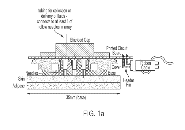
[0027] FIG. la is a schematic of the theragnostic device/system as
described herein,
according to an illustrative embodiment. The schematic shows a needle array
with wired 1/0
(input/output).
[0028] FIG. lb is a schematic of the theragnostic device/system as
described herein,
according to an illustrative embodiment. The schematic shows a needle array
with wireless
I/O (input/output).
[0029] FIG. 2 is a schematic showing disease progression, according to
an
illustrative embodiment. In some embodiments, PN extends to subcutaneous
adipose tissue
and involves dying back of nerves from the skin inwards. PN extending beyond
the skin to
the underlying adipose tissue contributes to overall metabolic dysfunction due
to disruption
of brain-adipose communication.
[0030] FIG. 3 shows measurement of protein levels in human and mouse
subcutaneous adipose tissue with (i) neuropathy with obesity (panels A and B),
and (ii)
neuropathy with aging (panels C and D), according to an illustrative
embodiment.
[0031] FIG. 4 shows results of stainless steel needle recordings of
nerve
conductance and delivery of solubilized material subdermally. (A) The system
as disclosed
CA 03182158 2022-11-02
WO 2021/226486
PCT/US2021/031337
herein (i.e comprising a needle (e.g. microneedle) array) was used to record
nerve
conductance from mouse flank skin. The blue vertical line indicates recorded
compound
action potentials. (B) Individual needles delivered 2% Evan's Blue dye
subdermally into the
subcutaneous adipose tissue of the mouse. (C) Photograph of the dye stained
inguinal
subcutaneous adipose depot, demonstrating feasibility of targeted substance
delivery via the
recording needles, directly to an area with recorded neuropathy deficits.
[0032] FIG. 5 is a schematic of an exemplary study for testing certain
embodiments
in known diet-induced and genetic models of diabetic peripheral neuropathy.
[0033] FIG. 6 is a schematic of a system for use in on-site processing
including
signal amplification and wireless communication between a device as disclosed
herein and a
computing device. A. Schematic of on-site signal amplification and wired
communication
between DEN Module prototype and FOX-DEN software. Needle arrays with wired
I/O are
connected to signal processing hardware on a computer. B. Schematic of in situ
signal
amplification and wireless communication between DEN Module prototype and FOX-
DEN
software. Recordings of electrical activity are transmitted via Bluetooth or
WiFi to data
processing software (e.g., an application on computer or tablet).
[0034] The features and advantages of the present disclosure will become
more
apparent from the detailed description set forth below when taken in
conjunction with the
drawings, in which like reference characters identify corresponding elements
throughout. In
the drawings, like reference numbers generally indicate identical,
functionally similar,
and/or structurally similar elements.
DEFINITIONS
[0035] In this application, unless otherwise clear from context, (i) the
term "a" may
be understood to mean "at least one"; (ii) the term "or" may be understood to
mean
"and/or"; (iii) the terms "comprising" and "including" may be understood to
encompass
itemized components or steps whether presented by themselves or together with
one or more
additional components or steps; and (iv) the terms "about" and "approximately"
may be
11
CA 03182158 2022-11-02
WO 2021/226486
PCT/US2021/031337
understood to permit standard variation as would be understood by those of
ordinary skill in
the art; and (v) where ranges are provided, endpoints are included.
[0036] About: The term "about", when used herein in reference to a
value, refers to
a value that is similar, in context to the referenced value. In general, those
skilled in the art,
familiar with the context, will appreciate the relevant degree of variance
encompassed by
"about" in that context. For example, in some embodiments, the term "about"
may
encompass a range of values that within 25%, 20%, 19%, 18%, 17%, 16%, 15%,
14%, 13%,
12%, 11%, 10%, 9%, 8%, 7%, 6%, 5%, 4%, 3%, 2%, 1%, or less of the referred
value.
[0037] Agent: In general, the term "agent", as used herein, may be used
to refer to
a compound or entity of any chemical class including, for example, a
polypeptide, nucleic
acid, saccharide, lipid, small molecule, metal, or combination or complex
thereof In
appropriate circumstances, as will be clear from context to those skilled in
the art, the term
may be utilized to refer to an entity that is or comprises a cell or organism,
or a fraction,
extract, or component thereof Alternatively or additionally, as context will
make clear, the
term may be used to refer to a natural product in that it is found in and/or
is obtained from
nature. In some instances, again as will be clear from context, the term may
be used to refer
to one or more entities that is man-made in that it is designed, engineered,
and/or produced
through action of the hand of man and/or is not found in nature. In some
embodiments, an
agent may be utilized in isolated or pure form; in some embodiments, an agent
may be
utilized in crude form. In some embodiments, potential agents may be provided
as
collections or libraries, for example that may be screened to identify or
characterize active
agents within them. In some cases, the term "agent" may refer to a compound or
entity that
is or comprises a polymer; in some cases, the term may refer to a compound or
entity that
comprises one or more polymeric moieties. In some embodiments, the term
"agent" may
refer to a compound or entity that is not a polymer and/or is substantially
free of any
polymer and/or of one or more particular polymeric moieties. In some
embodiments, the
term may refer to a compound or entity that lacks or is substantially free of
any polymeric
moiety.
[0038] Biocompatible: The term "biocompatible", as used herein, refers
to
materials that do not cause significant harm to living tissue when placed in
contact with such
12
CA 03182158 2022-11-02
WO 2021/226486
PCT/US2021/031337
tissue, e.g., in vivo. In certain embodiments, materials are "biocompatible"
if they are not
toxic to cells. In certain embodiments, materials are "biocompatible" if their
addition to
cells in vitro results in less than or equal to 20% cell death, and/or their
administration in
vivo does not induce significant inflammation or other such adverse effects.
[0039] Biological Sample: As used herein, the term "biological sample"
typically
refers to a sample obtained or derived from a biological source (e.g., a
tissue or organism or
cell culture) of interest, as described herein. In some embodiments, a source
of interest
comprises an organism, such as an animal or human. In some embodiments, a
biological
sample is or comprises biological cells. In some embodiments, a biological
sample is or
comprises biological tissue or fluid. In some embodiments, a biological sample
may be or
comprise blood; blood cells; tissue (e.g. skin) or fine needle biopsy samples;
cell-containing
body fluids; free floating nucleic acids; lymph; skin swabs; extracellular
fluid, interstitial
fluid and molecules contained therein, other body fluids (e.g. sweat),
secretions, and/or
excretions; and/or cells therefrom, etc. In some embodiments, a biological
sample is or
comprises interstitial fluid. In some embodiments, a biological sample is or
comprises cells
obtained from an individual. In some embodiments, obtained cells are or
include cells from
an individual from whom the sample is obtained. In some embodiments, a sample
is a
"primary sample" obtained directly from a source of interest by any
appropriate means. For
example, in some embodiments, a primary biological sample is obtained by
methods
selected from the group consisting of biopsy (e.g., fine needle aspiration or
tissue biopsy),
surgery, collection of body fluid (e.g., blood, lymph, feces etc.), etc. In
some embodiments,
as will be clear from context, the term "sample" refers to a preparation that
is obtained by
processing (e.g., by removing one or more components of and/or by adding one
or more
agents to) a primary sample. For example, filtering using a semi-permeable
membrane.
Such a "processed sample" may comprise, for example nucleic acids or proteins
extracted
from a sample or obtained by subjecting a primary sample to techniques such as
amplification or reverse transcription of mRNA, isolation and/or purification
of certain
components, etc.
[0040] Biomarker: The term "biomarker" is used herein, consistent with
its use in
the art, to refer to an entity, event, or characteristic whose presence,
level, degree, type,
13
CA 03182158 2022-11-02
WO 2021/226486
PCT/US2021/031337
and/or form, correlates with a particular biological event or state of
interest, so that it is
considered to be a "marker" of that event or state. To give but a few
examples, in some
embodiments, a biomarker may be or comprise a marker for a particular disease
state, or for
likelihood that a particular disease, disorder or condition may develop,
occur, or reoccur. In
some embodiments, a biomarker may be or comprise a marker for a particular
disease or
therapeutic outcome, or likelihood thereof Thus, in some embodiments, a
biomarker is
predictive, in some embodiments, a biomarker is prognostic, in some
embodiments, a
biomarker is diagnostic, of the relevant biological event or state of
interest. A biomarker
may be or comprise an entity of any chemical class, and may be or comprise a
combination
of entities. For example, in some embodiments, a biomarker may be or comprise
a nucleic
acid, a polypeptide, a lipid, a carbohydrate, a small molecule, an inorganic
agent (e.g., a
metal or ion), or a combination thereof In some embodiments, a biomarker is a
cell surface
marker. In some embodiments, a biomarker is intracellular. In some
embodiments, a
biomarker is detected outside of cells (e.g., is secreted or is otherwise
generated or present
outside of cells, e.g., in a body fluid such as blood, interstitial fluid,
tears, saliva, etc.). In
some embodiments, a biomarker is detected in a biological sample. In some
embodiments, a
biomarker may be or comprise a genetic or epigenetic signature. In some
embodiments, a
biomarker may be or comprise a gene expression signature.
[0041] Comparable: As used herein, the term "comparable" refers to two
or more
agents, entities, situations, sets of conditions, etc., that may not be
identical to one another
but that are sufficiently similar to permit comparison therebetween so that
one skilled in the
art will appreciate that conclusions may reasonably be drawn based on
differences or
similarities observed. In some embodiments, comparable sets of conditions,
circumstances,
individuals, or populations are characterized by a plurality of substantially
identical features
and one or a small number of varied features. Those of ordinary skill in the
art will
understand, in context, what degree of identity is required in any given
circumstance for two
or more such agents, entities, situations, sets of conditions, etc. to be
considered comparable.
For example, those of ordinary skill in the art will appreciate that sets of
circumstances,
individuals, or populations are comparable to one another when characterized
by a sufficient
number and type of substantially identical features to warrant a reasonable
conclusion that
differences in results obtained or phenomena observed under or with different
sets of
14
CA 03182158 2022-11-02
WO 2021/226486
PCT/US2021/031337
circumstances, individuals, or populations are caused by or indicative of the
variation in
those features that are varied.
[0042] Comprising: A composition or method described herein as
"comprising" one
or more named elements or steps is open-ended, meaning that the named elements
or steps
are essential, but other elements or steps may be added within the scope of
the composition
or method. It is also understood that any composition or method described as
"comprising"
(or which "comprises") one or more named elements or steps also describes the
corresponding, more limited composition or method "consisting essentially of'
(or which
"consists essentially of') the same named elements or steps, meaning that the
composition or
method includes the named essential elements or steps and may also include
additional
elements or steps that do not materially affect the basic and novel
characteristic(s) of the
composition or method. It is also understood that any composition or method
described
herein as "comprising" or "consisting essentially of' one or more named
elements or steps
also describes the corresponding, more limited, and closed-ended composition
or method
"consisting of' (or "consists of') the named elements or steps to the
exclusion of any other
unnamed element or step. In any composition or method disclosed herein, known
or
disclosed equivalents of any named essential element or step may be
substituted for that
element or step.
[0043]
Electrical activity: As used herein, the term "electrical activity" refers to
any
characteristics (e.g. frequency, amplitude, shape, voltage, rate, pattern,
temporal signature,
or other) of electrical signals measured within and beneath the skin for the
purpose of
determining clinical diagnosis.
[0044] Electrically conductive: As used herein, the term "electrically
conductive"
refers to the property of allowing the flow electrical current. For example,
an electrically
conductive material may be a metal, metal alloy, conducting polymers or glassy
carbon. In
some embodiments, an electrically conductive material has a conductivity of
105 siemens
per meter (S/m) or greater at room temperature.
[0045] In
vitro: The term "in vitro" as used herein refers to events that occur in an
artificial environment, e.g., in a test tube or reaction vessel, in cell
culture, etc., rather than
within a multi-cellular organism.
CA 03182158 2022-11-02
WO 2021/226486
PCT/US2021/031337
[0046] In vivo: as used herein refers to events that occur within a
multi-cellular
organism, such as a human and/or a non-human animal. In the context of cell-
based
systems, the term may be used to refer to events that occur within a living
cell in a multi-
cellular organism (as opposed to, for example, in vitro systems).
[0047] Microneedle: The term "microneedle" as used herein generally
refers to an
elongated structure with diameter less than a millimeter that is of suitable
length and shape
to penetrate skin. In some embodiments, a microneedle is arranged and
constructed (by
itself or within a device) to provide the ability to electrically communicate
with nerves when
inserted into skin, while still creating efficient pathways for drug delivery.
In some
embodiments, a microneedle has a diameter which is consistent along the
microneedle's
length. In some embodiments, a microneedle has a diameter that changes along
the
microneedle's length. In some embodiments, a microneedle has a diameter that
tapers along
the microneedle's length. In some embodiments, a microneedle's diameter is
narrowest at
the tip that penetrates skin. In some embodiments, a microneedle may be solid.
In some
embodiments, a microneedle may be hollow. In some embodiments a microneedle
may be
tubular. In some embodiments, a microneedle may be sealed on one end. In some
embodiments, a plurality of microneedles is utilized. In some embodiments, a
plurality of
microneedles is utilized in an array format. In some embodiments, a
microneedle may have
a length within a range of about 1 p.m to about 24,000 p.m. In some
embodiments, a
microneedle may have a length of at least about 50 p.m.
[0048] 'Neuropathy' or 'Peripheral neuropathy': As used herein, the
terms
"neuropathy" and "peripheral neuropathy" refer generally to dysfunction, for
example
caused by damage or disease affecting nerves (e.g., peripheral nerves).
Neuropathy may be
associated with a decrease in nerve conductance or aberrations in nerve
electrical activity, in
an affected portion of the body. Neuropathy may be idiopathic (i.e. no known
cause) or
caused by, for example, a disease (e.g., diabetes), physical trauma, genetic
background,
and/or an infection.
[0049] Prevention: The term "prevention", as used herein, refers to a
delay of onset,
and/or reduction in frequency and/or severity of one or more symptoms of a
particular
disease, disorder or condition. In some embodiments, prevention is assessed on
a population
16
CA 03182158 2022-11-02
WO 2021/226486
PCT/US2021/031337
basis such that an agent is considered to "prevent" a particular disease,
disorder or condition
if a statistically significant decrease in the development, frequency, and/or
intensity of one
or more symptoms of the disease, disorder or condition is observed in a
population
susceptible to the disease, disorder, or condition. Prevention may be
considered complete
when onset of a disease, disorder or condition has been delayed for a
predefined period of
time.
[0050] Risk: as will be understood from context, "risk" of a disease,
disorder,
and/or condition refers to a likelihood that a particular individual will
develop the disease,
disorder, and/or condition. In some embodiments, risk is expressed as a
percentage. In
some embodiments, risk is from 0, 1, 2, 3, 4, 5, 6, 7, 8, 9, 10, 20, 30, 40,
50, 60, 70, 80, 90
up to 100%. In some embodiments risk is expressed as a risk relative to a risk
associated
with a reference sample or group of reference samples. In some embodiments, a
reference
sample or group of reference samples have a known risk of a disease, disorder,
condition
and/or event. In some embodiments a reference sample or group of reference
samples are
from individuals comparable to a particular individual. In some embodiments,
relative risk
is 0,1, 2, 3, 4, 5, 6, 7, 8, 9, 10, or more.
[0051] Subject: As used herein, the term "subject" refers an organism,
typically a
mammal (e.g., a human). In some embodiments, a subject is suffering from a
relevant
disease, disorder or condition. In some embodiments, a subject is susceptible
to a disease,
disorder, or condition. In some embodiments, a subject displays one or more
symptoms or
characteristics of a disease, disorder or condition. In some embodiments, a
subject does not
display any symptom or characteristic of a disease, disorder, or condition. In
some
embodiments, a subject is someone with one or more features characteristic of
susceptibility
to or risk of a disease, disorder, or condition. In some embodiments, a
subject is a patient.
In some embodiments, a subject is an individual to whom diagnosis and/or
therapy is and/or
has been administered.
[0052] Substantially: As used herein, the term "substantially" refers to
the
qualitative condition of exhibiting total or near-total extent or degree of a
characteristic or
property of interest. One of ordinary skill in the biological arts will
understand that
biological and chemical phenomena rarely, if ever, go to completion and/or
proceed to
17
CA 03182158 2022-11-02
WO 2021/226486
PCT/US2021/031337
completeness or achieve or avoid an absolute result. The term "substantially"
is therefore
used herein to capture the potential lack of completeness inherent in many
biological and
chemical phenomena.
[0053] Therapeutic agent: As used herein, the phrase "therapeutic agent"
in
general refers to any agent that elicits a desired pharmacological or clinical
effect when
administered to an organism. In some embodiments, an agent is considered to be
a
therapeutic agent if it demonstrates a statistically significant effect across
an appropriate
population. In some embodiments, the appropriate population may be a
population of model
organisms. In some embodiments, an appropriate population may be defined by
various
criteria, such as a certain age group, gender, genetic background, preexisting
clinical
conditions, etc. In some embodiments, a therapeutic agent is a substance that
can be used to
alleviate, ameliorate, relieve, inhibit, prevent, delay onset of, reduce
severity of, and/or
reduce incidence of one or more symptoms or features of a disease, disorder,
and/or
condition. In some embodiments, a "therapeutic agent" is an agent that has
been or is
required to be approved by a government agency before it can be marketed for
administration to humans. In some embodiments, a "therapeutic agent" is an
agent for
which a medical prescription is required for administration to humans.
[0054] Therapeutic regimen: A "therapeutic regimen", as that term is
used herein,
refers to a dosing regimen whose administration across a relevant population
may be
correlated with a desired or beneficial therapeutic outcome.
[0055] Treat: As used herein, the term "treat," "treatment," or
"treating" refers to
any method used to partially or completely alleviate, ameliorate, relieve,
inhibit, prevent,
delay onset of, reduce severity of, and/or reduce incidence of one or more
symptoms or
features of a disease, disorder, and/or condition. In some embodiments,
treatment may be
administered to a subject who does not exhibit signs of a disease, disorder,
and/or condition.
In some embodiments, treatment may be administered to a subject who exhibits
only early
signs of the disease, disorder, and/or condition, for example for the purpose
of decreasing
the risk of developing pathology associated with the disease, disorder, and/or
condition.
18
CA 03182158 2022-11-02
WO 2021/226486
PCT/US2021/031337
DETAILED DESCRIPTION
[0056] Among other things, the present disclosure describes systems and
methods
for early detection, prevention, and/or treatment of certain disorders or
conditions, for
example, associated with the nervous system (e.g., conditions associated with
neuropathies)
through the use of a novel biomedical system that, inter alia, can detect and
monitor nerve
signals (e.g. nerve conductance). In accordance with various embodiments,
provided
systems and methods may also include the ability to sample a subject's local
environment
(e.g., tissue or body fluids), and/or, administer one or more treatments
(e.g., novel
therapeutic agents and/or nerve stimulation) to potentially delay and/or
prevent progression,
or treat a disease, disorder, or condition. In some embodiments, the present
disclosure
provides methods for treating neuropathies. In some embodiments, the present
disclosure
provides treatments for one or more of diabetic peripheral neuropathy (DPN),
chemotherapy-induced neuropathy (CIPN), HIV or AIDS-induced neuropathy; and/or
idiopathic peripheral neuropathies (IPN), neuropathies with no identifiable
known cause
(such as with aging). In some embodiments, the present disclosure provides
systems and
methods for diagnosis, prevention, and treatment for DPN.
[0057] In some embodiments, provided systems and methods as described
herein,
can not only record electrical signals (e.g. local field potentials, nerve
conductance, etc. as
described herein) but also sample biological fluids, for example, for
biomarker analysis, and
even administer one or more treatments such as one or more therapeutic agents
and/or one or
more forms of stimulation in a non-invasive (or minimally invasive) manner.
Delivery of
substances may also be utilized to stimulate nerve activity prior to or during
electrical
measurements (for example, delivery of capsaicin to stimulate skin and
subcutaneous
adipose sensory nerves expressing the cation channel TRPV1). In some
embodiments, an
array of needles is configured to allow electrical measurement and/or sampling
of a subject's
bodily fluid(s) within an appropriate surface area for a given application. In
some
embodiments, an array of needles is configured to allow electrical measurement
and/or
delivery of treatment(s) within an appropriate surface area for a given
application. In some
embodiments, systems and methods as described herein are used to record
electrical signals
(e.g. voltage, current, local field potentials, nerve conductance, etc. as
described herein) to
19
CA 03182158 2022-11-02
WO 2021/226486
PCT/US2021/031337
calculate a measure of nerve conductance of nerves under the surface of skin.
In some
embodiments, a subset of an array of needles may be used to record and measure
electrical
signals, while a different or even the same subset of the array of needles is
configured to
sample a subject's bodily fluid(s) and/or administer treatments.
[0058] In some embodiments, treatments may be or comprise therapeutic
agents. In
some embodiments, therapeutics agents may be or comprise drug therapies. In
some
embodiments, treatments may be or comprise nerve stimulation (e.g. to initiate
re-growth of
dying nerves), for example via temperature-based (e.g. hot versus cold)
stimulation, or
mechanical (e.g. vibration) stimulation.
[0059] Systems and methods disclosed herein, may be used to obtain nerve
recordings from varying depths from the surface of the skin. In some
embodiments, needles
may be used to obtain nerve recordings (i.e. electrical measurements) In some
embodiments,
length of the needles and depth of penetration dictate the depth at which
nerve recordings
may be obtained. In some embodiments, nerve recordings may be obtained from a
subject,
either at the surface of the skin, or in one or more sublayers beneath the
skin's surface.
Moreover, in some embodiments, needles may be used to obtain biological
samples from a
subject, which may be used for biomarker analysis. Thus, systems and methods
disclosed
herein enable early diagnosis and interventions for disclosed diseases,
disorders, or
conditions, and provide a promise to improve the quality of life for millions
of patients.
[0060] In some embodiments, detection and diagnosis of a disease,
disorder, or
condition, recording of nerve signals (i.e. electrical signals such as
voltage, conductance,
etc. as disclosed herein), and administration of treatment is achieved through
interaction
with and/or penetration of disclosed systems with one or more components of
the skin (e.g.,
transdermally). This allows for minimally invasive and painless detection,
recording,
sampling, and treatment of various diseases, disorders, and conditions, a
functionality that
currently not available to patients. For example, currently, diagnosis of
neuropathies is
performed by highly invasive and time-consuming tests (e.g. nerve function
tests, biopsies,
etc.) that detect late-stage neuropathies causing extreme discomfort to
patients and
disrupting their lives. Furthermore, there exists no treatment for neuropathy,
let alone late-
stage neuropathy, and treatment goals typically are to manage symptoms
including pain.
CA 03182158 2022-11-02
WO 2021/226486
PCT/US2021/031337
Thus, there exists a need for a system that not only accurately detects
neuropathy (e.g. PN)
early on, but also provides a way to manage its progression, and administer
therapies as and
when required in order to prevent and treat (e.g. stimulate nerve regrowth) in
neuropathy
patients.
[0061] In some embodiments, provided systems and methods as described
herein,
can be used for the monitoring of nerve regrowth.
[0062] In some embodiments, provided systems and methods as described
herein,
can be used as a means of clinical screening for small fiber peripheral
neuropathy, or other
nerve dysfunction.
I. Methods of Dia2nosis, Prevention, and Treatment
[0063] Methods for diagnosis, sampling, prevention, and treatment of
various
diseases, disorders, or conditions, are described herein. In some embodiments,
methods
disclosed herein are used to diagnose, sample, prevent, and treat
neuropathies. In some
embodiments, methods disclosed herein are used to diagnose, sample, prevent,
and treat PN.
In some embodiments, methods disclosed herein are used to diagnose, sample,
prevent, and
treat other conditions affecting skin and/or subdermal tissues (e.g.
peripheral arterial disease,
lymphatic dysfunction, fibromyalgia, etc.).
Diagnosis /Detection:
[0064] Systems disclosed herein may be used for early onset detection of
various
diseases, disorders, and/or conditions as disclosed herein. In some
embodiments, systems
disclosed herein may be used for detection and/or diagnosis of neuropathies,
for example,
peripheral neuropathy. In some embodiments, provided systems may be used to
measure
nerve conductance in small nerve fibers of the skin and underlying tissues,
allowing for
sensitive and early diagnosis of neuropathy by detecting loss of nerve signal
due to
neurodegeneration. In some embodiments, provided systems may be used to
measure nerve
conductance of one or more types of nerve fibers (e.g. peripheral nerve
fibers, sensory nerve
fibers, motor nerve fibers, autonomic nerve fibers, and associated nerve fiber
subtypes (i.e.
21
CA 03182158 2022-11-02
WO 2021/226486
PCT/US2021/031337
group A, B, and C fibers)) of the skin and underlying tissues and obtaining an
average of the
measure of nerve conductance from penetrated tissue (e.g. penetrated with one
or more
needles of needle array). In some embodiments, provided systems may be used to
measure
nerve conductance of two of more types of nerve fibers (e.g. peripheral nerve
fibers, sensory
nerve fibers, motor nerve fibers, autonomic nerve fibers, and associated nerve
fiber subtypes
(i.e. group A, B, and C fibers)) of the skin and underlying tissues and
obtaining an average
of the measure of nerve conductance from penetrated tissue (e.g. penetrated
with one or
more needles of needle array). In some embodiments, provided systems may be
used to
measure nerve conductance of multiple (e.g. one or more, two or more, three or
more, any
and all etc.) types of nerve fibers (e.g. peripheral nerve fibers, sensory
nerve fibers, motor
nerve fibers, autonomic nerve fibers, and associated nerve fiber subtypes
(i.e. group A, B,
and C fibers)) of the skin and underlying tissues and obtaining an average of
the measure of
nerve conductance from penetrated tissue (e.g. penetrated with one or more
needles of
needle array). In some embodiments, provided systems may be used to measure
nerve
conductance of all types of nerve fibers (e.g. peripheral nerve fibers,
sensory nerve fibers,
motor nerve fibers, autonomic nerve fibers, and associated nerve fiber
subtypes (i.e. group
A, B, and C fibers))of the skin and underlying tissues and obtaining an
average of the
measure of nerve conductance from penetrated tissue (e.g. penetrated with one
or more
needles of needle array). Fig. 2 is a schematic showing neuropathic disease
progression and
use of a theragnostic system to diagnose/detect (e.g. monitor disease, record
signals (e.g.
nerve conductance, impedance, voltage, current, etc.), etc.) and/or treat
neuropathic disease
progression according to an illustrative embodiment.
[0065] In one aspect, a method of diagnosing and/or evaluating a subject
potentially
at risk for or suffering from neuropathy comprises: placing a system as
disclosed herein on
the surface of skin of a subject, with needles protruding below the skin
surface. In some
embodiments, for example, a system as disclosed herein comprises a substrate
and an array
of needles disposed on or through the substrate, wherein one or more of the
needles is
hollow through the entire length of the needle, the array is configured to
allow fluid
transport through at least one hollow needle of the array, and the array is
configured to
receive and communicate one or more electrical signals. In some embodiments,
systems
22
CA 03182158 2022-11-02
WO 2021/226486
PCT/US2021/031337
disclosed herein are placed on skin surface of a subject. In some embodiments,
systems
disclosed herein are placed below skin surface of a subject.
[0066] Methods of diagnosing and/or evaluating a subject further
comprises
inserting one or more needles of an array into a subject's skin. In some
embodiments, one or
more needles are inserted into epidermis of a subject. In some embodiments,
one or more
needles are inserted into one or more layers of epidermis of a subject, for
example, into one
or more of the stratum comeum, stratum lucidum, stratum granulosum, stratum
spinosum,
and/or stratum basale. In some embodiments, one or more needles are inserted
into dermis
of a subject. In some embodiments, one or more needles are inserted into
hypodermis of a
subject. In some embodiments, one or more needles are inserted into
subcutaneous tissue of
a subj ect.
[0067] One or more needles of systems disclosed herein may be inserted
to one or
more desired target depths into or under the skin of a subject. In some
embodiments, target
depths may range between 1 p.m to 12 mm. In some embodiments, target depths
may range
between 10 p.m and 10 mm. In some embodiments, target depths may range between
100
pm and 5 mm. In some embodiments, target depths may range between 150 pm and 2
mm.
In some embodiments, a target depth may be at least 10 p.m. In some
embodiments, a target
depth may be at least 50 p.m. In some embodiments, a target depth may be at
least 100 p.m.
In some embodiments, a target depth may be at least 200 p.m. In some
embodiments, a target
depth may be at least 300 p.m. In some embodiments, a target depth may be at
least 400 p.m.
In some embodiments, a target depth may be at least 500 p.m. In some
embodiments, a target
depth may be at least 1000 p.m. In some embodiments, a target depth may be at
least 2000
p.m. In some embodiments, a target depth may be at most 12000 p.m. In some
embodiments,
a target depth may be at most 24000 p.m. In some embodiments, for example, a
substrate
and/or needle array of systems disclosed herein are configured to allow
adjustment of the
depth of penetration (i.e. target depth) of one or more needles into skin
and/or subcutaneous
tissue of a subject. This allows for detection (e.g. through signal
measurement), sampling,
and/or delivery of treatment at a plurality of target depths within the skin
and/or
subcutaneous tissue of a subject. For example, the depth of penetration of one
or more
needles of an array may be configured, in some embodiments, using a spacer of
adjustable
23
CA 03182158 2022-11-02
WO 2021/226486
PCT/US2021/031337
or varying thickness, through which the needle are inserted prior to
penetration of the skin.
In some embodiments, needle length may be used to configure and/or control
depth of
penetration under skin.
[0068] In some embodiments, methods disclosed herein comprise measuring
signals
at each of a plurality of target depths within skin. In some embodiments, a
substrate and/or
array are configured to allow adjustment of the depth of penetration of one or
more needles
into skin and/or subcutaneous tissue of a subject, thereby allowing
measurement at a
plurality of target depths within the skin of a subject. In some embodiments,
signals may be
measured at 2 or more different target depths. In some embodiments, signals
may be
measured at 2, 3, 4, 5, 6, 7, 8, 9, 10, or more different target depths. In
some embodiments,
signals may be measured at one target depth at an instance in time. In some
embodiments,
signals may be measured at two or more target depths at an instance in time.
[0069] In some embodiments, methods of diagnosing and/or evaluating a
subject
may further comprise measuring signals from penetrated tissue. In some
embodiments, at
least one of the inserted needles is capable of measuring and/or recording
signals from the
penetrated tissue. In some embodiments, measuring and/or recording signals
from nerve
fibers located in or near penetrated tissue is performed. In some embodiments,
measuring
and/or recording signals from a specific type of nerve fiber (e.g. peripheral
nerve fibers,
sensory nerve fibers, motor nerve fibers, autonomic nerve fibers, and
associated nerve fiber
subtypes (i.e. group A, B, and C fibers)) located in or near penetrated tissue
is performed. In
some embodiments, measuring and/or recording signals from a multiple types
(e.g. 2 or
more, 3 or more, etc.) of nerve fibers (e.g. peripheral nerve fibers, sensory
nerve fibers,
motor nerve fibers, autonomic nerve fibers, and associated nerve fiber
subtypes (i.e. group
A, B, and C fibers)) located in or near penetrated tissue is performed. In
some embodiments,
measuring and/or recording signals from all types of nerve fibers (e.g.
peripheral nerve
fibers, sensory nerve fibers, motor nerve fibers, autonomic nerve fibers, and
associated
nerve fiber subtypes (i.e. group A, B, and C fibers)) located in or near
penetrated tissue is
performed. In some embodiments, a signal measured is an electrical signal. In
some
embodiments, an electrical signal measured is an action potential. For
example, action
potential measured is an action potential of a nerve. In some embodiments, an
electrical
24
CA 03182158 2022-11-02
WO 2021/226486
PCT/US2021/031337
signal measured is a local field potential. As is known to one of skill in the
art, local field
potentials are transient electrical signals generated in nervous tissue and/or
other tissues by
the summed and synchronous electrical activity of the individual cells in that
tissue. In some
embodiments, an electrical signal measured is a nerve conductance. In some
embodiments,
an electrical signal measured is an electrical current. In some embodiments,
an electrical
signal measured is an electrical voltage.
[0070] In some
embodiments, methods disclosed herein comprise measuring space
averages of signals across a given area of tissue. In some embodiments,
signals may be
measured across a given area of tissue. In some embodiments, measured area may
be at
least about 1 square inch. In some embodiments, measured area may be at least
about 2
square inches. In some embodiments, measured area may be at least about 3
square inches.
In some embodiments, measured area may be at least about 4 square inches. In
some
embodiments, measured area may be at least about 5 square inches. In some
embodiments,
measured area may be at least about 10 square inches. In some embodiments,
measured area
may be less than 1 square inch. In some embodiments, measured area may be more
than 10
square inches. In some embodiments, measured signals may be averaged over a
measured
area. This allows for various skin surface area to be measured, as neuropathy
progresses
from hands/feet further up the arms and legs, and potentially to the torso.
[0071] In some
embodiments, each electrical measurement (e.g. measurement of a
signal (e.g. electrical signal)) may be performed over a period of time. That
is, in some
embodiments, each signal measurement may be performed as a function of time.
For
example, in some embodiments, a signal measurement may be performed over a
period of
between 30 seconds to 24 hours. In some embodiments, a signal measurement may
be
performed over a period of between 1 minute and 10 hours. In some embodiments,
a signal
measurement may be performed over a period of between 2 minutes and 1 hour. In
some
embodiments, a signal measurement may be performed for at least 1 minute. In
some
embodiments, a signal measurement may be performed for at least 2 minutes. In
some
embodiments, a signal measurement may be performed for at least 30 minutes. In
some
embodiments, a signal measurement may be performed for at least 1 hour. In
some
embodiments, a signal measurement may be performed for at least 2 hours. In
some
CA 03182158 2022-11-02
WO 2021/226486
PCT/US2021/031337
embodiments, a signal measurement may be performed for at least 3 hours. In
some
embodiments, a signal measurement may be are performed for at least 5 hours.
In some
embodiments, a signal measurement may be performed for at least 6 hours. In
some
embodiments, a signal measurement may be performed for at least 12 hours. In
some
embodiments, a signal measurement may be performed for at most 24 hours. In
some
embodiments, measured signals may be averaged over time.
[0072] In some embodiments, each electrical measurement (e.g.
measurement of a
signal (e.g. electrical signal)) may be performed over a period of time. That
is, in some
embodiments, each signal measurement may be performed as a function of time.
For
example, in some embodiments, a signal measurement may be performed over a
period of
between 1 day to 1 year. In some embodiments, a signal measurement may be
performed for
at least 1 day. In some embodiments, a signal measurement may be performed for
at least 2,
3, 4, 5, 6, or 7 days. In some embodiments, a signal measurement may be
performed for at
least 2, 3, or 4 weeks. In some embodiments, a signal measurement may be
performed for at
least 1 month. In some embodiments, a signal measurement may be performed for
at least 2,
3, 4, 5, 6, 7, 8, 9, 10, 11, or 12 months or more. In some embodiments, a
signal measurement
may be performed for at least 1 year or more. In some embodiments, measured
signals may
be averaged over time.
[0073] In some embodiments, for example, signals measured may be
electrical
signals. In some embodiments, electrical signals may be measured as a function
of
frequency. For example, electrical signals may be measured in a frequency
range of 1 Hz to
MHz. In some embodiments, electrical signals may be measured in a frequency
range of
100 Hz to 5 MHz. In some embodiments, electrical signals may be measured in a
frequency
range of 200 Hz to 2 MHz. In some embodiments, electrical signals may be
measured in a
frequency range of 250 Hz to 1 MHz.
[0074] In some embodiments, electrical signals may be measured as a
function of
voltage. For example, electrical signals may be measured in a voltage range of
1 fV to 1 V.
In some embodiments, electrical signals may be measured in a voltage range of
10 pV to 0.5
V. In some embodiments, electrical signals may be measured in a voltage range
of 20 pV to
26
CA 03182158 2022-11-02
WO 2021/226486
PCT/US2021/031337
0.2 V. In some embodiments, electrical signals may be measured in a voltage
range of 25
pV to 0.1 V.
[0075] In some embodiments, measurement of an electrical signal is
performed
using needles that serve as electrodes. In some embodiments, measurement of an
electrical
signal may be performed using a 2-electrode system. In some embodiments,
measurement of
an electrical signal may be performed using a 3-electrode system. In some
embodiments, an
electrical signal is measured against one or more reference electrodes. In
some
embodiments, one or more needles of an array serves as a reference electrode.
Reference
electrodes may comprise any application-appropriate materials (e.g., metals or
polymers).
For example, in some embodiments, reference electrodes may be biocompatible
and used in
combination with systems and methods disclosed herein. In some embodiments,
reference
electrodes may be aqueous reference electrodes. In some embodiments, reference
electrodes
may be non-aqueous reference electrodes. By way of additional example, in some
embodiments, reference electrodes may be or comprise Ag-AgC1 (Silver-silver
chloride)
electrodes, standard hydrogen electrodes, normal hydrogen electrodes,
reversible hydrogen
electrodes, saturated calomel electrodes, copper-copper(II) electrodes,
palladium-hydrogen
electrodes, dynamic hydrogen electrodes, mercury-mercurous sulfate electrodes,
or any
combination thereof In some embodiments, one or more needles of an array may
serve as a
ground electrode.
[0076] In some embodiments, methods disclosed herein comprise processing
measured electrical signals using a processor. In some embodiments, measured
electrical
signals are converted (e.g., by data processing) into an assessment of nerve
conductance. In
some embodiments, nerve conductance is determined as a function of frequency.
In some
embodiments, nerve conductance is determined for a characteristic frequency.
In some
embodiments, nerve conductance is determined as a function of voltage. In some
embodiments, nerve conductance is determined for a characteristic voltage. In
some
embodiments, nerve conductance is determined for an average of the measured
electrical
signals acquired from each inserted needle of the array.
[0077] In some embodiments, determining an assessment of nerve
conductance
from electrical signals comprises determining a characteristic parameter of
electrical signals
27
CA 03182158 2022-11-02
WO 2021/226486
PCT/US2021/031337
and comparing a value associated with the characteristic parameter to a value
of a pre-
determined threshold parameter (e.g., a reference level or reference value
that is comparable
to the characteristic parameter). In some embodiments, a value of a pre-
determined
threshold parameter may be obtained from one or more healthy subjects. In some
embodiments, value of a pre-determined threshold parameter may be obtained
from a
population of healthy subjects (e.g., an average reading (e.g., average
electrical signal
measurement taken from each of the members of the population)). In some
embodiments, a
value of a pre-determined threshold parameter may be obtained for one or more
specific
body locations. In some embodiments, a value of a pre-determined threshold
parameter may
be obtained for one or more specific skin depths. As is known to a person of
ordinary skill in
the art, when comparing a value of a characteristic parameter with a value of
a pre-
determined threshold parameter care must be taken that both have been obtained
under
comparable parameters (e.g., for a given skin depth, needle type, and/or body
location).
[0078] In some
embodiments, a characteristic parameter is a frequency or range of
frequencies. In some embodiments, a characteristic parameter is a potential
(or current)
maxima. In some embodiments, a potential (or current) maxima is an average of
one or more
maxima of a measured electrical potential (or current). In some embodiments, a
characteristic parameter is a voltage (or current) level of a measured
electrical potential (or
current). In some embodiments, a characteristic parameter is an amplitude of a
measured
electrical potential (or current). In some embodiments, a characteristic
parameter is a pattern
of firing of neurons at measured electrical potentials (or currents).
[0079] In some
embodiments, a pre-determined threshold parameter is a frequency.
In some embodiments, a pre-determined threshold parameter is a potential (or
current)
maxima. In some embodiments, a potential (or current) maxima is an average of
one or more
maxima of a measured electrical potential (or current). In some embodiments, a
pre-
determined threshold parameter is a voltage (or current) level of a measured
electrical
potential (or current). In some embodiments, a pre-determined threshold
parameter is an
amplitude of a measured electrical potential (or current). In some
embodiments, a pre-
determined threshold parameter is a pattern of firing of neurons at the
measured electrical
potential (or current).
28
CA 03182158 2022-11-02
WO 2021/226486
PCT/US2021/031337
[0080] Systems and methods disclosed herein may also be enabled to
transmit
recorded signals. Methods disclosed herein include, for example, transmitting
electrical
signals to and/or from penetrated tissue. In some embodiments, electrical
signals are
transmitted to a processor and/or a storage device. In some embodiments,
electrical signals
are further processed after transmission. In some embodiments, electrical
signals are further
processed before transmission.
[0081] In some embodiments, methods presented herein may further include
a
comparison of electrical signals obtained from a subject with electrical
signals obtained
from one or more control subjects. In some embodiments, for example,
electrical signals
(e.g. raw electrical signals, processed electrical signals etc.) obtained from
a subject are
compared to electrical signals obtained from control subjects (e.g. one or
more subjects that
do not suffer from a disease, disorder, or condition disclosed herein
(negative control)), to
diagnose a likelihood that subject suffers from a disease, disorder, or
condition disclosed
herein. In some embodiments, electrical signals obtained from a subject are
compared to
electrical signals obtained from control subjects (e.g. one or more subjects
that suffer from a
disease, disorder, or condition disclosed herein (positive control)).
[0082] In some embodiments, electrical signals obtained from a subject
are
compared with electrical signals obtained from controls (e.g. pre-determined
threshold
values). For example, by comparing electrical measurements obtained from a
subject with
comparable electrical control measurements, one can measure a likelihood that
a subject
suffers from a disease, disorder, or condition (e.g., neuropathies, PN).
Systems disclosed
herein may be used to measure reference values (i.e. pre-determined threshold
values) from
one or more healthy subject(s). In some embodiments, such thresholds or
reference values
may be used as comparators to diagnose a disease, disorder, or condition in
subjects.
[0083] In some embodiments, methods of detection and/or evaluation
disclosed
herein may be performed repeatedly in subjects to evaluate progression of a
disease,
disorder, or condition in subjects over time (e.g. lifetime of a subject). In
some
embodiments, for example, successive measurements may be separated by at least
1 day. In
some embodiments, for example, successive measurements may be separated by at
least 1
week. In some embodiments, for example, successive measurements may be
separated by at
29
CA 03182158 2022-11-02
WO 2021/226486
PCT/US2021/031337
least 2 weeks. In some embodiments, for example, successive measurements may
be
separated by at least 3 weeks. In some embodiments, for example, successive
measurements
may be separated by at least 1 month. In some embodiments, for example,
successive
measurements may be separated by at least 2, 3, 4, 5, 6, 7, 8, 9, 10, 11, or
12 months. In
some embodiments, for example, successive measurements may be separated by at
least 1
year or more. In some embodiments, successive measurements may be performed at
a
specific target depth. In some embodiments, successive measurements may be
performed to
monitor progression from healthy tissue to neuropathic tissue in a subject. In
some
embodiments, successive measurements may be performed to monitor progression
from
neuropathic tissue to healthy tissue in a subject. In some embodiments,
successive
measurements may be performed to monitor nerve re-growth after treatment.
Samp
[0084] Methods and systems disclosed herein may be equipped to obtain
samples
from subjects (e.g. healthy and/or diseased) and/or evaluate markers for
various stages of a
disease, disorder, or condition. In some embodiments, provided systems and
methods may
be configured to test and/or obtain biological samples from subjects. In some
embodiments,
samples tested and/or obtained may be used for diagnosis and/or biomarker
discovery for
various diseases, disorders, or conditions as disclosed herein. For example,
needles inserted
into skin of subjects for detection and/or evaluation of disease, disorder, or
condition may be
designed to be capable of sampling biological samples from penetrated tissue
(e.g., in one or
more body fluids).
[0085] Any of a variety of biological samples can be suitable for use
with particular
embodiments, as described below and elsewhere herein. In some embodiments, a
biological
sample is or comprises biological cells. In some embodiments, a biological
sample is or
comprises tissue. In some embodiments, a biological sample is or comprises
skin. In some
embodiments, a biological sample is or comprises biological fluids. In some
embodiments, a
biological fluid is or comprises interstitial fluid (ISF). In some
embodiments, a biological
fluid is or comprises blood. In some embodiments, a biological fluid is or
comprises sweat.
In accordance with various embodiments, a biological sample may be used for
biomarker
CA 03182158 2022-11-02
WO 2021/226486
PCT/US2021/031337
analysis for detection of a disease, disorder, or condition as disclosed
herein, and/or for
evaluating progression of a disease or condition.
[0086] In some embodiments, methods disclosed comprise obtaining one or
more
biological samples from a subject. In some embodiments, biological samples may
be
obtained from a specific target depth. In some embodiments, biological samples
may be
obtained using a one or more needles of an array. In some embodiments,
biological samples
may be obtained using all or substantially all of the needles of an array. In
some
embodiments, biological samples may be obtained using any of the needles of an
array. For
example, in some embodiments, biological samples may be obtained using needles
in the
center or substantially in the center of an array. In some embodiments,
biological samples
may be obtained using needles from an edge of an array.
[0087] Systems and methods disclosed herein may be used to evaluate
(i.e.
diagnose, record, and/or monitor) progression of a disease, disorder, or
condition (e.g.
neuropathy) by repeatedly sampling biological samples from a subject.
Accordingly, in
some embodiments, multiple biological samples may be obtained from a subject
over a
period of time. In some embodiments, successive biological samples may be
obtained at
least 1 day apart. In some embodiments, for example, successive biological
samples may be
obtained at least 1 week apart. In some embodiments, for example, successive
biological
samples may be obtained at least 2 weeks apart. In some embodiments, for
example,
successive biological samples may be obtained at least 3 weeks apart. In some
embodiments,
for example, successive biological samples may be obtained at least 1 month
apart. In some
embodiments, successive biological samples may be obtained at least 3 months,
6 month, 9
months, or 1 year apart. In some embodiments, successive biological samples
may be
obtained at a specific target depth. In some embodiments, successive
biological samples
may be obtained at different target depths. In some embodiments, successive
biological
samples may be obtained to evaluate progression from healthy tissue to
neuropathic tissue in
a subject. In some embodiments, successive biological samples may be obtained
to evaluate
progression from neuropathic tissue to healthy tissue in a subject. In some
embodiments,
successive biological samples may be obtained to evaluate (i.e. monitor,
record, and/or
diagnose) nerve re-growth after treatment.
31
CA 03182158 2022-11-02
WO 2021/226486
PCT/US2021/031337
[0088] Systems and methods disclosed herein may include, in some
embodiments,
the ability to analyze an obtained biological sample. In some embodiments, for
example,
obtained biological samples may be analyzed (i.e. detect qualitatively or
quantitatively one
or more specific biomarkers associated with a disease, disorder, or condition)
on-chip (i.e.
on the systems disclosed herein). For example, in some embodiments, obtained
biological
samples may be analyzed for one or more specific biomarkers while said
biological sample
is within one or more needles of a needle (e.g. microneedle) array. In some
embodiments,
obtained biological samples may be analyzed off-chip (for example, in a lab).
In some
embodiments, obtained biological samples may be analyzed quantitatively. In
some
embodiments, obtained biological samples may be analyzed qualitatively.
[0089] Obtained biological samples may be analyzed for one or more
biomarkers
using one or more sensing techniques. In some embodiments, for example, one or
more
biomarkers may be analyzed using electrochemical sensors. In some embodiments,
one or
more biomarkers may be analyzed using electronic sensors.
Treatment:
[0090] In some embodiments, provided methods and systems may also be
used for
preventing and/or treating various diseases, disorders, and/or conditions. In
some
embodiments, methods and systems of the present disclosure may be used for
prevention
and/or treatment of neuropathy. For example, in some embodiments, a method of
preventing
and/or treating a subject potentially at risk for or suffering from
neuropathy, comprises:
placing, as discussed elsewhere herein, a device/system as disclosed herein on
the surface of
skin of a subject, with needles protruding below the skin surface. Methods
further comprise
inserting one or more needles into skin and/or subcutaneous tissue of a
subject to one or
more desired depths and obtaining signal measurements as discussed elsewhere
herein. In
some embodiments, at least one of the inserted needles is capable of
transmitting signals to
and/or from the penetrated tissue. As discussed elsewhere herein, a number of
different
signals may be measured/recorded and transmitted from at least one of the
inserted needles,
and in some embodiments, based on the measured signals, one or more treatments
may be
administered.
32
CA 03182158 2022-11-02
WO 2021/226486
PCT/US2021/031337
[0091] In some embodiments, one or more treatments may be administered
using
systems disclosed herein. In some embodiments, systems disclosed herein are
able to
stimulate nerve re-growth and/or revive nerve functionality; for example,
through: delivery
of a therapeutic agent (for example any therapeutic in solution including, but
not limited to
an AAV-based therapy, a small molecule, a biological agent, etc.), electrical
stimulation (for
example a system as disclosed herein comprises one or more stimulating
electrodes included
in the needle array comprising recording needles), or stimulation (for
example, through
temperature regulation, mechanical vibration, or any other known
technologies). For
example, in some embodiments, one or more treatments are administered using
one or more
needles inserted into subject. In some embodiments, one or more treatments are
administered using all or substantially all of needles inserted into subject.
In some
embodiments, treatment is or comprises administering one or more therapeutic
agents. In
some embodiments, treatment is or comprises administering one or more drug
therapies. In
some embodiments, treatment is or comprises administering one or more AAV-
based
therapy. In some embodiments, treatment is or comprises administering one or
more
biological agents. In some embodiments, treatment is or comprises nerve
stimulation. In
some embodiments, nerve stimulation is or comprises temperature-based nerve
stimulation.
In some embodiments, nerve stimulation is or comprises cold nerve stimulation.
In some
embodiments, nerve stimulation is or comprises heat-based nerve stimulation.
For example,
the temperature of one or more inserted needles are raised or lowered to
administer
temperature-based nerve stimulation. In some embodiments, nerve stimulation is
or
comprises electrical stimulation. In some embodiments, nerve stimulation is or
comprises
mechanical stimulation (e.g. vibration (e.g. of needles)).
[0092] In some embodiments, treatments are administered over a period of
time to
prevent (e.g., delay onset or worsening) progression of disease, disorder, or
condition,
and/or to treat a subject. In some embodiments, electrical signal measurements
are
performed repeatedly in subjects to evaluate progression of disease, disorder,
or condition in
subjects over time. In some embodiments, methods of detection and/or
evaluation disclosed
herein may be performed repeatedly (i.e. successively) in subjects to evaluate
progression of
a disease, disorder, or condition in subjects over time. As disclosed herein,
in some
embodiments, successive measurements may be separated by varying time
intervals. In
33
CA 03182158 2022-11-02
WO 2021/226486
PCT/US2021/031337
some embodiments, these successive measurements may be used to evaluate a
course of
treatment. Thus, in some embodiments, measurements (e.g. of signals (e.g.
electrical
signals)) using systems disclosed herein are preceded by, followed by, or
occur substantially
coincident with, administration of one or more treatments.
[0093] In some embodiments, needles of systems disclosed herein are
designed to
be capable of transmitting signals to penetrated tissue. For example, in some
embodiments,
systems disclosed herein may apply stimulation to penetrated tissue and/or
tissue
components near penetrated tissue. In some embodiments, stimulation is
electrical. In some
embodiments, electrical stimulation (e.g. achieved by applying a particular
voltage, current,
etc.) is administered to diseased or damaged nerves of a subject. Such
electrical stimulation
may, in some embodiments, aide in preventing and/or reversing nerve death
and/or promote
nerve re-growth. In some embodiments, electrical stimulation is achieved by
administration
of a voltage and/or current that is approximately 20% greater than is required
for typical
nerve stimulation (e.g., a stimulus required to trigger a nerve to fire). In
some embodiments,
a stimulus is in the millivolt range. In some embodiments, a stimulus is less
than 1 volt. In
some embodiments, a stimulus is in the milliampere range. In some embodiments,
a
stimulus is in the microampere range. In some embodiments, a stimulus is less
than 1
ampere. In some embodiments, a stimulus is administered as pulses. In some
embodiments,
an amplitude of one of more pulses may be in a range of 0.1-100 A, 0.1-1000
A, 0.01-100
A, 0.01-1000 A, or 0.001-1000 A. In some embodiments, an amplitude of one of
more
pulses is in a range of 0.1-100 A. In some embodiments, an amplitude of one
of more
pulses may be at least 0.001 .A.In some embodiments, an amplitude of one of
more pulses
may be at least 0.01 A. In some embodiments, an amplitude of one of more
pulses may be
at least 0.1 A. In some embodiments, an amplitude of one of more pulses may
be at most
A. In some embodiments, an amplitude of one of more pulses may be at most 100
A.
In some embodiments, an amplitude of one of more pulses may be at most 1000
A. In
some embodiments, at least 2 pulses may be administered. In some embodiments,
at least 2
pulses may be administered. In some embodiments, at most 10 pulses may be
administered.
In some embodiments, at most 20 pulses may be administered. In some
embodiments, at
most 50 pulses may be administered. In some embodiments, a delay between
pulses may be
at least 1 mSec. In some embodiments, a delay between pulses may be at least
10mSec. In
34
CA 03182158 2022-11-02
WO 2021/226486
PCT/US2021/031337
some embodiments, a delay between pulses may be at least 100 mSec. In some
embodiments, a delay between pulses may be at least 1 second. In some
embodiments, the
delay between pulses may be at most 1 second. In some embodiments, a delay
between
pulses may be greater than 1 second. In some embodiments, a delay between
pulses may be
less than 1 second.
[0094] Apart from stimulation, treatments for a disease, disorder, or
condition
disclosed herein (e.g., neuropathy) may include administration of one or more
therapeutic
agents (e.g. drug therapies, biological agents, etc.). In some embodiments,
systems and
methods provided herein may be used for administration of such therapeutic
agents. In some
embodiments, needles inserted into a subject may be used for administration of
one or more
therapeutic agents. In some embodiments, one or more therapeutic agents are
administered
transdermally. In some embodiments, one or more therapeutics agents are
administered sub-
dermally. In some embodiments, one or more therapeutic agents are administered
subcutaneously.
[0095] Administration of treatments using systems and methods provided
herein
may be automated. For example, in some embodiments, delivery of one or more
treatments
(e.g. therapeutic agents or stimulation) may be automated. Automation, for
example of
administration of treatments may involve one or more processors. In some
embodiments,
one or more processors may be incorporated in to systems (e.g. on-device)
described herein.
In some embodiments, one or more processors may be located away (e.g. off-
device) from
systems described herein and may be connected to said systems wirelessly. In
some
embodiments, for example, a processor may be configured to, responsive to a
determination
by the processor that a subject suffers from a disease, disorder, or condition
(e.g.
neuropathy), automatically provide instructions to the systems disclosed
herein to provide
one or more treatments (e.g. one or more therapeutic agents (e.g. drug
therapies, biological
agents, etc.) and/or stimulation). In some embodiments, instructions received
from a
processor to a system may include information about one or more of treatment
duration,
treatment dosage, a period of time between successive treatment cycles, one or
more target
depths for treatment administration, and other such related parameters.
CA 03182158 2022-11-02
WO 2021/226486
PCT/US2021/031337
Combination Therapies:
[0096] As may be known to a person of ordinary skill in the art, one or
more signal
measurements and/or one or more treatments disclosed herein may be combined or
applied
in combination to achieve more accurate detection and maximum treatment
efficiency. For
example, in some embodiments, mechanical stimulation may be combined with
administration of one or more therapeutic agents. In another example,
electrical stimulation
may be combined with other forms of stimulation (e.g., mechanical or
temperature based
stimulation). In yet another embodiment, electrical signal measurements
obtained over one
or more target depths may be used as an indicator for initiating a specific
treatment, for
example, electrical stimulation.
[0097] Furthermore, any methods of recording and/or monitoring
electrical
measurements, and/or treatments (e.g. stimulation, therapeutic agents such as
drug therapies,
biological agents, etc.) disclosed herein may be combined with existing
traditional
treatments for a disease, disorder, or condition. For example, existing
treatments (e.g.,
injections for increasing nerve pressure, pain relievers, anti-depressants,
anti-seizure
medications, other medications (topical and/or oral), etc.) for a disease,
disorder, or
condition (e.g. neuropathy) may be combined with treatments (e.g. stimulation
and/or
therapeutic agents) administered using systems and methods disclosed herein.
In some
embodiments, existing treatments may be administered concurrently with
treatments
disclosed herein (e.g. stimulation and/or therapeutic agents). In some
embodiments, existing
treatments may be administered after administration of treatments disclosed
herein. In some
embodiments, existing treatments may be administered before administration of
treatments
disclosed herein. In some embodiments, administration of existing treatments
to a subject
may be separated by a period of time (e.g. at least 1 hour, 5 hours, 8 hours,
24 hours, 2 days,
3 days, 5 days, 1 week, 2 weeks, 1 month, etc.) from administration of
treatments disclosed
herein. In some embodiments, existing treatments may solely be administered.
In some
embodiments, existing treatments may be administered using one or more needles
of an
array of the disclosed systems.
36
CA 03182158 2022-11-02
WO 2021/226486
PCT/US2021/031337
II. Provided Systems
[0098] Theragnostic systems (or 'systems') of the present disclosure
comprise a
needle array (for example, a microneedle array), a substrate, and optionally
additional
components for heating/cooling, stimulation, drug delivery, etc. In some
embodiments,
systems disclosed herein may be portable. In some embodiments, systems
disclosed herein
may be benchtop systems. In some embodiments, systems disclosed herein may be
wearable
systems (e.g. skin adherent material). The footprint or base area of such
systems may be
more or less than 35 mm x 35 mm. In some embodiments, a system/device as
described
herein may comprise a shielded cap in place to reduce electronic noise and
protect the top of
the Printed Circuit Board (PCB) to needle connection. In some embodiments, a
shielded cap
may contain access holes for a needle tubing attachment. In some embodiments,
a shielded
cap may not contain access holes for a needle tubing attachment. Fig. la and
Fig. lb show
schematics of the theragnostic device/system as described herein, according to
illustrative
embodiments. As shown in Fig. la, a theragnostic system as described herein,
may comprise
a needle array (e.g. microneedle array) with wired I/O (input/output). Fig. lb
shows a
theragnostic system as described herein that may comprise a needle array (e.g.
microneedle
array) with wireless I/O (input/output).
[0099] In some embodiments, a shielded cap may be made of an
electrically
conductive material (e.g. a metal, an electrically conductive polymer, etc.).
In some
embodiments, a shielded cap may be made of an electrically non-conductive
material (e.g. a
plastic, an electrically non-conductive polymer, etc.). In some embodiments, a
shielded cap
made of an electrically non-conductive material may be coated with a conductor
(e.g. an
electrical conductor (e.g. a metal (e.g. copper, aluminum, tin, etc.), a metal
alloy (e.g. a
copper alloy, etc.))). In some embodiments, a conductor (e.g. electrical
conductor) may be a
pre-tin shielding. In some embodiments, a shielded cap is grounded (e.g.
connected to
ground). Such a shielded cap, for example, may be used to shield the
electrical connections,
components, and/or circuitry from interference (e.g. radio frequency
interference).
37
CA 03182158 2022-11-02
WO 2021/226486
PCT/US2021/031337
Needles and Needle Array:
[0100] In accordance with various embodiments, any application-
appropriate
needle(s) may be used in the systems provided herein. In some embodiments,
needles are
microneedles (i.e., for example, microneedles may have a length between about
1 p.m to
about 12,000 pm, or may have a length that may be at most 12,000 pm, or may
have a
length that may be at least 1 p.m). In some embodiments, a needle has a
diameter that is
consistent throughout the majority of the needle's length. In some
embodiments, the
diameter of a needle is greatest at the needle's base (i.e., the end opposite,
or distal to, the
tip). In some embodiments, a needle tapers to a point at the end distal to the
needle's base
(e.g., to facilitate piercing of a subject's skin). In some embodiments, a
needle may be solid.
In some embodiments, a needle may be hollow (e.g., through its entire length
or a portion
thereof). In some embodiments a needle may be tubular. In some embodiments, a
needle
may be sealed on one or both ends. In some embodiments, a needle is part of an
array of
needles. In some embodiments, a needle may have a length of between about 1 mm
to about
12mm. In some embodiments, a needle may have a length of between about 1 mm to
about
2 mm. In some embodiments, a needle may have a length of between about 2 mm to
about 3
mm. In some embodiments, a needle may have a length of between about 3 mm to
about 4
mm. In some embodiments, a needle may have a length of between about 4 mm to
about 5
mm. In some embodiments, a needle may have a length of between about 5 mm to
about 6
mm. In some embodiments, a needle may have a length of between about 6 mm to
about 7
mm. In some embodiments, a needle may have a length of between about 7 mm to
about 8
mm. In some embodiments, a needle may have a length of between about 8 mm to
about 9
mm. In some embodiments, a needle may have a length of between about 9 mm to
about 10
mm. In some embodiments, a needle may have a length of between about 10 mm to
about
11 mm. In some embodiments, a needle may have a length of between about 11 mm
to
about 12 mm. In some embodiments, a needle may have a length of greater than
about 12
mm. In some embodiments, a needle may have a length of less than about 1 mm.
In some
embodiments, a needle may have a length of at least 1 mm, at least 2 mm, at
least 3mm, at
least 4 mm, at least 5mm, at least 6 mm, at least 7 mm, at least 8 mm, at
least 9 mm, at least
mm, at least 11 mm, or at least 12 mm. In some embodiments, a needle may have
a
length of at most 1 mm, at most 2 mm, at most 3mm, at most 4 mm, at most 5mm,
at most 6
38
CA 03182158 2022-11-02
WO 2021/226486
PCT/US2021/031337
mm, at most 7 mm, at most 8 mm, at most 9 mm, at most 10 mm, at most 11 mm, or
at most
12 mm.
[0101] In some embodiments, a needle may be a 32 gauge, 33 gauge, 34
gauge, 35
gauge, 36 gauge, 37 gauge, 38 gauge, 39 gauge, 40 gauge, 41 gauge, or 42
gauge. In some
embodiments, a needle may be at least a 32 gauge. In some embodiments, a
needle may be
at most a 42 gauge. In some embodiments, a needle array may have needles of
the same
gauge. In some embodiments, a needle array may have needles of different
gauges (e.g. one
or more gauges, two or more gauges, etc.).
[0102] In some embodiments, needles for use in accordance with the
present
disclosure may be designed and constructed into needle arrays (e.g.
microneedle arrays).
Microneedles (MN) and MN Array:
[0103] In some embodiments, microneedles (MN), one or more of which are
arranged together to form MN arrays, for use in accordance with the present
disclosure are
or share features with minimally invasive systems, developed to overcome some
of the
disadvantages commonly associated with the use of hypodermic and subcutaneous
needles,
as well as improve patient comfort and compliance. Such disadvantages include,
for
example, potential for needle tip misplacement with a hypodermic needle
because a health
professional cannot visualize where exactly the needle is going. Other
advantages of MN are
that they may not cause bleeding, minimize introduction of pathogens through
MN produced
holes, and eliminate transdermal dosing variability. Other advantages are the
possibility of
self-administration, reduce risk of accidental needle stick injuries, reduce
risk of transmitting
infection, and ease of disposal. In some embodiments, MN are multiple
microscopic
projections assembled on one side of a support, such as a patch or a device
(e.g., stamp,
roller, array, applicator, pen).
[0104] In some embodiments, microneedles (MN) for use in accordance with
the
present disclosure may be designed and/or constructed in arrays in order to
improve skin
contact and facilitate penetration into the skin. In some embodiments, arrays
are flexible. In
some embodiments, arrays are rigid. In some embodiments, microneedling
technologies
39
CA 03182158 2022-11-02
WO 2021/226486
PCT/US2021/031337
described herein utilize MN of suitable length, width, and shape to allow for
proximity with
nerves when inserted into the skin, while still creating efficient pathways
for signal detection
and drug delivery.
[0105] In some embodiments, a suitable MN may be solid, coated, porous,
dissolvable, hollow, or hydrogel MN. As disclosed previously, MN arrays or
individual MN
may be used to delivery treatments for prevention and/or treatment of various
diseases,
disorders, or conditions, for example neuropathy (e.g. PN). Solid MN create
microholes in
the skin, thereby increasing transport of a drug formulation (e.g., "poke and
patch"
methods). Coated MN allow for rapid dissolution of a coated drug into the skin
(e.g., "coat
and poke" methods). Dissolvable MN allow for rapid and/or controlled release
of a drug
incorporated within the microneedles. Hollow MN may be used to puncture the
skin and
enable release of a composition following active infusion or diffusion of a
formulation
through a microneedle's bores (e.g., "poke and flow" methods"). In the case of
dissolvable
MN, MN can act as a drug depot, holding a drug composition until released by
dissolution in
the case of dissolvable MN or swelling in the case of hydrogel MN (e.g., "poke
and release"
methods).
[0106] In some embodiments, a microneedle has a diameter, which is
consistent
throughout the microneedle's length. In some embodiments, the diameter of a
microneedle
is greatest at the microneedle's base end. In some embodiments, a microneedle
tapers to a
point at the end distal to the microneedle's base. In some embodiments, a
microneedle may
be solid. In some embodiments, a microneedle may be hollow. In some
embodiments, a
microneedle may be tubular. In some embodiments, a microneedle may be sealed
on one
end. In some embodiments, a microneedle is part of an array of microneedles.
In some
embodiments, a microneedle may have an outer diameter of greater than about
100 p.m. In
some embodiments, a microneedle may have an outer diameter of greater than
about 200
p.m. In some embodiments, a microneedle may have an outer diameter of less
than about 100
p.m. In some embodiments, a microneedle may have an outer diameter of less
than about 200
p.m. In some embodiments, a microneedle may have an outer diameter of about
100 p.m, 200
p.m, 300 p.m, 400 p.m, or 500 p.m. In some embodiments, a microneedle may have
an outer
diameter of at most 500 p.m. In some embodiments, a microneedle may have an
outer
CA 03182158 2022-11-02
WO 2021/226486
PCT/US2021/031337
diameter of at least 20 p.m. In some embodiments, a microneedle may have an
inner
diameter of greater than about 10 p.m. In some embodiments, a microneedle may
have an
inner diameter of greater than about 20 p.m. In some embodiments, a
microneedle may have
an inner diameter of less than about 10 p.m. In some embodiments, a
microneedle may have
an inner diameter of less than about 20 p.m. In some embodiments, a
microneedle may have
an inner diameter of about 10 p.m, 20 p.m, 30 p.m, 40 p.m, or 50 p.m. In some
embodiments, a
microneedle may have an inner diameter of at most 50 p.m. In some embodiments,
a
microneedle may have an inner diameter of at least 1 p.m.
[0107] In some embodiments, microneedling as described herein comprises
applying
to skin a plurality of microneedles (e.g., a microneedle array) of common
length; in some
embodiments, microneedling as described herein comprises applying to skin a
plurality of
microneedles (e.g., a microneedle array) of different lengths.
[0108] Microneedles of various lengths may be used in the microneedling
technologies described herein. In some embodiments, the length of the
microneedles used as
described herein is adjusted based on skin thickness of the treatment site. In
some
embodiments, a MN or MN array comprises microneedles that may have a length of
between about 1 p.m to about 12,000 p.m. In some embodiments, a MN or MN array
comprises microneedles that may have a length of between about 1 p.m to about
4,000 p.m.
In some embodiments, a MN or MN array comprises microneedles that may have a
length of
between about 1 p.m to about 2,000 p.m. In some embodiments, a MN or MN array
comprises microneedles that may have a length of between about 50 p.m to about
400 p.m.
In some embodiments, a MN or MN array comprises microneedles that may have a
length of
between about 800 p.m to about 1500 p.m. In some embodiments, a MN or MN array
comprises microneedles of about 50 p.m length. In some embodiments, a MN or MN
array
comprises microneedles of about 100 p.m length. In some embodiments, a MN or
MN array
comprises microneedles of about 150 p.m length. In some embodiments, a MN or
MN array
comprises microneedles of about 200 p.m length. In some embodiments, a MN or
MN array
comprises microneedles of about 250 p.m length. In some embodiments, a MN or
MN array
comprises microneedles of about 300 p.m length. In some embodiments, a MN or
MN array
comprises microneedles of about 350 p.m length. In some embodiments, a MN or
MN array
41
CA 03182158 2022-11-02
WO 2021/226486
PCT/US2021/031337
comprises microneedles of about 400 p.m length. In some embodiments, a MN or
MN array
comprises microneedles of about 450 p.m length. In some embodiments, a MN or
MN array
comprises microneedles of about 500 p.m length. In some embodiments, a MN or
MN array
comprises microneedles of about 550 p.m length. In some embodiments, a MN or
MN array
comprises microneedles of about 600 p.m length. In some embodiments, a MN or
MN array
comprises microneedles of about 650 p.m length. In some embodiments, a MN or
MN array
comprises microneedles of about 700 p.m length. In some embodiments, a MN or
MN array
comprises microneedles of about 750 p.m length. In some embodiments, a MN or
MN array
comprises microneedles of about 800 p.m length. In some embodiments, a MN or
MN array
comprises microneedles of about 850 p.m length. In some embodiments, a MN or
MN array
comprises microneedles of about 900 p.m length. In some embodiments, a MN or
MN array
comprises microneedles of about 950 p.m length. In some embodiments, a MN or
MN array
comprises microneedles of about 1000 p.m length. In some embodiments, a MN or
MN
array comprises microneedles of about 1100 p.m length. In some embodiments, a
MN or MN
array comprises microneedles of about 1200 p.m length. In some embodiments, a
MN or
MN array comprises microneedles of about 1300 p.m length. In some embodiments,
a MN
or MN array comprises microneedles of about 1400 p.m length. In some
embodiments, a
MN or MN array comprises microneedles of about 1500 p.m length. In some
embodiments, a
MN or MN array comprises microneedles of about 2000 p.m length. In some
embodiments, a
MN or MN array comprises microneedles of about 3000 p.m length. In some
embodiments, a
MN or MN array comprises microneedles of about 4000 p.m length. In some
embodiments, a
MN or MN array comprises microneedles of about 5000 p.m length. In some
embodiments, a
MN or MN array comprises microneedles of at least about 50 p.m length. In some
embodiments, a MN or MN array comprises microneedles of at least about 1 p.m,
about 10
p.m, about 50 p.m, about 100 p.m, about 150 p.m, about 200 p.m, about 250 p.m,
about 300
p.m, about 350 p.m, about 400 p.m, about 450 p.m, about 500 p.m, about 550
p.m, about 600
p.m, about 650 p.m, about 700 p.m, about 750 p.m, about 800 p.m, about 850
p.m, about 900
p.m, about 950 p.m, about 1000 p.m, about 2000 p.m, about 3000 p.m, about 4000
p.m, about
5000 p.m, about 6000 p.m, about 7000 p.m, about 8000 p.m, about 9000 p.m,
about 10000
p.m, about 11000 p.m, about 12000 p.m length, about 13000 p.m length, about
14000 p.m
length, about 15000 p.m length, about 16000 p.m length, about 17000 p.m
length, about
42
CA 03182158 2022-11-02
WO 2021/226486
PCT/US2021/031337
18000 um length, about 19000 um length, about 20000 um length, about 21000 um
length,
about 22000 um length about 23000 um lengthõ or about 24000 um length. In some
embodiments, a MN or MN array comprises microneedles of at most about 1 um,
about 10
um, about 100 um, about 150 um, about 200 um, about 250 um, about 300 um,
about 350
um, about 400 um, about 450 um, about 500 um, about 550 um, about 600 um,
about 650
um, about 700 um, about 750 um, about 800 um, about 850 um, about 900 um,
about 950
um, about 1000 um, about 2000 um, about 3000 um, about 4000 um, about 5000 um,
about
6000 um, about 7000 um, about 8000 um, about 9000 um, about 10000 um, about
11000
um, about 12000 um length, about 13000 um length, about 14000 um length, about
15000
um length, about 16000 um length, about 17000 um length, about 18000 um
length, about
19000 um length, about 20000 um length, about 21000 um length, about 22000 um
length
about 23000 um lengthõ or about 24000 um length.
[0109] In some
embodiments, a MN or MN array comprises a plurality of needles.
MN arrays for use in accordance with the present disclosure may be fabricated
with varying
microneedle densities. In some embodiments, a MN or MN array may comprise 2
microneedles/cm2. In some embodiments, a MN or MN array may comprise 3
microneedles/cm2. In some embodiments, a MN or MN array may comprise 4
microneedles/cm2. In some embodiments, a MN or MN array may comprise 5
microneedles/cm2. In some embodiments, a MN or MN array may comprise 6
microneedles/cm2. In some embodiments, a MN or MN array may comprise 7
microneedles/cm2. In some embodiments, a MN or MN array may comprise 8
microneedles/cm2. In some embodiments, a MN or MN array may comprise 9
microneedles/cm2. In some embodiments, a MN or MN array may comprise 10
microneedles/cm2. In some embodiments, a MN or MN array may comprise 11
microneedles/cm2. In some embodiments, a MN or MN array may comprise 12
microneedles/cm2. In some embodiments, a MN or MN array may comprise 13
microneedles/cm2. In some embodiments, a MN or MN array may comprise 14
microneedles/cm2. In some embodiments, a MN or MN array may comprise 15
microneedles/cm2. In some embodiments, a MN or MN array may comprise 16
microneedles/cm2. In some embodiments a MN or MN array, may comprise 17
microneedles/cm2. In some embodiments, a MN or MN array may comprise 18
43
CA 03182158 2022-11-02
WO 2021/226486
PCT/US2021/031337
microneedles/cm2. In some embodiments, a MN or MN array may comprise 19
microneedles/cm2. In some embodiments, a MN or MN array may comprise 20
microneedles/cm2. In some embodiments, a MN or MN array may comprise 21
microneedles/cm2. In some embodiments, a MN or MN array may comprise 22
microneedles/cm2. In some embodiments, a MN or MN array may comprise 23
microneedles/cm2. In some embodiments, a MN or MN array may comprise 24
microneedles/cm2. In some embodiments, a MN or MN array may comprise 25
microneedles/cm2. In some embodiments, a MN or MN array may comprise 26
microneedles/cm2. In some embodiments a MN or MN array, may comprise 27
microneedles/cm2. In some embodiments, a MN or MN array may comprise 28
microneedles/cm2. In some embodiments, a MN or MN array may comprise 29
microneedles/cm2. In some embodiments, a MN or MN array may comprise 30
microneedles/cm2. In some embodiments, a MN or MN array may comprise 31
microneedles/cm2. In some embodiments, a MN or MN array may comprise 35
microneedles/cm2. In some embodiments, a MN or MN array may comprise 40
microneedles/cm2. In some embodiments, a MN or MN array may comprise 45
microneedles/cm2. In some embodiments, a MN or MN array may comprise 50
microneedles/cm2. In some embodiments, a MN or MN array may comprise 55
microneedles/cm2. In some embodiments a MN or MN array, may comprise 60
microneedles/cm2. In some embodiments, a MN or MN array may comprise 65
microneedles/cm2. In some embodiments, a MN or MN array may comprise 70
microneedles/cm2. In some embodiments, a MN or MN array may comprise 75
microneedles/cm2. In some embodiments, a MN or MN array may comprise 80
microneedles/cm2. In some embodiments, a MN or MN array may comprise 85
microneedles/cm2. In some embodiments, a MN or MN array may comprise 90
microneedles/cm2. In some embodiments, a MN or MN array may comprise 95
microneedles/cm2. In some embodiments, a MN or MN array may comprise 100
microneedles/cm2. In some embodiments, a MN or MN array may comprise 200
microneedles/cm2. In some embodiments a MN or MN array, may comprise 300
microneedles/cm2. In some embodiments, a MN or MN array may comprise 400
microneedles/cm2. In some embodiments, a MN or MN array may comprise 500
44
CA 03182158 2022-11-02
WO 2021/226486
PCT/US2021/031337
microneedles/cm2. In some embodiments, a MN or MN array may comprise less than
1000
microneedles/cm2. In some embodiments, a MN or MN array may comprise less than
2000
microneedles/cm2. In some embodiments, a MN or MN array may comprise at most
about 1,
about 2, about 3, about 4, about 5, about 6, about 7, about 8, about 9, about
10, about 11,
about 12, about 13, about 14, about 15, about 16, about 17, about 18, about
19, about 20,
about 21, about 22, about 23, about 24, about 25, about 26, about 27, about
28, about 29,
about 30, about 35, about 40, about 45, about 50, about 55, about 60, about
65, about 70,
about 75, about 80, about 85, about 90, about 95, about 100, about 200, about
300, about
400, about 500, about 1000, or about 2000 microneedles/cm2. In some
embodiments, a MN
or MN array may comprise at least about 1, about 2, about 3, about 4, about 5,
about 6,
about 7, about 8, about 9, about 10, about 11, about 12, about 13, about 14,
about 15, about
16, about 17, about 18, about 19, about 20, about 21, about 22, about 23,
about 24, about 25,
about 26, about 27, about 28, about 29, about 30, about 35, about 40, about
45, about 50,
about 55, about 60, about 65, about 70, about 75, about 80, about 85, about
90, about 95,
about 100, about 200, about 300, about 400, about 500, about 1000, or about
2000
microneedles/cm2.
101101 Microneedles of any shape may be used in the microneedling
technologies
described herein. In some embodiments, microneedles may have a circular cross-
section. In
some embodiments, microneedles may have a triangular cross-section. In some
embodiments, microneedles may have a rectangular cross-section. In some
embodiments,
microneedles may have a square cross-section. In some embodiments,
microneedles may
have a quadrangular cross-section. In some embodiments, microneedles may have
a
pentagular cross-section. In some embodiments, microneedles may have a
hexangular
cross-section. In some embodiments, microneedles may have a septangular cross-
section.
In some embodiments, microneedles may have an octangular cross-section. In
some
embodiments, microneedles may have a nonangular cross-section. In some
embodiments,
microneedles may have a decangular cross-section.
101111 In some embodiments, a MN in accordance with the present
disclosure may
be coated (e.g., partially or completely). As a person of ordinary skill in
the art may be
aware, coatings may aid needles in penetrating into epidermis, dermis,
hypodermis, or
CA 03182158 2022-11-02
WO 2021/226486
PCT/US2021/031337
further beneath the skin such as to adipose or muscle tissue. In some
embodiments, a MN
may be coated with a single material. In some embodiments, a MN may be coated
with two
or more materials. In some embodiments, a MN may be coated with a first
coating. In some
embodiments, a MN may be coated with two or more coatings. In some
embodiments, a MN
may be coated on the outside. In some embodiments, a MN may be coated on the
inside. In
some embodiments, a MN may be coated on the inside and outside. In some
embodiments, a
MN may be coated with a lubricant. In some embodiments, a MN may be coated
with an
electrically conductive material. In some embodiments, a MN may be coated with
stainless
steel, silcon, platinum, gold, silver, copper, or any combination thereof In
some
embodiments, one or more coatings may be to insulate a signal. In some
embodiments, a
MN may be coated with stainless steel. In some embodiments, a MN may be coated
with an
electrical insulator.
[0112] In some embodiments, a MN in accordance with the present disclose
may be
filled with one or more materials. In some embodiments, one or more MN may be
at least
partially filled. In some embodiments, one or more MN may be completely
filled. In some
embodiments, a MN may be at least partially filled. In some embodiments, a MN
may be
completely filled. In some embodiments, a MN may be filled with a filling
material. In some
embodiments, filling material is an electrically conductive material. In some
embodiments,
an electrically conductive material may be a metal. In some embodiments, an
electrically
conductive material is stainless steel, silicon, platinum, gold, silver,
copper, or any
combination thereof In some embodiments, an electrically insulating material
may be a
polymer. In some embodiments, a filling material may be an electrically
insulating material.
In some embodiments, filling material may be in the form of a liquid, a gel,
an emulsion, a
lotion, a cream, a fluid, or a solid.
[0113] In some embodiments, a MN for use in accordance with the present
disclosure may be fabricated from different materials. In some embodiments, a
MN may be
manufactured using various types of biocompatible materials including
polymers, metal,
ceramics, semiconductors, organics, composites, or silicon. In some
embodiments, a MN
may be manufactured using a metal. In some embodiments, a MN is manufactured
using
silicon, platinum, gold, silver, copper, any electrically conductive metal, or
any combination
46
CA 03182158 2022-11-02
WO 2021/226486
PCT/US2021/031337
thereof In some embodiments, a MN is manufactured using silicon. In some
embodiments,
a MN is manufactured using stainless steel. Unless they are designed to break
off into the
skin and dissolve, in some embodiments, microneedles have the mechanical
strength to
remain intact and to record nerve conductance, deliver drugs, or collect
biological fluid,
while being inserted into the skin and/or removed from the skin after
insertion. In some
embodiments MN are capable of remaining in place for up to a number of days
before intact
removal. In some embodiments, microneedles may be sterilizable using standard
technologies. In some embodiments, a MN may be biodegradable. In some
embodiments, a
MN may comprise a polymeric material. In some embodiments the polymeric
material
comprises poly-L-lactic acid, poly-glycolic acid, poly-carbonate, poly-lactic-
co-glycolic
acid (PLGA), polydimethylsiloxane, polyvinylpyrrolidone (PVP), a copolymer of
methyl
vinyl ether and maleic anhydride, sodium hyaluronate, carboxymethyl cellulose,
maltose,
dextrin, galactose, starch, gelatin, polypyrrole, polyacetylene, polyaniline,
or a combination
thereof In some embodiments, MN are disposable. In some embodiments, MN are
reusable.
[0114] In some embodiments, MN for use in accordance with the present
disclosure
may be fabricated using technologies including, but not limited to 3D-
printing, micro-
molding processes or lasers. In some embodiments, MN may be fabricated using
3D-
printing.
[0115] In some embodiments, MN for use in accordance with the present
disclosure
may have different functions. In some embodiments, a MN may be used as an
electrode. In
some embodiments, a MN may be used as a reference electrode. In some
embodiments, a
MN may be used as a ground electrode. In some embodiments, a MN may be used as
a
stimulating electrode. In some embodiments, a MN may be used to deliver
treatments (e.g.
therapeutic agents and/or stimulation) to treat a disease, disorder, or
condition (for example,
neuropathy (e.g. PN)). In some embodiments, a MN may be used for sampling
biological
samples. In some embodiments, a MN may be used for sampling biological fluids.
In some
embodiments, a MN may be used for fluid transport. In some embodiments, fluid
transport
may be bidirectional. In some embodiments, fluid transport may be
unidirectional. In some
embodiments, fluid transport may be from disclosed systems (e.g. needles) to
skin or other
47
CA 03182158 2022-11-02
WO 2021/226486
PCT/US2021/031337
tissue. In some embodiments, fluid transport may be from skin or other tissue
to disclosed
systems (e.g. needles). In some embodiments, fluid transport is between
disclosed systems
(e.g. needles) and one or more of skin, interstitial fluid, blood, sweat,
and/or subcutaneous
tissue of a subject. In some embodiments, a MN may be used to deliver
treatments. In some
embodiments, delivery is achieved transdermally. In some embodiments, delivery
is
achieved sub-dermally. In some embodiments, delivery is achieved
subcutaneously.
Substrates:
[0116] Needle arrays and/or MN arrays for use in accordance with the
present
disclosure may be fabricated on a substrate. In some embodiments, a substrate
may have
different surface areas. In some embodiments, a substrate may have a surface
area of at least
about 1 square inch. In some embodiments, a substrate may have a surface area
of at least
about 2 square inches. In some embodiments, a substrate may have a surface
area of at least
about 3 square inches. In some embodiments, a substrate may have a surface
area of at least
about 4 square inches. In some embodiments, a substrate may have a surface
area of at least
about 5 square inches. In some embodiments, a substrate may have a surface
area of at least
about 10 square inches. In some embodiments, a substrate may have a surface
area of less
than 1 square inch.
[0117] In some embodiments, a substrate may have at least two
dimensions. In
some embodiments, at least one dimension may be at least about 1 inch. In some
embodiments, at least one dimension may be at least about 2 inches. In some
embodiments,
at least one dimension may be at least about 2 inches. In some embodiments, at
least one
dimension may be at least about 4 inches. In some embodiments, at least one
dimension
may be at least about 5 inches. In some embodiments, at least one dimension
may be at least
about 6 inches. In some embodiments, at least one dimension may be at least
about 7 inches.
In some embodiments, at least one dimension may be at most about 8 inches.
[0118] In some embodiments, needle arrays and/or MN arrays for use in
accordance
with the present disclosure may be fabricated one a substrate with different
shapes. Substrate
shapes may be fabricated in order to accommodate various body parts or sizes
of different
subjects and/or patients. In some embodiments, a substrate may be shaped as a
circle or a
48
CA 03182158 2022-11-02
WO 2021/226486
PCT/US2021/031337
polygon. In some embodiments, a substrate may be shaped as a circle, triangle,
square,
rectangle, pentagon, quadrilateral, hexagon, octagon, or any other shape. In
some
embodiments, a substrate may be shaped as a square. In some embodiments, a
substrate may
be shaped as a rectangle.
[0119] Substrates for use in accordance with the present disclosure may
be
fabricated from different materials. In some embodiments, a substrate may be
fabricated
using the same material as a MN array or needle array. In some embodiments, a
substrate
may be fabricated using a different material from a MN array or needle array.
In some
embodiments, a substrate is flexible. In some embodiments, a substrate is
rigid. In some
embodiments, a substrate may be manufactured using various types of
biocompatible
materials including polymers, metal, ceramics, semiconductors, organics,
composites, or
silicon. In some embodiments, a substrate may be capable of remaining in place
for up to a
number of days before intact removal. In some embodiments, a substrate may be
biodegradable. In some embodiments, a substrate may comprise a polymeric
material. In
some embodiments, the polymeric material comprises poly-L-lactic acid, poly-
glycolic acid,
poly-carbonate, poly-lactic-co-glycolic acid (PLGA), polydimethylsiloxane,
polyvinylpyrrolidone (PVP), a copolymer of methyl vinyl ether and maleic
anhydride,
sodium hyaluronate, carboxymethyl cellulose, maltose, dextrin, galactose,
starch, gelatin,
polypyrrole, polyacetylene, polyaniline, or a combination thereof
[0120] Substrates for use in accordance with the present disclosure may
be
fabricated using technologies including, but not limited to 3D-printing, micro-
molding
processes or lasers. In some embodiments, substrates are fabricated using 3D-
printing.
Additional Components:
[0121] Systems as described herein may comprise additional components to
augment their diagnostic and therapeutic functionalities. In some embodiments,
additional
components are optional.
[0122] In some embodiments, systems as described herein comprise a means
for
providing fluid flow, which is fluidically coupled to at least one needle
(e.g. microneedle) of
49
CA 03182158 2022-11-02
WO 2021/226486
PCT/US2021/031337
disclosed systems. For example, in some embodiments, systems as described
herein may
comprise one or more reservoirs to store one or more therapeutic agents (e.g.
drug therapies,
biological agents, etc.), which is fluidically connected to needles of the
device for sustained
and timely release of one or more therapeutic agents as and when needed. In
some
embodiments, a means for providing fluid flow is a syringe pump. In some
embodiments, a
means for providing fluid flow is a mechanical pump.
[0123] In some embodiments, substrates are configured to allow
adjustment of the
depth of penetration of one or more needles into skin and/or subcutaneous
tissue of a
subject. In some embodiments, systems disclosed herein may comprise a spacer.
In some
embodiments, a space is adjustable. That is, in some embodiments, a spacer may
aide in
controlling the depth of penetration of one or more needles (e.g.
microneedles).
Accordingly, the dimensions of a spacer depend on the requisite depth of
penetration of
needles. In some embodiments, a spacer has a thickness of at least 50 p.m. In
some
embodiments, a spacer is at least about 500 p.m thick. In some embodiments, a
spacer is at
most about 5000 p.m thick. In some embodiments, a spacer is at most about
12000 p.m thick.
A person of ordinary skill in the art would appreciate that a spacer may have
a thickness
appropriate to adjust the depth of penetration of needles. Accordingly, in
some
embodiments, a spacer could have a thickness that varies or is in the range
from zero to 12
mm. In some embodiments, a spacer may have a thickness of about 50 p.m to
about 2 mm.
[0124] In some embodiments, systems disclosed herein may comprise a
cooling
mechanism. For example, cooling may be achieved through chemical or electronic
means. In
some embodiments, cooling is achieved using methanol. In some embodiments,
cooling is
achieved using a topical formulation. In some embodiments, a formulation is a
cream or
lotion. In some embodiments, cooling is achieved using a fan. In some
embodiments,
cooling is achieved using electrical, thermoelectric, or pyroelectric means.
In some
embodiments, cooling is achieved using cooling coil (e.g., with water flowing
through small
tube).
[0125] In some embodiments, systems disclosed herein may comprise a
mechanism
of stimulation of nerves to revive functionality in such nerves or stimulate
nerve regrowth.
In some embodiments, stimulation may be achieved using one or more needles of
systems
CA 03182158 2022-11-02
WO 2021/226486
PCT/US2021/031337
disclosed herein. In some embodiments, stimulation is mechanical. In some
embodiments,
stimulation is achieved by vibration (e.g. vibration of needles) and systems
disclosed herein
may comprise a device component that enables generation of vibrational motion
in one or
more needles. In some embodiments, the device component may be a motor. In
some
embodiments, stimulation is achieved by temperature regulation (e.g.
temperature regulation
of needles) and systems may comprise a device component that enables
temperature
regulation. In some embodiments, the device component may be an additional
flexible
backing. In some embodiments, a flexible backing may be used to provide cold
stimulation.
In some embodiments, the device component may be a cooling coil.
[0126] Systems as described herein may also comprise electronics for
recording
signals, for example from nerve fibers. In some embodiments, systems may
comprise one or
more digital processors and associated electronics configured to receive data
from a device
(e.g., needles) and/or system components. In some embodiments, systems may
comprise one
or more digital processors and associated electronics configured to transmit
data to a device
(e.g., needles) and/or system components. This will allow for systems as
disclosed herein to
be employed in various clinical settings, eliminating the need for patients to
be screened by
a specialist. In some embodiments, associated software (e.g. FOX-DEN) may be
provided to
integrate nerve recording data with interpretation of neuropathic state in a
simple
FDA/HIPPA-compliant interface, decreasing the need for specialized training in
understanding nerve recording data. Fig. 6 shows a schematic of a system for
use in on-site
processing including signal amplification and wireless communication between
system/device disclosed herein (termed 'DEN Module' in Fig. 6) and a computing
device.
[0127] In some embodiments, systems may comprise a power source to
provide
power. In some embodiments, a power source may be a battery. In some
embodiments,
systems may comprise a PC-card and/or an amplifier. In some embodiments,
systems may
comprise a wireless transmission module to wirelessly exchange data to and
from an
operating device. This allows for the systems described herein to be portable
and employed
in various clinical settings, eliminating the need for patients to be screened
by a specialist.
[0128] In some embodiments, systems may comprise one or more sensors to
analyze biological samples. In some embodiments, one or more sensors may be
51
CA 03182158 2022-11-02
WO 2021/226486
PCT/US2021/031337
electrochemical sensors. In some embodiments, one or more sensors may be
electronic (e.g.
Field Effect Transistor (FET), Ion channel, etc.).
Mann facturinv
[0129] The technologies disclosed in the present application may be
manufactured
using any known manufacturing techniques in the art. In some embodiments,
systems as
disclosed herein may be manufactured using microfabrication technology. In
some
embodiments, systems as disclosed herein may be manufactured using 3D
printing.
[0130] In some embodiments, all components of systems described herein
may be
manufactured using a single manufacturing process. In some embodiments, a
subset of
components of systems described herein may be manufactured using a single
manufacturing
process. In some embodiments, components of systems may be manufactured using
micfabrication technology. In some embodiments, components of systems are
manufactured
using 3D-printing.
[0131] In some embodiments, needles (e.g. microneedles) of systems
disclosed
herein may be manufactured using micfabrication technology. In some
embodiments,
needles may be manufactured by chemical isotropic etching. In some
embodiments, needles
may be manufactured by injection moulding. In some embodiments, needles may be
manufactured by reactive ion etching. In some embodiments, needles may be
manufactured
by surface micromachining. In some embodiments, needles may be manufactured by
bulk
micromachining. In some embodiments, needles may be manufactured by
micromolding. In
some embodiments, needles may be manufactured by lithography-electroforming-
replication. In some embodiments, needles may be manufactured by laser
drilling. See
Trichur et al., 2002; Yang & Zahn, 2004; Davis et al., 2005; Moon et al.,
2005; Park et al.,
2005; Stoeber & Liepmann, 2005, which are incorporated herein by reference. In
some
embodiments, needles may be manufactured by electroplating. In some
embodiments,
needles may be manufactured by photochemical etching. In some embodiments,
needles
may be manufactured by laser cutting. See Chandrasekaran & Frazier, 2003;
Chandrasekaran et al., 2003; Verbaan et al., 2008, which are incorporated
herein by
52
CA 03182158 2022-11-02
WO 2021/226486
PCT/US2021/031337
reference. In some embodiments, needles of systems disclosed herein may be
manufactured
using 3D-printing.
[0132] As discussed in the previous sections various materials may be
used to
construct components of systems disclosed herein. In some embodiments, one or
more
materials may be used to construct systems components disclosed herein. In
some
embodiments, one material may be used to construct systems components
disclosed herein.
In some embodiments, one or more materials used to construct system components
may be
biocompatible. In some embodiments, one or more materials used to construct
system
components may be biodegradable. For example, needles (e.g. microneedles) of
the present
disclosure may be constructed using various materials. In some embodiments,
needles may
be constructed using various types of one or more biocompatible materials
including
polymers, metal, ceramics, semiconductors, organics, composites, or silicon.
In some
embodiments, needles may be constructed using a semiconductor. In some
embodiments,
needles may be constructed of silicon. In some embodiments, needles may be
constructed
using metals. In some embodiments, needles may be constructed using one or
more of
stainless-steel, titanium, palladium, palladium-cobalt alloys, nickel,
platinum, gold, silver,
copper, any electrically conductive metal, or any combination thereof In some
embodiments, needles are constructed using stainless steel. In some
embodiments, needles
may comprise a polymeric material. In some embodiments, polymeric material
comprises
poly-L-lactic acid, poly-glycolic acid, poly-carbonate, poly-lactic-co-
glycolic acid (PLGA),
polydimethylsiloxane, polyvinylpyrrolidone (PVP), a copolymer of methyl vinyl
ether and
maleic anhydride, sodium hyaluronate, carboxymethyl cellulose, maltose,
dextrin, galactose,
starch, gelatin, polypyrrole, polyacetylene, polyaniline, or a combination
thereof In some
embodiments, needles may be microneedles.
[0133] Recording electronics of systems as described herein may be
manufactured
using fabrication technologies known in the art. In some embodiments,
recording electronics
may be fabricated using traditional microfabrication technologies. In some
embodiments,
recording electronics may be fabricated by chemical isotropic etching. In some
embodiments, recording electronics may be fabricated by injection moulding. In
some
embodiments, recording electronics may be fabricated by reactive ion etching.
In some
53
CA 03182158 2022-11-02
WO 2021/226486
PCT/US2021/031337
embodiments, recording electronics may be fabricated by surface
micromachining. In some
embodiments, recording electronics may be fabricated by bulk micromachining.
In some
embodiments, recording electronics may be fabricated by micromolding. In some
embodiments, recording electronics may be fabricated by lithography-
electroforming-
replication. In some embodiments, recording electronics may be fabricated by
laser drilling.
In some embodiments, recording electronics may be fabricated and/or assembled
on a
printed circuit board (PCB). As is known to a person of ordinary skill in the
art, electronic
components used in a PCB may be off-shelf components.
III. Skin
[0134] Aspects of present disclosure relates to interaction of disclosed
systems with
skin and/or various components of skin. As is known to a person of ordinary
skill in the art,
human skin is largest organ of the body and helps protect from microbes and
pathogens,
regulates body temperature, protects from excessive water loss, synthesizes
vitamin D, and
permits sensations such as touch, heat, cold, and pain.
Components:
[0135] Human skin comprises multiple layers, including the epidermis,
and dermis.
The epidermis has several layers of tissue, namely, stratum comeum, stratum
lucidum,
stratum granulosum, stratum spinosum, and stratum basale (identified in order
from the
outer surface of the skin inward).
[0136] The stratum comeum presents the most significant hurdle in
transdermal
delivery of medications. The stratum comeum is typically about 10 pm ¨ 15 pm
thick, and
it comprises flattened, keratised cells (comeocytes) arranged in several
layers. The
intercellular space between the comeocytes is filled with lipidic structures,
and may play a
role in the permeation of substances through skin (Bauerova et al., 2001, Eur.
I Drug
Metabolism Pharmacokinetics, 26:85; incorporated herein by reference). The
rest of the
epidermis below the stratum comeum is approximately 150 pm thick.
54
CA 03182158 2022-11-02
WO 2021/226486
PCT/US2021/031337
[0137] The dermis is about 1 mm ¨ 2 mm thick and is located below the
epidermis.
The dermis is supported by various capillaries as well as neuronal processes
and consists of
connective tissue and cushions the body from stress and strain. The dermis is
structurally
divided into two areas: a superficial area adjacent to the epidermis, called
the papillary
region, and a deep thicker area known as the reticular region.
[0138] The hypodermis or the subcutaneous tissue is not part of the skin
but lies
below the dermis. Its purpose is to attach the skin to underlying bone and
muscle as well as
supplying it with blood vessels and nerves. It consists of connective tissue,
adipose tissue,
and elastin, and comprises fibroblasts, macrophages, and adipocytes.
Interaction with Provided Systems:
[0139] As disclosed previously, systems disclosed herein can interact
with one or
more layers of the skin and/or the subcutaneous tissue. For example, needles
of systems
disclosed herein may be fabricated with specific lengths so that they interact
with specific
layers of the skin and/or subcutaneous tissue. In some embodiments, a spacer
may be used to
adjust the depth of penetration of needles of systems disclosed herein.
[0140] In some embodiments, detection (e.g. monitoring, recording etc.)
of signals
may be performed in any one of the layers of the skin. In some embodiments,
detection may
be performed in epidermis. In some embodiments, detection may be performed in
dermis. In
some embodiments, detection may be performed in subcutaneous tissue.
[0141] In some embodiments, sampling of biological samples may be
obtained from
any one of the layers of the skin. In some embodiments, samples may be
obtained from
epidermis. In some embodiments, samples may be obtained from dermis. In some
embodiments, samples may be obtained from subcutaneous tissue.
[0142] In some embodiments, treatment (e.g. therapeutic agents,
stimulation, etc.)
may be administered to any one of the layers of the skin. In some embodiments,
treatment
may be administered to epidermis. In some embodiments, treatment may be
administered to
dermis. In some embodiments, treatment may be administered to subcutaneous
tissue.
CA 03182158 2022-11-02
WO 2021/226486
PCT/US2021/031337
[0143] In some embodiments, needles of varying length may be used to
interact
(e.g., detect, sample, stimulate, administer treatment etc.) with more than
one layer of skin
and/or subcutaneous tissue. In some embodiments, such interaction with one or
more layers
is simultaneous. In some embodiments, such interaction is serial (i.e. one
interaction after
another).
IV. Diseases, Disorders, and Conditions
Peripheral NeuropatIrr
[0144] Peripheral neuropathy (PN) is a devastating condition affecting
patients,
their families, and society as a whole. PN refers to the conditions that
result when nerves
that carry signals to and from the brain and spinal cord from and to the rest
of the body are
damaged or diseased. The peripheral nerves are an intricate network of nerves
that connect
the brain and spinal cord to muscles and organs. These nerves come out of the
spinal cord
and are arranged along lines in the body called dermatomes. Damage to a nerve
affects one
or more dermatomes and can be tracked to specific areas in the body.
Typically, nerve
damage leads to interrupted communication between the brain and other parts of
the body. It
can also impair muscle movement, prevent normal sensation in arms and legs
(for example,
cause burning sensation in the feet or hands), cause tingling, numbness,
and/or pain.
[0145] According to a recent FDA report, The Voice of the Customer,
patients
sometimes experience PN unpredictably or suddenly and can struggle daily with
their PN
symptoms, thus limiting their ability to seek adequate and consistent pain
relief Many
patients describe its impact as loss or significant changes to their careers,
limited social
interactions, decreased quality time with family, and feelings of hopelessness
due to their
disease (see FDA, U.S.F.a.D.A., Neuropathic Pain Associated with Peripheral
Neuropathy,
FDA, Editor. 2017).
[0146] An estimated 30 million people in the US alone are affected by
some form of
PN according to The Foundation for Peripheral Neuropathy, including: diabetic
peripheral
neuropathy (DPN), chemotherapy-induced peripheral neuropathy (CIPN); HIV or
AIDS-
induced neuropathy; and idiopathic peripheral neuropathies (IPN), neuropathies
with no
56
CA 03182158 2022-11-02
WO 2021/226486
PCT/US2021/031337
identifiable known cause (such as with aging). Currently, patients are only
treated with
analgesics or pain medications, which cannot mitigate the range of symptoms
that include
numbness, tingling, hypersensitivity, and weakness, or in more severe cases
limb amputation
(due to tissue necrosis). Although there are situations in which intervention
can slow the
progression of PN, there is no therapy on the market to completely halt and
reverse the
neurodegeneration.
[0147] Diabetic Peripheral Neuropathy (DPN): DPN is known to occur in
patients
with diabetes. DPN represents the largest category of PN and involves 'dying-
back' of
axons in the skin of distal extremities, leading to loss of nerve conduction
as well as pain
and discomfort. Symptoms include numbness, tingling, tactile hypersensitivity,
loss of
thermal perception, weakness, impaired coordination, and loss of reflexes. The
International
Diabetes Federation estimates that 463 million individuals have diabetes, and
this disease
accounts for 10% ($760 billion USD) of global health expenditures (see
Federation, ID.,
IDF Diabetes Atlas - 9th edition. 2019, International Diabetes Federation:
Online). It is
estimated that over 60-70% of diabetes patients have DPN. However, these
numbers are
likely significantly underestimated; recent research has demonstrated that pre-
diabetic
patients show signs of PN, and it is also suggested that there is also a
'healthy' population
with early signs of neuropathy but with no dependable tool to diagnose it so
early. Diabetes
and other metabolic diseases also increase in incidence with age and with
obesity and given
that lifespan has increased for the latest aging population, it is expected
that incidences of
PN will continue to rise.
[0148] Chemotherapy-Induced Peripheral Neuropathy (CIPN): Following
diabetes,
the next leading cause of PN is CIPN with 30-40% of all cancer patients
presenting with the
condition. Chemotherapy is detrimental to the nervous system because nerve
cells are more
sensitive than other cells in the body. Sensory nerves (which are responsible
for sensation,
including pain) are more highly affected and at risk than motor nerves . With
CIPN,
symptoms may occur immediately after a dose of chemotherapy or have a delayed
onset up
to weeks, months or years after treatment is completed (see Neuropathy,
Peripheral
Neuropathy Risk Factors + Facts. 2019, The Foundation for Peripheral
Neuropathy:
Online).
57
CA 03182158 2022-11-02
WO 2021/226486
PCT/US2021/031337
[0149] AIDS-Induced Neuropathy: HIV or AIDs patients also often develop
PN and
the CDC estimates that 20-50% of these individuals have neuropathy.
[0150] Idiopathic Peripheral Neuropathies (IPN): Idiopathic peripheral
neuropathies
account for the third leading cause of PN, representing 23% of all neuropathy
patients. IPN
is often seen in middle-aged and older people. According to Martyn et al. 1997
(see Martyn,
C.N. and R.A. Hughes, Epidemiology of peripheral neuropathy. J Neurol
Neurosurg
Psychiatry, 1997. 62(4): p. 310-8), the prevalence of PN rises by nearly 6% in
people older
than 55 years of age. Altogether, there are over 30 conditions that can lead
to neuropathy.
These figures do not include traumatic peripheral nerve injuries, and as
England and Asbury,
2004 (see England, J.D. and A.K. Asbury, Peripheral neuropathy. Lancet, 2004.
363(9427):
p. 2151-61) stated, "the total burden of peripheral neuropathy on society is
even greater".
[0151] Auto-Immune Neuropathies: Patients of Autoimmune disease, i.e. a
disease
in which the immune system mistakenly attacks the patient's own body, may also
suffer
from damage to their nerves. In auto-immune neuropathies, the immune system
directly
targets nerves or the surrounding tissues that compress or entrap nerves.
Examples of such
diseases include Sjogren's syndrome, systemic lupus erythematosus, rheumatoid
arthritis
and celiac disease. Guillain-Barre syndrome is an autoimmune disease that
happens rapidly
and can affect autonomic nerves.
[0152] Other Virus-related Neuropathies: Patients suffering from other
viral diseases
and/or condition (e.g. COVID-19) may also develop PN. For example, viruses
such as
varicella-zoster virus (which causes chicken pox and shingles), West Nile
virus,
cytomegalovirus, and herpes simplex target sensory fibers, causing attacks of
sharp,
lightning-like pain.
Diagnosis:
[0153] PN is currently diagnosed by a variety of medical practitioners.
However,
only PN specialists are capable of providing diagnosis based on nerve function
analysis.
Besides physical exams that include blood tests, neuropathies like PN is
typically diagnosed
by neurological examinations and by performing one or more of imaging tests,
nerve
58
CA 03182158 2022-11-02
WO 2021/226486
PCT/US2021/031337
function tests, nerve biopsies, and skin biopsies. Blood tests can detect
vitamin deficiencies,
abnormal immune function and other indications of conditions that can cause
PN. Imaging
tests (e.g. CT or MRI scans) can aide in visualize herniated disks, tumors or
other
abnormalities that may cause nerve damage leading to neuropathy. Nerve
function tests like
an autonomic reflex screen that records how the autonomic nerve fibers work,
or a sweat test
that measures a body's ability to sweat enable a physician to record and
diagnose nerve
functionality. A common nerve function tests is the Electromyography (EMG)
that records
electrical activity of muscles to detect nerve damage. In an EMG a thin needle
(electrode) is
inserted into the muscle to measure electrical activity as the muscle is
contracted. At the
same time as an EMG, typically nerve conduction studies are also performed in
which flat
electrodes are placed on the skin and a low electric current stimulates the
nerves. Responses
to the stimulation are then recorded and used to diagnose nerve damage.
Finally, nerve and
skin biopsies may be performed to look for abnormalities in a nerve and to
look for
reduction in nerve endings in the skin. The common disadvantages of each of
these tests and
methods are that they are laborious, time-consuming, expensive, and most
importantly
invasive and painful, causing extreme discomfort to the patient. Furthermore,
these tests
typically are able to measure only large changes in nerve functionality due to
which
detection of neuropathies occur at very late stages leading to poor disease
management.
[0154] Table 1 provides a summary of PN clinicians' opinions from dozens
of
clinician interactions regarding current limitations in standard of care with
respect to PN
diagnosis and treatment. This reiterates the need for a reliable early
diagnostic tool for PN
that is rooted in nerve function assessment. As confirmed by Table 1, most
diagnoses of
neuropathy occur at specialized clinics, and tests are almost exclusively
administered by
neurologists. As listed in Table 1, an ideal system would enable diagnosis to
be performed
as part of a routine physical by general practitioners and nurses. Having a
neuropathy
diagnosis tool that can be used by non-neurological specialists would remove
some burden
from neurologists and reduce the time-to-diagnosis for the patient, allowing
for faster
implementation of treatment interventions.
59
CA 03182158 2022-11-02
WO 2021/226486 PCT/US2021/031337
Table .1
1.::rimtions.i,3.stand,u4:: .7 lack of teliable early diagnostic toois
caagncstic tools are. aimed iarger nerves; which die Pack iate in a
disaAW,000,ili
of
Care.
qU.Bjtative assessments problematic .fals.e negatives. faise oositives)::
::(early and accurate
inability to assess strucme AND ftinctio.n
ciJrrent meth OdS. rOt µ,,µery specific to ner,ie subitypWOOAdtwrfotomow
abty to measure adipose tissue neuropattly
Limitations in Standard = all treatments aimed at pain reef or reversal of
causative conditions (diabetes
of Care mitigation, i.iscontinue anlibk>tic or chemotherapy)
(treatment of p.eripheral = interventionstrategy controlline
biood sugar in diabetics) can onhi be
neurapathy.) implemented if diagnosis is esily
pie and quick: can ipe nerfo*OdWiti
easy to train personnel to use
.... .....
direct assessment that eii:mniatim00.040#40p*
in expensive S 100 -2 00 )
early a accurate diagnostic iw.Qt:ddsbairge.i.c4Frieht.44.-zndar.d.oft.4*:
.:.:.:.:.:.:.:.:.:.:.:.:.:.:.:.:.:.:.:.:.:.:.:.:.:.:.:.:.:.:.:.:.:.:.:.:.:.:.:.
:.:.:.:.:.:.:.:.:.:.:.:.:.:.:.:.:.:.:.:.:.:.:.:.:.:.:. ..
.....................................
[0155] As disclosed above, current methods for diagnosing PN in the
clinic are
either based on subjective patient reporting of sensations or rely on indirect
measures of
neuropathic pain or loss of sensory function (i.e., sweat gland, vibration,
etc.). Diagnostics
that rely on nerve conductance measurements provide a clear functional
assessment of nerve
fibers but are invasive, painful, and technically advanced techniques are
required to measure
large nerve function. There are currently no products on the market that allow
for sensitive
measurements of nerve conduction in small nerve fibers. In addition, there are
no products
on the market that allow for the other capabilities, including drug delivery
and other forms
of nerve growth stimulation.
Treatment:
[0156] While no treatment for neuropathy exits, treatment goals are to
manage the
condition causing neuropathy and to relieve symptoms. Typical medications
prescribed to
neuropathy patients include: pain relievers, including medications containing
opioids, such
as tramadol (Conzip, Ultram) or oxycodone (Oxycontin, Roxicodone, others),
which can
lead to dependence and addiction; topical treatments such as capsaicin creams,
lidocaine
patches etc. that may cause irritation, skin burning, dizziness or drowsiness
in patients;
antidepressants that have been found to relieve pain but that can lead to
addiction and have
CA 03182158 2022-11-02
WO 2021/226486
PCT/US2021/031337
various other side effects. While side effects for each of these medications
are well known,
none of these can treat neuropathy and only help alleviate its symptoms. To
this end various
therapies and procedures might help to further ease the symptoms of
neuropathy. These
include Transcutaneous electrical nerve stimulation (TENS), plasma exchange
and
intravenous immune globulin, physical therapy, and/or surgery. However, each
of these
therapies are expensive, time consuming, and/or invasive leading to decreased
quality of life
in patients and no permanent treatment to patients' condition.
[0157] As of 2014, it was estimated that costs of neuropathy pain
management
drugs were over $2000 per patient (see Schaefer, C., et al., Pain severity and
the economic
burden of neuropathic pain in the United States: BEAT Neuropathic Pain
Observational
Study. Clinicoecon Outcomes Res, 2014. 6: p. 483-96); this represents a
doubling since 2007
(see Barrett, A.M., et al., Epidemiology, public health burden, and treatment
of diabetic
peripheral neuropathic pain: a review. Pain Med, 2007. 8 Suppl 2: p. S50-62).
The devices,
systems, and methods as disclosed herein, will provide relief in the form of
indirect non-
medical costs estimated at over $19,000 per patient per year (see Schaefer,
C., et al., Pain
severity and the economic burden of neuropathic pain in the United States:
BEAT
Neuropathic Pain Observational Study. Clinicoeconomic Outcomes Res, 2014. 6:
p. 483-96
); this includes loss of productivity in individuals who are afflicted by this
condition. Most
importantly, none of the current devices on the market (or under research) are
designed to
diagnose and treat PN. Not only does the technology presented herein provide
early
detection, it is designed to deliver drugs for treatment, and collect
biological samples (i.e.
interstitial fluid) which could lead to the identification of one or more
biomarkers for this
condition, further bolstering the nerve conductance diagnostic aspect of the
disclosed
system. A new biomarker would allow a better understanding of implicated
pathways and
the development of new therapeutics, as well as the potential for an
interstitial diagnosis
alone using a test for the biomarker. The technologies as disclosed herein
would thus be a
first-to-market theragnostic for early detection of PN. Furthermore, once
therapeutic agents
are developed the technologies presented herein may then be used to treat PN
at the point
when disease progression may be slowed and possibly even reversed.
61
CA 03182158 2022-11-02
WO 2021/226486
PCT/US2021/031337
[0158] Technologies provided herein may be used for diagnosing,
evaluating,
treating and/or preventing any of a variety of systemic diseases, disorders,
and/or conditions.
In some embodiments, the present disclosure provides technologies for
diagnosing, treating
and/or preventing diseases, disorders, or conditions which are systemic. In
some
embodiments, the present disclosure provides technologies for diagnosing,
treating and/or
preventing diseases, disorders or conditions associated with the epidermal
and/or dermal
level of the skin. In some embodiments, the present disclosure provides
technologies for
diagnosing, treating and/or preventing diseases, disorders, or conditions
associated with
degradation of the peripheral nerves. In some embodiments, the present
disclosure provides
technologies for diagnosing, treating and/or preventing diseases, disorders,
or conditions
associated with neuropathy. In some embodiments, the present disclosure
provides
technologies for diagnosing, treating and/or preventing diseases, disorders,
or conditions
associated with peripheral neuropathy. In some embodiments, the present
disclosure
provides technologies for diagnosing, treating and/or preventing diseases,
disorders, or
conditions associated with diabetic neuropathy. In some embodiments, the
present
disclosure provides technologies for diagnosing, treating and/or preventing
diseases,
disorders, or conditions associated with diabetic peripheral neuropathy (DPN).
In some
embodiments, the present disclosure provides technologies for diagnosing,
treating and/or
preventing diseases, disorders, or conditions associated with cancer. In some
embodiments,
the present disclosure provides technologies for diagnosing, treating and/or
preventing
diseases, disorders, or conditions associated with chemotherapy-induced
neuropathy (CIPN).
In some embodiments, the present disclosure provides technologies for
diagnosing, treating
and/or preventing diseases, disorders, or conditions associated with HIV. In
some
embodiments, the present disclosure provides technologies for diagnosing,
treating and/or
preventing diseases, disorders, or conditions associated with AIDS-induced
neuropathy. In
some embodiments, the present disclosure provides technologies for diagnosing,
treating
and/or preventing diseases, disorders, or conditions associated with
idiopathic peripheral
neuropathies (IPN).
62
CA 03182158 2022-11-02
WO 2021/226486
PCT/US2021/031337
Administration of Treatment:
[0159] In some embodiments, technologies disclosed herein include
administration
of at least one treatment, for example therapeutic agents and/or stimulation,
administered
using systems as described herein, according to a dosing regimen sufficient to
achieve a
reduction in the degree and/or prevalence of a relevant neuropathic condition
of at least
about 20%; in some embodiments according to a dosing regimen sufficient to
achieve a of at
least about 25%; in some embodiments according to a dosing regimen sufficient
to achieve a
reduction of at least about 30%; in some embodiments according to a dosing
regimen
sufficient to achieve a reduction of at least about 31%, about 32%, about 33%,
about 34%,
about 35%, about 36%, about 37%, about 38%, about 39%, about 40%, about 41%,
about
42%, about 43%, about 44%, about 45%, about 46%, about 47%, about 48%, about
49%,
about 50%, about 51%, about 52%, about 53%, about 54%, about 55%, about 56%,
about
57%, about 58%, about 59%, about 60%, about 61%, about 62%, about 63%, about
64%,
about 65%, about 66%, about 67%, about 68%, about 69%, about 70%, about 71%,
about
72%, about 73%, about 74%, about 75%, about 76%, about 77%, about 78%, about
79%,
about 80%, about 81%, about 82%, about 83%, about 84%, about 85%, about 86%,
about
87%, about 88%, about 89%, about 90%, or more.
[0160] In some embodiments, technologies disclosed herein involves
administration
of at least one treatment, administered using the system as described herein,
according to a
dosing regimen sufficient to achieve a reduction in the degree and/or
prevalence of a
relevant neuropathic condition of at least about 20% in a specified percentage
of a
population of patients to which the treatment was administered; in some
embodiments
according to a dosing regimen sufficient to achieve a of at least about 25% in
a specified
percentage of a population of patients to which the treatment was
administered; in some
embodiments according to a dosing regimen sufficient to achieve a reduction of
at least
about 30% in a specified percentage of a population of patients to which the
treatment was
administered; in some embodiments according to a dosing regimen sufficient to
achieve a
reduction of at least about 31%, about 32%, about 33%, about 34%, about 35%,
about 36%,
about 37%, about 38%, about 39%, about 40%, about 41%, about 42%, about 43%,
about
44%, about 45%, about 46%, about 47%, about 48%, about 49%, about 50%, about
51%,
63
CA 03182158 2022-11-02
WO 2021/226486
PCT/US2021/031337
about 52%, about 53%, about 54%, about 55%, about 56%, about 57%, about 58%,
about
59%, about 60%, about 61%, about 62%, about 63%, about 64%, about 65%, about
66%,
about 67%, about 68%, about 69%, about 70%, about 71%, about 72%, about 73%,
about
74%, about 75%, about 76%, about 77%, about 78%, about 79%, about 80%, about
81%,
about 82%, about 83%, about 84%, about 85%, about 86%, about 87%, about 88%,
about
89%, about 90% or more in a specified percentage of a population of patients
to which the
treatment was administered. In some embodiments, the specified percentage of
population
of patients to which the treatment was administered is at least about 5%,
about 10%, about
15%, about 20%, about 25%, about 30%, about 35%, about 40%, about 45%, about
50%,
about 55%, about 60%, about 65%, about 70%, about 75%, about 80%, about 85%,
about
90%, about 95%, or about 100%. To give but a few illustrative examples, in
some
embodiments, technologies disclosed herein involves administration of at least
one provided
treatment according to a dosing regimen sufficient to achieve a reduction in
the degree
and/or prevalence of a relevant neuropathic condition of at least about 20% in
at least about
50% of the population of patients to which the treatment was administered. In
some
embodiments, technologies disclosed herein involves administration of at least
one provided
treatment according to a dosing regimen sufficient to achieve a reduction in
the degree
and/or prevalence of a relevant neuropathic condition of at least about 30% in
at least about
50% of the population of patients to which the treatment was administered.
[0161] In some embodiments, provided treatments comprise one or more
therapeutic agents. In some embodiments, one or more therapeutic agents may be
selected
from the group consisting of an AAV-based therapy or other gene delivery based
therapy,
messenger RNA or miRNA therapeutics, a pharmacological inhibitor, a growth
factor, a
gene therapy agent, a drug, a biological, a biomimetic, or synthetic therapy,
or a
combination thereof
[0162] The present disclosure provides technologies for treating and/or
preventing a
neuropathic condition comprising administration of a provided therapeutic
agent using
systems and methods as described herein to a subject suffering from,
susceptible to, and/or
displaying symptoms of the neuropathic condition. In some embodiments,
provided
therapeutic agents for treatment of a neuropathic condition as described
herein may be
64
CA 03182158 2022-11-02
WO 2021/226486
PCT/US2021/031337
formulated for any route of administration described herein. In some
embodiments,
provided therapeutic agents may be formulated for topical administration. In
some
embodiments, provided therapeutic agents may be formulated for sub-dermal
administration.
In some embodiments, provided therapeutic agents may be formulated for
transdermal
administration. In some embodiments, provided therapeutic agents may be
formulated for
subcutaneous administration.
[0163] In some embodiments, such a provided treatment (e.g. therapeutic
agents or
stimulation) may be administered locally using systems as described herein to
an affected
site (e.g., axillae, hands, feet, scalp, face, neck, back, arms, chest, legs,
etc., as appropriate to
a particular neuropathic condition being treated). In some embodiments, local
administration may be achieved by topical administration. In some embodiments,
local
administration may be achieved by sub-dermal administration. In some
embodiments, local
administration may be achieved by transdermal administration. In some
embodiments, local
administration may be achieved by subcutaneous administration.
V. Administration Sites
[0164] The technologies of the present disclosure are suitable for both
human and
veterinary use. Subjects suffering from any neuropathic disorder, which would
benefit from
early onset detection of nerve degradation, body fluid collection and
analyses, and/or
treatment delivery using technologies disclosed herein.
[0165] Any site suitable for needle (e.g. microneedle) based detection
and/or drug
delivery is a suitable administration site. In some embodiments, an
administration site is the
skin overlying a muscle or muscle group of a subject. In some embodiments,
administration
site is hairless. In some embodiments, administration site is on the torso. In
some
embodiments, administration site is on the back. In some embodiments,
administration site
is on the chest. In some embodiments, administration site is on the head. In
some
embodiments, administration site is on the scalp. In some embodiments,
administration site
is on the face. In some embodiments, administration site is on the neck. In
some
embodiments, administration site is on the hands. In some embodiments,
administration site
CA 03182158 2022-11-02
WO 2021/226486
PCT/US2021/031337
is on the feet. In some embodiments site is on the arms. In some embodiments,
administration site is on the legs.
[0166] In some embodiments, administration site is affected by a
neuropathic
condition. In some embodiments, administration site is the skin overlying a
muscle or
muscle group affected by a neuromuscular condition. In some embodiments,
length of
needles or microneedles used in systems described herein are adjusted based on
skin
thickness of administration site.
VI. Exemplification
Example 1: Measurement of adipose tissue neuropathy in human and mouse
subcutaneous tissue
[0167] The present Example demonstrates measurement of adipose tissue
neuropathy in human and mouse subcutaneous adipose tissue. Specifically, the
hypothesis
that adipose tissue becomes neuropathic in conditions that include aging,
diabetes, obesity,
and certain diets was verified.
[0168] Protein levels were tested in the adipose tissue of (i) obese
humans and mice
and (ii) aging humans and mice, in accordance with previously known protocols.
[0169] As shown in Fig. 3 measurement of protein levels of the pan-
neuronal
marker protein gene product (9.5) or PGFP 9.5 revealed a striking loss of
innervation in
subcutaneous white adipose tissue (scWAT) of humans (A,C) and mice (B, D) in
both
obesity/diabetes (top panels) and aging (bottom panels). The BTBR ob/ob mutant
mouse
(MUT) was used for diabetic peripheral neuropathy, as previously characterized
in skin and
hind paw by the Feldman Lab.
Example 2: Detecting and treating peripheral neuropathy in subjects
[0170] The present Example lists the protocol for detection and/or
treatment of
peripheral neuropathy in subjects using the system described herein.
66
CA 03182158 2022-11-02
WO 2021/226486
PCT/US2021/031337
[0171] The device as described herein is connected to portable
electronics, run by a
software system.
[0172] Step 1: The area of skin to be assessed is determined. This will
be a clinical
determination based on extent of disease progression ¨ early disease affects
skin surface of
feet and hands, later on the progression of nerve die back moves internally to
deeper tissue
layers, and up the legs and arms to the torso. Since skin depth varies
depending on location
on the body, the skin area being chosen determines the exact array fitted to
the electronics ¨
the size of the array to cover the skin surface, and the penetration depth of
the needles.
[0173] Step 2: Once the array is chosen and attached to the electronics,
the needles
(e.g. microneedles) are placed on the patient's skin and inserted until the
plastic backing is
flush with the skin surface (providing the optimal penetration depth). From
here, the
associated electronics/software can begin recording nerve electrical activity,
impedance or
other indicators of nerve conductance, such as compound action potentials,
from each needle
in succession.
[0174] Step 3: If recordings require stimulation, electrical stimulation
is applied
first, followed by nerve recordings. If recordings do not require stimulation,
then recordings
begin immediately. Data is recorded from each microneedle in the attached
software and
stored in an FDA/HIPPA compliant manner. The software also contains an
algorithm to
assess and diagnose based on data recorded.
[0175] Step 4: If desired, the needles remain in place and may be used
for either A)
delivery of substances, such as medications (such as, biologics or drugs); or
B) sampling of
interstitial fluid.
[0176] Step 5: If required, additional treatments are applied including
cold
stimulation (via chemical or physical means or otherwise), or mechanical
stimulation (such
as vibration).
Example 3: Measurement of nerve conductance and delivery of solubilized
product in
mice
67
CA 03182158 2022-11-02
WO 2021/226486
PCT/US2021/031337
[0177] The present Example demonstrates measurement of nerve conductance
in
mice and subdermal delivery of test treatment solution using the system
described herein.
Specifically, the hypothesis that nerve conductance is reduced in the skin and
underlying
tissue of diabetic mice is verified.
[0178] Nerve conductance measurements were obtained from mice using the
protocol outlined in Example 2.
[0179] A needle array was used to record nerve conductance from mouse
flank skin.
Compound action potentials were recorded after 50mV stimulation, shown in Fig.
4 as the
vertical blue line. The arrow in Fig. 4A indicates recorded compound action
potentials.
Subsequently, Evan's Blue dye was subdermally injected through the needle
array, as shown
in Fig. 4B. Fig. 4C shows successful delivery of the injected dye solution in
to the inguinal
subcutaneous adipose tissue.
Example 4: Peripheral Neuropathy assessments for dietary and genetic models of
DPN
[0180] The present Example demonstrates PN assessments in dietary and
genetic
mouse models using the system described herein.
[0181] Concurrent electrophysiological measurements are performed in a
cohort of
diabetic neuropathy mice (N=6 BTBR ob/ob and N=6 wild-type controls, followed
by N=6
chow and N=6 diet-induced DPN), as illustrated in Fig. 5 which are compared
with other
accepted assessments of DPN in mice including Von Frey, Acetone test, and Cold
Plate test.
These data are collected over the same time course of diabetes development
from 9-20
weeks, with weekly measurements in paw and flank skin. At the end of 20 weeks
animals
are sacrificed and degree of DPN assessed through immunofluorescent staining
of the skin
for PGP9.5 and quantification of intraepidermal nerve fiber density.
[0182] For dietary intervention model, adult (9 week old) male mice are
placed on
either a chow or neuropathy diet (58% high fat diet (HFD)) for 16 weeks (which
is the time
necessary to develop DPN). Standard assessments of DPN (left bullet points in
Fig. 5) are
taken along with nerve conductance recordings (functional assessments) weekly
for
comparison. Terminal assessments (right bullet points of Fig. 5) will confirm
DPN.
68
CA 03182158 2022-11-02
WO 2021/226486
PCT/US2021/031337
[0183] As anticipated, total nerve conductance recorded across the
array, as well as
nerve conductance at each individual needle is observed to reduce as animals
progress to
later stages of diabetes and neuropathy. In addition, loss of signal is
observed at skin
surface, then at deeper tissue layers, as indicated in the model figure.
Example 5: Measurement of nerve conductance in diabetic and healthy mice
[0184] The present Example demonstrates measurement of nerve conductance
in
obese/diabetic mice versus healthy mice using the system described herein.
Specifically, the
hypothesis that nerve conductance is reduced in the skin and underlying tissue
of diabetic
mice is verified.
[0185] Unless stated otherwise, materials and methods from Example 2
(e.g. for
detecting and treating peripheral neuropathy) are performed as needed. For
example, nerve
conductance measurements are obtained from mice as outlined in Example 2.
[0186] BTBR ob/ob (Mutant/diabetic), and BTBR WT (healthy/control) male
mice
that are 12-16 weeks old are used. A cohort size of N = 3-4 per group is used.
[0187] To anesthetize the mice, isoflurane 1-1.5% in pure 02 is used.
This
experiment is performed utilizing the BioPac nerve conductance rig (e.g. H03
nerve
conduction ¨ MP36/35) that has been successfully tested on Limulus optic nerve
with a
hollow recording MN array (an array of 9 needles).
[0188] Nerve Recordings: Initial nerve conductance recordings from
healthy mice,
followed by diabetic mice are conducted at a depth necessary to measure nerve
conductance
in mouse, for example at a depth of 0.5mm from the surface of the skin.
Subsequent
measurements in healthy and diabetic mice are taken at various depths ranging
1- 1.5mm, in
order to reach underlying tissues, including subcutaneous adipose tissue. For
each of these
recordings, random electrical potentials are detected from any and all
peripheral nerves
innervating the tissue(s). Microvolt/millivolt averages are collected over a
10 minute
period. If no differences in nerve conductance are detected, recordings may be
extended
beyond that time, or nerves may be stimulated by cold compress on skin.
69
CA 03182158 2022-11-02
WO 2021/226486
PCT/US2021/031337
[0189] As observed from the nerve conductance data collected, the nerve
conductance recordings are lower in in diabetic tissues found in diabetic mice
as compared
to healthy tissue in healthy mice. Furthermore, the nerve conductance
recordings are worse
(i.e. lower) closer to the skin surface where the neuropathy begins.
[0190] Interstitial Fluid Sampling: In the same animals, a needle array
(for example,
a 9 hollow needle array) is connected to tubing and a syringe in order to
apply negative
pressure and collect interstitial fluid, at each of the needle depths used for
recordings
(0.5mm, 1-1.5mm).
[0191] The collected interstitial fluid sample is used for downstream
testing and
comparison of various biomarkers in diabetic mice versus healthy mice.
[0192] Delivery of Substances: In the same animals, the hollow recording
needle (9-
needle array) is used to deliver substances at the same depths at which the
recordings were
obtained (0.5mm, 1-1.5mm). These substances may include treatments that
prevent or halt
the progression of PN in diabetic mice. Furthermore, nerve recordings after
delivery of
treatment are obtained to verify PN regression, and/or treatment.
Equivalents
[0193] Those skilled in the art will recognize, or be able to ascertain
using no more
than routine experimentation, many equivalents to the specific embodiments of
the invention
described herein. The scope of the present invention is not intended to be
limited to the
above Description, but rather is as set forth in the following claims: