Note: Descriptions are shown in the official language in which they were submitted.
SYSTEMS AND METHODS FOR TREATING CANCER AND/OR AUGMENTING
ORGAN FUNCTION
BACKGROUND
[0001] This application is a divisional application divided from Canadian
Patent
Application 2,926,088, which is the national phase application from
International Patent
Application PCT/US2014/060471 filed internationally on October 14, 2014, and
published as
WO/2015/057696 on April 23, 2015.
[0002] Tumor growth, spread, and eventual invasion into surrounding
tissues and
structures in the body continues to be an unresolved disease state, having a
profound impact on
cancer patient outcomes.
[0003] Aggressive therapies are often ineffective at stemming growth of
the tumors over
the long term, and can often contribute to pain and suffering of treated
patients.
[0004] Perineural invasion of cancerous tumors is a hallmark of many
aggressive forms
of cancer. Often, patient outcomes diminish dramatically once perineural
invasion has begun.
Furthermore, pain and patient discomfort may be associated with such
perineural invasion, the
direct effects of which can have negative impact on patient outlook, optimism,
and outcome.
Long term use of analgesic medications to counteract such pain can also have
detrimental effects
on patient outlook, optimism, and outcome.
[0005] There are several approaches available for treating cancer-related
pain.
[0006] Opioids are often used as full agonists at the morphine receptor
(e.g. morphine,
oxycodone, hydromorphone), or partial agonist opioids (e.g. buprenorphine).
Opioids
hyperpolarize nociceptive cell membranes, shorten the duration of their action
potentials, and
inhibit the release of excitatory mediators. Chronic use can lead to
neuropathic pain and
generally is accompanied by many side effects.
[0007] Anti-inflammatory drugs, non-steroidal anti-inflammatory drugs
(NSAIDS)
decrease inflammation by inhibiting the synthesis of peripheral
prostaglandins. NSAIDS are
often effective at treating cancer pain that does not originate from nerve
damage.
[0008] Neuropathic cancer pain is often treated with anticonvulsants,
antidepressants,
corticosteroids, capsaicin, opioids, and lidocaine patches.
- 1 -
Date Recue/Date Received 2022-11-28
[0009] Radiotherapy, radionuclide therapy, etc. employs ionizing radiation
focused at cancer
cells. Generally, this approach causes apoptotic death of tumor cells, and
radiosensitive
inflammatory cells.
[0010] Neurolytic celiac plexus block can be effective in the treatment of
cancer pain but is
accompanied by several risks and complications (including paraplegia). Often
the celiac plexus
is blocked with a 10% phenol solution or absolute alcohol solution. Celiac
block can also lead
to hypotension (complication of lumbar sympathetic block complications), or
paraplegia due to
volume spread of solution into the spinal cord. Thus, existing procedures are
fraught with
complications.
[0011] Intraspinal drug administration is an approach that is used to
delivery pain medication
directly into the spine, termed 'spinal analgesic chemotherapy' and can
improve the effect of
opioid, NSAIDS, and other drug treatments through localized delivery into the
spine.
[0012] Bone cancer can be particularly painful. Pain progression of bone
cancer pain is
usually a dull, constant pain, which gradually increases in intensity over
time. As the cancer
progresses, a breakthrough or severe pain can emerge spontaneously or with
movement or load
bearing. Such breakthrough pain is often acute, severe, debilitating, and
difficult to control.
SUMMARY
[0013] According to a first aspect, there is provided a system for
treating a cancerous tumor
and/or cancer pain coupled to a target organ, and/or altering the neural
traffic in a
microenvironment coupled to the target organ within a body, including a
catheter (i.e., a balloon
catheter, a needle catheter, a flexible catheter, etc.) or a guidewire sized
and dimensioned for
delivery into a lumen (i.e., an artery, vein, vessel, forma, or the like)
serving the target organ
and/or the tumor, the catheter or guidewire including a distal tip configured
to interface with the
walls of an artery, vein, vessel coupled to the target organ, the distal tip
configured for delivery
of energy and/or a substance to one or more nerves coupled to the target
organ.
[0014] According to certain embodiments there is provided a system,
comprising:
one of a catheter and a guidewire dimensioned for insertion into a lumen
comprising a
wall, the lumen being in fluid communication with at least one of a target
organ and a
tumor;
-2-
Date Regue/Date Received 2022-11-28
one of the catheter and the guidewire comprising a distal tip configured to
interface with
the wall of the lumen, the distal tip configured to deliver at least one of an
energy and a
substance to at least one of: one or more nerves coupled to the target organ;
and the wall
of the lumen;
one or more sensing elements coupled with the distal tip; and
a controller coupled to one of the catheter and the guidewire, the controller
being
configured:
to utilize at least one of the one or more sensing elements coupled with the
distal tip
to acquire positional infoimation related to placement of the distal tip
relative to the
target organ;
to apply a stimulus to the target organ while monitoring neural traffic along
the wall
of the lumen utilizing at least one of the one or more sensing elements;
to adjust placement of the distal tip relative to the target organ based on
the
monitored neural traffic along the wall of the lumen; and
to control delivery of at least one of the energy and the substance to the
target organ
to alter one or more neural structures coupled to the tumor;
wherein the controller is configured to adjust the placement of the distal tip
relative to the
target organ based on the monitored neural traffic along the wall of the lumen
to a desired
location relative to the target organ by monitoring the neural traffic along
the wall of the
lumen in response to the applied stimulus until the monitored neural traffic
registers a
desired response to the applied stimulus.
[0015] According to certain embodiments, there is provided a system
comprising:
one of a catheter and a guidewire dimensioned for insertion into a lumen
comprising a
wall, the lumen being in fluid communication with at least one of a target
organ and a
tumor;
one of the catheter and the guidewire comprising a distal tip configured to
interface with
the wall of the lumen, the distal tip configured to deliver at least one of an
energy and a
substance to at least one of: one or more nerves coupled to the target organ;
and the wall
of the lumen;
one or more sensing elements coupled with the distal tip; and
-3-
Date Regue/Date Received 2022-11-28
a controller coupled to one of the catheter and the guidewire, the controller
being
configured:
to utilize at least one of the one or more sensing elements coupled with the
distal tip
to acquire positional infoimation related to placement of the distal tip
relative to the
target organ;
to apply a stimulus to the target organ while monitoring neural traffic along
the wall
of the lumen utilizing at least one of the one or more sensing elements;
to adjust placement of the distal tip relative to the target organ based on
the
monitored neural traffic along the wall of the lumen; and
to control delivery of at least one of the energy and the substance to the
target organ
to alter one or more neural structures coupled to the tumor;
wherein the controller is configured to continually adjust placement of the
distal tip
relative to the target organ based on the monitored neural traffic along the
wall of the
lumen to a desired location relative to the target organ by applying a stress
test to a
subject while monitoring neural traffic along the wall of the lumen utilizing
at least one
of the one or more sensing elements until the monitored neural traffic does
not register a
response from the stress test.
100161 According to certain embodiments, there is provided a system,
comprising:
a microsurgical tool dimensioned for placement adjacent a wall of a lumen in a
vicinity
of a tumor, the microsurgical tool configured to deliver at least one of
energy and a
substance to the tumor, to one or more neural structures adjacent the tumor,
or to tissue
surrounding the tumor;
one or more sensors mounted to the microsurgical tool and configured to
generate
output signals representative of at least one of physiological data and
electrophysiological data from at least one of the tumor, the one or more
neural
structures, or a perivasculature of the lumen; and
a controller for receiving the output signals from the one or more sensors,
the controller
configured to control operation of the microsurgical tool based on the output
signals
generated by the one or more sensors.
100171 According to aspects, there is provided a system for altering a
function of a target
organ and/or altering the neural traffic in a microenvironment coupled to the
target organ within
-4-
Date Regue/Date Received 2022-11-28
a body, including a catheter (i.e., a balloon catheter, a needle catheter, a
flexible catheter, etc.) or
a guidewire sized and dimensioned for delivery into a lumen (i.e., an artery,
vein, vessel, forma,
or the like) serving the target organ, the catheter or guidewire including a
distal tip configured to
interface with the walls of an artery, vein, vessel coupled to the target
organ, the distal tip
configured for delivery of energy and/or a substance to one or more nerves
coupled to the target
organ.
[0018] In aspects, the distal tip may include a balloon, a basket, a
deployable helix, a
deployable microneedle, a combination thereof, or the like for interfacing
with the wall.
[0019] The energy may be thermal energy, RF (radio frequency) current, MW
(microwave)
current, ultrasound, MR (magnetic resonance) guided HIFU (high intensity
focused ultrasound),
radiation, cryotherapy, combinations thereof, or the like.
[0020] In aspects, the substance may be a medicament, a denervating agent,
an sympathetic
nerve specific denervating agent, a parasympathetic nerve specific denervating
agent, a
neuroblocking agent, a highly specific neuroblocking agent (i.e., an agent
specifically configured
for blocking of a particular receptor, nerve family, etc.), or the like. In
aspects, the denervating
agent may be ethanol, phenol, botulinum toxin, or the like. In aspects, the
highly specific
denervating agent may be a neural targeting chemical, etc.
[0021] In aspects, the catheter or guidewire may include one or more
sensing elements each
in accordance with the present disclosure, located within the vicinity of the
distal tip thereof,
configured to interface with and/or monitor electrophysiological activity from
one or more nerves
coupled to the target organ upon placement (i.e., during a surgical procedure,
etc.). One or more
sensing elements may be configured and dimensioned to monitor local
physiologic data,
electrophysiological data, neural traffic, sympathetic neural traffic,
parasympathetic neural
traffic, afferent neural traffic, efferent neural traffic, smooth muscle
response, or the like from
the target organ and/or within the vicinity of the target organ. Such
information may be
advantageous for determining the extent of a treatment, a disease state of the
organ, for predicting
the response of the organ and/or a neural circuit connected thereto to a
treatment, an ablation, a
delivery of energy, or the like.
[0022] In aspects, the catheter or guidewire may be equipped with a
substance eluting
element, configured to deliver a substance, a medicament, a denervating
substance, a combination
thereof, or the like into the target organ, into a perivascular site
surrounding the wall of the lumen,
-5-
Date Regue/Date Received 2022-11-28
into the adventitia of the lumen, into a microenvironment of the tumor, into
the lumen, into the
tissues surrounding the wall of the lumen, a combination thereof, or the like.
[0023] In aspects, the energy and/or substance may be delivered and
configured to interrupt,
block, and/or augment neural traffic along one or more nerves coupled to the
target organ. In
aspects, the energy and/or substance may be provided so as to block nerve
traffic to and/or from
the organ along the lumen into which the distal tip has been inserted.
[0024] In aspects, the system may include a balloon coupled with the
distal tip, the balloon
coupled to a fluid source so as to be expand-ably deployed during a procedure
so as to interface
with the walls of lumen upon placement of the distal tip therein. The balloon
may include one or
more energy delivery elements, and/or sensing elements to interface with the
wall of the lumen,
one or more of the nerves, to brace the distal tip against the wall of the
lumen, to alter blood flow
past the distal tip, or the like.
[0025] In aspects, the system may be configured to direct energy through
the energy delivery
elements based upon the information collected by the sensing elements. The
sensing elements
may be sized, dimensioned, shaped, and configured to monitor and/or determine
the signals
relating to regions of abnormal electrophysiological activity, determine the
direction of nerve
traffic along nerves in the vicinity of the lumen, sympathetic neural activity
in the vicinity of the
lumen, determine the type of nerves situated near the sensing element,
determine the effectiveness
of the energy and/or substance delivery, determining the response of nerve
traffic to a stress test
performed on the body or the organ, combinations thereof, or the like. In
aspects, the system may
be configured to direct the energy delivery into one or more regions of the
lumen wall, through
the lumen wall, into the adventitia, into the target organ, adjacent to the
lumen, into a
microenvironment of the tumor, combinations thereof, or the like.
[0026] The system may include a stress testing element, configured to
apply a local and/or
systemic stress to the body, one or more of the sensing elements configured to
monitor the
response of the nerves to the stress. Such stressed response may be
advantageous for assessing
the type, proportion of, and/or properties of the nerves in the vicinity of
the lumen wall, assess
the neural response to the stress state, assess the functionality of the
organ, or the like.
[0027] The distal tip may include a characteristic diameter of less than
lmm (millimeter),
less than 0.75mm, less than 0.5mm, or less than 0.3mm so as to access the
lumen near to or within
a site within the target organ.
-6-
Date Regue/Date Received 2022-11-28
[0028] According to aspects, there is provided use of a system in
accordance with the present
disclosure to treat pain, e.g., pain associated with perineural invasion of a
cancerous tumor, pain
associated with neural receptor damage in the vicinity of inflammation and/or
a tumor
microenvironment, or the like.
[0029] According to aspects, there is provided use of a system in
accordance with the present
disclosure to treat and/or slow the progression of a cancerous tumor. Some non-
limiting
examples of such cancer that may be treated include cancer of the prostate,
pancreas, breast,
colon, cervical, liver, bone cancer, and the like.
[0030] According to aspects, there is provided use of a system in
accordance with the present
disclosure to slow, hinder, and/or prevent perineural invasion of a cancerous
tumor into a
surrounding nerve structure.
[0031] According to aspects, there is provided use of a system in
accordance with the present
disclosure to interrupt, decrease, and/or stop neural communication to a
cancerous tumor and/or
the microenvironment surrounding the tumor (i.e., to interrupt nerve traffic
to/from a cancerous
tumor or the tissues thereby to the rest of the body).
[0032] According to aspects, there is provided use of a system in
accordance with the present
disclosure to destroy nerves in the vicinity of a tumor.
[0033] According to aspects, there is provided use of a system in
accordance with the present
disclosure to slow or even halt tumorigenesis of cancerous tissue.
[0034] According to aspects, there is provided use of a system in
accordance with the present
disclosure to treat local inflammation (such as for the treatment of
pancreatitis, prostatitis,
irritable bowel syndrome, etc.).
[0035] In aspects, the system may include a balloon coupled with the
catheter, situated in the
vicinity of the distal tip thereof, the balloon coupled to a fluid source so
as to be expand-ably
deployed during a procedure so as to interface with the walls of lumen into
which the distal tip
may be deployed.
[0036] In aspects, the balloon may include one or more energy delivery
elements, and/or
sensing elements each in accordance with the present disclosure configured to
interface with
tissues adjacent to the balloon during a procedure. In aspects, the sensing
elements may be
configured to monitor electrophysiological information associated with the
adjacent tissues.
-7-
Date Regue/Date Received 2022-11-28
[00371 In aspects, the system may be configured to direct energy through
the energy delivery
elements based upon the information collected by the sensing elements. In
aspects, the sensing
elements may be used to determine regions of abnottnal electrophysiological
activity, determine
the direction of nerve traffic along the lumen, determine the type of nerves
situated near the
sensing element, etc. In aspects, the energy delivery may be directed to one
or more regions of
the lumen wall, through the lumen wall, into the adventitia surrounding a
lumen, into an organ
(i.e., a pancreas, a liver, an intestinal wall, a cervix, a breast, a kidney,
a bone, etc.) adjacent to
the lumen, etc. as directed by data collected by the sensing elements during
the procedure.
[0038] In aspects, relating to a treatment for bone cancer, the energy
and/or chemical
substance may be directed to one or more regions of a periosteal space
surrounding a bone and/or
into a foramen at a site of vessel entry into the bone, to neural tissues
surrounding one or more
artery or vein segments near to the bone surface, within the margin of the
bone, along the artery
or vein heading to the bone, but after break away from a larger, less specific
vessel, near the
foramen of the bone, and/or periosteal space of the bone.
[0039] In aspects, the energy delivery elements and/or sensing elements
may be sized and
arranged such that they may be placed within an artery, vein, and/or foramen
of a bone. In
aspects, the delivery elements and/or sensing elements may be sized and
dimensioned such that
a characteristic diameter thereof is less than lmm, less than 0.75mm, less
than 0.5mm, less than
0.3mm, or the like.
[0040] In aspects, the system may include a stress testing component, the
stress testing
component configured to apply a stress (i.e., local and/or systemic) to the
body while monitoring
the response to the stress via one or more of the sensing elements. In
aspects, the stress testing
component may be configured to deliver one or more substances into the organ,
and/or artery
coupled thereto. The substances may be selected so as to alter the functional
state of the organ
upon delivery thereto, the sensing elements configured to monitor a change in
the
electrophysiological activity in response to the change in functional state.
In aspects, the system
may be used to diagnose a disease state, determine a function of the adjacent
tissues, and/or
determine the type of adjacent tissues (i.e., a nerve fiber, a type of nerve
fiber, etc.) based upon
the data obtained by the one or more sensing elements during the stress.
[0041] In aspects, there is provided a method for treating a cancerous
tumor, altering an organ
function, and/or altering neural traffic in a microenvironment coupled to the
tumor or a target
organ within a body accessing a wall of a lumen in the vicinity of the target
organ or the tumor,
and delivering energy and/or a substance to at least a portion of the wall of
the lumen, to a nerve
-8-
Date Regue/Date Received 2022-11-28
coupled with the tumor and/or organ, through at least a portion of the wall of
the lumen, and/or
into the tissues surrounding the tumor and/or the organ.
[0042] In aspects, the method may include collecting physiologic data,
electrophysiological
data, neural traffic, sympathetic neural traffic, parasympathetic neural
traffic, afferent neural
traffic, efferent neural traffic, smooth muscle response, or the like from the
target organ and/or
within the vicinity of the target organ. Such information may be advantageous
for determining
the extent of a treatment, a disease state of the organ, for predicting the
response of the organ
and/or a neural circuit connected thereto to a treatment, an ablation, a
delivery of energy, or the
like.
[0043] In aspects, the method may include directing the energy and/or
substance based upon
the collected physiologic data.
[0044] In aspects, the method may include collecting further physiologic
data after the
delivery of energy to determine if the treatment was successful.
[0045] The method may include collecting further physiologic data after
the delivery of the
energy and/or the substance to determine if the delivery affected the
microenvironment around
the tumor, the nerve coupled to the tumor, and/or the perivasculature of the
lumen.
[0046] The method may include applying a stress test to the subject during
the collecting of
physiologic data. Some non-limiting examples of a stress test include a
valsalva maneuver, a tilt
table test, elevating one or more legs, transient siting to standing
exercises, execute a change in
posture, move from a prone position to a sitting or standing position, a
breath hold technique, or
combinations thereof.
[0047] In aspects, the stress test may include injecting a vasodilator, a
vasoconstrictor, a
neuroblocker, a neurostimulant, a diuretic, insulin, glucose, beta-adrenergic
receptor antagonist,
angiotensin-11 converting enzyme inhibitor, calcium channel blocker, an HMG-
CoA (3-hydroxy-
3-methylglutaryl-coenzyme A) reductase inhibitor, digoxin, an anticoagulant, a
diuretic, a beta
blocker, an ACE (angiotensin-converting enzyme) inhibitor, a steroid, or
combination thereof to
the organ and/or subject and monitoring the local response thereto. In
aspects, the injection may
be directed into the lumen, into the tumor, the adventitia surrounding the
lumen, and/or into an
organ coupled thereto.
[0048] In aspects, one or more steps in a method in accordance with the
present disclosure
may be performed by a system in accordance with the present disclosure.
-9-
Date Regue/Date Received 2022-11-28
[0049] In aspects, the target organ may be a bone. The method may be used
to treat bone
pain, bone cancer pain, osteoporosis, etc. In aspects, the energy and/or
substance delivery may
be perfoimed in a vessel, a periosteal space, a foramen, a medullary cavity, a
combination thereof,
or the like of the bone. A non-limiting example of the bone may be a long bone
(e.g., a femur),
and the lumen may be a nutrient, epiphyseal, or metaphyseal artery, vein or
forma.
[0050] In aspects, the substance may include an antibody drug conjugate
(ADC), a
chemotherapeutic agent, etc. In aspects, the ADC substance may be configured
to affect the
function of a region or tissue type within the vicinity of the organ
alternatively to the other tissues
within the vicinity thereof. In aspects, the substance may include a sugar
attached to a therapeutic
agent to mask the therapeutic agent, such that it is to be taken up by the
region of tissue (i.e.,
appear as a sugar, a friendly protein, etc.). Such a configuration provides a
method for delivering
a highly potent medicament directly to a tissue of interest (i.e., directly
into a tumor), so as to
enhance the bioavail ability thereof, and to minimize the systemic dosage
required in order to
achieve significant therapeutic concentrations thereof within the region of
tissue.
[0051] In aspects, the substance may be delivered at a rate of less than
lmg/hr
(milligrams/hour), 0.01mg/hr, less than lug/hr (micrograms/hour), or the like.
Such a
configuration may be important so as to minimize local stress and damage
caused by the
introduction of the substance into the microenvironment of the tissue of
interest.
[0052] In aspects, a system in accordance with the present disclosure may
include a catheter
and/or a guidewire configured for percutaneous access to the arteries, veins,
or lumens, of a body,
for delivery through one or more arteries of the body to the vicinity of the
target organ. An
associated method in accordance with the present disclosure including
inserting a tip of the
catheter and/or guidewire into the artery or vein to access the neural
structures near to or within
the target organ.
100531 Aspects of the invention include treatment of subjects suffering
from neoplastic
disease conditions, i.e., disease conditions characterized by the occurrence
of unwanted cellular
proliferation, e.g., as manifested by the appearance/growth of one or more
solid tumors. By
treatment is meant at least an amelioration of the symptoms associated with
the disease condition
afflicting the subject (i.e., host), where amelioration is used in a broad
sense to refer to at least a
reduction in the magnitude of a parameter, e.g., symptom, associated with the
pathological
condition being treated, such as size of tumor, rate of growth of tumor,
spread of tumor, pain, etc.
As such, treatment also includes situations where the pathological condition,
or at least symptoms
associated therewith, are completely inhibited, e.g., prevented from
happening, or stopped, e.g.,
Date Regue/Date Received 2022-11-28
terminated, such that the host no longer suffers from the pathological
condition, or at least the
symptoms that characterize the pathological condition. Where the symptom being
treated is pain,
treatment in accordance with methods of the invention results in some
instances in a decrease in
the National Initiative on Pain Control (NIPC) numerical scale of 1 point or
more, such as 2 points
or more, 3 points or more, 4 points or more, 5 points or more, 6 points or
more, 7 points or more,
8 points or more, including 9 points or more. As such, treatment includes both
curing and
managing a pain condition. Where the symptom being treated is tumor growth,
treatment in
accordance with methods of the invention results in some instances in a
decrease in the rate of
tumor growth, e.g., as compared to a suitable control, where the magnitude of
the decrease in rate
may be 5 % or greater, such as 10% or greater, including 20% or greater. In
some instances,
treatment in accordance with methods of the invention results in a reduction
in tumor size, where
the reduction may be 5 % or more, including 10% or more, such as 15% or more,
e.g., 25% or
more, 50% or more, 75% or more, v/v.
100541 A variety of subjects are treatable according to the methods of the
invention. Subjects
treatable as described herein include "mammals" or "mammalian," where these
terms are used
broadly to describe organisms which are within the class mammalia, including
the orders
carnivore (e.g., dogs and cats), rodentia (e.g., mice, guinea pigs, and rats),
and primates (e.g.,
humans, chimpanzees, and monkeys). In some embodiments, the subject is human.
100551 Aspects of the invention include treatment of subjects suffering
from a tumor.
Examples of tumors including carcinomas, adenocarcinomas, lympohomas,
sarcomas, and other
solid tumors, as described in U.S. Pat. No. 5,945,403, solid tumors; benign
tumors, for example
hemangiomas, acoustic neuromas, neurofibromas, trachomas, and pyogenic
granulomas. In some
cases, methods and compositions described herein are employed for the
treatment of subjects
having, e.g., carcinomas, gliomas, mesotheliomas, melanomas, lymphomas,
leukemias,
adenocarcinomas, breast cancer, ovarian cancer, cervical cancer, glioblastoma,
leukemia,
lymphoma, prostate cancer, and Burkitt's lymphoma, head and neck cancer, colon
cancer,
colorectal cancer, non-small cell lung cancer, small cell lung cancer, cancer
of the esophagus,
stomach cancer, pancreatic cancer, hepatobiliary cancer, cancer of the
gallbladder, cancer of the
small intestine, rectal cancer, kidney cancer, bladder cancer, prostate
cancer, penile cancer,
urethral cancer, testicular cancer, cervical cancer, vaginal cancer, uterine
cancer, ovarian cancer,
thyroid cancer, parathyroid cancer, adrenal cancer, pancreatic endocrine
cancer, carcinoid cancer,
bone cancer, skin cancer, retinoblastomas, Hodgkin's lymphoma, non-Hodgkin's
lymphoma (see,
-11-
Date Regue/Date Received 2022-11-28
CANCER:PRINCIPLES AND PRACTICE (DeVita, V. T. et al. eds 1997) for additional
cancers), etc.
[0056] Where the methods are directed to treatment of subjects having one
or more solid
tumors, aspects of such embodiments may include methods where tumor tissue
itself is not
modulated as described herein. Instead, only nerve(s) operatively coupled to
the tumor is
modulated, e.g., ablated. As such, in these embodiments the tumor itself is
not ablated. Such may
be done following an assessment of which nerve(s) are suitable for modulation
to achieve the
desired treatment goal, e.g., using evaluation protocols as described herein.
[0057] According to aspects, there is provided a method for treating a
tumor including
neuromodulating electrophysiological activity of one or more nerves coupled to
the tumor and/or
a perineural microenvironment surrounding the tumor. The neuromodulation may
include
stimulating, stressing, and/or ablating the nerves in accordance with the
present disclosure.
[0058] In aspects, the method may include stimulating the neural circuit
with a stimulation
frequency suitable to provide a neural block there along.
[0059] In aspects, the method may include providing energy and/or a bolus
of a chemical
agent in an amount sufficient to provide a neural block to one or more regions
of the neural circuit,
and/or ablate one or more regions of the neural circuit.
[0060] The method may include decoupling a neurological connection between
the tumor
and a neural circuit in the body and/or a brain in the body, monitoring the
electrophysiological
activity before, during, and/or after the step of neuromodulating, determining
the effectiveness of
the step of neuromodulating based upon the monitoring, and/or determining the
type and/or
location for the step of neuromodulating based upon the monitoring.
[0061] According to aspects, there is provided use of a system and/or
method in accordance
with the present disclosure to treat pancreatic cancer, prostate cancer,
breast cancer, colon cancer,
liver cancer, cervical cancer, ovarian cancer, bladder cancer, bone cancer,
combinations thereof,
and the like.
[0062] According to aspects, there is provided use of a system or method
in accordance with
the present disclosure for preventing or slowing the growth rate and/or
tumorigenesis of a tumor,
modulating neural communication between a tumor and one or more neural
circuits coupled to
the target organ, augmenting/treating/ablating the perineural microenvironment
in the vicinity of
a tumor or along a neural circuit coupled thereto, and/or preventing or
slowing the process of
perineural invasion of a tumor into surrounding tissues
-12-
Date Regue/Date Received 2022-11-28
[00631 According to aspects, there is provided use of a system or method
in accordance with
the present disclosure to treat osteoporosis, augment bone density, adjust the
rate of bone
remodeling, alter the formation of osteoblasts, or the like.
[0064] In aspects, a method in accordance with the present disclosure may
include inserting
the distal tip of a device in accordance with the present disclosure into a
vessel coupled to the
tumor. In aspects, the method may include advancing the tip of the device
along the vessel such
that the tip may interact with a wall of the vessel sufficiently near to the
tumor so as to selectively
interact with the neural structures coupled specifically to the tumor. Such
positioning may be
advantageous to so as to minimally influence other neural structures in the
body while interacting
with those coupled to the tumor. In one non-limiting example related to the
treatment and/or pain
reduction of a bone cancer tumor located in the diaphysis region of a femur,
the method may
include advancing the tip of the device along an artery or vein within the
body so as to reach the
nutrient artery and/or vein near to the femur (i.e., sufficiently near such
that the nerves running
alongside the artery and/or vein are primarily coupled with the femur as
opposed to nearby
muscles, skin, peroneal nerves, or the like). In aspects, the tip may be
advanced along the nutrient
artery so as to enter a branch dedicated to the femur, so as to interact with
the vessels near to the
periosteum of the femur, near to the foramen where the nutrient artery or vein
enters the femur,
to pass within the medullary cavity of the femur, or the like. In aspects a
method to treat a tumor
and/or pain associated therewith in the epiphysis and/or metaphysis of a femur
may include
accessing an epiphyseal and/or metaphyseal artery with a tip of a device in
accordance with the
present disclosure.
[0065] In aspects, a method in accordance with the present disclosure may
include applying
energy and/or a chemical agent into an adventitia of the vessel.
[0066] In aspects, a method in accordance with the present disclosure may
include monitoring
electrophysiological activity along a wall of the vessel. The method may
include monitoring
neural activity, nerve traffic, sympathetic neural activity, parasympathetic
neural activity,
afferent neural traffic, efferent neural traffic, differentiating between one
or more of the types of
traffic, monitoring traffic during a stress test, before and/or after
stimulation and/or treatment of
the tissues, or the like.
[0067] In aspects, the method may include using the monitoring to
determine the extent of a
treatment, to alter a bolus of energy or chemical agent delivered, or the
like. In aspects, such
determination may be made by monitoring one or more changes in the
electrophysiological
signals, changes in the neural traffic, changes in a proportion of afferent
and/or efferent traffic in
-13-
Date Regue/Date Received 2022-11-28
the vicinity of the vessel wall, changes in the response of traffic to a
stress test, to a stimulation,
or the like.
[0068] According to aspects, there is provided a method for treating a
tumor including
inducing apoptosis within neural tissues within the vicinity of the tumor,
within a neural circuit
coupled with the tumor, or the like. Such treatment may be provided by a
system and/or method
in accordance with the present disclosure.
[0069] According to aspects, there is provided a method for treating a
tumor including
inducing necrosis within neural tissues within a neural circuit coupled with
the tumor.
[0070] In aspects, the method may include ablating one or more nerves
coupled to the tumor,
while substantially limiting damage to the tissues surrounding the nerves,
substantially limiting
damage to an organ coupled to the tumor, substantially limiting local
inflammation, or the like.
[0071] In aspects, induced necrosis will typically cause the corresponding
cells to exhibit
rapid swelling, lose membrane integrity, shut down metabolism, and release
their contents into
the environment. Cells that undergo rapid necrosis in vitro do not often have
sufficient time or
energy to activate apoptotic machinery and thus will often not express
apoptotic markers. Rather,
induced apoptosis typically causes the corresponding cells to exhibit
cytological and molecular
events such as a change in the refractive index of the cell, cytoplasmic
shrinkage, nuclear
condensation, and cleavage of DNA into regularly sized fragments.
[0072] In aspects, the chemical agent may be selected so as to induce
apoptosis in one or
more neural tissues (i.e., axon, dendrite, cell body, myelin sheath, synapse,
etc.).
[00731 According to aspects, there is provided use of one or more systems,
methods, and
devices each in accordance with the present disclosure for intervention ally
altering one or more
homeostatic processes within a body.
[0074] Some non-limiting examples of homeostatic processes include
production/release of
renin, insulin, cholesterol, bile salts, testosterone, progesterone, prion,
serotonin, endorphins,
dopamine, monoamine neurotransmitters, histamines, noradrenaline, glucose, and
the like,
adjustment of blood pressure, anti-inflammatory activity, testosterone,
estrogen, "uterine
hemorrhaging", hunger, bowel movement, nutritional uptake in the bowel, bone
density, a rate of
bone remodeling, foitnation of osteoblasts and the like.
[0075] In aspects, a system in accordance with the present disclosure may
include a substance
delivery aspect, configured for elution of a substance into the vicinity of
the target.
-14-
Date Regue/Date Received 2022-11-28
[00761 In aspects, the system may include one or more sensing elements
configured for
monitoring of one or more physiologic parameters associated with the target,
the homeostatic
process in question, a stress response, or the like.
[0077] In aspects, the system may include one or more energy delivery
elements configured
to deliver a bolus of energy to the target in order to alter the homeostatic
process.
100781 Aspects of the invention further include combining the disclosed
neuromodulatory
protocols with one or more neoplastic disease therapeutic and/or palliative
therapies. For
example, the present devices and methods may be used in combination with the
use of one or
more ant-cancer agents. As used herein, anti-cancer agents (used
interchangeably with "anti-
tumor or anti-neoplastic" agent) include any anti-cancer therapies, such as
radiation therapy,
surgery, hyperthermia or hyperthermia therapy, or anti-cancer compounds useful
in the treatment
of cancer. These include any agents, when used alone or in combination with
other agent, that
can alleviate, reduce, ameliorate, prevent, or place or maintain in a state of
remission of clinical
symptoms or diagnostic markers associated with neoplastic disease, tumors and
cancer, and can
be used in methods, combinations and compositions provided herein. Exemplary
anti-cancer
compounds include, but are not limited to, cytokines, chemokines, growth
factors, a
photosensitizing agents, toxins, anti-cancer antibiotics, chemotherapeutic
compounds,
radionuclides, angiogenesis inhibitors, signaling modulators, anti-
metabolites, anti-cancer
vaccines, anti-cancer digopeptides, mitosis inhibitor proteins, antimitotic
oligopeptides, anti-
cancer antibodies (e.g., single-chain antibodies), anti-cancer antibiotics,
immunotherapeutic
agents, bacteria and any combinations thereof. Exemplary cytokines and growth
factors include,
but are not limited to, interleukins, such as, for example, interleukin-1,
interleukin-2, interleukin-
6 and interleukin-12, tumor necrosis factors, such as tumor necrosis factor
alpha (TNF-a),
interferons such as interferon gamma (IFN-y) granulocyte macrophage colony
stimulating factors
(GM-CSF), angiogenins, and tissue factors. Photosensitizing agents include,
but are not limited
to, for example, indocyanine green, toluidine blue, aminolevulinic acid,
texaphyrins,
benzoporphyrins, phenothiazines, phthalocyanines, porphyrins such as sodium
porfimer, chlorins
such as tetra(m-hydroxyphenyl)chlorin or tin(IV) chlorin e6, purpurins such as
tin ethyl
etiopurpurin, purpurinimides, bacteriochlorins, pheophorbides,
pyropheophorbides or cationic
dyes. Radionuclides, which depending upon the radionuclide, amount and
application can be used
for diagnosis and/or for treatment. They include, but are not limited to, for
example, a compound
or molecule containing 11Carbon, 11Fluorine, 13Carbon, 15Nitrogen, 18Fluorine,
19Fluorine,
32Phosphate, 60Cobalt, 90Yttirum, 99Teclmetium, 103Palladium, 106Ruthenium,
111Indium,
-15-
Date Regue/Date Received 2022-11-28
117Lutetium, 125Iodine, 131Iodine, 137Cesium, 153Samarium, 186Rhenium,
188Rhenium,
192Iridium, 198Gold, 211Astatine, 212Bismuth or 213Bismuth. Toxins include,
but are not
limited to, chemotherapeutic compounds such as, but not limited to, 5-
fluorouridine,
calicheamicin, maytansine, double-chain ricin, ricin A chain, abrin, abrin A
chain, saporin,
modeccin, modeccin A chain, Pseudomonas aeruginosa exotoxin, Cholera toxin,
Shigella toxin,
E. coli heat labile toxin and Diptheria toxin, doxorubicin, daunomycin,
methotrexate, taxol, ricin
A, colchicine, cytochasins, monensin, ouabain, mitoxanthrone, vindesine,
vinblastine, vincristine
and enterotoxin. Anti-metabolites include, but are not limited to,
methotrexate, 5-fluorouracil, 6-
mercaptopurine, cytosine arabinoside, hydroxyurea and 20-chlorodeoxyadenosine.
Signaling
modulators include, but are not limited to, for example, inhibitors of
macrophage inhibitory
factor, toll-like receptor agonists and stat 3 inhibitors. Anti-cancer
antibiotics include, but are not
limited to, anthracyclines such as doxorubicin hydrochloride (adriamycin),
idarubicin
hydrochloride, daunorubicin hydrochloride, aclarubicin Hydrochloride,
epirubicin hydrochloride
and purarubicin hydrochloride, enomycin, phenomycin, pleomycins such as
pleomycin and
peplomycin sulfate, mitomycins such as mitomycin C, actinomycins such as
actinomycin D,
zinostatinstimalamer and polypeptides such as neocarzinostatin. Anti-cancer
antibodies include,
but are not limited to, Rituximab (RITUXAN), ADEPT, Trastuzumab (HERCEPTIN),
Tositumomab (BEXXAR), Cetuximab (ERBITUX), Ibritumomab (90Y-Ibritumomab
tiuexetan;
ZEVALIN), Alemtuzumab (Campath-1H), Epratuzumab (Lymphocide), Gemtuzumab
ozogamicin (MYLOTARG), Bevacimab (AVASTIN), and Edrecolomab (PANOREX).
Angiogenesis inhibitors include, but are not limited to, collagenase
inhibitors such as
metalloproteinases and tetracyclines such as minocycline, naturally occurring
peptides such as
endostatin and angiostatin, fungal and bacterial derivatives, such as
fumagillin derivatives like
TNP-470, aptamer antagonist of VEGF, batimastat, Captopril, cartilage derived
inhibitor (CDI),
genistein, interleukin 12, Lavendustin A, medroxyprogesterone acetate,
recombinant human
platelet factor 4(rPF4), taxol, D-gluco-D-galactan sulfate (Tecogalan(=SP-PG,
DS-4152)),
thalidomide, thrombospondin. Chemotherapeutic compounds include, but are not
limited to
platinum; platinum analogs (e.g., platinum coordination complexes) such as
cisplatin,
carboplatin, oxaliplatin, DWA2114R, NK121, IS 3 295, and 254-S;
anthracenediones;
vinblastine; alkylating agents such as thiotepa and cyclosphosphamide; alkyl
sulfonates such as
busulfan, improsulfan and piposulfan; aziridines such as benzodopa,
carboquone, meturedopa
and uredopa; ethylenimines and methylamelamines including altretamine,
triethylenemelamine,
trietylenephosphoramide, triethylenethiophosphaoramide and
trimethylolomelainime nitrogen
mustards such as chiorambucil, chlornaphazine, cholophosphamide, estramustine,
ifosfamide,
-16-
Date Regue/Date Received 2022-11-28
mechlorethamine, mechlorethamine oxide hydrochloride, melphalan, novembichin,
phenesterine, prednimustine, trofosfamide, uracil mustard; nitrosureas such as
carmustine,
chlorozotocin, fotemustine, lomustine, nimustine, ranimustine; antibiotics
such as
aclacinomysins, actinomycin, authramycin, azaserine, bleomycins, cactinomycin,
calicheamicin,
carabicin, carminomycin, carzinophilin, chromomycins, dactinomycin,
daunorubicin,
detorubicin, 6-diazo-5-oxo-L-norleucine, doxorubicin, epirubicin, esorubicin,
idarubicin,
marcellomycin, mitomycins, mycophenolic acid, nogalamycin, olivomycins,
peplomycin,
potfiromycin, puromycin, quelamycin, rodorubicin, streptonigrin, streptozocin,
tubercidin,
ubenimex, zinostatin, zorubicin; anti-metabolites such as methotrexate and 5-
fluorouracil (5-FU);
folic acid analogues such as denopterin, methotrexate, pteropterin,
trimetrexate; purine analogs
such as fludarabine, 6-mercaptopurine, thiamiprine, thioguanine; pyrimidine
analogs such as
ancitabine, azacitidine, 6-azauridine, carmofur, cytarabine, dideoxyuridine,
doxifluridine,
enocitabine, floxuridine; androgens such as calusterone, dromostanolone
propionate,
epitiostanol, mepitiostane, testolactone; anti-adrenals such as
aminoglutethimide, mitotane,
trilostane; folic acid replenisher such as frolinic acid; aceglatone;
aldophosphamide glycoside;
aminolevulinic acid; amsacrine; bestrabucil; bisantrene; edatraxate;
defofamine; demecolcine;
diaziquone; elfornithine; elliptinium acetate; etoglucid; gallium nitrate;
substituted ureas;
hydroxyurea; lentinan; lonidamine; mitoguazone; mitoxantone; mopidamol;
nitracrine;
pentostatin; phenamet; pirarubicin; podophyllinic acid; 2-ethylhydrazide;
procarbazine; anti-
cancer polysaccharides; poly saccharide-K; razoxane; sizofiran;
spirogermanium; tenuazonic
acid; triaziquone; 2,2',2"-trichlorotriethylamine; urethan; vindesine;
dacarbazine; mannomustine;
mitobronitol; mitolactol; pipobroman; gacytosine; cytosine arabinoside;
cyclophosphamide;
thiotepa; taxoids, such as paclitaxel and doxetaxel; chlorambucil;
gemcitabine; 6-thioguanine;
mercaptopurine; methotrexate; etoposide (VP-16); ifosfamide; mitomycin C;
mitoxantrone;
vincristine; vinorelbine; navelbine; novantrone; teniposide; daunomycin;
aminopterin;
XELODA; ibandronate; CPT11; topoisomerase inhibitor RFS 2000;
difluoromethylomithine
(DMF0); retinoic acid; esperamicins; capecitabine; methylhydrazine
derivatives; Erlotinib
(TARCEVA); sunitinib malate (SUTENT); and pharmaceutically acceptable salts,
acids or
derivatives of any of the above. Also included in this definition are anti-
hormonal agents that act
to regulate or inhibit hormone action on tumors such as anti-estrogens
including, for example,
tamoxifen, raloxifene, aromatase inhibiting 4(5)-imidazoles, 4-
hydroxytamoxifen, trioxifene,
keoxifene, LY117018, onapristone and toremifene (FARESTON); adrenocortical
suppressants;
and antiandrogens such as flutamide, nilutamide, bicalutamide, leuprolide and
goserelin; and
pharmaceutically acceptable salts, acids or derivatives of any of the above.
Such
-17-
Date Regue/Date Received 2022-11-28
chemotherapeutic compounds that can be used herein include compounds whose
toxicities
preclude use of the compound in general systemic chemotherapeutic methods. As
used herein,
an anti-cancer oligopeptide or an anti-tumor oligopeptide is short polypeptide
that has the ability
to slow or inhibit tumor growth and/or metastasis. Anti-cancer oligopeptide
typically have high
affinity for and specificity to tumors enabling them to target tumors. Such
oligopeptides include
receptor-interacting compounds, inhibitors of protein-protein interactions,
enzyme inhibitors, and
nucleic acid-interacting compounds. As used herein an antimitotic oligopeptide
is an oligopeptide
that inhibits cell division. An antimitotic oligopeptide is an exemplary anti-
cancer oligopeptide.
Exemplary antimitotic oligopeptides include, but are not limited to,
tubulysin, phomopsin,
hemiasterlin, taltobulin (HTI-286, 3), and cryptophycin.
BRIEF DESCRIPTION OF THE DRAWINGS
[0079] Several aspects of the disclosure can be better understood with
reference to the
following drawings. In the drawings, like reference numerals designate
corresponding parts
throughout the several views.
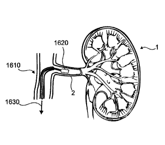
[0080] Fig. 1 shows aspects of a device in accordance with the present
disclosure inserted
into a lumen within a body coupled with a target organ.
[0081] Fig. 2 shows a schematic of aspects of a system in accordance with
the present
disclosure.
[0082] Figs. 3a ¨ c show aspects of access and treatment regions for a
target organ in
accordance with the present disclosure.
[0083] Fig. 4 shows aspects of access and treatment regions for a target
organ in accordance
with the present disclosure.
[0084] Fig. 5 shows aspects of access and treatment regions for a target
organ in accordance
with the present disclosure.
[0085] Fig. 6 shows aspects of access and treatment regions for a target
organ in accordance
with the present disclosure.
[0086] Figs. 7a-c show aspects of methods for treating and/or assessing
function of a neural
structure in accordance with the present disclosure.
[0087] Figs. 8a,b show aspects of access and treatment regions for a
target organ in
accordance with the present disclosure.
-18-
Date Regue/Date Received 2022-11-28
[0088] Figs. 9a-d show aspects of a device in accordance with the present
disclosure.
[0089] Figs. 10a-n show aspects of distal tips associated with a device
(e.g., guidewire,
catheter, micro-tool, etc.) in accordance with the present disclosure.
DETAILED DESCRIPTION
[0090] Particular embodiments of the present disclosure are described
hereinbelow with
reference to the accompanying drawings; however, the disclosed embodiments are
merely
examples of the disclosure and may be embodied in various forms. Therefore,
specific structural
and functional details disclosed herein are not to be interpreted as limiting,
but merely as a basis
for the claims and as a representative basis for teaching one skilled in the
art to variously employ
the present disclosure in virtually any appropriately detailed structure. Like
reference numerals
may refer to similar or identical elements throughout the description of the
figures.
[0091] Before the methods of the present disclosure are described in
greater detail, it is to be
understood that the methods are not limited to particular embodiments
described, as such may,
of course, vary. It is also to be understood that the terminology used herein
is for the purpose of
describing particular embodiments only, and is not intended to be limiting,
since the scope of the
methods will be limited only by the appended claims.
[0092] Where a range of values is provided, it is understood that each
intervening value, to
the tenth of the unit of the lower limit unless the context clearly dictates
otherwise, between the
upper and lower limit of that range and any other stated or intervening value
in that stated range,
is encompassed within the methods. The upper and lower limits of these smaller
ranges may
independently be included in the smaller ranges and are also encompassed
within the methods,
subject to any specifically excluded limit in the stated range. Where the
stated range includes one
or both of the limits, ranges excluding either or both of those included
limits are also included in
the methods.
[0093] Certain ranges are presented herein with numerical values being
preceded by the term
"about." The teim "about" is used herein to provide literal support for the
exact number that it
precedes, as well as a number that is near to or approximately the number that
the term precedes.
In detennining whether a number is near to or approximately a specifically
recited number, the
near or approximating unrecited number may be a number which, in the context
in which it is
presented, provides the substantial equivalent of the specifically recited
number.
[0094] Unless defined otherwise, all technical and scientific teinis used
herein have the same
meaning as commonly understood by one of ordinary skill in the art to which
the methods belong.
- 1 9-
Date Regue/Date Received 2022-11-28
Although any methods similar or equivalent to those described herein can also
be used in the
practice or testing of the methods, representative illustrative methods and
materials are now
described.
[0095] The citation of any publication herein is for its disclosure prior
to the filing date and
should not be construed as an admission that the present methods are not
entitled to antedate such
publication by virtue of prior invention. Further, the dates of publication
provided may be
different from the actual publication dates which may need to be independently
confirmed.
[0096] It is noted that, as used herein and in the appended claims, the
singular foinis "a",
"an", and "the" include plural referents unless the context clearly dictates
otherwise. It is further
noted that the claims may be drafted to exclude any optional element. As such,
this statement is
intended to serve as antecedent basis for use of such exclusive terminology as
"solely," "only"
and the like in connection with the recitation of claim elements, or use of a
"negative" limitation.
[0097] It is appreciated that certain features of the methods, which are,
for clarity, described
in the context of separate embodiments, may also be provided in combination in
a single
embodiment. Conversely, various features of the methods, which are, for
brevity, described in
the context of a single embodiment, may also be provided separately or in any
suitable sub-
combination. All combinations of the embodiments are specifically embraced by
the present
invention and are disclosed herein just as if each and every combination was
individually and
explicitly disclosed, to the extent that such combinations embrace operable
processes and/or
devices/systems/kits. In addition, all sub-combinations listed in the
embodiments describing such
variables are also specifically embraced by the present methods and are
disclosed herein just as
if each and every such sub-combination was individually and explicitly
disclosed herein.
[0098] As will be apparent to those of skill in the art upon reading this
disclosure, each of the
individual embodiments described and illustrated herein has discrete
components and features
which may be readily separated from or combined with the features of any of
the other several
embodiments without departing from the scope or spirit of the present methods.
Any recited
method can be carried out in the order of events recited or in any other order
which is logically
possible.
[0099] According to a first aspect there is provided a controlled nerve
ablation/neuromodulation system, which is configured for use in methods as
described herein and
may include the capability to sense one or more physiologic parameters at one
or more points in
the vicinity of a surgical site or within an affected/target organ, as well as
include the capability
-20-
Date Regue/Date Received 2022-11-28
to stimulate, deliver a chemical agent to, deliver energy to, and/or ablate
tissues at one or more
of the same points and/or an alternative point in the vicinity of a surgical
site. The nerve ablation
system may be configured so as to access vessels and/or surgical sites in the
body. The non-
limiting examples disclosed herein may be directed towards such configurations
(e.g., to
controllably provide neuromodulation procedures to an organ within a body, so
as to controllably
ablate renal nerves along a renal artery via an endoscopic or percutaneous
procedure, to treat a
cancerous tumor, to limit perineural invasion of cancerous cells into a nearby
nerve, to alter a
tumor microenvironment, etc.).
[00100] In aspects, a system/surgical tool in accordance with the present
disclosure may be
used to access, monitor, and/or to treat one or more neurological pathways,
ganglia, and/or
sensory receptors within a body: Ampullae of Lorenzini (respond to electric
field, salinity,
temperature, etc.), baroreceptors, chemoreceptors, hydroreceptors,
mechanoreceptors,
n ociceptors, osmoreceptors (osmol arity
sensing), photoreceptors, propri oceptors,
thermoreceptors, combinations thereof, and the like. Such receptors may be
associated with one
or more organs and/or physiologic processes within the body (i.e., a
regulatory process, etc.).
1001011 In aspects, a surgical tool in accordance with the present disclosure
may take the form
of a guidewire. The guidewire may be dimensioned and configured for placement
within a lumen
of a body at and/or beyond a surgical site and/or anatomical site of interest,
so as to monitor one
or more physiologic signals near the tip thereof. In aspects, the guidewire
may provide a pathway
for delivery of a second surgical device to the surgical site.
1001021 In aspects, a guidewire in accordance with the present disclosure may
include one or
more energy delivery means for delivering energy to an anatomical site within
and/or beyond the
wall of a lumen into which the guidewire tip has been placed.
[00103] In aspects, a guidewire in accordance with the present disclosure may
include one or
more sensors (e.g., as located on a micro-tool-tip, a clamp, a hook, a wire
element, an electrode
in a matrix, etc.) near to the tip thereof. One or more sensors may include a
pressure sensor, a
tonal sensor, a temperature sensor, an electrode (e.g., size, oriented, and
configured to interact
with a local tissue site, provide a stimulus thereto, measure a potential
therefrom, monitor current
to/from the tissues, to measure, dependent on configuration and design, a
bioimpedance, measure
an evoked potential, an electromyographic signal [EMG], an
electrocardiographic signal [ECG],
an extracellular potential faun a nearby neural structure, a mechanomyographic
signal [MMG],
local neural traffic, local sympathetic nerve traffic, local parasympathetic
nerve traffic, afferent
-21-
Date Regue/Date Received 2022-11-28
nerve traffic, efferent nerve traffic, etc.), an acoustic sensor, an oxygen
saturation sensor, or the
like.
[00104] In aspects, such sensing may be used in combination with a stress
test,
before/during/after an ablation, stimulation, administration of a chemical, or
the like to assess the
effect of the procedure on the neural traffic, tissue viability, or the like.
[00105] In aspects, a guidewire in accordance with the present disclosure may
include one or
more analyte sensors, configured to measure one or more analyte concentrations
or concentration
trend before, during, and/or after a procedure within a body. Such analyte
sensors may be
provided in an electrochemical form, a fluorescent form, an electro-optical
form, a swelling
responsive gel, etc.
[00106] A sensing guidewire in accordance with the present disclosure may be
advantageous
for accessing very small anatomical sites within a body, accessing adjunct
arteries and/or arteriole
pathways along a blood supply to a target organ, accessing a plurality of
vessels coupled to an
organ, accessing the parenchyma of an organ, for highly localized interaction
with a tissue site,
for accessing otherwise challenging lumens (i.e., a lumen with substantially
small diameter, with
substantially tortuous shape, etc.). In aspects, a guidewire in accordance
with the present
disclosure may provide a means for directing one or more additional tools to a
surgical site within
a body. In aspects, a guidewire in accordance with the present disclosure may
be configured to
sense physiologic parameters from and/or to treat tissues within such
miniature lumens as part of
a procedure (i.e., a surgical procedure, a diagnostic procedure, an ablation
procedure, etc.). Such
a configuration may be particularly advantageous for accessing a vessel within
a small organ or
microvascular region of an organ, such as with a bone, near to a foramen of a
bone, or the like.
[00107] In aspects, a system for treating a cancerous tumor coupled to a
target organ within a
body in accordance with the present disclosure may include a catheter (i.e., a
balloon catheter, a
needle catheter, a flexible catheter, etc.), or a guidewire, sized and
dimensioned for delivery into
a lumen (i.e., an artery, vein, vessel, or the like) serving the target organ,
the catheter or guidewire
including a distal tip configured to interface with the walls of an artery,
vein, vessel coupled to
the target organ, the distal tip configured for delivery of energy and/or a
substance to one or more
nerves coupled to the target organ.
[00108] In aspects, a system for augmenting function of a target organ within
a body in
accordance with the present disclosure may include a catheter (i.e., a balloon
catheter, a needle
catheter, a flexible catheter, etc.) or a guidewire, sized and dimensioned for
delivery into a lumen
-22-
Date Regue/Date Received 2022-11-28
(i.e., an artery, vein, vessel, or the like) serving the target organ, the
catheter or guidewire
including a distal tip configured to interface with the walls of an artery,
vein, vessel coupled to
the target organ, the distal tip configured for delivery of energy and/or a
substance to one or more
nerves coupled to the target organ. In aspects, the energy may be thermal
energy, RF current,
MW current, ultrasound, radiation, cryotherapy, or the like.
[00109] In aspects, the substance may be a medicament, a denervating agent, an
sympathetic
nerve specific denervating agent, a parasympathetic nerve specific denervating
agent, a
neuroblocking agent, a highly specific neuroblocking agent (i.e., an agent
specifically configured
for blocking of a particular receptor, nerve family, etc.), or the like. In
aspects, the denervating
agent may be ethanol, botulinum toxin, or the like. In aspects, the highly
specific denervating
agent may be a neural targeting chemical such as a poison, a toxin, or the
like.
[00110] In aspects, the catheter or guidewire may include one or more sensing
elements each
in accordance with the present disclosure, located within the vicinity of the
distal tip thereof,
configured to interface and record physiologic information associated with one
or more nerves
coupled to the target organ upon placement (i.e., during a surgical or
interventional procedure,
during a diagnostic procedure, a stress test, etc.). In aspects, the catheter
or guidewire may be
equipped with a substance eluting element, configured to deliver a substance,
a medicament, a
denervating substance, or the like into the target organ, into the tissues
surrounding the wall of
the lumen, etc. In aspects, the energy and/or substance is delivered to
interrupt and/or augment
neural traffic along one or more nerves coupled to the target organ. In
aspects, the energy and/or
substance is provided so as to block nerve traffic to and/or from the organ
along the lumen into
which the distal tip has been inserted. In aspects, a system a system in
accordance with the present
disclosure may be used to treat pain, pain associated with perineural invasion
of a cancerous
tumor, or the like.
[00111] Some non-limiting examples of systems, devices, and methods which may
be suitable
for performing one or more aspects of a surgery, interventional procedure,
diagnostic, and/or
treatment in accordance with the present disclosure are generally detailed in
co-pending
international patent applications W014070999, W013181137,W013112844,
W013042847,
W013067726, and W014031962.
[00112] In aspects, a system, device, and/or method in accordance with the
present disclosure
may be used to treat and/or slow the progression of a cancerous tumor. Some
non-limiting
examples of such cancer that may be treated include cancer of the prostate,
pancreas, breast,
colon, skin, liver, esophagus, cervix, bone, urogenitals, lung, and the like.
-23-
Date Regue/Date Received 2022-11-28
[00113] In aspects, a system, device, and/or method in accordance with the
present disclosure
may be used to slow, hinder, and/or prevent perineural invasion of a cancerous
tumor into a
surrounding nerve structure. In aspects, a system, device, and/or method in
accordance with the
present disclosure may be used to interrupt, decrease, and/or stop neural
communication to a
cancerous tumor and/or the microenvironment surrounding the tumor (i.e., to
interrupt nerve
traffic to/from a cancerous tumor or the tissues thereby to the rest of the
body). In aspects, a
system, device, and/or method in accordance with the present disclosure may be
used to decrease
pain signals communicated by nerve in the vicinity of the organ and/or tumor
to one or more
neural circuits, ganglia, etc. In aspects, a system, device, and/or method in
accordance with the
present disclosure may be used to block, deaden, and/or to destroy nerves in
the vicinity of a
tumor and/or surrounding tissues.
[00114] In aspects, a system, device, and/or method in accordance with the
present disclosure
may be used to slow or even halt tumorigenesis of cancerous tissue.
[00115] In aspects, a system, device, and/or method in accordance with the
present disclosure
may be configured to form a physical barrier (i.e., lesion, a collagen block,
etc.). In aspects, a
system, device, and/or method in accordance with the present disclosure may be
used to treat
local inflammation (such as for the treatment of pancreatitis, prostatitis,
irritable bowel syndrome,
etc.).
[00116] In aspects, the system may include a balloon coupled with the
catheter, situated in the
vicinity of the distal tip thereof, the balloon coupled to a fluid source so
as to be expand-ably
deployed during a procedure so as to interface with the walls of lumen into
which the distal tip
has been placed. In aspects, the balloon may include one or more energy
delivery elements,
and/or sensing elements each in accordance with the present disclosure
configured to interface
with tissues adjacent to the balloon during a procedure. In aspects, the
sensing elements may be
configured to monitor electiophysiological information associated with the
adjacent tissues.
[00117] In aspects, the system may be configured to direct energy through the
energy delivery
elements based upon the information collected by the sensing elements. In
aspects, the sensing
elements may be used to determine regions of abnormal electrophysiological
activity, determine
the direction of nerve traffic along the lumen, determine the type of nerves
situated near the
sensing element, etc. In aspects, the energy delivery may be directed to one
or more regions of
the lumen wall, through the lumen wall, into the adventitia in the vicinity of
the lumen, into an
organ (i.e., a pancreas, a liver, an intestinal wall, a kidney, a bone, etc.)
adjacent to the lumen,
etc. as directed by data collected by the sensing elements during the
procedure.
-24-
Date Regue/Date Received 2022-11-28
[00118] In aspects, the system may include a sn-ess testing aspect, configured
to apply a stress
(i.e., local and/or systemic) to the body while monitoring the response to the
stress via one or
more of the sensing elements. In aspects, the system may be used to diagnose a
disease state,
determine a function of the adjacent tissues, and/or determine the type of
adjacent tissues (i.e., a
nerve fiber, a type of nerve fiber, etc.) based upon the data obtained by the
one or more sensing
elements during the stress.
[00119] In aspects, a method in accordance with the present disclosure for
treating a cancerous
tumor, may include inserting at least a portion of a system in accordance with
the present
disclosure into a lumen with a wall in the vicinity of a target organ, and
delivering energy and/or
a substance to at least a portion of the wall of the lumen, through at least a
portion of the wall of
the lumen, into the target organ, and/or into the tissues surrounding the
target organ. The method
may include treating one or more nerves in the vicinity of the target organ.
[00120] In aspects, the method may include collecting physiologic data from
the target organ
and/or within the vicinity of the target organ, collecting data from one or
more neural structures
coupled to the organ, or the like. The method may include making a diagnostic
decision,
determining the state of the local neural structures, determining the extent
of a surgical procedure,
etc. based at least in part from the recorded data. In aspects, the method may
include directing
the energy and/or substance based upon the collected physiologic data. In
aspects, the method
may include collecting further physiologic data after the delivery of energy
to determine if the
desired effect has been achieved. In aspects, the method may include comparing
a neural activity
associated with the procedure, treatment, and/or target organ before and after
a procedure, to
determine the extent of the procedure, to confirm that the procedure
positively affected the
functionality of the nerves, etc.
[00121] In aspects, the substance may include an antibody drug conjugate
(ADC), a
chemotherapeutic agent, a toxin, a neurotoxin, etc. In aspects, the ADC
substance may be
configured to affect the function of a region or tissue type within the
vicinity of the organ
alternatively to the other tissues within the vicinity thereof. In aspects,
the substance may include
a sugar attached to a therapeutic agent to mask the therapeutic agent, such
that it is to be taken up
by the region of tissue (i.e., appear as a sugar, a friendly protein, etc.).
Such a configuration
provides a method for delivering a highly potent medicament directly to a
tissue of interest (i.e.,
directly into a tumor), so as to enhance the bioavailability thereof, and to
minimize the systemic
dosage required in order to achieve significant therapeutic concentrations
thereof within the
region of tissue.
-25-
Date Regue/Date Received 2022-11-28
[00122] In aspects, the substance may be delivered at a rate of less than
lmg/sec
(milligrams/second), lmg/min (milligrams/minute), lmg/hr, 0.01mg/hr, less than
lug/hr, or the
like. Such a configuration may be important so as to minimize local stress and
damage caused
by the introduction of the substance into the microenvironment of the tissue
of interest.
[00123] In aspects, a system in accordance with the present disclosure may
include a catheter
and/or a guidewire configured for percutaneous access to the arteries, veins,
or lumens, of a body,
for delivery through one or more arteries of the body to the vicinity of the
target organ.
[00124] In aspects, one or more energy delivery elements, sensing elements, a
diameter of the
catheter, guidewire, or the like may be sized and arranged such that it may be
placed within an
artery, vein in a region near the target organ, within the parenchyma of the
target organ, into a
vessel in the periosteal space of a bone, and/or through a foramen of a bone.
In aspects, the
delivery elements and/or sensing elements, catheter, guidewire, etc. may be
sized and
dimensioned such that a characteristic diameter thereof is less than lmm, less
than 0.75mm, less
than 0.5mm, less than 0.3mm, or the like.
[00125] According to aspects, there is provided a method for treating a tumor
including
stimulating, blocking, and/or ablating one or more regions of a neural circuit
coupled to the tumor
and/or perineural microenvironment surrounding a tumor. In aspects, the method
may include
performing the treatment without substantially increasing inflammation,
necrotizing tissues, or
the like in the vicinity of the tumor.
[00126] In aspects, the method may include stimulating the neural circuit with
a stimulation
frequency suitable to provide a neural block there along. In aspects, the
method may include
providing energy and/or a bolus of a chemical agent in an amount sufficient to
provide a neural
block to one or more regions of the neural circuit, and/or ablate one or more
regions of the neural
circuit.
[00127] In aspects, a system, device, and/or method in accordance with the
present disclosure
may be used to prevent or slow the growth rate and/or tumorigenesis of a
cancerous tissue,
modulating neural communication between a tumor and one or more neural
circuits coupled to
the target organ, augmenting/treating/ablating the perinetual microenvironment
in the vicinity of
a tumor or along a neural circuit coupled thereto, and/or preventing or
slowing the process of
perineural invasion of a tumor into surrounding tissues
[00128] In aspects, a method in accordance with the present disclosure for
treating a tumor
within a body may include neuromodulating one or more nerves coupled to the
tumor. In aspects,
-26-
Date Regue/Date Received 2022-11-28
a method in accordance with the present disclosure for treating a tumor within
a body may include
neuromodulating a perineural microenvironment in the vicinity of the tumor. In
aspects, a method
in accordance with the present disclosure for treating a tumor within a body
coupled with a neural
circuit within the body may include decoupling the neurological connection
between the tumor
and the neural circuit. In aspects, a method in accordance with the present
disclosure may be
used to treat prostate cancer, pancreatic cancer, breast cancer, colon cancer,
cervical cancer,
ovarian cancer, bladder cancer, or the like.
[00129] In aspects, a method in accordance with the present disclosure may
include inserting
the distal tip of a device in accordance with the present disclosure into a
vessel coupled to the
tumor. In aspects, the method may include placing the distal tip of a device
into a vessel, such as
an artery, which supplies blood to the tumor, and/or uniquely supplies blood
to the organ coupled
with the tumor.
[00130] In aspects, a method in accordance with the present disclosure may
include applying
energy and/or a chemical agent into an adventitia surrounding the vessel. In
aspects, a method in
accordance with the present disclosure may include monitoring
electrophysiological activity
along a wall of the vessel. In aspects, the method may include using the
monitoring to determine
the extent of a treatment, to alter a bolus of energy or chemical agent
delivered, or the like.
[00131] In aspects, a method in accordance with the present disclosure for
treating a tumor
may include inducing apoptosis within neural tissues within the vicinity of
the tumor, within a
neural circuit coupled with the tumor, or the like.
[00132] In aspects, a method in accordance with the present disclosure may
include inducing
necrosis, and/or apoptosis within neural tissues within a neural circuit
coupled with the tumor.
In aspects, induced necrosis will typically cause the corresponding cells to
exhibit rapid swelling,
lose membrane integrity, shut down metabolism, and release their contents into
the environment.
Cells that undergo rapid necrosis in vitro do not often have sufficient time
or energy to activate
apoptotic machinery and thus will often not express apoptotic markers. Rather,
induced apoptosis
typically causes the corresponding cells to exhibit cytological and molecular
events such as a
change in the refractive index of the cell, cytoplasmic shrinkage, nuclear
condensation, and
cleavage of DNA into regularly sized fragments. In aspects, the chemical agent
may be selected
so as to induce apoptosis in one or more neural tissues (i.e., axon, dendrite,
cell body, myelin
sheath, synapse, etc.).
-27-
Date Regue/Date Received 2022-11-28
[00133] In aspects, one or more systems, methods, and devices each in
accordance with the
present disclosure may be used to intervention-ally alter one or more
homeostatic processes
within a body. Some non-limiting examples of homeostatic processes include
production/release
of renin, insulin, cholesterol, bile salts, testosterone, progesterone, prion,
serotonin, endorphins,
dopamine, monoamine neurotransmitters, histamines, noradrenaline, glucose, and
the like,
adjustment of blood pressure, anti-inflammatory activity, testosterone,
estrogen, "uterine
hemorrhaging", hunger, bowel movement, nutritional uptake in the bowel, and
the like_
[00134] In aspects, a system in accordance with the present disclosure may
include a substance
delivery aspect, configured for elution of a substance into the vicinity of
the target. In aspects,
the system may include one or more sensing elements configured for monitoring
of one or more
physiologic parameters associated with the target, the homeostatic process in
question, a stress
response, or the like. In aspects, the system may include one or more energy
delivery elements
configured to deliver a bolus of energy to the target in order to alter the
homeostatic process.
[00135] Fig. 1 shows aspects of a device in accordance with the present
disclosure inserted
into a lumen within a body coupled with a target organ. A micro surgical tool
1610 in accordance
with the present disclosure is shown as placed into the renal artery 2 of a
subject as coupled to a
target organ 1 (i.e., here shown as a kidney) in accordance with the present
disclosure. In aspects,
the microsurgical tool 1610 may include one or more distal tips 1620 each
including one or more
sensing tips in accordance with the present disclosure to selectively sense,
stimulate, and/or treat
target anatomy based on the determined locations thereof. In aspects, the
sensing tips may be
configured to acquire positional and/or physiologic infoimation related to the
target anatomy,
placement of the micro surgical tool 1610 within the renal artery, the
parenchyma of the target
organ 1, and/or monitoring of the surgical procedure (i.e., ablation
procedure, chemical
denervation, chemical deployment, etc.), or the like. Such a feedback
mechanism may be used
to precisely guide the micro surgical tool 1610 during a surgical procedure
(i.e., ablation
procedure, etc.), to determine the extent of a surgical procedure, or the
like. In aspects, the distal
tip 1620 may be coupled with a controller 1630 in accordance with the present
disclosure for
performing one or more of the procedures.
[00136] Fig. 2 shows a schematic of aspects of a system for perfoiming a
surgical procedure
in accordance with the present disclosure. The system is shown interfacing
with a surgical site
2101 within a body, a subject, a patient, etc. The system may include a
microsurgical tool 2110
in accordance with the present disclosure. During use, the microsurgical tool
2110 may be
configured to interact 2112 with the surgical site 2101 in accordance with the
present disclosure.
-28-
Date Regue/Date Received 2022-11-28
In aspects, the microsurgical tool 2110 may be coupled to a connector 2120,
the connector
providing a mechanical and/or electrical interface between the microsurgical
tool 2110 and/or
one or more other modules of the system. In aspects, the microsurgical tool
2110 may include
an embedded local control circuit 2115a (e.g., a microcircuit, a switch
network, a signal
conditioning circuit, etc.) in accordance with the present disclosure. In
aspects, the connector
2120 may include a local control circuit 2115b in accordance with the present
disclosure. In
aspects, the connector 2120 may be coupled to an operator input device 2125
(i.e., a foot pedal,
an advancing slider, a torqueing mechanism, a recording button, an ablation
button, etc.). In
aspects, the connector 2120 may be coupled to a control unit 2130 configured
to accept one or
more signals from the microsurgical tool 2110, communicate one or more control
signals thereto,
send one or more pulsatile and/or radio frequency signals to the
microcontroller, record one or
more electrophysiological signals from the microsurgical tool, or the like.
[00137] In aspects, the control unit 2130 may be connected to a display 2135
configured to
present one or more aspects of the recorded signals from the microsurgical
tool 2110 to an
operator, to present a map, at least partially dependent on the recorded
signals, to present one or
more metrics relating to a physiologic parameter, a surgical procedure,
surgical outcome efficacy,
etc. In aspects, the control unit 2130 may be coupled to a surgical subsystem
2140, the surgical
subsystem 2140 configured to perform a surgical procedure 2145 to the surgical
site 2101. Some
non-limiting examples of suitable surgical procedures include an ablation, an
excision,
stimulation, a cut, a burn, a radio frequency ablation, radiosurgery, an
ultrasonic ablation, an
abrasion, a biopsy, and delivery of a substance. The control unit 2130 may be
configured to
influence, direct, control, and/or provide feedback for one or more aspects of
the surgical
procedure 2140, based upon one or more of the electrophysiological signals
conveyed by the
microsurgical tool 2110.
[00138] Figs. 3a ¨ c show aspects of access and treatment regions for a target
organ in
accordance with the present disclosure. Each of Figs. 3a ¨ c show a pancreas
2201 (i.e., a target
organ in accordance with the present disclosure), a spleen 2203 (i.e.,
optionally a target organ in
accordance with the present disclosure), and a duodenum 2205. Fig. 3a
illustrates aspects of a
vascular supply to the pancreas 2201, spleen 2203, and duodenum 2205 are
highlighted including
the aorta 2207, the celiac trunk 2209, the anterior superior
pancreaticoduodenal artery 2211, the
posterior superior pancreaticoduodenal artery 2213, the anterior inferior
pancreaticoduodenal
artery 2215, the posterior inferior pancreaticoduodenal artery 2217, the
superior mesenteric artery
2219, the dorsal pancreatic artery 2221, and the splenic artery 2223. As part
of a treatment,
-29-
Date Regue/Date Received 2022-11-28
monitoring session, etc. in accordance with the present disclosure, one or
more microsurgical
tools each in accordance with the present disclosure may be inserted into
and/or interfaced with
one or more of the vascular supply vessels to the pancreas 2201, spleen 2203,
and/or duodenum
2205. In aspects, one or more of the microsurgical tool tips may be delivered
through the aorta
2207, the celiac trunk 2209, and/or the superior mesenteric artery 2213, or
branches thereof, etc.
In aspects, one or more microsurgical tools in accordance with the present
disclosure may be
configured to treat the perivasculature in the vicinity of one or more of the
vascular supply lumens
in accordance with the present disclosure.
[00139] In aspects, a pancreatic tumor may be present within or coupled with
the pancreas
2201. In such aspects, a microsurgical tool in accordance with the present
disclosure may be
interfaced with one or more of the vascular supply lumens in order to treat
the pancreatic tumor
in accordance with the present disclosure.
[00140] In aspects, the treatment may be applied to one or more neurological
structure in the
vicinity of the vascular supply. In aspects, a procedure (i.e., a treatment,
biopsy, sensing,
stimulation, etc.) may be applied to a first zone 2227 located in the vicinity
of the posterior and/or
anterior superior pancreaticoduodenal arteries 2211, 2213. In aspects, such a
zone may be located
along the posterior and/or anterior pancreaticoduodenal arteries 2211, 2213
distally to the hepatic
artery 2225. In aspects, a procedure in accordance with the present disclosure
may be performed
on a second zone 2229 located in the vicinity of the anterior and/or posterior
inferior
pancreaticoduodenal arteries 2215, 2217 in accordance with the present
disclosure. In aspects,
the second zone 2229 may include tissues in the vicinity and/or wall of the
superior mesenteric
artery 2219. In aspects, a procedure in accordance with the present disclosure
may be performed
on a third zone 2231, located in the vicinity of the dorsal pancreatic artery
2221 or tributaries
formed therefrom. In aspects, a procedure in accordance with the present
disclosure may be
performed on a fourth zone 2233, located in the vicinity of the splenic artery
2233 or tributaries
fointed therefrom. In aspects, one or more zones 2227, 2231, 2233 may include
regions of the
celiac trunk 2209. In aspects, a surgical procedure may be applied
simultaneously within one or
more zones 2227, 2229, 2231, 2233, and/or vascular supply lumens, etc. In
aspects, one or more
distal tips of one or more surgical tools may be inserted into a zone 2227,
2229, 2231, 2233 as
part of a procedure in accordance with the present disclosure.
[00141] In aspects, a plurality of distal tips may be simultaneously located
within one or more
zones. Energy provided via one or more of the distal tips may be communicated
between zones
2227, 2229, 2231, 2233, and/or to an externally coupled component (i.e., an
electrode). In
-30-
Date Regue/Date Received 2022-11-28
aspects, the distal tips may be configured to perform a first procedure (i.e.,
a sensing procedure)
within a first zone 2227, 2229, 2231, 2233 and to perform a second procedure
(i.e., a stimulation,
ablation, chemical delivery, etc.) within a second zone 2227, 2229, 2231,
2233, or the like.
[00142] In aspects, such procedures may be perfolined to augment and/or
plastically change
neural communication to/from one or more regions of a target organ 2201 to the
brain, a ganglion,
etc. so as to influence the physiologic function thereof, to augment the
afferent traffic to the brain,
to augment the efferent traffic reaching the target organ, etc.
[00143] In aspects, a procedure may be applied to the celiac trunk 2209, the
third zone 2231,
and/or the fourth zone 2233 in order to affect function of the spleen 2203.
[00144] In aspects, coordination of two or more procedures applied within one
or more zones
2227, 2229, 2231, 2233 may be provided to treat one or more regions of the
target organ 2201
(i.e., in this case so as to selectively treat one or more regions of the
pancreas 2201 while
maintaining regular function of one or more other regions of the pancreas
2201, etc.). In aspects,
the zones 2227, 2229, 2231, 2233 to be treated may be selected based upon an
image of the target
organ 2201 (i.e., to determine the location and coupling of an anomaly, a
tumor, etc. within the
target organ 2201). In aspects, a first procedure, such as sensing and/or
stimulation, applied
within one or more of the zones 2227, 2229, 2231, 2233 may be provided to
detennine where
within the target organ to provide a second procedure (i.e., sensing,
ablation, etc.), or to determine
the extent of a previously applied procedure (i.e., an ablation procedure, a
neuromodulation
procedure, etc.).
[00145] In aspects, the zones and/or anatomical features shown in Fig. 3a may
be accessed
through the aorta 2207, such as from a descending approach 2235 or an
ascending approach 2237
as preferred by a surgeon or surgical planner.
[00146] Fig. 3b shows aspects of a pancreas 2201, a spleen 2203, and a
duodenum 2205 along
with the vascular supply thereto. Aspects of the vascular supply shown include
the portal vein
2241, the posterior superior pancreaticoduodenal vein 2243, the anterior
superior
pancreaticoduodenal vein 2245, the splenic vein 2247, the anterior inferior
pancreaticoduodenal
vein 2249, and the posterior inferior pancreaticoduodenal vein 2251. Aspects
of the tributaries
that serve the vascular supply are shown as line segments interconnecting the
larger vessels with
the pancreas 2201, the spleen 2203, and the duodenum 2205.
[00147] In aspects, one or more zones 2255, 2257, 2259 may be accessed as part
of a procedure
and monitored, stimulated, treated, etc. in accordance with the present
disclosure. In aspects as
-31-
Date Regue/Date Received 2022-11-28
part of a method in accordance with the present disclosure, one or more of the
zones 2255, 2257,
2259 may be accessed via a portal vein approach 2253 (i.e., via a catheter,
guidewire, surgical
tool, etc. placed into the portal vein). In aspects, one or more zones 2255,
2257, 2259 may be
accessed via direct needle stick into the body as part of a procedure.
[00148] In aspects, one or more treatments may be applied to one or more of
the zones 2255,
2257, 2259 as part of a procedure. Such treatments may be provided so as to
affect the perineural
microenvironment surrounding a tumor within the target organ, to affect one or
more receptors,
sensory nerves, or the like within the target organ, to affect one or more
physiologic functions of
the organ, etc.
[00149] Fig. 3c illustrates a pancreas 2201, spleen 2203, and a duodenum 2205,
and a
pancreatic duct 2265 running through the length of the pancreas 2201. The
pancreatic duct 2265
supplies pancreatic fluids into the duodenum via the duodenal papilla 2267. In
aspects, a system
and/or method in accordance with the present disclosure may be used to treat
one or more zones
2275, 2277 within the vicinity of the pancreatic duct 2265. In aspects, a
surgical tool in
accordance with the present disclosure may be introduced 2273 into the
pancreatic duct 2265 via
the duodenal papilla 2267, from a descending approach 2271 through the
esophagus, stomach,
and duodenum 2267.
[00150] In aspects, one or more of the zones 2275, 2277 may be monitored,
and/or treated in
accordance with the present disclosure. In aspects, such treatment may be used
to affect secretion
of pancreatic fluid into the duodenum 2205, affect cell function within the
vicinity of the
pancreatic duct 2265, affect the microenvironment of a tumor located within
the pancreas, to
disconnect one or more neural pathways between a tumor and another neural
circuit within the
body, etc. In aspects, one or more of the treatments may be provided as part
of a surgical
procedure coupled with a pancreas resection, as part of a surgical
intervention to treat pancreatic
cancer, etc. In aspects, one or more treatments (i.e., stimulations,
ablations, chemical agent
delivery, neural blocks, etc.) may be configured to influence one or more
functions of the target
organ. In the case of the pancreas 2201, the treatments may be employed to
affect one or more
functions such as production of insulin, glucagon, somatostatin, and/or
pancreatic polypeptide,
production/secretion of pancreatic juice containing digestive enzymes, glucose
metabolism,
and/or control of blood glucose concentration.
[00151] Generally speaking, the part of the pancreas with endocrine function
is made up of
approximately a million cell clusters called islets of Langerhans. Four main
cell types exist in the
islets: a cells secrete glucagon (increase glucose in blood), 13 cells secrete
insulin (decrease
-32-
Date Regue/Date Received 2022-11-28
glucose in blood), delta cells secrete somatostatin (regulates/stops a and 13
cells) and PP cells, or
gamma cells, secrete pancreatic polypeptide.
[00152] Secretion of hormones into the blood may be affected and/or regulated
by the effect
of hormones in the blood on the islets of Langerhans, and through the effect
of the autonomic
nervous system on the blood flow and cell function. In aspects, augmentation
of sympathetic
and/or parasympathic activity may affect secretion from beta cells, and alpha
cells within the
pancreas.
[00153] Fig. 4 shows aspects of access and treatment regions for a target
organ in accordance
with the present disclosure. A schematic diagram of a liver 2301, along with
coupled vasculature
including an aorta 2303, a hepatic vein 2305, hepatic arteries 2307, a portal
vein 2309, and ducts
including hepatic ducts 2313. A gallbladder 2315 is also shown, and may serve
as a target organ
for a procedure in accordance with the present disclosure. A plurality of
zones 2333, 2335, 2337,
2339 may be accessed via the vasculature and/or ducts, and may be monitored
and/or treated as
part of a procedure in accordance with the present disclosure. In aspects,
different neural circuits
(sympathetic, parasympathetic, and/or afferent circuits), may travel through
the various zones
2333, 2335, 2337, 2339. A monitoring procedure, optionally combined with a
stress test, may be
used to elucidate the type and/or function of neural circuits within the
vicinity of one or more
zones 2333, 2335, 2337, 2339, within a sub-region of a zone 2333, 2335, 2337,
2339, or the like.
[00154] In aspects, one or more neural circuits coupled with the liver 2301
may be selectively
treated in accordance with the present disclosure. In aspects, one or more
neural circuits passing
along a hepatic duct 2313 may be monitored and/or treated in accordance with
the present
disclosure. In aspects, a hepatic duct 2313 situated zone 2337, or a zone
2335, 2339 situated in
the vicinity of the hepatic arteries 2307 may be accessed through a hepatic
duct 2313 approach
2329, and/or via an ascending approach 2323 through the aorta 2303, a
descending approach
through the aorta 2323, an ascending approach 2135 through the portal vein
2309, an ascending
approach through the inferior vena cava 2325, and potentially a descending
approach 2319
through the inferior vena cava 2325.
[00155] In aspects, one or more procedures in accordance with the present
disclosure may be
applied to the liver 2301, the parenchyma of the liver 2301, hepatocytes of
the liver 2301, to
disruption and/or augmentation of signals that can be relayed electrically to
individual cells by
structures such as cell-to-cell connecting gap junctions, to gap junctions
within cells of the
parenchyma (i.e., via modulation of electrotonic coupling, to compensate for
the sparse direct
inputs to the hepatocytes, especially with respect to sympathetic signal
transduction), one or more
-33-
Date Regue/Date Received 2022-11-28
zones 2333,2335, 2337, 2339, within regions of receptors within the liver 2301
so as to modulate
hormone release into the organ, one or more vessels and/or perivascular
regions coupled with the
liver 2301, and/or the gallbladder 2315 to treat a disease state, to augment
organ function, or the
like.
[00156] Some aspects of liver function that may be augmented by a treatment
and/or
monitored in accordance with the present disclosure include glucose
storage/release, metabolic
sensing (and related signal traffic to the brain related thereto),
glucoregulatory function, afferent
vagal activity reaching the brain, chemoreceptor function (or related signal
traffic associated
therewith), lipid sensing/synthesis, regulation of hepatic insulin sensitizing
substance, afferent
traffic augmentation associated with glucosensors (i.e., primarily in the
region of the portal vein
2309, etc.), protein sensing, GLP-1,1eptin, CCK, FFA, PPAR alpha and gamma,
glycogenolysis,
gluconeogenesis, VLDL secretion, ketogenesis, hypoglucemia sensing, or the
like.
[00157] In
aspects, one or more procedures (i.e., sensing, a treatment, stimulation,
ablation,
etc.) may be applied to a one or more vagal branches including the hepatic
branch, the
gastroduodenal branch, the common hepatic branch, coupled with the left vagal
and right vagal
nerve branches. In aspects, such procedures may be performed along the
associated vasculature
serving the liver 2301 and/or within the parenchyma of the liver 2301 in the
vicinity of the
corresponding neural structures of the vagal branch in question. In aspects,
monitoring of the
vagal branch at a first location (i.e., along an artery supplying the liver
2301) and at a second
location (i.e., at a site within the parenchyma of the liver 2301) may be used
to confirm proper
placement of a surgical tool at a treatment site, confirm efficacy of a
treatment, confirm proper
targeting of the associated neural structures related to the vagal branch,
etc.
[00158] In aspects, one or more sympathetic procedures in accordance with the
present
disclosure may be applied to one or more sympathetic and/or afferent nerves in
the vicinity of the
liver 2301, the gallbladder 2315, along a vessel or duct serving the organs,
etc.
[00159] In aspects, one or more surgical tools in accordance with the present
disclosure may
be used to provide a physical and/or functional mapping of one or more neural
circuits within one
or more regions of the liver neuronal network, such as to determine location
and/or function of
parasympathetic postganglionic cell bodies, response to stress tests,
distinguish between sensory
and motor neuron nerves, or the like.
[00160] Fig. 5 shows aspects of access and treatment regions for a target
organ in accordance
with the present disclosure. The target organ is a stomach 2401. Also shown is
the esophagus
-34-
Date Regue/Date Received 2022-11-28
2403, duodenum 2405, aorta 2407, the right gastric artery 2409, the left
gastric artery 2411, the
splenic artery 2413, the gastric duodenal artery 2415, the right
gastroepiploic artery 2417, and
the lift gastroepiploic artery 2419. Access to one or more vessels coupled
with the stomach may
be provided via an ascending approach 2421 in the aorta 2407, via a descending
approach 2423
in the aorta 2407, or via a descending approach 2425 in the esophagus 2403.
[00161] In aspects, one or more neural structures may be monitored and/or
treated on the wall
of the stomach 2401, the esophagus 2403, the duodenum 2405, and/or one or more
of the vessels
coupled thereto. In aspects, a procedure may be applied in the vicinity of one
or more zones
2431, 2435, 2437, 2439, 2441, 2443, 2445, 2447 so as to treat a neurological
disorder, function,
etc. associated with the target organ 2401.
[00162] In aspects, a procedure and/or selective treatment may be applied to a
neural structure,
an afferent nerve, an efferent nerve, one or more sympathetic nerves (SNS), or
the like in the
vicinity of zones 2431, 2433, 2435, 2437, 2439, 2441, 2443, 2445, 2447 and/or
parasympathetic
nerves (PNS) in the vicinity of zones 2433, 2437, 2445, 2447. In aspects, a
treatment may be
applied selectively to SNS or PNS in order to balance a regulatory imbalance
in the activity there
between, or to create an imbalance in activity there between in order to
augment function of the
target organ, etc.
[00163] Fig. 6 shows aspects of access and treatment regions for a target
organ in accordance
with the present disclosure. The target organ shown is a femur bone 2501
(i.e., a representative
non-limiting example of a long bone). The femur 2501 generally includes a
diaphysis,
metaphysis, and epiphysis regions. The treatment may be directed towards a
bone related
homeostatic function (e.g., osteoblast production), ancUor one or more neural
structures coupled
with a tumor within one or more of the regions. The femur 2501 includes
regions of compact
bone 2510, spongy bone 2520, and a medullary cavity 2515 in which spongy bone
2520 is
innervated with nerves, and vascularized with associated blood vessels.
Exemplary epiphy seal
arteries and veins 2520a, 2520b, metaphyseal arteries and veins 2525a, 2525b,
and a nutrient
artery and vein 2530 are highlighted. A system in accordance with the present
disclosure may be
sized and dimensioned such that a distal tip thereof may be advanced along an
access point into
one or more of the epiphyseal, metaphyseal, or nutrient arteries or veins
2520a, 2520b, 2525a,
2525b, 2530 to treat one or more regions of the femur 2501.
[00164] In aspects, one or more neural structures may be monitored and/or
treated on one or
more walls of the epiphyseal artery/vein 2520a, 2520b metaphyseal artery/vein
2525a, 2525b,
and/or the nutrient artery/vein 2530, within the medullary cavity 2515, within
one or more sites
-35-
Date Regue/Date Received 2022-11-28
of the spongy bone 2520, near to the foramen of the femur 2501, within the
periosteal space of
the femur 2501, and/or one or more of the vessels coupled thereto. In aspects,
a procedure may
be applied in the vicinity of one or more zones 2535, 2537, 2539, 2541, 2543
so as to treat a
neurological disorder, a tumor, pain signals sent between the target organ
2501 and the body,
treatment of neural receptors, a homeostatic function, etc. associated with
the femur 2501.
[00165] In aspects, a procedure and/or selective treatment may be applied to a
neural structure,
an afferent nerve, an efferent nerve, one or more sympathetic nerves (SNS),
parasympathetic
nerves (PNS), motor nerves, receptors, and/or the like in the vicinity of
zones 2535, 2537, 2539,
2541, 2543. In aspects, a treatment may be applied selectively to SNS or PNS
in order to balance
a regulatory imbalance in the activity there between, or to create an
imbalance in activity there
between in order to augment one or more functions of the femur, etc. In
aspects, the procedure
may be used to treat pain associated with bone cancer, to augment the
microenvironment around
a bone cancer tumor so as to alter the growth rate thereof, to adjust the
production rate of
osteoblasts, to alter the bone density, or the like.
1001661 Figs. 7a-c show aspects of methods for treating and/or assessing
function of a neural
structure in accordance with the present disclosure. Fig. 7a illustrates
aspects of a method for
modulating or assessing neural traffic in accordance with the present
disclosure. The method
includes accessing one or more target sites within a body, stimulating,
sensing, or ablating the
nerves, augmenting neural activity, treating the afferent nerves and/or
receptors, and optionally
evaluating the afferent nerve activity post treatment to determine if the
traffic has been
modulated. In aspects, the evaluation maybe performed by comparing a nerve
activity metric
before and after treatment (e.g., a change in integrated activity level, a
change in phasic response
such a shift from a biphasic polarity to a monophasic polarity, a change in
action potential firing
rate, a change in the spectral content of the firing, etc. associated with the
local neural tissues).
In aspects, the method may include varying a pressure applied to the afferent
nerves and/or
receptors and monitoring afferent nerve activity during such changes in
applied pressure (i.e.,
monitoring activity during a variable pressure compression block).
1001671 Additionally, alternatively, or in combination with the monitoring of
electrophysiological activity, the method may include monitoring one or more
physiologic
parameters in accordance with the present disclosure and assessing changes in
the parameters
before, during, or for a period of time following application of a procedure
to the target tissues.
[00168] One or more of the steps may be completed with a guidewire or surgical
tool in
accordance with the present disclosure.
-36-
Date Regue/Date Received 2022-11-28
[00169] Fig. 7b shows aspects of a method for treating one or more neural
structures in a
periosteal space in a bone in a subject. The method including accessing the
periosteal space of
the bone (e.g., via one or more vessels coupled thereto), optionally
monitoring activity in one or
more regions around the periosteal space, treating the nerves, and assessing
based on a change in
the activity if the treatment was successful. In aspects, the assessment may
be determined based
on a change in activity level (e.g., pulses per unit of time, drop out of
pulses associated with a
particular nerve type, changes in traffic associated with a neural circuit
biorhythm, etc), a shift
in the polarity of the signals (i.e., a transition from a biphasic signal
related to multi-directional
traffic near the vessel, to a monophasic signal related to changes more
representative of a uni-
directional traffic near the vessel), a drop off in periodic behavior in the
captured signals, or the
like. In aspects, the, assessment may be deteimined by combining and/or
comparing activity
measured at multiple sites around the periosteal space, associated vessels, or
the like. Such
comparison may include assessing a change in coherence between two signals
collected from
different nearby sites, from a change in one signal with respect to the other
signal collected from
nearby sites, a change in a representative transfer function representative of
a correlation between
the traffic at one site and the other site, etc.
[00170] The assessment may include determining if a change in one or more
homeostatic
functions of the organ have changed in a desired direction, if the response of
the neural traffic to
a stress test has changed as desired by the therapy, assessing if the subject
feels the same,
increased, or decreased pain compared with an assessment made before the
procedure. If the
treatment has been finished, complete the procedure, pull out any system
component in the
subject, etc. otherwise, monitor activity, continue with treatment, and/or
move to a new treatment
site in the vicinity of the bone (i.e., exemplary organ).
[00171] Fig. 7c illustrates a method for assessing the neural structures in
the vicinity of a target
organ. The method includes accessing (i.e., such as communicating with) the
nerves associated
with the target organ, disease state to be treated, etc. The method may
include monitoring an
initial activity level, signal character, periodic element to a signal,
afferent or efferent traffic
proportion of the neural traffic, etc. The method may include monitoring such
activity or metrics
associated there with during a stress test as applied to the organ, or subject
as a whole. The
method may include generating and/or analyzing a metric associated with the
change in the
monitored activity and determining a suitability of the subject for performing
a surgical
procedure, determining a proportion of nerve types amongst the captured
responses, determining
-37-
Date Regue/Date Received 2022-11-28
if the nerves require treatment, determining the influence of the stressor on
the locally measured
electrophysiological activity, or the like.
[00172] The method may include modulating a functionality of, neural activity
from, afferent
activity from, or the like of the target organ of a subject, the method may
include selectively
stimulating and/or stressing one or more regions of the target organ and
monitoring the
physiologic response at one or more sites nearby and/or systemically to the
stimulus/stress. In
aspects, the stimulus/stress response maybe used to identify regions of the
target organ that are
suitable for neuromodulation to treat a particular condition. In aspects, the
method may include
selectively treating one or more sites within or in the vicinity of the target
organ. In aspects, the
method may include monitoring activity and/or local physiologic response to
the treatment at one
or more of the sites to determine the extent of the procedure, to evaluate
when the procedure has
been completed, to decide whether or not to continue with the procedure, etc.
The method may
include ablating a portion of the organ, or a neurological structure coupled
thereto, in accordance
with the present disclosure. In aspects, the method may include using a
guidewire and/or surgical
device in accordance with the present disclosure to perform one or more of the
above steps.
[00173] In aspects, an ablation may be performed so as to minimize damage to
surrounding
tissues. The ablation may include delivering energy to the local tissues in an
amount just
sufficient to induce irreversible damage to one or more adjacent nerves, but
not in an amount
sufficient to irreversibly damage other surrounding tissues.
[00174] In aspects, the method may include dragging one or more electrode
arrays in
accordance with the present disclosure along a lumen in the vicinity of the
target organ in order
to locate neurological features of interest associated with the organ, locate
one or more
baroreceptors, map activity thereof, map functional changes thereof due to
application of a
treatment or stress thereto, evaluate the function thereof, and/or treat one
or more such structures.
[00175] Figs. 8a,b show aspects of access and treatment regions for a target
organ in
accordance with the present disclosure. The target organ shown is a coxal bone
2601a (i.e., a
representative non-limiting example of a complex bone structure). Fig. 8a
illustrates an exopelvic
view of half of a coxal bone 2601a and the vasculature providing blood to the
coxal bone 2601a.
Also illustrated are sections A, B, C of the coxal bone 2601a (roughly
corresponding to the illium,
acetabulum, and ischium), which may be accessed by different vasculature
respectively. Section
A may be accessed via the superior gluteal artery 2605, the superficial branch
of the superior
gluteal artery 2607, the deep branch of the superior gluteal artery 2609, the
deep superior gluteal
artery 2611, deep inferior gluteal artery 2613, and/or the artery of the
acetabulum. The nerves
-38-
Date Regue/Date Received 2022-11-28
associated with sites in section A may be treated by applying a system or
method in accordance
with the present disclosure to zone 2620 or a site in the vicinity thereof.
Section B may be
accessed via the pudendal artery 2617 or a branch thereof, or the artery of
the ischium 2619. The
nerves associated with section B may be treated by applying use of a system or
method in
accordance with the present disclosure to zone 2630 or a site in the vicinity
thereof. Section C
may be accessed via the obturator artery 2623 and a treatment therefor applied
in the region of
zone 2640.
[00176] In general, it can be seen from Fig. 8a that the treatment may be
provided along any
of the indicated arteries, but that an improved therapy in terms of maximizing
localization of the
treatment, while minimizing collateral involvement of other nerves in the body
may be performed
near enough or in a deep enough branch, such that the branch under
consideration provides only
the region of the organ to be treated, while not getting so close to the
organ, or within the organ,
such that a high proportion of nerves are no longer within the reach of the
treatment for a given
site along the selected vessel.
1001771 Fig. 8b illustrates an endopelvic view of half of a coxal bone 260 lb
and the
vasculature providing blood to the coxal bone 2601b. Also illustrated are
sections D, E, F of the
coxal bone 260 lb (roughly corresponding to the ischium/pubic body/acetabulum,
the posterior
iliac spine, and the iliac crest), which may be accessed by different
vasculature respectively.
Section D may be accessed via the illiolumbar artery 2641. The nerves
associated with sites in
section D may be treated by applying or using a system or method in accordance
with the present
disclosure to zone 2641 or a site in the vicinity thereof. Section E may be
accessed via the
obturator artery 2643 or a branch thereof, or the pudendal artery 2645. The
nerves associated
with section E may be treated by applying or using a system or method in
accordance with the
present disclosure to zone 2660 or a site in the vicinity thereof. Section F
may be accessed via
the deep circumflex iliac artery 2647 and a treatment therefor applied to one
or more nerves in
the region of zone 2670 or a site in the vicinity thereof.
[00178] In aspects, one or more neural structures may be monitored and/or
treated on one or
more walls of the arteries or veins 2605, 2607, 2609, 2611, 2613, 2615, 2617,
2619, 2623, 2641,
2643, 2645, 2647, within the medullary cavity of the coxal bone 2601a, 2601b,
within one or
more sites of the spongy bone, near to the foramen of one or more of the
arteries/veins 2605,
2607, 2609, 2611, 2613, 2615, 2617, 2619, 2623, 2641, 2643, 2645, 2647 into
the coxal bone
260 la,b, within the periosteal space of the coxal bone 2601a, 2601b, one of
more of the sections
A,B,C,D,E,F, and/or one or more of the vessels coupled thereto. In aspects, a
procedure may be
-39-
Date Regue/Date Received 2022-11-28
applied in the vicinity of one or more zones 2620, 2630, 2640, 2650, 2660,
2670 so as to treat a
neurological disorder, a tumor, pain signals sent between one or more regions
of the coxal bone
2601a, 2601b and the body, treatment of neural receptors, a homeostatic
function, etc. associated
with the coxal bone 2601a, 2601b.
1001791 In aspects, a procedure and/or selective treatment may be applied to a
neural structure,
an afferent nerve, an efferent nerve, one or more sympathetic nerves (SNS),
parasympathetic
nerves (PNS), motor nerves, receptors, and/or the like in the vicinity of
zones 2620, 2630, 2640,
2650, 2660, 2670. In aspects, a treatment may be applied selectively to SNS or
PNS in order to
balance a regulatory imbalance in the activity there between, or to create an
imbalance in activity
there between in order to augment one or more functions of the femur, etc. In
aspects, the
procedure may be used to treat pain associated with bone cancer, to augment
the
microenvironment around a bone cancer tumor so as to alter the growth rate
thereof, to adjust the
production rate of osteoblasts, to alter the bone density, or the like.
[001801 In aspects, a method in accordance with the present disclosure may
include inserting
the distal tip of a device in accordance with the present disclosure into a
vessel coupled to the
tumor. In aspects, the method may include advancing the tip of the device
along the vessel such
that the tip may interact with a wall of the vessel sufficiently near to the
tumor so as to selectively
interact with the neural structures coupled specifically to the tumor. Such
positioning may be
advantageous to so as to minimally influence other neural structures in the
body while interacting
with those coupled to the tumor. In one non-limiting example related to the
treatment and/or pain
reduction of a bone cancer tumor located in the diaphysis region of a femur,
the method may
include advancing the tip of the device along an artery or vein within the
body so as to reach the
nutrient artery and/or vein near to the femur (i.e., sufficiently near such
that the nerves running
alongside the artery and/or vein are primarily coupled with the femur as
opposed to nearby
muscles, skin, peroneal nerves, or the like). In aspects, the tip may be
advanced along the nutrient
artery so as to enter a branch dedicated to the femur, so as to interact with
the vessels near to the
periosteum of the femur, near to the foramen where the nutrient artery or vein
enters the femur,
to pass within the medullary cavity of the femur, or the like. In aspects, a
method to treat a tumor
and/or pain associated therewith in the epiphysis and/or metaphysis of a femur
may include
accessing an epiphyseal and/or metaphyseal artery with a tip of a device in
accordance with the
present disclosure.
1001811 According to aspects there is provided a system for treating a nerves
coupled to a bone
of a subject, the system including a micro-tool in accordance with the present
disclosure, the
-40-
Date Regue/Date Received 2022-11-28
micro-tool including a tip sized and dimensioned for placement within a
nutrient, epiphyseal,
and/or periosteal vessel, artery, or vein coupled to the bone, the micro-tool
tip including means
for ablating tissues in the vicinity of the vessel in accordance with the
present disclosure.
[00182] In aspects, the micro-tool tip may include a sensing tip in accordance
with the present
disclosure, the sensing tip configured to measure one or more of an
electrophysiological signal,
a neural activity, an afferent neural signal, or the like associated with one
or more nerves in the
vicinity of the vessel to produce a sensory signal related thereto. In
aspects, the micro-tool tip
may be configured to provide a controlled ablation to one or more of the
nerves, while
substantially preserving tissues surrounding the nerves. In aspects, the micro-
tool tip may include
a substance delivery needle for providing a drug substance to one or more of
the nerves to perform
the ablation. In aspects, the micro-tool tip may include an energy delivery
means, for providing
an ablating current, ultrasound energy, high intensity focused ultrasound
(HIFU), MR guided
HIFU, thermal energy, cryogenic change, etc. to one or more of the nerves. In
aspects, the system
may include a signal conditioning circuit and a processor for identifying the
presence and/or
characterizing one or more of the nerves, to generate a feedback signal
therefrom, and to
coordinate the energy or substance delivery based upon the feedback signal.
[00183] In aspects, the micro-tool tip may have a characteristic diameter of
less than lmm,
less than 0.5mm, less than 0.25mm, or the like to facilitate placement into
the vessel.
[00184] In aspects, the micro-tool tip may include one or more electrodes in
accordance with
the present disclosure. One or more of the electrodes may be sized and
dimensioned to measure
the signal, and/or one or more of the electrodes may be sized and dimensioned
to stimulate and/or
ablate one or more of the nerves. In aspects, the micro-tool tip may include a
plurality of
electrodes, each electrode configured for sensing an electrophysiological
signal in accordance
with the present disclosure in the vicinity thereof, the electrodes
electrically isolated from each
other such that the collection of locally collected signals may be used to
determine activity over
region of tissues in the vicinity of the vessel. In aspects, a plurality of
electrodes configured for
sensing may be coupled to a source, the source configured to deliver a
stimulatory or ablation
current collectively through the electrodes into the adjacent tissues for
interacting with one or
more of the nerves. In aspects, the source may be configured such that a
current may be
substantially directed radially, circumferentially, and/or axially along the
vessel wall to interact
with one or more of the nerves. In aspects, the micro-tool tip may include a
plurality of electrodes
configured for sensing, the electrodes situated along the micro-tool tip so as
to monitor local
activity axially along the vessel.
-41-
Date Regue/Date Received 2022-11-28
[00185] In aspects, the micro-tool tip may include a reference electrode
configured for sensing
electrophysiological activity over a larger area than the other sensing
electrodes, one or more of
the sensing electrodes compared against the reference electrode to form one or
more of the
signals. In aspects, the source may be configured to ablate the nerves in
concert with the sensing,
such that the ablation stops in response to a change in one or more of the
sensory signals.
[00186] In aspects, there is provided, a method for treating nerves coupled to
a bone in a
subject, the method including ablating, and/or defunctionalizing one or more
nerves coupled to
the bone in the vicinity of the periosteal space of the bone, and/or in the
vicinity of a nutrient,
epiphy seal, and/or periosteal artery or vein coupled to the bone.
[00187] In aspects, the method may include delivering a micro-tool in
accordance with the
present disclosure through the nutrient, epiphyseal, and/or periosteal artery
or vein to interface
with the nerves, at least a portion of the ablating and/or defunctionalizing
performed by the micro-
tool. In aspects, the method may include determining the location of a tumor
in the skeleton of
the subject and planning a surgical approach to reach one or more vessels
coupled to the tumor,
or coupled to a region of the bone in which the tumor is located. In aspects,
the method may
include determining the afferent and/or the efferent neural traffic from the
electrophysiological
signals via an algorithm in accordance with the present disclosure.
[00188] In aspects, the method may include monitoring the polarity of one or
more signals to
determine if the signals predominantly include predominantly afferent or
efferent neural traffic.
In aspects the method may include counting positive facing action potentials
per unit time
associated with monitored neural traffic, counting the negative facing action
potentials per unit
time associated with the monitored neural traffic, and determining a
proportion of positive facing
action potentials per unit time from the total number of action potentials.
The method may
include deriving a metric to determine whether the overall traffic is
predominantly efferent or
afferent in nature the metric related to the proportion. In aspects, the
method may include
monitoring the electrophysiological signals during a stress test in accordance
with the present
disclosure to determine the type and/or function of one or more of the nerves.
[00189] In aspects, a system and/or method in accordance with the present
disclosure may be
used to treat bone cancer, to reduce, stop, or reverse a rate of tumor growth,
and/or to reduce or
stop cancer related pain. In aspects, a system and/or method in accordance
with the present
disclosure may be used to treat osteoporosis, and/or to augment bone density
of one or more
bones in a subject. By bone is meant one or more bones of the skeleton of a
subject. Some non-
-42-
Date Regue/Date Received 2022-11-28
limiting examples of bones include femur, coxal, sacrum, vertebrae, ribs,
humerous, ulna, radius,
and tibia.
[00190] Depending on the location of the tumor within the bone, the approach
for treating the
nerves may be directed along an artery or vein feeding the epiphysis region of
the bone, the joint,
cartilage in the joint, the diaphysis of the bone, the periosteal region of
the bone, or the like. In
aspects, the micro-tool may be inserted along the artery or vein to within the
margin of the bone,
and ablation may be perfoimed along the vessel walls to treat the nerves at
these sites.
[00191] In aspects, a micro-tool in accordance with the present disclosure may
be inserted
along the artery or vein in the periosteal region of the bone and may be used
to treat the nerves
as they approach the bone along such vessels.
[00192] A treatment for increasing bone density within one or more bones in a
subject may
include ablating the afferent and/or sympathetic nerves innervating a bone in
a subject. The
method may include ablating one or more nerves in the vicinity of the
epiphyseal, nutrient, and/or
metaphyseal founa or vessel (artery/vein) near to the bone. The method may
include ablating the
nerves at one or more sites along the path between the nerves and/or nutrient
vessels separating
from the parent plexus and the branches serving substantially just the bone in
question. Some
non-limiting examples of parent plexuses (dependent on the particular bone
under treatment)
include the peroneal nerve plexus, ulnar nerve plexus, a lumbar plexus,
obturator plexus, superior
gluteal nerve plexus, inferior gluteal nerve plexus, tibial nerve plexus,
accessory obturator plexus,
pudental nerve plexus, or the like. The treatment site may be selected such
that the branch from
the parent plexus is near to or into a region of marrow in the bone.
[00193] In a method in accordance with the present disclosure, a stress test
may be applied to
determine if the distal tip is properly placed for treatment of the nerves
substantially innervating
only the target bone/organ. In aspects, the stress test may include applying a
touch, heat, etc. to
a lower extremity, stimulation to a muscle, etc. while monitoring neural
traffic along the wall of
a target vessel. If a strong response is seen at the sensing site, advance the
distal tip further along
towards the bone/organ before testing again. If the distal tip does not
register a response from
the seemingly unrelated stimulus or stress, treat the nerves at that site.
[00194] In accordance with the present disclosure, there is described use of a
method and/or
system in accordance with the present disclosure to alter bone pain (i.e.,
associated with a
recurring injury, osteoporosis, bone cancer, etc.), bone density, bone tumor
progression, and/or
fertility in a subject. In aspects, ablation of one or more nerves through the
wall of a nutrient
-43-
Date Regue/Date Received 2022-11-28
artery or vein in a subject may be used to treat bone pain, alter bone tumor
growth, alter bone
density, and/or alter a fertility state of a subject.
[00195] Generally speaking, the broad distribution of innervation to bones may
explain why
pain originating from the joint presents in many ways, with variable and
complex referral patterns
for individual patients. The coxal bone and the medullary cavity of the coxal
bone are innervated
with nerves that travel along with the arteries and veins serving various
regions of the bone (e.g.,
the ilium, pubis, and acetabulum). Such innervation travels alongside the
femoral artery, the
superior and inferior gluteal arteries, the artery of the acetabulum, pudendal
artery, artery of the
ischium, obturator artery, and branches thereof. The innervation to the
various regions of the
coxal bone stems from the femoral nerve, obturator nerve, sciatic nerve, etc.
[00196] In aspects, a system or method in accordance with the present
disclosure may be
configured to provide therapy to one or more neural structures in the vicinity
of one or more such
arteries, generally in the vicinity of the periosteum of the coxal bone. In
aspects, the location of
a tumor within the coxal bone may be identified (i.e., via a sensing system or
method in
accordance with the present disclosure, via an imaging modality, etc.). Once
the location is
identified, a strategy to reach the nerves coupled to that region of the coxal
bone maybe
folinulated, as outlined in the Figs. 8a,b. As can be seen from the Figs.
8a,b, target regions for
treatment of the nerves coupled to the bone are generally coupled to branches
of the parent
arteries. In aspects, the treatment may be performed on a branch of an artery
or vein that is
entirely coupled with the intended bone (i.e., as opposed to treating the
parent vessel, which may
include a plurality of additional neural structures, not related to the target
region of the target
bone). Such an approach may be used to provide an effective and highly
selective treatment with
a minimum of -treatment volume, and while minimizing side effects, affecting
other nearby neural
circuits, etc.
[00197] Figs. 9a-c show aspects of a device 900 in accordance with the present
disclosure.
Fig. 9a shows the device 900, shaped and dimensioned for placement into an
organ, a vessel, a
foramen, etc. each in accordance with the present disclosure. The device 900
includes a sensing
tip 910 which is positioned within a region 907 (i.e., in this along a
nutrient artery 12 near the
foramen of a long bone 10) for purposes of treatment, monitoring, diagnostics,
etc. A region 907
defined in the vicinity of the sensing tip 910 may be coupled with the sensing
tip 910 during a
procedure (e.g., for purposes of monitoring, stimulating, treating, ablating,
delivering a substance
to, etc. tissues in the vicinity of the region 907). The device 900 has been
inserted endovascularly,
percutaneously, etc. into a lumen in the body in accordance with the present
disclosure and
-44-
Date Regue/Date Received 2022-11-28
directed to the monitoring site near the long bone 10. In the example shown,
the device 900 has
been directed along the nutrient artery 12 (alternatively along a nutrient
vein 4, epiphyseal
artery/vein, metaphyseal artery/vein, or the like, etc.) such that the tip 910
of the device 900 is
placed in intimate contact with one or more electroactive anatomical sites
there within. In aspects,
the device 900 may be placed such that the tip 910 is oriented within the
lumen of a vessel (e.g.,
an artery, a vein, nearby or into the bone, etc.) for obtaining physiologic
information therefrom.
[00198] As shown in Fig. 9a, the tip 910 is placed such that the
sensing/treatment elements
associated therewith are positioned so as to treat the targeted nerves without
causing extensive
damage to the target organ (e.g., in this case a femur bone). In aspects, the
tip 910 may be
positioned near the foramen of the bone, near to the foramen but outside of
the bone perimeter,
etc. In aspects, the tip 910 may be positioned just within the bone, the
energy/chemical delivering
portion of the tip 910 positioned so as to interact with the nerves near the
foramen, the periosteal
space, a joint, an epiphyseal space, a metaphyseal space vesicular, etc.
[00199] In
aspects, the device 900 (e.g., a guidewire, a microtool, a catheter, etc.),
placed
nearby and/or within the bone margin may be arranged so as to monitor
electrophysiological
activity during an associated stimulus event, surgical procedure/event, follow
up procedure, stress
test, etc. Such events may include a change in bone stress (e.g., as induced
by a change in posture,
introduction of bolus of fluid, altering blood pressure systemically, etc.),
introduction of a
vasodilator (e.g., bradykinin, etc.), inducing a thermal change (e.g.,
changing a room temperature,
introducing a hand into cold or warm water, cooling or warming the blood,
etc.), performance of
a surgical procedure in accordance with the present disclosure, combinations
thereof, or the like.
The local electrophysiological response to such stimulus may be an indicator
of the function of
bone receptors, sensitivity to bone pain, traffic relating to bone pain,
extent of cancer damage to
the bone, may help to quantify the state of the sympathetic nervous system in
the subject, may be
used to determine or predict the extent that a subject may respond to a
procedure, etc. In aspects,
the stimulus may cause a change in afferent signal activity from nerves
innervating a spongy
bone, a periosteal space, a joint, an epiphyseal space, a metaphyseal space,
etc. Such activity
may be monitored at a second location near a neural plexus along the femoral
artery, near to the
spine, or elsewhere in the body. The presence, change in, or absence of such
signals at the second
location may be indicative of the health of the neurological interconnection
there between (e.g.,
the state of the nerves located between the two sites, the extent of a
neuromodulation procedure,
etc.).
-45-
Date Regue/Date Received 2022-11-28
[00200] The device 900 may be connected to a controller 920 (not explicitly
shown) for
purposes of capturing signals from the tip 910 thereof. The sensing tip 910
may include one or
more sensors and/or electrodes, each in accordance with the present
disclosure. The device 900
may include one or more electrical interconnects (not explicitly shown) at the
proximal end for
interfacing with the controller 920.
[00201] Such a configuration may be advantageous for monitoring key
physiologic
information relating to a neuromodulation stimulus, a stress test, a surgical
outcome, disease state,
a surgical follow up, a neuroendocrine diagnostic, a neurological response to
one or more of the
above, etc. In aspects, such information may be used for purposes of
diagnosing a disease within
a subject, for determining the outcome of a stimulus or surgical procedure,
for predicting the
outlook of a subject after a surgery or a procedure, for predicting a
subject's response to or
suitability for a neuromodulation therapy, etc.
[00202] Fig. 9b shows a schematic of a sensing guidewire 900 in accordance
with the present
disclosure. The guidewire 900 includes a sensing tip 910 at the distal end
thereof. The sensing
tip 910 may include one or more sensors and/or electrodes each in accordance
with the present
disclosure. The guidewire 910 may also include one or more connectors 940
located at the
proximal end thereof. The connectors 940 may be dimensioned and configured to
interface with
an interconnection module 935 or a controller 920. Although shown separately,
the
interconnection module 935 and the controller 920 may be integrated into a
single unit. In
aspects, a system in accordance with the present disclosure may include both
an interconnection
module 935 and a controller 920 coupled together by a cable 945.
[00203] The guidewire 900 may include one or more leadwires and/or fibers to
connect
elements in the sensory tip 910 to the connectors 940 thereof. In aspects,
such leadwires may be
constructed from one or more materials known in the art. In aspects, the
leadwires and/or fibers
may be constructed from MRI compatible materials (e.g., resistive wires,
carbon fibers, etc.) so
as to minimize heating during use in MRI guided surgical procedures.
[00204] In aspects, the optional interconnection module 935 may include one or
more
preamplifiers, multiplexers, switching networks, etc. each in accordance with
the present
disclosure. Such a configuration may be advantageous to minimize the length of
leadwires
between the sensing tip 910 and the first signal amplification stage (i.e., a
preamplifier in the
interconnection module 935).
-46-
Date Regue/Date Received 2022-11-28
1002051 In aspects, the guidewire 900 may include one or more microcircuits
930 embedded
therein. The microcircuits 930 may be coupled with one or more elements within
the sensing tip
910 as well as coupled to the connectors 940. The microcircuits 930 may be
dimensioned and
configured to provide suitable preamplifier functionality, multiplexing
operations, digital
communication hardware, etc. in order to improve signal integrity from one or
more elements
within the sensing tip 910, to reduce lead wire count, etc. In aspects, the
microcircuits 930 may
be coupled to elements of the sensing tip 910 using an ultra-high density
interconnect technology
as known in the art and in accordance with the present disclosure.
[00206] In aspects, the microcircuit 930 may be implemented in an application
specific
integrated circuit, as one or more bare die chipsets, flip chips, ultrafine
pitch ball grid array
mounted chipsets, chip scale packages, ultra-fine blind via attachment,
flexible HDI
interconnects, wire bonded bare die, combinations thereof, or the like. In
aspects, the microcircuit
930 may be formed from a thinned silicon die, thinned to a thickness of less
than 100um, less
than 50um, less than 10turi, less than 5um. In aspects, the microcircuit 930
may be provided in
an ultralow profile flip-chip, chip scale package, with pitch scaling in the
range of 10 ¨ 50um.
1002071 In aspects, an array of microcircuits 930 may be arranged upon a
substrate in
accordance with the present disclosure to facilitate interconnection with the
sensing tip 910. The
array of microcircuits 930 may be arranged along the substrate and dimensioned
so as to maintain
the small diameter aspects of the guidewire 900 (i.e., arranged in a single
file linear pattern along
a predetermined length of the guidewire 900). In aspects, the microcircuit 930
may be
encapsulated in a polymer bead, inserted into a protective tube, inserted into
the core of a
guidewire spring shank, etc.
[00208] In aspects, the microcircuit 930 may be coupled with one or more
strengthening
members so as to minimize the risk of damage to the coupling between the
microcircuit 930 and
the sensing tip 910 or the connectors 940. In aspects, the strengthening
members may be
configured to as to allow for compression, tension, and/or torque transfer
through the region of
the guidewire 900 that includes the microcircuit 930.
[00209] In aspects, the controller 940 may include one or more user inputs
(e.g., buttons, foot
pedals, sliding mechanisms, touch screen displays, etc.) for providing the
controller with user
guided input so as to adjust signal gain, deploy an aspect of a surgical tool,
adjust a stimulation
parameter, apply a stimulation, combinations thereof, or the like. In aspects,
the controller 940
may include a display for providing a user with information relating to the
physiologic signals,
outcome of a procedure, an electrophysiological map, combinations thereof, or
the like.
-47-
Date Regue/Date Received 2022-11-28
[00210] Fig. 9c shows aspects of methods for using a device 900 (e.g., a
microtool, a catheter,
a guidewire, etc.) in accordance with the present disclosure. Although the
methods describe
accessing the parenchyma of an organ, foramen, organ margin, etc. they could
be equally adapted
to measuring electrophysiological activity in vessels within a body (e.g.,
within arteries, veins,
etc.), for accessing a miniature lumen within the body, etc. A first method
960 for diagnosing a
medical condition is described that includes accessing the parenchyma of an
organ. By accessing
the small vessels accessing or within the parenchyma of an organ is meant
coupling a sensor or
electrode in accordance with the present disclosure with one or more
anatomical sites within the
parenchyma of an organ, so as to measure, stimulate, and/or treat one or more
sites therefrom.
The first method 960 further includes recording physiologic activity from the
parenchyma of the
organ (e.g., with a sensor or electrode, a guidewire, a surgical tool, etc.
each in accordance with
the present disclosure), and monitoring a trend in the physiologic signal
(e.g., during a stimulation
event, during a stress test, etc.), and/or making a diagnosis or prognosis
based upon the recorded
signal (e.g., a diagnosis of a disease state associated with local physiologic
activity in the
parenchyma of the organ, making a prognosis relating to an outcome of a
disease state associated
with activity in the parenchyma of the organ, etc.).
[00211] In aspects, the first method 960 may include one or more additional
steps in
accordance with the present disclosure. In aspects, the first method 960 may
include placing an
additional tool including one or more sensors and/or electrodes at a remote
location (with respect
to the organ) in the body and stimulating the local anatomy at either the
remote site or within the
parenchyma of the organ and monitoring an evoked response within the
parenchyma of the organ
or at the remote site respectively. Such a configuration may be advantageous
for elucidating
information about the connectivity between the two sites (i.e., relevant to
determining if a
new-omodulation procedure applied there between has been successful, etc.).
[00212] A second method 970 is shown including accessing the parenchyma of an
organ in
accordance with the present disclosure. The second method 970 may further
include recording
physiologic activity from the parenchyma of the organ, performing a treatment
on the body,
recording a change in physiologic activity, and determining if the treatment
was successfully
applied. In aspects, the second method 970 may include one or more additional
steps in
accordance with the present disclosure.
[00213] A third method 980 is shown including accessing the parenchyma of an
organ
(alternatively an anatomical site of interest, a vessel, an artery, a vein, an
arteriole, a venule, a
foramen of a bone, into a spongy bone, into a joint, into a epiphy seal space,
a metaphy seal space,
-48-
Date Regue/Date Received 2022-11-28
etc.), and mapping the electrophysiological activity in the vicinity of the
anatomical site of
interest. The mapping may be provided by sweeping a sensory tip in accordance
with the present
disclosure over the anatomical site of interest, inserting and then
withdrawing the sensory tip,
deploying the sensory tip and then dragging and/or rotating the deployed tip
along/around the
lumen wall, combinations thereof, and the like. In aspects, the third method
980 may include
displaying the mapped physiologic information for a user, constructing an
anatomical model
therefrom, directing a surgical robot to perfoiiii a treatment therefrom,
comparing the map with
a previously determined map (e.g., as a means for monitoring the outcome of a
procedure,
tracking a therapy, etc.), combinations thereof, or the like. In aspects, the
method may include
providing one or more directions to a surgeon and/or a surgical robot to
access one or more
regions of the mapped anatomy, overlaying the present map with previously
generated maps (so
as to evaluate changes in functionality, activity, etc.), combinations
thereof, and the like.
[00214] A fourth method 990 is described including accessing an anatomical
site of interest
within the parenchyma of an organ, stimulating one or more physiologic systems
in the body, and
monitoring the evoked response at the anatomical site of interest. The fourth
method 990 may
include assessing the functionality of the anatomical site of interest, the
site of stimulation (i.e.,
if the stimulation is of a localized type), or an anatomical site there
between. In aspects, the
method may include ablating one or more anatomical sites within the body. A
device 2110, 900
in accordance with the present disclosure may include one or more electrodes,
chemical delivery
elements, etc. configured to perfoiming a treatment on the surrounding
tissues, etc. In aspects,
one or more methods in accordance with the present disclosure may be
completed, at least in part,
with a device 900 in accordance with the present disclosure.
1002151 Fig. 9d shows a schematic of a sensing guidewire 902 in accordance
with the present
disclosure. The guidewire 902 may include one or more zones such as a sensing
tip 912, a
sensing/ablation/stimulation zone 914, and/or a second sensing zone 932 each
located towards
the distal end thereof. One or more of the zones may include aspects for
sensing, ablating,
stimulating, biasing against adjacent tissues, etc. In aspects, the sensing
tip 912 may include one
or more sensors and/or electrodes each in accordance with the present
disclosure. In aspects, a
second zone 914 may be configured to bias 937 one or more aspects of the
guidewire 902 against
an adjacent lumen wall for purposes of coupling thereto (such as to perform a
procedure in
accordance with the present disclosure, etc.). In aspects, a third zone 932 is
shown, configured
so as to interface with an adjacent lumen wall for purposes of sensing,
ablation, stimulation,
combinations thereof, or the like.
-49-
Date Regue/Date Received 2022-11-28
[00216] In aspects, the guidewire 902 may also include one or more connectors
942 in
accordance with the present disclosure located at the proximal end thereof.
The connectors 942
may be dimensioned and configured to interface with an interconnection module
937 or a
controller 922. Although shown separately, the interconnection module 937 and
the controller
922 may be integrated into a single unit. In aspects, a system in accordance
with the present
disclosure may include both an interconnection module 938 and a controller 922
coupled together
by a cable 947.
[00217] In aspects, the optional interconnection module 938 may include one or
more
preamplifiers, multiplexers, switching networks, etc. each in accordance with
the present
disclosure. Such a configuration may be advantageous to minimize the length of
leadwires
between the sensing tip 912 and the first signal amplification stage (e.g., a
preamplifier in the
device 900, the vicinity of the sensing tip 912, the interconnection module
938).
[00218] In aspects, the guidewire 902 may include one or more microcircuits
embedded
therein (herein embedded within one or more of the zones 912, 914, 932). The
microcircuits may
be coupled with one or more elements within the sensing tip zone 912 as well
as coupled to the
connectors 942. The microcircuits may be dimensioned and configured to provide
suitable
preamplifier functionality, multiplexing operations, digital communication
hardware, etc. in
order to improve signal integrity from one or more elements within the sensing
tip zone 912, to
reduce lead wire count, etc. In aspects, the microcircuits may be coupled to
elements of the
sensing tip zone 912 using an ultra-high density interconnect technology as
known in the art
and/or in accordance with the present disclosure. In aspects, one or more of
the zones 912, 914,
932 may be configured so as to interface with an adjacent anatomical feature
along which a
treatment is desired. Information and/or treatment provided by each zone may
be used to
determine effective delivery of treatment to a region along the anatomical
feature (i.e.,
physiologic sensing and/or stimulation provided at sites within zones 912, and
932 may be used
to determine the effectiveness of a neuromodulation therapy provided to the
adjacent tissues in
the vicinity of zone 914). In aspects, a therapeutic, stimulatory, and/or
sensing configuration may
be coupled between zones 912, 914, 932. In aspects, one or more steps of a
method in accordance
with the present disclosure may be performed with one or more zones 912, 914,
932 of a
guidewire 902 in accordance with the present disclosure.
[00219] The connectors 942 may be dimensioned and configured to interface with
an
interconnection module 938 or a controller 922. Although shown separately, the
interconnection
module 938 and the controller 922 may be integrated into a single unit. In
aspects, a system in
-50-
Date Regue/Date Received 2022-11-28
accordance with the present disclosure may include both an interconnection
module 938 and a
controller 922 coupled together by a cable 947. In aspects, the optional
interconnection module
938 may include one or more preamplifiers, multiplexers, switching networks,
etc. each in
accordance with the present disclosure. Such a configuration may be
advantageous to minimize
the length of leadwires between the sensing tip 912 and the first signal
amplification stage (i.e.,
a preamplifier in the interconnection module 938).
[00220] Figs. 10a-n show aspects of sensing tips 910, and/or zones 912, 914,
932 associated
with a device 900, 902 (a device, a catheter, a guidewire, etc.) each in
accordance with the present
disclosure. Fig. 10a shows aspects of a device 1001 including one or more
sensors or electrodes
1002 located at the distal tip thereof. In aspects, the electrodes 1002 may be
arranged in patterns
around the circumference of the tip so as to contact a lumen wall if the
guidewire 1001 is
introduced deep enough into the lumen so as to bottom out (i.e., as the lumen
diameter shrinks
distally heading into the organ). The electrodes 1002 may be connected to a
controller 1005, a
preamp, a microcircuit, a connector, or the like in accordance with the
present disclosure. Such
interconnection may be provided by one or more leadwires 1004 arranged along
the length of the
device 1001. In aspects, one or more of the leadwires 1004 may be integrated
into the walls or
jacket of the device 1001. In such configurations, the leadwires 1004 may be
helically integrated,
and/or braided into the walls or jacket, or equivalently threaded, coextruded,
plated, shrink
wrapped, or pultruded within the walls of the device 1001 (i.e., or
equivalently threaded through
one or more microlumen within the wall of the device 1001).
[00221] The electrodes 1002 may be formed in accordance with the present
disclosure. In
aspects, the electrodes 1002 may be formed directly from the tips of the one
or more leadwires
1004. The tips of the leadwires 1004 may be formed into microelectrode
elements, with
predetermined exposed areas and tip profiles, suitable for monitoring
electrophysiological
activity at the site of interest. In aspects, the predetermined exposed areas
may be designed so as
to lean towards single unit recordings (e.g., electrode area less than
250iim2, less than 150um2,
less than 100um2), multi-unit recordings (e.g., electrode area of greater than
500um2, greater than
1000um2, greater than 2000um2), and large area or reference field recordings
(e.g., electrode area
greater than 10,000um2, greater than 1,000,000um2, or the like). In aspects,
the electrodes 1002
may be treated so as to alter the impedance thereof, during use. In aspects,
the electrodes may be
processed so as to increase the capacity thereof such as via conversion to,
plating of, or
augmentation with an electric energy storage (EES) material, an intercalating
material, surface
area increasing process, a plating process, combinations thereof, or the like.
In aspects, each
-51-
Date Regue/Date Received 2022-11-28
electrode 1002 may be configured with a profile suited for accessing the
anatomy of interest (e.g.,
a needle-like structure, an embossed structure, a whisker like structure, a
dendritic structure, etc.).
[00222] Fig. 10b illustrates aspects of a sensing tip of a guidewire 1006 with
a deployable tip
array 1008 arranged near to or at the distal tip thereof. Optionally, the
guidewire 1006 may
include a jacket 1007 arranged along the length thereof. The jacket 1007 may
be configured so
as to slide along a core structure, the core structure supporting the
deployable tip array 1008.
Thus, retraction of the jacket (or equivalently protrusion of the core
structure) may be used to
deploy the elements of the deployable tip array 1008 once the tip of the
guidewire 1006 has been
delivered to an anatomical site of interest. The deployable tip array 1008 may
include one or
more microfingers 1010 in accordance with the present disclosure. Each
microfinger 1010 may
include one or more sensors or electrodes in accordance with the present
disclosure. In Fig. 10b,
a guidewire 1006 is shown with an array of microfingers 1010, each equipped
with a
microelectrode 1009 upon the distal tip thereof. The microelectrodes 1009 and
microfingers 1010
may be configured so as to bias towards a lumen wall upon deployment, or
configured so as to
penetrate the lumen wall upon deployment or during a penetrating maneuver
(e.g., pushing the
deployed tip array 1008 forward along the lumen wall, etc.). In aspects, the
microfingers 1010
may be actuated so as to facilitate deployment (e.g., via an electroactive,
electrochemical,
mechanical, and/or thermomechanical activation means). In aspects, the
microfingers 1010 may
be one-time deployable via a biodegradable mechanism (e.g., dissolution of an
adhesive binding
element, a thermally activated material, etc.).
[00223] In aspects, one or more of the microfingers 1010 may be shaped such
that it forms the
desired shape upon deployment (subject to the dimensions of the local
anatomy). In aspects, the
microfingers 1010 may be configured to form an umbrella like structure, a
basket like structure,
a helical structure, a star like structure, a porcupine like structure, etc.
[00224] One or more elements of the sensing tip may be interconnected with a
controller 1011,
preamp, microcircuit, circuit, a connector, or the like in accordance with the
present disclosure.
[00225] Fig. 10c shows aspects of a sensing tip of a guidewire 1015 in
accordance with the
present disclosure. The sensing tip includes a j-curved segment 1016 which may
be configured
with a subminiature bend radius. In aspects, the j-curved segment 1016 may be
formed with a
radius of less than 4mm, less than 3mm, less than lmm. The sensing tip may
include one or more
electrodes 1017, 1018. As shown in Fig. 10c, the sensing tip may include one
or more
microelectrodes 1017 and one or more reference electrodes 1018 (optional). The
microelectrode
1017 may be exposed to the surroundings over a subset of the overall tip area
(e.g., over an area
-52-
Date Regue/Date Received 2022-11-28
most likely to bias against a lumen wall during insertion, over a region
facing away from the axis
of the j-curve segment 1016, etc.). In aspects, the reference electrode 1018
may be foiined by
exposing and/or processing a segment of the guidewire 1015 (e.g., removing an
insulating coating
therefrom, plating a material thereto, swaging a tube onto the guidewire
segment, etc.). The
electrodes 1017, 1018 may be coupled to a connector and/or a controller 1020,
preamp,
microcircuit, circuit, a connector, or the like in accordance with the present
disclosure.
[00226] The j-curved segment 1016 may be advantageous to maintain contact with
the walls
of a lumen during a placement procedure. In aspects, the j-curved segment 1016
may be
dimensioned with a predetermined radius and configured with a predetermined
stiffness such that
the electrodes 1017, 1018 may consistently contact the walls of vessels with a
characteristic
diameter within a predetermined range (e.g., 2 ¨ 8mm, 1 ¨ 4mm, 0.5 ¨ 2mm,
etc.). The j-curved
segment 1016 may also be configured so as to bias 1019 the electrodes against
the wall of a lumen
during a study. In aspects, the j-curved segment 1016 may include one or more
strain measuring
elements (e.g., a strain gauge, a piezoresistive material, etc.) configured to
measure the diameter
of the lumen into which the guidewire 1015 has been placed.
[00227] Fig. 10d illustrates aspects of a sensing tip of a sensing guidewire
1021 in accordance
with the present disclosure. The guidewire 1021 includes a pushable core 1023
or equivalently
a retractable sheath 1021 configured so that the core can be deployed once the
guidewire 1021
has been directed to an anatomical site of interest. In aspects, one or more
of the tip
configurations disclosure herein may be attached to the pushable core 1023 in
order to construct
a sensing guidewire 1021 with a deployable 1022a tip structure (e.g., with a
deployable tip array,
a basket arrangement, etc.).
[00228] In aspects, the core 1023 may be coupled with a controller 1025,
preamp, microcircuit,
circuit, a connector, or the like each in accordance with the present
disclosure.
[00229] Fig. 10e shows aspects of a sensing tip of a guidewire 1026 in
accordance with the
present disclosure. The guidewire 1026 includes a microbasket electrode array
1027 including
an array of microfingers 1029, each arranged in a bowed shape so as to extend
out from the axis
of the lumen into which the device is placed. Aspects of a single microfinger
1029 in the array
is shown in the detailed view A. The microfinger 1029 includes one or more
sensors or electrodes
1028, each in accordance with the present disclosure. In the example shown in
Fig. 10e, the
electrode 1028 is shown patterned so as to face radially outwards from the
center of the lumen
into which the sensing tip is deployed. The electrode 1028 may be formed in
accordance with
the present disclosure. One or more regions of the microfinger 1029 may be
isolated from the
-53-
Date Regue/Date Received 2022-11-28
surroundings with an insulating layer (e.g., a passivated layer, a dielectric
layer, a polymer, P
parylene, etc.). In aspects, the microfinger 1029 may be configured so as to
deploy to reach the
shape shown in Fig. 10e during a predetermined procedure (e.g., actuation,
sheath retraction, core
extension, biodegradation of a restraint, etc.). In aspects, the microbasket
array 1027 may be
deployed during use so as to interface with the walls of a lumen, in
accordance with the present
disclosure. One or more microfingers 1029 and/or sensors or electrodes 1028
may be coupled
with a connector or a controller 1030, preamp, microcircuit, circuit, a
connector, or the like each
in accordance with the present disclosure.
[00230] Fig. 10f illustrates aspects of a sensing tip of a sensing guidewire
1031 in accordance
with the present disclosure. The guidewire generally includes one or more
lumens and a
microporous tip 1032 which includes one or more ports 1038 through which one
or more
protruding microneedle elements 1034 may pass upon deployment. The guidewire
1031 is shown
in a retracted state 1036 which may be suitable for accessing a target
anatomical site in
accordance with the present disclosure, as well as in a deployed state 1037
which is suitable for
interfacing one or more sensors or electrodes with the target anatomical site
as part of a procedure.
One or more of the protruding microneedle elements 1034 may include a sensor
or an electrode
on the exposed tip 1033 thereof. One or more of the microneedle elements 1034
may include
one or more features 1035 such as bumps, step changes in insulation, etc.
configured so as to
limit the penetration depth of such exposed tips 1033 into the adjacent
tissues. One or more
aspects of the guidewire 1031 or aspects of the exposed tips 1033 may be
coupled to a controller
1039, preamp, microcircuit, circuit, a connector, or the like each in
accordance with the present
disclosure.
1002311 Fig. lOg shows aspects of a sensing tip of a sensing guidewire 1041 in
accordance
with the present disclosure. The sensing guidewire 1041 includes a plurality
of deployable tines
1042, each tine 1042 including one or more sensors and/or electrodes each in
accordance with
the present disclosure. The deployable tines 1042 may be held together during
storage and
delivery to a surgical site of interest by a restraint mechanism 1043 (such as
a biodegradable
adhesive, a water soluble matrix, a thermally stabilized shape set, etc.).
Upon deliver to the
anatomical site, upon contact with a fluid, etc. the restraint mechanism 1043
may release the tines
1042 to as to deploy 1044 them to form a deployed state. In the deployed
state, the tines 1042
may be significantly biased towards the walls of a lumen into which the
sensing tip has been
placed, etc. One or more aspects of the guidewire 1041 or aspects of the tines
1042 may be
-54-
Date Regue/Date Received 2022-11-28
coupled to a controller 1046, preamp, microcircuit, circuit, a connector, or
the like each in
accordance with the present disclosure.
[00232] Fig. 10h shows aspects of a sensing tip of a sensing guidewire 1051 in
accordance
with the present disclosure. The sensing tip includes one or more microfingers
1052 in
accordance with the present disclosure. The microfingers 1052 shown in Fig.
10h are equipped
with a plurality of sensing points 1053, each including a sensor or electrode
in accordance with
the present disclosure. The sensing guidewire 1051 is shown placed within a
lumen 25 within a
body and the microfingers 1052 have been deployed such that the sensing points
1053 may
interface with the walls of the lumen 25. One or more of the sensing points
1053 may be coupled
with a controller 1054, preamp, microcircuit, circuit, a connector, or the
like each in accordance
with the present disclosure in order to record signals therefrom during a
monitoring session. In
aspects, the sensing guidewire 1051 may be retracted while in the position
shown so as to drag
1055 the sensing points 1053 along the walls of the lumen 25, so as to map the
physiologic signals
there upon. In
aspects, such a configuration may be advantageous for mapping
electrophysiological information along the lumen wall, for generating an
anatomical map, for
evaluating the location of active neuromuscular sites, evaluating the type
and/or direction of
neurological traffic in the vicinity of each sensing point 1053, etc.
[00233] Fig. 10i illustrates aspects of a sensing tip of a sensing guidewire
1060 in accordance
with the present disclosure. The sensing tip includes a jacket 1062 and a
shaped tip 1064, the
jacket 1062 dimensioned with a diameter 1070 sufficiently small so as to
access an anatomical
site of interest within a body. The sensing tip further includes one or more
sensors 1066 each
nested into an access port. The guidewire 1060 also includes one or more lead
wires 1068
interconnected with the sensors 1066 and the proximal end of the guidewire
1060 (e.g., a
connector, a microcircuit, a controller 1072, a preamp, microcircuit, circuit,
a connector, etc.).
[00234] In aspects, one or more of the sensors may be configured to monitor a
local analyte
concentration (e.g., a hormone concentration, norepinephrine, catecholamine,
renin, angiotensin
II, an ion concentration, a water level, an oxygen level, etc.), a pH level,
etc.
[00235] Fig. 10j illustrates aspects of a delivery catheter 1069 in accordance
with the present
disclosure. The delivery catheter 1069 may provide a sheath through which one
or more
additional element may be guided 1072 to an anatomical site within the body
and/or to
interconnect a distal portion thereof with a controller 1070, preamp,
microcircuit, circuit, a
connector, or the like. The delivery catheter 1069 may include one or more
electrodes 1071
configured for purposes of sensing, stimulation, stress test analysis,
neuromodulation, surgical
-55-
Date Regue/Date Received 2022-11-28
procedural outcome, changes in traffic associated therewith, as reference
electrodes, or the like.
In aspects, the delivery catheter 1069 may include a bulbous feature 1073
sized and dimensioned
so as to provide a stop gap for entrance into a target lumen, for providing
hemostasis within a
target lumen, etc.
1002361 Fig. 10k illustrates aspects of a delivery catheter 1075 with a hollow
lumen configured
along the length thereof, including one or more sensors 1077, a bulbous
feature 1078 each in
accordance with the present disclosure. The delivery catheter 1075 is shown
with an associated
guidewire 1079, deployed from the tip thereof. The guidewire 1079 includes one
or more zones
1080, 1081, 1082 each in accordance with the present disclosure. The guidewire
1079 includes a
sensing tip 1080 attached to a soft guiding tip 1081 configured so as to
measure one or more
physiologic aspects of an adjacent tissue when positioned within a lumen of a
body. The
guidewire 1079 includes a biasing zone 1082 including one or more electrodes
and/or sensors,
each in accordance with the present disclosure. In aspects, the biasing zone
1082 may be
configured to deploy upon protrusion of the guidewire 1079 tip beyond the
delivery catheter
1075, upon retraction of the delivery catheter 1075, upon actuation of an
element within the
biasing zone 1082, upon adjustment of a repositionable core within the
guidewire 1079, or the
like. The guidewire 1079 may be configured so as to advance 1083 or retreat
1084 along the
length of a lumen into which it is placed during a procedure.
[00237] In aspects, the guidewire 1079 may include a repositionable core in
order to construct
a sensing guidewire 1079 with a deployable tip structure (e.g., with a
deployable tip array, a
basket arrangement, helical biasing zone 1082, etc.).
[00238] In aspects, one or more sensors and/or electrodes (i.e., included
within 1082, 1080)
on the guidewire 1079 may be configured to communicate with one or more
sensors and/or
electrodes 1077 on the delivery catheter 1075.
[00239] Fig. 101 illustrates aspects of a guidewire 1101 in accordance with
the present
disclosure coupled with a lumen wall 25 into which it has been deployed (i.e.,
as part of a
procedure). The guidewire 1101 may be coupled with a controller 1103 in
accordance with the
present disclosure. The guidewire 1101 may include one or more sensing tips
1105 for interfacing
with the lumen wall 25. The guidewire 1101 may include a soft tip 1107 for
assisting with
delivery of the guidewire 1101 into the lumen. In aspects, the guidewire 1101
may include one
or more electrodes 1109 positioned near to the distal tip of the guidewire
1101 within a biasing
zone 1111 in accordance with the present disclosure. The biasing zone 1111
includes a helically
-56-
Date Regue/Date Received 2022-11-28
shaped region (i.e., such as formed in a shape setting procedure, etc.), so as
to bias the electrodes
1109 against the lumen wall 25 upon deployment.
[00240] In aspects, the guidewire 1101 may be configured with a characteristic
diameter d, of
less than 1.5mm, less than lmm, less than 0.75mm, less than 0.5mm, less than
0.25mm, or the
like. The shape set aspects of the biased zone 1111 may be configured so as to
transition from a
disconnected region along the lumen wall 25 into a zone of contact, so as to
provide consistent
contact with the lumen wall 25 during a procedure. In aspects, the guidewire
1101 may be
configured so as to transition from a substantially elongate shape to a
deployed shape (e.g., a
helical electrode arrangement, etc.), upon deployment into the lumen of a
vessel within a body.
[00241] In aspects, the guidewire 1101 may be configured for placement within
a vessel, for
delivery to or within the parenchyma of an organ into which the vessel
extends, or the like as part
of a surgical procedure. In aspects, the guidewire 1101 may be configured for
nerve monitoring,
electrophysiological monitoring, stimulation, and/or ablation procedures in
accordance with the
present disclosure. In aspects, the guidewire 1101 may be configured to
provide a path, over
which a second surgical tool may be delivered to the vessel, the guidewire
sensing tip 1105
configured to monitor one or more physiologic functions relevant to the
operation and/or
evaluation of a procedure performed by the surgical tool. In aspects, one or
more of the zones
1105, 1111, etc. may be configured for sensing local electrophysiological
activity, stimulating
local neural anatomy, delivering a substance to local tissues, and/or
neuromodulating local neural
anatomy (e.g., ablating, denervating, etc.) in accordance with the present
disclosure. In aspects,
a guidewire in accordance with the present disclosure may include a sensing
zone 1105 located
at the distal tip thereof, an ablating/stimulating zone 1111 located along the
length of the
guidewire proximally to the distal tip, and a second sensing zone 932 located
along the length of
the guidewire proximally to the ablating/stimulating zone (not explicitly
shown). In aspects,
functions performed within each zone 912, 914, 932, 1105, 1111, etc. during a
procedure may be
coordinated by a controller in accordance with the present disclosure for
purposes of diagnosis,
determining the extent of a procedure, performing a neuromodulation procedure,
denervating a
neural structure, combinations thereof, or the like.
[00242] In aspects, the guidewire 1101 may be configured with a shape set
region 1111,
configured to bias 1113 one or more regions 1111 of the guidewire against a
wall of a lumen 25
into which it has been placed. In aspects, the guidewire 1101 may include a
wire basket, a helical
region, a balloon, etc. in order to provide such bias 1113 against an adjacent
lumen wall 25. In
aspects, the shape set region 1111 may be retractably collapsible into a
delivery sheath (i.e., a
-57-
Date Regue/Date Received 2022-11-28
sheath provided over the guidewire sized and dimensioned for delivery thereof
to an anatomical
site of interest). In aspects, the shape set region 1111 may be deployed so as
to bias against a
wall of a lumen 25 into which it is placed by an actuation procedure,
retraction of a delivery
sheath, protrusion of the guidewire distal tip beyond the distal tip of a
delivery sheath, etc.
[00243] In aspects, the biasing region 1111 may be deployed via actuation of
an actuator
element embedded therein. In aspects, such an actuator element may include an
active material
transducer in accordance with the present disclosure. In aspects, the
actuation may be provided
by a shape set shape memory alloy, such as may be introduced into the lumen at
a temperature
substantially below a threshold transition temperature, and undergo a
deployment so as to bias
against the lumen wall 25 upon increasing temperature to substantially above
the threshold
transition temperature (e.g., such as via natural heating from adjacent tissue
structures, via active
heating, via current flow associated with a stimulation and/or ablation
procedure, etc.). In
aspects, such deployment may be achieved by other forms of actuation such as
but not limited to
electroactive material expansion, retraction of a central core, pulling of a
tendon core, retraction
of a sheath, dissolution of a constraining element, etc.
[00244] In aspects, a guidewire in accordance with the present disclosure may
include a
bulbous feature located within the vicinity of the distal tip thereof. The
bulbous feature may be
configured to bottom out the guidewire within a lumen (e.g., when the lumen
diameter approaches
that of the bulbous feature, between a step between a feeding lumen and a
treatment lumen, etc.)
as it is advanced there along during a placement procedure. Such a feature may
be advantageous
to position the distal tip of the guidewire within a treatment lumen (e.g., a
vessel, an artery, a
vein, a tubule, etc.), to provide hemostasis to the treatment lumen, etc.
[00245] Fig. 10m illustrates aspects of a guidewire 1115 in accordance with
the present
disclosure. The guidewire 1115 may be coupled with a controller 1125 in
accordance with the
present disclosure. The guidewire 1115 may include one or more sensing tips
1117 for interfacing
with a local anatomical site during a procedure. The guidewire 1115 may
include a soft tip 1117
for assisting with delivery of the guidewire 1115 into a lumen within a body.
In aspects, the
guidewire 1115 may include one or more electrodes 1119 positioned near to the
distal tip of the
guidewire 1115 within a biasing zone 1118 in accordance with the present
disclosure. The biasing
zone 1118 shown in Fig. 10m includes a helically shaped region (e.g., such as
formed in a shape
setting procedure, etc.), so as to bias the electrodes 1119 against an
adjacent wall during a
procedure. hi the biasing zone 1118 may take a deployed form 1120 during
placement, or as part
-58-
Date Regue/Date Received 2022-11-28
of a placement procedure. In aspects, the deployed form 1120 may take on a
bulbous shape, an
expanded region with tapered ends, a cylindrical profile, or the like.
[00246] In aspects, the biasing zone 1118 may include a shape set aspect,
configured so as to
transition from a first shape that is not sufficiently biased so as to contact
an adjacent lumen wall,
to a region over which the biasing is sufficient to provide consistent contact
with an adjacent
lumen wall during a procedure. In aspects, the biasing zone 1118 of the
guidewire 1115 may be
configured so as to transition from a substantially elongate shape to a
deployed shape (e.g., a
helical electrode arrangement, etc.), upon deployment into the lumen of a
vessel within a body.
[00247] In aspects, the guidewire 1115 may be configured with one or more
diameters along
the length thereof. In aspects, a distal characteristic diameter dl, for the
guidewire 1115 may be
arranged such that dl is less than 1.5mm, less than lmm, less than 0.75mm,
less than 0.5mm, less
than 0.25mm, or the like. In aspects, a proximal characteristic diameter d2
may be arranged such
that d2 is less than 1.0mm, less than 0.75mm, less than 0.5mm, less than
0.025mm, or the like.
In aspects, the proximal diameter d2 may be sized so as to provide a
sufficiently miniature profile
over which an additional catheter and/or surgical tool may be deployed within
the body. In
aspects, the distal characteristic diameter d2 may be configured so as to
accommodate an
embedded microcircuit 1123 and/or interconnections thereto.
[00248] In aspects, a guidewire 1115 in accordance with the present disclosure
may include a
microelectronic circuit 1123 embedded within or coupled to the distal tip 1117
thereof, as well
as coupled to an interconnect and/or controller 1125 coupled to the proximal
end thereof,
configured to control signal flow to/from one or more zones 1118, 1117, etc.
of the guidewire
1115 for purposes of performing a procedure in accordance with the present
disclosure.
[00249] In aspects, a guidewire in accordance with the present disclosure may
include one or
more electrodes, each electrode configured to sense, stimulate, and/or ablate
a local anatomical
site within a body. In aspects, the guidewire may include a plurality of
ablation electrodes
configured to interface with a wall of a lumen into which the guidewire is
placed, so as to provide
coupling for delivery of radiofrequency, and/or microwave frequency energy
into the wall of the
lumen and/or tissues surrounding the lumen, as part of a procedure in
accordance with the present
disclosure. In aspects, the guidewire may be configured to monitor one or more
physiologic
aspects in conjunction with the energy delivery process (e.g., before, during,
after, etc.).
[00250] In aspects, a system in accordance with the present disclosure may
include a delivery
catheter including one or more electrodes, and a guidewire including one or
more electrodes, the
-59-
Date Regue/Date Received 2022-11-28
system configured to pass energy between the catheter electrode(s) and the
guidewire electrode(s)
as part of a procedure. In aspects, the system may be configured to monitor
electrophysiological
activity between the guidewire electrode(s) and the catheter electrode(s) as
part of a procedure.
[00251] In aspects, a guidewire in accordance with the present disclosure may
include a drug
eluting region (e.g., over an electrode, at the distal tip, etc.), configured
so as to elute a drug into
the vicinity of the region during a procedure (e.g., so as to minimize
clotting, minimize damage
to adjacent structures, etc.).
[00252] In aspects, a guidewire in accordance with the present disclosure may
include a
thrombus net coupled to the distal tip thereof. The thrombus net may be
configured so as to
bridge a cross section of a lumen into which the guidewire is placed during a
procedure. The
thrombus net may be configured to capture debris generated at a site along the
system, guidewire,
associated catheter, etc. during a procedure in accordance with the present
disclosure. The
thrombus net may be configured so as to withdraw any captured debris along
with the guidewire
during withdrawal from the body.
[00253] Fig. 10n illustrates aspects of a guidewire 1150 in accordance with
the present
disclosure placed within a lumen 25. The guidewire 1150 may include one or
more zones 1154,
1152 in accordance with the present disclosure. The guidewire 1150 includes a
sensing zone
1154 located along the length thereof for interfacing with the lumen wall
proximally to a
treatment site. The guidewire 1150 includes a sensing tip 1152 located at the
distal tip thereof
for interfacing with the lumen distally to a treatment site. The guidewire
1150 includes one or
more microneedles 1156, which may be advanced from the body of the guidewire
1150 into the
wall of the lumen 25 into which it has been placed as part of a procedure.
Such needle
advancement or retraction 1158 may be coordinated by an operator, a controller
1162, etc. In
aspects, the microneedles 1156 may provide a means for delivering a chemical
agent 1160 into
the tissues surrounding the lumen 25. In aspects, the microneedles 1156 may
include one or more
electrodes to monitor and/or interface (e.g., stimulate, ablate, etc.) the
local tissues upon
deployment therein. In aspects, the guidewire 1150 may be configured so as to
deliver the
microneedles 1156 into the adventitia of the lumen 25, or optionally directly
into the parenchyma
of an organ to be treated. Such a configuration may be advantageous to provide
a neurotoxin, a
cancer treating agent, a neuroblocking agent, a neurostimulating agent, etc.
into the target tissues
as part of a treatment procedure in accordance with the present disclosure.
-60-
Date Regue/Date Received 2022-11-28
[002541 The methods, devices, and systems disclosed in the present disclosure
will be better
understood by reference to the following examples which are offered by way of
illustration and
which one of skill in the art will recognize are not meant to be limiting.
EXAMPLES
[00255] Example 1:
[00256] The following non-limiting example is directed at the treatment of
neurogenic
pathways for tumor growth and metastasis of pancreatic ductal adenocarcinoma
(PDAC). A
control experiment was performed with 10 athymic nude mice. The 10 mice were
surgically
prepared under anesthesia, and the surgical site was cleaned with 75% ethanol
solution. A dorsal
incision was used to access the spleen, pancreas and associated arterial
supply. Mia PaCa2 human
pancreatic cancer cells were seeded into the pancreas of each mouse. In 5 of
the mice, the celiac
ganglion and surrounding nerves were ablated with ethanol. The surgical
procedures were
completed and the mice were returned to Plexiglass boxes for observation.
[00257] The animals were monitored for detectable sickness related behavior
and tumor
growth was monitored via in vivo bioluminescent imaging techniques (IVIS
imaging system).
Animals were monitored for relative changes in the following criteria:
detectable sickness-related
behavior, hunching and postural changes, signs of tumor growth in the animals'
back (visible
lumps), increases in piloerection, changes in skin tone and character, and
changes in voluntary
movement levels and exploratory behavior. At 2 weeks post-surgery, the
experimental mice
demonstrated improved skin tone and movement versus the control mice. At 3
weeks, IVIS
imaging of the mice showed that the average tumor size in the experimental
group was 55% the
average tumor size in the control group (based upon in vivo luciferase subject
ROI imaging of
the mice). As the tumor growth continues the experimental group may continue
to exhibit slowed
growth of the pancreatic ductal adenocarcinoma, reduced pain levels, and
reduced perineural
invasion of the PDAC cells into surrounding parasympathetic nerves, and the
like.
[00258] Example 2:
[00259] The following, non-limiting example, is directed to the
identification, evaluation, and
subsequent treatment of nerves in a subject. Relating to the alteration of
pancreatic
neuroendocrine or sensory function, a catheter in accordance with the present
disclosure may be
advanced along an arterial pathway to the superior and inferior
pancreaticoduodenal arteries and
branches thereof, the dorsal pancreatic artery, and the splenic artery. The
catheter tip, equipped
with one or more sensing elements, each in accordance with the present
disclosure, is advanced
-61-
Date Regue/Date Received 2022-11-28
into each of the arteries in sequence. Once placed within a corresponding
artery, baseline
readings of neural traffic are taken around the wall of the artery. Nerve
traffic may be
characterized as "normal", "abnormal", "hyperactive", "underactive", etc.
according to
population acquired data, previously acquired data, etc. The nerve types
nearest to each of the
sensors may be identified directly from the baseline traffic as being
somatosensory, sympathetic,
parasympathetic in nature, or the like. Such identification may be made based
upon the character
of the signals, the temporal changes in the signals with breathing, heart
rate, or reflex response to
a stimulus, the direction of neural traffic (afferent or efferent) along the
artery wall, the action
potential characteristics of the signals, etc. If identification or traffic
characterization cannot be
completed based upon the baseline recordings, a stress test may be performed
on the pancreas,
an associated organ, coupled neuroendocrine circuit, or the subject on the
whole in accordance
with the present disclosure.
[00260] In one non-limiting example of a neuro-specific stress test, a bolus
of a somatosensory
neuro agonist (e.g., low dose capsaicin) is injected into the artery and the
neural traffic is
monitored during the associated stress response. The monitored changes in
neural traffic
associated with the capsaicin stress test may be associated with somatosensory
receptor activity
in the region of the pancreas served by the associated artery.
[00261] Relating to the modulation of insulin production related
neuroendocrine circuit, the
neural traffic may be monitored in the associated artery during an associated
stress test. In such
a stress test, a bolus of glucose may be administered to the subject, and
neural traffic may be
monitored during the responding insulin regulating reflex reaction. Such
changes in neural traffic
may be used to identify neural pathways associated with the insulin regulating
neuroendocrine
circuit in the subject, as well as identify the location of those nerves with
respect to the positioning
of one or more of the sensors on the catheter situated in the associated
artery.
[00262] Relating to the alteration of pancreatic juice secretion in the
pancreas, the neural traffic
may be monitored in the associated artery during a sympathetic nervous system
or
parasympathetic altering stress test (e.g., via administration of a
sympathetic / parasympathetic
agonist/antagonist, vagal stimulation, breath hold, tilt test, etc.). Such
neural traffic may be used
to identify the vagal nerves and the location of the vagal nerves with respect
to the positioning of
one or more of the sensors on the catheter situated in the associated artery.
Vagal nerve traffic to
the pancreas influences the production, secretion, and composition of the
pancreatic juices. In
the lead-up to a pancreas resection surgery (e.g., so as to remove a
pancreatic tumor), the vagal
-62-
Date Regue/Date Received 2022-11-28
nerve pathway may be modulated or remodeled as described herein to reduce the
amount or alter
the composition of the pancreatic juices.
[00263] Upon identification of the nerve types and traffic characteristics, a
decision may be
made as to whether or not to treat, pace, modulate, ablate, etc. the nerves
(e.g., selectively,
collectively, etc.). If the decision is made to treat the nerves, one or more
nerve treating elements
in the catheter (e.g., energy or chemical delivery elements, RF ablation
elements, neuroselective
ablative agent delivery, general neural ablative agent delivery, placement of
a neuromodulating
interface, etc.) may be activated so as to complete the treatment procedure.
Upon completion,
the catheter may be moved to one or more of the other arteries, removed from
the body of the
subject, etc.
[00264] Example 3:
[00265] The following, non-limiting example, is directed to treatment of
pancreatic cancer in
a human subject. A catheter in accordance with the present disclosure may be
advanced along
an arterial pathway to the superior and inferior pancreaticoduodenal arteries
and branches thereof,
the dorsal pancreatic artery, and the splenic artery. The catheter tip,
equipped with one or more
sensing elements, each in accordance with the present disclosure, is advanced
into each of the
arteries in sequence. Once placed within a corresponding artery, baseline
readings of neural
traffic are taken around the wall of the artery. Once the presence, nerve
types, function, and/or
local neural activity levels are confirmed, an ablation procedure may be
completed with the
catheter to perform one or more of the following functions: 1. disconnect
and/or block the CNS
from the sympathetic receptors in the pancreas; 2. disconnect pain sensory
receptors in the
pancreas and/or surrounding organs from the CNS; and 3. disconnect and/or
block the CNS from
the parasympathetic receptors in the pancreas. Without being limited to one
given theory, the
ablation of the different nerve types contribute to the following roles in
hindering the cancer
progression and/or improving patient outcomes: 1. decreasing sympathetically
mediated
neurotransmitter release in the pancreas, decreases tumor growth rates; 2.
disconnection of
afferent nerves reduces pain associated with tumor growth; and 3.
disconnection of vagal nerves
down regulated parasympathetically mediated neurotransmitter release in the
pancreas, altering
pancreatic juice composition and secretion. Furthermore, physically blocking
the neural
pathways (e.g., such as with post procedural scar tissue, via disruption of
the nerve channels
through the tissues, etc.), may physically hinder the metastatic progression
of the cancer along
those channels and out into surrounding tissues.
-63-
Date Regue/Date Received 2022-11-28
[00266] In the associated or subsequent arteries, upon placement of the
catheter tip in the
vicinity of the target nerves, the catheter may be used to treat the nerves in
accordance with the
present disclosure. Sensors on the catheter tip may be used to confirm
completion of the
procedure and then the catheter may be advanced to another associated artery,
or removed from
the subject.
[0026'7] The procedure may be used to decrease growth rate of the cancerous
tumor, decrease
cancer related pain, alter pancreatic juice formation, composition, and/or
secretion rate, decrease
neural signaling in the vicinity of the microenvironment of the tumor, and/or
block off metastatic
pathways for the cancer cells.
[00268] Example 4:
[00269] The following, non-limiting example, is directed to treatment of
pancreatic cancer in
a human subject. A catheter in accordance with the present disclosure may be
advanced along
an arterial pathway to the superior and inferior pancreaticoduodenal arteries
and branches thereof,
the dorsal pancreatic artery, and the splenic artery. The catheter tip may be
equipped with one or
more electrode elements and/or drug delivery elements for treating target
nerves in the vicinity
of the associated artery.
[00270] Upon placement in the vicinity of the target nerves, the catheter may
be used to treat
the nerves in accordance with the present disclosure. Upon completion of the
procedure, the
catheter may be advanced to another associated artery, or removed from the
subject.
[00271] The procedure may be used to decrease growth rate of the cancerous
tumor, decrease
cancer related pain, alter pancreatic juice formation, composition, and/or
secretion rate, and/or
block off metastatic pathways for the cancer cells.
[00272] Example 5:
[002731 The following, non-limiting example, is directed to treatment of femur
bone cancer in
a human subject. A catheter in accordance with the present disclosure may be
advanced along
an arterial pathway to a nutrient artery and or a branch thereof serving the
femur bone so as to
interface with and/or to treat nerves innervating a spongy bone, a periosteal
space, a joint, an
epiphyseal space, a metaphyseal space, or the like. The catheter tip, equipped
with one or more
sensing elements, each in accordance with the present disclosure is positioned
within the nutrient
artery such that the sensing elements are coupled to the local nerves
surrounding the artery. Once
placed within a corresponding artery, baseline readings of neural traffic are
taken around the wall
of the artery. The nerve types, presence, and/or function are confirmed and an
ablation procedure,
-64-
Date Regue/Date Received 2022-11-28
stimulation procedure, or selective neuro-remodeling procedure is completed
with the catheter to
perform one or more of the following functions: 1. disconnect and/or block the
CNS from the
sympathetic receptors in the bone; 2. disconnect pain sensory receptors in the
bone and/or
surrounding organs from the CNS; and 3. disconnect the parasympathetic
receptors in the bone
from the CNS. Without being limited to one given theory, the ablation of the
different nerve
types contribute to the following roles in hindering the cancer progression
and/or improving
patient outcomes: 1. decreasing sympathetically mediated neurotransmitter
release in the bone,
decreases tumor growth rates, increases bone growth and decreases bone
resorption; 2.
disconnection of afferent nerves reduces pain associated with tumor growth in
the bone; and 3.
blocking of parasympathetic nerves cuts off metastatic pathways for the tumor
cells to migrate
into different regions of the body. One or more of the procedures may be
completed depending
on the goal of the procedure. Physically blocking the neural pathways (e.g.,
such as with post
procedural scar tissue, via disruption of the nerve channels through the
tissues, etc.), may
physically hinder the metastatic progression of the cancer along those
channels and out into
surrounding tissues.
[00274] Upon placement in the vicinity of the target nerves, the catheter may
be used to treat
the nerves in accordance with the present disclosure. Sensors on the catheter
tip may be used to
confirm completion of the procedure and then the catheter may be advanced to
another associated
artery, or removed from the subject.
[00275] The procedure may be used to decrease growth rate of the cancerous
tumor, decrease
cancer related pain, alter bone growth and resorption rates, and/or block off
metastatic pathways
for the cancer cells to escape into surrounding tissues.
[00276] Example 6:
[00277] The following, non-limiting example, is directed to treatment of femur
cancer and/or
associated cancer pain in a human subject. A catheter in accordance with the
present disclosure
may be advanced along an arterial pathway to a nutrient artery and or a branch
thereof serving
the femur bone. The catheter tip may be equipped with one or more electrode
elements and/or
drug delivery elements for treating nerves in the vicinity of the associated
artery.
[00278] Upon placement in the vicinity of the target nerves, the catheter may
be used to treat
the nerves in accordance with the present disclosure. Upon completion of the
procedure, the
catheter may be advanced to another associated artery, or removed from the
subject.
-65-
Date Regue/Date Received 2022-11-28
[002791 The procedure may be used to decrease growth rate of the cancerous
tumor, decrease
cancer related pain, alter bone growth and resorption rates, and/or block off
metastatic pathways
for the cancer cells.
[00280] Example 7:
[00281] The following, non-limiting example, is directed to bone reinforcement
to treat and/or
prevent development of osteoporosis in a human subject. A catheter in
accordance with the
present disclosure may be advanced along an arterial pathway to a nutrient
artery and or a branch
thereof serving the femur bone. The catheter tip, equipped with one or more
sensing elements,
each in accordance with the present disclosure is positioned within the
nutrient artery such that
the sensing elements are coupled to the local nerves surrounding the artery.
Once placed within
a corresponding artery, baseline readings of neural traffic are taken around
the wall of the artery.
The nerve types, presence, and/or function are confirmed and an ablation
procedure, stimulation
procedure, or selective neuro-modulation procedure is completed with the
catheter, or
alternatively using the catheter to guide placement of a neuromodulation
electrode, etc. to
perform one or more of the following functions: 1. disconnect and/or block the
CNS from the
sympathetic receptors in the bone (e.g., down regulate local sympathetic nerve
activity); 2.
disconnect pain sensory receptors in the bone and/or surrounding organs from
the CNS; 3.
stimulate the parasympathetic receptors in the bone (e.g., up regulate local
parasympathetic nerve
activity). Without being limited to one given theory, the ablation and/or
stimulation of the
different nerve types contribute to the following roles in altering the bone
density in the femur,
slowing/reversing the progression of osteoporosis in the subject, and/or
improving patient
outcomes: 1. decreasing sympathetically mediated neurotransmitter release in
the bone, increases
bone growth and decreases bone resorption rates; 2. disconnection of afferent
nerves reduces pain
associated with bone remodeling processes in the femur bone; and 3.
stimulation or increases in
activity associated with parasympathetic nerves up regulates
parasympathetically mediated
neurotransmitter release in the bone, increasing bone growth and decreasing
bone resorption.
One or more of the procedures outlined above may be completed depending on the
goal of the
procedure. Physically blocking the neural pathways (e.g., such as with post
procedural scar
tissue, via disruption of the nerve channels through the tissues, etc.), may
physically hinder the
re-innervation of the nerves along those channels, thus slowing the re-
innervation process and
improving the durability of the procedure.
[00282] Example 8:
-66-
Date Regue/Date Received 2022-11-28
100283] The following, non-limiting example, is directed to bone reinforcement
to treat and/or
prevent development of osteoporosis in a human subject or to the treatment of
bone cancer in a
human subject. A focused energy or chemical delivery element is guided to the
nutrient artery
of the target bone (e.g., femur bone). In one, non-limiting example, the
energy delivery is
provided by a focused high frequency ultrasound (HIFU) delivery system. The
focused energy
is delivered to the region surrounding the nutrient artery and/or the
perivascular region of the
bone. A thermal increase in the target tissues is used to ablate the nerves
within the target tissues.
In another, non-limiting example, a sympathetic nerve selective neurotoxin
(e.g., 6-
hydroxydopamine, o.)-conotoxin GVIA, bungarotoxin, etc.), or generally acting
neurotoxin (e.g.,
ethanol, phenol, etc.), is directed to the tissues surrounding the nutrient
artery or perivascular
space of the target bone (e.g., a femur). Such delivery of energy or chemical
may be guided by
one or more imaging techniques (e.g., ultrasound, CT, MRI, etc.). The energy
or chemical
delivery is used to perform one or more of the following functions: 1.
disconnect and/or block
the CNS from the sympathetic receptors in the bone (e.g., down regulate local
sympathetic nerve
activity); 2. disconnect pain sensory receptors in the bone and/or surrounding
organs from the
CNS; and 3. preserve parasympathetic innervation in the bone (e.g., such as
via application of a
neuro-selective toxin). Without being limited to one given theory, the
ablation and/or selective
preservation of the different nerve types contribute to the following roles in
altering the bone
density in the femur, slowing/reversing the progression of osteoporosis in the
subject, and/or
improving patient outcomes: I. decreasing sympathetically mediated
neurotransmitter release in
the bone, increases bone growth and decreases bone resorption rates; 2.
disconnection of afferent
nerves reduces pain associated with bone remodeling processes in the femur
bone; and 3.
maintains levels of activity associated with parasympathetic nerves up
regulates
parasympathetically mediated neurotransmitter release in the bone, increasing
bone growth and
decreasing bone resorption. One or more of the procedures outlined above may
be completed
depending on the goal of the procedure. Physically blocking the neural
pathways (e.g., such as
with post procedural scar tissue, via disruption of the nerve channels through
the tissues, etc.),
may physically hinder the re-innervation of the nerves along those channels,
thus slowing the re-
innervation process and improving the durability of the procedure.
1002841 Example 9:
1002851 The following, non-limiting example, is directed to treatment of
prostate cancer or
benign prostate hyperplasia in a human subject. A catheter in accordance with
the present
disclosure may be advanced along an arterial pathway to a prostatic artery and
or a branch thereof
-67-
Date Regue/Date Received 2022-11-28
serving the prostate. The catheter tip, equipped with one or more sensing
elements, each in
accordance with the present disclosure is positioned within the prostatic
artery such that the
sensing elements are coupled to the local nerves surrounding the artery. Once
placed within a
corresponding artery, baseline readings of neural traffic are taken around the
wall of the artery.
The nerve types, presence, and/or function are confirmed and an ablation
procedure, stimulation
procedure, or selective neuro-remodeling procedure is completed with the
catheter to perform
one or more of the following functions: 1. disconnect and/or block the CNS
from the sympathetic
receptors in the prostate; 2. disconnect pain sensory receptors in the
prostate and/or surrounding
organs from the CNS; and 3. disconnect the parasympathetic receptors in the
prostate from the
CNS. Without being limited to one given theory, the ablation of the different
nerve types
contribute to the following roles in hindering the cancer progression and/or
improving patient
outcomes: 1. decreasing sympathetically mediated neurotransmitter release in
the prostate
decreases tumor growth rates and slows or reverses prostate hyperplasia; 2.
disconnection of
afferent nerves reduces pain associated with tumor growth in the prostate; and
3. blocking of
parasympathetic nerves cuts off metastatic pathways for the tumor cells to
migrate into different
regions of the body. One or more of the procedures may be completed depending
on the goal of
the procedure. Physically blocking the neural pathways (e.g., such as with
post procedural scar
tissue, via disruption of the nerve channels through the tissues, etc.), may
physically hinder the
metastatic progression of the cancer along those channels and out into
surrounding tissues.
[00286] The baseline readings or readings made during one or more stress tests
in accordance
with the present disclosure, may be used to ensure that treatment is targeting
the correct nerves,
and not prone to causing collateral damage to surrounding structures. In one
non-limiting
example, a local electrical stimulus is provided to the surrounding nerves via
the catheter tip (e.g.,
via a collection of the sensing elements, via dedicated stimulation/ablation
electrodes, etc.) as
part of an identification, or functional assessment step of the procedure.
During stimulation,
hemodynamics and/or feelings of subject sensation in the penis are determined,
if penile sensation
or response is detected, the catheter may be advanced to a new site, an
alternative branch of the
prostatic artery, so as to ensure that the treatment does specifically targets
nerves coupled to the
prostate or a cancerous tumor associated therewith.
[00287] Upon placement in the vicinity of the target nerves, the catheter may
be used to treat
the nerves in accordance with the present disclosure. Sensors on the catheter
tip may be used to
confirm completion of the procedure and then the catheter may be advanced to
another associated
artery, or removed from the subject.
-68-
Date Regue/Date Received 2022-11-28
[00288] The procedure may be used to decrease growth rate of the cancerous
tumor, decrease
cancer related pain, alter prostate growth rates, slow, halt, or reverse
prostate hyperplasia, and/or
block off metastatic pathways for the cancer cells to escape into surrounding
tissues.
[00289] Example 10:
[00290] The following, non-limiting example, is directed to a method for using
a system in
accordance with the present disclosure to identify and assess the
functionality of one or more
nerves in the vicinity of an artery serving a target organ in a human subject.
A catheter in
accordance with the present disclosure may be advanced along an arterial
pathway to the artery
or a branch thereof serving the target organ. The catheter tip, equipped with
one or more sensing
elements, each in accordance with the present disclosure is positioned within
the artery such that
the sensing elements are coupled to the local nerves surrounding the artery.
Once placed within
a corresponding artery, baseline readings of neural traffic are taken around
the wall of the artery.
The nerve types, presence, and/or function are confirmed during baseline
testing or during
application of a stress test in accordance with the present disclosure. In one
non-limiting example
of such a stress test, one or more boluses of a neurotransmitter, hormone, a
medication, or the
like is released into the artery from the sensing tip of the catheter. The
corresponding neural
traffic is recorded during the stress test and the identity, location,
functionality, sensitivity of the
neural traffic to the bolus, or the like is analyzed to select one or more
targets for treatment, to
assess the subject for a treatment option, etc. Depending on the results of
the analysis, an ablation
procedure, stimulation procedure, or selective neuro-remodeling procedure may
be completed
with the catheter in accordance with the present disclosure, or the catheter
is moved to an
alternative test site, or removed from the subject. Such a configuration may
be advantageous for
assessing organ, neuroendocrine, function or sensitivity to one or more
aspects of the stress test,
to identify nerves in the vicinity of the catheter for possible treatment
thereof, etc.
[00291] After identification of the target nerves, a procedure may be
preferentially directed at
the target nerves while minimizing collateral damage or unwanted effects
associated with treating
the wrong nerve structures. In one non-limiting example, the stress test may
be used to identify
the regions with high levels of sympathetic nerves and the regions with high
levels of
parasympathetic nerves. Depending on the goal of the therapy (e.g.,
sympathectomy while
preserving the parasympathetic innervation, etc.), the therapy may be directed
towards the target
nerves.
[00292] Example 11:
-69-
Date Regue/Date Received 2022-11-28
[00293] The following, non-limiting example, is directed to a method for using
a system in
accordance with the present disclosure to treat one or more target nerves in
the vicinity of an
artery serving a target organ in a human subject. A catheter in accordance
with the present
disclosure may be advanced along an arterial pathway to the artery or a branch
thereof serving
the target organ. The catheter tip, equipped with one or more sensing elements
and therapeutic
elements, each in accordance with the present disclosure is positioned within
the artery such that
the sensing elements are coupled to the local nerves surrounding the artery_
Once placed within
a corresponding artery, baseline readings of neural traffic are taken around
the wall of the artery.
The nerve types, presence, and/or function are confiinted during baseline
testing or during
application of a stress test in accordance with the present disclosure.
Depending on the results of
the analysis, an ablation procedure, stimulation procedure, or selective neuro-
remodeling
procedure may be completed with the catheter in accordance with the present
disclosure, or the
catheter may be moved to an alternative test site, or removed from the
subject. The sensing
elements may be configured to monitor the associated neural traffic before,
during, and/or after
the procedure so as to assess when the procedure has been completed (e.g.,
when a target nerve
has been functionally disabled, when a non-target nerve is starting to be
affected by the therapy,
etc.). Such a configuration may be advantageous for performing such procedures
with a high
degree of confidence related to the completion thereof, and to the minimizing
of collateral
damage associated therewith.
[00294] Example 12:
[00295] The following, non-limiting example, is directed to a method for using
a system in
accordance with the present disclosure to assess and/or treat one or more
target nerves in the
vicinity of an artery serving a target organ in a human subject. A catheter in
accordance with the
present disclosure may be advanced along an arterial pathway to the artery or
a branch thereof
serving the target organ. The catheter tip, equipped with one or more sensing
elements,
therapeutic elements, and/or substance delivery elements, each in accordance
with the present
disclosure is positioned within the artery such that the sensing elements are
coupled to the local
nerves surrounding the artery. In aspects, the catheter may include a
therapeutic element
configured so as to deliver a therapeutic agent into or through the wall of
the artery (e.g., so as to
treat the target nerves), and a substance delivery element arranged such that
a bolus of a substance
may be delivered into the lumen of the artery (e.g., so as to stress test a
downstream organ, treat
receptors in the organ associated with the artery, etc.). Once placed within a
corresponding artery,
baseline readings of neural traffic are taken around the wall of the artery.
The nerve types,
-70-
Date Regue/Date Received 2022-11-28
presence, and/or function are confirmed during baseline testing or during
application of a stress
test in accordance with the present disclosure (e.g., such as via delivery of
a substance into the
artery via the substance delivery element, etc.). Depending on the results of
the analysis, an
ablation procedure, stimulation procedure, or selective neuro-remodeling
procedure may be
completed with the catheter in accordance with the present disclosure, or the
catheter may be
moved to an alternative test site, or removed from the subject. The sensing
elements may be
configured to monitor the associated neural traffic before, during, and/or
after the procedure so
as to assess when the procedure has been completed (e.g., when a target nerve
has been
functionally disabled, when a non-target nerve is starting to be affected by
the therapy, etc.).
Such a configuration may be advantageous for performing such procedures with a
high degree of
confidence related to the completion thereof, and to the minimizing of
collateral damage
associated therewith.
[00296] Notwithstanding the appended claims, the disclosure set forth herein
is also defined
by the following clauses:
[00297] 1. A system for treating a cancerous tumor, altering an organ
function, and/or altering
neural traffic in a microenvironment coupled to a target organ within a body,
comprising:
a catheter or guidewire dimensioned for insertion into a lumen with a wall,
the lumen
in fluid communication with the target organ and/or the tumor; and
the catheter or guidewire comprising a distal tip configured to interface with
the wall
of the lumen, the distal tip configured to deliver energy and/or a substance
to one or more nerves
coupled to the target organ, and/or the wall of the lumen.
[00298] 2. The system in accordance with clause 1, wherein the distal tip
comprises a balloon,
a basket, a deployable helix, a deployable microneedle, or a combination
thereof for interfacing
with the wall.
[00299] 3. The system in accordance with clause 1 or 2, wherein the energy is
theiinal energy,
RF current, MW current, ultrasound, radiation, cryotherapy, or combinations
thereof.
[00300] 4. The system in accordance with any one of clauses 1 ¨3, wherein the
substance is a
medicament, a denervating agent, an sympathetic nerve specific denervating
agent, a
parasympathetic nerve specific denervating agent, a neuroblocking agent, a
highly specific
neuroblocking agent, or a combination thereof.
-71-
Date Regue/Date Received 2022-11-28
[00301] 5. The system in accordance with clause 4, wherein the substance is
ethanol, phenol,
botulinum toxin, a derivative, or a combination thereof.
[00302] 6. The system in accordance with any one of clauses 1 ¨ 5, comprising
one or more
sensing elements coupled with the distal tip, each sensing element configured
to interface with
and/or monitor electrophysiological activity from one or more of the nerves.
[00303] 7. The system in accordance with any preceding clause, comprising a
substance
eluting element coupled to the distal tip, configured to deliver a substance,
a medicament, a
denervating substance, or combination thereof into the target organ, into a
perivascular site
surrounding the wall of the lumen, into the adventitia of the lumen, into a
microenvironment of
the tumor, into the lumen, or a combination thereof.
[00304] 8. The system in accordance with any preceding clause, wherein the
energy and/or
substance is configured to interrupt, block, and/or augment neural traffic
along one or more
nerves upon delivery from the distal tip.
[00305] 9. The system in accordance with any preceding clause, comprising a
balloon coupled
with the distal tip, the balloon coupled to a fluid source so as to be expand-
ably deployed during
a procedure so as to interface with the walls of lumen upon placement of the
distal tip therein.
[00306] 10. The system in accordance with clause 9, wherein the balloon
comprises one or
more energy delivery elements, and/or sensing elements to interface with the
wall of the lumen
and/or the nerves.
[0030'7] 11. The system in accordance with any one of clauses 6 ¨ 10, wherein
the system is
configured to direct energy through the energy delivery elements based upon
the information
collected by the sensing elements.
[00308] 12. The system in accordance with any one of clauses 6 ¨ 11, wherein
the sensing
elements are configured to monitor and/or determine the signals relating to
regions of abnormal
electrophysiological activity, determine the direction of nerve traffic along
nerves in the vicinity
of the lumen, sympathetic neural activity in the vicinity of the lumen,
determine the type of nerves
situated near the sensing element, determine the effectiveness of the energy
and/or substance
delivery, determining the response of nerve traffic to a stress test performed
on the body or the
organ, or combinations thereof.
[00309] 13. The system in accordance with any preceding clause wherein the
system is
configured to direct the energy delivery into one or more regions of the lumen
wall, through the
-72-
Date Regue/Date Received 2022-11-28
lumen wall, into the adventitia, into the target organ, adjacent to the lumen,
into a
microenvironment of the tumor, or combinations thereof.
[00310] 14. The system in accordance with any one of clauses 6 ¨ 13,
comprising a stress
testing element, configured to apply a local and/or systemic stress to the
body, one or more of the
sensing elements configured to monitor the response of the nerves to the
stress.
[00311] 15. The system in accordance with any one of clauses 1 ¨ 14, wherein
the distal tip
has a characteristic diameter of less than lnun, less than 0.75mm, less than
0.5mm, or less than
0.3mm so as to access the lumen near to or within a site within the target
organ.
[00312] 16. Use of a system in accordance with any one of clauses 6¨ 15 to
diagnose a disease
state, determine a function of the wall, and/or deteimine a type of tissues
adjacent to the lumen
based upon the data obtained by the one or more sensing elements.
[00313] 17. Use of a system in accordance with any one of clauses 1 ¨ 15 to
reduce, and/or
prevent communication of pain signals originating within a tumor
microenvironment or
associated organ from traveling along the nerve.
[00314] 18. Use of a system in accordance with any one of clauses 1 ¨ 15 to
treat and/or slow
the progression of a cancerous tumor.
[00315] 19. Use of a system in accordance with any one of clauses 1 ¨ 15 to
treat cancer of the
prostate, pancreas, breast, cervix, ovaries, bladder, bone, or combinations
thereof.
[00316] 20. Use of a system in accordance with any one of clauses 1 ¨ 15 to
slow, to reverse,
and/or to prevent perineural invasion of a cancerous tumor into a surrounding
neural
microenvironment.
[00317] 21. Use of a system in accordance with any one of clauses 1 ¨ 15 to
interrupt, decrease,
and/or stop neural communication to/from a cancerous tumor and/or the
microenvironment
surrounding the tumor to a remote site within a body.
[00318] 22. Use of a system in accordance with any one of clauses 1 ¨ 15 to
modulate, affect,
slow, or halt tumorigenesis of a cancerous tissue site within a body.
[00319] 23. A method for treating a cancerous tumor, altering an organ
function, and/or
altering neural traffic in a microenvironment coupled to the tumor or a target
organ within a body
comprising:
accessing a wall of a lumen in the vicinity of the tumor or organ; and
-73-
Date Regue/Date Received 2022-11-28
delivering energy and/or a substance to at least a portion of the wall of the
lumen,
through at least a portion of the wall of the lumen, to a nerve coupled with
the tumor, and/or into
the tissues surrounding the tumor or organ.
[00320] 24. The method in accordance with clause 23 comprising, collecting
physiologic data
from the tumor, from a nerve coupled to the tumor, and/or within the vicinity
of the tumor and/or
a perivasculature of the lumen.
[00321] 25. The method in accordance with clause 24 comprising, directing the
energy and/or
substance based upon the collected physiologic data.
[00322] 26. The method in accordance with clause 25 comprising, collecting
further
physiologic data after the delivery of the energy and/or the substance to
determine if the delivery
affected the microenvironment around the tumor, the nerve coupled to the
tumor, and/or the
perivasculature of the lumen.
[00323] 27. The method in accordance with any one of clauses 23 ¨ 25,
comprising applying
a stress test to the subject during the collecting of physiologic data.
[00324] 28. The method in accordance with clause 27, wherein the stress test
comprises a
valsalva maneuver, a tilt table test, elevating one or more legs, transient
siting to standing
exercises, execute a change in posture, move from a prone position to a
sitting or standing
position, a breath hold technique, or combinations thereof.
[00325] 29. The method in accordance with clause 28, wherein the stress test
comprises
injecting a vasodilator, a vasoconstrictor, a neuroblocker, a neurostimulant,
a diuretic, insulin,
glucose, beta-adrenergi c receptor antagonist, angi oten sin-11 converting
enzyme inhibitor, calcium
channel blocker, an HMG-CoA reductase inhibitor, digoxin, an anticoagulant, a
diuretic, a beta
blocker, an ACE inhibitor, a steroid, or combination thereof to the organ
and/or subject and
monitoring the local response thereto.
[00326] 30. The method in accordance with clause 29, wherein the injection is
directed into
the lumen, the adventitia surrounding the lumen, into the tumor, and/or into
an organ coupled
thereto.
[00327] 31. The method in accordance with one of clauses 29 ¨ 30, wherein the
step of
injection is provided by a system in accordance with any one of clauses 1 ¨
15.
[00328] 32. The method in accordance with any one of clauses 23 ¨ 29, wherein
one or more
steps is performed with a system in accordance with any one of clauses 1 ¨ 15.
-74-
Date Regue/Date Received 2022-11-28
[003291 33. The method in accordance with any one of clauses 23 ¨ 32, wherein
the target
organ is a bone.
[00330] 34. The method in accordance with clause 33, wherein the energy and/or
substance
delivery is performed into a vessel, a periosteal space, a foramen, and/or a
medullary cavity of
the bone, or a combination thereof.
[00331] 35. The method in accordance with one of clauses 33 or 34, wherein the
bone is a long
bone and the lumen is a nutrient, epiphyseal, or metaphyseal artery, vein or
forma.
[00332] 36. A method for treating a cancerous tumor within a body comprising,
neuromodulating electrophysiological activity of one or more nerves coupled to
the cancerous
tumor and/or a microenvironment surrounding the tumor.
[00333] 37. The method for treating a cancerous tumor in accordance with
clause 36, wherein
the step of neuromodulating comprises stimulating, and/or ablating the nerves.
[00334] 38. The method for treating a cancerous tumor in accordance with one
of clauses 36
or 37, comprising monitoring the electrophysiological activity before, during,
and/or after the
step of neuromodulating.
[00335] 39. The method for treating a cancerous tumor in accordance with
clause 38,
comprising determining the effectiveness of the step of neuromodulating based
upon the
monitoring.
[00336] 40. The method for treating a cancerous tumor in accordance with one
of clauses 38
or 39, comprising determining the type and/or location for the step of
neuromodulating based
upon the monitoring.
[00337] 41. The method for treating a cancerous tumor in accordance with any
one of clauses
36 ¨ 40 wherein one or more steps are provided by a system in accordance with
any one of clauses
1 ¨ 15.
[00338] 42. Use of a method in accordance with any one of clauses 23 ¨ 35 or
any one of
clauses 36 ¨ 41 to treat pancreatic cancer, prostate cancer, breast cancer,
liver cancer, cervical
cancer, ovarian cancer, bladder cancer, bone cancer, or combinations thereof.
[00339] 43. A method for treating a cancerous tumor, altering an organ
function, and/or
altering neural traffic in a microenvironment coupled to the tumor or a target
organ within a body
comprising:
accessing a wall of a lumen in the vicinity of the tumor or organ;
-75-
Date Regue/Date Received 2022-11-28
monitoring baseline neural traffic in the vicinity of the lumen to identify
one or more
target nerves; and
delivering energy and/or a substance to at least a portion of the wall of the
lumen,
through at least a portion of the wall of the lumen, to the target nerves
coupled with the tumor,
and/or into the tissues surrounding the tumor or organ.
[00340] 44.
The method in accordance with clause 43, wherein the step of monitoring
comprises collecting physiologic data from the tumor, from a nerve coupled to
the tumor, and/or
within the vicinity of the tumor and/or a perivasculature of the lumen.
[00341] 45. The method in accordance with clause 43 or 44, comprising
generating a metric
based upon the monitoring and/or physiologic data, the metric relating to
identification of the
target nerve types, characterization of the nerve traffic, determining the
direction of target nerve
traffic, locating nerve types in the vicinity of the lumen, or a combination
thereof.
[00342] 46. The method in accordance with clause 44 or 45 comprising,
directing the energy
and/or substance based upon the collected physiologic data and/or the metric.
[00343] 47. The method in accordance with any one of clauses 43 ¨46
comprising, collecting
further physiologic data and/or metrics after the delivery of the energy
and/or the substance to
determine if the delivery affected the neural traffic, the nerve function, the
microenvironment
around the tumor, the function of a target nerve coupled to the tumor, and/or
the perivasculature
of the lumen.
[00344] 48. The method in accordance with any one of clauses 43 ¨ 47,
comprising applying
a stress test to the subject during the collecting of physiologic data or
neural monitoring.
[00345] 49. The method in accordance with clause 48, wherein the stress test
comprises a
Valsalva maneuver, a tilt table test, elevating one or more legs, transient
siting to standing
exercises, execute a change in posture, move from a prone position to a
sitting or standing
position, a breath hold technique, or combinations thereof.
[00346] 50. The method in accordance with clause 48, wherein the stress test
comprises
injecting a vasodilator, a vasoconstrictor, a neuroblocker, a neurostimulant,
a diuretic, insulin,
glucose, beta-adrenergic receptor antagonist, angiotensin-11 converting enzyme
inhibitor, calcium
channel blocker, an HMG-CoA reductase inhibitor, digoxin, an anticoagulant, a
diuretic, a beta
blocker, an ACE inhibitor, a steroid, or combination thereof to the organ
and/or subject and
monitoring the local response thereto.
-76-
Date Regue/Date Received 2022-11-28
[0034'7] 51. A system for treating a cancerous tumor, altering an organ
function, and/or altering
neural traffic in a microenvironment coupled to a target organ within a body,
comprising:
a catheter or guidewire dimensioned for insertion into a lumen with a wall,
the lumen
in fluid communication with the target organ and/or the tumor;
the catheter or guidewire comprising a distal tip configured to interface with
the wall
of the lumen, the distal tip configured to deliver energy and/or a substance
to one or more nerves
coupled to the target organ, and/or the wall of the lumen; and
the distal tip comprising one or more sensing elements, the sensing elements
configured to interface with the nerves and monitor nerve traffic therefrom.
[00348] 52. The system in accordance with clause 51, wherein the system is
configured such
that the delivery of the energy and/or the substance is directed based on the
monitored nerve
traffic.
[00349] 53. The system in accordance with clause 51 or 52, comprising a
processor, coupled
with the sensing elements, the processor configured to identify, locate,
and/or assess the
functionality of one or more of the nerves based upon the monitored nerve
traffic.
[00350] 54. The system in accordance with any one of clauses 52 ¨ 53, wherein
the processor
is configured to modulate the effect of the delivery of the energy and/or the
substance on the
nerves based on the monitored nerve traffic.
[00351] 55. The system in accordance with any one of clauses 52 ¨ 54, wherein
the processor
is configured to determine when to stop the delivery of the energy and/or the
substance based on
the monitored nerve traffic.
[00352] 56. The system in accordance with any one of clauses 51 ¨ 55, wherein
the distal tip
comprises a balloon, a basket, a deployable helix, a deployable microneedle,
or a combination
thereof for interfacing with the wall.
[00353] 57. The system in accordance with any one of clauses 51 ¨56, wherein
the energy is
thermal energy, RF current, MW current, ultrasound, radiation, cryotherapy, or
combinations
thereof.
[00354] 58. The system in accordance with any one of clauses 51 ¨ 56, wherein
the substance
is a medicament, a denervating agent, an sympathetic nerve specific
denervating agent, a
parasympathetic nerve specific denervating agent, a neuroblocking agent, a
highly specific
neuroblocking agent, or a combination thereof.
-77-
Date Regue/Date Received 2022-11-28
[00355] 59. The system in accordance with clause 58, wherein the substance is
ethanol, phenol,
botulinum toxin, a derivative, or a combination thereof.
[00356] 60. The system in accordance with any one of clauses 51 ¨ 59, wherein
one or more
of the sensing elements is configured to interface with and/or monitor
electrophysiological
activity from one or more of the nerves.
[00357] 61. The system in accordance with any one of clauses 51 ¨60,
comprising a substance
eluting element coupled to the distal tip, configured to deliver a substance,
a medicament, a
denervating substance, or combination thereof into the target organ, into a
perivascular site
surrounding the wall of the lumen, into the adventitia of the lumen, into a
microenvironment of
the tumor, into the lumen, or a combination thereof.
[00358] 62. The system in accordance with any one of clauses 51 ¨ 61, wherein
the energy
and/or substance is configured to interrupt, block, and/or augment neural
traffic along one or
more nerves upon delivery from the distal tip.
[00359] 63. The system in accordance with any one of clauses 51 ¨ 62,
comprising a balloon
coupled with the distal tip, the balloon coupled to a fluid source so as to be
expand-ably deployed
during a procedure so as to interface with the walls of lumen upon placement
of the distal tip
therein.
[00360] 64. The system in accordance with clause 63, wherein the balloon
comprises one or
more energy delivery elements, and/or sensing elements to interface with the
wall of the lumen
and/or the nerves.
[00361] 65_ The system in accordance with any one of clauses 51 ¨64, wherein
the system is
configured to direct energy through the energy delivery elements based upon
the information
collected by the sensing elements.
[00362] 66. The system in accordance with any one of clauses 51 ¨ 65, wherein
the sensing
elements are configured to monitor and/or determine the signals relating to
regions of abnormal
electrophysiological activity, detelinine the direction of nerve traffic along
nerves in the vicinity
of the lumen, sympathetic neural activity in the vicinity of the lumen,
determine the type of nerves
situated near the sensing element, determine the effectiveness of the energy
and/or substance
delivery, determining the response of nerve traffic to a stress test performed
on the body or the
organ, or combinations thereof.
[00363] 67. The system in accordance with any one of clauses 51 ¨ 66, wherein
the system is
configured to direct the energy delivery into one or more regions of the lumen
wall, through the
-78-
Date Regue/Date Received 2022-11-28
lumen wall, into the adventitia, into the target organ, adjacent to the lumen,
into a
microenvironment of the tumor, or combinations thereof.
[00364] 68. The system in accordance with any one of clauses 51 ¨ 67,
comprising a stress
testing element, configured to apply a local and/or systemic stress to the
body, one or more of the
sensing elements configured to monitor the response of the nerves to the
stress.
[00365] 69. The system in accordance with any one of clauses 51 ¨ 68, wherein
the distal tip
has a characteristic diameter of less than lnun, less than 0.75mm, less than
0.5mm, or less than
0.3mm so as to access the lumen near to the target organ and/or near a site
within the target organ.
[00366] It will be appreciated that additional advantages and modifications
will readily occur
to those skilled in the art. Therefore, the disclosures presented herein and
broader aspects thereof
are not limited to the specific details and representative embodiments shown
and described
herein. Accordingly, many modifications, equivalents, and improvements may be
included
without departing from the spirit or scope of the general inventive concept as
defined by the
appended claims and their equivalents.
-79-
Date Regue/Date Received 2022-11-28