Note: Descriptions are shown in the official language in which they were submitted.
WO 2022/023882
PCT/1B2021/056563
SYSTEMS AND METHODS FOR EYE CATARACT REMOVAL
BACKGROUND
Field of the Disclosure
[0001] The present disclosure relates to systems and methods for
removal of a cataract from
an eye.
Description of Related Art
[0002] Cataract surgery involves removing the natural lens of an
eye and, in most cases,
replacing the natural lens with an artificial intraocular lens (TOL).
Typically, removal of the
natural lens involves phacoemulsification, which is a surgical practice of
using an ultrasonic
handpiece to emulsify the patient's natural lens and aspirate the emulsified
lens material from
the eye. In some cases, a patient and a surgeon will elect laser-assisted
surgery, which involves
using a laser (e.g. femtosecond laser) to make incisions in the lens capsule,
fragment and soften
the cataract, create limbal relaxing incisions (LRI), perform astigmatic
keratotomy (AK), etc.
[0003] To achieve an optimal post-operative visual outcome, a good
pre-operative surgical
plan is crucial. Some of the important pre-operative planning decisions
involve the selection of
appropriate patterns and/or settings for the laser, phacoemulsification,
and/or other equipment to
be used to remove the cataract from the eye prior to the implantation of the
IOL. Given the
complexity of the procedure and the variability in possible patterns and/or
settings for the laser,
the phacoemulsification, and/or the other equipment, planning and performance
of the cataract
removal procedure may be challenging. In addition, the variability between
different patients
(e.g., health history factors, etc.), different eyes, different cataracts
(e.g., shape, density, etc.),
and/or the like, further compounds the complexity of the planning and
performance of the
cataract removal.
SUMMARY
[0004] Some embodiments of the present technology involve systems,
computer-readable
media, and methods for obtaining pre-operative data for the eye, the pre-
operative data including
imaging data associated with the lens of the eye, determining a lens density
map based on the
imaging data associated with the lens, and generating laser fragmentation
patterns for a laser
fragmentation procedure based on the lens density map.
1
CA 03182976 2022- 12- 15
WO 2022/023882
PCT/1B2021/056563
[0005] Also described herein are embodiments of a non-transitory
computer readable
medium comprising instructions to be executed in a system, wherein the
instructions when
executed in the system perform the method described above.
[0006] Also described herein are embodiments of a system, wherein
software for the system
is programmed to execute the method described above.
[0007] Also described herein are embodiments of a system comprising
means for executing
the method described above.
BRIEF DESCRIPTION OF THE DRAWINGS
[0008] For a more complete understanding of the present technology,
its features, and its
advantages, reference is made to the following description, taken in
conjunction with the
accompanying drawings.
[0009] Figure 1 is a diagram of an example system for eye surgery
according to some
embodiments.
[0010] Figures 2A-2B show a diagram of a method of removing a
cataract according to some
embodiments.
[0011] Figure 3 is a diagram of an eye and characteristics of the
eye according to some
embodiments.
[0012] Figures 4A and 4B are diagrams of processing systems
according to some
embodiments.
[0013] Figure 5 is a diagram of a multi-layer neural network
according to some embodiments.
[0014] In the figures, elements having the same designations have
the same or similar
functions.
DETAILED DESCRIPTION
[0015] This description and the accompanying drawings that
illustrate inventive aspects,
embodiments, implementations, or modules should not be taken as limiting¨the
claims define
the protected invention. Various mechanical, compositional, structural,
electrical, and
operational changes may be made without departing from the spirit and scope of
this description
2
CA 03182976 2022- 12- 15
WO 2022/023882
PCT/1B2021/056563
and the claims. In some instances, well-known circuits, structures, or
techniques have not been
shown or described in detail in order not to obscure the invention. Like
numbers in two or more
figures represent the same or similar elements.
[0016] In this description, specific details are set forth
describing some embodiments
consistent with the present disclosure. Numerous specific details are set
forth in order to provide
a thorough understanding of the embodiments. It will be apparent, however, to
one skilled in the
art that some embodiments may be practiced without some or all of these
specific details. The
specific embodiments disclosed herein are meant to be illustrative but not
limiting. One skilled
in the art may realize other elements that, although not specifically
described here, are within the
scope and the spirit of this disclosure. In addition, to avoid unnecessary
repetition, one or more
features shown and described in association with one embodiment may be
incorporated into
other embodiments unless specifically described otherwise or if the one or
more features would
make an embodiment non-functional.
[0017] Before a more detailed discussion of the systems, methods,
prediction models,
optimized surgical plans, etc., a brief discussion of the technical problems
the present technology
solves is provided. As explained above, cataract surgery involves removing the
natural lens of
an eye and, in most cases, replacing the natural lens with an artificial
intraocular lens (IOL).
Typically, removal of the natural lens involves phacoemulsification, which is
a surgical practice
of using an ultrasonic handpiece to emulsify the patient's natural lens and
aspirate the emulsified
lens material from the eye. In some cases, a patient and a surgeon will elect
laser-assisted
surgery, which involves using a laser (e.g., femtosecond laser) to make
incisions in the lens
capsule, fragment and soften the cataract prior to phacoemulsification, create
limbal relaxing
incisions (LRI), perform astigmatic keratotomy (AK), etc.
[0018] When performing laser-assisted surgery, the patient is
fitted with a patient adaptor,
which is placed on the eye and which uses suction on the eye to maintain
alignment with the
laser. In some cases, one of the goals in planning a laser-assisted surgery is
to reduce the time
the patient's eye is under suction. In some cases, another goal is to reduce
the amount of laser
energy that is delivered to portions of the eye (e.g., to reduce or eliminate
gas bubbles created as
an unwanted side effect of the laser energy, which can lead to less than
optimal surgical
outcomes). Also, oftentimes a surgeon has a preferred pattern for
phacoemulsification and
aspiration of lens material. For example, a surgeon may have been trained to
complete
phacoemulsification and aspiration of lens material in a certain repeatable
pie-slice pattern. The
3
CA 03182976 2022- 12- 15
WO 2022/023882
PCT/1B2021/056563
surgeon can be accustomed to emulsifying and removing lens material from a
first pie slice and
rotating around the lens to subsequent slices to ensure that each area is
adequately emulsified
and aspirated.
[0019] In addition to minimizing time under suction and the total
amount of laser energy, in
certain embodiments, it may be advantageous to minimize the number of laser
spots, minimize
the total length of laser pattern lines, minimize the time required for
phacoemulsification,
minimize the total ultrasonic energy required for phacocmulsification,
minimize the time
required to aspirate the lens, minimize the amount of fluid required for
aspiration, and/or a wide
variety of other optimization criteria and surgeon preferences described in
more detail below.
[0020] However, existing ophthalmic systems (e.g., ophthalmic
surgical and/or diagnostic
systems) are not configured to automatically optimize for these parameters in
preparation for or
during cataract surgery, thereby, leading to inefficient use of resources,
such as laser energy,
ultrasonic energy, compute and memory resources of surgical systems and
consoles, amount of
fluid required for aspiration, etc.
[0021] Accordingly, certain embodiments described herein provide
technical solutions to the
technical problems associated with existing ophthalmic systems by obtaining
pre-operative
diagnostic images and/or other data for a patient and , e.g., automatically,
providing a
recommended fragmentation pattern, recommended laser settings, recommended
phacoemulsification settings, etc., based on the pre-operative data. The
recommendations may
he configured to optimize for the parameters described above, thereby not only
resulting in
resource efficiency but also more satisfactory patient outcomes.
[0022] For example, pre-operative images of a patient's eye can be
used to create a lens
density map for the patient's eye(s). Then, trained prediction models (e.g.,
trained based on
historical patient data, historical time under suction metrics, historical
laser energy metrics,
quantified surgical outcome metrics, etc.) may use, as input, the lens density
map to recommend
a fragmentation pattern, laser settings, phacoemulsification settings, etc.,
in order to optimize the
surgical outcome for a current patient. In particular, in some embodiments of
the present
technology, a recommended fragmentation pattern can conform with a surgeon' s
routine,
repeatable pattern (e.g., pie-slice pattern). In addition, the surgical plan
provided by the present
technology can include recommendations for which slices to treat with laser
energy, how much
laser energy to dedicate to each slice, how much ultrasonic power to deliver
to each area of each
4
CA 03182976 2022- 12- 15
WO 2022/023882
PCT/IB2021/056563
slice (e.g., based on a predicted ultrasonic power required after the
recommended amount of
laser energy is delivered to the particular slice), etc. In some other cases,
an optimized surgical
plan can recommend a custom fragmentation pattern and device settings based on
the lens
density map and on various surgical optimization criteria (e.g., reduction of
time under suction,
reduction of total laser energy, reduction of total ultrasonic power, etc.)
that are selected by a
surgeon and/or recommended by the prediction model.
[0023] Planning for cataract surgery typically also involves
selecting an JUL power with the
goal of achieving a desired refractive outcome (interchangeably referred to as
a refractive target)
post-surgery. Certain embodiments described herein provide systems and
techniques for
assisting a surgeon with selecting an JUL with an optimal JUL power. For
example, certain
embodiments described herein involve receiving pre-operative and/or intra-
operative
measurements from a patient's eye(s) and estimating a post-operative manifest
refraction in
spherical equivalent (MRSE), e.g., for each of a given set of JUL powers.
Using the post-
operative MRSEs, the surgeon may then select the JUL power that results in an
estimated post-
operative MRSE that is closest to the refractive target. Examples of these
techniques are
described in further detail in U.S. Pat. Serial No. 62/697,367 disclosing
"OPHTHALMIC
IMAGING SYSTEM FOR INTRAOCULAR LENS POSITION AND POWER SELECTION"
and U.S. Pat. Serial No. 16/171,515 disclosing -SYSTEMS AND METHODS FOR
INTRAOCULAR LENS SELECTION USING EMMETROPIA ZONE PREDICTION", both
of which are hereby incorporated by reference in their entirety.
[0024] With the above examples and considerations in mind, Figures
1-6 provide more detail
on the systems and methods for assisting eye cataract removal according to
some embodiments
of the present technology.
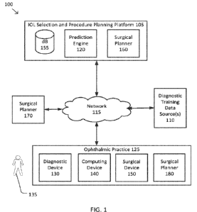
[0025] Figure 1 illustrates a system 100 for eye surgery according
to some embodiments. The
system 100 includes an JUL selection and procedure planning platform 105
(hereinafter "ISP
platform 105") coupled with one or more diagnostic training data sources 110
via a network 115.
In some examples, the network 115 may include one or more switching devices,
routers, local
area networks (e.g., an Ethernet), wide area networks (e.g., the Internet),
and/or the like. Each
of the diagnostic training data sources 110 may be a database, a data
repository, and/or the like
made available by an ophthalmic surgery practice, an eye clinic, a medical
university, an
electronic medical records (EMR) repository, and/or the like. Each of the
diagnostic training
data sources 110 may provide ISP platform 105 with training data in the form
of one or more of
CA 03182976 2022- 12- 15
WO 2022/023882
PCT/1B2021/056563
multi-dimensional images and/or measurements of patients' pre- and post-
operative eyes,
surgical planning data, surgical console parameter logs, surgical complication
logs, patient
medical history, patient demographic data, information on an implanted IOL,
patient preferences
(e.g., ability to drive at night, ability to read without glasses, etc.)
and/or the like. The ISP
platform 105 may store the training data in one or more databases 155 which
may be configured
to anonymize, encrypt, and/or otherwise safeguard the training data.
[0026] The ISP platform 105 includes a prediction engine 120 which
may process the
received training data, perform raw data analysis on the training data, and
train and iteratively
optimize one or more machine learning models (interchangeably referred to as
prediction
models). The trained machine learning models may be used to assist in the
planning and
performance of a surgical procedure (e.g., cataract removal, IOL implantation,
and/or the like).
For example, based on the patient's pre-operative measurements, the prediction
engine 120 may
generate a custom and optimized surgical plan that includes recommended
patterns and/or device
settings for the surgical procedure, and estimated post-operative MRSEs, e.g.,
for each of a given
set of IOL powers. Note that herein, the recommended patterns and device
settings may include
a recommended fragmentation pattern, recommended laser settings, recommended
phacoemulsification settings, recommendations for which slices to treat with
laser energy, how
much laser energy to dedicate to each slice, how much ultrasonic power to
deliver to each area
of each slice (e.g., based on a predicted ultrasonic power required after the
recommended amount
of laser energy is delivered to the particular slice).
[0027] In some examples, the machine learning models (e.g., one or
more neural networks)
are trained at least in part based on pre-operative measurements and
corresponding intra-
operative measurements and/or post-operative outcomes obtained from the one or
more
diagnostic training data sources 110. As an example, eye care professionals
can take efforts to
quantify surgical outcomes. For example, a wide collection of surgical
parameters and pre-,
intra-, and post-operative diagnostic can be gathered for a group of patients
and the patients can
be given a post-operative satisfaction survey. The results of the survey can
be used to train a
computational model to train machine learnings models for optimizing settings,
techniques,
materials for future procedures. Examples of this technique are described in
greater detail in
U.S. Provisional Patent Application No. 63/032195, entitled "SELECTION OF
INTRAOCULAR LENS BASED ON PREDICTED SUBJECTIVE OUTCOME SCORE",
which is incorporated by reference in its entirety.
6
CA 03182976 2022- 12- 15
WO 2022/023882
PCT/1B2021/056563
[0028] The ISP platform 105 is further coupled, via network 115, to
one or more devices of
an ophthalmic practice 125. The one or more devices include one or more
diagnostic devices
130. The one or more diagnostic devices 130 are used to obtain one or more
multi-dimensional
images and/or other measurements of an eye of a patient 135. The one or more
diagnostic devices
130 may be any of a number of devices for obtaining multi-dimensional images
and/or
measurements of ophthalmic anatomy such as an optical coherence tomography
(OCT) device,
a rotating camera (e.g., a Scheimpflug camera), a magnetic resonance imaging
(MRI) device, a
keratometer, an ophthalmometer, an optical biometer, a three-dimensional
stereoscopic digital
microscope (such as NGENUITYCD 3D Visualization System (Alcon Inc.,
Switzerland), any type
of intra-operative optical measurement device, such as an intra-operative
aberrometer, and/or
any other type of optical measurement/imaging device. Examples of OCT devices
are described
in further detail in U.S. Pat. No. 9,618,322 disclosing "Process for Optical
Coherence
Tomography and Apparatus for Optical Coherence Tomography" and U.S. Pat. App.
Pub. No.
2018/0104100 disclosing "Optical Coherence Tomography Cross View Image", both
of which
are hereby incorporated by reference in their entirety. An example of an intra-
operative
aberrometer is OraTM with VerifeyeTm (Alcon Inc., Switzerland), which is
partially described in
more detail in commonly owned U.S. Pat. No. 7,883,505 disclosing "Integrated
Surgical
Microscope and Wavefront Sensor" and U.S. Pat. No. 8,784,443 disclosing "Real-
Time Surgical
Reference Indicium Apparatus and Methods for Astigmatism Correction", both of
which are
hereby incorporated by reference in their entirety.
[0029] The ophthalmic practice 125 may also include one or more
computing devices 140 for
obtaining, from the one or more diagnostic devices 130, the multi-dimensional
images and/or
measurements of patient 135 and sending them to the ISP platform 105. The one
or more
computing devices 140 may be one or more of a stand-alone computer, a tablet
and/or other
smart device, a surgical console, a computing device integrated into the one
or more diagnostic
devices 130, and/or the like.
[0030] The ISP platform 105 may receive data relevant to patient
135 (e.g., measurements,
images, etc.), which is then utilized by the prediction engine 120 to generate
a custom and
optimized surgical plan for the patient, thereby assisting in the planning and
performance of
cataract surgery for the patient. For example, as described above, the
prediction engine 120 may
generate recommended fragmentation patterns and/or device settings for
cataract removal. The
prediction engine 120 may further help the user select an IOL by providing the
user with post-
7
CA 03182976 2022- 12- 15
WO 2022/023882
PCT/1B2021/056563
operative MRSE estimates for different JUL powers. Therefore, by providing the
different types
of outputs described above, the prediction engine 120 helps with improving
post-operative
patient outcomes. In addition, configuring an ophthalmic system, such as
system 100, to
automatically provide recommended fragmentation patterns and/or device
settings as well as,
e.g., targets, and/or feedback to allow a surgeon to perform the surgery based
on the
recommended fragmentation patterns and/or device settings improves the
technical field of
ophthalmic surgery as well as the ophthalmic system itself, which includes
ophthalmic surgical
systems and consoles (e.g., surgical device 150).
[0031] The ophthalmic practice 125 may also include one or more
surgical devices 150 to
perform one or more procedures on an eye, such as cataract removal, JUL
implantation, and/or
the like. The one or more surgical devices 150 may include a laser system for
pre-fragmentation
of a cataract, such as the laser systems described in more detail in commonly
owned U.S. Pat.
No. 9,427,356 disclosing "Photodisruptive Laser Fragmentation of Tissue" and
U.S. Pat. No.
9,622,913 disclosing "Imaging-Controlled Laser Surgical System", both of which
are hereby
incorporated by reference in their entirety. The one or more surgical devices
150 may further
include a phacoemulsification device for using ultrasonics and fluidics to
further fragment and
remove the cataract from the eye, such as the phacoemulsification system
described in more
detail in commonly owned U.S. Pat. No. 8,939,927 disclosing "Systems and
Methods for Small
Bore Aspiration", which is hereby incorporated by reference in its entirety.
The one or more
surgical devices 150 may also refer to surgical consoles that incorporate a
laser system, a
phacoemulsification device, and/or other components for performing additional
ophthalmic
procedures.
[0032] In some examples, the ISP platform 120 provides a custom and
optimized surgical
plan for a patient to the one or more surgical devices 150. The custom and
optimized surgical
plan may include recommendations for a laser fragmentation procedure (e.g.,
further described
in relation to process 215 of FIG. 2) as well as recommendations for a
phacoemulsification
procedure (e.g., further described in relation to process 220 of FIG. 2),
among other
recommendations. Based on the laser fragmentation and phacoemulsification
recommendations,
the one or more surgical devices 150 may be configured to (e.g., automatically
or in response to
surgeon confirmation), provide settings, patterns, targets, and/or feedback
(e.g., auditory, optical,
and/or haptic feedback) during the surgical procedure.
8
CA 03182976 2022- 12- 15
WO 2022/023882
PCT/1B2021/056563
[0033] As an example, these laser fragmentation and
phacoemulsification recommendations
may include recommended devices settings to be used during each procedure. In
certain
embodiments, having received the recommended device settings from the ISP
platform 150, a
surgical device 150 may reconfigure itself after a user, e.g., surgeon,
confirms the recommended
device settings. In another example, having received the recommended device
settings from the
ISP platfoi _____ la 150, in certain embodiments, a surgical device 150 may
automatically reconfigure
itself based on the recommended device settings. Having configured the
surgical device 150
with the recommended device settings, the surgical device 150 may be
subsequently operated
with the recommended device settings by the surgeon to perform a surgery on
the corresponding
patient.
[0034] In addition, in certain embodiments, the surgical device 150
may further provide
targets and/or feedback to help the surgeon with following the laser
fragmentation and
phacoemulsification recommendations (e.g., laser fragmentation patterns, etc.)
or help ensure
that the surgeon's uses of the surgical device 150 is aligned with the laser
fragmentation and
phacoemulsification recommendations. For example, the surgical device 150 may
use visual
indicators on a display of the surgical device 150 (or a connected display,
e.g., computing devices
140) to help ensure the surgeon follows the recommended laser fragmentation
lines. In another
example, feedback may be used to help ensure the surgeon does not apply more
laser power than
necessary or apply the recommended laser power for longer than necessary.
[0035] In some examples, intra-operative data may be collected from
surgical devices 150,
diagnostic devices 130, etc., and include tracked and/or recorded intra-
operative settings,
parameters, metrics and/or the like of the one or more surgical devices 150
during the surgical
procedure, images and measurements associated with the eye during the
procedure, etc.. In
certain embodiments, the intra-operative data that is collected over the
course of the cataract
surgery may include or be derived from a surgical video captured during the
surgery as well as
device log files that capture various sensor I/0 parameters from the equipment
(e.g., surgical
device 150, or any consoles involved) during the surgical procedure. A
surgical video can be
captured by imaging and camera devices associated with the equipment (e.g.,
surgical device
150, or any consoles involved) and analyzed using computer vision algorithms
and techniques.
[0036] The tracked and/or recorded intra-operative settings,
patterns, and/or metrics may then
be used in multiple ways. For example, the tracked and/or recorded intra-
operative settings,
patterns, and/or metrics may be used, in real-time, as input into one or more
trained models (e.g.,
9
CA 03182976 2022- 12- 15
WO 2022/023882
PCT/1B2021/056563
fifth one or more models described below in relation to processes 240-245 of
FIG. 2) to provide
adjusted laser fragmentation and phacoemulsification recommendations. In such
an example,
by monitoring the real-time conditions relating to the patient' s eye during
the surgical procedure,
intra-operative data can be generated and used to provide dynamic updates to
laser fragmentation
and phacoemulsification recommendations.
[0037]
The tracked and/or recorded intra-operative settings, patterns, and/or
metrics may be
also sent to the ISP platform 105 for use in iteratively training and/or
updating the machine
learning models (e.g., first and second sets of models described in relation
to processes 215 and
220) used by prediction engine 120 so as to incorporate the information from
the surgical
procedure performed on patient 135 for use in the planning of future surgical
procedures. In
some cases, the tracked and/or recorded intra-operative settings, patterns,
and/or metrics are
stored as unstructured or structured data in an ERM database, a cloud-based
storage repository,
etc.
[0038]
Example settings, parameters, metrics that may be recorded for each
patient (e.g.,
intraoperatively) include laser fragmentation parameters and metrics (e g ,
position and
orientation of laser fragmentation lines, distance between laser fragmentation
lines (which may
be variable), separation distance between laser treatment spots along laser
fragmentation lines,
use of curved lines (e.g., to trace density contours), use of spiral or other
patterns, depth of cuts
along each laser fragmentation line, angle of incidence for each fragmentation
line (e.g., relative
to central axis 480), total time under suction, or other parameters that may
be indicative of the
features of the laser fragmentation pattern. Example settings, parameters,
metrics that may be
recorded for each patient may also include laser device settings (e.g.,
frequency of laser, power
level of laser, speed of laser along the laser fragmentation lines, type of
laser). Example settings,
parameters, metrics that may be recorded for each patient may also include
total length of laser
cuts (e.g., a total length of the laser fragmentation pattern lines, a total
length of time for laser
fragmentation, a total laser energy expended, and/or the like).
[0039]
Example settings, parameters, metrics that may be recorded for each
patient (e.g.,
intraoperatively) may also include phacoemulsification related parameters and
metrics (e.g.,
location of one or more targets within the cataract and/or the lens of the eye
where ultrasonic
cutting and/or fragmentation energy and/or emulsification are perfat
________________ lied, total length of time for
phacoemulsification, a total ultrasonic energy, a total volume of applied
fluid, total number of
laser spots, total ultrasonic energy expended for phacoemulsification, amount
of time spent to
CA 03182976 2022- 12- 15
WO 2022/023882
PCT/1B2021/056563
aspirate the lens, amount of fluid used for aspiration etc.). Example
settings, parameters, metrics
that may be recorded for each patient may also include phacoemulsification
devise settings (e.g.,
frequency of ultrasonics, power level of ultrasonics, duration of application
of ultrasonics, rate
and/or volume of fluid to apply, pressure of applied fluid).
[0040] The one or more diagnostic devices 130 may further be used
to obtain post-operative
measurements of patient 135 after the patient undergoes cataract removal and
IOL implantation
using the selected JUL. The one or more computing devices 140 may then send
the post-operative
multi-dimensional images and/or measurements of patient 135 and the selected
JUL to the ISP
platform 105 for use in iteratively training and/or updating the models used
by prediction engine
120 so as to incorporate post-operative information associated with patient
135 for use with
future patients, as explained in more detail below.
[0041] The recommendations provided by a surgical plan may be
displayed on one or more
computing devices 140 and/or another computing device, display, surgical
console, and/or the
like. Additionally, the ISP platform 105 and/or the one or more computing
devices 140 may
identify in the measurements various characteristics of the anatomy of patient
135, as explained
below in more detail. Further, the ISP platform 105 and/or the one or more
computing devices
140 may create graphical elements that identify, highlight, and/or otherwise
depict the patient
anatomy, the procedure plan, and/or the measured characteristics for display
to the surgeon or
other user to further aid in the surgical planning process. The ISP platform
105 and/or the one or
more computing devices 140 may supplement the measurements with the graphical
elements.
[0042] In some embodiments, the ISP platform 105 may further
include a surgical planner
160 that creates and provides an optimized surgical plan to ophthalmic
practice 125 that uses the
recommended patterns and settings for the one or more surgical devices 150
and/or the estimated
post-operative MRSEs. In some embodiments, system 100 may further include a
stand-alone
surgical planner 170 and/or ophthalmic practice 125 may further include a
surgical planner
module 180 on the one or more computing devices 140 as is described in further
detail below.
[0043] As discussed above and further emphasized here, Figure 1 is
merely an example which
should not unduly limit the scope of the claims. One of ordinary skill in the
art would recognize
many variations, alternatives, and modifications. According to some
embodiments, the ISP
platform 105 and/or one or more components thereof, such as databases 155,
prediction engine
120, and/or surgical planner 160, may be integrated into the one or more
devices of ophthalmic
11
CA 03182976 2022- 12- 15
WO 2022/023882
PCT/1B2021/056563
practice 125. In some examples, one or more computing devices 140 may host the
ISP platform
105, databases 155, prediction engine 120, and/or surgical planner 160. In
some examples,
surgical planner 160 may be combined with surgical planner 180.
[0044] Note that the collection of the ISP platform 105, at least
one of the one or more
diagnostic devices 130, at least one of the one or more computing devices 140,
at least one of
the one or more surgical devices 150 may be referred to as a surgical
ophthalmic system that
works to implement one or more of the embodiments described herein.
[0045] Figures 2A-2B show a diagram of a method 200 of removing a
cataract according to
some embodiments. One or more of the processes 205-265 of method 200 may be
implemented,
at least in part, in the form of executable code stored on non-transitory,
tangible, machine-
readable media that when run by one or more processors (e.g., the processors
of prediction engine
120, the ISP platform 105, the one or more diagnostic devices 130, the one or
more computing
devices 140, the one or more surgical devices 150, and/or one or more of the
surgical planners
160, 170, and/or 180) may cause the one or more processors to perform one or
more of the
processes 205-265. According to some embodiments, the process 240 may be
performed
concurrently with the process 235. According to some embodiments, the process
215 may be
performed before the process 210 and/or concurrently with the process 210.
Furthermore, the
sequence diagram 200 is not required to perform each of or only the shown
steps and is not
limited to performing the indicated steps in any particular order.
[0046] At a process 205, pre-operative information for a patient is
obtained. According to
some embodiments, the pre-operative information for the patient may include
information about
the patient, the eye from which a cataract is to be removed, the cataract,
and/or the like. For
example, in certain embodiments, pre-operative information includes one or
more pre-operative
images (also referred to as imaging data) and/or one or more pre-operative
measurements of an
eye. In some examples, the one or more pre-operative images may be extracted
from one or more
pre-operative images of the eye obtained using a diagnostic device, such as
one or more
diagnostic devices 130 (e.g., an OCT device, a rotating (e.g., Scheimpflug)
camera, an MRI
device, a three-dimensional stereoscopic digital microscope (such as NGENUITY
3D
Visualization System (Alcon Inc., Switzerland) and/or the like). In some
examples, the one or
more pre-operative images may be previously obtained and retrieved from a
database (e.g.,
database 155), storage maintained by ISP platform 105 and/or ophthalmic
practice 125, and/or
the like.
12
CA 03182976 2022- 12- 15
WO 2022/023882
PCT/1B2021/056563
[0047] In certain embodiments, one or more pre-operative
measurements of the eye may be
determined from the one or more pre-operative images. In certain embodiments,
one or more of
the pre-operative measurements may be determined using one or more measuring
devices, such
as one or more diagnostic devices 130. The pre-operative measurements of the
eye are described
herein by reference to Figure 3, which is a diagram of an eye 300, according
to some
embodiments. As shown in Figure 3, eye 300 includes a cornea 310, an anterior
chamber 320,
and a lens 330.
[0048] In some embodiments, one measurement of interest for eye 300
is the white-to-white
diameter of cornea 310. In some examples, the white-to-white diameter of
cornea 310 may he
measured using an optical biometer. In some examples, the white-to-white
diameter of cornea
310 may be determined by analyzing the one or more pre-operative images of eye
300. In some
examples, the one or more pre-operative images may be analyzed to identify
nasal and temporal
angles 340 and 350, respectively, of anterior chamber 320. In some examples,
nasal and temporal
angles 340 and 350 of anterior chamber 320 may be determined from the one or
more pre-
operative images by (1) identifying structures that are indicative of anterior
chamber 320 (e.g.,
using one or more edge detection and/or region detection algorithms) and (2)
noting the acute
angles at the edges of anterior chamber 320 located toward the temporal and
nasal extents of
anterior chamber 320. Once identified, a distance between the nasal and
temporal angles 340 and
350 may be measured to determine the white-to-white diameter of cornea 310,
which
corresponds to a length of line 360 between nasal and temporal angles 340 and
350.
[0049] In some embodiments, one measurement of interest for eye 300
is the average
keratometry or roundness of the anterior surface of cornea 310. In some
examples, the average
keratometry of cornea 310 may be measured using the one or more pre-operative
images of eye
300, a keratometer, and/or the like. In some examples, the average keratometry
of cornea 310
may be based on an average of the steep keratometry and the shallow
keratometry measurements
of cornea 310. In some examples, the average keratometry of cornea 310 may be
expressed as a
radius of curvature (rc) of cornea 310, which is 437.5 divided by the average
keratometry.
[0050] In some embodiments, one measurement of interest from eye
300 is the axial length
370 of eye 300 as measured from the anterior surface of cornea 310 to the
retina along central
axis 380 of eye 300. In some examples, axial length 370 may be determined
using the one or
more images of eye 300, biometry of the eye, and/or the like.
13
CA 03182976 2022- 12- 15
WO 2022/023882
PCT/1B2021/056563
[0051] In certain embodiments, in addition to the one or more pre-
operative images of the
patient's eye, the patient history may also be obtained as part of the pre-
operative information.
In some examples, the patient history may include one or more relevant
physiological
measurements for the patient that are not directly related to the eye, such as
one or more of age,
height, weight, body mass index, genetic makeup, race, ethnicity, sex, blood
pressure, other
demographic and health related information, and/or the like. In some examples,
the patient
history may further include one or more relevant risk factors including
smoking history, diabetes,
heart disease, other underlying conditions, prior surgeries, and/or the like
and/or a family history
for one or more of these risk factors.
[0052] At process 210, a lens density map for the eye is determined
based on the pre-operative
information for the patient (e.g., the one or more pre-operative images). In
some examples, the
intensity of each pixel and/or voxel from the one or more images may be used
to determine the
density of a corresponding portion of the lens of the eye captured by the one
or more images.
Examples of these techniques are described in further detail in commonly owned
U.S. Pat. No.
10,314,747 disclosing "Adjusting Laser Energy in Accordance with Optical
Density and U.S.
Pat. No. 10,433,722 disclosing "Diagnosis System and Diagnosis Method", which
are hereby
incorporated by reference in their entireties. In certain embodiments, a type
of the cataract (e.g.,
nucleolus, posterior, anterior cataract) is determined based on the lens
density map.
[0053] At process 215, one or more recommendations for a laser
fragmentation procedure are
prepared. In certain embodiments, one or more recommendations for a laser
fragmentation
procedure are prepared based on the pre-operative information obtained at
process 205 and/or
the lens density map (including information about the type of cataract)
determined at process
210. According to some embodiments, the one or more recommendations may
include
recommendations for a laser fragmentation pattern to be traced by a laser,
such as a femto-second
laser, across the cataract and/or the lens of the eye. A laser fragmentation
pattern refers to the
pattern of laser fragmentation lines to be traced by the laser. In some
examples, the
recommendations for the laser fragmentation pattern may include one or more
of:
o Position and orientation of laser fragmentation lines
= E.g., horizontal, vertical, angle
o Distance between laser fragmentation lines (which may be variable)
o Separation distance between laser treatment spots along laser
fragmentation lines
o Use of curved lines (e.g., to trace density contours)
14
CA 03182976 2022- 12- 15
WO 2022/023882
PCT/1B2021/056563
o Use of spiral or other patterns
o Depth of cuts along each fragmentation line
o Angle of incidence for each fragmentation line (e.g., relative to central
axis 480)
o Other parameters associated with one or more features of the laser
fragmentation
pattern
[0054] According to some embodiments, the one or more recommendations may
include one
or more device settings for the laser device at one or more control points
along the laser
fragmentation lines. In some examples, the settings may include one or more
of:
o Frequency of laser
o Power level of laser
o Speed of laser along the laser fragmentation lines
o Type of laser
[0055] According to some embodiments, the one or more recommendations may
include one
or more estimates for the laser fragmentation procedure. In some examples, the
one or more
estimates may include one or more of a total length of laser cuts (e.g., a
total length of the laser
fragmentation pattern, a total length of time for laser fragmentation, a total
laser energy, and/or
the like).
[0056] In some examples, a first one or more models, such as one or
more of the machine
learning models of prediction engine 120, may be used to determine the
patterns of the
fragmentation lines, the one or more settings, and/or the one or more
estimates based on the lens
density map and/or combinations of any of the pre-operative information. In
some examples,
various learning algorithms may be used to train the first one or more models
using the training
data associated with previous patients, as provided by the diagnostic training
data sources 110
and described above. For example, supervised, unsupervised, or other types of
machine learning
algorithm may be used to train the first one or more models. In some examples,
the first one or
more models may each include a neural network (e.g., recurrent neural network)
trained using
the training data.
[0057] In certain embodiments, the first one or more models may be
trained to determine
fragmentation line patterns, settings, and/or estimates that maximize a post-
operative survey
score indicative of the post-operative surgical outcome. In order to maximize
the post-operative
survey score the first one or more models may optimize and be trained on
features such as time
under suction, total laser energy, the number of laser spots, the total length
of laser pattern lines,
CA 03182976 2022- 12- 15
WO 2022/023882
PCT/1B2021/056563
the time required for phacoemulsification, the total ultrasonic energy
required for
phacoemulsification, time required to aspirate the lens, the amount of fluid
required for
aspiration, etc.
[0058] At a process 220, one or more recommendations for a
phacoemulsification procedure
are prepared. In certain embodiments, one or more recommendations for a
phacoemulsification
procedure are prepared based on the lens density map (including information
about the type of
cataract) and/or combinations of any of the pre-operative information, and/or
the
recommendations for the laser procedure provided at process 215. According to
some
embodiments, the one or more recommendations may include one or more
recommendations for
a location of one or more targets within the cataract and/or the lens of the
eye where ultrasonic
cutting and/or fragmentation energy and/or emulsification fluid should be
applied.
[0059] According to some embodiments, the one or more recommendations, may
include one
or more settings for the phacoemulsification device at each of the targets. In
some examples, the
one or more settings may include one or more of:
o Frequency of ultrasonics
o Power level of ultrasonics
o Duration of application of ultrasonics
o Rate and/or volume of fluid to apply
o Pressure of applied fluid
[0060] According to some embodiments, the one or more
recommendations may include one
or more estimates for the phacoemulsification procedure. In some examples, the
one or more
estimates may include one or more of a total length of time for
phacoemulsification, a total
ultrasonic energy, a total volume of applied fluid, and/or the like.
[0061] In some examples, a second one or more models, such as one
or more of the machine
learning models of prediction engine 120, may be used to determine the targets
for
phacoemulsification, the one or more settings, and/or the one or more
estimates based on the
lens density map and/or combinations of any of the pre-operative information,
and/or the
recommendations for the laser procedure provided at process 215. In some
examples, various
learning algorithms may be used to train the second one or more models using
the training data
associated with previous patients, as provided by the diagnostic training data
sources 110 and
described above. For example, supervised, unsupervised, or other types of
machine learning
16
CA 03182976 2022- 12- 15
WO 2022/023882
PCT/1B2021/056563
algorithm may be used to train the second one or more models. In some
examples, the second
one or more models may each include a neural network (e.g., recurrent neural
network) trained
using the training data.
[0062] In certain embodiments, the second one or more models may be
trained to determine
phacoemulsification targets, settings, and/or estimates that maximize a post-
operative survey
score indicative of the post-operative surgical outcome. In order to maximize
the post-operative
survey score the first one or more models may optimize and be trained on
features such as time
under suction, total laser energy, the number of laser spots, the total length
of laser pattern lines,
the time required for phacoemulsification, the total ultrasonic energy
required for
phacoemulsification, time required to aspirate the lens, the amount of fluid
required for
aspiration, etc.
[0063] At a process 225, a cataract removal procedure is planned.
In some examples, the
recommendations from processes 215 and/or 220 and/or the pre-operative
information obtained
during process 205 may be provided to a surgical planner, such as, one or more
of surgical
planners 160, 170, and/or 180. In some examples, the surgical planner may
include a user
interface that displays a surgical plan to the surgeon that incudes the
recommendations from
processes 210 and/or 215 and/or the pre-operative information. For example,
the surgical
planner may display the laser fragmentation pattern lines and/or the targets
determined during
processes 210 and 215, respectively, superimposed on one or more images of the
eye and/or the
cataract (e.g., as obtained during process 205). In some examples, the user
interface may further
display any of the settings and/or the estimates generated during processes
210 and/or 215. In
some examples, the settings may be displayed when the user mouses over and/or
clicks on any
of the laser fragmentation lines and/or targets. In some examples, the user
interface may allow
the user to reposition any of the laser fragmentation lines and/or targets
and/or change any of the
settings.
[0064] In some examples, the surgical planner may re-determine any of the
recommendations, settings, and estimates based on the changes to the laser-
fragmentations lines,
targets, and/or settings, such as by repeating portions of processes 215
and/or 220. In certain
embodiments, a third one or more models, such as one or more of the machine
learning models
of prediction engine 120, may be used to re-determine recommendations,
settings, and estimates
based on changes to the laser-fragmentations lines, targets, and/or settings.
In other words, the
third one or more models may be trained to take, as input, the changed laser-
fragmentations lines,
17
CA 03182976 2022- 12- 15
WO 2022/023882
PCT/1B2021/056563
targets, and/or settings, and output laser-fragmentations lines, targets,
and/or settings based on
the input.
[0065] As explained above, a surgeon may have a preferred pattern
for phacoemulsification
and aspiration of lens material. For example, a surgeon may have been trained
to complete
phacoemulsification and aspiration of lens material in a certain repeatable
pie-slice pattern. The
surgeon can be accustomed to emulsifying and removing lens material from a
first pie slice and
rotating around the lens to subsequent slices to ensure that each area is
adequately emulsified
and aspirated. Accordingly, one or more of surgical planners 160, 170, and/or
180 can include
options for a surgeon or other eye care professional to select a pre-generated
or custom made
phacoemulsification pattern and a recommended fragmentation pattern that
conform with a
surgeon's routine, repeatable pattern (e.g., pie-slice pattern).
[0066] In addition to a recommended fragmentation pattern, the
present technology can
include a recommendation for which slices to treat with laser energy, how much
laser energy to
dedicate to each slice, how much ultrasonic power to deliver to each area of
each slice (e.g.,
based on a predicted ultrasonic power required after the recommended amount of
laser energy is
delivered to the particular slice), etc. For example, in some cases, a desired
phacoemulsification
pattern (e.g., pie-slices) can be designated as well as a total laser energy
for pre-conditioning the
lens. The total laser energy can be selected by the surgeon or other care
professional or can be
a recommended value based on the historical data processed by the prediction
engine 120. For
example, the prediction engine 120 can recommend a total laser energy based on
a threshold
reduction in gas bubble creation and/or a quantified elimination of negative
surgical outcome
due to gas bubble creation. One or more of surgical planners 160, 170, and/or
180 can use the
specified fragmentation pattern, the selected and/or recommended total laser
energy, and/or the
lens density map to recommend how much laser energy should be applied to the
various regions
to the cataract to optimize the efficiency of the laser energy in order to pre-
condition the most
needed areas of the lens for optimal phacoemulsification and aspiration.
[0067] In some other cases (sometimes in the absence of a preferred
phacoemulsification and
aspiration pattern), an optimized surgical plan can recommend a custom
fragmentation pattern
and device settings based on the lens density map and on various surgical
optimization criteria
(e.g. reduction of time under suction, reduction of total laser energy, etc.)
that are selected by a
surgeon and/or recommended by the prediction engine 120, as described above.
The one or
more surgical planners 160, 170, and/or 180 can recommend using the custom
fragmentation
18
CA 03182976 2022- 12- 15
WO 2022/023882
PCT/1B2021/056563
pattern and optimization criteria in order to pre-condition the most needed
areas of the lens for
optimal phaccemul s ific a tion and aspiration (even in the absence of
preferred
phacoemulsification and aspiration pattern).
[0068] At a process 230, a post-operative MRSE is estimated for,
e.g., each of a given set of
JUL powers based on the pre-operative information obtained during process 205.
Note that
process 250 may be peiformed before or after process 210. In certain
embodiments, a post-
operative MRSE may be estimated for each of a plurality of JUL powers that are
available on
the market based on the patient's pre-operative measurements and/or images
(including one or
more of the patient's axial length of the eye, corneal curvature, anterior
chamber depth, while-
to-white diameter of the cornea, lens thickness, an effective lens position
(which itself is
calculated based on one or more of these pre-operative measurements), etc.).
In such
embodiments, the surgeon may able to see which one of the JUL powers is
estimated to result in
a post-operative MRSE that is closest to a desired refractive outcome. In
certain other
embodiments, a post-operative MRSE may be estimated for a specific IOL power
that has been
selected by the surgeon. In such embodiments, if the estimated post-operative
MRSE is close to
a desired refractive outcome, the surgeon may determine that the selected JUL
power is likely
going to result in a satisfactory refractive outcome for the patient.
[0069] Examples of how to use a given JUL power in the estimation
of post-operative MRSE
are described in further detail in commonly-owned U.S. Pat. App. No.
16/171,515 filed October
26, 2018 entitled "Systems and Methods for Intraocular Lens Selection,- U.S.
Serial No.
16/746.231, filed January 17, 2020 entitled "Systems and Methods for
Intraocular Lens Selection
Using Emmetropia Zone Prediction," and U.S. Pat. App. No. 16/239,771 filed
January 4, 2019
entitled "Systems and Methods for Intraocular Lens Selection," all of which
are hereby
incorporated by reference in their entirety. The post-operative MRSE is
indicated in diopters (D).
In some examples, a fourth one or more models, such as one or more of the
models of prediction
engine 120, may be used to estimate a post-operative MRSE for, e.g., each of a
given set of IOL
powers, for a certain patient. In certain embodiments, the fourth one or more
models may be
trained based on historical patients' pre-operative information (e.g., pre-
operative images and/or
measurements, patient history, etc.) and post-operative outcomes. For
instance, depending on
the type of IOL power calculations, example pre-operative measurements used
for training the
fourth one or more models may include one or more of the patient's axial
length of the eye,
corneal curvature, anterior chamber depth, white-to-white diameter of the
cornea, lens thickness.
19
CA 03182976 2022- 12- 15
WO 2022/023882
PCT/1B2021/056563
an effective lens position (which itself is calculated based on one or more of
these pre-operative
measurements), etc. In some examples, various learning algorithms may be used
to train the
fourth one or more models using the training data associated with previous
patients, as provided
by the diagnostic training data sources 110 and described above. For example,
supervised,
unsupervised, or other types of machine learning algorithm may be used to
train the fourth one
or more models. In some examples, the fourth one or more models may each
include a neural
network (e.g., recurrent neural network) trained using the data from the eyes
and/or cataract
removals of previous patients.
[0070] At a process 235, the cataract removal procedure is
performed. In some examples, the
cataract removal procedure is performed according to the optimized surgical
plan provided at
process 220. In some examples, a laser may be used to trace the fragmentation
pattern using the
corresponding one or more settings recommended by process 215. In some
examples, when the
laser fragmentation is guided by the surgeon, the surgical plan may be used to
provide auditory,
visual, and/or haptic feedback to help the surgeon guide the laser, as
described above. Examples
of lasers and laser systems are described in more detail in commonly owned
U.S. Pat. No. 9,427,
356 disclosing "Photodisruptive Laser Fragmentation of Tissue" and U.S. Pat.
No. 9,622,913
disclosing "Imaging-Controlled Laser Surgical System", both of which are
hereby incorporated
by reference in their entirety. In some examples, a phacoemulsification device
may be used to
apply ultrasonic energy to the targets and then use applied fluids to remove
the fragmented pieces
of the cataract and/or lens. In some examples, when the phacoemulsification is
guided by the
surgeon, the surgical plan may be used to provide auditory, visual, and/or
haptic feedback to help
the surgeon guide the phacoemulsification device.
[0071] At an optional process 240, intra-operative data is
collected. In certain embodiments,
intra-operative data refers to settings, parameters, and/or metrics used for
the cataract removal
procedure. Process 240 may be performed concurrently with process 235 such
that as the
cataract removal procedure is being performed during process 235, one or more
settings,
parameters, and metrics are tracked and recorded.
[0072] In certain embodiments, intra-operative data that is
collected over the course of the
cataract surgery may include or be derived from a surgical video captured
during the surgery as
well as device log files that capture various sensor input/output parameters
from the equipment
(e.g., surgical device 150, or any consoles involved) during the surgical
procedure. A surgical
video can be captured by imaging and camera devices associated with the
equipment (e.g.,
CA 03182976 2022- 12- 15
WO 2022/023882
PCT/1B2021/056563
surgical device 150, or any consoles involved) and analyzed using computer
vision algorithms
and techniques. The intra-operative data collected may include any data point
or metric relating
to inputs and outputs of the models described herein. For example, intra-
operative data may
include time-stamped eye-related information, such as changes to any aspect
(e.g., tissues, lens,
other components, etc.) of the eye as the procedure is being performed, time-
stamped settings,
parameters, metrics collected during the procedure (e.g., laser fragmentation
and/or
phacoemulsification).
[0073] Example settings, parameters, metrics that may be recorded
for each patient include
laser fragmentation parameters and metrics (e.g., position and orientation of
laser fragmentation
lines, distance between laser fragmentation lines (which may be variable),
separation distance
between laser treatment spots along laser fragmentation lines, use of curved
lines (e.g., to trace
density contours), use of spiral or other patterns, depth of cuts along each
laser fragmentation
line, angle of incidence for each fragmentation line (e.g., relative to
central axis 480), total time
under suction, or other parameters that may be indicative of the features of
the laser
fragmentation pattern. Example settings, parameters, metrics that may be
recorded for each
patient may also include laser device settings (e.g., frequency of laser,
power level of laser, speed
of laser along the laser fragmentation lines, type of laser). Example
settings, parameters, metrics
that may be recorded for each patient may also include total length of laser
cuts (e.g., a total
length of the laser fragmentation pattern lines, a total length of time for
laser fragmentation, a
total laser energy expended, and/or the like).
[0074] Example settings, parameters, metrics that may be recorded
for each patient may also
include phacoemulsification related parameters and metrics (e.g., location of
one or more targets
within the cataract and/or the lens of the eye where ultrasonic cutting and/or
fragmentation
energy and/or emulsification are performed, total length of time for
phacoemulsification, a total
ultrasonic energy, a total volume of applied fluid, total number of laser
spots, total ultrasonic
energy expended for phacoemulsification, amount of time spent to aspirate the
lens, amount of
fluid used for aspiration etc.). Example settings, parameters, metrics that
may be recorded for
each patient may also include phacoemulsification devise settings (e.g.,
frequency of ultrasonics,
power level of ultrasonics, duration of application of ultrasonics, rate
and/or volume of fluid to
apply, pressure of applied fluid).
[0075] In certain embodiments, the intra-operative data may include
one or more intra-
operative images and/or measurements. The one or more intra-operative images
and/or
21
CA 03182976 2022- 12- 15
WO 2022/023882
PCT/IB2021/056563
measurements may include images and/or measurements of the eye as the
procedure is being
performed, prior to the lens being completely removed. The one or more intra-
operative images
and/or measurements may also include intra-operative images and/or
measurements of an
aphakic eye. For example, an intra-operative optical measurement device 130
(e.g., the OraTM
with VerifeyeTM (Alcon Inc., Switzerland) is used to provide intra-operative
measurements of
the eye, including one or more of the curvature of the cornea, axial length of
the eye, white-to-
white diameter of the cornea, etc.
[0076]
At an optional process 245, the laser fragmentation procedure
recommendations
and/or the ph acoem ul si fi cati on procedure recommendations are adjusted
based on the in tra-
operative data collected at optional process 240. Process 245 may be performed
concurrently
with process 235 and 240. For example, the intra-operative data may be
provided as input into
a fifth one or more models to provide adjusted recommendations.
Adjusting the
recommendations provided by processes 215 and 220 may be advantageous because
the
collected intra-operative data may make such recommendations sub-optimal. For
example, in
certain cases, intra-operative images associated with the eye may provide data
points that were
unknown pre-operatively and or not entirely accurate. In addition, the
recommendations
provided by processes 215 and 220 may impact the patient's eye in ways that
were not
anticipated. Also, a surgeon may not fully follow some of the recommendations
provided by
processes 215 and 220, causing the rest of the recommendations provided by
processes 215 and
220 sub-optimal or useless. Therefore, the fifth one or more models may
continuously and
periodically take the time-stamped intra-operative data as input during the
procedure and provide
adjusted or updated laser fragmentation procedure recommendations and/or the
phacoemulsification procedure recommendations.
[0077]
The fifth one or more models may include one or more reinforcement
leaning models.
Reinforcement learning (RL) is an area of machine learning concerned with
designing intelligent
agents that are responsive to changes in a real-world situation and can take
actions in order to
maximize the notion of a cumulative reward. An intelligent agent includes (A)
a policy and (B)
an algorithm (e.g., reinforcement learning algorithm) for updating the policy.
The policy is a
model (e.g., sometimes a deep neural network or a simpler supervised learning
model) that
decides what action to take (i.e., what recommendations to provide in terms of
parameters,
settings, and metrics used during the procedure) given a set of state
observations (i.e., the state
of the environment as it pertains to the eye and the surgical devices being
used). In other words,
22
CA 03182976 2022- 12- 15
WO 2022/023882
PCT/1B2021/056563
the policy is the brain of the agent that takes in state observations and maps
them to actions. The
RL algorithm updates the policy, as the policy may not be mapped correctly to
take the best
action or the environment (e.g., defined by all the data points derived from
the intra-operative
data discussed above) may change, making the mapping not optimal. The RL
algorithm changes
the policy based on the actions that were taken, the observations from the
environment, and the
amount of reward collected, as determined by a reward function, described
below. Using the RL
algorithm, the agent, therefore, modifies its policy as it interacts with the
environment so that,
eventually, given any state, it will always take the most advantageous action
that corresponds to
the most reward in the long run.
[0078] At a process 250, a post-operative MRSE is estimated for,
e.g., each of a given set of
JUL powers based on the intra-operative information obtained during process
245. In certain
embodiments, a post-operative MRSE may be estimated for each of a plurality of
JUL powers
that are, for example, available on the market. In such embodiments, the
surgeon may able to
see which one of the JUL powers is estimated to result in a post-operative
MRSE that is closest
to a desired refractive outcome. In certain other embodiments, a post-
operative MRSE may be
estimated for a specific JUL power that has been selected by the surgeon. In
such embodiments,
if the estimated post-operative MRSE is close to a desired refractive outcome,
the surgeon may
determine that the selected JUL power is likely going to result in a
satisfactory refractive outcome
for the patient.
[0079] In certain embodiments, one or more post-operative MRSEs are
intra-operatively
estimated for the patient based the patient's aphakic measurements including
on one or more of
the axial length, corneal curvature, anterior chamber depth, white-to-white
diameter of the
cornea, lens thickness, an effective lens position. In some examples, the one
or more post-
operative MRSEs calculated based on the patient's intra-operative measurements
at process 250
may be different than the one or more post-operative MRSEs calculated based on
the patient's
pre-operative measurements at process 230. In such examples, the surgeon may
select an IOL
power based on the one or more post-operative MRSEs calculated using the
patient's intra-
operative measurements and ignore a previously selected IOL power. Performing
intra-operative
measurements using a device, such as the UraTM with VerifeyeTM (Alcon Inc.,
Switzerland), is
therefore advantageous for ensuring that an optimal IOL power is used,
resulting in a satisfactory
refractive outcome.
23
CA 03182976 2022- 12- 15
WO 2022/023882
PCT/1B2021/056563
[0080] In certain embodiments, the sixth one or more models may be
trained based on
historical patients' intra-operative information and post-operative outcomes.
In some examples,
various learning algorithms may be used to train the sixth one or more models
using the training
data associated with previous patients, as provided by the diagnostic training
data sources 110
and described above. For example, supervised, unsupervised, or other types of
machine learning
algorithm may be used to train the sixth one or more models. In some examples,
the sixth one
or more models may each include a neural network trained (e.g., recurrent
neural network) using
the data from the eyes and/or cataract removals of previous patients.
[0081] At process 255, a lens implantation procedure is performed
for implanting an TOL
with the selected IOL power to replace the fragmented and removed lens.
[0082] At an optional process 260, one or more post-operative
measurements of the eye are
obtained and/or a post-operative satisfaction score is recorded. In some
examples, the one or
more post-operative measurements may include an actual post-operative MRSE
after
implantation of the IOL during process 255 and/or the like. In some examples,
the actual post-
operative MRSE may be determined based on one or more images of the post-
operative eye, one
or more physiological and/or optical measurements of the post-operative eye,
and/or the like.
[0083] At a process 265, the first, second, third, fourth, fifth
and/or sixth sets of models used
by method 200 are updated. In some examples, the pre-operative information
determined during
process 205, the lens density map determined at process 210, the settings,
parameters, and
metrics recorded during process 240, the one or more intra-operative
measurements obtained
during process 245, the one or more post-operative measurements obtained
during process 260,
and/or the like may be used as additional training data for any of the first,
second third, fourth,
fifth, and/or sixth sets of models. In some examples, the additional training
data may be added
to a data source, such as data source 110. In some examples, the updating may
include one or
more of updating least-squares fits, feedback to neural networks (e.g., using
back propagation),
and/or the like.
[0084] Figures 4A and 4B are diagrams of processing systems
according to some
embodiments. Although two embodiments are shown in Figures 4A and 4B, persons
of ordinary
skill in the art will also readily appreciate that other system embodiments
are possible. According
to some embodiments, the processing systems of Figures 4A and/or 4B are
representative of
computing systems that may be included in one or more of IOL selection and
procedure planning
24
CA 03182976 2022- 12- 15
WO 2022/023882
PCT/1B2021/056563
platform 105, ophthalmic practice 125, prediction engine 120, one or more
diagnostic devices
130, the one or more computing devices 140, any of surgical planner 160, 170,
and/or 180, and/or
the like.
[0085] Figure 4A illustrates a computing system 400 where the
components of system 400
are in electrical communication with each other using a bus 405. System 400
includes a processor
410 and a system bus 405 that couples various system components including
memory in the form
of a read only memory (ROM) 420, a random access memory (RAM) 425, and/or the
like (e.g.,
PROM, EPROM, FLASH-EPROM, and/or any other memory chip or cartridge) to
processor
410. System 400 may further include a cache 412 of high-speed memory connected
directly with,
in close proximity to, or integrated as part of processor 410. System 400 may
access data stored
in ROM 420, RAM 425, and/or one or more storage devices 430 through cache 412
for high-
speed access by processor 410. In some examples, cache 412 may provide a
performance boost
that avoids delays by processor 410 in accessing data from memory 415, ROM
420, RAM 425,
and/or the one or more storage devices 430 previously stored in cache 412. In
some examples,
the one or more storage devices 430 store one or more software modules (e.g.,
software modules
432, 434, 436, and/or the like). Software modules 432, 434, and/or 436 may
control and/or be
configured to control processor 410 to perform various actions, such as the
processes of methods
200 and/or 300. And although system 400 is shown with only one processor 410,
it is understood
that processor 410 may be representative of one or more central processing
units (CPUs), multi-
core processors, microprocessors, microcontrollers, digital signal processors
(DSPs), field
programmable gate arrays (FPGAs), application specific integrated circuits
(ASICs), graphics
processing units (GPUs), tensor processing units (TPUs), and/or the like. In
some examples,
system 400 may be implemented as a stand-alone subsystem and/or as a board
added to a
computing device or as a virtual machine.
[0086] To enable user interaction with system 400, system 400
includes one or more
communication interfaces 440 and/or one or more input/output (I/0) devices
445. In some
examples, the one or more communication interfaces 440 may include one or more
network
interfaces, network interface cards, and/or the like to provide communication
according to one
or more network and/or communication bus standards. In some examples, the one
or more
communication interfaces 440 may include interfaces for communicating with
system 400 via a
network, such as network 115. In some examples, the one or more I/0 devices
445 may include
on or more user interface devices (e.g., keyboards, pointing/selection devices
(e.g., mice, touch
CA 03182976 2022- 12- 15
WO 2022/023882
PCT/IB2021/056563
pads, scroll wheels, track balls, touch screens, and/or the like), audio
devices (e.g., microphones
and/or speakers), sensors, actuators, display devices, and/or the like).
[0087] Each of the one or more storage devices 430 may include non-
transitory and non-
volatile storage such as that provided by a hard disk, an optical medium, a
solid-state drive,
and/or the like. In some examples, each of the one or more storage devices 430
may be co-located
with system 400 (e.g., a local storage device) and/or remote from system 400
(e.g., a cloud
storage device).
[0088] Figure 4B illustrates a computing system 450 based on a
chipset architecture that may
be used in performing any of the methods (e.g., methods 200 and/or 300)
described herein.
System 450 may include a processor 455, representative of any number of
physically and/or
logically distinct resources capable of executing software, firmware, and/or
other computations,
such as one or more CPUs, multi-core processors, microprocessors,
microcontrollers, DSPs,
FPGAs, ASICs, GPUs, TPUs, and/or the like. As shown, processor 455 is aided by
one or more
chipsets 460, which may also include one or more CPUs, multi-core processors,
microprocessors, microcontrollers, DSPs, FPGAs, ASICs, GPUs, TPUs, co-
processors, coder-
decoders (CODECs), and/or the like. As shown, the one or more chipsets 460
interface processor
455 with one or more of one or more I/0 devices 465, one or more storage
devices 470, memory
475, a bridge 480, and/or one or more communication interfaces 490. In some
examples, the one
or more 110 devices 465, one or more storage devices 470, memory, and/or one
or more
communication interfaces 490 may correspond to the similarly named
counterparts in Figure 4A
and system 400.
[0089] In some examples, bridge 480 may provide an additional
interface for providing
system 450 with access to one or more user interface (UT) components, such as
one or more
keyboards, pointing/selection devices (e.g., mice, touch pads, scroll wheels,
track balls, touch
screens, and/or the like), audio devices (e.g., microphones and/or speakers),
display devices,
and/or the like.
[0090] According to some embodiments, systems 400 and/or 460 may
provide a graphical
user interface (GUI) suitable for aiding a user (e.g., a surgeon and/or other
medical personnel)
in the peiformance of the processes of methods 200 and/or 300. The GUI may
include depictions
of editable surgical plans, instructions regarding the next actions to be
performed, diagrams of
annotated and/or un-annotated anatomy, such as pre-operative and/or post-
operative images of
26
CA 03182976 2022- 12- 15
WO 2022/023882
PCT/1B2021/056563
an eye (e.g., such as depicted in Figure 4), requests for input, and/or the
like. In some examples,
the GUI may display true-color and/or false-color images of the anatomy,
and/or the like.
[0091] Figure 5 is a diagram of a multi-layer neural network 500
according to some
embodiments. In some embodiments, neural network 500 may be representative of
a neural
network used to implement each of the first, second, third, fourth, fifth and
sixth sets of models
as well as any other models described herein (e.g., with respect to method 200
and used by
prediction engine 120). Neural network 500 processes input data 510 using an
input layer 520.
In some examples, input data 510 may correspond to the input data (e.g., data
provided by the
training data source(s) 110) provided to the one or more models and/or the
training data provided
to the one or more models, e.g., during the updating during process 265 used
to train the one or
more models. Input layer 520 includes a plurality of neurons that are used to
condition input data
510 by scaling, range limiting, and/or the like. Each of the neurons in input
layer 520 generates
an output that is fed to the inputs of a hidden layer 531. Hidden layer 531
includes a plurality of
neurons that process the outputs from input layer 520. In some examples, each
of the neurons in
hidden layer 531 generates an output that are then propagated through one or
more additional
hidden layers that end with hidden layer 539. Hidden layer 539 includes a
plurality of neurons
that process the outputs from the previous hidden layer. The outputs of hidden
layer 539 are fed
to an output layer 540. Output layer 540 includes one or more neurons that are
used to condition
the output from hidden layer 539 by scaling, range limiting, and/or the like.
It should be
understood that the architecture of neural network 500 is representative only
and that other
architectures are possible, including a neural network with only one hidden
layer, a neural
network without an input layer and/or output layer, a neural network with
recurrent layers, and/or
the like.
[0092] In some examples, each of input layer 520, hidden layers 531-
539, and/or output layer
540 includes one or more neurons. In some examples, each of input layer 520,
hidden layers 531-
539, and/or output layer 540 may include a same number or a different number
of neurons. In
some examples, each of the neurons takes a combination (e.g., a weighted sum
using a trainable
weighting matrix W) of its inputs x, adds an optional trainable bias b, and
applies an activation
function f to generate an output a as shown in Equation 1. In some examples,
the activation
function f may be a linear activation function, an activation function with
upper and/or lower
limits, a log-sigmoid function, a hyperbolic tangent function, a rectified
linear unit function,
27
CA 03182976 2022- 12- 15
WO 2022/023882
PCT/IB2021/056563
and/or the like. In some examples, each of the neurons may have a same or a
different activation
function.
[0093] a=f(Wx-Fb)
Equation 1
[0094]
In some examples, neural network 500 may be trained using supervised
learning (e.g.,
during process 265) where combinations of training data that include a
combination of input data
and a ground truth (e.g., expected) output data. Differences between the
output of neural network
500 as generated using the input data for input data 510 and comparing output
data 550 as
generated by neural network 500 to the ground truth output data. Differences
between the
generated output data 550 and the ground truth output data may then be fed
back into neural
network 500 to make corrections to the various trainable weights and biases.
In some examples,
the differences may be fed back using a back propagation technique using a
stochastic gradient
descent algorithm, and/or the like. In some examples, a large set of training
data combinations
may be presented to neural network 500 multiple times until an overall loss
function (e.g., a
mean-squared error based on the differences of each training combination)
converges to an
acceptable level.
[0095]
As described above, one example of a neural network that may be used as
part of the
first, second, third, fourth, fifth, and sixth set of models may be a
recurrent neural network
(RNN). An RNNs is a type of neural network model that can learn from temporal
data. RNNs
have multiple connected neural networks that are connected through an internal
state that can
preserve temporal information. Process of training such an RNN model may
include, but not
limited to, setting up a training dataset from prior patients to train the
model (training dataset
may include a combination of patient pre-op, intra-op and post-op info),
setting up a success
criteria by formulating the "loss function which optimized all relevant
surgical parameters of
interest" so as to provide a good post-op outcome (e.g., maximize the post-op
survey score),
customizing and using a Long Short Tenn Memory (LSTM) model, a type of RNN,
that may
apply backpropagation and other optimization techniques to "objectify surgical
tasks" and learn
"optimal parameter settings" from the intra-op data.
[0096]
Methods according to the above-described embodiments may be implemented
as
executable instructions that are stored on non-transitory, tangible, machine-
readable media. The
executable instructions, when run by one or more processors (e.g., processor
510 and/or process
555) may cause the one or more processors to perform one or more of the
processes of methods
28
CA 03182976 2022- 12- 15
WO 2022/023882
PCT/1B2021/056563
200 and/or 300. Some common forms of machine-readable media that may include
the processes
of methods 200 and/or 300 are, for example, floppy disk, flexible disk, hard
disk, magnetic tape,
any other magnetic medium, CD-ROM, any other optical medium, punch cards,
paper tape, any
other physical medium with patterns of holes, RAM, PROM, EPROM, FLASH-EPROM,
any
other memory chip or cartridge, and/or any other medium from which a processor
or computer
is adapted to read.
[0097] Devices implementing methods according to these disclosures
may comprise
hardware, firmware, and/or software, and may take any of a variety of form
factors. Typical
examples of such form factors include laptops, smart phones, small form factor
personal
computers, personal digital assistants, and/or the like. Portions of the
functionality described
herein also may be embodied in peripherals and/or add-in cards. Such
functionality may also be
implemented on a circuit board among different chips or different processes
executing in a single
device, by way of further example.
[0098] Although illustrative embodiments have been shown and
described, a wide range of
modification, change and substitution is contemplated in the foregoing
disclosure and in some
instances, some features of the embodiments may be employed without a
corresponding use of
other features. One of ordinary skill in the art would recognize many
variations, alternatives, and
modifications. Thus, the scope of the invention should he limited only by the
following claims,
and it is appropriate that the claims be construed broadly and in a manner
consistent with the
scope of the embodiments disclosed herein.
29
CA 03182976 2022- 12- 15