Note: Descriptions are shown in the official language in which they were submitted.
WO 2021/263166
PCT/US2021/039188
1
ANTI-PD! ANTIBODIES AND USES THEREOF
This patent application claims priority to United States provisional
application
63/044,808 filed June 26, 2020, the contents of which is incorporated herein
by reference in
its entirety for all purposes.
Throughout this application various publications, patents, and/or patent
applications
are referenced. The disclosures of the publications, patents and/or patent
applications are
hereby incorporated by reference in their entireties into this application in
order to more fully
describe the state of the art to which this disclosure pertains.
SEQUENCE LISTING
The instant application contains a Sequence Listing which has been submitted
electronically in ASCII format and is hereby incorporated by reference in its
entirety. Said
ASCII copy, created on June 22, 2021, is named 087735 0333 SL.txt and is
24,739 bytes in
size.
TECHNICAL FIELD
The present disclosure provides antigen binding proteins that bind
specifically to PD-
1 and nucleic acids that encode the antigen binding proteins, vectors
comprising the nucleic
acids, host cells harboring the vectors, and method of use thereof.
BACKGROUND
Programmed cell death protein-1 (PD-1) is a type I membrane protein of 268
amino
acids and is a member of the extended CD28/CTLA-4 family of T cell regulators
PD-1 (The
Ell/IBO Journal (1992), vol. 11, issue 11, p. 3887-3895,). Human PD-1 cDNA is
composed of
the base sequence shown in EMBL/GenBank Acc. No. NM 005018 and mouse PD-1 cDNA
is composed of the base sequence shown in Acc. No. NM 008798, and those
expressions are
observed when thymus cells differentiate from CD4-CD8- cell into CD4-FCD8+
cell
(International Immunology (1996), vol. 18, issue 5, p. 773-780., J.
Experimental Med.
(2000), vol. 191, issue 5, p. 891-898.). It is reported that PD-1 expression
in periphery is
observed in myeloid cells including T cells or B lymphocytes activated by
stimulation from
antigen receptors, or activated macrophages (International Immunology (1996),
vol. 18, issue
5, p. 765-772).
PD-1 is a member of the CD28 family of receptors, which includes CD28, CTLA-4,
ICOS, PD-1, and BTLA. The initial member of the family, CD28, was discovered
by
functional effect on augmenting T cell proliferation following the addition of
monoclonal
antibodies (Hutloff et al. (1999) Nature 397:263-266; Hansen et al. (1980)
Immunogenics
CA 03183034 2022- 12- 15
WO 2021/263166
PCT/US2021/039188
2
10:247-260). Two cell surface glycoprotein ligands for PD-1 have been
identified, PD-Li and
PDL-2, and have been shown to down-regulate T cell activation and cytokine
secretion occur
upon binding to PD-1 (Freeman et al (2000)J. Exp. Med. 192.1027-34; T,atchma.n
et al
(2001) Nat. Immunol. 2:261-8; Carter et al. (2002) Ent-. I linmunol. 32:634-
43; Ohigashi et
al. (2005) Cl/n. Cancer Res. 11:2947-53). Both PD-Li (B7-H1) and PD-L2 (B7-DC)
are B7
homologs that bind to PD-1. Expression of PD-1 on the cell surface has also
been shown to
be upregulated through IFN-y stimulation.
SUMMARY
In one aspect, provided herein is a fully human anti-PD-1 antibody, or an
antigen-
binding fragment thereof, comprising a heavy chain and a light chain, the
heavy chain and the
light chain comprising: a) a heavy chain complementarity determining region 1
(CDR1)
having the amino acid sequence of SEQ ID NO:6, a heavy chain CDR2 having the
amino
acid sequence of SEQ ID NO:7, a heavy chain CDR3 having the amino acid
sequence of
SEQ ID NO:8, a light chain CDR1 having the amino acid sequence of SEQ ID
NO:10, a light
chain CDR2 having the amino acid sequence of SEQ ID NO: ii, and a light chain
CDR3
having the amino acid sequence of SEQ ID NO:12 (e.g., herein called HPD-BB9);
or b) a
heavy chain complementarity determining region 1 (CDR1) having the amino acid
sequence
of SEQ ID NO:14, a heavy chain CDR2 having the amino acid sequence of SEQ ID
NO:15, a
heavy chain CDR3 having the amino acid sequence of SEQ ID NO:16, a light chain
CDR1
having the amino acid sequence of SEQ ID NO:18, a light chain CDR2 having the
amino
acid sequence of SEQ ID NO:19, and a light chain CDR3 having the amino acid
sequence of
SEQ ID NO:20 (e.g., herein called HPD-BB9N).
In an aspect, provided herein is a fully human anti-PD-1 antibody, or an
antigen-
binding fragment thereof, comprising; a) a heavy chain and a light chain, the
heavy chain
comprising a heavy chain variable region having at least 95% sequence identity
to the amino
acid sequence of SEQ ID NO:5; and the light chain comprising a light chain
variable region
having at least 95% sequence identity to the amino acid sequence of SEQ ID
NO:9; or b) a
heavy chain and a light chain, the heavy chain comprising a heavy chain
variable region
having at least 95% sequence identity to the amino acid sequence of SEQ ID
NO:13; and the
light chain comprising a light chain variable region having at least 95%
sequence identity to
the amino acid sequence of SEQ ID NO:17.
In embodiments, the fully human anti-PD-1 antibody, or the antigen-binding
fragment
thereof, includes a) the heavy chain variable region and the light chain
variable region which
CA 03183034 2022- 12- 15
WO 2021/263166
PCT/US2021/039188
3
comprise the amino acid sequences of SEQ ID NOS:5 and 9 (e.g., herein called
HPD-BB9);
orb) the heavy chain variable region and the light chain variable region which
comprise the
amino acid sequences of SEQ TD NOS.13 and 17 (e g , herein called HPD-BR9N)
In embodiments, the fully human anti-PD-1 antibody, or the antigen-binding
fragment
thereof, wherein the antigen-binding fragment is a Fab fragment comprising a
variable
domain region from a heavy chain and a variable domain region from a light
chain, includes
a) the variable domain region from the heavy chain comprises a sequence having
at least 95%
sequence identity to the amino acid sequence of SEQ ID NO:5, and wherein the
variable
domain region from the light chain comprises a sequence having at least 95%
sequence
identity to the amino acid sequence of SEQ ID NO:9; orb) the variable domain
region from
the heavy chain comprises a sequence having at least 95% sequence identity to
the amino
acid sequence of SEQ ID NO: i3, and wherein the variable domain region from
the light
chain comprises a sequence having at least 95% sequence identity to the amino
acid sequence
of SEQ ID NO:17.
In embodiments, the Fab fragment includes a) the variable domain region from
the
heavy chain and the variable domain region from the light chain are SEQ ID
NOS:5 and 9
(e.g., herein called HPD-BB9); or b) the variable domain region from the heavy
chain and the
variable domain region from the light chain are SEQ ID NOS: 13 and 17 (e.g.,
herein called
HPD-BB9N).
In embodiments, the fully human anti-PD-1 antibody, or the antigen-binding
fragment
thereof, wherein the antigen-binding fragment is a single chain antibody
comprising variable
domain region from a heavy chain and a variable domain region from a light
chain joined
together with a peptide linker, includes a) the variable domain region from
the heavy chain
comprises a sequence having at least 95% sequence identity to the amino acid
sequence of
SEQ ID NO:5, and wherein the variable domain region from the light chain
comprises a
sequence having at least 95% sequence identity to the amino acid sequence of
SEQ ID NO:9;
or b) the variable domain region from the heavy chain comprises a sequence
having at least
95% sequence identity to the amino acid sequence of SEQ ID NO: 13, and wherein
the
variable domain region from the light chain comprises a sequence having at
least 95%
sequence identity to the amino acid sequence of SEQ ID NO: i7.
In embodiments, the single chain human anti-PD-1 antibody includes a) the
variable
domain region from the heavy chain and the variable domain region from the
light chain are
SEQ ID NOS:5 and 9 (e.g., herein called HPD-BB9); orb) the variable domain
region from
CA 03183034 2022- 12- 15
WO 2021/263166
PCT/US2021/039188
4
the heavy chain and the variable domain region from the light chain are SEQ ID
NOS:13 and
17 (e.g., herein called HPD-BB9N).
Tn embodiments, any one of the disclosed fully human anti-PD-1 antibodies, or
the
antigen-binding fragment thereof, comprises an IgGl, IgG2, IgG3 or IgG4
antibody. In
embodiments, any one of the disclosed fully human anti-PD-1 antibodies, or the
antigen-
binding fragment thereof, comprises an IgG1 or IgG4 isotype antibody.
In embodiments, any one of the disclosed fully human anti-PD-1 antibodies, or
the
antigen-binding fragment thereof, blocks binding of PD-1 protein to human PD-
Li protein. In
embodiments, any one of the disclosed fully human anti-PD-1 antibodies, or the
antigen-
binding fragment thereof, binds to human PD-1 protein and cross-reacts with PD-
1 protein
from any one or any combination of cynomolgus monkey, rhesus monkey, mouse
and/or dog.
In embodiments, any one of the disclosed fully human anti-PD-1 antibodies, or
the antigen-
binding fragment thereof, binds to human PD-1 protein and does not cross-react
with PD-1
protein from any one or any combination of cynomolgus monkey, rhesus monkey,
mouse
and/or dog.
In embodiments, any one of the disclosed fully human anti-PD-1 antibodies, or
the
antigen-binding fragment thereof, binds to human PD-1 protein expressed on the
surface of
human cells. In embodiments, any one of the disclosed fully human anti-PD-1
antibodies, or
the antigen-binding fragment thereof, binds human PD-1 protein with a KD of 10-
7M or less.
In embodiments, any one of the disclosed fully human anti-PD-1 antibodies, or
the antigen-
binding fragment thereof, binds cynomolgus monkey PD-1 protein with a KD of 10-
7 M or
less.
In embodiments, any one of the disclosed fully human anti-PD-1 antibodies, or
the
antigen-binding fragment thereof, binds rhesus monkey PD-1 protein with a KD
of 10-8M or
less. In embodiments, any one of the disclosed fully human anti-PD-1
antibodies, or the
antigen-binding fragment thereof, binds mouse PD-1 protein with a KD of 10-7M
or less.
In an aspect, provided herein is a pharmaceutical composition, comprising a
pharmaceutically-acceptable excipient and any one of the disclosed human anti-
PD-1
antibody or antigen-binding fragment. In an aspect, provided herein is a kit
comprising any
one of the disclosed human anti-PD-1 antibodies.
In an aspect, disclosed herein is a first nucleic acid that encodes a first
polypeptide
having the heavy chain variable region of any one of the disclosed human anti-
PD-1
antibodies. In another aspect, disclosed herein is a second nucleic acid that
encodes a second
CA 03183034 2022- 12- 15
WO 2021/263166
PCT/US2021/039188
polypeptide having the heavy chain variable region of any one of the disclosed
human anti-
PD-1 antibodies. In another aspect, provided herein is a first nucleic acid
that encodes a first
polypeptide having the heavy chain variable region of any one of the disclosed
human anti-
PD-1 antibodies, and a second nucleic acid that encodes a second polypeptide
having the
5 light chain variable region of any one of the disclosed human anti-PD1
antibodies. In another
aspect, provided herein is a nucleic acid that encodes a single chain antibody
comprising a
polypeptide having the heavy chain variable region of any one of the disclosed
human anti-
PD-1 antibodies, and encodes the light chain variable region of any one of the
disclosed
human anti-PD-1 antibodies.
In an aspect, provided herein is a first vector comprising the first nucleic
acid. In an
aspect, provided herein is a second vector comprising the second nucleic acid.
In an aspect,
provided herein is a (single) vector comprising the first and second nucleic
acids. In an
aspect, provided herein is a first vector comprising the first nucleic acid
and a second vector
comprising the second nucleic acid.
In an aspect, provided herein is a host cell harboring the first vector. In
embodiments,
the first vector, comprises a first expression vector, and the first host cell
expresses the first
polypeptide comprising the heavy chain variable region.
In an aspect, provided herein is a host cell harboring the second vector. In
embodiments, the second vector, comprises a second expression vector, and the
second host
cell expresses the second polypeptide comprising the heavy chain variable
region.
In an aspect, provided herein is a host cell harboring the (single) vector. In
embodiments, the host cell includes the (single) vector which comprises an
expression vector,
wherein the host cell expresses the first polypeptide comprising the heavy
chain variable
region and expresses the second polypeptide comprising the light variable
region.
In an aspect, provided herein is a host cell harboring the first vector and
harboring the
second vector. In embodiments, the host cell includes the first vector which
comprises a first
expression vector and the second vector comprises a second expression vector,
and wherein
the host cell expresses the first polypeptide comprising the heavy chain
variable region and
expresses the second polypeptide comprising the light variable region. In
embodiments, the
host cell includes the (single) vector which comprises an expression vector,
and wherein the
host cell expresses the single chain antibody comprising a polypeptide having
the heavy
chain variable region and the light variable region.
CA 03183034 2022- 12- 15
WO 2021/263166
PCT/US2021/039188
6
In an aspect, provided herein is a method for preparing a first polypeptide
having an
antibody heavy chain variable region, the method comprising: culturing a
population of the
host cell under conditions suitable for expressing the first polypeptide
having the antibody
heavy chain variable region. In embodiments, the method further comprising:
recovering
from the host cells the expressed first polypeptide having the antibody heavy
chain variable
region.
In an aspect, provided herein is a method for preparing a polypeptide having
an
antibody light chain variable region, the method comprising: culturing a
population of the
host cell under conditions suitable for expressing the second polypeptide
having the antibody
light chain variable region. In embodiments, the method further comprising:
recovering from
the host cells the expressed second polypeptide having the antibody light
chain variable
region.
In an aspect, provided herein is a method for preparing a first polypeptide
having the
antibody heavy chain variable region and a second polypeptide having the
antibody light
chain variable region, the method comprising: culturing a population of the
host cell under
conditions suitable for expressing the first polypeptide having the antibody
heavy chain
variable region and the second polypeptide having the antibody light chain
variable region. In
embodiments, the method further comprising: recovering from the host cells the
expressed
first polypeptide having the antibody heavy chain variable region and the
expressed second
polypeptide having the antibody light chain variable region.
In an aspect, provided herein is a method for preparing a single chain
antibody having
a heavy chain variable region and a light chain variable region, the method
comprising:
culturing a population of the host cell under conditions suitable for
expressing polypeptide
comprising the heavy chain variable region and the light chain variable
region. In
embodiments, the method further comprising: recovering from the host cells the
expressed
polypeptide comprising the heavy chain variable region and the light chain
variable region.
In an aspect, provided herein is a method (e.g., in vitro or in vivo method)
for
blocking interaction between a PD-1-expressing cell and a PD-Ll-expressing
cell
comprising: contacting any of the disclosed anti-PD1 antibodies with a PD-1-
expressing cell
and a PD-Li-expressing cell, under conditions suitable for binding between the
anti-PD1
antibody and the PD-1-expressing cells and for blocking between the PD-1-
expressing cell
and the PD-Li-expressing cell. In embodiments, the PD-1-expressing cell
comprises a T cell.
In embodiments, the PD-Li-expressing cell comprises a tumor cell. In
embodiments, the
CA 03183034 2022- 12- 15
WO 2021/263166
PCT/US2021/039188
7
blocking of the interaction between the PD-1-expressing cell (e.g., T cell)
and the PD-Li-
expressing cell (e.g., tumor) by the anti-PD1 antibody blocks activation of a
PD-1 receptor on
the PD-1-expressing cell Tn embodiments, the blocking of the interaction
between the PD-1-
expressing cell (e.g., T cell) and the PD-Li-expressing cell (e.g., tumor) by
the anti-PD1
antibody causes activation of the PD-1-expressing cell (e.g., activation of
the T cell).
In an aspect, provided herein is a method for treating a subject having a
disease
associated with PD-Li over-expression or PD-Li detrimental expression, the
method
comprising: administering to the subject an effective amount of a therapeutic
composition
comprising any one of the disclosed human anti-PD-1 antibodies. In
embodiments, the
disease associated with PD-Li over-expression or PD-Li detrimental expression
is selected
from a group consisting of cancer of the lung (including non-small cell lung
and small cell
lung cancers), prostate, breast, ovary, head and neck, thyroid, parathyroid
gland, adrenal
gland, bladder, intestine, skin, colorectal, anus, rectum, pancreas,
leiomyoma, brain, glioma,
glioblastoma, esophagus, liver, kidney, stomach, colon, cervix, uterus,
fallopian tubes,
endometrium, vulva, larynx, vagina, bone, nasal cavity, paranasal sinus,
nasopharynx, oral
cavity, oropharynx, larynx, hypolarynx, salivary glands, ureter, urethra,
penis and testis.
DESCRIPTION OF THE DRAWINGS
Figure lA shows an SPR sensorgram of binding kinetics of antibodies HPD-BB9,
Keytruda and Opdivo, to human PD-1 antigen, with their respective binding
affinity KD
values.
Figure IB shows an SPR sensorgram of binding kinetics of antibodies HPD-BB9,
Keytruda and Opdivo, to cynomolgus monkey PD-1 antigen, with their respective
binding
affinity KD values.
Figure IC shows an SPR sensorgram of binding kinetics of antibodies HPD-BB9,
Keytruda and Opdivo, to rhesus monkey PD-1 antigen, with their respective
binding affinity
KD values.
Figure ID shows an SPR sensorgram of binding kinetics of antibody HPD-BB9 to
mouse PD-1 antigen, with its binding affinity KD values.
Figure lE shows a table that summarizes binding kinetics obtained from SPR
data,
from Figures 1A-D, of antibodies 1-IPD-BB9, Keytruda and Opdivo, with PD-1
antigen from
different species.
CA 03183034 2022- 12- 15
WO 2021/263166
PCT/US2021/039188
8
Figure 2A shows a graph of a cell binding assay of antibodies HDP-BB9,
Keytruda
and a control IgG4 isotype, binding to Raji cells that are not engineered to
express human
PD-1
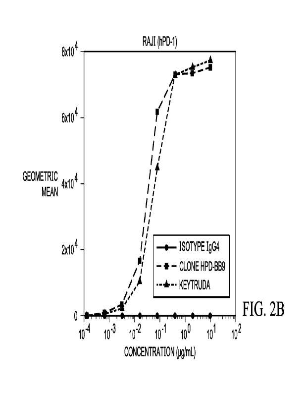
Figure 2B shows a graph of a cell binding assay of antibodies HDP-BB9,
Keytruda
and a control IgG4 isotype, binding to Raji cells engineered to express human
PD-1 antigen.
Figure 3 shows histograms of flow cytometry data of binding between antibodies
HDP-BB9, Keytruda or control secondary antibody, to PBMCs from human or dog.
Figure 4 shows a bar graph comparing the level of interferon gamma (IFNy)
release
generated from a mixed lymphocyte reaction (MRL) assay, comparing antibodies
HDP-BB9,
Keytruda, Opdivo and a control IgG4 isotype.
Figure 5A shows a bar graph comparing the level of interferon gamma (IFNy)
release
generated from a first experiment of a three-way mixed lymphocyte reaction
(MLR) assay,
comparing antibodies HDP-BB9, Keytruda, Opdivo and a control IgG4 isotype.
Figure 5B shows a bar graph comparing the level of interferon gamma (IFNy)
release
generated from a second experiment of a three-way mixed lymphocyte reaction
(MLR) assay,
comparing antibodies HDP-BB9, Keytruda, Opdivo and a control IgG4 isotype
Figure 6 shows the amino acid sequences of PD-1 antigens from human,
cynomolgus
monkey, rhesus monkey and mouse
Figure 7 shows the amino acid sequences of anti-PD1 antibody HDP-BB9,
including
the heavy chain variable region, and heavy chain CDRs 1, 2 and 3, and the
light chain
variable region, and light chain CDRs 1, 2 and 3. The CDR regions in the heavy
and light
chains are underlined.
Figure 8 shows the amino acid sequences of anti-PD1 antibody HDP-BB9N,
including the heavy chain variable region, and heavy chain CDRs 1, 2 and 3,
and the light
chain variable region, and light chain CDRs 1, 2 and 3. The CDR regions in the
heavy and
light chains are underlined.
Figure 9 shows the amino acid sequences of anti-PD1 antibody Keytruda
including
the heavy chain variable region and the light chain variable region, and the
amino acid
sequences of anti-PD1 antibody Opdivo including the heavy chain variable
region and the
light chain variable region.
Figure 10 shows a graph of dose-dependent response of the blocking of PD1/PD-
L1
interaction using human anti-PD1 clones HPD-13B9 and KEYTRUDA and a negative
control
antibody (Isotype IgG4).
CA 03183034 2022- 12- 15
WO 2021/263166
PCT/US2021/039188
9
Figure 11 shows the effect of human anti-PD1 clone HPD-BB9 on bladder tumor
growth in MB-49 syngeneic tumor model. Figure 11A shows the effect of 5 and 15
mg/kg of
HPD-11119 clone and 15 mg/kg of isotype control on the tumor volume of each
individual
mouse, measured over 24 days. Figure 11B shows the effect of 5 and 15 mg/kg of
HPD-BB9
clone and 15 mg/kg of isotype control on the tumor volume - averaged for the
10 mice,
measured over 24 days. Figure 11C shows percent tumor growth inhibition by HPD-
BB9
clone (TGI = (1-[mean HPD-BB9 / mean Isotype]) x 100) calculated at the end of
the study
day 24 post tumor cell implantation.
Figure 12 shows the effect of human anti-PD1 clone HPD-BB9 and a negative
(isotype) control IgG4 on body weight of each mouse with MB-49 syngeneic tumor
model.
DESCRIPTION
Definitions:
Unless defined otherwise, technical and scientific terms used herein have
meanings
that are commonly understood by those of ordinary skill in the art unless
defined otherwise.
Generally, terminologies pertaining to techniques of cell and tissue culture,
molecular
biology, immunology, microbiology, genetics, transgenic cell production,
protein chemistry
and nucleic acid chemistry and hybridization described herein are well known
and commonly
used in the art. The methods and techniques provided herein are generally
performed
according to conventional procedures well known in the art and as described in
various
general and more specific references that are cited and discussed herein
unless otherwise
indicated. See, e.g., Sambrook et al. Molecular Cloning: A Laboratory Manual,
2d ed., Cold
Spring Harbor Laboratory Press, Cold Spring Harbor, N.Y. (1989) and Ausubel et
al.,
Current Protocols in Molecular Biology, Greene Publishing Associates (1992). A
number of
basic texts describe standard antibody production processes, including,
Borrebaeck
(ed) Antibody Engineering, 2nd Edition Freeman and Company, NY, 1995;
McCafferty et
al. Antibody Engineering, A Practical Approach IRL at Oxford Press, Oxford,
England,
1996; and Paul (1995) Antibody Engineering Protocols Humana Press, Towata,
N.J., 1995;
Paul (ed.), Fundamental Immunology, Raven Press, N.Y, 1993; Coligan (1991)
Current
Protocols in Immunology Wiley/Greene, NY; Harlow and Lane (1989) Antibodies: A
Laboratory Manual Cold Spring Harbor Press, NY; Stites et al. (eds.) Basic and
Clinical
Immunology (4th ed.) Lange Medical Publications, Los Altos, Calif., and
references cited
therein; Coding Monoclonal Antibodies: Principles and Practice (2nd ed.)
Academic Press,
CA 03183034 2022- 12- 15
WO 2021/263166
PCT/US2021/039188
New York, N.Y., 1986, and Kohler and Milstein Nature 256: 495-497, 1975. All
of the
references cited herein are incorporated herein by reference in their
entireties. Enzymatic
reactions and enrichment/purification techniques are also well known and are
performed
according to manufacturer's specifications, as commonly accomplished in the
art or as
5 described herein. The terminology used in connection with, and the
laboratory procedures
and techniques of, analytical chemistry, synthetic organic chemistry, and
medicinal and
pharmaceutical chemistry described herein are well known and commonly used in
the art.
Standard techniques can be used for chemical syntheses, chemical analyses,
pharmaceutical
preparation, formulation, and delivery, and treatment of patients.
10 The headings provided herein are not limitations of the various
aspects of the
disclosure, which aspects can be understood by reference to the specification
as a whole.
Unless otherwise required by context herein, singular terms shall include
pluralities
and plural terms shall include the singular. Singular forms "a","an" and
"the", and singular
use of any word, include plural referents unless expressly and unequivocally
limited on one
referent.
It is understood the use of the alternative (e.g., "or") herein is taken to
mean either
one or both or any combination thereof of the alternatives.
The term "and/or" used herein is to be taken mean specific disclosure of each
of the
specified features or components with or without the other. For example, the
term "and/or"
as used in a phrase such as "A and/or B" herein is intended to include "A and
B," "A or B,"
"A" (alone), and "B" (alone). Likewise, the term "and/or" as used in a phrase
such as "A, B,
and/or C- is intended to encompass each of the following aspects: A, B, and C;
A, B, or C; A
or C; A or B; B or C; A and C; A and B; B and C; A (alone); B (alone); and C
(alone).
As used herein, terms "comprising", "including", "having" and "containing",
and
their grammatical variants, as used herein are intended to be non-limiting so
that one item or
multiple items in a list do not exclude other items that can be substituted or
added to the listed
items. It is understood that wherever aspects are described herein with the
language
"comprising," otherwise analogous aspects described in terms of "consisting
of' and/or
"consisting essentially of- are also provided.
As used herein, the term -about" refers to a value or composition that is
within an
acceptable error range for the particular value or composition as determined
by one of
ordinary skill in the art, which will depend in part on how the value or
composition is
measured or determined, i.e., the limitations of the measurement system. For
example,
CA 03183034 2022- 12- 15
WO 2021/263166
PCT/US2021/039188
11
"about" or "approximately" can mean within one or more than one standard
deviation per the
practice in the art. Alternatively, "about" or "approximately" can mean a
range of up to 10%
(i e , 10%) or more depending on the limitations of the measurement system
For example,
about 5 mg can include any number between 4.5 mg and 5.5 mg. Furthermore,
particularly
with respect to biological systems or processes, the terms can mean up to an
order of
magnitude or up to 5-fold of a value. When particular values or compositions
are provided in
the instant disclosure, unless otherwise stated, the meaning of "about" or
"approximately"
should be assumed to be within an acceptable error range for that particular
value or
composition.
The terms "peptide", "polypeptide" and "protein" and other related terms used
herein
are used interchangeably and refer to a polymer of amino acids and are not
limited to any
particular length. Polypeptides may comprise natural and non-natural amino
acids.
Polypeptides include recombinant or chemically-synthesized forms. Polypeptides
also
include precursor molecules and mature molecule. Precursor molecules include
those that
have not yet been subjected to cleavage, for example cleavage by a secretory
signal peptide
or by non-enzymatic cleavage at certain amino acid residue. Polypeptides in
include mature
molecules that have undergone cleavage. These terms encompass native proteins,
recombinant proteins and artificial proteins, protein fragments and
polypeptide analogs (such
as muteins, variants, chimeric proteins and fusion proteins) of a protein
sequence as well as
post-translationally, or otherwise covalently or non-covalently, modified
proteins.
Polypeptides comprising amino acid sequences of binding proteins that bind PD-
1 (e.g., anti-
PD-1 antibodies or antigen-binding portions thereof) prepared using
recombinant procedures
are described herein.
The terms "nucleic acid", "polynucleotide" and "oligonucleotide" and other
related
terms used herein are used interchangeably and refer to polymers of
nucleotides and are not
limited to any particular length. Nucleic acids include recombinant and
chemically-
synthesized forms. Nucleic acids include DNA molecules (cDNA or genomic DNA),
RNA
molecules (e.g., mRNA), analogs of the DNA or RNA generated using nucleotide
analogs
(e.g., peptide nucleic acids and non-naturally occurring nucleotide analogs),
and hybrids
thereof. Nucleic acid molecule can be single-stranded or double-stranded. In
one
embodiment, the nucleic acid molecules of the disclosure comprise a contiguous
open
reading frame encoding an antibody, or a fragment or scFv, derivative, mutein,
or variant
thereof. In one embodiment, nucleic acids comprise a one type of
polynucleotides or a
CA 03183034 2022- 12- 15
WO 2021/263166
PCT/US2021/039188
12
mixture of two or more different types of polynucleotides. Nucleic acids
encoding anti-PD-1
antibodies or antigen-binding portions thereof, are described herein.
The term "recover" or "recovery" or "recovering", and other related terms,
refers to
obtaining a protein (e.g., an antibody or an antigen binding portion thereof),
from host cell
culture medium or from host cell lysate or from the host cell membrane. In one
embodiment,
the protein is expressed by the host cell as a recombinant protein fused to a
secretion signal
peptide sequence (e.g., leader peptide sequence) which mediates secretion of
the expressed
protein. The secreted protein can be recovered from the host cell medium. In
one
embodiment, the protein is expressed by the host cell as a recombinant protein
that lacks a
secretion signal peptide sequence which can be recovered from the host cell
lysate. In one
embodiment, the protein is expressed by the host cell as a membrane-bound
protein which
can be recovered using a detergent to release the expressed protein from the
host cell
membrane. In one embodiment, irrespective of the method used to recover the
protein, the
protein can be subjected to procedures that remove cellular debris from the
recovered protein.
For example, the recovered protein can be subjected to chromatography, gel
electrophoresis
and/or dialysis. In one embodiment, the chromatography comprises any one or
any
combination or two or more procedures including affinity chromatography,
hydroxyapatite
chromatography, ion-exchange chromatography, reverse phase chromatography
and/or
chromatography on silica. In one embodiment, affinity chromatography comprises
protein A
or G (cell wall components from Staphylococcus aureus).
The term "isolated" refers to a protein (e.g., an antibody or an antigen
binding portion
thereof) or polynucleotide that is substantially free of other cellular
material. A protein may
be rendered substantially free of naturally associated components (or
components associated
with a cellular expression system or chemical synthesis methods used to
produce the
antibody) by isolation, using protein purification techniques well known in
the art. The term
isolated also refers in some embodiments to protein or polynucleotides that
are substantially
free of other molecules of the same species, for example other protein or
polynucleotides
having different amino acid or nucleotide sequences, respectively. The purity
or
homogeneity of the desired molecule can be assayed using techniques well known
in the art,
including low resolution methods such as gel electrophoresis and high
resolution methods
such as HPLC or mass spectrometry. In one embodiment, any of the anti-PD-1
antibodies or
antigen binding protein thereof are isolated
CA 03183034 2022- 12- 15
WO 2021/263166
PCT/US2021/039188
13
Antibodies can be obtained from sources such as serum or plasma that contain
immunoglobulins having varied antigenic specificity. If such antibodies are
subjected to
affinity purification, they can be enriched for a particular antigenic
specificity. Such enriched
preparations of antibodies usually are made of less than about 10% antibody
having specific
binding activity for the particular antigen. Subjecting these preparations to
several rounds of
affinity purification can increase the proportion of antibody having specific
binding activity
for the antigen. Antibodies prepared in this manner are often referred to as
"monospecific."
Monospecific antibody preparations can be made up of about 10%, 20%, 30%, 40%,
50%,
60%, 70%, 75%, 80%, 85%, 90%, 95%, 97%, 99%, or 99.9% antibody having specific
binding activity for the particular antigen. Antibodies can be produced using
recombinant
nucleic acid technology as described below.
The term "leader sequence" or "leader peptide" or "peptide signal sequence" or
"signal peptide" or "secretion signal peptide" refers to a peptide sequence
that is located at
the N-terminus of a polypeptide. A leader sequence directs a polypeptide chain
to a cellular
secretory pathway and can direct integration and anchoring of the polypeptide
into the lipid
bilayer of the cellular membrane. Typically, a leader sequence is about 10-50
amino acids in
length. A leader sequence can direct transport of a precursor polypeptide from
the cytosol to
the endoplasmic reticulum. In one embodiment, a leader sequence includes
signal sequences
comprising CD8a, CD28 or CD16 leader sequences. In one embodiment, the signal
sequence
comprises a mammalian sequence, including for example mouse or human Ig gamma
secretion signal peptide. In one embodiment, a leader sequence comprises a
mouse Ig
gamma leader peptide sequence MEWSWVFLFFLSVTTGVHS (SEQ ID NO: 26).
An "antigen binding protein" and related terms used herein refers to a protein
comprising a portion that binds to an antigen and, optionally, a scaffold or
framework portion
that allows the antigen binding portion to adopt a conformation that promotes
binding of the
antigen binding protein to the antigen. Examples of antigen binding proteins
include
antibodies, antibody fragments (e.g., an antigen binding portion of an
antibody), antibody
derivatives, and antibody analogs. The antigen binding protein can comprise,
for example, an
alternative protein scaffold or artificial scaffold with grafted CDRs or CDR
derivatives. Such
scaffolds include, but are not limited to, antibody-derived scaffolds
comprising mutations
introduced to, for example, stabilize the three-dimensional structure of the
antigen binding
protein as well as wholly synthetic scaffolds comprising, for example, a
biocompatible
polymer. See, for example, Korndorfer et al., 2003, Proteins: Structure,
Function, and
CA 03183034 2022- 12- 15
WO 2021/263166
PCT/US2021/039188
14
Bioinformatics, Volume 53, Issue 1:121-129; Roque et al., 2004, Biotechnol.
Prog. 20:639-
654. In addition, peptide antibody mimetics ("PAMs") can be used, as well as
scaffolds based
on antibody mimetics utilizing fibronecti on components as a scaffold Antigen
binding
proteins that bind PD-1 are described herein.
An antigen binding protein can have, for example, the structure of an
immunoglobulin. In one embodiment, an "immunoglobulin" refers to a tetrameric
molecule
composed of two identical pairs of polypeptide chains, each pair having one
"light" (about 25
kDa) and one "heavy" chain (about 50-70 kDa). The amino-terminal portion of
each chain
includes a variable region of about 100 to 110 or more amino acids primarily
responsible for
antigen recognition. The carboxy-terminal portion of each chain defines a
constant region
primarily responsible for effector function. Human light chains are classified
as kappa or
lambda light chains. Heavy chains are classified as mu, delta, gamma, alpha,
or epsilon, and
define the antibody's isotype as IgM, IgD, IgG, IgA, and IgE, respectively.
Within light and
heavy chains, the variable and constant regions are joined by a "J" region of
about 12 or more
amino acids, with the heavy chain also including a "D" region of about 10 more
amino acids.
See generally, Fundamental Immunology Ch. 7 (Paul, W., ed., 2nd ed. Raven
Press, N.Y.
(1989)) (incorporated by reference in its entirety for all purposes). The
heavy and/or light
chains may or may not include a leader sequence for secretion. The variable
regions of each
light/heavy chain pair form the antibody binding site such that an intact
immunoglobulin has
two antigen binding sites. In one embodiment, an antigen binding protein can
be a synthetic
molecule having a structure that differs from a tetrameric immunoglobulin
molecule but still
binds a target antigen or binds two or more target antigens. For example, a
synthetic antigen
binding protein can comprise antibody fragments, 1-6 or more polypeptide
chains,
asymmetrical assemblies of polypeptides, or other synthetic molecules. Antigen
binding
proteins having immunoglobulin-like properties that bind specifically to PD-1
are described
herein.
The variable regions of immunoglobulin chains exhibit the same general
structure of
relatively conserved framework regions (FR) joined by three hypervariable
regions, also
called complementarity determining regions or CDRs. From N-terminus to C-
terminus, both
light and heavy chains comprise the segments FR1, CDR1, FR2, CDR2, FR3, CDR3
and
FR4.
One or more CDRs may be incorporated into a molecule either covalently or
noncovalently to make it an antigen binding protein. An antigen binding
protein may
CA 03183034 2022- 12- 15
WO 2021/263166
PCT/US2021/039188
incorporate the CDR(s) as part of a larger polypeptide chain, may covalently
link the CDR(s)
to another polypeptide chain, or may incorporate the CDR(s) noncovalently. The
CDRs
permit the antigen binding protein to specifically bind to a particular
antigen of interest
The assignment of amino acids to each domain is in accordance with the
definitions of
5 Kabat et al. in Sequences of Proteins of Immunological Interest, 5th Ed.,
US Dept. of Health
and Human Services, PHS, NIH, NUT Publication no. 91-3242, 1991 (e.g., "Kabat
numbering"). Other numbering systems for the amino acids in immunoglobulin
chains
include IMGT® (international ImMunoGeneTics information system; Lefranc et
al, Dev.
Comp. Immunol. 29:185-203; 2005) and AHo (Honegger and Pluckthun, J. Mol.
Biol.
10 309(3):657-670; 2001); Chothia (Al-Lazikani et al., 1997 Journal of
Molecular Biology
273:927-948; Contact (Maccallum et al., 1996 Journal of Molecular Biology
262:732-745,
and Aho (Honegger and Pluckthun 2001 Journal of Molecular Biology 309:657-670.
An "antibody" and "antibodies" and related terms used herein refers to an
intact
immunoglobulin or to an antigen binding portion thereof (or an antigen binding
fragment
15 thereof) that binds specifically to an antigen. Antigen binding portions
(or the antigen
binding fragment) may be produced by recombinant DNA techniques or by
enzymatic or
chemical cleavage of intact antibodies. Antigen binding portions (or antigen
binding
fragments) include, inter alia, Fab, Fab', F(ab')?, Fv, domain antibodies
(dAbs), and
complementarity determining region (CDR) fragments, single-chain antibodies
(scFv),
chimeric antibodies, diabodies, triabodies, tetrabodies, and polypeptides that
contain at least a
portion of an immunoglobulin that is sufficient to confer specific antigen
binding to the
polypeptide.
Antibodies include recombinantly produced antibodies and antigen binding
portions.
Antibodies include non-human, chimeric, humanized and fully human antibodies.
Antibodies
include monospecific, multispecific (e.g., bispecific, trispecific and higher
order
specificities). Antibodies include tetrameric antibodies, light chain
monomers, heavy chain
monomers, light chain dimers, heavy chain dimers. Antibodies include F(ab')2
fragments,
Fab' fragments and Fab fragments. Antibodies include single domain antibodies,
monovalent
antibodies, single chain antibodies, single chain variable fragment (scFv),
camelized
antibodies, affibodies, disulfide-linked Fvs (sdFv), anti-idiotypic antibodies
(anti-Id),
minibodies. Antibodies include monoclonal and polyclonal populations. Anti-PD-
1
antibodies are described herein.
CA 03183034 2022- 12- 15
WO 2021/263166
PCT/US2021/039188
16
An "antigen binding domain," "antigen binding region," or "antigen binding
site" and
other related terms used herein refer to a portion of an antigen binding
protein that contains
amino acid residues (or other moieties) that interact with an antigen and
contribute to the
antigen binding protein's specificity and affinity for the antigen. For an
antibody that
specifically binds to its antigen, this will include at least part of at least
one of its CDR
domains. Antigen binding domains from anti-PD-1 antibodies are described
herein.
The terms "specific binding", "specifically binds" or "specifically binding"
and other
related terms, as used herein in the context of an antibody or antigen binding
protein or
antibody fragment, refer to non-covalent or covalent preferential binding to
an antigen
relative to other molecules or moieties (e.g., an antibody specifically binds
to a particular
antigen relative to other available antigens). In one embodiment, an antibody
specifically
binds to a target antigen if it binds to the antigen with a dissociation
constant KD of 10-5 M or
less, or 10-6M or less, or 10-7M or less, or 10-8 M or less, or 10-9M or less,
or 1040 M or
less, or 10-11 or less, or 10-12 or less. Anti-PD-1 antibodies that
specifically bind PD-1 are
described herein.
In one embodiment, a dissociation constant (I(D) can be measured using a
BIACORE
surface plasmon resonance (SPR) assay. Surface plasmon resonance refers to an
optical
phenomenon that allows for the analysis of real-time interactions by detection
of alterations
in protein concentrations within a biosensor matrix, for example using the
BIACORE system
(Biacore Life Sciences division of GE Healthcare, Piscataway, NJ).
An "epitope" and related terms as used herein refers to a portion of an
antigen that is
bound by an antigen binding protein (e.g., by an antibody or an antigen
binding portion
thereof). An epitope can comprise portions of two or more antigens that are
bound by an
antigen binding protein. An epitope can comprise non-contiguous portions of an
antigen or
of two or more antigens (e.g., amino acid residues that are not contiguous in
an antigen's
primary sequence but that, in the context of the antigen's tertiary and
quaternary structure, are
near enough to each other to be bound by an antigen binding protein).
Generally, the variable
regions, particularly the CDRs, of an antibody interact with the epitope. Anti-
PD-1
antibodies, and antigen binding proteins thereof, that bind an epitope of a PD-
1 polypeptide
are described herein.
With respect to antibodies, the term "antagonist" and "antagonistic" refers to
a
blocking antibody that binds its cognate target antigen and inhibits or
reduces the biological
activity of the bound antigen. The term "agonist" or "agonistic" refers to an
antibody that
CA 03183034 2022- 12- 15
WO 2021/263166
PCT/US2021/039188
17
binds its cognate target antigen in a manner that mimics the binding of the
physiological
ligand which causes antibody-mediated downstream signaling.
An "antibody fragment", "antibody portion", "antigen-binding fragment of an
antibody", or "antigen-binding portion of an antibody" and other related terms
used herein
refer to a molecule other than an intact antibody that comprises a portion of
an intact antibody
that binds the antigen to which the intact antibody binds. Examples of
antibody fragments
include, but are not limited to, Fv, Fab, Fab', Fab'-SH, F(abl)2; Fd; and Fv
fragments, as well
as dAb; diabodies; linear antibodies; single-chain antibody molecules (e.g.
scFv);
polypeptides that contain at least a portion of an antibody that is sufficient
to confer specific
antigen binding to the polypeptide. Antigen binding portions of an antibody
may be produced
by recombinant DNA techniques or by enzymatic or chemical cleavage of intact
antibodies.
Antigen binding portions include, inter alia, Fab, Fab', F(ab')2, Fv, domain
antibodies (dAbs),
and complementarity determining region (CDR) fragments, chimeric antibodies,
diabodies,
triabodies, tetrabodies, and polypeptides that contain at least a portion of
an immunoglobulin
that is sufficient to confer antigen binding properties to the antibody
fragment. Antigen-
binding fragments of anti-PD-1 antibodies are described herein.
The terms "Fab", "Fab fragment" and other related terms refers to a monovalent
fragment comprising a variable light chain region (VIA constant light chain
region (CL),
variable heavy chain region (VH), and first constant region (CH1). A Fab is
capable of
binding an antigen. An F(abl), fragment is a bivalent fragment comprising two
Fab fragments
linked by a disulfide bridge at the hinge region. A F(Ab')2 has antigen
binding capability.
An Fd fragment comprises VH and Cm regions. An Fv fragment comprises VL and VH
regions. An Fv can bind an antigen. A dAb fragment has a VH domain, a VL
domain, or an
antigen-binding fragment of a VH or VL domain (U.S. Patents 6,846,634 and
6,696,245; U.S.
published Application Nos. 2002/02512, 2004/0202995, 2004/0038291,
2004/0009507,
2003/0039958; and Ward et al., Nature 341:544-546, 1989). Fab fragments
comprising
antigen binding portions from anti-PD-1 antibodies are described herein.
A single-chain antibody (scFv) is an antibody in which a VL and a VH region
are
joined via a linker (e.g., a synthetic sequence of amino acid residues) to
form a continuous
protein chain. In one embodiment, the linker is long enough to allow the
protein chain to fold
back on itself and form a monovalent antigen binding site (see, e.g., Bird et
al., 1988, Science
242:423-26 and I-Tuston et al., 1988, Proc. Natl Acad. Sci. USA 85:5879-83).
Single chain
CA 03183034 2022- 12- 15
WO 2021/263166
PCT/US2021/039188
18
antibodies comprising antigen binding portions from anti-PD-1 antibodies are
described
herein.
Di abodi es are bivalent antibodies comprising two polypeptide chains, wherein
each
polypeptide chain comprises VH and VL domains joined by a linker that is too
short to allow
for pairing between two domains on the same chain, thus allowing each domain
to pair with a
complementary domain on another polypeptide chain (see, e.g., Holliger et al.,
1993, Proc.
Natl. Acad. Sci. USA 90:6444-48, and Poljak et al., 1994, Structure 2:1121-
23). If the two
polypeptide chains of a diabody are identical, then a diabody resulting from
their pairing will
have two identical antigen binding sites. Polypeptide chains having different
sequences can
be used to make a diabody with two different antigen binding sites. Similarly,
tribodies and
tetrabodies are antibodies comprising three and four polypeptide chains,
respectively, and
forming three and four antigen binding sites, respectively, which can be the
same or different.
Diabody, tribody and tetrabody constructs can be prepared using antigen
binding portions
from any of the anti-PD1 antibodies described herein.
The term -human antibody" refers to antibodies that have one or more variable
and
constant regions derived from human immunoglobulin sequences. In one
embodiment, all of
the variable and constant domains are derived from human immunoglobulin
sequences (e.g.,
a fully human antibody). These antibodies may be prepared in a variety of
ways, examples of
which are described below, including through recombinant methodologies or
through
immunization with an antigen of interest of a mouse that is genetically
modified to express
antibodies derived from human heavy and/or light chain-encoding genes. Fully
human anti-
PD-1 antibodies and antigen binding proteins thereof are described herein.
A "humanized" antibody refers to an antibody having a sequence that differs
from the
sequence of an antibody derived from a non-human species by one or more amino
acid
substitutions, deletions, and/or additions, such that the humanized antibody
is less likely to
induce an immune response, and/or induces a less severe immune response, as
compared to
the non-human species antibody, when it is administered to a human subject. In
one
embodiment, certain amino acids in the framework and constant domains of the
heavy and/or
light chains of the non-human species antibody are mutated to produce the
humanized
antibody. In another embodiment, the constant domain(s) from a human antibody
are fused to
the variable domain(s) of a non-human species. In another embodiment, one or
more amino
acid residues in one or more CDR sequences of a non-human antibody are changed
to reduce
the likely immunogenicity of the non-human antibody when it is administered to
a human
CA 03183034 2022- 12- 15
WO 2021/263166
PCT/US2021/039188
19
subject, wherein the changed amino acid residues either are not critical for
immunospecific
binding of the antibody to its antigen, or the changes to the amino acid
sequence that are
made are conservative changes, such that the binding of the humanized antibody
to the
antigen is not significantly worse than the binding of the non-human antibody
to the antigen.
Examples of how to make humanized antibodies may be found in U.S. Pat. Nos.
6,054,297,
5,886,152 and 5,877,293.
The term "chimeric antibody" and related terms used herein refers to an
antibody that
contains one or more regions from a first antibody and one or more regions
from one or more
other antibodies. In one embodiment, one or more of the CDRs are derived from
a human
antibody. In another embodiment, all of the CDRs are derived from a human
antibody. In
another embodiment, the CDRs from more than one human antibody are mixed and
matched
in a chimeric antibody. For instance, a chimeric antibody may comprise a CDR1
from the
light chain of a first human antibody, a CDR2 and a CDR3 from the light chain
of a second
human antibody, and the CDRs from the heavy chain from a third antibody. In
another
example, the CDRs originate from different species such as human and mouse, or
human and
rabbit, or human and goat. One skilled in the art will appreciate that other
combinations are
possible.
Further, the framework regions may be derived from one of the same antibodies,
from
one or more different antibodies, such as a human antibody, or from a
humanized antibody.
In one example of a chimeric antibody, a portion of the heavy and/or light
chain is identical
with, homologous to, or derived from an antibody from a particular species or
belonging to a
particular antibody class or subclass, while the remainder of the chain(s)
is/are identical with,
homologous to, or derived from an antibody (-ies) from another species or
belonging to
another antibody class or subclass. Also included are fragments of such
antibodies that
exhibit the desired biological activity (i.e., the ability to specifically
bind a target antigen).
Chimeric antibodies can be prepared from portions of any of the anti-PD-1
antibodies
described herein.
As used herein, the term "variant" polypeptides and "variants" of polypeptides
refers
to a polypeptide comprising an amino acid sequence with one or more amino acid
residues
inserted into, deleted from and/or substituted into the amino acid sequence
relative to a
reference polypeptide sequence. Polypeptide variants include fusion proteins.
In the same
manner, a variant polynucleotide comprises a nucleotide sequence with one or
more
CA 03183034 2022- 12- 15
WO 2021/263166
PCT/US2021/039188
nucleotides inserted into, deleted from and/or substituted into the nucleotide
sequence relative
to another polynucleotide sequence. Polynucleotide variants include fusion
polynucleotides.
As used herein, the term "derivative" of a polypepti de is a polypepti de (e g
,
an antibody) that has been chemically modified, e.g., via conjugation to
another chemical
5 moiety such as, for example, polyethylene glycol, albumin (e.g., human
serum albumin),
phosphorylation, and glycosylation. Unless otherwise indicated, the term
"antibody"
includes, in addition to antibodies comprising full-length heavy chains and
full-length light
chains, derivatives, variants, fragments, and muteins thereof, examples of
which are
described below.
10 The term "hinge" refers to an amino acid segment that is generally
found between two
domains of a protein and may allow for flexibility of the overall construct
and movement of
one or both of the domains relative to one another. Structurally, a hinge
region comprises
from about 10 to about 100 amino acids, e.g., from about 15 to about 75 amino
acids, from
about 20 to about 50 amino acids, or from about 30 to about 60 amino acids. In
one
15 embodiment, the hinge region is 10, 11, 12, 13, 14, 15, 16, 17, 18, 19,
20, 21, 22, 23, 24, 25,
26, 27, 28, 29, 30, 35, 40, 45, 50, 55, 60, 65, 70, 75, 80, 85, 90, 95, or 100
amino acids in
length. The hinge region can be derived from is a hinge region of a naturally-
occurring
protein, such as a CD8 hinge region or a fragment thereof, a CD8ct hinge
region, or a
fragment thereof, a hinge region of an antibody (e.g., IgG, IgA, IgM, IgE, or
IgD antibodies),
20 or a hinge region that joins the constant domains CH1 and CH2 of an
antibody. The hinge
region can be derived from an antibody and may or may not comprise one or more
constant
regions of the antibody, or the hinge region comprises the hinge region of an
antibody and the
CH3 constant region of the antibody, or the hinge region comprises the hinge
region of an
antibody and the CH2 and CH3 constant regions of the antibody, or the hinge
region is a non-
naturally occurring peptide, or the hinge region is disposed between the C-
terminus of the
scEv and the N-terminus of the transmembrane domain. In one embodiment, the
hinge region
comprises any one or any combination of two or more regions comprising an
upper, core or
lower hinge sequences from an IgGI, IgG2, IgG3 or IgG4 immunoglobulin
molecule. In one
embodiment, the hinge region comprises an IgG1 upper hinge sequence EPKSCDKTHT
(SEQ ID NO: 27). In one embodiment, the hinge region comprises an IgG1 core
hinge
sequence CPXC, wherein X is P, R or S. In one embodiment, the hinge region
comprises a
lower hinge/CH2 sequence PAPELLGGP (SEQ ID NO: 28). In one embodiment, the
hinge
is joined to an Fc region (CH2) having the amino acid sequence SVFLFPPKPKDT
(SEQ ID
CA 03183034 2022- 12- 15
WO 2021/263166
PCT/US2021/039188
21
NO: 29). In one embodiment, the hinge region includes the amino acid sequence
of an upper,
core and lower hinge and comprises EPKSCDKTHTCPPCPAP ELLGGP (SEQ ID NO: 30).
Tn one embodiment, the hinge region comprises one, two, three or more
cysteines that can
form at least one, two, three or more interchain disulfide bonds.
The term "Fc" or "Fe region" as used herein refers to the portion of an
antibody heavy
chain constant region beginning in or after the hinge region and ending at the
C-terminus of
the heavy chain. The Fe region comprises at least a portion of the CH2 and CH3
regions and
may, or may not, include a portion of the hinge region. An Fe domain can bind
Fe cell
surface receptors and some proteins of the immune complement system. An Fe
region can
bind a complement component Cl q. An Fe domain exhibits effector function,
including any
one or any combination of two or more activities including complement-
dependent
cytotoxicity (CDC), antibody-dependent cell-mediated cytotoxicity (ADCC),
antibody-
dependent phagocytosis (ADP), opsonization and/or cell binding. An Fe domain
can bind an
Fe receptor, including FcyRI (e.g., CD64), FcyRII (e.g, CD32) and/or FcyRIII
(e.g., CD16a).
In one embodiment, the Fe region can include a mutation that increases or
decreases any one
or any combination of these functions. In one embodiment, the Fe domain
comprises a
LALA mutation (e.g., equivalent to L234A, L235A according to Kabat numbering)
which
reduces effector function. In one embodiment, the Fe domain comprises a LALA-
PG
mutation (e.g., equivalent to L234A, L235A, P329G according to Kabat
numbering) which
reduces effector function. In one embodiment, the Fe domain mediates serum
half-life of the
protein complex, and a mutation in the Fe domain can increase or decrease the
serum half-life
of the protein complex. In one embodiment, the Fe domain affects thermal
stability of the
protein complex, and mutation in the Fe domain can increase or decrease the
thermal stability
of the protein complex.
The term "labeled" or related terms as used herein with respect to a
polypeptide refers
to joinder antibodies and their antigen binding portions thereof that are
unlabeled or joined to
a detectable label or moiety for detection, wherein the detectable label or
moiety is
radioactive, colorimetric, antigenic, enzymatic, a detectable bead (such as a
magnetic or
electrodense (e.g., gold) bead), biotin, streptavidin or protein A. A variety
of labels can be
employed, including, but not limited to, radionuclides, fluorescers, enzymes,
enzyme
substrates, enzyme cofactors, enzyme inhibitors and ligands (e.g., biotin,
haptens). Any of
the anti-PD-1 antibodies described herein can be unlabeled or can be joined to
a detectable
label or moiety.
CA 03183034 2022- 12- 15
WO 2021/263166
PCT/US2021/039188
22
The term "labeled" or related terms as used herein with respect to a
polypeptide refers
to joinder thereof to a detectable label or moiety for detection. Exemplary
detectable labels
or moieties include radioactive, col ori m etri c, antigenic, enzymatic 1 abel
s/m oi eti es, a
detectable bead (such as a magnetic or electrodense (e.g., gold) bead),
biotin, streptavidin or
protein A. A variety of labels can be employed, including, but not limited to,
radionuclides,
fluorescers, enzymes, enzyme substrates, enzyme cofactors, enzyme inhibitors
and ligands
(e.g., biotin, haptens). Any of the anti-PD-1 antibodies described herein or
antigen-binding
portions thereof that described herein can be unlabeled or can be joined to a
detectable label
or detectable moiety.
The "percent identity" or "percent homology" and related terms used herein
refers to
a quantitative measurement of the similarity between two polypeptide or
between two
polynucleotide sequences. The percent identity between two polypeptide
sequences is a
function of the number of identical amino acids at aligned positions that are
shared between
the two polypeptide sequences, taking into account the number of gaps, and the
length of
each gap, which may need to be introduced to optimize alignment of the two
polypeptide
sequences. In a similar manner, the percent identity between two
polynucleotide sequences is
a function of the number of identical nucleotides at aligned positions that
are shared between
the two polynucleotide sequences, taking into account the number of gaps, and
the length of
each gap, which may need to be introduced to optimize alignment of the two
polynucleotide
sequences. A comparison of the sequences and determination of the percent
identity between
two polypeptide sequences, or between two polynucleotide sequences, may be
accomplished
using a mathematical algorithm. For example, the "percent identity" or
"percent homology"
of two polypeptide or two polynucleotide sequences may be determined by
comparing the
sequences using the GAP computer program (a part of the GCG Wisconsin Package,
version
10.3 (Accelrys, San Diego, Calif.)) using its default parameters. Expressions
such as
"comprises a sequence with at least X% identity to Y" with respect to a test
sequence mean
that, when aligned to sequence Y as described above, the test sequence
comprises residues
identical to at least X% of the residues of Y.
In one embodiment, the amino acid sequence of a test antibody may be similar
but not
necessarily identical to any of the amino acid sequences of the polypeptides
that make up any
of the anti-PD-1 antibodies, or antigen binding protein thereof, described
herein. The
similarities between the test antibody and the polypeptides can be at least
95%, or at or at
least 96% identical, or at least 97% identical, or at least 98% identical, or
at least 99%
CA 03183034 2022- 12- 15
WO 2021/263166
PCT/US2021/039188
23
identical, to any of the polypeptides that make up any of the anti-PD-1
antibodies, or antigen
binding protein thereof, described herein. In one embodiment, similar
polypeptides can
contain amino acid substitutions within a heavy and/or light chain Tn one
embodiment, the
amino acid substitutions comprise one or more conservative amino acid
substitutions. A
"conservative amino acid substitution" is one in which an amino acid residue
is substituted by
another amino acid residue having a side chain (R group) with similar chemical
properties
(e.g., charge or hydrophobicity). In general, a conservative amino acid
substitution will not
substantially change the functional properties of a protein. In cases where
two or more amino
acid sequences differ from each other by conservative substitutions, the
percent sequence
identity or degree of similarity may be adjusted upwards to correct for the
conservative nature
of the substitution. Means for making this adjustment are well-known to those
of skill in the
art. See, e.g., Pearson (1994) Methods Mol. Biol. 24: 307-33 1, herein
incorporated by
reference in its entirety. Examples of groups of amino acids that have side
chains with
similar chemical properties include (1) aliphatic side chains: glycine,
alanine, valine, leucine
and isoleucine; (2) aliphatic-hydroxyl side chains: serine and threonine; (3)
amide-containing
side chains: asparagine and glutamine; (4) aromatic side chains:
phenylalanine, tyrosine, and
tryptophan; (5) basic side chains: lysine, arginine, and histidine; (6) acidic
side chains:
aspartate and glutamate, and (7) sulfur-containing side chains are cysteine
and methionine.
A "vector" and related terms used herein refers to a nucleic acid molecule
(e.g., DNA
or RNA) which can be operably linked to foreign genetic material (e.g.,
nucleic acid
transgene). Vectors can be used as a vehicle to introduce foreign genetic
material into a cell
(e.g., host cell). Vectors can include at least one restriction endonuclease
recognition
sequence for insertion of the transgene into the vector. Vectors can include
at least one gene
sequence that confers antibiotic resistance or a selectable characteristic to
aid in selection of
host cells that harbor a vector-transgene construct. Vectors can be single-
stranded or double-
stranded nucleic acid molecules. Vectors can be linear or circular nucleic
acid molecules. A
donor nucleic acid used for gene editing methods employing zinc finger
nuclease, TALEN or
CRISPR/Cas can be a type of a vector. One type of vector is a "plasmid," which
refers to a
linear or circular double stranded extrachromosomal DNA molecule which can be
linked to a
transgene, and is capable of replicating in a host cell, and transcribing
and/or translating the
transgene. A viral vector typically contains viral RNA or DNA backbone
sequences which
can be linked to the transgene. The viral backbone sequences can be modified
to disable
infection but retain insertion of the viral backbone and the co-linked
transgene into a host cell
CA 03183034 2022- 12- 15
WO 2021/263166
PCT/US2021/039188
24
genome. Examples of viral vectors include retroviral, lentiviral, adenoviral,
adeno-associated,
baculoviral, papovaviral, vaccinia viral, herpes simplex viral and Epstein
Barr viral vectors.
Certain vectors are capable of autonomous replication in a host cell into
which they are
introduced (e.g., bacterial vectors comprising a bacterial origin of
replication and episomal
mammalian vectors). Other vectors (e.g., non-episomal mammalian vectors) are
integrated
into the genome of a host cell upon introduction into the host cell, and
thereby are replicated
along with the host genome.
An "expression vector" is a type of vector that can contain one or more
regulatory
sequences, such as inducible and/or constitutive promoters and enhancers.
Expression
vectors can include ribosomal binding sites and/or polyadenylation sites.
Expression vectors
can include one or more origin of replication sequence. Regulatory sequences
direct
transcription, or transcription and translation, of a transgene linked to the
expression vector
which is transduced into a host cell. The regulatory sequence(s) can control
the level, timing
and/or location of expression of the transgene. The regulatory sequence can,
for example,
exert its effects directly on the transgene, or through the action of one or
more other
molecules (e.g., polypeptides that bind to the regulatory sequence and/or the
nucleic acid).
Regulatory sequences can be part of a vector. Further examples of regulatory
sequences are
described in, for example, Goeddel, 1990, Gene Expression Technology: Methods
in
Enzymology 185, Academic Press, San Diego, Calif. and Baron et al., 1995,
Nucleic Acids
Res. 23:3605-3606. An expression vector can comprise at least a portion of any
of the anti-
PD-1 antibodies described herein.
A transgene is "operably linked- to a vector when there is linkage between the
transgene and the vector to permit functioning or expression of the transgene
sequences
contained in the vector. In one embodiment, a transgene is "operably linked"
to a regulatory
sequence when the regulatory sequence affects the expression (e.g., the level,
timing, or
location of expression) of the transgene.
The terms "transfected" or "transformed" or "transduced" or other related
terms used
herein refer to a process by which exogenous nucleic acid (e.g., transgene) is
transferred or
introduced into a host cell. A "transfected" or "transformed" or "transduced"
host cell is one
which has been introduced with exogenous nucleic acid (transgene). The host
cell includes
the primary subject cell and its progeny. Exogenous nucleic acids encoding at
least a portion
of any of the anti-PD-1 antibodies described herein can be introduced into a
host cell.
Expression vectors comprising at least a portion of any of the anti-PD-1
antibodies described
CA 03183034 2022- 12- 15
WO 2021/263166
PCT/US2021/039188
herein can be introduced into a host cell, and the host cell can express
polypeptides
comprising at least a portion of the anti-PD-1 antibody.
The terms "host cell" or "or a population of host cells" or related terms as
used herein
refer to a cell (or a population thereof or a plurality of a host cell) into
which foreign
5 (exogenous or transgene) nucleic acids have been introduced. The foreign
nucleic acids can
include an expression vector operably linked to a transgene, and the host cell
can be used to
express the nucleic acid and/or polypeptide encoded by the foreign nucleic
acid (transgene).
A host cell (or a population thereof) can be a cultured cell or can be
extracted from a subject.
The host cell (or a population thereof) includes the primary subject cell and
its progeny
10 without any regard for the number of passages. The host cell (or a
population thereof)
includes immortalized cell lines. Progeny cells may or may not harbor
identical genetic
material compared to the parent cell. Host cells encompass progeny cells. In
one
embodiment, a host cell describes any cell (including its progeny) that has
been modified,
transfected, transduced, transformed, and/or manipulated in any way to express
an antibody,
15 as disclosed herein. In one example, the host cell (or population
thereof) can be introduced
with an expression vector operably linked to a nucleic acid encoding the
desired antibody, or
an antigen binding portion thereof, described herein. Host cells and
populations thereof can
harbor an expression vector that is stably integrated into the host's genome
or can harbor an
extrachromosomal expression vector. In one embodiment, host cells and
populations thereof
20 can harbor an extrachromosomal vector that is present after several cell
divisions or is present
transiently and is lost after several cell divisions.
A host cell can be a prokaryote, for example, E. coil, or it can be a
eukaryote, for
example, a single-celled eukaryote (e.g., a yeast or other fungus), a plant
cell (e.g., a tobacco
or tomato plant cell), an mammalian cell (e.g., a human cell, a monkey cell, a
hamster cell, a
25 rat cell, a mouse cell, or an insect cell) or a hybridoma. In one
embodiment, a host cell can be
introduced with an expression vector operably linked to a nucleic acid
encoding a desired
antibody thereby generating a transfected/transformed host cell which is
cultured under
conditions suitable for expression of the antibody by the
transfected/transformed host cell,
and optionally recovering the antibody from the transfected/transformed host
cells (e.g.,
recovery from host cell lysate) or recovery from the culture medium. In one
embodiment,
host cells comprise non-human cells including CHO, BHK, NSO, SP2/0, and YB2/0.
In one
embodiment, host cells comprise human cells including TIEK293, HT-1080, Huh-7
and
PER.C6. Examples of host cells include the COS-7 line of monkey kidney cells
(ATCC CRL
CA 03183034 2022- 12- 15
WO 2021/263166
PCT/US2021/039188
26
1651) (see Gluzman et al., 1981, Cell 23: 175), L cells, C127 cells, 3T3 cells
(ATCC CCL
163), Chinese hamster ovary (CHO) cells or their derivatives such as Veggie
CHO and
related cell lines which grow in serum- free media (see Rasmussen et al.,
1998,
Cytotechnology 28:31) or CHO strain DX-B 11, which is deficient in DHFR (see
Urlaub et
al., 1980, Proc. Natl. Acad. Sci. USA 77:4216-20), HeLa cells, BHK (ATCC CRL
10) cell
lines, the CV1/EBNA cell line derived from the African green monkey kidney
cell line CV1
(ATCC CCL 70) (see McMahan et al., 1991, EMBO J. 10:2821), human embryonic
kidney
cells such as 293, 293 EBNA or MSR 293, human epidermal A431 cells, human Colo
205
cells, other transformed primate cell lines, normal diploid cells, cell
strains derived from in
vitro culture of primary tissue, primary explants, HL-60, U937, HaK or Jurkat
cells. In one
embodiment, host cells include lymphoid cells such as YO, NSO or Sp20. In one
embodiment, a host cell is a mammalian host cell, but is not a human host
cell. Typically, a
host cell is a cultured cell that can be transformed or transfected with a
polypeptide-encoding
nucleic acid, which can then be expressed in the host cell. The phrase
"transgenic host cell"
or "recombinant host cell" can be used to denote a host cell that has been
introduced (e.g.,
transduced, transformed or transfected) with a nucleic acid either to be
expressed or not to be
expressed. A host cell also can be a cell that comprises the nucleic acid but
does not express
it at a desired level unless a regulatory sequence is introduced into the host
cell such that it
becomes operably linked with the nucleic acid. It is understood that the term
host cell refers
not only to the particular subject cell but also to the progeny or potential
progeny of such a
cell. Because certain modifications may occur in succeeding generations due
to, e.g.,
mutation or environmental influence, such progeny may not, in fact, be
identical to the parent
cell, but are still included within the scope of the term as used herein.
Polypeptides of the present disclosure (e.g., antibodies and antigen binding
proteins)
can be produced using any methods known in the art. In one example, the
polypeptides are
produced by recombinant nucleic acid methods by inserting a nucleic acid
sequence (e.g.,
DNA) encoding the polypeptide into a recombinant expression vector which is
introduced
into a host cell and expressed by the host cell under conditions promoting
expression.
General techniques for recombinant nucleic acid manipulations are described
for
example in Sambrook et al., in Molecular Cloning: A Laboratory Manual, Vols. 1-
3, Cold
Spring Harbor Laboratory Press, 2 ed., 1989, or F. Ausubel et al., in Current
Protocols in
Molecular Biology (Green Publishing and Wiley-Interscience: New York, 1987)
and periodic
updates, herein incorporated by reference in their entireties. The nucleic
acid (e.g., DNA)
CA 03183034 2022- 12- 15
WO 2021/263166
PCT/US2021/039188
27
encoding the polypeptide is operably linked to an expression vector carrying
one or more
suitable transcriptional or translational regulatory elements derived from
mammalian, viral,
or insect genes Such regulatory elements include a transcriptional promoter,
an optional
operator sequence to control transcription, a sequence encoding suitable mRNA
ribosomal
binding sites, and sequences that control the termination of transcription and
translation. The
expression vector can include an origin or replication that confers
replication capabilities in
the host cell. The expression vector can include a gene that confers selection
to facilitate
recognition of transgenic host cells (e.g., transformants).
The recombinant DNA can also encode any type of protein tag sequence that may
be
useful for purifying the protein. Examples of protein tags include but are not
limited to a
histidine tag, a FLAG tag, a myc tag, an HA tag, or a GST tag. Appropriate
cloning and
expression vectors for use with bacterial, fungal, yeast, and mammalian
cellular hosts can be
found in Cloning Vectors: A Laboratory Manual, (Elsevier, N.Y., 1985).
The expression vector construct can be introduced into the host cell using a
method
appropriate for the host cell. A variety of methods for introducing nucleic
acids into host cells
are known in the art, including, but not limited to, electroporation;
transfection employing
calcium chloride, rubidium chloride, calcium phosphate, DEAE-dextran, or other
substances;
viral transfection; non-viral transfection; microprojectile bombardment;
lipofection; and
infection (e.g., where the vector is an infectious agent). Suitable host cells
include
prokaryotes, yeast, mammalian cells, or bacterial cells.
Suitable bacteria include gram negative or gram positive organisms, for
example, E.
coil or Bacillus spp. Yeast, for example from the Saccharomyces species, such
as S.
cerevisiae, may also be used for production of polypeptides. Various mammalian
or insect
cell culture systems can also be employed to express recombinant proteins.
Baculovirus
systems for production of heterologous proteins in insect cells are reviewed
by Luckow and
Summers, (Bio/Technology, 6:47, 1988). Examples of suitable mammalian host
cell lines
include endothelial cells, COS-7 monkey kidney cells, CV-1, L cells, C127,
3T3, Chinese
hamster ovary (CHO), human embryonic kidney cells, HeLa, 293, 293T, and BHK
cell lines.
Purified polypeptides are prepared by culturing suitable host/vector systems
to express the
recombinant proteins. For many applications, E. coli host cells are suitable
for expressing
small polypeptides. The protein is then purified from culture media or cell
extracts. Any of
the anti-PD-1 antibodies, or antigen binding protein thereof, can be expressed
by transgenic
host cells.
CA 03183034 2022- 12- 15
WO 2021/263166
PCT/US2021/039188
28
Antibodies and antigen binding proteins disclosed herein can also be produced
using
cell-translation systems. For such purposes the nucleic acids encoding the
polypeptide must
be modified to allow in vitro transcription to produce mRNA and to allow cell-
free
translation of the mRNA in the particular cell-free system being utilized
(eukaryotic such as a
mammalian or yeast cell-free translation system or prokaryotic such as a
bacterial cell-free
translation system.
Nucleic acids encoding any of the various polypeptides disclosed herein may be
synthesized chemically. Codon usage may be selected so as to improve
expression in a cell.
Such codon usage will depend on the cell type selected. Specialized codon
usage patterns
have been developed for E. coil and other bacteria, as well as mammalian
cells, plant cells,
yeast cells and insect cells. See for example: Mayfield et al., Proc. Natl.
Acad. Sci.
USA. 2003 100(2):438-42; Sinclair et al. Protein Expr. Purif. 2002 (1):96-105;
Connell N
D. Curr. Opin. Biotechnol. 2001 12(5):446-9; Makrides et al. Microbiol. Rev.
1996
60(3):512-38; and Sharp et al. Yeast. 1991 7(7):657-78.
Antibodies and antigen binding proteins described herein can also be produced
by
chemical synthesis (e.g., by the methods described in Solid Phase Peptide
Synthesis, 2nd ed.,
1984, The Pierce Chemical Co., Rockford, Ill.). Modifications to the protein
can also be
produced by chemical synthesis.
Antibodies and antigen binding proteins described herein can be purified by
isolation/purification methods for proteins generally known in the field of
protein chemistry.
Non-limiting examples include extraction, recrystallization, salting out
(e.g., with ammonium
sulfate or sodium sulfate), centrifugation, dialysis, ultrafiltration,
adsorption chromatography,
ion exchange chromatography, hydrophobic chromatography, normal phase
chromatography,
reversed-phase chromatography, gel filtration, gel permeation chromatography,
affinity
chromatography, electrophoresis, countercurrent distribution or any
combinations of these.
After purification, polypeptides may be exchanged into different buffers
and/or concentrated
by any of a variety of methods known to the art, including, but not limited
to, filtration and
dialysis.
The purified antibodies and antigen binding proteins described herein can be
at least
65% pure, at least 75% pure, at least 85% pure, at least 95% pure, or at least
98% pure.
Regardless of the exact numerical value of the purity, the polypeptide is
sufficiently pure for
use as a pharmaceutical product Any of the anti-PD-1 antibodies, or antigen
binding protein
CA 03183034 2022- 12- 15
WO 2021/263166
PCT/US2021/039188
29
thereof, described herein can be expressed by transgenic host cells and then
purified to about
65-98% purity or high level of purity using any art-known method.
Tn certain embodiments, the antibodies and antigen binding proteins herein can
further
comprise post-translational modifications. Exemplary post-translational
protein modifications
include phosphorylation, acetylation, methylation, ADP-ribosylation,
ubiquitination,
glycosylation, carbonylation, sumoylation, biotinylation or addition of a
polypeptide side
chain or of a hydrophobic group. As a result, the modified polypeptides may
contain non-
amino acid elements, such as lipids, poly- or mono-saccharide, and phosphates.
In one
embodiment, a form of glycosylation can be sialylation, which conjugates one
or more sialic
acid moieties to the polypeptide. Sialic acid moieties improve solubility and
serum half-life
while also reducing the possible immunogenicity of the protein. See Raju et
al. Biochemistry. 2001 31; 40(30):8868-76.
In one embodiment, the antibodies and antigen binding proteins described
herein can
be modified to become soluble polypeptides which comprises linking the
antibodies and
antigen binding proteins to non-proteinaceous polymers. In one embodiment, the
non-
proteinaceous polymer comprises polyethylene glycol ("PEG"), polypropylene
glycol, or
polyoxyalkylenes, in the manner as set forth in U.S. Pat. Nos. 4,640,835;
4,496,689;
4,301,144; 4,670,417; 4,791,192 or 4,179,337.
PEG is a water soluble polymer that is commercially available or can be
prepared by
ring-opening polymerization of ethylene glycol according to methods well known
in the art
(Sandler and Karo, Polymer Synthesis, Academic Press, New York, Vol. 3, pages
138-161).
The term "PEG- is used broadly to encompass any polyethylene glycol molecule,
without
regard to size or to modification at an end of the PEG, and can be represented
by the formula:
X-0(CH2CH20)11¨CH2CH2OH (1), where n is 20 to 2300 and X is H or a terminal
modification, e.g., a C1-4 alkyl. In one embodiment, the PEG terminates on one
end with
hydroxy or methoxy, i.e., X is H or CH3("methoxy PEG"). A PEG can contain
further
chemical groups which are necessary for binding reactions; which results from
the chemical
synthesis of the molecule; or which is a spacer for optimal distance of parts
of the molecule.
In addition, such a PEG can consist of one or more PEG side-chains which are
linked
together. PEGs with more than one PEG chain are called multiarmed or branched
PEGs.
Branched PEGs can be prepared, for example, by the addition of polyethylene
oxide to
various polyols, including glycerol, pentaerythriol, and sorbitol. For
example, a four-armed
branched PEG can be prepared from pentaerythriol and ethylene oxide. Branched
PEG are
CA 03183034 2022- 12- 15
WO 2021/263166
PCT/US2021/039188
described in, for example, EP-A 0 473 084 and U.S. Pat. No. 5,932,462. One
form of PEGs
includes two PEG side-chains (PEG2) linked via the primary amino groups of a
lysine
(Monfardini et al., Bioconjugate Chem 6 (1995) 62-69)
The serum clearance rate of PEG-modified polypeptide may be modulated (e.g.,
5 increased or decreased) by about 10%, 20%, 30%, 40%, 50%, 60%, 70%, 80%,
or even 90%,
relative to the clearance rate of the unmodified antibodies and antigen
binding proteins
binding polypeptides. The PEG-modified antibodies and antigen binding proteins
may have a
half-life (ti/2) which is enhanced relative to the half-life of the unmodified
polypeptide. The
half-life of PEG-modified polypeptide may be enhanced by at least 10%, 20%,
30%, 40%,
10 50%, 60%, 70%, 80%, 90%, 100%, 125%, 150%, 175%, 200%, 250%, 300%, 400%
or
500%, or even by 1000% relative to the half-life of the unmodified antibodies
and antigen
binding proteins. In some embodiments, the protein half-life is determined in
vitro, such as in
a buffered saline solution or in serum. In other embodiments, the protein half-
life is an in vivo
half-life, such as the half-life of the protein in the serum or other bodily
fluid of an animal.
15 The present disclosure provides therapeutic compositions comprising
any of the anti-
PD-1 antibodies, or antigen binding protein thereof, described herein and a
pharmaceutically-
acceptable excipient. An excipient encompasses carriers, stabilizers and
excipients.
Excipients of pharmaceutically acceptable excipients includes for example
inert diluents or
fillers (e.g., sucrose and sorbitol), lubricating agents, glidants, and anti-
adhesives (e.g.,
20 magnesium stearate, zinc stearate, stearic acid, silicas, hydrogenated
vegetable oils, or talc).
Additional examples include buffering agents, stabilizing agents,
preservatives, non-ionic
detergents, anti-oxidants and isotonifiers.
Therapeutic compositions and methods for preparing them are well known in the
art
and are found, for example, in "Remington: The Science and Practice of
Pharmacy" (20th
25 ed., ed. A. R. Gennaro A R., 2000, Lippincott Williams & Wilkins,
Philadelphia, Pa.).
Therapeutic compositions can be formulated for parenteral administration may,
and can for
example, contain excipients, sterile water, saline, polyalkylene glycols such
as polyethylene
glycol, oils of vegetable origin, or hydrogenated napthalenes. Biocompatible,
biodegradable
lactide polymer, lactide/glycolide copolymer, or polyoxyethylene-
polyoxypropylene
30 copolymers may be used to control the release of the antibody (or
antigen binding protein
thereof) described herein. Nanoparticulate formulations (e.g., biodegradable
nanoparticles,
solid lipid nanoparticles, liposomes) may be used to control the biodistributi
on of the
antibody (or antigen binding protein thereof). Other potentially useful
parenteral delivery
CA 03183034 2022- 12- 15
WO 2021/263166
PCT/US2021/039188
31
systems include ethylene-vinyl acetate copolymer particles, osmotic pumps,
implantable
infusion systems, and liposomes. The concentration of the antibody (or antigen
binding
protein thereof) in the formulation varies depending upon a number of factors,
including the
dosage of the drug to be administered, and the route of administration.
Any of the anti-PD-1 antibodies (or antigen binding portions thereof) may be
optionally administered as a pharmaceutically acceptable salt, such as non-
toxic acid addition
salts or metal complexes that are commonly used in the pharmaceutical
industry. Examples of
acid addition salts include organic acids such as acetic, lactic, pamoic,
maleic, citric, malic,
ascorbic, succinic, benzoic, palmitic, suberic, salicylic, tartaric,
methanesulfonic,
toluenesulfonic, or trifluoroacetic acids or the like; polymeric acids such as
tannic acid,
carboxymethyl cellulose, or the like; and inorganic acid such as hydrochloric
acid,
hydrobromic acid, sulfuric acid phosphoric acid, or the like. Metal complexes
include zinc,
iron, and the like. In one example, the antibody (or antigen binding portions
thereof) is
formulated in the presence of sodium acetate to increase thermal stability.
Any of the anti-PD-1 antibodies (or antigen binding portions thereof) may be
formulated for oral use include tablets containing the active ingredient(s) in
a mixture with
non-toxic pharmaceutically acceptable excipients. Formulations for oral use
may also be
provided as chewable tablets, or as hard gelatin capsules wherein the active
ingredient is
mixed with an inert solid diluent, or as soft gelatin capsules wherein the
active ingredient is
mixed with water or an oil medium.
The term "subject" as used herein refers to human and non-human animals,
including
vertebrates, mammals and non-mammals. In one embodiment, the subject can be
human,
non-human primates, simian, ape, murine (e.g., mice and rats), bovine,
porcine, equine,
canine, feline, caprine, lupine, ranine or piscine.
The term "administering", "administered" and grammatical variants refers to
the
physical introduction of an agent to a subject, using any of the various
methods and delivery
systems known to those skilled in the art. Exemplary routes of administration
for the
formulations disclosed herein include intravenous, intramuscular,
subcutaneous,
intraperitoneal, spinal or other parenteral routes of administration, for
example by injection or
infusion. The phrase "parenteral administration" as used herein means modes of
administration other than enteral and topical administration, usually by
injection, and
includes, without limitation, intravenous, intramuscular, intraarterial,
intrathecal,
intralymphatic, intralesional, intracapsular, intraorbital, intracardiac,
intradermal,
CA 03183034 2022- 12- 15
WO 2021/263166
PCT/US2021/039188
32
intraperitoneal, transtracheal, subcutaneous, subcuticular, intraarticular,
sub capsular,
subarachnoid, intraspinal, epidural and intrasternal injection and infusion,
as well as in vivo
el ectroporati on In one embodiment, the formulation is administered via a non-
parenteral
route, e.g., orally. Other non-parenteral routes include a topical, epidermal
or mucosal route
of administration, for example, intranasally, vaginally, rectally,
sublingually or topically.
Administering can also be performed, for example, once, a plurality of times,
and/or over one
or more extended periods. Any of the anti-PD-1 antibodies described herein (or
antigen
binding protein thereof) can be administered to a subject using art-known
methods and
delivery routes.
The terms "effective amount", "therapeutically effective amount" or "effective
dose"
or related terms may be used interchangeably and refer to an amount of
antibody or an
antigen binding protein (e.g., any of the anti-PD-1 antibodies described
herein or antigen
binding protein thereof) that when administered to a subject, is sufficient to
effect a
measurable improvement or prevention of a disease or disorder associated with
tumor or
cancer antigen expression. Therapeutically effective amounts of antibodies
provided herein,
when used alone or in combination, will vary depending upon the relative
activity of the
antibodies and combinations (e.g. , in inhibiting cell growth) and depending
upon the subject
and disease condition being treated, the weight and age and sex of the
subject, the severity of
the disease condition in the subject, the manner of administration and the
like, which can
readily be determined by one of ordinary skill in the art.
In one embodiment, a therapeutically effective amount will depend on certain
aspects
of the subject to be treated and the disorder to be treated and may be
ascertained by one
skilled in the art using known techniques. In general, the polypeptide is
administered to a
subject at about 0.01 g/kg - 50 mg/kg per day, about 0.01 mg/kg - 30 mg/kg per
day, or about
0.1 mg/kg - 20 mg/kg per day. The polypeptide may be administered daily (e.g.,
once, twice,
three times, or four times daily) or less frequently (e.g., weekly, every two
weeks, every three
weeks, monthly, or quarterly). In addition, as is known in the art,
adjustments for age as well
as the body weight, general health, sex, diet, time of administration, drug
interaction, and the
severity of the disease may be necessary.
The present disclosure provides methods for treating a subject having a
disease
associated with detrimental expression or over-expression of PD-Li. The
disease comprises
cancer or tumor cells expressing the tumor-associated antigens. In one
embodiment, the
cancer or tumor includes cancer of the lung (including non-small cell lung and
small cell lung
CA 03183034 2022- 12- 15
WO 2021/263166
PCT/US2021/039188
33
cancers), prostate, breast, ovary, head and neck, thyroid, parathyroid gland,
adrenal gland,
bladder, intestine, skin, colorectal, anus, rectum, pancreas, leiomyoma,
brain, glioma,
glioblastoma, esophagus, liver, kidney, stomach, colon, cervix, uterus,
fallopian tubes,
endometrium, vulva, larynx, vagina, bone, nasal cavity, paranasal sinus,
nasopharynx, oral
cavity, oropharynx, larynx, hypolarynx, salivary glands, ureter, urethra,
penis and testis.
In one embodiment, the disease or cancer includes Hodgkin's Disease, non-
Hodgkin's
Disease, chronic or acute leukemias including acute myeloid leukemia, chronic
myeloid
leukemia, acute lymphoblastic leukemia, chronic lymphocytic leukemia, solid
tumors of
childhood, lymphocytic lymphoma, Kaposi's sarcoma, and T-cell lymphoma.
The present disclosure provides methods for treating a subject having an
inflammatory disorder including intestinal mucosa inflammation wasting
diseases associated
with colitis, multiple sclerosis, systemic lupus erythematosus, viral
infections, rheumatoid
arthritis, osteoarthritis, psoriasis, and Crohn's disease.
The present disclosure provides methods for treating a subject having an auto-
immune
reaction or auto-immune disease, including allergies and asthma.
The present disclosure provides PD-1 binding proteins, particularly anti-PD-1
antibodies, or antigen-binding portions thereof, that specifically bind PD-1
and uses thereof.
In one embodiment, the anti-PD-1 antibodies bind an epitope of PD-1
(programmed cell
death protein, also known as CD247), for example an extracellular domain of PD-
1 protein.
In one embodiment, the anti-PD-1 antibodies bind an epitope on PD-1 antigen to
block
interaction between PD-1 antigen and PD-L1/2 ligands. In one embodiment, the
anti-PD-1
antibodies induce release of IFNy, IL-2, TNFa, IL-4, IL-6 and/or IL-10, for
example in an in
vitro mixed lymphocyte reaction (MLR) assay. In one embodiment, the anti-PD-1
antibodies specifically bind to PD-1 antigen and exhibit little to no
detectable binding to other
members of the CD28/CTLA-4 family of T cell regulators including CD28, ICOS,
CTLA4
and/or BTLA. In one embodiment, the anti-PD-1 antibodies bind an epitope of PD-
1 protein
that overlaps with the epitope bound by Keytruda (pembrolizumab) and/or Opdivo
(nivolumab).
Various aspects of the anti-PD-1 antibodies relate to antibody fragments,
single-chain
antibodies, pharmaceutical compositions, nucleic acids, recombinant expression
vectors, host
cells, and methods for preparing and using such anti-PD-1 antibodies. Methods
for using the
anti-PD-1 antibodies include in vitro and in vivo methods for binding PD-1
protein, blocking
CA 03183034 2022- 12- 15
WO 2021/263166
PCT/US2021/039188
34
interaction between PD-1 and PD-L1, detecting PD-1, and treating diseases
associated with
PD-L1 detrimental expression or over-expression.
The present disclosure provides antigen binding proteins that bind
specifically to a
PD-1 polypeptide (e.g., target antigen) or fragment of the PD-1 polypeptide or
PD-1
expressed on a cell. In one embodiment, the antigen binding proteins bind PD-1
expressed
on activated T cell, B cells and/or myeloid cells. In one embodiment, the PD-1
target
antigen, as a target polypeptide or expressed by a cell, comprises a
polypeptide from human
(e.g., UniProtKB Q15116; SEQ ID NO:1), cynomolgus monkey (e.g., UniProtKB
BOLAJ3;
SEQ ID NO:2), rhesus monkey (e.g., UniProtKB BOLAJ2; SEQ ID NO:3), or mouse
(e.g.,
UniProtKB Q02242; SEQ ID NO:4). In one embodiment, the PD-1 target antigen
comprises
a wild-type or polymorphic or mutant amino acid sequence. The PD-1 target
antigen can be
prepared by recombinant methods or can be chemically synthesized. The PD-1
target antigen
can be in soluble form or membrane-bound form (e.g., expressed by a cell or
phage). The
PD-1 target antigen can be a fusion protein or conjugated for example with a
detectable
moiety such as a fluorophore. The PD-1 target antigen can be a fusion protein
or conjugated
with an affinity tag, such as for example a His-tag_
In one embodiment, wild type and/or mutated human PD-1 antigen can be used in
an
assay comparing binding capabilities of any of the anti-PD-1 antibodies
described herein
compared to a control anti-PD-1 antibody, and/or in an epitope mapping assay
comparing
binding capabilities of any of the anti-PD-1 antibodies described herein
compared to a control
anti-PD-1 antibody.
The present disclosure provides an anti-PD-1 antibody or antigen-binding
fragment
which binds an epitope of PD-1 from a human, or can bind (e.g., cross-
reactivity) with an
epitope of PD-1 (e.g., homologous antigen) from any one or any combination of
non-human
animals such as mouse, rat, goat, rabbit, hamster, dog and/or monkey (e.g.,
cynomolgus or
rhesus).
In one embodiment, the anti-PD-1 antibody or antigen-binding fragment binds
human
PD-1 antigen with a binding affinity KD of 10-5M or less, or 10' M or less, or
10-7M or less,
or 10-8M or less, or 10-9M or less, or 101 M or less.
In one embodiment, the anti-PD-1 antibody or antigen-binding fragment binds
cynomolgus monkey PD-1 antigen with a binding affinity KD of 10-5 M or less,
or 10' M or
less, or 10-7M or less, or 10-8M or less, or 10-9 M or less, or 104 M or less
CA 03183034 2022- 12- 15
WO 2021/263166
PCT/US2021/039188
In one embodiment, the anti-PD-1 antibody or antigen-binding fragment binds
rhesus
monkey PD-1 antigen with a binding affinity KD of 10 M or less, or 10'M or
less, or 10-
7 M or less, or 10-8M or less, or 10-9M or less, or 1040 M or less
In one embodiment, the anti-PD-1 antibody or antigen-binding fragment binds
mouse
5 PD-1 with a binding affinity KD of 10'M or less, or 10' M or less, or 10'
M or less, or 10-
M or less, or 10-9M or less, or 101 M or less.
In one embodiment, human PD-1 protein is commercially-available from any one
of
numerous companies, including for example Sino Biological (e.g., catalog
#10377-H08H-B).
In one embodiment, cynomolgus PD-1 protein is commercially-available from Sino
10 Biological (e.g., catalog #90311-CO8H). In one embodiment, rhesus PD-1
protein is
commercially-available from Sino Biological (e.g., catalog # 90305-KO8H). In
one
embodiment, mouse PD-1 protein is commercially-available from Sino Biological
(e.g.,
catalog # 50124-MO8H).
The present disclosure provides a fully human antibody of an IgG class that
binds to a
15 PD-1 polypeptide. In one embodiment, the anti-PD-lantibody comprises a
heavy chain
variable region having at least 95% sequence identity, or at least 96%
sequence identity, or at
least 97% sequence identity, or at least 98% sequence identity, or at least
99% sequence
identity to the amino acid sequence of SEQ ID NO:5 or 13, or combinations
thereof; and/or
the anti-PD-1 antibody comprises a light chain variable region having 95%
sequence identity,
20 or at least 96% sequence identity, or at least 97% sequence identity, or
at least 98% sequence
identity, or at least 99% sequence identity to the amino acid sequence of SEQ
ID NO:9 or 17,
or combinations thereof. In one embodiment, the anti-PD-1 antibody comprises
an IgGl,
IgG2, IgG3 or IgG4 class antibody. In one embodiment, the anti-PD-1 antibody
comprises
an IgG1 or IgG4 class antibody.
25 In one embodiment, the anti-PD-1 antibody, or fragment thereof,
comprises an
antigen binding portion that binds an epitope of a PD-1 target antigen with a
binding affinity
(KD) of 10' M or less, 10' M or less, 10-8M or less, 10-9 M or less, or 101 M
or less (see
Figures 3-10 and Tables 2 and 3). In one embodiment, the PD-1 antigen
comprises a cell
surface PD-1 antigen or a soluble PD-1 antigen. In one embodiment, the PD-1
antigen
30 comprises an extracellular portion of a cell surface PD-1 antigen. In
one embodiment, the
PD-1 antigen comprises a human or non-human PD-1 antigen. In one embodiment,
the PD-1
antigen is expressed by a human or non-human cell In one embodiment, the anti-
PD-1
antibody binds a human PD-1 expressed by a human PD-1 cell. In one embodiment,
binding
CA 03183034 2022- 12- 15
WO 2021/263166
PCT/US2021/039188
36
between the anti-PD-1 antibody, or fragment thereof, can be detected and
measured using
surface plasmon resonance, flow cytometry and/or ELISA.
The present disclosure provides a fully human antibody comprising both heavy
and
light chains, wherein the heavy/light chain variable region amino acid
sequences have at least
95% sequence identity, or at least 96% sequence identity, or at least 97%
sequence identity,
or at least 98% sequence identity, or at least 99% sequence identity to any of
the following
amino acid sequence sets: SEQ ID NOS:5 and 9 (herein called HPD-BB9); or SEQ
ID
NOS:.13 and 17 (herein called HPD-BB9N).
The present disclosure provides a Fab fully human antibody fragment,
comprising a
heavy variable region from a heavy chain and a variable region from a light
chain, wherein
the sequence of the variable region from the heavy chain is at least 95%
identical, or at least
96% identical, or at least 97% identical, or at least 98% identical, or at
least 99% identical to
the amino acid sequence of SEQ ID NO:5 or 13, or combinations thereof. The
sequence of
the variable region from the light chain is at least 95% identical, or at
least 96% identical, or
at least 97% identical, or at least 98% identical, or at least 99% identical
to the amino acid
sequence of SEQ ID NO:9 or 17, or combinations thereof.
The present disclosure provides a Fab fully human antibody fragment,
comprising a
heavy chain variable region and a light chain variable region, wherein the
heavy/light chain
variable region amino acid sequences are at least 95% identical, or at least
96% identical, or
at least 97% identical, or at least 98% identical, or at least 99% identical
to any of the
following amino acid sequence sets: SEQ ID NOS:5 and 9 (herein called HPD-
BB9), SEQ ID
NOS: i3 and 17 (herein called HPD-BB9N).
The present disclosure provides a single chain fully human antibody comprising
a
polypeptide chain having a variable region from a fully human heavy chain and
a variable
region from a fully human light chain, and optionally a linker joining the
variable heavy and
variable light chain regions, wherein the variable heavy region comprises at
least 95%
sequence identity, or at least 96% sequence identity, or at least 97% sequence
identity, or at
least 98% sequence identity, or at least 99% sequence identity to the amino
acid sequence of
SEQ ID NO:5 or 13, or combinations thereof. The variable light region
comprises at least
95% sequence identity, or at least 96% sequence identity, or at least 97%
sequence identity,
or at least 98% sequence identity, or at least 99% sequence identity to the
amino acid
sequence of SEQ ID NO:9 or 17, or combinations thereof. In one embodiment, the
linker
comprises a peptide linker having the sequence (GGGGS)N (SEQ ID NO: 31)
wherein 'N' is
CA 03183034 2022- 12- 15
WO 2021/263166
PCT/US2021/039188
37
1-6. In one embodiment, the linker comprises a peptide linker having the
sequence
GGGGSGGGGSGGGGS (SEQ ID NO:25).
The present disclosure provides a single chain fully human antibody comprising
a
polypeptide chain having heavy chain variable region and a light chain
variable region,
wherein the heavy/light chain variable region amino acid sequence sets are at
least 95%
identical, or at least 96% identical, or at least 97% identical, or at least
98% identical, or at
least 99% identical to any of the following amino acid sequence sets: SEQ ID
NOS:5 and 9
(herein called HPD-BB9), SEQ ID NOS:13 and 17 (herein called HPD-BB9N).
The present disclosure provides pharmaceutical compositions comprising any of
the
anti-PD-1 antibodies or antigen-binding fragments described herein and a
pharmaceutically-
acceptable excipient. An excipient encompasses carriers and stabilizers. In
one embodiment,
the pharmaceutical compositions comprise an anti-PD-1 antibody, or antigen
binding
fragment thereof, comprising a heavy chain variable region and a light chain
variable region,
wherein the heavy/light chain variable region amino acid sequences are at
least 95%
identical, or at least 96% identical, or at least 97% identical, or at least
98% identical, or at
least 99% identical to any of the following amino acid sequence sets: SEQ ID
NOS:5 and 9
(herein called HPD-BB9), SEQ ID NOS:13 and 17 (herein called HPD-BB9N).
The present disclosure provides a kit comprising any one or any combination of
two
or more of the anti-PD-1 antibodies, or antigen binding fragments thereof,
described herein.
In one embodiment, the kit comprises any one or any combination of two or more
anti-PD-1
antibodies, or antigen binding fragments thereof, comprising a heavy chain
variable region
and a light chain variable region, wherein the heavy/light chain variable
region amino acid
sequences are at least 95% identical, or at least 96% identical, or at least
97% identical, or at
least 98% identical, or at least 99% identical to any of the following amino
acid sequence
sets: of SEQ ID NOS:5 and 9 (herein called HPD-BB9), or SEQ ID NOS:13 and 17
(herein
call HPD-BB9N). The kit can be used to detect the presence or absence of a PD-
1 antigen for
example in a biological sample. The kit can be used for conducting an in vitro
reaction such
as antigen binding assays in the form of ELIZA, flow cytometry or plasmon
surface
resonance; in vitro cell activation assays including NT-KB activation assays;
luciferase-
reporter assays; Western blotting and detection; and other such in vitro
assays. The kit can be
used for treating a subject having a PD 1-associated disease or condition,
such as multiple
myeloma.
CA 03183034 2022- 12- 15
WO 2021/263166
PCT/US2021/039188
38
The present disclosure provides a first nucleic acid encoding a first
polypeptide
comprising the anti-PD-1 antibody heavy chain variable region having at least
95% sequence
identity, or at least 96% sequence identity, or at least 97% sequence
identity, or at least 98%
sequence identity, or at least 99% sequence identity with SEQ ID NO:5 or 13.
The present disclosure provides a first nucleic acid encoding a first
polypeptide
comprising the anti-PD-1 antibody (e.g., HPD-BB9) heavy chain variable region
having a
heavy chain complementarity determining region 1 (CDR1) having the amino acid
sequence
of SEQ ID NO:6, a heavy chain CDR2 region having the amino acid sequence of
SEQ ID
NO:7, and a heavy chain CDR3 region having the amino acid sequence of SEQ ID
NO:8.
The present disclosure provides a first nucleic acid encoding a first
polypeptide
comprising the anti-PD-1 antibody (e.g., HPD-BB9N) heavy chain variable region
having a
heavy chain complementarity determining region 1 (CDR1) having the amino acid
sequence
of SEQ ID NO:14, a heavy chain CDR2 region having the amino acid sequence of
SEQ ID
NO:15, and a heavy chain CDR3 region having the amino acid sequence of SEQ ID
NO:16.
The present disclosure provides a first vector operably linked to a first
nucleic acid
encoding a first polypeptide comprising the anti-PD-1 antibody heavy chain
variable region
having at least 95% sequence identity, or at least 96% sequence identity, or
at least 97%
sequence identity, or at least 98% sequence identity, or at least 99% sequence
identity with
SEQ ID NO:5 or 13. In one embodiment, the first vector comprises an expression
vector. In
one embodiment, the first vector comprises at least one promoter which is
operably linked to
the first nucleic acid.
The present disclosure provides a first vector operably linked to a first
nucleic acid
encoding a first polypeptide comprising the anti-PD-1 antibody (e.g., HPD-BB9)
heavy chain
variable region having a heavy chain complementarity determining region 1
(CDR1) having
the amino acid sequence of SEQ ID NO:6, a heavy chain CDR2 region having the
amino acid
sequence of SEQ ID NO:7, and a heavy chain CDR3 region having the amino acid
sequence
of SEQ ID NO:8. In one embodiment, the first vector comprises a first
expression vector. In
one embodiment, the first vector comprises at least one promoter which is
operably linked to
the first nucleic acid.
The present disclosure provides a first vector operably linked to a first
nucleic acid
encoding a first polypeptide comprising the anti-PD-1 antibody (e.g., HPD-
BB9N) heavy
chain variable region having a heavy chain complementarity determining region
1 (CDR1)
having the amino acid sequence of SEQ ID NO:14, a heavy chain CDR2 region
having the
CA 03183034 2022- 12- 15
WO 2021/263166
PCT/US2021/039188
39
amino acid sequence of SEQ ID NO:15, and a heavy chain CDR3 region having the
amino
acid sequence of SEQ ID NO:16. In one embodiment, the first vector comprises a
first
expression vector. Tn one embodiment, the first vector comprises at least one
promoter which
is operably linked to the first nucleic acid.
The present disclosure provides a first host cell harboring the first vector
operably
linked to the first nucleic acid which encodes the anti-PD-1 antibody heavy
chain variable
region having at least 95% sequence identity, or at least 96% sequence
identity, or at least
97% sequence identity, or at least 98% sequence identity, or at least 99%
sequence identity
with SEQ ID NO:5 or 13. In one embodiment, the first vector comprises a first
expression
vector. In one embodiment, the first host cell expresses the first polypeptide
comprising the
antibody heavy chain variable region having at least 95% sequence identity to
the amino acid
sequence of SEQ ID NO:5 or 13.
The present disclosure provides a method for preparing a first polypeptide
having an
antibody heavy chain variable region, the method comprising: culturing a
population of the
first host cells (e.g., a plurality of the first host cell) harboring the
first expression vector
under conditions suitable for expressing the first polypeptide having the
antibody heavy chain
variable region having at least 95% sequence identity to the amino acid
sequence of SEQ ID
NO:5 or 13. In one embodiment, the method further comprises: recovering from
the
population of the first host cells the expressed first polypeptide having at
least 95% sequence
identity to the amino acid sequence of SEQ ID NO:5 or 13.
The present disclosure provides a second nucleic acid encoding a second
polypeptide
comprising the anti-PD-1 antibody light chain variable region having at least
95% sequence
identity, or at least 96% sequence identity, or at least 97% sequence
identity, or at least 98%
sequence identity, or at least 99% sequence identity with SEQ ID NO:9 or 17.
The present disclosure provides a second nucleic acid encoding a second
polypeptide
comprising the anti-PD-1 antibody (e.g., HPD-BB9) light chain variable region
having a light
chain complementarity determining region 1 (CDR1) having the amino acid
sequence of SEQ
ID NO: 10, a light chain CDR2 region having the amino acid sequence of SEQ ID
NO: 11, and
a light chain CDR3 region having the amino acid sequence of SEQ ID NO:12.
The present disclosure provides a second nucleic acid encoding a second
polypeptide
comprising the anti-PD-1 antibody (e.g., HPD-BB9N) light chain variable region
having a
light chain complementarity determining region 1 (CDR1) having the amino acid
sequence of
CA 03183034 2022- 12- 15
WO 2021/263166
PCT/US2021/039188
SEQ ID NO:18, a light chain CDR2 region having the amino acid sequence of SEQ
ID
NO:19, and a light chain CDR3 region having the amino acid sequence of SEQ ID
NO:20.
The present disclosure provides a second vector operably linked to a second
nucleic
acid encoding a second polypeptide comprising the anti-PD-1 antibody light
chain variable
5 region having at least 95% sequence identity, or at least 96% sequence
identity, or at least
97% sequence identity, or at least 98% sequence identity, or at least 99%
sequence identity
with SEQ ID NO:9 or 17. In one embodiment, the second vector comprises a
second
expression vector. In one embodiment, the second vector comprises at least one
promoter
which is operably linked to the second nucleic acid.
10
The present disclosure provides a second vector operably linked to a second
nucleic
acid encoding a second polypeptide comprising the anti-PD-1 antibody (e.g.,
HPD-BB9) light
chain variable region having a light chain complementarity determining region
1 (CDR1)
having the amino acid sequence of SEQ ID NO:10, a light chain CDR2 region
having the
amino acid sequence of SEQ ID NO:11, and a light chain CDR3 region having the
amino
15 acid sequence of SEQ ID NO:12. In one embodiment, the second vector
comprises a second
expression vector. In one embodiment, the second vector comprises at least one
promoter
which is operably linked to the second nucleic acid.
The present disclosure provides a second vector operably linked to a second
nucleic
acid encoding a second polypeptide comprising the anti-PD-1 antibody (e.g.,
HPD-BB9N)
20 light chain variable region having a light chain complementarity
determining region 1
(CDR1) having the amino acid sequence of SEQ ID NO:18, a light chain CDR2
region
having the amino acid sequence of SEQ ID NO:19, and a light chain CDR3 region
having the
amino acid sequence of SEQ ID NO:20. In one embodiment, the second vector
comprises a
second expression vector. In one embodiment, the second vector comprises at
least one
25 promoter which is operably linked to the second nucleic acid.
The present disclosure provides a second host cell harboring the second vector
operably linked to the second nucleic acid which encodes the anti-PD-1
antibody light chain
variable region having at least 95% sequence identity, or at least 96%
sequence identity, or at
least 97% sequence identity, or at least 98% sequence identity, or at least
99% sequence
30 identity with SEQ ID NO:9 or 17. In one embodiment, the second vector
comprises a second
expression vector. In one embodiment, the second host cell expresses the
second polypeptide
comprising the antibody light chain variable region having at least 95%
sequence identity to
the amino acid sequence of SEQ ID NO:9 or 17.
CA 03183034 2022- 12- 15
WO 2021/263166
PCT/US2021/039188
41
The present disclosure provides a method for preparing a second polypeptide
having
an antibody light chain variable region, the method comprising: culturing a
population of the
second host cells (e g , a plurality of the second host cell) harboring the
second expression
vector under conditions suitable for expressing the second polypeptide having
the antibody
light chain variable region having at least 95% sequence identity to the amino
acid sequence
of SEQ ID NO:9 or 17. In one embodiment, the method further comprises:
recovering from
the population of the second host cells the expressed second polypeptide
having at least 95%
sequence identity to the amino acid sequence of SEQ ID NO:9 or 17.
The present disclosure provides a first and second nucleic acid, wherein (a)
the first
nucleic acid encodes a first polypeptide comprising the anti-PD-1 antibody
heavy chain
variable region having at least 95% sequence identity, or at least 96%
sequence identity, or at
least 97% sequence identity, or at least 98% sequence identity, or at least
99% sequence
identity with SEQ ID NO:5 or 13, and (b) the second polypeptide comprising the
anti-PD-1
antibody light chain variable region having at least 95% sequence identity, or
at least 96%
sequence identity, or at least 97% sequence identity, or at least 98% sequence
identity, or at
least 99% sequence identity with SEQ ID NO:9 or 17.
The present disclosure provides a vector operably linked to a first and a
second
nucleic acid, wherein (a) the first nucleic acid encodes a first polypeptide
comprising the anti-
PD-1 antibody heavy chain variable region having at least 95% sequence
identity, or at least
96% sequence identity, or at least 97% sequence identity, or at least 98%
sequence identity,
or at least 99% sequence identity with SEQ ID NO:5 or 13, and (b) the second
polypeptide
comprising the anti-PD-1 antibody light chain variable region having at least
95% sequence
identity, or at least 96% sequence identity, or at least 97% sequence
identity, or at least 98%
sequence identity, or at least 99% sequence identity with SEQ ID NO:9 or 17.
In one
embodiment, the vector comprises an expression vector. In one embodiment, the
vector
comprises at least a first promoter which is operably linked to the first
nucleic acid. In one
embodiment, the vector comprises at least a second promoter which is operably
linked to the
second nucleic acid.
The present disclosure provides a host cell harboring a vector operably linked
to a
first and second nucleic acid, wherein (a) the first nucleic acid encodes a
first polypeptide
comprising the anti-PD-1 antibody heavy chain variable region having at least
95% sequence
identity, or at least 96% sequence identity, or at least 97% sequence
identity, or at least 98%
sequence identity, or at least 99% sequence identity with SEQ ID NO:5 or 13,
and (b) the
CA 03183034 2022- 12- 15
WO 2021/263166
PCT/US2021/039188
42
second nucleic acid encodes a second polypeptide comprising the anti-PD-1
antibody light
chain variable region having at least 95% sequence identity, or at least 96%
sequence
identity, or at least 97% sequence identity, or at least 9g% sequence
identity, or at least 99%
sequence identity with SEQ ID NO:9 or 17. In one embodiment, the vector
comprises an
expression vector. In one embodiment, the host cell expresses (a) the first
polypeptide
comprising the antibody heavy chain variable region having at least 95%
sequence identity to
the amino acid sequence of SEQ ID NO:5 or 13 and (b) the second polypeptide
comprising
the antibody light chain variable region having at least 95% sequence identity
to the amino
acid sequence of SEQ ID NO:9 or 17.
The present disclosure provides a method for preparing a first polypeptide
having an
antibody heavy chain variable region and a second polypeptide having an
antibody light
chain variable region, the method comprising: culturing a population of the
host cells (e.g., a
plurality of the host cell) harboring an expression vector which is operably
linked to a first
and a second nucleic acid encoding the first and second polypeptides,
respectively. In one
embodiment, the culturing is conducted under conditions suitable for
expressing (a) the first
polypeptide having the antibody heavy chain variable region having at least
95% sequence
identity to the amino acid sequence of SEQ ID NO:5 or 13, and (b) the second
polypeptide
having the antibody light chain variable region having at least 95% sequence
identity to the
amino acid sequence of SEQ ID NO:9 or 17. In one embodiment, the method
further
comprises: recovering from the population of the host cells the expressed
first polypeptide
having the antibody heavy chain variable region having at least 95% sequence
identity to the
amino acid sequence of SEQ ID NO:5 or 13 and the expressed second polypeptide
having at
least 95% sequence identity to the amino acid sequence of SEQ ID NO:9 or 17.
In one embodiment, the host cell, or population of host cells, harbor one or
more
expression vectors that can direct transient introduction of the transgene
into the host cells or
stable insertion of the transgene into the host cells' genome, where the
transgene comprises
nucleic acids encoding any of the first and/or second polypeptides described
herein. The
expression vector(s) can direct transcription and/or translation of the
transgene in the host
cell. The expression vectors can include one or more regulatory sequences,
such as inducible
and/or constitutive promoters and enhancers. The expression vectors can
include ribosomal
binding sites and/or polyadenylation sites. In one embodiment, the expression
vector, which
is operably linked to the nucleic acid encoding the first and/or second
polypeptide, can direct
production of the first and/or second polypeptide which can be displayed on
the surface of the
CA 03183034 2022- 12- 15
WO 2021/263166
PCT/US2021/039188
43
transgenic host cell, or the first and/or second polypeptide can be secreted
into the cell culture
medium.
The present disclosure provides in vitro and in vivo methods for blocking
interaction
(e.g., binding) between PD-1 and its cognate ligand PD-Li.
In one embodiment, the methods for blocking interaction between PD-1
polypeptide
and PD-Li polypeptide comprise: contacting any of the anti-PD1 antibodies
described herein
(e.g., BB9 or BB9N) with a PD-1 polypeptide and a PD-Li polypeptide, under
conditions
suitable for binding between the anti-PD1 antibody and the PD-1 polypeptide
and for
blocking between the PD-1 polypeptide and the PD-Li polypeptide. In one
embodiment, the
anti-PD1 antibody can be contacted with the PD-1 polypeptide and the PD-Li
polypeptide at
the same time (essentially simultaneously) or sequentially in any order. In
one embodiment,
the blocking method can be conducted in vitro or in vivo.
In one embodiment, the methods for blocking interaction between a PD-1-
expressing
cell and a PD-Li-expressing cell comprise: contacting any of the anti-PD1
antibodies
described herein (e.g., BB9 or BB9N) with a PD-1-expressing cell and a PD-Li-
expressing
cell, under conditions suitable for binding between the anti-PD1 antibody and
the PD-1-
expressing cells and for blocking between the PD-1-expressing cell and the PD-
L1-
expressing cell. In one embodiment, the anti-PD1 antibody can be contacted
with the PD-1-
expressing cell and the PD-Li-expressing at the same time (essentially
simultaneously) or
sequentially in any order. In one embodiment, the blocking method can be
conducted in vitro
or in vivo. In one embodiment, the PD-1-expressing cell comprises a T cell. In
one
embodiment, the PD-Li-expressing cell comprises a tumor cell. In one
embodiment, the
blocking of interaction between the PD-1-expressing cell (e.g., T cell) and
the PD-L1-
expressing cell (e.g., tumor) by the anti-PD1 antibody blocks activation of a
PD-1 receptor on
the PD-1-expressing cell. In one embodiment, the blocking of interaction
between the PD-1-
expressing cell (e.g., T cell) and the PD-Li-expressing cell (e.g., tumor) by
the anti-PD1
antibody causes activation of the PD-1-expressing cell (e.g., activation of
the T cell).
The present disclosure provides methods for treating a subject having a
disease
associated with PD-Li over-expression (or detrimental expression) or a PD-Li-
positive
cancer, the method comprising: administering to the subject an effective
amount of a
therapeutic composition comprising an anti-PD-1 antibody described herein or
antigen
binding fragment thereof, e g , which is selected from a group consisting of
any of the fully
human anti-PD-1 antibodies described herein, any of the Fab fully human anti-
PD-1
CA 03183034 2022- 12- 15
WO 2021/263166
PCT/US2021/039188
44
antibodies described herein, and any of the single chain human anti-PD-1
antibodies
described herein.
Tn one embodiment, the disease or cancer associated with PD-L1 over-expression
(or
detrimental expression) comprises: cancer of the lung (including non-small
cell lung and
small cell lung cancers), prostate, breast, ovary, head and neck, thyroid,
parathyroid gland,
adrenal gland, bladder, intestine, skin, colorectal, anus, rectum, pancreas,
leiomyoma, brain,
glioma, glioblastoma, esophagus, liver, kidney, stomach, colon, cervix,
uterus, fallopian
tubes, endometrium, vulva, larynx, vagina, bone, nasal cavity, paranasal
sinus, nasopharynx,
oral cavity, oropharynx, larynx, hypolarynx, salivary glands, ureter, urethra,
penis and testis.
In one embodiment, the disease or cancer includes Hodgkin's Disease, non-
Hodgkin's
Disease, chronic or acute leukemias including acute myeloid leukemia, chronic
myeloid
leukemia, acute lymphoblastic leukemia, chronic lymphocytic leukemia, solid
tumors of
childhood, lymphocytic lymphoma, Kaposi's sarcoma, and T-cell lymphoma.
The present disclosure provides methods for treating a subject having an
inflammatory disorder including intestinal mucosa inflammation wasting
diseases associated
with colitis, multiple sclerosis, systemic lupus erythematosus, viral
infections, rheumatoid
arthritis, osteoarthritis, psoriasis, and Crohn's disease.
The present disclosure provides methods for treating a subject having an auto-
immune
reaction or auto-immune disease, including allergies and asthma.
EXAMPLES
The following examples are meant to be illustrative and can be used to further
understand embodiments of the present disclosure and should not be construed
as limiting the
scope of the present teachings in any way.
Example 1: Measuring binding affinities using surface plasmon resonance.
Binding kinetics of anti-PD1 antibodies with PD-1 proteins from different
species was
measured using surface plasmon resonance (SPR). The anti-PD1 antibodies tested
included
HPD-BB9, and commercially-obtained anti-PD1 antibodies Keytruda and Opdivo.
The PD-1
proteins from human (catalog #10377-H08H-B), cynomolgus monkey (catalog #90311-
COSH), rhesus monkey (catalog # 90305-KO8H), and mouse (catalog # 50124-MO8H)
were
obtained from Sino Biological. Anti-human fragment crystallizable region (Fc
region)
antibody was immobilized on a CM5 sensor chip to approximately 8,000 RU using
standard
N-hydroxysuccinimide/N-Ethyl-N'-(3-dimethylaminopropyl) carbodiimi de
hydrochloride
(NHS/EDC) coupling methodology. The anti-PD1 antibody HPD-BB9 (2 ug/mL) were
CA 03183034 2022- 12- 15
WO 2021/263166
PCT/US2021/039188
captured for 60 seconds at a flow rate of 10 p.L/minute. Recombinant human,
cyno, rhesus
and mouse PD-1 -His were serially diluted in a running buffer of 0.01 M flEPES
pH 7.4, 0.15
M NaCl, 3 mM EDTA, O.05% v/v Surfactant P20 (T-IRS-EP+) All measurements were
conducted in HBS-EP+ buffer with a flow rate of 30 [IL/minute. A 1:1
(Langmuir) binding
5 model was used to fit the data. All BIACORE assays were performed at room
temperature.
The SPR sensorgrams of anti-PD1 antibodies HPD-BB9, Keytruda and Opdivo are
shown in Figures 1A-D, and their corresponding binding kinetics are listed in
the table shown
in Figure 1E.
Example 2: Cell Binding Assay by Flow Cytometry
10 The human PD-1 (hPD-1)-expressing RAJI cell line (Invivogen; Cat.
code: raji-hpdl;
lot. 40-01-rajihpd1) was cultured in complete IMDM (IMDM, 10% heat-inactivated
FCS,
Pen/Strep) supplemented with 10 pg/mL Blasticidin. Wild-type (WT) RAJI cells
were
cultured in complete IMDM.
Cells were plated at 80,000 cells/well in a V-bottom 96-well plate and washed
twice
15 using 170 pL/well of FACS buffer (PBS 1X, 2% heat-inactivated FCS, 0.1%
sodium azide).
Anti-PD-1 (clone HPD-BB9 or competitor Keytruda) and isotype control
antibodies were
diluted in FACS buffer at various concentrations (ranging from 10 to 0.000128
pg/mL) and
incubated with either WT or hPD-1-expressing Jurkat cells in 100 pL/well for
30 min at 4 C.
After 2 washes in 150 pL/well of FACS buffer, cells were incubated with 70
pL/well of an
20 APC-conjugated anti-human Fc-specific IgG secondary antibody (Biolegend;
Cat. no.
409306, lot. B232398; dilution 1:17.5 in FACS buffer) for 20 min at 4 C.
Cells were washed
twice, resuspended in 120 pL/well of FACS buffer and acquired by flow
cytometry on the
Attune NxT. Data were analyzed by using FlowJo v10. The Raji (WT) results are
shown in
Figure 2A, and the Roll (hPD-1) result are shown in Figure 2B.
25 Example 3: Cell Binding on Human and Canine PBMCs
Human or canine peripheral blood mononuclear cells (PBMCs) were plated at
100,000 cells/well in a V-bottom 96-well plate and washed twice using 170
pL/well of FACS
buffer (PBS IX, 2% heat-inactivated FCS, 2mM EDTA).
Anti-PD-1 clone HPD-BB9 or Keytruda antibodies were diluted in FACS buffer at
10
30 p.g/mL and incubated with either human or dog PBMCs in 50 pL/well for 30
min at 4 C.
After 2 washes in 170 [tL/well of FACS buffer, cells were incubated with 50
pL/well of an
AF647-conjugated mouse anti-human IgG Fc-specific secondary antibody
(Biolegend; Cat.
no. 409320; dilution 1:200 in FACS buffer) for 20 min at 4 C. Some PBMCs were
only
CA 03183034 2022- 12- 15
WO 2021/263166
PCT/US2021/039188
46
stained with the secondary antibody alone (secondary alone) at the same
dilution (1:200) as
negative control. Cells were washed twice, fixed in 100 [IL of fixation buffer
for 20 minutes
at room temperature in the dark. Then, cells were washed once and resuspended
in 200
[IL/well of FACS buffer and acquired by flow cytometry on the Attune NxT. Data
were
analyzed by using FlowJo v10. The flow cytometry results are shown in Figure
3.
Example 4: Mixed Lymphocyte Reaction (MLR) Assay
CD14+ cells were isolated from fresh human peripheral blood mononuclear cells
(PBMCs) using anti-CD14 biotin antibody (Biolegend; Cat No. 325624) in
conjunction with
anti-biotin beads (Miltenyi; Cat No. 130-042-401) and a LS separation column
(Miltenyi; Cat
No. 130-042-401). The CD14+ cells were resuspended in complete IMDM media
supplemented with 10% FBS, GM-CSF (Biolegend; Cat No. 572903 at 150 ng/mL) and
IL-4
(Biolegend; Cat No. 574004 at 150 ng/mL). Cells were then plated out at
2.0E+06 cells/well
in 4 mL of media in a 12-well plate and placed in a 37 C incubator for 7
days. On day 7,
total T cells were isolated from human PBMCs of a different donor using a
Miltenyi PAN T-
Cell isolation Kit, human (Miltenyi; Cat No. 130-096-535) and mixed with the
pre-cultured
CD14+-derived dendritic cells at a ratio of 5.0E+05 T cells/well to 5.0E+04
dendritic
cells/well. This mixture was plated out in 100 [EL/well of IMDM media
supplemented with
10% FBS. In addition, 100 !IL of a 2X concentration of the antibodies was
added to their
respective wells. The tested antibodies included HPD-BB9, Keytruda, Opdivo and
control
human IgG4 isotype. The plate was placed back in the 37 C incubator for 5
days.
On day 5 post-co-culture, the cells were spun at 300 g for 5 minutes and the
supernatants were collected and the IFNy content of each well measured using
the
proinflammatory panel 1 (human) kit from Meso Scale Discovery (MSD; Cat. No.
K15049D)
by following the manufacturer recommendations. The results of interferon gamma
release is
shown in Figure 4.
Example 5: Three-Way Mixed Lymphocyte Reaction (MLR) Assay
On day 0, peripheral blood mononuclear cells (PBMCs) from three different
human
healthy donors were prepared and resuspended into complete RPMI-10AB medium
(RPMI1640 supplemented with 10% human AB serum from Life Technologies; Cat.
No.340055). An equal number of PBMCs from each donor was plated on a flat-
bottom 96-
well plate to obtain a 1.65E+05 cells/donor/well (-5.0E+05 cells/well final)
seeding density
in 200 [IL of RPMI-10AB. Isotype control or anti-PD-1 (clone HPD-BB9 or
Keytruda)
human IgG4 antibodies were diluted in complete RPMI-10AB medium at a 2X
concentration
CA 03183034 2022- 12- 15
WO 2021/263166
PCT/US2021/039188
47
(20, 2 or 0.2 !_tg/mL), then subsequently 100 !IL/well was added to the
appropriate wells for a
final concentration of 10, 1 or 0.1 lag/mL. The plate was incubated for 5 days
in a humidified
tissue culture incubator (37 C, 5% CO2)
On day 5 post-co-culture, the cells were spun at 300 g for 5 minutes and the
supernatants were collected and the IFN7 content of each well measured using
the
proinflammatory panel 1 (human) kit from Meso Scale Discovery (MSD; Cat. No.
K15049D)
by following the manufacturer recommendations.
Data are shown as IFN7 concentrations from two independent experiments
performed
with 6 different blood healthy donors (3 for each experiment). The results of
the first and
second experiments are shown in Figures 5A and B, respectively.
Example 6: PD1/PD-L1 Blockade Reporter Bioassay
Functional activity of anti-PD 1 antibody clones HPD-BB9, KEYTRUDA and isotype
control IgG4 antibody from Biolegend (Cat. No. 403702) was evaluated using the
PD 1/PD-
Li blockade assay from Promega (Cat. No. J1250) as recommended by the
manufacturer.
The antibodies were diluted in assay buffer (RPMI1640 + 1% FBS) incubated at
concentrations ranging from 10 to 0.0024 p.g/mL (1:4 serial dilutions).
Figure 10 shows dose-dependent increase in luciferase signal (RLU, relative
light
unit) detected when the anti-PD1 antibodies blocked the interaction between
PD1 and PD-Ll.
Data represents an average of duplicate values and are given as a fold
induction of
Relative Light Units (RLU) which was calculated as follows: [RLU(induced-
background) RI-U(no
antibody control-background)].
Human anti-hPD-1 clone HPD-BB9 shows a functional dose-dependent blockade of
PD-1/PD-L1 interaction with an EC50 of 0.4929 pg/mL similar to KEYTRUDA EC50
of
0.2438 !..tg/mL.
Example 7: In vivo efficacy study of anti-PD! clone HPD-BB9 in MB-49
syngeneic tumor model
Anti-tumor activity of human anti-PD1 clone HPD-BB9 was evaluated in MB-49
syngeneic tumor model. C57BL/6 mice were inoculated subcutaneously into the
right flank
with 1.0E+05 MB-49 mouse bladder tumor cells prepared in EIBSS 1X (100
pL/mouse) and
randomized into three different treatment groups on day 7 (when a tumor bump
was present
in more than 80% of the animals). If a mouse did not present a tumor bump at
treatment start,
it was removed from the study.
CA 03183034 2022- 12- 15
WO 2021/263166
PCT/US2021/039188
48
Anti-PD-1 human IgG4 clone HPD-BB9 (n=10 mice) at 5 or 15 mg/kg and isotype
human IgG4 control (n=10 mice) at 15 mg/kg were administered systemically by
subcutaneous injections (150 jiL/mouse) on day 7, 10 and 13 post tumor
inoculation.
Figure 11A shows the effect of 5 and 15 mg/kg of HPD-BB9 clone and 15 mg/kg of
isotype control on the tumor volume of each individual mouse, measured over 24
days.
Figure 11B shows the effect of 5 and 15 mg/kg of HPD-BB9 clone and 15 mg/kg of
isotype
control on the tumor volume - averaged for the 10 mice, measured over 24 days.
Figure 11C
shows percent tumor growth inhibition by HPD-BB9 clone (TGI = (1-[mean HPD-BB9
/
mean Isotypel) x 100) calculated at the end of the study day 24 post tumor
cell implantation.
Human Anti-PD-1 Clone HPD-BB9 shows anti-tumor activity at 15 mg/kg in the MB-
49
syngeneic tumor model.
Figure 12 shows the percent body weight change from baseline (day 0). The body
weight of each mouse was collected on day 0 and at several time points during
the course of
the study, then a percent body weight change from baseline (day 0) was
calculated. *p<0.05
is considered statistically significant.
CA 03183034 2022- 12- 15