Note: Descriptions are shown in the official language in which they were submitted.
WO 2021/257836
PCT/US2021/037836
UNIVERSAL RESPIRATORY DETECTOR
CROSS REFERENCE TO RELATED APPLICATIONS
[0001] This application claims the benefit of U.S. Provisional
Application No. 63/040.372,
filed June 17, 2020, which is herein incorporated by reference in its
entirety.
INCORPORATION BY REFERENCE
[0002] All publications and patent applications mentioned in this
specification are herein
incorporated by reference in their entirety to the same extent as if each
individual publication or
patent application was specifically and individually indicated to be
incorporated by reference.
FIELD
[0003] Described herein are sensors useful for detecting breathing.
In particular, described
herein are sensors for detecting a respiratory parameter such as a respiratory
gas from an
individual, and displaying a visual signal indicative of the breathing status
of the individual
based on the detected respiratory parameter.
BACKGROUND
[0004] Oxygen is essential to life. Human beings cannot store very
much oxygen in their
bodies. Regular breathing, referred to as ventilation, for supplying oxygen to
the body is
important to sustain body functions. Permanent brain damage can occur after a
person stops
breathing for as little as three minutes and death can occur a few more
minutes after that unless
ventilation is restored. Insufficient breathing, such as shallow or irregular
breathing, can lead to
insufficient oxygen and problems such as headaches, confusion, shortness of
breath, weakness,
and poor heart and brain function.
[0005] The chest rises up and down during breathing and one way of
monitoring breathing is
to observe adequate chest rise. However, chest rise can be subtle and
difficult to observe, such as
if person is covered by a blanket, wearing bulky clothes, in a poorly lit
area, or has shallow
breathing. Some hospitals monitor a patient' s breathing status using a
specially designed
monitoring device for monitoring air exhaled from the patient. Commonly used
monitoring
devices measure and numerically or graphically display the amount or
concentration of carbon
dioxide in exhaled air. These devices variably have air sampling lines,
detectors, displays,
batteries or another power source and may need to be mounted on a device that
supplies oxygen
to the patient. For example, monitoring devices are commonly used by
anesthesiologists by
- 1 -
CA 03183190 2022- 12- 16
WO 2021/257836
PCT/US2021/037836
attaching a monitoring device to a tube placed in the patient's airway (an
intubated patient) and
taking a sample of the exhaled air during surgery or by medical personnel
taking a sample of
expired air from an oxygen face mask. These monitoring devices are limited in
the circumstances
in which they can be used and require training for proper use. These devices
are subject to
contamination by viruses and other biologic agents. They can also be bulky,
relatively expensive,
difficult to decontaminate, or require warm up time. Another monitoring device
is a patch
mounted on a device that supplies oxygen to the patient. These patches can be
bulky, have a
short life span, and require an oxygen-supplying device for attachment.
[0006] Accordingly, there is a need for improved devices for
monitoring breathing to
overcome these and other problems. Described herein are systems, devices, and
methods for
determining a person's breathing status that may address these and other
problems.
SUMMARY OF THE DISCLOSURE
[0007] One aspect of the disclosure provides universal
respiratory detector for detecting
a respiratory gas and displaying a respiratory status based on the gas, the
respiratory detector
including a first side and a second side; a cover layer; and a respiratory
sensor layer comprising a
backing and a visual indicator on the backing, the visual indicator configured
to reversibly
change color when a respiratory gas parameter changes and to display the color
change wherein
the color change is visible from both the first side and the second side and
wherein the cover
layer covers at least part of the backing. Some detectors include an adhesive
ring on the second
side, the adhesive ring adhering the respiratory sensor layer to the cover
layer, the adhesive ring
comprising a center region configured to allow a respiratory air to flow
therethrough.
[0008] In any of these detectors, the universal respiratory
detector is a sticker.
In any of these detectors, the universal respiratory detector may be
configured to conform to a
curved or variable surface contour of an oxygen delivery device or a face of a
user and flex and
move together with movement of the oxygen delivery device or a face of a user.
[0009] In any of these detectors, the universal respiratory
detector includes a low off-gassing
adhesive, a no off-gassing adhesive, a silicone adhesive, a low volatile
organic compound
adhesive (low VOC), and/or a low volatile organic compound adhesive (low VOC)
acrylic on the
adhesive ring. In any of these detectors, the universal respiratory detector
may further include a
biocompatible adhesive on the cover and a release liner on top of the
biocompatible adhesive.
[00010] In any of these detectors, the universal respiratory detector the
backing may include
polyethersulfone, polysulfone, or polyphenylene sulfone.
- 2 -
CA 03183190 2022- 12- 16
WO 2021/257836
PCT/US2021/037836
[00011] In any of these detectors, the universal respiratory detector has a
maximum thickness
less than 0.1 inches. In any of these detectors. the universal respiratory
detector has a longest
dimension of less than about 1 inch.
[00012] In any of these detectors, the universal respiratory detector is
configured to reversibly
change color in response to carbon dioxide.
[00013] In any of these detectors, the universal respiratory detector is
biocompatible.
[00014] In any of these detectors, the adhesive ring in the universal
respiratory detector
includes a transparent or translucent membrane in the middle.
[00015] In any of these detectors, the visual indicator is configured to
reversibly change color
when a respiratory gas parameter changes and to display the color change for a
period of time
lasting at least 10 minutes, at least one hour, at least ten hours, at least
one day, at least three
days, at least one week, or at least two weeks.
[00016] In any of these detectors, the detector may be non-metallic, latex
free, and configured
to be single use and disposable.
BRIEF DESCRIPTION OF THE DRAWINGS
[00017] The novel features of the invention are set forth with particularity
in the claims that
follow. A better understanding of the features and advantages of the present
invention will be
obtained by reference to the following detailed description that sets forth
illustrative
embodiments, in which the principles of the invention are utilized, and the
accompanying
drawings of which:
[00018] FIG. 1A-FIG. 1C show examples of universal respiratory detectors
applied to
different parts of an individual's face for detecting a respiratory gas from
the individual.
[00019] FIG. 1D shows the anatomy of a face.
[00020] FIG. 2A shows a universal respiratory detector for detecting a
respiratory gas on the
face of an individual using an oxygen cannula for receiving a supply of
oxygen.
[00021] FIG. 2B shows a nasal cannula similar to the one shown in FIG. 2A
ready for use for
a man with facial hair. The universal respiratory detector for detecting a
respiratory gas is
adhered to an outside surface of the nasal cannula.
[00022] FIG. 2C shows a man with facial hair with a universal respiratory
detector for
detecting a respiratory gas attached to his moustache.
[00023] FIG. 3A shows a universal respiratory detector for detecting a
respiratory gas adhered
to the inside surface of an oxygen facemask. The rapidly reversible visual
signal of the universal
respiratory detector that responds to the presence or absence of the
respiratory gas is readily
visible to a health care provider or other individual looking at the facemask.
- 3 -
CA 03183190 2022- 12- 16
WO 2021/257836
PCT/US2021/037836
[00024] FIG. 3B shows a universal respiratory detector for detecting a
respiratory gas adhered
to the outside surface of an oxygen facemask similar to the one shown in FIG.
3A but the
detector is mounted on the mask in opposite orientation relative to the
detector shown in FIG.
3A. The rapidly reversible visual signal of the universal respiratory detector
that responds to the
presence or absence of the respiratory gas is readily visible to a health care
provider or other
individual looking at the facemask.
[00025] FIG. 4A shows a universal respiratory detector for detecting a
respiratory gas adhered
to the inside surface of an oxygen tent for an infant. The rapidly reversible
visual signal of the
universal respiratory detector that responds to the presence or absence of the
respiratory gas is
visible to a health care provider or individual looking at the tent.
[00026] FIG. 4B shows a universal respiratory detector for detecting a
respiratory gas adhered
to the face of an infant. The infant is in an oxygen tent similar to the one
shown in FIG. 4A, but
the respiratory detector in FIG. 4B is adhered in the opposite orientation
relative to the detector
shown in FIG. 4A. The rapidly reversible visual signal of the universal
respiratory detector that
responds to the presence or absence of the respiratory gas is readily visible
to a health care
provider looking at the infant's face.
[00027] FIG. 5A shows a universal respiratory detector 4 for detecting a
respiratory gas
adhered to the neck skin of a tracheostomy patient with a stoma in the neck
for breathing.
[00028] FIG. 5B shows a universal respiratory detector 4 for detecting a
respiratory gas
adhered to a tracheostomy tube of a tracheostomy patient with a stoma in the
neck for breathing.
The universal respiratory detector 4 is located close to the opening where air
exchange takes
place.
[00029] FIG. 5C shows a universal respiratory detector 4 for detecting a
respiratory gas
adhered to the inside of a tracheostomy mask of a tracheostomy patient with a
stoma in the neck
for breathing. The respiratory detector in FIG. 5C is similar to the one shown
in FIG. 5B, but is
adhered in the opposite orientation relative to the detector shown in FIG. 5B.
The rapidly
reversible visual signal of the universal respiratory detector that responds
to the presence or
absence of the respiratory gas is readily visible to a caregiver or health
care provider.
[00030] FIG. 6A-FIG. 6C illustrate different views of a universal respiratory
detector with a
visual indicator visible from either side and an adhesive ring serving
multiple purposes. FIG. 6A
shows a top view of the universal respiratory detector. For clarity, the
transparent cover 86 on
the top is omitted from this view. FIG. 6B shows a top view of the universal
respiratory detector
including the clear film 86 on top. The middle circles show areas of overlap
of layers. FIG. 6C
shows an exploded view of the detector and how the differing diameters work
together to create
a versatile and easy to manufacture detector.
- 4 -
CA 03183190 2022- 12- 16
WO 2021/257836
PCT/US2021/037836
[00031] FIG. 7 shows an exploded view of a multilayered universal respiratory
detector with
an adhesive ring and a transparent backing.
[00032] FIG. 8A-FIG. 8H show examples of variously shaped universal
respiratory detectors.
[00033] FIG. 9 shows an individual in a containment chamber with a universal
respiratory
detector applied near the nasal area, and a health care professional viewing
the detector to
determine the breathing status of the individual.
[00034] FIG. 10 shows an exploded view of a multilayered universal respiratory
detector with
an indicator with tabs.
DETAILED DESCRIPTION
[000351
Described herein are systems, devices, and methods useful for determining
if an
individual is breathing and if breathing is adequate. The systems, devices,
and methods described
herein may be useful for detecting a respiratory characteristic, such as a
carbon dioxide gas level
in respired gas, and for displaying a respiratory status of an individual
based on detecting the
respiratory characteristic to indicate if the individual is adequately
breathing. The devices
described herein provide rapid response visual detectors that respond to
changes in breathing and
rapidly display signals in response to the changes (such as with transition
times of less than '1/2
second). The devices described herein may replace existing devices for
detecting breathing
status, as well as provide novel solutions for currently unmet needs. The
devices herein may
sometimes be referred to as universal respiratory detectors as these detectors
may be useful for
detecting a respiratory characteristic (such as carbon dioxide or another
respiratory gas) under a
broad range of health conditions, environmental conditions, and situations. A
universal
respiratory detector as described herein may be adapted to conform to and
adhere to a range of
different types of surface and surface compositions, including various dry
surfaces such as facial
skin and oxygen delivery devices. A visual indicator of a universal
respiratory detector may be
double-sided and can be viewed from either side. The universal respiratory
detector configured
for easy application to a surface, sometimes by removing a release and
sticking the universal
respiratory detector to a surface. A universal respiratory detector as
described herein may be
useful for individuals regardless of personal characteristics (young, old,
with facial hair, without
facial hair, intubated, non-intubated, in a hospital, in a public place, etc.)
and regardless of
whether the individual uses a respiratory aid (e.g., a face mask, an oxygen
delivery cannula, a
CPAP mask, a trache collar, a hyperbaric chamber) or does not use a
respiratory aid. A universal
respiratory detector as described herein can be very low-profile, comfortable,
and easy to wear
and use. Thus a home medicine cabinet, first aid kit, car, combat hospital,
ambulance, or clinic
may need to stock only one or very few types of respiratory detector.
Additionally, a universal
- 5 -
CA 03183190 2022- 12- 16
WO 2021/257836
PCT/US2021/037836
respiratory detector as described herein may be easy for anyone to apply. A
family member,
friend, or healthcare worker can apply the respiratory detector to
individuals. An individual can
apply it on themselves.
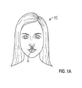
[00036] FIG. lA shows a universal respiratory detector with a circular
universal respiratory
detector 4 placed below the nose or nostril of an individual 10 and above the
upper lip region on
the philtum, for detecting a respiratory characteristic, such as carbon
dioxide in exhaled breath.
Carbon dioxide, a respiratory gas, may be used in this disclosure by way of
example of a
respiratory parameter for ease of explaining, although any of the universal
respiratory detectors
described herein may alternatively or additionally detect other respiratory
characteristics or gases
(e.g., pH of exhaled gas, oxygen concentration). Below the nostril may be a
good place to detect
a respiratory gas expelled from the nostril.
[00037] As explained in more detail below, the universal respiratory detector
4 has a
respiratory indicator configured to reversibly respond such as to change color
when a respiratory
gas parameter changes and to display a visual signal such as color change
based on the response.
The respiratory indicator may be configured to reversibly respond quickly,
such as with each
breath. The respiratory indicator may be configured to reversibly respond
within 1/2 second. A
universal respiratory detector may be configured to conform to and attach to
various surfaces. A
universal respiratory detector may be biocompatible and include a
biocompatible adhesive so it
can be attached to a person's face and skin, and stay in place for hours or
days without little or
no irritation. A universal respiratory detector may be removable so it can be
removed from a
person's face and skin with little or no damage to the face and skin. As seen
in FIG. 1D, the
face, and in particular the region around the nose and mouth has complex
geometry, with various
dips and extensions and abruptly changing concave and convex surfaces. FIG. 1D
shows the
philtrum under the nose extending and dipping at the philtral ridge. FIG. 1A
shows universal
respiratory detector 4 resting against the contours of the irregular skin
surface, conforming to the
convex and concave surfaces. When a person breathes through their nose, the
air exits from a
nostril in the nose, and the region under the nostril and near the nostril may
be a good location
for a respiratory detector. However, some individuals may breathe out through
their mouth or
may not be able to wear a respiratory detector directly below or near their
nose such as due to an
injury. FIG. 1B shows a respiratory detector for detecting a respiratory gas
with circular
universal respiratory detector 4 on the chin 24 of the individual 10. FIG. 1D
shows chin anatomy
in more detail. A chin has round or egg shape, with a crease in the chin,
referred to as the
mentolabial sulcus. The universal respiratory detector 4 is configured to
conform to the round or
egg shape as well as the crease to provide a smooth and comfortable fit. FIG.
1C shows another
variation of a respiratory detector for detecting a respiratory gas with an
ovoid shaped universal
- 6 -
CA 03183190 2022- 12- 16
WO 2021/257836
PCT/US2021/037836
respiratory detector 6 below both nostrils and crossing the philtrum and
philtral ridge of the
individual 10. In other variations, more than one universal respiratory
detector may be applied to
an individual, such as one below each nostril, or one below the nose and one
below the lips,
which may be useful for detecting a respiratory gas from an individual who
sometimes breathes
out of his or her nose and sometimes out the mouth. FIGS. 1A-1C show
individuals in need of
respiratory air monitoring who are not using a supplemental oxygen source. The
respiratory
detectors described herein may be especially useful for assessing and/or
monitoring breathing
status for an individual having or suspected of having an infectious disease
(e.g., Avian flu,
covid-19, chickenpox, Ebola, influenza, Middle Eastern Respiratory Syndrome
(MERS), Severe
Acute Respiratory Syndrome (SARS)). The respiratory detectors described herein
can be applied
and assessed/monitored with minimal contact between a caregiver and a
potentially infectious
individual, and in particular, their breath. FIG. 9 shows an individual in a
containment chamber
with a respiratory sensor 4 visible by a medical personnel 138. The
respiratory detectors
described herein can be used without a need for sterilizing or discarding
expensive equipment.
The respiratory detectors described herein could be useful for rapidly
assessing and/or
monitoring an individual who is suspected of having an illness or recovering
from an illness,
sleep apnea or other disordered sleep breathing condition, a seizing (e.g.,
epileptic) individual,
experiencing bronchospasm (e.g., asthma, COPD, an allergic reaction). The
respiratory detectors
described herein may help recognize airway obstructions before the individual
shows signs of
attack. The respiratory detectors described herein may be useful for triaging
and rapidly
determining who needs supplemental oxygen or other aid, such as while triaging
a group of
individuals involved in an accident or an attack with multiple potential
victims. These and other
respiratory detectors described herein could be useful for assessing and/or
monitoring
insufficient breathing as well as excess breathing. The respiratory detectors
described herein may
be useful for assessing and/or monitoring treatment for an individual in
respiratory distress or
transporting individuals in an ambulance or other medical transport vehicle.
The respiratory
detectors described herein may be portable and small and not require any
electrical source or
battery power. Although FIGS. 1A-1C show an individual who is not using a
supplemental
oxygen source for breathing and the universal respiratory detector placed on
the individual's
face, a universal respiratory detector may be also used on an individual using
a supplemental
oxygen source and a detector may be placed on the supplemental oxygen source
itself. FIG. 2A
shows an individual 12 using an oxygen cannula 16 for oxygen delivery to the
patient through
prongs 18 placed in the nostrils in the individual's nose. FIG. 2A shows the
individual 12 with
universal respiratory detector 4 placed under the individual's nose 32. As
shown, the universal
respiratory detector 4 is offset from the midline of the individual, which may
allow the universal
- 7 -
CA 03183190 2022- 12- 16
WO 2021/257836
PCT/US2021/037836
respiratory detector 4 to be more easily visible to a health care professional
(the detector is not
obscured from view by the oxygen cannula 16). An offset position may prevent
excess friction
from the oxygen cannula 16 removing or irritating the universal respiratory
detector 4. FIG. 2B
shows an oxygen cannula 16 being readied for placement to individual 20 with
facial hair. Facial
hair may make placing a respiratory sensor 4 on appropriate skin surface of
the individual 20
more difficult. Instead, the universal respiratory detector may be adhered to
and wrapped
partially, mostly, or all the way around part of the oxygen cannula 16. FIG.
2B shows the
universal respiratory detector 4 adhered to a surface of the oxygen cannula
16. The universal
respiratory detector 4 is readily visible to a health care provider or another
individual who can
readily ascertain if the universal respiratory detector is visually changing,
e.g., changing color,
indicative of whether or not the person is breathing. FIG. 2C shows the
universal respiratory
detector 4 attached to the facial hair (moustache) of the individual 20. A
sticker-type detector as
described herein may be well-suited for this and other indications.
[00038] FIG. 3A shows an oxygen face mask 26 for placing over an individual's
mouth and
nose to provide oxygen to the individual. Oxygen enters the interior of the
mask through oxygen
inlet 30, which uses positive pressure to move the oxygen into the interior of
the mask, where the
oxygen can be inhaled by the individual. When the individual exhales, the
expiratory gases exit
from the individual into the mask and then exit out of the mask through
exhalation ports 28.
Since gases exit the mask through the exhalation ports 28, the area near the
exhalation ports may
be a good place for sampling and sensing expiratory cases from the individual.
FIG. 3A shows
circular universal respiratory detector 4 adhered to the inside of the oxygen
face mask 26 for
sensing expiratory gases from the individual. The oxygen face mask 26 is clear
or translucent
and the universal respiratory detector 4 is visible through the material of
the mask. In particular,
any color changes in the universal respiratory detector 4 are visible from
outside the mask. This
orientation may be considered a first orientation, relative to an observer,
such as a health care
provider, outside the mask. FIG. 3B shows the oxygen face mask 26 shown in
FIG. 3A, except in
this face mask the circular universal respiratory detector 4 is adhered to the
outside of the mask
rather than on inside. Of note, the circular universal respiratory detector 4
shown in FIG. 3B is in
a second or opposite orientation and the universal respiratory detector 4 is
"flipped over"
relative to the circular universal respiratory detector 4 shown in FIG. 3A. As
will be discussed in
more detail below, the visual signal associated with the respiratory detector
is visible from both
sides. Thus, whether the universal respiratory detector is in the first
orientation shown in FIG.
3A or in the second, "flipped over" orientation shown in FIG. 3B, the visual
signal associated
with the respiratory indicator is visible to an observer. Having a universal
respiratory detector
configured for mounting and visualization in either orientation simplifies use
of the respiratory
- 8 -
CA 03183190 2022- 12- 16
WO 2021/257836
PCT/US2021/037836
indicator, as the visual sensor can be detected regardless of how the detector
is mounted. FIGS.
4A and 4B show another example of how circular universal respiratory detector
4 can be used in
a first orientation or in a second "flipped over" orientation. FIGS. 4A and 4B
show a baby 14
inside an oxygen tent 36. The oxygen tent 36 is provided with oxygen through
oxygen inlet 38.
FIG. 4A shows the universal respiratory detector 4 adhered or mounted to an
inside surface of
oxygen tent 36 in a first orientation, similar to the orientation shown in
FIG. 3A. Similar to as
described above in FIGS. 1A-1C, FIG. 4B shows the universal respiratory
detector 4 adhered to
a surface of the baby's face; in particular on chin 24 and in the opposite, or
second orientation,
compared with the universal respiratory detector 4 shown in FIG. 4A. A
detector on an oxygen
tent that is close to a face may readily receive exhaled air as the body
expels respired air under
pressure. Since the visual detector can be detected from both sides, an
observer (medical
personal or another individual) can readily assess breathing status regardless
of detector
orientation and can change from one detector to another.
[00039] FIG. 5A shows the universal respiratory detector 4 for
detecting a respiratory gas
adhered to the neck skin of a tracheostomy patient with a stoma 44 in the neck
for breathing.
FIG. 5B shows the universal respiratory detector 4 for detecting a respiratory
gas adhered to a
tracheostomy tube 46 of a tracheostomy patient with a stoma 44 in the neck for
breathing. The
universal respiratory detector 4 is located close to the opening where air
exchange takes place.
FIG. 5C shows a universal respiratory detector 4 for detecting a respiratory
gas adhered to the
inside of a tracheostomy mask 48 of a tracheostomy patient with a stoma in the
neck for
breathing. The respiratory detector in FIG. 5C is similar to the one shown in
FIG. 5B, but is
adhered in the opposite orientation relative to the detector shown in FIG. 5B.
The rapidly
reversible visual signal of the universal respiratory detector that responds
to the presence or
absence of the respiratory gas is readily visible to a caregiver or health
care provider.
[00040] A universal respiratory detector can include multiple layers, such as
2 layers, 3 layers,
4 layers, 5 layers or more than 5 layers. Layers may be different from each
other (unique) or may
be the same (duplicated, and a duplicated layer may be in the same or opposite
orientation). FIG.
6A-FIG. 6C illustrate different views of a universal respiratory detector 84
with multiple layers
and a visual indicator visible from either side. The universal respiratory
detector 84 has an
adhesive ring serving multiple purposes. FIG. 6A shows a top view of the
universal respiratory
detector. For clarity, the transparent cover 86 on the top has been omitted
from this view. Visual
indicator on the bottom of backing on respiratory indictor 88 is visible from
the top through the
transparent or translucent backing on respiratory indictor 88. FIG. 6B shows a
top view of the
universal respiratory detector including the clear film 86 on top. The middle
circles show areas
of overlap of layers. The thin ring 70 represents the overlap of respiratory
indictor 88 and
- 9 -
CA 03183190 2022- 12- 16
WO 2021/257836
PCT/US2021/037836
adhesive layer 90. FIG. 6C shows an exploded view of the universal respiratory
detector 84 and
how the differing diameters of the different layers work together to create a
versatile and easy to
manufacture detector. The universal respiratory detector 84 has a respiratory
sensor 88
configured to respond to a respiratory gas and to display a visual signal
based on the response, a
first cover 86 on a detector first (or top) side 92 and an adhesive layer 90
on a detector second (or
bottom) side 94, opposite to the first side 92. The respiratory sensor 88 is
at least partially
located between the first cover 86 and adhesive layer 90. The respiratory
sensor 88, and in
particular a color of the respiratory sensor 88, is visible from both the
sensor first side 92 and the
sensor second side 94. As explained above (and with reference to FIGS. 1A-4B)
the universal
respiratory detector 84 can be mounted in either a first orientation or a
second (-flipped over")
orientation and the color of the respiratory sensor 88 can be visualized and
assessed/monitored
by an individual from either (or both) sides. The cover 86 can be transparent
or translucent and
the respiratory sensor 88 can be viewed from the first side 92. The adhesive
layer 90 includes an
adhesive ring or -frame- and a central open region in the center of the ring
or frame through
which a visual indicator on respiratory sensor 88 can be viewed from the
second side 94.
Adhesive layer 90 has adhesive on the top side (the side facing first side 92
in FIG. 6C). In this
example, adhesive layer 90 has an outer diameter larger than an outer diameter
of the other
layers of universal respiratory detector 84. and the adhesive layer (e.g., an
outermost ring of the
adhesive layer 90 can be used for attaching the universal respiratory detector
84 to a surface
(which would be at the top side 92), such as to an individual's cheek or face,
or the inside or
outside of a mask or tent. Because of the different diameters, the adhesive of
adhesive layer 90
adheres the respiratory sensor 88 and cover 86 to itself along with the excess
material beyond the
outer diameter of cover 86 being what adheres the detector to its final
surface (cheek, mask, etc.).
[00041] A respiratory detector as described herein, such as respiratory sensor
88, includes a
respiratory sensor, such as with a visual indicator. A respiratory sensor can
include a backing and
a visual indicator disposed on the backing. In some variations, a respiratory
sensor can include a
visual indicator without a backing. A backing can be useful for providing
support for a visual
indicator, especially for a chemical indicator. A backing can be a relatively
flat layer and may
have surface features such as pores or openings. A visual indicator disposed
on a backing may be
disposed in pores or openings in the backing and/or disposed in a coating or
layer on the
backing, or both. A backing may be a first side of a respiratory sensor layer
and a visual
indicator on the second side of the layer. A transparent or translucent
backing can allow a visual
indicator to be viewed through the backing, and the visual indicator can be
detected from either
side of the backing. In some variations, a backing may be in the middle of a
respiratory sensor
layer with visual indicator on both sides (e.g., first (top) side and second
(opposite) side) of the
- 10 -
CA 03183190 2022- 12- 16
WO 2021/257836
PCT/US2021/037836
backing. A backing with visual indicator on both sides may be transparent,
translucent, or
opaque. A universal respiratory detector may have a single respiratory sensor
layer or may have
two or more than two layers of respiratory sensors. A transparent or
translucent backing can
allow multiple respiratory sensor layers to be stacked together in a
respiratory detector as the
visual signal will be visible through the transparent or translucent substrate
in the multiple layers.
A respiratory detector with multiple layers of visual indicator may provide a
stronger, brighter,
or otherwise more easily detectable visual change. In some embodiments, a
respiratory device
may include two backings (back to back), each with visual indicator on one
side. The backings
may be stacked together with the indicators facing away from each other so
that indicator can be
viewed from either side of a detector. A universal respiratory detector may
include one layer or
more than one layer (two layers, three layers, four layers, five layers, six
layers, or more than six
layers). In some embodiments of a respiratory sensor, a visual indicator is
contained within a
clear film on one or both sides.
[00042] This or any detector described herein can include an indicator
material, and in
particular a visual indicator (colorimetric) material, for detecting a
respiratory characteristic or
other chemical agent and producing and displaying visual signals in response,
such as a color
signals. The indicator material may be configured to rapidly respond to
changes, and show a
reversible and detectable color change with each inhalation and exhalation.
The indicator
material may detect presence, absence, and/or concentration or level of a
respiratory
characteristic such as a respiratory gas. A visual indicator material can
display different visible
properties in response to the presence, absence, and/or concentration or level
of a respiratory
characteristic such as a respiratory gas. A visual indicator may change
between at least two
different colors (e.g., yellow and blue; red and blue; green and red) as the
concentration of a
respiratory characteristic changes during respiration. A visual indicator may
change in color,
amount of color or shade (e.g., along the light spectrum), especially in the
visible light spectrum.
The indicator material may visually indicate the presence, absence, and/or
concentration or level
of a respiratory characteristic in a rapidly reversible reaction. A presence,
absence, and/or
concentration or level of a respiratory characteristic can be assessed
qualitatively or
quantitatively. Presence or absence of a respiratory characteristic including
of a respiratory gas
may refer to relative levels, rather than absolute levels. For example, an
indicator material may
detect the presence of sufficient carbon dioxide in exhaled air to individual
is exhaling (breathing
or respiring); however, a low of level of carbon dioxide is normally present
in non-respired air.
The low level of carbon dioxide in non-expired air is sufficiently low and a
detector may be
configured to register or consider carbon dioxide as absent or undetectable
(e.g.. as an absence of
expired carbon dioxide since the carbon dioxide present in air is not due to
an individual's
- 11 -
CA 03183190 2022- 12- 16
WO 2021/257836
PCT/US2021/037836
breathing/expiration). Thus, in practice, an absence of carbon dioxide
indicate that breathing or
respiring is not occurring at a sufficient level to support the individual.
Exhaled gas is typically
4% to 5% carbon dioxide while air or inhaled gas is typically 0.03% to 0.04%
carbon dioxide.
Exhaled gas shows a 100 fold increase in the amount of carbon dioxide relative
to inhaled gas
(non-respired air). A qualitative or quantitative assessment of gas showing or
suggesting less
than about 4% to 5% carbon dioxide, such as more than 5X (e.g. 1.2% or 1.0%)
lower, more than
10X lower, more than 50X lower, or more than 100X lower, or less than 1%
carbon dioxide, less
than 0.5% carbon dioxide, less than 0.1% carbon dioxide, or less than 0.05%
carbon dioxide with
an indicator material may be considered as a sufficiently low level of carbon
dioxide to indicate
that respiration or breath expiration is not adequately detected. It is noted
that although respired
air generally contains more than 4% carbon dioxide, a respiratory detector as
described herein
may detect less than that and respiration may be considered acceptable. For
example, a
respiratory detector placed on an inner surface of an oxygen tent or a check
of an individual may
encounter respired air mixed with room air, resulting in a lower, but still
acceptable amount of
carbon dioxide, indicative of acceptable respiration for that situation.
Similarly, room air or other
inhalable air can contain around 21% or more oxygen, while exhaled air
contains around 16%. A
respiratory detector as described herein for detecting oxygen may detect more
than 16% oxygen;
however the individual may be respiring. Detection may be calibrated by
considering the
difference or cycling behavior of the indicator, rather than by absolute
signal, such as absolute
signal intensity or signal strength.
[000431 A respiratory indicator may be configured to change colors in response
to changes in
a respiratory characteristic, and in particular, to reversibly change colors
as a respiratory
characteristic cycles with the respiratory cycle of inhalation and exhalation.
A respiratory
indicator for detecting carbon dioxide can include sodium carbonate with
thymol blue and
glycerol or propylene glycol. Another reaction includes monocthanoloamine with
mctacrestol
purple or thymol blue with propylene glycol.
[00044] In the broadest sense, a carbon dioxide (C09) indicator may be any
convenient
indicator that is capable of transducing a change in CO2 concentration of a
gas contacting the
indicator into a detectable change, such as a detectable visual change, e.g.,
a colorimetric change.
CO2 indicators of interest include, but are not limited to, those described in
U.S. Pat. Nos.
4,728,499; 4,879,999; 4,994,117; 5,005,572; 5,156,159; 5,166,075; 5,179,002;
6,436,347;
6,584,974; and U.S. Patent Application Publication No. 2006/02168282; the
disclosures of
which with respect to CO2 indicator compositions are herein incorporated by
reference.
[00045] Some variations include a long lasting CO2 indicator that exhibits a
dynamic, rapid
response reversible CO2 indication with breath-to-breath sensitivity and is
storage stable. The
- 12 -
CA 03183190 2022- 12- 16
WO 2021/257836
PCT/US2021/037836
colorimetric CO2 indicator of embodiments disclosed herein changes color upon
exposure to
changes in concentrations of CO2 found in expired air (e.g. from purple to
yellow). In certain
embodiments, the CO2 indicator can change color, e.g. from purple to yellow,
in 2.5 seconds or
less, such as 2 seconds or less and including 0.75 seconds or less in response
to a change in CO2
concentration in a gas contacting the indicator. The indicator is sensitive to
changes in CO,
concentration of 3% or less, such as 2% or less. including 1% or less. At CO,
concentrations of
0.05% or less, such as 0.03% or less, the indicator is a first color, while at
concentrations above
these amounts, the indicator is a second color. For example, in certain
embodiments, the
indicator exhibits the following colors at the following CO, concentrations:
<0.03%, purple;
0.5% light purple; 2% brownish yellow; 5% yellow. The color change can be any
of a variety of
different color changes, e.g., purple to yellow, blue to yellow, red to
yellow, orange to yellow,
etc.
[00046] Some embodiments include the combination of various components in a
concentration and ratio sufficient to provide a dynamic, rapid response
reversible CO2 indicator
with breath-to-breath sensitivity, e.g., as described above. In one
embodiment, the components
of the CO, indicator include a pH sensitive indicator dye(s) and a phase
transport enhancer.
[00047] pH sensitive indicator dyes of interest include, but are not limited
to: bromothymol
blue, phenolphthalein, thymol blue, phenol red, rosolic acid, m-nitrophenol,
xylenol blue,
curcumin, cresolphthalein, thymolphthalein, malachite green, N,N-
dimethylaniline, and cresol
dyes, e.g., bromocresol green, bromocresol purple, cresol red, m-cresol
purple, etc. In certain
embodiments, the pH sensitive indicator dye is a cresol dye or combination
thereof, e.g., a
combination of m-cresol purple and cresol red.
[00048] In addition, the pH sensitive indicator dye, another component present
in the indicator
described herein can be a phase transport enhancer. Phase transport enhancers
contained as part
of the dye solution applied to the support surface, enhance response of the
dye to CO2 gas as well
as alter the color and visibility of the indicator. Phase transport enhancers
include, but are not
limited to: quaternary ammonium, phosphonium or pyridinium salts. Quaternary
salts which are
useful in sensors described herein have the formula (I):
R2
R1-X+-R3 Y-
1
[00049] R4
[00050] wherein:
[00051] X=N or P;
- 13 -
CA 03183190 2022- 12- 16
WO 2021/257836
PCT/US2021/037836
[00052] R 1. R2, R3 and R4 are selected from the group consisting of C1-C16,
such as C1-C12
alkyl, triphenylmethyl, phenyl, naphthyl and benzyl. Cl-C4 substituted alkyl
wherein the
substituent is a C1-C4 alkyl or phenyl group, wherein R1, R2, R3 and R4 may be
the same or
different, e.g., have the same or different number of carbon atoms; and Y¨ is
an anion selected
from the group consisting of hydroxide, fluoride, chloride, bromide, iodide,
carbonate and
tetrafluoroborate.
[00053] Phase transport enhancers which are useful in some embodiments
include, but are not
limited to: tetrabutylammonium hydroxide; tetrabutylammonium chloride;
tetraethylammonium
bromide; tetraethylammonium p-toluenesulphonate; phenyltrimethylammonium
chloride;
benzyltrimethylammonium bromide; tetra-n-propylammonium bromide;
benzyltriethylammonium tetrafluoroborate; n-Dodecyltrimethylanamonium bromide;
tetraphenylphosphonium chloride; n-Hexadecylpyridinium bromide; and
(Triphenylmethyl)triphenyl phosphonium chloride.
[00054] Some embodiments can be produced by combining the various components
of the
indicator composition to produce a precursor indicator reagent fluid and then
contacting the fluid
with a suitable solid support in a manner sufficient to produce the desired
indicator composition.
In certain embodiments, the precursor fluid is an aqueous solution, such as a
basic aqueous
solution, that includes the above described pH sensitive dye and phase
transport components.
The basic solution has, in certain embodiments, a pH ranging from 10 to 12.5.
The composition
may include one or combination of pH sensitive indicator dyes. In certain
embodiments, the
composition includes more than one pH sensitive indicator dyes, such as 2 to 5
different dyes,
e.g., 2 to 4 different dyes, including 2 to 3 different dyes, e.g., 2
different dyes. In certain
embodiments, the dyes are cresol dyes, such as 2 different cresol dyes. When
the composition
includes two different pH sensitive indicator dyes, the pH sensitive indicator
dyes can be present
in a concentration ranging from 0.0001 Molar to 0.01 Molar, including about
0.002 Molar to
0.003 Molar. In certain embodiments, the dyes are m-Cresol purple and cresol
red. M-cresol
purple can be present in the reagent fluid in a concentration ranging from
0.001 Molar to 0.01
Molar, including about 0.002 Molar to 0.003 Molar. Cresol red sodium salt can
be present in
reagent fluid in a concentration ranging from 0.0001 Molar to 0.001 Molar,
including about
0.002 Molar to 0.003 Molar. The concentration of phase transport enhancer may
vary. In certain
embodiments, the amount of phase transport enhancer present in the reagent
fluid ranges from
0.001 Molar to 0.02 Molar, such as from 0.005 Molar to 0.01 Molar.
[00055] Following preparation of the precursor fluid, the methods can include
contacting the
fluid with a solid support, and then removing excess fluid from the solid
support to produce the
indicator. Any convenient solid support may be employed. In certain
embodiments, the solid
- 14 -
CA 03183190 2022- 12- 16
WO 2021/257836
PCT/US2021/037836
support is a flexible solid support (e.g. a cellulosic material), e.g., paper.
In certain embodiments,
the solid support may be a filter paper, e.g., having a porosity ranging from
1 pm to about 60 pm,
such as from 20 lam to about 30 pm. The solid support can be a material
dimensioned to fit on
skin and or inside an oxygen delivery device, such as described herein. The
support of the CO2
indicator can be shaped into any desired configuration, including but not
limited to: circular or
spiral strips, a sphere or portion of a sphere, a propeller, an accordion
shape, etc. The support of
the indicator can further comprise a pattern, and/or can have perforations, as
described in the
above embodiments.
[00056] The above described indicators can be used in any of a number of
different
respiratory detectors. In certain embodiments, the indicators are employed
with respiratory
detectors that do not include a sterilization barrier, as the indicator of
such embodiments can
survive the sterilization, e.g., EtO, process.
[00057] Returning to FIG. 6A-FIG. 6C, the adhesive layer 90 can be in the form
of a ring or
frame surrounding an open center portion. The open center portion allows
respired air to contact
the respiratory sensor 88 during detector use. The respiratory sensor 88 is
visible through the
center or "picture" portion of the ring or frame such as to an individual
assessing and/or
monitoring the individual's respiratory status. Adhesive layer 90 may include
a substrate with
one or more adhesive compounds on a top side of the adhesive layer (e.g., the
side facing the
respiratory sensor 88 and sensor second side 94). The adhesive layer 90 may
adhere to an outer
portion of respiratory sensor 88, as well as to the bottom of cover 86 and may
hold the adhesive
layer 90, respiratory sensor 88, and cover 86 together. An adhesive in
adhesive layer 90 that
contacts the respiratory sensor 88 may be an adhesive that does not interfere
with or only
minimally interferes with visual indicator performance. Examples of adhesives
that may be used
in adhesive layer 90 include no or low off-gassing adhesives such a silicone
adhesive, a low
volatile organic compound adhesive (low VOC) such as a low VOC acrylic. In
some variations,
the adhesive layer 90 can additionally or instead have an adhesive surface
facing the sensor first
side 92 for attachment of the respiratory indicator to a surface at the sensor
first side 92. The
adhesive portion on adhesive layer 90 may cover part or all of the top side of
adhesive layer 90.
In some variations, the adhesive layer 90 may not cover the entire top side of
adhesive layer 90.
Adhesive layer 90 may take other forms, such as a discontinuous adhesive
(e.g., an array of
small circles or squares of adhesive) or a 2-dimensional spiral form with an
adhesive spiral and
open (non-adhesive) areas, or another form, and the respiratory sensor 88 may
be visible through
the discontinuities or open areas in the adhesive. In some variations, the
adhesive layer 90
includes an air permeable or breathable membrane in part or all of the center
portion. A
breathable membrane allows expired air from the individual to flow through and
contact
- 15 -
CA 03183190 2022- 12- 16
WO 2021/257836
PCT/US2021/037836
respiratory sensor 88, and may provide protection for the otherwise exposed
(bottom) surface of
respiratory sensor 88. In some variations, an adhesive layer, such as adhesive
layer 90, may
cover substantially an entire sensor second surface. An adhesive that is clear
(or translucent)
may be well suited to cover an entire sensor first surface as a signal (visual
signal) from a
respiratory indicator could be visualized through a clear or translucent
adhesive surface. In some
variations, an adhesive may be opaque. Although FIG. 6A-FIG. 6C show the
adhesive layer 90
adjacent the respiratory sensor 88 in the respiratory detector 84, in some
variations, an adhesive
could be provided separately from the respiratory indicator and attached to a
respiratory detector
during application of the respiratory detector to an individual with the
adhesive. For example, an
adhesive could be configured (and packaged) as a separate layer (with a
backing) or a gel that
may be applied/attached to the rest of respiratory sensor 84 (cover 86 and
respiratory sensor 88).
In some variations, a detector may have an adhesive on adhesive layer such as
adhesive layer 90
in FIG. 6A-FIG. 6C for attaching respiratory sensor 88 to a cover 86. In some
variations,
adhesive layer 90 may attach a detector to an individual. In some variations,
a detector may have
an adhesive, such as on the top of cover 86 for attaching a detector to an
individual, in addition
to or instead of adhesive on adhesive layer 90. A respiratory detector placed
on and adhered to
facial skin or an oxygen delivery device may stay in place on the skin or
delivery device for a
period of time (minutes, hours, days, weeks) and then be removed and thus a
respiratory detector
may need to be removable. In some cases, a respiratory detector may be removed
and replaced
one time or more than one time by a fresh/new additional respiratory
detector(s) placed on facial
skin or an oxygen delivery. These additional respiratory detector( s) may also
stay in place for a
period of time (minutes, hours, days, weeks). Ease of applying and replacing
the respiratory
detector may be important. Other detector characteristics that may be of
interest for a respiratory
detector described herein include stability, ease of use, conformability,
flexibility, ability to
adhere, comfort, skin irritability, and comfort of removal. In some
embodiments, a visual
indicator is configured to reversibly change color when a respiratory gas
parameter changes and
to display the color change for a period of time lasting at least 10 minutes,
at least one hour, at
least ten hours, at least one day, at least three days, at least one week, or
at least two weeks or
between these amounts. A respiratory detector may be configured for
conformability and
flexibility for conforming to uneven facial skin or an irregularly shaped
oxygen delivery device,
such as an oxygen cannula and for flexibility to stay in place and be
comfortable to wear after
being placed. As indicated above, it may be desirable to place a respiratory
detector close to
where breath is exhaled, such as on facial skin between the nose and mouth,
areas which may be
irregular or flat and may have either or both convex and concave areas. A
detector that can
sufficiently conform for mounting and is flexible as the person speaks,
laughs, or otherwise
- 16 -
CA 03183190 2022- 12- 16
WO 2021/257836
PCT/US2021/037836
moves their face may be desirable. It can be very difficult to get anything to
adhere to skin, and
especially to adhere to facial skin. Facial skin is delicate, sensitive, and
prone to damage,
irritation, skin rashes, and acne and an adhesive of a respiratory indicator
(as well as other parts
of a respiratory indicator) may be hypoallergenic and non-irritating for a
period of hours or days.
An optimal adhesive for adhering a respiratory indicator to an oxygen delivery
device may not
be the optimal adhesive for adhering a respiratory indicator to an
individual's facial skin and vice
versa. Facial skin has a low surface energy in the best of circumstances,
making it difficult to
adhere an adhesive to it. Different individuals also have a wide range of skin
conditions that
affect the ability of an adhesive to adhere a respiratory indicator to facial
skin and a universal
respiratory detector may be configured to provide the best adhesion for a
number of
circumstances. For example, individuals have different sebum levels, dryness,
sweating, and
facial hair. The presence of any face cream, ointment, or sunscreen on facial
skin affects
adhesion and respiratory detector adhesion to the facial skin. It can be very
difficult to get
anything to adhere to facial skin and painful or damaging to the skin to
remove it, even on
healthy skin.
[00058] As indicated above, other adhesive characteristics that may be of
interest for a
respiratory detector described herein are ease and comfort of removal.
Concerns about skin
trauma during adhesive removal may include concerns about skin tears and skin
stripping. Facial
skin in younger patients such as babies as well as in elderly patients may be
particularly sensitive
to skin trauma. As skin ages, its dermal thickness decreases, leading to a
thinning of the skin,
making the skin more vulnerable to damage. Aging skin predisposes an
individual to skin tears,
such as painful and unsightly separation of the epidermis layer of the skin
from the underlying
dermal layer. These factors make atraumatic removal of an adhesive in a
respiratory detector
more challenging. Ease of wear of a respiratory detector, comfort during
removal of a respiratory
detector, and ensuring the respiratory detector factor stays in place during
use may be considered
when choosing an adhesive. For example, if the degree of adhesion is too low,
the respiratory
sensor might not reliably stay in place. If the degree of adhesion is too
high, the respiratory
sensor may be difficult to remove and removal may cause damage to facial skin
or an oxygen
delivery device. An adhesive for adhering a respiratory device to skin or an
oxygen delivery
device and for gentle removal may include the adhesive and detector releasing
cleanly from skin
or device, leaving the skin area intact and leaving behind no or little
residue during removal.
Gentle removal from skin may also include minimal or no pain during removal.
An adhesive
may be configured to be atraumatic during use and removal. Adhesive
performance for a
respiratory sensor may balance different characteristics. Adhesive performance
can be
characterized in part by adhesive tack, peel, and/or shear. Tack is a measure
of how quickly a
- 17 -
CA 03183190 2022- 12- 16
WO 2021/257836
PCT/US2021/037836
bond is formed between two surfaces, such as between an adhesive and a surface
(e.g., skin or
oxygen delivery device), and may be used to refer to pressure sensitive
adhesives. In some
embodiments, an adhesive is configured to be tacky or non-tacky at room
temperature. To assay
tack, two surfaces are brought together briefly under light pressure, then
pulled apart. The more
force needed to separate them, the higher the tack. Lower tack may allow an
adhesive to be
repositioned. Another characteristic of an adhesive is peel. Peel is a measure
of the force needed
to break a bond between the adhesive and the surface (e.g., skin or oxygen
delivery device) to
which it has been applied. A peel test to assay peel can be performed. In peel
testing, an adhesive
tape is applied to a surface, allowed to sit, and then pulled away. Peel angle
or direction,
application pressure and the length of time the surfaces stay bonded may be
defined, such as in
ASTM D330D standard test method to measure peel adhesion strength in a
pressure sensitive
tape. In some examples, an adhesive is allowed to sit on a surface at least
one hour, at least two
hours, at least three hours, at least five hours, at least ten hours, at least
twenty-four hours, at
least forty eight hours or at least sixty hours or less than sixty hours, less
than forty eight hours,
less than twenty four hours, less than ten hours, less than five hours, less
than four hours, less
than three hours, less than two hours, less than one hour, or any amount of
time between these (at
least ten hours and less than forty eight hours, etc.). Another characteristic
of an adhesive is
shear. Shear refers to one surface sliding over another. In a shear test, a
sample is mounted
vertically and has a weight attached. The time is takes for the sample to slip
off the substrate
shows the durability of the bond. An adhesive may include sufficient tackiness
to adhere to a
device and/or skin and good shear and peel character to remain on the device
and/or skin and be
readily removable from a device and/or skin. An adhesive for a respiratory
detector may be or
include acrylics, hydrocolloids, hydrogels, rubber-based adhesives, or
polyurethane based
adhesives. Examples of adhesive polymers for an adhesive include polysiloxane
or silicone
(BIO-PSA from Dow Corning ), polyisobutene (Oppano10), a syrene-isoprene-
styrene
copolymer (JSR-SIS), or an acrylic polymer (Duro-Tak). A substrate for an
adhesive may be a
sheet or film, such a foam, polymer, plastic, or polyester resin (mylar) sheet
or film. A substrate
may be clear, translucent, or opaque. In some examples, a diameter (outer
diameter) or other
longest dimension (e.g., a length or diagonal of a non-circular shaped layer)
of an adhesive layer
may be about 1 inch, or less than 2 inches, less than 1.5 inches, less than 1
inch or less than 0.5
inch or less than 0.25 inches or at least 0.5 inches, at least 1.0 inches, at
least 1.5 inches or at
least 2.0 inches or between these amounts (e.g., at least 0.5 inches and less
than 1 inch, at least
0.5 inches and less than 1.5 inches).
[00059] In some variations, the sensor first side may have a cover over at
least the center
("picture") portion of the ring or frame of adhesive layer 90 instead or in
addition to the first
- 18 -
CA 03183190 2022- 12- 16
WO 2021/257836
PCT/US2021/037836
cover. As indicated above, a universal respiratory detector first side may
have a cover over the
center or "picture" portion of the ring or frame of adhesive layer 90 instead
or in addition to the
first cover. In some embodiments, a cover is a film. In some variations, a
respiratory sensor does
not have a cover.
[000601 FIG. 7 shows an exploded view of another respiratory detector,
universal respiratory
detector 116. Similar to as described above for universal respiratory detector
84 in FIG. 6A-FIG.
6C, the universal respiratory detector 116 shown in FIG. 7 has a respiratory
sensor 88 configured
to respond to a respiratory parameter (e.g., a gas, such as CO?) and to
display a visual signal
based on the response. The universal respiratory detector 116 has an adhesive
layer 90 on a
sensor second side 114, and a first cover 86 and respiratory sensor 116
includes release liner 96
on detector first side 112. As indicated above, respiratory sensor 88 and any
other respiratory
sensor described herein may have a backing and a visual indicator on the
backing. A backing
may be a porous material and the backing may be configured to allow a visual
indicator to
penetrate the pores or otherwise hold a visual indicator during detector
manufacture, storage, and
use. Pores of a backing may be irregular (e.g., in a polymer) or regular
(e.g., laser drilled). Pores
may have an average pore size of around 0.65 um, or from about 0.45 urn to 0.8
urn, or from
about 0.1 um to about 2 um, or from about 0.03 um to about 5.0 um or any size
between these. In
some variations and as indicated elsewhere herein, average pore size may be
larger than 5 um. A
visual indicator may be configured to indicate/change with each breath such as
within 3 seconds,
within 2 seconds, within 1 second, or within 0.5 seconds. A backing may be a
polymer such as
polyethersulfone, polysulfone, or polyphenylene sulfone. A backing may be
clear or translucent
such that a visual indicator can be visualized through the backing (e.g., the
visual indicator can
be assessed from both sides of the universal respiratory detector 94) or may
be opaque. Visual
indicators, such as indicated elsewhere herein, can be adhered or attached to
a backing, such as
using pad printing. Visual indicators can be printed to the bottom side (e.g.,
facing second side
114) of respiratory sensor 88 by pad printing. In some examples, visual
indicators printed on the
second side of a respiratory sensor, such as respiratory sensor 88, contact
adhesive, such as on
adhesive layer 90 when the respiratory sensor 88 and adhesive layer 90 are
joined together. In
some examples, visual indicators are printed so that they do not contact
adhesive when the
respiratory sensor 88 and adhesive layer 90 are joined together. Contact of
visual indicator by an
adhesive may have a detrimental effect on a visual indicator, such as reducing
shelf life or visual
indicator efficacy. Preventing or minimizing contact between visual indicators
and adhesive may
improve visual indicator life. A solution including visual indicator,
plasticizer, and isopropyl
alcohol may be placed onto a backing (e.g., polyethersulfone membrane) and the
visual indicator
cured onto the substrate and isopropyl alcohol flashed off by heating at 80 C
for 10 min or 90 C
- 19 -
CA 03183190 2022- 12- 16
WO 2021/257836
PCT/US2021/037836
for 5 min. Respiratory sensor 88 also includes first cover 86. First cover 86
may be laminated to
the top side of respiratory sensor 88 and the top side of respiratory sensor
88 may be sealed by
first cover 86 to protect the visual indicator 88 and increase sensor
durability. First cover 86 may
protect the visual indicator from humidity and prevent oxidation. In some
examples, the visual
indicator is configured and protected to provide visual indication for at
least one day, at least two
days, at least three days, or at least four days of use. In some examples, a
universal respiratory
detector described herein such as universal respiratory detector 84 is
protected on its edges by a
coating, film, or bead.
[00061] FIG. 7 also shows respiratory sensor 88 includes a release liner 96. A
release liner,
such as release liner 96, can be a thin, removable layer that protects the
universal respiratory
detector 116. In particular, release liner 96 may protect adhesive layer 90
and maintain its
adhesive integrity prior to detector mounting, keeping it out of contact with
air, dirt, or other
objects and preventing it from premature adhering. In some variations, a
respiratory detector may
have a second release liner for a second adhesive layer or second adhesive
side. A release liner,
such as release liner 96, is removed prior to universal respiratory detector
94 mounting and use.
Steps in methods of use of a release liner may include protecting a universal
respiratory detector
with a release liner, and removing or separating the release liner from rest
of the universal
respiratory detector. A release liner may be larger or longer (in diameter,
width, and/or length)
than another part of a universal respiratory detector, or may be smaller or
shorter (in diameter,
width, and/or length) than another part of a universal respiratory detector,
or may be the same
size as another part of a universal respiratory detector. In some variations,
a universal respiratory
detector may have a release liner on the sensor first side instead of, or in
addition to, the release
liner on the sensor second side 114 and a respiratory detector may have zero,
one, two, or more
release liners. A release liner may be a substrate coated on one or both sides
with a release agent,
configured to separate the release liner from adhesive (and/or other material)
of the respiratory
sensor. A release liner can be a paper, plastic, fluoropolymer, polyethylene
film or layer coated
with silicone. A release liner can be smooth or textured with release
properties to allow for
adhesion of the adhesive while permitting clean separation from the adhesive
during respiratory
sensor application to a surface. A release liner may include one or more cut
features or
extensions, such as a cutout or hole in the middle as shown in FIG. 7 or a
partial line cut(s) or a
tab extension that may aid is respiratory sensor application and/or removal.
In some examples, a
diameter (inner diameter) or other dimension (e.g., diagonal of a rectangular
shaped layer) of an
internal opening on release layer may be less than 2 inches, less than 1.5
inches, less than 1 inch
or less than 0.5 inch or at least 0.5 inches, at least 1.0 inches, at least
1.5 inches or at least 2.0
inches or between these amounts (e.g., at least 0.5 inches and less than 1
inch, at least 0.5 inches
- 20 -
CA 03183190 2022- 12- 16
WO 2021/257836
PCT/US2021/037836
and less than 1.5 inches), such as at least 0.5 inches and less than 1.0
inches, at least 0.5 inches
and less than 1.5 inches. A release liner may be sized to extend beyond the
perimeter of the rest
of the respiratory sensor, such as by at least 0.25 inches, at least 0.5
inches, or at least 1 inch.
Another layer may also or instead have a tab or ring extension. A cut feature
or extension may
aid in detector placement or aid in release liner separation from the adhesive
or another part of
the universal respiratory detector. A cut feature or extension such as in an
adhesive layer and a
detector chemical may aid in minimizing contact between the adhesive and the
detector
chemical. Such minimization may be advantageous, for example, if an adhesive
has a detrimental
effect on the stability of a detector chemical. A tab may be rectangular, V-
shaped (narrow near
the body), inverted V-shaped (wide near the body), rounded, or another shape.
A tab may be flat
and may have the same or similar thickness as the layer to which it is
connected. A tab may be
flexible or inflexible. As indicated above, a respiratory detector may be
circular or ovoid. In
these and other embodiments, the universal respiratory detectors has no sharp
edges or corners.
A circle can be the most efficient and stable shape since sensor degradation
can start from the
outer edges and move inward. Other shapes are also contemplated for a
respiratory detectors.
FIG. 8A shows a rectangular universal respiratory detector 120. FIG. 8B shows
a rounded square
rectangular universal respiratory detector 122. FIG. 8C shows a triangular
rectangular universal
respiratory detector 124. FIG. 8D shows a cut rectangular universal
respiratory detector 126.
FIG. 8E shows a decorated rectangular universal respiratory detector 128 with
graphics. FIG. 8F
shows a crescent shaped rectangular universal respiratory detector 130. FIG.
8G shows a split
rectangular universal respiratory detector 132 with two parts 131. Parts 131
may contain
adhesive, indicator, or any respiratory detector material as described herein.
FIG. 8H shows a
starburst shaped 134. FIG. 10 shows an exploded view of a universal
respiratory detector 146
similar to as described above but with respiratory sensor 148 with four tabs
150. When adhesive
layer 152 is in place over respiratory sensor 148, the amount of contact
between adhesive layer
152 and respiratory sensor 148 can be minimized. For example, the contact may
be limited to the
region between a tab and the adhesive layer and/or a thin ring 154 around the
outer
circumference of the respiratory sensor 148. A respiratory sensor 148 may have
three, four, or
five tabs (or may have another number of tabs such as one tab, two tabs, or
more than five tabs
or no tabs). The tabs (or extent of respiratory sensor 148 if no tabs) may
extend so that the
outermost dimension size of the respiratory sensor 148 is the same outermost
dimension size
(e.g., circumference) as some or all of the other layers in a respiratory
sensor. Having the same
outermost dimensions for some or all of the layers may aid in manufacturing: a
series of layers of
materials may be layered together and respiratory sensors, such as respiratory
sensor 146, may be
punched out from the layers. A plurality of detectors can be manufactured
together in sheet or
- 21 -
CA 03183190 2022- 12- 16
WO 2021/257836
PCT/US2021/037836
web/roll form. In another embodiment, the various layers in a respiratory
sensor, such as
respiratory sensor 146, can be separately shaped (punched or cut), and then
lined up after
shaping (punching or cutting) to form an assembled respiratory sensor either
separately or in
groups in which a number of detectors are simultaneously assembled. The tabs
may extend
beyond the circumference of the main body (e.g., outwardly from ring 154 as
indicated by the
arrow in FIG. 10 by not more than 0.1 inches, not more than 0.2 inches, not
more than 0.3
inches, not more than 0.4 inches, or more than 0.4 inches. The tabs may be not
more than 0.1
inches in width as indicated by the double arrow in FIG. 10, not more than 0.2
inches, not more
than 0.3 inches, not more than 0.4 inches, or more than 0.4 inches in width.
These extension and
width dimensions may also apply to other shaped tabs, such as to the biggest,
smallest, or
average size of a V shaped tab.
[00062] A respiratory sensor, such as universal respiratory detector
4, 6, 116, 120, 122, 124,
126, 128, 130, 132, 134 may be configured as stickers (e.g., be flexible,
pliable, and thin and
able to conform to a surface, able to adhere to a surface upon contact or
minimal applied
pressure) and may be configured as removable stickers. Such sensors may be
about 0.01 inches
thick, such as between 0.005 inches and 0.05 inches or between 0.001 inches
and 0.1 inches.
Each layer may be thin and flexible. Release liner 156 in FIG. 10 may be about
0.002 inches
thick, such as between 0.0005 inches and 0.008 inches or between 0.0002 inches
and 0.02 inches
or between 0.001 inches and 0.1 inches. Cover 158 in FIG. 10 may be about
0.002 inches thick,
such as between 0.0005 inches and 0.008 inches or between 0.0002 inches and
0.02 inches or
between 0.001 inches and 0.1 inches. Cover 158 in FIG. 10 may be about 0.002
inches thick,
such as between 0.0005 inches and 0.008 inches or between 0.0002 inches and
0.02 inches or
between 0.001 inches and 0.1 inches. Respirator indicator 148, which may
include a backing
with a visual indicator may be about 0.004 inches thick, such as between
0.0004 inches and 0.04
inches or between 0.0002 inches and 0.02 inches. In some variations, a
respiratory sensor may
have a rigid housing. A universal respiratory detector as described herein may
be conformable to
many types of oxygen delivery systems (e.g., facemask, nasal cannula,
continuous positive
airway pressure machines (CPAP), tracheostomy collar, oxygen tent). A
universal respiratory
detector may be a single use, disposable, latex free, non-metallic, magnetic
resonance imaging
(MRI) safe and/or computerized tomography (CT) safe. A universal respiratory
detector as
described herein may have sufficient visibility to be visible from at least 5
feet away or at least
10 feet away. Once manufactured, a universal respiratory detector as described
herein can be
packaged in an airtight packet (e.g., mylar). The entire packet may be small
(e.g., less than 1.5
inches or less than 1 inch on each side and less than 0.1 inches, or less than
0.05 inches thick.
Some embodiments of a respiratory detector include a backing, an adhesive
layer, a visual
- 22 -
CA 03183190 2022- 12- 16
WO 2021/257836
PCT/US2021/037836
indicator, and an opaque surrounding layer. An opaque surrounding layer may
contain printed
graphics such as instructions or decoration.
[00063] Examples:
[00064] Example 1: Universal respiratory detectors manufactured as described
herein were
subjected to accelerated stability testing to quickly and accurately measure
and estimate the
stability of universal respiratory detectors. The universal respiratory
detectors were subject to
extreme conditions that increase the rate of chemical and/or physical
degradation that would
occur under normal storage conditions. Universal respiratory detectors were
manufactured as
shown in FIG. 10 with a (top) release liner layer, a clear polyester layer
with adhesive on the top
surface, a visual indicator on the topside of a polyethersulfone backing, and
a (bottom) polyester
with a low off-gassing acrylic adhesive on top and a cutout in the middle. The
sensors were
sealed in a mylar foil package, heat sealed, and artificially aged in an oven
at 55 C. After 18 days
at 55 C, which equates to 6 months of real time, the sensors showed no signs
of degradation.
[00065] Example 2: Universal respiratory detectors manufactured as described
herein were
subjected to accelerated stability testing to quickly and accurately measure
and estimate the
stability of universal respiratory detectors. The universal respiratory
detectors were subject to
extreme conditions that increase the rate of chemical and/or physical
degradation that would
occur under normal storage conditions. Universal respiratory detectors were
manufactured as
shown in FIG. 10 with a (top) release liner layer, a clear polyester layer
with adhesive on the top
surface, a respiratory indicator layer on the topside of a polyethersulfone
backing, and a (bottom)
polyester with a silicone adhesive on top and a cutout in the middle. The
sensors were sealed in a
mylar foil package, heat sealed, and artificially aged in an oven at 55 C.
After 36 days at 55 C,
which equates to 12 months of real time, the sensors showed no signs of
degradation.
[00066] Example 3: Universal respiratory detectors manufactured as described
herein were
subjected to accelerated stability testing to quickly and accurately measure
and estimate the
stability of universal respiratory detectors during universal respiratory
detector use. The
universal respiratory detectors were subject to extreme conditions that
increase the rate of
chemical and/or physical degradation that would occur under normal storage
conditions.
Universal respiratory detectors were manufactured as shown in FIG. 10 with a
(top) release liner
layer, a clear polyester layer with adhesive on the top surface, a visual
indicator layer on the
topside of a polyethersulfone backing, and a (bottom) polyester with an
adhesive on top and a
cutout in the middle. The sensors were not sealed in packaging. The sensors
were left open to the
atmosphere (air). The sensors were artificially aged in an oven at 55 C. After
10 days at 55 C,
which equates to 3 months of real time, the samples showed a 50% reduction in
efficiency: 50%
of the chemistry indicator was permanently yellow and 50% would still change
from blue to
- 23 -
CA 03183190 2022- 12- 16
WO 2021/257836
PCT/US2021/037836
yellow and back. The pattern of degradation was typically in the shape of a
circle/oval, in which
the degradation moved from the outer edges inward (e.g., degradation was noted
nearer the
interface of the respiratory sensor layer (chemical) with the adhesive,
suggesting that adhesive
may have a negative impact on respiratory indicator (chemical) stability.
[00067] Example 4: Polyethersulfone membranes were overlaid with solution of
visual
indicator, plasticizer, and isopropyl alcohol. Membranes were heated from 80 C
to 180 C and
visually assessed. Membranes heated at 80 C for 10 minutes or 90 C for 5 min
had acceptable
color. Membranes heated at 180 C for 5 min were unacceptable.
[00068] When a feature or element is herein referred to as being "on" another
feature or
element, it can be directly on the other feature or element or intervening
features and/or elements
may also be present. In contrast, when a feature or element is referred to as
being "directly on"
another feature or element, there are no intervening features or elements
present. It will also be
understood that, when a feature or element is referred to as being
"connected", "attached" or
"coupled" to another feature or element, it can be directly connected,
attached or coupled to the
other feature or element or intervening features or elements may be present.
In contrast, when a
feature or element is referred to as being "directly connected", "directly
attached" or "directly
coupled" to another feature or element, there are no intervening features or
elements present.
Although described or shown with respect to one embodiment, the features and
elements so
described or shown can apply to other embodiments. It will also be appreciated
by those of skill
in the art that references to a structure or feature that is disposed
"adjacent" another feature may
have portions that overlap or underlie the adjacent feature.
[00069] Terminology used herein is for the purpose of describing
particular embodiments
only and is not intended to be limiting of the invention. For example, as used
herein, the singular
forms "a", "an" and "the" are intended to include the plural forms as well,
unless the context
clearly indicates otherwise. It will be further understood that the terms
"comprises" and/or
"comprising," when used in this specification, specify the presence of stated
features, steps,
operations, elements, and/or components, but do not preclude the presence or
addition of one or
more other features, steps, operations, elements, components, and/or groups
thereof. As used
herein, the term "and/or" includes any and all combinations of one or more of
the associated
listed items and may be abbreviated as "/".
[00070] Spatially relative terms, such as "under", "below",
"lower", "over", "upper" and
the like, may be used herein for ease of description to describe one element
or feature's
relationship to another element(s) or feature(s) as illustrated in the
figures. It will be understood
that the spatially relative terms are intended to encompass different
orientations of the device in
use or operation in addition to the orientation depicted in the figures. For
example, if a device in
- 24 -
CA 03183190 2022- 12- 16
WO 2021/257836
PCT/US2021/037836
the figures is inverted, elements described as "under" or "beneath" other
elements or features
would then be oriented "over" the other elements or features. Thus, the
exemplary term "under"
can encompass both an orientation of over and under. The device may be
otherwise oriented
(rotated 90 degrees or at other orientations) and the spatially relative
descriptors used herein
interpreted accordingly. Similarly, the terms "upwardly", "downwardly",
"vertical", "horizontal"
and the like are used herein for the purpose of explanation only unless
specifically indicated
otherwise.
[00071] Although the terms "first- and "second" may be used
herein to describe various
features/elements (including steps), these features/elements should not be
limited by these terms,
unless the context indicates otherwise. These terms may be used to distinguish
one
feature/element from another feature/element. Thus, a first feature/element
discussed below
could be termed a second feature/element, and similarly, a second
feature/element discussed
below could be termed a first feature/element without departing from the
teachings of the present
invention.
[00072] Throughout this specification and the claims which follow, unless
the context
requires otherwise, the word "comprise", and variations such as "comprises"
and "comprising"
means various components can be co-jointly employed in the methods and
articles (e.g.,
compositions and apparatuses including device and methods). For example, the
term
"comprising" will be understood to imply the inclusion of any stated elements
or steps but not
the exclusion of any other elements or steps.
[00073] In general, any of the apparatuses and methods described herein should
be understood
to be inclusive, but all or a sub-set of the components and/or steps may
alternatively be
exclusive, and may be expressed as "consisting of' or alternatively
"consisting essentially of'
the various components, steps, sub-components or sub-steps.
[00074] As used herein in the specification and claims, including as used
in the examples
and unless otherwise expressly specified, all numbers may be read as if
prefaced by the word
"about" or -approximately," even if the term does not expressly appear. The
phrase "about" or
"approximately" may be used when describing magnitude and/or position to
indicate that the
value and/or position described is within a reasonable expected range of
values and/or positions.
For example, a numeric value may have a value that is +/- 0.1% of the stated
value (or range of
values), +/- 1% of the stated value (or range of values), +/- 2% of the stated
value (or range of
values), +/- 5% of the stated value (or range of values), +/- 10% of the
stated value (or range of
values), etc. Any numerical values given herein should also be understood to
include about or
approximately that value, unless the context indicates otherwise. For example,
if the value "10"
is disclosed, then "about 10" is also disclosed. Any numerical range recited
herein is intended to
- 25 -
CA 03183190 2022- 12- 16
WO 2021/257836
PCT/US2021/037836
include all sub-ranges subsumed therein. It is also understood that when a
value is disclosed that
"less than or equal to" the value, "greater than or equal to the value" and
possible ranges between
values are also disclosed, as appropriately understood by the skilled artisan.
For example, if the
value "X" is disclosed the "less than or equal to X" as well as "greater than
or equal to X" (e.g.,
where X is a numerical value) is also disclosed. It is also understood that
the throughout the
application, data is provided in a number of different formats, and that this
data, represents
endpoints and starting points, and ranges for any combination of the data
points. For example, if
a particular data point "10" and a particular data point "15" are disclosed,
it is understood that
greater than, greater than or equal to, less than, less than or equal to, and
equal to 10 and 15 are
considered disclosed as well as between 10 and 15. It is also understood that
each unit between
two particular units are also disclosed. For example, if 10 and 15 are
disclosed, then 11, 12, 13,
and 14 are also disclosed.
[00075] Although various illustrative embodiments are described above, any of
a number of
changes may be made to various embodiments without departing from the scope of
the invention
as described by the claims. For example, the order in which various described
method steps are
performed may often be changed in alternative embodiments, and in other
alternative
embodiments one or more method steps may be skipped altogether. Optional
features of various
device and system embodiments may be included in some embodiments and not in
others.
Therefore, the foregoing description is provided primarily for exemplary
purposes and should
not be interpreted to limit the scope of the invention as it is set forth in
the claims.
[00076] The examples and illustrations included herein show, by way of
illustration and not of
limitation, specific embodiments in which the subject matter may be practiced.
As mentioned,
other embodiments may be utilized and derived there from, such that structural
and logical
substitutions and changes may be made without departing from the scope of this
disclosure.
Such embodiments of the inventive subject matter may be referred to herein
individually or
collectively by the term "invention" merely for convenience and without
intending to voluntarily
limit the scope of this application to any single invention or inventive
concept, if more than one
is, in fact, disclosed. Thus, although specific embodiments have been
illustrated and described
herein, any arrangement calculated to achieve the same purpose may be
substituted for the
specific embodiments shown. This disclosure is intended to cover any and all
adaptations or
variations of various embodiments. Combinations of the above embodiments, and
other
embodiments not specifically described herein, will be apparent to those of
skill in the art upon
reviewing the above description.
- 26 -
CA 03183190 2022- 12- 16