Note: Descriptions are shown in the official language in which they were submitted.
WO 2021/258013
PCT/US2021/038131
OXAZOLIDINONE COMPOUNDS, LIPOSOME COMPOSITIONS COMPRISING
OXAZOLIDINONE COMPOUNDS AND METHODS OF USE THEREOF
RELATED APPLICATIONS
[0001] This application claims priority to and the benefit of
U.S. Provisional Patent
Application No. 63/040,810, filed June 18, 2020, and U.S. Utility Patent
Application No. 17/351,631,
filed June 18, 2021, the entire contents of which are hereby incorporated
herein by reference.
FIELD
[0002] The present disclosure relates to novel aminoalkyl
oxazolidinone compounds,
liposome compositions comprising novel aminoalkyl oxazolidinone compounds and
use of the
aminoalkyl oxazolidinone compounds in the treatment of Mycobacterium
tuberculosis and other
gram-positive bacterial infections.
BACKGROUND
[0003] Mycobacteria is a genus of bacteria responsible for
tuberculosis (TB). According
to the World Health Organization, worldwide, TB is one of the top 10 causes of
death and the
leading cause of death from a single infectious agent. Rifampicin is the most
effective first-line
drug to treat TB. However, there is a growing number of cases infected with
mycobacterium
tuberculosis that is resistant to rifampicin. Multidrug-resistant tuberculosis
(MDR-TB) is a form
of TB caused by bacteria that do not respond to isoniazid and rifampicin.
SUMMARY
[0004] Compositions and methods for the treatment of -Where'll
osis, as well as other
mycobacteri al and gram positive bacterial infections are disclosed.
[0005] One aspect of the disclosure provides a compound of
Formula I or pharmaceutically
acceptable salts thereof:
0
41, Op
Formula I
wherein R2 is an amine (NH2) or an acetamide (NHCOCE13), and
1
CA 03183397 2022- 12- 19
WO 2021/258013
PCT/US2021/038131
wherein Ri is a tetrazole ring substituted at position 2' with an aminoalkyl.
[0006] In some embodiments, the aminoalkyl is a
dimethylaminoalkyl. In some
embodiments, the aminoalkyl derivatives of oxazolidinone compounds include
either an amine or
acetamide group at the R2 positions of the oxazolidinone ring and a
dimethylaminoethyl group on
the tetrazole ring.
[0007] In some embodiments, a compound of Formula la is provided:
0
r\LO CP
NH,
T-i
Formula la.
[0008] In some embodiments, a compound of Formula lb is provided:
N 0 eP
N
3
NCI HL
Me
2 T-I
Formula lb.
[0009] In some embodiments, a compound of Formula lc or
pharmaceutically acceptable
salts thereof is provided:
0
\¨/NHAc
N,
1-i
Formula lc.
[0010] In some embodiments, a compound of Formula ld or
pharmaceutically acceptable
salts thereof is provided:
41* NHAc
Formula ld
[0011] In some embodiments, a compound of Formula le is provided:
* r\Lo
Nt..N 0
/JNHAG
Et2N". 1-1
Formula le
[0012] In some embodiments, the compound has a Selectivity Index
(SI) index for
Erd/HepG2 and H37Rv/HepG2 ranges from 100 to 1700.
[0013] In some embodiments, the compound has a SI index for
Erd/HepG2 and
H37Rv/HepG2 ranges from 200 to 1700.
2
CA 03183397 2022- 12- 19
WO 2021/258013
PCT/US2021/038131
[0014] In some embodiments, the compound has a SI index for
Erd/HepG2 and
H37Rv/HepG2 ranges from 300 to 1700.
[0015] Another aspect of the disclosure provides a liposomal
composition comprising
liposome vesicles, the liposome vesicles comprising a compound of Formula I or
pharmaceutically
acceptable salts thereof therein
0
WI
Formula I
wherein R2 is an amine (NH2) or an acetamide (NHCOCH3), and
wherein Ri is a tetrazole ring substituted at position 2' with an aminoalkyl
[0016] In some embodiments, the aminoalkyl is a
dimethylaminoalkyl. In some
embodiments, the aminoalkyl derivatives of oxazolidinone compounds include
either an amine or
acetamide group at the R2 positions of the oxazolidinone ring and a
dimethylaminoethyl group on
the tetrazole ring.
[0017] In some embodiments, a liposomal composition is provided
comprising liposome
vesicles, the liposome vesicles comprising a compound of Formula la:
0
e P
N
NI-11
T-i
Formula la
[00 I 8] In some embodiments, a liposomal composition is provided
comprising liposomes
vesicles, the liposome vesicles comprising a compound of Formula lb:
0 cP
40, NtLNH3
Ns
Formula lb
[0019] In some embodiments, a liposomal composition is provided
comprising liposomes
vesicles, the liposome vesicles comprising a compound of Formula lc or a
pharmaceutically
acceptable salt thereof:
3
CA 03183397 2022- 12- 19
WO 2021/258013
PCT/US2021/038131
0
N, \õõ1-.../NHAC
Formula lc
[0020] In some embodiments, a liposomal composition is provided
comprising liposomes
vesicles, the liposome vesicles comprising a compound of Formula id:
..11"/ itiLdNHAC
Formula 1 d
[0021] In some embodiments, a liposomal composition is provided
comprising liposomes
vesicles, the liposome vesicles comprising a compound of Formula le:
N
\¨/ * NHAG
I /
Et2N
Formula le
[0022] In some embodiments, the liposome vesicles are in an
aqueous medium.
[0023] In some embodiments, the compound is entrapped in the
liposome vesicle with a
trapping agent, wherein the trapping agent comprises a polyanion. In some
embodiments, the
trapping agent is triethylammonium sucrose octasulfate or ammonium sulfate. In
some
embodiments, the trapping agent is triethylammonium sucrose octasulfate. In
some embodiments,
the trapping agent is ammonium sulfate.
[0024] In some embodiments, the liposomal composition comprises a
salt of the
compound, wherein the salt is sulfate, citrate, sucrosofate, a salt with a
phosphorylated or sulfated
polyol, or a salt with a phosphorylated or sulfated polyanionic polymer. In
some embodiments, the
liposomal composition comprises a sulfate salt of the compound.
[0025] In some embodiments, the compound in the liposome vesicle
has an aqueous
solubility less than 1 mg/mL. In some embodiments, the compound in the
liposome vesicle has
an aqueous solubility less than 0.1 mg/mL.
[0026] In some embodiments, the liposome vesicle comprises a
membrane comprising
phosphatidylcholine and cholesterol. In some embodiments, the liposome vesicle
comprises a
membrane comprising phosphatidylcholine and cholesterol, wherein the membrane
separates the
inside of the liposome vesicles from the aqueous medium. In some embodiments,
the
phosphatidylcholine is di stearoylphosphatidylcholine (DSPC) or hydrogenated
soy
4
CA 03183397 2022- 12- 19
WO 2021/258013
PCT/US2021/038131
phosphatidylcholine (HSPC). In some embodiments, the phosphatidylcholine to
cholesterol molar
ratios is from about 60:40 to 35:65. In some embodiments, the
phosphatidylcholine to cholesterol
molar ratio is from about 55:45 to about 35:65. In some embodiments, the
phosphatidylcholine to
cholesterol molar ratio is from about 50:50 to about 40:60.
[0027] In some embodiments, the phosphatidylcholine to
cholesterol molar ratio is from
about 50:50 to about 45:55.
[0028] In some embodiments, the membrane further comprises a
polymer-conjugated
lipid.
[0029] In some embodiments, the liposome vesicle comprises HSPC,
cholesterol and
polymer-conjugated lipid in a about 55:45:2.75 molar ratio.
[0030] In some embodiments, the polymer-conjugated lipid is
PEG(Mol. weight 2,000)-
di stearoylglycerol (PEG-DSG) or PEG(Mol. weight 2,000)-
distearoylphosphatidylethanolamine
(PEG-D SPE).
[0031] In some embodiments, the liposomes in the liposome
composition have Z-average
particle size from about 80 to about 130 nm.
[0032] In some embodiments, the composition is a liquid
pharmaceutical formulation for
parenteral administration.
[0033] Other aspects of the disclosure relate to a method of
treating bacterial infection, the
method comprising administering to a subject in need thereof a therapeutically
effective amount
of the liposomal composition provided herein.
[0034] In some embodiments, the bacterial infection is
Mycobacterium tuberculosis
infection. In some embodiments, the compound in the liposome vesicle has a
minimum inhibitory
concentration (MIC) ranging from about 0.01 [tg/m1 to about 0.25 pg/ml. In
some embodiments,
the compound in the liposome vesicle has a minimum inhibitory concentration
(MIC) ranging from
about 0.01 [tg/m1 to about 0.1 mg/mi.
[0035] In some embodiments, the liposomal composition is
administered parenterally.
[0036] In some embodiments, the method comprises administering
simultaneously or
sequentially one of more additional active agent. In some embodiments, the one
or more active
agents comprise bedaquiline, pretomanid, pyrazinamide, moxifloxacin, a
pharmaceutically
acceptable salt thereof or a combination thereof.
[0037] In some embodiments, the liposomal composition is
administered once a week to
CA 03183397 2022- 12- 19
WO 2021/258013
PCT/US2021/038131
once every six weeks.
[0038] In some embodiments, the percentage of compound remaining
in blood is greater
than 20 % of the administered amount at 6 hours following administration to
the subject in need
thereof. In some embodiments, the percentage of compound remaining in blood is
greater than 10
% of the administered amount.
[0039] Aspects of the disclosure relate to method of making
liposome composition
comprising the steps of: (i) preparing the liposomes comprising phospholipid,
cholesterol, and
PEG-lipid, and having an interior space containing a trapping agent, in a
medium substantially free
from the trapping agent; (ii) contacting the liposomes with a compound of any
one of claims 1 to
8 in an aqueous medium to effect encapsulation of the compound in the
liposomes; (iii) removing
unencapsulated compound; and (iv) providing the liposomes in a physiologically
acceptable
medium suitable for parenteral use.
BRIEF DESCRIPTION OF THE DRAWINGS
[0040] FIG. 1 is a graph showing the effect of pH on the liposome
loading of compounds
AKG-3, AKG-5, and AKG-16.
[0041] FIG. 2A and FIG. 2B are graphs showing the encapsulation
of compounds AKG-3,
AKG-5, and AKG-16 into liposomes with IEA-SOS trapping agent at different drug-
to-lipid (DL)
ratios FIG. 2A shows the effect of the added drug-to-lipid (DLO) ratio, in
grams of the drug per
mole of liposome phospholipid (PhL), on the liposome payload, expressed as
post-load drug-to-
lipid ratio (DL). FIG. 2B shows the effect the DLO ratio (drug-to-lipid input
ratio) on liposome
loading efficiency, calculated as percent of post-load DL relative to DLO.
[0042] FIG. 3A, FIG. 3B FG. 3C, and FIG. 3D are graphs showing
the encapsulation of
compounds AKG-3, AKG-5, and AKG-16 into liposomes with 0.5M ammonium sulfate
as a
trapping agent at different DL ratios. FIG. 3A shows the effect the DLO ratio
on liposome payload
for AKG-5, and AKG-16. FIG. 3B shows the effect the DLO ratio on liposome
loading efficiency
for AKG-5, and AKG-16. FIG. 3C shows the effect the DLO ratio on liposome
payload for AKG-
3. FIG. 3D shows the effect the DLO ratio on liposome loading efficiency for
AKCi-3.
[0043] FIG. 4A and FIG. 4B are graphs showing the encapsulation
of AKG-28 and AKG-
38 with TEA-SOS and ammonium sulfate as trapping agents at different DLO
ratio. FIG. 4A shows
the effect the DLO ratio on liposome payload. FIG. 4B shows the effect the DLO
ratio on loading
efficiency.
6
CA 03183397 2022- 12- 19
WO 2021/258013
PCT/US2021/038131
[0044] FIG. 5A, FIG. 5B, FIG. 5C, and FIG. 5D are graphs showing
the dependence of
fast drug leakage from the liposomes encapsulating compounds AKG-28 (FIG. 5A,
FIG. 5C) and
AKG-38 (FIG. 5B, FIG. 5D) upon in vitro contact with blood plasma of a mouse
(denoted
"mouse-) or a human (denoted "human-) as described in Example 19 below.
Liposomes contained
mol% of PEG(2000)-DSPE (denoted "DSPE") or PEG-DSG (denoted "DSG"). Trapping
agents:
0.5M ammonium sulfate (AS) (FIG. 5A, FIG. 5B), 1N triethylammonium sucrose
octasulfate
(TEA-SOS) (FIG. 5C, FIG. 5D).
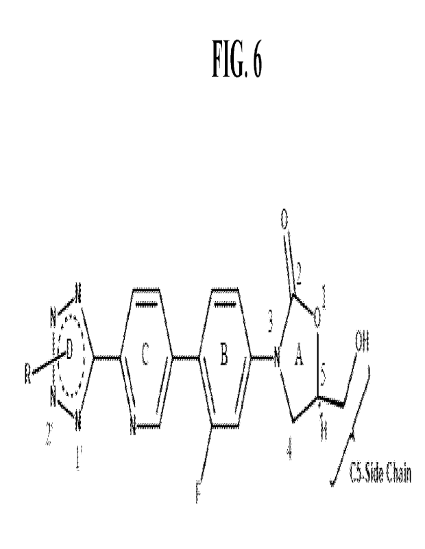
[0045] FIG. 6 represents the numbered ring structure of a
compound of Formula I.
[0046] FIG. 7 is a graph showing the plasma concentration versus
time profiles for total
drug in Sprague-Dawley rats after administration of a single intravenous dose
(IV x 1) of Ls-
AKG28 at 10 mg/kg (diamonds), 20 mg/kg (squares), and 40 mg/kg (circles).
Plasma
concentration versus time profiles of linezolid at 50 mg/kg (single oral dose,
PO x 1) in 5 % methyl
cellulose (pH 3-4) was also included for comparison. The mean and SD
concentration are
presented at each time point.
[0047] FIG. 8 is a graph showing the plasma concentration versus
time profiles for total
drug in Sprague-Dawley rats after administration of a single intravenous dose
(IV x 1) of Ls-
AKG38 at 20 mg/kg (diamonds), 40 mg/kg (squares), and 80 mg/kg (diamonds).
Plasma
concentration versus time profiles of linezolid at 50 mg/kg (single oral dose,
PO x 1) in 5 % methyl
cellulose (pH 3-4) was also included for comparison. The mean and SD
concentration are
presented at each time point.
[0048] FIG. 9A, FIG. 9B, and FIG. 9C are graphs showing the
plasma concentration versus
time profiles for total drug in Sprague-Dawley rats after administration of Ls-
AKG28 at 10 mg/kg
(FIG. 9A), 20 mg/kg (FIG. 9B), and 40 mg/kg (FIG. 9C), IV x 1, on day 1
(circles), day 15
(squares), day 29 (diamonds), and day 43 (triangles). The mean and SD
concentration are
presented at each time point.
[0049] FIG. 10A, FIG. 10B, and FIG. 10C are graphs showing the
plasma concentration
versus time profiles for total drug in Sprague-Dawley rats after
administration of Ls-AKG38 at
20 mg/kg (FIG. 10A), 40 mg/kg (FIG. 10B), and 80 mg/kg (FIG. 10C), IV x 1, on
day 1 (circles),
day 15 (squares), day 29 (diamonds), and day 43 (triangles). The mean and SD
concentration are
presented at each time point.
7
CA 03183397 2022- 12- 19
WO 2021/258013
PCT/US2021/038131
[0050] FIG. 11A, FIG. 11B, and FIG. 11C are graphs showing the
plasma concentration
versus time profiles of both lipid (using nonexchangeable DiIC18(3)-DS label),
drug for liposomal
AKG-28 (FIG. 11A) and liposomal AKG-38 (FIG. 11B), and the change in plasma
drug-to-lipid
ratio, a measure of drug release rate from the liposomes, for both Ls-AKG28
and Ls-AKG38 (FIG.
11C) in CD-1 mice after single intravenous injection in CD-1 mice. The mean
and SD are
presented at each time point.
[0051] FIG. 12 is a graph showing the plasma drug concentration
presented as % injected
dose for Ls-AKG28 and Ls-AKG38 were compared were multiple formulations of
liposomal
AKG-28 and liposomal AKG-38 after the first and fourth weekly doses. Mice were
injected with
the indicated dose and formulation once per week for a total of 4 injections.
[0052] FIG. 13A is a graph showing the effect of Ls-AKG28 dose
escalation on female
CD-1 mice body weight over time.
[0053] FIG. 13B is a graph showing the effect of Ls-AKG38 dose
escalation on female
CD-1 body weight in mice over time.
[0054] FIG. 13C are graphs showing the effects of Ls-AKG28 and Ls-
AKG38 in
combination with BP or BPM on hematology (RBC, HTC, PLT, WBC) and blood
biochemistry
(ALT, AST) parameters in female CD-1 mice.
[0055] FIG. 13D is a heat map showing the effect of monotherapy
Ls-AKG28 or Ls-
AKG38 on tissue pathological findings in female CD-1 mice.
[0056] FIG. 14A is a graph showing the effect of Ls-AKG28 in
combination with
bedaquiline and pretomanid (BP) or bedaquiline, pretomanid, and moxifloxacin
(BPM) on female
CD-1 mice body weight over time.
[0057] FIG. 14B is a graph showing the effect of Ls-AKG38 in
combination with BP or
BPM on female CD-1 mice body weight over time.
[0058] FIG. 14C are graphs showing the effect of Ls-AKG28 and Ls-
AKG38 in
combination with BP or BPM on hematology (RBC, HTC, PLT, WBC) and blood
biochemistry
(ALT, AST) parameters in female CD-1 mice.
[0059] FIG. 14D is a heat map showing the effect of Ls-AKG28 and
Ls-AKG38 in
combination with BP or BPM on tissue pathology findings in female CD-1 mice.
8
CA 03183397 2022- 12- 19
WO 2021/258013
PCT/US2021/038131
[0060] FIG. 15A is a graph showing the body weight change in
female CD-1 mice treated
with Ls-AKG28 injected twice a week (2qw) at 50 mg/kg or once a week (1 qw) at
100 mg/kg
alone or in in combination with BP over time.
[0061] FIG. 15B is a graph showing the body weight change in
female CD-1 mice treated
with Ls-AKG38 injected 2qw at 100 mg/kg or lqw at 200 mg/kg alone or in
combination with BP.
[0062] FIG. 15C are graphs showing the hematology and blood
biochemistry parameters
in female CD-1 mice treated with Ls-AKG28 (2qw at 50 mg/kg or lqw at 100
mg/kg) or Ls-
AKG28 (2qw at 100 mg/kg or lqw at 200 mg/kg) alone or in combination with BP.
[0063] FIG. 15D is a heat map showing the hi stopathology results
of female CD-1 mice
treated with Ls-AKG28 (2qw at 50 mg/kg or lqw at 100 mg/kg) or Ls-AKG28 (2qw
at 100 mg/kg
or lqw at 200 mg/kg) alone, or in combination with BP.
[0064] FIG. 16A is a graph showing the effect of Ls-AKG28 on body
weight in male
Sprague-Dawley rats treated chronically for a total of eight weeks over time.
[0065] FIG. 16B is a graph showing the effect of Ls-AKG38 on body
weight in male
Sprague-Dawley rats treated chronically for a total of eight weeks over time.
DETAILED DESCRIPTION
[0066] It is to be understood that both the foregoing general
description and the following
detailed description are exemplary and explanatory only and are not
restrictive of the compositions
and methods of the present disclosure.
[0067] Disclosed herein are compounds, compositions and methods
related to the
treatment of bacterial infections. As used herein, the term "compound" and
"drug" are used
interchangeably. Some aspects of the disclosure relate to novel aminoalkyl
derivatives of
oxazolidinones. Some aspects of the disclosure relate to the process for the
synthesis of the novel
aminoalkyl derivatives of oxazolidinone compounds. Other aspects relate to
compositions
comprising aminoalkyl derivatives of oxazolidinone compounds in liposomes.
Other aspects of
the disclosure relate to the use of aminoalkyl derivatives of oxazolidinone
compounds or liposome
compositions comprising aminoalkyl derivatives of oxazolidinone compounds in
the treatment of
bacterial infections. In some embodiments, the compounds and compositions
described herein can
be used to treat infections from mycobacteria and gram-positive bacteria. In
some embodiments,
the bacterial infection is mycobacterium tuberculosis. In some embodiments,
the compounds and
9
CA 03183397 2022- 12- 19
WO 2021/258013
PCT/US2021/038131
compositions described herein inhibits growth of mycobacteria and gram-
positive bacteria. These
include, but are not limited to, Mycobacterium tuberculosis, Mycobacterium
avium complex,
Mycobacterium leprae, Mycobacterium gordonae, Mycobacterium abscessus,
Mycobacterium
mucogenicum, streptococci, vancomycin-resistant enterococci (VRE), methicillin-
resistant
Staphylococcus aureus (MRSA), Staphylococcus pneumoniae, Enterococcus faecium,
Streptococcus agalactiae, Streptococcus pneumoniae, Streptococcus pyogenes,
the viridans group
streptococci, Li steria monocytogenes, Nocardia, and Corynebacterium.
[0068] In some embodiments, the aminoalkyl derivatives of
oxazolidinone compounds
described herein are selectively active against mycobacterium tuberculosis,
when compared to
mammalian cells, such as human kidney or hepatocyte cells. In some
embodiments, the
aminoalkyl derivatives of oxazolidinone compounds described herein exhibit an
unexpectedly
high selectivity of at least 1000-fold towards mycobacterium tuberculosis
compared to mammalian
cells, such as kidney or hepatocyte mammalian cells. In some embodiments, the
aminoalkyl
derivatives of oxazolidinone compounds described herein exhibit an
unexpectedly high selectivity
of at least 100-fold. In some embodiments, the aminoalkyl derivatives of
oxazolidinone
compounds described herein exhibit an unexpectedly high selectivity of from
100 to 6,500 folds,
100 to 6,000 folds, 100 to 5,500 folds, 100 to 5,000 folds, 100 to 4,500
folds, 100 to 4,000 folds,
100 to 3,500 folds, 100 to 3,000 folds, 100 to 2,500 folds, 100 to 2,000
folds, 100 to 1,500 folds,
100 to 1,000 folds, 500 to 6,500 folds, 500 to 6,000 folds, 500 to 5,500
folds, 500 to 5,000 folds,
500 to 4,500 folds, 500 to 4,000 folds, 500 to 3,500 folds, 500 to 3,000
folds, 500 to 2,500 folds,
500 to 2,000 folds, 500 to 1,500 folds, 500 to 1,000 folds, 1,000 to 6,500
folds, 1,000 to 6,000
folds, 1,000 to 5,500 folds, 1,000 to 5,000 folds, 1,000 to 4,500 folds, 1,000
to 4,000 folds, 1,000
to 3,500 folds, 1,000 to 3,000 folds, 1,000 to 2,500 folds, 1,000 to 2,000
folds, 1,000 to 1,500 folds
towards mycobacterium tuberculosis compared to mammalian cells, such as kidney
or hepatocyte
mammalian cells.
[0069] In some embodiments, the compounds and compositions
described herein may
promote selective uptake in mycobacterium-residing macrophages in the liver,
spleen, or lungs,
helping to provide potent intracellular killing. Macrophages are responsible
for the clearance of
foreign particles via phagocytosis, including both foreign infectious agent
like mycobacterium, as
well as laboratory-derived nanoparticles such as liposomes. This results in
the opportunity for
CA 03183397 2022- 12- 19
WO 2021/258013
PCT/US2021/038131
both to be co-localized in the same biological reservoir, effectively
concentrating the active agent
at an important depot for the disease
[0070] Aspects of the disclosure relate to compounds that are
aminoalkyl derivatives of
oxazolidinones (see FIG. 6). In some embodiments, the compounds having the
following
chemical Formula I and pharmaceutically acceptable salts thereof:
)1'0 Ri2
Fi \
Formula I
wherein R2 is an amine (NH2) or an acetamide (NHCOCH3), and
wherein Ri is a tetrazole ring substituted at position 2' with an aminoalkyl.
[0071] In other embodiments, the compounds having the following
chemical Formula I
and pharmaceutically acceptable salts thereof:
R _ *
Formula I
wherein R2 is an amine (NH2) or an acetamide (NHCOCH3), and
wherein Ri is a tetrazole ring substituted at l' with an aminoalkyl group.
[0072] In some embodiments, the aminoalkyl is a
dimethylaminoalkyl. In some
embodiments, the aminoalkyl derivatives of oxazolidinone compounds include
either an amine or
acetamide group at the R2 positions of the oxazolidinone ring and a
dimethylaminoethyl group on
the tetrazole ring.
[0073] The present disclosure shows a very specific structure-
activity relationship (SAR)
for the aminoalkyl derivatives of oxazolidinone compounds described herein
that included either
an amine or acetamide group at the R2 positions of the oxazolidinone ring and
a
dimethylaminoethyl group on the tetrazole ring. These compounds are (1) highly
selective against
mycobacterium tuberculosis when compared to activity in mammalian cells (for
example human
11
CA 03183397 2022- 12- 19
WO 2021/258013
PCT/US2021/038131
kidney or hepatocyte cells), (2) highly active against mycobacterium
tuberculosis, and (3)
efficiently loaded into liposomes.
[0074]
In some embodiments, the aminoalkyl derivatives of oxazolidinones
described
herein load in liposomes with 85 % or better efficiency using gradient-based
drug loading methods.
In some embodiments, the loading efficiency of these derivatives is 90% or
more. In some
embodiments, the loading of these derivatives is 95% or more, or even
quantitative. In some
embodiments, methods for loading the aminoalkyl derivatives of oxazolidinones
in liposomes are
described. In some embodiments, the loading methods employs transmembrane
gradients and
trapping agents to efficiently load, and subsequently stabilize, weakly basic
amphipathic drugs in
the liposomal interior aqueous space. The gradients can include (1) simple pH
gradients formed,
for example, using citric acid solutions, (2) ammonium ion gradients employing
citrate or sulfate
ammonium salts, (3) alkyl, dialkyl, or trialkylammonium salts, (4) gradients
of transition metals
(cu2+, mn2+, zn2+,
) or even (5) transmembrane gradients of drug solubility. See U.S. Patent
Nos. 5,316,771, 5,800,833, 8,147,867, 7,744,921, 8,349,360, 6,110,491, U.S.
Patent Application
Publication No.
20180369143A1 and International Patent Application Publication No.
W0199001405, which are incorporated herein by reference in their entireties.
See also Allen et
al. (1995) Int J Cancer 62:199-204. Without being bound by the theory, the
cation contained in
the liposome interior plays a role in establishing a pH gradient across the
membrane that helps
drive the accumulation of weakly basic drugs into the liposome interior, or
directly exchanges with
the drug molecule. This results in some embodiments, in a quantitative loading
of the drug below
the total capacity of the gradient. The counterion can play an important role
in stabilizing the
formulation to premature leakage in the circulation or during storage by
forming stable complexes
with the drugs in the liposome interior (see Drummond et al. (2008) J. Pharm
Sci 97, 4696-4740).
Definitions
[0075]
For convenience, certain terms employed in the specification, examples,
and
appended claims are collected here. Unless defined otherwise, all technical
and scientific terms
used herein have the same meaning as commonly understood by one of ordinary
skill in the art to
which this disclosure belongs.
[0076]
As used herein, the following terms and phrases are intended to have
the following
meanings:
[0077]
The articles "a" and "an" are used herein to refer to one or to more
than one (i.e.,
12
CA 03183397 2022- 12- 19
WO 2021/258013
PCT/US2021/038131
to at least one) of the grammatical object of the article. By way of example,
"an element" means
one element or more than one element.
[0078] As used herein the term "comprising" or "comprises" is
used in reference to
compositions, methods, and respective component(s) thereof, that are present
in a given
embodiment, yet open to the inclusion of unspecified elements.
[0079] As used herein the term "consisting essentially of' refers
to those elements required
for a given embodiment. The term permits the presence of additional elements
that do not
materially affect the basic and novel or functional characteristic(s) of that
embodiment of the
disclosure.
[0080] The term "consisting of' refers to compositions, methods,
and respective
components thereof as described herein, which are exclusive of any element not
recited in that
description of the embodiment.
[0081] The term "comprising" when used in the specification
includes "consisting of" and
"consisting essentially of'.
[0082] If it is referred to "as mentioned above" or "mentioned
above", "supra" within the
description it is referred to any of the disclosures made within the
specification in any of the
preceding pages.
[0083] If it is referred to "as mentioned herein", "described
herein", "provided herein," or
"as mentioned in the present text," or "stated herein" within the description
it is referred to any of
the disclosures made within the specification in any of the preceding or
subsequent pages.
[0084] As used herein, the term "about" means acceptable
variations within 20%, within
10% and within 5% of the stated value. In certain embodiments, "about" can
mean a variation of
+/-1%, 2%, 3%, 4%, 5%, 10% or 20%.
[0085] The term "effective amount" as used herein with respect to
a compound or the
composition means the amount of active compound (also referred herein as
active agent or drug)
sufficient to cause a bactericidal or bacteriostatic effect. In one
embodiment, the effective amount
is a "therapeutically effective amount" meaning the amount of active compound
that is sufficient
alleviate the symptoms of the bacterial infection being treated.
[0086] The term "subject" (or, alternatively, "patient") as used
herein refers to an animal,
preferably a mammal, most preferably a human that receives either prophylactic
or therapeutic
treatment.
13
CA 03183397 2022- 12- 19
WO 2021/258013
PCT/US2021/038131
[0087] The term "administration" or "administering" as used
herein includes all means of
introducing the compounds or the pharmaceutical compositions to the subject in
need thereof,
including but not limited to, oral, intravenous, intramuscular,
intraperitoneal, subcutaneous,
transdermal, inhalation, buccal, ocular, sublingual, vaginal, rectal and the
like. Administration of
the compound or the composition is suitably parenteral. For example, the
compounds or the
composition can be preferentially administered intravenously, but can also be
administered
intraperitoneally or via inhalation like is currently used in the clinic for
liposomal amikacin in the
treatment of mycobacterium avium (see Shirley et al., Amikacin Liposome
Inhalation Suspension:
A Review in Mycobacterium avium Complex Lung Disease. Drugs 2019 Apr;
79(5):555-562)
[0088] The tenns "treat," "treating," and "treatment," as used
herein, refer to therapeutic
or preventative measures such as those described herein
[0089] The terms "synergy" and "synergistic" as used herein,
means that the effect
achieved with the compounds used together is greater than the sum of the
effects that results from
using the compounds separately, i.e. greater than what would be predicted
based on the two active
ingredients administered separately.
[0090] The term "pharmaceutically acceptable salt" refers to a
relatively non-toxic,
inorganic or organic acid addition salt of a compound of the present
disclosure which salt possesses
the desired pharmacological activity.
[0091] The term "alkyl" means saturated carbon chains which may
be linear or branched
or combinations thereof, unless the carbon chain is defined otherwise.
Examples of alkyl groups
include methyl, ethyl, propyl, isopropyl, butyl, sec- and tert-butyl, pentyl,
hexyl, heptyl, octyl, and
the like.
[0092] The term "aminoalkyl" means an alkyl wherein at least one
carbon of an alkyl
carbon chain forms the bond with an amino group, wherein said amino group is
primary amino
group, mono-alkyl-substituted (secondary) amino group, di-alkyl-substituted
(tertiary) amino
group, or an alkyl-substituted amino group where the amine nitrogen atom and
the alkyl chain that
substitutes for amine hydrogens form a heterocycle.
[0093] The term "liposomes" means vesicles composed of a bilayer
(unilamellar) and/or a
concentric series of multiple bilayers (multi-lamellar) separated by aqueous
compartments formed
by amphipathic molecules such as phospholipids that enclose a central aqueous
compartment. In
a liposome drug product, the drug substance is generally contained in
liposomes Typically, water
14
CA 03183397 2022- 12- 19
WO 2021/258013
PCT/US2021/038131
soluble drugs are contained in the aqueous compartment(s) and hydrophobic
drugs are contained
in the lipid bilayer(s) of the liposomes. Release of drugs from liposome
formulations, among other
characteristics such as liposomal clearance and circulation half-life, can be
modified by the
presence of polyethylene glycol and/or cholesterol or other potential
additives in the liposome.
[0094] "Unilamellar liposomes," also referred to as "unilamellar
vesicles," are liposomes
that include one lipid bilayer membrane which defines a single closed aqueous
compartment. The
bilayer membrane includes two layers of lipids; an inner layer and an outer
layer (leaflet). Lipid
molecules in the outer layer are oriented with their hydrophilic ("head")
portions toward the
external aqueous environment and their hydrophobic ("tail") portions pointed
downward toward
the interior of the liposome. The inner layer of the lipid lays directly
beneath the outer layer, the
lipids are oriented with their heads facing the aqueous interior of the
liposome and their tails toward
the tails of the outer layer of lipid.
[0095] "Multilamellar liposomes" also referred to as
"multilamellar vesicles" or "multiple
lamellar vesicles," include more than one lipid bilayer membrane, which
membranes define more
than one closed aqueous compartment. The membranes are concentrically arranged
so that the
different membranes are separated by aqueous compartments.
[0096] The terms "encapsulation- and "entrapped,- as used herein,
refer to the
incorporation or association of the oxazolidinone pharmaceutical agent in or
with a liposome.
[0097] The terms "DL", "DL ratio", "D/L", or "D/L ratio" are used
interchangeably and
refer to the ratio of the drug to the liposome lipid. Unless indicated
otherwise, it is expressed as
grams of the drug per mole of liposome phospholipid (PhL).
[0098] The term "mol%" with regard to cholesterol refers to the
molar amount of
cholesterol relative to the sum of the molar amounts of cholesterol and non-
PEGylated
phospholipid expressed in percentage points. For example, "55 mol.%
cholesterol" in a liposome
containing cholesterol and HSPC refers to the composition of 55 mol. parts of
cholesterol per 45
mol. parts of HSPC.
[0099] The term -mol%" with regard to PEG-lipid refers to the
ratio of the molar amount
of PEG-lipid and non-PEGylated phospholipid expressed in percentage points.
For example, -5
mol.% PEG-DSPE" in a liposome containing HSPC and PEG-DSPE refers to the
composition
having 5 mol. parts of PEG-DSPE per 100 mol. parts of HSPC.
[00100] The terms "sucrose octasulfate", "sucrosofate', and
"sucrooctasulfate" refer the
CA 03183397 2022- 12- 19
WO 2021/258013
PCT/US2021/038131
same compound, sucrose octasulfuric acid or an anion thereof, and are used
herein
interchangeably.
[00101] The symbols "Ac", "Me", and "Er, as found in chemical formulas, refer
to acetyl
group (CH3C0), methyl group (CH3), and ethyl group (C2115), respecrively.
[00102] Various aspects and embodiments are described in further detail in the
following
subsections.
Compounds
[00103] Oxazolidinones are synthetic antibiotics that exert their
function by inhibiting
protein synthesis. Linezolid (LZD) is an oxazolidinone compound that exhibits
bacteriostatic
activity against M. tuberculosis. However, administration of LZD may cause
severe side effects
such as anemia, thrombocytopenia, and peripheral neuropathy. Tedizolid is an
oxazolidinone
compound which has been shown to inhibit gram positive bacteria. The side
effects for tedizolid
phosphate are similar, but generally less severe than observed for linezolid,
although the
experience with prolonged dosing such as that required for the treatment of
tuberculosis has been
limited for tedizolid phosphate compared to the extensive experience with
linezolid.
[00104] Aspects of the disclosure relate to compounds that are aminoalkyl
derivatives of
oxazolidinone (see FIG. 6). In some embodiments, the compounds having the
following chemical
Formula I and pharmaceutically acceptable salts thereof:
1,4
0
2
Formula I
wherein R2 is an amine (NH2) or an acetamide (NHCOCH3),
wherein RI_ is a tetrazole ring substituted at position 2' with an aminoalkyl.
[00105] In some embodiments, the aminoalkyl is a dimethylaminoalkyl. In some
embodiments, the aminoalkyl derivatives of oxazolidinone compounds include
either an amine or
acetam i de group at the R2 positions of the oxazolidinone ring and a dim
ethyl am i n oethyl group on
the tetrazole ring.
[00106] In other embodiments, the compounds having the following chemical
Formula I
and pharmaceutically acceptable salts thereof:
16
CA 03183397 2022- 12- 19
WO 2021/258013
PCT/US2021/038131
0
14$ * t4N,õõ),1R2
II
Formula I
wherein R2 is an amine (NH2) or an acetamide (NHCOCH3), and
wherein Ri is a tetrazole ring substituted l' with an aminoalkyl.
[00107] The aminoalkyl derivatives of oxazolidinone compounds having the
chemical
structure of the Table 1 below were synthesized as described in Example 1.
[00108] The compounds of the present disclosure can exist in free form, e.g.
as a free base,
or as a free acid, or as a zwitterion, or can exist in the form of a salt.
Said salt may be any salt,
either an organic or inorganic addition salt or a cocrystal, particularly any
pharmaceutically
acceptable organic or inorganic addition salt or a cocrystal, customarily used
in pharmacy. It is
understood that the chemical formula showing a compound in a particular salt
form or ionic form
also discloses this compound in its non-dissociated, free base (or free acid)
form.
[00109] The present disclosure encompasses all stereoisomeric forms of the
compounds. In
some embodiments, the compounds of Table 1 below are substantially pure (i.e.
at least 60%, 70%,
80%, 90%, 91%, 92%, 93%, 94%, 95%, 96%, 97%, 98%, 99%, e.g. 100%)
TABLE 1
Name SiTUCture
0
N-:..N
AKG-1 NM / e2
0
N
N>\*--- N Et2
AKG-2 I /
N,
0
1)L0 CP
AKG-3
N, 4104 \ õJr.-1N H3
1-i
17
CA 03183397 2022- 12- 19
WO 2021/258013
PCT/US2021/038131
0 ....c.x.-Nme2
N:it I \ * Nt...../0
N/ 0
AKG-5 /
N Me2
0
AKG-6
N -.:.N ¨ * N)L0 d
IL i µ /
/
0
AKG-7 1 /
N._ \ / ./--NEt2 * \..J:---/
..=
'H
0 N Et2
N ---/---/
iliN l. / µ / * Ntilp
AKG-8
.== '
0
NI.N N>\..,..0 ......p-NEt2
AKG-9 11 / \ / 'I,,\.....k..- H/
N
1-I
0
AKG-1 1 Me2N )1.-0
_r_01 \/ * \......LiOH
0
*tl....../
AKG-1 2 HN N OH \ /
Me211-1
0
0 niA
AKG-1 3 HN ,s, / * r\etj..... j- -
Et211--/
0
AKG-1 4 H N N)V- 0
µ / * \..... J....JO H
M e2N
18
CA 03183397 2022- 12- 19
WO 2021/258013
PCT/US2021/038131
0
AKG-15 HN \ / ti...../ OH
Et2N¨/-1
0
Nt...N K>L0
AK G-16 Ns / µ / * "\õ...L...../OH
Me 2N"'
0 WH 3CiS I
AKG-17 N-.....N =-0
h.. / \--/ Iro Nc.....i. N¨C
.0'
ea 0
C0 * N
_>
AKG-18 NIP 0 __Cr-11H3C1
HN
/
/
_ it .....0
Nt.-..N 0 H ¨N H3cp
AKG-19 IL / "
µ / JN--/
:-..../
1-i
0 N Et2
N t...
AKG-20
IL / µ / *
.."
1-i
NMe2
AKG-21 N-
0
0 OH
1-i
19
CA 03183397 2022- 12- 19
WO 2021/258013
PCT/US2021/038131
o NH3cp
0
MG-222
N- "..0
N,i / \ / 4* 1, \,,,I.-. ...../0 H
1-I
0
Ns..N
1 ' , µ / 1p N./ OH
H N
AKG-23 19"..õ,./
3 e_.µ
0
N 1.N N)L0 0 H
AKG-24 i /
N , µ / * \.--t----/
0
Nr.N
AKG-25 J., / µ / * Nt.t.d0H
0
ezAKG-26 \ / * ),\V,.... j.... joOH
Et2N...../e-...../
-T--1
0
N )L0 NI
AKG-27 0
I / Ni
\ / * /0 H
T-1
Cle
0
N.:.N i?'LO ID CP
AKG-28 IL l \ / * \..J----/NH3
0
N /h µ * t 0 e cif
=N iLi
AKG-29
H
00^.."
N I NH3
Ti 3 aN
Cr
CA 03183397 2022- 12- 19
WO 2021/258013
PCT/US2021/038131
0
>L0 cP
AKG-30 / / N NH3
T-i
LO CP
AKG-31
/ )NH,
0
N
N)L0
AKG-38 NHAcN,
Me 2 1-I
0
N
)LO
AKG-39 N NHAc
N,
Et2N"" T-I
0
Nt.:1\1
tr.
AKG-40 NJHAc
/
T-1
[00110] In some embodiments, the compound has the following chemical formula:
0
0 0f3
N.:1N
N>C' NH
Ns 3
Ti
Formula la
[00111] In some embodiments, the compound has the following chemical formula:
0
C) CP
rtIN: 41,
Me2N"/ T-1
Formula lb
[00112] In some embodiments, the compound has the following chemical formula:
0
N NJNHAC
/
Ti
Formula lc
21
CA 03183397 2022- 12- 19
WO 2021/258013
PCT/US2021/038131
[00113] In some embodiments, the compound has the following chemical formula:
* NHAc
-11
Formula id
[00114] In some embodiments, the compound has the following chemical formula:
N
NtliNHAc
N, =
Et2N".4"='
Formula le
[00115] Disclosed herein are compounds of Formula I or pharmaceutically
acceptable salts
thereof that are useful for the treatment of mycobacterium infections. In some
embodiments, the
compounds have the chemical formula la, lb, lc, ld or le_ In some embodiments,
the compounds
have the chemical formula lb. In some embodiments, the compounds of Formula I
have a
minimum inhibitory concentration (MIC), for example against Mycobacterium
tuberculosis,
ranging from 0.1 p.g/m1 to 1 jig/ml, from 0.25 ps/m1 to 1 p.g/ml, from 0.5
litg/m1 to 1 ps/ml, from
0.1 g/m1 to 0.25 _tg/ml, from 0.1 ps/m1 to 0.5 _tg/ml, from 0.25 ps/m1 to 0.
5 pg/ml, from 0.01
jig/m1 to 1 jig/ml, from 0.01 pg/ml to 0.25 jig/ml, from 0.01 p..g/m1 to 0.5
jig/ml, from 0.01 1.t.g/m1
to 0.1 gg/ml. In some embodiments, the compounds of Formula I have a minimum
inhibitory
concentration (MIC), for example against Mycobacterium tuberculosis of less
than 1 g/ml, less
than 0.25 jig/ml, or less than 0.1 jig/ml. In some embodiments, the compounds
of Formula I have
a MIC ranging from 0.01 litg/m1 to 0.25 jig/ml. In some embodiments, the
compound of Formula
I have a MIC ranging from 0.01 jig/ml to 0.1 vig/ml. It should be appreciated
that the MIC values
can be lower or than the ranges provided herein depending on the bacteria.
[00116] In some embodiments for the treatment of mycobacterium, for example M.
tuberculosis, the compound (AKG-28 or AKG-38) has a MIC below 0.1 ps/mL. In
some
embodiments for the treatment of mycobacterium, for example M. tuberculosis,
the compound has
a selectivity index (SI) for killing M. tuberculosis vs human kidney cells
(VERO) of at least 1,000.
In some embodiments for the treatment of mycobacterium, for example M.
tuberculosis, the
compound has a MIC below 0.1 ps/mL and a selectivity index (SI) for killing M.
tuberculosis vs
human kidney cells (VERO) of at least 1,000. In some embodiments, the compound
has the
structure of AKG-28 (Formula 1 b) or AKG-38 (Formula 1c). In some embodiments,
the MIC is
22
CA 03183397 2022- 12- 19
WO 2021/258013
PCT/US2021/038131
less than 0.05 ng/mL and the selectivity index for MIC in M. tuberculosis
relative to mitochondrial
protein synthesis inhibition (SI-MPS) is greater than 20, such as for AKG-28.
[00117] In some embodiments, the compounds described herein have a 2-to-20
fold increase
(about 2, about 3, about 4, about 5, about 6, about 7, about 8, about 9, about
10, about 11, about
12, about 13, about 14, about 15, about 16, about 17, about 18, about 19,
about 20) in potency
adjusted dose compared to linezolid for M. tuberculosis.
[00118] In some embodiments for the treatment of methicillin-resistant
Staphylococcus
aureus (MRSA), the compound has a MIC against MRSA strains of less than 2
ng/mL. In some
embodiments for the treatment of methicillin-resistant Staphylococcus aureus
(MRSA), the
compound has an IC50 of greater than 100 litg/mL against human VERO kidney
cells. In some
embodiments for the treatment of methicillin-resistant Staphylococcus aureus
(MRSA), the
compound has a MIC against MRSA strains of less than 2 ps/mL and an IC50 of
greater than 100
ng/mL against human VERO kidney cells. In some embodiments, the compound has
the structure
of AKG-38 (Formula 1c), AKG-39 (Formula le) , and AKG-40 (Formula 1d).
Aqueous S011ibility
[00119] In some embodiments, the compounds are in the form of salts, e.g., a
hydrochloride
or mesylate salt and are soluble in water at greater than 1 mg/ml, and
preferably greater than 10
mg/ml (and up to 1 g/m1) prior to encapsulation in liposomes. Additional salts
prior to
encapsulation can include, but are not limited to, besylate, bitartrate,
carbonate, citrate, esylate,
gluconate, glutamate, glycolate, lactate, malate, maleate, mandelate,
methylsulfate, napsylate,
phosphate, propionate, salicylate, succinate, tartrate, and tosylate. In some
embodiments, the
compounds are in the form of hydrate or solvate or a cocrystal prior to
encapsulation in the
liposomes.
[00120] In some embodiments, the drug is entrapped in the interior of the
liposomes in a
different salt form with a reduced aqueous solubility, for example less than 1
mg/mL and
preferably less than 0.1 mg/mL (0.1 ¨ 0.001 mg/mL). The salt of the compound
once entrapped
in the liposomes includes, but not limited to sulfate, citrate, phosphate,
sucrosofate, or various
phosphorylated or sulfated polyols or polyanionic polymers. Exemplary polyols
include, but not
limited to, sucrose, erythritol, mannitol, xylitol, sorbitol, inositol, and
combinations thereof.
Exemplary polyanionic polymers include but not limited to, polyvinylsulfonate,
polyvinylsulfate,
polyphosphate, copolymers of acrylic acid and vinylalcohol sulfate, and
combinations thereof.
23
CA 03183397 2022- 12- 19
WO 2021/258013
PCT/US2021/038131
[00121] Working stocks of the compounds were prepared as follows: to an
aliquot of a
compound (free base) in a powder form 1-1.5 equivalents of HC1 in the form of
1 N aqueous
solution was added, and the mixture was yortexed until homogeneity. To the
resulting cake or
syrup, water was added typically to the final 10 mg/ml, and complete
dissolution was observed. In
some instances, 0.95 equivalents of HC1 were added to the free base form of
the drug, and 20
mg/ml stock solution was prepared.
[00122] Aqueous solubility of the compounds of the present disclosure is
illustrated by the
following observations of obtaining visually clear solutions:
Compound Amount, mg Volume of 1 N Volume of water
Concentration %
HC1 added, ml added, ml (w/w)
of free
base
AKG-16 (free base) 22.3 0.052 30.0
AKG-28 (2HC1) 32.5 0.35 7.3
AKG-38 (free base) 31.7 0.067 0.35 7.1
[00123] These results show that the compounds provided herein have an aqueous
solubility
that is higher than the known aqueous solubilities of:
- linezolid (3 mg/ml) (www.drugbank.ca/drugs/DB00601)
0 0
cr¨\rq N)L?
\--/ NW/ JNHk
- sutezolid (0.237 mg/ml) (www. drugbank.ca/drugs/DB 11905)
0 0
"MN] N? H
N__/
, and
- tedizolid (0.382 mg/ml) (www.drugbank.ca/drugs/DB14569)
0
N)1.-0 OH
24
CA 03183397 2022- 12- 19
WO 2021/258013
PCT/US2021/038131
[00124] In some embodiments, the aqueous solubility of the compounds described
herein,
prior to encapsulation into the liposomes, is at least 5 times, at least 10
times, at least 20 times, at
least 30 times, or at least 40 times of the above oxazolidinones.
[00125] The excellent aqueous solubility of the compounds of described herein
and their
properties of amphiphilic weak bases allows efficient use of transmembrane-
gradient-based and
intraliposomal complexation (active loading) approach to creating liposome-
encapsulated forms
of these compounds with high drug/carrier (drug/lipid) ratio and
pharmacokinetic properties
favorable for encapsulated drug delivery to the infected tissues after
systemic administration of the
drug. As used herein, an amphiphilic weak base has a pKa of between 7 and 12
and alogP between
1 and 6.
Liposome loading properties and antimycobacterial activity.
[00126] An important feature of the compounds described herein is their weak
amphiphilic
base property that facilitates transmembrane gradient-driven loading of these
compounds into
liposomes. In some embodiments, a weak base property of the compounds of the
present
disclosure is characterized by an electrolytic dissociation constant in the
pKa range of 7.0-12.0,
7.5-11.0, 7.8-10.5, or 8.0 -10Ø In some embodiments, the amphiphilic
property of the compounds
described herein is characterized by a logP parameter in the range of 0.5-5.0,
1.0-4.0, 1.0-3.5, or
1.0-3Ø It was unexpectedly discovered that certain embodiments having these
favorable
properties with regard to the liposome loading, also have superior activity
against mycobacteria
that matches or surpasses the activity of similar compounds in the same class
of drugs whose
properties are less favorable for efficient and stable liposome encapsulation.
Liposome compositions
[00127] Compositions and use of the compositions for the treatment of
tuberculosis, as well
as other mycobacterial and gram positive bacterial infections are disclosed.
These compositions
provided herein contain a highly potent and selective oxazolidinone
encapsulated with high
efficiency to maximize dosing potential of low toxicity drugs, and are stable
in the presence of
plasma. In some embodiments, the compositions are long circulating and retain,
their encapsulated
drug while in the circulation following intravenous dosing to allow for
efficient accumulation at
the site of the bacterial or mycobacterial infection.. In some embodiments,
high doses that can be
achieved when combined with the long circulating properties and highly stable
retention of the
CA 03183397 2022- 12- 19
WO 2021/258013
PCT/US2021/038131
drug allow for a reduced frequency of administration when compared to daily or
twice daily
administrations of other drugs typically utilized to treat these infections
[00128] Disclosed herein are pharmaceutical compositions for treating
bacterial infections,
in particular a Mycobacterium tuberculosis infection. In some embodiments, the
pharmaceutical
composition is a liposomal composition comprising a polyanion or a sulfate
containing polyanion
and an aminoalkyl oxazolidinone compound.
[00129] In some embodiments, the composition comprises liposomes in a medium,
wherein
the intraliposomal space comprises an aqueous phase with a polyanion and the
compound of
Formula I. In some embodiments, the composition comprises liposomes in a
medium, wherein
the intraliposomal space comprises a polyanion or a sulfate containing
polyanion and the
compound AKG-16, AKG-28, or AKG-38. In some embodiments, the medium is an
aqueous
medium, where the primary composition in that media is the compound of formula
I and a
corresponding trapping agent.
[00130] The compound of Formula I can be entrapped within the liposome with a
suitable
polyanion, such as sucrose octasulfate (e.g. derived from triethylammonium
sucrose octasulfate,
(TEA-SOS) gradients) or sulfate (e.g. derived from ammonium sulfate
gradients). Additional
polyanion trapping agents include but are not limited to inositol
hexaphosphate, inositol
hexasulfate, polyvinylsulfonate, dextran sulfate, citrate, polyphosphate, and
suramin.
[00131] The exterior aqueous medium is typically composed of a suitable buffer
and an
isotonicity agent. Suitable buffers may include histidine, citrate, HEPES,
MOPS, MES, TRIS,
phosphate, glycine, and imidazole, borate, carbonate, and succinate.
Isotonicity agents may
include salts such as sodium chloride, potassium chloride, sucrose, glycerin,
dextrose, or mannitol.
[00132] In some embodiments, the composition comprises a compound of Formula I
or the
Formula la, lb, lc, or ld or pharmaceutical acceptable salt thereof,
encapsulated with a polyanion
in a primarily unilamellar vesicle formed from one or more phospholipid, a
sterol and optionally
a lipid conjugated to a hydrophilic polymer (a polymer-conjugated lipid). In
some embodiments,
the composition can comprise a compound of Formula I or the Formula la, lb lc,
or Id, or
pharmaceutical acceptable salt thereof, encapsulated with a polyanion in
unilamellar and
multilamellar vesicles (e.g. having two or three lamella).
It should be appreciated that
multilamellar vesicles can be cleared more quickly from circulation than
unilamellar vesicles. In
some embodiments, the phospholipid is hydrogenated soy phosphatidyl choline
(HSPC),
26
CA 03183397 2022- 12- 19
WO 2021/258013
PCT/US2021/038131
distearoylphosphatidylcholine (DSPC), or egg sphingomyelin (ESM). The term
"phospholipid as
used herein refers to any one phospholipid or combination of phospholipids
capable of forming
liposomes. Neutral phospholipids can include
di acylphosphati dyl cholines,
di al kylphosphati dyl chol ines, sphingomyelins, and
di acylpho sphati dyl ethanolamines.
Phosphatidylcholines (PC), including those obtained from egg, soybeans or
other plant sources or
those that are partially or wholly synthetic, or of variable lipid chain
length and unsaturation are
suitable for use in the present compositions. Synthetic, semisynthetic and
natural product
phosphatidylcholines including, but not limited to,
distearoylphosphatidylcholine (DSPC),
hydrogenated soy phosphatidylcholine (TISPC), soy phosphatidylcholine (soy
PC), egg
phosphatidylcholine (egg PC), hydrogenated egg phosphatidylcholine (HEPC),
dipalmitoylphosphatidylcholine (DPPC) and dimyristoylphosphatidylcholine
(DMPC) are
suitable phosphatidylcholines for use in this disclosure. Charged
phospholipids can include
phosphatidylserines, phosphati di c acids,
phosphatidylinositols, phosphatidylglycerols,
cardiolipins, or headgroup modified lipids such as N-succinyl-
phosphatidylethanolamines, N-
glutaryl-phosphatidylethanolamines, and PEG-derivatized
phosphatidylethanolamines.
[00133] Polymer-conjugated lipids may include poly(ethylene glycol)-conjugated
(pegylated)phospholipids (PEG-lipids) such as PEG(Mol. weight 2,000) methoxy-
poly(ethylene
glycol)- 1,2-di stearoyl-sn-glycerol (PEG(2000)-di stearoylglycerol, PEG-DSG),
PEG(Mol. weight
2,000) 1,2-distearoyl-sn-glycero-3-phosphoethanolamine-N-
rrriethoxy(polyethylene glycol)-
2000 (PEG(Mol. weight 2,000)-distearoylphosphatidylethanolamine, PEG-DSPE), or
PEG(Mol.
weight 2,000) N-palmitoyl-sphingosine-1- succinyl [methoxy(polyethylene
glycol)-2000] } (PEG-
ceramide). The molecular weight of the PEG portion in the PEG-lipid component
can also vary
from 500-10,000 g/mol, from 1,500-6000 g/mol, but is preferably about 2,000
MW. Other
polymers used for conjugation to lipid anchors may include poly(2-methyl-2-
oxazoline) (PMOZ),
poly(2-ethyl-2-oxazoline) (PEOZ), poly-N-vinylpyrrolidone
(PVP), polyglycerol,
poly(hydroxyethyl L-asparagine) (PHEA), and poly(hydroxyethyl L-glutamine)
(PHEG).
[00134] In some embodiments, the sterol is cholesterol. Other exemplary
sterols include,
but are not limited to, ergosterol, phytosterols such as 13-sitosterol, and
hopanoids. In some
embodiments, the ratio of the phospholipid(s) and the cholesterol is selected
to provide a desired
amount of liposome membrane rigidity while maintaining a sufficiently reduced
amount of leakage
of the compound of formula I from the liposome. In some embodiments, the
optional polymer-
27
CA 03183397 2022- 12- 19
WO 2021/258013
PCT/US2021/038131
conjugated lipid can be added to reduce the tendency of the liposomes to
aggregate. The type and
amount of polymer-conjugated lipid can be selected to provide desirable levels
of protein binding,
liposome stability and circulation time in the blood stream. For example, the
liposome vesicle
comprises phosphatidylcholine (e.g. DSPC or HSPC) and cholesterol in an about
45:55 molar
ratio. Phosphatidylcholine to cholesterol molar ratios can vary from about
60:40 to 35:65, about
50:50 to 35:65, about 50:50 to about 45:55. In particular, the liposome can
comprise a vesicle
consisting of HSPC, cholesterol and polymer-conjugated lipid (PEG-DSG or PEG-
DSPE) in a
about 55:45:2.75 molar ratio, corresponding to a PEG-lipid concentration of 5
mol % relative to
the concentration of phospholipid. The concentration of PEG-lipid can vary
from 0.5-to-10 mol
% relative to (non-PEGylated) phospholipid, with a preferred ratio of 3-10 mol
%, and an even
more preferred ratio of 4-8 mol %.
[00135] In some embodiments, liposomes compositions provide desirable
pharmacokinetic
properties such as extended plasma half-life, measured as the percentage of
the injected dose (ID)
(or injected amount) remaining in blood after 6 or 24 hours following
injection intravenously in
immunocompetent mice, and stable encapsulation of drug over 24 hours in plasma
as determined
by changes in the drug-to-lipid ratio (DL ratio) following iv administration
in mice. In some
embodiments, the percentage of drug remaining in blood is greater than 20 %,
preferably greater
than 30 %, and most preferably greater than 40 % of the injected dose at 6
hours. The percent
retained in blood after 24 h is preferably greater than 10 %, and more
preferably greater than 20
% of the injected dose. The DL ratio is greater than 20 % at 24 hours,
preferably greater than 50
%, and most preferably greater than 80 % of the originally injected liposomal
drug. Desirable
liposome compositions also display stable encapsulation in the presence of
human plasma in vitro
using a burst release method, with liposomes retaining greater than 50 % of
the drug over 20 min,
greater than 60%, greater than 70%, preferably greater than 80 %, and most
preferably greater than
90 % of encapsulated drug over 20 min.
[00136] Liposomes of the present disclosure can be made by any method known in
the art.
See, for example, G. Gregoriadis (editor), Liposome Technology, vol. 1-3, 1st
edition, 1983; 2nd
edition, 1993; 3'd edition, 2006; CRC Press, Boca Raton, Fla. Examples of
methods suitable for
making liposome composition of the present disclosure include membrane
extrusion, reverse phase
evaporation, sonication, solvent (e.g., ethanol) injection (including
microfluidic, Y-junction and
T-junction mixing), mi croflui di zati on, detergent
dialysis, ether injection, and
28
CA 03183397 2022- 12- 19
WO 2021/258013
PCT/US2021/038131
dehydration/rehydration. The size of liposomes can be controlled by
controlling the pore size of
membranes used for extrusions or the pressure and number of passes utilized in
microfluidization
or any other suitable methods. In some embodiments, the desired lipids are
first hydrated by thin-
film hydration or by ethanol injection and subsequently sized by extrusion
through membranes of
a defined pore size, such as, 50 nm, 80 nm, 100 nm, or 200 nm, or the
combinations thereof,
producing the liposomes with the average size in the range of 70-150 nm, or 80-
130 nm, and
polydispersity index of OA or less. The drug compound to be encapsulated can
be added to the
liposome lipids prior to the liposome formation, dissolved in the aqueous
medium in which the
liposomes are formed by the above methods, whereby the drug is sequestered
within the liposom es.
In some embodiments, the drug compound is encapsulated in the liposomes using
a trapping agent
incorporated into the interior space of the liposomes (see Drummond, D.C., et
al. (2006) in:
Liposome Technology, Third Edition (Ed. Gregoriadis, G.) Volume 2, p.149-168).
[00137] In some embodiments, the method of making liposome composition of the
present
disclosure comprises the steps of: (i) preparing the liposomes comprising
phospholipid,
cholesterol, and PEG-lipid, and having an interior space containing a trapping
agent, in a medium
substantially free from said trapping agent; (ii) contacting said liposomes
with the compound of
the present disclosure in an aqueous medium to effect encapsulation of the
compound in the
liposomes; (iii) removing unencapsulated compound; and (iv) providing the
liposomes in a
physiologically acceptable medium suitable for parenteral use. In some
embodiments, the process
to generate the liposomes with the compound therein includes the steps of (a)
preparing a liposome
containing a trapping agent composed of an ammonium or substituted ammonium
salt of a
polyanion, (b) subsequently removing extra-liposomal trapping agent to form an
electrochemical
gradients across the membrane, and (c) contacting the liposome with the
compound under
conditions effective for the compound to enter the liposome and to permit a
corresponding amount
of the ammonia or substituted ammonia to leave the liposome (thereby
exhausting or reducing the
pH gradient across the resulting liposome). Liposome compositions containing a
trapping agent
in the interior of the liposome can be made by formation of the liposomes in a
solution of the
trapping agent. The transmembrane concentration gradient of the trapping agent
can be formed
across the liposome by the removal of the trapping agent outside of the or
dilution of the liposomes
either following liposome formation or before loading (entrapping) of the
drug.
29
CA 03183397 2022- 12- 19
WO 2021/258013
PCT/US2021/038131
[00138] In some embodiments said contacting includes incubation of the
liposomes with the
drug in an aqueous medium at the temperature above ambient and below the
boiling point of water,
preferably between 30 C and 90 C, between 40 C and 80 C, between 50 C and 80
C, or between
60 C and 75 C. In some embodiments, the incubation is carried at ionic
strength of less than that
equivalent to 50 mM NaCl, or more preferably, less than that equivalent to 30
mM NaCl.
Following the incubation, a concentrated salt, e.g., NaCl, solution may be
added to raise the ionic
strength to higher than that of 50 mM NaCl, or of about 100 mM NaCl. The
increase of ionic
strength after the drug loading incubation step aided in reducing post-loading
aggregation of the
liposomes. The incubation times may range from few minutes to several hours.
In some
embodiments, the incubation times are from 5 to 40 min, from 10 to 30 min, or
from 15-25 min.
After the incubation, the liposomes are cooled down and then allowed to reach
the ambient
temperature. In some embodiments, the liposomes are cooled down to 2-15 C. In
some
embodiments, the liposomes are cooled down to 4-10 C. Following the cooling
step, a
concentrated salt, e.g., NaCl, solution may be added to raise the ionic
strength to higher than that
of 50 mM NaC1, or of about 100 mM NaCl. The increase of ionic strength after
the drug loading
incubation step aided in reducing post-loading aggregation of the liposomes.
[00139] In some embodiments, said contacting also included incubation of the
liposomes
with the drug in aqueous medium in the presence of an osmotic (tonicity)
balancing agent. In some
embodiments, the osmotic balancing agent (osmotic agent) is a non-ionic agent.
Exemplary non-
ionic osmotic agents include, but are not limited to, dextrose (glucose),
sucrose, trehalose, lactose,
mannitol, sorbitol, and polyvinylpyrrolidone. In some embodiments, the
concentration of osmotic
agent has osmotic concentration (expressed as osmolarity or osmolality) equal
to the osmotic
concentration of the trapping agent solution in the interior space of the
liposomes prior to drug
loading. The osmotic concentration of the trapping agent solution can be
measured by any known
method before the solution is combined with the lipids to form liposomes. In
another embodiment,
the concentration of osmotic agent provides osmotic concentration that is
lower than the osmotic
concentration of the trapping agent solution, and is less than about 90%, less
than about 80%, less
than about 70%, less than about 60%, less than about 50%, less than about 40%,
less than about
30%, less than about 20%, or less than about 10% of the osmotic concentration
of the trapping
agent solution. In yet another embodiment, the concentration of osmotic agent
during the drug
loading process is in the range of 200-400 mmol/kg, preferably 250-350
mmol/kg. In yet another
CA 03183397 2022- 12- 19
WO 2021/258013
PCT/US2021/038131
embodiment, the osmotic agent is dextrose, and the concentration is 45 g/L. In
yet another
embodiment, no osmotic agent is used during the incubation of the liposomes
with the drug. In yet
another embodiment, said incubation is performed in the presence of ionic
strength adjusting agent.
An example of the ionic strength adjusting agent is sodium chloride, added to
the liposome-drug
solution for example at the concentration between 5 and 50 mM, between 10 and
20 mM, or about
mM. Ccontrary to the convention in the field of liposomes, the compounds of
the present
disclosure, for example, AKG-28 and AKG-38, are loaded into the liposomes of
the present
disclosure in a stable and highly efficient manner even if, during the drug-
liposome contacting
step, the amount of osmotic agent provides osmotic concentration that is lower
than the osmotic
concentration of the trapping agent solution (osmotically imbalanced
liposomes), up to complete
absence of the added osmotic agent.
Methods of use
[00140] Disclosed herein are methods for inhibiting the growth of
mycobacteria, such as
Mycobacterium tuberculosis, or gram positive bacteria, such as methicillin-
resistant
Staphylococcus aureus (MRSA). Additional mycobacteria and gram positive
bacteria include, but
are not limited to, Mycobacterium avium complex, Mycobacterium leprae,
Mycobacterium
gordonae, Mycobacterium ab s ce s sus, Mycobacterium ab s ce s sus, My cob
acterium mucogeni cum,
streptococci, vancomycin-resistant enterococci (VRE), Staphylococcus
pneumoniae,
Enterococcus faecium, Streptococcus agalactiae, Streptococcus pneumoniae,
Streptococcus
pyogenes, the viridans group streptococci, Li steria monocytogenes, Nocardia,
and
Corynebacterium. In some embodiments, the compounds and compositions provided
herein
inhibit the growth of drug resistant strains of Mycobacterium tuberculosis. In
some embodiments,
methods of treating mycobacterial infections are provided. In some
embodiments, the compounds
and compositions provided herein can be used to treat nontuberculosis
mycobacteria infections.
In some embodiments, the method comprises administering a therapeutically
effective amount of
an aminoalkyl oxazolidinone of the disclosure and/or a pharmaceutical
acceptable salt thereof to a
subject in need thereof. In some embodiments, the method comprises
administering a
therapeutically effective amount of a liposomal composition comprising an
aminoalkyl
oxazolidinone compound of the disclosure and/or a pharmaceutical acceptable
salt thereof to a
subject in need thereof.
31
CA 03183397 2022- 12- 19
WO 2021/258013
PCT/US2021/038131
In some embodiments, the composition is a liquid pharmaceutical formulation
for parenteral
administration. In some embodiments, the liquid pharmaceutical formulation is
a liposomal
formulation containing a suitable amount of the oxazolidinone compound
described herein,
wherein the oxazolidinone compound is encapsulated in the interior of the
liposomes. In another
embodiment, that compound is in a salt form in the interior of the liposome
with a polyanion
such as sulfate, citrate, sucrose octasulfate, inositol hexaphosphate. In some
embodiments, the
compound is an precipitated or gelated salt with sulfate inside a liposome
composed of multiple
lipid excipients, including but not limited to, phosphatidylcholine,
cholesterol, and pegylated
phosphatidylethanolamine. The liposomes of the present disclosure show
entrapment efficiencies
of more than 85%, more than 90%, and more than 95%. In some embodiments, the
residual
amount of the unentrapped drug is removed from the liposome composition. This
can be
achieved by various means, such as size exclusion chromatography, ion
exchange, dialysis,
ultrafiltration, tangential flow filtration, adsorption, or precipitation.
During or after the
unentrapped drug removal step, the liposomes may be brought into a desired
pharmaceutically
acceptable carrier, for example, normal saline, isotonic dextrose, isotonic
sucrose, Ringer's
solution, or Hanks' solution. A buffer substance can be added to provide
desired physiologically
acceptable pH. The liposomal composition may be adjusted for desired drug
concentration, and
sterilized, e.g., by aseptic filtration through 0.2-0.22 pm filters. In some
embodiments, the
compound concentration in the liposomal composition is in the range of 1-50
mg/ml, 3-30
mg/ml, or 5-25 mg/ml.
[00141] In some embodiments, the liposomes are mixed with one or
more additional
excipients for isotonicity or pH control. In some embodiments, the excipients
include but are not
limited to sodium chloride, Hepes buffer, phosphate buffer, and histidine
buffer.
[00142] In other embodiments, the composition is an oral formulation. In some
embodiments, the composition is a liquid formulation. In some embodiments, the
composition is
a solid formulation (e.g. tablet, capsule, pill, dragees, caplets etc...).
When used for oral use for
example, tablets, troches, lozenges, aqueous or oil suspensions, dispersible
powders or granules,
emulsions, hard or soft capsules, syrups or elixirs may be prepared
(Remington's Pharmaceutical
Sciences (Mack Publishing Co., Easton, Pa.). Compositions intended for oral
use may be prepared
according to any method known to the art for the manufacture of pharmaceutical
compositions.
The compositions may contain one or more agents including antioxidants,
sweetening agents,
32
CA 03183397 2022- 12- 19
WO 2021/258013
PCT/US2021/038131
flavoring agents, coloring agents and preserving agents, in order to provide a
palatable preparation.
Tablets containing the active ingredient in admixture with non-toxic
pharmaceutically acceptable
excipient or auxiliary agents which are suitable for manufacture of tablets
are acceptable. Suitable
excipients or auxiliary agents include but are not limited to, for example,
inert diluents,
solubilizers, suspending agents, adjuvants, wetting agents, sweeteners,
perfuming or flavoring
substances, isotonic substances, colloidal dispersants and surfactants.
[00143] Tablets, dragees, capsules, pills, granules,
suppositories, solutions, suspensions and
emulsions, pastes, ointments, gels, creams, lotions, powders and sprays can be
suitable
pharmaceutical compositions.
[00144] The compound or the composition can be administered loca fly, orally,
parenteraUy.
ntra peritoneally and/or rectally.
[00145] Dosage regimens are adjusted to provide the optimum desired response
(e.g., a
therapeutic response). For example, one or more doses may be administered over
time or the dose
may be proportionally reduced or increased as indicated by the exigencies of
the therapeutic
situation.
[00146] The dosage of the compounds and/or of their pharmaceutically
acceptable salts or
the liposomes comprising the compounds and/or of their pharmaceutically
acceptable salts may
vary within wide limits and should naturally be adjusted, in each particular
case, to the individual
conditions and to the pathogenic agent to be controlled.
[00147] In some embodiments, for a use in the treatment of bacterial
infections, the
compound or the pharmaceutical liposomal composition is administered once
every 7 days (i.e.,
once every week), once every 14 days (i.e., once every two weeks), once every
21 days (i.e., once
every three weeks), once every 28 days (i.e., once every four weeks) and once
every 42 days (i.e.,
once every six weeks) to the subject in need thereof. In some embodiments, the
average weekly
dosage is from about 1 mg to about 1500 mg, about 10 to about 700 mg, about 25
to about 500
mg, or about 70 to about 250 mg. In some embodiments, the average weekly
dosage is from about
1 mg to about 10 mg, from about 10 mg to about 25 mg, from about 25 mg to
about 50 mg, from
about 50 mg to about 100 mg, from about 100 mg to about 200 mg, from about 200
mg to about
300 mg, from about 300 mg to about 400 mg, from about 400 mg to about 500 mg,
from about
500 mg to about 600 mg, from about 600 mg to about 700 mg, from about 700 mg
to about 800
mg, from about 800 mg to about 900 mg, from about 900 mg to about 1000 mg,
from about 1000
33
CA 03183397 2022- 12- 19
WO 2021/258013
PCT/US2021/038131
mg to about 1100 mg, from about 1100 mg to about 1200 mg, from about 1200 mg
to about 1300
mg, from about 1300 mg to about 1400 mg, from about 1400 mg to about 1500 mg.
In some
embodiments, the compound or composition is administered for up to one month,
up to two
months, up to three months, up to four months or more. The specific
therapeutically effective
amount will depend on a variety of factors, including the bacterial infection
being treated, the
activity of the specific compound being administered, the pharmaceutical
composition employed,
the age, body eight, gender etc. of the subject, the route of administration,
the severity of the
bacterial infection, the optional drugs/active agents used in combination
(sequentially or
simultaneously) with the specific compound, and the like factors known to the
medical doctor of
ordinary skill. In some embodiments, the compounds or the composition can be
used for the
treatment of tuberculosis or other mycobacterium infections. In some
embodiments, the compound
can be used as a monotherapy. In some embodiments, the treatment can include
administering
simultaneously and/or sequentially an effective amount of the compound
described herein and an
effective amount of one or more additional active agents to treat
mycobacterium tuberculosis and
other gram-positive bacterial infections. In some embodiments, the treatment
can include
administering simultaneously and/or sequentially an effective amount of the
compound described
herein and an effective amount of two or more additional active agents (two,
three, four, etc.) to
treat mycobacterium tuberculosis and other gram-positive bacterial infections.
A synergistic
antibacterial effect denotes an antibacterial effect which is greater than the
predicted purely
additive effects of the individual compounds of the combination. When
administered
simultaneously, the compound and the active agent can be contained in the same
composition or
in separate compositions. When administered sequentially, the composition
comprising the
compound and the composition comprising the additional active agent can be
administered with a
time separation (e.g. 20 minutes, 40 minutes, 60 minutes or more). In some
embodiments, the
additional active agents can be administered using a different administration
route or by different
injections. For example, the compounds of the disclosure can be administered
intravenously and
one or more additional agents can be administered orally.
[00148] In some embodiments, the administration of the compounds with one or
more (e.g.
one, two, three or four) additional active agents can result in a reduction of
the length of the
treatment duration. For example, administration of the compounds with one or
more (e.g. one,
two, three or four) additional active agent can result in a treatment duration
at least three times, at
34
CA 03183397 2022- 12- 19
WO 2021/258013
PCT/US2021/038131
least twice, at least 1.5 times shorter than compared to the treatment with
only one active agent.
In some embodiments, the additional agent(s) is an antibacterial agent. In
some embodiments, the
additional active agent can include, but are not limited to, fluoroquinolines,
such as moxifloxacin,
gatifloxacin, or levofloxacin, bedaquiline and other diaryl quinoline analogs
(e.g. TBAJ-587 and
TBAJ-876), del amanid, pretomanid, isoniazid, rifampicin, rifapentine,
pyrazinamide, clofazimine,
spectinamide, ethambutol, streptomycin, kanamycin, capreomycin, amikacin, the
Leucyl-tRNA
Synthetase (LeuRS) inhibitor GSK 3036656, tryptophan synthase inhibitor
GSK839, DprEl
inhibitors OPC-167832 and Macozinone (PBTZ-169), Telacebec, GSK-656, TBA-7371,
and
amoxicillin plus clavulanate, a pharmaceutically acceptable salt of each
thereof and any
combinations thereof. For the treatment of gram positive bacterial infections,
the additional active
agent can include, but are not limited to, vancomycin, gentamycin, daptomycin,
teicoplanin,
ceftaroline, ceftrobiprole, telavancin, dalbavancin, oritavancin,
fluoroquinolines (e.g.
delafloxacin), tetracyclines (e.g. eravacycline and omadacycline),
sulfonamides (e.g.
sulfamethoxazole), trimetrhoprim, lefamulin, and any combinations thereof
In some
embodiments, the treatment can include administering simultaneously and/or
sequentially an
effective amount of the compound described herein and an effective amount of
bedaquiline,
pretomanid, pyrazinamide, moxifloxacin or a pharmaceutically acceptable salt
of each thereof or
a combination of the foregoing.
[00149] Actual dosage levels of the active ingredients in the pharmaceutical
compositions
disclosed herein may be varied so as to obtain an amount of the active
ingredient which is effective
to achieve the desired therapeutic response for a particular patient,
composition, and mode of
administration, without being toxic to the patient.
[00150] "Parenteral" as used herein in the context of administration means
modes of
administration other than enteral and topical administration, usually by
injection, and includes,
without ii nii tati on, intravenous, intramuscular, intraarteri al, intrathec
al, intracapsular, intraorbi tal,
intracardiac, intradermal, intra.peritoneal, transtracheal, subcutaneous,
subcuticular, intraarticular,
subcapsular, subarachnoid, intraspinal, epidural and intrasternal injection
and infusion.
[00151] The phrases "parenteral administration" and 'administered
parenterally" as used
herein refer to modes of administration other than enteral (i.e., via the
digestive tract) and topical
administration, usually by injection or infusion, and includes, without
limitation, intravenous,
intramuscular, intraarterial, i ntrathecal, i ntracapsular, intraorbital ,
intracardiac, intradenn al,
CA 03183397 2022- 12- 19
WO 2021/258013
PCT/US2021/038131
intraperitoneal, transtracheal, subcutaneous, subcuticular, intraarticular,
inhalation, subeapsular,
subarachnoid, intraspinal, epidural and intrasternal injection and infusion.
Inttavenous injection
and infusion are often (but not exclusively) used for liposomal drug
administration.
[00152] in some embodiments, the liquid composition is injected
intravenously, In some
embodiments, the compound or the pharmaceutical composition is administered
once every 7 days
(i.e., once every week), once every 14 days (i.e., once every two weeks), once
every 21 days (i.e.,
once every three weeks), once every 28 days (i.e., once every four weeks) and
once every 42 days
(i.e., once every six weeks) to the subject in need thereof. In some
embodiments, the average
weekly dosage is from about 1 mg to about 1500 mg, about 10 to about 700 mg,
about 25 to about
500 mg, or about 70 to about 250 mg. In some embodiments, the average weekly
dosage is from
about 1 mg to about 10 mg, from about 10 mg to about 25 mg, from about 25 mg
to about 50 mg,
from about 50 mg to about 100 mg, from about 100 mg to about 200 mg, from
about 200 mg to
about 300 mg, from about 300 mg to about 400 mg, from about 400 mg to about
500 mg, from
about 500 mg to about 600 mg, from about 600 mg to about 700 mg, from about
700 mg to about
800 mg, from about 800 mg to about 900 mg, from about 900 mg to about 1000 mg,
from about
1000 mg to about 1100 mg, from about 1100 mg to about 1200 mg, from about 1200
mg to about
1300 mg, from about 1300 mg to about 1400 mg, from about 1400 mg to about 1500
mg. The
specific therapeutically effective amount will depend on a variety of factors,
including the bacterial
infection being treated, the activity of the specific compound being
administered, the
pharmaceutical composition employed, the age, body weight, gender etc.. of the
subject, the route
of administration, the severity of the bacterial infection, the optional
drugs/active agents used in
combination (sequentially or simultaneously) with the specific compound, and
the like factors
known to the medical doctor of ordinary skill in the art.
[00153] In some embodiments, for a use in the treatment of bacterial
infections, the
compound or the pharmaceutical oral composition is administered once or twice
daily. The specific
therapeutically effective amount will depend on a variety of factors,
including the bacterial
infection being treated, the activity of the specific compound being
administered, the
pharmaceutical composition employed, the age, body eight, gender etc.. of the
subject, the route
of administration, the severity of the bacterial infection, the optional
drugs/active agents used in
combination (sequentially or simultaneously) with the specific compound, and
the like factors
known to the medical doctor of ordinary skill.
36
CA 03183397 2022- 12- 19
WO 2021/258013
PCT/US2021/038131
EXAMPLES
[00154] The following examples, including the experiments conducted and
results achieved
are provided for illustrative purposes only and are not to be construed as
limiting the disclosure.
Example 1- Synthesis of oxazolidinone derivatives
[00155] Compounds AKG-1, AKG-2, AKG-6, AKG-8, AKG-9 and AKG-19 were
synthesized by reacting Tedizolid mesylate (Tedizolid-MS) with respective
amines at 60 C in N-
methy1-2-pyrrolidone (NMP) as a solvent (Scheme-1). Tedizolid-MS was obtained
by mesylation
of the 10 hydroxyl group of Tedizolid with methanesulfonyl chloride in the
presence of a base at
room temperature (RT). Treatment of Tedizolid-MS with sodium azide followed by
reduction of
the resulting azide (AKG-3-A) gave either Intermediate-1 as a free base or AKG-
3 as a
hydrochloride salt depending on eluant selected for purification. Amidation of
Intermediate-1 with
the corresponding acid followed by hydrochloride salt formation using
HC1/Et0Ac resulted in
compounds AKG-17 and AKG-18. Reacting Tedizolid with the corresponding
dialkylamino acid
under standard esterification conditions resulted in compounds AKG-5 and AKG-
20. 0-alkylation
of Tedizolid with 2-chloro-NN-diethylamino ethylamine using sodium hydride as
a base gave
compounds AKG-7.
[00156] Intermediate-2 was synthesized by boronation of commercially available
aryl
bromide using bis(pinocolato)diboron (Scheme-2). Suzuki coupling of
Intermediate-2 with readily
available 5-bromo-2-fluoropyridine resulted in Intermediate-3, which was
heated in NMP in a
sealed tube with the corresponding amine to give compounds AKG-11 to AKG-15.
[00157] Compounds AKG-16, AKG-21 to AKG-27 were prepared in a convergent
synthesis starting from Intermediate-4 (Schemes-3 and 4). Click chemistry
using sodium azide on
5-bromo-2-cyanopyridine gave Intermediate-4. N-alkylation of the tetrazole in
Intermediate-4
resulted in Intermediates 5 and 6 in 3:1 ratio. The structure of these
intermediates was deduced
from HMBC analysis. Intermediates 7 to 12 were synthesized and the
regioisomers were obtained
in a similar manner (Only desired isomers are shown in Scheme-4). Suzuki
coupling of
Intermediates 5 to 12 with Intermediate 2 and deprotection of amine group
where applicable
resulted in compounds AKG-16, AKG-21 to AKG-27.
[00158] Intermediate-13 was synthesized by mesylation of readily available
aryl bromide.
Intermediate-15 was obtained by reducing Intermediate-14 with hydrazine
(Scheme-5). Boc
37
CA 03183397 2022- 12- 19
WO 2021/258013 PCT/US2021/038131
protection or acetylation of the primary amine in Intermediate-15 followed by
boronation resulted
in Intermediates-18 and 19, respectively. Suzuki (U.S. Pat. Appl. Publ. No.
20100022772, PCT
Int. Appl. Publ. No. W02013044845, which are incorporated herein by reference
in their
entireties) coupling of the boronate intermediates with the corresponding aryl
bromide
intermediates and deprotection of the amine group where applicable resulted in
compounds AKG-
28 to AKG-31 and AKG-38 to AKG-40.
Synthetic Schemes
[00159] See U.S. Pat. Appl. Publ. No. 20100022772, PCT Int. Appl. No.
2013044845 which
are incorporated herein by reference in their entireties, for the synthesis of
Intermediate-19.
Scheme 1
F 0 F 0 F 0
¨ _
Tedizolid Tedizolid-Ms I42
I g or h or i I c R
4KG-1 R1R2NH=d1methy1ami1e
AKG-2 R1R2NH=diethylamine
F 0 F 0 AKG-6 RiR2NH=N,N-
dimethy1-2-(piperidin-4-ypethan-1-amine
RiR2NH=N1'N1-diethylpropane-1,3-diamine
AKG-9 e2NH=NI:N1- iethylethane-1,2-diamine
N-
-N
AKG-19 1=H, IR2- ' 1-diethylethane-1,2-diamine
\---4-1-NI AKG-3-1 3
AKG-5 R=4-(dimethylamino)butanoate
When g: AKG-7 R=N,N-diethylamino ethyl ether
When h:AKG-20 R=4-(diethylamino)butanoate I d
VVhen i: F 0 F
o e
NN-N N_ I\O eJ
\--j7nRl
F-1
Intermediate 1: R=NH2 AKG-17
(n=2), AKG-18 (n=3)
AKG-3: R=NH2.HCI
Conditions: (a) CH2Cl2, MsCI, TEA, r.t 2h; (b) R11R2NH, NMP, 60 C 16 h; (c)
NaN3, DMF, 90 C, 3 h; (d) Ph3P, THF/H20, reflux 1 h; (e) HATU, acid, DMF; (f
HCl/Et0Ac; (g) DCC, DMF, TEA,DMAP, 4-(dimethylamino)butanoic acid hydrogen
chloride, r.t, 16 h; (h) NaH, DMF, 2-chloro-N,N-diethylethan-1-amine, 0 C
r.t, 3 h; (i) DCC, DMF,DMAP, 4-(diethylamino)butanoic acid hydrogen chloride,
r.t, 16 h.
Scheme 2
38
CA 03183397 2022- 12- 19
WO 2021/258013
PCT/US2021/038131
0
0 0
N"----0
N)\----0 a )-(3`g = N--/:3 OH b F
/ \ OH
Br = \õ....L..../OH_,...
______________________________________ ¨d \_¨J,-.../
H-TH -1-i
Intermediate-2 Intermediate-3
0
c Ri
N)\---0
N / \ OH
1:2 ¨
-.PI
AKG-11 RiR2NH=N,N-dimethy1-2-(piperidin-4-ypethan-1-amine
R .
AKG-12 R1R2NH=N1,N1-dimethylethane-1,2-diamine
AKG-13 R1R2NH=N1,N1-diethylethane-1,2-diamine
AKG-14 1R2NH=N1,N1-dimethylpropane-1,3-diamine
AKG-15 RiR2NH=N1,Ni-diethylpropane-1,3-diamine
,
Conditions: (a) Bis(pinacolato)diboron , KOAc, (Ph3P)2PdC12 dioxane, 90 0C;
(b), 5-bromo-2-fluoropyridine, K3PO4, (dppf)PdC12
dioxane/H20, 90 C, 16 h; (c), R1R2NH, DMAP, NMP, 100 C, 16 h.
Scheme 3
0
me2N,,,,,N.,-)_aN 3
Br
11
Intermediate-5 AKG-14
b 4
NMe2 NMe2
0
d N- ¨
N- ¨
/ H p
kisf)--.0¨E3r ¨0- \ \--A:--/
I-I
Intermediate-6 AKG-21
NC-0.¨Br Hr--0¨, Br NHBuc NHBoc III'H,C'
Intermediate-4 0
0
N-N>_aBr d N-- ¨
_,.. N-
¨
0H
H I-I
Intermediate-7
C AKG-22-1
AKG-22 0
0
BocHNris--0¨Br.-
Intermediate-3
AKG-23-1
AKG-23
Conditions: (a) NaNz/ZnClz, pyridine, 120 `1C, 2 h; (b) (2-Bromoethyhd
imethylamine hydrobromide, Ca(OH)o, HoOMMF, 00 C, 24 h; (c) NHBoc(CH4z1Br,
C2(01-)2, HoO/DMF, 80 `1C, 24 h (d) Intermediate-2, (dppf)F
K5PO4, dioxone, 90 C, 16 h; (e) HCl/Dioxa ne, It., 5 h
Scheme 4
39
CA 03183397 2022- 12- 19
WO 2021/258013 PCT/US2021/038131
0
N
__________________________ I. ri: b
Et2N"-- N11¨ Et2N ,,
N
---"-
II
Intermediate-9
AKG-24
0
Ns....N _
1\1.:-N IV __ ()Br
b
__________________________ . ...11 /_)
-T-I
N -
Intermediate-10 AKG25
a N/ 0
=N\ /¨ __________ \
HN N // \j_/Br _________
-
OH
N
y=_¨j_
Intermediate-4 b
Br
,.., i ii
Et2N,..7---õ,..N
AKG-26
Intermediate-11
0
N=N
¨ r¨B b BocHN
,._ BocHNN i-Ni_// ...f--.....,-
Intermediate-12 AKG-27-1
1 c
0
N=N
-1-1
CP
AKG-27
Conditions: (a) Amine, Ca(OH)2, H20/DMF, 80 C, 24 h; (b) Intermediate-2,
(dppf)PdC12, K3PO4, dioxane, 90 C, 16 h; (c) HCl/Dioxane, r.t., 5 h.
Scheme 5
c
o o :JIo 0 o
o
Br . )Lo
/ a
HO ¨.- Br * N).\---C) K'
N____L..../0Ms - Br *
¨.-- Br = Nitl.../NH d ore
2 _... Br .
'f-1 'f-1 I-I 'f-I
\ _...-1,-../
T-I
Intermediate-13 Intermediate-14 Intermediate-15 Intermediate-16
R =NHBoc
Intermediate-17 R =NHAc
Ns,NINI>_0_
1 Br
0
IR,,N_
'13 = N 0/R Intermediate-5/8/10/11 N.-PI ¨ )\--
C) R 9 N=N ¨ ,,, cP
Nh-', NH3
cf t-..
i-I IR( Fl RI/ --N -1-I
9 R R
Intermediate-18 R =NHBoc AKG-28-1 Bi.2.(N,N-
dimethylamino)ethyl R=NHBoc AKG-28 Ri=2-(N,N-dimethylamino)ethyl
Intermediate-19 R =NHAc AKG-29-1 Ri=2-((tert-
but0xycarb0ny1)0min0)ethyl R=NHBoc AKG-29 Ri=2-am1n0 ethyl HCI
AKG-30-1 RI=3-(N,N-dimethylamino)propyl R=NHBoc AKG-
30 R1=3-(N,N-dimethylamino)propyl
AKG41-1 i=3-(N,N-diethylamino)propyl R=NHBoc AKG-
31 1=3-(N ,N-diethylamino)propyl
,N-dimethylam IR ino)ethyl
=NHAc
AAKKGG-3-398:: i2
-N
-(N,N-diethylamino)ethyl R=NHAc
AKG-40: 1=2-(N ,N-diethylamino)propyl R=NHAc
Conditions: (a) 511%,nTeE6b DCM, PT. 2h; (b) DMF, 80 c, 16h; (c) NH2NH2, DOH,
80 C; (d) Boo20, THF/H20, NaHCO3, RT: (e) AcCl, TEA, DCM, PT; (f)
Bis(pinacolato)diboron , K0Ac,
(Ph3P)2PdC12 ' ' C; (g) K3PO4, (dppf)PdC12, dioxane/H20, 90 C, 18 h;
(h) HCI n ELOAc, DCM, RT, 2h,
CA 03183397 2022- 12- 19
WO 2021/258013
PCT/US2021/038131
Synthesis
Materials and Methods.
[00160] Tedizolid, (R)-3 -(4-brom o-3 -fluoropheny1)-5-(hydroxymethyl)oxazoli
din-2-one
were purchased from Skychemical and Dimethyl-(2-piperdin-4-yl-ethyl)-amine was
purchased
from Enamine, the other reagents and solvents were purchased from Adams and
were used as
received. The chemical structures of final products were characterized by
nuclear magnetic
resonance spectra (1H NMR, 13C NMR) determined on a Bruker NAIR spectrometer
(500 MHz or
400 MHz). 13C NMR spectra were fully decoupled. Chemical shifts were in parts
per millions
(ppm) using deuterated solvent peak or tetram ethyl silane (internal) as the
internal standards. Data
for 1H NMR are recorded as follows: chemical shift (d, ppm), multiplicity (s,
singlet; br s, broad
singlet; d, doublet; t, triplet; m, multiplet), integration, coupling constant
(Hz). Data for 13C NMR
are recorded in terms of chemical shift (d, ppm).
[00161] The purity of final products (>95%) was confirmed by analytical HPLC.
Analytical
liPLC was performed on an Agilent analytical HPLC system using a Sunfire
column, 3.5p.m (150
cm
4.6 mm) and a gradient system (water (0.01%TFA)/ACN (0.01%TFA)) and a
flow rate of 1
mL/min with detection at 254 and 214 nm. Flash Chromatographic (FC)
purifications were
performed with Silica Gel 60 from Santai Technologies (0.04-0.063 nm; 230-400
mesh).
[00162] Procedure A. The reaction mixture of Tedizolid-Ms (1.0 eq), R1R2NH
(4.0 eq) in
NMP (10 mL) was heated to 60 'C for 15 h in a sealed tube. Upon completion
(LCMS), the reaction
was diluted with H20 (40 mL) and extracted with Et0Ac (2X50 mL). The combined
extracts were
washed with saturated brine dried over Na2SO4 and filtered. The solvent was
removed in vacuo
and the residue was purified using FC to give the product with >95% purity.
1. Synthesis of Tedizolid-Ms
0 0
TEA/MsCI N
N CH2C 12 N
\--41:7)Ms
Tedizoli d Tedizo1id-M4
[00163]
To a solution of Tedizolid (7.00 g, 18.90 mmol) and triethylamine (3.83
g, 37.80
mmol) in CH2C12(50 mL) at 0 C was added dropwi se methanesulfonyl chloride
(3.25 g, 28.36
mmol) at 0 C, under Ar. After stirring at RT for 2 h, the reaction mixture
was poured into water
and extracted with CH2C12. The organic layer was washed with brine, dried over
Na2SO4 and
41
CA 03183397 2022- 12- 19
WO 2021/258013
PCT/US2021/038131
collected by filtration. The solvent was removed in vacuum to give the pure
product Tedizolid-Ms
(7.0 g, 82.6% yield) as a yellow solid. 1H NIVIR (400 MHz, DMSO-d6) 6 8.95 (s,
1H), 8.31 - 8.14
(m, 2H), 7.88 -7.65 (m, 2H), 7.53 (d, J= 8.6 Hz, 1H), 5.14 -4.96 (m, 1H), 4.59
- 4.39 (m, 5H),
4.28 (t, J= 9.4 Hz, 1H), 3.92 (dd, J = 9.2, 6.3 Hz, 1H), 3.28 (s, 3H). MS
(ESI+) m/z 449.1 ([M +
1] ).
2. Synthesis qf AKG-1, 2, 6, 8, 9 and 19
0
N
\--172\11
AKG-1
[00164] Using procedure A, AKG-1 was obtained from Tedizolid-Ms and
dimethylamine
as a white solid (0.5 g, 56.4% yield). 1H NMR (400 MHz, DMSO-d6) 5 8.94 (s,
1H), 8.32 - 8.13
(m, 2H), 7.83 -7.64 (m, 2H), 7.54 (d, J= 7.6 Hz, 1H), 4.87 (s, 1H), 4.49 (s,
3H), 4.21 (t, J = 8.6
Hz, 1H), 3.84 (t, J= 7.4 Hz, 1H), 2.62 (s, 2H), 2.25 (s, 6H). 13C NMR (101
MHz, DMSO-d6) 6
164.3, 161.0, 158.6, 154.6, 149.9, 145.5, 140.9, 137.6, 132.1, 131.4, 122.6,
119.1, 114.6, 106.0,
72.0, 62.1, 48.7, 46.4, 40.2. MS (ESI+) m/z 398.2 ([M + 1]+).
NEt2
N = /
AKG-2
[00165] Using procedure A, AKG-2 was obtained from Tedizolid-Ms and
diethylamine as
a white solid (0.52 g, 54.8% yield). 1H NMR (400 MHz, DMSO-d6)6 8.94 (s, 1H),
8.29 - 8.11 (m,
2H), 7.81 - 7.65 (m, 2H), 7.52 (dd, J= 8.6, 1.8 Hz, 1H), 4.89 - 4.73 (m, 1H),
4.49 (s, 3H), 4.19(t,
J= 8.8 Hz, 1H), 3.82 (dd, J= 8.7, 7.0 Hz, 1H), 2.75 (dd, J= 5.1, 3.7 Hz, 2H),
2.57 (q, J= 6.9 Hz,
4H), 0.97 (t, J=7.1 Hz, 6H). 13C NMR (101 MHz, DMS0-616) 6 164.3, 161.0,
158.6, 154.7, 149.9,
145.5, 141.0, 137.6, 132.1, 131.3, 122.5, 119.1, 114.6, 106.1, 72.6, 56.1,
48.6, 47.7, 40.3, 12.3.
MS (ESI+) m/z 426.3 ([M +
42
CA 03183397 2022- 12- 19
WO 2021/258013
PCT/US2021/038131
Nme2
c-j
N=;-= N>L=
/
AKG-6
[00166] Using procedure A, AKG-6 was obtained from Tedizolid-Ms and N,N-
Dimethy1-
2-(piperidin-4-ypethan-1-amine as a white solid (0.66 g, 58.2% yield).
NMR (400 MHz,
CDC13) 6 8.93 (s, 1H), 8.30 (dd, J = 8.1, 2.4 Hz, 1H), 8.05 (d, J= 7.8 Hz,
1H), 7.62 (d, J= 12.9
Hz, 1H), 7.56 - 7.47 (m, 1H), 7.45 - 7.37 (m, 1H), 4.89 - 4.74 (m, 1H), 4.48
(s, 3H), 4.11 (t, ./=
8.6 Hz, 1H), 3.86 (t, J= 7.8 Hz, 1H), 2.93 (dd, J= 28.8, 10.9 Hz, 2H), 2.80 -
2.64 (m, 2H), 2.50
- 2.04 (m, 11H), 1.69 (d, J = 10.8 Hz, 2H), 1.48 (d, J = 7.1 Hz, 2H), 1.37 -
1.19 (m, 4H). '3C NMR
(101 MHz, CDC13) 6 164.7, 161.3, 158.8 , 154.3 , 149.9 , 145.4 , 140.2 ,
137.0, 132.3 , 130.5 ,
122.0, 120.0, 113.8, 106.4, 71.5 , 61.4 , 57.0 , 55.3 , 54.3 , 48.9 , 45.0 ,
39.7, 33.6, 32.4. MS
(ESI+) m/z 509.2 ([M + 1]+).
0 N Et2
-
AKG-8
[00167] Using procedure A, AKG-8 was obtained from Tedizolid-Ms and N1,N1-
diethylpropane-1,3-diamine as a white solid (0.62 g, 57.6% yield). 1H NMR (400
MHz, DMS0-
do) 6 8.94 (s, 1H), 8.34 - 8.11 (m, 2H), 7.84 - 7.59 (m, 2H), 7.52 (dd, J=
8.6, 2.0 Hz, 1H), 4.80
(dd, J = 8.3, 5.7 Hz, 1H), 4.49 (s, 3H), 4.18 (t, J = 8.9 Hz, 1H), 3.90 (dd,
J= 8.8, 6.5 Hz, 1H), 2.94
-2.77 (m, 2H), 2.66 - 2.53 (m, 7H), 1.65- 1.51 (m, 2H), 0.99 (t, J= 7.1 Hz,
6H). 13C NMR (101
MHz, DMSO-do) 6 164.3, 161.0, 158.6, 154.6, 149.9, 145.5, 141.0, 137.6, 132.1,
131.4, 122.6,
119.1, 114.6, 106.1, 73.2, 52.1, 50.7, 48.2, 48.0, 46.7, 40.3, 26.4, 11.4. m/z
483.2 ([M + 1]+).
0
N /
-N
AKG-9
43
CA 03183397 2022- 12- 19
WO 2021/258013
PCT/US2021/038131
[00168] Using procedure A, AKG-9 was obtained from Tedizolid-Ms and N1,N1-
diethylethane-1,2-diamine as a white solid (0.36 g, 34.4% yield). 1HNIVIR (500
MHz, DMSO-d6)
(58.94 (s, 1H), 8.26 - 8.16 (m, 2H), 7.78 - 7.66 (m, 2H), 7.53 (d, J= 8.5 Hz,
1H), 4.85 - 4.73 (m,
1H), 4.49 (s, 3H), 4.18 (t, J= 8.8 Hz, 1H), 3.90 (t, J= 7.5 Hz, 1H), 2.88 (t,
J= 5.4 Hz, 2H), 2.65
(t, J= 6.1 Hz, 2H), 0.95 (t,J= 7.0 Hz, 6H). 13C NMR (101 MHz, DMSO-d6) (5
164.3, 161.0, 158.6,
154.7, 149.9, 145.5, 141.0, 137.6, 132.1, 131.4, 122.6, 119.1, 114.6, 106.1,
73.3, 52.6, 52.2, 48.1,
47.6, 47.1, 40.3, 12Ø m/z 469.3 ([M + 1] ).
[00169] Using procedure A, AKG-19 was obtained from Tedizolid-Ms and ethane-
1,2-
di amine as a white solid (0.60 g, 55% yield). 1H NIVIR (500 MHz, DMSO-d6) 6
10.33 (s, 1H), 9.89
(s, 1H), 8.95 (s, 1H), 8.58 (s, 3H), 8.23 (q, J- 8.3 Hz, 2H), 7.79 (t, J- 8.8
Hz, 1H), 7.69 (d, J-
13.5 Hz, 1H), 7.49 (d, J= 8.7 Hz, 1H), 5.25 - 5.19 (m, 1H), 4.49 (s, 3H), 4.33
(t, J= 9.2 Hz, 1H),
4.05 (dd, J= 9.1, 6.7 Hz, 1H), 3.52 (s, 2H), 3.43 -3.23 (m, 4H). 11C NMR (101
MHz, DMSO-do)
(5164.26, 160.94, 158.50, 153.79, 149.84, 145.51, 140.58, 137.78, 132.04,
131.42, 122.61, 119.50,
114.94, 106.46, 69.38, 49.59, 47.87, 45.16, 40.34, 35.58.
3. Synthesis of AKG-3
0 0
N-N N
Nzz- ______________________________________________ 0.
DMF
\I3
Tedizolid-Ms AKG-3-1
[00170] To a solution of Tedizolid-Ms (1.00 g, 2.23 mmol) in DME (20 mL) was
added
NaN3 (0.44 g, 6.69 mmol). After stirring at 90 C for 3 h, the reaction
mixture was poured into
water and extracted with Et0Ac. The organic layer was washed with brine, dried
over anhydrous
MgSO4, filtered and concentrated in vacua The residue was further purified by
column
chromatography to obtain the title compound AKG-3-1 (0.7 g, 79.4% yield) as
white solid.
0
0
Ph3P
N----N0 N3
/ THF/H20 N
3
T-I
AKG-3-1 AKG-3
44
CA 03183397 2022- 12- 19
WO 2021/258013
PCT/US2021/038131
[00171] The reaction mixture of AKG-3-1 (0.7 g, 1.77 mmol) and Ph3P (1.39 g,
5.31 mmol)
in H20 (2 mL) and TIFF (20 mL) was heated to reflux for 1 h. After completion
of (LCMS), the
reaction was concentrated in vacuo and purified using reverse phase FC. While
purification using
Me0H in DCM 0-10% as eluant and freeze drying gave freebase Intermediate-1
(2.5 g, 76.5%
yield) as a yellow solid, FC purification with MeCN in 0.006M HC1 in H20/ 0-
30% as eluant gave
hydrochloride salt AKG-3 (0.35 g, 48.8% yield) as a yellow solid after freeze
drying. 1H NMR
(400 MHz, DMSO-d6) 6 8.95 (s, 1H), 8.61 (s, 3H), 8.28 - 8.18 (m, 2H), 7.79 (t,
.1 = 8.8 Hz, 1H),
7.69 (dd, .1= 13.5, 2.1 Hz, 1H), 7.48 (dd, .1=8.6, 2.1 Hz, 1H), 5.13 - 5.00
(m, 1H), 4.49 (s, 3H),
4.29 (t, J= 9.2 Hz, 1H), 4.02 (dd, J= 9.3, 6.6 Hz, 1H), 3.34 - 3.23 (m,
214).13C NMR (101 MHz,
DMSO-d6) 6 163.8, 160.4, 158.0, 153.4, 149.4, 145.1, 140.2, 137.2, 131.5,
130.9, 122.1, 118.9,
114.3, 105.9, 69.8, 47.1, 41.4, 39.8. m/z 370.3 ([M -HC1 + 1]).
4. Synthesis of AKG-17
NHBoc
HO-cl
NH2 1,HATU/DMF
0
NH3
2, HCl/Et0Ac
Intermediate 1 AKG-17
[00172] To a solution of 3-((tert-butoxycarbonyl)amino)propanoic
acid (0.62 g, 3.25 mmol,
1.2 eq) and TEA (0.63 g, 6.25 mmol, 2.5 eq) in DMF (10 mL) was added HATU
(1.44 g. 3.78
mmol, 1.4 eq) at RT under Ar. The mixture was stirred for 0.5 h and then
Intermediate 1 (1.0 g,
2.70 mmol, 1.0 eq) was added. The whole mixture was stirred at RT overnight.
LCMS showed the
reaction was complete, it was poured into H20 and the solid was collected by
filtration and washed
with 1120. The solid was dried in vacuo and the residue was used in the next
step directly by
dissolving it into Et0Ac and then HC1/Et0Ac (4 M, 20 mL) was added. The whole
mixture was
stirred for 16 h and the solvent was removed by N2. The residue was purified
by reverse phase FC
(eluant with MeCN in 0.006M HC1 in H20/ 0-30%) to give the product AKG-17 (0.5
g, 39.5 %
yield) after freeze drying as a yellow solid. 1H NMR (500 MHz, DMSO-d6) 6 8.95
(s, 1H), 8.67
(s, 1E1), 8.23 (q, J = 8.3 Hz, 2H), 8.14 (s, 3H), 7.77 (t, J= 8.6 Hz, 1H),
7.69 (d, J= 13.5 Hz, 1H),
7.50 (d, J = 8.6 Hz, 1H), 4.87- 4.78 (m, 111), 4.49 (s, 3H), 4.22 (t, J= 9.0
Hz, 1H), 3.89 (dd, J=
9.0, 6.5 Hz, 1H), 3.50 (t, J = 5.3 Hz, 2H), 2.98 (dd, J= 12.5, 6.4 Hz, 2H),
2.58 (t, J= 7.1 Hz, 2H).
CA 03183397 2022- 12- 19
WO 2021/258013
PCT/US2021/038131
13C NMR (101 MHz, DMSO-d6) 6 170.60, 164.22, 160.97, 158.53, 154.42, 149.78,
145.42,
140.89, 137.79, 132.11, 131.41, 122.61, 119.19, 114.72, 106.23, 105.95, 72.13,
47.77, 40.33,
35.58, 32.58.
5. Synthesis of AKG-18
0 CI
0
N..,N
N /
-
AKG-1 8
[00173] Using the procedure of AKG-17, AKG-18 was obtained from Intermediate-1
and
4-((tert-butoxycarbonyl)amino)butanoic acid as a yellow solid (0.5 g, 37.6%
yield). 1-1-1NMR (500
MHz, DMSO-d6) 6 8.95 (s, 111), 8.52 (t, J = 5.7 Hz, 1H), 8.29 - 8.08 (m, 5H),
7.77 (t, J = 8.8 Hz,
1H), 7.69 (d, .1= 13.6 Hz, 1H), 7.50 (d, .1= 8.7 Hz, 1H), 4.87 - 4.76 (m, 1H),
4.50 (s, 3H), 4.22 (t,
= 9.0 Hz, 1H), 3.93 -3.83 (m, 1H), 3.49 (t, .1= 5.3 Hz, 2H), 2.83 -2.72 (m,
2H), 2.28 (t, .1= 7.2
Hz, 211), 1.88 -1.75 (m, 2H). 13C N1VIR (101 MHz, DMSO-do) 5172.59, 164.23,
160.96, 158.52,
154.44, 149.00, 145.39, 140.88, 137.78, 132.10, 131.40, 122.61, 119.17,
114.70, 106.21, 72.17,
47.78, 41.88, 40.37, 38.78, 32.44, 23.60.
6. Synthesis of AKG-5
NMe2
0
N
Tedizolid AKG-5
[00174] To a mixture of Tedizolid (1.0 g, 2.70 mmol), 4-
(dimethylamino)butanoic acid
hydrogen chloride (0.57 g, 3.37 mmol) and TEA (0.27 g, 2.70 mmol), cat amount
of DMAP in
DMF (20 mL) was added DCC (0.84 g, 4.05 mmol) at under N2. The mixture was
stirred at RT
for 16 h. Upon completion of reaction (T,CMS), it was diluted with H20 (100
mL) and filtrated.
The filtrate was acidified with 0.02 M HC1 to pH=5-6 and then purified using
RP-FC (eluant with
MeCN in 0.5 % formic acid/1120) to give the product AKG-5 as a formic acid
salt after freeze
drying. The product was re-dissolved into H20 and 1 eq of aq. HC1 (0.02 M) was
added. Freeze
drying the product resulted in AKG-5 as a HC1 salt (600 mg, 42.7% yield). 1-1-
1 NMI& (400 MHz,
46
CA 03183397 2022- 12- 19
WO 2021/258013 PCT/US2021/038131
DMSO-dc) 6 10.40 (br, 1H), 8.95 (s, 1H), 8.23 (q, J= 8.5 Hz, 2H), 7.78 (t, J=
8.8 Hz, 1H), 7.71
(dd, J= 13.6, 2.1 Hz, 1H), 7.53 (dd, J= 8.6, 2.1 Hz, 1H), 5.03 (dd, J= 5.6,
3.1 Hz, 1H), 4.48 (s,
3H), 4.36 (qd, J= 12.4, 4.2 Hz, 2H), 4.26 (t, J= 9.3 Hz, 1H), 3.95 (dd, J=
9.2, 6.2 Hz, 1H), 2.99
-2.86 (m, 2H), 2.64 (s, 6H), 2.45 (t, J= 7.3 Hz, 2H), 1.87 (m, 2H). 13C NNIR
(126 MHz, DMSO-
d6) 6 172.3, 164.3, 158.8, 154.3, 149.9, 145.6, 140.8, 137.7, 132.0, 131.5,
122.6, 119.4, 114.7,
106.2, 71.1, 64.8, 56.3, 46.7, 40.3, 30.9, 19.9. m/z 469.3 ([M+ 1]-). m/z
484.1 ([M -HC1 + 1]+).
7. Synthesis of AKG-7
0 HCI 0
N / -
\-476 NaH/DMF
Tedizolid AKG-7
[00175] To a mixture of Tedizolid (1.0 g, 2.70 mmol in DMF (20 mL) was added
NaH (0.13
g, 60%, 5.40 mmol) at RT under N2. The mixture was stirred at 0 C, for 0.5 h
and then 2-
Diethylaminoethylchloride hydrochloride (930 mg, 5.40 mmol) was added in one
portion. The
whole mixture was stirred at RT for 3 h. LCMS showed completion of the
reaction. The reaction
was carefully poured into ice/H20 (20 mL) and extracted with DCM (2X50 mL).
The combined
organic extracts were washed with saturated brine followed by the drying over
Na2SO4. The
solvent was removed in vacuo and the residue was purified using FC (eluant
with Me0H in DCM
0-15%) to give AKG-7 as a white solid (0.5 g, 39.4% yield). 1-FI N1VIR (500
MHz, CDC13) 6 8.93
(s, 1H), 8.30 (d, J = 8.2 Hz, 1H), 8.05 (d, J= 8.2 Hz, 1H), 7.72 (d, J= 12.9
Hz, 1H), 7.53 (t, J=
8.5 Hz, 1H), 7.42 (d, J= 8.5 Hz, 1H), 4.88 (d, J= 3.5 Hz, 1H), 4.48 (s, 3H),
4.34 - 4.26 (m, 1H),
4.18 - 4.08 (m, 2H), 4.00 - 3.93 (m, 1H), 3.87 (qd, J= 10.8, 2.9 Hz, 2H), 3.19
- 3.11 (m, 2H),
3.06 (q, J = 7.1 Hz, 4H), 1.26 (t, J = 7.2 Hz, 6H). DC NMR (126 MHz, CDC13) 6
164.7, 161.1,
159.1, 154.3, 149.8, 145.5, 140.0, 137.0, 132.2, 130.6, 122.0, 120.1, 113.8,
106.3, 71.3, 71.3, 66.9,
51.9, 48.2, 46.6, 39.7, 8.9. m/z 470.3([M + 1]).
8. Synthesis of AKG-20
.õHEt2
- ci
/
-N
AKG-20
47
CA 03183397 2022- 12- 19
WO 2021/258013
PCT/US2021/038131
[00176] To a reaction mixture of Tedizolid (1.0 g, 2.70 mmol), 4-
(diethylamino)butanoic
acid hydrogen chloride (0.61 g, 3.37 mmol) and DMAP (0.05 g) in DMF (20 mL)
was added DCC
(0.84 g, 4.05 mmol) at RT under N2. The mixture was stirred at RT for 16 h.
Upon completion
(LCMS), the reaction was diluted with H20 (100 mL) and filtrated. The filtrate
was acidified with
0.02 M HC1 to pH=5-6 and then purified using RP-FC (eluant with MeCN in 0.5 %
FA/1-120) to
give the product as a formic acid salt after freeze drying. The salt was then
re-dissolved into H20
and 1 eq of HC1 (0.02 M) was added, after freeze drying the product AKG-20 as
a HC1 salt was
obtained (0.61 g, 42% yield). 1-11 NMR (400 MHz, DMSO-d6) 6 8.94 (s, 1H), 8.28
- 8.14 (m, 2H),
7.82 - 7.66 (m, 214), 7.53 (d, J= 8.7 Hz, 114), 5.11 - 4.97 (m, 114), 4.49 (s,
314), 4.43 - 4.33 (m,
2H), 4.27 (t, J= 9.3 Hz, 1H), 4.01 -3.91 (m, 1H), 3.08 -2.99 (m, 2H), 2.90-
2.69 (m, 6H), 1.08
(t, J = 7.2 Hz, 6H). "C NMR (101 MHz, DMSO-do) 6 171.00, 164.33, 158.55,
154.32, 149.90,
145.58, 140.71, 137.63, 132.02, 131.45, 122.58, 119.33, 114.69, 106.21, 71.01,
65.04, 46.86,
46.63, 40.31, 29.99, 9.95(s).
9. Synthesis of Intenvediate-3
(a) Bis(pinacolato)diboron
0 , KOAc, (Ph3P)2PdC12 0
Br
0H dioxane, 90 C
/ =
(b) 5-bromo-2-fluoropyridine,
OH
K3PO4, (dppf)PdC12,
dioxane/H20, 90 C, 16 h
Intermediate-3
[00177] A mixture of (R)-3-(4-bromo-3-fluoropheny1)-5-
(hydroxymethyl)oxazolidin-2-one
(9.0 g, 31.02 mmol), Bis(pinacolato)diboron (11.88 g, 46.54 mmol) and KOAc
(4.56 g, 46.54
mmol) in dioxane (200 mL) was purged with Ar for 10 min and then (Ph3P)2PdC12
(1.09 g, 1.55
mmol) was added. After purging the mixture with Ar again, it was heated to 90
C for 15 h. LCMS
showed completion of reaction. It was cooled to RT and filtrated over Celite
to give Intermediate-
2 as a filtrate. To the filtrate, 5-bromo-2-fluoropyridine (6.55 g, 37.22
mmol), K3PO4 (14.47 g,
6.80 mmol) and H20 (20 mL) were added. The mixture was purged with Ar for 10
min. and (dppf)
PdC12 (2.27 g, 3.10 mmol) was added. The mixture was purged with Ar again. It
was then heated
to 90 C for 15 h. Reaction was monitored by LCMS. Upon completion, it was
concentrated in
vacuo and the residue was diluted with H20 (200 mL) and extracted with Et0Ac
(2X200 mL). The
combined extracts were washed with saturated brine followed by the drying over
Na2SO4. Filtering
48
CA 03183397 2022- 12- 19
WO 2021/258013
PCT/US2021/038131
and solvent removal in vacuo resulted in a residue that was purified using FC
(eluant with Me0H
in DCM 0-15%) to give the product Intermediate-3 (6.8 g, 71.6% yield for two
steps) as a yellow
solid. 1-11 NMR (400 MHz, DMSO-d6) 6 8.43 (s, 1H), 8.23 - 8.14 (m, 1H), 7.72 -
7.61 (m, 2H),
7.49 (dd, J = 8.6, 2.2 Hz, 1H), 7.32 (dd, J = 8.6, 2.7 Hz, 1H), 5.27 (t, J=
5.6 Hz, IH), 4.80 - 4.71
(m, 1H), 4.15 (t, J= 9.1 Hz, 1H), 3.90 (dd, J= 8.9, 6.1 Hz, 1H), 3.75 -3.67
(m, 1H), 3.63 -3.55
(m, 1H). MS (ESI+) m/z 307 ([M + 1]+).
10. Synthesis of AKG-11, 12, 13, 14, 15
[00178] Procedure B. A mixture of Intermediate-3 (1.0 eq), R1R2NH (4.0 eq) and
cat.
amount of DMAP in NMP (10 mL) was heated to 100 'C for 16 h in a sealed tube.
On completion
of reaction (LCMS), it was diluted with H20 (50 mL) and extracted with Et0Ac
(2X50 mL). The
combined organic extracts were washed with saturated brine followed by drying
over Na2SO4 and
filtering. The solvent was removed in vacuo and the residue was purified using
RPFC (Eluant with
MeCN in 0.1% NH4HCO3/H20, 0-40%, C18) to give the product.
Me2-/ _________________________________
AKG-11
[00179] Using procedure B. AKG-11 was obtained from Intermediate-3 and N,N-
Dimethy1-
2-(piperidin-4-yl)ethan- 1-amine as a white solid (0.40 g, 30.1% yield).
NMIR (400 MHz,
DMSO-d6) 6 8.28 (s, 1H), 7.73 - 7.65 (m, 1H), 7.60 (dd, .1= 13.6, 2.1 Hz, 1H),
7.54 (t, .1= 8.9 Hz,
1H), 7.41 (dd, .7 = 8.6, 2.1 Hz, 1H), 6.89 (d, .1" 9.0 Hz, 1H), 5.25 (t, .1=
5.6 Hz, 1H), 4.78 - 4.68
(m, 1H), 4.33 (d, J= 13.0 Hz, 2H), 4.12 (t, J= 9.0 Hz, 1H), 3.87 (dd,J = 8.9,
6.2 Hz, 1H), 3.74 -
3.64 (m, 1H), 3.62- 3.52 (m, 1H), 2.87 -2.71 (m, 2H), 2.23 (t, J= 7.3 Hz, 2H),
2.11 (s, 6H), 1.72
(d, J = 11.5 Hz, 2H), 1.64 - 1.49 (m, 1H), 1.34 (dd, J = 14.3, 7.0 Hz, 2H),
1.18 - 1.04 (m, 2H).
13C NMR (101 MHz, DMSO-d6) 6 160.68, 158.33, 154.81, 147.54, 139.15, 137.82,
130.32,
120.72, 119.11, 114.35, 106.97, 106.03, 105.74, 73.82, 62.09, 56.98, 46.45,
45.73, 45.39, 34.29,
34.15, 31.94.MS (ESI+) m/z 443.1 ([M + 1] ).
49
CA 03183397 2022- 12- 19
WO 2021/258013
PCT/US2021/038131
0
HN
/OH
me2Nrz
AKG-12
[00180] Using procedure B. AKG-12 was obtained from Intermediate-3 and N1,N1-
dimethylethane-1,2-diamine as a white solid (0.52 g, 42.6% yield). 1HNMR (400
MHz, DMSO-
d6) 6 8.16 (s, 1H), 7.64- 7.46 (m, 31-1), 7.39 (dd, J= 8.6, 2.2 Hz, 11-1),
6.58 (dd, J= 9.9, 5.6 Hz,
2H), 5.25 (t, J= 5.6 Hz, 1H), 4.80 -4.66 (m, 1H), 4.11 (t, = 9.0 Hz, 1H), 3.86
(dd, = 8.9, 6.2
Hz, 111), 3.76 -3.65 (m, 1H), 3.61 -3.49 (m, 1H), 3.37 (dd, J = 12.3, 6.5 Hz,
2H), 2.42 (t, J = 6.6
Hz, 2H), 2.18 (s, 6H). 13C NMR (101 MHz, DMSO-d6) 6 160.60, 158.48, 158.19,
154.82, 147.57,
138.92, 137.10, 130.22, 121.18, 118.53, 114.30, 108.37, 106.02, 105.74, 73.81,
62.10, 58.76,
46.45, 45.76, 39.21. MS (ESI+) m/z 375.1 ([M + 1]+).
0
HNNOH
:ti
AKG-13
[00181] Using procedure B. AKG-13 was obtained from Intermediate-3 and NI,N1-
diethylethane-1,2-diamine as a white solid (0.68 g, 51.9% yield),IH NIVIR (400
MHz, DMSO-d6)
8.17 (s, 1H), 7.67 - 7.46 (m, 3H), 7.39 (dd, J= 8.6, 2.1 Hz, 1H), 6.63 - 6.44
(m, 2H), 5.25 (s,
1H), 4.74 (dd, J= 9.2, 5.8 Hz, 1H), 4.12 (t, J= 9.0 Hz, 1H), 3.87 (dd, J =
8.9, 6.2 Hz, 1H), 3.76 -
3.65 (m, 1H), 3.63 - 3.52 (m, 1H), 3.34 (dd, J = 13.2, 6.2 Hz, 2H), 2.60 -
2.55 (m, 2H), 2.54 -
2.50 (m, 4H), 0.97 (t, J = 7.1 Hz, 6H).13C NMR (101 MHz, DMSO-d6) 5 160.60,
158.53, 158.18,
154.81, 147.62, 138.91, 137.13, 130.20, 121.16, 118.54, 114.29, 108.24,
105.87, 73.80, 62.10,
52.19, 47.13, 46.45, 39.48, 12.31. MS (EST+) m/z 417.1 ([M +
HN OH
Me2N-/
AKG-14
[00182] Using procedure B. AKG-14 was obtained from Intermediate-3 and N1,N1-
dimethylpropane-1,3-diamine as a white solid (0.6 g, 47.3% yield).1HNMR (400
MHz, DMSO-
d6) (58.16 (s, 1H), 7.63 - 7.54 (m, 2H), 7.50 (t, J= 8.9 Hz, 1H), 7.39 (dd, J=
8.6, 2.2 Hz, 1H),
CA 03183397 2022- 12- 19
WO 2021/258013
PCT/US2021/038131
6.73 (t, J= 5.6 Hz, 1H), 6.54 (d, J= 8.7 Hz, 1H), 5.25 (t, J= 5.5 Hz, 1H),
4.78 - 4.68 (m, 1H),
4.11 (t, J = 9.0 Hz, 1H), 3.87 (dd, J = 8.9, 6.2 Hz, 1H), 3.75 - 3.66 (m, 1H),
3.64 - 3.53 (m, 1H),
3.33 - 3.23 (m, 2H), 2.28 (t, J= 7.1 Hz, 2H), 2.113 (s, 6H), 1.72 - 1.62 (m,
2H). 1-3C NMR (101
1VIHz, DMSO-d6) 6 160.60, 158.62, 158.18, 154.811, 147.62, 138.89, 137.08,
130.19, 121.21,
118.39, 114.29, 108.10, 106.02, 105.74, 73.81, 62.10, 57.44, 46.45, 45.72,
39.58, 27.54. MS
(ES1+) m/z 389.1 ([M + 1]+).
HN
_____________________________________ /
Et2N-/
AKG-15
[00183] Using procedure B. AKG-15 was obtained from Intermediate-3 and N1,N1-
diethylpropane-1,3-diamine as a white solid (0.65 g, 48.0% yield).1H NMR (400
MHz, DMS0-
do) 6 8.16 (s, 1H), 7.63 - 7.54 (m, 2H), 7.50 (t, J= 8.9 Hz, 1H), 7.39 (dd, J=
8.6, 2.2 Hz, 1H),
6.75 (t, J= 5.5 Hz, 1H), 6.54 (d, J= 8.7 Hz, 1H), 5.25 (t, J = 5.4 Hz, 1H),
4.79 - 4.68 (m, 1H),
4.12 (t, J = 9.0 Hz, 1H), 3.87 (dd, J = 8.9, 6.2 Hz, 1H), 3.75 - 3.66 (m, 1H),
3.63 - 3.54 (m, 1H),
3.32 - 3.23 (m, 2H), 2.49 - 2.40 (m, 6H), 1.70- 1.61 (m, 2H), 0.95 (t, J= 7.1
Hz, 6H). -13C NMR
(101 MHz, DMSO-d6) 6 160.60, 158.65, 158.18, 154.81, 147.64, 138.89, 137.05,
130.18, 121.21,
118.37, 114.29, 108.01, 106.02, 105.74, 73.80, 62.09, 50.81, 46.80, 46.45,
40.11, 27.10, 12.23.
MS (ESI+) m/z 417.1 (IM +
11. ,Srynthesis of AKG-16
NaN3/ZnCl2
H_LNC-(1-Br
/ r
pyridine/reflux
5-Bromopicolinonitrile Intermediate-4
[00184] ZnC12 (11.2 g, 81.9 mmol) was added potion wise to pyridine (40 mL)
followed by
the addition of NaN3 (8.90 g, 137 mmol) and 5-bromo-2-cyanopyridine (10.0 g,
54.6 mmol) at RT,
and the reaction mixture was heated to reflux at 120 C for 2 h. After the
mixture was cooled to RT
it was diluted with water (200 mL), stirred at RT for 1 h, filtered and washed
with water (200 mL).
The filtered solid was collected and suspended into HC1 (200 mL, 6 M) at RT
for 2 h. The product
was collected by filtration and washed with H20. It was dried in vacuo to give
Intermediate-4 (10.0
51
CA 03183397 2022- 12- 19
WO 2021/258013
PCT/US2021/038131
g, 81.3% yield) as a white solid. 1I-1 NMR (400 MHz, DMSO-do) 6 8.96 (s, 1H),
8.36 (dd, J = 8.4,
2.2 Hz, 1H), 8.18 (d, J= 8.4 Hz, 1H). MS (ESI+) m/z 225.9 227.9 ([M + 1]+).
NMe2
N.11-ij
FI -Br ______________________ rjsj j-Br
Ca(OH)2/DRAF/1-120 Me2N -N
80 C 24h
Intermediate-4
Intermediate-5
Intermediate-6
[00185] A mixture of Intermediate-4 (10.0 g, 44.25 mmol) and Ca(OH)2 (7.20 g,
97.35
mmol) in H20 (150 mL) and DMF (20 mL) was stirred at r,t for 0.5 h and then (2-
bromoethyl)dimethylamine hydrobromide (25.0 g, 107.3 mmol) was added. The
mixture was
heated at 80 C for 24 h. LCMS showed 3:1 mixture of Intermediates 5 and 6,
respectively. The
mixture was diluted with H20 (40 mL) and extracted with Et0Ac (2X50 mL). The
combined
extracts were washed with saturated brine, dried over Na2SO4 and filtered. The
solvent was
removed in vacuo and the residue was purified using FC (eluant with Me0H in
DCM 0-15%) to
give the crude product. The crude product was further purified by RPFC (MeCN
in 0.1%
NH4HCO3/H20 0-30%, C18, Intermediate-5 eluted first followed by Intermediate-
6) to give
Intermediate-5 (0.74 g, 5.6 % yield) as a white solid and Intermediate-6 (0.25
g as light yellow
solid).
[00186] Intermediate-5: NMR (400 MHz, DMSO-d6) 6 8.89 (dd, J = 2.3, 0.6 Hz,
1H),
8.27 (dd, J= 8.4, 2.4 Hz, 1H), 8.10 (dd, J= 8.4, 0.6 Hz, 1H), 4.87 (t, J = 6.1
Hz, 2H), 2.87 (t, J =
6.1 Hz, 2H), 2.17 (s, 6H). 13C NMR (101 MHz, DMSO-do) 6 163.74, 151.48,
145.51, 140.81,
124.40, 122.21, 57.73, 51.54, 45.29. MS (ESI+) m/z 297.1, 299.1 ([M + 1[+).
Intermediate-6: 1f1
NMR (400 MHz, DMSO-d6) 6 8.98 (s, 1H), 8.38 (dd, .1= 8.4, 2 Hz, 1H), 8.20 (d,
.1= 8.4 Hz, 1H),
5.00 (t, .1 = 6.4 Hz, 2H), 2.75 (t, .1 = 6 Hz, 2H), 2.10 (s, 6H), MS (ESI+)
m/z 297.1, 299.1 ([M +
Ir1)-
N
0
N 11-0-Br
0
MeN
Intermediate-5 NJ_ /
* I
OH ________
Me2N
(dppf)PdC12
K3PO4/dioxane, 90 C
AKG-16
Intermediate-2
52
CA 03183397 2022- 12- 19
WO 2021/258013
PCT/US2021/038131
[00187] A mixture of freshly prepared Intermediate-2 (1.68 g, 4.98 mmol) (from
1.44 g of
(R)-3-(4-bromo-3-fluoropheny1)-5-(hydroxymethypoxazoliclin-2-one using the
procedure of
Intermediate-3), Intermediate-5 (740 mg, 2.49 mmol) and K3PO4 (1.16 g, 5.48
mmol) in dioxane
(50 mL) and H20 (5 mL) was purged with Ar for 10 min. To this (dppf)PdC12 (182
mg, 0.25 m
mol) was added. The mixture was purged with Ar again. It was then heated to 90
nC for 15 h.
LCMS showed completion of reaction. It was concentrated in vacuo and the
residue was diluted
with H20 (200 mL) and extracted with Et0Ac (2X200 mL). The combined extracts
were washed
with saturated brine, dried over Na2SO4 and filtered. The solvent was removed
in vacuo and the
residue was purified using RPFC (Eluant with MeCN in H20, 0-40%) to give the
product AKG-
16 (520 mg, 49.0% yield) as a white solid. 1H NMR (400 1VIElz, DMSO-d6) 6 8.95
(s, 1H), 8.23
(dd, J = 18.3, 8.2 Hz, 2H), 7.82- 7.66 (m, 2H), 7.54 (dd, J= 8.6, 2.1 Hz, 1H),
5.28 (s, IH), 4.89
(t, J = 6.1 Hz, 2H), 4.82 -4.71 (m, 1H), 4.17 (t, J= 9.0 Hz, 1H), 3.98 - 3.87
(m, 1H), 3.77 - 3.65
(m, 1H), 3.64 - 3.53 (m, 1H), 2.90 (t, J= 6.1 Hz, 2H), 2.19 (s, 6H). 13C NNIR
(101 MHz, DMS0-
do) 6 164.17, 161.02, 158.58, 154.81, 149.91, 145.59, 141.03, 137.63, 132.10,
131.39, 122.59,
119.05, 114.47, 105.97, 105.69, 73.95, 62.07, 57.72, 51.46, 46.46, 45.26. MS
(ESI+) m/z 428.1
([M 1]).
12. Synthesis of AKG-21
NMe2
NMe2
-RB * N)\---C) OH
)-
--0'
'H Intermediate-6
(dppf)PdC12 N-- -
\,,---Ir--/
K3PO4/dioxane, 90 C
AKG-21
Intermediate-2
[00188] A solution of Intermediate-2 (1.5 g, 4.5 mmol),
Intelmediate-6 (0.9g, 3 mmol),
Pd(dppf)C12 (247 mg, 0.3mmo1) and K3PO4 (1.3 g, 6 mmol) in dioxane (30 mL) and
H20 (5 mL)
was purged with Ar for 10 min. and heated to 100 C for 15 h. On completion of
reaction (LCMS),
it was concentrated in vacuo and the residue was diluted with H20 (100 mL) and
extracted with
Et0Ac (2X50 mL). The combined extracts were washed with saturated brine, dried
over Na2SO4
53
CA 03183397 2022- 12- 19
WO 2021/258013
PCT/US2021/038131
and filtered. The solvent was removed in vacuo and the residue was purified
using FC (eluant with
Me0H in DCM (10% of NH4OH) from 0 to 10%) to give AKG-21 (450 mg as white
solid) in 35%
yield. 1H NMIR (400 MHz, DMSO-d6) 6 9.01 (s, 1H), 8.36 (d, J = 8.4 Hz, 1H),
8.30 (d, J = 8.4 Hz,
1H), 7.81 (t, J= 8.8 Hz, 1H), 7.74 (dd, J= 13.6, 2.0 Hz, 1H), 7.55 (dd, J =
8.4, 2.0 Hz, 1H), 5.26
(t, J = 5.6 Hz, 1H), 5.08 (t, J = 6.4 Hz, 2H), 4.79 ¨4.75 (m, 1H), 4.18 (t, J=
9.2 Hz, 1H), 3.93 ¨
3.89 (m, 1H), 3.74 ¨ 3.68 (m, 1H), 3.62 ¨ 3.56 (m, 1H), 2.80 (t, J= 6.4 Hz,
2H), 2.13 (s, 6H). 1-3C
NMR (101 MHz, DMSO-d6): 6 161.09, 158.64, 154.80, 152.10, (149.45, 149.40),
143.46, (141.35,
141.23), (138.19, 138.15), (132.77, 132.76), (131.53, 131.48), 124.58,
(118.69, 118.56), (114.51,
114.49), (105.97, 105.68), 73.96, 62.06, 58.38, 47.26, 46.47, 45.42.
13. Synthesis of AKG-22
NHBoc NHBoc
Intermediate-2
0
>\-- 0 OH
-N K3PO4, (dppf)PdC12,
dioxane/H2C), 9000, 16 h
Intermediate-7
AKG-22-1
[00189] To a mixture of Intermediate-7 (500 mg, 1.354 mmol) in H20 (2 mL) and
dioxane
(8 mL) was added Intermediate-2 (685 mg, 2.03 mmol), K3PO4 (862 mg, 4.06 mmol)
and
(dppf)PdC12 (99 mg, 0.135 mmol). The flask was evacuated and backfilled with
Ar. Then the
mixture was stirred at 90 C for 16h. Water (20 mL) was added, extracted with
Et0Ac (2X20 mL).
The organic phase was washed with brine, dried over Na2SO4, filtered and
concentrated. The
residue was purified by silica gel column chromatography (Biotage, 40 g silica
gel column
@30mL/min, eluting with 0-100% Et0Ac in Petroleum Ether) to give the desired
product AKG-
22-1 (450 mg, yield: 66%) as a gray solid.
54
CA 03183397 2022- 12- 19
WO 2021/258013
PCT/US2021/038131
o e
NHBoc NH3CI
0
HCl/Dioxane 0
N- OH _____________ N-
OH
/
DCM,RT,5h
AKG-22-1 AKG-22
[00190] To a mixture of AKG-22-1 (450 mg, 0.9 mmol) in DCM (8 mL) was added 4M
HC1/Dioxane (2 mL). Then the mixture was stirred at RT. for 5 h. The solvent
was removed under
vacuum to give the desired product AKG-22 (390 mg, yield: 99%) as a gray
solid. 1H NMR (400
MHz, DMSO-d6) (59.03 (s, 1H), 8.39 (d, J = 8.0 Hz, 1H), 8.32 (dõI = 8.0 Hz,
1H), 8.19 (brs, 3H),
7.82 - 7.70 (m, 2H), 7.57 (dd, .1 = 8.8, 2.0 Hz, 1H), 5.18 (t, .1= 5.8 Hz,
2H), 4.81 - 4.74 (m, 1H),
4.17 (t, .i= 9.2 Hz, 1H), 3.93 (dd, .1 = 9.2, 6.4 Hz, 1H), 3.71 (dd, = 12.4,
3.2 Hz, 1H), 3.59 (dd,
1= 12.4, 4.0 Hz, 1H), 3.53 -3.47 (m, 2H). 13C NMR (400MHz, DMSO-d6) 6
(161.07,158.63),
154.82, 152.52, 149.54, 143.25, (141.39,141.28), 138.24, 124.54,
(118.66,118.53), 114.60,
(106.01,105.73), 73.97, 62.02, 47.37, 46.48, 38.84.
14. Synthesis of AKG-23
>
= N)\--...0
1.8eq --Cf \--17--/OH
0
BocHN
BocHN
N-r-NN>_0_Br N./NN-N
Intermediate-2
/ / \
/OH
2.0eq K3PO4, 0.1eq (dppf)PdC12,
'F1
Intermediate-8 1,4-dioxane,H20,100 C, 16h
AKG-23-1
[00191] To Intermediate-8 (1.0 g, 2.71 mmol) in 20 mL 1,4-dioxane and 5 mL H20
Intermediate-2 (4.86 mmol, 1.63 g), K3PO4(1.14 g, 5.42 mmol) and
(dppf)PdC12(0.23 g, 0.27
mmol) were added and the mixture was stirred at 100 C for 16h. After starting
material was
consumed, 100 mL sat NaHCO3 was added. The aqueous phase was extracted with
Et0Ac (3X30
mL), combined organic extracts were washed with H2O, concentrated in mezzo and
purified by FC
to afford desired compound AKG-23-1 (1.0g, 70% yield).
CA 03183397 2022- 12- 19
WO 2021/258013 PCT/US2021/038131
i
c
0 0
BocHN HqN'
N- ¨ N¨
OH 2.0eq HCI(in 1,4-dioxane)
/
"H DCM, rt,10min
AKG-23-1 AKG-23
[00192] To AKG-23-1 (1.0 g, 2 mmol) in 30 mL DCM was added 1 mL HC1 (4M in 1,4-
dioxane) and the mixture was allowed to stir at for 1 h. After starting
material was consumed, the
mixture was tlittered to afford crude product. The crude was stirred in 3 mL
Me0H at for 1 h,
flittered to afford desired product AKG-23 as a white solid (0.53 g, 63%
yield). 111 NMR (400
MHz, DMSO-d6) 58.97 (s, 1H), 8.26 (m, 5H), 7.83-7.64 (m, 2H), 7.54 (dd, J=
8.6, 1.9 Hz, 1H),
5.09(s, 2H), 4.77(m, 1H), 4.16 (t, J= 9.1 Hz, 1H), 3.92 (dd, J= 8.8, 6.2 Hz,
1H), 3.71 (dd, J=
12.3, 3.2 Hz, 1H), 3.61-3.57 (dd, J= 12.3, 3.2 Hz, 1H), 3.53 (m, 3H). 1-3C NMR
(125 MHz, DMS0-
do) 6 38.31, 46.49, 50.86, 62.01, 73.96, [105.69, 105.97], 114.46, [118.90,
119.03], 122.73,
[131.37, 131.47], 132.21, [137.66, 137.70], [140.99, 141.10], 145.42, 149.86,
154.82, [158.57,
161.02], 164.50.
15. Synthesis of AKG-24
0
Intiorm@diatit4
rNib?--c)¨Br
/ 14)\---0
OH
Ei2N 1.1
Int@rmediate4 AK0.24
[00193] A mixture of Intermediate-2 (1.66 g, 4.98 mmol), Intermediate-9 (800
mg, 2.49
mmol) and K3PO4 (1.16 g, 5.48 mmol) in dioxane (50 mL) and H20 (5 mL) was
purged with Ar
for 10 min. and (dppf)PdC12 (182 mg, 0.25 mmol) was added. The mixture was
purged again with
Ar. It was then heated to 90 C for 15 h. LCMS showed completion of the
reaction; it was
concentrated in vacno and the residue was diluted with H20 (200 mL) and
extracted with Et0Ac
(2X200 mL). The combined extracts were washed with saturated brine followed by
the drying over
Na2SO4 and filtered. The solvent was removed in vacito and the residue was
purified using RPFC
(Eluant with MeCN in H20, 0-40%) to give the product AKG-24 (440 mg, 39.0%
yield) as a white
solid. 1H NMR (400 MHz, DMSO-d6) 6 8.95 (s, 1H), 8.28¨ 8.16 (m,2H), 7.81 ¨7.67
(m,2H), 7.54
(dd, J = 8.6, 2.1 Hz, 1H), 5.27 (t, J = 5.6 Hz, 1H), 4.93 ¨ 4.70 (m, 3H), 4.17
(t, J = 9.1 Hz, 1H),
3.92 (dd, J = 8.9, 6.1 Hz, 1H), 3.72 (m, 1H), 3.60 (m, 1H), 3.04 (s, 2H), 0.87
(t, J = 6.9 Hz, 6H).
13C NMR (101 MHz, DMSO-d6) 6 164.13, 161.03, 158.59, 154.81, 149.94, 149.90,
145.66,
56
CA 03183397 2022- 12- 19
WO 2021/258013
PCT/US2021/038131
141.09, 140.98, 137.66, 137.62, 132.10, 132.08, 131.41, 131.37, 122.54,
119.15, 119.02, 114.50,
114.47, 105.99, 105.71, 73.95, 62.08, 52.09, 51.60, 46.85, 46.48, 12.26. MS
(ESI+) m/z 456 ([M
+
16. Synthesis of AKG-25
HCI
Br
(Me2N1¨
¨ (2.5 eq) r!sj
õõ). ¨Br
(, ____________________________________________ 80,),c
K2CO3 4e0
Intermediate-4 Intermediate-10
[00194] To a mixture of Intermediate-4 (2.25 g, 10 mmol) and K2CO3 (5.52 g, 40
mmol) in
DMF (20 mL) 3-chloro-N, N-dimethylpropan-l-amine hydrogen chloride (3.95 g, 25
mmol) was
added and the mixture was heated to 80 C for 4 h. It was diluted with H20 (40
mL) and extracted
with Et0Ac (2X100 mL)_ The combined extracts were washed with saturated brine
followed by
the drying over Na2SO4 and filtered. The solvent was removed in vacuo and the
residue indicated
presence of two regioisomers of N-alkylation. The isomers were separated using
FC (eluant with
Me0H in DCM 0-15%) to give the Intermediate-10 (0.98 g, 31.6 % yield) as a
white solid. 'El
NMR (400 MHz, CDC13) 6 8.83 (dd, J= 2.4, 0.8 Hz, 1H), 8.16 (dd, J = 8.0, 0.4
Hz, 1H), 8.01 (dd,
J ¨ 8.4, 2.4 Hz, 1H), 4.78 (I, J ¨ 6.8 Hz, 2H), 2.38 (1, J ¨ 3.2 Hz, 2H), 2.27-
2.22 (in, 8H). MS
(ESI+) m/z 311.1, 313.1 ([M + 1]).
N
Nss.N AKG-25-A
Me2NN/
/H\ (2eq), Pd(dppi),C1
N
r"31-(34 2
dioxane/I-120, 90 C, 18h AKG-25
[00195] A mixture of Intermediate-10 (0.74 g, 2.4 mmol), Intermediate-2 (1.62
g, 4.8 mmol)
and K3PO4 (1 g, 4.8 mmol) in dioxane (30 mL) and H20 (5 mL) was purged with Ar
for 10 min.
and Pd(dppf)C12 (175 mg, 0.24 mmol) was added. The mixture was purged with Ar
again and
heated to 90 "V for 15 h. It was concentrated in vacuo and the residue was
diluted with H20 (80
mL) and extracted with Et0Ac (2X100 mL). The combined extracts were washed
with saturated
brine followed by the drying over Na2SO4 and filtered. The solvent was removed
in vacuo and the
residue was purified using FC (eluant with Me0H in DCM 0-15%) to give the
product AKG-25
(0.73g, 69.5% yield) as a grey solid. NIVIR (400 MHz, DMSO-d6) 6 8.94 (s,
1H), 8.24-8.26 (m,
1H), 8.19-8.21 (m, 1H), 7.78-7.70 (m, 2 H), 7.54 (dd, J= 8.4, 2.0 Hz, 1H),
5.27 (t, J=5.6 Hz, 1H),
57
CA 03183397 2022- 12- 19
WO 2021/258013
PCT/US2021/038131
4.80 (t, J= 6.8 Hz, 2H), 4.77-4.75 (m, 1H), 4.16 (t, J= 9.2 Hz, 1H), 3.92 (dd,
J=8.8 Hz, 6.0 Hz,
1H), 3.74 - 3.69 (m, 1H), 3.63 - 3.58 (m, 1H), 2.28 (t, J= 7.2 Hz, 2H), 2.10 -
2.17 (m, 8H). 1-3C
NMR (101 MHz, DMSO-d6) 6 164.26, 16L03, 158.58, 154.81, 149.92, 145.58,
141.09, 137.65,
132.10, 131.37, 122.60, 119.12, 118.99, 114.46, 105.98, 105.70, 73.95, 62.07,
55.96, 51.65, 46.47,
45.53, 27.21. MS (ESI+) m/z 442.1 ([M+
17. Synthesis qf AKG-26
HCI
CI
_\ -Br _______________________________ K2003 /-
4-0 Br
4
DMF, 80 C, 3 h
Intermediate-4
Intermediate-I1
[00196] To a solution of Intermediate-4 (5.0 g, 22.12 mmol) in DMF (30 mL) was
added
(3-chloropropyl)diethylamine hydrochloride (8.23 g, 55.30 mmol) and K2CO3
(9.17 g, 66.36
mmol) at 80 C for 3 h. The reaction was cooled down and poured into an ice-
water bath and
extracted with EA (2X200 mL). Combined organic phases was washed with brine
(2X50 mL),
dried over Na2SO4. Upon removal of solvent, the crude product with N-
alkylation regioisomers
was purified by FC (PE/EA=1:10) to give Intermediate-11 (1.70 g, 22.65%) as a
white solid.
NMR (500 MHz, CDC13) 6 8.34 (d, J= 2.0 Hz, 1H), 8.15 (d, J= 8.0 Hz, 1H), 8.00
(dd, J = 8.0,
2.0 Hz, 2H), 4.77 (t, J= 7.0 Hz, 2H), 2.53-2.49 (m, 6H), 2.26-2.20 (m, 2H),
0.99 (t, J = 7.5 Hz,
6H). MS (ESI+) m/z 339.1, 341.1 ([M+ 1] ).
1\1, Intermediate-2N
N)\--- 0 OH
Pd(dppf)Cl2, K3PO4 2
Dioxane/H20,16 h, 80 C
Intermediate-11 AKG-26
[00197] A mixture of Intermediate-11 (0.68 g, 2.00 mmol), Intermediate-2 (1.07
g, 3.99
mmol), tripotassium phosphate (0.85 g, 3.985 mmol ) and Pd(dppf)C12 (0.15 g,
0.20 mmol) were
suspended in 1,4-dioxane:water (12 mL, 6:1). The reaction was stirred at
reflux for 16 h. The
mixture was partitioned between Et0Ac (2X100 mL) and water, washed with brine,
dried over
Na2SO4 and filtered. Up to removal of solvent, the residue containing
regioisomers was purified
58
CA 03183397 2022- 12- 19
WO 2021/258013 PCT/US2021/038131
using FC eluting with (DCM/Me0H=20/1) to afford AKG-26 (0.54 g, 56.04%) as a
grey solid.1H
NMR (500 MHz, DMSO-d6) 6 8.95 (s, 1H), 8.26-8.19 (m, 2H), 7.78-7.70 (m, 2H),
7.53 (dd, J=
10.5, 2.5 Hz, 1H), 5.27 (d, J= 7.5 Hz, 1H), 4.83-4.75 (m ,2H), 4.17(t, J= 11.5
Hz, 1H), 3.92 (dd,
J= H.0, 7.5 Hz, 1H), 3.74-3.69(m, 1H), 3.62-3.58 (m, 1H), 3.51-3.28 (m, 8H),
2.14 (t, J = 8.0
Hz, 2H), 0.93 (t, J= 8.5 Hz, 6H). 13C N1MR (101 MHz, DMSO-d6) 6 164.24,
161.03, 158.59,
154.52, 149.91, (d, J = 3.2 Hz),145.59, 141.03 (d, J = 11.8 Hz), 137.64 (d, J
= 3.21-1z), 132.12,
131.40 (d, .1 = 4.5 Hz), 122.57, 119.06(d, .1 = 12.8 Hz), 114.47 (d, .1 = 2.8
Hz,), 105.97, 105.70,
73.96, 62.07, 51.70, 49.19, 46.75, 46.48. MS (ESI ) m/z 470.1 ([M+ 1r).
18. Synthesis of AKG-27
Br
Nr.
2CO3
Nr.N - N_Nf>-0-Br
HIL / Br
DMF/80 C/3h
Intermediate-4 Intermediate-12
[00198] To a solution of Intermediate-4 (6.3 g, 27.87 mmol) in DMF (42 mL) was
added
BocNH(CH2)3Br (16.6 g, 69.71 mmol) and K2CO3 (11.1 g, 80.02 mmol) at 80 C for
3 hours. The
reaction was cooled down and poured into an ice-water bath and extracted with
Et0Ac (2X200
mL).The organic phase was washed with brine (2X50 mL), dried over Na2SO4 and
filtered,
evaporating the solvent under reduced pressure. The crude product with
regioisomers of N-
alkylation was purified by FC (PE/EA=2:1) to give Intermediate-12 (14 g,
13.1%) as a yellow
solid. 11-INIVIR (400 MHz, DMSO-d6) 6 8.89 (d, J= 2.4 Hz, 1H), 8.28 (dd, J=
8.4 Hz, 1H), 8.11
(dõ/ = 8.4 Hz, 1H), 6.97(s, 1H), 4.77 (tõ/= 6.8 Hz, 2H), 3.03 (qõ/= 12.4 Hz,
2H), 2.15-2.08 (m,
2H), 1.37 (s, 9H) ppm. MS (ESI+) m/z 383.0 ([M + 1]+).
Br Intermediate-2
irh
NIP/
OH
NaHC0,3, Pd(dppf)C12'
Intermediate-12 AKG-27-1
H20/Dioxane /16 h 80 C
[00199] To a solution of Intermediate-12 (0.83 g, 2.15 mmol), NaHCO3 (0.36 g,
4.31 mmol)
and Intermediate-2 (1.24 g, 3.68 mmol) were suspended in 1,4-di oxacte (32 mL)
and water (8 mL).
The mixture was bubbled with N2 for 5 minutes then charged with Pd(dppf)C12
(0.078 g, 0.095
59
CA 03183397 2022- 12- 19
WO 2021/258013
PCT/US2021/038131
mmol). The mixture was stirred at 90 C for 15 h and then cooled to RT. The
mixture was
partitioned between Et0Ac (2X100 mL) and water. The organic layer was dried on
Na2SO4,
filtered, and concentrated. The filtrate was concentrated and purified by
silica gel column
chromatography on silica gel (DCM/Me0H=20/1) to give AKG-27-1 (0.75 g; 66.9 %)
as a white
solid. 1H NIVIR (400 MHz, DMSO-d6) 68.95 (s, 1H), 8.25 (d, J= 8.5 Hz, 1H),
8.21 (d, J= 8.4
Hz, 1H), 7.78-7.70 (m, 2H), 7.54 (d, J= 8.5 Hz, 1H), 6.93 (s, 1H), 5.26 (t, J
= 5.0 Hz, 1H), 4.80-
4.75 (m, 3H), 4.17 (t, .1 = 9.0 Hz, 1H), 3.91 (t, .1= 8.5 Hz, 1H), 3.71-3.69
(m, 1H), 3.60-3.59 (m,
1H), 3.06-3.03 (m, 2H), 2.15-2.12 (m, 2H), 1.37 (s, 9H) ppm. MS (ESI+) m/z
514.0 ([M + 1r).
HC1/Dioxane
¨ N).\--. OHN ¨
H31\ths
T-1
CP
AKG-27
AKG-27-1
[00200] A solution AKG-27-1 (0.9g. 1.75 mmol) in dry DCM (16 mL) was added HC1
in
dioxane (4.0 mL) under N2 atmosphere at RT. The reaction mixture was stirred
at the same
temperature for 6 hours and cooled down RT. The mixture reaction was
evaporating the solvent
under reduced pressure, gave AKG-27 (0.65 g, 82.5%) as a pale yellow solid. 1H
NN4R (400 MHz,
DMSO-d6) 6 8.95 (s, 1H), 8.26-8.20 (m, 5H), 7.77-7.70 (m, 2H), 7.53 (d, J= 7.6
Hz, 1H), 4.94
(d, J = 6.4 Hz, 2H), 4.77 (s, 1H), 4.51 (s, 2H), 4.16 (t, J= 8.8 Hz, 1H), 3.93
(t, J= 7.0 Hz, 1H),
3.71 (d, J = 12.4 Hz, 1H), 3.60 (d, J = 12.4 Hz, 1H), 2.94 (s, 2H), 2.34 (t,
J= 6.8 Hz, 2H) ppm.
MS (ESI+) m/z 414.0 ([M + 1]).
19. Synthesis of AKG-28 to 31
[00201] To a solution of (R)-3 -(4-bromo-3 -fluoropheny1)-5-
(hydroxymethyl)oxazoli din-2-
one (9g, 31 mmol) in DCM (100 mL) was added (3.92 g, 34 mmol) and TEA (3.76 g,
37 mmol).
The mixture was stirred at RT for 2 h. The mixture was washed with water (2X30
mL) and brine
(2X30 mL), dried over Na2SO4, filtered and concentrated to give Intermediate-
13 (11.4g, yield
99%). MS (ESI+) m/z 368 ([M + 1r).
[00202] To a solution of Intermediate-13 (11.4 g, 31mmol) in DMF (200 mL) was
added
potassium 1,3-dioxoisoindolin-2-ide (6.02 g, 32 mmol). The mixture was stirred
at 90 C
overnight. The mixture was cooled to and poured into water (1000 mL) and
stirred for 0.5 h. The
precipitate was collected and dried in mem) to give Intermediate-14 (11 g,
yield 85%). MS (ESI+)
m/z 419 ([M + 1]).
CA 03183397 2022- 12- 19
WO 2021/258013
PCT/US2021/038131
[00203] To a solution of Intermediate-14 (11g, 26.3 mmol) in Et0H (150 mL) was
added
NH2NH2-H20 (85%, 7.7 g, 131 mmol). The mixture was stirred at 90 C overnight.
The mixture
was filtered and rinsed with Et0H (2X50 mL). The filtrate was concentrated to
give Intermediate-
15 (7.6 g, yield 100%). MS (ESI+) m/z 289 ([M +
[00204] To a solution of Intermediate-15 (7.6 g, 26.4 mmol) in THF (50 mL) and
water (50
mL), (Boc)20 (6.9 g, 32 mmol) and K2CO3 (7.29 g, 52.8 mmol) were added and the
mixture was
stirred at for 2 h. The mixture was diluted with water (100 mL), extracted
with Et0Ac (3X50 mL).
The combined organic extract was washed with brine (2X50 mL), dried over
Na2SO4, filtered and
concentrated. The residue was purified by FC (Biotage, 80g silica gel column a
65mL/min,
eluting with 0-60% Et0Ac in petroleum ether for 30 min) to give Intermediate-
16 (7.8 g, yield
75%). MS (ESI+) m/z 411 ([M + 23]).
[00205] The mixture of Intermediate-16 (7.8 g, 20 mmol),
bis(pinacolato)diboron (6.54 g,
30 mmol) and KOAc (2.94 g, 30 mmol) in dioxane (100 mL) was purged with Ar for
10 min and
then (Ph3P)2PdC12(1.06 g, 1.5 mmol) was added. The mixture was purged with Ar
again and stirred
at 90 C overnight. The mixture was cooled to and diluted with water (300 mL),
extracted with
Et0Ac (3X100 mL). The combined extract was washed with brine (2X50 mL), dried
over Na2SO4,
filtered and concentrated. The residue was purified by FC (Biotage, 80g silica
gel column g
65mL/min, eluting with 0-60% Et0Ac in petroleum ether for 30 min) to give
Intermediate-18 (6.2
g, yield 70%). MS (ESI+) m/z 459 GM + 23]+).
[00206] Procedure C: A mixture of one of Intermediates-S/8/9/10/11 (1.0eq),
one of
Intermediates-18/19 (1.5eq), Pd(dppf)C12 DCM (0.1eq), and K3PO4 (2.0eq) in
dioxane/H20(10:1,
0.06M) was purged with N2 and stirred at 90 C overnight. The mixture was
diluted with Et0Ac,
washed with water and brine, dried over anhydrous magnesium sulfate, filtered
and concentrated.
The residue was purified by FC to give one of the compounds AKG-28-1/ AKG-29-
1/ AKG-30-
1/ AKG-31-1/AKG-38/AKG-39/AKG-40.
[00207] To a solution of one of the compounds AKG-28-1/ AKG-29-1/ AKG-30-1/
AKG-
31-1 in DCM (1 mL/100 mg) was added 3N HC1 in Et0Ac (20 eq). The mixture was
stirred at for
2 h and then filtered. The solid was dried in vacno or lyophilized to give one
of the final compounds
AKG-28/ AKG-29/ AKG-30/ AKG-31 (yield 35-44% for two steps).
61
CA 03183397 2022- 12- 19
WO 2021/258013
PCT/US2021/038131
0
CP ci)
AKG-28
[00208] Using procedure C. This product was obtained from Intermediate-5 and
Intermediate-18 as a white solid (0.35 g, 35% yield). 1H NMR (500 MHz, DMSO-
d6) 6 10.53 (s,
1H), 8.97 (s, 1H), 8.37 (s, 3H), 8.29 (d, .1= 8.5 Hz, 1H), 8.24 (d, J= 8.5 Hz,
1H), 7.80 (t, .1= 8.5
Hz, 1H), 7.69 (dd, J= 13.5, 2.0 Hz, 1H), 7.50 (dd, J= 8.5, 2.0 Hz, 1H), 5.31
(t, J = 6.0 Hz, 2H),
5.04-4.99 (m, 1H), 4.28 (t, J= 9.0 Hz, 1H), 3.96 (dd, J= 9.0, 6.5 Hz, 1H),
3.84 (s, 2H), 3.28 (s,
2H), 2.87 (s, 6H) ppm. 13C NMR (126 MHz, D20) 6 163.90 (s), 160.30 (s), 158.33
(s), 154.86 (s),
148.55 (s), 142.64 (s), 138.93 (d, J= 11.0 Hz), 137.74 (s), 132.43 (s), 130.39
(s), 122.46 (s), 119.03
(s), 114.36(s), 106.41 (s), 106.18 (s), 70.31 (s), 55.23 (s), 48.07 (s), 47.69
(s), 43.29 (s), 42.19 (s)
ppm. MS (ESI+) m/z 427.1 ([M + 1] ).
0
N:=N 0 CP
N /
IC)>- H3 -
N
of'
AKG-29
[00209] Using procedure C. This product was obtained from Intermediate-8 and
Intermediate-18 as a white solid (0.4 g, 44% yield). 1H NIVIR (400 MHz, DMSO-
d6) 6 8.97 (s, 1H),
8.57-8.41 m, 6H),8.29-8.13 (m, 2H), 7.80 (t, J = 9.0 Hz, 1H), 7.69 (dd, J =
13.5, 2.5 Hz, 1H), 7.49
(dd, J = 8.5, 2.0 Hz, 1H), 5.12 - 5.03 (m, 3H), 4.28 (t, J = 9.0 Hz, 1H), 4.02-
3.98 (m, 1H), 3.54-
3.51 (m, 2H), 3.33-3.26 (m, 2H). 8.28 (s, 1H), 7.73 -7.65 (m, 1H), 7.60 (dd,
J= 13.6, 2.1 Hz, 1H),
7.54 (t, J= 8.9 Hz, 1H), 7.41 (dd, J= 8.6, 2.1 Hz, 1H), 6.89 (d, J= 9.0 Hz,
1H), 5.25 (t, J= 5.6
Hz, 1H), 4.78 - 4.68 (m, 1H), 4.33 (d, J= 13.0 Hz, 2H), 4.12 (t, J= 9.0 Hz,
1H), 3.87 (dd, J= 8.9,
6.2 Hz, 1H), 3.74 - 3.64 (m, 1H), 3.62 - 3.52 (m, 1H), 2.87 - 2.71 (m, 2H),
2.23 (t, J= 7.3 Hz,
2H), 2.11 (s, 6H), 1.72 (d, J= 11.5 Hz, 2H), 1.64- 1.49 (m, 1H), 1.34 (dd, J =
14.3, 7.0 Hz, 2H),
1.18- 1.04 (m, 2H) ppm. 13C NMR (101 MHz, D20) 6 161.52, 160.59, 158.12,
154.86, 145.70,
141.05, 140.16, 139.65, 133.57, 130.44, 123.64, 117.70, 114.54, 106.50,
106.22, 70.34, 50.81,
47.68 ppm. MS (ESI+) m/z 399.2 ([M + 1] ).
62
CA 03183397 2022- 12- 19
WO 2021/258013
PCT/US2021/038131
0
NH
AKG-30
[00210] Using procedure C. This product was obtained from Intermediate-10 and
Intermediate-18 as a white solid (0.36 g, 40% yield). 1-1-1 NMR (400 MHz, DMSO-
d6) 6 10.94 (s,
1H), 8.96 (s, 1H), 8.52 (s, 3H), 8.28-8.22 (m, 2H), 7.79 (t, .I= 8.8 Hz, 1H),
7.69 (dd, = 13.6, 2.0
Hz, 1H), 7.49 (dd, J= 8.8, 2.0 Hz, 1H), 5.08-5.01 (m, 1H), 4.93 (t, J= 6.8 Hz,
2H), 4.28 (t, J = 9.2
Hz, 1H), 4.00 (dd, J= 9.2, 6.8 Hz, 1H), 3.29- 3.26 (m, 2H), 3.21-3.16 (m, 2H),
2.75 (d, J = 4.8
Hz, 6H), 2.49-2.43 (m, 2H) ppm. 13C NIVIR (101 MHz, D20) 6 162.39 (s), 160.58
(s), 158.11 (s),
154.88 (s), 147.20 (s), 141.66 (s), 139.28 (d, J= 11.3 Hz), 132.86 (s), 130.45
(s), 122.90 (s), 118.44
(d, J = 12.0 Hz), 114.44 (s), 106.46 (s), 106.17 (s), 70.30 (s), 54.56 (s),
50.62 (s), 47.68 (s), 42.89
(s), 42.16 (s), 23.74 (s) ppm. MS (ESI+) m/z 441 ([M 1r).
0
8 CP
e e
cl AKG-31
[00211] Using procedure C. This product was obtained from Intermediate-11 and
Intermediate-18 as a white solid (0.36 g, 42% yield). 1-1-1 NMR (400 MHz, DMSO-
d6) 6 10.10 (s,
1H), 8.96 (s, 1H), 8.390- 8.21 (m, 5H), 7.80 (t, = 8.8 Hz, 1H), 7.69 (dd, =
13.6, 2.0 Hz, 1H),
7.50 (dd, J= 8.8, 2.0 Hz, 1H), 5.04-4.97 (m, 1H), 4.94 (t, J= 6.8 Hz, 2H),
4.28 (t, J= 9.2 Hz, 1H),
3.94 (dd, J = 9.6, 6.4 Hzõ 1H), 3.31-3.26 (m, 2H), 3.21-3.17 (m, 2H), 3.15-
3.12 (m, 4H), 2.46-
2.42 (m, 2H), 1.21 (t, J= 7.2 Hz, 6H) ppm. 13C NMIR (101 MHz, D20) 5 163.04
(s), 160.53 (s),
158.07 (s), 154.84 (s), 147.95 (d, J= 5.4 Hz), 142.36 (s), 139.05 (d, J = 11.3
Hz), 138.32 (s),
132.44 (s), 130.38 (d, J= 4.0 Hz), 122.50 (s), 118.72 (d, I = 12.8 Hz), 114.35
(s), 106.37 (s),
106.09 (s), 70.29 (s), 50.65 (s), 48.55 (s), 47.64 (d, J= 7.4 Hz), 42.17 (s),
23.05 (s), 8.24 (s) ppm.
MS (ESI+) m/z 469 ([M + 1]).
[00212] To a solution of Intermediate-15 (7.6 g, 26.4 mmol) in DCM(150 mL) was
added
triethylamine (TEA, 4.57 g, 6.27 mL, 52.77 mmol, 2.0 equiv) followed by acetyl
chloride (AcC1,
63
CA 03183397 2022- 12- 19
WO 2021/258013
PCT/US2021/038131
2.6 g, 2.74 mL, 39.58 mmol, 1.5 equiv) and 4-N,N-dimethylaminopyridine (DMAP,
0.028 g, 2.64
mmol, 0.01 equiy) at 0 - 5 C under N2. The resulting reaction mixture was
subsequently stirred at
0 - 5 C for 2 h. When TLC and LCMS showed that the reaction was complete, the
reaction mixture
was quenched with H20 (100 mL). The two layers were separated, and the aqueous
layer was then
extracted with CH2C12 (2X50 mL), and the combined organic extracts were washed
with H20 (2X
100 mL) and saturated NaCl aqueous solution (100 mL), dried over MgSO4, and
concentrated in
vacuo. The residue was purified by FC (Biotage, 80g silica gel column @
65mL/min, eluting with
0-60% Et0Ac in petroleum ether for 30 min) to give Intermediate-17 (6.5 g,
yield 75%). MS
(EST+) m/z 332 ([M + 1]).
[00213] To a solution of Intermediate-17 (6.5 g, 19.7 mmol) in 1,4-dioxane
(100 mL) was
added 1,1'-Bis(diphenylphosphino)ferrocene-palladium(II)dichloride
dichloromethane complex
(1.61 g, 1.97 mmol), Bis(pinacolato)diboron (10 g, 39.39 mmol) and KOAc (4.83
g, 49.24 mmol).
The resulting reaction stirred at 90 C for 4h. When TLC and LCMS showed that
the reaction was
complete, the reaction mixture was cooled to RT before being treated with
water (100 mL) and
Et0Ac (100 mL). The two layers were separated, and the aqueous layer was
extracted with Et0Ac
(2X50 mL). The combined organic extracts were washed with water (2X50 mL) and
saturated
aqueous NaCl (50 mL), dried over MgSO4, and concentrated in vacno. The
residual brown oil was
purified by FC (Biotage, 80g silica gel column @ 60mL/min, eluting with 0-100%
Et0Ac in
petroleum ether for 30 min) give Intermediate-19 (6.6 g, yield 88.7%). MS
(ESI+) m/z 379 ([M +
20. Synthesis of AKG-38 to 40.
0
N=N NHAc
/
AKG-313
[00214] Using procedure (7. This product was obtained from Intermediate-5 and
Intermediate-19 as a white solid (0.48 g, 60% yield).
NMR (400 MHz, DMSO) 6 8.95 (s, 1H),
8.29-8.19 (m, 31-1), 7.77 (t, J= 8.8 Hz, 1H), 7.69 (ddõ/= 13.6, 2.0 Hz, 1H),
7.50 (ddõ/- = 8.8, 2.0
Hz, 11-1), 4.89 (t, J= 6.0 Hz, 2H), 4.81-4.76 (m, 11-1), 4.20 (t, J= 9.2 Hz,
11-1), 3.82 (dd, J= 9.2, 6.8
Hz, 111), 3.45 (t, J= 5.6Hz, 2H), 2.90 (t, J= 6.0 Hz, 2H), 2.19 (s, 6H), 1.85
(s, 3H) ppm. 13C NMR
64
CA 03183397 2022- 12- 19
WO 2021/258013
PCT/US2021/038131
(101 MHz, DMSO-d6) 6 170.51 ,164.17 , 154.46, 149.94, 145.62 , 140.90, 137.63,
132.07 , 131.39
, 122.59, 119.25 , 114.66, 106.18 , 105.90, 72.34 , 57.71 , 51.45 , 47.67 ,
45.25 ,41.87, 22.92
ppm. MS (EST+) m/z 469.2 ([M +
0
)\--0
/
AKG-39
[00215] Using procedure C. This product was obtained from Intermediate-9 and
Intermediate-19 as a white solid (0.35 g, 40% yield). 1I-1 NMR (400 MHz, DMS0)
6 8.95 (s, 1H),
8.29-8.19 (m, 311), 7.76 (t, J= 8.8 Hz, 1H), 7.69 (dd, J= 13.6, 2.0 Hz, 1H),
7.50 (dd, J = 8.8, 2.0
Hz, 1H), 4.84-4.76 (m, 3H), 4.21 (t, J = 9.2 Hz, 1H), 3.82 (dd, J= 9.2, 6.4
Hz, 1H), 3.46 (t, J= 5.6
Hz, 2H), 3.04 (t, J= 5.6 Hz, 2H), 2.50-2.47 (m, 4H), 1.85 (s, 3H), 0.87 (t, J=
7.2 Hz, 6H) ppm.
13C NIVIR (101 MHz, DMSO-d6) 6 170.50, 164.11, 154.46, 149.91, 145.68, 137.68,
132.04,
131.40, 122.53, 119.27 (d,J= 13.3 Hz), 114.65, 106.18, 105.90, 72.34, 52.11,
51.61, 47.67, 46.83,
41.87, 40.63, 40.42, 40.22, 40.01, 39.80, 39.59, 39.38, 22.92, 12.28 ppm. MS
(ESI+) m/z 497 ([M
+ 1]+).
0
/NHAc
-N
AKG-40
[00216] Using procedure C. This product was obtained from Intermediate-11 and
Intermediate-19 as a white solid (0.36 g, 50% yield). 1H NMIR (400 MHz, DMSO-
d6) 6 8.95 (s,
1H), 8.31 -8.18 (m, 3H), 7.77 (t, J= 8.8 Hz, 1H), 7.69 (dd, J= 13.6, 2.0 Hz,
1H), 7.50 (dd, J =
8.8, 2.0 Hz, 1H), 4.88 - 4.71 (m, 3H), 4.20 (t, J= 9.2 Hz, 1H), 3.81 (dd, J=
9.2, 6.4 Hz, 1H), 3.45
(t, J= 5.6 Hz, 2H), 2.45 (s, 6H), 2.14 (s, 2H), 1.85 (s, 311), 0.93 (s, 611)
ppm. 1-3C NMR (101 MHz,
DMSO-d6) 6 137.70, 131.45, 114.69, 46.79, 41.86, 40.64, 40.43, 40.22, 40.01,
39.80, 39.59, 39.38
ppm. MS (EST+) m/z 511 ([M + 1]+).
Example 2. Assay for in vitro activity in mycobacterium tuberculosis
CA 03183397 2022- 12- 19
WO 2021/258013
PCT/US2021/038131
[00217] The broth microdilution MIC method used is described in Collins et
al., 1997 and
Gruppo et al., 2006. MIC or the minimum inhibitory concentration of the
chemical compound
which prevents visible growth of a bacteria after overnight incubation.
[00218] Briefly, MICs were determined by broth microdilution assay with an
Alamar blue
endpoint (MABA), as described by Collins et al., 1997 (Collins L, Franzblau SG
(1997).
Microplate alamar blue assay versus BACTEC 460 system for high-throughput
screening of
compounds against Mycobacterium tuberculosis and Mycobacterium avium. AAC.
41(5):1004-
1009) and Gruppo etal., 2006 (Gruppo V, Johnson CM, Marietta KS, Scherman H,
Zink EE, Crick
DC, Adams LB, Orme IM, Lenaerts AT. (2006) Rapid microbiologic and
pharmacologic
evaluation of experimental compounds against Mycobacterium tuberculosis. AAC
50:1245-1250).
MABA is a 96-well colorimetric assay in which the redox indicator Alamar blue
turns from blue
to pink in the presence of mycobacterial growth activity in the broth medium.
[00219] Briefly, 7H9 complete media was prepared by adding Middlebrook 4.7 g
of 7H9
broth powder (Millipore Sigma Cat #M0178), 2 mL glycerol, and 898 purified
water in 1 L flask
with mixing until dissolved, and subsequently adding 100 mL of ADC solution (6
g bovine serum
albumin, 2 g dextrose, and 3 mg catalase dissolved in 100 mL water) to the
same 1 L flask.
Compounds were made to a concentration of 10 mg/mL in DMSO and then diluted
with DMSO
further to 80 ug/mL, or forty times the desired starting concentration of 2
ug/mL. A series of nine
1:2 dilutions was prepared by adding 50 p.1 of drug solution in the first well
to 50 pi of DMSO in
the subsequent well and the carrying forward this process to the next eight
wells in a drug
preparation plate. Stocks of M.tuberculosis (M.tb) H34Rv and M.tb Erdman
strains were diluted
from their initial concentration of 3-4x10⁷ CFU/mL with media to a final
concentration of 5x10⁵
CFU/mL, mixed thoroughly by pipetting up and down with a multi-channel
pipettor.
[00220] Assay plates were prepared by transferring 100 ul of the 5x10⁵ CFU/mL
inoculated
media into all wells. Subsequently, 2.5 uL of each drug dilution from the drug
preparation plate
was transferred to the corresponding well in the assay plate. Assay plates
were subsequently
placed in ziplock bags and placed inside an incubator where they were
incubated at 37 C. The
plates were subsequently read at OD 600 nm on a plate reader on days 3 and 10.
After the day ten
0D600 reading, 10 ul of Alamar Blue dye was added to each analytical well. On
day 12, all assay
plates were scanned on a flatbed color scanner. The lowest consecutive
antimicrobial
concentration (typically two-fold serial dilutions) that does not produce
visible color change from
66
CA 03183397 2022- 12- 19
WO 2021/258013
PCT/US2021/038131
blue to pink with Alamar Blue, and/or shows a > 80% reduction in 0D600
relative to drug-free
control wells, was regarded as the MIC for these compounds.
[00221] Assays were conducted using two unique drug sensitive strains (M.tb
Erdman and
M.tb H37Rv). MIC assays can also be performed in presence of 4% (w/v) human
serum albumin
(huSA) (Sigma # A1653) in order to assess potential protein binding (serum
shift assay).
Generally, a shift in MIC of two wells (4-fold shift in MIC) is considered to
be significant. For
PA-824 (positive control), a 4-fold shift in MIC is to be expected.
[00222] MICs were measured by the Alamar Blue (MABA) readout or by optical
density
readout (0D600) agreed or differed only by one 2-fold dilution, which is
within the limits of the
assay. All compounds tested showed consistency in MIC values against both Mtb
Erdman and
H37Rv, or were within one 2-fold dilution, with the exception of one compound
AKG-40, which
showed a higher MIC value of 1-2 ug/mL vs Erdman, and an MIC of 0.5 vs H37Rv.
This
discrepancy could be due to slower growth (lower OD readings) on the Erdman
plate.
[00223] Linezolid showed an expected MIC value of 21,tg/mL, Tedizolid at 0.25
I.tg/mL and
Bedaquiline at 0.125 lig/mL. These values are consistent with past MIC data
and published values
(Ruiz et al. Antimicrob. Agents Chemother. 2019, Mar 27;63(4), pii: e01939-18
, Reddy et al.
Antimicrob Agents Chemother. 2010 Jul;54(7).2840-6 , Torrea et al J Antimicrob
Chemother.
2015 Aug; 70(8):2300-5). AKG-28 showed an MIC of 0.03-0.015 vig/mL,
significantly more
active than Tedizolid. Of the oxazolidinone analogues containing an acetamide
group, AKG-39
showed an MIC of 0.5 1.tg/mL, and AKG-40 an MIC of 1-0.5 ,g/mL. AKG-38 with
an MIC of
0.06 [tg/mL also showed several folds greater activity than Tedizolid.
[00224] Molecules with an amine group or acetamide group at the C5 position of
oxazolidinone were more active (AKG-3 vs Tedizolid, AKG-28 or AKG-38 vs AKG-
16, AKG-39
vs AKG-24, AKG-40 vs AKG-26), and compounds with aminoalkyl side chain on the
tetrazole
showed favorable activity. Substitution of t-butoxycarbonylamino (Boc-NH)
group at
oxazolidinone position C5 for primary amine (AKG-28-1 vs. AKG-28) or acetamide
(AKG-28-1
vs AKG-38) led to the decrease of activity. Compounds containing a
dimethylaminoalkyl side
chain were particularly superior when compared to aminoethyl or
diethylaminoethyl analogs
(AKG-16 vs AKG-24, AKG-28 vs AKG-29, AKG-30 vs AKG-31). Likewise, shorter
dialkylaminoalkyl side chains (such as ethylene versus propylene) on the
tetrazole ring showed
greater activity (AKG-16 vs AKG-25, AKG-24 vs AKG-26, AKG-28 vs AKG-30).
Analogs with
67
CA 03183397 2022- 12- 19
WO 2021/258013
PCT/US2021/038131
substitutions on the 2' position of the tetrazole were more active than those
with substitutions at
the l' position (AKG-16 vs AKG-21, AKG-23 vs AKG-22).
0
0 R
R1 ,k, / 4, Nt......./- -2
1-1
Ntt..N H H
B: Me2N1-0¨ 1 C: Me2N =-=%.,0Ny D: Et2N's`..0-N=y
Nz.N
H ,
E:Me2Nsirlõis F: Et2N,...õ,...N,.), G: me2N 4>--4,,...."-1
NMe2 lisH3CP
NI.N Ntt.N
H: I:
N.-= N-. i j: H siiiAsr\l>-4 K: Et2N
his4)--4 Alisiµh 3C1
NI.N
Ntt.N Ntt.N (;)
gl ish
L: Me2N...," Et2N....../
g,..rsp¨I M: i.sre-1 N:H3N......,"...,õõe
"=
.e......,e'" .....,-11
CP
TABLE 2
Compound Ri R2 Mycobacterium
tuberculosis
ID
MIC ( g/m1)
Erdman
H37Rv
Linezolid -NHCOMe 1
1
Sutezolid -NHCOMe 0.5
0.5
Tedizolid -OH 0.25
0.25
AKG-1 A -1Me2 >8
>8
AKG-2 A -NEt2 >8
>8
AKG-3 A -NH2.HC1 0.125
0.06
AKG-5 A -000(CH2)3NMe2 1
1
AKG-6 A B >8
>8
AKG-7 A -0 (CH2)2NEt2 >8
>8
AKG-8 A -N (CH2)3NEt2 >8
>8
AKG-9 A D >g
>8
AKG-11 B -OH 4
4
AKG-12 C -OH 4
4
68
CA 03183397 2022- 12- 19
WO 2021/258013
PCT/US2021/038131
AKG-13 D -OH >8
>8
AKG-14 E -OH 8
8
AKG-15 F -OH >8
>8
AKG-16 G -OH 0.25
0.25
AKG-17 A
NHCO(CH2)2NH2.HC1 2
2
AKG-18 A
NHCO(CH2)3NH2.HC1 2
2
AKG-19 A -NH(CH2)2NI-12.HC1 >8
>8
AKG-20 A -000(CH2)2NEt2 0.5
0.5
AKG-21 H -OH >8
>8
AKG-22 I -OH 1
0.5
AKG-23 J -OH 0.25
0.25
AKG-24 K -OH 1
1
AKG-25 L -OH 1
1
AKG-26 M -OH 2
2
AKG-27 N -OH 0.5
0.5
AKG-28 G - NH2.HC1 0.03
0.015
AKG-28-1 G - NHCOOCMe3 0.25
0.125
AKG-29 J - NH2.HC1 0.25
0.125
AKG-30 L - NH2.HC1 0.125
0.125
AKG-31 M - NH2.HC1 0.5
0.5
AKG-38 G -NHCOMe 0.06
0.06
AKG-39 K -NHCOMe 0.5
0.5
AKG-40 M -NHCOMe 1
0.5
Example 3. Assay for in vitro cytotoxicity to human kidney and human
hepatocyte cells
[00225] Compounds were tested in vitro over a series of 10 dilutions to
determine IC50 in
African green monkey kidney (Vero; ATCC # CCL81) or human hepatocyteiliver
(HepG2; ATCC
#HB8065) cells. As these molecules are generally expected to be nontoxic, a
positive control of
doxorubicin is included in all studies. Data is reported out as the full cell
viability curve, as well
as a calculation of the actual IC50 value for each compound.
[00226] Adherent cells were grown to ¨80% confluency. The cells were
trypsinized by
adding 0.25 % trypsin-EDTA (Gibco # 25200-072) and the cells subsequently spun
down, and 5
ml of growth medium (MEM media; Corning # 10010 CM) added to disperse the
cells. The cell
density was determined using a hemocytometer. Growth medium (MEM media
containing 10%
FBS; Corning # 35015 CV) was added to the cells to adjust to an appropriate
concentration of
cells. Then, 200 p..1 of the cells (5,000 cells/well) were added to a 96-well
clear flat-bottom plate
69
CA 03183397 2022- 12- 19
WO 2021/258013
PCT/US2021/038131
(Costar #9804) and incubated in the plate at 37 C in a humidified incubator
with 5% CO2 for 24
h.
[00227] Prepare serial dilutions of testing compounds using growth medium as
solvent
(Table 2). These compounds were provided as sterile aqueous solutions of HC1
salts with a
concentration of 5 mg/ml. For making dilutions, each drug stock was warmed to
room temperature,
vortexed and was visually inspected for precipitation. If solid drug was
present, the stock was
heated on a 60 C water bath and then allowed to cool to near room
temperature. Based on the
treatment concentrations, 20x working stocks were made by serial dilution.
These were further
diluted to lx in the growth media to the highest drug concentration tested of
250 ug/ml.
[00228] Compounds were added to the wells at a series of 1:2 dilutions from
the initial 250
ps/m1 concentration for each compound by aspirating out the old media and
replacing it with 200
pi of the drug containing media. The plates were incubated at 37 C in a
humidified incubator with
5% CO2 for 72 h. At the end of the compound incubation period, replace the
media in each well
with 100 IA of 1X PrestoBlue Cell Viability Reagent (ThermoFisher Cat #
A13261). Incubate the
plate at 37 C in a humidified incubator with 5% CO2 30 min to 2 h. Take
readings at 30, 60, and
120 min. Read fluorescence with 560 nm excitation and 590 nm emission using
SpectraMax M5
plate reader (Molecular Devices). Correct background by subtracting the RFU of
the control
containing only the culture medium (background control well) from all sample
readings. Calculate
the percentage of cytotoxicity using the formula below:
% Cytotoxicity = [(RFU.medium ¨ RFU Treatment)/ RFU.Medium] x 100%
[00229] The IC50 was determined using GraphPad Prism using the following
formula:
Y=100/(1+10^((LogIC50-X)*Hill Slope)))
TABLE 3
Compound Cell viability
ID IC50 ( g/m1)
Cell line VERO HepG2
AKG-1 112.6 116.0
AKG-2 67.9 24.5
AKG-3 17.8 21.2
AKG-5 32.5 30.8
AKG-6 27.4 15.5
AKG-7 103.6 11.2
AKG-9 139.3 28.1
AKG-11 31.9 42.8
AKG-12 89.1 67.6
CA 03183397 2022- 12- 19
WO 2021/258013
PCT/US2021/038131
AKG-13 93.9 3L8
AKG-14 155.8 45.6
AKG-15 156.2 35.6
AKG-16 97 91
AKG-17 >250 37.3
AKG-18 >250 9.5
AKG-19 >250 42.2
AKG-20 >250 15.1
AKG-21 >250 156.3
AKG-22 57.7 17.2
AKG-23 >250 7.3
AKG-24 31.3 11.4
AKG-25 182 9.4
AKG-26 128.5 9.0
AKG-27 5.6 9.0
AKG-28 83.0 13.4
AKG-29 109.9 74.4
AKG-30 110.5 18.9
AKG-31 111.5 13.2
AKG-38 395 97
AKG-39 201 70
AKG-40 >250 113
[00230] Surprisingly, most of the analogs containing a hydroxyl group on the
C5 side chain
of the oxazolidinone ring, which mimics the substituent of the active
metabolite tedizolid for
tedizolid phosphate, were the most hepatotoxic showing single digit IC50 for
the HepG2
hepatocyte cell line. Tedizolid is the most active oxazolidinone currently
approved for the
treatment of MRSA, and has structural similarities to the compounds described
here in the tetrazole
D-ring, pyridyl C-ring, and the aryl B-ring (See FIG. 6). Here, however, the
increased toxicity to
hepatocytes results in a comparatively low Selectivity Index for
theoxazolidinones with a hydroxyl
on the C5 side chain (AKG-23, AKG-25, AKG-26, and AKG-27) when compared to
those with
an amino or acetamide group at the same position on the C5 side chain (AKG28-
31, AKG38-40,
and AKG-3).
Example 4. Determination of Selectivity Index
[00231] A Selectivity Index (SI) was calculated to determine the relative
inhibitory
activities of the compounds on the two Mycobacterium tuberculosis strains,
Erdman and H37Rv,
compared to that on mammalian cells, namely, African green monkey kidney
(VERO) or human
71
CA 03183397 2022- 12- 19
WO 2021/258013
PCT/US2021/038131
hepatocyte-derived (HepG2) cells, as described in Experimental Examples 2 and
3, respectively.
A high SI is preferable as it indicates preferred killing of the bacteria of
tuberculosis strains at
concentrations of the drug that are less harmful to normal cells in the body.
The selectivity index
was calculated using the formula below:
SI = IC50,mamma1ianiMIChacteria
where bacteria are M. tuberculosis of either Erdman or H37Rv strains, and
mammalian
cells are VERO or HepG2 cell lines.
[00232] If the IC50 was greater than the highest value tested for the VERO or
HepG2 cells,
the SI is shown as greater than (>) the ratio calculated using that highest
concentration. Likewise,
if the MIC for Erdman or H37Rv strains is greater than the highest
concentration of drug tested (8
1.1g/m1), then the SI is shown as less than (<) the ratio calculated using
that highest concentration.
Calculations where both numbers are above the highest concentrations tested
are shown as not
determined (nd). The results are shown in TABLE 4. The SI did not correlate
directly to the
activity of the molecules in either mycobacterial strains or mammalian cell
lines, and increased
potency in mycobacterial strains did not correlate directly to increased
toxicity against the
mammalian cell lines. For example, AKG-38 demonstrated nanomolar MIC against
both strains
of mycobacterium tuberculosis, whereas it was relatively inactive against both
VERO and HepG2
cell lines compared to other molecules in the panel, giving it a high SI. This
was similarly seen
for AKG-28. It is notable that both molecules, AKG-28 and AKG-38, had a
dimethylaminoethyl
sub stituent at the 2' position of the tetrazole ring.
TABLE 4
Compound Selectivity Index (SI)
ID
Erd/VERO Erd/HepG2 H37Rv !VERO H37Rv
/HepG2
AKG-1 <14.1 <14.5 <14.1
14.5
AKG-2 <8.5 <3.1 <8.5
3.1
AKG-3 142.4 169.6 296.7
353.3
AKG-5 32.5 30.8 32.5
30.8
AKG-6 3.4 <1.9 <3.4
1.9
72
CA 03183397 2022- 12- 19
WO 2021/258013
PCT/US2021/038131
AKG-7 13.0 <L4 <13.0
1.4
AKG-9 <17.4 <3.5 <17.4
3.5
AKG-11 8.0 10.7 8.0
10.7
AKG-12 22.3 16.9 22.3
16.9
AKG-13 <11.7 <4.0 <11.7
<4.0
AKG-14 19.5 5.7 19.5
5.7
AKG-15 <19.5 <4.5 <19.5
<4.5
AKG-16 388.0 364.0 388.0
364.0
AKG-17 >125 18.7 >125
18.7
AKG-18 >125 4.8 >125
4.8
AKG-19 >31.3 5.3 >31.3
5.3
AKG-20 >500 30.2 >500
30.2
AKG-21 nd <19.5 nd
<19.5
AKG-22 57.7 17.2 115.4
34.4
AKG-23 >1000 29.2 >1000
29.2
AKG-24 31.3 11.4 31.3
11.4
AKG-25 182.0 9.4 182.0
9.4
AKG-26 64.3 4.5 64.3
4.5
AKG-27 11.2 18.0 11.2
18.0
AKG-28 2766.7 446.7 5533.3
893.3
AKG-29 439.6 297.6 879.2
595.2
AKG-30 884.0 151.2 884.0
151.2
AKG-31 223.0 26.4 223.0
26.4
AKG-38 6583.3 1616.7 6583.3
1616.7
AKG-39 402.0 140.0 402.0
140.0
AKG-40 >250.0 113.0 >500.0
226.0
[00233] In some embodiments, the compounds of interest have a SI index for
Erd/HepG2
and H37Rv/HepG2 higher than 100, higher than 200, higher 300, higher than 400,
higher than 500,
higher than 1000, higher than 1500, higher than 2000, higher than 2500, higher
than 3000, higher
73
CA 03183397 2022- 12- 19
WO 2021/258013
PCT/US2021/038131
than 3500, higher than 4000, higher than 4500, higher than 5000, higher than
5500, higher than
6000, higher than 6500, between 100 and 7000, between 100 and 6000, between
100 and 5000,
between 100 and 4000, between 100 and 3000, between 100 and 2000, between 100
and 1000,
between 100 and 900, between 100 and 800, between 100 and 700, between 100 and
600, between
100 and 500, between 100 and 400, between 100 and 300, between 100 and 200,
between 200 and
7000, between 200 and 6000, between 200 and 5000, between 200 and 4000,
between 200 and
3000, between 200 and 2000, between 200 and 1000, between 200 and 900, between
200 and 800,
between 200 and 700, between 200 and 600, between 200 and 500, between 200 and
400, between
200 and 300, between 300 and 7000, between 300 and 6000, between 300 and 5000,
between 300
and 4000, between 300 and 3000, between 300 and 2000, between 300 and 1000,
between 300 and
900, between 300 and 800, between 300 and 700, between 300 and 600, between
300 and 500,
between 300 and 400. In some embodiments, the compounds of interest have a SI
index for
Erd/HepG2 and H37Rv/HepG2 ranges from 100 to 1700, 200 to 1700, 300 to 1700.
[00234] Compounds with an amino or acetamide groups on the C5 side chain of
the
oxazolidinone ring and an aminoalkyl group at the 2' position of the tetrazole
ring displayed a
comparatively higher SI compared to those with a hydroxyl group on the C5 side
chain. In
addition, the specific tetrazole substitution further improved the SI with a
dimethylaminoethyl
substitution at the 2' position of the tetrazole ring being preferred (AKG-28
and AKG-38) over
methyl, diethylaminoethyl, aminoethyl, or dimethylaminopropyl substitutions at
this same
position. Moving a dimethyaminoethyl group to position l' of the tetrazole
ring (compound AKG-
21 vs. AKG-28) unexpectedly resulted in dramatic loss of activity against
Mycobacterium
tuberculosis.
Example 5. Assay for in vitro activity against methicillin resistant
Staphylococcus aureus
(MRSA).
[00235] The activity of the lead oxazolidinone inhibitors was measured to
demonstrate
sufficient potency against the gram positive bacterium methicillin resistant
Staphylococcus aureus
(MRSA) to justify their subsequent delivery in the form of liposomes for the
treatment of the same.
In some embodiments, the MIC in two of the three evaluated strains of less
than 6 pg/mL. In some
embodiments, the MIC in two of the three evaluated strains of less than 2
ittg/mL less than 2 mg/mL
is more preferred.
74
CA 03183397 2022- 12- 19
WO 2021/258013
PCT/US2021/038131
[00236] Three S. aureus strains were grown overnight at 37 C in an ambient
atmosphere
on trypticase soy agar plates supplemented with 5% sheep blood cells. The
cultures were
aseptically swabbed and transferred to tubes of sterile water, and the optical
density was adjusted
to 0.5 at 600 nm. The cultures were then diluted 1:100 to deliver
approximately 5 x 10⁵ cells per
well in 120 [IL. Following incubation, the MIC of the test article was
determined by
presence/absence of growth in each well. MIC analyses were performed in
triplicate.
[00237] Tedizolid showed an MIC of 0.206-0.617 tig/ml, similar to the 0.5
tig/m1 described
in US Patent No. 7,816,379. Interestingly, all of the molecules (AKG-3, AKG-
28, AKG-29, and
AKG-30) with a primary amine modification at R2 of the oxazolidinone ring
showed negligible
activity against all three MRSA strains (>50 lag/m1). The molecules with an
acetamide group at
the same position (AKG-38, AKG-39, and AKG-40) were between 3 and 9-fold less
active than
tedizolid itself against the three MRSA strains.
TABLE 5
Compound rnethicillin
resistant Staphylococcus aureus (MRSA)
ID MIC (tig/m1)
strain ATCC BAA-
ATCC 43300 880 NR-49120
1556
Tedizolid 0.617 0.617 0.206
AKG-3 50 50 50
AKG-16 1.85 1.85 1.85
AKG-22 1.85-5.55 16.67 5.55
AKG-28 >50 >50 >50
AKG-29 50 >50 50
AKG-30 >50 >50 >50
AKG-38 1.85 1.85 0.617
AKG-39 1.85 1.85 1.85
AKG-40 1.85 5.55 1.85
Example 6: Liposome compositions.
General protocols.
CA 03183397 2022- 12- 19
WO 2021/258013 PCT/US2021/038131
[00238]
1. The lipid components (phospholipid (PhL), cholesterol, and
optionally ¨ a PEG-
lipid derivative and/or a lipid fluorescent label were combined in an amount
of 100% ethanol
equal to one-tenth of a volume (V) calculated to obtain lipid suspension with
about 60 mM
phospholipid and stirred at the temperature of 65-68 'DC until complete
dissolution of the lipids.
[00239] Neutral phospholipids can include diacylphosphatidylcholines,
dialkylphosphatidylcholines, sphingomyelins, and
diacylphosphatidylethanolamines.
Hydrogenated soyphosphatidylcholine, distearoylphosphatidylcholine, and egg
sphingomyelin are
some of the preferred phospholipids.
[00240] PEG-lipid components may include PEG(Mol. weight 2,000)-
distearoylglycerol
(PEG-D SG),
1, 2-di stearoyl-sn-glycero-3 -phosphoethanolamine-
N4methoxy(polyethylene
glycol)-2000]
(PEG-DSPE) or N-palmitoyl-sphingosine-1-{succinyl[methoxy(polyethylene
glycol)2000] } (PEG-ceramide). The molecular weight of the PEG-lipid component
can also vary
from 1,500-6,000 g/mol, but is preferably around 2,000 MW.
[00241] Lipid fluorescent labels
can include 1,1' -Dioctadecy1-3,3,3',3'-
Tetramethylindocarbocyanine-5,5'-Disulfonic Acid (DiIC18(3)-DS), 1,1'-
Dioctadecy1-
3,3,3',3'-Tetramethylindodicarbocyanine-5,5'-Disulfonic Acid (DiIC8(5)-DS),
1,2-
distearoyl-sn-glycero-3-phosphoethanolamine-N-(Cyanine 7) (18:0 Cy7 PE), 1,2-
distearoyl-sn-
glycero-3-phosphoethanolamine-N-[amino(polyethylene glycol)-2000]-N-(Cyanine
7) (DSPE
PEG(2000)-N-Cy7), 1,2-distearoyl-sn-glycero-3-phosphoethanolamine-N-(Cyanine
5) (18:0 Cy5
PE), 1,2-distearoyl-sn-glycero-3-phosphoethanolamine-N-[amino(polyethylene
glycol)-2000]-N-
(Cyanine 5) (DSPE PEG(2000)-N-Cy5), 1-01eoy1-2-[12-[(7-nitro-2-1,3-
benzoxadiazol-4-
yl)amino]dodecanoy1]-sn-Glycero-3 -Phosphocholine (18:1-12:0 NBD PC).
[00242] 2. The ethanolic lipid solution was combined with volume V of the
trapping agent
solution (0.25-0.5 M ammonium sulfate or 1 N triethylammonium sucrose
octasulfate) upon
stirring at 65-68 C until a uniform suspension was obtained.
[00243] Potential trapping agents may include but are not limited to
diethylammonium or
triethylammonium salts of sucrose octasulfate, ammonium sulfate, ammonium
citrate, citric acid,
dextran sulfate, polyvinylsulfonate, or ammonium salts of inositol
hexaphosphate, in the
concentrations of 0.1-2 g-equivalents/L (0.1-2 N), preferably 0.2-1.5 N.
Ammonium salts are
typically employed and may include ammonium itself, monoalkyl-, dialkyl-, or
trialkyl-
ammonium salts.
76
CA 03183397 2022- 12- 19
WO 2021/258013
PCT/US2021/038131
[00244] 3. The lipid suspension was extruded at least three times through a
stack of track-
etched polycarbonate membranes, typically, two or four membranes with the
nominal pore size of
100 nm and one with the nominal pore size 200 nm (Whatman Nuclepore, USA),
using a
thermobarrel extruder (Lipex, Canada) at 65-68 C, at the pressure of 400-450
psi. When two 100-
nm membranes were used in a 100-ml Lipex extruder, the extrusion pressure was
typically 260-
300 psi. The resulting liposomes have Z-average particle size (diameter) Xz
between about 80
and about 130 nm, and PDI less than 0.1.
[00245] 4. The extruded lipid suspension (known to contain unilamellar and/or
oligolamellar liposomes) was chilled in refrigerator (2-8 C) and filtered
through a 0.2-micron
Polyethersulfone (PES) membrane filter under positive pressure
[00246] 5. An aliquot of the extruded, filtered liposome suspension so made
was
chromatographed on a gravity-fed Sepharose CL-4B size exclusion column (eluent
¨ Type 1
water), to purify the liposomes from extraliposomal trapping agent. The
purified liposomes were
collected near the void volume fraction of the column. For a scale-up studies,
this step was
performed using a tangential flow filtration (TFF) on a hollow fiber cartridge
(Repligen Spectrum
MicroKros PS or mPES membrane with MWCO of 500 KDa) effecting 8-10 volume
exchanges
(or until the conductivity of the liposome suspension dropped below 200
!_tS/cm) with Type 1 or
USP "Water for injection- endotoxin-free water.
[00247] 6. The lipid concentration in a purified extruded liposome preparation
was
determined using HPLC with UV detection, by measuring the concentration of
cholesterol and
correcting for the known phospholipid/cholesterol molar ratio Alternatively, a
spectrophotometric
blue phosphomolybdate method was used to directly quantify the phospholipid
content.
[00248] 7. The drug was dissolved in Type 1 or endotoxin-free pure water in
the form of a
hydrochloric acid salt (e.g., AKG-3 and AKG-5 were used as monohydrochloride,
AKG-28 and
AKG-29 were used as dihydrochloride) at the concentration of 5-20 mg/ml of the
drug. To the
drug prepared in free base form (e.g., AKG-16, AKG-38), an equivalent amount
of HC1 was added.
If necessary, pH of the solution was brought between pH 2.5 -5.5, using 1 N
NaOH, HC1, or
tris(hydroxymethyl)aminomethane (Tris)-base solution, and the solution was
filtered through a
0.2-micron PES filter under positive pressure. When necessary, the drug
concentration in the stock
solution so made was verified by HPLC with UV detection at 305 nm.
77
CA 03183397 2022- 12- 19
WO 2021/258013
PCT/US2021/038131
[00249] 8. Purified liposomes of step 5 and the drug stock solution were
combined in the
presence of an osmotic agent (typically dextrose) and water in the amounts
necessary to provide a
desired drug-to-phospholipid (DL) ratio, the drug concentration in the range
1.5-3.3 mg/ml, at the
osmolality equal to the measured osmolality of the trapping agent solution of
step 2. Optionally, a
buffer at a desired pH (typically pH 4 to pH 7) was added. In some instances,
the amount of added
osmotic agent (e.g., dextrose at about 45 g/L) provided osmolality less that
the measured
osmolality of the trapping agent solution, and the loading was effected at 6-8
mg/ml of the drug..
[00250] 9. The drug-liposome mixture was incubated with constant agitation at
65-68 C for
about 15-20 min and quickly chilled on ice. After 5-10 min, the mixture was
allowed to reach
ambient temperature an adjusted to 0.1M NaCl by adding a calculated amount of
3 M NaCl stock
solution.
[00251] 10. The drug-loaded liposomes were purified from the
unencapsulated drug by size
exclusion chromatography (SEC) on a gravity-feed Sepharose CL-4B column,
eluent ¨ 10 mM
HEPES-buffer pH 7.0 in 140-144 mM NaC1 (HES-7). The liposome fractions were
collected near
the column void volume. For scale-up studies, the purification and buffer
exchange were
performed using TFF as described under item 5 above, using 10 volume exchanges
with the HB S-
7 buffer. In a scaled-up process, about 8 volume exchanges were typically
used. Optionally, the
purified liposomes were concentrated by continuing the TFF process without
buffer feed. The
purified, drug-loaded liposomes were aseptically filtered using 0.2-micron
sterile PES filter under
positive pressure and stored in refrigerator (2-8 C).
[00252] 11. The drug and lipid concentrations in the purified drug-
loaded liposome
preparations were determined by HPLC. Alternatively, a spectrophotometric
(blue
phosphomolybdate) method was used for phospholipid quantification, and the
drug was quantified
by UV absorption (302-305 nm) in a liposome sample solubilized in 70%
isopropano1-0.1N HC1
in the presence of 6.5 mg/ml sodium dodecylsulfate. Encapsulation efficiency
was determined as:
EE, % = DL/DLO * 100%
where DLO is drug-to-phospholipid ratio in the liposome loading mixture before
SEC or TFF
purification, and DL is the drug-to-phospholipid ratio in the drug-loaded
liposomes after
purification (step 10).
78
CA 03183397 2022- 12- 19
WO 2021/258013
PCT/US2021/038131
[00253] 12. The average liposome size (Z-average diameter, Xz) and
polydispersity index
(PDI) were determined using dynamic laser scattering by a method of cumulants
on a Zetasizer
mu-V, Zetasizer Nano, or Zetasizer Pro (Malvern Panalytical, US).
Example 7. In vivo stability and blood clearance of the liposomes.
[00254] The stability of drug encapsulation and the blood clearance rates of
the liposomes
that encapsulate the compounds of the present disclosure was studied in mice
according to the
following general protocol. Mice of a given laboratory strain (C3H female or
CD-1 male) in
groups of three were injected with the drug-loaded liposomes via tail vein at
the dose of 9 mg of
the drug per kg of the body weight. At timepoints 1 and 2, the blood was
sampled from the
retroorbital sinus, and the animals were sacrificed. Typically, the blood
sampling timepoints
included 5 min, 1 hour, 6 hours, and 24 hours post injection. The plasma was
separated by
centrifugation, extracted with acidified isopropanol, optionally containing a
solubilizing agent
(sodium octanesulfonate), and analyzed for the drug and the lipid (when a
liposome the
incorporated a lipid label, DiIC18(3)-DS) by HPLC. Blood clearance of the
liposomal drug was
expressed at percent of injected dose remaining at a given timepoint. In vivo
stability of the drug
encapsulation was assessed by the percent change (decrease) of DL ratio in the
plasma at a given
timepoint compared to the pre-injection DL value.
Example 8. Loading of AKG-3, AKG-5, and AKG-16 into liposomes at different pH
[00255] Trimethylammonium sucrose octasulfate trapping agent solution was
prepared by
passing a solution of commercial potassium sucrose octasulfate heptahydrate
(40.2 g in 145 ml of
water) through a 500-ml ion exchange column of Dowex 50Wx8 100-200 mesh in a
hydrogen
form and titration of the resulting free acid form of sucrose octasulfate with
neat triethylamine to
pH 6.2. The concentration of triethylammonium sucrose octasulfate (TEA-SOS) (1
N,
corresponding to 0.125 M sucrose octasulfate) was estimated from the amount of
triethylamine
consumed in titration. Residual potassium was estimated using Horiba LAQUATwin
K-11
potassium analyzer by the method of additions and was less than 0.1% of the
initial potassium
amount.
[00256] Liposomes composed of hydrogenated soy phosphatidylcholine (HSPC)
(Lipoid,
Germany), cholesterol (3:2 molar ratio), and methoxypoly(ethyleneglycol) ether
of 1, 2-
79
CA 03183397 2022- 12- 19
WO 2021/258013
PCT/US2021/038131
di stearoylglycerol (PEG-D SG, PEG mol.weight 2000, NOF, Japan) (0.5 mol.% of
HSPC) with 1
N trimethylammonium sucrose octasulfate (TEA-SOS) as a trapping agent were
prepared
essentially as described in the General protocol above. The drug loading step
was performed at the
DL ratio (DLO) of 500 g/mol PhL in the presence of 16 mM
morpholinoethanesulfonic acid (MES)
-4 mM sodium citrate buffer having pH in the range of 4.3 -7.1, as well as
without addition of any
buffer substance (p1-15.2-5.9). All drugs were encapsulated into the liposomes
with high efficiency
(over 98%, except for AKG-16 at pH 4.38, that was loaded with the efficiency
of 93.3%) in the
whole studied range of pH (FIG. 1). Addition of a buffer substance was not
required for efficient
encapsulation.
Example 9. Encapsulation of AKG-3, AKG-5, and AKG-16 into liposomes with TEA-
SOS
trapping agent at different DL ratios.
[00257] Liposomes composed of HSPC, cholesterol (3:2 molar ratio), and PEG-DSG
(0.5
mol.% of HSPC) with 1 N TEA-SOS as a trapping agent were prepared essentially
as described in
the General protocol (Example 6). The drug loading step was performed at the
DLO ratios in the
range of 750-1500 g/mol PhL without addition of a buffer substance (pH 4.98-
6.22). Maximum
drug loads for compounds 3, 5, and 16 were observed in the range 900-930 g/mol
PhL, 982-1197
g/mol PhL, and 938-951 g/mol PhL, respectively, and the loading efficiencies
at or near the
maximum drug loads were at least 97.6%, 96.0%, or 85.2%, respectively (FIG. 2A
and FIG. 2B).
Example 10. Encapsulation of AKG-3, AKG-5, and AKG-16 into liposomes with
higher
degree of PEGylation or with 0.25 M ammonium sulfate (AS) as a trapping agent.
[00258] Liposomes composed of HSPC and cholesterol (3:2 molar ratio) having
various
PEG-DSG content and trapping agents were prepared according to the General
protocol and loaded
with compounds AKG-3, AKG-5, and AKG-16, as in Example 9, at DLO ratios of 250
or 500
g/mol PhL. All three compounds were loaded into the liposomes with high
efficiency as shown in
the Table 6 below:
TABLE 6.
PEG-D SG, Trapping agent DLO ratio Drug loading efficiency
mol% of PhL g/mol PhL AKG-3 AKG-5 AKG-16
CA 03183397 2022- 12- 19
WO 2021/258013
PCT/US2021/038131
0.5* 1 N TEA-SOS 500 100.8 10L3 102.6
1 N TEA-SOS 500 107.4 99.3 104.8
0.5 0.25 M AS 250 100.4 98.5 90.6
0.5 0.25 M AS 500 80.3 97.7 82.9
* The data for this line are from Example 8, "no added buffer" loading.
[00259] Thus, compounds AKG-3, AKG-5, and AKG-16 were effectively loaded into
phospholipid-cholesterol liposomes with increased level of PEGylation and with
ammonium
sulfate as an intraliposomal drug trapping agent. However, the efficiency of
loading was reduced
with two of the three oxazolidinones (AKG-3 and AKG-16) when loaded at the
higher drug-to-
lipid ratio of 500 g drug/mol PhL using 0.25 M ammonium sulfate as the
trapping agent.
Example 11. Loading of Compounds AKG-3, AKG-5, and AKG-16 into the liposomes
using
0.5 M AS as a trapping agent.
[00260] Liposomes composed of HSPC and cholesterol (3:2 molar ratio) having
0.5 mol%
or 5 mol% PEG-DSG (relative to PhL) and 0.5 M ammonium sulfate (AS) as a
trapping agent
were prepared according to the General protocol and loaded with compounds AKG-
3, AKG-5, and
AKCi-16, as in Example 8, at DLO ratios in the range of 500-1500 g/mol PhL.
The results are
shown on FIG. 3A, FIG. 3B, FIG. 3C and FIG. 311 All three compounds were
loaded in both
liposomes to the DL ratio of 420-450 g/mol PhL with encapsulation efficiency
of 93-100%;
maximum drug payloads were as follows:
TABLE 7
Drug Maximum encapsulated drug payload, g/mol
PhL
0.5 mol% PEG 5 mol% PEG
AKG-3 590 600-606
AKG-5 627 nd
AKG-16 668-675 614-619
[00261] All three compounds tested were loadable into 0.5 M ammonium sulfate
liposomes
at greater than 500 g drug/mol PhL in preparations with 0.5 mol % PEG-DSG and
for compounds
AKG-3 and AKG-16, for formulations containing 5 mol % PEG-DSG. These high
levels of
loading are important in being able to reach sufficient doses of administered
drug for the treatment
81
CA 03183397 2022- 12- 19
WO 2021/258013
PCT/US2021/038131
of disease. The loading was significantly improved over Example 10, where the
loading efficiency
was lower using 0.25 M ammonium sulfate, demonstrating that the higher
ammonium sulfate
concentration of 0.5 M, despite the higher osmolarity and potential for
osmotic burst, is improved
with respect to the amount of drug that can be loaded per mol of phospholipid,
and preferable for
anti-infectives where low toxicity and high dosing can lead to improved
outcomes.
Example 12. Loading of compounds AKG-3, AKG-5, AKG-16, and AKG-28 into
liposomes
of various compositions including a fluorescent lipid label.
[00262] Liposomes composed of HSPC and cholesterol (60:40 molar ratio) having
0.5
mol% PEG-DSG (relative to PhL), 0.15 mol.% lipid fluorescent label DiIC18(3)-
DS
(ThermoFisher, USA), and 0.5 M ammonium sulfate (AS) or 1 N TEA-SOS as
trapping agents
were prepared according to the General protocol and loaded with compounds AKG-
3, AKG-5, and
AKG-16, as in Example 11, at pH 4.7-5.8 (no added buffer substance). The
liposomes had the
following characteristics:
TABLE 8
Batch Compound Trapping DLO, Encapsulated Liposome
Liposome
ID agent g/mol PhL drug, g/mol z-average
PDI
PhL size, nm
76 AKG-3 1 N TEA-SOS 500 497.0 112.7
0.008
85 AKG-3 1 N TEA-SOS 1000 830.7 104.3
0.111
78 AKG-3 0.5 M AS 693 632.9 104.9
0.013
79 AKG-5 1 N TEA-SOS 500 515.6 114.3
0.124
80 AKG-5 1 N TEA-SOS 1000 1003.2 110.5
0.071
81 AKG-5 0.5 M AS 693 709.3 111.4
0.060
82 AKG-16 1 N TEA-SOS 500 489.2 114.8
0.046
86 AKG-16 1 N TEA-SOS 1000 854.2 107.6
0.097
84 AKG-16 0.5 M AS 693 708.0 112.6
0.092
82
CA 03183397 2022- 12- 19
WO 2021/258013
PCT/US2021/038131
[00263] All three drugs were efficiently loaded into the liposomes.
Degradation of AKG-5
during the liposome loading was detected as an appearance of a second peak on
HPLC.
[00264] Liposomes composed of various phospholipids (HSPC,
distearoylphosphatidylcholine (DSPC, Avanti Polar Lipids, USA), or egg
sphingomyelin (ESM,
Lipoid, Germany) and cholesterol (60:40 molar ratio), containing various
amounts of PEG-DSG
or N-methoxypoly(ethyleneglycol)oxycarbony1-1,2-
distearoylphosphatidylethanolamine (PEG-
DSPE, PEG mol. weight 2000, Lipoid, Germany), and a lipid fluorescent label
DiIC18(3)-DS
(0.15 mol. /0 related to PhL) were prepared with different trapping according
to the same General
protocol, and loaded with AKG-16 in a similar way. When indicated, the
liposome extrusion step
of the General protocol was supplemented with extrusion through two stacked
polycarbonate
membranes with 50 nm pore size. The liposomes had the following
characteristics:
TABLE 9
Batch Phospho 50 nm PEG-lipid Trapping DLO ratio Drug
Liposome PDI
ID -lipid extrusion (mol. /0) agent g/mol load, z-
average
PhL g/mol size,
nm
PhL
88 HSPC no PEG-DSG 1 N TEA-
(0.5) SOS 500 547.0
90 DSPC yes PEG-DSG 1 N TEA-
0.182
(0.5) SOS 500 514.0 83.3
91 DSPC yes PEG-DSG 1 N TEA-
(0.5) SOS 1000 599
93 HSPC no PEG-DSG 1 N TEA-
0.046
(5) SOS 500 490.2
108.6
94 DSPC no PEG-DSG 1 N TEA- 500 504.0
112.7 0.081
(0.5) SOS
83
CA 03183397 2022- 12- 19
WO 2021/258013
PCT/US2021/038131
95 ESM no PEG-DSG 1 N TEA- 500 539.9
101.3 0.074
(0.5) SOS
97 HSPC yes PEG-DSPE 0.25M AS 150 128.2
81.5 0.073
(9.2)
[00265] Liposomes composed of HSPC and cholesterol (3:2 molar ratio) having
9.2 mol%
PEG-DSPE (relative to PhL), 0.15 mol.% lipid label DiIC18(3)-DS, and 0.25 M
ammonium
sulfate (AS) as a trapping agent were prepared according to the General
protocol and Example 12
with additional 50-nm extrusion, and loaded with AKG-28 at the drug-lipid
ratio ( DLO) 150 g/mol
PhL. The liposomes (Batch ID 98) has DL ratio of 73.8 g/mol PhL, Z-average
liposome size 77.8
nm, and size polydispersity index (PDI) 0.090.
[00266] These studies demonstrate that AKG-3, AKG-5 and AKG-16 could be
efficiently
loaded into liposomes with range of lipid compositions, including HSPC, DSPC,
or ESM as the
neutral phospholipid component, or low (0.5 mol %) or high (5 mol%) PEG-lipid
content.
However, the efficiency was reduced significantly from about 500 g AKG-16/mol
PhL to 128 g
AKG-16/mol PhL when using 0.25 M AS, as compared to 1 N TEA-SOS. A similar low
loading
efficiency (i.e. 73.8 g/mol PhL) was observed when loading AKG-28 with 0.25 M
AS. This
suggests that either TEA-SOS or higher concentrations of AS may be preferable
for loading high
concentrations of the compounds into liposomes.
Example 13- Blood persistence and in vivo encapsulation stability of the
liposomes of
Example 12 in mice.
[00267] The study was performed on male CD-1 mice as described in General
protocol
above.
TABLE 10
Liposome batch % ID in plasma (Liposome % initial DL
ratio
ID lipid)
min 6 hours 5 min 6
hours
88 82.4 + 9.8 30.6 + 7.0 89.2 + 0.6
75.6 + 2.7
90 84.7 1.2 35.2 + 5.1 95.9 + 0.2
81.0 + 2.5
93 85.4 + 3.5 38.2 1.4 94.5 + 0.5
82.9 1.1
84
CA 03183397 2022- 12- 19
WO 2021/258013
PCT/US2021/038131
94 83.1 6.1 23.9 1.2 97.7 0.5
89.2 0.0
95 79.3 3.5 36.7 2.0 97.9 0.8
101.9 8.8
96 80 2 4 3 39 7 7 0 102 8 L7
99 2 2 8
97 83.5 6.9 34.4 2.8 88.7 10.8
3.7 0.3
98 80.3 6.6 43.7 4.7 100.2+2.8
86.7+ 1.1
[00268] These studies demonstrate that liposomes composed of varying neutral
phospholipid components (HSPC, DSPC, or SM) and loaded with AKG-16 using the
TEA-SOS
trapping agent were cleared slowly with more than 30% Injected Dose remaining
in plasma at 6 h
for most formulation. In addition, most formulations showed good retention of
drug, except for
Liposome batch ID 97, which include AKG-16 loaded using 0.25 M AS, suggesting
that loading
of the drug using 0.25 M ammonium sulfate results not only in low loading
efficiency as shown in
Table 9, but also a low DL ratio (3.7 %) at 6 hours due to significant leakage
from the liposomes
in this formulation.
Example 14. Encapsulation of Compounds AKG-28 and AKG-38 into liposomes with
various trapping agents, at different DL ratios.
[00269] Liposomes composed of HSPC and cholesterol (3:2 molar ratio) having
0.5 mol%
PEG-DSG (relative to PhL), 0.15 mol.% lipid label DiIC18(3)-DS, and 0.5 M
ammonium sulfate
(AS) or 1 N TEA-SOS as trapping agents were prepared according to the General
protocol and
loaded with compounds AKG-28 and AKG-38, as in Example 8, at pH 4.95-5.17 (no
added buffer
substance) and DLO ratios in the range of 300-1050 g/mol PhL (AKG-28) or 400-
1400 g/mol PhL
(AKG-38). Using 0.5 M AS, maximum drug loads for compounds AKG-28 and AKG-38
were in
the range 404-424 g/mol PhL, and 818-842 g/mol PhL, respectively, and the
loading efficiencies
of more than 95% were at drug loads of 302 g/mol PhL (quantitative loading)
and 387-764 g/mol
PhL (95.5-96.7% loading), respectively. Using 1 N TEA-SOS, maximum drug loads
for
compounds AKG-28 and AKG-38 were in the range 315-328 g/mol PhL, and 989 g/mol
PhL,
respectively, and the maximum loading efficiencies were 83.5% at the drug load
of 250 g/mol
PhL, and 400-777 g/mol PhL (more than 97.2% loading), respectively (FIG. 4A
and FIG. 4B).
[00270] AKG-38 showed nearly quantitative loading between 400 and 800 g AKG-
38/mol
PhL, while the resulting drug-to-lipid ratio remained fl at for AKG-28 over
the range of 250-1000
CA 03183397 2022- 12- 19
WO 2021/258013
PCT/US2021/038131
g AKG-28/mol PhL suggesting a lower maximum drug load for AKG-28 than for AKG-
38. It
should be appreciated that the higher potency previously demonstrated for AKG-
28 would allow
liposome formulations of AKG-28 to be effective for treating infectious
diseases like tuberculosis.
Example 15. Encapsulation of Compounds AKG-28 and AKG-38 into liposomes with
various
phospholipid composition, degree of PEGylation, and trapping agents.
[00271] Liposomes composed of a phospholipid (PhL) and cholesterol (3:2 molar
ratio),
PEG-DSG, and DiIC18(3)-DS (0.15 mol.% of PhL) with 0.5 M AS or 1 N TEA-SOS as
trapping
agents were prepared according to the General protocol and loaded with
compounds AKG-28 and
AKG-38 (in the absence of added buffer substance) at DLO ratios chosen to
optimize the drug
load and the encapsulation efficiency (EE). The results are in the Tables 10
and 11 below.
TABLE 11. Encapsulation of compound AKG-28.
Batch PhL PEG- Trapping DLO ratio Drug
EE, Liposome z- Liposome
ID DSG, agent g/mol PhL load,
average PDI
mol% of g/mol size,
nm
PhL PhL
128 HSPC 0.5 0.5M AS 300 265.9 88.6
116.7 0.031
129 HSPC 0.5 1N TEA-SOS 300 273.9 91.3
115.2 0.038
130 HSPC 5 1N TEA-SOS 300 260.4 86.8
109.2 0.031
131 ESM 0.5 1N TEA-SOS 300 109.4 36.5
109.8 0.054
138 HSPC 0.5 0.5M AS 400 260.4 65.1
139 HSPC 0.5 0.5M AS 400 232.2 58.1
TABLE 12. Encapsulation of compound AKG-38.
Batch PhL PEG- Trapping DLO ratio Drug
EE, Liposome z- Liposome
ID DSG, agent g/mol PhL load,
average PDI
mol% of g/mol size,
nm
PhL PhL
132 HSPC 0.5 0.5M AS 600 532.1 88.7
127 0.023
86
CA 03183397 2022- 12- 19
WO 2021/258013
PCT/US2021/038131
133 HSPC 0.5 1N TEA-SOS 600 590.7
98.5 115.1 0.019
134 HSPC 5 1N TEA-SOS 600 565.6
94.3 110.6 0.074
135 ESM 0.5 1N TEA-SOS 600 529.3
88.2 106.7 0.049
140 HSPC 0.5 0.5M AS 800 739.5 92.4
141 HSPC 0.5 1N TEA-SOS 800 864.0
108.0
[00272] This example shows that AKG-28 can be efficiently loaded into
liposomes
composed of HSPC using either 0.5 M AS or 1 N thA-SOS as the trapping agent
with a maximum
drug load between 230-275 g AKG-28/mol PhL. However, formulations
containing
sphingomyelin as the neutral phospholipid for this compound showed comparably
lower loading,
with a maximum of only about 110 g AKG-28/mol PhL.
[00273] Compound AKG-38 was loaded to significantly higher D/L ratios, between
525-
600 g/mol using 0.5 M AS or 1 N TEA-SOS when drug was added at 600 g AKG-
38/mol PhL, or
more than 735 g/mol when added at 800 g AKG-38/mol PhL. Loading for compound
AKG-38
was less sensitive to the presence of sphingomyelin than was AKG-28.
Example 16. Encapsulation of Compounds AKG-16, AKG-28, AKG-29, and AKG-38 into
liposomes with increased PEGylation and 0.5 M ammonium sulfate as trapping
agent.
[00274] Liposomes composed of HSPC and cholesterol (3:2 molar ratio), PEG-DSG
(5
mol.%), and DiIC18(3)-DS (0.15 mol.%) with 0.5 M ammonium sulfate as trapping
agent were
prepared according to the General protocol and loaded with compounds AKG-16,
AKG-28, AKG-
29, or AKG-38 (in the absence of added buffer substance) at DLO ratios chosen
to optimize the
drug load and the encapsulation efficiency (EE). The results are in the Table
13 below.
TABLE 13
Batch ID Compound DLO ratio, Drug
load, EE,
g/mol PhL g/mol PhL %
145 AKG- 16 600 598.9 99.8
142 AKG-28 300 292.3 97.4
143 AKG-29 300 43.6 14.5
144 AKG-38 600 506.2 84.4
87
CA 03183397 2022- 12- 19
WO 2021/258013
PCT/US2021/038131
[00275] This data shows that all compounds containing a dimethylaminoethyl
substituent at
the 2 position of the tetrazole ring were efficiently loaded into liposomes at
greater than 80 %,
while AKG-29 with an aminoethyl substituent at the same position was only
poorly loaded into
liposomes with an efficiency of 14.5% and a final drug load of 43.6 g AKG-
29/mol PhL. This
illustrates that, despite the presence of titratable amines in all of the
compounds tested, compounds
with a substituted ammonium (for example, N,N-dimethylaminoethyl group)at the
tetrazole ring
unexpectedly allowed more efficient drug loading than those with a primary
amine (aminoethyl
group) at the same position.
Example 17. Blood persistence and in vivo encapsulation stability of the
liposomes of
Examples 15 and 16 in mice.
[00276] The study was performed on male CD-1 mice as described in General
protocol
above.
TABLE 14
Liposome batch %ID in plasma (Liposome % initial DL
ratio
ID lipid)
min 6 hours 5 min 6
hours
128 80.6 1.3 34.9 + 0.8
75.2 1.1 67.9 4.0
129 83.6 + 0.6 42.2 + 3.4
95.7 + 0.9 88.7 + 3.1
130 83.4 11.9 54.6 3.8
94.4 + 4.0 93.2 1.1
132 83.2 + 8.3 42.4 1.7
48.6 + 0.2 29.5 0.5
133 71.3 + 6.0 21.9 + 8.1
97.3 + 2.6 93.2 + 4.3
134 78.8 + 2.3 46.5 + 3.2
93.8 + 2.6 83.3 + 4.7
135 77.9 + 3.6 38.7 + 2.9
102.6 2.5 102.3 1.8
142 80.9 + 2.9 48.9 + 2.8
52.9+ 1.1 40.0 + 0.3
144 83.3 + 5.6 42.0 + 4.4
50.4 1.1 24.0 + 0.5
145 80.6 + 1.3 40.3 + 0.1
41.4 + 1.7 13.1 + 0.0
[00277] The data showed that the drug in liposome Batch ID 128, 132, 142, 144,
and 145,
all having 0.5 M AS as a trapping agent, lost 25-60% of the encapsulated drug
almost immediately
upon contact with blood as shown by the low DL ratio at 5 min, and further
decrease of the DL
ratio at 6 hours, especially pronounced for AKG-38 and AKG-16-loaded
liposomes. Thus, the
88
CA 03183397 2022- 12- 19
WO 2021/258013 PCT/US2021/038131
formulation of 0.5 mol% or 5 mol % PEG-DSG and 40 mol % cholesterol, 0.5 M AS
(as a trapping
agent) was not able to retain drug as efficiently as the formulations
employing 1N TEA-SOS
(Liposome Batch ID 129, 130, 133-135), where the % of initial DL ratio at both
5 mm and 6 hour
time points was greater than 80 %.
Example 18. Preparation and loading of AKG-28 and AKG-38 into pegylated
liposomes
with varying ratios of phospholipid-to-cholesterol.
[00278] Liposomes containing 5 mol% PEG-DSG or PEG-DSPE (relative to PhL),
0.15
mol.% lipid label DiIC18(3)-DS, and 0.5 M ammonium sulfate (AS) or 1 N TEA-SOS
as trapping
agents, were prepared according to the General protocol and loaded with
compounds AKG-28 and
AKG-38, as in Example 8, at pH 5.07-5.82 (no added buffer substance).
[00279] In an attempt to stabilize the liposomes having 0.5 M AS at a trapping
agent against
fast drug release upon contact with blood (as described in Example 17), the
liposomes using DSPC
(generally known to produce more drug leakage-stable liposomes compared to
HSPC) and
decreasing proportion of cholesterol (Chol) were prepared and loaded with AKG-
28 at DLO 250
g/mol PhL, or with AKG-38 at 600 g/mol PhL (Table 15). Contrary to
expectations, decreasing
cholesterol content from 40 mol% down to 10 mol% cholesterol resulted in a
dramatically reduced
encapsulation efficiency for both AKG-28 and AKG-38. Lower cholesterol also
destabilized the
liposomes against aggregation. At 30 mol.% cholesterol (PhL-cholesterol molar
ratio 70:30),
AKG-28-containing liposomes made using 1 N TEA-SOS and 5 mol% of either PEG-
DSG or
PEG-DSPE irreversibly aggregated during the drug loading_ as did 30 mol%
cholesterol, 5 mol%
PEG-DSPE containing formulation of AKG-38, while the 5 mol % PEG-DSG
formulation of
AKG-38 at 30 mol% cholesterol showed reduced loading efficiency of 77.1 %, or
462.4 g/mol
PhL
TABLE 15
Batch Compound PEG-lipid Trapping Chol Drug load,
EE,
ID (lipid Agent (mol %) (g/mol
(%)
portion) PhL)
158 AKG-28 DSG 0.5 M AS 40 231.2
92.5
89
CA 03183397 2022- 12- 19
WO 2021/258013 PCT/US2021/038131
159 AKG-28 DSG 0.5 M AS 30 173.5
69.4
160 AKG-28 DSG 0.5 M AS 20 116.4
46.6
161 AKG-28 DSG 0.5 M AS 10 92.4
36.9
162 AKG-28 DSPE 0.5 M AS 30 130.5
52.2
166 AKG-38 DSG 0.5 M AS 40 462.8
77.1
167 AKG-38 DSG 0.5 M AS 30 280.2
46.7
168 AKG-38 DSG 0.5 M AS 20 120.0
20.0
169 AKG-38 DSG 0.5 M AS 10 69.8
11.6
170 AKG-38 DSPE 0.5 M AS 30 114.9
19.1
[00280] In contrast, liposomes prepared using HPSC and containing 40 mot% or
more of
cholesterol, up to 65 mol% of cholesterol (maximum studied), showed excellent
encapsulation
efficiency over 87% and no liposome aggregation for both AKG-28 (DLO 250 g/mol
PhL) and
AKG-38 (DLO 500 g/mol PhL), PEG-lipids (PEG-DSG and PEG-DSPE), and trapping
agents (AS
or TEA-SOS) (Table 16).
[00281] In addition, the potential of the optimized formulation to load the
current standard
of care drug from this class, linezolid, in both 0.5 M AS and 1 N TEA-SOS
formulations was
evaluated. Tedizolid was not soluble enough in water to perform a
transmembrane gradient-
assisted loading into liposomes following the general protocol of Example 6.
In both cases with
linezolid, the encapsulation efficiency was less than 5%, demonstrating that
these liposomal
formulations of AKG-28 and AKG-38 were dramatically superior in their ability
to stably
encapsulate drug, when compared to linezolid.
[00282] Z-average size (x7) and polydispersity index (PDI) of the liposomes
were
determined by dynamic light scattering (DLS) cumulants method using Malvern
Zetasizer Pro
(Malvern Panalyti cal) at 1730 measurement angle.
TABLE 16
Batch Compound PEG- Trapping Chol Drug Liposome Poly-
EE,
ID
lipid Agent (mol load, average dispersity (%)
%) index
CA 03183397 2022- 12- 19
WO 2021/258013
PCT/US2021/038131
(lipid (g/mol size xz,
portion) PhL) nm
174 AKG-28 DSG 0.5 M AS 40 249.8
99.9
175 AKG-28 DSG 0.5 M AS 45 253.9
101.6
176 AKG-28 DSG 0.5 M AS 50 252.6
101.0
201 AKG-28 DSG 0.5 M AS 55 228.2 107.2
0.0495 91.3
202 AKG-28 DSG 0.5 M AS 60 233.0 109.4
0.0333 93.2
203 AKG-28 DSG 0.5 M AS 65 235.3 111.0
0.0359 94.1
177 AKG-28 DSG 1 N TEA- 40 223.5
89.4
SOS
178 AKG-28 DSG 1 N TEA- 45 258.7
103.5
SOS
179 AKG-28 DSG 1 N TEA- 50 259.5
103.8
SOS
180 AKG-38 DSG 0.5 M AS 40 440.4
88.1
181 AKG-38 DSG 0.5 M AS 45 488.7
97.7
182 AKG-38 DSG 0.5 M AS 50 468.4
93.7
207 AKG-38 DSG 0.5 M AS 55 460.7 109.2
0.0034 92.1
208 AKG-38 DSG 0.5 M AS 60 475.1 111.3
0.0201 95.0
209 AKG-38 DSG 0.5 M AS 65 475.3 111.9
0.0118 95.1
183 AKG-38 DSG 1 N TEA- 40 505.4
101.1
SOS
184 AKG-38 DSG 1 N TEA- 45 495.8
99.2
SOS
185 AKG-38 DSG 1 N TEA- 50 489.4
97.9
SOS
186 AKG-28 DSPE 0.5 M AS 40 244.4 108.9
0.0250 97.8
187 AKG-28 DSPE 0.5 M AS 45 241.6 107.8
0.002 96.6
188 AKG-28 DSPE 0.5 M AS 50 250.4 109.1
0.0076 100.1
204 AKG-28 DSPE 0.5 M AS 55 223.5 117.0
0.0037 89.4
91
CA 03183397 2022- 12- 19
WO 2021/258013
PCT/US2021/038131
205 AKG-28 DSPE 0.5 M AS 60 237.8 114.8
0.0113 95A
206 AKG-28 DSPE 0.5 M AS 65 233.0 115.1
0.0266 93.2
189 AKG-28 DSPE 1 N TEA- 40 244.6 109.3
0.0480 97.8
SOS
190 AKG-28 DSPE 1 N TEA- 45 245.6 107.7
0.0165 98.2
SOS
191 AKG-28 DSPE 1 N TEA- 50 245.0 107.9
0.0221 98.0
SOS
192 AKG-38 DSPE 0.5 M AS 40 434.8 115.1
0.0040 87.0
193 AKG-38 DSPE 0.5 M AS 45 462.7 112.9
0.0202 92.5
194 AKG-38 DSPE 0.5 M AS 50 467.0 109.4
0.0323 93.4
210 AKG-38 DSPE 0.5 M AS 55 463.3 114.5
0.0183 92.7
211 AKG-38 DSPE 0.5 M AS 60 479.6 114.9
0.0020 95.9
212 AKG-38 DSPE 0.5 M AS 65 475.0 114.2
0.0511 95.0
195 AKG-38 DSPE 1 N TEA- 40 503.6 109.1
0.002 100.7
SOS
196 AKG-38 DSPE 1 N TEA- 45 489.8 108.5
0.0298 98.0
SOS
197 AKG-38 DSPE 1 N TEA- 50 481.4 107.3
0.0395 96.3
SOS
229 Linezolid DSPE 0.5 M AS 55 21.0
4.2
230 Linezolid DSPE 1 N TEA- 55 19.7
3.9
SOS
Example 19. In vitro burst release of pegylated liposomes containing AKG-28 or
AKG-38
and varying ratios of phospholipid-to-cholesterol in the presence of plasma.
[00283] The in vitro stability ofliposomal formulations of AKG-28 and AKG-38
containing
mol % PEG-DSPE or PEG-DSG and varying ratios of HSPC-to-Chol (40-65 mol %
Chol) were
evaluated for stability in the presence of Mouse CD-1 or human pooled plasma
(Lithium-Heparin-
stabilized from Innovative Research). The plasma was thawed, if necessary,
adjusted to pH 7.4
92
CA 03183397 2022- 12- 19
WO 2021/258013
PCT/US2021/038131
with 1 N HC1, and sequentially filtered through glass microfiber filters
(GF/C), 1 p.m
polyethersulfone (PES), and 0.22 urn PES filters. Plasma (80 ii1) was mixed
with liposomal drug
formulations (20 Ill) in a 0.5 ml Eppendorf tube. The mixture was subsequently
incubated for 20
min at 37 C and then put into chilled water. The mixture (0.1 mL) was
chromatographed without
delay on a 2 mL Sepharose CL-4B column, eluted with Hepes-buffered saline (pH
7.0) and 0.25
mL of liposomal drug was collected in the void volume fraction. The drug and
DiI(3)-DS lipid
label were then analyzed by HPLC as described in Example 7, and the % drug
remaining
encapsulated determined using the following formula:
(Ad/Ai /(Ad,o /Aro)*100 = % drug remaining encapsulated
Where Ad ¨ are of the drug peak, Ai-area of the lipid label peak, Ad,o ¨ area
of the drug peak pre-
incubation with plasma, and Aro- are of the lipid label peak pre-incubation.
[00284] The results are shown on FIG. 5A,FIG. 5B, FIG. 5C and FIG. 5D. For the
liposomes with encapsulated AKG-28 (FIG. 5A), burst release phenomenon (a
rapid drop of the
DL ratio signifying the drug release from the liposomes) was observed in human
plasma for the
formulations containing 40 mol.% cholesterol, but not for the formulations
with 45 mol.% or more
of cholesterol. For the liposomes with encapsulated AKG-38 (FIG. 5B), burst
release phenomenon
was observed in both human and mouse plasma for the formulations with
cholesterol content of
40 mol.% and 45 mol.%, but not at cholesterol content of 50 mol.% or more.
Example 20. In vitro plasma release and in vivo pharmacokinetics of 5 mol %
PEG-lipid
liposomes containing AKG-38 and 40 or 55 mol % cholesterol
[00285] Three of the liposome formulations of Example 18 using the 0.5 M AS
trapping
agent were evaluated in a two time point pharmacokinetic study in female CD-1
mice as described
in Example 7, measuring percent of the injected dose (% ID) of the liposome
lipid remaining in
the blood at both 5 min and 6 h, and measuring drug release from the liposomes
through
determination of the drug-to-lipid ratio (DL). While the liposomes having
either 5 mol% PEG-
DSG or 5 mol% PEG-DSPE and containing 55 mol % Chol showed more than 95 % of
the pre-
injection D/L ratio at 5 min and >85 % at 6 hours, the PEG-DSG formulation
containing 40 mol
% Chol showed dramatically reduced DL ratios at both 5 min and 6 hours,
consistent with the drug
leakage data in the presence of plasma in vitro (Table 17). This finding was
in contrast with
previous experience with drug-loaded liposome formulations, as a number of
highly stable
liposomal drugs, approved for clinical use, like pegylated liposomal
doxorubicin and
93
CA 03183397 2022- 12- 19
WO 2021/258013 PCT/US2021/038131
nanoliposomal irinotecan, contain cholesterol at a ratio of about 40 mol %
(see, e.g., Doxil drug
information package insert, updated 08/2019, and Drummond, D. C., et al.
(2006). "Development
of a highly active nanoliposomal irinotecan using a novel intraliposomal
stabilization strategy."
Cancer Res. 66(6): 3271-3277)
TABLE 17
Liposome lot ID 180 207
210
Chol, mol% (of Chol+PC) 40 55
55
PEG-lipid DSG D SG
DSPE
D/L ratio post load 440.4 8.5 460.7 16.2
463.3 13.7
Encapsulation eff-cy, % 88.1 1.7 92.1
3.2 92.7 2.7
Plasma stability in vitro, 20
min 37 C Mouse CD1 54.6 1.6 92.4
0.7 95.0 0.8
(% drug remaining
encapsulated) Human 55.9 3.7 94.3
0.6 96.8 0.9
Two-point PK data (CD-1 mouse, 9 mg/kg iv):
min 103.4 11.7 114.9 14.9
104.3 5.6
Liposome lipid, % ID
6 hours 51.1 5 2 52.6
3.6 50.3 5.7
D/L ratio, % of pre-injection 5 min 60.8 1.2 95.1
1.2 96.8 0.2
value 6 hours 30.9 2.4 86.6 5.7 90.0 + 4.1
Example 21. Inhibition of mitochondrial protein synthesis (MPS) by AKG-3, AKG-
16,
AKG-22, AKG-28, AKG-29, AKG-30, AKG-38, AKG-39, and AKG-40 and selectivity for
M.
tuberculosis (H37Rv) inhibition over MPS inhibition.
[00286] Inhibition of mitochondrial protein synthesis was determined using a
colorimetric
MitoBiogenesisTm in-cell ELISA kit from AbCam (Catalog #ab11021), as per the
manufacturer's
instructions. Mitochondrial protein synthesis inhibition has been correlated
to important toxicities
for linezolid and other oxazolidinones, most notably ocular and peripheral
neuropathy, and lactic
acidosis (Renslo (2010) Expert Reve Anti Infect Ther 8(5) 565-574; Flanagan et
al. (2015)
Antimicrob Agents Chemother 59(1) 178-185; Santini et al. (2017) Expert Opin
Drug Saf 16(7)
833-843). The levels of two mitochondrial proteins were measured
simultaneously, including the
mitochondria] DNA-encoded subunit I of Complex IV (COX-1) and the nuclear DNA-
encoded
701(13a subunit of Complex II (SDH-A). The H9C2 rat BDIX heart myoblast cell
line was used in
94
CA 03183397 2022- 12- 19
WO 2021/258013
PCT/U52021/038131
these studies in a 384 well plate assay format. Cells were grown in DMEM media
with 10 % FBS
and lxGlutamine at 37 -C and 5 % CO2. Cells were plated at a density of 1,500
cells/well in 384
well plates in 47.5 p.1/well. Ten concentrations of each compound, starting at
a high concentration
of 200 p,1\4 and including nine 3-fold dilutions and one replicate per
condition, were added to the
cells in 2.5 1 and incubated with the cells for five days at 37 -C and 5 %
CO2. The compounds
tested included tedizolid and linezolid controls, as well as AKG-3, AKG-16,
AKG-22, AKG-28,
AKG-29, AKG-30, AKG-38, AKG-39, and AKG-40.
[00287] The MitoBiogenesis In-Cell Elisa was then performed according to the
manufacturer's instructions (Abcame Catalog #ab11021) and alkaline phosphatase
(AP)
developed for detection of SDH-1A at 405 nm in kinetic model for 15 min (20
sec-1 min interval)
and HRP developed for detection of COX-I at 600 nm in kinetic mode for 15 min
(20 sec-1 min
interval) in plate reader. COX-I and SDH-A signals were plotted as a ratio of
COX-1/SDH-A
against concentration of each compound, and the IC50 were calculated for each
of the 9
investigational compounds and two controls.
[00288] An MPS selectivity index (SI-MPS) was determined by dividing the MPS
IC50 in
ug/ml by the MIC in the drug sensitive H37Rv M. tuberculosis strain as
determined in Example 2.
Two of the compounds tested, AKG-28 and AKG-29, had an SI-MPS that was more
than ten times
higher than that determined for linezolid and more than twenty times higher
than determined for
tedizolid. Both of these compounds contained a primary amino group at the R2
position of the
oxazolidinone ring. Due to its high potency (MIC <0.1) and high selectivity
for M. tuberculosis
compared to mitochondrial protein synthesis, AKG-28 is excellent candidate for
encapsulation in
liposomes and treatment of tuberculosis or other mycobacterial diseases.
TABLE 18
Compound Tetrazole ring position, R2 MIC MPS IC50 SI-
MPS
R1 (Formula I) (H37Rv) (p.g/m1)
MPS/H37
(Formula!) (rig/m1)
Linezolid Not applicable -NHCOCH3 1.00 2.157
2.16
Ted izol id 2, CH3- -OH 0.25 0.143
0.57
AKG-3 2, CH3- -NH2 0.06 0.115
1.91
AKG-16 2, (CH3)2N(CH2)2- -OH 0.25 0.244
0.98
CA 03183397 2022- 12- 19
WO 2021/258013
PCT/US2021/038131
AKG-22 1, NH2 (CH2)2- -OH 0.5 1.184
2.37
AKG-28 2, (CH3)2N(CH2)2- -NH2 0.015 0.411
27.41
AKG-29 2, NH2(CH2)2- -NH2 0.125 4.11
32.80
AKG-30 2, (C2H5)2N(CH2)3- -NH2 0.125 0.252
2.02
AKG-38 2, (CH3)71\1(CH)))- -NHC00-1.3 0.06 0.041
0.69
AKG-39 2, (C2H5)2N(CH2)2- -NHCOCH3 0.5 0.060
0.12
AKG-40 2, (C2H5)2N(CH2)3- -NHCOCH3 0.5 0.064
0.13
Example 22. Scaled-up preparation of liposomal AKG-28 lot 275.
[00289] Lot 267. The general procedure of Example 6 was followed. HSPC (Lipoid
AG)
4.95 g (6.30 mmol), cholesterol (Dishman, High purity) 2.98 g (7.71 mmol), and
PEG-DSPE
(Lipoid AG) 850 mg (0.315 mmol) (HSPC:Chol:PEG-DSPE 45:55:2.25 molar ratio)
were
combined with 9 ml of absolute ethanol (Sigma, E-7023) and heated with
stirring on a 68 C bath
until all lipids dissolved. In a separate container 93.3 g of 0.5 M aqueous
ammonium sulfate (0.2-
micron filtered) was preheated on a 68 C bath and poured with stirring into
the hot lipid ethanolic
solution. The obtained suspension was stirred on a 68 C bath for 20 min. and
extruded eight times
at 260-300 psi through the stack of two 47-mm 100-nm pore size and one 200-nm
pore size
polycarbonate track-etched membranes (Whatman Nucleopore) using Lipex 100-ml
therm barrel
liposome extruder (Northern Lipids, Inc.) heated with circulating 68 C water.
The resulting
extruded liposomes were kept overnight in a refrigerator (2-8 `V) and filtered
through 0.2-1.tm
polyethersulfone (PES) filter under positive pressure. Extraliposomal trapping
agent (ammonium
sulfate) was removed by TFF buffer exchange for endotoxin-free water on a
KrosFlo TFF system
using poly sulfone hollow fiber cartridge with MW cut-off 500 KDa (Spectrum
Laboratories) until
residual conductivity dropped to less than 200 S/cm (143 0/cm after 5.2
volume exchanges).
The phospholipid concentration in the post-TFF liposome suspension was
determined by blue
phosphomolybdate method to be 57.4 mM.
[00290] 720 mg of AKCi-28 (as dihydrochloride salt) in the form of 20 mg/ml
aqueous stock
solution (adjusted to pH 5.03 with NaOH) were combined with post-TFF liposome
suspension to
form the loading mixture at drug-to-phospholipid (DL) ratio of 250 g/mol in
the presence of 45
mg/ml dextrose and AKG-28 concentration of 6 mg/ml. The mixture was quickly
heated to 60-63
C by external heating under constant stirring, and the incubation continued
with stirring on the
96
CA 03183397 2022- 12- 19
WO 2021/258013
PCT/US2021/038131
65 C bath. After 20 min. incubation, the mixture was quickly chilled in an ice-
water to less than
C, and kept at this temperature for about 10 min. After reaching the ambient
temperature and
adjustment to 0.1 M NaCl, the drug-loaded liposomes were purified by TFF using
polysulfone
hollow fiber cartridge with molecular weight cutoff 500 KD. The liposomes were
pre-concentrated
by diafiltration to about 12 mg/ml of AKG-28 and purified from any
extraliposomal drug by TFF
exchange into 10 mM HEPES-Na buffer pH 7.0, containing 0.144 M NaCl made with
endotoxin-
free water (HBS-7 buffer) for the total of about 8 volume exchanges. The
proportion of
unencapsulated drug prior to purification was estimated spectrophotometrically
at 305 nm in the
pre-concentration di afiltrate and found to be about 0.9% (corresponds to
99.1% loading
efficiency). The concentrated, purified liposomes were aseptically passed
through 0 sterile
filter and analyzed for the particle size by DLS, and for the drug and
phospholipid concentration
by spectrophotometry. This procedure was repeated three more times (lots 269,
271, 273).
Obtained liposomes had the characteristics shown in TABLE 19.
TABLE 19
Lot ID Scale, DL ratio Average particle size
PDI
mg of AKG-28 g AKG-28/mol PhL Xz, nm
267 720 251.9 115.4
0.0248
269 750 248.7 112.6
0.0282
271 750 268.0 114.0
0.0482
273 766 244.9 114.7
0.0153
[00291] These lots were combined to obtain lot 275 having 12.0 mg/ml AKG-28 in
the
liposomal form, particle size Xz 113.7 nm, PDI 0.0417.
Example 23. Scaled-up preparation of liposomal AKG-38 lot 276.
[00292] Lot 268. The protocol of Example 22 was used with the following
differences: the
stock aqueous solution of AKG-38 (as free base) was prepared by dissolving the
drug in the
equivalent amount of 1 N HC1 and adjusting the volume to obtain 20 mg/ml of AG-
38 (as free
base), pH 5.08. The loading mixture contained 1300 mg of AKG-38 and was
prepared at 8 mg/ml
of AKG-38 and DL ratio of 450 g/mol phospholipid, and additionally contained
10 mM NaCl. The
post-loading liposomes were pre-concentrated to about 22 mg/ml of the drug;
the proportion of
97
CA 03183397 2022- 12- 19
WO 2021/258013
PCT/US2021/038131
unencapsulated drug prior to purification was estimated spectrophotometrically
at 305 nm in the
pre-concentration diafiltrate and found to be about 3.2% (corresponds to 96.8%
loading
efficiency). The process was repeated three more times (lots 270, 272, 274).
Obtained liposomes
had the characteristics shown in TABLE 20.
TABLE 20
Lot ID Scale DL ratio Average particle PDT
mg of AKG-38 g AKG-38/ mol PhL size Xz, nm
268 1300 445.9 114.6 0.0419
270 1360 444.9 114.2 0.0456
272 1350 463.7 115.3 0.0245
274 1375 437.3 115.0 0.0349
[00293] These lots were combined to obtain lot 276 having 22.3 mg/ml AKG-38 in
the
liposomal form, particle size Xz 113.1 nm, PDI 0.0454.
Example 24. Preparation of "empty liposome" lot 277.
[00294] 2 mmol HSPC, 2.444 mmol cholesterol and 0.1 mmol PEG-DSPE
(HSPC:Chol:PEG-DSPE 45:55:2.25 molar ratio) were dissolved in ethanol, formed
into liposome
suspension and extruded through polycarbonate membranes as described in
Example 22, except
that instead of 0.5M ammonium sulfate a sulfate salt of non-exchanging cation,
0.13 M sodium
sulfate, was taken. The extruded liposomes were purified from extraliposomal
sodium sulfate and
brought into HBS-7 buffer by TFF buffer exchange using polysulfone hollow
fiber cartridge with
MWCO 500 KDa for the total of 10 volume exchanges. The purified liposomes had
42.9 mM
phospholipid, the particle size Xz 113.7 nm, and PDT 0.0612_ They were
aseptically passed through
0.2- m sterile filter and adjusted to 20 mM phospholipid with sterile HBS-7.
Example 25. Liposomal AKG-38 lot 279.
[00295] The general procedure of Example 6 was followed. HSPC (Lipoid AG)
13.102 g
(16.67 mmol), cholesterol (Dishman, High purity) 7.877 g (20.37 mmol), and PEG-
DSPE (Lipoid
AG) 2.250 g (0.833 mmol) (HSPC:Chol:PEG-DSPE 45:55:2.25 molar ratio) were
combined with
25 ml of absolute ethanol (Sigma, E-7023) and heated with stirring on a 68 C
bath until all lipids
98
CA 03183397 2022- 12- 19
WO 2021/258013
PCT/US2021/038131
dissolved. In a separate container 259.1 g (250 ml) of 0.5 M aqueous ammonium
sulfate (0.2-
micron filtered) was preheated on a 70 C bath and poured with stirring into
the hot lipid ethanolic
solution. The obtained suspension was stirred on a 70 C bath for at least 20
min. and divided into
four portions. Each portion was extruded five times at 280 psi through the
stack of two 47-mm
100-nm pore size and one 200-nm pore size polycarbonate track-etched membranes
(Whatman
Nucleopore) using Lipex 100-ml thermobarrel liposome extruder (Northern
Lipids, Inc.) heated
with circulating 70 C water. These partially extruded liposome portions were
combined (Xz 129.7
nm) and extruded together through the same membrane stack five more times,
resulting in the
liposomes of the size X7 115.9 nm, PDT 0.0212. The liposomes kept overnight in
a refrigerator
(2-8 C) and filtered through 0.2-um polyethersulfone (PES) filter under
positive pressure.
Phospholipid concentration was found 60.22 0.34 mM. Extraliposomal trapping
agent
(ammonium sulfate) was removed by TFF buffer exchange for endotoxin-free water
on a KrosFlo
TFF system using polysulfone hollow fiber cartridge with MW cut-off 500 KDa
(Spectrum
Laboratories) until residual conductivity dropped to 180 p.S/cm after 5.1
volume exchanges). The
phospholipid concentration in the post-TFF liposome suspension was determined
by blue
phosphomolybdate method to be 54.97 0.32 mM.
[00296] AKG-38 (free base) was mixed with 0.95 equivalents of 1 N HC1 and made
up with
endotoxin-free water to obtain 20 mg/ml aqueous stock solution (pH 5.16). The
solution was
passed through 0.2-p.m filter, and the amount of filtrate containing 3958 mg
of the drug was
combined with the post-TFF liposome suspension to form the loading mixture at
drug-to-
phospholipid (DL) ratio of 450 g/mol in the presence of 44.5 mg/ml dextrose,
10 mM NaCl, and
AKG-38 concentration of 8 mg/ml, pH 5.54. The mixture was heated to 61 C by
external heating
under constant stirring over the period of 5 min, and the incubation continued
with stirring on the
65 C bath for another 22 min. Then the mixture was transferred into ice-water
bath, stirred for 7
minutes to let the temperature drop to 10 C, and kept in the ice-water bath
for another 8 min. After
being taken out of the ice bath, having reached the ambient temperature, and
adjustment to 0.1 M
NaCl by addition of 3 M NaCl stock, the drug-loaded liposomes (pH 6.53) were
purified by TFF
using polysulfone hollow fiber cartridge with molecular weight cutoff 500 KD.
The liposomes
were pre-concentrated by diafiltration to about 22 mg/ml of AKG-38 and
purified from any
extraliposomal drug by TFF exchange into 1113S-7 buffer for the total of 8
volume exchanges. The
concentrated, purified liposomes were aseptically passed through 0.2-jim PBS
high-flow sterile
99
CA 03183397 2022- 12- 19
WO 2021/258013
PCT/US2021/038131
filter and analyzed for the particle size by DLS, and for the drug and
phospholipid concentration
by spectrophotometry. The liposomes had the following characteristics: AKG-38
21.1 0.19
mg/ml, DL ratio 454 4.7 g/mol phospholipid, Xz 116.4 nm, PDI 0.0231. Yield
of the formulated
drug 3834 mg (96.9%).
Example 26. Liposomal AKG-28 lot 281.
[00297] The general procedure of Example 6 was followed. Extruded liposomes
composed
of HSPC, cholesterol, and PEG-DSPE in the molar ratio of 45:55:2.25 containing
0.5 M
ammonium sulfate were prepared as described in Example 25. Extraliposomal
trapping agent
(ammonium sulfate) was removed by TFF exchange for endotoxin-free water on a
KrosFlo TFF
system using polyethersulfone hollow fiber cartridge with MW cut-off 500 KDa
(Spectrum
Laboratories) until residual conductivity dropped to 150 uS/cm (4.1 volume
exchanges). The
phospholipid concentration in the post-TFF liposome suspension was determined
by blue
phosphomolybdate method to be 55.4 mM.
[00298] 969.5 mg of AKG-28 (as dihydrochloride salt) in the form of 20 mg/ml
aqueous
stock solution (adjusted to pH 5.24 with NaOH) were combined with post-TFF
liposome
suspension to form the loading mixture at drug-to-phospholipid (DL) ratio of
250 g/mol in the
presence of 44.5 mg/ml dextrose and AKG-28 concentration of 6 mg/ml. The
mixture was heated
to 65.4 C in 2.5 min by external heating under constant stirring, and the
incubation continued with
stirring on the 65 C bath. After 20 min. incubation, the mixture was chilled
in ice-water to 9.3 C
in 2.75 min, and kept in the ice-water bath for about 10 min. Then the mixture
was allowed to
reach the ambient temperature and adjusted to 0.1 M NaCl; pH 6.43. 133.4 g of
the loading mixture
was subjected to purification by TFF using polysulfone hollow fiber cartridge
with molecular
weight cutoff 500 KD. The liposomes were pre-concentrated by diafiltration to
about 12 mg/ml of
AKG-28 and purified from any extraliposomal drug by TFF exchange into HBS-7
buffer for the
total of 8.1 volume exchanges. The proportion of unencapsulated drug prior to
purification was
estimated spectrophotometrically at 302 nm in the pre-concentration
diafiltrate and found to be
about 0.7% (corresponds to 99.3% loading efficiency). The concentrated,
purified liposomes were
aseptically passed through 0.2-nm sterile filter and analyzed for the particle
size by DLS, and for
the drug and phospholipid concentration by spectrophotometry. The liposomes
had the following
100
CA 03183397 2022- 12- 19
WO 2021/258013
PCT/US2021/038131
characteristics: AKG-28 13.26 0.21 mg/ml, DL ratio 258.2 3.7 g/mol
phospholipid, Xz 117.3
nm, PDI 0.0421.
Example 27. Liposomal AKG-38 lot 285.
[00299] The general procedure of Example 6 was followed. Extruded liposomes
composed
of HSPC, cholesterol, and PEG-DSPE in the molar ratio of 45:55:2.25 containing
0.5 M
ammonium sulfate were prepared essentially as described in Example 25.
Extraliposomal trapping
agent (ammonium sulfate) was removed by TFF exchange for endotoxin-free water
on a KrosFlo
TFF system using polyethersulfone hollow fiber cartridge with MW cut-off 500
KDa (Spectrum
Laboratories) until residual conductivity dropped to 138 MS/cm (5.6 volume
exchanges). The
phospholipid concentration in the post-TFF liposome suspension was determined
by blue
phosphomolybdate method to be 53.1 mM.
[00300] AKG-38 (free base) was mixed with 0.95 equivalents of 1 N HC1 and made
up with
endotoxin-free water to obtain 19.9 mg/ml aqueous stock solution (pH 5.13).
The solution was
passed through 0.2-p.m filter, and the amount of filtrate containing 1400 mg
of the drug was
combined with the post-TFF liposome suspension to form the loading mixture at
drug-to-
phospholipid (DL) ratio of 450 g/mol in the presence of 44.5 mg/ml dextrose,
10 mM NaCl, and
AKG-38 concentration of 8 mg/ml, pH 5.58. The mixture was heated to 63 C by
external heating
under constant stirring over the period of 2.25 min, and the incubation
continued with stirring on
the 65 C bath for the total of 21 min. Then the mixture was transferred into
ice-water bath, stirred
for 3 minutes to let the temperature drop to 10.3 C, and kept in the ice-
water bath for another 7
min. After being taken out of the ice bath, having reached the ambient
temperature, and adjustment
to 0.1 M NaCl by addition of 3 M NaCl stock, the drug-loaded liposomes (pH
6.70) were purified
by TFF using polysulfone hollow fiber cartridge with molecular weight cutoff
500 KD. The
liposomes were pre-concentrated by diafi ltrati on to about 22 mg/ml of AKG-38
and purified from
any extraliposomal drug by TFF exchange into HB S-7 buffer for the total of
7.7 volume exchanges.
The concentrated, purified liposomes had AKG-38 concentration of 23.1 mg/ml.
The drug
concentration was adjusted to 20 mg/ml with HBS-7 buffer, the liposomes were
aseptically passed
through 0.2- m PES high-flow sterile filter and analyzed for the particle size
by DLS, and for the
drug and phospholipid concentration by spectrophotometry. The liposomes had
the following
101
CA 03183397 2022- 12- 19
WO 2021/258013
PCT/US2021/038131
characteristics: AKG-38 20.35 0.26 mg/ml, DL ratio 437.8 6.5 g/mol
phospholipid, Xz 121.1
nm, PDI 0.0200. Yield of the formulated drug 1355 mg (96.8%).
Example 28. Liposomal AKG-28 lot 286.
[00301] Extruded liposomes (HSPC.Chol:PEG-DSPE 45:55:2.25 molar ratio)
containing
0.5M ammonium sulfate, free from extraliposomal trapping agent, were obtained
as in Example
27.
600 mg of AKG-28 (as dihydrochloride salt) in the form of 20 mg/ml aqueous
stock solution
(adjusted to pH 5.18 with NaOH) were combined with post-TFF liposome
suspension to form the
loading mixture at drug-to-phospholipid (DL) ratio of 250 g/mol in the
presence of 44.5 mg/ml
dextrose and AKG-28 concentration of 6 mg/ml. The mixture was placed on a 65 C
water bath
with stirring and reached 60 C in 4.5 min. The incubation continued with
stirring for the total of
20 min, the mixture was chilled in ice-water to 10.0 C in 2 min, and kept in
the ice-water bath for
about 10 min. Then the mixture was allowed to reach the ambient temperature
and adjusted to 0.1
M NaCl; pH 6.23. 104.6 g of the loading mixture was subjected to purification
by TFF using
polysulfone hollow fiber cartridge with molecular weight cutoff 500 KD. The
liposomes were pre-
concentrated by diafiltration to about 12 mg/ml of AKG-28 and purified from
any extraliposomal
drug by TFF exchange into I-1B S-7 buffer for the total of 8.3 volume
exchanges. The concentrated,
purified liposomes were aseptically passed through 0.2-p.m sterile filter
(chased with HBS-7
buffer) and analyzed for the particle size by DLS, and for the drug and
phospholipid concentration
by spectrophotometry. The liposomes had the following characteristics: AKG-28
12.05 + 0.13
mg/ml, DL ratio 239.4 g/mol phospholipid, Xz 120.1 nm, PDI 0.0294. Yield of
the formulated
drug 555.5 mg (92.6%).
Example 29. Liposomal AKG-38 lot 292.
[00302] Lot 288. The general procedure of Example 6 was followed. HSPC (Lipoid
AG)
9.17 g (1167 mmol), cholesterol (Dishman, High purity) 5.51 g (14.26 mmol),
and PEG-DSPE
(Lipoid AG) 1.575 g (0.583 mmol) were combined with 17.5 ml of absolute
ethanol (Sigma, E-
7023) and heated with stirring on a 69-70 C bath until all lipids dissolved.
In a separate container
181.4 g (175 ml) of 0.5 M aqueous ammonium sulfate (0.2-micron filtered) was
preheated on a
70 C bath and poured with stirring into the hot lipid ethanolic solution. The
obtained suspension
102
CA 03183397 2022- 12- 19
WO 2021/258013
PCT/US2021/038131
was stirred on a 70 C bath for at least 20 min. and divided into three
portions. Each portion was
extruded five times at 280 psi through the stack of two 47-mm 100-nm pore size
and one 200-nm
pore size polycarbonate track-etched membranes (Whatman Nucleopore) using
Lipex 100-ml
thermobarrel liposome extruder (Northern Lipids, Inc.) heated with circulating
70 C water. These
partially extruded liposome portions were combined (Xz 126.7 nm) and extruded
together through
the same membrane stack four more times, resulting in the liposomes of the
size Xz 119.2 nm,
PDI 0.0385. The liposomes kept overnight in a refrigerator (2-8 C) and
filtered through 0.2- m
polyethersulfone (PES) filter under positive pressure. Phospholipid
concentration was found 59.08
+ 0.44 mM. Extraliposomal trapping agent (ammonium sulfate) was removed by TFF
buffer
exchange for endotoxin-free water on a KrosFlo TFF system using polysulfone
hollow fiber
cartridge with 1\4W cut-off 500 KDa (Spectrum Laboratories) until residual
conductivity dropped
to 152 pS/cm after 5.4 volume exchanges). The phospholipid concentration in
the post-TFF
liposome suspension was determined by blue phosphomolybdate method to be 57.76
0.53 mM.
[00303] AKG-38 (free base) was mixed with 0.95 equivalents of 1 N HC1 and made
up with
endotoxin-free water to obtain 19.7 mg/ml aqueous stock solution (pH 5.11).
The solution was
passed through 0.2-pm filter, and the amount of filtrate containing 3509 mg of
the drug was
combined with the post-TFF liposome suspension to form the loading mixture at
drug-to-
phospholipid (DL) ratio of 450 g/mol in the presence of 44.5 mg/ml dextrose,
10 mM NaCl, and
AKG-38 concentration of 8 mg/ml, pH 5.50. The mixture was heated to 61.6 C by
external heating
under constant stirring over the period of 5 min, and the incubation continued
with stirring on the
65 C bath for another 20 min. Then the mixture was transferred into ice-water
bath, stirred for 7
minutes to let the temperature drop to 10 C, and kept in the ice-water bath
for another 8 min. After
being taken out of the ice bath, having reached the ambient temperature, and
adjustment to 0.1 M
NaC1 by addition of 3 M NaC1 stock, the drug-loaded liposomes were purified by
TFF using
polysulfone hollow fiber cartridge with molecular weight cutoff 500 KD. The
liposomes were pre-
concentrated by diafiltration to about 22 mg/ml of AKG-38 and purified from
any extraliposomal
drug by TFF exchange into FIBS-7 buffer for the total of 7.8 volume exchanges.
The concentrated,
purified liposomes were aseptically passed through 0.2-pm PES high-flow
sterile filter and
analyzed for the particle size by DLS, and for the drug and phospholipid
concentration by
spectrophotometry. The liposomes had the following characteristics: AKG-38
22.47 0.38 mg/ml,
103
CA 03183397 2022- 12- 19
WO 2021/258013
PCT/US2021/038131
DL ratio 441.6 g/mol phospholipid, Xz 121.3 nm, PDI 0.0465. Yield of the
formulated drug 3375
mg (962%).
[00304] Lot 289. The process of Ls-288 was repeated using 1506 mg of AKG-38
(as
similarly prepared 20.0 mg/ml aqueous stock solution, pH 5.15). The solution
was combined with
the same post-TFF extruded liposome suspension to form the loading mixture at
drug-to-
phospholipid (DL) ratio of 450 g/mol in the presence of 44.5 mg/ml dextrose,
10 mM NaC1, and
AKG-38 concentration of 8 mg/ml, pH 5.53. The mixture was heated to 64.3 C by
external heating
under constant stirring over the period of 2 min, and the incubation continued
with stirring on the
65 C bath for another 20 min. Then the mixture was transferred into ice-water
bath, stirred for
2.75 minutes to let the temperature drop to 9.6 C, and kept in the ice-water
bath for another 14
min. After being taken out of the ice bath, the loading mixture was allowed to
reach the ambient
temperature and adjusted to 0.1 M NaC1 with 3 MNaC1 stock; pH 6.54. The drug-
loaded liposomes
were purified by TFF using polysulfone hollow fiber cartridge with molecular
weight cutoff 500
KD. The liposomes were pre-concentrated by diafiltration to about 22 mg/ml of
AKG-38 and
purified from any extraliposomal drug by TFF exchange into HBS-7 buffer for
the total of 8.1
volume exchanges. The concentrated, purified liposomes were aseptically passed
through 0.2-[im
PES high-flow sterile filter and analyzed for the particle size by DLS, and
for the drug and
phospholipid concentration by spectrophotometry. The liposomes had the
following
characteristics: AKG-38 22.84 0.41 mg/ml, DL ratio 452.7 g/mol phospholipid,
Xz 120.3 nm,
PDI 0.0522. Yield of the formulated drug 1407 mg (93.4%).
[00305] Lot 290. The general procedure of Example 6 was followed. HSPC (Lipoid
AG)
7.86 g (10.00 mmol), cholesterol (Dishman, High purity) 4.73 g (12.22 mmol),
and PEG-DSPE
(Lipoid AG) 1.35 g (0.50 mmol) (HSPC:Chol:PEG-DSPE 45:55:2.25 molar ratio)
were combined
with 15 ml of absolute ethanol (Sigma, E-7023) and heated with stirring on a
69-70 C bath until
all lipids dissolved. In a separate container 155.5 g (150 ml) of 0.5 M
aqueous ammonium sulfate
(0.2-micron filtered) were preheated on a 70 C bath and poured with stirring
into the hot lipid
ethanolic solution. The obtained suspension was stirred on a 70 C bath for at
least 20 min. and
divided into two portions. Each portion was extruded four times at 280 psi
through the stack of
two 47-mm 100-nm pore size and one 200-nm pore size polycarbonate track-etched
membranes
(Whatman Nucleopore) using Lipex 100-ml thermobarrel liposome extruder
(Northern Lipids,
Inc.) heated with circulating 70 C water. These partially extruded liposome
portions were
104
CA 03183397 2022- 12- 19
WO 2021/258013
PCT/US2021/038131
combined (Xz 131.5 nm) and extruded together through the same membrane stack
four more times,
resulting in the liposomes of the size Xz 122.7 nm, PDI 0.0215. The liposomes
were kept
overnight in a refrigerator (2-8 C) and filtered through 0.2-ttm
polyethersulfone (PES) filter under
positive pressure. Phospholipid concentration was found 58.99 0.22 mM.
Extraliposomal
trapping agent (ammonium sulfate) was removed by TFF buffer exchange for
endotoxin-free water
on a KrosFlo TFF system using polysulfone hollow fiber cartridge with MW cut-
off 500 KDa
(Spectrum Laboratories) until residual conductivity dropped to 146 R.S/cm
after 5.5 volume
exchanges. The phospholipid concentration in the post-TFF liposome suspension
was determined
by blue phosphomolybdate method to be 56.94 0.41 mM
[00306] AKG-38 (free base) was mixed with 0.95 equivalents of 1 N HC1 and made
up with
endotoxin-free water to obtain 20 mg/ml aqueous stock solution (pH 5.15). The
solution was
passed through 0.2-p.m filter, and the amount of filtrate containing 2315 mg
of the drug was
combined with the post-TFF liposome suspension to form the loading mixture at
drug-to-
phospholipid (DL) ratio of 450 g/mol in the presence of 44.5 mg/ml dextrose,
10 mM NaCl, and
AKG-38 concentration of 8.02 mg/ml, pH 5.52. The mixture was heated to 64.4 C
by external
heating under constant stirring over the period of 3.25 min, and the
incubation continued with
stirring on the 65 C bath for another 17 min. Then the mixture was transferred
into ice-water bath,
stirred to let the temperature drop to below 10 C, kept in the ice-water bath
for the total of 10 min,
allowed to reach the ambient temperature, and adjusted to 0.1 M NaCl with 3 M
NaC1 stock; pH
6.63. The drug-loaded liposomes were purified by TFF using polysulfone hollow
fiber cartridge
with molecular weight cutoff 500 KD. The liposomes were pre-concentrated by
diafiltration to
about 22 mg/ml of AKG-38 and purified from any extraliposomal drug by TFF
exchange into
HBS-7 buffer for the total of 8.0 volume exchanges. The concentrated, purified
liposomes were
aseptically passed through 0.2-p.m PES high-flow sterile filter and analyzed
for the particle size
by DLS, and for the drug and phospholipid concentration by spectrophotometry.
The liposomes
had the following characteristics: AKG-38 22.07 0.23 mg/ml, DL ratio 441.6
g/mol
phospholipid, Xz 120.4 nm, PDI 0.0395. Yield of the formulated drug 2141 mg
(92.5%).
[00307] Lot 292. Lots 288 (150.3 g), 289 (61.2 g), and 290 (19.5 g) were
combined to give
278.4 g of the lot 292 at 22.5 mg/ml of liposomally formulated AKG-38. All
liposomal
formulations were stored at 2-8 'C.
105
CA 03183397 2022- 12- 19
WO 2021/258013
PCT/US2021/038131
Example 30. Preparation of liposomal AKG-28 lot 235.
[00308] The general procedure of Example 6 was followed. HSPC (Lipoid AG) 940
mg
(1.20 mmol), cholesterol (Dishman, High purity) 568 mg (1.47 mmol), PEG-DSPE
(Lipoid AG)
163 mg (0.06 mmol), and 0.0018 mmol of the lipophilic fluorescent label
DiIC18(3)-DS (AAT
Bioquest, USA) (HSPC:Chol:PEG-DSPE:DiICu3(3)-DS 45:55:2.25:0.0675 molar ratio,
0.15
mol% DiI3-DS relative to HSPC) were combined in 2 ml of absolute ethanol
(Sigma, E-7023) and
heated with stirring on a 68 C bath until all lipids dissolved. In a separate
container 20 ml of 0.5
M aqueous ammonium sulfate solution (0.2-micron filtered) was preheated on a
68 C bath and
poured with stirring into the hot lipid ethanolic solution The obtained
suspension was stirred on a
68 C bath for 20 min. and extruded eight times at 300 psi through the stack of
two 47-mm 100-
nm pore size and one 200-nm pore size polycarbonate track-etched membranes
(Whatman
Nucleopore) using Lipex 100-ml thermobarrel liposome extruder (Northern
Lipids, Inc.) heated
with circulating 68 C water. The resulting extruded liposomes were kept
overnight in a
refrigerator (2-8 C) and filtered through 0.2-um polyethersulfone (PES)
filter under positive
pressure. Extraliposomal trapping agent (ammonium sulfate) was removed by TFF
buffer
exchange for endotoxin-free water on a KrosFlo TFF system using polysulfone
hollow fiber
cartridge with MW cut-off 500 KDa (Spectrum Laboratories) until the
conductivity of the retentate
drops to 60 uS/cm (10 volume exchanges). The phospholipid concentration in the
post-TFF
liposome suspension was determined by blue phosphomolybdate method to be 37.56
0.62 mM.
[00309] 50 mg of AKG-28 (as dihydrochloride salt) in the form of 20 mg/ml
aqueous stock
solution (adjusted to pH 4.99 with NaOH) were combined with post-TFF liposome
suspension to
form the loading mixture at drug-to-phospholipid (DL) ratio of 250 g/mol in
the presence of 140
mg/ml dextrose and AKG-28 concentration of 3 mg/ml. The mixture (pH 5.53) was
incubated with
stirring on a 65 C bath for 20 min, quickly chilled in ice-water and kept in
the ice-water bath for
about 10 min. After reaching the ambient temperature and adjustment to 0.1 M
NaC1 with 3 M
NaCl stock solution, the pH was 5.80. Drug-loaded liposomes were purified by
TFF using
polysulfone hollow fiber cartridge with molecular weight cutoff 500 KD. The
liposomes were pre-
concentrated by diafiltration to about 5 mg/ml of AKG-28 and purified from any
extraliposomal
drug by TFF exchange into 1-IBS-7 buffer for the total of about 10 volume
exchanges. The purified
liposomes were further concentrated two-fold by TFF using syringe-operated
small 500 KD
hollow fiber cartridge (MicroKros, Spectrum). The concentrated, purified
liposomes were
106
CA 03183397 2022- 12- 19
WO 2021/258013
PCT/US2021/038131
aseptically passed through 0.2-tim sterile filter and analyzed for the
particle size by DLS, and for
the drug and phospholipid concentration by spectrophotometry. The liposomes
had the following
characteristics: AKG-28 8.221 0.16 mg/ml, DL ratio 257.3 1 10.3 g/mol
phospholipid, liposome
size Xz 118.2 nm, PDI 0.0188. Yield of the formulated drug 41.4 mg (82.8%).
Example 31. Preparation of liposomal AKG-38 lot 236.
[00310] Post-TFF extruded liposomes containing 0.5 M ammonium sulfate of
Example 30
were used. AKG-38 (free base) was mixed with 0.95 equivalents of 1 N HC1 and
made up with
endotoxin-free water to obtain 20 mg/ml aqueous stock solution (pH 5.11). The
solution was
passed through 0.2-litm filter, and the amount of filtrate containing 70 mg of
the drug was combined
with the post-TFF liposome suspension (Example 30) to form the loading mixture
at drug-to-
phospholipid (DL) ratio of 450 g/mol in the presence of 140 mg/ml dextrose and
AKG-38
concentration of 3 mg/ml. The mixture was incubated with stirring on a 65 C
bath for 20 min,
quickly chilled in ice-water and kept in the ice-water bath for about 10 min.
After reaching the
ambient temperature and adjustment to 0.1 M NaCl with 3 M NaCl stock solution,
the pH was
6.33. Drug-loaded liposomes were purified by TFF using polysulfone hollow
fiber cartridge with
molecular weight cutoff 500 KD. The liposomes were pre-concentrated by
diafiltration to about 6
mg/ml of AKG-38 and purified from any extraliposomal drug by TFF exchange into
HBS-7 buffer
for the total of about 10 volume exchanges. The purified liposomes were
further concentrated two-
fold by TFF using syringe-operated small 500 KD hollow fiber cartridge
(MicroKros, Spectrum).
The concentrated, purified liposomes were aseptically passed through 0.2-1.tm
sterile filter and
analyzed for the particle size by DLS, and for the drug and phospholipid
concentration by
spectrophotometry. The liposomes had the following characteristics: AKG-38
9.04 0.16 mg/ml,
DL ratio 463.9 19.8 g/mol phospholipid, liposome size Xz 119.3 nm, PDI
0.0267. Yield of the
formulated drug 56 mg (80%).
Example 32. Retention of encapsulated drugs in the liposomes of the lots 235
and 236 in vitro
in the presence of plasma.
[00311] Retention of the encapsulated drug in the liposomes in the presence of
80% mouse
of human blood plasma at 37 C was determined as described in Example 19
herein. Incubation
time was 20 min.
107
CA 03183397 2022- 12- 19
WO 2021/258013
PCT/US2021/038131
TABLE 21.
Liposome lot ID 235 236
Mouse pl asm a 100.2 4.3 91.0 + 2.5
Human plasma 99.6 3.9 94.8 2.6
[00312] These liposomes were stable against burst-release of the drug in
contact with blood
plasma.
Example 33. Preparation of liposomal AKG-28 and AKG-38 lots 231, 232
(HSPC:cholesterol:PEG-DSPE 45:55:2.25 molar ratio, trapping agent 0.5 M
ammonium
sulfate).
[00313] The general procedure of Example 6 was followed. HSPC (Lipoid AG)
4.255 g
(5.41 mmol), cholesterol (Dishman, High purity) 2.56 g (6.62 mmol), and PEG-
DSPE (Lipoid AG)
729 mg (0.27 mmol) (HSPC:Cholsterol:PEG-DSPE 45:55:2.25 molar ratio) were
combined in 9
ml of absolute ethanol (Sigma, E-7023) and heated with stirring on a 70 C bath
until all lipids
dissolved. In a separate container 90 ml of 0.5 M aqueous ammonium sulfate
solution (0.2-micron
filtered) were preheated on a 70 C bath and poured with stirring into the hot
lipid ethanolic
solution. The obtained suspension was stirred on a 70 C bath for 25 min. and
extruded eight times
at 260 psi through the stack of two 47-mm 100-nm pore size and one 200-nm pore
size
polycarbonate track-etched membranes (Whatman Nucleopore) using Lipex 100-ml
thermobarrel
liposome extruder (Northern Lipids, Inc.) heated with circulating 70 C water.
The resulting
extruded liposomes were kept overnight in a refrigerator (2-8 C) and filtered
through 0.2- m
polyethersulfone (PES) filter under positive pressure. Extraliposomal trapping
agent (ammonium
sulfate) was removed by TFF buffer exchange for endotoxin-free water on a
KrosFlo TFF system
using poly sulfone hollow fiber cartridge with MW cut-off 500 KDa (Spectrum
Laboratories) until
the conductivity of the retentate drops to 60 ttS/cm (10 volume exchanges).
The phospholipid
concentration in the post-TFF liposome suspension was determined by blue
phosphomolybdate
spectrophotometric method to be 46.97 0.80 mM.
[00314] Lot 231. 350 mg of AKG-28 (as dihydrochloride salt) in the form of 20
mg/ml
aqueous stock solution (adjusted to pH 5.02 with NaOH) were combined with post-
TFF liposome
suspension to form the loading mixture at drug-to-phospholipid (DL) ratio of
250 g/mol in the
108
CA 03183397 2022- 12- 19
WO 2021/258013
PCT/US2021/038131
presence of 137.6 mg/ml dextrose and AKG-28 concentration of 2.53 mg/ml. The
mixture (pH
5.60) was incubated with stirring on a 65 C bath for 20 min, quickly chilled
in ice-water and kept
in the ice-water bath for about 10 min. After reaching the ambient temperature
and adjustment to
0.1 M NaC1 with 3 M NaC1 stock solution, the pH was 5.68. Drug-loaded
liposomes were purified
by TFF using polysulfone hollow fiber cartridge with molecular weight cutoff
500 KD. The
liposomes were pre-concentrated by diafiltration to about 9 mg/ml of AKG-28
and purified from
any extraliposomal drug by TFF exchange into HBS-7 buffer for the total of
10.9 volume
exchanges. The purified liposomes were further concentrated to about 12 mg/ml
of the drug by
continuing TFF diafiltration without buffer feed. The concentrated, purified
liposomes were
aseptically passed through 0.2-nm sterile filter and analyzed for the particle
size by DLS, and for
the drug and phospholipid concentration by spectrophotometry. The liposomes
had the following
characteristics: AKG-28 11.42 0.09 mg/ml, DL ratio 247.7 7.1 g/mol
phospholipid, liposome
size Xz 116.5 nm, PDI 0.0511. Yield of the formulated drug 322.7 mg (92.2%).
[00315] Lot 232. AKG-38 (free base) was mixed with 0.95 equivalents of 1 N HC1
and
made up with endotoxin-free water to obtain 20 mg/ml aqueous stock solution
(pH 5.09). The
solution was passed through 0.2- m filter, and the amount of filtrate
containing 580 mg of the
drug was combined with the post-TFF liposome suspension of this Example to
form the loading
mixture at drug-to-phospholipid (DL) ratio of 500 g/mol in the presence of
137.6 mg/ml dextrose,
AKG-38 concentration of 2.53 mg/ml, pH 5.72. The mixture was incubated with
stirring on a 65 C
bath for 20 min, quickly chilled in ice-water and kept in the ice-water bath
for about 10 min. After
reaching the ambient temperature and adjustment to 0.1 M NaC1 with 3 M NaC1
stock solution,
the pH was 6.40. Drug-loaded liposomes were purified by TFF using polysulfone
hollow fiber
cartridge with molecular weight cutoff 500 KD. The liposomes were pre-
concentrated by
diafiltration to about 12 mg/ml of AKG-38 and purified from any extraliposomal
drug by TFF
exchange into HBS-7 buffer for the total of 8.5 volume exchanges. The purified
liposomes were
further concentrated two-fold by continuing TFF diafiltration without buffer
feed. The
concentrated, purified liposomes were aseptically passed through 0.2-nm
sterile filter and analyzed
for the particle size by DLS, and for the drug and phospholipid concentration
by
spectrophotometry. The liposomes had the following characteristics: AKG-38
16.03 + 0.07 mg/ml,
DL ratio 487.3 13.9 g/mol phospholipid, liposome size Xz 120.0 nm, PDI
0.0069. Yield of the
formulated drug 538.9 mg (92.9%)
109
CA 03183397 2022- 12- 19
WO 2021/258013
PCT/US2021/038131
Example 34. Preparation of liposomal AKG-28 lot 233 (HSPC:cholesterol:PEG-DSG
60:40:3
molar ratio, trapping agent 1 N triethylammonium sucrooctasulfate)
[00316] The general procedure of Example 6 was followed. HSPC (Lipoid AG) 1.88
g (2.4
mmol), cholesterol (Dishman, High purity) 619 mg (1.6 mmol), and PEG-DSG
(Sunbright GS-
020, NOF, Japan) 312 mg (0.12 mmol) were combined in 3 ml of absolute ethanol
and heated with
stirring on a 67 C bath until all lipids dissolved. In a separate container
31.5 g (30 ml) of 1 N
aqueous triethylammonium sucrooctasulfate solution (0.2-micron filtered, pH
6.20, see Example
8) were preheated on a 65 C bath and poured with stirring into the hot lipid
ethanolic solution. The
obtained suspension was stirred on a 65 C bath for 5 min, and extruded three
times at 400 psi
through the stack of four 47-mm 100-nm pore size and one 200-nm pore size
polycarbonate track-
etched membranes (Whatman Nucleopore) using Lipex 100-ml thermobarrel liposome
extruder
(Northern Lipids, Inc.) heated with circulating 65 C water. The resulting
extruded liposomes were
kept overnight in a refrigerator (2-8 C) and filtered through 0.2-um
polyethersulfone (PES) filter
under positive pressure. 9.2 g of the extruded liposomes were purified from
the extraliposomal
trapping agent (TEA-SOS) by TFF buffer exchange for endotoxin-free water on a
KrosFlo TFF
system using polysulfone hollow fiber cartridge with MW cut-off 500 KDa
(Spectrum
Laboratories) until the conductivity of the retentate drops to 21 uS/cm (14.5
volume exchanges).
The phospholipid concentration in the post-TFF liposome suspension was
determined by blue
phosphomolybdate spectrophotometric method to be 31.32 0.85 mM.
[00317] 140 mg of AKG-28 (as dihydrochloride salt) in the form of 20 mg/ml
aqueous stock
solution (adjusted to pH 5.02 with NaOH) were combined with post-TFF liposome
suspension to
form the loading mixture at drug-to-phospholipid (DL) ratio of 250 g/mol in
the presence of 116.1
mg/ml dextrose and AKG-28 concentration of 2.52 mg/ml. The mixture (pH 5.43)
was incubated
with stirring on a 65 C bath for 20 min, quickly chilled in ice-water and kept
in the ice-water bath
for about 10 min. After reaching the ambient temperature and adjustment to 0.1
M NaCl with 3 M
NaCl stock solution, the pH was 5.80. Drug-loaded liposomes were purified by
TFF using
polysulfone hollow fiber cartridge with molecular weight cutoff 500 KD. The
liposomes were pre-
concentrated by diafiltration to about 9 mg/ml of AKG-28 and purified from any
extraliposomal
drug by TFF exchange into HBS-7 buffer for the total of 10.9 volume exchanges.
The purified
liposomes were further concentrated to about 12 mg/ml of the drug by
continuing TFF di afiltrati on
110
CA 03183397 2022- 12- 19
WO 2021/258013
PCT/US2021/038131
without buffer feed. The concentrated, purified liposomes were aseptically
passed through 0.2-[tm
sterile filter (chased with HBS-7 buffer) and analyzed for the particle size
by DLS, and for the
drug and phospholipid concentration by spectrophotometry. The liposomes had
the following
characteristics: AKG-28 10.64 0.20 mg/ml, DL ratio 246.8 11.7 g/mol
phospholipid, liposome
size Xz 116.3 nm, PDI 0.0022. Yield of the formulated drug 118.2 mg (84.4%).
Example 35. Preparation of liposomal AKG-38 lot 234 (HSPC:cholesterol:PEG-DSPE
45:55:2.25 molar ratio, trapping agent 1 N triethylammonium sucrooctasulfate)
[00318] The general procedure of Example 6 was followed. HSPC (Lipoid AG) 3.30
g (4.20
mmol), cholesterol (Dishman, High purity) 1.985 g (5.13 mmol), and PEG-DSPE
(Lipoid AG) 567
mg (0.21 mmol) (HSPC:Cholsterol:PEG-DSPE 45:55:2.25 molar ratio) were combined
in 7 ml of
absolute ethanol (Sigma, E-7023) and heated with stirring on a 70 C bath until
all lipids dissolved.
In a separate container 10 ml of 1 N aqueous triethylammonium sucrooctasulfate
(TEA-SOS)
solution (0.2-micron filtered) were preheated on a 70 C bath and poured with
stirring into the hot
lipid ethanolic solution. The obtained suspension was stirred on a 70 C bath
for 10 min. and
extruded eight times at 260 psi through the stack of two 47-mm 100-nm pore
size and one 200-nm
pore size polycarbonate track-etched membranes (Whatman Nucleopore) using
Lipex 100-ml
thermobarrel liposome extruder (Northern Lipids, Inc.) heated with circulating
70 C water. The
resulting extruded liposomes were kept overnight in a refrigerator (2-8 C)
and filtered through
polyethersulfone (PES) filter under positive pressure. Phospholipid
concentration was 54.6
mM. 11.33 g of the extruded liposomes were purified from the extraliposomal
trapping agent
(TEA-SOS) by TFF buffer exchange for endotoxin-free water on a KrosFlo TFF
system using
polysulfone hollow fiber cartridge with MW cut-off 500 KDa (Spectrum
Laboratories) until the
conductivity of the retentate drops to 64 0/cm (13.8 volume exchanges). The
phospholipid
concentration in the post-TFF liposome suspension was determined by blue
phosphomolybdate
spectrophotometric method to be 28.67 1.01 mM.
[00319] AKG-38 (free base) was mixed with 0.95 equivalents of 1 N HC1 and made
up with
endotoxin-free water to obtain 20 mg/ml aqueous stock solution (pH 5.09). The
solution was
passed through 0.2-[tm filter, and the amount of filtrate containing 250 mg of
the drug was
combined with the post-TFF liposome suspension of this Example to form the
loading mixture at
drug-to-phospholipid (DL) ratio of 500 g/mol in the presence of 116.4 mg/ml
dextrose, AKG-38
111
CA 03183397 2022- 12- 19
WO 2021/258013
PCT/US2021/038131
concentration of 2.53 mg/ml, pH 5.24. The mixture was incubated with stirring
on a 65 C bath for
20 min, quickly chilled in ice-water and kept in the ice-water bath for about
10 min. After reaching
the ambient temperature and adjustment to 0.1 M NaCl with 3 M NaC1 stock
solution, the pH was
6.60. Drug-loaded liposomes were purified by TFF using polysulfone hollow
fiber cartridge with
molecular weight cutoff 500 KD. The liposomes were pre-concentrated by
diafiltration to about
mg/ml of AKG-38 and purified from any extraliposomal drug by TFF exchange into
HBS-7
buffer for the total of 8.0 volume exchanges. The purified liposomes were
further concentrated
approximately two-fold by continuing TFF diafiltration without buffer feed.
The concentrated,
purified liposomes were aseptically passed through 0.2-1.tm sterile filter and
analyzed for the
particle size by DLS, and for the drug and phospholipid concentration by
spectrophotometiy. The
liposomes had the following characteristics: AKG-38 15.71 0.33 mg/ml, DL
ratio 518.6 18.4
g/mol phospholipid, liposome size Xz 114.3 nm, PDI 0.0284. Yield of the
formulated drug 235.7
mg (94.3%).
Example 36. Effect of osmotic agent concentration on the loading efficiency of
AKG-28
and AKG-38 into the liposomes and drug retention by the liposomes in plasma.
[00320] The general protocol of Example 6 was followed. Extruded liposomes
containing
0.5 M ammonium sulfate and the lipid composition of HPSC, cholesterol, PEG-
DSPE, and
DiIC18(3)-DS (fluorescent lipid label) in the molar ratio of 45:55:2.25:0.0675
were prepared as
described in Example 30. The liposomes were purified from the extraliposomal
ammonium sulfate
by TFF exchange for endotoxin-free "water for injection"(WFI)-quality water
(Hyclone) using
syringe-operated MicroKros polysulfone hollow fiber cartridge (MWCO 500 KDa,
Spectrum
Laboratories) (13.8 volume exchanges, residual conductivity 88 S/cm,
phospholipid
concentration 55.4 mM) The liposomes were loaded with AKG-28 or AKG-38 by
incubation of
the drugs (prepared as aqueous 20 mg/ml stocks as described in Examples 30 and
31) with the
purified extruded liposomes in aqueous solution in a 65 C water bath for 20
min in the presence
of various concentrations of osmotic agent (dextrose), at the drug
concentration 2.22 mg/ml and
DL ratio of 250 g/mol phospholipid (AKG-28) or 450 g/mol phospholipid (AKG-
38).
Unencapsulated drug was removed by size-exclusion chromatography on Sepharose
CL-4B,
eluent HBS-7 buffer, and the loading (encapsulation) efficiency was determined
from the results
of drug and phospholipid analysis. The osmotic agent concentration was
expressed both in absolute
112
CA 03183397 2022- 12- 19
WO 2021/258013
PCT/US2021/038131
terms and as percent of the 168 mg/ml dextrose concentration determined to be
isoosmotic to
the0.5 M ammonium sulfate solution used to form the liposomes. Contrary to
expectations from
the general consensus in the liposome field, the drugs were effectively loaded
into the liposomes
of the disclosure (encapsulation efficiency more than 85%, and mostly more
than 90%) even under
hypoosmotic conditions (i.e., at the osmolality of the extraliposomal solution
lower than that of
the intraliposomal trapping agent solution), down to complete absence of the
added osmotic
balance agent (dextrose) (Table 22). Moreover, upon exposure to blood plasma
under the
conditions of in vitro plasma release assay described in Example 19, the drug
encapsulation in the
liposomes loaded at the lowest concentrations of the osmotic agent was at
least as stable as in those
loaded at nearly complete (86.3%) osmotic balance.
[00321] The results show that liposomes with 55 mol% Chol, 45 mol% PC, PEG-
DSPE at
mol% of HSPC, and 0.5 M AS trapping agent load both AKG-28 (TABLE 22) and AKG-
38
(TABLE 23) at 250 or 500 g/mol PhL with efficiency >85%, mostly >90%, under
hypoosmotic
conditions up to zero percent dextrose, and the liposomes loaded under
hypoosmotic conditions
efficiently retain the drug in the presence of blood plasma.
TABLE 22
Lot ID Dextrose, % Osmotic Output DL Encapsulation % drug
retained in
g/L balance with ratio, g/mol efficiency,
% liposomes, 80% plasma,
0.5 M AS PhL 20 min, 37 C
Mouse
Human
237 144.9 86.3 243.1 7.5 97.2 + 3.0 98.8 + 4.6
98.0 + 4.0
238 130.4 77.6 243.1 + 4.9 97.2 2.0
239 115.9 69.0 253.1 + 5.2 101.2 + 2.1
240 86.9 51.8 246.1 + 3.5 98.4 + 1.4
241 58.0 34.5 253.7 + 8.1 101.5 + 3.3
242 29.0 17.3 246.5 + 5.4 98.6 + 2.2 99.4 +
4.3 100.3 + 4.9
243 0 0 249.2 + 5.4 99.7 + 2.2 100.6 +
4.3 102.2 + 4.8
TABLE 23
113
CA 03183397 2022- 12- 19
WO 2021/258013
PCT/US2021/038131
Lot ID Dextrose, % Osmotic Output DL Encapsulation % drug
retained in
g/L balance ratio, g/mol efficiency, %
liposomes, 80% plasma,
relative to 0.5 PhL 20 min, 37 C
M AS
Mouse
Human
244 144.9 86.3 464.9 14.6 93.0 2.9 82.0 1
2.4 86.8 1 2.8
245 130.4 77.6 463.5 18.2 92.7 3.6
246 115.9 69.0 453.2 8.7 90.6 1.7
247 86.9 51.8 459.8 13.8 92.0 2.8
248 58.0 34.5 466.1 14.5 93.2 2.9 85.0 3.3
89.6 1 3.7
249 29.0 17.3 455.8 12.9 91.2 2.6 89.7 3.1
92.8 + 3.0
250 0 0 433.1 + 17.7 86.6 3.5 93.8 +
3.5 91.2 + 3.2
Example 37. Single dose Pharmacokinetic Studies of the Total Form
(Encapsulated +
Released Drug) of Ls-AKG28 & Ls-AKG38 in rats
[00322] This study was performed to evaluate the PK of AKG28 and AKG 38
administered
as a single dose Ls-AKG28 and Ls-AKG38 in rats. The study was performed on
male Sprague-
Dawl ey rats using IV administration of liposomal AKG-38 (Ls-AKG38) at 20, 40,
or 80 mg per
kg of the body weight or liposomal AKG-28 (Ls-AKG28) at 10, 20, and 40 mg per
kg of the body
weight. Ls-AKG28 (Lot 275) and Ls-AKG38 (Lot 276) were prepared as described
in Examples
22 and 23, respectively. For comparison, Linezolid at 50 mg/kg of the body
weight was
administered orally as a gavage formulated with 0.5 % methyl cellulose and
acidified to pH 3-4
(Sigma M0430) at a concentration of 20 mg/mL. For plasma drug measurements,
0.5 ml blood
was collected in lithium heparin tubes at 5 min, 15 min, 1 h, 3 h, 6 h, 24 h,
48 h, and 72 h. The
samples were centrifuged and the resultant plasma was separated and
transferred to duplicate clear
polypropylene tubes, frozen immediately over dry ice, and stored at -80 -C
until analysis. The
plasma concentration in rats was determined by HPLC. Non-compartment PK
analyses were
performed using Phoenix WinNonlin (Version 7.0). For Ls-AKG28 and Ls-AKG38,
this PK
software was used to estimate the plasma maximum concentration (Craw), plasma
maximum
concentration divided by dose (Cmax/dose), time of Cmax (Imax), last measured
concentration
(Ciast), time of last measured concentration (Tiast), area-under the plasma
concentration versus time
114
CA 03183397 2022- 12- 19
WO 2021/258013
PCT/US2021/038131
curve from Oh to last time point (AUCo and Oh to infinity (AUCo-inf), AUCo
-last,1 -last divided by dose
(AUCo-last . / dose), clearance (CL), volume of distribution (Vd), and
elimination half-life (T1/2). For
linezolid, this PK software was used to estimate the same PK parameters except
for apparent
clearance (CL/F) and apparent volume of distribution (Vd/F).
[00323] The plasma concentration versus time profiles for total drug after
administration of
Ls-AKG28 at 10, 20, and 40 mg/kg single IV dose (IV x 1) are presented in FIG.
7. The summary
of plasma PK parameters for total drug after administration of Ls-AKG28 at 10,
20, and 40 mg/kg
IV x 1 are presented in TABLE 24.
[00324] At all doses the plasma concentration versus time profiles for Ls-
AKG28 were
detectable from 5 min to 72 h. Based on the results of Cmax/dose and AUC/dose,
the plasma PK
of Ls-AKG28 is linear (dose proportional) after administration of 10, 20, and
40 mg/kg. At all
doses the plasma clearance (CL) of Ls-AKG38 (-2.59 mL/h/kg) was greater than
Ls-AKG28
(-1.67 mL/h/kg). At the same dose (20 or 40 mg/kg), the Vd of Ls-AKG28 was
greater than for
Ls-AKG38.
TABLE 24. Summary of plasma PK parameters for total drug after administration
of Ls-AKG28
at 10, 20, and 40 mg/kg IV.
Linezolid Ls-AKG28
PK Parameter
Total AKG28 50 mg/kg 10 mg/kg 20
mg/kg 40 mg/kg
Cmax [Pg/nIL-1 26.73 272.23 413.83
577.70
Dose fug/mLlifmg/kg] 0.53 27.22 20.69
14.44
T. [hi] 1.0 0.083 0.083
0.083
Ciast [Pg/inN 5.35 8.95 31.05
94.89
Tiast Mr-1 24.0 72 72
72
AUC0-last [hr*pg/mL] 282.01 5,565.17
11,287.31 20,712.25
AUCo_iast / Dose 5.64 556.52 564.37
517.81
[hr*,ug/mLF[mg/kg1
AUCo_inf [hr*pg/nil] 373.53 5,764.67
12,143.11 24,218.25
Clearance PnL/hr/Ity] 133.86 1.73 1.64
1.65
115
CA 03183397 2022- 12- 19
WO 2021/258013
PCT/US2021/038131
Vd [mL/kg/ 2,291.26 38.66 45.40
61.03
Tin [hr] 11.86 15.45 19.11
25.61
[00325] The plasma concentration versus time profiles for total drug after
administration of
Ls-AKG38 at 20, 40, and 80 mg/kg IV x I are presented in FIG. 8.
[00326] The summary of plasma PK parameters for total drug after
administration of Ls-
AKG38 at 20, 40, and 80 mg/kg IV x 1 are presented in TABLE 25.
[00327] At all doses the plasma concentration versus time profiles for Ls-
AKG38 were
detectable from 5 min to 72 h. Based on the results of Cmax/dose and AUC/dose,
the plasma PK
of Ls-AKG38 is linear (dose proportional) after administration of 20, 40, and
80 mg/kg. At all
doses the plasma clearance (CL) of Ls-AKG38 (-2.59 mL/h/kg) was greater than
Ls-AKG28
(-1.67 mL/h/kg). At the same dose (20 or 40 mg/kg), the Vd of Ls-AKG28 was
greater than for
Ls-AKG38.
TABLE 25. Summary of plasma PK parameters for total drug after administration
of Ls-AKG38
at 20, 40, and 80 mg/kg IV.
Linezolid Ls-AKG38 Dose
PK Parameter
Total AKG38 50 mg/kg 20 mg/kg 40 mg/kg 80
mg/kg
Cmax [cigimL1 26.73 452.84
856.42 1,555.85
Cmax / Dose [ pg/mUfmg/kg/ 0.53 22.64 21.41
19.44
Tmaõ 1hr] 1.0 0.083 0.083
0.083
Ciast Lug/mil 5.35 1.29 5.29
27.14
Tiast [hi] 24.0 72 72
72
AUC0-1ast
282.01 7,532.62 15,402.45 31,313.26
AUCo_iast / Dose 5.64 376.63
385.06 391.42
/hr *jug/neL]fmg/kg1
AUCo_mf [hr* ,ug/mL] 373.53 7,548.80
15,468.95 31,784.91
Clearance [mL/hr/kg] 133.86 2.65 2.59
2.52
Vd [mL/kg/ 2,291.26 33.26 32.48
43.74
116
CA 03183397 2022- 12- 19
WO 2021/258013
PCT/US2021/038131
1172 Mr] 11.86 8.70 8.71
12.05
[00328] The plasma concentration versus time profiles for total drug after
administration of
Ls-AKG28 at 10, 20, and 40 mg/kg IV x 1 and Ls-AKG38 at 20, 40, and 80 mg/kg
IV x 1 are
presented in FIG. 7 and FIG. 8, respectively. In the single IV dose studies,
at all doses the plasma
conc vs time profiles for Ls-AKG28 and Ls-AKG38 were detectable from 5 min to
72 h. The
plasma PK of Ls-AKG28 is linear (dose proportional) after administration of
10, 20, and 40 mg/kg.
The plasma PK of Ls-AKG38 is linear (dose proportional) after administration
of 20, 40, and 80
mg/kg. At all doses the plasma clearance (CL) of Ls-AKG38 (-2.59 mL/h/kg) was
greater than
Ls-AKG28 (-1.67 mL/h/kg). At the same dose (20 or 40 mg/kg), the Vd of Ls-
AKG28 was greater
than for Ls-AKG38. The total plasma PK exposure of Ls-AKG28 at 40 mg/kg and Ls-
AKG38
were ¨73-fold and ¨110-fold higher than plasma PK of linezolid (using AUC from
0 to last).
[00329] The plasma AUC and the drug persistence in circulation were much
greater for both
liposome formulations compared to linezolid. The PK of both liposome
formulations has a linear
dose dependance as seen by the values for AUC/dose are very similar each for
Ls-AKG28 and Ls-
AKG38.
Example 38. Plasma pharmacokinetics (PK) of the total form of (encapsulated +
released
drug) Ls-AKG28 and Ls-AKG38 after multiple IV doses in Sprague-Dawley rats.
[00330] This study was performed to evaluate the PK of AKG28 and AKG 38
administered
at escalating doses of Ls-AKG28 and Ls-AKG38, weekly for a total of eight
weeks in rats. The
study was performed on Sprague-Dawley rats using IV administration. Ls-AKG28
(Lot 275) and
Ls-AKG38 (Lot 276) were prepared as described in Examples 22 and 23,
respectively The plasma
concentration in rats was determined by FIPLC. The plasma concentration versus
time profiles for
total drug after administration of Ls-AKG28 at 10, 20, and 40 mg/kg IV x 1 on
days 1, 15, 29, and
43 are presented in TABLE 26 and FIG. 9A, FIG. 9B, and FIG. 9C. The summary of
plasma PK
parameters for total drug after administration of Ls-AKG28 at 10, 20, and 40
mg/kg IV x 1 on days
1, 15, 29, and 43 are presented in TABLE 26. All of the data in FIG. 9A, FIG.
9B, and FIG. 9C
was used to generate the PK parameter results in TABLE 26.
[00331] In the single and multi-dose PK studies, the plasma disposition of Ls-
AKG28 were
similar after the first dose. For Ls-4KG28 at 10 mg/kg, the plasma Cmax and
AUC were similar
on days 1, 15, 29, and 43. For Ls-AKG28 at 20 mg/kg, the plasma Cmax and AUC
increase on
117
CA 03183397 2022- 12- 19
WO 2021/258013
PCT/US2021/038131
days 29 and 43. For Ls-AKG28 at 40 mg/kg, the plasma Cmax and AUC increase
after doses on
days 1 to 43.
[00332] The plasma concentration versus time profiles for total drug after
administration of
Ls-AKG38 at 20, 40, and 80 mg/kg IV x 1 on days 1, 15, 29, and 43 are
presented in TABLE 27
and FIG. 10A, FIG. 10B, and FIG. 10C. The summary of plasma PK parameters for
total drug
after administration of Ls-AKG38 at 20, 40, and 80 mg/kg IV x 1 on days 1, 15,
29, and 43 are
presented in TABLE 27. In the single and multi-dose PK studies, the plasma
disposition of Ls-
A_KG38 were similar after the first dose. For Ls-AKG38 at 20, 40, and 80
mg/kg, the plasma Cmax
and AUC increase from days 1 to 43. Given the concern for accelerated blood
clearance (ABC)
for pegylated liposomes containing noncytotoxic drug payloads, the lack of
increased clearance at
later cycles was surprising, and suggests liposomes containing AKG-28 or AKG-
38 can be
chronically dosed in mammals.
TABLE 26. Summary of plasma PK parameters for total drug after administration
of Ls-AKG28
at 10, 20, and 40 mg/kg IV x 1 on days 1, 15, 29, and 43.
Cmax AUC0-last Clearance Vd
T1/2
hig/mL1
[hr*pg/h1L] [mL/hr/kg1 ['TIT/kg]
[hr]
Ls-AKG28 (10 mg/kg) - dl 271.28 7,766.70 0.92 36.01
27.27
Ls-AKG28 (10 mg/kg) - d15 270.78 6,093.17 1.36 36.94
18.83
Ls-AKG28 (10 mg/kg) - d29 277.67 6,807.48 1.19 34.84
20.23
Ls-AKG28 (10 mg/kg) - d43 292.14 7,780.12 0.93 34.37
25.48
Ls-AKG28 (20 mg/kg) - dl 423.81 11,133.88 1.49 42.87
19.99
Ls-AKG28 (20 mg/kg) - d15 514.07 11,757.75 1.36 39.97
20.41
Ls-AKG28 (20 mg/kg) - d29 560.15 14,997.08 1.00 35.06
24.31
Ls-AKG28 (20 mg/kg) - d43 561.51 15,702.04 0.91 35.13
26.73
Ls-AKG28 (40 mg/kg) - dl 596.89 19,970.35 1.63 53.70
22.88
Ls-AKG28 (40 mg/kg) - d15 1,018.25 26,606.26 1.07 40.29
26.02
Ls-AKG28 (40 mg/kg) - d29 1,163.03 33,635.70 0.81 34.14
29.19
Ls-AKG28 (40 mg/kg) - d43 1,269.07 37,580.42 0.73 30.78
29.17
118
CA 03183397 2022- 12- 19
WO 2021/258013
PCT/US2021/038131
TABLE 27. Summary of plasma PK parameters for total drug after administration
of Ls-AKG38
at 20, 40, and 80 mg/kg IV x 1 on days 1, 15, 29, and 43.
Cmax AU Co-last Clearance Vd
11/2
[pig/infl
[hr*pg/n7L] [mL/hr/kg]
[hr]
Ls-AKG38 (10 mg/kg) - dl 485.39 7,702.72 2.49 38.12
10.61
Ls-AKG38 (10 mg/kg) - d15 529.93 8,792.21 2.16 35.41
11.36
Ls-AKG38 (10 mg/kg) - d29 576.14 9,875.80 1.89 34.05
12.52
Ls-AKG38 (10 mg/kg) - d43 639.70 12,053.96 1.53 29.59
13.43
Ls-AKG38 (20 mg/kg) - dl 880.03 15,364.53 2.50 38.91
10.78
Ls-AKG38 (20 mg/kg) - d15 1,011.05 18,314.30 2.07 35.16
11.78
Ls-AKG38 (20 mg/kg) - d29 1,017.30 21,373.77 1.69 35.96
14.74
Ls-AKG38 (20 mg/kg) - d43 1,013 54 23,414 28 1.51 35.57
16.38
Ls-AKG38 (40 mg/kg) - dl 1,648.38 31,180.74
2.41 43.10 12.40
Ls-AKG38 (40 mg/kg) - d15 2,025.67 38,874.97 1.90 36.31
13.21
Ls-AKG38 (40 mg/kg) - d29 2,231.92 45,921.63 1.62 31.15
13.36
Ls-AKG38 (40 mg/kg) - d43 2,428.35 50,694.28 1.45 29.22
13.99
Example 39. Pharmacokinetic Studies of drug and liposome lipid of Ls-AKG28 and
Ls-
AKG38 in CD-1 mice.
[00333] This study was designed to determine the blood pharmacokinetic
parameters for
both drug and liposome lipid and the stability of drug retention in the
liposomes of the liposome
formulations of AKG-28 and AKG-38 in the blood plasma in vivo. The study was
performed on
male CD-1 (20-22 g) mice as described in General protocol above in Example 7,
(5 mice per time
point). Ls-AKG28 (Lot 235) and Ls-AKG38 (Lot 236) were prepared as described
in Examples
30 and 31, respectively. The liposomes at the dose of 50 mg/kg (Ls-AKG28) or
90 mg/kg (Ls-
AKG38) were injected in the lateral tail vein at time 0 and the blood was
sampled at 0.083, 1, 3,
6, 24, and 48 hours post injection. The plasma concentration of AKG-28, AKG-
38, and a
fluorescent liposome lipid label (DiIC18(3)-DS) was determined by HPLC. Plasma
concentration
of the liposome phospholipid was calculated from the fluorescent label
quantification using
liposome lots 235 and 236 as standards. Because the tissue affinity of non-
encapsulated
119
CA 03183397 2022- 12- 19
WO 2021/258013
PCT/US2021/038131
oxazolidinone drugs is expected to be many times higher than that of the
liposome-encapsulated
ones (as supported, for example, by the Vd of 2,291.26 mL/kg of non-
encapsulated oxazolidinone,
linezolid, in comparison with the Vd of 33.27-43.74 mL/kg for liposome-
encapsulated AKG-28 in
rats, see Example 37), the plasma drug concentration could be attributed
predominantly to
liposome-associated drug, and the plasma drug-liposome lipid (DL) ratio
normalized to the
original (pre-injection) DL value was taken as the measure of drug retention
by the liposomes.
Non-compartment PK analyses were performed using Summit Research Services, PK
Solutions
2Ø For Ls-AKG28 and Ls-AKG38, this PK software was used to estimate the
plasma maximum
concentration (Cmax), plasma maximum concentration divided by dose
(Cmax/dose), time of Cmax
(Tmax), last measured concentration (Clast), time of last measured
concentration (Tlast), area-under
the plasma concentration versus time curve from 0 h to last time point (AUCo-
last) and Oh to infinity
(AUCo-mt), AUCo-last divided by dose (AUCo-last / dose), clearance (CL),
volume of distribution
(Vd), and elimination half-life.
[00334] The plasma concentration versus time profiles for drug after
administration of Ls-
AKG28 (FIG. 11A) and Ls-AKG38 (FIG. 11B) are presented. The summary of plasma
PK
parameters for Ls-AKG28 and Ls-AKG38 drug in plasma are presented in TABLE 28
and of the
liposomal phospholipid is presented in TABLE 29. Dynamics of the DL ratio
indicative of the
stability of the drug encapsulation in vivo is presented in FIG. 11C and TABLE
30.
[00335] Ls-AKG28 has a near perfect in vivo stability with an undetectable
loss of drug up
to 48 hours after IV injection in mice. The half-life of drug release for Ls-
AKG28 is 866.3 h using
a monoexponential equation (R2=0.822). Ls-AKG28 has a faster drug release
rate. The half-life of
drug release for Ls-AKG38 is 22.9 h using a monoexponential equation
(R2=0.950).
TABLE 28. Summary of plasma PK parameters for the drug after administration of
Ls-AKG28
and Ls-AKG38.
PK Parameter Ls-AKG28 Ls-AKG38
[mg/LJ 1261.1 2320.3
Cmax/Dose 25.22 25.78
fing/Lning/kg]
TmalhrJ 0.083 0.083
120
CA 03183397 2022- 12- 19
WO 2021/258013
PCT/US2021/038131
Ciast kng/14 81.17 41.48
Tiasi 48 48
AUCo_iasi *rieg/ 19,820 30,346
AUCo_iast /Dose 396.4 337.2
[hr *mg/Wing/kg]
AUCo_mf Ihr*mg/L1 21,156 45,114
Clearance 2.35 2.93
[inL/hr/kg]
Vd PnL/kg] 42.13 30.12
Tin [hri 12.42 7.14
TABLE 29. Summary of plasma PK parameters for liposomal phospholipid after
administration
of Ls-AKG28 and Ls-AKG38.
PK Parameter Ls-AKG28 Ls-AKG38
C. [mol/LJ 0.00485 0.00493
Cmax/Dose 24.87 25.54
[mol/IJImokkg]
T. [hi] 0.083 0.083
Ciasi [mail 0.00033 0.00041
Tiasi 48 48
AUC0.1ast 0.0769 0.0817
[hr*mobl
AUCo_last I Dose 394.3 421.1
[hr*mol/LHmol/kg]
AUCo.inf 1hr *rnol/L] 0.0826 0.0979
Clearance 2.34 2.17
[rnLihr/kg]
Vd [mL/kg] 43.3 41.0
121
CA 03183397 2022- 12- 19
WO 2021/258013
PCT/US2021/038131
T112 [hr] 12.82 13.09
TABLE 30. Plasma drug to liposomal phospholipid ratio of Ls-AKG28 and Ls-AKG38
after IV
administration in mice.
Time (h) Ls-4KG28 drug to phospholipid Ls-AKG38 drug to
phospholipid
ratio ratio
(%) of original Standard (%) of
original Standard
deviation
deviation
0.083 101.1 1.75 101.7
4.29
1 102.4 2.10 97.6
1.56
3 101.9 1.27 94.2
4.20
6 101.2 0.89 97.4
2.08
24 98.7 1.92 63.3
2.36
48 98.4 2.54 22.7
1.75
Example 40. Pharmacokinetic Studies of Ls-AKG213 & 1,s-AKG38 drug in mice
after
multiple doses of the liposomes the presence of ABC effect
[00336] The generation of anti-PEG antibodies has been shown to cause faster
clearance of
the liposomes containing PEG-lipid conjugates (pegylated liposomes) after
repeated multiple
injections (Ishida et al. Journal of Controlled Release 105 (2005) 305-317;
Laverman et al. WET
298 (2001) 607-612), a phenomenon known as accelerated blood clearance (ABC)
effect. This
study was performed to determine if there is an ABC effect after multiple
repeated administration
of various doses of Ls-AKG28 and Ls-AKG38 having various compositions. The
liposomes were
prepared according to Examples 33-35, lots 231, 232, 233, and 234. This study
was performed on
male CD-1 mice as generally described in Example 7. Five mice were used per
group. The plasma
concentration of AKG-28 and AKG-38 in mice was determined by HPLC. Mice were
injected
with the indicated dose and formulation once per week for a total of 4
injections. The drug was
measured in the plasma at the 6 h time point after the 1st and 4th doses (Fig.
12). None of the
groups tested had a significant accelerated clearance of the 4th injection (2-
tailed, unequal variance
122
CA 03183397 2022- 12- 19
WO 2021/258013
PCT/US2021/038131
t-test all p values >0.05). This data confirms that these liposomal
oxazolidinones can be dosed
chronically for multiple weekly cycles with no significant negative impact on
drug exposure.
TABLE 3 L Plasma drug concentration for Ls-AKG28 and Ls-AKG38 for liposomal
phospholipid
after administration of Ls-AKG28 and Ls-AKG38. Abbreviations: SOS, 1 N TEA-
SOS; AS, 0.5
M ammonium sulfate; Chol, cholesterol content as mol% of the sum of
cholesterol and HSPC; DL,
drug-to-lipid ratio, g/mol liposome phospholipid, %ID -percent of injected
dose, average per
group; SD- standard deviation.
1St dose
4th dose
Injected
trapping Chol dose
group drug agent (mol%) DL ratio (mg/kg) %ID SD %ID SD
1 AKG-28 SOS 40 266.9 65 61.99 7.20 55.71
13.88
2 AKG-28 SOS 40 266.9 90 55.74 6.73 63.23
11.07
3 AKG-28 AS 55 252.5 65 56.15 18.3 70.17
3.91
4 AKG-28 AS 55 252.5 90 62.90 3.41 67.11
7.67
AKG-38 SOS 55 538.1 90 54.67 6.49 51.57 8.55
6 AKG-38 SOS 55 538.1 120 58.53 9.52 56.79
6.29
7 AKG-38 AS 55 524.5 90 53.57 1.20 44.25
8.66
8 AKG-38 AS 55 524.5 120 55.39 7.99 48.95
8.66
[00337] This data shows that after four cycles of treatment, the blood
clearance rate of
liposomal AKG-28 or liposomal AKG-38 of the disclosure did not increase, in
contrast to what
has been previously reported for other pegylated liposomes not containing a
cytotoxic drug
associated with the liposome.
Example 41. Dose-Dependent tolerability of liposomal AKG-28 and liposomal AKG-
38 in
CD-1 mice
[00338] The aim of this studies was to evaluate tolerability of Ls-AKG28 and
Ls-AKG38
injected as a single agent at different doses in mice. Female CD-1 mice of 20-
22 grams (5 per each
group) were administered with Ls-AKG28 (50, 65, 90 or 100 mg/kg/dose) or Ls-
AKG38 (50, 90,
120 or 200 mg/kg/dose) by intravenous injection (tail vein) once weekly for 4
weeks. The
liposomal formulations (Ls-AKG28 1ot231 and Ls-AKG38 lot 232) were prepared as
described
previously in Example 33. The control group was injected once weekly for 4
weeks with an equal
123
CA 03183397 2022- 12- 19
WO 2021/258013
PCT/US2021/038131
volume of HEPES buffered saline (FIBS, pH 7). The body weights were measured 3
times a week
throughout the study and data were presented as a percentage of body weight
change relative to
the body weight measured at day zero.
[00339] The animals were humanely euthanized at the end of the study (72 hours
post last
treatment) using CO2 inhalation. Blood samples were collected by a cardiac
puncture and
transferred to EDTA prefilled microtainers for hematology analysis
(Homological ADVIA
120/2120i Analyzer) and to microtainers prefilled with lithium heparin for
plasma separation.
Plasma was separated from the cell fraction by centrifugation at 10000 rpm for
5 min and used for
the biochemistry analysis (Cobas 6000 Analyzer). Tissue samples (liver,
spleen, kidney, ling,
heart, small intestine, and column) were collected in 50 ml tubes prefilled
with 10% buffered
formalin, which was replaced with 70% ethanol after 24 hrs. The tissues were
embedded in
paraffin, sectioned, stained with hematoxylin and eosin (H&E) and evaluated
for histopathology
by a board-certified veterinary pathologist.
[00340] As shown in FIG. 13A and FIG. 13B, there was no significant impact on
the mice
body weight observed for both Ls-AKG28 and Ls-AKG38 when treating for a total
of four weekly
doses at doses up to 90 mg/kg for Ls-AKG28, and 200 mg/kg for Ls-AKG38
relative to the saline
control group.
[00341] Relative to the control group, there was no significant decrease in
red blood cells
count and hematocrit (FIG. 13C) in the mice treated with high doses of Ls-
AKG38 (90, 120 and
200 mg/kg) relative to the saline control group. No such effect was observed
in mice treated with
Ls-AKG28. A significant decrease in the platelet count relative to the control
group was found in
the mice treated with Ls-AKG28 at the highest dose of 90 mg/kg (FIG. 13C), but
still less than 25
% reduction compared to saline controls. Treatment with either Ls-AKG28 or Ls-
AKG38 did not
significantly affect the white blood cell (WBC) count or that of blood liver
enzymes (ALT and
AST).
[00342] The histopathology analysis showed no test article-related findings in
animals that
received 50 and 65 mg/kg Ls-AKCi28 (FIG. 13IJ). There were test article-
related findings, that
consisted of minimal vacuolization of macrophages (including Kupffer cells),
in the liver, spleen,
and kidney of animals that received 90 mg/kg LS-AKG28. Treatment with Ls-AKG38
was
associated with test article-related findings in the liver and spleen at doses
of 90 mg/kg and 120
mg/kg. In the liver, there was minimal to mild vacuolation and hypertrophy of
Kupffer cells at 50
124
CA 03183397 2022- 12- 19
WO 2021/258013
PCT/US2021/038131
and 90 mg/kg, moderate vacuolation and hypertrophy of Kupffer cells at 50 and
120 mg/kg, and
minimal multifocal aggregation of vacuolated macrophages at 90 mg/kg and 120
mg/kg.
[00343] Treatment at the highest does of Ls-AKG38 (200 mg/kg) was associated
with
minimally increased extramedullary hematopoiesis (EM11) in the liver and
spleen, minimal to mild
multifocal mixed cell infiltrates and minimal individual hepatocellular
necrosis in the liver and
minimal focal hepatocellular necrosis (FIG. 13D). These microscopic findings
were not
considered test article-related due to their common occurrence as a background
finding in this
species.
[00344] Overall, both Ls-AKG28 and Ls-AKG38 monotherapy show good in vivo
tolerability in mice even when the liposomal drugs were injected at the
highest evaluated dose for
each, 90 and 200 mg/kg for Ls-AKG28 and Ls-AKG38, respectively.
Example 42. In vivo tolerability of Ls-AKG28 and Ls-AKG38 combined with
BDQ/PMD or
BDQ/PMD/MOX in mice.
[00345] In this example in vivo tolerability of liposomal oxazolidinones was
evaluated in
combination with therapeutically relevant anti-TB drugs. The three drug
regimens of bedaquiline,
pretomanid, and linezolid (BDQ/PMD/LNZ or BPL) or bedaquiline, pretomanid, and
moxifloxacin (BDQ/PMD/MOXI or BPM) have shown strong activity in the clinic
when treating
multidrug resistant tuberculosis (Conradie et al (2020) N Engl J Med 382(10)
893-902 and Tweed
et al. (2019) Lancet Respir Med 7(12)1048-1058), although the BPL regimen has
been limited by
toxicities primarily related to the addition of linezolid (Conradie et al
(2020) N Engl J Med 382(10)
893-902). Here, we evaluated the safety and tolerability of two liposomal
oxazolidinones, Ls-
AKG28 and Ls-AKG38, when used as a part of both regimens, either by replacing
linezolid in the
BPL regimen, or through addition to the BPM regimen. CD-1 mice (5 per each
group) were
treated with either Ls-AKG28 (lot 231) or Ls-AKG38 (lot 232) alone or together
with bedaquiline
(BDQ) and pretomanid (PMD) combination. Ls-AKG28 (Lot 231) and Ls-AKG38 (Lot
232) were
prepared as described in Example 33. Additionally, mice were co-treated with a
triple combination
of BDQ, PMD and moxifloxacin (MOXI) and liposomal oxazolidinones.
[00346] Ls-AKG28 (50 mg/kg/dose) and Ls-AKG38 (90 mg/kg/dose) were
administered
by intravenous injection via a tail vein once weekly for 4 weeks. Combination
of BDQ, PMD and
MOXI (25/100/100 mg/kg/dose respectively) was given by oral gavage daily, five
times a week
125
CA 03183397 2022- 12- 19
WO 2021/258013
PCT/US2021/038131
for 4 weeks. As an additional control, mice were treated with only BDQ/PMD/MOX
or
BDQ/PMD (25/100 mg/kg/dose respectively) plus linezolid (LNZ) given orally at
100 mg/kg/dose
daily, five times a week for 4 weeks. The body weight measurements, tissue
collection and
analysis were conducted as described previously in Example 41.
[00347] As demonstrated in FIG. 14A and FIG. 14B, no significant effect of
either Ls-
AKG28 or Ls-AKG38 co-treated in combination with BDQ/PMD (BP) or BDQ/PMD/MOX
(BPM) on the mice body weight was observed during the study. Both Ls-AKG28 and
LsAKG38
show good tolerability in combination with BDQ/PMD or BDQ/PMD/MOX and did not
affect
hematology or blood biochemistry in the treated mice (FIG. 14C).
[00348] The histopathology data (FIG. 14D) showed no treatment related changes
in case
of Ls-AKG28 combined with BDQ/PMD. Ls-AKG28 + BDQ/PMD/MOX combination has
minimal events associated with mixed cell and mononuclear cell infiltration in
lung and heart.
Treatment with Ls-AKG38 as a monotherapy was associated with minimal test
article-related
findings in the liver (inflammatory infiltration and hepatocellular necrosis).
Administration of Ls-
AKG38 + BDQ/PMD did not show any treatment related findings and combination of
Ls-AKG38
+ BDQ/PMD/MOX was associated with minimal mixed cell infiltration in lung.
Animals treated
with BDQ/PMD/LNZ combination were associated with the treated-related finding
of
inflammatory infiltration in the liver, minimal hepatocellular necrosis and
infiltration of
vacuolated macrophages in the lung. Therefore, both Ls-AKG28 (50 mg/kg/dose)
and Ls-AKG38
(90 mg/kg/dose) administered once weekly for 4 weeks showed good tolerability
in mice in
combination with BDQ/PMD or BDQ/PMD/MOX.
126
CA 03183397 2022- 12- 19
WO 2021/258013
PCT/US2021/038131
Example 43. Effect of dose scheduling on tolerability of Ls-AKG28 and Ls-AKG38
combined with BDQ/PMD in mice.
[00349] In this study in vivo tolerability of Ls-AKG28 (50 mg/kg/dose) or Ls-
AKG38 (100
mg/kg/dose) administrated twice a week was compared with Ls-AKG28 (100
mg/kg/dose) or Ls-
AKG38 (200 mg/kg/dose) given once a week. The liposomes were prepared
according to Example
25 (Ls-AKG38, lot 279) and Example 26 (Ls-AKG28, lot 281). Both liposomal
drugs were
injected into CD-1 female mice (5 per each group) alone or in combination with
BDQ/PMD (BP).
BDQ/PMD (25 and 100 mg/kg/dose respectively) was given by oral gavage daily,
five times a
week for 4 weeks The control group was injected once weekly for 4 weeks with
HEPES buffered
saline (HBS, pH 7). Blood and tissue samples were collected and analyzed as
described above in
Examples 41 and 42.
[00350] Both monotherapy and combination treatment of mice with Ls-AKG28 or Ls-
AKG38 administered twice a week or once a week at higher dose did not affect
neither body weight
(FIG. 15A and FIG. 15B) or blood cell count and biochemistry (FIG. 15C).
[00351] Histopathology analysis of collected tissues (FIG. 15D) showed minimal
interstitial mixed cell infiltrates composed of macrophages and neutrophils in
2 out of 5 mice that
received Ls-AKG28 at 50 mg/kg (2qw) and mild interstitial mixed cell
infiltrates in 1 out of 5
animals that received Ls-AKG28 at 100 mg/kg (lqw).
[00352] In the lungs of mice that received Ls-AKG28 + BP at 50 mg/kg (lqw)
there were
minimal interstitial infiltrates composed of macrophages (1 out of 5 animals)
or mixed
(macrophages and neutrophils) inflammatory cells (3 out of 5 mice). In mice
that received Ls-
AKG28 + BP at 100 mg/kg (lqw) there were minimal interstitial mixed cell
infiltrates in 4 out of
animals. In addition, there were minimal multifocal foreign body granulomas
associated with
pale basophilic foreign material in the lungs of 2 out of 5 animals that
received Ls-AKG28 + BP
at 100 mg/kg ( 1 qw). These microscopic findings were not considered test
article-related due to
their common occurrence as a background finding in this species included
minimal multifocal
mixed cell infiltrates and minimal individual hepatocellular necrosis in the
liver of 1 out of 5
animals that received Ls-AKG28 + BP at 50 mg/kg (2qw).
[00353] The similar microscopic findings associated with Ls-AKG38 treatment
(alone or in
combination) were not considered to be test article related included minimally
increased
extramedullary hematopoiesis (EMT-1) in the liver and spleen, minimal to mild
multifocal mixed
127
CA 03183397 2022- 12- 19
WO 2021/258013
PCT/US2021/038131
cell infiltrates and minimal individual hepatocellular necrosis in the liver,
minimal focal
hepatocellular necrosis, minimal focal foreign body granuloma (associated with
pale basophilic
foreign material) in the lung, and minimal mixed cell infiltrates in the lung.
Due to their minimal
to mild nature, sporadic incidence, presence in the saline control group, and
occurrence as common
background findings in this species, these findings were not considered
treatment related.
[00354] Therefore, both Ls-AKG28 and Ls-AKG38 (alone or in combination with
BDQ/PMD) administrated twice a week at doses 50 mg/kg and 100 mg/kg
respectively or at
doubled doses of 100 mg/kg and 200 mg/kg once a week were well tolerated in
mice and did not
affected body weight, hematology, or hi stop at hology of the treated animals.
Example 44. In vivo tolerability of Ls-AKG28 and Ls-AKG38 in rats.
[00355] The objectives of this study were to determine the potential toxicity
of Ls-AKG28
(lot 275) and Ls-AKG38 (lot 276) in rats. Ls-4KG28 (Lot 275) and Ls-AKG38 (Lot
276) were
prepared as described in Examples 22 and 23, respectively. Male Sprague-Dawley
rats were
administered with Ls-AKG28 (10, 20 or 40 mg/kg/dose) or LsAKG-38 (20, 40 or 80
mg/kg/dose)
by intravenous injection (tail vein) once weekly for 8 weeks. The control
group was injected once
weekly for 8 weeks with an equal volume of HEPES buffered saline (HBS, pH 7).
Before the
endpoint of the study animals were humanely euthanized by exsanguination from
the abdominal
aorta following isoflurane anesthesia. Blood and tissue samples were collected
for evaluation of
clinical pathology parameters. Representative samples of tissues were
collected and preserved in
10% neutral buffered, embedded in paraffin, sectioned, mounted on glass
slides, stained with
hematoxylin and eosin, and evaluated for histopathology by a board-certified
veterinary
pathologist. Blood hematology analysis was performed using Homological ADVIA
120/2120i
Analyzer and blood biochemistry was analyzed using Cobas 6000 Analyzer.
[00356] The following parameters and end points were also evaluated: mortality
and
moribundity check, clinical observations, body weights, food consumption,
nerve conduction
velocity (NCV) and muscle action potential (MAP), functional observation
battery (FOB).
[00357] Nerve Conduction Velocity (NCV) and Muscle Action Potential (MAP) were
conducted on week 8. During the recording sessions, the animals were
anesthetized with
isoflurane. Caudal nerve NCV measures the speed of conduction in the caudal
nerve, which runs
along the central bone of the tail. This nerve is approximately 50% longer
than any other nerve in
128
CA 03183397 2022- 12- 19
WO 2021/258013
PCT/US2021/038131
the rat and it is especially vulnerable to a length-dependent distal
axonopathy. NCV was measured
over a distance of 50 mm and is sensitive to nodal and transmembrane currents,
the structure and
mean cross-sectional diameter of the responding axons and the integrity of the
associated myelin
sheaths. The amplitude of the evoked response reflects the number and
synchrony of the activated
fibers. Data were recorded with the active recording electrode positioned
approximately 10 mm
below the hair line on the tail (determined visually) and the stimulating
cathode 50 mm further
distal. The amplitude and the onset latency of the signal were recorded, and
velocity was calculated
by dividing the distance between the stimulating cathode and the active
electrode by the absolute
onset latency of the initial depolarizing current.
[00358] Digital nerve NCV measures the speed of conduction in the sensory
digital nerve.
The digital nerve is the distal extreme of the sciatic nerve innervating the
dorsal surface of the hind
paw. Nerve conduction velocity is sensitive to the nodal and transmembrane
currents, structure
and mean cross-sectional diameter of the responding axons and the integrity of
the associated
myelin sheaths. Data were recorded with the active recording electrode
positioned at the ankle
behind the lateral malleolus and the stimulating cathode at the base of the
second digit of the hind
paw. The amplitude and the onset latency of the signal were recorded, and
velocity was calculated
by dividing the distance between the stimulating cathode and the active
electrode by the absolute
onset latency of the initial depolarizing current.
[00359] Tibial motor conduction (onset latency) measures the response
properties of the
intrinsic muscles of the rat hind paw following stimulation of the motor
fibers at the distal portion
of the tibial nerve. Data were recorded with the active electrode positioned
in a lateral dorsal
muscle of the hind paw (equivalent to the extensor digitorum brevis muscle in
humans) and the
stimulating cathode positioned proximal to the ankle, behind the lateral
malleolus. The speed of
nerve conduction in the motor axons was estimated from the onset latency of
the induced
compound muscle action potential (CMAP). The amplitude of the CMAP was
determined at the
peak of the response following supramaximal stimulation of the associated
nerve.
[00360] There were no Ls-AKG28, Ls-AKCi38-related unscheduled deaths, clinical
observations, or effects on body weights (FIG. 16A and FIG. 16B), NCV and MAP
(TABLE 34),
FOB (TABLE 35), food consumption, coagulation parameters, organ weights or
macroscopic
findings (data is not shown). The greatest decline nerve conductance velocity
for the caudal or left
digital nerves in any liposomal treatment group was less than 5%, despite a
16.5-fold increase in
129
CA 03183397 2022- 12- 19
WO 2021/258013
PCT/US2021/038131
potency adjusted dose (based on free drug potency against M. tuberculosis
Erdmann strain in
Example 2) for Ls-AKG28 or Ls-AKG38 compared to linezolid
[00361] Administration of Ls-AKG28 and Ls-AKG38 at doses? 20 mg/kg resulted in
a
statistically significant decrease of platelet count (difference up to 20%
compared to control
group). No additional effects on the other hematological and blood
biochemistry parameters were
observed (TABLE 32, TABLE 33)
[00362] Administration of Ls-AKG28 by intravenous injection to male Sprague-
Dawley
rats once weekly for 8 weeks at doses > 10 mg/kg/dose resulted in spleen,
kidney, and liver
microscopic findings (Table 36). The spleen had minimal to moderate macrophage
vacuolation
with basophilic granules and minimal to mild accumulation of basophilic
material in rats given 20
or 40 mg/kg/dose. The kidneys of rats given 40 mg/kg/dose had minimal
glomerular mesangial
cell vacuolation. The liver had minimal centrilobular single cell necrosis and
minimal to mild
centrilobular hepatocellular degeneration at all doses.
TABLE 32 Impact of liposomal AKG-28 and AKG-38 on level of liver enzymes in
blood
following treatment for eight weekly doses in male Sprague Dawley rats.
Group ALT (U/L) AST (U/L)
saline 105.7 23.0 52.2
9.2
Ls-AKG28 (10 mg/kg) 89.5 16.5 37.7
9.4
Ls-AKG28 (20 mg/kg) 97.5 20.3 38.0
8.1
Ls-AKG28 (40 mg/kg) 98.5 10.5 56.3
16.8
Ls-AKG38 (10 mg/kg) 98.7 14.0 38.8
10.2
Ls-AKG38 (20 mg/kg) 92.0 23.6 38.2
8.1
Ls-AKG38 (40 mg/kg) 93.3 9 4 45.8
10.1
TABLE 33 Impact of liposomal AKG-28 and AKG-38 on blood cell counts and
hematocrit (HCT)
following treatment for eight weekly doses in male Sprague Dawley rats.
Group RBC (106/41) HCT (%) WBC (103/0) PLT
(103/ 1)
saline 52.2 9.2 8.11 0.48 9.92
3.13 1234 113
Ls-AKG28 (10 mg/kg) 37.7 9.4 8.13 0.26 9.26
1.10 1143 88
130
CA 03183397 2022- 12- 19
WO 2021/258013
PCT/US2021/038131
Ls-AKG28 (20 mg/kg) 38.0 + 8.1 8.26 0.35 8.83
1.30 1089 118
Ls-AKG28 (40 mg/kg) 56.3 16.8 8.15 0.34 8.79
2.44 973 66
Ls-AKG38 (10 mg/kg) 38.8 + 10.2 7.74 + 0.41 10.46 +
1.62 1090 + 50
Ls-AKG38 (20 mg/kg) 38.2 8.1 7.90 0.39 7.23 1.18
1039 75
Ls-AKG38 (40 mg/kg) 45.8 10.1 7.65 0.43 9.37
3.56 984 74
TABLE 34 Impact of liposomal AKG-28 and AKG-38 on nerve conductance following
treatment
for eight weekly doses in male Sprague Dawley rats.
Group Caudal NCV Left Digital NCV
Left Tibial MAP
(m/sec) (m/sec)
(msec)
saline 48.7 2.7 31.5 1.8 1.675
0.099
Ls-AKG28 (10 mg/kg) 49.6 1.9 32.6 2.3 1.678
0.092
Ls-AKG28 (20 mg/kg) 49.1 2.3 32.3 4.6 1.707
0.124
Ls-AKG28 (40 mg/kg) 46.8 3.4 33.9 2.2 1.667
0.078
Ls-AKG38 (10 mg/kg) 47.6 1.8 32.7 1.8 1.680
0.055
Ls-AKG38 (20 mg/kg) 45.6 2.6 32.9 3.2 1.752
0.084
Ls-AKG38 (40 mg/kg) 48.6 4.8 31.0 1.7 1.717
0.153
TABLE 35. Impact of liposomal AKG-28 and AKG-38 on nerve functional
observational battery
following treatment for eight weekly doses in male Sprague Dawley rats.
Group Hindlind Splay Hindlimb Grip Forelimb
Grip Mean
(cm) Mean (g) (g)
saline 11.10 1.50 670.2 68.9
1274.8 132.9
Ls-AKG28 (10 mg/kg) 11.20 2.00 715.9 50.9
1335.2 153.0
Ls-AKG28 (20 mg/kg) 12.20 1.50 695.7 125.9
1331.9 117.1
Ls-AKG28 (40 mg/kg) 11.50 1.60 709.9 61.9
1140.2 169.9
Ls-AKG38 (10 mg/kg) 12.10 1.30 741.5 92.9
1373.6 107.2
Ls-AKG38 (20 mg/kg) 11.70 2.70 740.9 74.6
1281.6 114.8
Ls-AKG38 (40 mg/kg) 11.40 2.60 737.6 75.7
1350.1 147.7
131
CA 03183397 2022- 12- 19
WO 2021/258013
PCT/US2021/038131
TABLE 36. Summary of microscopic findings in tissues following treatment for
eight weekly
doses with liposomal AKG-28 and AKG-38 in male Sprague Dawley rats.
Group 1 2 3 4 5 6 7 8
Test material linezolid Ls-AKG28 Ls-AKG38
Dose (mg/kg/dose) 0 50 10 20 40 20 40 80
No. Animals per Group 6 6 6 6 6 6 6 6
Liver (No. Examined) 6 6 6 6 6 6
6 6
Degeneration, hepatocellular,
(0)a (0) (1) (3) (6)
(1) (4) (6)
centrilobular
Minimal 0 0 1 3 2 1
3 1
Mild 0 0 0 0 4 0
1 5
Single cell necrosis, centrilobular (0) (2) (3) (4) (6)
(4) (5) (6)
Minimal 0 2 3 4 6 4
5 6
Spleen (No. Examined) 6 6 6 6 6 6
6 6
Vacuolation, macrophages (0) (0) (0) (6) (6)
(0) (0) (0)
Minimal 0 0 0 1 0 0
0 0
Mild 0 0 0 2 0 0
0 0
Moderate 0 0 0 3 6 0
0 0
Accumulation, basophilic material (0) (0) (0) (4) (6)
(0) (0) (0)
Minimal 0 0 0 1 0 0
0 0
Mild 0 0 0 3 6 0
0 0
Kidney (No. Examined) 6 6 6 6 6 6
6 6
Vacuolation, mesangial cell,
(0) (0) (0) (0) (6)
(0) (0) (0)
glomerular
Minimal 0 0 0 0 6 0
0 0
a Numbers in parentheses represent the number of animals with the finding.
[00363] Administration of Ls-AKG38 at doses > 20 mg/kg/dose resulted in liver
microscopic findings of minimal centrilobular single cell necrosis and minimal
to mild
centrilobular hepatocellular degeneration at all doses.
[00364] In comparison, the Ls-AKG28 and Ls-AKG38 dosed rats had an increased
incidence of liver single cell necrosis at all doses compared to linezolid
dosed rats. The liver of
Ls-AKG28 and Ls-AKG38 dosed rats had a similar incidence and severity of
centrilobular
hepatocellular degeneration at all doses. The Ls-AKG28 dosed rats also had
vacuolated
macrophages and basophilic material in the spleen at 20 or 40 mg/kg/dose and
glomerular
mesangial cell vacuolation in the kidneys at 40 mg/kg/dose.
[00365] In conclusion, administration of Ls-AKG28 by multiple intravenous
injections over
8 weeks was well tolerated in rats at levels of 10, 20 and 40 mg/kg/dose.
Administration of Ls-
132
CA 03183397 2022- 12- 19
WO 2021/258013
PCT/US2021/038131
AKG38 by multiple intravenous injections over 8 weeks was well tolerated in
rats at levels of 20,
40 and 80 mg/kg/dose. Example 2 showed that AKG-28 is 33-fold more potent, and
AKG-38 17-
fold more potent, in killing M. tuberculosis in vitro (Erdmann strain) than
linezolid. Thus, when
corrected for potency, the rats are showing no significant neuropathy (changes
to nerve
conductance velocity), elevation of liver enzymes, reduced red blood cell
counts or hematocrit, or
reductions in body weight at linezolid-equivalent doses of 1320-1336 mg/kg,
which is 16.5-fold
higher than the clinically relevant dose of 80 mg/kg for linezolid.
Example 45. Efficacy of liposomal AKG-28 and AKG-38 in combination with
bedaquiline
and pretomanid, or with bedaquiline (B), pretomanid (Pa), and moxifloxacin (M)
in
Kramnik (C3HeB/FeJ mouse mode of pulmonary M. tuberculosis infection.
[00366] The C3HeB/FeJ (Kramnik) mouse infection model exhibits advanced,
hypoxic,
caseating granulomas in lungs after TB infection (Driver E., et al.,
Antimicrobial Agents and
Chemotherapy, 2012, vol.56, p.3181-3195). The lung pathology observed in
C3HeB/FeJ mice
resembles more closely the heterogeneity in lesion pathology and bacterial
populations as seen in
TB patients and was used to evaluate the efficacy of liposomal AKG-28 and AKG-
38 at moderate
weekly doses of 50 and 90 mg/kg. Ls-AKG28 (Lot 275) and Ls-AKG38 (Lot 276)
were prepared
as described in Examples 22 and 23, respectively. The lung pathology in
C3HeB/FeJ mice shows
three different types of lesions that were classified as caseous necrotic
lesions delineated by a
collagen rim (Type I), fulminant neutrophilic alveolitis (Type II), and
cellular lesions (Type III)
(see Irwin et al. (2015) Dis Model Mech 8, 591-602). 8-10-week-old C3HeB/FeJ
female mice
were infected with LDA (Low Dose Aerosol infection). A Glas-Col Inhalation
Exposure System
was utilized to infect the mice with a target of ¨50-75 bacilli/mouse (Erdman
strain). Five mice
per aerosol run were sacrificed day 1 post-infection to determine bacterial
uptake.
[00367] At 8 weeks post-infection, 8 mice were sacrificed to determine
bacterial load in the
lungs and spleens at the start of therapy. Mice were weighed prior to
sacrifice. Gross pathology
observations of the lungs and spleens were made. Whole lungs and spleens were
extracted and
frozen at -80 C. Previously frozen tissues were recovered and homogenized in
1X PBS using a
Precellys homogenizer. Lung and spleen homogenates were plated on 7H11 agar
quad plates.
Enumeration of CFU occurred after 3-5 weeks incubation at 37 C in a dry-air
incubator. Therapy
was administered via oral gavage or intraperi ton eal ( i .p.) injection,
starting 8 weeks post-infection
133
CA 03183397 2022- 12- 19
WO 2021/258013
PCT/US2021/038131
and continuing for 4-6 consecutive weeks (M-F for gavage, once weekly for i.p.
injection).
Bedaquiline (B), Pretomanid (Pa), moxifloxacin (M), and linezolid (L) were
given 5 days per week
for 4 or 6 weeks in total, 200 L/dose, by gavage. Bedaquiline (25 mg/kg) was
dosed first, then
pretomanid (100 mg/kg), given no less than one hour later. Moxifloxacin (100
mg/kg) or linezolid
(100 mg/kg) were given 4 hours later than the pretomanid dose. The liposomal
formulations were
given once per week for 4 or 6 weeks in total.
[00368] Daily observations of the mice were made at the time of dosing and
weights were
taken at least once per week. The sacrifices occurred 2 weeks after the four
or six week treatment
had been completed. Eight mice per treatment group were weighed prior to
sacrifice. Whole lungs
and spleens were aseptically harvested for all treatment groups. Gross
pathology observations of
the lungs and spleens were diagrammed. Lungs were photographed for gross
lesion analysis.
Whole lungs and spleens were frozen at -80 C. Previously frozen tissues were
recovered and
homogenized in either 1X PBS or 10% Bovine Serum Albumin (BSA) in 1X PBS (to
avoid drug
carry-over, *see below for explanation) using a Precellys homogenizer. Lung
and spleen
homogenates were plated on 7H11 agar or charcoal containing 7H11 quad plates,
after
homogenization and serially diluted in 1X PBS or 10% BSA. Enumeration of CFU
occured after
weeks incubation at 37 C in a dry-air incubator.
[00369] Addition of Ls-AKG28 to BPaM treatment resulted in a further 0.64
log10 CFU
reduction vs. BPaM in lungs, while BPaM + Ls-AKG38 treatment gave a further
log 10CFU
reduction of 0.25 versus BPaM after 4 weeks of treatment. The substitution of
either Ls-AKG28
or Ls-AKG38 for linezolid (L) in the NIX (BPaL) regimen resulted in improved
efficacy over
BPaL at six weeks of treatment. At 6 weeks of treatment, the substitution of
Ls-AKG38 for
linezolid in the BPaL regimen did significantly improve efficacy compared the
BPaL treated
group. Specifically, BPaL treatment for 6 weeks resulted in a 4.18 log10 CFU
reduction, with
plates for one of 8 animals having no CFU. BPa + Ls-AKG28 treatment for 6
weeks resulted in a
4.68 logIO CFU reduction, which was not a statistically significant difference
from BPaL. BPa +
Ls-AKG38 treatment for 6 weeks resulted in a 5.26 log10 CFU reduction, with
plates for 2 of 8
animals having no CFU. This was a statistically significant reduction vs. BPaL
(p=0.04, Dunnett's
test).
[00370] In the spleen at 6 weeks of treatment, substitution of either
liposomal formulation
for linezolid in the NIX regimen resulted in a slight improvement in lung
efficacy relative to the
134
CA 03183397 2022- 12- 19
WO 2021/258013
PCT/US2021/038131
BPaL treated group. Treatment for 6 weeks with the NIX regimen resulted in a
3.56 10g10 CFU
reduction, with plates for 3 of 8 animals showing no CFU. Treatment for 6
weeks with BPa + Ls-
AKG28 resulted in a 4.18 log10 CFU reduction, with plates for 5 of 8 mice
showing no CFU.
Treatment for 6 weeks with BPa + Ls-AKG38 resulted in a 4.38 logIO CFU
reduction, with plates
for 6 of 8 mice showing no CFU. CFU loads in the spleens of mice on 6 weeks of
drug treatment
were low and approaching the lower limit of detection of 0.66 log10 CFU.
TABLE 37- Lung Log CFU
Treatment Doses 4 week (Log CFU)
6 week (Log
CFU)
Untreated 7.71 + 0.65
BPaM 25/100/100 mg/kg 3.05 + 0.31
BPaM+Ls-AKG28 25/100/100/50 mg/kg 2.41 + 0.37
BPaM+Ls-AKG38 25/100/100/90 mg/kg 2.80 0.57
BPaL 25/100/100 mg/kg
2.831 1.28
BPa + Ls-AKG28 25/100/100/50 mg/kg
2.33 + 0.95
BPa + Ls-AKG38 25/100/100/90 mg/kg
1.75 0.59*
*2/8 mice had no measurable CFU; all mice were listed at the detection limit
of 0.96 log CFU.
TABLE 38 ¨ Spleen Log CFU
Treatment Doses 4 week (Log CFU)
6 week (Log
CFU)
Untreated 5.361 0.48
BPaM 25/100/100 mg/kg 2.07 0.65
BPaM+Ls-AKG28 25/100/100/50 mg/kg 1.15 0.45
BPaM+Ls-AKG38 25/100/100/90 mg/kg 1.85 + 0.96
BPaL 25/100/100 mg/kg
1.56 + 0.82*
BPa + Ls-AKG28 25/100/100/50 mg/kg
0.94 0.45**
BPa + Ls-AKG38 25/100/100/90 mg/kg
0.74 + 0.23***
*3/8 mice had no measurable CFU
135
CA 03183397 2022- 12- 19
WO 2021/258013
PCT/US2021/038131
**5/8 mice had no measurable CFU;.
***6/8 mice had no measurable CFU; all mice were listed at the detection limit
of 0.66 log CFU.
all mice that had no measurable CFU were listed at the detection limit of 0.66
log CFU. This
shows that Ls-AKG38 and Ls-AKG28 are more active than linezolid when combined
with
bedaquiline and pretomanid at a moderate and highly tolerable dose of both
drug after only six
weeks of treatment. As Example 42 and 43 show, both Ls-AKG28 and Ls-AKG38 can
be safely
dosed in this combination at doses at least twice as high as those used in
this study. It also shows
that when Ls-AKG28 is added to a regimen of BPaM there is a further decrease
in CFU in both
lungs and spleen at this same highly tolerable dose.
Example 46. Efficacy of monotherapy with liposomal AKG-28 in Balb/c model of
pulmonary
Mycobacterium tuberculosis infection.
[00371] The schedule and dose dependent efficacy of Ls-AKG28 was determined in
comparison to free linezolid at clinically relevant doses of 50 and 100 mg/kg
in a chronic Balb/c
model of tuberculosis. In the chronic Balb/c mouse model, the bacterial load
in lungs reaches a steady
state 4-5 weeks after M. tuberculosis infection (Lenaerts et al. (2005) AAC
49(6) 2294-2301). Ls-AKG28
(Lot 286) was prepared as described in Example 28. 6-8 week old Balb/c female
mice were
obtained from Jackson Laboratories, and the mice were infected \with a LDA
(Low Dose Aerosol
infection), using the Glas-Col Inhalation Exposure System to infect the mice
with ¨50-100 bacilli/mouse
of 111 (uherculo,sis Erdman.
[00372] Mice (n=3) were sacrificed day 1 post-infection to determine bacterial
uptake.
Whole lungs were aseptically harvested in Precellys tubes (Bertin cat/ KT03961-
1-396.7) and
homogenized in 4 ml of 1X PBS using a Precellys tissue homogenizer. Undiluted
homogenate was
transferred to two large 7H11 agar plates (150 x 15 mm) and the plates were
incubated in sealed
zip top bags at 37 C in a dry-air incubator for at least 21 days until
colonies could be enumerated.
At Day 28 post-aerosol infection, mice (n=5) were sacrificed to determine
bacterial load in the
lungs and spleens at the start of therapy. Mice were weighed prior to
sacrifice. Gross pathology
observations of the lungs and spleens were made. Lungs (divided into left lobe
and upper right
[cranial] lobes) + accessory lobe) and spleens were aseptically harvested and
frozen at -80 C.
Lower right lung lobes [caudal] were collected in 4% paraformaldehyde (PFA)
for histology.
Previously frozen tissues were recovered and homogenized in 1X PBS using a
Precellys
136
CA 03183397 2022- 12- 19
WO 2021/258013
PCT/US2021/038131
homogenizer. Lung and spleen homogenates were plated on 7H11 agar quad plates.
Enumeration
of CFU occurs after 3-5 weeks incubation at 37 C in a dry-air incubator.
[00373] Linezolid in 5% PEG-200(Sigma P3015, lot MKBW3119V) /95% (0.5%)
methylcellulose (Sigma M0430, lot 0310051) was administered via oral gavag4
(200 uL per
mouse) is started day 28 post-aerosol infection (Mon) and continued for 2 - 8
weeks 5 of 7 days
per week. Ls-AKG28 was administered by injection i.p, once or twice per week
at doses of 50 or
100 mg/kg. The final sacrifice occurred 3 days following the last day of
dosing for mice treated
with drug for 2, 4 or 8 weeks. Mice were weighed prior to sacrifice. Gross
pathology observations
of the lungs and spleens were made. Lungs (divided into left lobe, upper right
lobes + accessory
lobe) and spleens were aseptically harvested and frozen at -80 C. Lower right
lung lobes were
collected in 4% PFA for histology. Previously frozen tissues were recovered
and homogenized in
10% Bovine Serum Albumin (BSA) in 1X PBS to avoid drug carry-over. After
homogenization,
lung and spleen homogenates were serially diluted in 1X PBS and 10% BSA and
then plated on
7H11 agar or charcoal containing 7H1 1 quad plates. Enumeration of CFU
occurred after 3-5 weeks
incubation at 37 C in a dry-air incubator.
[00374] The reduction in Lung CFU counts is shown in TABLE 39 and in Spleen
CFU
counts in TABLE 40. Treatment with Ls-AKG28 at 50 mg/kg twice weekly or once
weekly at
100 mg/kg results in a 1.5 Logl 0 CFU reduction after only two weeks in the
lungs, compared to
less than 0.15 Log10 CFU reduction for linezolid at 100 mg/kg (q1x5). This
reduction was nearly
3 Log10 CFU in the spleen at two weeks for Ls-AKG28. At eight weeks, all of
the mice treated
with Ls-AKG28 were completely sterile (or below the detection limit of 1.13 in
lungs and 0.66 in
spleen) at eight weeks compared to 2.45 log10 CFU in lungs and 3.15 log10 CFU
in spleen for
linezolid at the higher 100 mg/kg dose of linezolid. This monotherapy activity
is surprising for an
oxazolidinone in the absence of an active combination partner like
bedaquiline, and a relatively
short period of time in only eight weeks. For example, at eight weeks of
treatment, linezolid
monotherapy showed log10 CFU counts in the 4-6 range for Balb/c and C3HeB/FeJ
mice (Lanoix
et al. (2015) Dis Models Mech. 8, 603-610), and activity remains modest even
up to 1000
mg/kg/week at schedules ranging from 3-14 doses/week (Bigelow et al (2021) J
Infec. Dis. 223(11)
1855-1864).
TABLE 39- Lung Log CFU
137
CA 03183397 2022- 12- 19
WO 2021/258013
PCT/US2021/038131
Treatment Dose, schedule 2 week 4 week 8
week
(Log CFU) (Log CELT) (Log
CFU)
Untreated 5.71+0.15 5.47 + 0.19
Linezolid 50 mg/kg, qlx5 4.61 0.31
100 mg/kg, qlx5 5.58 + 0.10 4.17 + 0.21 2.45
+ 0.37
Ls-AKG28 50 mg/kg, 3.81 0.33
Ls-AKG28 50 mg/kg, 4.01 0.20 2.30 0.57
Ls-AKG28 100 mg/kg, 4.09 + 0.34 2.65 + 0.70 1.13
+ 0.00*
*6/6 mice had no measurable CFU; all mice were listed at the detection limit
of 1.13 log CFU.
TABLE 40- Spleen Log CFU
Treatment Dose, schedule 2 week 4 week 8
week
(Log CFU) (Log CFU) (Log
CFU)
Untreated 5.08 + 0.31 4.77 + 0.21
Linezolid 50 mg/kg, qlx5 4.32 0.28
100 mg/kg, q1x5 4.53 0.25 3.90 0.28 3.15
0.27
Ls-AKG28 50 mg/kg, 1.74 + 0.65
Ls-AKG28 50 mg/kg, 1.71 0.59 0.67 0.02*
Ls-AKG28 100 mg/kg, 2.25 + 0.82 1.07 + 0.93** 0.66
+ 0.00***
*4/5 mice had no measurable CFU
**1/6 mice had no measurable CFU.
***6/6 mice had no measurable CFU; all mice were listed at the detection limit
of 0.66 log CFU.
all mice that had no measurable CFU were listed at the detection limit of 0.66
log CFU.
Example 47. Efficacy of Liposomal AKG-38 in rabbit endocarditis model of
methicillin-
resistant Staphylococcus aureus (MRSA).
[00375] Staphylococcus aureus infections, especially involving the
endovascular system
(e.g., IE; cardiac and hemodialysis device infections, etc) are prevalent, and
are associated with
unacceptably high morbidity, mortality and post-therapy relapse rates. This is
particularly true
when such infections are caused by multi-drug-resistant strains of MRSA.
Moreover, even when
138
CA 03183397 2022- 12- 19
WO 2021/258013
PCT/US2021/038131
MRSA strains have minimal inhibitory concentrations (MICs) for vancomycin (the
"workhouse"
anti-MRSA agent) within the accepted Clinical Standards Laboratory Institute
(CLSI)
"susceptible" range (i.e., < 2 ug/ml), clinical outcomes remain suboptimal.
[00376] A prototypical high-inoculum endovascular biofilm MRSA infection
model, left-
sided aortic valve rabbit IE, was employed in female New Zealand white rabbits
of six months of
age and 2.2-2.5 kg. Rabbits underwent general anesthesia with an intramuscular
injection of
xylazine and ketamine. They then had their fur clipped over the right carotid
artery to expose skin.
The cut-down site over the right carotid artery was locally anesthetized with
1% lidocaine. A cut-
down was then performed to expose the right carotid artery. This was isolated,
proximally ligated,
then cannulated retrograde with a polyethylene catheter, across the aortic
valve into the left
ventricle, where it was then tied in-place and left indwelling for the
duration of the study. For left-
sided IE at 48 h after catheter placement (to induce sterile aortic valve and
ventricular vegetations),
animals had IE induced by an IV challenge of ¨2 x 10⁵ cfu of the MW2 strain.
The MRSA strain
MW-2 (USA 400 ¨ clonal complex [CC] 1) used: i) is clinically-derived; ii) is
genome-sequenced;
iii) represents a common hospital-acquired MRSA clonotypes; iv) is virulent in
the experimental
IE model; and v) is daptomycin (DAP)-susceptible in vitro. Infection spreads
from the heart valve
infected vegetations to kidneys and spleen.
[00377] Liposomal AKG-38 (Ls-AKG38) was given, in separate animal groups,
either once
(in combination therapy with DAP) or twice (once in combination therapy with
DAP; then a
second infusion at the time of the post-DAP treatment sacrifice in a "relapse
group of animals- not
receiving further DAP therapy) at a dose of 40 mg/kg/dose. Ls-AKG38 (Lot 292)
was prepared as
described in Example 29. The first Ls-AKG38 infusions will follow the first
DAP iv dose by ¨1
h. The DAP was given at a sublethal dose of 2 mg/kg daily for four days,
either alone or in
combination with Ls-AKG38.
[00378] Animals were humanely euthanized, and key target organs sterilely
removed and
quantitatively cultured (blood, cardiac vegetations; kidneys and spleen for
left-sided IE) on either
day 6 (DAP alone or DAP + single dose of Ls-AKCi38) or day 12 (DAP + two doses
of Ls-AKG38
on days 1 and 6). Quantitative target tissue cultures were performed by
standard preparation of
sterilely removed organs by weighing, homogenization, serial dilutions and
plate cultures. Serial
dilution of blood and quantitative cultures were performed similarly. Data for
blood cultures and
139
CA 03183397 2022- 12- 19
WO 2021/258013
PCT/US2021/038131
each target organ for the different treatment groups were calculated as mean
and median logio
cfu/ml or logio cfu/gm of tissue ( SD), respectively.
[00379] Preliminary data from a left ventricular endocarditis model of MRSA in
rabbits is
shown below in Table 41. Daptomycin alone or Daptomycin plus a single dose of
Ls-AKG38
showed no significant efficacy on day 6 post inoculation. Surprisingly, a
second injection of Ls-
AKG38 resulted in remarkably efficacy on day 12, including sterilization in
4/5 rabbits in all five
tissues, and a more than 6 Log reduction in CFU in multiple organs. This data
suggests that
endocarditis could effectively be treated with Ls-AKG38 following
discontinuation of daily
daptomyci n.
TABLE 41
Vegetations Kidneys Spleen Liver Lungs
Control (4) 7.45 0.60 5.93 0.54 5.71 0.85
5.03 0.51 4.73 0.46
Daptomycin 8.13 0.17 6.90 0.61 7.43 0.38
6.77 0.90 6.37 0.64
Daptomycin 9.18 0.22 8.11 0.49 7.91 0.54
7.65 0.26 8.00 0.66
(relapse)
Daptomycin 8.26 0.66 6.70 0.83 5.65 0.61
5.75 0.83 5.97 0.77
+Ls-AKG38
(1 dose)
Daptomycin 1.14 0.97 1.05 0.69 0.89 0.08
0.93 0.46 1.18 0.61
+Ls-AKG38 (4/5 sterile) (4/5 sterile) (5/5
sterile) (4/5 sterile) (4/5 sterile)
(2 doses)
Example 48. Activity of AKG-28 and AKG-38 in various species of
nontuberculosis
mycobacteria in vitro.
[00380] MIC testing was performed by microbroth dilution method (Obregon-Henao
et al.
(2015) Antimicrobial Agents Chemother 59, 6904-6912) using Mueller Hinton (M1-
1) broth
(Cation Adjusted) to the calcium and magnesium ion concentration recommended
in the CLSI
standard M7-A7 (Becton Dickinson). MIC testing also was performed using the
microbroth
dilution method using 7H9 broth (Sigma-Aldrich) (Shang et al. (2011) PLoS One
6, e24726; Chan
etal. (2010) Am J Respir Cell Mol Biol 43, 287-393). The goal was to optimize
the ability to detect
more compounds with activity against NTMs by using different broths in our
microbroth dilution
140
CA 03183397 2022- 12- 19
WO 2021/258013
PCT/US2021/038131
method. NTMs were grown on 7H11 agar plates (Sigma-Aldrich) for 3-25 days at
35-37 C in
ambient air (depending on bacterial strain). The CFUs were taken from the agar
plates and placed
in either MH broth with 0.05% tween-80 and grown at 35-37 C in ambient air
until the optical
density (OD) absorbance taken after 7 days of growth is an (OD) 0.08 - 0.1
(0.5 McFarland
Standard). The bacterial cell suspensions were then confirmed by preparing
them in saline,
matching the (OD) 0.08 - 0.1 (0.5 McFarland Standard).
[00381] The broth (MR) 180 ul was added to the first column in the 96 well
plates. Then
100 1 of the broth (MH) was added to the other columns in the 96 well plate.
Compounds are
made using 1.28 mg/mL in DMSO and used immediately for test range 64-0.062
Rg/m1 and 20 ul
of compound added to the first column of wells and 100 ul serially diluted.
Finally, 100 ul NTM
cell suspension was added in all the wells except the media only control
wells. QC agents specific
for each organism 1) bacteria only negative control 2) media only negative
control 3) or tedizolid
positive drug control 4) optional E. coil control.
[00382] RGMs were assayed for ODs on day 3. After that, the plate is assayed
by using the
Resazurin Microtiter Assay Plate method as recommended by the Clinical and
Laboratories
Standards Institute (Brown-Elliott et al. (2012) Clin Microbiol Rev vol.
25(3), p.545-582). Briefly,
the method used the addition of resazurin (7-Hydroxy-3H-phenoxazin-3-one 10-
oxide) to the MIC
96 well plate. Resazurin is a blue dye, itself weakly fluorescent until it is
irreversibly reduced to
the pink colored and highly red fluorescent resorufin. It was used as an
oxidation-reduction
indicator in bacterial cell viability MIC assays.
[00383] The results show the both AKG-28 and AKG-38 were generally more potent
than
tedizolid in a range of different NTM species and strains. This included in
M.avium, M chelonae,
M abscessus and M. kansasil. Only inM. massihense was tedizolid more active in
all three strains
evaluated.
TABLE 42
NTM Species Strain AKG-28 MIC AKG-38 MIC
Tedizolid
(pg/mL) (pg/mL) MIC
(pg/mL)
M. avium sbsp. avium 2285 S 0.125 0.125
0.25
M. avium sbsp. avium 3993 0.125 0.125
0.25
141
CA 03183397 2022- 12- 19
WO 2021/258013
PCT/US2021/038131
M. avium sbsp. avium 2285 R 0.06 0.06
0.25
M. chelonae 49 0.06 0.06
0.25
M. chelonae 69 0.5 0.5 2
M. chelonae 35752 0.125 0.06
0.125
M. abscessus 21 0.25 0.06 0.5
sbsp. abscessus
M. abscessus 1948 0.125 0.06 0.5
sbsp. abscessus
M. abscessus 1513 32 32 4
sbsp. abscessus
M. massiliense CIP 108297 1 0.5
0.25
M. massiliense CRM0019 0.25 0.25
0.06
M. massiliense M154 0.25 0.25
0.06
M. kansasii 662 >32 >32 >32
M. kansasii 824 0.125 0.06
0.25
M. kansasii 732 0.125 0.03
0.25
M. kansasii MK13D6 0.125 0.03
0.25
M. kansasii WT171017 0.125 0.03
0.125
Example 49. Activity of selected compounds against drug resistant strains of
Mycobacterium
tuberculosis in vitro.
[00384] Compounds of the present disclosure showing activity against drug-
susceptible
strains of M. tuberculosis were further evaluated for activity against several
multidrug resistant
(MDR) clinical isolate strains M70, M28, M94, M14 (Cheng A.F., et al., 2004,
Antimicrob. Agents
Chemother. v. 48, p. 596-601) and 'TN5904 (Palanisamay G.S., et al., 2008,
Tuberculosis (Edinb.)
vol.88 p. 295-306). These strains are characterized by the following
resistance features:
TABLE 43. Drug resistance features of the M. tuberculosis MDR strains used in
the study.
(Abbreviations: R- resistant; S -susceptible; STR - streptomycin, INTI -
isoniazid; RIF - rifampin,
ElVIB - ethambutol, PZA - pyrazinamide.
142
CA 03183397 2022- 12- 19
WO 2021/258013 PCT/US2021/038131
TABLE 43
Strain\Drug STR RIF EMB PZA
M70
M28
M14
M94
TN5904
[00385] MIC of the test compounds, including comparators/resistance controls
(RIF, INH,
STR, moxifloxacin (MOX), and Linezolid (LNZ)) was determined using broth
microdilution
method with an Alamar Blue endpoint (MABA) essentially as described in Example
2, with the
following modifications. Test compounds and comparators serially diluted by
the factor of two in
DMSO were added to the wells of a 96-well assay plate containing 100 ttL of
ADC-supplemented
7H9-glycerol medium. The compounds were diluted in DMSO so as to keep the
compound
concentration in the desired range and the final DMSO concentration in the
well at 2% (M70, M28,
M94) or 2.5% (M14, TN5904), except that due to low solubility in DMSO, STR was
serially
diluted and added as aqueous solution. Bacterial stocks of MDR strains and of
the susceptible
H37Ry strain (positive control) were taken from the cold storage, thawed and
diluted with 7H9-
ADC-glycerol medium to provide for the bacterial density of 106 CFU/mL
(H371tv, TN5904),
2x10⁶ CFU/mL (M70, M14), or 3x10⁶ CFU/mL (M28, M94), and 50 ttL of the diluted
bacterial
stocks were added to the compound-containing medium in the wells. The ranges
of final drug
concentrations in the wells are shown in the Table below. The plates were
sealed in Ziplock bags,
incubated at 37 C, and monitored for the bacterial growth by periodic optical
density reading at
600 nm (0D600). On Day 14 (if 0D600 reached or exceeded 0.40) or Day 17 15 uL
of Alamar
Blue solution was added to the wells, the incubation was continued, and the
color of the incubation
mixtures was documented three days later (seven days in the case of slow
growing M28 strain).
The lowest consecutive antimicrobial concentration of the two-fold serial
dilutions that did not
produce visible color change with Alamar Blue relative to drug-free control
wells, was regarded
as the WC for these compounds. The 0D600-based MIC determination (>80% 0D600
reduction
143
CA 03183397 2022- 12- 19
WO 2021/258013
PCT/US2021/038131
relative to the drug-free control wells) was in agreement with the MABA
results. A shift in MIC
of two wells (4-fold) was considered significant. The results are summarized
in TABLE 44 below.
TABLE 44. Minimum inhibitory concentrations (MIC) of various compounds in drug-
susceptible
and drug-resistant strains ofM tuberculosis in vitro (MABA assay).
Compound Concentration MIC, iug/mL
range, lig/mL in Al tuberculosis strain:
H37Rv M70 M28 M94 M14 TN5904
RIF 8 - 0.03 0.06 >8 >8 0.125 0.06
>8
INH 2 - 0.008 0.06 >2 >2 >2 >2 2
MOX 8 - 0.03 0.125 1 2 0.125 0.125
0.125
STR 8 - 0.03 0.5 2 0.125 >8 >8 1
LNZ 8 - 0.03 2 0.5 0.5 0.5 0.5 1
AKG-3 4 - 0.015 0.125 <0.015 0.06 0.06 0.03
0.06
AKG-16 8 - 0.03 0.25 0.125 0.25 0.06 0.06
0.06
AKG-28 1 - 0.004 0.03 <0.004 0.015 0.015 0.015
0.015
AKG-29 8- 0.03 0.25 <0.03 0.125 0.125 0.06
0.06
AKG-38 4 - 0.015 0.03 <<0.015 0.03 0.03 0.03
0.03
AKG-39 g - 003 0_5 0.125 0.5 1 0.5
0_25
[00386] The comparator/control compounds RIF, IHN, MOX, and STR showed the
expected in vitro activity against the DR-TB/MDR-TB strains as well as H37Rv.
Within the
margin of variance of typical MIC assays, all tested compounds of the present
disclosure were at
least as active against the MDR-TB strains as they were against drug-
susceptible strain H37Rv.
The highest activity was shown by AKG-28, followed by AKG-38 and AKG-3.
Compounds AKG-
28 and AKG-38 stood out as the most active ones compared even to their
structurally close analogs.
[00387] Various aspects of the present disclosure may be used alone, in
combination, or in
a variety of arrangements not specifically discussed in the embodiments
described in the foregoing
and is therefore not limited in its application to the details and arrangement
of components set
forth in the foregoing description or illustrated in the drawings. For
example, aspects described in
one embodiment may be combined in any manner with aspects described in other
embodiments.
144
CA 03183397 2022- 12- 19
WO 2021/258013
PCT/US2021/038131
[00388] While specific embodiments of the subject disclosure have been
discussed, the
above specification is illustrative and not restrictive. Many variations of
the disclosure will
become apparent to those skilled in the art upon review of this specification.
The full scope of the
disclosure should be determined by reference to the claims, along with their
full scope of
equivalents, and the specification, along with such variations.
INCORPORATION BY REFERENCE
[00389] All publications, patents and patent applications
referenced in this specification are
incorporated herein by reference in their entirety for all purposes to the
same extent as if each
individual publication, patent or patent application were specifically
indicated to be so
incorporated by reference.
145
CA 03183397 2022- 12- 19