Note: Descriptions are shown in the official language in which they were submitted.
CA 03184318 2022-11-21
WO 2021/245173
PCT/EP2021/064860
1
ELECTROSURGICAL APPARATUS
FIELD OF THE INVENTION
The invention relates to an electrosurgical apparatus for
generating radiofrequency and/or microwave frequency
electromagnetic energy which may be used to treat biological
tissue. In particular, the invention relates to an
electrosurgical apparatus having a rechargeable power source
which may be charged wirelessly. In some embodiments, the
rechargeable power source is configured for wired charging.
BACKGROUND TO THE INVENTION
Electrosurgery utilises radiofrequency (RF) and/or
microwave frequency electromagnetic (EM) energy to treat
biological tissue, for example by using the RF and/or
microwave EM energy to cut and/or coagulate tissue. Typically,
electrosurgery requires the use of large generators to provide
the RF and/or microwave EM energy. However, advances in solid
state technology mean that smaller generators are now possible
and these generators may be transportable.
GB 2 486 343 discloses a control system for an
electrosurgical apparatus which delivers both RF and microwave
energy to treat biological tissue. The energy delivery profile
of both RF energy and microwave energy delivered to a probe is
set based on sampled voltage and current information of RF
energy conveyed to the probe and sampled forward and reflected
power information for the microwave energy conveyed to and
from the probe.
Fig. 1 shows a schematic diagram of an electrosurgical
apparatus 400 as set out in GB 2 486 343. The apparatus
comprises a RF channel and a microwave channel. The RF channel
contains components for generating and controlling an RF
frequency electromagnetic signal at a power level suitable for
treating (e.g. cutting or desiccating) biological tissue. The
microwave channel contains components for generating and
controlling a microwave frequency electromagnetic signal at a
power level suitable for treating (e.g. coagulating or
ablating) biological tissue.
CA 03184318 2022-11-21
WO 2021/245173
PCT/EP2021/064860
2
The microwave channel has a microwave frequency source
402 followed by a power splitter 424 (e.g. a 3 dB power
splitter), which divides the signal from the source 402 into
two branches. One branch from the power splitter 424 forms a
microwave channel, which has a power control module comprising
a variable attenuator 404 controlled by controller 406 via
control signal Vn and a signal modulator 408 controlled by
controller 406 via control signal VII, and an amplifier module
comprising drive amplifier 410 and power amplifier 412 for
generating forward microwave EM radiation for delivery from a
probe 420 at a power level suitable for treatment. After the
amplifier module, the microwave channel continues with a
microwave signal coupling module (which forms part of a
microwave signal detector) comprising a circulator 416
connected to deliver microwave EM energy from the source to
the probe along a path between its first and second ports, a
forward coupler 414 at the first port of the circulator 416,
and a reflected coupler 418 at the third port of the
circulator 416. After passing through the reflected coupler,
the microwave EM energy from the third port is absorbed in a
power dump load 422. The microwave signal coupling module also
includes a switch 415 operated by the controller 406 via
control signal V12 for connecting either the forward coupled
signal or the reflected coupled signal to a heterodyne
receiver for detection
The other branch from the power splitter 424 forms a
measurement channel. The measurement channel bypasses the
amplifying line-up on the microwave channel, and hence is
arranged to deliver a low power signal from the probe. In this
embodiment, a primary channel selection switch 426 controlled
by the controller 406 via control signal V13 is operable to
select a signal from either the microwave channel or the
measurement channel to deliver to the probe. A high band pass
filter 427 is connected between the primary channel selection
switch 426 and the probe 420 to protect the microwave signal
generator from low frequency RF signals.
The measurement channel includes components arranged to
detect the phase and magnitude of power reflected from the
probe, which may yield information about the material e.g.
biological tissue present at the distal end of the probe. The
measurement channel comprises a circulator 428 connected to
CA 03184318 2022-11-21
WO 2021/245173
PCT/EP2021/064860
3
deliver microwave EM energy from the source 402 to the probe
along a path between its first and second ports. A reflected
signal returned from the probe is directed into the third port
of the circulator 428. The circulator 428 is used to provide
isolation between the forward signal and the reflected signal
to facilitate accurate measurement. However, as the circulator
does not provide complete isolation between its first and
third ports, i.e. some of the forward signal may break through
to the third port and interfere with the reflected signal, a
carrier cancellation circuit is used that injects a portion of
the forward signal (from forward coupler 430) back into the
signal coming out of the third port (via injection coupler
432). The carrier cancellation circuit include a phase
adjustor 434 to ensure that the injected portion is 180 out
of phase with any signal that breaks through into the third
port from the first port in order to cancel it out. The
carrier cancellation circuit also include a signal attenuator
436 to ensure that the magnitude of the injected portion is
the same as any breakthrough signal.
To compensate for any drift in the forward signal, a
forward coupler 438 is provided on the measurement channel.
The coupled output of the forward coupler 438 and the
reflected signal from the third port of the circulator 428 are
connected to respective input terminal of a switch 440, which
is operated by the controller 406 via control signal V14 to
connect either the coupled forward signal or the reflected
signal to a heterodyne receiver for detection.
The output of the switch 440 (i.e. the output from the
measurement channel) and the output of the switch 415 (i.e.
the output from the microwave channel) are connect to a
respective input terminal of a secondary channel selection
switch 442, which is operable by the controller 406 via
control signal V15 in conjunction with the primary channel
selection switch to ensure that the output of the measurement
channel is connected to the heterodyne receiver when the
measurement channel is supplying energy to the probe and that
the output of the microwave channel is connected to the
heterodyne receiver when the microwave channel is supplying
energy to the probe.
The heterodyne receiver is used to extract the phase and
magnitude information from the signal output by the secondary
CA 03184318 2022-11-21
WO 2021/245173
PCT/EP2021/064860
4
channel selection switch 442. A single heterodyne receiver is
shown in this system, but a double heterodyne receiver
(containing two local oscillators and mixers) to mix the
source frequency down twice before the signal enters the
controller may be used if necessary. The heterodyne receiver
comprises a local oscillator 444 and a mixer 448 for mixing
down the signal output by the secondary channel selection
switch 442. The frequency of the local oscillator signal is
selected so that the output from the mixer 448 is at an
intermediate frequency suitable to be received in the
controller 406. Band pass filters 446, 450 are provided to
protect the local oscillator 444 and the controller 406 from
the high frequency microwave signals.
The controller 406 receives the output of the heterodyne
receiver and determines (e.g. extracts) from it information
indicative of phase and magnitude of the forward and/or
reflected signals on the microwave or measurement channel.
This information can be used to control the delivery of high
power microwave EM radiation on the microwave channel or high
power RF EM radiation on the RF channel. A user may interact
with the controller 406 via a user interface 452, as discussed
above.
The RF channel shown in Fig. 1 comprises an RF frequency
source 454 connected to a gate driver 456 that is controlled
by the controller 406 via control signal V16. The gate driver
456 supplies an operation signal for an RF amplifier 458,
which is a half-bridge arrangement. The drain voltage of the
half-bridge arrangement is controllable via a variable DC
supply 460. An output transformer 462 transfers the generated
RF signal on to a line for delivery to the probe 420. A low
pass, band pass, band stop or notch filter 464 is connected on
that line to protect the RF signal generator from high
frequency microwave signals.
A current transformer 466 is connected on the RF channel
to measure the current delivered to the tissue load. A
potential divider 468 (which may be tapped off the output
transformer) is used to measure the voltage. The output
signals from the potential divider 468 and current transformer
466 (i.e. voltage outputs indicative of voltage and current)
are connected directly to the controller 406 after
conditioning by respective buffer amplifiers 470, 472 and
CA 03184318 2022-11-21
WO 2021/245173
PCT/EP2021/064860
voltage clamping Zener diodes 474, 476, 478, 480 (shown as
signals B and C in Fig. 1).
To derive phase information, the voltage and current
signals (B and C) are also connected to a phase comparator 482
5 (e.g. an EXOR gate) whose output voltage is integrated by RC
circuit 484 to produce a voltage output (shown as A in Fig. 1)
that is proportional to the phase difference between the
voltage and current waveforms. This voltage output (signal A)
is connected directly to the controller 406.
The microwave/measurement channel and RF channel are
connected to a signal combiner 114, which conveys both types
of signal separately or simultaneously along cable assembly
116 to the probe 420, from which it is delivered (e.g.
radiated) into the biological tissue of a patient.
A waveguide isolator (not shown) may be provided at the
junction between the microwave channel and signal combiner.
The waveguide isolator may be configured to perform three
functions: (i) permit the passage of very high microwave power
(e.g. greater than 10 W); (ii) block the passage of RF power;
and (iii) provide a high withstanding voltage (e.g. greater
than 10 kV). A capacitive structure (also known as a DC break)
may also be provided at (e.g. within) or adjacent the
waveguide isolator. The purpose of the capacitive structure is
to reduce capacitive coupling across the isolation barrier.
The present invention provides improvements to an
electrosurgical apparatus.
SUMMARY OF THE INVENTION
At its most general, the invention provides an
electrosurgical apparatus having a rechargeable power source
which may be charged wirelessly.
According to a first aspect of the invention, there is
provided an electrosurgical apparatus comprising an oscillator
for generating electromagnetic (EM) energy (e.g.
radiofrequency energy or microwave frequency energy); a
controller operable to select an energy delivery profile for
the oscillator; a feed structure for conveying the
electromagnetic energy to an output; a rechargeable power
source arranged to supply power to the oscillator; and a
receiver circuit comprising an inductive coupler configured to
CA 03184318 2022-11-21
WO 2021/245173
PCT/EP2021/064860
6
wirelessly receive power from a transmitter and supply
received power to the rechargeable power source. The selection
of an energy delivery profile may involve switching the
oscillator on or off in one example, or may comprise more
complex operation such as the selection of a pulse profile in
some embodiments.
In this way, the electrosurgical apparatus of the present
invention may be charged wirelessly. This may facilitate an
electrosurgical apparatus having improved ergonomics, for
example by being handheld and easier to manipulate, which may
be particularly important in surgical settings or
environments. It is envisaged that the electrosurgical
apparatus according to the present invention may not be
limited to use in electrosurgery (for example cutting,
coagulation, ablation and the like), but may also be used with
other instruments requiring EM energy, such as sterilisation
equipment (for example involving the production of a thermal
or non-thermal plasma) or the like.
Advantageously, the receiver circuit may form a resonant
circuit, such as, a resonant inductive circuit. For example,
the receiver circuit may further comprise a capacitor and,
optionally, a resistor which may be connected in series or in
parallel with the inductive coupler. In this way, the receiver
circuit may be configured to receive power by resonant
inductive coupling, which may increase the efficiency of
energy transfer from a transmitter to the receiver circuit.
Optionally, the receiver circuit may further comprise a
rectifier and a regulator to convert the received alternating
current (AC) signal to a direct current (DC) signal. For
example, a rectifier may be a full wave bridge rectifier, a
half-wave rectifier or a centre tap rectifier.
Preferably, the feed structure may comprise a
transformer. For example, the transformer may transfer the
generated EM energy to a line for delivery to the output. A
transformer may be particularly preferable in embodiments
wherein the EM energy is radiofrequency (RF) EM energy, as
discussed below. Preferably, for every one turn of a primary
coil of the transformer there are at least ten turns of a
secondary coil of the transformer. For example, a primary coil
of the transformer may have 4 turns and a secondary coil of
the transformer may have 40 turns, such that there are 10
CA 03184318 2022-11-21
WO 2021/245173
PCT/EP2021/064860
7
turns of the secondary coil for every turn of the primary
coil. Alternatively, the primary coil of the transformer may
have 15 turns and the secondary coil of the transformer may
have 200 turns, such that there are more than 13 turns of the
secondary coil for every turn of the primary coil. In some
examples the length of each coil may be 20 mm and the diameter
of each coil may be 25 mm. A capacitor may be connected to the
secondary coil, for example having a capacitance of around 158
nF. For example, a resonant frequency of the secondary coil
may be 400 kHz. Also, the primary coil and/or the secondary
coil may be a solenoid coil (e.g. a straight core coil), for
example, having an air core or a solid core. By providing a
resonant frequency at 400 kHz, the transformer may be
particularly suited for a frequency of operation of the
electrosurgical apparatus, e.g. for performing electrosurgery,
to ensure optimal power delivery from the oscillator to the
output. Of course, these parameters may be varied in any other
suitable way to achieve a desired resonant frequency, which
may be a frequency other than 400 kHz, for example to
facilitate electrosurgery or optimise wireless charging, and
it is also envisaged that a tuned resonant frequency of 400
kHz may be achieved by using other values for the described
parameter, or in another suitable way. In some embodiments,
the transformer may have a solid core of magnetic material,
e.g. ferrite or iron dust. This may be in the form of a
toroidal core, for example, wherein the core may be formed of
two U-shaped sections, a first section on which the primary
coil is wound and a second section on which the secondary coil
is wound, wherein field coupling takes place at the end of
each arm of the U-shape. A solid core may be advantageous over
an air core in reducing coil size or resistive losses.
Alternative numbers of turns and turns ratios may be
employed in order to match the characteristics of the
rechargeable power source to the required voltage and power to
be delivered to a diversity of loads and load impedances at
the output. In some embodiments, chokes and capacitors may be
used on the primary coil and/or the secondary coil of the
transformer, and may form a resonant filter structure to
improve electromagnetic interference (EMI) filtering and
switching characteristics. Preferably, the overall passband of
such a transformer and filter structure is tuned to have a
CA 03184318 2022-11-21
WO 2021/245173
PCT/EP2021/064860
8
resonant peak at 400 kHz, though any suitable resonant
frequency may be chosen.
Advantageously, the inductive coupler may comprise a
secondary coil of the transformer. Such an arrangement allows
the power source to be recharged wirelessly without requiring
an additional receiver coil for wireless charging, which
reduces the weight of the apparatus, further improving
ergonomics of the device. Alternatively, the characteristics
and parameters (e.g. length, number of turns, core-type) of
the secondary coil of a transformer as discussed herein may be
used for the inductive coupler of the receiver circuit.
Optionally, the apparatus may comprise a radiofrequency
(RF) electromagnetic energy generator, and the feed structure
may comprise a radiofrequency channel to convey the microwave
frequency EM energy to the output. For example, the
radiofrequency channel may be adapted for conveying RF EM
energy, and may comprise any or all features of an RF channel
as described above with respect to Fig. 1. In this way the
electrosurgical apparatus may be adapted for delivering RF
energy to an electrosurgical instrument. In some embodiments,
certain components of the RF channel of Fig. 1 may be omitted.
For example, the controller 406 may provide some of the
functionality provided by some other components (e.g.
components 470, 472, 474, 476, 478, 480, 482, 484) such that
these other components can be omitted without reducing
functionality.
Additionally or alternatively, the apparatus may comprise
a microwave frequency EM energy generator, and the feed
structure may comprise a microwave channel to convey the
microwave frequency EM energy to the output. For example, the
microwave frequency channel may be adapted for conveying
microwave EM energy, and may comprise any features of a
microwave channel as described above with respect to Fig. 1.
As mentioned above in respect of the RF channel, in some
embodiments, certain hardware components of the microwave
channel may be omitted and their functionality may be
performed by controller software instead.
In embodiments where each of a RF EM energy generator and
microwave frequency EM energy generator are present, the RF
channel and the microwave channel may comprise physically
separate signal pathways for conveying the respective RF and
CA 03184318 2022-11-21
WO 2021/245173
PCT/EP2021/064860
9
microwave energy. In some examples, the feed structure may
comprise a signal combiner (which may also be referred to
herein as a power combiner) for conveying both the RF and the
microwave frequency EM energy to the output.
For example, the oscillator may be a RF oscillator or a
microwave frequency oscillator, and may form part of the RF EM
energy generator or the microwave frequency EM energy
generator, respectively. That is, the electrosurgical
apparatus may comprise only an RF EM energy generator, and so
the oscillator may form part of the RF EM energy generator.
Alternatively, the electrosurgical apparatus may comprise only
a microwave EM energy generator, and so the oscillator may
form part of the microwave EM energy generator. In other
embodiments, each of an RF EM energy generator and a microwave
EM energy generator may be present, such that the oscillator
may form part of either the RF EM energy generator or the
microwave EM energy generator, as required. For example, the
oscillator may be capable of generating only one of RF EM
energy and microwave frequency EM energy, and a second
oscillator may be provided that is capable of generating the
other one of RF EM energy and microwave frequency EM energy.
The second oscillator may receive power from the rechargeable
power source, and may be operated by the controller, and may
be analogous to the oscillator. Alternatively, the oscillator
may be capable of generating both RF EM energy and microwave
frequency EM energy, and no second oscillator may be present.
Preferably, the rechargeable power source is a battery,
though a capacitor or a supercapacitor may also be used. For
example, the battery may be a lithium-ion battery, or a
lithium-ion polymer or lithium polymer (LiPo) battery. The
choice of power source may depend on the desired
characteristics of the device. For example, a power source may
be chosen based on its ability to provide a higher current or
a higher voltage. In some examples, the apparatus may comprise
a DC-DC converter which may change the supply voltage from the
power source, for example to vary the output power or make
better use of power as the power source voltage drops upon
discharge.
Preferably, the electrosurgical apparatus further
comprises a switching circuit to switch the rechargeable power
source between a first mode for receiving power from the
CA 03184318 2022-11-21
WO 2021/245173
PCT/EP2021/064860
receiver circuit a second mode and providing power to the
oscillator. For example, the controller may be configured to
operate the switching circuit, or the switching circuit may be
operated independently of the controller.
5 Preferably, the receiver circuit may also be configured
to allow wired charging of the rechargeable power source, in
addition to wireless charging using the inductive coupler. For
example, to allow wired charging the receiver circuit may
comprise a connector to receive energy for charging the
10 rechargeable power source. In one embodiment, the connector
may be provided in the form of one or more galvanic contacts,
or any other suitable electrical connector. Additionally or
alternatively, the output may be configured to provide the
connector, such that the rechargeable power source may be
charged by delivering energy into the electrosurgical
apparatus to the receiver circuit via the output. By
configuring the receiver circuit in this way, the rechargeable
power source may additionally be recharged without using
wireless charging, wherein wired charging can provide faster
charging speeds which may be desirable in certain
circumstances. For example, clinical conditions (e.g.
sterility) may dictate that the rechargeable power source
should be charged either wirelessly or by a wired connection.
Wired charging may use mains power, for example. Optionally,
the connector may be adapted to receive a fast-charge current
to charge the rechargeable power source via the receiver
circuit.
Optionally, the electrosurgical apparatus may comprise an
electrosurgical instrument connected to receive
electromagnetic energy from the output and, possibly,
configured to deliver the received electromagnetic energy into
biological tissue, for example, at a treatment site on or in a
patient. For example the electrosurgical instrument may be
detachably connected to the output, via a QMA connector or the
like, to allow the electrosurgical apparatus to be used with a
variety of electrosurgical instruments. Alternatively, the
electrosurgical instrument may be unitary with the
electrosurgical apparatus. In certain embodiments the
electrosurgical instrument may be a cutting instrument, a
coagulating instrument, an ablation instrument or any other
instrument which may use EM energy, such as RF or microwave EM
CA 03184318 2022-11-21
WO 2021/245173
PCT/EP2021/064860
11
energy. Preferably, the electrosurgical instrument may
comprise a bipolar coaxial cutting tool and, for example, the
electrosurgical apparatus with the instrument may be capable
of producing a 400 kHz 150W continuous wave signal that can be
used for cutting tissue. Other electrosurgical instruments may
also be considered, for example an electrosurgical instrument
which may be configured to generate a thermal or non-thermal
plasma. In some examples, the electrosurgical instrument may
comprise a coaxial cable and a probe tip mounted at a distal
end of the coaxial cable, wherein the probe tip may radiate EM
energy to tissue.
Advantageously, the electrosurgical apparatus may
comprise a housing which is adapted to be handheld by a user.
The housing may include enclose (e.g. completely) the
oscillator, the controller, the feed structure, the
rechargeable power source, and the receiver circuit. Where the
electrosurgical apparatus includes an electrosurgical
instrument, the housing may not enclose some or all of the
instrument.
According to a second aspect of the invention, there is
provided an electrosurgical system comprising an
electrosurgical apparatus as described above with respect to
the first aspect of the invention, and a transmitter for
wirelessly providing power to the electrosurgical apparatus.
Preferably, the transmitter may comprise a transmitter
circuit having an inductive coupler arranged to transmit power
to the receiver circuit via inductive coupling (e.g. non-
resonant inductive coupling). In some examples, the power may
be delivered wirelessly to the electrosurgical apparatus by
resonant inductive coupling, wherein the receiver circuit, and
in some examples also the transmitter circuit, is a resonant
circuit.
Optionally, the transmitter may comprise a housing which
is adapted to receive a portion of the electrosurgical
apparatus. For example, the housing of the apparatus and the
housing of the transmitter may have corresponding interlocking
parts to hold them in a fixed relative position which ensures
maximum efficiency of power transfer between the transmitter
and the electrosurgical apparatus.
Optionally, the electrosurgical system may further
include a wired charger configured to form a wired electrical
CA 03184318 2022-11-21
WO 2021/245173
PCT/EP2021/064860
12
connection with the electrosurgical apparatus. The wired
charger may be configured to deliver power non-wirelessly to
the electrosurgical apparatus, for example, to recharge a
power source of the electrosurgical apparatus. The wired
connection may include one or more galvanic contacts, or any
other suitable electrical connector. Additionally or
alternatively, the output may be configured to provide the
connector, such that the rechargeable power source may be
charged by delivering energy into the electrosurgical
apparatus to the receiver circuit via the output.
As used herein, the term "receiver circuit" is generally
used to denote any circuitry which is involved in charging of
the rechargeable power source. This may include features which
are provided only for charging of the rechargeable power
source (such as an inductive coupler in some embodiments), as
well as features which also perform other functions (such as
the output where it may also form a connector for wired
charging, a secondary coil of a transformer where it is used
as an inductive coupler for wireless charging).
Herein, the term "inner" means radially closer to the
centre (e.g. axis) of the coaxial cable, probe tip, and/or
applicator. The term "outer" means radially further from the
centre (axis) of the coaxial cable, probe tip, and/or
applicator.
The term "conductive" is used here to mean electrically
conductive, unless the context dictates otherwise.
Herein, the terms "proximal" and "distal" refers to the
ends of the applicator. In use, the proximal end is closer to
a generator for providing the RF and/or microwave energy,
whereas the distal end is further from the generator.
In this specification "microwave" may be used broadly to
indicate a frequency range of 400 MHz to 100 GHz, but
preferably in the range 1 GHz to 60 GHz. Specific frequencies
that have been considered are: 915 MHz, 2.45 GHz, 3.3 GHz, 5.8
GHz, 10 GHz, 14.5 GHz, and 25 GHz. In contrast, this
specification uses "radiofrequency" or "RF" to indicate a
frequency range that is at least three orders of magnitude
lower, e.g. up to 300 MHz, preferably 10 kHz to 1MHz, and most
preferably 400 kHz. The microwave frequency may be adjusted to
enable the microwave energy delivered to be optimised. For
example, a probe tip may be designed to operate at a certain
CA 03184318 2022-11-21
WO 2021/245173
PCT/EP2021/064860
13
frequency (e.g. 900 MHz), but in use the most efficient
frequency may be different (e.g. 866 MHz).
The term "electrosurgical" is used in relation to an
instrument, apparatus, or tool which is used during surgery
and which utilises radiofrequency and/or microwave frequency
electromagnetic (EM) energy.
BRIEF DESCRIPTION OF THE DRAWINGS
Features of the invention are now explained in the
detailed description of examples of the invention given below
with reference to the accompanying drawings, in which:
Fig. 1 is an overall schematic diagram of a prior art
electrosurgical apparatus, and is discussed above;
Fig. 2 is a simplified schematic diagram of an
electrosurgical apparatus;
Fig. 3 is a schematic diagram of an electrosurgical
system according to a first embodiment of the present
invention;
Fig. 4 is a schematic diagram of an electrosurgical
system according to a second embodiment of the present
invention;
Fig. 5 is a schematic diagram of an electrosurgical
system according to a third embodiment of the present
invention;
Fig. 6 is a schematic diagram of an electrosurgical
system according to a fourth embodiment of the present
invention;
Fig. 7 is a schematic diagram of a transmitter which may
be used in embodiments of the present invention;
Figs. 8a and 8b shows an electrosurgical system according
to an embodiment of the present invention;
Figs. 9a and 9b show cut through images of an
electrosurgical apparatus according to embodiments of the
present invention; and
Fig. 10 is a schematic circuit diagram of an
electrosurgical system according to another embodiment of the
present invention.
CA 03184318 2022-11-21
WO 2021/245173
PCT/EP2021/064860
14
DETAILED DESCRIPTION; FURTHER OPTIONS AND PREFERENCES
This invention relates to electrosurgical apparatus
having a rechargeable power source which may be charged
wirelessly.
Fig. 2 shows a simplified schematic diagram of an
electrosurgical apparatus 10, with respect to which the
advantages of the present invention will be described below.
In general, the schematic shows a simplified version of an
electrosurgical apparatus 10 which is similar to that
described above with respect to Fig. 1. However, the
electrosurgical apparatus 10 comprises only a single
oscillator 12 for producing radiofrequency (RF) or microwave
frequency electromagnetic (EM) energy, and so the apparatus 10
comprises only one of an RF channel or a microwave channel,
whereas the apparatus 400 comprises both a RF channel and a
microwave channel.
Other components such as amplifiers, power splitters and
the like, for example as discussed above with respect to Fig.
1, may be present to manipulate the RF or microwave EM energy,
and/or to monitor the RF or microwave energy which is
delivered and/or reflected, but are omitted in Fig. 2 for
clarity. In particular, in examples where the oscillator 12 is
configured to produce RF EM energy, the apparatus 10 may
comprise a transformer in the RF channel to transfer the RF
signal on to a line for delivery to a coaxial cable 18. For
example, the coaxial cable 18 may form part of an
electrosurgical instrument, or may be provided to deliver
energy to an electrosurgical instrument. In certain
embodiments, the coaxial cable 18 may be detachably connected
to the apparatus 10, for example by a QMA connector or the
like.
A controller 14 is provided, which may be configured to
perform many of the functions as discussed above with respect
to Fig. 1, but in particular the controller 14 is operable to
select an energy delivery profile for the oscillator 12. The
controller 14 may also monitor radiation which is transmitted
and/or reflected from an electrosurgical instrument. For
example, in embodiments where RF EM energy is supplied, the
controller 14 may monitor current and voltage of a transmitted
signal. In embodiments where microwave EM energy is supplied,
CA 03184318 2022-11-21
WO 2021/245173
PCT/EP2021/064860
the controller 14 may monitor transmitted and reflected
signals.
The electrosurgical apparatus 10 comprises a rechargeable
power source 16 for supplying energy to the oscillator 12. For
5 example, the rechargeable power source 16 may comprise a
battery, such as a lithium-polymer battery, though any
suitable rechargeable power supply may be considered, such as
a capacitor or supercapacitor. As the electrosurgical
apparatus 10 comprises an internal power source 16 which is
10 rechargeable, the apparatus 10 is easily portable and more
convenient when compared with apparatuses or generators which
require mains power to operate. The present invention is
particularly concerned with means for wirelessly charging the
power source 16.
15 The oscillator 12 is connected to a coaxial cable 18 via
a feed structure, wherein the feed structure may form part of
the RF or microwave channel. The coaxial cable 18 is used to
convey electrosurgical energy to an electrosurgical instrument
(not shown). For example, the electrosurgical apparatus 10 may
be used with a probe which is able to perform cutting,
dissection, coagulation or ablation of biological tissue using
the RF or microwave energy, and may be used to generate plasma
for treating tissue or for sterilisation more generally (e.g.
sterilisation of devices and machines).
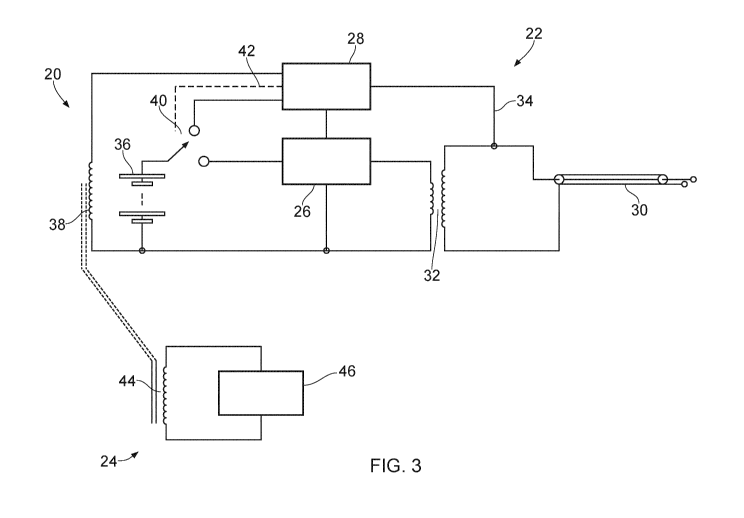
Fig. 3 shows a schematic diagram of an electrosurgical
system 20 which is an embodiment of the present invention. The
electrosurgical system 20 comprises an electrosurgical
apparatus 22 and a transmitter 24 for wirelessly providing
power to the electrosurgical apparatus 22.
The electrosurgical apparatus 22 comprises an oscillator
26 for producing radiofrequency (RF) energy. A controller 28
is operable to select an energy delivery profile for the
oscillator 26, as well as controlling other functions of the
apparatus 22. For example, the controller 28 may be operable
to turn the oscillator 26 off and on. A feed structure conveys
the RF energy to a coaxial cable 30, which may be used to
deliver the RF energy to an electrosurgical instrument. The
feed structure comprises a transformer 32 to transfer the
generated RF signal to the coaxial cable 30. In some
embodiments, the feed structure may comprise a twisted pair
cable to convey energy from a secondary coil of the
CA 03184318 2022-11-21
WO 2021/245173
PCT/EP2021/064860
16
transformer 32 to the coaxial cable 30. A feedback path 34
from the coaxial cable 30 is connected to the controller 28 to
enable the controller 28 to monitor current and voltage of the
RF signal which is conveyed to the output and adjust the
output of the oscillator 26 accordingly. Other features of an
RF channel, for example as discussed above with respect to
Fig. 1, may also be present, but are omitted in Fig. 3 for
clarity. A rechargeable power source 36 provides power for the
oscillator 26. To recharge the power source 36, the apparatus
22 comprises a receiver circuit 38 for wirelessly receiving
power from the transmitter 24. The receiver circuit 38
comprises an inductive coupler, for example comprising an
inductor in the form of a coil of wire, for receiving power by
inductive coupling from a corresponding inductive coupler in
the transmitter 24. For example, the coil of wire may comprise
200 turns, and may have a length of 25 mm and a diameter of 20
mm. In certain embodiments, the coil of wire may be wrapped
around a core, which is preferably made of a magnetic material
such as ferrite or an iron powder core. Of course, the
parameters of the inductive coupler may be varied such that
the inductive coupler may take any suitable form. In some
examples the core may be provided generally in a U-shape,
which may correspond with a matching U-shaped core of a coil
in the transmitter 24 (such that the transmitter and receiver
cores form a generally toroidal shape when positioned together
for wireless power transfer), to increase efficiency of energy
transfer between the transmitter 24 and the receiver circuit
38. Of course, it is envisaged that the core may be provided
in any suitable shape. The controller 28 is configured to
operate a switch 40 via a control line 42. By operating the
switch 40, the power source 36 can be selectively connected
with the oscillator 26 in an operating mode, for example to
perform electrosurgery, or the receiver circuit 38 in a
recharging mode, for example to charge the rechargeable power
source 36.
In some examples, the receiver circuit 38 may
additionally comprise a capacitor and, optionally, a resistor
which may be connected in series or in parallel with the
inductive coupler such that the receiver circuit forms a
resonant inductive circuit. For example, for resonance at 400
CA 03184318 2022-11-21
WO 2021/245173
PCT/EP2021/064860
17
kHz, a capacitance of 158 nF may be used (C = 1/((2n x
400x103)2 x 1x10-6), though any combination of capacitor and
resistor may be chosen to obtain desirable resonant
characteristics. For example, the receiver circuit 38 may be
configured to resonate at any suitable frequency, and 400 kHz
is given only by way of example. By providing a circuit and,
optionally, a resistor in this way, the receiver circuit 38
may be configured to receive power from the transmitter 24 by
resonant inductive coupling. Advantageously, the receiver
circuit 38 may also comprise a rectifier and a regulator to
convert a received voltage from AC to DC.
The inductive coupler is preferably positioned near a
sidewall of a housing of the electrosurgical apparatus 22. In
this way, the coil is positioned in a manner which ensures
that, when the electrosurgical apparatus 22 is suitably
positioned relative to the transmitter 24, substantially all
of the magnetic field generated by the transmitter 24 passes
through the secondary coil, maximising efficiency of power
transfer between the transmitter 24 and the electrosurgical
apparatus 52.
The transmitter 24 also comprises an inductive coupler 44
which is configured to receive power from a charging source 46
to generate an oscillating magnetic field and thereby induce a
current in the corresponding inductive coupler of the receiver
circuit 38. The charging source 46 may comprise mains power or
a battery pack, for example. An example of a transmitter which
may be used in the electrosurgical system 20 is shown in Fig.
7.
In addition to monitoring current and voltage of the RF
signal, the controller 28 may also be configured to monitor
charging and discharging of the rechargeable power source 36.
For example, the controller 28 may comprise a charge balancing
circuit, an over temperature cut out and other features to
form a battery management system to help maximise the life of
the rechargeable power source 36. In an embodiment, the
controller 28 may include a rectification circuit to convert a
received voltage from AC to DC. It is to be understood that in
some embodiments the coil of the receiver circuit 38 may have
a different type of core to the coil of the transmitter 24.
For example, one coil may have an air core and the other coil
CA 03184318 2022-11-21
WO 2021/245173
PCT/EP2021/064860
18
may have a solid core (e.g. iron powder/dust core).
Alternatively, both cores may be the same, e.g. an air core or
a solid core.
Fig. 4 shows a schematic diagram of a second
electrosurgical system 50 which is a further embodiment of the
present invention. Components which are equivalent to those
described above are given corresponding reference numerals,
and description thereof is not repeated.
The electrosurgical system 50 comprises an
electrosurgical apparatus 52 and a transmitter 24. The
transmitter 24 may be a transmitter 24 as shown in Fig. 7, for
example.
In this embodiment, the electrosurgical apparatus 52 does
not include a dedicated inductive coupler for wirelessly
receiving power from the transmitter 24. Instead, a secondary
coil of the transformer 32 is used to perform this function.
The inductive coupler 44 of the transmitter 24 receives power
from the charging source 26 to generate an oscillating
magnetic field, and thereby induce a current in the second
coil of the transformer 32. In some examples, a capacitor and,
optionally, a resistor may be connected to the secondary coil
of the transformer 32, either in series or in parallel, to
form a resonant inductive circuit, as described above with
respect to Fig. 3. The controller 28 is configured to operate
switches 54, 56 via a control line 58 to selectively connect
the rechargeable power source 36 to the secondary coil of the
transformer 32 for charging by the induced current. In an
operating mode, the controller 28 can operate the switches 54,
56 to electrically connect the power source 36 to the
oscillator 26 to generate RF EM energy for electrosurgery.
Although not shown, additional circuitry such as chokes and
capacitors may be connected to the primary and/or secondary
coils of the transformer 32 to filter out electromagnetic
interference (EMI) and improve switching characteristics. In
certain embodiments, each of the primary and secondary coils
of the transformer 32 may be an air-cored solenoid having a
diameter of 25 mm and a length of 20 mm. The primary coil may
have 15 turns, and the secondary coil may have 200 turns. A
capacitor of around 158 nF may be connected to the secondary
coil. In this way, the transformer 32 may have a tuned
resonant frequency of 400 kHz, which is particularly suitable
CA 03184318 2022-11-21
WO 2021/245173
PCT/EP2021/064860
19
for use as a receiver for wireless charging, for example in
combination with the transmitter 24. Of course, these
parameters may be varied in any other suitable way to achieve
a desired resonant frequency, which may be a frequency other
than 400 kHz, and it is also envisaged that a tuned resonant
frequency of 400 kHz may be achieved by using other values for
the described parameters, or in another suitable way.
By using the secondary coil as a receiver for wireless
charging, the larger number of turns compared with the primary
coil means that a higher voltage can be obtained from a flux
linked from the transmitter 24. Of course, the transformer 32
may comprise other core materials, preferably a magnetic
material such as ferrite or an iron powder or dust.
By using the secondary coil of the transformer 32 for
wireless charging of the power source 36 in this way, no
dedicated wireless charging coil is required. This keeps the
weight and size of the components of the electrosurgical
apparatus 52 small, enabling portability and, in some
examples, the electrosurgical apparatus 52 may be hand-held.
To allow the secondary coil of the transformer 32 to be
used as an inductive coupler for wireless charging, the
transformer 32 is preferably positioned near a sidewall of a
housing of the electrosurgical apparatus 52. In this way, the
secondary coil is positioned in a manner which ensures that,
when the electrosurgical apparatus 52 is suitably positioned
relative to the transmitter 24, substantially all of the
magnetic field generated by the transmitter 24 passes through
the secondary coil, maximising efficiency of power transfer
between the transmitter 24 and the electrosurgical apparatus
52.The primary coil of the transformer 32 will receive a much
lower induced voltage when charging than the secondary coil.
However, in some examples, the controller 28 may comprise
circuitry to protect components connected to the primary coil
side of the transformer 32 when the apparatus is charging.
Fig. 5 shows a schematic diagram of a third
electrosurgical system 60 which is a further embodiment of the
present invention. Components which are equivalent to those
described above are given corresponding reference numerals,
and description thereof is not repeated.
The electrosurgical system 60 comprises an
electrosurgical apparatus 62 and a transmitter 24. In this
CA 03184318 2022-11-21
WO 2021/245173
PCT/EP2021/064860
embodiment, the electrosurgical apparatus 62 comprises an
oscillator 64 which is configured to generate microwave
frequency electromagnetic (EM) energy for delivery to an
electrosurgical instrument via a coaxial cable 30. The
5 electrosurgical apparatus 62 therefore comprises a microwave
channel between the oscillator 64 and the coaxial cable 30,
but no RF channel. Features of a microwave channel as
described above with respect to Fig. 1 may therefore be
included in some arrangements, but are omitted from Fig. 5 for
10 clarity. The transmitter 24 may be a transmitter as described
below with respect to Fig. 7, for example.
The microwave channel comprises a circulator 66 connected
to deliver microwave EM energy from the oscillator 64 to the
coaxial cable 30 along a path between its first and second
15 ports. A third port (not shown) of the circulator 66 may be
connected to a reflected coupler to be absorbed in a power
dump load, for example, as described above with respect to
Fig. 1. A coupler 68 is provided in the microwave channel,
which directs a portion of a reflected signal to the
20 controller 28 to allow the controller 28 to monitor and
analyse reflected signals via the feedback path 34. For
example, the operation of coupler 68 may be analogous to that
of coupler 414 and/or 418 of Fig. 1. Of course, it is
envisaged that other methods of feedback or measurement of the
microwave channel may be considered as an alternative, or in
addition to, those methods described herein. For example, in
some embodiments, coupler 68 may be omitted.
The electrosurgical apparatus 62 comprises a receiver
circuit 38 configured to recharge the rechargeable battery 36
using energy received from a transmitter 24 in substantially
the same manner as described above with respect to Fig. 3.
Fig. 6 is a schematic diagram of an electrosurgical
system 70 according to a fourth embodiment of the present
invention. Components which are equivalent to those described
above are given corresponding reference numerals, and
description thereof is not repeated.
The electrosurgical system comprises an electrosurgical
apparatus 72 and a transmitter 24. In this embodiment, the
electrosurgical apparatus 72 comprises both an RF oscillator
26 and a microwave frequency oscillator 64, which are each
configured to supply energy to a coaxial cable 30. The
CA 03184318 2022-11-21
WO 2021/245173
PCT/EP2021/064860
21
electrosurgical apparatus therefore comprises an RF channel
configured to convey RF energy from the RF oscillator 26 to
the coaxial cable 30, and a microwave channel configured to
convey microwave frequency energy from the microwave
oscillator 64 to the coaxial cable 30. The RF channel and the
microwave channel may each comprise components as discussed
above with respect to Fig. 1, in some examples, as well as
components discussed above with respect to Figs. 3-5. The
electrosurgical apparatus 72 comprises a combiner 74 which is
configured to take RF energy from the RF channel and microwave
frequency energy from the microwave channel and combine them
onto a single output to be delivered to the coaxial cable 30.
The controller 28 is configured to monitor the microwave
frequency energy delivered to and reflected via the coaxial
cable 30 through a microwave feedback channel 34a, and monitor
RF energy delivered to the coaxial cable through an RF
feedback channel 34b.
In this embodiment, the electrosurgical apparatus 72 may
receive power wirelessly from the transmitter 24 for charging
the battery 36 using a secondary coil of a transformer 32 on
the RF channel, as described above with respect to Fig. 4.
The electrosurgical system 70 thereby provides an
electrosurgical apparatus 72 for delivering RF and/or
microwave frequency EM energy, and which is rechargeable
wirelessly. The electrosurgical apparatus 72 is therefore more
convenient, and may be used in situations where a portable
apparatus is advantageous.
Fig. 7 shows a schematic diagram of a transmitter 24
which may be used with embodiments of the present invention.
For example, the transmitter 24 may be positioned in a
charging cradle which is used to charge an electrosurgical
apparatus.
As seen in Fig. 7, an oscillator 100 provides an
oscillating control signal to an amplifier 102. The
oscillating control signal may be an oscillating voltage
signal having a frequency in the MHz range (e.g. 9.9MHz). The
amplifier 102 amplifies this oscillating control signal to
form an oscillating drive signal which has the same frequency
as the oscillating control signal but is more powerful such
that the oscillating drive signal possesses enough power to
drive a MOSFET 104. Specifically, the MOSFET 104 is a voltage
CA 03184318 2022-11-21
WO 2021/245173
PCT/EP2021/064860
22
controlled current source and, therefore, generates an
oscillating current signal (using current supply 105) based on
the oscillating drive signal. The oscillating current signal
has the same frequency as the control signal and drive signal.
This oscillating current signal is then provided to the
primary (or transmitter) inductive coupler 110. The primary
inductive coupler 110 uses the oscillating current signal to
generate an oscillating magnetic field via electromagnetic
induction.
The primary inductive coupler 110 comprises a series
inductor-capacitor (LC) circuit having capacitor 106 and
inductor 108. It is to be understood that the inductor 108
comprises a coil of wire, which in some embodiments may be
wound on a core material. As such, the primary inductive
coupler 110 is a resonant circuit. The specific values of the
frequency of the oscillator 100, the capacitance of the
capacitor 106 and inductance of the inductor 108 are chosen
such that resonance occurs. Resonance may be set to occur
based on parameters set by the physical geometry of the
transmitter and receiver. In this way, the coil of the
inductor 108 generates an oscillating magnetic field. The
oscillating magnetic field may be used to induce a current in
a corresponding inductive coupler within an electrosurgical
apparatus as described above, and so recharge a rechargeable
power source 36. It is to be understood that the inductive
coupler 110 may be a non-resonant inductive coupler in some
embodiments.
In certain embodiments, the primary inductive coupler 110
is located near a sidewall of a housing of the transmitter 24
to ensure that substantially all of the magnetic field
generated by the transmitter 24 passes through the receiver
coil of an electrosurgical apparatus (such as described above
with respect to Figs. 3-6), maximising efficiency of power
transfer between the transmitter 24 and the electrosurgical
apparatus. The primary inductive coupler 110 may comprise a
coil of wire wound on a magnetic core material, such as
ferrite or an iron powder core. In some examples, the core may
be generally U-shaped so as to correspond with an inductive
coupler of a receiver circuit within an electrosurgical
apparatus such that the two U-shaped cores are positioned
CA 03184318 2022-11-21
WO 2021/245173
PCT/EP2021/064860
23
together for wireless power transfer to form a generally
toroidal shape.
Fig. 8a shows an image of an electrosurgical system 80
according to an embodiment of the present invention. The
electrosurgical system 80 comprises an electrosurgical
apparatus 82 and a transmitter 92. Fig. 8b shows a cut-through
image showing the placement of charging coils in the
arrangement of Fig. 8a.
The electrosurgical apparatus 82 may be an
electrosurgical apparatus as described above with respect to
any of Figs. 3 to 6, for example. In particular, the
electrosurgical apparatus 82 comprises a housing 84 which
contains a circuit for producing electrosurgical energy as
shown in any of Figs. 3 to 6. The housing 84 is preferably
sized and shaped to be handheld by a user for performing
electrosurgery or the like. On an upper surface of the housing
84 there is provided a control panel 86 for the apparatus 82.
For example the control panel 86 may have an on/off button
which is operable by a user to activate the RF and/or
microwave frequency oscillator to generate EM energy for
electrosurgery. The on/off button may be connected to a
controller within the apparatus 82 to choose operating modes
of the apparatus 82. In some embodiments the on/off button may
be operable by a user to cycle through modes, such as an RF
only mode, a microwave only mode, and/or a mode in which both
RF and microwave frequency EM energy is generated. In some
embodiments, when the electrosurgical apparatus is turned off,
the controller is configured to operate switches within the
apparatus 82 to connect a rechargeable battery within the
apparatus 82 to a receiver circuit, as discussed with respect
to Figs. 3 to 6 above.
The outer surface of the housing, and in particular the
control panel 86, may also contain other visual displays, for
example a battery status indicator, which may be provided by a
screen or by an LED, for example. The battery status indicator
allows a user to see the amount of charge left within the
rechargeable battery and so indicates when charging may be
needed, or when the battery is fully charged or is charging,
for example. Other visual displays or indicators, or audible,
vibrational or haptic transducers may be present on the
housing 84 or within the apparatus 82 as appropriate.
CA 03184318 2022-11-21
WO 2021/245173
PCT/EP2021/064860
24
As shown in Fig. 8b, the electrosurgical apparatus 82
comprises a receiving inductive coupler 88 within the housing
84 for receiving energy wirelessly from the transmitter 92. In
particular, the inductive coupler 88 is a coil of wire. For
example, the inductive coupler 88 may be a dedicated coil for
wireless charging or may form part of a transformer, as
described above with respect to Figs. 3 to 6. In some
embodiments, the coil of wire may be wound on a solid core,
for example of a magnetic material such as ferrite or an iron
powder core. The inductive coupler 88 is positioned at a lower
side of the housing 84 in order to maximise inductive coupling
with a transmitting inductive coupler 98 within the
transmitter 92.
The electrosurgical apparatus 82 further comprises an
electrosurgical instrument 90 which may be used to perform
electrosurgery. For example, the electrosurgical instrument 90
may be used to cut and/or ablate biological tissue. The
instrument 90 is connected to an output of the circuit within
the housing 84, for example as discussed above with respect to
Figs. 3-6, in order to receive generated EM energy. The
electrosurgical instrument 90 may be detachably mounted to the
housing 84, or in some embodiments may be a permanent fixture
thereof.
The transmitter 92 is provided as a docking station or
cradle for the electrosurgical apparatus 82, and transmits
energy wirelessly to the electrosurgical apparatus 82 for
charging a battery thereof. The transmitter 92 comprises a
housing 94, an upper surface of which is adapted to receive
the electrosurgical apparatus 82 when the apparatus 82 is not
in use. The housing 94 may contain a circuit as shown in Fig.
7, for example, for recharging the apparatus 82. The housing
94 comprises a projection 96 which engages a corresponding
recess in the housing 84 of the apparatus 82. The housing 94
thereby holds the apparatus 82 in an optimal position for
charging, and the projection 96 helps to ensure that the
apparatus 82 is not accidentally knocked from the transmitter
92, and so ensures continuity of charging.
As shown in Fig. 8b, the transmitter comprises a
transmitting inductive coupler 98 positioned within the
housing 94 for transmitting energy wirelessly to the
electrosurgical apparatus 82. In particular, the inductive
CA 03184318 2022-11-21
WO 2021/245173
PCT/EP2021/064860
coupler 98 is a coil of wire. In some embodiments, the coil of
wire may be wound on a solid core, for example of a magnetic
material such as ferrite or an iron powder core. The inductive
coupler 98 is positioned at an upper side of the housing 84 in
5 order to maximise inductive coupling with the receiving
inductive coupler 86 within the electrosurgical apparatus 82.
Figs. 9a and 9b are cross-sectional views of an
electrosurgical apparatus 120a, 120b showing alternative
positions of a receiving inductive coupler 122a, 122b, 122c
10 within the apparatus 120a, 120b. The electrosurgical apparatus
120a, 120b may comprise any of the features of an
electrosurgical apparatus as described above. A transmitting
inductive coupler 124a, 124b, 124c is also shown, connected to
a transmitter circuit 126a, 126b, 126c. The transmitting
15 inductive coupler 124a, 124b, 124c and the transmitter circuit
126a, 126b, 126c may be housed within a transmitter, such as
that shown in Figs 8a and 8b, for example, and may comprise
any of the features of a transmitter as described above. Of
course, it will be understood that in preferred embodiments
20 only one of the inductive couplers 122a, 122b, 122c may be
present in an electrosurgical apparatus 120a, 120b. It will
also be understood that receiving inductive coupler 122a is
positioned for use with transmitting inductive coupler 124a,
and transmitting circuit 126a. The remaining inductive
25 couplers corresponding in a like manner.
Fig. 10 shows a circuit diagram of an electrosurgical
system including: an electrosurgical apparatus 200, a
transmitter 210, and a wired charger 220. In this embodiment,
the electrosurgical apparatus 200 contains a receiver circuit
which is configured to allow both wireless and wired charging
of the rechargeable power source. Other components may be
included in addition to those shown, according to
requirements. It is to be understood that for clarity the
circuit diagram of Fig. 10 is a generalised schematic of the
output sections of the electrosurgical apparatus that relate
to wireless and wired charging. Remaining aspects of the
electrosurgical apparatus would be clear to the skilled person
from the previous figures that are described above.
In this embodiment, the electrosurgical apparatus 200
comprises two oscillators. A first oscillator provides
microwave frequency energy via a microwave channel and the
CA 03184318 2022-11-21
WO 2021/245173
PCT/EP2021/064860
26
input MW (the input MW may form part of the microwave
channel). A second oscillator provides RF energy via an RF
channel and inputs PRI 1, PRI 2 (the inputs PRI 1 and PRI 2
may form part of the RF channel). The RF channel comprises a
transformer having a primary coil (L4) and a secondary coil
(L5), which function in a similar manner as described with
respect to Figs. 3, 4 and 6 above. The RF channel also
comprises capacitors (C9, C13) connected in parallel on either
side of the transformer. The capacitors (C9, C13) help form a
filter structure with the transformer (L4, L5) to improve
conveyance of RF power to the output connector (CONNECTOR,
GND) and also block unwanted harmonics of the RF power that in
some circumstances may cause electromagnetic interference.
The microwave channel and the RF channel are each
connected to the output (CONNECTOR, GND) in order to supply
microwave and/or RF energy to an electrosurgical instrument,
for example. In some embodiments the output (CONNECTOR, GND)
may comprise a QMA connector or the like. A choke (X2) and a
capacitor (C5) form an example of a combiner circuit which
allows energy from both the microwave channel and the RF
channel to reach the output (CONNECTOR, GND) while also
preventing microwave energy reaching the RF channel and RF
energy reaching the microwave channel. For example, the choke
(X2) may be a quarter-wave short-circuit which may be
implemented as a microstrip, a stripline, or a cavity
resonator.
It is to be understood that the RF channel and the
microwave channel may include one or more additional
components, as described above with reference to Fig. 1.
Although a controller is not directly shown, sensing
circuitry is indicated (CPL, V SENSE, I SENSE, GND) which is
connected to a controller to allow the controller to monitor
the RF or microwave energy which is delivered and/or
reflected. A coupler (X1) is present on the microwave channel
to allow the controller to sense the microwave power (CPL);
the coupler (X1) is not sensitive to RF power. A capacitor
(C5) ensures that RF power is prevented from reaching the
microwave oscillator and the coupler (X1) due to its high
impedance. A RF current-sensing circuit is formed by a
transformer having a primary winding (L3) and a secondary
winding (L6), a resistor (R1) and, optionally, a DC blocking
CA 03184318 2022-11-21
WO 2021/245173
PCT/EP2021/064860
27
capacitor (Cl) - the RF current-sensing circuit is used to
sense a proportion of the RF current flowing to the connector
(CONNECTOR, GND) and is not sensitive to microwave power. A RF
voltage-sensing circuit is formed by a potential divider
connected to the RF channel and comprising two resistors (R9,
R10) and, optionally, a DC blocking capacitor (C4) - the RF
voltage-sensing circuit measures a proportion of the RF output
voltage. The RF current-sensing circuit (L3, L6, R1, Cl), RF
voltage-sensing circuit (R9, R10, C4) and the microwave power
sensing coupler (X1) are not essential to the operation of the
charging system (either wired or wireless charging) and are
shown only as an example to demonstrate how the circuit may be
arranged to allow the control to monitor RF and/or microwave
delivery.
The electrosurgical apparatus 200 also comprises a
receiver circuit, which is connected to a rechargeable power
source (not shown) via connection CHG. The receiver circuit is
configured to allow charging by means of a wired or a wireless
connection. The receiver circuit includes the secondary coil
(L5) which forms an inductive coupler for wirelessly receiving
power from the transmitter 210. The receiver circuit also
includes the output (CONNECTOR, GND) for receiving power via a
wired connection from the wired charger 220. It is to be
understood that the receiver circuit may include one or more
additional components as described above with reference to the
previous figures.
A transmitter 210 comprises a power source (V2) and a
transmitting inductive coupler (L1), which may be used to
induce a current in the secondary coil (L5) of the transformer
on the RF channel to allow wireless charging in substantially
the manner described above with respect to Figs. 3, 4, 6 and 7
above. In some embodiments, the transmitter 210 may
additionally comprise a capacitor and, optionally, a resistor
to allow wireless charging by resonant inductive coupling. The
current induced in the secondary coil (L5) of the transformer
is prevented from reaching the microwave channel by the
capacitor (C5).
A wired charger 220 comprises a power source (V3) and a
pair of contacts (CONNECTOR, GND). The power source (V3) may
be mains power, for example, or may be a power source (e.g. a
battery) internal to the wired charger 220. The wired charger
CA 03184318 2022-11-21
WO 2021/245173
PCT/EP2021/064860
28
220 is configured to deliver energy into the electrosurgical
apparatus 200 and to the receiver circuit via a connector
formed by the output (CONNECTOR, GND). In other embodiments,
the electrosurgical apparatus 200 may comprise one or more
additional contacts which are configured to couple with the
wired charger 220 to deliver energy to the receiver circuit.
The current provided from the wired charger (220) is prevented
from reaching the microwave channel by the capacitor (C5).
It is to be understood that in one version of Fig. 10,
the transmitter 210 and the wired charger 220 are physically
separate devices. For example, the transmitter 210 may be a
wireless charging cradle similar to the one shown in Fig. 8a,
and the wired charger may be a separate connecting device
(e.g. cable) for connecting to a mains supply. However, in
another version of Fig. 10, the transmitter 210 and the wired
charger 220 may be housed within the same physical device. For
example, the device may be a charging cradle similar to the
one shown in Fig. 8a, but modified in order to provide wired
charging from a power source (e.g. battery) contained within
the cradle.
The features disclosed in the foregoing description, or
in the following claims, or in the accompanying drawings,
expressed in their specific forms or in terms of a means for
performing the disclosed function, or a method or process for
obtaining the disclosed results, as appropriate, may,
separately, or in any combination of such features, be
utilised for realising the invention in diverse forms thereof.
While the invention has been described in conjunction
with the exemplary embodiments described above, many
equivalent modifications and variations will be apparent to
those skilled in the art when given this disclosure.
Accordingly, the exemplary embodiments of the invention set
forth above are considered to be illustrative and not
limiting. Various changes to the described embodiments may be
made without departing from the spirit and scope of the
invention.
For the avoidance of any doubt, any theoretical
explanations provided herein are provided for the purposes of
improving the understanding of a reader. The inventors do not
wish to be bound by any of these theoretical explanations.
CA 03184318 2022-11-21
WO 2021/245173
PCT/EP2021/064860
29
Throughout this specification, including the claims which
follow, unless the context requires otherwise, the words
"have", "comprise", and "include", and variations such as
"having", "comprises", "comprising", and "including" will be
understood to imply the inclusion of a stated integer or step
or group of integers or steps but not the exclusion of any
other integer or step or group of integers or steps.
It must be noted that, as used in the specification and
the appended claims, the singular forms "a," "an," and "the"
include plural referents unless the context clearly dictates
otherwise. Ranges may be expressed herein as from "about" one
particular value, and/or to "about" another particular value.
When such a range is expressed, another embodiment includes
from the one particular value and/or to the other particular
value. Similarly, when values are expressed as approximations,
by the use of the antecedent "about," it will be understood
that the particular value forms another embodiment. The term
"about" in relation to a numerical value is optional and
means, for example, +/- 10%.
The words "preferred" and "preferably" are used herein
refer to embodiments of the invention that may provide certain
benefits under some circumstances. It is to be appreciated,
however, that other embodiments may also be preferred under
the same or different circumstances. The recitation of one or
more preferred embodiments therefore does not mean or imply
that other embodiments are not useful, and is not intended to
exclude other embodiments from the scope of the disclosure, or
from the scope of the claims.