Note: Descriptions are shown in the official language in which they were submitted.
TISSUE-INTEGRATING SENSORS
TECHNICAL FIELD
[0001] The present disclosure is in the field of biosensors.
BACKGROUND
[0002] In the management of many conditions, the regular measurement
of
analytes in vivo is required. It has been a long-standing objective of both
medical
science and the military to implant sensors inside the human body that
continuously
and accurately determine changes in physiologic, metabolic, or fatigue status;
measure the concentration of biothreat or therapeutic agents in vivo; and
provide early
detection of disease prior to the onset of symptoms. Doing so non-invasively
with
minimal user maintenance is essential, and sensor longevity of months to years
is
crucial in actual user environments.
[0003] For example, measurement of glucose in the blood is essential
in order
to ensure correct insulin dosing in diabetic patients. Furthermore, it has
been
demonstrated that in the long term care of the diabetic patient better control
of the
blood glucose levels can delay, if not prevent, the onset of retinopathy,
circulatory
problems and other degenerative diseases often associated with diabetes. Thus
there is
a need for reliable and accurate self-monitoring of blood glucose levels by
diabetic
patients.
[0004] Currently, blood glucose is monitored by diabetic patients with
the use
of commercially available calorimetric test strips or electrochemical
biosensors (e.g.
enzyme electrodes), both of which require the regular use of a lancet-type
instrument
to withdraw a suitable amount of blood each time a measurement is made. On
average, the majority of diabetic patients would use such instruments to take
a
measurement of blood glucose twice a day. However, the US National Institutes
of
Health recently recommended that blood glucose testing should be carried out
at least
four times a day, a recommendation that has been endorsed by the American
Diabetes
Association. This increase in the frequency of blood glucose testing imposes a
considerable burden on the diabetic patient, both in terms of financial cost
and in
terms of pain and discomfort, particularly in the long-term diabetic who has
to make
regular use of a lancet to draw blood from the fingertips. Thus, there is
clearly a need
1
Date Recue/Date Received 2022-12-30
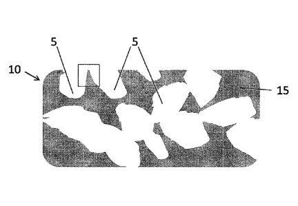
for a better long-term glucose monitoring system that does not involve drawing
blood
from the patient.
[0005] Over the last several decades, many attempts have been made to
develop implanted sensors that provide frequent or continuous monitoring. For
example, U.S. Pat. No. 4,703,756 to Gough et al., filed May 6, 1986, describes
a
sensor module for implantation in the body to monitor glucose and oxygen
levels.
However, due to electrical failure, degradation of the analyte recognition
element
(typically an enzyme), component degradation and delamination, these sensors
typically fail after a relatively short period of time (e.g.,. hours to days).
Another
major failure mode of in vivo sensors is not failure of the sensor itself, but
rather
changes in the tissue immediately adjacent to the sensor due to the
implantation of the
sensor. The tissue at the interface of the sensor changes in such a way that
it is no
longer representative of the overall body state or disease state or analyte of
interest.
[0006] U.S. Patent No. 7,228,159 describes a senor comprising a
plurality of
non-biodegradable sensing particles embedded in a biodegradable matrix for
injection
into the dermis. However, as the matrix degrades, the sensing particles are
ingested
by macrophages and removed from the implant site. Similarly, U.S. Patent No.
6,671,527 describes a sensor which is injected into epidermis and is ejected
over time
due to the normal sloughing of skin. U.S. Patent Application No. 2009/0131773
describes a carbohydrate (e.g., glucose) sensor made up of at least two
different
variants of an appropriate competitive binding assay.
[0007] Nielsen et al. (2009) J. Diabetes Science and Technology 3(1)
:98-109,
Billingsley et al. (2010) Anal. Chem. 82(9):3707-3713 and McShane et al.
(2000)
IEEE Engineering in Medicine and Biology Magazine 19:36-45 describe
implantation
of analyte-sensing microspheres or nanospheres. These individual sensing
particles
are taken up by macrophages if they are too small, and can migrate through the
tissue,
which is not desirable for explanation and not desirable to have the
fluorescent signal
disperse in an uncontrolled way. If the sensing particles are too big to be
taken up by
macrophages, they undergo the typical foreign body response (FBR), which
limits the
proximity of capillaries with respect to the implant. As sensors become
encapsulated
by avascular tissue, they lose ability to accurately sense blood borne
analytes and as
they become engulfed by phagocytic cells (small particles), they lose contact
with
interstitial fluid, which is the compaitment necessary to be sensed for
components
2
Date Recue/Date Received 2022-12-30
such as glucose. Therefore, current sensing technologies typically fail after
only a
short time in the body (e.g., 2-7 days for commercially available sensors).
[0008] Thus, there remains a clear need for sensing technologies that
are
tissue integrating to provide long-term (e.g., weeks, months or years) and
accurate
readings by remaining in contact with interstitial fluid (not the internal
cellular
environment) and remaining in close proximity to the vasculature so that the
interstitial fluid surrounding the sensor is in constant rapid equilibrium
with nearby
capillaries.
SUMMARY
[0009] Disclosed herein are tissue-integrating sensors, systems
comprising
these sensors and methods of using these sensors and systems for the
measurement of
various analytes.
[0010] Currently, continuous analyte sensors for monitoring body
chemistry
(microdialysis, electrochemical, skin tattoo sensors, etc.) do not provide
accurate,
long-term data due to the progressively declining capillary density and/or
foreign
body response. The integration of capillaries into and throughout the sensor
(sensing
media) is a major improvement over what currently exists. The capillary
enhancement gives rise to improved accuracy and reduced lag time.
[0011] In one aspect, provided herein are a tissue-integrating sensor for
detecting an analyte, the sensor comprising a tissue-integrating scaffold; and
one or
more sensing moieties, wherein the sensing moieties produce a detectable
signal in
the presence of the analyte; and further wherein the sensor provides detection
of the
analyte when placed (e.g., implanted) into the tissue of a subject. The tissue-
integrating sensors as described herein can provide long-term detection of the
analyte(s). In certain embodiments, the tissue-integrating scaffold consists
of the one
or more sensing moieties (e.g., polymeric sensing moieties formed into a
scaffold).
The tissue-integrating sensors may comprise one or more polymers, for example
one
or more hydrogels. The sensing moieties may be embedded and/or attached to the
exterior of the scaffold or may form the scaffold itself. In certain
embodiments, the
scaffold is porous and further wherein at least two of the pores are
interconnected. In
certain embodiments, the sensing moieties comprise microspheres or
nanospheres.
Any of the sensors described herein may include one or more layers (with
sensing
moieties in one or more of the layers) and/or one or more fibers.
3
Date Recue/Date Received 2022-12-30
[0012] Any of the sensors described herein may further comprise
additional
components, for example, a coating on the exterior of the sensor and/or one or
more
additional reference (calibration) moieties, for example for calibrating the
signal
detected from the sensing moieties.
[0013] In yet another aspect, provided herein is a system for detecting an
analyte, the system comprising one or more of the tissue-integrating sensors
as
described herein; and an interrogator that generates (e.g., light that causes
the sensing
moieties to emit light) and/or measures the signal produced by the sensing
moieties.
In certain embodiments, the system further includes one or more of the
following: a
detector, a signal receiver, a signal transmitter, a signal processing
component, an
energy storage component, a data storage component, a data transmitter, a data
display device, a data processing component and combinations thereof.
[0014] In yet another aspect, provided herein are methods of making
and
using the sensors and systems as described herein. In certain embodiments,
provided
herein is a method for detection of an analyte in a tissue of a subject, the
method
comprising integrating one or more sensors as described herein into the tissue
and
detecting the presence of the analyte.
BRIEF DESCRIPTION OF THE DRAWINGS
[0015] Figure 1 depicts a cross-section of exemplary tissue-integrating
implant as described herein showing the pores and solid scaffold portions.
[0016] Figure 2, panels A to E, depict a cross-sections of exemplary
tissue
integrating implants as described herein following implantation into a tissue
and
showing tissue in-growth into the pores following implantation into a subject.
FIG.
2A is a schematic cross-section of a portion (boxed area) of the device shown
in FIG.
1. FIGs. 2B and 2C are reproductions of histology photographs showing a cross-
section of tissue including the implanted sensor 1 week (FIG. 2B) or one more
(FIG.
2C) after implantation of a sensor as described herein. FIGs. 2D and 2E are
reproductions of immunohistochemistry photographs (staining for capillaries
for
.. CD31) showing a cross-section of tissue including the implanted sensor 1
week (FIG.
2D) and 1 month (FIG. 2E) post-implantation of the sensor.
[0017] Figure 3 depicts a cross-section (boxed area of Figure 1) of an
exemplary tissue-integrating implant (also known as the sensing media) as
described
4
Date Recue/Date Received 2022-12-30
herein in which sensing moieties are embedded (physically entrapped or
chemically
bound) within the solid scaffold portions.
[0018] Figure 4 depicts a cross-section of a portion (boxed area of
Figure 1)
of an exemplary tissue-integrating implant as described herein in which
sensing
.. moieties are attached to the surface of the solid scaffold portions.
[0019] Figure 5 depicts a cross-section (boxed area of Figure 1) of an
exemplary tissue-integrating implant as shown in Figure 4 and further
including an
exterior coating on or over the sensing moieties.
[0020] Figure 6 depicts a cross-section (boxed area of Figure 1) of an
exemplary tissue-integrating implant as described herein in which solid
scaffold
portions are made from sending moieties in the form of particles bonded
together.
[0021] Figure 7 depicts a cross-section (boxed area of Figure 1) of an
exemplary tissue-integrating implant as described herein in which solid
scaffold
portions are made from a polymer in which the polymer is composed of sensing
materials.
[0022] Figure 8 depicts a cross-section of an exemplary tissue-
integrating
implant as shown in Figure 3 and further including additional moieties (e.g.,
reference
particle for calibration) embedded in the scaffold.
[0023] Figure 9, panels A and F, are overviews and cross-sections of
exemplary sensors as described herein. FIG. 9A shows an exemplary single-
layered
(e.g., single layer fibers) cylindrical sensing media (tissue integrating
sensor
embodiment in which the sensing moieties are embedded in the scaffold and FIG.
9B
shows an embodiment in which the sensing moieties are attached to the surface
of the
scaffold. FIG. 9C depicts an overview of an embodiment including sensing media
on
the surface and embedded within the sensor. FIG. 9D depicts a cross-section of
an
exemplary sensor as described herein. FIG. 9E and 9F are overviews of
exemplary
sensors as described herein including one or more fibers containing sensing
moieties.
[0024] Figure 10, panels A and B, are overviews of exemplary multi-
layered
cylindrical sensing media (tissue integrating sensor) as described herein.
FIG. 10A
shows an embodiment with two layers and in which in the sensing moieties are
embedded in the inner layer. FIG. 10B shows an embodiment with a hollow core
and
outer layer containing embedded sensing moieties therein.
[0025] Figure 11 is a cross-section of an exemplary sensing media as
shown
in FIG. 9A.
5
Date Recue/Date Received 2022-12-30
[0026] Figure 12 is a cross-section of an exemplary sensing media as
shown
in FIG. 9B.
[0027] Figure 13 is a cross-section of an exemplary sensing media as
shown
in FIG. 12 and further including a coating exterior to the sensing moieties on
the
surface of the scaffold.
[0028] Figure 14 is a cross-section of an entire (e.g., cylindrically
shaped) or
portion of (e.g., individual fiber) an exemplary sensing implant as described
herein in
which the scaffold is made from polymer where the polymer itself is composed
of
sensing moieties.
[0029] Figure 15 is a cross-section of an entire (e.g., cylindrically
shaped) or
portion of (e.g., individual fiber) an exemplary sensing implant including
multi-layers,
and in which the sensing media are embedded in the inner layer.
[0030] Figure 16 is a cross-section of an entire (e.g., cylindrically
shaped) or
portion of (e.g., individual fiber) an exemplary sensing implant including
multi-layers,
and in which the sensing media are embedded in the outer layer.
[0031] Figure 17 is a cross-section of an exemplary hollow
cylindrically
shaped (or individual fiber of a) sensor in which the sending media is
embedded in the
layer surrounding the hollow core.
[0032] Figure 18 is a schematic cross-section depiction of a sensing
media
implant as described herein following implantation into the skin of a subject.
[0033] Figure 19, panels A to C, are schematic representations of
exemplary
systems including tissue-integrating, vascularizing sensor and interrogators.
[0034] Figure 20, panels A and B, show photographs of subjects (mice)
comprising oxygen sensing media ("OD") as described herein and reference
moieties
("QD") produced with reference implants comprising qtracker 800 quantum dots
from
Invitrogen. Implants were injected with a trocar approximately 2 mm under the
surface of mice skin. Mice were imaged with Caliper whole animal imaging
system
(IVISTM) with an excitation of 535 nm and emission light was collected at 760
nm
under oxygenated (FIG. 20A) and deoxygenated conditions (FIG. 20B). As shown,
the reference implants (comprising reference moieties) ("QD") maintained their
signal
in deoxygenated conditions, whereas the oxygen sensing media ("OD") modulated
with oxygen concentration.
[0035] Figure 21 is a graph depicting glucose monitoring from glucose
sensors as described herein (Example 2). Data show percent change of PDP
emission
6
Date Recue/Date Received 2022-12-30
as a function of glucose concentration. Disk of glucose sensor scaffold
material were
punched from the rectangular pieces (microscope slide-shape) that were
produced as
described in Example 2. Sensor scaffold discs were fixed inside an automated
flow-
through system with a built in flourimeter. Glucose solutions (in PBS) of
various
concentrations were flowed over the sensor scaffold discs. Fluorescence and
lifetime
readings were collected at various glucose concentrations over successive
runs. A
plot of the change in sensor signal compared to baseline (zero glucose
concentration)
is shown below.
DETAILED DESCRIPTION
[0036] Described herein are tissue-integrating sensors useful for
accurate and
optionally long term measurements of analytes in vivo. Also described herein
are
methods of using these sensors for optical detection of various biochemical
analytes.
Using reversible binding ligands and/or chemistries, the implantable sensors,
systems
and methods described herein provide for continuous or semi-continuous
collection of
data of various biochemical analytes, optionally without the use of
implantable
hardware of any type and/or enzymatic and electrochemical detection methods.
[0037] In particular, the tissue-integrating sensors that are the
subject of this
invention remain in good contact (close proximity) to blood vessels and have
direct
access to measurements of interstitial fluid. The tissue-integrating scaffold
encourages capillary growth into and/or nearby the sensing media. The sensing
media
is devoid of electronics, making the sensing media seem less foreign to the
body than
implants that contain electronics. Additionally the tissue-integrating sensing
media
may have a modulus closer to the texture of tissue, thus enhancing the
integration in
the tissue.
[0038] Thus, unlike other devices, the sensors described herein allow
capillaries to grow in close proximity to all regions of the sensor (e.g., on
the surface
and inside), which results in accurate analyte measurements, including over
long term.
Embedding, attaching or forming scaffolds out of nano-sized sensing elements
results
in tissue-integrating sensing media that allows in-growth, including of tissue
and
capillaries, in and/or around the sensors. Tissue integrating sensors minimize
the
foreign body response and/or promote vascularization. Capillary growth
directly into
and throughout the sensor allows unencumbered access to analytes of interest
in the
blood (e.g. glucose, lactate, pyruvate, cortisol, ions, proteins, nucleic
acids, alcohols,
7
Date Recue/Date Received 2022-12-30
urea, etc.). The level of tissue integration and proximity of capillaries to
all regions of
the sensor will provide a close, stable relationship between the analyte
concentration
in the blood and in the tissue surrounding the sensing media.
[0039] Advantages of the device and methods described herein include,
but
are not limited to: (1) providing devices that integrate into the subject
(e.g., through
tissue and/or capillary in-growth; (2) providing devices which can be
implanted
through syringe injection, meaning that no surgery is required to put the
sensing
media in place in the body; (3) providing devices that do not include sensor
electronics in the body; (4) providing devices comprising material(s) having
properties more similar to actual tissue (e.g., modulus that is more similar
to tissue's
modulus and hydrogel water content) to allow a better acceptance into the
tissue; (5)
providing devices that accurately assess analyte(s) for long periods of time
(e.g.,
greater than a week, typically weeks, months or years) and/or (6) providing
devices of
small dimensions which will give result in increased patent comfort and better
acceptance by the body.
[0040] It must be noted that, as used in this specification and the
appended
claims, the singular forms "a", "an", and "the" include plural referents
unless the
content clearly dictates otherwise. Thus, for example, reference to a sensor
comprising "a sensing moiety" includes devices comprising of two or more
sensing
moieties. Likewise, reference to "an analyte" refers to two or more analytes.
Definitions
[0041] The term "tissue integrating" refers to a material (e.g.,
scaffold) which,
when integrated into living tissue remains in close proximity with the blood
vessels of
the tissue (e.g., capillaries). By "close proximity," is meant that the
average distance
from any point within the material (scaffold) implanted into the tissue to the
nearest
blood vessel is no greater than 100 microns more than the average distance
from any
point in the native (original) tissue to the nearest blood vessel.
[0042] By "long-term" is meant that the implant senses the analyte for
greater
than about 7 days, for example weeks, months, or years.
[0043] By "biodegradable" or "bioabsorbable" is meant that the
material is
capable of being broken down by the subject's body over a period of time,
ranging
from days to weeks to months or years.
8
Date Recue/Date Received 2022-12-30
[0044] By "water-soluble" is meant that the molecules of the material
are
capable of dissolving in water. Thus, biodegradable materials may include
water-
soluble biomaterials.
[0045] By "hydrogel" is meant a material that absorbs a solvent (e.g.
water),
undergoes rapid swelling without discernible dissolution, and maintains three-
dimensional networks capable of reversible deformation.
Sensing Media
[0046] Described herein are sensors (or sensing media) for
implantation in a
subject. The sensors are made up of tissue-integrating scaffolds and at least
one
sensing moiety.
A. Tissue Integrating Scaffolds
[0047] The sensors described herein typically comprise a tissue-
integrating
scaffold (also referred to as a matrix) material. Preferably, the tissue-
integrating
scaffold of the invention may be constructed with materials and/or micro-
architecture
such that the scaffold promotes tissue-integration and/or vascularization. For
example, porous scaffolds provide tissue biomaterial anchoring and promote in-
growth throughout the pores. The resulting "hallway" or "channel" pattern of
tissue
growth are healthy, space-filling masses that persist over time and promote
host cell
integration. Most or all of the pores of the biomaterials described herein are
preferably interconnected (co-continuous). The co-continuous pore structure of
the
biomaterials promotes space-filling in-growth of cells in the implant, which
in turn
limits the foreign body response and leads to long-term (greater than one week
and up
to years) persistence of the implant's ability to act as a sensor. Alternative
structures
that provide tissue integrating scaffolds include fibers (e.g., 1 to 10 or
more microns
in diameter, such as 5, 6, 7, 8, 9, 10 or more microns), which may be arranged
in non-
random or random configuration. Tissue-integrating scaffolds (in any
configuration)
can also be formed by multiphoton polymerization techniques. Kaehr et al.
(2008)
Proc. Nat'l. Acad. Sci. USA 105(26):8850-8854; Nielson et al. (2009) Small
1:120-
125; Kasprzak, Doctoral Dissertation, Georgia Institute of Technology, May
2009.
[0048] The tissue-integrating scaffold of the invention may comprise
any
material, including but not limited to synthetic polymers, naturally-occurring
substances, or mixtures thereof. Exemplary synthetic polymers include, but are
not
9
Date Recue/Date Received 2022-12-30
limited to polyethylene glycol (PEG), 2-hydroxyethyl methacrylate (HEMA),
silicone
rubber, polyaepsilonl-caprolactone) dimethylacrylate, polysulfone, (poly)methy
methacrylate (PMMA), soluble Teflon-AF, (poly) ethylenetetrapthalate (PET,
Dacron), Nylon, polyvinyl alcohol, polyacrylamide, polyurethane, and mixtures
thereof. Exemplary naturally-occurring materials include, but are not limited
to,
fibrous or globular proteins, complex carbohydrates, glycosaminoglycans,
extracellular matrix, or mixtures thereof. Thus, the polymer scaffold may
include
collagens of all types, elastin, hyaluronic acid, alginic acid, desmin,
versican,
matricelluar proteins such as SPARC (osteonectin), osteopontin, thrombospondin
1
and 2, fibrin, fibronectin, vitronectin, albumin, chitosan etc. Natural
polymers may be
used as the scaffold or as an additive.
[0049] In certain embodiments, the tissue-integrating scaffold
comprises a
hydrogel. For example, the polymer scaffold may comprise a hydrogel, for
example
by reacting hydroxyethyl methacrylate (HEMA), poly (hydroxyethyl
methacrylate),
pHEMA. Furthermore, various comonomers can be used in combination to alter the
hydrophilicity, mechanical and swelling properties of the hydrogel (e.g. PEG,
NVP,
MAA). Non-limiting examples of polymers include 2-Hydroxyethyl methacrylate,
polyacrylamide, N-vinylpyrrolidone, N,N-Dimethylacrylamide, poly(ethylene
glycol)
monomethacrylate (of varying molecular weights), di ethylene glycol
methacrylate, N-
(2-hydroxypropyl)methacrylamide, glycerol monomethacrylate, 2,3-
dihydroxypropyl
methacrylate and combinations thereof. Non-limiting examples of cross-linkers
include tetraethylene glycol dimethacrylate, poly(ethylene glycol) (n)
diacrylate (of
varying molecular weights), ethoxylated trimethylolpropane triacrylate,
bisacrylamide
and combinations thereof. Non-limiting examples of initiators include irgacure
Series
(UV), Azobisisobutyronitrile (AIBN) (thermal), Ammonium Persulfate (APS)
(thermal).
[0050] The tissue-integrating scaffold may be a sphere-templated
hydrogel,
for instance an inverse colloid crystal, for example as described in U.S.
Patent
Publication No. 2008/0075752 to Ramer, et al. or other tissue integrating
materials.
[0051] The scaffold may be degradable, either by the body (biodegradable)
or
by the application of an external initiator to start or speed up the
degradation process
(e.g. UV, ultrasonics, radio frequency, or other exogenous sources to initiate
degradation.). For example, the tissue-integrating scaffold may be comprised
of any
biodegradable or bioresorbable polymers, including but not limited to
degradable
Date Recue/Date Received 2022-12-30
forms of alginates, poly(lactic acid), poly(vinyl alcohol), polyanhydrides,
poly(glycolic acid), microporous polyesters, microporous polyethers and cross-
linked
collagen. One specific example is UV-photopolymerization of poly(ethylene
glycol)-
diacrylate and acrylated protease-degradable peptides and VEGF as described by
Phelps, et al (2010) Proc. Nat'l. Acad. Sci. USA 107(8):3323-3328.
[0052] Other specific examples are polymers described by Kloxin et al
(2009)
Science 324:59-63 and U.S. Patent No. 6,013,122 whose degradation is
controlled
through exposure to exogenous energy forms as well as Alexeev et al. (2003)
Anal.
Chem. 75:2316-2323; Badylak et al. (2008) Seminars in Immunology 20:109-116;
Bridges et al. (2010) 94(1):252-258; Isenhath et al. (2007) Research 83A:915-
922;
Marshall et al. (2004) Polymer Preprints, American Chemical Society, Division
of
Polymer Chemistry 45:100-101; Phelps et al. (2010) Proc Nat'l Acad Sci US A.
107(8):3323-8; Ostendorf and Chichkov (2006) Two Photon Polymerization: A New
Approach to MicroMachining, Photonics Spectra; Ozdemir et al. (2005)
Experimental
and Clinical Research, Plast. Reconstr. Surg. 115:183; U.S. Patent Publication
No.
20080075752; Sanders et al. (2003) Journal of Biomedical Materials Research
Part A
67A(4):1181-1187; Sanders et al. (2002) Journal of Biomedical Materials
Research
62(2):222-227; Sanders et al. (2003) Journal of Biomedical Materials Research
65(4):462-467; Sanders et al. (2005) Biomaterials 26:813-818; Sanders et al.
(2005)
Journal of Biomedical Materials Research Part A 72(3):335-342; Sanders (2003)
Journal of Biomedical Materials Research 67(4):1412-1416; Sanders et al.
(2000)
Journal of Biomedical Materials Research 52(1):231-237; and Young Min Ju et
al.
(2008) J Biomed Mater Res 87A:136-146.
[0053] In certain embodiments, the tissue-integrating scaffold of the
invention
is constructed such that tissue response modifiers are released from the
scaffold
material to promote or enhance tissue-integration and vascularization.
[0054] In addition, the tissue-integrating scaffold of the invention
may be
constructed such that it has conduits, pores or pockets that are hollow or
filled with
degradable, angiogenic, or other substances (e.g. stem cells). As noted above,
once in
the body, the biodegradation of the material filling the conduits, pores or
pockets,
creates space for tissue, including capillaries to integrate with the
material. The
degradable material that initially fills the conduits, pores or pockets may
enhance
vessel growth or tissue growth within the scaffold. This architecture promotes
new
vessel formation and maintains healthy viable tissue within and around the
implant.
11
Date Recue/Date Received 2022-12-30
[0055] The tissue-integrating scaffold of the invention may be
constructed
such that it is permeable to analytes of interest (e.g. glucose can diffuse
into a tissue-
integrating hydrogel scaffold and reach the sensing moieties that are embedded
within
the hydrogel matrix).
[0056] FIG. 1 depicts an exemplary embodiment of a porous tissue-
integrating
implants described herein. The device as a whole is generally designated 10
and is
shown in cross-section in a three-dimensional block. FIG. 1 shows an
embodiment in
which all of the pores 5 are interconnected. The pores 5 are within the solid
scaffold
portions 15.
[0057] FIG. 2A depicts an exemplary embodiment of a porous tissue-
integrating implant as described herein following implantation and tissue in-
growth.
The scaffold 15 is shown following growth of blood vessels 45, cells 50 and
extracellular matrix material 55 (e.g., collagen) in and around the implant
after
implantation. FIGs. 2B and 2C show histology photographs of tissue (rat skin)
.. including an integrated implant 15 as described herein. FIG. 2B shows the
implant in
the tissue 1 week following implantation and FIG. 2C shows the implant 1 month
following implantation into Sprague-Dawley rats. As shown, the tissue 19 grows
into
the implant, keeping the implant in close proximity to the blood vessels of
the tissue
and without a significant foreign body response. FIGs. 2D and 2E are
reproductions
of photographs showing immunohistochemistry staining for vasculature (using
CD31
antibodies) 1 week (FIG. 2D) and 1 month (FIG. 2E) following implantation into
skin
(subcutaneous) of Sprague-Dawley rats. The approximate boundaries of the
scaffold
16 are shown as well as capillary ingrowth 18 into the implanted scaffold.
[0058] In certain embodiments, the tissue-integrating scaffold is made
up
solely or primarily of sensing moieties (see, e.g., FIGs. 5 and 6). For
example,
sensing particles can be bonded together using any suitable method (chemical,
adhesive, thermal, etc.). In certain embodiments, the sensing particles
comprise a
polymer, for example PEG-coated particles (e.g., microspheres). In other
embodiments, the scaffold comprises a polymer that itself is composed of
sensing
moieties. See, FIG. 6.
[0059] The tissue integrating implant can be of any suitable form,
including,
but not limited to block-like (or any thickness), cube-like, disk-shaped,
cylindrical,
oval, round, random or non-random configurations of fibers and the like. In
certain
embodiments, the sensor comprises one or more fibers, which may be organized
in a
12
Date Recue/Date Received 2022-12-30
non-random fashion (e.g., grid, layered grid, etc., see, FIG. 9E) or in a
random fashion
(see, e.g., FIG. 9F).
B. Sensing Moieties
[0060] The tissue-integrating scaffolds described herein are typically
combined with (or made up of) sensing moieties that detect one or more
analytes.
[0061] Non-limiting examples of analytes that may be detected by the
sensing
moieties include oxygen, reactive oxygen species, glucose, lactate, pyruvate,
cortisol,
creatinine, urea, sodium, magnesium, calcium, potassium, vasopressin, hormones
(e.g., Luteinizing hormone), pH, cytokines, chemokines, eicosanoids, insulin,
leptins,
small molecule drugs, ethanol, myoglobin, nucleic acids (RNAs, DNAs),
fragments,
polypeptides, single amino acids and the like.
[0062] Any suitable moiety can be used to sense the analyte of
interest,
including not limited to analyte binding molecules (e.g. glucose binding
proteins),
competitive binding molecules (e.g. phenylboronic acid based chemistries),
analyte
specific enzymes (e.g. glucose oxidase), ion sensitive materials, or other
analyte
sensitive molecules (e.g. oxygen sensitive dyes such as porphyrins). The
sensing
moieties may be in any form, for example, microspheres, nanospheres, fibers,
etc. A
single implant (tissue-integrating scaffold) typically includes a plurality of
sensing
moieties. In certain embodiments, the sensing moieties are all the same while
in other
embodiments, a mixture of two or more sensing moieties is used.
[0063] To enhance or create a detectable signal, sensing molecules may
be
labeled with a reporter (e.g., one or more fluorophores, one or more gold
particles,
one or more quantum dots and/or one or more single-walled carbon nanotubes).
Sensing molecules may also create a signal through swelling, optical
diffraction,
change in absorbance FRET, quenching.
[0064] Non-limiting examples of suitable sensing molecules include but
are
not limited to dye labeled Concanavalin A with glycodendrimer or dextran (see,
e.g.,
Ballerstedt et al. (1997) Anal. Chim. Ada 345:203-212) and alcohol sensitive
oxo-
bacteriochlorin derivative fluorescent binding protein developed by Takano, et
al
(2010) The Analyst 135:2334-2339 as well as Vladimir et al. (2004) Clinical
Chemistry 50:2353-2360; Asian et al. (2005) Chem. 1;77(7):2007-14; Ballerstadt
et
al. (1997) Anal. Chim. Ada 345:203-212 (1997); Billingsley et al. (2010) Anal.
Chem
82(9):3707-3713; Brasuel et al. (2001) Anal. Chem 73(10):2221-2228; Brasuel,
et al.
13
Date Recue/Date Received 2022-12-30
(2003) The Analyst 128(10):1262-1267; Horgan et al. (2006) Biosensors and
Bioelectronics 211838-1845; Ibey et al. (2005) Anal Chem 77:7039-7046; Nielsen
et
al. (2009) Journal of Diabetes Science and Technology 3(1):98-109; McShane et
al.
(2000) IEEE Engineering in Medicine and Biology Magazine 19:36-45; Mansouri &
Schultz (1984) Bio/Technology 23:885-890; Rounds, et al. (2007) Journal of
Fluorescence 17(1):57-63; Russell et al. (1999) Analytical Chemistry
71(15):3126-
3132; Schultz et al. (1982) Diabetes Care 5:245-253; Srivastava, & McShane
(2005)
Journal of Microencapsulation 22(4):397-411; Srivastava et al. (2005)
Biotechnology
and Bioengineering 91(1):124-131; Takano et al. (2010) The Analyst 135:2334-
2339.
[0065] The sensing moiety element may comprise other molecules besides
sensing molecules, such as carrier molecules/polymers (e.g. the sensing moiety
element may comprise PEG nanospheres, alginate particles or other carrier
materials
that contain sensing molecules). The sensing moiety element may also contain
reference molecules or stabilizing molecules that do not sense any analytes,
but that
serves as calibrators (e.g., a reference dye or any substance that provides a
reference
signal to which the signal modulated by the analyte of interest may be
compared for
calibration) or stabilizer (e.g. catalayse, any free-radical scavenger which
helps
preserve the sensing moieties or other stabilizer).
[0066] The sensing moiety element may be thermally responsive
material,
pressure-responsive material or materials that swell, shrink, change optical
properties,
or change other measurable properties in response to a stimulus.
C. Sensing Media
[0067] The combination of the tissue-integrating scaffold with the
analyte
sensing moieties may be termed implantable sensing media, sensing media,
tissue
integrating sensor, tissue-integrating biosensor, tissue-integrating sensing
media or
variations thereof.
[0068] The analyte sensing moieties may be combined with the tissue-
integrating scaffolds in a variety of ways to produce tissue-integrating
sensors. In
some embodiments the sensing moieties are physically entrapped or chemically
bound
within the scaffold. In other embodiments, the sensing moieties are attached
directly
(e.g., via covalent or noncovalent linkages) to the surface of the tissue-
integrating
scaffold and may optionally be covered by an exterior coating. The purpose of
the
exterior coating is described as, but not limited to the following: to hold
the sensing
14
Date Recue/Date Received 2022-12-30
moieties in place, to protect the sensing moieties from external forces, to
limit/impede
diffusion of various molecules and/or to provide a desired exterior surface,
and to
conduct or transduce the sensing signal from the chemistry to the scaffold
and/or
external detector.
[0069] In some embodiments the tissue-integrating scaffold itself is
composed
of sensing moieties where the sensing moieties are in the form of particles
(spherical
or other shapes) that are bonded together (e.g. chemically, thermally,
pressure, etc) or
where the polymer itself provides the sensing capability (e.g. stimuli-
sensitive
polymers).
[0070] In another embodiment, the tissue-integrating scaffold is composed
of
distinct layers where sensing moieties are physically entrapped or chemically
bound
to or within specific layers of the scaffold, and other layers provide other
features
such as mechanical strength, elasticity, conductivity or other properties.
[0071] In another embodiment, the tissue-integrating scaffold is
composed of
a polymer that swells or shrinks in response to a stimulus (e.g. concentration
of an
analyte of interest, temperature, or other stimuli). The shrinking or swelling
may
cause optical change (e.g. due to light diffraction, change in distances
between gold
nanoparticles contained within the matrix, or other interaction (Aleexev et al
and
AsIan, et al)).
[0072] Table 1 below provides a matrix showing how sensing moieties can be
combined with tissue-integrating scaffolds in a variety of ways to tissue-
integrating
sensing media.
Table 1: Sensing Media/Scaffold Matrix
Sensing Sensing particles Sensing chemistry Any other
Stimuli responsive
Moieties 4 (e.g. PEG (e.g. boronic acid fluorescent
sensing moieties
microspheres based chemistry, assay (e.g.
glucose (temperature,
Tissue- containing ConA sensing chemistry oxidase with
pressure, other)
integrating with attached to quantum porphyrin dye)
Scaffolds glycodendrimer, dots or gold nano-
alginate nanospheres rods)
containing ApoGox
with reported dye.)
Permeable Polymerization Polymerization Polymerization
Polymerization
Biomaterial (SM contained (SM contained (SM contained (SM
contained
Scaffold (e.g. within mesh of within mesh of within mesh of
within mesh of
hydrogel ICC) scaffold scaffold scaffold scaffold
(Kotov, polymer) polymer) polymer) polymer)
Marshall) Immobilization Immobilization Immobilization
Immobilization
(conjugation or (conjugation or (conjugation or
(conjugation or
physical physical physical physical
entrapment) of entrapment) of entrapment) of
entrapment) of
SM on surface SM on surface SM on surface SM on
surface
Date Recue/Date Received 2022-12-30
= Making scaffold Making Making Making
of sensing scaffold of scaffold of scaffold of
moiety sensing moiety sensing moiety sensing
moiety
Non- Immobilization Immobilization Immobilization
Immobilization
Permeable of SM on of SM on of SM on of SM on
Scaffold (ICC) surface surface surface surface
(e.g. Porex, Physical Physical Physical Physical
MedPor) entrapment of entrapment of entrapment of
entrapment of
SM on surface SM on surface SM on surface SM on
surface
Naturally SM contained SM contained SM contained SM contained
derived within mesh of within mesh within mesh of within
mesh of
scaffolds (e.g. naturally naturally naturally naturally
fibrin, BSA, derived matrix derived matrix derived matrix
derived matrix
collagen Immobilization Immobilization Immobilization
Immobilization
synthetic or of SM on of SM on of SM on of SM on
decellularized surface surface surface surface
ECM (sECM), Physical Physical Physical Physical
Prestwich, entrapment of entrapment of entrapment of
entrapment of
Badylak, SM on surface SM on surface SM on surface SM on
surface
Taylor,
Small fibers Polymerization Polymerization Polymerization
Polymerization
(Sanders) (SM trapped IN (SM trapped IN (SM trapped IN
(SM trapped IN
fiber matrix) fiber matrix) fiber matrix) fiber
matrix)
= Immobilization Immobilization
Immobilization Immobilization
(conjugation or (conjugation or (conjugation or
(conjugation or
physical physical physical physical
entrapment) of entrapment) of entrapment) of
entrapment) of
SM on surface SM on surface SM on surface SM on
surface
= Making scaffold Making Making Making
of sensing scaffold of scaffold of scaffold of
moiety sensing moiety sensing moiety sensing
moiety
= Multi-layer Multi-layer Multi-layer
Multi-layer
fibers (e.g. fibers (e.g. fibers (e.g. fibers (e.g.
sensing layer, sensing layer, sensing layer, sensing
layer,
biocompatibility biocompatibilit biocompatibilit
biocompatibilit
layer, stabilizing y layer, y layer, y layer,
or structural stabilizing or stabilizing or
stabilizing or
layer, voids or structural layer, structural layer,
structural layer,
cellular conduits voids or cellular voids or voids
or cellular
conduits cellular conduits
conduits
[0073] In certain embodiments, the implant (sensing media) further
comprises
additional moieties (e.g., non-sensing or additional sensing moieties
different from the
sensing moieties), for example reference (or calibration) moieties. Reference
or
calibration moieties include, but are not limited to, dyes, fluorescent
particles,
lanthanides, nanoparticles, microspheres, quantum dots or other additives or
elements
of the implant whose signal does not change due to the presences of the
analyte (e.g.,
glucose). See, e.g., Chaudhary et al. (2009) Biotechnology and Bioengineering
104(6):1075-1085. Fluctuations in the reference (calibration) signal(s) can be
used to
correct or calibrate the sensing signal(s). Reference signals might fluctuate
due to
16
Date Recue/Date Received 2022-12-30
changes in the amount of light reaching the implant (ambient light changes,
fluctuating LED or laser source). Sensing signals would also be subject to
fluctuations in the amount of light reaching the implant; however it is
desirable that
the signal of interest only fluctuates based on analyte (e.g., glucose)
fluctuations.
Therefore the reference signal is used to correct or calibrate the sensing
signal when it
fluctuates due to influences other than changes in glucose concentration.
Reference
signals might also fluctuate due to changes in the reference moiety itself
(e.g.
photodegratation, chemical degradation). The sensing signal(s) would have the
same
degradation or a rate of degradation that is relatable to the reference to
allow for
correction or calibration by the reference. Reference signals might also
fluctuate due
to physiological fluctuations that alter the light propagation through tissue
(e.g.
dehydration, oxygenation, blood flow). Sensing signals would be affected in
the same
way or in a way that is relatable to the reference fluctuations thereby
permitting
correction or calibration of the sensing signal by the one or more references.
Thus,
the sensing signal can be calibrated by reference to the signal(s) obtained
from the
calibration (reference) moieties.
[0074] In certain embodiments, the sensing moieties detect glucose and
the
reference moiety comprises a molecule that measures (produces a detectable
signal in
the presence of) oxygen (02). As noted above, the sensing moieties can
comprise an
enzyme, for example glucose oxidase which is specific for the substrate
glucose. The
reaction of glucose oxidase causes the substrate glucose to be converted to D-
glucono-1,5-lactone, which then hydrolyzes to gluconic acid. Oxygen is
consumed
and converted to H202. The reduction of 02 in the vicinity of the enzyme can
be
measured by using an 02-sensitive fluorescent dye, such as a porphyrin dye.
These
dye molecules are quenched in the presence of 02, so the reduction of 02 by
the
action of G0x, causes an increase in fluorescence. The amount of fluorescence
emitted from the 02 calibration moieties is thus proportional to the
concentration of
glucose in the sensor.
[0075] The concentration of 02 in the tissue can also vary
physiologically,
thereby changing or limiting the reaction of the enzyme in the sensing
moieties.
Therefore, the 02 concentration in the sensor can be measured independent of
the
glucose concentration. Such a reference measurement of 02 would allow
corrections
to be made to the glucose-specific signal from the sensing moieties.
17
Date Recue/Date Received 2022-12-30
[0076] In another embodiment, an analyte-specific enzyme that causes a
change in pH would require the use of a separate pH-sensitive fluorescent dye
with an
emission spectral peak different and distinguishable from the analyte-specific
dye
reporting on the activity of the analyte-specific enzyme, for example when the
sensing
moieties comprise, urease used for measuring urea.
[0077] In still further embodiments, the sensing moieties comprise a
first
fluorescent dye and the reference molecule comprises a second (different)
fluorescent
dye. As noted above, the sensing moieties may utilize an analyte-specific
chemistry
that includes a ligand receptor moiety and an analyte analogue moiety. One of
the
binding members is labeled with a fluorescent dye and the other binding member
is
labeled with a dye that quenches the fluorescent dye when the analyte analogue
moiety binds to the ligand receptor moiety. Non-limiting examples include
glycodendrimer, which binds to Concanavalin A, wherein the Concanavalin A is
labeled with Alexafluor 647 and the glycodendrimer is labeled with QDY21 dark
quencher. Concanavalin A binds to glucose and the glycodendrimer competes with
glucose for the binding to Concanavalin A. The chemistry is immobilized as
described in this invention within the tissue-integrating scaffold and
implanted into
the dermis or subcutaneous tissue. To measure glucose in the tissue, the
tissue-
integrating scaffold is illuminated from a patch reader on top of the skin
above the
implant with 650nm light at desired intervals over the long-term life of the
implant
(e.g., every 5-60 minutes over a period of 90 days or more). The amount of
fluorescent signal (e.g., from a molecule such as Alexafluor 647) detected is
proportional to the concentration of glucose in the tissue. However, over the
long-
term life of the implants described herein, the dye can photobleach, i.e., the
amount of
fluorescent signal emitted back through the skin at a given glucose
concentration is
diminished. Thus, a reduction of fluorescence due to photobleaching can make
it
appear that analyte is at a lower concentration than it really is.
[0078] To correct for this effect, a separate internal photobleaching
control is
employed. In certain embodiments, the separate internal control is a second
fluorescent dye, different from the fluorescent molecule included in the
sensing
moieties (e.g., Alexafluor 750 in the reference moieties when the sensing
moieties
comprise Alexafluor 647), which included immobilized in the scaffold. The
fluorescence of reference moieties is not affected by the concentration of
glucose, and
both the first (e.g., Alexafluor 647) and second (e.g., Alexafluor 750)
fluorescent dyes
18
Date Recue/Date Received 2022-12-30
have predictable and well-characterized photobleaching rates. To control for
the
photobleaching of the dye of the sensing moieties, the fluorescence is
measured for
both dyes. The fluorescence value of the dye in the reference moieties can
then be
used to correct for any photobleaching of the dye in the sensing moieties.
[0079] In another embodiment, internal reference control materials can be
employed that facilitate correcting for tissue optical variation. The tissue-
integrating
implanted biosensor typically resides 3-4 mm under the surface of the scan. It
is well
known that in skin excitation light and emitted fluorescent light in the near
infrared
range are highly scattered as the light traverses the tissue between the
reader patch
and the implant. The extent of absorption and scattering is affected by
physical
properties such as temperature or by tissue composition, including but not
limited to
variations in blood perfusion, hydration, and melanin concentration. Skin
variations
can occur between users or between different time points for a single patient,
and
these variations can affect the fluorescence excitation and emissions signals
causing
in accurate signals for the analyte-specific signal. Accordingly, a separate
fluorescence molecule with emission spectra distinguishable from the analyte-
specific
fluorescence can be immobilized into the scaffold. The fluorescence from the
molecule can be measured separately from the analyte-specific fluorescence to
measure a signal that informs about variations in tissue composition. The dye
selected is based on having a similar response to tissue variations as the
analyte-
specific dye. Dyes such as Alexafluor 750, various quantum dots (QD's), or
lanthanide dye nanocrystals all can provide this capability.
[0080] FIGs. 3 to 8 depict cross-sections of exemplary tissue
integrating
implants as described herein. In each Figure, only a portion of the implant is
depicted
(e.g., boxed area of Figure 1) and the pore 5 is depicted as a void. In
particular, FIG.
3 depicts a cross-section of an exemplary tissue-integrating implant as
described
herein in which sensing moieties 20 are embedded within the solid scaffold
portions
15. The sensing moieties 20 may be physically entrapped and/or chemically
bound
within the solid scaffold portions 15.
[0081] FIG. 4 depicts a cross-section of an exemplary tissue-integrating
implant as described herein in which sensing moieties 20 are attached to the
surface of
the solid scaffold portions 15 (sensing moieties are within pores 5). FIG. 5
depicts the
exemplary embodiment shown in FIG. 4 and further comprising an exterior
coating
30 surrounding the sensing moieties.
19
Date Recue/Date Received 2022-12-30
[0082] FIG. 6 depicts a cross-section of an exemplary tissue-
integrating
implant as described herein in which solid scaffold portions 15 are made from
sensing
moieties 20 in the form of particles bonded together. FIG. 7 depicts a cross-
section of
a solid scaffold portion 15 made from a polymer in which the polymer is
composed of
sensing materials.
[0083] FIG. 8 depicts a cross-section of an exemplary tissue-
integrating
implant as shown in FIG. 3 and further including additional moieties 40
embedded in
the solid portion 15 of the scaffold. The additional moieties 40 can be, for
example,
reference particles for calibration, including but not limited to particles
that provide a
stable signal (e.g., optical, magnetic, electrochemical, electrical,
temperature,
pressure, ultrasound, acoustic, radiation) to which the analyte sensing
signals may be
compared for calibration purposes. As shown, one or more different types of
additional (reference) moieties can be used.
[0084] FIGs. 9A-F, 10A and 10B are overviews and cross-sections of
exemplary tissue-integrating sensors as described herein that are
cylindrically shaped.
FIG. 9A shows an embodiment that comprises a single layered cylindrical tissue
scaffold (or individual fiber) 15 with sensing moieties 20 and additional
moieties 40
embedded in the scaffold 15. FIG. 9B shows an embodiment that comprises a
single
layered cylindrical tissue scaffold (or individual fiber) 15 with sensing
moieties 20
attached to the surface of the scaffold 15. FIG. 9C shows an embodiment in
which
the sensing moieties 20 are attached to the surface and embedded within the
scaffold
15. FIG. 9D is a cross section of the exemplary sensors with sensing moieties
embedded in the scaffold. FIG. 9E and FIG. 9F show exemplary fibrous
embodiments in which the sensors are made up of one or more fibers 17. FIG.
10A
shows an embodiment that comprises multiple (two) layers of scaffold material
15
with sensing moieties 20 and additional moieties 40 embedded in the innermost
layer
of the scaffold 15. FIG. 10B shows an embodiment comprising a hollow interior
17
with an outer layer of scaffold material 15 with sensing moieties 20 and
additional
moieties 40 embedded in the outer layer. It will be apparent that any number
of layers
can be used (composed of the same or different materials) and that the sensing
moieties (and optional additional moieties) may be present in one, some or all
of the
layers (and/or on the surface of the scaffold).
[0085] FIG. 11 shows a cross-section of an exemplary sensing media as
shown in FIG. 9A, including sensing moieties 20 embedded in the tissue-
integrating
Date Recue/Date Received 2022-12-30
scaffold 15. FIG. 12 is a cross-section of an exemplary sensing media as shown
in
FIG. 9B and FIG. 13 is a cross-section of an exemplary sensing media as shown
in
FIG. 12 further including a coating 30 exterior to the sensing moieties 20
attached to
the surface of the scaffold 15.
[0086] FIG. 14 depicts a cross-section of an exemplary cylindrically shaped
sensor implant (whole device) or a portion of an implant (e.g., individual
fiber) in
which the scaffold 15 is made from polymer where the polymer itself is
composed of
sensing moieties 20.
[0087] FIG. 15 is a cross-section of an exemplary multi-layered
cylindrical
sensor implant (or individual fiber of an implant) including two layers of
scaffold 15,
16 with sensing moieties 20 embedded in the inner layer 15. The inner 15 and
outer
16 layers may be made of the same or different polymers. FIG. 16 is a cross-
section
of an exemplary multi-layered cylindrical sensor implant including two layers
of
scaffold 15, 16 with sensing moieties 20 embedded in the outer layer 16. The
inner
.. 15 and outer 16 layers may be made of the same or different polymers. FIG.
17 is a
cross-section of an exemplary hollow cylindrical sensor implant including a
scaffold
15 surrounding a hollow core 17 with sensing moieties 20 embedded in the
scaffold
15. Additional layers, without or without sensing moieties, can also be
present and
may be made of the same or different materials.
[0088] Tissue-integrating sensors comprised of one or more cylindrical
shaped
elements (e.g., fibers) eliminate or greatly reduce the foreign body response
as
compared to currently available implants. Moreover, the average diffusion
distances
from the capillary supply to all parts of the sensing media are comparable to
native
tissue, unlike other known sensors.
[0089] It will be apparent that the overall dimensions of the sensing media
(implantable sensor) will vary according to the subject and/or the analyte(s)
to be
measured. Typically, the implant will be between about .001 mm to 2 mm in
thickness (or any value therebetween) and between 1 mm and 1 cm in diameter
(or an
equivalent cross sectional area of a non-circular shape, for example
length/width) and
15 mm in length or less, for example a disk shaped sensor that is 2mm or less
thick
and 10 mm or less in diameter. In certain embodiments, the approximate sensor
size
is approximately 100-1000 microns in diameter and the length is between 0.25
mm
and 10 mm. The size of the tissue-integrating sensing media in disk form is
typically
2mm or less thick and 10 mm or less in diameter.
21
Date Recue/Date Received 2022-12-30
[0090] The injected sensing media may be a single piece of tissue-
integrating
material, or it may be several pieces or particles of tissue-integrating
sensing material.
It may be injected with a carrier substance (e.g. saline, PBS with anti-
inflammatory
drugs or other tissue-response modifiers). Furthermore, the sensing media may
be
implanted into any part of the subject, including, for example, shoulder, arm,
leg,
abdomen, etc. Preferably, the sensing media is implanted into the skin, for
example,
the epidermis, the dermis and or the subcutaneous layer of skin.
Systems
[0091] Another aspect of the present invention is a tissue-integrating
biosensor system for semi-continuous, continuous and/or long-term use within a
mammalian body. A biosensor system as described herein comprises the tissue-
integrating biosensor (described above). Other components include one or more
of the
following: interrogator, illuminator, detector, signal receiver, signal
transmitter, signal
processing component, energy storage component, data storage component, data
transmitter, data display, data processing component and combinations thereof.
One
or more of these other components may be incorporate into a wearable patch
that
resides over the sensor to detect the sensor signal, or they may be integrated
into a
hand held or other device, which is periodically held over the implanted
sensor to take
the measurement. See, FIG. 18.
[0092] FIG. 19 shows exemplary embodiments of a system including an
interrogator. Figure 19A shows a patch 85 including an interrogator and/or
detector
that may be worn continuously above implanted sensor. FIG. 19B shows a module
90
that can be placed above implanted sensor as desired to interrogate and/or
detect
continuous or discrete measurements. Non-limiting examples of such modules
include hand-held devices such as wands and the like. FIG. 19C depicts how a
field
95 that can be used to interrogate (monitor) the subject remotely. Any of the
systems
described herein may further include an additional component 97 that delivers
one or
more therapeutics (e.g., analytes) to the subject based on the measurements
obtained
from the sensor (e.g., an insulin pump that delivers glucose to form a closed
loop
artificial pancreas). Although depicted separated from the
interrogator/detector, it
will be apparent that the delivery device 97 may be incorporated into the
system (e.g.,
interrogator and/or detector). The delivery device 97 may be controlled by the
22
Date Recue/Date Received 2022-12-30
operator based on the measurements from the sensor or may be controlled by the
data
reader directly (e.g., smart phone) or remotely (e.g., telemedicine).
[0093] The tissue-integrating scaffold combined with (or comprised
of) the
one or more sensing moieties are the necessary elements in the tissue-
integrating
sensor system. Thus, the combination of analyte sensing moieties with tissue-
integrating scaffolds comprises the tissue-integrating sensor that is
implanted in the
body. This tissue-integrating sensor is one component of the biosensor system
for
continuous monitoring or long-term use within the mammalian body. Other
components, including, for example, means to read the signal coming from the
tissue-
integrating biosensor, show, collect and/or transmit the signal coming from
the
implanted biosensor. In certain embodiments, the signal is read directly by
the human
eye. In other embodiments, the signal reader comprises one or more of the
following: a hand-held device that detects biosensor signals; a removable
patch that
resides over the area of the tissue integrating biosensor to continuous or
semi-
continuous detection of biosensor signals; an implant near, but not touching
the
tissue-integrating sensing media and/or an implant near and touching a portion
of the
tissue-integrating sensing media.
[0094] The implant may send signal to a watch, a cell phone, a hand-
held
device, a computer or other data collection and/or read-out device, either
directly or,
alternatively, via the signal reader. The data may or may not first be
processed before
sending to these devices and/or these devices may process data received. Data
may
further be relayed to a database, an email account a cell phone or other
storage,
processing or display.
[0095] The invention works by means of chemical, physical and
biological
interactions. The tissue-integrating scaffold promotes capillary in-growth
into or
nearby the sensing scaffold (FIG. 2). Small molecules that diffuse in the
interstitial
space (e.g. glucose, urea, lactate, pyruvate, glycerol, glutamate, cortisol,
acetone,
ethanol and other molecules) also diffuse to the surface and/or into the
tissue-
integrating scaffold and have some interaction with the sensing moieties. In
one
embodiment, the tissue integrating scaffold is composed of a biomaterial that
has
sensing moieties contained and/or attached on the exterior of the scaffold.
When the
analyte diffuses to the surface and interacts with the sensing moieties, a
measurable
signal is produced (e.g. fluorescence), which is the measured by a detector
(signal
reader) that is inside or outside the body, but not immediately touching the
tissue-
23
Date Recue/Date Received 2022-12-30
integrating biosensor. In another embodiment, the tissue integrating scaffold
is
composed of a polymer with mesh size large enough to permit molecules of
interest to
diffuse inside the scaffold. The sensing moieties are contained within the
polymer
scaffold. When the analyte diffuses into the hydrogel of the tissue-
integrating scaffold
and interacts with the sensing moieties, a measurable signal is produced (e.g.
fluorescence), which is the measured by a detector (signal reader) that is
inside or
outside the body, but not immediately touching the tissue-integrating
biosensor.
[0096] In another embodiment, the tissue-integrating scaffold is
composed of
a polymer with mesh size large enough to permit molecules of interest to
diffuse
inside the scaffold. The sensing moieties compose the polymer scaffold. When
the
analyte diffuses into the tissue-integrating scaffold and interacts with the
sensing
moieties of the scaffold, a measurable signal is produced (e.g. fluorescence),
which is
the measured by a detector that is inside or outside the body, but not
immediately
touching the tissue-integrating biosensor.
[0097] It will be apparent that one or more analytes can be assayed, and
that
these analytes are selected by the operator, for example, based on the
recommendation
of medical personnel, based on interest of monitoring of health and well-
being, based
on specific biological threats, or based on any other rationale for which the
subject
has interest to monitor analytes continually or periodically. Typically, the
subject
.. would inject, have injected, implant or have implanted the tissue-
integrating biosensor
or biosensors for the specific analyte or analytes of interest in the tissue
to be
monitored. The implant can be placed anywhere in the subject. In certain
embodiments, the sensing media is injected into the skin (e.g., the dermis or
subcutaneously). In other embodiments, the sensor is integrated into
alternative spots,
including, but not limited to, muscle, visceral fat, peritoneal cavity, gums,
cheek, eye,
etc.
[0100] FIG. 18 is a schematic cross-section of a skin sample showing
an
exemplary embodiment in which the sensing media (tissue integrating implant)
15 is
implanted into the subcutaneous tissue 70 of a subject's skin. Also shown are
the
.. epidermis 60, the dermis 65 and an optional signal reader 75, depicting as
a patch on
the surface of the skin. In this embodiment, the detector patch sends
interrogation
light to the tissue integrating sensing media. The sensing moieties contained
in the
tissue-integrating sensing media 15, provide a measurable signal (e.g.,
fluorescence,
luminescence, magnetic, etc.) in a manner dependent on the concentration of
the
24
Date Recue/Date Received 2022-12-30
analyte(s) of interest. The signal (e.g., fluorescent light) is detected by
the detector
patch (signal receiver) 75. Also shown in FIG. 18 is optional data reader
device 80
that can receive process and/or display information received from the signal
reader 75
(e.g., patch). Non-limiting examples of data readers include cell phones,
smart
.. phones, watches, computers, and the like. Data may be further relayed to a
database,
an email account, a cell phone or other storage, processing or display.
[0101] The data obtained with the tissue-integrating biosensor system
is used
by persons to better understand and manage body chemistries (e.g., glucose in
the
case of diabetics, urea in the case of dialysis patients) and health status.
[0102] Methods have long been sought for creating long-lasting in vivo
analyte sensors. Reliable, consistent, and continuous sensor data can improve
patient
care. For example continuous glucose sensors are of great interest to
populations with
diabetes, and it has been shown that continuous glucose monitoring
significantly
improves health outcomes (The Juvenile Diabetes Research Foundation Continuous
Glucose Monitoring Study Group). Other analytes such as lactate, pyruvate,
glycerol,
cortisol, urea, dopamine, serotonin, glutamate, ions, hormones, cytokines,
insulin,
PSA, C reactive protein, biomarkers and a myriad of other analytes are of
interest for
monitoring of health. Currently, blood samples are withdrawn and analyzed in
the lab
for various analytes. More recently, bedside or hand-held analyzers for some
substances can give more immediate data in close proximity to the patients
with quick
turn-around time. Even more desirable is the ability to continually monitor
analytes of
interest to detect changes in states of health.
Date Recue/Date Received 2022-12-30
[0103] In addition to substances naturally produced in the body, real-
time
monitoring of exogenous substances is of interest. For example, over the
course of
administration of drugs or chemotherapeutic agents that have narrow ranges of
effective concentration, in vivo monitoring can provide the clinician with
feedback
upon which to make adjustments to dosing to assure proper concentrations are
achieved and maintained. Constant monitoring of food additives, sodium,
alcohol,
caffeine, nicotine, vitamin levels, lead, pesticides and a variety of other
substances
can help individuals and caregivers understand their intake and exposure to
certain
chemicals and to take control of their own health.
[0104] Thus, the tissue-integrating biosensors can be used in the for
personal
monitoring, physician monitoring of patients, clinical research, animal
research
studies, and veterinary health for continuously or semi-continuously
monitoring
analyte concentrations inside a living body. Non-limiting examples of uses of
the
sensors include for monitoring of diabetic health, dehydration, hemodialysis,
acute
respiratory distress, stress assessment, congestive heart failure, metabolism
status,
lifestyle, fitness and training, peripheral vascular resistance, hyonatramia,
acute
decompensated heart failure, fertility status (e.g., ovulation cycle), cancer
detection
(early, recurrent, etc.), inflammatory responses (various types), therapeutic
drug,
including drug concentrations or drug response indicators, ethanol for example
for
alcoholism treatment, infection disease monitoring, pesticide monitoring,
heavy metal
monitoring and the like.
[0105] In vivo tissue-integrating biosensors for endogenous and
exogenous
analytes can be used day and night at home and during daily activities (work,
exercise, meals, etc). They can also be used in a care giving setting (e.g.
hospital).
They may be used in a continuous or intermittent fashion.
[0106] Unlike current biosensors, the sensors (also termed sensing
media)
described herein integrates with the tissue in which it is implanted. The
tissue
integrating sensing scaffold promotes capillary growth directly into the
sensor itself
unlike all other marketed sensors or sensors in development (that are known to
the
authors at the time of submitting this patent).
26
Date Recue/Date Received 2022-12-30
Methods
[0107] Another aspect of this invention is a method for making tissue-
integrating sensors. The method(s) for creating a tissue-integrating sensor
comprises
a process for combining the sensing moieties and the tissue-integrating
scaffold in a
manner that preserves the integrity of the sensing moieties sufficiently such
that they
produce measurable signal(s) in response to the analyte of interest.
[0108] It will be apparent that the relative amounts of scaffold,
sensing
moieties and/or reference moieties in the sensor will depend on the polymers
and
sensing moieties used. For example, in certain embodiments, the sensor will be
made
with between about 2-95% vol/vol of a monomer or polymer (e.g., 2-85% vol/vol
HEMA). Likewise, when present, the amount of cross-linker used will depend on
the
polymer, for example typically about .1 and 10% vol/vol of TEGDMA may be used.
Water and or other solvents may be present in any amount (e.g., 5-95% vol/vol
water
or polyethylene glycol). Initiators may also present in any amount, for
example 0.35
to 5% vol/vol of Irgacure. Sensing moieties may be present in any suitable
amount,
for example, oxygen sensing porphyrins (PdP) may be included at a
concentration of
about 200 nM to 1 nM. See, also, Example 1.
[0109] In some embodiments, the methods of the invention involve a
tissue-
integrating sensor that is formed by embedding or containing the sensing
moieties
within the tissue-integrating scaffold. The process may begin with combining
the
sensing moieties and the scaffold precursor (e.g. monomer, polymer beads,
etc.),
followed by the formation of the scaffold (e.g. polymerization around template
beads,
multiphoton polymerization, electrospinning, micro- and nano-printing
fabrication
techniques, polymer foaming, salt leaching, etc.) and the removal of any
residuals
(e.g. dissolution of template beads, removal of unpolymerized monomers, etc.).
[0110] Non-limiting exemplary methods for embedding or containing the
sensing moieties within the tissue-integrating scaffold include (but are not
limited to):
polymerization around template beads with or without subsequent dissolution,
matrix
or other structure, polymerization of a three-dimensional structure using
multiphoton
polymerization or 3D printing, electrospinning of small fibers, sintering or
melting
scaffold precursor structures, or swelling scaffold to permit entry of sensing
moieties
followed by shrinking of scaffold. In certain embodiments, the method
comprises
polymerizing glucose sensing moieties (nanospheres) into an inverted crystal
colloid
27
Date Recue/Date Received 2022-12-30
(ICC) scaffold. For example, glucose-sensing nanospheres are mixed with ICC
scaffold pre-polymer during polymerization, causing the nanospheres to be
integrated
into the pHEMA scaffold as detailed in EXAMPLE 1.
[0111] In other embodiments, the tissue-integrating sensor is formed
by
immobilizing (conjugation or physical entrapment) the sensing moieties on (or
to) the
surface of the tissue-integrating scaffold. The process begins with an
existing
scaffold (e.g. extracellular matrix) or the forming of a scaffold (e.g. ICC,
synthetic or
processed ECM or PoreX Medpore), followed by the attachment of the sensing
moieties to the scaffold. The method may also include a coating step that
protects or
holds in place (e.g. physical entrapment) the sensing moieties to the
scaffold. The
coating may have the added benefit(s) of (1) protecting the surface chemistry
from
degradation (e.g. proteases); (2) a diffusion barrier (surface fouling); (3)
improving
the biocompatibility (e.g. PEG, chitosan, pHEMA, etc.); (4) altering or
improving the
surface characteristics (e.g. smoothness, pore size, hydrophilicity, etc.).
The method
may also include step(s) for the sterilization of the tissue-integrating
sensor prior to
implantation (e.g. ethylene oxide gas, radiation) or in vitro use. Exemplary
methods
for immobilizing the sensing moieties on the tissue-integrating scaffold
include, but
are not limited to: conjugation chemistry, adsorption, electrostatics and
covering with
a continuous coating. Exemplary coatings include PEG, pHEMA and chitosan.
[0112] In still further embodiments, the tissue-integrating sensor is
formed by
constructing a tissue-integrating scaffold made of the sensing moieties. The
procedure begins with the sensing moieties of some physical dimension smaller
than
the desired scaffold features that are then processed into the tissue-
integrating
material or tissue-integrating precursor. Sensing particles may be bonded
together in a
tissue-integrating structure through heat or chemical bonds. Pre-polymer
solution
composed of the sensing moieties may be crosslinked in the desired scaffold
structure.
Exemplary methods for constructing a tissue-integrating scaffold made OF the
sensing moieties include, but are not limited to: bonding the sensing
particles using
heat, pressure or polymerization; electrospinning, thermal or UV initiated
crosslinking
of sensing polymers into a tissue integrating structure, including multiphoton
polymerization.
[0113] In additional embodiments, a sensing media as described herein
is
formed by tissue-integrating scaffold particles. The process begins with
deconstructing a tissue-integrating scaffold into particles that maintain
their tissue-
28
Date Recue/Date Received 2022-12-30
integrating properties. The particles are mixed with the sensing moieties and
then
reconstructed into desirable scaffold form and function. One example is the
particulation, e.g., extraction and powdering, of extracellular matrix (ECM)
to create
particles. The ECM particles are then combined with selected sensing moieties.
The
mixture may be injected as is or may be combined with a crosslinking agent or
polymer (e.g. pHEMA) to add mechanically stability.
[0114] In some embodiments, a sensor that is formed by constructing
simple or
multi-layer fiber(s) implants. The sensing moiety is part of one or more of
the base
materials from which the fiber scaffold is created or the sensing moiety(ies)
are
contained or compose one of the layers of sequential building up layers. Some
example processes for producing such multi-layers fibers and/or for creating
the
layers on top of already formed fibers is extrusion, electrospinning, dip
coating, spray
forming, printing, stamping, rolling, multiphoton polymerization and plasma
deposition.
[0115] In forming any of the tissue-integrating sensors as described
herein, the
methods may also include step(s) for the sterilization of the tissue-
integrating sensor
prior to implantation (e.g. ethylene oxide gas) or in vitro use.
EXAMPLES
Example 1: Production of an oxygen sensing media with oxygen sensitive dye
immobilized in a hydrogel scaffold
[0116] The following describes one proposed method for making a tissue-
integrating sensor as described herein. This method involves the use of non-
crosslinked PMMA templating microspheres and pHEMA as the scaffold material.
The PMMA microsphere template was prepared using sieved PMMA spheres (36 um
with a CV less than 5%) and placing the template beads between two glass
slides with
Teflon spacers. The sintering process included sonicating for at least 10
minutes (one
or more times) to closely pack the beads. Following sonication, the template
is heated
to a sufficient temperature for a sufficient time to fuse the beads (for
example, heat to
approximately 177 C for 24 hours).
[0117] The general preparation of an oxygen sensing poly(2-
hydroxyethyl
methacrylate) (pHEMA) scaffold was performed as follows: HEMA 2-hydroxyehtyl
methacrylate (56.9 %vol/vol), TEGDMA(triethyleneglycol-dimethacrylate) (2.7
%vol/vol), ethylene glycol (16.7 %vol/vol), water (16.7 %vol/vol), the
photoinitiator
29
Date Recue/Date Received 2022-12-30
Irgacure 651 (0.2% vol/vol) and 6.7 %vol/vol of a 5 mM solution of Pd(II) meso-
Tetra(4-carboxyphenyl)porphine (PdP) were mixed, yielding a final
concentration of
335 uM PdP in the polymer precursor solution. Polymer, solvents and sensing
reagents were mixed as described to achieve sufficiently high sensing
chemistry
concentration to measurably detect a change in signal through tissue.
[0118] The pre-polymer solution was filled into the PMMA. The solution
was
placed under vacuum to remove any bubbles and completely infiltrate the PMMA-
mold and then polymerized by exposing the mold to UV for 5-10 minutes. Next,
the
PMMA microspheres were dissolved by frequent exchange of dichloromethane or
other solvent system for 24-48 hours using a Soxhlet extractor or frequent
volume
changes.
[0119] Implants comprising reference moieties were also prepared as
described
above except instead of porphyrins, qtracker 800 quantum dots (Invitrogen, 50-
800
nM) were included in the scaffold.
[0120] The oxygen sensing media and reference moieties were injected with a
trocar approximately 2 mm under the surface of mice skin (in different
locations on
the animal). Mice were imaged with Caliper whole animal imaging system
(IVISTM)
with an excitation of 535 nm and emission light was collected at 760nm under
oxygenated and deoxygenated conditions.
[0121] As shown in Figure 20, both the oxygen sensing implant ("02") and
the reference moieties ("QD") produced a signal under oxygenated conditions
(FIG.
20A). However, under deoxygenated conditions, only the reference moieties
produced a detectable signal (FIG. 20B).
Example 2: Production of a glucose sensing media with glucose sensitive assay
immobilized in a hydrogel scaffold
[0122] The following describes one method for making a tissue-
integrating
sensor as described herein. This method involves the use of non-crosslinked
PMMA
templating microspheres and pHEMA as the scaffold material. The PMMA
microsphere template was prepared using sieved PMMA spheres (36 um with a CV
less than 5%) and placing the template beads between two glass slides with
Teflon
spacers. The sintering process included sonicating for at least 10 minutes (to
closely
pack the beads), then heating the template to 177 C for 24 hours to fuse the
beads (the
Date Recue/Date Received 2022-12-30
conditions will vary for different ovens and may also vary for different
batches of
beads).
[0123] The preparation of glucose sensing poly(2-hydroxyethyl
methacrylate)
(pHEMA) scaffolds was done as follows. The polymer precursor solution was
prepared by mixing HEMA 2-hydroxyethyl methacrylate (57.1% %vol/vol),
TEGDMA(triethyleneglycol-dimethacrylate) (2.9 v%vol/vol), ethylene glycol
(14.8
%vol/vol) water (25.1 %vol/vol) and the photoinitiator Irgacure 651 (0.2%
vol/vol).
Next, the dye/enzyme solution was prepared by adding 5 mg of glucose oxidase
enzyme (G0x) and equimolar catalyze in 100 uL of DI water and then adding
100uL
of 1.5mM Pd(II) meso-Tetra(4-carboxyphenyl)porphine (PdP) in DMSO. The
polymer precursor solution and the dye/enzyme solution were combined in a 1:1
ratio
for a 39uM final concentration of GOx and 375 uM PdP. The pre-polymer solution
was filled into the mold and placed under vacuum to remove any bubbles and
completely infiltrate the PMMA-mold and then polymerized by exposing to UV for
5-
10 minutes. Next the PMMA microspheres were dissolved by frequent exchange of
dichloromethane or other solvent system for 24-48 hrs using a Soxhlet
extractor or
frequent volume changes.
[0124] Disk of the glucose sensor scaffold material were punched from
the
rectangular pieces (microscope slide-shape) and fixed inside an automated flow-
through system with a built in flourimeter. Glucose solutions (in PBS) of
various
concentrations were flowed over the sensor scaffold discs and fluorescence and
lifetime readings were collected at various glucose concentrations over
successive
runs (e.g., PdP emission was measured as a function of glucose concentration).
[0125] As shown in Figure 21, the signal emitted from the sensor
modulated
in response to glucose concentration.
Example 3: Production of an analyte sensing media with analyte sensitive dye
immobilized in a hydrogel scaffold
[0126] The following describes one proposed method for making a tissue-
integrating sensor as described herein. This method involves the use of non-
crosslinked PMMA templating microspheres and pHEMA as the scaffold material.
The PMMA microsphere template is prepared using sieved PMMA spheres (36 um
with a CV less than 5%) and placing the template beads between two glass
slides with
Teflon spacers. The sintering process includes sonicating for 10 minutes (to
closely
31
Date Recue/Date Received 2022-12-30
pack the beads), then heating the template to 177 C for 24 hours to fuse the
beads (the
conditions will vary for different ovens and may also vary for different
batches of
beads).
[0127] Polymer pre-cursor that will form the hydrogel scaffold is then
prepared. The general preparation of poly(2-hydroxyethyl methacrylate) (pHEMA)
scaffold is as follows: In separate vials, two solutions are prepared: 0.89 ml
of a 20%
solution of APS (ammonium persulfate) in water and 0.3 ml of a 15% solution
TEMED (tetramethylethylenediamine) in water. To a third vial the HEMA 2-
hydroxyehtyl methacrylate (9.26 ml), TEGDMA(triethyleneglycol-dimethacrylate)
(0.46 ml), ethylene glycol (2.6 ml) and water (2.68 ml) are added by volume
measurement and mixed.
[0128] The TEMED solution is added to the main pre-polymer vial.
Sensing
nanospheres ranging from 2-95% volume of the total reactant volume (e.g. 5 ml
of
100-200 nm alginate nanospheres containing fluorescent glucose sensing
chemistry)
are mixed with the pre-polymer solution. The pre-polymer solution is filled
into the
mold and then the APS solution added. The solution is placed under vacuum to
remove any bubbles and completely infiltrate the PMMM-mold and then
polymerized
at room temperature for one hour. Next, the PMMA microspheres are dissolved by
frequent exchange of dichloromethane or other solvent system for 24-48 hrs
using a
Soxhlet extractor or frequent volume changes.
Example 4: Implantation
[0129] A tissue integrating sensor produced in rods that are 300-500
um in
diameter and 5 mm long are placed in a 19-23 Gauge insertion needle, trochar,
modified biopsy device or other devices engineered for injection under the
skin. The
sensor is optionally dehydrated or compressed before insertion to allow for
the use of
a smaller insertion needle.
[0130] Upon insertion, skin is pinched up so that the insertion needle
is placed
parallel to the surface of the skin 1-4 mm beneath the surface. Fluid or a
reverse
displacement plunger (or trochar) is used to leave the sensor in the tissue as
the
syringe is withdrawn. Insertion site may include any subcutaneous area,
typically the
abdomen, arm and thigh.
32
Date Recue/Date Received 2022-12-30
Example 5: Measurement
[0131] Data from the sensor collected, processed and displayed on a
smart
phone, other hand-held device, computer screen or other visualization format,
for
example using commercially available data display devices available for
example
from Medtronic. Raw data is converted to an analyte concentration or some non-
quantitative representation of the analyte concentration (e.g. high, low,
within range).
Values at any given point in time or trends (graphs over time) or summary
statistics
over a period of time are provided. An indication of the quality of the data
is
optionally provided.
[0132] Although disclosure has been provided in some detail by way of
illustration and example for the purposes of clarity of understanding, it will
be
apparent to those skilled in the art that various changes and modifications
can be
practiced. Accordingly, the foregoing descriptions and examples should not be
construed as limiting.
33
Date Recue/Date Received 2022-12-30