Note: Descriptions are shown in the official language in which they were submitted.
WO 2022/013423
PCT/EP2021/069962
1
MICROFLUIDIC SYSTEM TO CONTROL PERFUSION, DIFFUSION AND COLLECTION OF
MOLECULES OVER LONG PERIODS IN AN EX-VIVO SKIN MODEL
The present patent application claims the priority of the European patent
application
EP20186345.3 filed on July 16, 2020, which is incorporated herein by
reference.
FIELD OF THE INVENTION
The present invention relates to a microfluidic device implanted in an ex-vivo
skin
explant to control perfusion, diffusion and collection of molecules over long
periods.
BACKGROUND OF THE INVENTION
The skin is an organ composed of many different cell types and structures with
specialized functions. The main role of the skin is to act as a protective
barrier between
the inside and the outside of the body. Exposure of the skin to chemicals or
other
materials may result in a variety of pathological effects ranging from surface
effects to
deeper topical effects or even systemic effects if penetration through the
skin occurs.
When a compound manages to cross the epidermis, it becomes accessible for the
dermis
and potentially accessible to the systemic circulatory and lymphatic systems.
Blood flow to the skin relies on complex ultrastructure and organization of
the
cutaneous microvasculature. The microvessels in the papillary dermis and in
the deep
dermis have a range size of about 10-50 tm, whereas microvessels of up to
1001..tm such
as arterioles or venules are also occasionally present. The cutaneous arteries
arise from
the subcutaneous tissue and enter the dermis to form the cutaneous arterial
plexuses.
The arterioles and venules of the cutaneous microcirculation form two
important
horizontal plexuses parallel to the skin surface: an upper horizontal plexus
in the
papillary dermis (subepidermal or subpapillary plexus) from which the
nutritive capillary
loops arise and a lower horizontal plexus at the dermal-subcutaneous border.
These two
plexuses communicate with each other with arterioles and venules and represent
the
physiologically important areas in the skin.
CA 03184875 2023- 1- 3
WO 2022/013423
PCT/EP2021/069962
2
Apart from being essential for human thermoregulation, the skin
microcirculation has
to ensure nutrient delivery to the epidermis and its adnexa, and to take a
major part in
inflammation and wound healing.
Penetration of a potentially harmful exogenous agent though the skin barrier
can trigger
numerous toxic effects such as an inflammatory reaction, an allergic reaction
and more
dramatically a carcinogenic process. The Organization For Economic Cooperation
And
Development (OECD) recommends methods for the assessment of dermal penetration
of compounds in in vitro, ex vivo and in vivo models.
In vivo studies should be conducted using OECD TG 427. Briefly, the test
sample is
lo applied on the clipped skin of animals for the desired amount of time,
under occlusive,
semi-occlusive or non-occlusive conditions. The animals are kept in metabolism
cages in
order to study the complete metabolic profile of the compound tested. However,
pre-
treatment of the skin such as shaving, depilation and clipping commonly used
to prepare
skin for in vivo studies, may also affect the results obtain in terms of
dermal absorption.
Moreover, due to species differences in terms of skin structure, especially
dermal layer
thickness, lipid composition, and pelage density, differences in terms of
dermal
penetration/ absorption can differ markedly between animal and human skin.
Lastly,
more and more States concerned with the welfare of animals used for
experimental and
scientific purposes are banning this type of experimentation.
In vitro/ex vivo studies should be conducted using OECD TG 428. The test
consists of
applying a test substance onto the surface of a skin preparation that
separates two
chambers: donor and receptor chambers. After exposure for the desired amount
of time
under the chosen conditions, the test sample is cleaned from the skin and the
receptor
fluid is analyzed for the test compound or its metabolites. The skin
preparation can be
of human or animal origin. Nevertheless, human skin remains the "gold
standard" for
assessing the dermal penetration/absorption of compounds in humans.
Reconstituted
three dimensional human skin models such as EpiDerm and EpiSkin can also be
used.
Unfortunately, these reconstructed human epidermis were more permeable than
human skin and therefore were also over estimating the dermal absorption of
benchmark compounds. Moreover, reconstructed human epidermis does not have
CA 03184875 2023- 1- 3
WO 2022/013423
PCT/EP2021/069962
3
microvessels. Another important drawback is that the in vitro models to study
dermal
penetration are usually static. However, stimulation of the skin by natural
movement of
a living animal, or massage can influence the level of dermal penetration/
absorption of
a compound.
There is therefore a need for implementing new tools to overcome these
shortcomings,
and in particular for obtaining human ex-vivo human skin explant which closely
resemble native human skin vasculature.
SUMMARY OF THE INVENTION
The inventors established that, by using a skin explant perfused in its dermis
by a
microfluidic device, they obtain a stable ex-vivo model of skin. Moreover,
they
established unexpectedly that, by using a skin explant having its hypodermis,
a much
better diffusion of substances of any size though the explant was obtained
¨i.e. as
compared to an explant excised from its hypodfermis-.
The present invention allows compensating for the above-mentioned
disadvantages by
providing an implantable microfluidic device allowing for the infusion and
extraction of
molecular species or colloidal suspensions in an ex-vivo model of skin.
HOLMGAARD et al. (Pharm. Res., vol.29, p: 1808-1820, 2012) describes two
sampling
methods ¨i.e. dermal Open-Flow Microperfusion (d0FM) and dermal MicroDialysis
(mDM)- on human ex-vivo skin. Now, this ex vivo skin model does not keep the
structure
of the skin resulting in the destruction of the tissue. The inventors
established that this
destruction results from the freezing of the used skin sample.
BAUMANN et al. (C/in. Transl. Allergy, vol.9 (24), 2019) describes the use of
Skin
MicroDialysis (SMD) on human ex-vivo skin. Now, this experimental protocol
results in
the destruction of adipocytes and therefore in a loss of the integrity and
architecture of
the tissue. The inventors established that this destruction results from the
conservation
of the used skin sample at 4 C. Moreover, it should noticed that the
associated Standard
operating procedure (SOP) specifies that the used human skin explant must be
excised
from its hypodermis.
CA 03184875 2023- 1- 3
WO 2022/013423
PCT/EP2021/069962
4
TERN ULLO etal. (Ear. J. Pharm. Sci., vol.96, p: 334-341, 2017) uses a
catheter perfusing
the skin explant, said catheter being connected to the vascular system of the
skin
explant. Now, the inventors established that such a connexion of the catheter
to the
vascular system could be correctly done only if realized before the surgery on
the patient
to obtain to the skin explant. Moreover, the inventors further established
that such a
perfused "connected" skin explant has multiple leaks resulting from all the
cuts of the
surgery in the vascular system. Finally, such a model can not be correctly
used for
analyzing the diffusion of agents within the skin.
Thus, a first object of the invention relates to an in vitro method for
culturing an ex-vivo
skin explant comprising:
(a) inserting an implantable microfluidic device through the dermis of an
ex-
vivo skin explant so that the device is positioned substantially parallel to
the
epidermis and traverses the skin explant from side to side, both ends of the
device protruding slightly beyond the skin explant, said explant comprising
the
epidermis, dermis, epidermal appendages, and between 5 and 15 mm of
hypodermis and, preferably, between 5 and 10 mm of hypodermis;
(b) connecting the two ends of the implantable microfluidic device to two
separate tubings,
(c) immersing the skin explant obtained in step (b) in a liquid matrix
capable
of solidifying so that the upper surface of the epidermis is not covered,
which
matrix is itself contained in a cell culture insert, the bottom of which
consists of
a porous membrane,
(d) solidifying the matrix so as to trap the immersed part of the skin
explant,
where the upper surface of the epidermis is not covered, and to cause the
solidified matrix to adhere to the side walls and the porous membrane of the
insert,
(e) putting the culture insert containing the skin explant obtained in step
(d)
in a culture chamber containing appropriate culture medium, and
(f) culturing the skin explant.
CA 03184875 2023- 1- 3
WO 2022/013423
PCT/EP2021/069962
Pores present in a portion of the implanted microfluidic device that passes
through the
ex-vivo skin explant have a diameter from about 10 p.m to about 250 p.m and
allow
molecules, drugs or compounds of interest to be delivered in the dermis of the
skin
explant.
5 In a preferred embodiment, the method of the invention further comprises
a step (g) of
connecting the tubings to a circulating system comprising means for providing
a
continuous or discontinuous/pulsatile flow and/or for modulating the
difference of
pressure between the inlet and the outlet of the implantable device, said
connecting
being mediated by input and output ports at the inlet and outlet of the
implantable
device.
The complete circulating system allows a control of the flow rate of a fluid
perfused
within the ex-vivo skin explant, and/or its inlet and outlet pressure.
The system obtained by the method of the invention can be connected to vials
and
means for delivering culture media, drugs or any molecule of interest for
performing
further studies, such as pharmacokinetic and/or pharmacodynamics profile of
drugs.
In another preferred embodiment, the method of the invention is for further
detecting
and/or quantifying at least one biomarker of interest contained in the
interstitial fluid
of the ex-vivo skin explant, and said method further comprises the following
steps:
(h) collecting the liquid secreted by the ex-vivo skin explant through the
output
port, and
(i) isolating, identifying, and optionally quantifying the at least one
biomarker
of interest contained in the collected fractions of the liquid secreted by the
ex-vivo skin explant.
In another preferred embodiment, the method of the invention is for further
assessing
permeation of the at least one biomarker of interest though the skin, said
method
further comprises a step (g') of applying topically to the epidermis of the ex-
vivo skin
explant a composition comprising the at least one biomarker of interest after
the step
CA 03184875 2023- 1- 3
WO 2022/013423
PCT/EP2021/069962
6
(g) of connecting the tubings to a circulating system and prior to the step
(h) of collecting
the liquid secreted by the ex-vivo skin explant.
In still another preferred embodiment, the method of the invention is for
further
assessing the activity of at least one compound intradermally injected, said
method
further comprises a step (g") of injecting a composition comprising the at
least one
compound of interest after the step (g) of connecting the tubings to a
circulating system
and prior to the step (h) of collecting the liquid secreted by the ex-vivo
skin explant, the
composition being injecting in the dermis of the ex-vivo skin explant by using
the
microfluidic implantable device.
In another embodiment, the method of the invention is for further assessing
the activity
of at least one compounds subcutaneously injected, said method further
comprises a
step (g'") of injecting a composition comprising the at least one compound of
interest
after the step (g) of connecting the tubings to a circulating system and prior
to the step
(h) of collecting the liquid secreted by the ex-vivo skin explant, the
composition being
injecting in the hypodermis of the ex-vivo skin explant by using the
microfluidic
implantable device.
In another embodiment, the method of the invention is for further oxygenating
the ex-
vivo skin explant, said method further comprises a step (g") of injecting an
oxygen
carrier after the step (g) of connecting the tubings to a circulating system
In another preferred embodiment, the method of the invention is for further
studying
inflammation of the skin, said method further comprises a step (g
.................. ) of injecting a
composition comprising a cocktail of cytokines after the step (g) of
connecting the
tubings to a circulating system and prior to the step (h) of collecting the
liquid secreted
by the ex-vivo skin explant, the composition being injecting in the dermis of
the ex-vivo
skin explant by using the microfluidic implantable device.
Yet another object of the invention relates to a system comprising a cell
culture insert
the bottom of which consists of a porous membrane, said culture insert
containing an
ex-vivo skin explant embedded in a solidified matrix, wherein the ex-vivo skin
explant
comprises the epidermis, the dermis and optionally the hypodermis
characterized in
CA 03184875 2023- 1- 3
WO 2022/013423
PCT/EP2021/069962
7
that an implantable microfluidic device is inserted through the dermal part of
the ex-
vivo skin explant, wherein the microfluidic device is positioned substantially
parallel to
the epidermis and traverses the ex-vivo skin explant from side to side, both
ends of the
device protruding slightly beyond the ex-vivo skin explant.
The intended uses of the system described above are the following ones:
- identifying percutaneous penetration of potentially dangerous exogenous
agents from the environment,
- administering high molecular weight molecules in a time-controlled
manner,
- studying the diffusion of an infused compound into the dermis,
- comparing different routes of administration,
- testing different modes of administration (over time),
- studying skin inflammation,
- assessing toxicity of drugs administered by subcutaneous route,
- assessing bioavailability of a cosmetic agent administered by topical
route,
- assessing permeation rate of a compound administered by topical route, or
- obtaining long term culture of ex-vivo skin explant.
Several tools were developed to improve the reproducibility of the ex-vivo
skin explant
models obtained by the method of the invention, and trade named FLOWSKIN .
Among
them, a cell culture plate and a device used as mold for inserting the
microfluidic device
in the ex-vivo skin explant are particularly suitable for the implementation
of the method
of the invention.
Another object of the invention relates to a device for inserting a
microfluidic
implantable device into an ex-vivo skin explant, said device comprising two
separate and
removable stackable parts, which one assembled together, form:
(a) a central
cavity suitable for receiving and maintaining an ex-vivo skin
explant, and
(b)
ducts through which a microfluidic implantable device is guided to go
through the ex-vivo skin explant from side to side.
CA 03184875 2023- 1- 3
WO 2022/013423
PCT/EP2021/069962
8
Finally, the invention relates to a method for inserting a microfluidic
implantable device
in an ex-vivo skin explant, comprising the steps of:
(a) placing the skin explant in the recess of the first stackable part of the
device
as defined above, where the epidermis is in contact with this first element
and the hypodermis is in contact with the air,
(b) assembling the second stackable part of the device on the first stackable
part
containing the skin explant so that the hypodermis of the explant is in
contact
with the recess of the second stackable part of the device,
(c) fastening the two stackable parts of the device comprising the skin
explant,
(d) introducing the implantable microfluidic device in the groove,
(e) passing the implantable microfluidic device through the skin explant from
side to side so as to position the porous part of the implantable microfluidic
device is located in the center of the skin explant.
The present invention and its preferred embodiments are described in further
details
below.
BRIEF DESCRIPTION OF THE DRAWINGS
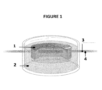
Figure 1 is an illustration of the ex-vivo skin explant implanted with a
microfluidic device
obtained by the method of the invention. A cell culture insert, the bottom of
which
consists of a porous membrane, contains the ex-vivo skin explant (1) implanted
with a
microfluidic device (4) containing culture medium, a drug or any other
molecule of
interest (3). The ex-vivo skin explant is embedded in a solidified matrix (2).
Figure 2 shows an exemplary system of the invention that is used in examples:
tubings
(1) and (8), connection element (2), ring of hydrophobic material (3), ex-vivo
skin explant
(4), solidified matrix (5), microfluidic device (6), cell culture insert (7),
cell culture
container (9), inlet end (10) and outlet end (11).
Figure 3 shows drawings and illustrations of an implantable device according
to the
invention.
CA 03184875 2023- 1- 3
WO 2022/013423
PCT/EP2021/069962
9
Figure 3A is a mechanical drawing of the porous design of a part of the
implantable
microfluidic device.
Figure 3B depicts porous part of the catheter used as implantable microfluidic
device
(light microscope). A gap of approximately 0.8 mm is present between two
consecutive
pores aligned on one side of the implantable device.
Figure 3C is an enlarged view of the holes performed in a part of the catheter
used as
implantable microfluidic device (Light microscope). An offset of approximately
0.2 mm
occurs between the pores of two sides of the implantable device.
Figure 3D shows a pore with a diameter of approximately 50 pm (Scanning
electron
microscope).
Figure 4 shows a drawing (A and B) and an illustration (C and D) of the device
used for
inserting a microfluidic implantable device into an ex-vivo skin explant. Both
base parts
(A) and (C) and top parts (B) and (D) of the device have grooves (3) in
contact with a
central recess (2). Means for preventing the ex-vivo skin explant from
slipping of the
device are present in the top part of the device (B and D), such as a crown
(4) and pins
(5). Both base parts (A) and (C) and top parts (B) and (D) of the device also
have fastening
means suitable (1), such as for example magnets, for stacking the two parts of
the
device. The skin explant is deposited in the recess, epidermis against the
lower part (E).
The upper part is stacked to the lower part, trapping the biopsy (F). A
catheter is inserted
into the grooves provided for this purpose (G). The two parts are separated to
release
the skin explant, then the catheter needle is removed (H).
Figure 5 is a schematic view of the microfluidic system.
Figure 6 is an illustration of a culture chamber (B) and a culture insert (A)
suitable for
receiving an ex-vivo skin explant having an implantable microfluidic device
through the
dermis.
Figure 7 is the quantification of cell viability of ex-vivo skin explants
issued from seven
different donors (1 to 7) and cultured over 10 days in conditions described in
the
examples, in dark the NativeSkin models and in grey the FlowSkin models.
Viability is
CA 03184875 2023- 1- 3
WO 2022/013423
PCT/EP2021/069962
measured using the Cell Counting Kit-8 (Sigma) and is expressed as the
percentage of
skin viability measured the day of models' production. Each bar represents
means SEM
of cell viability values measured for each condition.
Figure 8 depicts a representative image of the histological characteristics
(Hematoxylin
5 and Eosin staining) of ex-vivo skin explants issued from one donor and
cultured over 10
days in conditions described in the examples, (A) NativeSkin model and (B)
FlowSkin
model.
Figure 9 A is the quantification of the number of proliferating cells in the
epidermis of
the ex-vivo skin explants issued from six different donors (1, 2, 3, 4, 5 and
6) and cultured
10 over 10 days in conditions described in the examples, NativeSkin models
in dark and
FlowSkin models in grey. The number of proliferating cells is assessed using
an anti-
Ki67 immunostaining and expressed as the percentage of proliferation measured
the
day of models' production. Each bar represents means SEM of proliferation
measured
for each condition.
Figure 9 B is the quantification of the percentage of apoptosis in the
epidermis of the
ex-vivo skin explants issued from six different donors (1, 2, 3, 4, 5 and 6)
and cultured
over 10 days in conditions described in the examples, NativeSkin models in
dark and
FlowSkin models in grey. The percentage of apoptosis is assessed using an
anti-cleaved
Caspase-3 immunostaining. Each bar represents means SEM of apoptosis measured
for
each condition.
Figure 10 is the characterization of the cell metabolic activity of the ex-
vivo skin explants
issued from three different donors (5, 6 and 7) and cultured over 10 days in
conditions
described in the examples, NativeSkin models in dark and FlowSkin models in
grey
Metabolic activity is assessed by glucose consumption (A) and lactic acid
production (B).
Each bar represents means SEM of the percentage of glucose consumed (A) or
the
concentration of lactic acid ( M) produced (B).
Figure 11 is illustrative of dye diffusion in the ex-vivo skin explants model
cultured over
24 hours in conditions described in the examples. The graph represents the
color
intensity (arbitrary unit) - which is correlated with dye diffusion ¨ measured
following a
CA 03184875 2023- 1- 3
WO 2022/013423
PCT/EP2021/069962
11
line perpendicular to the catheter (0 cm is one edge of the biopsy and 12 mm
is the
other edge of the biopsy), at different time points (0, 2, 4, 7 and 24 hours
of perfusion).
Figure 12 is a diffusion modeling in NativeSkin and FlowSkin models using
COMSOL
software. The graphs represent the evolution of the concentration of molecules
of
interest on a sectional view at the center of the ex-vivo skin explant, at
times ranging
from 0 to 24 hours of culture. A and B: NativeSkin . C and D: FlowSkin .
A and C: Modeling the diffusion of a molecule of interest present both in the
culture
medium in the well under ex-vivo skin explant and in the perfusate at an
arbitrary initial
concentration set at 1, and in the matrix at a concentration of 0.4. This
model can be
equated to simulating the diffusion of nutrients present in the culture medium
(as this
culture medium is also present in the matrix).
B and D: Modeling the diffusion of a molecule of interest present only in the
well under
ex-vivo skin explant and in the perfusate at an arbitrary initial
concentration set at 1.
This model can be equated to simulating the diffusion of a drug administered
systemically.
Figure 13 is a follow up of IL-22 release in the culture media of ex-vivo skin
explants
inflammatory model over 3 days. T-cell activation of ex-vivo skin explants
issued from
two donors (D1 and D5) was achieved using a cocktail of anti-CD3 and anti-CD28
antibodies. After T-cell differentiation, the ex-vivo skin explants were
treated with a
cocktail of pro-inflammatory cytokines (IL-113 + TGF-P + IL-23) mixed only in
the culture
media (full and doted black lines) or in the culture media and infused
intradermally (full
and doted grey lines) using the FlowSkin system. Cytokine concentration is
expressed
in pg/ml.
Figure 14 is illustrative of dye diffusion in the ex-vivo skin explants model
during the first
hours of infusion in conditions described in the examples.
DETAILED DESCRIPTION OF THE INVENTION
CA 03184875 2023- 1- 3
WO 2022/013423
PCT/EP2021/069962
12
The inventors have now developed a new ex-vivo skin explant model in which an
implantable microfluidic device makes it possible to control perfusion,
diffusion and
collection of molecules over long periods. Hence, such a modified ex-vivo skin
explant
model is suitable for the administration and study of drug absorption. In
addition, the
control of perfusion and diffusion of molecules over long periods may allow to
increase
viability of the implanted ex-vivo skin explant.
In a first aspect, the present invention relates to an in-vitro method for
culturing an ex-
vivo skin explant comprising:
(a) inserting an implantable microfluidic device through the dermis of an
ex-vivo skin
explant so that the device is positioned substantially parallel to the
epidermis and
traverses the skin explant from side to side, both ends of the device
protruding slightly
beyond the skin explant,
(b) connecting the two ends of the implantable microfluidic device to two
separate
tubings,
(c) immersing the skin explant obtained in step (b) in a liquid matrix
capable of
solidifying so that the upper surface of the epidermis is not covered, which
matrix is
itself contained in a cell culture insert, the bottom of which consists of a
porous
membrane,
(d) solidifying the matrix so as to trap the immersed part of the skin
explant, where
the upper surface of the epidermis is not covered, and to cause the solidified
matrix to
adhere to the side walls and the porous membrane of the insert,
(e) putting the culture insert containing the skin explant obtained in step
(d) in a
culture chamber containing appropriate culture medium, and
(f) culturing the skin explant.
By "ex-vivo skin explant" is meant a skin fragment that comprises at least the
epidermis,
dermis, hypodermis, and epidermal appendages.The hypodermis is located
immediately
below the dermis and forms a protective cushion separating the skin from the
fibrous
CA 03184875 2023- 1- 3
WO 2022/013423
PCT/EP2021/069962
13
membranes surrounding the deeper organs, muscles, tendons, and bones. The
hypodermis consists of adipocytes, nerves, a network of blood and lymphatic
capillaries,
a small number of fibroblasts involved in the synthesis of extracellular
matrix
components, and immune cells such as dendritic cells and macrophages. The
hypodermis is divided into adipose lobules containing adipocytes and separated
by
connective walls that allow the passage of nerves and vessels. The functions
of the
hypodermis are to isolate, to provide a reserve of energy to the skin cells,
and to absorb
physical stress. In addition, studies have shown the important role played by
lymphatic
transport in the absorption of peptide-based drugs.
Preferably said ex-vivo skin explant is taken from an animal, in particular a
mammal, and
preferably from a human. As biopsies that can be used in the method of the
invention
may be mentioned those obtained from surgical waste or slaughterhouses. By
"surgical
waste" is meant skin samples obtained from cosmetic surgery, including post-
blepharoplasty, lift, abdominoplasty, cruroplasty, brachioplasty or breast
reduction.
Skin biopsy sampling techniques are well known to the one skilled in the art.
In order to ensure its in-vitro survival, sampling of the ex-vivo skin explant
must have
been carried out within 1h to less than 72h, preferably within 1h to less than
48h, before
inserting an implantable microfluidic device in it.
The ex-vivo skin explant may be issued from healthy skin biopsy. By "healthy
skin biopsy"
is meant a skin fragment that does not show any sign of inflammation
perceptible to the
eye (namely break in the skin, redness, swelling, heat, or excessive
scaling...), or express
any inflammation marker. Furthermore, it is imperative that the healthy skin
biopsy
used in the method of the invention be taken from an individual with no
dermatological
pathology, in particular inflammatory.
It is also possible to treat a healthy skin biopsy using UV light to create an
erythema.
Such biopsies allow the study of compounds capable of reducing this type of
skin lesion.
The ex-vivo skin explant may be issued from diseased skin biopsy. By "diseased
skin
biopsy" is meant a skin fragment that shows particular damage related to skin
CA 03184875 2023- 1- 3
WO 2022/013423
PCT/EP2021/069962
14
pathologies such as for example atopic dermatitis, psoriasis, eczema,
dermatoses,
allergic contact dermatitis, lichen, pruritus, Netherton's syndrome,
ichthyosis vulgaris
Preferably, the ex-vivo skin explant has a cylindrical shape.
However, any other geometrical shape of ex-vivo skin explant is also suitable
for the
method according to the invention, in particular a shape which is square,
rectangular,
oval, triangular, etc.
Preferably, the cylindrical shape has a diameter ranging from 1 mm to 50 mm,
more
particularly from 5 mm to 20 mm, even more particularly from 7 mm to 17 mm,
and a
thickness varying from 1 mm to 20 mm, more particularly from 2 mm to 15 mm,
even
lo more particularly from 2 mm to 10 mm, and even more particularly from 2
mm to 5 mm.
Preferably, the surface of the ex-vivo skin explant is about 1 cm2 to about 10
cm2.
By "implantable microfluidic device" is meant a thin, rigid, flexible or semi-
flexible
hollow tube. By "flexible" means capable of being bent or flexed at least
under the effect
of a lateral action due to the displacement of a fluid. A deformable or
stretchable
implantable microfluidic device may recapitulate most of the physiological
properties of
blood vessels, i.e. lower inner hydrodynamic resistance, wall porosity and
deformability.
The implantable microfluidic device is biocompatible, and may not trigger
physiological
events. The material of the implantable microfluidic device may not cause
inflammation,
immune response, infection, or any other sort of rejection within the ex-vivo
skin
explant. It could be functionalized with biologically active species or cells.
Examples of a
suitable material include, but are not limited to, silicone rubber, nylon,
polyurethane,
polyethylene terephthalate (PET), latex, Teflon, hydrogel materials and
thermoplastic
elastomers. In all embodiments, the two extremities of the implantable
microfluidic
device are open, thus enabling the transport of solutions, suspensions or
compositions.
Preferably, the external diameter of the implantable microfluidic device is
from 0.3 mm
to 2.5 mm, as an example from 0.7 mm to 2.2 mm, more preferably from 0.9 mm to
1.3
mm, and most preferably is 1.1 mm.
CA 03184875 2023- 1- 3
WO 2022/013423
PCT/EP2021/069962
Preferably, the internal diameter of the implantable microfluidic device is
from 0.05 mm
to 2.25 mm, as an example from 0.51 mm to 1.75 mm, more preferably from 0.61
mm
to 0.96 mm, and most preferably is 0.9 mm. With an internal diameter superior
to 100
p.m, the inner hydrodynamic resistance is lowered.
5 Preferably, the length of the implantable microfluidic device is from 19
mm to 50 mm,
more preferably from 25 mm to 45 mm, and most preferably is 32 mm.
In all embodiments, the total length of the implantable microfluidic device is
greater
than the size of the ex-vivo skin explant. For example, if the ex-vivo skin
explant is
cylindrical in shape, the total length of the implantable microfluidic device
is greater
10 than the diameter of the ex-vivo skin explant.
The implantable microfluidic device may be a catheter, a microdialysis probe
or any
material suitable for being implanted in an ex-vivo skin explant. Preferably,
the
implantable microfluidic device is a catheter.
In order to allow the administration, the delivery, the diffusion of the
species from an
15 injected solution towards the ex-vivo skin explant or to provide an
efficient and
continuous collection of the metabolic wastes or molecules secreted by the ex-
vivo skin
explant, the portion of the implantable microfluidic device that passes
through the ex-
vivo skin explant is porous.
Preferably, in the method of the invention, the portion of the implantable
microfluidic
device that passes through the ex-vivo skin explant is porous.
In all embodiments, the porosity is ensured by a plurality of pores, i.e.
micro holes or
apertures, arranged along the length of the portion of the implantable
microfluidic
device that passes through the ex-vivo skin explant. The pores can be arranged
on all
sides of the implantable microfluidic device to ensure a homogeneous diffusion
around
it, or only on certain sides to direct the diffusion. Pores may be formed
using well
established standard techniques such as 3D lithography, i.e. photolithography
or
etching, water-jet cutting, micro-mechanical drilling or laser drilling.
CA 03184875 2023- 1- 3
WO 2022/013423
PCT/EP2021/069962
16
The rate of infusion and diffusion from the porous portion of the implantable
microfluidic device into the ex-vivo skin explant can be influenced by the
pore size, shape
and their number. The average pore size, or pore diameter, can be at least
about 10 p.m
to about 250 pm, more preferably from about 20 tm to about 100 p.m, most
preferably
of about 50 pm. All the pores can have the same pore size or different pore
sizes. The
pore sizes can vary by as much as 5%, 10%, 20% or even 30% when all pores have
the
same pore size. The pore size can vary by as much as 50%, 100%, 200%, 300%,
400% or
more when pores have different pore sizes. The pore size can be adapted to
provide a
desired diffusion rate for a specific species or compound.
Preferably, in the method of the invention, the porous portion of the
implantable
microfluidic device is obtained by making pores having a diameter from about
40 p.m to
about 250 pm, more preferably from about 50 pm to about 200 pm, most
preferably of
about 50 p.m.
The pore size is a relevant criterion for the selection of molecules that will
be able to
enter or leave the implantable microfluidic device. In fact, the pore size
also acts as a
molecular weight cut-off.
The pore shape may also be a relevant criterion for the selection of molecules
that will
be able to enter or leave the implantable microfluidic device. Thus, the pores
can be
round, square, rectangular, triangular, star-shaped, loophole-shaped, ... When
pores are
rectangular or loophole-shaped, they are about 50 p.m wide and over 100 p.m
long.
The rate of infusion and diffusion from the porous portion of the implantable
microfluidic device into the ex-vivo skin explant can be influenced by the
pressure drop
across the implantable device. This pressure drop results from the difference
between
the inlet pressure of the solution injected into the implantable device and
its outlet
pressure. The atmospheric pressure is the reference point above the epidermis
of the
skin explant.
To facilitate the inserting of the implantable microfluidic device into the ex-
vivo skin
explant, one of the two extremities of the device may be sharpened. This
sharpened end
CA 03184875 2023- 1- 3
WO 2022/013423
PCT/EP2021/069962
17
limits the risk of rupture, tearing of the skin explant at the time of
insertion of the
implantable device.
The implantable microfluidic device can be inserted in the dermis or the
hypodermis of
the ex-vivo skin explant. The choice of the skin layer for receiving the
implantable device
depends on the intended application.
Preferably, in the method of the invention, the implantable microfluidic
device is
inserted into the dermis. In order to avoid tearing the ex-vivo skin explant,
the insertion
of the implantable microfluidic device should take place just below or in
close proximity
to the epidermis.
By "close proximity to the epidermis" means that the implantable microfluidic
device is
inserted between 0.1 mm and 3 mm, preferably between 0.25 mm to 1 mm, below
the
epidermis.
In all embodiments, the implantable device is inserted substantially parallel
to the
dermis or the hypodermis of the ex-vivo skin explant. By "substantially
parallel to the
epidermis" is meant that the longitudinal axis of the implantable microfluidic
device is
substantially parallel to the epidermis layer of the ex-vivo skin explant. By
"substantially
parallel" is intended the same as or very close to parallel. A slight
deviation from strict
parallelism necessitated by the physical separation is acceptable. As an
example, a
deviation of less than 100 is acceptable, preferably from less than 50
.
By "protruding slightly" means that both ends of the implantable microfluidic
device
emerge from the ex-vivo skin explant so as to position the porous portion of
the
microfluidic device in the center of the skin explant. Moreover, both ends of
the
implantable microfluidic device has to be free to be connected with the other
connecting elements of the culture device. The length of the protruding ends
may be up
to 10%, 20%, 30%, 40%, 50% or 60% of the total length of the implantable
device. In all
embodiments, it is essential that the length of the implantable microfluidic
device is
superior to the length of the ex-vivo skin explant so as to cross the ex-vivo
skin explant
from side to side.
CA 03184875 2023- 1- 3
WO 2022/013423
PCT/EP2021/069962
18
Preferably, in the method of the invention, the length of the two ends of the
implantable
microfluidic device protrude from the ex-vivo skin explant by a length of at
least 10%,
more preferably at least 30%, and more preferably 50% of the total length of
the
microfluidic implantable device.
As an example, for an ex-vivo skin explant having a diameter of 15 mm and an
implantable microfluidic device having a length of 32 mm, the length of each
of the
protruding ends is about 8.5 mm. In this example, the portion of the
microfluidic
implantable device that passes through the ex-vivo skin explant has a length
of about 15
mm.
The microfluidic implantable device can be inserted in the ex-vivo skin
explant manually,
using a curve clamp or a device comprising two stackable parts, one of which
has a notch
that is adapted to guide the implantable device during the step a) of the
method
according to the invention.
In step (b) of the method of the invention, the implantable microfluidic
device that
crosses the ex-vivo skin explant is connected to two separate tubings. The
tubings have
diameter that are similar or different. In all embodiments the diameter of the
tubing is
suitable to fit over the ends of the implantable microfluidic device. Each
ends of the
microfluidic implantable device is connected to a tubing, directly or using,
for example
a male or female luer lock connector. This type of connector is illustrative
and should
not restrict the scope of the present invention. The material of tubing is
biocompatible,
flexible or rigid, has low kink ability, torque strength, and lubricity. When
the
implantable microfluidic device is a catheter, one of the tubing is connected
to a hub
present at one end of the device.
By "liquid matrix capable of solidifying", also named solidifying or
solidifiable matrix, is
meant any liquid solution comprising at least one specific compound or
composition the
concentration of which in said liquid solution is such that, when implementing
suitable
conditions, especially particular temperature conditions, the liquid solution
takes on a
solid or gel-like consistency. Now, the nature of this solidifiable matrix
must allow the
ex-vivo skin explant cells to remain alive, i.e. the matrix has no cytotoxic
effect and has
CA 03184875 2023- 1- 3
WO 2022/013423
PCT/EP2021/069962
19
a solidification/polymerization temperature at room temperature (i.e. from 15
C to
25 C). Said specific compound or composition may be of animal, vegetable or
synthetic
origin, or a mixture thereof, its nature and its concentration are determined
according
to the desired physico-chemical characteristics of the matrix when solidified,
especially
the flexibility and strength of the matrix. The volume of the liquid matrix
will be 1/3 to
2/3 of the total volume of the cell culture insert, preferably 2/5 to 3/5 of
the total
volume; half of the total volume of the insert being the preferred volume.
According to an embodiment of the method according to the invention, in step
(c), said
liquid matrix capable of solidifying is selected among any liquid solution,
preferably
nutritive, capable of solidifying or gelling under particular conditions
compatible with
the survival and the culture of skin cells constituting said ex-vivo skin
explant, preferably
selected from the group consisting of blood plasma or a solution derived from
blood
plasma (i.e. blood plasma diluted with a physiological buffer to at least 10%,
at least
20%, at least 30% and preferably at least 40% w/w of the total weight of the
matrix), a
fibrinogen solution, a collagen solution, gelatin, synthetic polymeric gels,
natural gels,
such as agarose gels, in particular agarose or agar-agar gels with low melting
points,
starch or polysaccharide gels, commercially available matrix such as MATRIGEL
, or a
mixture thereof.
In a more preferred embodiment, the liquid matrix capable of solidifying is
composed of
two solutions which are mixed together in the culture insert prior to step (c)
of
immersing the ex-vivo skin explant. This matrix is then composed of a first
solution
selected from a blood plasma solution, a solution derived from blood plasma, a
fibrinogen solution, a collagen solution, and mixtures thereof, and a second
solution of
agar-agar or low melting point agarose.
The first solution, selected from a blood plasma solution, a blood plasma-
derived
solution, a fibrinogen solution, a collagen solution, or mixtures thereof, is
advantageously a nutrient solution. Now, this first solution is mainly able to
solidify
under the action of an increase or decrease in temperature and/or by the
addition of a
specific compound or composition. Preferably, the compound allowing this first
solution
to solidify is the Ca2+ ion. Thus, the liquid matrix has a Ca2+ concentration
of between 1
CA 03184875 2023- 1- 3
WO 2022/013423
PCT/EP2021/069962
mM and 5 mM, preferably between 1.5 mM and 4.5 mM; which concentration will
cause
it to solidify.
According to a first particular embodiment, the liquid matrix has a Ca21-
concentration of
between 1 mM and 2 mM, preferably between 1.2 mM and 1.4 mM.
5 According to a second particular embodiment, the liquid matrix has a Ca'
concentration
comprising 2 mM and 3 mM, preferably between 2.5 mM and 2.9 mM and more
preferably 2.8 mM of Ca2+.
It should be noted that in the case of a blood plasma solution or a solution
derived from
blood plasma, the solution is first treated with an anticoagulant agent with
reversible
10 properties. To do so, this solution comprises at least one anti-
fibrinolytic agent, such as
sodium citrate, tranexamic acid or aprotinin, and in sufficient concentration
to obtain
the desired anti-fibrinolytic activity. Preferably, the liquid matrix has a
final
concentration (weight/total matrix weight) of between 2 and 5% of this at
least one anti-
fibrinolytic agent.
15 Now, the first solution will preferably be a fibrinogen solution.
The second solution of low melting agar-agar or low melting agarose is
preheated for a
time and at a temperature sufficient to be liquid and to remain liquid at
about 37 C for
the time sufficient to be mixed with the first solution in said insert and
until the time of
immersion of the skin explant. Typically, this second solution is
preliminarily heated to
20 its melting temperature or to a slightly higher temperature, preferably
to a temperature
between 65 C and 70 C. The choice of agar-agar or low melting point agarose is
made
so as to benefit, for a 1.5% solution (weight/weight total composition), from
a gelling
temperature between 24 C and 28 C, and a melting temperature higher than 65.5
C. As
an example, we can mention the agarose called LMP Agarose Low melting point
(GIBCOBRL, LIFE TECHNOLOGIES). Still in connection with this second solution,
its
concentration of agar-agar or low melting point agarose is between 1% and 5%
(preferably in a physiological solution), more preferably between 2% and 5%,
between
3% and 4.5%, between 3.5% and 4.5% or between 3.8% and 4.2% or between 3.9%
and
4.1%, 4% being the most preferred concentration (by weight in relation to the
total
CA 03184875 2023- 1- 3
WO 2022/013423
PCT/EP2021/069962
21
weight of the composition). At this concentration and once heated to its
melting
temperature or to a slightly higher temperature, this second solution of agar-
agar or low
melting point aga rose can be kept in liquid form for at least 1 hour at 37
or, ideally, at
least 4 hours, 10 hours or 16 hours. Preferably, the solidifiable liquid
matrix comprising
said first and said second solution of agar-agar or low-melting agarose has a
final
concentration of agar-agar or low-melting agarose of between 0.1% and 2%,
preferably
between 0.2% and 1.8% (weight/weight total matrix). Such a concentration makes
it
possible not only to obtain a matrix which, once solidified, makes it possible
to retain
the three-dimensional structure and to keep said fragment or ex-vivo skin
explant alive,
but also to obtain a matrix which is solid but sufficiently flexible to be non-
brittle and
resistant to point shocks. The solidification of this liquid matrix taking
place after
deposition of the ex-vivo skin explant, leaving the device thus obtained at a
temperature
of between 37 C and room temperature, preferably at 20 C.
According to a first particular embodiment, the final concentration of agar-
agar or low
melting point agarose in the liquid matrix (comprising the first and said
second solution)
is between 0.5% and 2%, preferably between 0.5% and 1.25%, more preferably
between
0.5% and 1.0%, with a concentration of 0.7% (weight/weight total matrix) being
the
most preferred concentration.
According to a second particular embodiment, the final concentration of agar-
agar or
low melting point agarose in the liquid matrix (comprising the first and said
second
solution) is between 0.1% and 2%, preferably between 0.2% and 1.75%, 0.25%
being the
most preferred concentration. Such a concentration makes it possible to obtain
a matrix,
which, once solidified, makes it possible both to preserve the three-
dimensional
structure and to keep the skin explant alive, and simultaneously to obtain a
matrix which
is sufficiently flexible to be non-brittle and resistant to mechanical effects
applied to the
skin explant. The solidification of this liquid matrix takes place after
immersion of the
skin explant by allowing the assembly to cool down.
According to another preferred embodiment, the liquid matrix capable of
solidifying
further comprises cells other than the cells that make up the skin explant,
which cells
are selected from the group consisting of fibroblasts, endothelial cells and
nerve cells.
CA 03184875 2023- 1- 3
WO 2022/013423
PCT/EP2021/069962
22
Preferably these cells are fibroblasts and ideally primary fibroblasts (as
opposed to
fibroblast cell lines), such as dermal fibroblasts obtained from human
foreskin. These
primary fibroblasts, especially dermal fibroblasts, can be prepared and
obtained from
standard methods well known to the skilled person (see for example HOWARD BV
et al,
A new method for the establishment of diploid fibroblast cell cultures from
human
foreskins, Proc. Soc. Exp. Biol. Med. vol.153(2), p:280-3, 1976). Preferably,
these cells,
and in particular fibroblasts, are contained in the matrix at a concentration
between
5.103 and 5.105 cells/ml, preferably between 104 and 105 cells/ml, the range
between
3.104 and 5.104ce11s/m1 being the most preferred concentration range.
In addition, the liquid matrix may include various compounds such as
preservatives, pH
agents, etc. For example, the liquid matrix will contain between 5 and 500
mg/mL of
ascorbic acid, preferably between 25 and 75 mg/mL, with a preferred ascorbic
acid
concentration of 50 mg/mL.
According to a third, also preferred embodiment, the liquid matrix capable of
solidifying
is a solution derived from blood plasma and comprises:
a) 25 to 75% (total volume/volume) fibrinogen, preferably 35 to 45% (v/v),
(b) 5 % to 12 % (total volume/volume) of a 1 %, preferably 8 %, CaCl2 salt
solution,
c) from 5% to 2%, preferably the anti-fibrinolytic agent being selected from
tranexamic
acid or aprotinin,
(d) from 0.5% to 4% low melting point agarose, preferably from 1% to 2%, and
e) a physiological solution such as a 0.9% NaCI solution, qsp at 100%.
Matrices capable of solidifying and methods for placing ex-vivo skin explant
therein are
detailed in European patent EP 2 882 290 B1 and are incorporated herein by
reference.
The purpose of the matrix capable of solidifying is to allow the implanted ex-
vivo skin
explant obtained at the end of step (e) of the method of the invention to be
cultured,
and easier handling thereof. By "cultured" is meant the maintenance of the
physiological
CA 03184875 2023- 1- 3
WO 2022/013423
PCT/EP2021/069962
23
and morphological state of the ex-vivo skin explant that is to say of all the
tissues and
cells constituting the biopsy.
The purpose of the matrix capable of solidifying is also to allow the ex-vivo
skin explant
to be long term preserved and/or transported, while allowing stable cell
functions within
a controlled microenvironment.
According to a particular embodiment, the in vitro method according to the
invention
comprises, prior to step (a), a step of fixing, to the epidermal surface of
the implanted
ex-vivo skin explant obtained at the end of step (a), a ring, consisting of a
hydrophobic
material, the outer diameter of said ring being similar to the diameter of the
epidermal
lo surface of the ex-vivo skin explant.
Preferably, the hydrophobic material of the ring is a material that is not
toxic to the skin.
It may be a paraffin polymer, such as a PARAFILM (SIGMA), or a silicon
polymer.
According to a particular mode, the ring is prepared from a film of
hydrophobic material,
by perforating said film according to the desired dimensions. Hence, the ring
corresponds to a disc perforated at its center. The thickness of the ring is
preferably
between 0.1 mm and 2 mm, preferably between 0.1 and 1 mm, more preferably
between 0.1 and 0.5 mm, and even more preferably between 0.12 and 0.2 mm.
Said ring may be made of an opaque or translucent material. According to a
particular
embodiment, the ring is made of an opaque material.
According to a more particular embodiment, said ring is attached to the
epidermal
surface of the biopsy using glue, preferably added to the lower surface of the
ring. Said
glue can be selected from any type of material that is not toxic to the skin
and which has
the effect of adhering the ring to the skin, where this material may be
silicon. Preferably,
said glue is hydrophobic.
The materials, glues and attachment methods of this hydrophobic ring are
detailed in
European patent EP 2 882 290 B1 and are incorporated herein by reference.
The cell culture insert is configured to receive the ex-vivo skin explant
inserted by the
implantable microfluidic device. For instance, the structure of commercially
available
CA 03184875 2023- 1- 3
WO 2022/013423
PCT/EP2021/069962
24
cell culture wells can be modified to create notches to accommodate the
protruding
ends of the implanted device. The culture insert can be of any shape and can
be
suspended on piles or using lugs. The bottom of the culture insert consists of
a porous
membrane with a porosity for preventing the liquid matrix from flowing through
the
membrane before solidification. Preferably, the porosity of this membrane may
be in a
range of 0.2 p.m to 10 urn, more preferably between 114 p.m and 8 p.m, 8 jim
being the
preferred porosity. The material of this porous membrane can be polyethylene
terephthalate (PET), nitrocellulose, or polycarbonate. Among cell culture
inserts may be
mentioned those supplied in particular by the NUNC company (Roskilde,
Danemark), BD
FALCON (BECTON DICKINSON), MILLICELL (EMD MILLIPORE CORPORATION), or
COSTAR (GROSSERON), for example the inserts with a membrane made of
polycarbonate, PET or nitrocellulose pre-packaged in multi-well plates with 6,
8, 12, and
24 wells, the membrane porosity of which may vary between 0.4 p.m and 8 p.m.
Typically,
the culture insert used in the method of the invention has a PET membrane
which
porosity ranges from 0.8 p.m to 8 p.m and is configured for the culture
chamber used in
the method of the invention.
In step (e) of the method of the invention, the culture chamber is also
configured to
receive the cell culture insert containing the skin explant obtained in step
d). A "culture
chamber" is generally defined by a base and a wall. In the present invention,
the shape
of the wall is suitable for receiving the culture insert containing the ex-
vivo skin explant
implanted by the microfluidic device. The culture chamber may be constructed
from a
material that withstands sterilization, including, for example, sterilization
by irradiation
(beta or gamma radiation), steam autoclave, ethylene oxide, chemical
disinfectants, or
dry heat sterilization. In some embodiments, the culture chamber may be made
from a
thermoplastic material and/or from a material that is formed, for instance, by
injection
molding. Examples of materials that are suitable for use in the present
context include
for example, but are not limited to, polyethylene, polypropylene, polystyrene,
polycarbonate, polyurethane, polysulfone, polymethylpentene,
polydimethylsiloxane
(PDMS), polymethylmetacrylate, polyethyleneterepthtalate,
polytetrafluoroethylene,
or ABS (acrylonitrilbutadiene styrene). However, the examples given here only
CA 03184875 2023- 1- 3
WO 2022/013423
PCT/EP2021/069962
exemplary in nature a person skilled in the art would readily appreciate how
to select
other materials suitable for use in constructing the device.
In a particular embodiment, the material of the chamber culture is
polydimethylsiloxane
(PDMS).
5 In all embodiments, the bottom of the culture chamber has a rim adapted
to receive the
cell culture insert containing the ex-vivo skin explant inserted by the
implantable
microfluidic device. The culture chamber holds the cell culture insert and
facilitates its
handling and transport. In a preferred embodiment, the bottom of the culture
insert is
at a distance of between land 2.5 mm from the bottom of the culture plate
containing
10 it.
In some embodiments, the culture chamber is coated with an agent. By way of
example,
and without this being restrictive, the coating agent can be selected among
bovine
serum albumin (BSA), surfactant such as PLURONIc F-127, synthetic,
hydrophilic and
biocompatible polymer such as Poly(ethylene glycol) (PEG) or, PLL-g-PEG
(Poly(L-Lysine)-
15 g-Polyethylene glycol), any other biocompatible natural or synthetic
coating molecule,
or a mixture thereof. In some other embodiments, the surface of the culture
chamber
can also be treated with oxygen plasma or PECVD (Plasma enhanced chemical
vapor
deposition) and thereafter treated with silicon analogue of methane, such as
fluorinated
or carboxylated silane.
20 At the end of step (e) of the method of the invention, the ex-vivo skin
explant has the
trade name FLOWSKIN .
By "appropriate culture medium" is meant a culture medium containing all the
elements
necessary for the survival of the skin explant such as for example William's
E, DMEM,
KBM, ... This culture medium, by its nature as much as by its volume in the
culture
25 chamber, helps the survival of the ex-vivo skin explant and the cells
constituting it over
time. In addition to the survival of the cells of the ex-vivo skin explant,
the culture
medium makes it possible, in particular by limiting the stress on them, to
maintain the
cells of the ex-vivo skin explant in their initial state, whether in
structural or functional
terms.
CA 03184875 2023- 1- 3
WO 2022/013423
PCT/EP2021/069962
26
The presence of culture medium in the culture chamber allows the step (f) of
culturing
the ex-vivo skin explant for several days. By "culturing" is meant maintaining
physiological and morphological state of the ex-vivo skin explant, that is to
say of all the
tissues and cells constituting the explant. Thus, the aim of this step is to
limit the
phenomena of cell death and to maintain the state of differentiation of the
cells (in fact
to limit the phenomena of dedifferentiation or inappropriate differentiation).
Preferably, step (f) of the method of the invention can last at least 5 days,
6 days, 7 days,
8 days, 9 days, 10 days or more.
A cover, a lid or a protective film may be applied on the top of the culture
chamber in
which the culture insert containing the implanted skin explant of a
microfluidic device
has been placed in order to seal the culture plate. In this way, the resulting
culture
chamber can be transported without difficulty by land, sea or air. Indeed, the
skin
explant is not only firmly held by the solid matrix but also nourished.
The culture chamber can also be equipped with two needles so as to permit
further
connecting with a complete circulation system. In these embodiments, the two
needles
pass through two opposite walls of the culture chamber, the needles extending
substantially parallel to the bottom of the culture chamber. Each needle has
two ends,
the first one being located inside the culture chamber; the second end being
located
outside the culture chamber. The two needles are connected at one of their
ends to the
tubings, the other one being free for further connecting with a complete
circulation
system.
In a preferred embodiment, the method of the invention further comprises a
step (g) of
connecting the tubings to a circulating system comprising means for providing
a
continuous or discontinuous/pulsatile flow and/or for modulating the
difference of
pressure between the inlet and the outlet of the implantable device, said
connecting
being mediated by input and output ports at the inlet and outlet of the
implantable
device. By controlling the difference of pressure across the implantable
device, it
enables injection, suction and pressure control within the implantable device
in
comparison to external pressure (usually atmospheric pressure). When the
internal
CA 03184875 2023- 1- 3
WO 2022/013423
PCT/EP2021/069962
27
pressure in the implantable device is lower than the external pressure, no
leaks can
occur. In this embodiment, the tubings act as connectors between the
microfluidic
implantable device and the needles.
The two needles constitute input and output ports of the culture system
obtained in
step (g) of the method of the invention. In some embodiments, the inlet port,
the culture
chamber and the outlet port are configured to provide a continuous or
discontinuous
flow of fluid through the ex-vivo skin explant. For this purpose, the culture
system
obtained in step (g) of the method of the invention, includes or is coupled
to, means for
achieving a flow of fluid in the implantable microfluidic device. These means
include any
system capable of imposing a pressure drop by controlling the level of the
liquid, i.e. the
hydrostatic pressure. This passive method, heavily used in biology, is quite
similar to the
infusion system used clinically in-vivo. The circulating system may also
include a pump,
such as for example a syringe pump, a peristaltic pump, a pressure regulating
pump or
a similar device capable of providing a continuous or discontinuous/pulsatile
flow having
any desired flow rate, or able to modulate the pressure within the implantable
device
by controlling independently the value of the pressure at the inlet and outlet
of the
device, thanks to the addition of pressure regulators connected to the inlet
and outlet
or the integration of one way or multiple ways valves in the microfluidic
system, to
temporarily blocked the outlet of the system and switch from one reservoir to
another.
It may be desired to select a flow rate or a pressure that leaves the ex-vivo
skin explant
intact or that does not substantially, not significantly, or not at all
interfere with the
integrity, the biological function or the physiological characteristics of the
skin explant.
In some embodiments, the flow has a flow rate that is substantially equivalent
to an in
vivo hemodynamic flow rate and/or has a pressure that is substantially
equivalent to an
in vivo hemodynamic pressure. The perfusion should recapitulate a native skin
function.
Preferably, the flow rate of a fluid perfused within the ex-vivo skin explant
ranges from
about 0 p.1/minute to 1000 1.11/minute, as for example about 1 p.1/minute to
20 p1/minute,
more preferably from about 2.5 p.1/minute to 10 I/minute, and most preferably
from
about 2.5 I/minute to 5 I/minute. In a more preferred embodiment, the flow
rate of
CA 03184875 2023- 1- 3
WO 2022/013423
PCT/EP2021/069962
28
the fluid perfused through the ex-vivo skin explant using the implantable
microfluidic
device is 2.5 p.1/minute.
In another embodiment, pressure within the implantable device can be
controlled with
pressure differences between inlet and outlet ports. By exercising pressure-
vacuum
cycles, the flow of a fluid perfused within the ex-vivo skin explant is
regulated and the
internal pressure inside the implantable device remains below of above the
pressure of
caused by the air on the epidermis.
Preferably, the difference of pressure across the implantable device ranges
from about
-1 bar to 1 bar, preferably from about -200mBar to 200 mbar and even more
preferably
lo from about -50 mbar to 50 mbar.
Cycle of pressure may also be modulated from about 1.10-3 to 100 Hertz.
Injection and pressure within the implantable device can be continuous,
discontinuous
or transient.
In one embodiment, the implantable microfluidic device is used to deliver at
least one
compound of interest through the ex-vivo skin explant. Such a compound of
interest can
be culture media, oxygen carrier or a drug.
By "oxygen carrier" is mean any composition or compound that allows a
physiological
oxygen supply to the cells in the ex-vivo skin explant. Oxygen is delivered
according to
their needs. More interesting are oxygen carriers that also have intrinsic
antioxidant
activity so as to protect the cells from the harmful effect of free radicals
generated by
cellular respiration. Perfluorocarbon- and hemoglobin-based substitutes are
oxygen
carrier suitable for the purpose of the present invention.
By "drug" is mean any agent or compound that modulate the metabolic activity
of the
cells or which has a physiological effect on the ex-vivo skin explant.
Therapeutic
compounds are of particular interest. Among them, one can cite small
molecules,
peptides, proteins such as for example monoclonal antibodies, nucleic acids,
or any
other compounds that can be used topically of in a systemic way to cure or
alleviate a
pathological condition.
CA 03184875 2023- 1- 3
WO 2022/013423
PCT/EP2021/069962
29
In one particular embodiment, the method of the invention is for further
detecting
and/or quantifying at least one biomarker of interest contained in the liquid
secreted by
the ex-vivo skin explant, and said method further comprises the following
steps:
(h) collecting the liquid secreted by the ex-vivo skin explant through the
output port,
and
(i) isolating, identifying, and optionally quantifying the at least one
biomarker of
interest.
By "biomarker of interest" is meant any compound, analyte or molecule implied
in
metabolic activities of cells of the ex-vivo skin explant. For example, the
concentration
of known markers of metabolic stress and fatigue (i.e. lactate, glucose,
ketones, cortisol,
and TNF-a) in extracted liquid secreted by the ex-vivo skin explant can be
determined
using standard assays. These assays require between 1 and 50 microliters of
sample
fluid. A NANODROP ND100 spectrophotometer capable of analyzing 2 I volumes
of
solution can be used if the extracted IF volumes are insufficient for standard
assays. The
IF biomarker concentrations can be correlated with levels found in whole blood
or
serum. The Human Metabolome Database (HMDB, www.hmdb.ca) and the literature
searches can be used to identify useful biomarkers. These markers can be
changed
according to the need. Mass spectrometry, HPLC, infrared can be used to
directly
analyze the biomarker composition of extracted IF.
The liquid secreted by the ex-vivo skin explant can be compared to
interstitial fluid
existing in in-vivo skin. By "interstitial fluid' (IF) is meant a fluid that
surrounds cells and
tissues throughout the body and is formed by extravasation of plasma from
capillaries
and modified by metabolic and other processes in the tissue. Interstitial
fluid shuttles
nutrients and waste products between blood vessels and cells and is roughly a
combination of serum and cellular materials. Even though 83% of proteins found
in
serum are also in IF, about 50% of proteins in IF are not present in serum,
suggesting
that IF may be a source of unique biomarkers. Moreover, IF is also interesting
for
continuous monitoring due to absence of clotting factors. Skin is the most
accessible
organ and therefore an attractive source of IF containing both systemic and
CA 03184875 2023- 1- 3
WO 2022/013423
PCT/EP2021/069962
dermatological biomarkers. Suction using a pump to create negative pressure on
the
skin surface, or osmotic driving force can be used to flow IF from skin
through the
implanted microfluidic device to a sample reservoir. For example, a pump can
be a
vacuum pump, a capillary force pump, a microdialysis pump, or a pulsatile
vacuum
5 pump. Saline solution can also be injected through the implantable
microfluidic device
and spread in the ex-vivo skin explant by the pores of the porous portion of
the
implantable microfluidic device.
In one preferred embodiment, the step (h) of collecting the liquid secreted by
the ex-
vivo skin explant is done repeatedly at regular or irregular intervals so as
to follow up
10 the concentration of said at least one biomarker of interest.
The success of topical and transdermal administration of drugs is directly
related to the
methods used for evaluation of the formulations, which enable optimization of
the skin
absorption of the drug so that it can reach effective drug concentrations at
the
therapeutic site. Cutaneous microdialysis as implemented in the present
invention is a
15 technique in which a microfluidic device is implanted in the dermis of
the ex-vivo skin
explant, directly under the formulation to be tested.
In another embodiment, the method of the invention is for further assessing
permeation
of the at least one biomarker of interest through the skin, said method
further comprises
a step (g') of applying topically to the epidermis of the ex-vivo skin explant
a composition
20 comprising the at least one biomarker of interest after the step (g) of
connecting the
needles with the tubings and prior to the step (h) of collecting the liquid
secreted by the
ex-vivo skin explant.
By "permeation" is meant the diffusion of a compound into a certain skin
layer.
Permeation should not be confused with skin absorption that implies that the
25 compound becomes systemically available, nor with skin penetration which
is the
diffusion into deeper layers. Permeation is diffusion of a penetrant into a
certain skin
layer. Subsequent diffusion through that layer represents penetration.
Penetration
through layers of skin to either the site of action or systemic circulation
represents
CA 03184875 2023- 1- 3
WO 2022/013423
PCT/EP2021/069962
31
absorption. Absorption through the skin considers full penetration of the skin
layers so
that the penetrant can become systemically available.
In this particular embodiment, the at least one biomarker of interest can be a
cosmetic
agent, a gas, a potentially dangerous exogenous agent from the environment,
etc...
In this preferred embodiment, the step (h) of collecting the liquid secreted
by the ex-
vivo skin explant is done repeatedly at regular or irregular intervals so as
to assess
permeation rate of said at least one biomarker of interest.
In another embodiment, the method of the invention is for further assessing
the activity
of at least one compounds intradermally injected, said method further
comprises a step
(g") of injecting a composition comprising the at least one compound of
interest after
the step (g) of connecting the needles with the tubings and prior to the step
(h) of
collecting the liquid secreted by the ex-vivo skin explant, the composition
being injected
in the dermis of the ex-vivo skin explant by using the microfluidic
implantable device.
In this embodiment, the purpose is to identify macro- and micro-signs of
modification
or alteration of the epidermis, dermis or hypodermis layer. By "signs" is
meant any
perceptible change such as, but with no limitation to, dryness, cracking,
peeling,
redness, redness, thickening, thinning, or any other sign occurring in the
epidermis.
In another embodiment, the method of the invention is for further assessing
the activity
of at least one compounds subcutaneously injected, said method further
comprises a
step (g¨) of injecting a composition comprising the at least one compound of
interest
after the step (g) of connecting the needles with the tubings and prior to the
step (h) of
collecting the liquid secreted by the ex-vivo skin explant, the composition
being injecting
in the hypodermis of the skin explant by using the microfluidic implantable
device.
By injecting the composition in the hypodermis, the purpose is to mimic
extravasation
that naturally occurs in-vivo from skin vasculature.
In another embodiment, the method of the invention is for further studying
inflammation of the skin, said method further comprises a step (g¨) of
injecting a
composition comprising a cocktail of cytokines, after the step (g) of
connecting the
CA 03184875 2023- 1- 3
WO 2022/013423
PCT/EP2021/069962
32
needles with the tubings and prior to the step (h) of collecting the liquid
secreted by the
ex-vivo skin explant, the composition being injecting in the dermis or
hypodermis of the
skin explant by using the microfluidic implantable device.
In one preferred embodiment, the cocktail of cytokines comprises an effective
amount
of pro-inflammatory cytokines. Preferably, the cocktail of cytokines is a
mixture of
interleukine-1 beta (lLip), tissue growth factor beta (TGF-13) and interleukin-
23 (IL23).
This cocktail enables the differentiation of activated resident T cells into
LTh1 and/or
LTh17 T helper cell lineages. By "effective amount" is meant an amount that is
sufficient
to achieve the expected effect. In the instant case, the expected effect is
obtaining the
polarization of the activated resident T cells into LTh1 and/or LTh17, and the
synthesis
of inflammation markers.
Preferably, the concentration of IL-1r3 in the mixture of the composition used
in
step (g¨) of the method of the invention ranges from 1 ng/ml to 50 ng/ml,
preferably
from 5 ng/ml to 30 ng/ml, and particularly preferably from 7 ng/ml to 15
ng/ml.
Preferably, the concentration of IL-23 in the mixture of the composition used
in step
(g") of the method of the invention ranges from 10 ng/ml to 100 ng/ml,
preferably
from 20 ng/ml to 80 ng/ml, and particularly preferably from 30 ng/ml to 60
ng/ml.
According to a particular embodiment, the concentration of IL-23 in the
mixture of the
composition used in step b) of the method of the invention is 50 ng/ml.
Preferably, the TGF-I3 concentration in the mixture of the composition used in
step (g")
of the method of the invention ranges from 1 ng/ml to 50 ng/ml, preferably
from
5 ng/ml to 30 ng/ml, and particularly preferably from 7 ng/ml to 15 ng/ml.
According to a more particular embodiment, the concentrations of IL-113, IL-
23, and TGF-
13 in the mixture of the composition used in step (g") of the method of the
invention
are 10 ng/ml, 50 ng/ml, and 10 ng/ml, respectively.
Preferably, step (g") of injecting the ex-vivo skin explant with a composition
comprising
at least one mixture of IL-113, IL-23, and TGFI3 is carried out for a period
of at least 3 days,
preferably between 3 and 10 days.
CA 03184875 2023- 1- 3
WO 2022/013423
PCT/EP2021/069962
33
Prior to the step (g¨) of injecting the cocktail of cytokines in the ex-vivo
skin explant as
described above, it may be necessary to activate resident T cells in the skin.
Such
activation can be obtained by injecting a composition comprising an effective
amount
of an anti-CD3 antibody and an anti-CD28 antibody, and optionally IL-2 into
the dermis
of the ex-vivo skin explant. Activation of resident T cells in the dermis can
be highlighted
directly using activation markers, such as, but not limited to, CD69. The
effective
amounts of anti-CD3 antibody, anti-CD28 antibody, and optionally IL-2 vary
according to
the size of the ex-vivo skin explant.
As an example of an effective quantity may be mentioned the use of injection
volumes
in relation to the concentrations listed above.
According to a particular embodiment, the effective amounts of anti-CD3
antibody, anti-
CD28 antibody, and IL-2 in the composition injected into the dermis of the ex-
vivo skin
explant according to the method of the invention are 500 ng, 500 ng, and 0.1
ng,
respectively, for a biopsy with a diameter equal to 8 mm.
According to another particular embodiment, the effective amounts of anti-CD3
antibody, anti-CD28 antibody, and IL-2 in the composition injected into the
dermis of
the ex-vivo skin explant according to the method of the invention are 1750 ng,
1750 ng,
and 0.35 ng, respectively, for a biopsy with a diameter equal to 15 mm.
The pore size of the implantable microfluidic device allows antibodies to
diffuse from
the lumen of the device toward the dermis of the ex-vivo skin explant.
Advantageously,
the injection of the composition comprising anti-CD3 and CD28, and optionally
IL-2 using
the implantable microfluidic device makes it easier to reach the resident T
cells in the
dermis, which are randomly distributed.
Methods for obtaining inflamed ex-vivo skin explant are detailed in
international
publication WO 2019/063122 A2 and are incorporated herein by reference.
According to a particular embodiment of the invention, the ex-vivo model of
inflamed
skin obtainable by the method described above allows compounds for treating
inflammatory skin diseases to be identified. Among inflammatory skin diseases
may be
CA 03184875 2023- 1- 3
WO 2022/013423
PCT/EP2021/069962
34
mentioned, but with no limitation, atopic dermatitis, psoriasis, eczema,
dermatoses,
allergic contact dermatitis, lichen, pruritus, Netherton's syndrome,
ichthyosis vulgaris...
In a second aspect, the present invention relates to a system obtainable at
the end of
step (d) of the method of the invention, said system comprising a cell culture
insert the
bottom of which consists of a porous membrane, said culture insert containing
an ex-
vivo skin explant embedded in a solidified matrix, wherein the ex-vivo skin
explant
comprises the epidermis, the dermis and optionally the hypodermis
characterized in
that an implantable microfluidic device is inserted through the dermal part of
the ex-
vivo skin explant, wherein the microfluidic device is positioned substantially
parallel to
the epidermis and traverses the ex-vivo skin explant from side to side, both
ends of the
device protruding slightly beyond the ex-vivo skin explant.
In the system of the invention, the portion of the implanted microfluidic
device that
passes through the skin is porous.
In another embodiment, the system of the invention further comprises a culture
chamber configured to receive the cell culture insert containing the ex-vivo
skin explant.
In a preferred embodiment, the culture chamber further comprises a lid or a
protective
film.
The material suitable for the culture chamber is as described above.
The ex-vivo skin explant in the system of the invention is preferably a
mammalian skin
explant, preferably a human skin explant, preferably obtained from surgical
specimens.
An advantage of the system of the invention is to provide an accurate control
of the
fluidic exchange in the skin both at spatial and temporal level. The system of
the
invention thus offers a consistent delivery of nutrients via a continuous or
alternate
perfusion through the pores of the implantable microfluidic device to ensure
cell survival
throughout the entire ex-vivo skin explant for optimal structural and
physiological
maintenance. Moreover, connecting the system of the invention to a flow
control
system not only provides oxygen and nutrients to the skin explant but also
potentially
increases waste product removal.
CA 03184875 2023- 1- 3
WO 2022/013423
PCT/EP2021/069962
The system of the invention is suitable for several uses, such as to study
pharmacokinetic
and/or pharmacodynamic profile of drugs. Unlike the traditional
pharmacokinetic
evaluations assess the total drug in the sample (drug¨protein binding and free
drug
fraction), the system of the invention allows separate calculation of the free
and protein-
5 bound fractions in the ex-vivo skin explant.
Another advantage offered by the system of the invention is that the free drug
fraction
only in the liquid secreted by the ex-vivo skin explant diffuses into the
implanted
microfluidic device in the collecting tubes.
Other possible uses for the system of the invention include the following
ones:
10 - to identify percutaneous penetration of potentially dangerous
exogenous
agents from the environment,
- to administer high molecular weight molecules in a time-controlled
manner,
- to study the diffusion of an infused compound into the dermis,
- to compare different routes of administration,
15 - to test different modes of administration (over time),
- to study skin inflammation,
- to assess toxicity of drugs administered by subcutaneous route,
- to assess bioavailability of a cosmetic agent administered by topical
route,
- to assess permeation rate of a compound administered by topical route, or
20 - to obtain long term culture of ex-vivo skin explant.
By "assessing toxicity of drugs administered by subcutaneous route" is
intended to
mimic the administration of a drug - that occurs in the hypodermis ¨ by the
administration in the ex-vivo skin explant by using a porous microfluidic
implantable
device. Potential toxic effects of the drugs can then be assessed by
histological analysis.
25 Long term culture of ex-vivo skin explant is of high interest for many
in-vitro studies such
as repeated dose toxicology and development of pathological models.
CA 03184875 2023- 1- 3
WO 2022/013423
PCT/EP2021/069962
36
Inserting of the microfluidic implantable device through the dermis of an ex-
vivo skin
explant may be done manually as described above, or by using a device
specially
designed to hold the skin explant during step (a) of the method of the
invention.
In a third aspect, the present invention relates to a device for inserting a
microfluidic
implantable device into an ex-vivo skin explant, said device comprising two
separate and
removable stackable parts which, once assembled together, form:
(a) a central cavity suitable for receiving and maintaining an ex-vivo skin
explant, and
(b) ducts through which a microfluidic implantable device is guided to go
through
the ex-vivo skin explant from side to side.
Preferably, the central cavity of the device is formed by the assembly of a
recess existing
in the center of each of the removable and stackable parts and the conduits
result from
the assembly of two grooves present in each of the removable and stackable
parts, each
of the grooves being in contact with the recess.
In a preferred embodiment, the device further comprises fastening means, such
as for
example magnets, protrusions, pliers, screws, metal springs, clips, clamps.
In another preferred embodiment, the device of the invention further comprises
means
to prevent the skin explant from slipping off the device. As an example,
spikes or pins
may be present at the inner surface of at least one of the stackable parts.
Preferably, one of the removable and stackable parts of the device of the
invention is
transparent.
In a fourth aspect, the present invention relates to a method for inserting a
microfluidic
implantable device in an ex-vivo skin explant, comprising the steps of:
(a) placing the skin explant in the recess of the first
stackable part of the device as
defined previously, where the epidermis is in contact with this first element
and the
hypodermis is in contact with the air,
CA 03184875 2023- 1- 3
WO 2022/013423
PCT/EP2021/069962
37
(b) assembling the second stackable part of the mold on the first stackable
part
containing the skin explant so that the hypodermis of the explant is in
contact with the
recess of the second stackable part of the device,
(c) fastening the two stackable parts of the device comprising the skin
explant,
(d) introducing the implantable microfluidic device in the groove,
(e) passing the implantable microfluidic device through the
skin explant from side to
side so as to position the porous part of the implantable microfluidic device
is located in
the center of the skin explant.
In a preferred embodiment, the method further comprises a step of unlocking
the two
stackable parts of the device comprising the ex-vivo skin explant so as to
obtain an ex-
vivo skin explant inserted with an implantable microfluidic device.
The following examples are provided to illustrate certain preferred
embodiments and
aspects of the present invention and are not to be construed as limiting the
scope
thereof.
EXAMPLES
1 ¨ FLOWSKIn model production
1.1 ¨ Preparation of ex-vivo skin explant
The ex-vivo skin explants are prepared from a complete skin biopsy, including
the
epidermis, dermis, and hypodermis. Based on these biopsies, two different
embodiments are possible.
In a first embodiment called NATIVESKIN , the adipose tissue (namely the
hypodermis)
is cut with curved scissors in order to separate it from the dermis. The
biopsies
(epidermis and dermis), the thickness of which is about 3 mm, are then cut out
using a
metal punch. This first embodiment is for testing all types of applications
intended by
the present invention, except those related to subcutaneous injection.
CA 03184875 2023- 1- 3
WO 2022/013423
PCT/EP2021/069962
38
In a second embodiment called FLOWSKIN , the hypodermis is preserved. The
biopsies
(epidermis, dermis, and hypodermis), the thickness of which is about 1 cm, are
then cut
out using a metal punch. This second embodiment is for testing applications
related to
subcutaneous injection.
Once prepared, the biopsies are maintained floating in buffered saline
solution of HBSS
supplemented with 1% penicillin/streptomycin and 0.2% amphotericin B until the
implantable microfluidic device is inserted.
1.2 - Microfluidic device preparation
This example illustrates the design of the porous portion of an implantable
microfluidic
device to be inserted into the dermis of the ex-vivo skin explant. Thin-walled
siliconized
polyurethane catheter Introcan Certo catheter (B-BRAUN, Sempach, Switzerland)
is the
implantable microfluidic device selected. The length of the implantable device
is 32 mm
with internal diameter of the implantable device of 0.9 mm and an external
diameter of
1.1 mm. This implantable microfluidic device is suitable for a 15 mm diameter
ex-vivo
skin explant. However, the length and diameters of the implantable
microfluidic device
can be changed to adapt to varying applications. Figure 3 A is an enlarged
schematic
representation of the porous portion of the device.
The mechanical design of pores is obtained via laser ablation as shown is
figure 3B. Pores
having a size of 50 pm (figure 3D) are drilled on the four sides of the porous
portion of
the implantable microfluidic device for in-plane delivery of fluids, on a
length of 10 mm,
centered on the device. The distance of the pores of the same side in this
example is 0.8
mm, with an offset of 0.2 p.m at each 90 rotation. The size, the number and
the distance
between pores can be changed to adapt to varying applications.
1.3 ¨ Processing of the culture insert and culture chamber
1.3.1 - Culture insert
Slots are made in the culture insert to allow the passage of the free ends of
the
implantable microfludic device and the tubings. These slots are made in the
side wall of
the culture insert at a sufficient height, and perpendicular to the bottom of
the insert
CA 03184875 2023- 1- 3
WO 2022/013423
PCT/EP2021/069962
39
(figure 6, A). The slots are sawn up to 3 mm from the bottom of the insert.
Under no
circumstances should these slots be made in the full height of the side wall
of the insert,
as their height should not exceed the height of the solidifiable liquid matrix
that will be
poured into the insert to embed the ex-vivo skin explant.
1.3.2 - Culture chamber
PDMS solution (Sylgard 184) is obtained by mixing the base and hardener in a
10:1
ratio. Once thoroughly mixed, the solution is degassed in a vacuum bell. This
PDMS is
then poured between the two parts of a mold which was designed on the FreeCad
software. Place the assembly in an oven at 60 C for a minimum of 4 hours, then
remove
the culture chamber from the mold. 4 small discs of PDMS are glued on the 4
corners of
the culture chamber, using a bi-component silicone adhesive. The culture
chamber is
then wash with 70% ethanol and then autoclave them. A coating is performed
using a
solution of BSA at 4.5g/L in PBS, and the culture chamber is incubated
overnight at 37 C.
After washing with PBS, both ends of the culture chamber are pierced with a
PDMS
punch. Needles are inserted into each hole formed; one corresponds to the
inlet of the
system, the other to the outlet (figure 5, B).
1.4 - Implantation of the microfluidic device through the ex-vivo skin explant
The microfluidic device is implanted in the dermis of the ex-vivo skin
explant, parallel to
the epidermis. The implantation should be as close as possible to the
epidermis, without
piercing it. The microfluidic device is implanted through the ex-vivo skin
explant.
Implantation can be done manually or using an internally developed tool and
printed in
3D (Figure 4).
1.4.1¨ Manually
Implantation is performed using curved forceps with the epidermis down and the
bevel
of the catheter needle upwards. Once implanted, the needle is removed and only
the
catheter remains in the ex vivo skin explant.
1.4.2 ¨ With a device
CA 03184875 2023- 1- 3
WO 2022/013423
PCT/EP2021/069962
A tool consisting of two stackable and magnetized parts was designed on the
FreeCad
software and then printed in 3D (figure 4). The ex-vivo skin explant is placed
on the lower
part (A, C and E), epidermis down. The upper part of the tool (B and D)
magnetizes on
the lower part to block the ex-vivo skin explant (F). The catheter is inserted
between the
5 two parts in the notch provided for this purpose and then implanted into
the ex-vivo
skin explant until it comes to a stop against the tool (G and H). The two
parts are
separated to retrieve the ex-vivo skin explant. The needle is removed and only
the
catheter remains in the ex vivo skin explant.
1.5 -Complete assembly of the ex-vivo skin explant model
10 As shown in figure 2, a silicone ring (3) with an inner diameter of 12
mm and an outer
diameter of 15 mm is glued to a 15 mm diameter ex-vivo skin explant (4)
surface, using
a bi-component silicone glue. The outlet end of the catheter (6) is connected
to a
microfluidic tubing (8) of about 2 cm long. The inlet end of the catheter is
connected to
a micro-fluidic tubing (1) of about 2 cm long using a Male Luer-Lock connector
(2). A
15 solidifiable liquid matrix is poured in the culture insert (7) placed in
the PDMS culture
chamber (9), the matrix (5) being a mixture of first solution of fibrinogen
and tranexamic
acid at a 10:1 ratio and a second solution of aga rose 1.07%, 130 mM NaCI and
10 mM
CaCl2. The floating ex-vivo skin explant is deposited on the matrix by
inserting both ends
of the protruding ends of the microfluidic device into the slots of the
culture insert
20 provided for this purpose. The figure 1 illustrates a culture insert
containing the ex-vivo
skin explant (1) implanted with a microfluidic device (4) and embedded in the
solidified
matrix (2). The system is placed at 4 C for a minimum of 10 minutes to allow
the matrix
to gelify. Lastly, bi-component silicone adhesive is placed all around and
over the ring to
solidify the model. A lid is placed on the top of the PDMS culture chamber.
25 1.6 - Connecting the ex-vivo skin explant model to a perfusion system
Once the adhesive has set, the two tube ends are connected to the needles (10
and 11)
placed in the walls of the PDMS culture chamber. The needle corresponding to
the inlet
is connected to the microfluidic system using a Male Luer-Lock connector and
CA 03184875 2023- 1- 3
WO 2022/013423
PCT/EP2021/069962
41
microfluidic tubing. The microfluidic system includes a syringe pump (neMESYS,
Cetoni)
and a bubble trap (Fluigent). The needle corresponding to the outlet is
connected to a
micro-fluidic tubing using a Male Luer-Lock type connector. It can be placed
into a
reservoir to collect the outgoing flow, or connected to a valve in order to
modulate
pressure inside the catheter by periodically blocking the outlet of the
microfluidic
system. Figure 5 is a schematic view of the microfluidic system. Culture
medium is placed
in the well of the culture plate which is then placed in a controlled
atmosphere CO2
incubator (37 C, 5% CO2). The infusion may be started according to the desired
parameters.
II ¨ Assessment of the impact of infusion on skin physiology
11.1 - Perfusion of a solution over 10 days
In order to assess the impact of infusion on skin physiology, a comparison
between the
NativeSkin and FlowSkin models' integrity, viability and metabolism has been
carried
out after 10 days of ex vivo culture. Ex-vivo skin explants are issued from
several
independent donors. The number of skin explant replicates for each condition
varies
from 2 to 4.
NativeSkin models are cultured in 2mL of culture medium renewed every day.
Culture
medium is composed of William'S E (Pan Biotech) commercial medium supplemented
with 0.1 % CaCl2 at 1mM, 1 % Penicillin/Streptomycin, 0.2 % Amphotericin B and
2 %
Vitamin C at 10 ring/nnL.
FlowSkin models are cultivated in 2mL of the same culture medium. In addition
to the
passive diffusion of the medium through the pores of the culture insert, the
culture
medium is infused at a rate of 5 p.1/min for 30 seconds, followed by 0 p.1/min
for 30
seconds on a cyclic basis for 10 days. No pressure control is performed during
this
experiment.
11.1.1 ¨ Skin cells viability and skin integrity
CA 03184875 2023- 1- 3
WO 2022/013423
PCT/EP2021/069962
42
Skin cells viability is assessed using the Cell Counting Kit-8 (Sigma
Aldrich). After 10 days
of culture, six 2 mm in diameter biopsies were produced from the samples to
analyze,
and immersed in 250 pi of WST-8 solution diluted at 1X in culture medium.
After 3 hours
of incubation at 37 C, biopsies were removed and the optical density of the
WST-8
solutions was measured using a microplate reader (Victor Nivo, Perkin Elmer)
at 450 nm.
Optical density of the solution is directly correlated to skin cells
viability.
Ex-vivo skin explants are issued from 7 donors, numbered 1 to 7 in the x-axis
of figure 7.
This figure represents the average +/- SEM of the viability measured for each
replicates
of each condition: in black for the NativeSkin models and in grey for the
FlowSkin
models. Skin viability is expressed as a percentage of fresh skin viability
measure the day
of models' production.
Figure 7 shows that no significative differences are visible between viability
levels of the
NativeSkin (black bars) and FlowSkin (grey bars) models, with values in the
order of
76% of the viability observed at DO.
In order to assess skin integrity, a Hematoxylin and Eosin staining was
performed on
paraffin skin cross-sections. After 10 days of culture, skin biopsies were cut
in half
(perpendicularly to catheter implantation axis in the case of the FlowSkin
models) and
fixed in 10 % buffered-formalin for 48 hours. Biopsies were then dehydrated in
several
baths of ethanol and xylene and then embedded in paraffin wax. 5 p.m thickness
skin
cross sections were produced using a microtome. Cross sections were rehydrated
in
several baths of xylene, ethanol and water, and stained by immersion in
Mayer's
Hematoxylin for 3 minutes, followed by 2 minutes in Eosin Y. Finally, skin
slices were
mounted between slides and coverslips using EuKitt (Sigma). Images were
acquired
using a DMi1 (Leica).
Representative images of NativeSkin (A) and FlowSkin (B) models are presented
in
Figure 8.
CA 03184875 2023- 1- 3
WO 2022/013423
PCT/EP2021/069962
43
Figure 8 shows that after 10 days of infusion, the integrity of the NativeSkin
(A) and
FlowSkin (B) models is preserved, without detectable alteration of the
tissue.
11.1.2 - Skin cells proliferation and apoptosis
Skin cell proliferation and apoptosis is assessed using an anti-Ki67 and an
anti-cleaved
Caspase-3 immunostaining respectively, performed on 5p.m paraffin cross-
sections
(similar protocol of fixation, dehydration and paraffin embedding than for the
Hematoxylin and Eosin staining). After rehydration, skin slices were unmasked
for 20
minutes in Target Retrieval Solution, pH9 (Dako) at 95 C in a water bath. They
were then
blocked and permeabilized for 40 minutes at 56 C with 5% (v/v) goat serum in
PBS plus
0.5% Triton X-100 (Euromedex). Primary mouse anti-human Ki67 antibody (Dako)
or
primary rabbit anti-human cleaved Caspase-3 antibody (Abcam) was applied
overnight.
Next, skin slices were incubated for 1 hour at room temperature with goat-anti
mouse
or goat -anti rabbit IgG Alexa 555 (LifeTech) at 0.05% in PBS plus 0.1% DAPI
1000X
(Sigma). Slices were mounted between slides and coverslips using Fluoromount
(Sigma).
10 fields were acquired per sample using a DM5000 (Leica).
The number of cells in which both DAPI and KI67 signals colocalize and the
percentage
of the epidermis area which was positive for Caspase 3 was quantified using
Image J.
Figure 9 A is the quantification of the number of proliferating cells in the
epidermis of
the ex-vivo skin explants issued from six different donors (1, 2, 3, 4, 5 and
6) and cultured
over 10 days in conditions described in the examples, NativeSkin models in
dark and
FlowSkin models in grey. The number of proliferating cells is expressed as
the
percentage of proliferation measured the day of models' production. Each bar
represents means SEM of proliferation measured for each condition. Figure 9 A
shows
that after 10 days of culture, the rate of epidermal cell proliferation is
still relatively high
for both NativeSkin (black bars) and FlowSkin (grey bars) models.
Figure 9 B is the quantification of the percentage of apoptosis in the
epidermis of the
ex-vivo skin explants issued from six different donors (1, 2, 3, 4, 5 and 6)
and cultured
CA 03184875 2023- 1- 3
WO 2022/013423
PCT/EP2021/069962
44
over 10 days in conditions described in the examples, NativeSkin models in
dark and
FlowSkin models in grey. The percentage of apoptosis is assessed using an
anti-cleaved
Caspase-3 immunostaining. Each bar represents means SEM of apoptosis measured
for
each condition. Figure 9 B shows that the rate of apoptosis is low in the
epidermis for
both NativeSkin (black bars) and FlowSkin (grey bars) models.
11.1.3 ¨ Metabolic activity of skin cells
Metabolic activity of skin cells is measured by dosing glucose consumption and
lactate
secretion in the ex vivo skin models. Metabolites dosing was performed using
the
Glucose-GbTM assay and the Lactate-GbTM assay (Promega) following
manufacturer's
instructions. Metabolites were dosed in culture media of both NativeSkin
(dark bars)
and FlowSkin models (grey bars), sampled after 10 days of ex-vivo culture.
Glucose
consumption (Figure 10 A) is expressed in percentage of the initial glucose
value in the
medium, and lactate production (Figure 10 B) is expressed in p.M.
Figure 10 shows that for both conditions (NativeSkin or FlowSkin ), a high
glucose
consumption (figure 10 A) and a high lactate production (figure 10 B) is
visible still after
10 days of culture. This shows that skin cells are still metabolically active
for both
FlowSkin (grey bars) and the NativeSkin (black bars) models.
All these results clearly establish that ex-vivo skin explants can be infused
for up to 10
days while maintaining their integrity and viability.
11.2 - Characterization of the diffusion in the FlowSkin model
In order to characterize diffusion during the first hours of infusion in the
NativeSkin
and in the FlowSkin model, two low molecular weight contrast agents (KI:
potassium
Iodide, 560 Da) and V patent blur (E131, 1kDa) at 0.5 mg/mL have been infused
in both
models.
CA 03184875 2023- 1- 3
WO 2022/013423
PCT/EP2021/069962
As illustrated in Figure 14, the color intensity of KI visible at the surface
of the
FLOWSKIN model is growing faster than at the surface of the NATIVESKIN
model. The
result was similar with V patent blue (E131). Accordingly, it seems that the
hypoderm
facilitate the diffusion of low molecular weight agent within the FLOWSKIN
model.
5 To further characterize diffusion parameters in the NATIVESKIN and
FLOWSKIN
models, a 70kDa and a 500 kDa fluorescent dextran have been perfused through
the
implantable microfluidic device of both models.
The determination of the fluorescence at 24, 72 and 96h of perfusion in both
models
has shown that, like the low molecular weight agent, the diffusion of 50kDa
and 500 kDa
10 agents is faster within the FLOWSKIN model.
Thus, these experiments established that the hypoderm facilitate the diffusion
of all
agents (low and high molecular weight) within the FLOWSKIN model.
In order to characterize diffusion in the FlowSkin model, a dye has been
perfused
through the implantable microfluidic device. The dye has been infused at a
rate of 5
15 p.1/min for 30 seconds, followed by 0 p.1/min for 30 seconds on a cyclic
basis for 24 hours.
No pressure control is performed during this experiment.
Pictures of the ex-vivo skin models has been taken at different time points:
0, 2, 4, 7 and
24 hours of perfusion. At each time point, color intensity has been measured
following
an axis perpendicular to the catheter implantation axis, from one edge of the
biopsy to
20 the other. This color intensity is directly correlated to dye
concentration in the biopsy.
As illustrated in Figure 11, a growing color intensity is visible at the
surface of the
FlowSkin model, reflecting an increasing concentration of dye in the tissue.
III ¨ Applications of the FlowSkine model
111.1 - Simulation of the diffusion in the FlowSkine model
CA 03184875 2023- 1- 3
WO 2022/013423
PCT/EP2021/069962
46
Figure 12 is a diffusion modeling in NativeSkin and FlowSkin models using
COMSOL
software. The graphs represent the evolution of the concentration of molecules
of
interest on a sectional view at the center of the ex-vivo skin explant, at
times ranging
from 0 to 24 hours of culture. A and 6: NativeSkin . C and D: FlowSkin .
111.1.1 ¨ Hypothesis
The diffusion coefficient in the skin is 1.26.10-6 cm2/s. The diffusion
coefficient in the
matrix is 1.1.10-5 cm7s. The skin is homogeneous. The skin is the same from
one donor
to another. The volume of an ex-vivo skin explant of 15mm diameter is always
the same.
A and C: Modeling the diffusion of a molecule of interest present both in the
culture
medium in the well under ex-vivo skin explant and in the perfusate at an
arbitrary initial
concentration set at 1, and in the matrix at a concentration of 0.4. This
model can be
equated to simulating the diffusion of nutrients present in the culture medium
(as this
culture medium is also present in the matrix).
B and D: Modeling the diffusion of a molecule of interest present only in the
well under
ex-vivo skin explant and in the perfusate at an arbitrary initial
concentration set at 1.
This model can be equated to simulating the diffusion of a drug administered
systemically.
111.1.2 ¨ Results
As illustrated in figure 12 A and C, after 24 hours of infusion, the maximum
concentration of nutrients reached in the ex-vivo skin explant is higher in
the edges for
the FlowSkin model (70% of the culture medium concentration) than for the
NativeSkin model (60% of the culture medium concentration). Moreover, unlike
in
NativeSkin model wherein the concentration of nutrients is homogeneous both
in the
edges and in the center of the biopsy, a real gradient of concentration of
nutrients
occurs from center of the biopsy to the edges for the FlowSkin model.
CA 03184875 2023- 1- 3
WO 2022/013423
PCT/EP2021/069962
47
The entire ex-vivo skin explant reaches a concentration of at least 50% of the
culture
medium concentration in 14 hours for the NativeSkin model compared to 12
hours for
the FlowSkin model.
Similarly, as illustrated in figure 12 B and D, the maximum concentration of
the molecule
of interest reached in the ex-vivo skin explant is of 60% of the culture
medium
concentration for the FlowSkin model instead of 45% for the NativeSkin
model.
111.2 ¨ Skin inflammation model
In order to evaluate the potential interest of the FlowSkin system to induce
an
inflammation in a healthy skin and to reduce inter donor variability observed
with the
InflammaSkin model, a comparison between the standard InflammaSkin model and
the lnflarnmaSkin model perfused using the FlowSkin system has been carried
out.
Models of both conditions have been activated by injection of an activation
cocktail, and
have then been cultured in culture medium supplemented with pro-inflammatory
cytokines. These cytokines have also been added in the perfused culture medium
for the
perfused InflammaSkin model.
111.2.1 ¨ Activation of resident T cells
Using a clamp, the ex-vivo skin explant is held flat, with the dermis on top.
A needle with
no dead volume is inserted into the ex-vivo skin explant from the side of the
dermis
using a 05N Hamilton syringe35 IL of the activation solution (50 ng/p.L anti-
CD3
antibody, 50 ng/p.L anti-CD28 antibody, and 10 ng/mL IL-2) are injected from
the side of
the biopsy to avoid damage to the epidermis.
The activation solution containing the mixture of anti-CD3, anti-CD28 and IL-2
at the
same concentration could also be injected continuously for the first 24 hours
thanks to
the implanted microfluidic device of the FlowSkin model. Injection could be
performed
in recirculation for 24 hours, at the same flow-rate and cycle of perfusion
than described
in paragraph 7.
111.2.2 ¨ Differentiation of T cells into Th1/Th17
CA 03184875 2023- 1- 3
WO 2022/013423
PCT/EP2021/069962
48
In WILLIAM'S E synthetic culture medium, the following supplements are added:
10
ng/mL of IL-113, 50 ng/mL of IL-23, and 10 ng/mL of TGFp. The culture is
carried out for
7 days in a CO2 incubator at 37 'C. The culture medium containing the
supplements is
changed daily for the ex-vivo skin explant of the NativeSkin model and of the
FlowSkin
model.
In addition, the culture medium containing the supplements is also injected
continuously, using the microfluidic implantable device having pores of 50 p.m
diameter
in its porous portion, in the ex-vivo skin explant of the FlowSkin model, at
the same
flow-rate and cycle of perfusion than described in paragraph 7
111.2.3 ¨ Production of inflammation markers
The cytokine IL-22 produced by the models were analyzed in the culture media
by an
ELISA assay (Human IL-22 Quantikine ELISA Kit, R&D Systems).
111.2.4 ¨ Results
Figure 12 shows the evolution of the concentration of IL-22 (pg/mL) released
into the
culture medium over time for two donors. For Donors 1 (D1) and 5 (D5), the
concentration of IL-22 is higher in FlowSkin models (full and doted grey
lines) as
compared to NativeSkin models (full and doted black lines); the highest
concentration
of IL-22 being observed at day 2. This means that infusion of cytokine-
supplemented
culture medium improves the inflammatory response of the FlowSkin models, as
compared to NativeSkin models.
In addition, the concentration of IL-22 released between DO and D1 is higher
in perfused
FlowSkin models for donor 5 than in non-perfused NativeSkin models. Infusion
improves cytokine diffusion during the first 24 hours of culture for donor 5.
111.3 - Impact of oxygen carrier on long term culture
111.3.1 ¨ Culture conditions
Oxygen delivery is based on a 3.6 MDa marine macroparticule able to carry 156
molecules of oxygen when saturated. HemoxCell is a ready-to use solution that
brings
CA 03184875 2023- 1- 3
WO 2022/013423
PCT/EP2021/069962
49
oxygen and releases it according to the P02 gradient. HemoxCell is added in
the culture
media (William's E synthetic culture medium supplemented with 0.1 % CaCl2 at
1mM, 1
% Penicillin/Streptomycin, 0.2 % Amphotericin B and 2 % Vitamin C at 10 mg/mL)
at a
concentration of 0.05 g/I.
A comparative study is performed on NativeSkin and FlowSkin models to assess
the
impact of oxygen carrier administered by passive diffusion (NativeSkin ) or by
intra-skin
perfusion (FlowSkin ) on physiology.
Twelve ex-vivo skin explants (6 NativeSkin and 6 FlowSkin ) issued from 3
donors are
prepared according to the method of the present invention or according to the
method
described in European patent EP 2882290 B1. The ex-vivo skin explants are 15
mm
diameter sized, with 12/15 mm in diameter silicon rings. The liquid matrix
capable of
solidifying results from a mixture of a first solution of fibrinogen and
tranexamic acid at
a 10:1 ratio and a second solution of agarose 1.07%, 130 mM NaCI and 10 mM
CaCl2.The
porous portion of the implantable microfluidic device contains pores having a
diameter
of 50 p.m. The ex-vivo skin explants are cultured over 10 days in standard
culture
conditions in an incubator at 37 C, 5% CO2.
For NativeSkin model, the 2 ml of culture medium containing HemoxCell is
renewed
every day. For FlowSkin model, culture medium containing HemoxCell is
perfused
cyclically at a flow rate of 5p.1/min for 30 seconds and 0 p.1/min for 30
seconds.
111.3.2 ¨Sampling
At each time points (DO and D10 days), ex-vivo skin explants are carefully
unmolded and
cut in half, perpendicularly to catheter implantation axis in the case of the
FlowSkin
models. One half is fixed in 10% buffered forma lin for 48 hours at room
temperature, to
perform histological analysis. Three punches of 2mm in diameter are produced
in the
other half to perform WST-8 assay. The leftover is frozen at -80 C for further
analysis.
Culture media is sampled and frozen at -80 C for further analysis.
111.3.3 ¨ Evaluation of cell metabolism and skin structure integrity
CA 03184875 2023- 1- 3
WO 2022/013423
PCT/EP2021/069962
WST-8 assay is performed on 2mm in diameter fresh ex-vivo skin explants
according to
protocol described in paragraph 7.1. WST-8 solutions of the DO control are
frozen at -
20 C until reading with the D10 samples. Absorbance is read using a microplate
reader
(Victor Nivo, Perkin Elmer). Formalin fixed skin explant are dehydrated and
paraffin-
5 embedded. 5p.m thickness skin cross-sections are sliced using a
microtome.
Hematoxylin and Eosin staining is performed on 5p.m skin cross-sections
according to
conditions described in paragraph 7.1. Images are taken using a Transmitted-
Light
microscope (DMi1, Leica) equipped with a MC170 HD camera.
Anti-Ki67 immunostaining is performed on 5p.m skin cross-sections by using
mouse anti-
10 Ki67 monoclonal antibody (DAKO M724029) at a dilution of 1/500 and goat
anti-mouse
secondary antibody (Life Tech A21235) at a dilution of 1/500. Images are taken
using an
Axiolmager M2.
Anti-cleaved Caspase-3 immunostaining is performed on 5p.m skin cross-sections
by
using rabbit anti-cleaved Caspase-3 polyclonal antibody (Abcam ab23020) at a
dilution
15 of 1/100 and goat anti-rabbit secondary antibody (Life Tech A21244) at a
dilution of
1/500. Images are taken using an Axiolmager M2.
CA 03184875 2023- 1- 3