Note: Descriptions are shown in the official language in which they were submitted.
WO 2022/013759
PCT/IB2021/056317
1
Device, Method, and System for Collection of Blood
CROSS-REFERENCE TO RELATED APPLICATIONS
[0001] This application claims the benefit of U.S. Provisional
Patent Application No.
63/051,562 filed July 14, 2020 entitled "Device, Method, and System for
Collection of Blood",
which is incorporated by reference herein in its entirety.
BACKGROUND
[0002] Blood sample and analysis are an indispensable part of
patient's diagnostics. Blood
quality is the metric that is of utmost importance in clinical
chemistry/pathology. Over 500
million blood specimens are collected annually in the US. Each blood draw is
performed by a
phlebotomist and the procedure, as well as follow-ups, are often inconvenient
and time-
consuming. For example, after the initial visit to the physician, the patients
requiring a blood
draw are often required to visit a secondary location for this service, adding
time, inconvenience,
and systemic costs.
[0003] Traditional methods of blood extraction are based on decades-
old technologies such
as the venipuncture (phlebotomy). But the phlebotomy process can be traumatic
and
inconvenient for some patients. In addition, the use of a hypodermic needle
poses a risk of
needle-stick injury, and may cause pain and anxiety in a patient. Some
approaches, such as a
finger prick (using a lancet), allow drawing blood without the need for
phlebotomy. This method
is the most common method for checking blood glucose levels. For neonates, a
heal prick is used
to extract small blood sample for a select few screening tests. The chief
shortcoming of these
methods is that the volume of blood extracted is limited by the amount of
blood available in the
capillary blood vessels that have been severed as a result of the lancing
process, before the repair
process is initiated by the body. Repeated squeezing (milking) can be used to
slightly increase
the volume of expelled blood but it is quite uncomfortable and laborious.
[0004] Some existing approaches to collecting capillary blood, as
opposed to venous blood,
allow collecting larger volumes of blood. Thus, some approaches allow creating
several puncture
wounds for collecting about 200 uL of blood from capillaries after several
minutes of use.
However, one of the concerns with testing capillary blood as opposed to venous
blood is the fact
CA 03185215 2023- 1- 6
WO 2022/013759 PCT/IB2021/056317
2
that this method of extraction of blood has adverse effects on some blood
parameters, which
would then result in mis-diagnosis of a patient. The parameters that are most
susceptible are
white blood cells (WBC) count, red blood cells (RBC) count, platelet count,
and potassium, more
generally complete blood count (CBC) and electrolyte panels. The CBC and
electrolyte panels
are two of the most commonly requisitioned panels and these parameters are
some of the most
important parameters considered by physicians to determine the overall health
of a patient. Thus,
any deviation from the actual values can lead to misdiagnosis and therefore
mistreatment of the
patient.
[0005] Non-phlebotomy approaches to blood collection, as compared
to phlebotomy-based
approaches, are complicated due to the increase in WBC count (which can be
caused by the
body's response to managing the wound as well as potential clumping of
platelets that are
mistakenly counted as WBCs), decrease in RBC count (the destruction of these
fragile cells via
the hemolysis process as a result of shear forces while the blood is being
forced through the flesh
wound), decrease in platelet count (these cells are responsible for blood
coagulation and they
clump and attempt to stop the bleeding when they come in contact with air and
also as a result of
shear forces as the blood is being forced through the flesh wound), and
increase in potassium
concentration (a side effect of hemolysis as red cells include a large amount
of potassium inside
which is not indicative of the true concentration of potassium).
[0006] In general, capillary blood collection methods have not been
able to address the above
issues and therefore have limited clinical utility as a general-purpose blood
extraction method. In
addition to the blood quality issues, lancing the finger may be an
uncomfortable and painful
process as there are many nerve endings at the tip of fingers. The amount of
blood available for
collection is also limited, which means that the finger will have to be
"milked" in order to
increase the sample volume, which reduces the quality of the extracted blood.
[0007] Accordingly, there is a need for improved devices and
methods for collecting a blood
sample from a patient.
SUMMARY
[0008] In some aspects, the present disclosure provides a handheld
device for collecting a
blood sample from a subject. The handheld device comprises an actuator
assembly and a body
housing the actuator assembly and having a proximal end and a cavity
configured to releasably
receive a cartridge to couple to the actuator assembly, the cartridge being
configured to capture
CA 03185215 2023- 1- 6
WO 2022/013759 PCT/IB2021/056317
3
the blood sample from the subject. The handheld device also comprises a handle
coupled to the
proximal end of the body, and a position detection system coupled to the body
and having at
least one sensor configured to determine a position of the cartridge relative
to a target area of the
subject when the cartridge is disposed within the cavity.
[0009] In some aspects, a cartridge for use with a handheld blood
collection device is
provided. In embodiments, the cartridge comprises a housing; at least one
blood storage
container contained within the housing; a distal body-contacting interface of
the housing; a
puncture assembly contained within the housing and comprising a puncture
element moveable
relative to the housing and contained within the housing in a first, retracted
position and
extending from the body-contacting interface in a second, extended position;
and a blood
drawing assembly coupled to the puncture element configured to collect a blood
sample from a
puncture created in a target area of a subject's body by the puncture element
in the extended
position. The blood drawing assembly delivers the blood sample to the at least
one blood storage
container.
[0010] In some aspects, a method of collecting a blood sample from
a subject is provided.
The method comprises inserting a cartridge into a handheld device for
collecting blood samples;
positioning the handheld device with the cartridge against a forearm of a
subject's body;
adjusting a position of the handheld device relative to the forearm until an
indicator interface of
the handheld device indicates that the cartridge is properly positioned over a
vein in the forearm;
pressing a body-contacting interface of the cartridge onto the forearm to
stabilize the vein;
actuating the handheld device thereby causing the cartridge to deploy a
puncture element of a
puncture assembly of the cartridge so that the puncture element moves from a
first, retracted
position to a second, extended position to create a puncture in the vein,
wherein the actuating
causes the cartridge to withdraw a blood sample through the puncture from the
vein to at least
one blood storage container included in the cartridge; when the blood sample
is received in the at
least one blood storage container, actuating the handheld device thereby
causing the puncture
element to move from the second, extended position to the first, retracted
position; and
separating the cartridge from the handheld device.
[0011] A puncture element of the puncture assembly can be a lancet
or it can be a solid or
hollow needle. In some embodiments, the needle can have perforations (e.g.,
through openings)
CA 03185215 2023- 1- 6
WO 2022/013759 PCT/IB2021/056317
4
forming various patterns on the needle's wall. For example, the needle having
a lumen can have
one, two, or more perforations formed through its walls.
[0012] In some embodiments, a puncture element of the puncture
assembly is in the form of
an extendable needle that has a lumen extending therethrough. The needle is
activated to move
from the initial, retracted position to the second, extended position in which
the needle creates a
puncture in the subject's body (e.g., in the vein). The needle then remains
inserted into the
puncture while the blood is being collected, such that the blood is delivered
to the cartridge
through the lumen of the needle.
[0013] In some embodiments, a puncture element of the puncture
assembly is a lancet that is
activated to move from the initial, retracted position to the second, extended
position in which
the lancet create a puncture in the subject' vein and then returns to the
initial position. Thus, the
blood collection through the puncture is performed without any puncture
element being inserted
into the puncture.
BRIEF DESCRIPTION OF THE DRAWINGS
[0014] Various features, objects, and advantages of the present
invention will be described in
connection with the accompanying drawings, which are incorporated in and
constitute a part of
this disclosure. The drawings illustrate exemplary embodiments of the
invention and do not
therefore limit its scope. In the drawings:
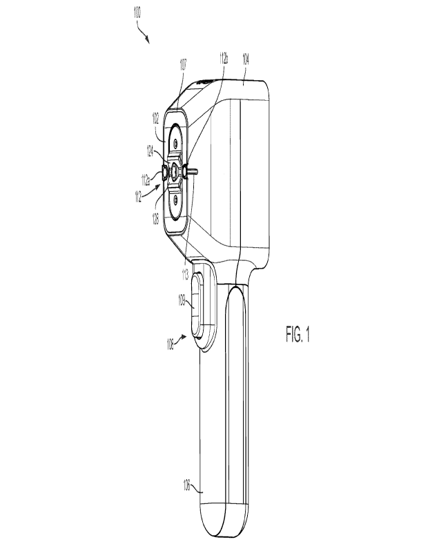
[0015] FIG. 1 is a perspective view of a handheld device in
accordance with embodiments of
the present disclosure, wherein the handheld device is shown with a removable
cartridge inserted
in the device.
[0016] FIG. 2 is a perspective view of the device of FIG. 1,
wherein the cartridge is shown
removed from the handheld device.
[0017] FIG. 3A is a perspective view of the cartridge of FIG. 1.
[0018] FIG. 3B is another perspective view of the cartridge of FIG.
1, showing a puncture
element in an extended position.
[0019] FIG. 3C is a perspective view of a solid puncture element,
showing the solid puncture
element penetrating a skin surface.
[0020] FIG. 3D is a perspective view of a puncture element having a
lumen extending
therethrough, showing the solid puncture element penetrating a skin surface.
CA 03185215 2023- 1- 6
WO 2022/013759 PCT/IB2021/056317
[0021] FIG. 4A is a side, perspective view of a cartridge in
accordance with embodiments of
the present disclosure.
[0022] FIG. 4B is a top, perspective view of the cartridge of FIG.
4A.
[0023] FIG. 4C is a side view of the cartridge of FIG. 4A.
[0024] FIG. 4D is a top view of the cartridge of FIG. 4A.
[0025] FIG. 4E is a bottom, perspective view of the cartridge of
FIG. 4A.
[0026] FIG. 4F is a side, partially transparent view of a cartridge
in accordance with
embodiments of the present disclosure.
[0027] FIG. 4G is a side, partially transparent view of a cartridge
in accordance with
embodiments of the present disclosure illustrating a path that blood travels
as it is collected using
the cartridge.
[0028] FIG. 4H is another side, partially transparent view of a
cartridge in accordance with
embodiments of the present disclosure illustrating a path that blood travels
as it is collected using
the cartridge.
[0029] FIG. 41 is a side view of a blood storage container that is
accessed from its top
(proximal side) to obtain blood therefrom.
[0030] FIG. 4J is a side view of a blood storage container that is
accessed from its bottom
(distal end) to obtain blood therefrom.
[0031] FIG. 5A is a perspective view of a handheld device in
accordance with embodiments
of the present disclosure, wherein the handheld device is shown with a
removable cartridge
inserted in the device, and the device is inserted into a cradle.
[0032] FIG. 5B is a perspective view of the cradle of FIG. 5A.
[0033] FIG. 6A illustrates the handheld device of FIG. 1 in use,
positioned over a forearm of
a subject.
[0034] FIG. 6B is another view of the handheld device of FIG. 6A.
[0035] FIG. 7A is a perspective view of a tray with multiple
compartments for storing
cartridges.
[0036] FIG. 7B is a perspective view of the tray of FIG. 7A with
the cartridges stored in the
compartments, also showing the device of FIG. 1.
[0037] FIGs. 8A to 8F illustrate results of measurements of various
blood parameters: WBC
(while blood count) (FIG. 8A), Hematocrit (FIG. 8B), Platelets (FIG. 8C),
Sodium (FIG. 8D),
CA 03185215 2023- 1- 6
WO 2022/013759 PCT/IB2021/056317
6
Chloride (FIG. 8E), and Potassium (FIG. 8F), using the present approach
("Rogue") and the
finger prick approach, as compared to standard phlebotomy.
DETAILED DESCRIPTION
[0038] In some aspects, the present disclosure provides a handheld
device for collecting a
blood sample from a subject. The device is reusable, and it is used in
conjunction with a removal
disposable cartridge configured to create a puncture in a target area of a
patient's body (e.g., a
vein in the forearm) and to withdraw the blood from the puncture. The
cartridge has a puncture
assembly and components of a blood drawing assembly for acquiring a blood
sample and storing
it in at least one blood storage container. A puncture element of the puncture
assembly is
disposed inside the cartridge and it is active only after the cartridge has
been inserted into the
handheld device and a trigger on the device is engaged.
[0039] The handheld device is easy to use and may be operated with
no specialized training.
The handheld device is able to detect a location of a vein on the patient's
forearm and allows
precise targeting of a puncture assembly of the device. The handheld device
has a position
detection system that allows determining the position of the cartridge, or,
more specifically, a
puncture element, relative to the vein. In some embodiments, additionally or
alternatively, the
position detection system (comprising, e.g., one or more optical sensor) can
be included in or
associated with the cartridge. Also, the interface between the patient's skin
and the cartridge is
designed to mechanically stabilize the vein in preparation for creating a
puncture therein. This
stabilization prevents the vein from moving away from the cartridge (more
specifically, from the
puncture element) and thus allows precise targeting of the vein. The vein may
include any vein
of a subject and/or capillary blood vessels of the subject.
[0040] The described device and cartridge allow for accurate,
reliable, and minimally
invasive blood withdrawal. The described approach is as efficient as
traditional phlebotomy
where the patient is only required to roll up their sleeve.
[0041] FIGs. 1 and 2 show an example of a handheld blood collection
device 100 for
drawing blood from a subject in accordance with embodiments of the present
disclosure. The
handheld device 100 can be a reusable device that is used in conjunction with
a removable
cartridge 102 that is configured to capture the blood sample from the subject.
The cartridge 102
can be disposable. As discussed in more detail below, the cartridge 102 is
used to puncture a
subject's body and to collect a blood sample from the subject's body through
the puncture, such
CA 03185215 2023- 1- 6
WO 2022/013759 PCT/IB2021/056317
7
that the collected blood is transferred into the cartridge. FIG. 1 illustrates
the handheld device
100 with the cartridge 102 inserted therein, and FIG. 2 shows the handheld
device 100 and the
cartridge 102 separately.
[0042] As shown in FIGs. 1 and 2, the handheld device 100 comprises
a body 104 having a
proximal end, and a handle 106 coupled to the proximal end of the body. The
handle 106 can
have a grip portion and other features that allow it to be conventicle held by
a user. The body 104
of the handheld device 100 houses an actuator assembly 108 and has a cavity
110 (visible in FIG.
2) configured to releasably receive the cartridge 102 to couple to the
actuator assembly 108. In
the illustrated embodiment, the cavity 110 is formed in a distal portion of
the body 104 that faces
a subject's body in use. It should be appreciated however that the cavity 110
can be formed in
other ways in the body 104 of the device 100. Regardless of the specific
position and
configuration of the cavity, it is formed such that it releasably fits the
cartridge 102.
[0043] The actuator assembly 108 comprises an actuator 109 (e.g., a
button, switch, or pad)
disposed on the handle 106 and configured to be activated by a user action,
e.g., by pressure
applied thereto, as discussed in more detail below. It should be appreciated
that the actuator can
be in any other form. It can be activated by the user pressing it or,
depending on its
configuration, in other ways.
[0044] In embodiments of the present disclosure, in use, the
cartridge 102 is inserted into the
cavity 110 of the device's body 104 such that the cartridge 102 is partially
exposed and operably
couples with the device 100 to position the puncture element 126 (see FIG. 3B)
relative to the
patient and control actuation of the needle and blood collection. The process
of blood sample
collection using the cartridge 102 is thus controlled via the device.
[0045] In order to more accurately and efficiently position the
puncture element 126 relative
to the patient, the handheld device 100 comprises a position detection system
112 coupled to the
body 104 of the device 100 and has at least one sensor configured to determine
a position of the
cartridge 102 relative to a target area of the subject when the cartridge 102
is disposed within the
cavity 110. In the illustrated implementation, the position detection system
112 includes first
and second optical sensors 112a, 112b positioned on a distal side of the body,
proximate to
opposite sides of the cavity. The handheld device 100 can have other
components and features
that assist in proper positioning of the puncture element for blood
withdrawal. Thus, as shown in
FIGs. 1 and 2, the body 104 of device 100 may include indicia such as grooves
or lines extending
CA 03185215 2023- 1- 6
WO 2022/013759 PCT/IB2021/056317
8
substantially perpendicular to a longitudinal axis of the handheld device 100
that denote the
location of the first and second optical sensors 112a, 112b. One of such
lines, line 113,
demarking the location of the sensor 112b is shown in FIGs. 1 and 2. The
identical line,
demarking the location of the sensor 112a, is obscured in FIGs. 1 and 2.
[0046] In some embodiments, the position detection system can
include components that
assist the user operating the device 100 in determining whether the cartridge
102, inserted in the
device 100, is properly positioned over the target area of the subject's body,
e.g., over a vein in
the subject's forearm. For example, in some embodiments, the handheld device
includes an
indicator interface that provides a visual indication of the position of the
cartridge 102 relative to
the target area, which includes the indication of the proper, ready-to-deploy,
position of the
cartridge 102.
[0047] The position detection system can be activated in various
ways. In some
embodiments, it is activated automatically when the handheld device 100
detects that the
cartridge 102 is received in the cavity 110 and coupled to the actuator
assembly.
[0048] Thus, FIG. 6A shows the indicator interface of the position
detection system such as a
display or graphical user interface 300 configured to present a graphical
indication of the
position of the cartridge 102 relative to the target area. The graphical user
interface 300 can be
disposed on the proximal side 105p of the body 104 of the device 100 having
proximal and distal
sides 105p, 105d, as shown in FIG. 6A. As also shown in FIG. 6A, when the
cartridge 102 is
positioned over the target area, such as the vein, the graphical user
interface 300 displays a first
line corresponding to a position of a first portion of the vein and a second
line corresponding to a
position of a second portion of the vein. In use, a position of the handheld
device relative to the
forearm is adjusted until the first and second lines are aligned (e.g., are on
the same axis), which
indicates that the cartridge is properly positioned over the vein. In this
way, the graphical user
interface 300 allows the user to determine whether or not the cartridge 102 is
properly positioned
for deployment.
[0049] The graphical user interface 300 can have any other visual
indicators that facilitate
the use of the device 100. For example, as shown in FIG. 6A, the graphical
user interface 300
includes the "Ready" visual indicator that additionally indicates to the user
that the cartridge 102
is in the proper position for firing the puncturing element into the vein. It
should be appreciated
that such indicator is shown by way of example only, as any suitable words,
phrases, signs,
CA 03185215 2023- 1- 6
WO 2022/013759
PCT/IB2021/056317
9
and/or symbols can be displayed (and they can be in any language).
Furthermore, it should be
appreciated that the position detection system can have any other component(s)
that facilitate
determining or determine the position of the cartridge relative to the target
area, such as the vein
in the subject's forearm. Also, the components of the position detection
system described herein
can be implemented in various ways. For example, in some implementations, in
addition or
alternatively to the visual indicators of the cartridge's position, of the
position detection system
can generate an audio signal indicating the proper position of the cartridge.
[0050] As shown in FIGs. 1 and 2, and also in FIGs. 3A and 3B that
illustrate the cartridge
102, the cartridge 102 comprises a housing 120 that includes various
components of the cartridge
102. Thus, the housing 120 includes at least one blood storage container (such
as blood storage
containers 122a, 122b), a distal body-contacting interface 124 of the housing
120, and an
isolation chamber 128 coupled to the housing 120.
[0051] The housing 120 can also include components for removable
attachment of the
cartridge 102 to the device 100. Thus, as shown in FIGs. 3A and 3B, in this
example, an outer
surface of the housing 120 has at least one longitudinal groove 115 extending
from a proximal
end 121p of the housing 120 towards a distal end 121d of the housing 120 and
configured to
mate with a corresponding protrusion formed on an inner surface of the cavity
110 (obscured).
The housing 120 can have a groove, such as the longitudinal groove 115, formed
on each of its
sides. The grooves formed in the housing 120 thus mate with corresponding
protrusions when
the cartridge 102 is inserted into the device 100. It should be appreciated
however that the device
100 and cartridge 102 can have any other features that allow for reversible
coupling between the
cartridge 102 and the device 100, as the groove 115 is shown as an example
only. In some
embodiments, one or more magnets can be used to hold the cartridge at least
partially inside the
device. Any other external features (e.g., formed on the device's housing
and/or on the
cartridge's housing) or internal features can be used to reversibly mate the
cartridge to the
device. The features can include release feature(s) configured to be activated
to disengage the
cartridge from the device after use.
[0052] In the embodiment illustrated in FIGs. 3A and 3B, the
cartridge 102 can be inserted
into the cavity 110 and removed therefrom, as it slides along the protrusions
on the inner surface
of the cavity 110. The longitudinal groove 115 is shown by way of example, as
one or more
other attachment elements, of any suitable size and configuration, can be
formed in and/or on the
CA 03185215 2023- 1- 6
WO 2022/013759
PCT/IB2021/056317
housing 120. The attachment elements can be formed in the surface of the
housing or they can be
coupled to the housing 120. The longitudinal groove(s) 115 and/or other
attachment elements of
the cartridge 102 can be releasably mated with corresponding features within
the cavity 110 such
that the cartridge 102 securely remains in place while in use, until the blood
sample is collected
and the cartridge 102 is separated from the device 100.
[0053] Also, the cartridge 102, for example, the proximal end 121p
of the housing 120, has
contact elements that mate with the interior of the cavity 110 of the device
100. Thus, the
cartridge 102 operably couples to the actuator assembly 108 of the device 100
(e.g., to its
circuity) such that operation of the cartridge 102 is controlled via the
actuator assembly 108.
[0054] In the illustrated embodiments, the body-contacting
interface 124 is a grooved
channel having a shape that corresponds to a shape of the target area of the
subject. In the
example illustrated, the grooved channel is generally V-shaped, though it
should be appreciated
that the channel can have any other concave shape such as, e.g., C or U
shaped. In use, prior to
activating the handheld device 100 to cause the cartridge 102 to create a
puncture in the target
area of the subject's body, the body-contacting interface 124 of the cartridge
102 is pressed onto
the target area to stabilize that area. In embodiments, the target area is the
subject's forearm and
the body-contacting interface 124 is pressed onto the body-contacting
interface 124 to stabilize a
vein in the forearm before the vein is punctured for blood withdrawal. In some
embodiments, the
body-contacting interface can be formed of at least partially compressible
material, which
facilitates the stabilization of the target area when the cartridge 102
inserted into the device 100
is positioned over and pressed onto the target area.
[0055] The handheld device 100 can also have features that
facilitate the contact of the
cartridge 102 inserted into the device 100 with the surface of the target area
of the subject's
body, such as the forearm. Thus, as shown in FIGs. 1 and 2, a portion 107 of
the body 104 of the
device 100 proximate to the cavity 110 has curved skin contacting edges. Thus,
the distal portion
of the body 104 is shaped so as to conveniently facilitate firm contact
between the cartridge 102
and the target area, while also ensuring the subject's comfort during the
blood withdrawal
procedure.
[0056] The cartridge 102 also comprises a puncture assembly
contained within the housing
120 and comprising a puncture element 126 moveable relative to the housing
120. The puncture
assembly, such as, e.g., a lancet assembly, allows puncturing a target area of
a subject's body
CA 03185215 2023- 1- 6
WO 2022/013759
PCT/IB2021/056317
11
(e.g., a vein in the forearm) to create a puncture in the target area. In some
embodiments, the
cartridge 102 includes a plurality of puncture elements 126 to allow the
puncture assembly to
create a plurality of punctures at a target area of the subject's body. As
shown in FIGs. 3A and
3B, the puncture assembly comprises a puncture element 126, such as a lancet
or needle (which
can be solid or hollow), that is configured to move from an initial (also
referred to herein as
"first") retracted position to an extended ("second") position. Thus, in FIG.
3A, the puncture
element 126 is positioned within the housing 120 of the cartridge 102, such
that the puncture
element 126 is in the first, retracted position and therefore not visible.
FIG. 3B illustrates the
puncture element 126 in the second, extended position, in which the puncture
element 126
extends distally from the surface of the cartridge's housing 120. In the
extended position, the
puncture element 126 extends from the body-contacting interface 124 of the
housing 120. The
handheld device 100 may be configured to control the amount that the needle
that extends
distally from the surface of the cartridge's housing 120, when the needle is
in the extended
position. Thus, the depth of the needle in a subject, when the needle is in
the extended position,
may be controlled by the handheld device 100.
[0057]
In some embodiments, a puncture element can have various forms. For
example, it
can be a hollow or solid needle, or a lancet. In some embodiments, the needle
can have
perforations (e.g., through openings) forming various patterns on the needle's
wall. The
perforations can facilitate withdrawal of blood from the vein. In some
embodiments, the needle
(with perforations or without) is associated with features that allow
detection that the needle has
entered the vein and detection that the needle has encountered liquid. The
needle can thus be able
to detect contact with liquid using one or more of electrical impedance,
capacitance, or
conductance. The cutting/leading edge at a distal end of the puncture element
can have any
suitable shape, including an angled shape (e.g., V-shape) and/or a guillotine-
type shape, a star
shape, or a cross shape. In some embodiments, the needle is comprised of a
plastic material. In
one embodiment, the needle is comprised of a translucent material to allow for
an optical wave
guide to extend through the needle such that a user may view the wave guide to
assist with
locating a subject's vein.
[0058]
FIG. 3C shows an example of a puncture element in the form of a solid
needle 126'
that is shown penetrating, with its distal end, a subject's skin surface to
access blood inside a
vein. The needle 126' can be extended to create a puncture in the skin in an
extended
CA 03185215 2023- 1- 6
WO 2022/013759
PCT/IB2021/056317
12
configuration and then retracted once the puncture is created. A puncture
element in the form of
a lancet can similarly be used to create a puncture in an extended
configuration and then
retracted into the initial configuration once the puncture is created.
[0059] In some embodiments, a puncture element of the puncture
assembly is in the form of
an extendable needle that has a lumen extending therethrough. FIG. 3D shows an
example of a
puncture element in the form of a needle 126- having a lumen 127' extending
therethrough that
can receive blood during blood collection. In FIG. 3D, the needle 126- is
shown inserted
through the skin surface such that its distal end 126d- extends partially or
entirely through the
skin layer, to access the vein (not shown) underneath the skin layer. The
needle 126- is activated
to move from the first, retracted configuration to the second, extended
configuration in which it
punctures the skin and penetrates the vein to access blood. The needle 126-
remains in the
extended configuration while the blood is collected and the blood is provided
to the cartridge
through the lumen 127- of the needle 126".
[0060] In embodiments, a gauge of a needle or another puncture
element can be in the range
of from 17 mm to 22 mm. In some embodiments, the device 100 and/or the
cartridge 102
includes a mechanism to control a penetration depth of the needle. The
penetration depth of the
needle or lancet can be controlled to be from about 2 mm to about 10 mm. In
some
embodiments, the penetration depth can be about 7 mm. The penetration depth
can be measured
using a position detection system or another system employing one or more
optical sensors.
[0061] In some embodiments, a puncture element of the puncture
assembly can include or
can be associated with liquid detection sensor(s) that allow detecting when
the puncture element
reaches blood as it is moved through the skin. In such embodiments, for
example, with reference
to FIG. 3D, it can be detected that the puncture element 126- has passed
through the skin and its
distal end 126d- has reached the blood. The sensor can be a capacitance
sensor, an impedance
sensor, or a conductance sensor. It should be appreciated that embodiments of
the present
disclosure are not limited to any specific type of sensor configured to sense
blood or another
liquid.
[0062] In addition, in some embodiments, a puncture assembly of a
cartridge can include
more than one puncturing element 126 (e.g., more than one needle).
[0063] Referring back to FIGs. 3A and 3B, the cartridge's housing
120 includes a blood
drawing assembly configured to collect the blood sample from a puncture
created in the target
CA 03185215 2023- 1- 6
WO 2022/013759
PCT/IB2021/056317
13
area by the puncture element 126 in the extended position. The blood drawing
assembly delivers
the collected blood sample to the blood storage containers 122a, 122b. In some
embodiments, the
blood drawing assembly can be coupled to the puncture assembly.
[0064] In embodiments of the present disclosure, the cartridge 102
includes components that
facilitate blood withdrawal from the vein. Thus, as mentioned above, and as
shown in FIGs. 1, 2,
3A, and 3B, the cartridge 102 comprises an isolation chamber 128 coupled to
the housing 120.
The body-contacting interface 124 comprises an opening 130 (see FIG. 3B) that
receives
therethrough the isolation chamber 128. The isolation chamber 128 can be
generally conical
such that its diameter increases in a distal direction_ The isolation chamber
128 has a central
opening or passage that receives therethrough the puncture element 126 in the
extended position,
as shown in FIG. 3B. In embodiments, the isolation chamber 128 can be
retractable such that it
is retracted proximally towards the housing 120 when the puncture element 126
moves from the
initial position to the extended position. Thus, in FIG. 3A, the isolation
chamber 128 is shown in
the original position in which a distal rim 129 of the isolation chamber 128
extends at a distance
from the opening in the body-contacting interface 124. In this configuration,
as mentioned above,
the puncture element 126 is in the initial position and it is disposed within
the housing 120. As
the puncture element 126 moves from the initial position to the extended
position, the isolation
chamber 128 is retracted proximally towards the housing 120, as shown in FIG.
3B. In such
configuration, the isolation chamber 128 sits deeper within the opening 130 in
the body-
contacting interface 124. In use, the isolation chamber 128 is positioned
around the puncture
created by the puncture element 126 in the extended position and creates a
seal around the
puncture to assist in blood withdrawal through the puncture while preventing
the puncture from
prematurely closing, as discussed in more detail below. In some embodiments,
after blood is
collected and the cartridge is withdrawn away from the puncture in the
subject's skin, the
isolation chamber 128 is positioned so that it protects the puncture element.
Such configuration
allows keeping the puncture element (e.g., without limitation, a needle) in a
safe position in
which the isolation chamber 128 at least partially "hides" the puncture
element, particularly its
sharp end.
[0065] It should be appreciated that the isolation chamber 128 is
shown in FIGs. 3A and 3B
by way of example, and that the isolation chamber can have other
configurations and sizes, and it
can be positioned in a suitable way with respect to the puncture element.
CA 03185215 2023- 1- 6
WO 2022/013759
PCT/IB2021/056317
14
[0066] The puncture element 126 of the puncture assembly is
activated using the actuator
assembly of the handheld device. Referring back to FIGs. 1 and 2, the actuator
assembly 108 is
configured to be activated to cause the puncture element 126 to puncture a
skin surface of the
subject at the target area and to cause the blood withdrawal assembly to
collect the blood sample
from the target area through the puncture. For example, the actuator 109
(e.g., a button) disposed
on the handle of the handheld device can be pressed to activate the puncture
element 126. The
activation can occur after it has been determined e.g., by the position
detection system, that the
device (the cartridge inserted in the device, to be exact) is properly
positioned with respect to the
target area, e.g., the vein, and the vein is stabilized.
[0067] In embodiments, the actuator assembly 108 configured to
apply negative pressure to
the target area while the blood withdrawal assembly draws the blood sample
through the
puncture in the target area. The negative pressure is applied to the puncture
wound having the
isolation chamber 128 disposed therein such that, as the blood is sucked into
the cartridge, the
skin at the target area adopts a dome-like shape with the puncture wound
(which is stretched
open) at the apex. The application of the negative pressure to the puncture
thus assists in
maintaining the puncture open during the blood withdrawal process. This also
affects positively
the quality of the withdrawn blood.
[0068] Referring back to FIGs. 3A and 3B, the blood drawing
assembly delivers the blood
sample, as the blood is being collected, to the blood storage containers 122a,
122b. The blood
drawing assembly can comprise at least one fluid conduit (e.g., one or more
tubes, not shown)
configured to deliver the blood sample acquired through the puncture to the
blood storage
containers 122a, 122b. The blood drawing assembly can include a pump
(positioned in the
handheld device) and any other suitable component(s). Each of the blood
storage containers
includes an access port for removing the blood sample from that blood storage
container. Thus,
as shown in FIGs. 3A and 3B, the blood storage containers 122a, 122b have
access ports 123a,
123b, respectively. In use, after the blood sample is transferred into the
blood storage containers
and the cartridge 102 is removed from the device 100, the blood storage
containers 122a, 122b
can be accessed via the ports 123a, 123b to remove the blood therefrom, for
analysis. In some
embodiments, one or more of the blood storage containers 122a, 122b are
removable from the
cartridge housing 120.
CA 03185215 2023- 1- 6
WO 2022/013759
PCT/IB2021/056317
[0069] It should be appreciated that two blood storage containers
122a, 122b are shown by
way of example only, as the cartridge can have a single blood storage
container or more than two
blood storage containers (e.g., three, four, or more than four blood storage
containers). Also, the
one or more blood storage containers can be disposed in various ways within
the cartridge, and
the containers can be accessed in various ways as the present disclosure is
not limited in this
respect. The blood storage containers of the cartridge can have the same or
different
configuration.
[0070] In some embodiments, the blood storage container(s) (or
vials) can be plastic vessels
which are coated with blood anticoagulants and preservatives, e.g., k2EDTA and
Lithium
Heparin. The coating can be spray coating or any other technique. In some
embodiments, each
blood storage container can carry a maximum volume of about 600 uL of blood.
Blood storage
containers configured to hold a suitable volume of blood, including other than
600 uL, can be
used as well.
[0071] The handheld device 100 can have other components and
features that assist in skin
puncture and blood withdrawal. For example, in some embodiments, the device
100 has a depth
control actuator coupled to the body 104 and configured to be actuated to
control a depth of the
puncture to be created by the puncture element 126 in the extended position.
The depth control
actuator can be in the form of a dial, button, switch, or another device
configured to be
manipulated to adjust the desired depth of the penetration. The depth control
actuator can be
disposed on the handle 106 of the device in some implementations. Also, in
some embodiments,
instead of using a depth control actuator configured to receive used input,
the depth of the
puncture can be controlled automatically, e.g., via a computing device that
controls operation of
the device 100.
[0072] FIGs. 4A, 4B, 4C, 4D, and 4E illustrate several views of a
cartridge 102 which is
similar to the cartridge 102 shown in FIGs. 1,2, 3A, and 3B.
[0073] In embodiments, blood storage containers can have various
configurations. In some
embodiments, for example, they can be configured as tubes which can have
various shapes. For
example, the tubes can have conical or rounded tips, or tips having other
shapes. Other
configurations can be used additionally or alternatively.
[0074] Blood can be transmitted to the interior of the blood
storage containers in various
ways. In some embodiments, for example, blood storage containers can have one
or more
CA 03185215 2023- 1- 6
WO 2022/013759
PCT/IB2021/056317
16
openings in their walls which can be in fluid communication with fluid
conduits (e.g., tubes) that
deliver blood from a puncture to the blood storage containers. Each of the
blood storage
containers can have one or more openings in fluid communication with fluid
conduits, and the
opening(s) can be disposed in various locations on the blood storage
containers, at various
distances from a distal end of the cartridge.
[0075] FIG. 4G illustrates an example of a cartridge 202 (which can
be similar to cartridge
102 in FIGs. 3A and 3B) that is operably coupled to a handheld device (not
shown) in
accordance with embodiments of the present disclosure. As shown in FIG. 4G,
the cartridge 202
has blood storage containers 222a, 222b, each of which has an opening that
communicates with a
respective tube or fluid conduit through which the blood is delivered from the
puncture to the
blood storage containers 222a, 222b. After a puncture element (not shown),
having an isolation
chamber 228 disposed therearound, creates the puncture in the vein, constant
negative pressure
(shown as "-P" in FIG. 4G) is applied by the handheld device which causes the
blood to be
delivered from the puncture via the fluid conduits 219a, 219b to the blood
storage containers
222a, 222b, respectively (as also shown by arrows drawn within the blood
storage containers).
In the example of FIG. 4G, the constant negative pressure is applied at the
blood storage
containers 222a, 222b, as well as at the isolation chamber 228. The constant
negative pressure is
applied by a blood drawing assembly, which can have parts (e.g., fluid
conduits) disposed in the
cartridge and some parts (e.g., a pump) positioned in the handheld device.
[0076] FIG. 4H shows another example of a cartridge 202' that can
be similar to cartridge
202 of FIG. 4G. In FIG. 4H, respective openings 221a', 221b' in blood storage
containers 222a',
222b' of the cartridge 202', in fluid communication with respective fluid
conduits, are positioned
more distally along the length of the blood storage containers than the
openings in the blood
storage containers of FIG. 4G. In this way, depending on the position of the
openings, the blood
storage containers are filled from their bottoms (as shown in FIG. 4H) or
closer to the tops (FIG.
4G). In the example of FIG. 4H, the constant negative pressure is applied to
the blood storage
containers 222a', 222b'.
[0077] Furthermore, in some embodiments, blood storage containers
of a cartridge in
accordance with embodiments of the present disclosure can have more than one
fluid conduit
connected thereto, and the fluid conduits can be disposed at different
positions along a length of
the blood storage containers. Furthermore, in embodiments in which the
cartridge has two or
CA 03185215 2023- 1- 6
WO 2022/013759
PCT/IB2021/056317
17
more blood storage containers, the blood storage containers can have different
number of fluid
conduits (tubes) coupled thereto, and the fluid conduits can be coupled to the
blood storage
containers at different positions along the length and/or other dimensions of
the blood storage
containers.
[0078] In embodiments, proximal ends of the blood storage
containers can have membranes,
e.g, hydrophobic membranes. FIG. 4E shows an example of an implementation of a
bottom, or
proximal side, of the cartridge 102'. In FIG. 4E, proximal sides 125a', 125b'
of blood storage
containers 122a2, 1221:,' (which are similar to blood storage containers 122a,
122b) are shown,
respectively. The proximal sides 125a', 125b' can each include a hydrophobic
membrane, as
shown schematically in FIG. 4F illustrating blood storage containers 122a',
12213'. The blood
storage containers can have their proximal sides formed from hydrophobic
membranes made of
suitable materials. In this way, the vacuum can be created through the back of
the blood storage
container(s), but no blood would seep through the membranes as the blood is
being withdrawn.
[0079] The handheld device 100 in accordance with the present
disclosure can be configured
such that a constant negative pressure (shown as -P in Fig. 4F) is created
and, as air is pulled
proximally through the blood storage containers, the blood is acquired from a
subject's vein and
is delivered to the blood storage containers. The actuator assembly 108 of the
handheld device
100 may include a pump (e.g., a vacuum pump) that operates to create the
constant negative
pressure that allows the blood to be withdrawn from the puncture in the
subject's vein. In some
embodiments, the vacuum created in the blood storage container(s) has a
corresponding vacuum
pressure and the actuator assembly 108 of the handheld device 100 is
configured to modulate
(e.g., increase or decrease) the vacuum pressure. In some embodiments, the
vacuum pump is
configured to modulate the vacuum pressure. By modulating the vacuum pressure,
a user may
experience a rubbing or kneading sensation (e.g., a massaging sensation) on
the area of the
subject's body proximate the puncture in the subject's vein, which may provide
additional
comfort to the user and improve the withdrawal of blood from the puncture in
the subject's vein.
For example, the actuator assembly 108 may increase the vacuum pressure to a
first
predetermined pressure and then decrease the vacuum pressure to a second
predetermined
pressure, or vice versa, one or more times to cause the user to experience a
kneading sensation
on the area of the subject's body proximate the puncture in the subject's
vein. In some
embodiments, the first predetermined pressure is about -7 psi and the second
predetermined
CA 03185215 2023- 1- 6
WO 2022/013759
PCT/IB2021/056317
18
pressure is about -2 psi. In some embodiments, the increasing and decreasing
of the vacuum
pressure from the first predetermined pressure to the second predetermined
pressure may be done
gradually over a period of time (e.g., one second, two seconds, three seconds,
four seconds, five
seconds, or more than five seconds).
[0080] FIG. 4F also illustrates that a distal end of a blood
storage container can be generally
conical. Such shape allows minimizing "dead" volume of blood (blood pooling)
that may
otherwise accumulate in the storage container.
[0081] Blood storage containers have features that allow accessing
blood therefrom. For
example, as discussed above and shown in FIGs. 3A and 3B, blood storage
containers have
access ports, such as access ports 123a, 123b, respectively. Blood in the
blood storage containers
can be accessed for analysis in various ways. For example, as shown
schematically in FIG. 41, a
blood storage container 322 can be accessed from the top (its proximal end)
using a suitable
element, such as, e.g., without limitation a pipette 330 shown in FIG. 41.
[0082] In some embodiments, after the blood is collected and stored
in the blood storage
containers, as the cartridge with the blood storage containers is transferred
from the handheld
device to an analyzer (e.g., to a tray or receptacle of the analyzer), the
access ports can be pierced
through. In such embodiments, the receptacle, configured to receive the
cartridge with the
collected blood therein, can have piercing features with a sharp end that are
disposed on the
receptacle such that, as the cartridge is placed onto the receptacle, the
piercing features are
aligned with the blood storage containers' access ports and the access ports
are pierced to access
the blood stored therein. The blood can thus be transferred to a receptacle.
FIG. 4J shows
schematically a blood storage container 322' that can have its distal end
pierced by a piercing
element 303 (e.g., a needle) positioned in a tray or receptacle of an
analyzer.
[0083] FIGs. 5A and 5B illustrate a handheld device 100 having the
cartridge 102' inserted
therein. As shown in FIG. 5A, a distal end of a handle 106' of the device 100'
can couple with a
charging cradle 505 that charges the device 100'. The cradle 205 can have an
opening 507 (FIG.
5B) configured to removably seat therein the distal end of the handle 106'. It
should be
appreciated that the cradle 505 is shown as generally circular by way of
example only, as it can
have any other shape and configuration.
[0084] Regardless of its specification configuration, a handheld
device, configured to receive
a removable cartridge for use with the device in blood withdrawal from
subjects, is used for
CA 03185215 2023- 1- 6
WO 2022/013759
PCT/IB2021/056317
19
collecting a blood sample from a subject in an improved manner. In particular,
as discussed
above, the present approach is as convenient as phlebotomy such that the
patient is only required
to expose his/her forearm. Moreover, the present device allows to physically
trap the vein (which
can naturally "move around-) to thereby limit its range of motion. By thus
trapping the vein and
stabilizing it in place, the likelihood of accessing the vein from a single
attempt for withdrawal
of the desired volume of blood is increased. The present approach is thus more
reliable and more
convenient to a patient and to a user (medical personnel) as compared to the
standard
phlebotomy. Also, as discussed above, the application of the negative pressure
to the puncture
wound as blood is being withdrawn therefrom allows keeping the puncture open
during the blood
withdrawal, which in turn allows reducing the amount of sheer stress imposed
on the blood
during its withdrawal.
[0085] Furthermore, because of the ease of the use of the device in
accordance with
embodiments of the present disclosure, it can be used by a healthcare
professional (e.g., nurse,
medical assistant, technician, etc.) with minimal training related to the
device operation. In this
way, the device can be used in any setting, including settings where a highly
skilled trained
professional (phlebotomist) is not present.
[0086] As discussed above, the handheld device is reusable, whereas
the cartridge is
disposable. The cartridge can be picked up with the device (i.e. directly from
a manufacturing
package), as discussed in more detail below, and used for blood extraction
from a patient. After
the blood sample is acquired, the cartridge is removed from the device and is
transferred to an
analyzer or stored or shipped to another location. The sample collection
device can comprise a
plurality of compartments or receptacles each configured to fit a cartridge.
In some
embodiments, the handheld device is configured to be attached or otherwise
coupled to a subject
(e.g., via one or more straps) rather than be held by a user when in use.
[0087] It should be appreciated however that, in some embodiments,
the cartridge can be
reusable. The cartridge may be configured to be recycled/refurbished. For
example, the cartridge
may have removable reservoirs (blood storage containers) and, after cleaning
and sterilizing, and
installing new reservoirs, the cartridge may be reused.
[0088] In some aspects, a method of collecting a blood sample from
a subject is provided.
The method comprises inserting a cartridge into a handheld device for
collecting blood sample.
For example, device 100 (FIGs. 1 and 2) can be used, which receives therein
cartridge 102
CA 03185215 2023- 1- 6
WO 2022/013759
PCT/IB2021/056317
(FIGs. 1, 2, 3A, and 3B). As shown in FIGs. 6A and 6B (illustrating device 100
and cartridge
102), the method further includes positioning the handheld device with the
cartridge against a
forearm 630 (shown very schematically) of a subject's body and adjusting a
position of the
device relative to the forearm until an indicator interface of the handheld
device indicates that the
cartridge is properly positioned over a vein 632 in the forearm 630. As
discussed above, the
indicator interface can be, e.g., the graphical user interface 300 of the
position detection system
of the device 100 illustrated in FIG. GA. The indicator interface is coupled
to at least one optical
sensor of the handheld device that determines a location of the vein. The
position detection
system, such as, in some implementations, at least two optical sensors, allows
detecting the
location of a superficial vein on the patient's forearm and allows for precise
targeting of the
puncture assembly. The optical sensors sense absorption due to hemoglobin in
the blood. In
some implementations, the device 100 can perform stereo-like measurement to
measure the
approximate depth of the vein under the skin surface.
[0089] In the illustrated example, with reference to FIG. 6A, the
graphical user interface 300
displays a first line 302a corresponding to a position of a first portion of
the vein 632 and a
second line 302b corresponding to a position of a second portion of the vein
632. The position of
the handheld device 100 relative to the forearm 630 is adjusted until the
first and second lines
302a, 302b are aligned (e.g., are on the same axis), which indicates that the
cartridge 102 is
properly positioned over the vein 632. It should be appreciated that the
graphical user interface
300 in FIG. 6A is shown by way of example only. Accordingly, the first and
second lines 302a,
302b are only an example of an implementation of indicator features that are
indicative of the
proper position of the device for puncturing the vein. For example, indicator
feature(s) can
include lights of different colors, e.g., red, yellow/orange, and green, with
red indicating that the
device is not positioned properly, and green indicating a proper position. As
another example,
the indicator features can include a blinking light indicator (e.g., the
indicator can blink until a
proper device position is detected). The device can be implemented to generate
any suitable
visual and/or audio signals as indicator(s) of a current position of the
device and indicator(s) of a
device's position with respect to the subject's body in which the device can
be operated to
puncture the vein to acquire blood therefrom.
[0090] In some embodiments, the position detection system can be
activated automatically,
once the cartridge is inserted into the device. The device can sense that the
cartridge is connected
CA 03185215 2023- 1- 6
WO 2022/013759
PCT/IB2021/056317
21
thereto, and the position detection system can begin scanning for the vein. In
other embodiments,
the position detection system can be activated using a trigger. For example,
the actuator
assembly can be used to activate the position detection system
[0091] Adjusting the position of the handheld device 100 relative
to the subject's forearm
630 involves moving the device 100 over the forearm 630 until the position
detection system
indicates that the cartridge 102 is properly positioned over the vein 632. The
position detection
system can be associated with the handheld device and/or with the cartridge.
For example, one
or more optical sensors can be disposed on the handheld device and/or on the
cartridge. Once it
is determined that the cartridge 102 is properly positioned, the method
further includes pressing
the body-contacting interface 124 of the cartridge 102 onto the forearm to
stabilize the vein. In
some embodiments, as shown, e.g., in FIGs. 1, 2, 3A, and 3B, the body-
contacting interface is a
grooved channel formed of at least partially compressible material. As
discussed above, the
body-contacting interface 124 that is positioned over the patient's skin is
used to mechanically
stabilize the vein in preparation for creating a puncture (e.g., lancing) the
vein. This stabilization
allows avoiding the possibility that the vein moves or slips out of the way of
the puncture device
(e.g., a needle or lancet), which would result in a miss and would require the
process to be
repeated.
[0092] Once the vein is "found" and stabilized, the method in
accordance with embodiments
of the present disclosure includes actuating the handheld device 100 thereby
causing the
cartridge 102 to deploy the puncture element 126 of the puncture assembly of
the cartridge so
that the puncture 126 element moves from an initial, retracted position to an
extended, extended
position to create a puncture in the vein. In some embodiments, the actuation
is performed using
an actuating assembly of the handheld device, by applying force to an
actuation element
disposed on a proximal handle of the handheld device. This can involve, for
example, pressing
the actuator element 109, or actuating another element, which can be different
depending on the
implementation of the device. The puncture is created to a certain depth
within the vein, and, in
some embodiments, the depth of the penetration of the puncture element 126 is
controllable (e.g.,
using a depth control actuator or another suitable component). For example, in
some
embodiments, the puncture in the vein has a depth of no greater than 10 mm
(e.g., or about 2
mm, or about 3 mm, or about 4 mm, or about 5 mm, or about 6 mm, or about 7 mm,
or about 8
mm, or about 9 mm, or about 10 mm). In some embodiments, the puncture in the
vein has a
CA 03185215 2023- 1- 6
WO 2022/013759
PCT/IB2021/056317
22
depth of no greater than 7 mm. In some embodiments, the puncture in the vein
has a depth of
about 7 mm.
[0093] The actuation of the actuator assembly to cause the puncture
element 126 to create a
puncture also causes the cartridge to withdraw a blood sample through the
puncture from the
vein to at least one blood storage container included in the cartridge, such
as the blood storage
containers 122a, 122b. The blood sample of a suitable volume can be acquired.
For example, in
some embodiments, the blood sample has a volume of at least 1 mL, or at least
1.5 mL, or at
least 2 mL. In some embodiments, the blood sample has a volume of about 1 mL,
or about 1.5
mL, or about 2 mL. In some embodiments, the blood sample has a volume of less
than 1 mL, or
less than 1.5 mL, or less than 2 ml. In some embodiments, the blood sample has
a volume of at
least 1 mL and less than 2 mL. In some embodiments, the blood sample has a
volume of at least
0.5 mL or about 500 uL. The blood sample can be acquired in a time period that
is less than two
minutes, less than one minute, or less than 45 seconds, or less than 30
seconds.
[0094] Thus, the present devices and methods allow collecting blood
faster than existing
approaches to collecting blood without the need for phlebotomy. Those existing
approaches
pertain to extracting capillary blood in larger volumes, which is accomplished
by creating several
puncture wounds, as opposed to one. Some of the existing blood drawing
approaches are meant
to be self-administered, much like the finger prick method, but with a
decreased user intervention
in the collection process. The devices in those approaches can adhere to the
skin (usually below
the shoulder), with a press of a button, create the puncture wounds and
collect the blood into an
internal blood tube. These methods were shown to be able to collect about 200
iaL of blood after
several minutes of use, and the quality of the blood is typically poor, which
limits these methods'
clinical utility and value. The devices and methods in accordance with the
present disclosure
may allow collecting a blood sample in less than one minute.
[0095_1 As discussed above, in some embodiments, pressure is applied
to the puncture, which
has the isolation chamber 128 (see FIGs. 3A and 3B) disposed therearound such
that, as the
blood is sucked into the cartridge, the skin around the puncture adopts a dome-
like shape with
the puncture (which is stretched open) at the apex. In some embodiments, the
pressure can be
negative pressure, such as constant negative pressure. The application of the
negative pressure to
the puncture assists in maintaining the puncture open during the blood
withdrawal process. This
also affects positively the quality of the withdrawn blood. In particular, by
causing the puncture
CA 03185215 2023- 1- 6
WO 2022/013759
PCT/IB2021/056317
23
wound to remain open, an unhindered conduit is created and maintained for the
blood to exit the
subject's body. Such unrestricted pathway allows decreasing the hemolysis
(bursting of RBCs),
and decreases the amount of stress that the platelets experience, hence
delaying the coagulation
process and thereby improving the quality of the collected sample. Keeping the
puncture open
allows reducing the amount of shear stress on the cells to minimize cell lysis
and platelet
activation. A pressure applied to a puncture can have any suitable value. In
some embodiments,
for example, a pressure of -4 psi can be applied to a puncture, to enable
collection of about 1 mL
of blood in less than a minute. The pressure can have other values as well,
and other volumes of
blood can be collected (e.g., less than 2 mL).
[0096] In some embodiments, the described handheld device can be
used for administering
blood or another liquid (e.g., without limitation, a therapeutic) into the
blood stream. In such
embodiments, a cartridge can, for example, have a vaccine or another
therapeutic stored therein
that can be administered to a subject when the cartridge is inserted into the
device. Embodiments
of the present disclosure are not limited to any specific type of therapeutic
or another
composition that can be administered using a handheld device and a cartridge
in accordance with
embodiments of the present disclosure.
[0097] The method of collecting the blood sample from the subject
further includes, when
the blood sample is received in the at least one blood storage container,
actuating the handheld
device thereby causing the puncture element to move from the extended position
to the initial,
retracted position. For example, the actuator assembly can be activated. FIG.
3B illustrates the
puncture element in the extended position, while FIG. 3A illustrates the
cartridge 102 is the
initial, retracted position. FIGs. 3C and 3D illustrate examples of puncture
elements that can be
used. As discussed above, in some embodiments, the actuator assembly remains
activated while
the blood withdrawal takes place such that the puncture element, in the
extended position,
remains in the puncture that it created. In other implementations, however,
the puncture element
is extended to create a puncture in the subject's skin to access blood through
the puncture, and
retracted once the puncture is created, such that the blood withdrawal occurs
while the puncture
element is in the initial, retracted position.
[0098] The device 100 can be configured to generate an indication
that the desired volume of
the blood sample is received in the blood storage containers. The indication
can be provided,
e.g., via the graphical user interface 300, or via another component(s). Also,
in some
CA 03185215 2023- 1- 6
WO 2022/013759
PCT/IB2021/056317
24
embodiments, a desired volume of blood can be acquired automatically, such
that the blood
drawing assembly automatically stops the blood withdrawal once the desired
amount is acquired.
The puncture element can also move from the second, extended position to the
initial, retracted
position automatically, or using a trigger. For example, the actuator assembly
(e.g., actuator 109)
can be manipulated to cause the puncture element to move to the initial,
retracted position.
[0099] After the blood sample is received in the at least one blood
storage container, the
cartridge is separated from the handheld device. This can be performed using a
suitable trigger.
The handheld device can have a release component or another feature that can
be manipulated by
the user to remove the cartridge from the device. In some implementations, the
handheld device
and/or the cartridge can be configured such that the cartridge can be
separated from the handheld
device only into a receptacle of a sample analyzer. A suitable trigger and/or
features can be used
for this purpose. For example, the cartridge can have one or more magnets and
a receptacle can
have one or more magnets, such that the cartridge can be released from the
handheld device upon
the interaction between the magnet(s). Any other features can be used that
allow releasing the
cartridge specifically into a receptacle of a sample analyzer. For example, a
Bluetooth pairing
between the cartridge (or the device) and the receptacle or another portion of
the sample analyzer
can be used to determine that the cartridge is in proximity to the receptacle.
[00100] In some embodiments, after the cartridge is separated from the
handheld device, the
cartridge can be coupled with a sample collection device, or the cartridge can
be transferred to an
analyzer. The sample collection device may comprise a plurality of
compartments each
configured to fit a cartridge. In some embodiments, as discussed above, the
cartridge can be
separated from the handheld device to be transferred directly to the analyzer.
Also, in some
implementations, the cartridge is released from the handheld device only once
the cartridge is
coupled to or otherwise associated with a suitable compartment or receptacle
of a sample
collection device or an analyzer. In some implementations, as also discussed
above, the analyzer
can have features that allow acquiring blood from blood storage containers of
the cartridge.
[00101] In some embodiments, the blood sample is removed from the cartridge,
e.g., by
accessing ports of the at least one blood storage container. The "spent"
cartridge, which can be
disposable, can then be disposed.
[00102] The handheld device is reusable. Accordingly, in some embodiments,
after the
cartridge (with the blood sample stored therein) is separated from the
handheld device, a second
CA 03185215 2023- 1- 6
WO 2022/013759
PCT/IB2021/056317
cartridge can be inserted into the handheld device. The handheld device with
the second
cartridge inserted herein can be used for collecting a blood sample from a
patient. Multiple
disposable cartridges can be used with the handheld device.
[00103] FIGs. 7A and 7B show a tray 700 having multiple compartments 702 (ten,
in this
example, though the tray can include any other number of compartments) formed
in a body 704
of the tray 700, of which compartments 702a, 702b, and 702c are labeled. The
compartments 702
of the tray 700 can each include a cartridge for use with a handheld device.
The tray 700 can be
manufactured in the form as shown in FIG. 7B, and a handheld device 100¨ can
be used to pick
up a (new) cartridge from a respective compai
tinent, as shown in FIG. 7B. FIG. 7B illustrates, .. by
way of example, the device 100¨ as it is picking up the cartridge from the
compartment 702.
After the cartridge is used to acquire a blood sample, the device 100¨ can be
used to pick up
another cartridge from the tray's compartment.
[00104] Additional information on embodiments of the present disclosure, as
well as other
relevant information, can be found in the Appendix submitted along with the
present application.
EXAMPLES
Example 1
[00105]
Figs. 8A to 8F illustrate study results of measurements of several blood
parameters,
analyzed by FDA-approved clinical analyzers from Beckman Coulter (DxH 500 and
AU 480). In
the study, blood drawn from ten subjects was acquired using an approach of the
present
disclosure ("Rogue," values and lines are shown as a dotted line) and the
finger prick approach
("FP," values and lines are shown as an evenly spaced dashed line), and
concordance correlation
coefficients were used to determine how well the measurements from these blood
samples
compare to measurements from blood samples acquired using the standard
phlebotomy
approach. FIGs. 8A to 8F illustrate results of measurements of various blood
parameters such as
WBC (while blood count) (FIG. 8A), Hematocrit (FIG. 8B), Platelets (FIG. 8C),
Sodium (FIG.
8D), Chloride (FIG. 8E), and Potassium (FIG. 8F). FIGs. 8A-8F show measured
values and
CA 03185215 2023- 1- 6
WO 2022/013759
PCT/IB2021/056317
26
linear regression (best-fit) line (for both Rogue and finger prick) for a
respective blood
parameter, as well as a linear (identity) line (shown as a solid line).
[00106] As shown in the table below, there is concordance between the present
approach and
phlebotomy. At the same time, concordance is not observed between data for the
finger prick and
phlebotomy approaches, for the most sensitive analytes (i.e., platelets and
potassium).
[00107] Additional information on results of the conducted experiments is
shown in the
Appendix submitted along with the present application.
EQUIVALENTS
[00108] While the invention has been described in connection with specific
embodiments
thereof, it will be understood that it is capable of further modifications and
this application is
intended to cover any variations, uses, or adaptations of the invention
following, in general, the
principles of the invention and including such departures from the present
disclosure as come
within known or customary practice within the art to which the invention
pertains and as may be
applied to the essential features hereinbefore set forth and as follows in the
scope of the
appended claims.
[00109] Those skilled in the art will recognize, or be able to
ascertain, using no more than
routine experimentation, numerous equivalents to the specific embodiments
described
specifically herein. Such equivalents are intended to be encompassed in the
scope of the
following claims.
INCORPORATION BY REFERENCE
[00110] All patents and publications referenced herein are hereby incorporated
by reference in
their entireties. The publications discussed herein are provided solely for
their disclosure prior to
the filing date of the present application. Nothing herein is to be construed
as an admission that
the present invention is not patentable in view of such publications.
[00111] As used herein, all headings are simply for organization and are not
intended to limit
the disclosure in any manner. The content of any individual section may be
equally applicable to
all sections.
CA 03185215 2023- 1- 6