Note: Descriptions are shown in the official language in which they were submitted.
1
End-to-End Cell Therapy Automation
Field of the Invention
[0001] The present disclosure provides an automated method of producing
genetically modified immune cells, including chimeric antigen receptor T
(CART) cells,
utilizing a fully-enclosed cell engineering system.
Background of the Invention
[0002] As anticipation builds about accelerated clinical adoption of
advanced cell
therapies, more attention is turning to the underlying manufacturing
strategies that will
allow these therapies to benefit patients worldwide. While cell therapies hold
great
promise clinically, high manufacturing costs relative to reimbursement present
a
formidable roadblock to commercialization. Thus, the need for cost
effectiveness,
process efficiency and product consistency is driving efforts for automation
in
numerous cell therapy fields, and particularly for T cell immunotherapies
(see, e.g.,
Wang 2016).
[0003] Recent successful clinical results from immunotherapy trials using
chimeric
antigen receptor (CAR) T cells provide new hope to patients suffering from
previously
untreatable cancers (see, e.g., Lu 2017; Berdeja 2017; Kebriaei 2016). As
these novel
therapeutics move from the clinical trial stage to commercial scale-up,
challenges arise
related to cell manufacturing (see, e.g., Morrissey 2017).
[0004] The production of these cells may require significant manual
involvement
due to the patient-specific product. Automation of CAR T cell culture is
particularly
challenging due to the multiple sensitive unit operations, including cell
activation,
transduction and expansion. Activation may be particularly important as the
efficiency
of this process can impact transduction and expansion.
[0005] Integration of cell activation, transduction and expansion into a
commercial
manufacturing platform is critical for the translation of these important
immunotherapies to the broad patient population. For these life-saving
treatments to
be applicable to the global patient population, a shift in manufacturing
techniques must
Date Recue/Date Received 2022-12-19
2
be implemented to support personalized medicine. The benefits of automation
have
previously been described (see, e.g., Trainor 2014; Mandavi 2015). These
benefits
include labor time savings associated with using automation as well as
improved
product consistency, decreased room classification, decreased clean room
footprint,
decreased training complexities, and improved scale-up and tracking logistics.
Furthermore, software can be used to streamline the documentation processes by
using automatically generated electronic batch records to provide a history of
all
processing equipment, reagents, patient identification, operator
identification, in-
process sensor data, and so forth.
Summary of the Invention
[0006] In some embodiments provided herein is a method for automated
production of a genetically modified immune cell culture, the method
comprising:
activating an immune cell culture with an activation reagent to produce an
activated
immune cell culture; transducing the activated immune cell culture with a
vector, to
produce a transduced immune cell culture; expanding the transduced immune cell
culture; concentrating the expanded immune cell culture of (c); and harvesting
the
concentrated immune cell culture of (d) to produce a genetically modified
immune cell
culture, further comprising washing either or both the expanded immune cell
culture
and the concentrated immune cell culture, wherein (a) through (e) are
performed by a
fully enclosed cell engineering system and (a) through (e) are optimized via a
process
to produce the genetically modified immune cell culture.
[0007] In further embodiments, provided herein is a method for promoting a
preferred phenotype of a genetically modified immune cell culture, the method
comprising: activating an immune cell culture with an activation reagent to
produce an
activated immune cell culture, wherein the activation reagent and activating
conditions
promote the phenotype of the genetically modified immune cell culture;
transducing
the activated immune cell culture with a vector, to produce a transduced
immune cell
culture; expanding the transduced immune cell culture; concentrating the
expanded
immune cell culture of (c); and harvesting the concentrated immune cell
culture of (d)
to produce a genetically modified immune cell culture, wherein (a) through (e)
are
performed by a fully enclosed, automated cell engineering system.
Date Recue/Date Received 2022-12-19
3
[0007.1] In
additional embodiments, provided herein is a method for automated
production of a genetically modified immune cell culture, the method
comprising: (a)
activating an immune cell culture with an activation reagent to produce an
activated
immune cell culture; (b)
transducing the activated immune cell culture with a
vector, to produce a transduced immune cell culture; (c) expanding the
transduced
immune cell culture; (d) concentrating the expanded immune cell culture of
(c); and
(e) harvesting the concentrated immune cell culture of (d) to produce a
genetically
modified immune cell culture, further comprising washing either or both the
expanded
immune cell culture and the concentrated immune cell culture, wherein (a)
through (e)
are performed by a fully enclosed cell engineering system and (a) through (e)
are
optimized via a process to produce the genetically modified immune cell
culture, and
wherein the cell engineering system recirculates nutrients, waste, released
cytokines,
and/or dissolved gasses during steps (a) to (e), and wherein the cell
engineering
system includes a single cassette that contains the immune cell culture of
(a), the
activation reagent, the vector, and a cell culture medium prior to starting
the method.
[0007.2] In
additional embodiments, provided herein is a method for automated
production of a genetically modified immune cell culture, the method
comprising:
(a) activating an immune cell culture with an activation reagent to produce
an
activated immune cell culture;
(b) transducing the activated immune cell culture with a vector, to produce
a
transduced immune cell culture;
(c) expanding the transduced immune cell culture;
(d) concentrating the expanded immune cell culture of (c); and
(e) harvesting the concentrated immune cell culture of (d) to produce a
genetically
modified immune cell culture,
further comprising washing either or both the expanded immune cell culture and
the
concentrated immune cell culture,
wherein (a) through (e) are performed by a fully enclosed cell engineering
system to
produce the genetically modified immune cell culture, and wherein the cell
engineering
system recirculates nutrients, waste, released cytokines, and/or dissolved
gasses
during steps (a) to (e),
Date Recue/Date Received 2022-12-19
4
and wherein the cell engineering system contains at least one of the immune
cell
culture of (a), the activation reagent, the vector, and a cell culture medium
prior to
starting the method,
wherein the cell engineering system includes:
a cell culture chamber;
one or more of a temperature sensor, a pH sensor, a glucose sensor, an oxygen
sensor, a carbon dioxide sensor, a lactic acid sensor/monitor, and/or an
optical density
sensor;
a low temperature chamber for storage of a cell culture media;
a high temperature chamber comprising the cell culture chamber, wherein the
high
temperature chamber is separated from the low temperature chamber by a thermal
barrier; and
one or more fluidics pathways connected to the cell culture chamber.
[0007.3] In
additional embodiments, provided herein is a method for automated
production of a genetically modified immune cell culture, the method
comprising:
(a) activating an immune cell culture with an activation reagent to produce
an activated immune cell culture;
(b) transducing the activated immune cell culture with a viral vector
encoding an ectodomain, a transmembrane domain, and an
endodomain, to introduce the viral vector into the activated immune cell
culture and produce a transduced immune cell culture;
(c) expanding the transduced immune cell culture wherein the transduced
immune cell culture is not shaken during the expanding;
(d) concentrating the expanded immune cell culture of (c); and
(e) harvesting the concentrated immune cell culture of (d) to produce a
genetically modified immune cell culture,
further comprising washing either or both the expanded immune cell culture and
the
concentrated immune cell culture,
wherein (a) through (e) are performed by a fully enclosed cell engineering
system to
produce the genetically modified immune cell culture, and wherein the cell
engineering system recirculates nutrients, waste, released cytokines, and/or
dissolved gasses during steps (a) to (e),
Date Recue/Date Received 2022-12-19
5
and wherein the cell engineering system contains at least one of the immune
cell
culture of (a), the activation reagent, the viral vector, and a cell culture
medium prior
to starting the method,
wherein the cell engineering system includes:
a cell culture chamber;
one or more of a temperature sensor, a pH sensor, a glucose sensor, an oxygen
sensor, a carbon dioxide sensor, a lactic acid sensor/monitor, and/or an
optical
density sensor;
a low temperature chamber for storage of a cell culture media;
a high temperature chamber comprising the cell culture chamber, wherein the
high
temperature chamber is separated from the low temperature chamber by a thermal
barrier; and
one or more fluidics pathways connected to the cell culture chamber, and
wherein expansion of the transduced immune cell culture in (c) produces at
least 20%
more genetically modified immune cells than expansion utilizing manual cell
culture
with a flexible, gas permeable bag.
[0008] In additional embodiments, provided herein is a method for automated
production of a genetically modified immune cell culture, the method
comprising:
activating an immune cell culture with an activation reagent to produce an
activated
immune cell culture; transducing the activated immune cell culture with a
vector, to
produce a transduced immune cell culture; expanding the transduced immune cell
culture; concentrating the expanded immune cell culture of (c); and harvesting
the
concentrated immune cell culture of (d) to produce a genetically modified
immune cell
culture, wherein (a) through (e) are performed by a fully enclosed, automated
cell
engineering system, and wherein each of (a) through (e) are performed with
immune
cell cultures having an optimized cell density (cells/mL) and an optimized
cell
confluency (cells/cm2).
[0009] In additional embodiments, provided herein is a method for automated
production of a genetically modified immune cell culture, the method
comprising:
activating an immune cell culture with an activation reagent to produce an
activated
immune cell culture; transducing the activated immune cell culture with a
vector, to
Date Recue/Date Received 2022-12-19
6
produce a transduced immune cell culture; expanding the transduced immune cell
culture, wherein the transduced cell culture is not shaken during the
expanding;
concentrating the expanded immune cell culture of (c); and harvesting the
concentrated immune cell culture of (d) to produce a genetically modified
immune cell
culture, wherein (a) through (e) are performed by a fully enclosed, automated
cell
engineering system.
[0010] In still further embodiments, provided herein is a method for
automated
production of a genetically modified immune cell culture, the method performed
by a
cell engineering system, comprising: activating an immune cell culture with an
activation reagent to produce an activated immune cell culture in a first
chamber of the
cell engineering system; transducing the activated immune cell culture, the
transducing comprising: transferring the activated immune cell culture from
the first
chamber to an electroporation unit; electroporating the activated immune cell
culture
with a vector, to produce a transduced immune cell culture; transferring the
transduced
immune cell culture to a second chamber of the cell engineering system;
expanding
the transduced immune cell culture; concentrating the expanded immune cell
culture
of (c); and harvesting the concentrated immune cell culture of (d) to produce
a
genetically modified cell culture.
[0011] In additional embodiments, provided herein is a cassette for use in
an
automated cell engineering system, comprising: a low temperature chamber, for
storage of a cell culture media; a high temperature chamber for carrying out
activation,
transduction and expansion of an immune cell culture, wherein the high
temperature
chamber is separated from the low temperature chamber, by a thermal barrier,
the
high temperature chamber including a cell culture chamber; and one or more
fluidics
pathways connected to the cell culture chamber, wherein the fluidics pathways
provide
recirculation, removal of waste and homogenous gas exchange and distribution
of
nutrients to the cell culture chamber without disturbing cells within the cell
culture
chamber.
[0012] In still further embodiments, provided herein is a cassette for use
in an
automated cell engineering system, comprising: a cell culture chamber for
carrying out
activation, transduction and/or expansion of an immune cell culture having a
chamber
Date Recue/Date Received 2022-12-19
7
volume that is configured to house an immune cell culture, a satellite volume
for
increasing the working volume of the chamber by providing additional volume
for
media and other working fluids without housing the immune cell culture,
wherein the
satellite volume is fluidly connected to the cell culture chamber via one or
more fluidics
pathways such that media is exchanged with the culture chamber without
disturbing
the immune cell culture.
Brief Description of the Figures
[0013] FIG. 1 shows a generalized manufacturing process for chimeric
antigen
receptor (CAR) T cells.
[0014] FIG. 2 shows a lab space containing exemplary cell engineering
systems as
described in embodiments herein.
[0015] FIG. 3 shows a CAR T cell production process that can be performed
in a
cell engineering system as described in embodiments herein.
[0016] FIG. 4 shows comparisons between the COCOON system and control
methods for maintaining populations of CD8+ and CD4+ cells.
[0017] FIG. 5 shows comparisons between the COCOON system and control
methods for amount of CAR T cells in the CD8+ and CD4+ cell populations.
[0018] FIGS. 6A-6C show an overview of a COCOON system as used in Example
I. FIG. 6A shows a COCOON system in the closed configuration. FIG. 6B shows a
Cassette that can be inserted into the COCOON. FIG. 6C shows a COCOON system
in the open configuration.
[0019] FIGS. 6D-6E show the location and orientation of a cell culture
chamber
utilized in a COCOON system.
[0020] FIG. 6F shows a more detailed view of the cell culture chamber
utilized in a
COCOON system.
[0021] FIG. 6G shows process flow legend for a COCOON system.
Date Recue/Date Received 2022-12-19
8
[0022] FIG. 6H shows gas transfer data using the COCOON system.
[0023] FIGS. 7A-7C show results of experiments described in Example 1,
comparing GFP transduction in the COCOON system and manual manipulation. FIG.
7A shows a comparison of average harvest yields. FIG. 7B shows a comparison of
average harvest viability. FIG. 7C shows a comparison of average transduction
efficiency.
[0024] FIGS. 8A-8B show results of experiments described in Example 1,
comparing HER-2 CAR T transduction in the COCOON system and PERMALIFE bag.
FIG. 8A shows a comparison of the viable cell yield. FIG. 8B shows a
comparison of
viability and transduction efficiency.
[0025] FIGS. 9A-9D show results of experiments described in Example 1,
comparing the COCOON system and PERMALIFE bag. FIG. 9A shows a comparison
of relative CAR T purity. FIG. 9B shows a comparison of CD8+ cell percentage.
FIGS.
9C and 9D show production of TNFa and INFy, respectively.
[0026] FIGS. 10A-10B show results of experiments described in Example 1,
comparing the killing of target tumor cells by CAR T cells cultured in the
COCOON
system (FIG. 10A) and the PERMALIFE bag (FIG. 10B).
[0027] FIGS. 11A-11E show another configuration of a COCOON system as
described in embodiments herein. FIG. 11A shows a disposable T cell cassette
that
can be loaded into the COCOON system. FIG. 11B shows a COCOON system in the
open configuration. FIG. 11C shows the cassette loaded into the COCOON. FIG.
11D
shows the COCOON in a closed configuration. FIG. 11E shows a detailed view of
a
cassette for use with the COCOON.
[0028] FIG. 11F shows the use of a syringe and a bag to sample from the
cassette.
[0029] FIG. 12A shows a process overview for the CAR T cell production
process.
FIG. 12B shows a COCOON cassette cell proliferation chamber with a CAR T cell
culture in progress. FIG. 12C shows a manually manipulated CAR T cell
production
process using a cell culture bag in an incubator.
Date Recue/Date Received 2022-12-19
9
[0030] FIGS. 13A-13H show results of experiments described in Example 2,
comparing the PERMALIFE bag and COCOON system, as well as T cell activation by
DYNABEADS or OKT3. FIG. 13A compares viable cell yield. FIG. 13B compares
population doubling level (PDL). FIG. 13C compares viable CD3+ T cell yield.
FIG.
13D compares CD3+ cells PDL. FIG. 13E compares percentage of CD3+ subsets
(CD4+ and CD8+). FIG. 13F compares cell exhaustion as measured by anti-PD-1.
FIGS. 13G and 13H show cytometry plots of CD8+ CD3+ T cells activated with
DYNABEADS or OKT3, respectively.
[0031] FIGS. 14A-14F show results of experiments described in Example 2,
comparing the PERMALIFE bag and COCOON system. FIG. 14A compares
transduction efficiency of CD3+ cells. FIG. 14B compares total number of
viable CAR
T cells. FIG. 14C compares transduction efficiency of T cell subsets (CD4+ and
CD8+).
FIG. 14D compares total CART cells by subsets. FIGS. 14E and 14F show
cytometry
plots of CD3+ OKT3 activated cells in COCOON and PERMALIFE bags, respectively.
[0032] FIGS. 15A-15F show results of experiments described in Example 2,
comparing the PERMALIFE bag and COCOON system. FIG. 15A compares
percentage of cells producing TNFa. FIG. 15B compares percentage of cells
producing IFNy. FIGS. 15C and 15D show cytometry plots of DYNABEAD-activated
COCOON-produced cells secreting TNFa and IFNy, respectively. FIGS. 15E and 15F
show tumor killing efficiency of CAR T cells produced from PERMALIFE bags or
COCOON system, respectively.
[0033] FIG. 16 shows a summary of the comparison between COCOON and
PERMALIFE, and activation by DYNABEADS or OKT3.
[0034] FIG. 17 shows the incorporation of an electroporation unit with a
cell
engineering system, in accordance with embodiments hereof.
[0035] FIG. 18 shows the flow of immune cell culture from a cell
engineering
system to an electroporation unit and back again.
[0036] FIGs. 19-20 show the results of human stem cell experiments, as
described
herein.
Date Recue/Date Received 2022-12-19
10
[0036.1] FIG. 21 shows the phenotype of differentiated cells, in accordance
with
embodiments described herein.
[0036.2] FIG. 22
shows that single colonies are capable of forming multi-lineage
differentiation, in accordance with embodiments described herein.
Detailed Description of the Invention
[0037] The present disclosure provides an automated method of producing
chimeric antigen receptor T (CAR T) cells. The production of CAR T cells
typically
requires manual involvement due to the patient-specific product. Automation of
CAR
T cell culture has been particularly challenging due to the multiple sensitive
unit
operations, including cell activation, transduction, and expansion. Thus,
disclosed
herein are automated methods of CAR T cell production utilizing a fully-
enclosed cell
engineering system.
Automated Cell Processing
[0038] For
autologous cell treatments such as T cell therapy, the need for cost
effectiveness, process efficiency, and product consistency is particularly
acute, as
manufacturing micro-lot (one patient per lot) batches lacks the economies of
scale that
allogeneic (multiple patients per lot) processes can exploit (see, e.g., Jones
2012;
Trainor 2014). The larger and more localized workforce and facilities required
for
micro-lots places considerable demands on logistics, GMP compliance for manual
production, especially with respect to availability and training of staff. In
addition, the
potential for variability in technique between operators can pose an
undesirable risk
to consistently meeting release criteria and ensuring a safe and dependable
product.
[0039] As described herein, installation and comprehensive validation of
automated manufacturing provides a solution to these logistical and
operational
challenges. An important approach to introducing automation to a production
process
is identifying the key modular steps where the operator applies a physical or
chemical
change to the production material, termed "unit operations." In the case of
cell
manufacturing, this includes steps such as cell separation, genetic
manipulation,
proliferation, washing, concentration, and cell harvesting. Manufacturers
often identify
Date Recue/Date Received 2022-12-19
11
focal process bottlenecks as the immediate opportunities for introducing
automation.
This is reflected in the technical operation spectrum of the majority of
commercially
available bioreactors, which tend to focus on discrete process steps. Process
challenges in cell manufacturing (from sterility maintenance to sample
tracking) are
addressed herein by end-to-end automation that generates consistent cellular
outputs
while ameliorating inevitable process variability. The methods described
herein also
provide simplification, and the associated electronic records aid in complying
with
GMP standards (see, e.g., Trainor 2014).
Automation of Unit Operations and Key Process Sensitivities
[0040] The recent rapid progress of the clinical development of modified
autologous T cells for cancer immunotherapy has led to planning for the
associated
translation and scale up/out implications.
[0041] While specific protocols may vary for T cell manufacturing, a
generalized
chimeric antigen receptor T cell (CAR T) process is illustrated in FIG 1. FIG.
1
describes unit operations of CAR T cell manufacturing, from initial processing
of a
patient blood sample to formulating output cells for autologous T cell
therapy.
[0042] As described herein, to achieve cell manufacturing automation, the
methods
described herein provide for understanding the status of the cells at each
transition
point and how they are impacted by the specific unit operation. The micro-lot
production for patient-specific therapies should be respectful of key process
sensitivities that impact the feasibility of automation. Automation described
herein
successfully embraces various process steps.
[0043] Table 1 below highlights the challenges of some process steps
identified for
T cell automation and notes the impact of the sensitivity on the automation
strategy.
Note that for all unit operations, open transfer of cells between respective
equipment
is a key sensitivity due to the risk of contamination.
Table 1: Automation Challenges and Benefits
Unit Challenges of Key Benefit of Automating
Operation Process Steps
Date Recue/Date Received 2022-12-19
12
Fractionation = Highly variable based on = High purity of target starting
donor cells and operator population
technique (see e.g., = More consistent and improved
Nilsson 2008) product
= Residual impurities can
impact performance
Cell Seeding = Inhomogeneous cell = Homogenous automated
distribution leads to seeding strategy can improve
variability in growth rates consistency and potency
Activation = Stable contact between = Automated loading can ensure
cells and activation reproducibly homogeneous
reagent distribution and activation
= Uniform activation - which can
be difficult to
homogeneous consistently achieve with
distribution manual methods
Transduction = Efficiency can be = Volume reduction prior to virus
affected by the degree of addition enables high degree of
cell-virus mixing, which cell-virus contact
may vary based on = Time-based operation enables
operator handling cell transfer regardless of time
= Increased exposure time of day
may have negative = Closed system decreases risk
impact on cells to operator
Electroporation = Efficiency can vary = Standardized protocols ensure
based on operator consistent results when
mixing, washing and upstream and downstream
concentration technique steps are integrated
Feeding = Timing of media = Biofeedback can optimize
exchange needs to feeding schedule (see, e.g., Lu
consider nutritional 2013) and minimize media use
requirements based on
Date Recue/Date Received 2022-12-19
13
cell growth (see, e.g., = Components can be stored at
Bohenkamp 2002), and refrigerated temperatures to
the component stability prolong stability and
at 37 C automatically pre-warmed
before use
Selection = Extensive handling steps = Full automation improves
can result in cell loss consistency
= Operator variability
Harvest = Acellular materials (such = Cells automatically transferred
as cell separation from culture vessel regardless
beads) to be removed of time of day
prior to final formulation = Improved final yield
(see e.g., Hollyman consistency over manual
2009) pipetting
= Manual pipetting
variability can impact
final yield
Washing = Aggressive washing may = Gentle washing, filtration, or
induce shear stress or sedimentation without moving
cause cell loss during the culture vessels, can be
supernatant removal utilized to reduce cell loss and
remove residuals
Concentration = Cell recovery may vary = Automated volume reduction
by operator during reduces operator variability
aspiration = Filtration methods also
minimize cell loss
Formulation = Product must be well = Automated mixing ensures
mixed homogenous distribution of
= Small working volumes cells in
final formulation
magnify impact of
volume inaccuracies
Date Recue/Date Received 2022-12-19
14
= Viability decreases with =
Automated volume addition
longer exposure times to removes risk of manual
cryopreservative pipetting error or variability
= Increased automation reduces
variability in temperature
sensitive steps
[0044] Tailoring the automation of a manual process around the
sensitivities listed
in Table 1 can support successful translation, maintenance or improvement on
the
performance of the cell therapy.
Integration of Automated Unit Operations
[0045] Along with considering the GMP logistics, economics and patient
safety
implications of automation, unit operations can be assessed in the context of
typical
labor hours per unit operation (including working hours for both the operator
and the
quality assurance monitor). Table 2 identifies nominal manual processing
timelines for
representative steps in CAR T automation. This table highlights the resource
commitments required for each unit operation in a generalized CAR T cell
process.
For each step, the estimated remaining labor time for an automated process is
identified, as well as the rationale for the reduction.
Table 2: Automation Reduction of Labor Hours
Manual Automated
Unit Operation Labor Reduction
Labor Labor
= Identification, sample tracking
Incoming details and operation log all initiated
2 hours 0.5 hours
Documentation by uniform labelling and
corresponding software
= Single reagent preparation step with
Reagent storage in a refrigerated zone
4 hours 2 hours
Preparation removes need to prepare reagents
before each unit operation
Date Recue/Date Received 2022-12-19
15
= Once blood sample is loaded,
automated PBMC isolation from
Isolation from whole blood possible using
3 hours 0.5 hours
whole blood centrifugation (see, e.g., FDA 2011),
filtration (see, e.g., Wegener 2014)
and/or antibody selection
= Automated seeding immediately
Cell Seeding 1 hour 0 hours
after fractionation
= T cell activation by common
methods such as antibodies or
beads performed by automated
mixing of reagents with cell culture
Activation 2 hours 0 hours (see, e.g., Trickett 2003)
= Activation by dendritic cell co-culture
would invoke the same automated
culture principles (see, e.g.,
Hasegawa 2006)
= T cells automatically transferred to a
transduction chamber (with optional
coating if viral vectors used)
Transduction 6 hours 2 hours
= Manual interaction required to
attach viral vectors if not stable in
refrigerated conditions
= Integrated electroporation removes
Electroporation 2 hours 0 hours the need for additional
preparation
steps
13.5 = Media removal and feeding
Cell Feeding 0 hours
hours automated
= Automated and integrated gentle
Washing 1 hour 0 hours
cell washing
Date Recue/Date Received 2022-12-19
16
= Cell concentration by filtration
reduces time spent washing
compared with centrifugation
= Biosensor monitoring (e.g. pH,
oxygen, glucose)
= Responses to process readouts pre-
programmed; potentially averting
In-Process
emergencies
Documentation/ 2 hours 1 hours
= Application of imaging technology to
Monitoring
processes such as fluid monitoring
(see, e.g., Odeleye 2014) and cell
counting (see, e.g., Grishagin) being
developed for automated processes
= Automated mixing of cells and
selection reagents
= Magnetic cell sorting performed by
Selection 2 hours 0 hours
binding antibody-conjugated beads
to cells and passing them through a
magnetized chamber
= Cell centrifugation or filtration all
Concentration 2 hours 0 hours
automated
= T cells automatically harvested by
Harvest 2 hours 0 hours
agitation, fluid flow and washing
= Biomass or capacitance detection
indicate relative abundance of cells
= Automated cell counters, flow
cytometers, and other analysis
Release Testing 9 hours 7 hours
equipment reduce manual counting
time
= Phenotypic and functional assays
still likely require manual labor
Date Recue/Date Received 2022-12-19
17
= Cell concentration and mixing with
Final
formulation solution automated
Formulation
2 hours 1 hour = Notifications to operator
required for
or
quick transfer to controlled rate
Cryopreservation
freeze if not being shipped
= Identification, sample tracking
Outgoing
details and operation log all
Tracking and 4 hours 2 hours
generated by software for labelling
Documentation
and delivery to patient
57.5 Automation can lead to a
Total Labor 16 hours
hours 72% reduction in labor time
[0046] Based on the methods described herein, the automation of unit
operations
can reduce a nominal manual process by nearly 40 hours to approximately a
quarter
of the original time.
Discrete versus Fully Integrated Automation
[0047] While there is compelling evidence for the value of automation (see,
e.g.,
Trainor 2014; Levine 2017), there needs to be a subsequent analysis on the
value and
practicality of integrating these automation steps in an end-to end sequence
with
automated transfers. There are different perspectives on the advantages of
discrete
process automation versus the advantages of end-to-end integration.
[0048] The key benefit to discrete automation is flexibility. This relates
to the areas
of:
1) Maintenance of unique process operations
2) Acceleration of translational activities based on individual unit operation
validation
3) Ability to modify processing steps to accommodate donor-to-donor
variability
Date Recue/Date Received 2022-12-19
18
[0049] The first point related to increased flexibility provides the
operator with more
control of the process. This is important in circumstances where the process
has highly
sensitive steps that can impact the final product. Switching to an all-in-one
system may
impose constraints that influence the product outcome. A discrete approach
provides
the flexibility to choose how to perform each step, which may be particularly
important
with highly sensitive unit operations. The discrete approach also allows
gradual
translation into automation from manual processing, which helps to demonstrate
equivalency if each unit operation can be tested independently. Additionally,
automating specific unit operations provides the flexibility for decisions to
be made
based on the cell performance. For example, if cells are growing rapidly,
there may be
the need to expand from one cell culture bag to two. Lastly, the approach to
automation
using discrete systems also enables groups to pick-and-choose which equipment
to
use for each unit operation.
[0050] Equipment utilization is another argument for discrete automation.
There
may be some unit operations that require significantly more time than others.
An end-
to-end processing system requires all multiple unit operations to run on a
single
system, thus occupying the equipment for the duration of the culture process.
[0051] While there are benefits to discrete automation, an end-to-end
approach
offers different, though no less compelling benefits. Firstly, a fully
integrated system
greatly reduces the risk of contamination. As there is increased handling
required with
a discrete approach, there is a greater chance of product variability due to
operator
interventions. Secondly, and as previously mentioned, this inevitably leads to
higher
labor costs.
[0052] The flexibility provided by the discrete approach is important. In
situations
where the process is important in defining the product, an end-to-end system
should
have the flexibility to integrate unique sensitivities. This may include
certain feeding
strategies, oxygen levels, surface treatments, and so forth. Such an approach
requires
flexibility in both the software and the disposable component. The system
should
provide the option to pull cell and media samples at various points in the
process to
confirm that specific unit operations meet product specification checkpoints.
If
modifications need to be made, the software should be able to implement these
Date Recue/Date Received 2022-12-19
19
changes to provide ideal conditions. While easy-to-use and flexible software
is highly
beneficial for translational purposes, it is important that the software can
be easily
locked down to comply with clinical standards (FDA 21 CFR Part 11). Once
locked
down, there should be limited if any ability for the operator to change the
protocol.
However, to address issues with inherent donor-variability, there should be
the option
to select from a range of validated protocols based on cell growth rates. For
example,
if the cells are growing rapidly, the system should be able to respond to this
and adjust
the feed or harvest time points, accordingly.
[0053] The selection of end-to-end integration versus discrete automation
is also
dependent upon the long-range vision for the clinical process. A single all-in-
one
system can offer significantly greater space efficiency to minimize the
required
footprint in expensive GMP clean rooms. For example, as shown in FIG. 2, fully
integrated automated systems are designed to maximize required footprint to
reduce
expensive GMP clean room space. FIG. 2 shows 96 patient-specific end-to-end
units
running in a standard lab space.
[0054] A single system also provides greater ease of data tracking, whereas
discrete systems may not offer compliant software that links together all
electronic
data files. Software platforms such as VINETI (Vineti Ltd) and TRAKCEL
(TrakCel Ltd)
allow electronic monitoring and organization of supply chain logistics.
However, single
all-in-one culture systems can go further still by incorporating a history of
both
processing events and biomonitoring culture conditions associated with each
unit
operation into a batch record. Accordingly, the benefits of end-to-end
integration offer
a significant competitive advantage.
Commercial Platforms for Integration of Unit Operations
[0055] Clinical trial success in a number of autologous cell therapies,
especially
immunotherapy for blood-based cancers, has highlighted the importance of
enabling
translation of new clinical protocols to robust production platforms to meet
projected
clinical demand (see, e.g., Levine 2017; Locke 2017). For autologous
therapies,
processing each patient-specific cell treatment suitably utilizes
comprehensive
manufacturing activities and operations management. The methods herein link
unit
Date Recue/Date Received 2022-12-19
20
operations in a turnkey automated system to achieve process optimization,
security
and economy.
[0056] The challenge in designing an autologous process is two-fold.
Firstly, unlike
allogeneic manufacturing in which separate processing steps can occur in
physically
separate and optimized pieces of equipment, scaled-out autologous platforms
suitably
perform all of the necessary steps in a single closed, self-contained
automated
environment. Secondly, unlike an allogeneic process in which every run
theoretically
starts with a high-quality vial from a cell bank, with known quality and
predictable
process behavior, the starting material in an autologous process is highly
variable, and
generally comes from individuals with compromised health.
[0057] Thus, provided herein are methods that are able to sense culture
conditions
and respond accordingly as a sophisticated bioreactor, by controlling factors
such as
physical agitation, pH, feeding, and gas handling. Furthermore, there are
significantly
different challenges with technology transfer related to autologous treatments
compared to allogeneic treatments. Autologous products may have greater
restrictions
on stability between the manufacturing process and the patient treatment.
Sites can
be located globally rather than at a single center. Having a locked down
(e.g., fully
enclosed) all-in-one system significantly improves the technology transfer
process
between sites.
[0058] While source variability cannot be eliminated, automation helps to
remove
variability of the final autologous product through standardization and
reproducibility.
This practice is adopted by leading cell system providers to obtain a cell
performance
reference point via biosensors that monitor the status of the active cell
cultures. In
end-to-end integration, output from any specific stage in the process should
be within
acceptable parameters for the onward progression of the process.
[0059] As described herein, in embodiments, the methods provided utilize
the
COCOON platform (Octane Biotech (Kingston, ON)), which integrates multiple
unit
operations in a single turnkey platform. Multiple cell protocols are provided
with very
specific cell processing objectives. To provide efficient and effective
automation
translation, the methods described utilize the concept of application-
specific/sponsor-
Date Recue/Date Received 2022-12-19
21
specific disposable cassettes that combine multiple unit operations - all
focused on the
core requirements of the final cell therapy product.
[0060] The methods described herein have been used to expand CAR T cells
(including activation, viral transduction and expansion, concentration and
washing) in
a fully-integrated closed automation system (FIG. 3).
[0061] In the experiments conducted, the fold expansion of CAR T cells, in
10-14
day cultures, reached around 40 to 60. Both CD4+ and CD8+ T cell subsets are
required for successful CAR T therapy. Therefore, the runs and associated
controls
were evaluated via flow cytometry for their ability to maintain cultures of
both T cell
subsets. FIG. 4 shows that all runs as well as all controls were able to
maintain both
T cell subsets. The percentage of CAR T cells present was also evaluated in
each
population of T cell subset (FIG. 5). In all samples, there was a higher
detection of
NGFR (indicative of CAR construct) in the CD4+ fraction compared to the CD8+
fraction although in all samples, the NGFR+ fraction in the CD8+ portion was
>50% of
the fraction found in the paired CD4+ population. In summary, automated CAR T
process using the methods described herein yields healthy populations of T
cell
subsets.
Advantages of Automation
[0062] Automation of unit operations in cell therapy production provides
the
opportunity for universal benefits across allogeneic and autologous cell
therapy
applications. In the unique scenario of patient-specific, autologous cell
products, and
ever more emphasized by the recent clinical success of these therapies, the
advantages of automation are particularly compelling due to the significant
micro-lot
complexities of small batch GMP compliance, economics, patient traceability
and early
identification of process deviations. The associated emergence of complex
manufacturing protocols draws attention to the fact that the value of end-to-
end
integration of automated unit operations in micro-lot cell production has not
been a
point of significant study. However, the expected demand for these therapies
following
their impending approval indicates that implementation of a fully closed end-
to-end
Date Recue/Date Received 2022-12-19
22
system can provide a much needed solution to manufacturing bottlenecks, such
as
hands-on-time and footprint.
[0063] Developers of Advanced Therapies are encouraged to consider
automation
early in the rollout of clinical translation and scale up of clinical trial
protocols. Early
automation can influence protocol development, avoid the need for
comparability
studies if switching from a manual process to an automated process at a later
stage,
and provide a greater understanding of the longer-term commercialization
route.
Methods of Producing Genetically Modified Immune Cells, Including CAR T
Cells
[0064] In embodiments, provided herein is a method for automated production
of a
genetically modified immune cell culture. As used herein a "genetically
modified
immune cell culture" (or genetically modified immune cells) refers to cells of
the
immune system that are modified or primed (e.g., through co-culture with
antigen
presenting cells), resulting in cells that have a desired phenotype useful in
treating,
preventing or ameliorating one or more diseases in an animal, including a
human. As
used herein an "immune cell culture" refers to a collection of cells prepared
by a
method described herein, and can include a cell population for use in research
or
clinical trials, as well as for administration to a mammal, including a human
patient, for
a medical therapy. The genetically modified immune cell cultures that can be
produced using the methods described herein can include mast cells, dendritic
cells,
naturally killer cells, B cell, T cells, etc.
[0065] The various methods described herein can also be extended to other
genetically modified cell cultures, including for example, the generation of
genetically
modified human stem cell cultures, including hematopoietic stem cells.
[0066] In exemplary embodiments, the method comprises activating an immune
cell culture with an activation reagent to produce an activated immune cell
culture,
transducing the activated immune cell culture with a vector, to produce a
transduced
immune cell culture, expanding the transduced immune cell culture,
concentrating the
expanded immune cell culture and harvesting the concentrated immune cell
culture to
produce a genetically modified immune cell culture. Suitably, the method
further
Date Recue/Date Received 2022-12-19
23
includes either or both the expanded immune cell culture and the concentrated
immune cell culture. In embodiments, the various steps of the method are
performed
by a fully enclosed cell engineering system and are optimized via a process to
produce
the genetically modified immune cell culture.
[0067] Methods for optimizing the process for producing the genetically
modified
immune cells include optimization of cell culture conditions before beginning
an
automated method, as well as the use of feedback from various sensors, etc.,
to assist
with real-time modifications to growth conditions (e.g., gas concentration,
media
conditions, temperature, pH, waste and nutrient concentrations, etc.).
[0068] In embodiments, the optimizing process is a self-adjusting process,
that is
one that does not require input from an external (human) user, and is able via
various
computer programs and conditions to determine the required modifications to a
cell
culture or other characteristics to optimize the automated process. In
embodiments,
the self-adjusting process includes monitoring with one or more of a
temperature
sensor, a pH sensor, a glucose sensor, an oxygen sensor, a carbon dioxide
sensor,
and an optical density sensor. As described herein, the use of these various
sensors
in the fully enclosed cell engineering system occurs at various times and
locations
within the system, and work together in concert to provide the optimization.
For
example, the self-adjusting process can adjust (e.g., raise or lower) one or
more of a
temperature, a pH level, a glucose level, an oxygen level, a carbon dioxide
level, and
an optical density of the transduced T cell culture, based on the monitoring.
[0069] The optimization process can also be based on the unique
characteristics
of the starting cell population, including for example, the total cell number,
the source
of the cells, the density of the cells, the age of the cells, etc. These
starting cell
population characteristics can be input into a computer control system prior
to
beginning the automated methods, upon which the system will make various
initial
modifications to optimize the methods, e.g., oxygen and carbon dioxide
concentration,
flow rates, incubation times, pH, etc. Alternately, the monitoring of cell
processes
enables the automated characterization of the progress of the cell culture
sequence
from the starting population to enable case-by-case adjustment of conditions
for
optimized final cell culture properties.
Date Recue/Date Received 2022-12-19
24
[0070] In exemplary embodiments, the methods described herein produce at
least
about 50 million viable genetically modified immune cells. In suitable
embodiments,
the methods described produce at least about 100 million viable genetically
modified
immune cells, or at least about 200 million cells, at least about 300 million
cells, at
least about 400 million cells, at least about 500 million cells, at least
about 600 million
cells, at least about 700 million cells, at least about 800 million cells, at
least about 1
billion cells, at least about 1.1 billion cells, at least about 1.2 billion
cells, at least about
1.3 billion cells, at least about 1.4 billion cells, at least about 1.5
billion cells, at least
about 1.6 billion cells, at least about 1.7 billion cells, at least about 1.8
billion cells, at
least about 1.9 billion cells, at least about 2 billion cells, least about 2.1
billion, at least
about 2.2 billion, at least about 2.3 billion, at least about 2.4 billion, at
least about 2.5
billion, at least about 2.6 billion, at least about 2.7 billion, at least
about 2.8 billion, at
least about 2.9 billion, or at least about 3.0 billion genetically modified
immune cells.
[0071] As described herein, the genetically modified immune cell culture
produced
by the methods is suitably a T cell culture, including a chimeric antigen
receptor T
(CAR T) cell culture. In such embodiments, the vector utilized to produce such
CAR
T cells is a vector encoding a chimeric antigen receptor. Suitably the immune
cell
culture comprises peripheral blood mononuclear cells and/or purified T cells.
In
embodiments, the immune cell culture comprises at least one accessory cell,
suitably
a monocyte or a monocyte-derived cell. As described herein, in embodiments,
the
accessory cell comprises antigens for a T cell receptor, including CD28, CD40,
CD2,
CD4OL and/or ICOS.
[0072] Suitably, the activation reagent comprises an antibody or a
dendritic cell. In
embodiments, the antibody is immobilized on a surface, which can include an
polystyrene plastic, silicone or other surface, including for example, the
surface of a
bead.
[0073] In other embodiments, the activation reagent comprises an antibody
that is
a soluble antibody, including at least one of an anti-CD3 antibody and an anti-
CD28
antibody. Exemplary antibodies include OKT3.
Date Recue/Date Received 2022-12-19
25
[0074] Various methods for transducing the cells can be utilized in the
automated
methods, including for example, viral infection, electroporation, membrane
disruption,
or combinations thereof.
[0075] In exemplary embodiments, the vector that is utilized in the methods
is a
lentiviral vector or a retrovirus. Suitably, the transducing comprises mixing
the vector
in cell culture media and delivering the vector in the media uniformly to the
activated
immune cell culture. As described herein, the uniform delivery of the vector
in a
homogenous manner to the cells provides for optimization of the various cell
characteristics of high output of desired genetically modified immune cells.
[0076] As described herein, the methods of expanding the cells suitably
include at
least one or more of feeding, washing, monitoring, and selecting of the
transduced
immune cell culture.
[0077] The various methods described herein are conducted in a manner such
that
the oxygen level of the transduced immune cell culture is optimized for the
immune
cell culture. This optimization allows for production of a large number of
viable cells
having the desired phenotypic characteristics, including, as described herein,
the
promoting of a desired cell phenotype. In embodiments, oxygen level or
concentration
is optimized by the cell engineering system recirculating cell culture media
through an
oxygenation component during one or more of steps (a) to (e). As described
herein,
oxygenation suitably occurs through one or more fluidic pathways, including
silicone-
based tubing components.
[0078] In further embodiments, the cell engineering system recirculates
nutrients,
waste, released cytokines, and/or dissolved gasses during the various method
processes. This recirculation helps aid in the production of a large number of
viable
cells having the desired phenotype(s). Suitably, the carbon dioxide level
provided by
the cell engineering system decreases during step the expansion step so as to
optimize cell growth, etc. In other embodiments, the CO2 level can be raised,
for
example, if a complete media exchange is utilized.
Date Recue/Date Received 2022-12-19
26
[0079] Other
mechanisms for optimizing the growth conditions for the cells include
modifying and controlling the flow rate of the media provided to the cells. As
the cells
begin to grow, the circulation rate of the media provided is increased, which
improves
gas exchange and allows oxygen and carbon dioxide to either enter or leave the
cell
culture, depending on the conditions of the cells and the requirements at the
time.
[0080] In
embodiments, the cell engineering system is configured to perform
several rounds of one or more of feeding, washing and monitoring, and in
embodiments, selecting of the transduced immune cell culture. These various
activities can be performed in any order, and can be performed alone or in
combination
with another activity. In
embodiments, concentrating of the cells comprises
centrifugation, supernatant removal following sedimentation, or filtration.
Suitably, the
optimization process further includes adjusting parameters of the
centrifugation or
filtration, suitably in a self-adjusting process. Selecting of the transduced
cells can be
carried out by, for example, magnetic separation, filtration, adherence to a
plastic or
other substrate, etc.
[0081] In
embodiments as described herein, the cell engineering system comprises
a plurality of chambers, and wherein each of the steps of the method is
performed in
a different chamber of the plurality of chambers of the cell engineering
system.
[0082]
Suitably, the method further includes removing the activation reagent from
the activated immune cell culture after step (a), and can include removing the
vector
following the transducing step. The activation reagent is suitably removed
from the
immune cell culture by washing, draining or physically removing the cells or
the
activation reagent. The vector can be removed by washing, or by binding the
vector
to a surface (e.g., a retronectin or fibronectin coated surface) and then
transferring the
cells to a different chamber.
[0083] In
exemplary embodiments, the cell engineering system contains the cell
culture, the activation reagent, the vector, and cell culture medium prior to
starting the
method. In other embodiments, the activation reagent and/or the vector can be
added
separately following the start of the method of production, or at any suitable
time during
the process.
Date Recue/Date Received 2022-12-19
27
[0084] In additional embodiments, provided herein is method for promoting a
preferred phenotype of a genetically modified immune cell culture, the method
comprising activating an immune cell culture with an activation reagent to
produce an
activated immune cell culture, wherein the activation reagent and activating
conditions
promote the phenotype of the genetically modified immune cell culture,
transducing
the activated immune cell culture with a vector, to produce a transduced
immune cell
culture, expanding the transduced immune cell culture, concentrating the
expanded
immune cell culture, and harvesting the concentrated immune cell culture of
(d) to
produce a genetically modified immune cell culture. As described herein, the
methods
are suitably performed by a fully enclosed, automated cell engineering system.
[0085] As described herein, selection of the appropriate activation reagent
and the
appropriate activation conditions provide for the promotion of a desired
phenotype of
a genetically modified immune cell culture. That is, the phenotype of the
immune cell
culture can be specifically selected and promoted, so that suitable a majority
of the
cells that are produced by the methods have the desired, preferred phenotype.
In
other embodiments, a desired ratio of one cell phenotype to another phenotype
can
be controlled and promoted, providing a desired, preferred phenotype balance.
[0086] As described herein, it has been found that through the use of
activation
reagents that are antibodies, and particularly soluble antibodies, the desired
phenotype of a genetically modified immune cell can be promoted. Suitably, the
antibodies that are utilized are at least one of an anti-CD3 antibody, an anti-
CD28
antibody and an anti-CD2 antibody, including the soluble antibody OKT3.
[0087] In embodiments, the activating conditions provide a substantially
undisturbed immune cell culture allowing for stable contact between the
activation
reagent and the immune cell culture. As described herein, it has been found
that
allowing the cells to activate under substantially undisturbed conditions, and
via the
use of a cell culture chamber that is flat and substantially non-flexible.
This provides
an environment where the cells can be homogenously contacted with the
activation
reagent, as well as interact with the necessary nutrients, dissolved gasses,
etc., to
achieve the desired and promoted phenotype.
Date Recue/Date Received 2022-12-19
28
[0088] The methods described herein can influence the characteristics of
the final
immune cell culture product by selecting an appropriate activation method to
provide
the preferred phenotype. For example, activation utilizing a bead-based
process as
described herein promotes a more balanced CD4:CD8 ratio, whereas use of a
soluble
anti-CD3 promotes a higher population of CD8 than CD4. Other levels of CD8 and
CD4 can also be provided using the methods described herein. In exemplary
embodiments, as described herein, the methods can be utilized to prepare CAR T
cells. Suitably, the methods can be utilized to promote a phenotype of the
CART cells
that has a ratio of CD8+ cells to CD4+ of about 0.1:1 to about 10:1, including
a ratio
of CD8+ cells to CD4+ cells of about 0.5:1 to about 5:1, about 0.8: to about
3:1, or
about 1:1, about 2:1, etc.
[0089] In additional embodiments, methods are provided for automated
production
of a genetically modified immune cell culture, the method comprising,
activating an
immune cell culture with an activation reagent to produce an activated immune
cell
culture, transducing the activated immune cell culture with a vector, to
produce a
transduced immune cell culture, expanding the transduced immune cell culture,
concentrating the expanded immune cell culture of (c), and harvesting the
concentrated immune cell culture of (d) to produce a genetically modified
immune cell
culture. As described herein, the method is suitably performed by a fully
enclosed,
automated cell engineering system. In embodiments, each of the steps of the
method
is performed with immune cell cultures having an optimized cell density
(cells/mL) and
an optimized cell confluency (cells/cm2).
[0090] As described herein, it has been determined that utilizing an
optimized cell
density (cells per mL of cell media) and/or cell confluency (cells per area
(cm2) of a
cell culture chamber on which the cells are being acted one and grown),
provide for
increased production of viable cells, as well as better control of cell
phenotype, etc.
[0091] In embodiments, the optimized cell density for is about 0.05*106
cells/mL to
about 60*106 cells/mL, about 0.05*106 cells/mL to about 40*106 cells/mL, or
about
0.05*106 cells/mL to about 20*106 cells/mL,. The optimized cell density can
vary over
the course of the methods of production, such that at each stage of the method
(i.e.,
activating, transducing, expanding, concentrating), the cell density is
controlled or
Date Recue/Date Received 2022-12-19
29
manipulated to provide the best cell density for that particular step of the
method. The
cell density can be optimized by, for example, selection of the optimal
starting cell
density, increasing or decreasing oxygen and/or carbon dioxide concentration,
regulating pH, temperature, nutrients, removal of waste, etc. Exemplary cell
densities
include about 0.05*106 cells/mL, about 0.08*106 cells/mL, about 1*106
cells/mL, about
5*106 cells/mL, about 10*106 cells/mL, about 20*106 cells/mL, about 30*106
cells/mL,
about 40*106 cells/mL, about 50*106 cells/mL, or about 60*106 cells/mL, etc.
[0092] In embodiments, the optimized cell confluency for is about 0.1*106
cells/cm2
to about 60*106 cells/cm2, or about 0.1*106 cells/cm2 to about 40*106
cells/cm2, or
about 0.1*106 cells/cm2 to about 20*106 cells/cm2. The optimized cell
confluency can
vary over the course of the methods of production, such that at each stage of
the
method (i.e., activating, transducing, expanding, concentrating), the cell
confluency is
controlled or manipulated to provide the best cell confluency for that
particular step of
the method. The cell confluency can be optimized by, for example, selection of
the
optimal starting cell confluency, material selection of the cell culture
chamber,
increasing or decreasing oxygen and/or carbon dioxide concentration,
regulating pH,
temperature, nutrients, removal of waste, etc. Exemplary cell confluency
include
about 0.1*106 cells/cm2, about 0.5*106 cells/cm2, about 1*106 cells/cm2, about
0.5*106
cells/cm2, about 10*106 cells/cm2, about 20*106 cells/cm2, about 30*106
cells/cm2,
about 40*106 cells/cm2, about 50*106 cells/cm2, or about 60*106 cells/cm2,
etc.
[0093] In embodiments, the methods include the recirculation of nutrients,
waste,
released cytokines, and/or dissolved gasses are homogenously provided to the
cells
having a density of about 0.05*106 cells/mL to about 20*106 cells/mL and a
confluency
of about 0.1*106 cells/cm2 to about 20*106 cells/cm2.
[0094] In further embodiments, methods for automated production of a
genetically
modified immune cell culture are provided, the method comprising activating an
immune cell culture with an activation reagent to produce an activated immune
cell
culture, transducing the activated immune cell culture with a vector, to
produce a
transduced immune cell culture, expanding the transduced immune cell culture,
wherein the transduced cell culture is not shaken during the expanding,
concentrating
the expanded immune cell culture, and harvesting the concentrated immune cell
Date Recue/Date Received 2022-12-19
30
culture of to produce a genetically modified immune cell culture. As described
herein,
suitably the methods are performed by a fully enclosed, automated cell
engineering
system.
[0095] As described herein, it has been surprisingly found that allowing
the cells to
expand under conditions where they are not shaken (i.e., not rotated or shaken
in
order to cause the cells to flow over top of one another), the methods provide
optimal
cell characteristics, including high viable cell yield and desired phenotypes.
It has
been determined that a large, un-shaken cell culture chamber, can provide
homogenous access of the cells to the necessary reagents, nutrients, gas
exchange,
etc., while removing cellular waste, without the requirement to shake or
disturb the
cells to achieve the desired outcome. In fact, as described herein, it has
been found
that such methods for the automated production of genetically modified immune
cells
produce higher numbers of viable cells, greater numbers/ratios of desired
cells types,
and more robust cellular characteristics, as compared to methods that utilize
cellular
shaking, for example, as described in Miltenyi et a/., "Sample Processing
System and
Methods," U.S. Patent No. 8,727,132.
[0096] Suitably, the expanding step of the methods include at least one or
more of
feeding, washing, monitoring, and selecting of the transduced immune cell
culture,
without shaking the immune cell culture.
[0097] Also provided herein are methods for automated production of a
genetically
modified immune cell culture, the method performed by a cell engineering
system,
comprising activating an immune cell culture with an activation reagent to
produce an
activated immune cell culture in a first chamber of the cell engineering
system,
transducing the activated immune cell culture. In exemplary methods, the
transducing
comprises transferring the activated immune cell culture from the first
chamber to an
electroporation unit, electroporating the activated immune cell culture with a
vector, to
produce a transduced immune cell culture, and transferring the transduced
immune
cell culture to a second chamber of the cell engineering system. The methods
further
include expanding the transduced immune cell culture, concentrating the
expanded
immune cell culture of, and harvesting the concentrated immune cell culture of
(d) to
produce a genetically modified cell culture.
Date Recue/Date Received 2022-12-19
31
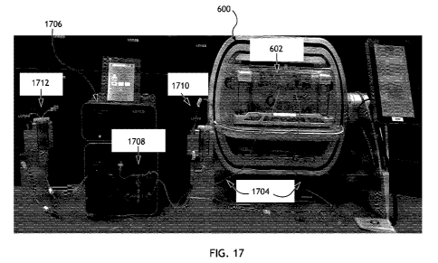
[0098] For example, as shown in FIG. 17, an activated immune cell culture
is
transferred, e.g., via connection tubing 1704, from cassette 602 of a cell
engineering
system 600 to an electroporation unit 1706. Electroporation unit 1706 suitably
includes an electroporation cartridge 1708, which holds the cell culture
during the
electroporation process. Following the electroporation process, the transduced
immune cell culture is transferred back, via connection tubing 1704, to cell
engineering
system 600. FIG. 17 also shows the use of two optional reservoirs 1710 and
1712,
which are used to hold the cell culture prior to and after electroporation, to
help in the
transfer between the cell engineering system and the electroporation unit as a
result
of different pump speeds, required pressures and flow rates. However, such
reservoirs can be removed and the cell culture transferred directly from cell
engineering system 1702 to electroporation unit 1706.
[0099] FIG. 18 shows a flow diagram of the cell culture 1) from the cell
engineering
system to a first reservoir, 2) to the electroporation unit, 3) to a second
reservoir, and
finally 4) back to cell engineering system.
[00100] In exemplary embodiments, as shown in FIGS. 17 and 18, electroporation
unit 1706 is located outside of cell engineering system 1702. In such
embodiments,
the transducing comprises transferring via a first sterile, closed connection
(e.g.,
connection tubing 1704), the activated immune cell culture from the first
chamber to
the electroporation unit, electroporating the activated immune cell culture
with the
vector, to produce the transduced immune cell culture, and transferring via a
second
sterile, closed connection (e.g., connection tubing 1704), the transduced
immune cell
culture to the second chamber of the cell engineering system.
[00101] It should also be understood that multiple, separate cell engineering
systems 600 (see, e.g., FIG. 2) can be connected to a single electroporation
unit, and
run in appropriate order such that cell cultures are transferred from the cell
engineering
systems, to the electroporation unit, and then back to the appropriate cell
engineering
system.
[00102] In other embodiments, electroporation unit 1706 can be located within
cell
engineering system 600, such that the entire system is a closed, self-
contained
Date Recue/Date Received 2022-12-19
32
system. Methods for including electroporation unit 1706 inside of cell
engineering
system 600 are known by those of ordinary skill in the art, and utilize
various
miniaturization strategies, etc.
[00103] The various methods described herein allow for the production of
genetically
modified immune cell cultures where the transduction efficiency of the method
is at
least 20% higher than the transduction efficiency of the method utilizing a
flexible, gas
permeable bag for cell culture. As described herein, and as demonstrated in
the
Examples, the methods utilizing a cell engineering system as described herein
are
superior to traditional methods which rely on the use of a flexible, gas
permeable bag
for carrying out the cell culture. In further embodiments, the transduction
efficiency of
the method is at least 10% higher than the transduction efficiency of the
method
utilizing a flexible, gas permeable bag for cell culture, more suitably at
least 20%
higher, at least 25% higher, at least 30% higher, at least 35% higher, or in
embodiments, at least 40% higher.
[00104] Suitably, the methods described herein produce at least 20% more
genetically modified immune cells than a method utilizing manual cell culture
with a
flexible, gas permeable bag. More suitably, the methods produce at least 25%
more
genetically modified immune cells, at least 30% more genetically modified
immune
cells, at least 35% more genetically modified immune cells, or at least 40%
more
genetically modified immune cells than a method utilizing manual cell culture
with a
flexible, gas permeable bag.
[00105] In exemplary embodiments, the cell engineering systems described
herein
comprise a plurality of chambers, and wherein each of steps of the various
method
described herein are performed in a different chamber of the plurality of
chambers of
the cell engineering system, each of the activation reagent, the vector, and
cell culture
medium are contained in a different chamber of the plurality of the chambers
prior to
starting the method, and wherein at least one of the plurality of chambers is
maintained
at a temperature for growing cells (e.g., at about 37 C) and at least one of
the plurality
of chambers is maintained at a refrigerated temperature (e.g., at about 4-8
C).
Date Recue/Date Received 2022-12-19
33
[00106] In some embodiments, the disclosure provides a method of producing
chimeric antigen receptor T cells, the method including: (a) activating a
peripheral
blood mononuclear cell culture, suitably with culture media comprising at
least one of
an anti-CD3 antibody and an anti-CD28 antibody, to produce an activated T cell
culture; (b) transducing the activated T cell culture with a lentiviral
vector, the vector
encoding a chimeric antigen receptor, to produce a transduced T cell culture;
(c)
expanding the transduced T cell culture to a pre-defined culture size; (d)
concentrating
the expanded T cell culture of (c) to a volume of about 20 mL to about 500 mL,
suitably
about 50 mL to about 200 mL; and (e) harvesting the concentrated T cell
culture of (d)
to produce a chimeric antigen receptor T (CAR T) cell culture, wherein the
activated T
cell culture is substantially undisturbed during steps (a) to (b); wherein the
method is
performed by a fully enclosed cell engineering system, suitably having
instructions
thereon for performing steps (a) to (e). Suitably steps (a) to (e) are
performed in one
or more chambers of the cell engineering system. As described herein, in
embodiments, the method produces at least 20% more CAR T cells than a method
utilizing a flexible, gas permeable bag for cell culture. In exemplary
embodiments, the
method produce at least 2 billion viable CAR T cells.
[00107] A chimeric antigen receptor T cell , or "CART cell," is a T cell that
is modified
with a chimeric antigen receptor (CAR) to more specifically target cancer
cells. In
general, a CAR includes three parts: the ectodomain, the transmembrane domain,
and
the endodomain. The ectodomain is the region of the receptor that is exposed
to
extracellular fluid and includes three parts: a signaling peptide, an antigen
recognition
region, and a spacer. The signaling peptide directs the nascent protein into
the
endoplasmic reticulum. In CAR, the signaling peptide is a single-chain
variable
fragment (scFv). The scFv includes a light chain (VL) and a heavy chain (VH)
of
immunoglobins connected with a short linker peptide. In some embodiments, the
linker
includes glycine and serine. In some embodiments, the linker includes
glutamate and
lysine.
[00108] The transmembrane domain of the CAR is a hydrophobic a-helix that
spans
the membrane. In some embodiments, the transmembrane domain of a CAR is a
CD28 transmembrane domain. In some embodiments, the CD28 transmembrane
Date Recue/Date Received 2022-12-19
34
domain results in a highly expressed CAR. In some embodiments, the
transmembrane
domain of a CAR is a CD3-transmembrane domain. In some embodiments, the CD3-
transmembrane domain results in a CAR that is incorporated into a native T
cell
receptor.
[00109] The endodomain of the CAR is generally considered the "functional" end
of
the receptor. After antigen recognition by the antigen recognition region of
the
ectodomain, the CARs cluster, and a signal is transmitted to the cell. In some
embodiments, the endodomain is a CD3- endodomain, which includes 3
immunoreceptor tyrosine-based activation motifs (ITAMs). In this case, the
ITAMs
transmit an activation signal to the T cell after antigen binding, triggering
a T cell
immune response.
[00110] During production of CAR T cells, T cells are removed from a human
subject, genetically altered, and re-introduced into a patient to attack the
cancer cells.
CAR T cells can be derived from either the patient's own blood (autologous),
or derived
from another healthy donor (allogenic). In general, CAR T cells are developed
to be
specific to the antigen expressed on a tumor that is not expressed in healthy
cells.
[00111] Activation of T Cells. In some embodiments, an immune cell culture
produced by the methods described herein is a CAR T cell culture. CAR T cells
can
be activated to form an activated T cell culture. In vivo, antigen-presenting
cells
(APCs), such as dendritic cells, act as the stimulus for T cell activation
through the
interaction of the T Cell Receptor (TCR) with the APC major histone
compatibility
complex (MHC). TCR associates with CD3, a T cell co-receptor that helps to
activate
both cytotoxic T cells (e.g., CD8+ naïve T cells) and T helper cells (e.g.,
CD4+ naïve
T cells). In general, T cell activation follows a two-signal model, requiring
stimulation
of the TCR/CD3 complex as well as a co-stimulatory receptor. Activation of T
cells is
further described in, e.g., Kochenderfer 2015; Kalos 2011.
[00112] Without the co-stimulatory signal, the cells are susceptible to anergy
and
become non-responsive. Thus, T cell co-stimulation may be important for T cell
proliferation, differentiation, and survival. Non-limiting examples of co-
stimulatory
molecules for T cells include CD28, which is a receptor for CD80 and CD86 on
the
Date Recue/Date Received 2022-12-19
35
membrane of APC; and CD278 or ICOS (Inducible T-cell COStimulator), which is a
CD28 superfamily molecule expressed on activated T cells that interacts with
ICOS-
L. Thus, in some embodiments, the co-stimulatory molecule is CD28. In other
embodiments, the co-stimulatory molecule is ICOS. In vivo, the co-stimulatory
signal
can be provided by the B7 molecules on the APC, which bind to the CD28
receptor on
T cells. B7 is a peripheral transmembrane protein found on activated APCs that
can
interact with CD28 or CD152 surface proteins on a T cell to produce a co-
stimulatory
signal. Thus, in some embodiments, the co-stimulatory molecule is B7. Co-
stimulatory
receptors are further described in, e.g., Lafferty 1975; Harding 1992;
Clavreul 2000;
Charron 2015; Fathman 2007; Greenwald 2005. Co-stimulation is further
described in,
e.g., Carpenter 2000; Andris 2004. B7 molecules are further described in,
e.g.,
Fleischer 1996; Schwartz 2003.
[00113] Various methods of activation are utilized in vitro to simulate T cell
activation. In embodiments, a T cell culture is activated with an activation
reagent. In
further embodiments, the activation reagent is an antigen-present cell (APC).
In still
further embodiments, the activation reagent is a dendritic cell. Dendritic
cells are APCs
that process antigen and present it on the cell surface to T cells. In some
embodiments, the activation reagent is co-cultured with the T cell culture. Co-
culturing
may require separate purification and culturing of a second cell type, which
may
increase labor requirements and sources of variability. Thus, in some
embodiments,
alternative activation methods are used.
[00114] In embodiments, the cells maintain stable contact with the activation
reagent
during the activating step. One way to maintain stable contact between the
cells and
the activation reagent is by preventing unnecessary or excessive movement of
the
cells. Accordingly, in embodiments, the cell culture is substantially
undisturbed during
the activation step. "Substantially undisturbed" means that the cells
generally remain
in the same area of the cell culture chamber, e.g., the bottom of the chamber,
while
the cell culture media is being changed. Cells may be disturbed if they are
moved
between different vessels, e.g., transferred from one culture flask to
another, or cells
may be disturbed if the vessel is flexible. A flexible vessel such as, e.g., a
culture bag,
can cause the cells to move when the bag is handled. As described herein, the
Date Recue/Date Received 2022-12-19
36
methods suitably utilize a cell culture chamber that is substantially flat,
and low, to
allow for uniform access of the cells to various nutrients and gases, also
allowing for
ease of removal of waste products and media transfer. The substantially flat
cell
culture chamber also allows for the cells to touch each other during various
stages of
the methods which can enhance cell growth and production of the desired cell
phenotype(s).
[00115] In some embodiments, the activation reagent is an antibody. In some
embodiments, the cell culture is activated with an antibody bound to a
surface,
including a polymer surface, including a beads In further embodiments, the one
or
more antibodies is an anti-CD3 and/or anti-CD28 antibody. For example, the
beads
may be magnetic beads such as, e.g., DYNABEADS, coated with anti-CD3 and anti-
CD28. The anti-CD3 and anti-CD28 beads can suitably provide the stimulatory
signals
to support T cell activation. See, e.g., Riddell 1990; Trickett 2003.
[00116] In other embodiments, the cell culture is activated with a soluble
antibody.
In further embodiments, the soluble antibody is a soluble anti-CD3 antibody.
OKT3 is
a murine monoclonal antibody of the immunoglobulin IgG2a isotype and targets
CD3.
Thus, in some embodiments, the soluble anti-CD3 antibody is OKT3. OKT3 is
further
described in, e.g., Dudley 2003; Manger 1985; Ceuppens 1985; Van Wauwe 1980;
Norman 1995.
[00117] In some embodiments, the co-stimulatory signal for T cell activation
is
provided by accessory cells. Accessory cells may include, for example, a Fc
receptor,
which enables cross-linking of the CD3 antibody with the TCR/CD3 complex on
the T
cell. In some embodiments, the cell culture is a mixed population of
peripheral blood
mononuclear cells (PBMCs). PBMC may include accessory cells capable of
supporting T cell activation. For example, CD28 co-stimulatory signals can be
provided
by the B7 molecules present on monocytes in the PBMC. Accordingly, in some
embodiments, the accessory cells include a monocyte or a monocyte-derived cell
(e.g., a dendritic cell). In additional embodiments, the accessory cells
include B7,
CD28, and/or ICOS. Accessory cells are further described in, e.g., Wolf 1994;
Chai
1997; Verwilghen 1991; Schwartz 1990; Ju 2003; Baroja 1989; Austyn 1987; Tax
1983.
Date Recue/Date Received 2022-12-19
37
[00118] As described herein, activation reagent may determine the phenotype of
the
CAR T cells produced, allowing for the promotion of a desired phenotype. In
some
embodiments, the activation reagent determines the ratio of T cell subsets,
i.e., CD4+
helper T cells and CD8+ cytotoxic T cells. The cytotoxic CD8+ T cells are
typically
responsible for killing cancer cells (i.e., the anti-tumor response), cells
that are infected
(e.g., with viruses), or cells that are damaged in other ways. CD4+ T cells
typically
produce cytokines and help to modulate the immune response, and in some cases
may support cell lysis. CD4+ cells activate APCs, which then primes naïve CD8+
T
cells for the anti-tumor response. Accordingly, in embodiments, the methods of
the
present disclosure further include producing CAR T cells of a pre-defined
phenotype
(i.e., promoting cells of a desired phenotype). The pre-defined phenotype may
be, for
example, a pre-defined ratio of CD8+ cells to CD4+ cells. In some embodiments,
the
ratio of CD8+ cells to CD4+ cells in a population of CAR T cells is about 1:1,
about
0.25:1, or about 0.5:1. In other embodiments, the ratio of CD8+ cells to CD4+
cells in
a population of CAR T cells is about 2:1, about 3:1, about 4:1, or about 5:1.
[00119] In embodiments, the activation reagent is removed from the activated T
cell
culture after the activation step. The activation reagent, e.g., an anti-CD3
antibody
and/or an anti-CD28 antibody may be present in the cell culture media. Thus,
in some
embodiments, the cell culture media containing the activation reagent, e.g.,
an anti-
CD3 antibody and/or an anti-CD28 antibody, is removed from the activated T
cell
culture after the activation step. In some embodiments, removal of the
activation
reagent includes removing a soluble antibody. For example, the soluble
antibody can
be removed by exchanging the cell culture media. The soluble antibody can also
be
removed by affinity methods specific for the soluble antibody. In other
embodiments,
removal of the activation reagent includes removing the bead containing the
antibody.
Bead removal can include, for example, filtering the beads or removal by a
magnet.
[00120] Transduction of Activated T Cells. In some embodiments, the
genetically
modified immune cell culture is an activated T cell culture that is transduced
with a
vector encoding a chimeric antigen receptor to produce a transduced T cell
culture. In
some embodiments, the transduction includes viral infection, transposons, mRNA
transfection, electroporation, or combinations thereof. In some embodiments,
the
Date Recue/Date Received 2022-12-19
38
transduction includes electroporation. Accordingly, in embodiments, the cell
engineering system includes an electroporation system or electroporation unit,
as
described herein. In additional embodiments, the transduction includes viral
infection.
The vector may be a viral vector, such as, for example, a lentiviral vector, a
gammaretroviral vector, an adeno-associated viral vector, or an adenoviral
vector. In
embodiments, the transduction includes introducing a viral vector into the
activated T
cells of the cell culture. In additional embodiments, the vector is delivered
as a viral
particle.
[00121] In some embodiments, the transduction step includes transducing the
activated T cells with a lentiviral vector, wherein the lentiviral vector is
introduced at a
multiplicity of infection (M01) of about 0.5 to about 50, about 0.5 to about
30, or about
0.5 to about 20. In some embodiments, the lentiviral vector is introduced at a
MOI of
about 0.5 to about 8. In some embodiments, the lentiviral vector is introduced
at a MOI
of about 0.5 to about 6. In some embodiments, the lentiviral vector is
introduced at a
MOI of about 0.5 to about 4. In some embodiments, the lentiviral vector is
introduced
at a MOI of about 0.5 to about 2. In some embodiments, the lentiviral vector
is
introduced at a MOI of about 0.6 to about 1.5. In some embodiments, the
lentiviral
vector is introduced at a MOI of about 0.7 to about 1.3. In some embodiments,
the
lentiviral vector is introduced at a MOI of about 0.8 to about 1.1. In some
embodiments,
the lentiviral vector is introduced at a MOI of about 0.5, about 0.6, about
0.7, about
0.8, about 0.9, about 1, about 1.1, about 1.2, about 1.3, about 1.4, about
1.5, about
1.6, about 1.7, about 1.8, about 1.9, or about 2.
[00122] In some embodiments, after the activation step, the cell culture media
from
the T cell culture is removed, and the media is then mixed with the vector
(e.g.,
lentiviral vector) and distributed uniformly to the cells. In some
embodiments, the
removed cell culture media is used to dilute and uniformly deliver the vector
to the
activated T cell culture. Uniform distribution and consequent homogeneous
exposure
of the vector (e.g., lentiviral vector) in the T cell culture improves
transduction
efficiency. In some embodiments, the volume of the cell culture is reduced
after
activation, and prior to addition of the vector. Volume reduction may enable a
higher
degree of cell-vector contact. In some embodiments, the activated T cell
culture is
Date Recue/Date Received 2022-12-19
39
substantially undisturbed during the transduction. In some embodiments, the
cell
culture is substantially undisturbed during the activation and transduction
steps, i.e.,
the cells remain generally in the same area of the chamber (e.g., the bottom
of the cell
culture chamber) while the activation reagent or the vector is being provided
to the
cells. This may facilitate uniform distribution and homogeneous exposure of
the
activation reagent and/or vector to the cells, and thus may improve the
activation
and/or transduction efficiency.
[00123] Accordingly, in some embodiments, the transduction efficiency of the
method using the cell engineering system is higher than the transduction
efficiency of
a method using a flexible, gas-permeable bag for cell culture. In some
embodiments,
the transduction efficiency of the method for automated production of CAR T
cells as
described herein has at least 10% greater, at least 15% greater, at least 20%
greater,
at least 25% greater, at least 30% greater, at least 35% greater, at least 40%
greater,
at least 45% greater, at least 50% greater, at least 55% greater, at least 60%
greater,
at least 65% greater, at least 70% greater, at least 75% greater, at least 80%
greater,
at least 85% greater, at least 90% greater, at least 95% greater, or at least
100%
greater than the transduction efficiency of a method utilizing a flexible, gas-
permeable
bag.
[00124] Expansion of Transduced T Cells. In some embodiments, the transduced
T cell culture (or other immune cell culture) is expanded to a pre-defined
culture size
(i.e., number of cells). The pre-defined culture size may include a sufficient
number of
cells suitable for clinical use, i.e., transfusion into a patient, research
and development
work, etc. In some embodiments, a clinical or therapeutic dose of CAR T cells
for
administration to a patient is about 105 cells, about 108 cells, about 107
cells, about
108 cells, about 109 cells, or about 1019 cells. In some embodiments, the
method
produces at least 1, at least 2, at least 3, at least 4, at least 5, at least
10, at least 15,
at least 20, at least 25, at least 30, at least 35, at least 40, at least 45,
at least 50, at
least 60, at least 70, at least 80, at least 90, or at least 100 clinical
doses of CART
cells. In some embodiments, the transduced T cell culture is expanded to a
total
volume of from about 0.1 L to about 5 L, from about 0.1 L to about 2 L, or
from about
0.2 L to about 2 L. In some embodiments, the transduced T cell culture is
expanded
Date Recue/Date Received 2022-12-19
40
to a total volume of about 0.1 L, about 0.2 L, about 0.3 L, about 0.4 L, about
0.5 L,
about 0.6 L, about 0.7 L, about 0.8 L, about 0.9 L or about 1.0 L. The volume
can also
be varied through the process, as required based on the stage of the cell
production
process. In some embodiments, the pre-defined culture size is input by a user
of the
cell engineering system. The user may input the pre-defined culture size as a
desired
cell count to be produced (e.g., 1010 CART cells), or, the pre-defined culture
size may
be input as a desired number of clinical or therapeutic doses to be produced
(e.g., 10
clinical or therapeutic doses of CAR T cells). In embodiments, the number of
CAR T
cells produced by the methods described herein is at least about 100 million
(i.e.,
1*106) cells, or at least about 300 million, at least about 500 million, at
least about 600
million, at least about 700 million, at least about 800 million, at least
about 900 million,
at least about 1 billion (i.e., 1*106), at least about 1.1 billion, at least
about 1.2 billion,
at least about 1.3 billion, at least about 1.4 billion, at least about 1.5
billion, at least
about 1.6 billion, at least about 1.7 billion, at least about 1.8 billion, at
least about 1.9
billion, at least about 2 billion (i.e., 2*106) cells, including at least
about 2.1 billion, at
least about 2.2 billion, at least about 2.3 billion, at least about 2.4
billion, at least about
2.5 billion, at least about 2.6 billion, at least about 2.7 billion, at least
about 2.8 billion,
at least about 2.9 billion, or at least about 3.0 billion CAR T cells.
[00125] In some embodiments, the expanding of the transduced T cell culture
includes at least one round of feeding, washing, monitoring, and selecting of
the
transduced T cell culture. Feeding the cell culture may include supplementing
the cell
culture with media and/or additional nutrients. Washing the cell culture may
include
removing spent media (i.e., media that is depleted of nutrients and/or
contains cellular
waste products) and replenishing the cell culture with fresh media. Monitoring
the cell
culture may include monitoring the temperature, pH, glucose, oxygen level,
carbon
dioxide level, and/or optical density of the cell culture. Selecting the cell
culture may
include selecting the cells with the desired characteristics such as, e.g.,
viability, type,
and/or morphology, and removing cells that do not have the desired
characteristics. In
some embodiments, the cell engineering system is configured to perform several
rounds of the feeding, washing, monitoring, and/or selecting of the transduced
T cell
culture to achieve the pre-defined culture size. In some embodiments, the cell
engineering system performs at least 2, at least 3, at least 4, at least 5, at
least 6, at
Date Recue/Date Received 2022-12-19
41
least 7, at least 8, at least 9, at least 10, at least 15, at least 20, at
least 25, at least
30, at least 35, at least 40, at least 45, at least 50, or at least 100 rounds
of the feeding,
washing, monitoring, and/or selecting of the transduced T cell culture to
achieve the
pre-defined culture size.
[00126] In embodiments, one or more of the feeding, washing and monitoring can
be removed, or the order of the events can be changed depending on the desired
cellular phenotype or number of cells, etc.
[00127] In embodiments, the monitoring includes monitoring with a temperature
sensor, a pH sensor, a glucose sensor, an oxygen sensor, a carbon dioxide
sensor,
and/or an optical density sensor. Accordingly, in some embodiments, the cell
engineering system includes one or more of a temperature sensor, a pH sensor,
a
glucose sensor, an oxygen sensor, a carbon dioxide sensor, and/or an optical
density
sensor. In additional embodiments, the cell engineering system is configured
to adjust
the temperature, pH, glucose, oxygen level, carbon dioxide level, and/or
optical
density of the cell culture, based on the pre-defined culture size. For
example, if the
cell engineering system detects that the current oxygen level of the cell
culture is too
low to achieve the necessary growth for a desired cell culture size, the cell
engineering
system will automatically increase the oxygen level of the cell culture by,
e.g.,
introducing oxygenated cell culture media, by replacing the cell culture media
with
oxygenated cell culture media, or by flowing the cell culture media through an
oxygenation component (i.e., a silicone tubing). In another example, if the
cell
engineering system detects that the current temperature of the cell culture is
too high
and that the cells are growing too rapidly (e.g., possible overcrowding of the
cells may
lead to undesirable characteristics), the cell engineering system will
automatically
decrease the temperature of the cell culture to maintain a steady growth rate
(or
exponential growth rate, as desired) of the cells. In still further
embodiments, the cell
engineering system automatically adjusts the schedule of cell feeding (i.e.,
providing
fresh media and/or nutrients to the cell culture) based on the cell growth
rate and/or
cell count, or other monitored factors, such as pH, oxygen, glucose, etc. The
cell
engineering system may be configured to store media (and other reagents, such
as
wash solutions, etc.) in a low-temperature chamber (e.g., 4 C or -20 C), and
to warm
Date Recue/Date Received 2022-12-19
42
the media in a room temperature chamber or a high-temperature chamber (e.g.,
25 C
or 37 C, respectively) before introducing the warmed media to the cell
culture.
[00128] In embodiments, the washing includes washing the cells by filtration
or
sedimentation. In some embodiments, the washing step does not require moving
the
cell culture vessels or flasks, i.e., the cells can be washed in the same cell
culture
vessel or flask. In further embodiments, the cells remain substantially
undisturbed
during the washing step. In embodiments, the selecting includes mixing the
cell culture
with one or more selection reagents. The selection reagent may be a bead,
e.g., a
magnetic bead, that is specific for the desired cell type, and the cells bound
to the
beads are then separated from non-bound cells, e.g., by passing through a
magnetic
chamber. For example, the selection bead includes an antibody specific for a
desired
cell type, e.g., an anti-CD8 antibody or an anti-CD4 antibody. Selection can
also be
performed by filtration to remove or select certain cell types based on size.
Cell
selection by plastic-adhesion (i.e. cells can start in one chamber, the
unwanted cells
stick to the surface and then the desired cells, that are still in suspension,
are moved
to another chamber), can also be utilized.
[00129] Suitably, during the expansion stage, the cells are not shaken or
rotated. It
has been determined that maintaining the cells in a relatively stationary
position during
expansion helps aid in overall cell production, as well as providing the
desired cellular
phenotype.
[00130] Concentration of the Expanded Culture. In some embodiments, the
expanded T cell culture (or other immune cell culture) is concentrated to a
pre-defined
concentration. The pre-defined concentration is of a volume that can be
suitably
infused into a patient. For example, the expanded T cell culture can be
concentrated
to about 1 ml, about 2 ml, about 5 ml, about 10 ml, about 15 ml, about 20 ml,
about 25
ml, about 30 ml, about 35 ml, about 40 ml, about 45 ml, about 50 ml, about 55
ml,
about 60 ml, about 65 ml, about 70 ml, about 75 ml, about 80 ml, about 85 ml,
about
90 ml, about 95 ml, or about 100 ml. In some embodiments, the concentration is
performed by centrifugation. In some embodiments, the concentration is
performed by
filtration. In some embodiments, the filtration is ultrafiltration and/or
diafiltration. In
some embodiments, the pre-defined concentration is input by a user of the cell
Date Recue/Date Received 2022-12-19
43
engineering system. In other embodiments, the pre-defined concentration is
determined by the cell engineering system, based on a different parameter
input by
the user, for example, the number or volume of clinical or therapeutic doses
to be
produced; or the number of cells to be produced. In some embodiments, the cell
engineering system automatically adjusts the volume or number of clinical or
therapeutic doses produced, based on the input parameters. In some
embodiments,
the cell engineering system automatically adjusts parameters of the
centrifugation
(e.g., speed, duration of centrifuging) or filtration (e.g., filter size,
volume, duration)
based on the pre-defined concentration.
[00131] Sedimentation based on the port position and design of the chamber can
also be utilized. That is, the fluid volume can be reduced in the chamber to
approximately 0.5 mL without removing the cells.
[00132] CAR T Cell Culture Harvest. In some embodiments, the concentrated T
cell culture (or other immune cell culture) is harvested, suitably to produce
a chimeric
antigen receptor (CAR) T cell culture. In some embodiments, the harvesting
includes
agitation, fluid flow, and washing of the CAR T cells. In some embodiments,
the
harvesting includes separation of the cells from undesired products, which
include,
e.g., cellular waste products, selection reagents such as beads (e.g., beads
containing
antibodies and/or beads used for separation of cells), or excess viral
vectors. In some
embodiments, the harvesting includes uniform distribution of the CART cells
into one
or more flasks, vials or vessels. In some embodiments, the harvesting includes
resuspending the CAR T cells in a formulation reagent, e.g., a solution that
stabilizes
the CAR T cells for long-term storage. In some embodiments, the harvesting
includes
cryopreservation of the CAR T cells.
[00133] Further Downstream Processes. In some embodiments, the CART cells
undergo further downstream processing prior to therapeutic use in a patient.
For
example, the cryopreserved CAR T cells may be filtered by sterile filtration
to remove
potential viral particle remnants. After sterile filtration, the CAR T cells
may undergo at
least one more concentration step before packaged in one or more vials,
flasks,
vessels, or containers. The packaged CAR T cells may be subjected to quality
assessment and/or quality control testing. In some embodiments, the CAR T
cells
Date Recue/Date Received 2022-12-19
44
undergo minimal downstream processing prior to administration to a patient.
For
example, in some embodiments, harvested CAR T cells are not cryopreserved but
transferred to the patient within a short time period after harvest. Avoiding
the
cryopreservation step may increase the viability of the cells.
[00134] Cell Engineering Systems. In some embodiments, the methods described
herein are performed by a fully enclosed cell engineering system 600 (see
FIGS. 6A,
6B), suitably having instructions thereon for performing the activating,
transducing,
expanding, concentrating, and harvesting steps. Cell engineering systems for
automated production of genetically modified immune cells, including CAR T
cells, are
described herein, and are also called automated cell engineering system,
COCOON,
or COCOON system throughout. For example, a user can provide a cell
engineering
system pre-filled with a cell culture and reagents (e.g., an activation
reagent, a vector,
cell culture media, nutrients, selection reagent, and the like) and parameters
for the
cell production (e.g., starting number of cells, type of media, type of
activation reagent,
type of vector, number of cells or doses to be produced, and the like), the
cell
engineering system is able to carry out the methods of producing genetically
modified
immune cell cultures, including CAR T cells, without further input from the
user. At the
end of the automated production process, the cell engineering system may alert
the
user (e.g., by playing an alert message or sending a mobile app alert) for
collecting
the produced cells. In some embodiments, the fully enclosed cell engineering
system
includes sterile cell culture chambers. In some embodiments, the fully
enclosed cell
engineering system minimizes contamination of the cell cultures by reducing
exposure
of the cell culture to non-sterile environments. In additional embodiments,
the fully
enclosed cell engineering system minimizes contamination of the cell cultures
by
reducing user handling of the cells.
[00135] As described herein, the cell engineering systems suitably include a
cassette 602. Thus, in embodiments, provided herein is a cassette for use in
an
automated cell engineering system. As used herein a "cassette" refers to a
largely
self-contained, removable and replaceable element of a cell engineering system
that
includes one or more chambers for carrying out the various elements of the
methods
Date Recue/Date Received 2022-12-19
45
described herein, and suitably also includes one or more of a cell media, an
activation
reagent, a vector, etc.
[00136] FIG. 6B shows an embodiments of a cassette 602 in accordance with
embodiments hereof. In embodiments, cassette 602 includes a low temperature
chamber 604, suitably for storage of a cell culture media, as well as a high
temperature
chamber 606, suitably for carrying out activation, transduction and/or
expansion of an
immune cell culture. Suitably, high temperature chamber 606 is separated from
low
temperature chamber 606 by a thermal barrier 1102 (see FIG. 11B). As used
herein
"low temperature chamber" refers to a chamber, suitably maintained below room
temperature, and more suitably from about 4 C to about 8 C, for maintenance of
cell
media, etc., at a refrigerated temperature. The low temperature chamber can
include
a bag or other holder for media, including about 1L, about 2L, about 3L, about
4L, or
about 5L of fluid. Additional media bags or other fluid sources can be
connected
externally to the cassette, and connected to the cassette via an access port.
[00137] As used herein "high temperature chamber" refers to chamber, suitably
maintained above room temperature, and more suitably maintained at a
temperature
to allow for cell proliferation and growth, i.e., between about 35-40 C, and
more
suitably about 37 C.
[00138] In embodiments, high temperature chamber 606 suitably includes a cell
culture chamber 610 (also called proliferation chamber or cell proliferation
chamber
throughout), as shown in FIG. 6D and FIG. 6E.
[00139] The cassettes further include one or more fluidics pathways connected
to
the cell culture chamber, wherein the fluidics pathways provide recirculation,
removal
of waste and homogenous gas exchange and distribution of nutrients to the cell
culture
chamber without disturbing cells within the cell culture chamber. Cassette 602
also
further includes one or more pumps 605, including peristaltic pumps, for
driving fluid
through the cassette, as described herein, as well as one or more valves 607,
for
controlling the flow through the various fluidic pathways.
Date Recue/Date Received 2022-12-19
46
[00140] In exemplary embodiments, as shown in FIG. 6D, cell culture chamber
610
is flat and non-flexible chamber (i.e., made of a substantially non-flexible
material such
as a plastic) that does not readily bend or flex. The use of a non-flexible
chamber
allows the cells to be maintained in a substantially undisturbed state. As
shown in
FIG. 6E, cell culture chamber 610 is oriented so as to allow the immune cell
culture to
spread across the bottom 612 of the cell culture chamber. As shown in FIG. 6E,
cell
culture chamber 610 is suitably maintained in a position that is parallel with
the floor
or table, maintaining the cell culture in an undisturbed state, allowing the
cell culture
to spread across a large area of the bottom 612 of the cell culture chamber.
In
embodiments, the overall thickness of cell culture chamber 610 (i.e., the
chamber
height 642) is low, on the order of about 0.5 cm to about 5 cm. Suitably, the
cell culture
chamber has a volume of between about 0.50 ml and about 300 ml, more suitably
between about 50 ml and about 200 ml, or the cell culture chamber has a volume
of
about 180 ml. The use of a low chamber height 642 (less than 5 cm, suitably
less than
4 cm, less than 3 cm, or less then 2 cm) allows for effective media and gas
exchange
in close proximity to the cells. Ports are configured to allow mixing via
recirculation of
the fluid without disturbing the cells. Larger height static vessels can
produce
concentration gradients, causing the area near the cells to be limited in
oxygen and
fresh nutrients. Through controlled flow dynamics, media exchanges can be
performed without cell disturbance. Media can be removed from the additional
chambers (no cells present) without risk of cell loss.
[00141] As described herein, in exemplary embodiments the cassette is pre-
filled
with one or more of a cell culture, a culture media, an activation reagent,
and/or a
vector, including any combination of these. In further embodiments, these
various
elements can be added later via suitable injection ports, etc.
[00142] As described herein, in embodiments, the cassettes suitably further
include
one or more of a pH sensor, a glucose sensor, an oxygen sensor, a carbon
dioxide
sensor, a lactic acid sensor/monitor, and/or an optical density sensor. The
cassettes
can also include one or more sampling ports and/or injection ports. Examples
of such
sampling ports and injection ports (1104) are illustrated in FIG. 11A., and
can include
an access port for connecting the cartridge to an external device, such as an
Date Recue/Date Received 2022-12-19
47
electroporation unit or an additional media source. FIG. 11A also shows the
location
of the cell input 1105, reagent warming bag 1106 which can be used to warm
cell
media, etc., as well as the culture zone 1107, which holds various components
for use
in the culture media, including for example, cell media, vectors, nutrients
and waste
products, etc.
[00143] FIG. 11B shows the COCOON cell engineering system with cassette 602
removed. Visible in FIG. 11B are components of the cell engineering system,
including
gas control seal 1120, warming zone 1121, actuators 1122, pivot 1123 for
rocking or
tilting the cell engineering system as desired, and low temperature zone 1124
for
holding low temperature chamber 606. Also shown is an exemplary user interface
1130, which can include a bar code reader, and the ability to receive using
inputs by
touch pad or other similar device. FIG. 11E shows an additional detailed view
of
cassette 602, including the location of secondary chamber 1150, which can be
used
is additional cell culture volume is required, as well as harvesting chamber
1152, which
can be used to recover the final cell culture as produced herein.
[00144] In exemplary embodiments, as shown in FIG. 6F, cell culture chamber
610
further comprises at least one of: a distal port 620 configured to allow for
the removal
of air bubbles from the cell culture chamber and/or as a recirculation port; a
medial
port 622 configured to function as a recirculation inlet port; and a proximal
port 624
configured to function as a drain port for cell removal.
[00145] In still further embodiments, provided herein is cassette 602 for use
in an
automated cell engineering system 600, comprising cell culture chamber 610 for
carrying out activation, transduction and/or expansion of an immune cell
culture having
a chamber volume that is configured to house an immune cell culture and a
satellite
volume 630 for increasing the working volume of the cell culture chamber by
providing
additional volume for media and other working fluids without housing the
immune cell
culture (i.e., satellite volume does not contain any cells). Suitably, the
satellite volume
is fluidly connected to the cell culture chamber such that media is exchanged
with the
culture chamber without disturbing the immune cell culture. In exemplary
embodiments, satellite volume is a bag, and in other embodiments, satellite
volume is
a non-yielding chamber. In embodiments, the satellite volume is between about
0.50
Date Recue/Date Received 2022-12-19
48
ml and about 300 ml, more suitably between about 50 ml and about 200 ml. FIG.
6D-
6E show the position of a satellite volume 630 in cassette 602.
[00146] FIG. 6G shows a schematic illustrating the connection between cell
culture
chamber 610, and satellite volume 630. Also illustrated in FIG. 6G are the
positioning
of various sensors (e.g., pH sensor 650, dissolved oxygen sensor 651), as well
as
sampling/sample ports 652 and various valves (control valves 653, bypass check
valves 654), as well as one or more fluidic pathways 640, suitably comprising
a
silicone-based tubing component, connecting the components. As described
herein,
use of a silicone-based tubing component allows oxygenation through the tubing
component to facilitate gas transfer and optimal oxygenation for the cell
culture. Also
show in FIG. 6G is the use of one or more hydrophobic filters 655 or
hydrophilic filters
656, in the flow path of the cassette, along with pump tube 657 and bag/valve
module
658.
[00147] FIG. 6H shows gas exchange data using the COCOON system, as
compared to traditional bags.
[00148] In embodiments, satellite volume 630 is further configured to allow
media
removal without loss of cells of the immune cell culture. That is, the media
exchange
between the satellite volume and the cell culture chamber is performed in such
a
manner that the cells are not disturbed and are not removed from the cell
culture
chamber.
[00149] In additional embodiments, as shown in FIG. 6G, cassette 602 suitably
further includes a crossflow reservoir 632 for holding additional media, etc.,
as needed.
Suitably, the crossflow reservoir has a volume of between about 0.50 ml and
about
300 ml, more suitably between about 100 ml and about 150 ml.
[00150] The cell engineering systems described herein suitably have three
relevant
volumes, the cell culture chamber volume, the working volume, and the total
volume.
Suitably, the working volume used in the cassette ranges from 180 mL to 460 mL
based on the process step, and can be increased up to about 500 mL, about 600
mL,
about 700 mL, about 800 mL, about 900 mL or about IL. In embodiments, the
cassette
Date Recue/Date Received 2022-12-19
49
can readily achieve 4*109 cells - 10*109 cells. The cell concentration during
the
process varies from 0.3*106 cells/ml to approximately 10*106 cells/ml. The
cells are
located in the cell culture chamber, but media is continuously recirculated
through
additional chambers (e.g., crossflow reservoir and satellite volume) to
increase the
working volume, as described herein.
[00151] As described herein, unlike a flexible bag, which changes shape when
filled
with liquid (e.g., a cell culture) and when picked up or moved, a
"substantially non-
yielding chamber" (e.g., an exemplary cell culture chamber 610) does not
change
shape (e.g., bend, curve, or deform) when filled with liquid, picked up, or
moved during
typical handling conditions. Thus, in some embodiments, a substantially non-
yielding
chamber allows cells to remain substantially in the same area of the chamber,
even
when the chamber is picked up or moved. A substantially non-yielding chamber
also
does not have the curvature associated with a bag. Thus, in some embodiments,
the
cells are distributed more uniformly in a substantially non-yielding chamber
compared
with a bag. In some embodiments, the activation reagent and/or the vector are
distributed more uniformly in a substantially non-yielding chamber compared
with a
bag.
[00152] In some embodiments, the cell engineering system includes a plurality
of
chambers. In further embodiments, each of the activating, transducing,
expanding,
concentrating, and harvesting steps of the method for cells described herein
is
performed in a different chamber of the plurality of chambers of the cell
engineering
system. In some embodiments, the cells are substantially undisturbed during
transfer
from one chamber to another. In other embodiments, the steps of the method are
performed in the same chamber of the cell engineering system, and the cell
engineering system automatically adjusts the chamber environment as needed for
each step of the method. Thus further allows for the cells to not be disturbed
during
the various steps.
[00153] In some embodiments, the cell engineering system has improved gas
exchange compared with a flexible, gas-permeable bag for cell culture. In some
embodiments, the cell engineering system includes gas exchange lines. The gas
exchange lines may be made from a gas-permeable material such as, e.g.,
silicone.
Date Recue/Date Received 2022-12-19
50
In some embodiments, the gas permeability coefficient of the gas exchange
lines is
higher than the permeability coefficient of the material used in the flexible,
gas-
permeable bag. In some embodiments, the cell engineering system recirculates
oxygen throughout the substantially non-yielding chamber during the cell
production
methods. Thus, in some embodiments, the oxygen level of a cell culture in the
cell
engineering system is higher than the oxygen level of a cell culture in a
flexible, gas-
permeable bag. Higher oxygen levels may be important in the cell culture
expansion
step, as increased oxygen levels may support increased cell growth and
proliferation.
[00154] In some embodiments, the cell engineering system continuously
recirculates media throughout the chambers without disturbing the cells. For
example,
the cell engineering system can continuously replenish nutrients, remove
waste, and
circulate released cytokines and dissolved gases through the chamber, while
the cells
remain in the same area of the chamber. The continuous circulation can improve
the
uniform distribution of positive factors and uniform removal of negative
factors, which
reduces localized effects that are caused by uneven distribution, without
disturbing the
cells.
[00155] In some embodiments, the cell engineering system provides carbon
dioxide
throughout the chamber during the cell production methods (including CAR T
production). CO2 can help to maintain a target pH in the cell culture, which
can be
important for cell growth and proliferation. In some embodiments, the cell
engineering
system monitors the CO2 level of the cell culture and adjusts the amount of
CO2
provided based on the measured CO2 level. For example, as the cell culture
increases,
there is a corresponding increase in the amount of CO2 produced by the cells,
and the
cell engineering system reduces the amount of CO2 provided. The desired CO2
level
of the cell culture may be defined by the user, for example, about 1%, about
2%, about
3%, about 4%, about 5%, about 6%, about 7%, about 8%, about 9%, or about 10%
CO2. Since the cell engineering system is constantly adjusting the amount of
CO2
provided based on the measured CO2 level of the cell culture, the cell
engineering
system is able to maintain a desired CO2 level throughout the production
process. The
amount of CO2 in a cell culture may also affect the pH of the culture, since
dissolved
CO2 generally acidifies a solution (through reacting with water to form
carbonic acid).
Date Recue/Date Received 2022-12-19
51
Thus, maintaining a steady CO2 level in the cell culture may result in a more
stable
pH. Accordingly, in embodiments, the pH level of the cell culture remains
substantially
constant during the production process. In further embodiments, the pH level
of the
transduced cell culture remains substantially constant during the expansion
step.
[00156] Yields from genetically modified immune cell production, including CAR
T
cell production, may be influenced by activation and transduction efficiency,
as well as
growth conditions of the cells. Activation efficiency can improve with more
stable
contact between the cells and the activation reagent. Movement of the cells
throughout
the culture vessel may lead to an uneven distribution of the cells, and thus
create
localized effects when activation reagent is added to the cell culture
chamber. In
contrast to a flexible culture bag, cells grown in a non-yielding chamber
remain
undisturbed during the activation process, which may contribute to a higher
activation
efficiency.
[00157] Improving activation efficiency may also lead to greater vector
transduction
efficiency. If cells are activated and are actively dividing, the vector
(e.g., lentiviral
vector) could integrate more effectively into the cells. Homogeneous
distribution of the
cells in the cell culture chamber 610 may facilitate homogeneous exposure of
the
vector to the cells, whereas cells may be unevenly distributed, and thus
receive
different vector exposure, in a flexible cell culture bag. Thus, in some
embodiments,
the transduction efficiency of the method for automated production of
genetically
modified immune cells, including CAR T cells as described herein, is at least
10%
greater, at least 15% greater, at least 20% greater, at least 25% greater, at
least 30%
greater, at least 35% greater, at least 40% greater, at least 45% greater, at
least 50%
greater, at least 55% greater, at least 60% greater, at least 65% greater, at
least 70%
greater, at least 75% greater, at least 80% greater, at least 85% greater, at
least 90%
greater, at least 95% greater, or at least 100% greater than the transduction
efficiency
of a method utilizing a flexible, gas-permeable bag.
[00158] Growth conditions of the cell cultures may also improve cell yields.
For
example, higher oxygen levels in the cell engineering system, facilitated by
highly gas-
permeable tubing and continuous recirculation of oxygen in the cell culture
chamber,
may increase cell proliferation. The ability of the cell engineering system to
constantly
Date Recue/Date Received 2022-12-19
52
monitor the state of the cell culture, and make adjustments accordingly, may
also be
advantageous. For example, the cell engineering system can monitor the CO2 02,
N2,
and/or pH level of the cell culture and adjust the level of CO2 02, or N2.
Nutrients can
also be provided in a timely and consistent manner and distributed uniformly
to the
cell culture. Thus, the automated methods for producing genetically modified
immune
cells, including CAR T cells, described herein advantageously results in
higher cell
yields compared with manual methods, or methods utilizing a flexible culture
bag.
Accordingly, in some embodiments, the method for automated production of
genetically modified immune cells, including CAR T cells utilizing a cell
engineering
system as described herein, produces at least 10% more, at least 15% more at
least
20% more, at least 25% more at least 30% more, at least 35% more at least 40%
more, at least 45% more at least 50% more, at least 55% more at least 60%
more, at
least 65% more at least 70% more, at least 75% more at least 80% more, at
least 85%
more at least 90% more, at least 95% more or at least 100% more cells than a
method
utilizing a flexible, gas permeable bag for cell culture. In embodiments, the
number of
cells produced by the methods described herein is at least about 2 billion
(i.e., 2*109)
cells, including at least about 2.1 billion, at least about 2.2 billion, at
least about 2.3
billion, at least about 2.4 billion, at least about 2.5 billion, at least
about 2.6 billion, at
least about 2.7 billion, at least about 2.8 billion, at least about 2.9
billion, or at least
about 3.0 billion cells.
Additional Exemplary Embodiments
[00159] Embodiment 1 is a method for automated production of a genetically
modified immune cell culture, the method comprising activating an immune cell
culture
with an activation reagent to produce an activated immune cell culture,
transducing
the activated immune cell culture with a vector, to produce a transduced
immune cell
culture, expanding the transduced immune cell culture, concentrating the
expanded
immune cell culture, and harvesting the concentrated immune cell culture to
produce
a genetically modified immune cell culture, further comprising washing either
or both
the expanded immune cell culture and the concentrated immune cell culture,
wherein
the steps are performed by a fully enclosed cell engineering system and the
steps are
optimized via a process to produce the genetically modified immune cell
culture.
Date Recue/Date Received 2022-12-19
53
[00160] Embodiment 2 includes the method of embodiment 1, wherein the process
is a self-adjusting process and includes monitoring with one or more of a
temperature
sensor, a pH sensor, a glucose sensor, an oxygen sensor, a carbon dioxide
sensor,
and an optical density sensor; and adjusting one or more of a temperature, a
pH level,
a glucose level, an oxygen level, a carbon dioxide level, and an optical
density of the
transduced T cell culture, based on the monitoring.
[00161] Embodiment 3 includes the method of embodiments 1-2, wherein the
method produces at least about 100 million viable genetically modified immune
cells
[00162] Embodiment 4 includes the method of embodiments 1-3, wherein the
method produces at least about 2 billion viable genetically modified immune
cells
[00163] Embodiment 5 includes the method of embodiments 1-4, wherein the
immune cell culture is a T cell culture.
[00164] Embodiment 6 includes the method of embodiment 5, wherein T cell
culture
is a chimeric antigen receptor T (CAR T) cell culture.
[00165] Embodiment 7 includes the method of embodiment 6, wherein the vector
encodes a chimeric antigen receptor.
[00166] Embodiment 8 includes the method of embodiments 1-7, wherein the
immune cell culture comprises peripheral blood mononuclear cells and/or
purified T
cells.
[00167] Embodiment 9 includes the method of embodiments 1-8, wherein the
immune cell culture comprises at least one accessory cell.
[00168] Embodiment 10 includes the method of embodiment 9, wherein the
accessory cell comprises a monocyte or a monocyte-derived cell.
[00169] Embodiment 11 includes the method of embodiment 9, wherein the
accessory cell comprises antigens for a T cell receptor, including CD28, CD40,
CD2,
CD4OL and/or ICOS.
Date Recue/Date Received 2022-12-19
54
[00170] Embodiment 12 includes the method of embodiments Ito 11, wherein the
activation reagent comprises an antibody or a dendritic cell.
[00171] Embodiment 13 includes the method of embodiment 12, wherein the
antibody is immobilized on a surface.
[00172] Embodiment 14 includes the method of embodiment 13, wherein the
surface
is a surface of a bead.
[00173] Embodiment 15 includes the method of embodiment 12, wherein the
antibody is a soluble antibody.
[00174] Embodiment 16 includes the method of embodiments 12-15, wherein the
antibody comprises at least one of an anti-CD3 antibody and an anti-CD28
antibody.
[00175] Embodiment 17 includes the method of embodiments 1-16, wherein the
transducing comprises viral infection, electroporation, membrane disruption,
or
combinations thereof.
[00176] Embodiment 18 includes the method of embodiments 1-17, wherein the
vector is a lentiviral vector or a retrovirus.
[00177] Embodiment 19 includes the method of embodiments 1-18, wherein the
transducing comprises mixing the vector in cell culture media and delivering
the vector
in the media uniformly to the activated immune cell culture.
[00178] Embodiment 20 includes the method of embodiments 1-19, wherein the
expanding comprises at least one or more of feeding, washing and monitoring of
the
transduced immune cell culture.
[00179] Embodiment 21 includes the method of embodiments 2-20, wherein the
oxygen level of the transduced immune cell culture is optimized for the immune
cell
culture.
[00180] Embodiment 22 includes the method of embodiments 1-21, wherein the
cell
engineering system recirculates cell culture media through an oxygenation
component
during one or more of steps (a) to (e).
Date Recue/Date Received 2022-12-19
55
[00181] Embodiment 23 includes the method of embodiments 1-22, wherein the
cell
engineering system recirculates nutrients, waste, released cytokines, and/or
dissolved
gasses during steps (a) to (e).
[00182] Embodiment 24 includes the method of embodiments 2-23, wherein the
carbon dioxide level provided by the cell engineering system decreases during
step
(c).
[00183] Embodiment 25 includes the method of embodiments 1-24, wherein the
cell
engineering system is configured to perform several rounds of one or more of
feeding,
washing, monitoring, and selecting of the transduced immune cell culture.
[00184] Embodiment 26 includes the method of embodiments 1-25, wherein the
concentrating comprises centrifugation, supernatant removal following
sedimentation,
or filtration.
[00185] Embodiment 27 includes the method of embodiment 26, wherein the
process further includes adjusting parameters of the centrifugation or
filtration.
[00186] Embodiment 28 includes the method of embodiments 1 to 27, wherein the
cell engineering system comprises a plurality of chambers, and wherein each of
steps
(a) to (e) is performed in a different chamber of the plurality of chambers of
the cell
engineering system.
[00187] Embodiment 29 includes the method of embodiments 1-28, further
comprising removing the activation reagent from the activated immune cell
culture
after step (a).
[00188] Embodiment 30 includes the method of embodiments 1-29, wherein the
cell
engineering system contains the cell culture of (a), the activation reagent,
the vector,
and cell culture medium prior to starting the method.
[00189] Embodiment 31 is a method for promoting a preferred phenotype of a
genetically modified immune cell culture, the method comprising activating an
immune
cell culture with an activation reagent to produce an activated immune cell
culture,
wherein the activation reagent and activating conditions promote the phenotype
of the
Date Recue/Date Received 2022-12-19
56
genetically modified immune cell culture, transducing the activated immune
cell culture
with a vector, to produce a transduced immune cell culture, expanding the
transduced
immune cell culture, concentrating the expanded immune cell culture; and
harvesting
the concentrated immune cell culture to produce a genetically modified immune
cell
culture, wherein the steps are performed by a fully enclosed, automated cell
engineering system.
[00190] Embodiment 32 includes the method of embodiment 31, wherein the
activation reagent comprises an antibody or a dendritic cell.
[00191] Embodiment 33 includes the method of embodiment 32, wherein the
antibody is immobilized on a surface.
[00192] Embodiment 34 includes the method of embodiments 33, wherein the
surface is a surface of a bead.
[00193] Embodiment 35 includes the method of embodiments 32, wherein the
antibody is a soluble antibody.
[00194] Embodiment 36 includes the method of embodiments 32-35, wherein the
antibody comprises at least one of an anti-CD3 antibody, an anti-CD28 antibody
and
an anti-CD2 antibody.
[00195] Embodiment 37 includes the method of embodiment 36, wherein the
soluble
antibody is OKT3.
[00196] Embodiment 38 includes the method of embodiments 31-37, wherein the
activating conditions provide a substantially undisturbed immune cell culture
allowing
for stable contact between the activation reagent and the immune cell culture.
[00197] Embodiment 39 includes the method of embodiments 31-38, wherein the
method produces at least about 100 million viable genetically modified immune
cells
[00198] Embodiment 40 includes the method of embodiment 39, wherein the
method produces at least about 2 billion viable genetically modified immune
cells
Date Recue/Date Received 2022-12-19
57
[00199] Embodiment 41 includes the method of embodiments 31-40, wherein the
immune cell culture is a T cell culture.
[00200] Embodiment 42 includes the method of embodiments 41, wherein T cell
culture is a chimeric antigen receptor T (CAR T) cell culture.
[00201] Embodiment 43 includes the method of embodiments 42, wherein the
vector
encodes a chimeric antigen receptor.
[00202] Embodiment 44 includes the method of embodiments 31-43, wherein the
immune cell culture comprises peripheral blood mononuclear cells and/or
purified T
cells.
[00203] Embodiment 45 includes the method of embodiments 31-44, wherein the
cell culture comprises at least one accessory cell.
[00204] Embodiment 46 includes the method of embodiment 45, wherein the
accessory cell comprises a monocyte or a monocyte-derived cell.
[00205] Embodiment 47 includes the method of embodiment 45, wherein the
accessory cell comprises antigens for a T cell receptor, including CD28, CD40,
CD2,
CD4OL and/or ICOS.
[00206] Embodiment 48 includes the method of embodiments 41-47, wherein the
phenotype of the T cell culture has a ratio of CD8+ cells to CD4+ of about
0.1:1 to
about 10:1.
[00207] Embodiment 49 includes the method of embodiments 31-48, wherein the
transducing comprises viral infection, electroporation, membrane disruption,
or
combinations thereof.
[00208] Embodiment 50 includes the method of embodiments 31-49, wherein the
vector is a lentiviral vector or a retrovirus.
[00209] Embodiment 51 includes the method of embodiments 31-50, wherein the
transducing comprises mixing the vector in cell culture media and delivering
the vector
in the media uniformly to the activated immune cell culture.
Date Recue/Date Received 2022-12-19
58
[00210] Embodiment 52 includes the method of embodiments 31-51, wherein the
expanding comprises at least one or more of feeding, washing and monitoring
the
transduced immune cell culture.
[00211] Embodiment 53 includes the method of embodiments 31-52, wherein an
oxygen level of the transduced immune cell culture is optimized for the
promoted
phenotype.
[00212] Embodiment 54 includes the method of embodiments 31-53, wherein the
cell engineering system recirculates cell culture media through an oxygenation
component during one or more of steps (a) to (e).
[00213] Embodiment 55 includes the method of embodiments 31-54, wherein the
cell engineering system recirculates nutrients, waste, released cytokines,
and/or
dissolved gasses during steps (a) to (e).
[00214] Embodiment 56 includes the method of embodiments 31-55, wherein a
carbon dioxide level provided by the cell engineering system decreases during
step
(c).
[00215] Embodiment 57 includes the method of embodiments 31-56, wherein the
cell engineering system is configured to perform several rounds of the
feeding,
washing, monitoring, and selecting of the transduced immune cell culture.
[00216] Embodiment 58 includes the method of embodiments 31-57, wherein the
concentrating comprises centrifugation, supernatant removal following
sedimentation,
or filtration.
[00217] Embodiment 59 includes the method of embodiments 31-58, wherein the
cell engineering system comprises a plurality of chambers, and wherein each of
steps
(a) to (e) is performed in a different chamber of the plurality of chambers of
the cell
engineering system.
[00218] Embodiment 60 includes the method of embodiments 31-59, further
comprising removing the activation reagent from the activated immune cell
culture
after step (a).
Date Recue/Date Received 2022-12-19
59
[00219] Embodiment 61 includes the method of embodiments 31-60, further
comprising removing the vector following the transducing in (b).
[00220] Embodiment 62 includes the method of embodiments 31-61, wherein the
cell engineering system contains the cell culture of (a), the activation
reagent, the
vector, and cell culture medium prior to starting the method.
[00221] Embodiment 63 is a method for automated production of a genetically
modified immune cell culture, the method comprising activating an immune cell
culture
with an activation reagent to produce an activated immune cell culture,
transducing
the activated immune cell culture with a vector, to produce a transduced
immune cell
culture, expanding the transduced immune cell culture, concentrating the
expanded
immune cell culture, and harvesting the concentrated immune cell culture to
produce
a genetically modified immune cell culture, wherein the steps are performed by
a fully
enclosed, automated cell engineering system, and wherein each of the steps are
performed with immune cell cultures having an optimized cell density
(cells/mL) and
an optimized cell confluency (cells/cm2).
[00222] Embodiment 64 includes the method of embodiment 63, wherein the
optimized cell density for (a) is about 0.05*106 cells/mL to about 60*106
cells/mL.
[00223] Embodiment 65 includes the method of embodiments 63 or claim 64,
wherein the optimized cell confluency for (a) is about 0.1*106 cells/cm2 to
about 60*106
cells/cm2.
[00224] Embodiment 66 includes the method of embodiments 63-65, wherein the
activation reagent comprises an antibody or a dendritic cell.
[00225] Embodiment 67 includes the method of embodiment 66, wherein the
antibody is immobilized on a surface.
[00226] Embodiment 68 includes the method of embodiment 67, wherein the
surface
is a surface of a bead.
[00227] Embodiment 69 includes the method of embodiment 66, wherein the
antibody is a soluble antibody.
Date Recue/Date Received 2022-12-19
60
[00228] Embodiment 70 includes the method of embodiments 66-69, wherein the
antibody comprises at least one of an anti-CD3 antibody and an anti-CD28
antibody.
[00229] Embodiment 71 includes the method of embodiments 63-70, wherein the
method produces at least about 100 million viable genetically modified immune
cells.
[00230] Embodiment 72 includes the method of embodiments 63-71, wherein the
method produces at least about 2 billion viable genetically modified immune
cells.
[00231] Embodiment 73 includes the method of embodiments 63-72, wherein the
immune cell culture is a T cell culture.
[00232] Embodiment 74 includes the method of embodiments 73, wherein T cell
culture is a chimeric antigen receptor T (CAR T) cell culture.
[00233] Embodiment 75 includes the method of embodiments 74, wherein the
vector
encodes a chimeric antigen receptor.
[00234] Embodiment 76 includes the method of embodiments 64-75, wherein the
immune cell culture comprises peripheral blood mononuclear cells and/or
purified T
cells.
[00235] Embodiment 77 includes the method of embodiments 64-76, wherein the
cell culture comprises at least one accessory cell.
[00236] Embodiment 78 includes the method of embodiment 77 wherein the
accessory cell comprises a monocyte.
[00237] Embodiment 79 includes the method of embodiment 77, wherein the
accessory cell comprises antigens for a T cell receptor, including CD28, CD40,
CD2,
CD4OL and/or ICOS.
[00238] Embodiment 80 includes the method of embodiments 63-79, wherein the
transducing comprises viral infection, electroporation, membrane disruption,
or
combinations thereof.
Date Recue/Date Received 2022-12-19
61
[00239] Embodiment 81 includes the method of embodiments 63-80, wherein the
vector is a lentiviral vector or a retrovirus.
[00240] Embodiment 82 includes the method of embodiments 63-81, wherein the
transducing comprises mixing the vector in cell culture media and delivering
the vector
in the media uniformly to the activated immune cell culture.
[00241] Embodiment 83 includes the method of embodiments 63-82, wherein the
expanding comprises at least one or more of feeding, washing, monitoring, and
selecting of the transduced immune cell culture.
[00242] Embodiment 84 includes the method of embodiments 63-83, wherein an
oxygen level of the transduced immune cell culture is optimized for the cell
density
and cell confluency.
[00243] Embodiment 85 includes the method of embodiments 63-84, wherein the
cell engineering system recirculates cell culture media through an oxygenation
component during one or more of steps (a) to (e).
[00244] Embodiment 86 includes the method of embodiment 85, wherein the oxygen
recirculation is provided by silicone tubing during steps (a) through (c).
[00245] Embodiment 87 includes the method of embodiments 63-86, wherein the
cell engineering system recirculates nutrients, waste, released cytokines,
and/or
dissolved gasses during steps (a) to (e).
[00246] Embodiment 88 includes the method of embodiments 63-87, wherein a
carbon dioxide level provided by the cell engineering system decreases during
step
(c).
[00247] Embodiment 89 includes the method of embodiments 63-88, wherein the
recirculation of nutrients, waste, released cytokines, and/or dissolved gasses
is
homogenously provided with the cells having a density of about 0.05*106
cells/mL to
about 60*106 cells/mL and a confluency of about 0.1*106 cells/cm2 to about
60*106
cells/cm2.
Date Recue/Date Received 2022-12-19
62
[00248] Embodiment 90 includes the method of embodiments 63-89, wherein the
cell engineering system is configured to perform several rounds of feeding,
washing,
monitoring, and selecting of the transduced immune cell culture.
[00249] Embodiment 91 includes the method of embodiments 63-90, wherein the
concentrating comprises centrifugation, supernatant removal following
sedimentation,
or filtration.
[00250] Embodiment 92 includes the method of embodiments 63-91, wherein the
cell engineering system comprises a plurality of chambers, and wherein each of
steps
(a) to (e) is performed in a different chamber of the plurality of chambers of
the cell
engineering system.
[00251] Embodiment 93 includes the method of embodiments 63-92, further
comprising removing the activation reagent from the activated immune cell
culture
after step (a).
[00252] Embodiment 94 includes the method of embodiments 63-93, further
comprising removing the vector following the transducing in (b).
[00253] Embodiment 95 includes the method of embodiments 63-94, wherein the
cell engineering system contains the cell culture of (a), the activation
reagent, the
vector, and cell culture medium prior to starting the method.
[00254] Embodiment 96 is a method for automated production of a genetically
modified immune cell culture, the method comprising activating an immune cell
culture
with an activation reagent to produce an activated immune cell culture,
transducing
the activated immune cell culture with a vector, to produce a transduced
immune cell
culture, expanding the transduced immune cell culture, wherein the transduced
cell
culture is not shaken during the expanding, concentrating the expanded immune
cell
culture, and harvesting the concentrated immune cell culture to produce a
genetically
modified immune cell culture, wherein the steps are performed by a fully
enclosed,
automated cell engineering system.
Date Recue/Date Received 2022-12-19
63
[00255] Embodiment 97 includes the method of embodiment 96, wherein the
activation reagent comprises an antibody or a dendritic cell.
[00256] Embodiment 98 includes the method of embodiment 97, wherein the
antibody is immobilized on a surface.
[00257] Embodiment 99 includes the method of embodiment 98, wherein the
surface
is a surface of a bead.
[00258] Embodiment 100 includes the method of embodiment 97, wherein the
antibody is a soluble antibody.
[00259] Embodiment 101 includes the method of embodiments 96-100, wherein the
antibody comprises at least one of an anti-CD3 antibody, an anti-CD28 antibody
and
an anti-CD2 antibody.
[00260] Embodiment 102 includes the method of embodiments 96-101, wherein the
method produces at least about 100 million viable genetically modified immune
cells
[00261] Embodiment 103 includes the method of embodiment 102, wherein the
method produces at least about 2 billion viable genetically modified immune
cells
[00262] Embodiment 104 includes the method of embodiments 96-103, wherein the
immune cell culture is a T cell culture.
[00263] Embodiment 105 includes the method of embodiment 104, wherein T cell
culture is a chimeric antigen receptor T (CAR T) cell culture.
[00264] Embodiment 106 includes the method of embodiment 105, wherein the
vector encodes a chimeric antigen receptor.
[00265] Embodiment 107 includes the method of embodiments 96-106, wherein the
immune cell culture comprises peripheral blood mononuclear cells and/or
purified T
cells.
[00266] Embodiment 108 includes the method of embodiments 96-107, wherein the
cell culture comprises at least one accessory cell.
Date Recue/Date Received 2022-12-19
64
[00267] Embodiment 109 includes the method of embodiment 108 wherein the
accessory cell comprises a monocyte or a monocyte-derived cell.
[00268] Embodiment 110 includes the method of embodiment 109, wherein the
accessory cell comprises antigens for a T cell receptor, including CD28, CD40,
CD2,
CD4OL and/or ICOS.
[00269] Embodiment 111 includes the method of embodiments 96-110, wherein the
transducing comprises viral infection, electroporation, membrane disruption,
or
combinations thereof.
[00270] Embodiment 112 includes the method of embodiments 96-111, wherein the
vector is a lentiviral vector or a retrovirus.
[00271] Embodiment 113 includes the method of embodiments 96-112, wherein the
transducing comprises mixing the vector in cell culture media and delivering
the vector
in the media uniformly to the activated immune cell culture.
[00272] Embodiment 114 includes the method of embodiments 96-113, wherein the
expanding comprises at least one or more of feeding, washing, monitoring, and
selecting of the transduced immune cell culture, without shaking the immune
cell
culture.
[00273] Embodiment 115 includes the method of embodiments 96-114, wherein an
oxygen level of the transduced immune cell culture is optimized for the immune
cell
culture.
[00274] Embodiment 116 includes the method of embodiments 96-115, wherein the
cell engineering system recirculates cell culture media through an oxygenation
component during one or more of steps (a) to (e).
[00275] Embodiment 117 includes the method of embodiments 96-116, wherein the
cell engineering system recirculates nutrients, waste, released cytokines,
and/or
dissolved gasses.
Date Recue/Date Received 2022-12-19
65
[00276] Embodiment 118 includes the method of embodiments 96-117, wherein a
carbon dioxide level provided by the cell engineering system decreases during
step
(c).
[00277] Embodiment 119 includes the method of embodiments 96-118, wherein the
cell engineering system is configured to perform several rounds of feeding,
washing,
monitoring, and selecting of the transduced immune cell culture.
[00278] Embodiment 120 includes the method of embodiments 96-119, wherein the
concentrating comprises centrifugation, supernatant removal following
sedimentation,
or filtration.
[00279] Embodiment 121 includes the method of embodiments 96-120, wherein the
cell engineering system comprises a plurality of chambers, and wherein each of
steps
(a) to (e) is performed in a different chamber of the plurality of chambers of
the cell
engineering system.
[00280] Embodiment 122 includes the method of embodiments 96-121, further
comprising removing the activation reagent from the activated immune cell
culture
after step (a).
[00281] Embodiment 123 includes the method of embodiments 96-122, further
comprising removing the vector following the transducing in (b).
[00282] Embodiment 124 includes the method of embodiments 96-123, wherein the
cell engineering system contains the cell culture of (a), the activation
reagent, the
vector, and cell culture medium prior to starting the method.
[00283] Embodiment 125 is a method for automated production of a genetically
modified immune cell culture, the method performed by a cell engineering
system,
comprising, activating an immune cell culture with an activation reagent to
produce an
activated immune cell culture in a first chamber of the cell engineering
system,
transducing the activated immune cell culture, the transducing comprising,
transferring
the activated immune cell culture from the first chamber to an electroporation
unit,
electroporating the activated immune cell culture with a vector, to produce a
Date Recue/Date Received 2022-12-19
66
transduced immune cell culture, transferring the transduced immune cell
culture to a
second chamber of the cell engineering system, expanding the transduced immune
cell culture, concentrating the expanded immune cell culture; and harvesting
the
concentrated immune cell culture to produce a genetically modified cell
culture.
[00284] Embodiment 126 includes the method of embodiment 125, wherein the
transducing comprises transferring via a first sterile, closed connection, the
activated
immune cell culture from the first chamber to the electroporation unit,
electroporating
the activated immune cell culture with the vector, to produce the transduced
immune
cell culture, transferring via a second sterile, closed connection, the
transduced
immune cell culture to the second chamber of the cell engineering system.
[00285] Embodiment 127 includes the method of embodiment 126, wherein the
electroporation unit is located outside of the cell engineering system.
[00286] Embodiment 128 includes the method of embodiments 125-127, wherein
the method produces at least about 100 million viable genetically modified
immune
cells.
[00287] Embodiment 129 includes the method of embodiment 128, wherein the
method produces at least about 2 billion viable genetically modified immune
cells.
[00288] Embodiment 130 includes the method of embodiments 125-129, wherein
the immune cell culture is a T cell culture.
[00289] Embodiment 131 includes the method of embodiment 130, wherein T cell
culture is a chimeric antigen receptor T (CAR T) cell culture.
[00290] Embodiment 132 includes the method of embodiment 131, wherein the
vector encodes a chimeric antigen receptor.
[00291] Embodiment 133 includes the method of embodiments 125-132, wherein
the immune cell culture comprises peripheral blood mononuclear cells and/or
purified
T cells.
Date Recue/Date Received 2022-12-19
67
[00292] Embodiment 134 includes the method of embodiments 125-132, wherein
the cell culture comprises at least one accessory cell.
[00293] Embodiment 135 includes the method of embodiment 134, wherein the
accessory cell comprises a monocyte or a monocyte-derived cell.
[00294] Embodiment 136 includes the method of embodiments 134, wherein the
accessory cell comprises antigens for a T cell receptor, including CD28, CD40,
CD4OL
and/or ICOS.
[00295] Embodiment 137 includes the method of embodiments 125-136, wherein
the activation reagent comprises an antibody or a dendritic cell.
[00296] Embodiment 138 includes the method of embodiments 137, wherein the
antibody is immobilized on a surface.
[00297] Embodiment 139 includes the method of embodiments 138, wherein the
surface is a surface of a bead.
[00298] Embodiment 140 includes the method of embodiments 137, wherein the
antibody is a soluble antibody.
[00299] Embodiment 141 includes the method of embodiments 138-140, wherein
the antibody comprises at least one of an anti-CD3 antibody, an anti-CD28
antibody
and an anti-CD2 antibody.
[00300] Embodiment 142 includes the method of embodiments 125-141, wherein
the vector is a lentiviral vector or a retrovirus.
[00301] Embodiment 143 includes the method of embodiments 125-142, wherein
the expanding comprises at least one or more of feeding, washing, monitoring,
and
selecting of the transduced immune cell culture.
[00302] Embodiment 144 includes the method of embodiments 125-143, wherein an
oxygen level of the transduced immune cell culture is optimized for the immune
cell
culture.
Date Recue/Date Received 2022-12-19
68
[00303] Embodiment 145 includes the method of embodiments 125-144, wherein
the cell engineering system recirculates cell culture media through an
oxygenation
component during one or more of steps (a) to (e).
[00304] Embodiment 146 includes the method of embodiments 125-145, wherein
the cell engineering system recirculates nutrients, waste, released cytokines,
and/or
dissolved gasses during steps (a) to (e).
[00305] Embodiment 147 includes the method of embodiments 125-146, wherein a
carbon dioxide level provided by the cell engineering system decreases during
step
(c).
[00306] Embodiment 148 includes the method of embodiments 125-147, wherein
the cell engineering system is configured to perform several rounds of
feeding,
washing, monitoring, and selecting of the transduced immune cell culture.
[00307] Embodiment 149 includes the method of embodiments 125-148, wherein
the concentrating comprises centrifugation, supernatant removal following
sedimentation, or filtration.
[00308] Embodiment 150 includes the method of embodiments 125-149, wherein
the cell engineering system comprises a plurality of chambers, and wherein
each of
steps (a) to (e) is performed in a different chamber of the plurality of
chambers of the
cell engineering system.
[00309] Embodiment 151 includes the method of embodiments 125-150, further
comprising removing the activation reagent from the activated immune cell
culture
after step (a).
[00310] Embodiment 152 includes the method of embodiments 125-151, further
comprising removing the vector following the transducing in (b).
[00311] Embodiment 153 includes the method of embodiments 125-152, wherein
the cell engineering system contains the cell culture of (a), the activation
reagent, the
vector, and cell culture medium prior to starting the method.
Date Recue/Date Received 2022-12-19
69
[00312] Embodiment 154 includes the method of embodiments 1 to 153, wherein
transduction efficiency in step (c) of the method is at least 20% higher than
the
transduction efficiency of the method utilizing a flexible, gas permeable bag
for cell
culture.
[00313] Embodiment 155 includes the method of embodiments 1 to 154, wherein
the method produces at least 20% more genetically modified immune cells than a
method utilizing manual cell culture with a flexible, gas permeable bag.
[00314] Embodiment 156 includes the method of embodiments 1 to 155, wherein
the cell engineering system comprises a plurality of chambers, and wherein
each of
steps (a) to (e) is performed in a different chamber of the plurality of
chambers of the
cell engineering system, each of (a), the activation reagent, the vector, and
cell culture
medium are contained in a different chamber of the plurality of the chambers
prior to
starting the method, and wherein at least one of the plurality of chambers is
maintained
at a temperature for growing cells and at least one of the plurality of
chambers is
maintained at a refrigerated temperature.
[00315] Embodiment 157 is a cassette for use in an automated cell engineering
system, comprising a low temperature chamber, for storage of a cell culture
media, a
high temperature chamber for carrying out activation, transduction and
expansion of
an immune cell culture, wherein the high temperature chamber is separated from
the
low temperature chamber, by a thermal barrier, the high temperature chamber
including a cell culture chamber; and one or more fluidics pathways connected
to the
cell culture chamber, wherein the fluidics pathways provide recirculation,
removal of
waste and homogenous gas exchange and distribution of nutrients to the cell
culture
chamber without disturbing cells within the cell culture chamber.
[00316] Embodiment 158 includes the cassette of embodiment 157, wherein the
cell
culture chamber is flat and non-flexible chamber, having a low chamber height.
[00317] Embodiment 159 includes the cassette of embodiments 157 or 158,
wherein
the cell culture chamber is oriented so as to allow the immune cell culture to
spread
across the bottom of the cell culture chamber.
Date Recue/Date Received 2022-12-19
70
[00318] Embodiment 160 includes the cassette of embodiments 157-159, wherein
the cassette is pre-filled with cell culture, culture media, activation
reagent, and a
vector.
[00319] Embodiment 161 includes the cassette of embodiments 157-160, further
comprising one or more of a pH sensor, a glucose sensor, an oxygen sensor, a
carbon
dioxide sensor, and/or an optical density sensor.
[00320] Embodiment 162 includes the cassette of embodiments 157-161, further
comprising one or more sampling ports and/or injection ports.
[00321] Embodiment 163 includes the cassette of embodiments 157-162, wherein
the cell culture chamber further comprises at least one of a distal port
configured to
allow for the removal of air bubbles from the cell culture chamber and/or as a
recirculation port; a medial port configured to function as a recirculation
inlet port; and
a proximal port configured to function as a drain port for cell removal.
[00322] Embodiment 164 includes the cassette of embodiments 157-163, further
comprising an access port for connecting the cartridge to an external device.
[00323] Embodiment 165 includes the cassette of embodiment 164, wherein the
external device includes an electroporation unit or an additional media
source.
[00324] Embodiment 166 is cassette for use in an automated cell engineering
system, comprising a cell culture chamber for carrying out activation,
transduction
and/or expansion of an immune cell culture having a chamber volume that is
configured to house an immune cell culture, a satellite volume for increasing
the
working volume of the chamber by providing additional volume for media and
other
working fluids without housing the immune cell culture, wherein the satellite
volume is
fluidly connected to the cell culture chamber via one or more fluidics
pathways such
that media is exchanged with the culture chamber without disturbing the immune
cell
culture.
[00325] Embodiment 167 includes the cassette of embodiment 166, wherein the
satellite volume is a bag.
Date Recue/Date Received 2022-12-19
71
[00326] Embodiment 168 includes the cassette of embodiment 166, wherein the
satellite volume is a non-yielding chamber.
[00327] Embodiment 169 includes the cassette of embodiments 166-168, wherein
the satellite volume is further configured to allow media removal without loss
of cells
of the immune cell culture.
[00328] Embodiment 170 includes the cassette of embodiments 166-169, further
comprising a crossflow reservoir.
[00329] Embodiment 171 includes the cassette of embodiments 166-170, wherein
the cell culture chamber has a volume of between about 0.50 ml and about 300
ml.
[00330] Embodiment 172 includes the cassette of embodiment 171, wherein the
cell
culture chamber has a volume of between about 50 ml and about 200 ml.
[00331] Embodiment 173 includes the cassette of embodiment 172, wherein the
cell
culture chamber has a volume of about 180 ml.
[00332] Embodiment 174 includes the cassette of embodiments 166-173, wherein
the satellite volume is between about 0.50 ml and about 300 ml.
[00333] Embodiment 175 includes the cassette of embodiment 174, wherein the
satellite volume is between about 150 ml and about 200 ml.
[00334] Embodiment 176 includes the cassette of embodiments 166-175, wherein
the crossflow reservoir has a volume of between about 0.50 ml and about 300
ml.
[00335] Embodiment 177 includes the cassette of embodiments 176, wherein the
crossflow reservoir has a volume of between about 100 ml and about 150 ml.
[00336] Embodiment 178 includes the cassette of embodiments 166-177, wherein
the working volume is about 180 mL to about 1L.
[00337] Embodiment 179 includes the cassette of embodiment 178, wherein the
working volume is about 180 mL to about 460 mL.
Date Recue/Date Received 2022-12-19
72
[00338] Embodiment 180 includes the cassette of embodiments 157-179, wherein
one or more of the fluidic pathways comprise a silicon-based tubing component
that
allows oxygenation through the tubing component.
Date Recue/Date Received 2022-12-19
73
EXAMPLES
Example 1 ¨ Automated Production of CAR T Cells Using the COCOON
System
[00339] In this Example, GFP and HER-2 lentivirus were used to transduce T
cells
using the following process parameters: starting inoculation of 60 million
peripheral
blood mononuclear cells (PBMC), CD3/CD28 activation, IL-2 and IL-7 were
supplemented into T-cell growth media for culture expansion. Single-use
sensors in
the disposable cassette were used to monitor temperature, pH and optical
density
(OD) in real time. The multiple cassette chambers that are connected via
fluidic
channels enabled automated feeding and addition of process components. Some of
the chambers are temperature controlled at 4 C for media and reagent storage,
while
others included elements for warming, mixing, washing, and concentrating
cells,
allowing for a fully enclosed process. The in-process samples were drawn for
cell
counts and viability. At the end of the harvesting process, FACS analysis was
performed with the following panel: CD4, CD8, NGFR, IFN-y, TNF-a, etc. An
overview
of the COCOON System used in this Example is shown in FIG. 6. FIG. 6A shows
the
COCOON system in the closed configuration along with an external user control
display, which can be used to adjust parameters or monitor the cell culture.
Sterile,
single-use cell culture "cassettes" can be loaded into the COCOON (FIG. 6C).
As
shown in a detailed view of the cassette (FIG. 6B), each cassette includes an
upper
chamber maintained at 37 C for growing cells, and a lower chamber maintained
at
4 C for storing media, viral vector, and other temperature-sensitive reagents.
The
cassette is configured such that fluids can be exchanged through the interior
fluidics
pathways, and also pumped into or out of the cassette. Sensors installed in
the
cassette can monitor, e.g., the pH and optical density of the cell culture.
[00340] Results are shown in FIGS. 7-10. FIG. 7A, 7B, and 7C show,
respectively,
the average harvest yields, average harvest viability, and average
transduction
efficiency for GFP transduction using the automated COCOON System, compared
with manual manipulation and expansion of the cells using the G-REX
(WilsonWolf)
cell culture plates as control. The G-REX plates have gas-permeable bottoms,
and
Date Recue/Date Received 2022-12-19
74
media exchange is typically performed by the user every 4 to 5 days when using
the
G-REX.
[00341] FIG. 8A and 8B show, respectively, the viable cells and the viability
and
transduction efficiency for HER-2 CAR-T transduction. In 10-day cultures, the
HER-2
CAR-T cells reached approximately 2.2 billion with viability of 97% and
transduction
of 65% (n=4) in the COCOON system.
[00342] Performance of the automated COCOON System was also compared with
manual manipulation and growth of cells using the PERMALIFE Cell Culture Bag
(OriGen) as a control. The PERMALIFE Bag is a sealable and gas-permeable cell
culture bag made of inert fluorinated ethylene propylene (FEP), with valves to
facilitate
cell feeding and harvest by the user. FIG. 9A indicates the relative T Cell
purity level
using the COCOON System compared with the PERMALIFE Bag, as assessed by the
percentage of CD3+ cells. FIG. 9B shows a greater percentage of CD8+ cells
cultured
in the COCOON System compared with the PERMALIFE Bag control. FIG. 9C and 9D
show that transfected cells produce TNF-a and INF-y, respectively.
[00343] FIG. 10A and 10B show effective and specific killing of target tumor
cells by
CAR T cells cultured in the COCOON System and the PERMALIFE Bag, respectively.
[00344] In conclusion, the COCOON System, a fully enclosed cell engineering
system, is a viable solution to translate the labor-intensive CART process
into a fully
automated and highly controlled system, thus allowing scalability, high yield,
reduction
of manufacturing cost, and gaining better process control to yield high
quality CAR-T
cells.
Example 2 - Comparison of Activation Methods in the COCOON System
[00345] This Example compares cell culture performance using different methods
of
activation in the clinical scale production of CAR T cells in the COCOON
automated
manufacturing system and a PERMALIFE Bag.
[00346] Magnetic anti-CD3/anti-CD28 DYNABEAD activator beads may be used to
activate T cells. These beads provide the two necessary stimulatory signals to
support
Date Recue/Date Received 2022-12-19
75
effective T cell activation. Another method of activating naïve T cells may
utilize a
soluble anti-CD3 antibody (OKT3). OKT3 is a monoclonal IgG2a antibody,
originally
used as an immunosuppressant. The costimulatory signals can be provided by
accessory cells. Initiating T cell culture from a mixed population of
peripheral blood
mononuclear cells (PBMC) can provide the necessary accessory cells to support
T
cell activation when using OKT3.
[00347] As OKT3 and DYNABEADS utilize distinct activation mechanisms, the
selection of one method over the other could influence the final product
characteristics;
specifically, the ratio of T cell subsets, CD4+ helper T cells and CD8+
cytotoxic T cells.
The cytotoxic CD8 T cells are responsible for the anti-tumor response. CD4
cells
produce cytokines and help to regulate the immune response. It has been
demonstrated that CD4 cells also support cell lysis, although the killing is
delayed
compared to CD8 cells. CD4 cells signal to APCs, thus activating APCs and
subsequently priming naive CD8 T cells. The ideal target ratio of CD8 to CD4
cells is
not well understood due to limited clinical data. Studies have shown that a
combination
of CD8 and CD4 cells are preferred over the delivery of CD8 cells alone (see,
e.g.,
Church 2014; Feldmann 2012; Reusch 2015).
[00348] There are advantages and disadvantages of both methods of in vitro
activation. Antibody-bound beads offer consistency and ensure stable
simultaneous
activation of the TCR/CD3 complex as well as the CD28 co-stimulatory pathway.
A
major disadvantage of the bead approach is the high cost associated with this
product.
The beads must also be effectively removed from culture before implantation.
OKT3
offers a low-cost option for activating T cells. The major disadvantages
associated with
the soluble anti-CD3 approach are the dependency on accessory cells and
sensitivity
to the culturing environment. Patient samples may have highly variable
accessory cells
and negative interactions that might functionally inactivate the T cells after
previous
stimulation. To understand the impact of each method of activation on the
growth,
phenotype and functionality of the cells, T cells activated by DYNABEADS and
OKT3
were cultured in a clinical-scale automation platform.
[00349] COCOON provides the environmental control of gases and temperatures.
This includes a 37 C zone as well as a linked refrigerated zone. There is no
fluid
Date Recue/Date Received 2022-12-19
76
contact between the COCOON and the Cassette, minimizing the required cleaning
between runs. All reagents can be loaded into the Cassette on the day of
seeding and
stored in the refrigerated zone of the COCOON until needed. Fluid is warmed to
37 C
before delivery to the cells. Due to the stability of lentivirus, this can be
thawed on the
day of transduction and delivered into the Cassette via sterile connectors.
Gas
exchange (oxygenation and CO2 buffering) is achieved via recirculation of the
culture
fluid through gas permeable tubing. Embedded biosensors provided real-time
data on
dissolved oxygen and pH. As the T cells require stable contact with other
cells or the
activating agent, media exchanges, washing and recirculation for gas exchange
can
be performed via perfusion without disturbing the cells. Rocking can be used
to
facilitate efficient harvesting.
Methods
[00350] Cell Culture. Peripheral blood mononuclear cells (PBMC) (Lonza) were
thawed with DNase (Sigma) and allowed to recover overnight at 37 C at a
density of
<2 x 10⁶ cells/mL. Cell counting was performed using the NUCLEOCOUNTER 200
with the Blood Assay protocol, including Solution 17 (Chemometec). A third-
generation
lentiviral vector, encoded with a low affinity nerve growth factor receptor
(NGFR) as a
marker of transduction, was used to transduce the cells. This lentivirus was
manufactured at Lonza's cGMP virus manufacturing facility (Houston, Texas)
based
on a protocol and primers originating from the Bramson Lab at McMaster
University
(Hamilton, Canada). A multiplicity of infection (M01) of 1 was used in all
conditions.
The viral titer was determined by using HEK293TM cells and detection of NGFR
using
flow cytometry. Activation media consisted of X-VIVO 15 (Lonza) supplemented
with
22 IU/mL IL-2 (Cedarlane) and 1% penicillin-streptomycin (Sigma). In
conditions
activated with soluble anti-CD3, OKT3 (Biolegend) was added to the activation
media
fora final concentration of 50 ng/mL. In conditions activated with DYNABEADS,
a ratio
of 1:1 beads to cells was added to the activation media. Expansion media
consisted
of X-VIVO 15 (Lonza) supplemented with 29 IU/mL IL-2 (Cedarlane), 5% human
serum from male AB plasma (Sigma), 1% GLUTAMAX (Thermo Fisher) and 1%
penicillin-streptomycin (Sigma).
Date Recue/Date Received 2022-12-19
77
[00351] Automated CAR T Cell Production. On Day 0, 60 x 106 PBMC were
loaded into the input bag of the Cassette. In conditions activated using anti-
CD3/anti-
CD28 beads, 60 x 106 anti-CD3/anti-CD28 DYNABEADS (ThermoFisher) were also
added to the input bags for a ratio of 1:1 beads to cells. The input bag was
connected
to the Cassette and brought to the COCOON (Octane Biotech Inc.). Following
operator
sign-in, the Cassette was loaded into the COCOON. On Day 1, virus (Lonza
Houston)
was thawed and then transferred to the cell culture chamber via the Cassette
access
port at a MOI of 1. Prior to delivery of the virus to the cells, activation
media was used
to dilute the media. The activation media was removed from the culture chamber
and
returned with the virus without disturbing the cells. The total working volume
was
increased on Day 4 with the addition of expansion media. Partial media
exchanges
were performed with expansion media on Day 6 and Day 8. Following the
expansion
steps, the COCOON decreased the final volume to less than 100 mL before the
cells
were removed. Throughout the culture, data was continuously collected by the
COCOON. This included every pump and actuator step, each time the door was
opened and closed and so forth. Comprehensive sensor data was collected
including
thermal values, gas concentrations, fluid pH and dissolved oxygen. The
operator was
able to remotely monitor the status of the culture using a phone or external
computer.
[00352] Manual CAR T Cell Production. Manual production of CAR T cells was
performed in parallel to COCOON in PERMALIFE cell culture bags. On Day 0, 60 x
106 PBMC were seeded in activation media at 0.27 x 10⁶ cells/mL on Day 0.
These
cultures utilized the same donor cells as well as the same media for
activation and
expansion as the automated cultures. Cultures were initiated in PERMALIFE bags
(PL240, Origen) and were transferred to larger PERMALIFE bags on Day 6 (PL325,
Origen) as the cells expanded. Cells were expanded into PL240 and PL325 bags
on
Day 8 as the volume increased. On Day 1, lentivirus was added to the bags at a
MOI
of 1. Cells were fed with an equivalent volume as COCOON cultures; however,
unlike
the COCOON conditions, no media was sent to waste. The volume used maintained
the cultures at less than 2 x 10⁶ cells/mL. On Day 10, culture volumes were
obtained
by mass and a sample of the total cells was removed from the bags for counting
and
analysis. The cells were centrifuged to reduce the residuals as well as the
volume
before use in functional assays.
Date Recue/Date Received 2022-12-19
78
[00353] Non-Transduced and Non-Activated Conditions. Non-transduced and
non-activated negative controls used for fluorescence activated cell sorting
(FACS)
analysis were cultured at a small scale according to protocols previously
described.
Briefly, 1 x 10⁵ cells were seeded in 96 well plates with X-VIVO 15 (Lonza)
supplemented with 5% human AB serum (Sigma) and 22 ng/mL IL-2 (Cedarlane).
Activated, but non-transduced controls were set up using a similar protocol.
After the
cells were seeded, an equal volume of media was added. Conditions activated
with
soluble anti-CD3 were supplemented with 100 ng/mL OKT3 (Biolegend) for a final
concentration of 50 ng/mL. Conditions activated with anti-CD3/anti-CD28 beads
had
DYNABEADS added at a ratio of 1:1. Activated cultures were expanded from 96
well
plates to 24 well on Day 4 and transferred into T25 and T75 flasks based on
their
growth and fed every two days from Day 4.
[00354] Flow Cytometry. To phenotype starting populations, cells were stained
with
the following primary antibodies: Pacific blue CD3 (clone UCHT1, BD
Biosciences),
PE CD14 (clone 61D3, ThermoFisher), APCeFluor780 CD4 (clone OKT4,
ThermoFisher), PerCP-Cy5.5 CD8a (clone RPA-T8, ThermoFisher), BV605 CD279
(PD-1, clone EH12.2H7 BioLegend) and LIVE/DEAD Fixable Violet Dead Cell Stain
(ThermoFisher). To assess the efficiency of HER2 transduction, cells were
stained as
above except instead of staining for monocytes (CD14), cells were stained with
BV421
CD271 (C40-1457 NGFR, BD Biosciences) and LIVE/DEAD Fixable Green Dead Cell
Stain (ThermoFisher). Cells were then fixed and washed. Greater than 20,000
events
were acquired per condition on a 5A3800 Sony Spectral Analyzer. FACS analysis
was
performed using FlowJo 10.4.2. Non-transduced and non-activated conditions
were
used to set gates along with fluorescence minus one (FMO) controls.
[00355] Tumor Cell Lines. HER2 negative tumor cells, LOX-IMVI cells (National
Cancer Institute), derived from metastatic amelanotic melanoma were expanded
in
RPMI (Sigma) with 10% FBS (Sigma) as previously described. HER2 positive tumor
cells, SKOV-3 (ATCC) cells, derived from an ovarian serous cystadenocarcinoma
were expanded in McCoy's 5a (modified) media (ThermoFisher) with 10% FBS as
previously described. Cells were passaged before confluence using 0.25%
trypsin for
Date Recue/Date Received 2022-12-19
79
to 10 minutes. Low passage numbers were cryopreserved and tumor lines were
passaged 2 to 3 times before use in ALAMARBLUE or ICS assays.
[00356] Cytokine Secretion Assay. As previously described (e.g., Atkuri 2005;
Avgoustiniatos 2008), 50,000 LOX IMVI or SKOV-3 tumor cells were seeded in
triplicate for each culture condition into round bottom 96 well plates. The
following day,
T cells were seeded at 8:1 per well of the tumor lines with a protein
transport inhibitor
brefeldin A (Golgi Plug, BD Biosciences) for 4 hours at 37 C. Cells were
stored at 4
C overnight. Cells were then pooled for staining and analysis. As described
above,
cells were stained for surface phenotype CD3, CD4, CD8a, NGFR, and LIVE/DEAD
Fixable Green Dead Cell Stain. Intracellular cytokine staining (ICS) was
completed
following fixation and permeabilization with BD Cytofix/Cytoperm
Fixation/Permeabilization Solution Kit (554714, BD Biosciences). Activated
cytokines
tested include APC IFNy (clone B27, BD Biosciences) and PE TNFa (clone MAb11,
BD Biosciences). More than 230,000 events (maximum 500,000) were collected on
the Sony 5A3800 for ICS analysis. The difference between production of
cytokines on
SKOV-3 and LOX-IMVI tumor lines was reported as the percentage of the
population
secreting TNFa or IFNy. Non-transduced and non-activated conditions were used
to
set gates along with FMO controls.
[00357] Cytotoxicity Assay. Cytotoxicity was tested as previously described
(e.g.,
Atkuri 2005; Avgoustiniatos 2008). Adherent tumor cell lines were plated at
2 x 10⁴ cells/well (SKOV-3 or LOX-IMVI) overnight in 96-well flat bottom
tissue culture
treated plates. CAR T cells from the COCOON and control conditions were added
to
wells of tumor cells at various effector (E) T cells to tumor (T) E:T ratios
(from 0.25:1
to 8:1) and co-incubated overnight at 37 C. Wells were washed three times
with
warmed PBS or RPMI media to remove any non-adherent cells. 100 pL of a 10 %
solution of ALAMARBLUE cell viability reagent (Life Technologies) was added
and
wells were incubated at 37 C for 3 hours. ALAMARBLUE, a metabolic indicator
of
viable cells that fluoresces upon mitochondrial reduction, was measured by
fluorescence (excitation 530 nm, emission 595 nm) on a Tecan Infinite M200 Pro
plate
reader (Tecan, Maennendorf, Switzerland). Tumor cell viability was calculated
as the
Date Recue/Date Received 2022-12-19
80
loss of fluorescence in experimental wells compared to untreated target cells.
Each
condition was tested in triplicate.
Results
[00358] The automation platform, COCOON, was utilized to demonstrate the
feasibility in achieving clinical-scale production of CAR T cells using two
different
activation methods. The platform consists of a single-use disposable COCOON
Cassette (FIG. 11A, 11E) and a COCOON control system (FIG. 11B). FIG. 11F
shows
how a syringe 1170 or bag 1172 can be used for cassette 602 sampling. The
cassette
is designed with multiple reagent bags to enable all reagents required for the
process
to be pre-loaded and stored in the refrigerated zone of the cassette with cell
processing
occurring in the culture zone. The cassette supports multiple unit operations
linked as
a closed system, including cell activation, transduction, expansion, real-time
dissolved
oxygen and pH monitoring, washing, and cell concentration. The lower portion
of the
Cassette contains multiple bags to hold the various reagents and waste
required for
the culture. COCOON provides the control system for cassettes. This includes
control
of fluid and cell transfers, as well as rocking, agitation and remote
monitoring of control
sensors. Actuators enable automated valve control without fluid contact.
Without
actuator interaction, valves remain closed enabling the cassette to be moved
between
rooms or to a microscope while preventing uncontrolled fluid movement. After
loading
the required reagents into the fluid reservoir of the Cassette, it is snapped
on to the
culture zone of the Cassette in which various unit operations occur. Sterile
sample
removal or injection of virus utilizes ICU Spiros connectors. Prior to sample
removal
or virus addition, the operator is promoted at a specific time, as defined in
the pre-
programmed protocol. Following operator sign-in and acknowledgment of the
notification, the COCOON automatically opens to enable sample removal or virus
addition. The operator acknowledges that the action has been completed before
the
door automatically closes and environmental control resumes. When the Cassette
is
loaded into the COCOON (FIG. 11C) and the outer shell is closed (FIG. 11D),
the
lower portion of the Cassette is separated from the upper portion by a thermal
barrier.
The lower portion is maintained at refrigerated temperatures and the upper
portion is
Date Recue/Date Received 2022-12-19
81
maintained at 37 C. The closed COCOON enables gas and thermal control. Cells
are
maintained at 37 C while reagents are maintained in a cold zone to prolong
stability.
The opaque shell prevents light-induced toxicity related to the breakdown of
media
components. A pre-warming chamber is located in the 37 C zone to warm media
before it is transferred to the cells. All culture steps can be automated from
the PBMC
loading to the final concentration and cell collection. As shown in FIG. 11A,
the
Cassette has a series of access ports which can be used for loading the virus
following
activation. Real time dissolved oxygen and pH sensors are incorporated into
the
Cassette to provide feedback to the COCOON software. Real time data as well as
historical graphs can be monitored to ensure that these factors were
maintained within
the target ranges.
[00359] An overview of the COCOON process steps is shown in FIG. 12A. Gas
permeable PERMALIFE bags were used for parallel control cultures and the
expansion of CAR T cells (e.g., Lu 2016). FIGS. 12B (COCOON) and 12C
(PERMALIFE bag) demonstrate the cell distribution in the two formats with the
cells in
the COCOON cultured in the top chamber of Cassette. An equivalent volume of
media
was used for both systems. The PERMALIFE cell culture bag utilized a fed batch
process, with the area expanded as total volume increased, as is commonly
performed. The COCOON Cassette utilized a fixed area, employed an initial fed
batch
feeding strategy and then used partial media exchanges on Day 6 and Day 8 of
culture.
[00360] To assess the impact of the activation method and the performance of
the
automated platform, the following criteria were used: viability, cell number,
phenotype,
exhaustion, transduction efficiency, functional intracellular cytokine
secretion, and
cytotoxicity. Results are summarized in FIG. 16 and discussed herein.
[00361] The same donor cells were used for all conditions, unless otherwise
indicated as Donor 2. All conditions were seeded with 60 x 106 PBMC and fed
with the
same media volume and composition. The starting cell population contained
66.6%
CD3+ T cells and 12.0% CD14+ cells. Of the CD3+ cells, 71.2% were CD4+ and
28.1% were CD8+ cells. A second donor was used to determine the impact of
donor-
to-donor variability. This second population of PBMC originally contained
75.0% CD3+
Date Recue/Date Received 2022-12-19
82
T cells and 4.5% CD14+ cells. Of the CD3+ cells, 65.0% were CD4+ and 32.9%
were
CD8+ cells.
[00362] The Day 10 viable cell yield from the COCOON cultures activated with
OKT3
and DYNABEADS were 2.55 x 10⁹ 0.1 x 10⁹ and 2.15 x 10⁹ 0.1 x 10⁹
respectively.
The viable cell yield from the PERMALIFE bag cultures activated with OKT3 and
DYNABEADS were 2.08 x 10⁹ 0.1 x 10⁹ and 1.53 x 10⁹ 0.1 x 10⁹ respectively
(FIG.
13A). The viability in all conditions was greater than 95% (FIG. 13A). The
population
doubling level (PDL) was 5.2-5.4 in COCOON (36-43 fold) and 4.7-5.1 in the
PERMALIFE bags (25-35 fold) (FIG. 13B).
[00363] All conditions exhibited a high level of purity of T cells, with
greater than
88% of the viable cells expressing CD3. The total viable T cells generated in
10 days
was greater than 2 billion, with the exception of bead activated PBMCs grown
in the
PERMALIFE bags (FIG. 13C). Regardless of the activation method, the total T
cell
yield was greater in the COCOON conditions compared to the bags. The Day 10
COCOON Cassette T cell yield was 2.0-2.4 x 10⁹. The PERMALIFE bags produced
1.5-2.0 x 10⁹ T cells (FIG. 13C). Using the same donor cells, the PDL of CD3+
cells
was 5.7 and 5.9 (51 and 60 fold) in COCOON activated using DYNABEADS or OKT3
respectively (FIG. 13D). The PDL of the CD3+ cells was 5.2 and 5.6 (38 and 49
fold)
in the PERMALIFE bags activated using DYNABEADS and OKT3 respectively.
[00364] The percentage of CD3+ T cells expressing CD4 and CD8 glycoproteins,
indicative of helper or cytotoxic T cells respectively, are shown in FIG. 13E.
The most
significant result related to the T cell subpopulations was the increased
number of CD8
cells in the conditions activated with OKT3 compared to the DYNABEAD-activated
cells. OKT3 activation resulted in 83-86% CD8+ and 6-11% CD4+ cells while
DYNABEAD activated conditions resulted in subpopulations of 48-56% CD8+ and 41-
48% CD4+ cells. In all cultures with the same donor, the exhaustion associated
marker, PD-1 was below 10%, indicating low levels of cell exhaustion (FIG.
13F). The
second donor expressed PD-1 in 21% of the cells when cultured in COCOON with
DYNABEADS. FIGS. 13G and 13H show representative contour plots highlighting
the
significant difference in CD8+ cells in the DYNABEAD-activated conditions
compared
to the OKT3-activated conditions.
Date Recue/Date Received 2022-12-19
83
[00365] High transduction efficiency was determined by surrogate surface
marker
CD271 (NGFR) expression for T cell HER2 specificity with 62-78% of CD3+ cells
in
COCOON and 42-60% of CD3+ cells in PERMALIFE bags expressing NGFR (FIG.
14A). The transduction efficiency was greater in the COCOON compared to the
bag
cultures. With the high transduction and expansion, the total number of viable
CAR T
cells ranged from 1.26-1.66 x 10⁹ in COCOON and 0.62-1.20 x 10⁹ in PERMALIFE
bags (FIG. 14B). The percentage and total number of CART cells in the CD4 and
CD8
subpopulations are shown in FIGS. 14C and 14D respectively. The percentage of
transduced CD4 cells was greater than the CD8 cells with 75.4-80.9% of CD4
cells
and 64-73.2% of CD8 cells in COCOON expressing NGFR. In the PERMALIFE bags,
54.7-79.9% of the CD4 cells and 36.1-58.9% of the CD8 cells expressed NGFR. As
the expansion of the CD8 cells was significantly greater than the CD4 cells,
the total
number of CD8+ transduced cells was significantly greater than the CD4+
transduced
cells in all conditions except DYNABEAD-activated bag cultures (FIG. 14D). In
the
COCOON there were 0.25-0.64 x 10⁹ transduced CD4 cells and 0.66-1.43 x 10⁹
transduced CD8 cells. In the PERMALIFE bag conditions, there were 0.09-0.41 x
109
transduced CD4 cells and 0.25-1.06 x 10⁹ transduced CD8 cells. Representative
contour plots of the transduction efficiency in the COCOON conditions and
PERMALIFE bag conditions are shown in FIGS. 14E and 14F respectively.
[00366] Functionality testing of the cells was performed using an
intracellular
cytokine release assay and an ALAMARBLUE killing assay (see Nociari 1998)
(FIG.
15). In all cases, the cells demonstrated production of TNFa and IFNy (FIGS.
15A and
15B), characteristic of type 1 T helper CD4+ cells and cytotoxic CD8+ cells
(see, e.g.,
Romagnani 1991). Higher proportions of CD4+ cells secreted TNFa. The
production
of TNFa secreting cells was greater in the COCOON conditions compared to the
bag
cultures for the same donor cells. The DYNABEAD-activated conditions produced
higher percentages of TNFa and IFNy secreting transduced cells than the OKT3-
activated conditions. The ALAMARBLUE killing assay demonstrated effective
killing
of ovarian carcinoma cell line SKOV-3 HER2+ tumor cells by the CAR T cells
(FIGS.
15C and 15D). The trends of killing effectiveness followed the serial dilution
of the
effector T cells with strong response from both PERMALIFE and COCOON generated
cells. HER2- tumor cells, LOX IMVI, were also exposed to the T cells to
demonstrate
Date Recue/Date Received 2022-12-19
84
HER2 specificity. No killing trends were identified in the HER2 negative
cultures in
response to the CAR T cells.
Discussion
[00367] Activation Method. Assessment of CAR T cell production included
activation using soluble anti-CD3 (OKT3) as well as the bead-bound anti-
CD3/anti-
CD28 DYNABEADS. The cultures activated with OKT3 demonstrated improved
growth of 19-36% over DYNABEAD-activated cultures (FIG. 13A). The method of
activation also generated a significant difference in the final phenotype
(FIG. 13E).
The DYNABEAD-activated conditions had an average of 52.7% CD3+CD8+ cells
compared to OKT3-activated conditions, which had 84.5% CD3+CD8+. This
represents a CD8+ to CD4+ ratio of approximately 1.2:1 for DYNABEAD-activated
conditions compared to 9.8:1 when activated with OKT3. The increased number of
CD8+ cells were found regardless of whether the cells were cultured in bags or
COCOON conditions.
[00368] The improved yield with OKT3 activation was an unexpected result.
DYNABEADS activate T cells by binding to the TCR/CD3 complex as well as the
CD28
co-stimulatory receptor. Unlike DYNABEADS, which have an anti-CD28 antibody
for
co-stimulation, activation with soluble anti-CD3 relies on monocytes to
present B7
receptors, CD80 and CD86, which are ligands to CD28 (see, e.g., Fleischer
1996).
However, the B7 receptors can also bind to CTLA-4 and stimulate this
inhibitory
pathway, thus inhibiting T cell growth. The improved total cell yield based on
activation
method was found regardless of whether the cells were cultured in bags or
COCOON
conditions. Bead-bound anti-CD3/anti-CD28 antibodies may promote the expansion
of helper T cells (CD4+ cells) while OKT3 may promote the expansion of
cytotoxic T
cells (CD8+ cells) (see, e.g., Fleischer 1996; Laux 2000; Li 2010; Zhu 2007).
[00369] The higher cell yield, and specifically, the CD8+ cell predominance
may be
attributed to the stimulation of additional receptors when activated using
OKT3 and
monocytes. It has previously been reported that 95% of CD4+ T cells express
CD28
while only 50% of CD8 cells express CD28 (see Ledbetter 1990). Consequently,
Date Recue/Date Received 2022-12-19
85
DYNABEADS may only activate a maximum of 50% of the CD8+ cells. The cultures
activated with OKT3 may benefit from other co-stimulatory ligands that are
present on
the monocytes and not on the beads.
[00370] For example, monocytes express CD58 (LFA-3) and CD40 receptors, which
are ligands for CD2 and CD4OL. Stimulation of these receptors is known to
promote T
cell growth. These accessory cells may also express CD137L, which interacts
with
CD137 and may stimulate CD8+ cell expansion. The interaction with these other
receptors may representative a more physiologic antigen presentation compared
to
DYNABEAD activation.
[00371] As OKT3 activation is dependent on other cells, the impact of donor
variability may be more significant than activation with DYNABEADS. The
starting cell
population in this study was comprised of 12.0% CD14+ cells and 66.6% CD3+
cells
on Day 0. A dose study could be performed to determine the impact of monocyte-
sensitivity on the final yield and phenotype.
[00372] Automation. The COCOON generated a greater yield of viable CART cells
compared to the manual conditions when activated with either OKT3 or
DYNABEADS.
When activated with DYNABEADS, the COCOON cultures yielded 40% more growth
than bag cultures. With OKT3 cultures, the COCOON yielded 23% more cells than
bag cultures. The COCOON conditions also demonstrated greater transduction
efficiency and consequently a greater total yield of CAR T cells (FIG. 14).
With
DYNABEAD-activated conditions, the total CAR T cell yield in the COCOON was
more
than double that of the bags. With OKT3-activated conditions, the yield of CAR
T cells
was approximately 40% more in COCOON than the bags.
[00373] The improved yield in COCOON over the PERMALIFE bags may be
increased activation. This may be due to the distribution across culture area.
The
COCOON utilizes a solid non-yielding chamber whereas the bags are flexible.
Following cell settling, it was observed that the curvature of the bag caused
an uneven
distribution of cells. This may have caused an uneven distribution of
activation agent
and/or cells. Another possible cause may be related to the amount of agitation
during
the activation phase. As the cells were transduced the day after activation,
activation
Date Recue/Date Received 2022-12-19
86
may still have been in progress or the activation agent may not have been
internalized
by the cells. During the transduction step, the bag cultures are moved from
the
incubator to the biosafety cabinet to deliver the cells using sterile
technique. The
movement of the bags facilitates virus distribution in the bags; however, the
cells are
also disturbed during the transfer of the bags to and from the incubator. As
stable
contact may be important for cell activation, this movement may have
negatively
impacted the cells. The cells in the COCOON cultures are not disturbed between
the
activation or transduction step. In the COCOON, the media used for activation
is
removed from the culture prior to transduction. A small volume of media is
left in the
chamber that enables the cells to remain at the bottom of the chamber,
undisturbed
during the volume transfers. The media removed from the chamber is used to
dilute
and mix the virus and is then transferred back to the cell population. During
this
process, the cells remain undisturbed.
[00374] Efficient activation may correlate to more efficient transduction.
That is, if
the cells are activated and are actively dividing, the lentivirus could
integrate more
effectively. To assess this, samples could be taken prior to transduction to
determine
the activation efficiency. The improved transduction efficiency may also be
related to
the homogeneous distribution of the virus to the cells. In COCOON cultures,
the virus
is mixed with the media and uniformly distributed to the cells. Using a flat,
non-flexible,
vessel helps to improve homogeneous distribution and consequently
homogeneously
exposure of the virus amongst the cell population.
[00375] Another reason for the improved performance may be related to gas
exchange. Increased oxygen levels may support increased proliferation. High
oxygen
levels were maintained in the automated platform by using recirculation of the
culture
supernatant through a silicone gas exchange line. Gas exchange is achieved in
the
bag conditions by diffusion through the bag material, fluorinated ethylene
propylene
(FEP). The permeability coefficient of silicone is significantly greater than
the
permeability of the FEP (see, e.g., Avgoustiniatos 2008). The COCOON protocol
was
created to ensure sufficient oxygen concentration. This was confirmed by
biosensor
data generated throughout the culture period.
Date Recue/Date Received 2022-12-19
87
[00376] The gas exchange via the silicone tubing also supports pH level. That
is, at
the beginning of the culture, the media maintains the target pH by gas
exchange with
a CO2 enriched environment. As the cell number in the culture increases, the
cells
produce lactic acid and CO2, to remove the need for a CO2 environment. The CO2
in
the COCOON environment decreased over the culture duration to help to maintain
pH.
The PERMALIFE bags followed a conventional protocol of being stored in a 5%
CO2
environment throughout the culture process.
[00377] An additional advantage of the continuous recirculation, without
disturbing
the cells, is a more homogeneous distribution of positive and negative
factors. This
includes nutrients, waste, released cytokines and dissolved gases. Continuous
recirculation may help to reduce localized effects and improve the media
efficiency by
evenly distributing factors.
[00378] Automation Translation. In this Example, a closed and automated
production system, COCOON, was used to generate CAR T cells activated by
either
bead-bound antibodies or soluble OKT3. The results demonstrate that a
clinically-
relevant yield can be generated from COCOON with a high transduction
efficiency
using a low concentration of virus. Furthermore, the phenotype of the cells
can be
driven by the activation method.
[00379] The results were primarily generated from a single donor to compare
the
impact of activation method. The variability between conditions was very low.
When
the test was repeated with a different donor, the results were similar between
donors
when using the same method of activation. This study demonstrates an efficient
method of effectively automating the production of CAR T cells in a clinically-
relevant,
scalable, and easy to use method.
Example 3 ¨ Transduction via Electroporation with a Cell Engineering System
Background
[00380] The Octane CocoonTM system is an automated, closed, end-to-end
bioreactor system for the manufacture of cell therapy products. Octane's
Automated
Cell & Tissue Engineering System (ACTES) is comprised of three main
components:
Date Recue/Date Received 2022-12-19
88
the base instrument, software, and customizable disposable cassette. The
Cocoon TM
system is capable of automated isolation, expansion, concentration, and buffer
exchange for both upstream and downstream cell culture processes.
[00381] An electroporation unit enables transfection of cells traditionally
known to
have low transfection efficiency via electroporation and other non-viral
methods,
including primary cells, stem cells, neurons, and resting or non-proliferating
cells. The
system includes an electroporation unit, electroporation solutions,
electroporation
Cartridges and optimized electroporation protocols. The electroporation unit
is
comprised of a Core Unit and 1 ¨ 3 additional functional add-on units
addressing
different needs. For example, the electroporation unit can be used to
transfect varying
cell numbers in 20pL - 100pL and 1x10⁷ to 1x10⁹ in 1mL ¨ 20mL volume.
[00382] Described herein is an automated, completely closed, sterile and
robust
Transfection and cell expansion procedure using an electroporation Unit and
Octane
CocoonTM systems. In the proof-of-concept (PoC) evaluations, the respective
electroporation Software and Octane Cocoon TM ACTES software will operate
independently of one another. In other embodiments, the software is fully
integrated
between the systems.
Methods
[00383]
Evaluation of Peripheral Blood Monocyte Cell (PBMC) transfection and
expansion using the electroporation unit and Cocoon TM systems was divided
into three
main focus areas:
[00384] Cell concentration in the CocoonTM cassette, cell transfer between the
Octane CocoonTM and the electroporation Unit, expansion of transfected cells
transferred between the Cocoon TM and electroporation Unit and cell
concentration in
the Cocoon TM cassette.
[00385] The Cocoon TM ACTES cassette recirculates about 450mL of culture media
in its culture chamber. The cell proliferation chamber typically holds a
constant volume
of up to 180mL of media within its 260cm2 area. Additional media volume beyond
the
180mL capacity of the 260cm2 proliferation chamber is provided from various
satellite
Date Recue/Date Received 2022-12-19
89
reservoirs and chambers of the Cocoon TM cassette. The additional media from
these
satellite reservoirs can be recirculated within the culture portion of the
disposable
CocoonTM to provide fresh nutrients and remove waste products from cells in
the
260cm2 proliferation chamber.
[00386] An exemplary volume that the electroporation Unit can transfect is
20mL.
The 20mL volume should suitably be comprised of at least 90% of the
appropriate
electroporation Solution. Thus, for PoC studies, the original culture volume
was
reduced to 10mL, then diluted in an additional 90mL of supplemented P3 Primary
Cell
electroporation Solution, and concentrated to a final volume of 10mL ¨ 18mL.
[00387] The Proof of Concept studies described utilized the following:
[00388] A 20 gauge, 0.024" I.D./0.036" 0.D., flow restrictor from Nordson EFD,
which was added to the end of the permeate line.
[00389] 1x10⁸ PBMCs were stimulated with 1x10⁸ CD3+:CD28+ Dynabeads
(Invitrogen) and expanded in Complete T-cell Media comprised of X-VIVO 15
media
(Lonza) supplemented with 5% Human Serum A/B (Sigma) and 1Ong/mL IL-2
(Peprotech) using multiple GREX 100 (Wilson Wolf) culture vessels for up to 10
days.
Test concentrations of cells were transferred to 250mL conical vials and
allowed to
settle in 37 C incubators with 5% CO2 in air humidified for 2 ¨ 4 hours. The
supernatant of the settled cell suspension was reduced to 10mL and excess
supernatant discarded. 90mL of supplemented P3 Primary Cell electroporation
Solution (Lonza) was added to the concentrated cell suspension for a final
volume of
100mL. The 100mL cell suspension was then concentrated to a volume of 10mL. A
control sample of cells were incubated at 37 C.
[00390] Counts were performed in duplicate using the Nucleocounter NC-200
(Chemometec) on the pre-diluted cell culture, the diluted culture and the
final
concentrated cell suspension. Volumes were measured using a serological
pipette
and KrosFlo scales. Residual testing samples were obtained from the initial
culture
pre-dilution, supernatant, and final concentrated cell suspension. A Human
Serum
ELISA Kit (Bethyl Laboratories) was used to determine the percentage of serum
Date Recue/Date Received 2022-12-19
90
remaining post dilution and concentration. FACS analysis was performed on
control
cells and concentrated cell suspensions for CD4+ and CD8+ expression.
[00391] Successful demonstration of volume reduction for CocoonTM transfection
protocols was defined as follows: 85% recovery of cells, 10% decrease in cell
viability and 10% residual human serum of the initial concentration.
[00392] Cell Transfer between the Octane Cocoon TM and the electroporation
Unit
[00393] The transfer of cells between the CocoonTM and electroporation Unit
requires several disposable consumables: the Cocoon TM cassette, the
electroporation
Cartridge, two modified electroporation Reservoirs, and two Connection Tubing
Sets
(See FIG. 17).
[00394] The modified electroporation Reservoirs include inlet and outlet
weldable
tubing with a luer lock connection endings, a cell inlet port within the
Reservoir housing
connected to the external inlet Reservoir tubing for sterile cell transfer
into the
Reservoir, a luer lock substrate addition port on the inlet tubing of the LV
Reservoir,
and a vent filter on the cap for air escape during volume transfer. The Cocoon
TM
cassette is designed with a port capable of automating transfer of fluids and
cell
suspensions outside of the Cocoon TM in a controlled manner, without
compromising
the sterility or cellular health of the culture.
[00395] Successful demonstration of aseptic transfer of cells between the
Cocoon TM
and electroporation Unit demonstrated: supernatant of transferred, transfected
cells
passed sterility testing, no mycoplasma detected in pre- and post transfected
culture
samples 90% recovery of pre-transfected cell/volume in the Cocoon TM cassette
post
transfection and delivery to the CocoonTM cassette proliferation chamber,
5%
change in viability of non-transfected cells between Cocoon TM and
electroporation Unit
cell transfer movements and 20% change in CD3+, CD4+, and CD8+ cells when
comparing cells transfected with and without automated transfers between the
Cocoon TM and electroporation unit.
[00396] The Cocoon TM ACTES Cassette has two sampling ports with BD Q-Syte
female luer lock endings, as well as inlet and outlet ports with cannulas that
allow for
Date Recue/Date Received 2022-12-19
91
automated transfer of cell suspensions out of the Cocoon TM cassette and
through
Connection Tubing Sets aseptically connected to these locations. During PoC
studies,
connections between the Cocoon TM cassette, electroporation Reservoirs,
electroporation Cartridge, and Connection Tubing Sets were aseptically
connected to
produce a sterile loop between the Cocoon TM and electroporation systems as
follows.
[00397] Connection Tubing Sets with ICU Medical Spiros0 male luer lock ending
connectors were connected to the two BD Q-Syte female luer lock sampling ports
of
the CocoonTM. To make a sterile pathway from the CocoonTM cassette to the
electroporation Reservoir, the other Spiros0 male luer lock connection (ICU
Medical)
of the Connection Tubing Set was connected to the female luer lock inlet
tubing of the
electroporation Reservoir. To connect the modified electroporation Reservoir
to the
electroporation Cartridge, the female luer lock ending of the modified
electroporation
Reservoir drain line was attached to the Spiros0 male luer lock connection
(ICU
Medical) of the electroporation Cartridge inlet. For collection of the
transfected cells,
the Spiros0 male luer lock output connection of the electroporation Cartridge
was
connected to the female luer lock connector inlet of a second electroporation
Reservoir. The female luer lock ending of the second electroporation Reservoir
drain
line was connected to the Spiros0 male luer lock connector of the Connection
Tubing
Set on the second automated sampling port of the Cocoon TM cassette.
[00398] In embodiments, the Cocoon TM pump transfers the transfected cells to
the
CocoonTM proliferation chamber, the second electroporation Reservoir or other
collection vessel capable of aseptic transfer of cells is utilized to collect
the newly
transfected cells before delivery to the proliferation chamber of the Cocoon
TM cassette.
Sterile welding techniques can be used in place of aseptic luer lock
connections
between the inlet and outlet PVC tubing lines of the modified electroporation
Reservoirs and Connection Tubing Sets with PVC tubing is feasible.
[00399] The cell engineering systems (Cocoon) described herein also allows for
sterile, closed connections between the Cocoon TM cassette and an
electroporation
Unit, via tubing guided from the internal Cocoon TM environment through a
hollow shaft
of the Cocoon TM instrument. This hollow shaft, referred to as the "Trumpet
Arm",
provides access to the internal environment of the Cocoon TM culture chamber
from the
Date Recue/Date Received 2022-12-19
92
external environment without loss of control over key process parameters. Cell
movement between the Cocoon TM and electroporation Unit used the peristaltic
pumps
and software of the two separate control systems, but can also utilize
software of a
combined system to control the separate pumping systems.
[00400] Prior to transfection, cells/fluid were either manually transferred to
a sterile
electroporation Reservoir to mimic pre-expansion (Day 0) transfection
procedures or
transferred from the Cocoon TM cassette proliferation chamber to mimic post-
expansion transfection procedures to the sterile electroporation Reservoir
using the
Cocoon TM pump, software, and Connection Tubing Sets (previously described).
The
CocoonTM pump and software then automated the transfer of cells/fluid from the
Cocoon TM cassette to the inlet of the electroporation Reservoir. The
electroporation
system executed pre-programmed pump movements of up to 20mL from the
electroporation Reservoir, through the electroporation Cartridge, and to the
second
electroporation Reservoir. The Cocoon TM pump then transferred the collected
transfected cells/buffer from the second electroporation Reservoir to the
proliferation
chamber of the Cocoon TM cassette.
[00401] A second electroporation Reservoir was incorporated to collect the
transfected cells and hold them until ready to be transferred by the Cocoon TM
pump to
the Cocoon TM proliferation chamber. To use only the electroporation Unit pump
to
move the transfected cells from the electroporation Unit to the Cocoon TM
proliferation
chamber, a "Connection Tubing Set Clearing" program can be utilized. In
addition, the
Connection Tubing Sets should be consistent in length.
[00402] Using the CocoonTM cassette, Connection Tubing Sets, and modified
electroporation Reservoir connections previously described (FIG. 17), 11mL of
Phosphate Buffer Solution (Lonza) was transferred from the Cocoon TM cassette
to the
modified electroporation Reservoir using the Cocoon TM pump. An
electroporation
program was used to perform a mock transfection of the PBS solution and move
the
11mL volume to the second modified electroporation Reservoir. The Cocoon TM
pump
and software was then used to transfer the 11mL volume from the second
modified
electroporation Reservoir to the output bag of the CocoonTM cassette. Volume
transferred to the satellite bag from the Cocoon TM reservoir was estimated at
11mL
Date Recue/Date Received 2022-12-19
93
per run. Actual volume was measured using serological pipette after transfer
to the
first modified electroporation Reservoir, second modified electroporation
Reservoir,
and Cocoon TM output bag. Passing criteria was established at 90% fluid
recovery
from the first modified electroporation Reservoir to the Cocoon TM output bag.
Cell Suspension Testing
[00403] 1x10⁸ and 5 x10⁸ total viable PBMCs will be expanded in 450mL of
Complete T-cell Media, comprised of X-VIVO 15 media (Lonza) supplemented with
5% Human Serum A/B (Sigma) and lOng/mL IL-2 (Peprotech), in the sterile Cocoon
TM
ACTES cassettes. On day 3, 440mL of the culture supernatant will be removed
and
held for sterility and mycoplasma testing. The cells will be diluted in 90mL
of
supplemented P3 electroporation Solution (Lonza). The
cells will then be
concentrated in the Cocoon TM cassette to approximately 10mL of cell
suspension and
transferred to the Cocoon TM satellite bag. An option to wash the
proliferation chamber
with an additional 10 mL of supplemented P3 electroporation Solution and added
to
the cell suspension in the Cocoon TM satellite bag will be evaluated. A sample
will be
removed from the concentrated cells in the satellite bag for duplicate cell
counts using
the Nucleocounter NC-200 (Chemometec), mycoplasma, and sterility retains. The
cells will then be transferred to a modified electroporation Reservoir via the
Cocoon TM
pump and Connection Tubing Sets, as previously described. The electroporation
Unit
pump and EO-210 program will be used to transfect the T-cells with pmax GFP
Vector
(Lonza) and transfer the transfected cells to a second modified
electroporation
Reservoir. The Cocoon TM pump will then transfer the cells from the modified
electroporation Reservoir to the proliferation chamber of the Cocoon TM ACTES
cassette. A sample of the cells will be removed from the ACTES cassette
proliferation
chamber for duplicate cell count, mycoplasma, and sterility testing. This
procedure
will be repeated with a control culture in which cells will not be
transfected, but instead
passed through the electroporation Unit using the mock electroporation Program
CA-
100. This procedure will be evaluated using three different donors; both
freshly
isolated and from cryopreserved PBMC lots.
[00404] Change in cell viability will be measured in the non-transfected cell
cultures.
Cell recovery, sterility, and mycoplasma load will be assessed in all
cultures. Flow
Date Recue/Date Received 2022-12-19
94
cytometry will be used to evaluate GFP, CD3+, CD4+, CD8+, and additional
marker
expression.
[00405] Aseptic Transfer of Cell Suspensions between CocoonTM and
electroporation Unit provide sterile and mycoplasma-free supernatant pre- and
post-
movements, 90% recovery of pre-transfected cells in the Cocoon TM cassette
post
transfection and delivery to the CocoonTM cassette proliferation chamber,
5%
change in viability of non-transfected cells, 20% change in CD3+, CD4+, and
CD8+
cell ratios post transfection.
Expansion of Transfected Cells transferred between the CocoonTM and
electroporation Unit
[00406] 1x10⁸
and 5 x10⁸total viable PBMCs will be expanded and concentrated
in the Cocoon TM cassette, transfected via sterile connection the
electroporation LV
Unit, and sterilely transferred to the Cocoon TM , as previous described in
the methods
section for "Cell Transfer between the Octane Cocoon TM and the
electroporation LV
Unit, Cell Suspension Testing". Transfected cells will be cultured for up to
15 days in
the Cocoon TM cassette proliferation chamber using the most relevant and
optimized
automated Cocoon TM protocol. A control will be expanded in a T-225 flask
(Corning)
or GREX 100 (Wilson Wolf) culture vessel for 3 day. On Day 3, the control
culture will
be aseptically and manually concentrated, transfected via the electroporation
LV Unit
EO-210 program, and transferred back to the original vessel for continued
expansion
of up to 15 days. This procedure will be evaluated using three different
donors, from
either freshly isolated or cryopreserved PBMC lots.
[00407] Expansion of transfected cells transferred between the CocoonTM and
electroporation Unit provided 10% variability in transfection efficiency when
compared to the control culture 24 hours post transfection and on day of
harvest, a
final cell concentration 80% of the control culture, 5% variability in Final
Cell Viability
when compared to the control culture, 10% variability when compared to the
control
culture in GFP+, CD3+, CD4+, and CD8+ expression, as determined via FACS,
supernatant of transferred transfected cells passed sterility testing and no
mycoplasma detected in pre- and post transfected culture samples.
Date Recue/Date Received 2022-12-19
95
Results
[00408] Cell Concentration in the Cocoon TM Cassette
[00409] The cells from two donors were concentrated by settling to a 10mL
volume
with 4.4x10⁸ and 4.2 x10⁸ total viable cells. These two cell suspensions were
then
diluted with 90mL of supplemented electroporation Solution (NFS) and
concentrated.
Cell recovery post concentration was 92% and 87%. Cell viability prior to
transfection
were 92% and 74% and decreased by less than 5%. In both runs, 6% and 8% of the
initial culture supernatant was detected in the final concentrated cell
suspension.
Table 3: Percent of detectable human serum A/B in the original culture
supernatant,
post diluted and concentrated permeate, and final cell suspension supernatant.
Erreur ! Source du renvoi introuvable.
Human Serum Human
Serum Human Serum Human Serum Human Serum
Concentration of Concentration Concentration
Concentration Concentration
Sample ID
Initial Culture Pre Pre Post Post
(ng/mL) , (ng/mL) (% of initial) (ng/mL)
(% of initial)
i
Donor 1 4.98E+06 2.19E+05 4% 2.84E+05 6.00%
Donor 2 4.28E+06 3.48E+05 9% 3.30E+05 8.20%
[00410] There was no difference in CD4+:CD8+ profiles post concentration
compared to the control culture that was not concentrated.
[00411] Results demonstrated recovery of fluids from the Cocoon TM satellite
bag to
the Cocoon TM output bag. Expansion of transfected cells were transferred
between
the Cocoon TM and electroporation Unit. Successful electroporation is carried
out in
the electroporation Unit, resulting in the transduced cells.
[00412] Automated, completely closed transfection using a closed loop between
the
electroporation Unit and Cocoon TM systems, is provided herein. Methods can be
used
for concentration of cells in the Cocoon TM system.
Example 4 ¨ Hematopoietic Stem Cell Expansion
Date Recue/Date Received 2022-12-19
96
[00413] CD34+ focused on the expansion of cord blood. The specific application
of
this was the expansion of CD34+ from cord blood samples that contain a low
CD34+
number in order for single well-matched cords to be used for adult treatments.
Therefore, the starting cell number and concentration was very low compared to
some
other protocols. It is expected that with larger starting numbers and
concentration, the
cell expansion would be lower.
[00414] CD34+ cells selected and expanded
[00415] Total nucleated cell (TNC) tracked over time
[00416] Starting cell concentrations were lower than many other protocols (0.1
M
cells/ml)
[00417] Cell expansion was found to vary based on collection protocol (FIG.
19).
[00418] Changes in cell phenotype are tracked during the culture period
[00419] 25.3% of the TNC are CD34+ following 12 days of expansion (FIG. 20)
[00420] Differentiated cell phenotype is shown in FIG. 21. FIG. 22
demonstrates
that single colonies are capable of forming multi-lineage differentiation.
References Cited
FDA, Regenerative Medicine Advanced Therapy Designation. (2017). Available at:
https://www.fda.gov/BiologicsBloodVaccines/CellularGeneTherapyProducts/u
cm537670.htm. (Accessed: 8th August 2017)
Wang, X. & Riviere, I. Clinical manufacturing of CAR T cells: foundation of a
promising therapy. Mol. Ther. - Oncolytics 3, 16015 (2016).
Jones, S. D., McKee, S. & Levine, H. L. Emerging challenges in cell therapy
manufacturing. BioProcess Int 10, 54-57 (2012).
Trainor, N., Pietak, A. & Smith, T. Rethinking clinical delivery of adult stem
cell
therapies. Nat Biotech 32, 729-735 (2014).
Date Recue/Date Received 2022-12-19
97
Nilsson, C. et al. Optimal Blood Mononuclear Cell Isolation Procedures for
Gamma
Interferon Enzyme-Linked Immunospot Testing of Healthy Swedish and
Tanzanian Subjects. Clin. Vaccine Immunol. 15,585-589 (2008).
Bohnenkamp, H., Hilbert, U. & Noll, T. Bioprocess development for the
cultivation of
human T-lymphocytes in a clinical scale. Cytotechnology 38,135-145 (2002).
Lu, F. et al. Automated dynamic fed-batch process and media optimization for
high
productivity cell culture process development. Biotechnol. Bioeng. 110,191-
205 (2013).
Hollyman, D. et al. Manufacturing validation of biologically functional T
cells targeted
to CD19 antigen for autologous adoptive cell therapy. J. Immunother. 32,
169-180 (2009).
FDA, Sepax Cell Separation System and single use kits. (2011). Available at:
https://www.fda.gov/downloads/BiologicsBloodVaccines/BloodBloodProducts/
ApprovedProducts/SubstantiallyEquivalent510kDeviceInformation/UCM27838
5.pdf. (Accessed: 8th November 2017)
Wegener, C. Cell Washing with the LOVO Cell Processing System. BioProcess Int
Industry Y, p78 (2014).
Trickett, A. & Kwan, Y. L. T cell stimulation and expansion using anti-
CD3/CD28
beads. J. Immunol. Methods 275,251-255 (2003).
Hasegawa, K. et al. In vitro stimulation of CD8 and CD4 T cells by dendritic
cells
loaded with a complex of cholesterol-bearing hydrophobized pullulan and NY-
ESO-1 protein: Identification of a new HLA-DR15-binding CD4 T-cell epitope.
Clin. Cancer Res. 12,1921-1927 (2006).
Odeleye, A. 0. 0., Marsh, D. T. J., Osborne, M. D., Lye, G. J. & Micheletti,
M. On the
fluid dynamics of a laboratory scale single-use stirred bioreactor. Chem. Eng.
Sci. 111,299-312(2014).
Grishagin, I. V. Automatic cell counting with ImageJ. Anal. Biochem. 473,63-65
(2015).
Levine, B. L., Miskin, J., Wonnacott, K. & Keir, C. Global Manufacturing of
CAR T
Cell Therapy. Mol. Ther. Methods Clin. Dev. 4,92-101 (2017).
Locke, F. L. et al. Abstract CT019: Primary results from ZUMA-1: a pivotal
trial of
axicabtagene ciloleucel (axicel; KTE-C19) in patients with refractory
Date Recue/Date Received 2022-12-19
98
aggressive non-Hodgkin lymphoma (NHL). Cancer Res. 77, CT019 LP-CT019
(2017).
Lu YC, Parker LL, Lu T, Zheng Z, Toomey MA, White DE, Yao X, Li YF, Robbins
PF,
Feldman SA, van der Bruggen P, Klebanoff CA, Goff SL, Sherry MS,
Kammula US, Yang JC, Rosenberg SA. Treatment of patients with metastatic
cancer using a major histocompatibility complex class II¨restricted T-cell
receptor targeting the cancer germline antigen MAGE-A3. Journal of Clinical
Oncology (2017) 35: 29,3322-3329.
FDA, Available online at:
https://www.fda.gov/downloads/AdvisoryCommittees/CommitteesMeetingMat
erials/Drugs/OncologicDrugsAdvisoryCommittee/UCM566166.pdf
Berdeja JG, Lin Y, Raje NS, Siegel DSD, Munshi NC, Liedtke M, Jagannath S,
Maus
MV, Turka A, Lam LP, Hege K, Morgan R, Quigley MT, Kochenderfer J. First-
in-human multicenter study of bb2121 anti-BCMA CAR T-cell therapy for
relapsed/refractory multiple myeloma: Updated results. Journal of Clinical
Oncology 2017 35:15_suppl, 3010-3010
Kebriaei P, Singh H, HuIs MH, Figiola MJ, Bassett R, Olivares S, Jena B,
Dawson
MJ, Kumaresan PR, Su S, Maiti S, Dai J, Moriarity B, Forget MA, Senyukov V,
Orozco A, Liu T, McCarty J, Jackson RN, Moyes JS, Rondon G, Qazilbash M,
Ciurea S, Alousi A, Nieto Y, Rezvani K, Mann D, Popat U, Hosing C, ShpaII
EJ, Kantarjian H, Keating M, Wierda W, Do KA, Largaespada DA, Lee DA,
Hackett PB, Champlin RE, Cooper UN. Phase I trials using Sleeping Beauty
to generate CD19-specific CART cells. J Clin Invest. 2016 Sep 1; 126(9):
3363-3376.
Morrissey JB, Shi Y, Trainor N. End-to-end cell therapy automation: an
immunotherapy case study. BioProcess International (2017) 10-18.
Lafferty KJ, Cunningham AJA. New analysis of allogeneic interactions. J.
Immunol.
(1975) 112: 436-437.
Harding F, McArthur J, Gross J, Raulet D, Allison J. CD28-mediated signalling
co-
stimulates murine T cells and prevents induction of anergy in T-cell clones
(1992) Nature 356: 607-609.
Date Recue/Date Received 2022-12-19
99
Clavreul A, Fisson S, D'hellencourt CL, Couez D. Interelationship between CD3
and
CD28 pathways in a murine T cell thymoma. Mol Immunol. (2000) 37(10):
571-7.
Charron L, Doctrinal A, Choileain SN, Astier AL. Monocyte:T cell interaction
regulates human T cell activation through a CD28/CD46 crosstalk. Immunol
Cell Biol. (2015) 93(9): 796-803.
Fathman CG1, Lineberry NB. Molecular mechanisms of CD4+ T-cell anergy. Nat
Rev Immunol. (2007) 7(8): 599-609.
Greenwald RJ, Freeman GJ, Sharpe AH. The B7 family revisited. Annual Review of
Immunology (2005) 23(1): 515-548.
Kochenderfer JN, Dudley ME, Kassim SH, et al. Chemotherapy-refractory diffuse
large B-cell lymphoma and indolent B-cell malignancies can be effectively
treated with autologous T cells expressing an anti-CD19 chimeric antigen
receptor. Journal of Clinical Oncology (2015); 33(6): 540-549.
Kalos M, Levine BL, Porter DL, et al. T Cells with Chimeric Antigen Receptors
Have
Potent Antitumor Effects and Can Establish Memory in Patients with
Advanced Leukemia. Science Translational Medicine (2011) 3(95): 95ra73.
Riddell SR, Greenberg PD. The use of anti-CD3 and anti-CD28 monoclonal
antibodies to clone and expand human antigen-specific T cells. J Immunol
Methods (1990) Apr 17; 128(2): 189-201.
Trickett A, Kwan YL. T cell stimulation and expansion using anti-CD3/CD28
beads.
Journal of Immunological Methods. 275 (2003) 251-255.
Dudley ME, Wunderlich JR, Shelton TE, Even J, Rosenberg SA. Generation of
Tumor-Infiltrating Lymphocyte Cultures for Use in Adoptive Transfer Therapy
for Melanoma Patients. Journal of immunotherapy (2003) 26(4): 332-342.
Dudley ME, Wunderlich JR, Shelton TE, Even J, Rosenberg SA. Generation of
tumor-infiltrating lymphocyte cultures for use in adoptive transfer therapy
for
melanoma patients. J Immunother. (2003) 26(4): 332-342.
Manger B, Weiss A, Weyand C, Goronzy J, Stobo JD. T cell activation:
differences
in the signals required for IL 2 production by nonactivated and activated T
cells. J Immunother. (1985) 135 (6) 3669-3673.
Date Recue/Date Received 2022-12-19
100
Ceuppens J, Bloemmen FJ, Van Wauwe JP. T cell unresponsiveness to the
mitogenic activity of OKT3 antibody results from a deficiency of monocyte Fc
gamma receptors for murine IgG2a and inability to cross-link the T3-Ti
complex. J Immunol (1985) 135 (6)3882-3886.
Van Wauwe JP, De Mey JR, Goossens JG. OKT3: a monoclonal anti-human T
lymphocyte antibody with potent mitogenic properties. J Immunol.
(1980)124(6): 2708-13.
Carpenter PA, Pavlovic S, Tso JY, Press OW, Gooley T, Yu XZ, Anasetti C. Non-
Fc
receptor-binding humanized anti-CD3 antibodies induce apoptosis of
activated human T cells. J Immunol (2000) 165 (11) 6205-6213.
Andris F, Denanglaire S, de Mattia F, Urbain J, Leo 0. Naive T cells are
resistant to
anergy induction by anti-CD3 antibodies. J of Immunology (2004) 173 (5)
3201-3208.
Wolf H, Muller Y, Salmen S, Wilmanns W, Jung G. Induction of anergy in resting
human T lymphocytes by immobilized anti-CD3 antibodies. EurJ Immunol.
(1994) 24(6): 1410-1417.
Chai JG, Lechler RI. Immobilized anti-CD3 mAb induces anergy in murine naive
and
memory CD4+ T cells in vitro. Int Immunol. (1997) 9(7): 935-944.
Verwilghen J, Baroja ML, Van Vaeck F, Van Damme J, Ceuppens JL. Differences in
the stimulating capacity of immobilized anti-CD3 monoclonal antibodies:
variable dependence on interleukin-1 as a helper signal for T-cell activation.
Immunology (1991) 72(2): 269-276.
Schwartz RH. A cell-culture model for lymphocyte-T clonal anergy. Science
(1990)
248: 1349-1356.
Ju SW, Ju SG, Wang FM, Gu ZJ, Qiu YH, Yu GH, Ma HB, Zhang XG. A functional
anti-human 4-1BB ligand monoclonal antibody that enhances proliferation of
monocytes by reverse signaling of 4-1BBL. Hybridoma and Hybridomics.
(2003) 22: 333-338.
Baroja ML, Lorre K, Van Vaeck F, Ceuppens JL. The anti-T cell monoclonal
antibody
9.3 (anti-CD28) provides a helper signal and bypasses the need for accessory
cells in T cell activation with immobilized anti-CD3 and mitogens. Cell
Immunol. (1989) 120(1): 205-217.
Date Recue/Date Received 2022-12-19
101
Austyn JM, Smith KG, Morris PJ. T cell activation by anti-CD3 antibodies:
function of
Fc receptors on B cell blasts, but not resting B cells, and CD18 on the
responding T cells. Eur J Immunol. 1987 17(9): 1329-35.
Tax WJM, Willems HW, Reekers PPM, Cape! PJA, Koene RAP. Polymorphism in
mitogenic effect of IgG1 monoclonal antibodies against T3 antigen on human
T cells. Nature (1983) 304: 445-447.
Fleischer J, Soeth E, Reiling N, Grage-Griebenow E, Flad HD, Ernst M.
Differential
expression and function of CD80 (B7-1) and CD86 (B7-2) on human
peripheral blood monocytes. Immunology (1996) 89(4): 592-598.
Schwartz RH. T cell anergy. Annual Review Immunology (2003) 21: 305-34.
Feldmann A, Arndt C, TOpfer K, Stamova S, Krone F, Cartellieri M, Koristka S,
Michalk I, Lindemann D, Schmitz M, Temme A, Bornhauser M, Ehninger G,
Bachmann M. Novel humanized and highly efficient bispecific antibodies
mediate killing of prostate stem cell antigen-expressing tumor cells by CD8+
and CD4+ T cells. J Immunol. (2012) 189(6): 3249-3259.
Reusch U, Duell J, Ellwanger K, Herbrecht C, Knackmuss SH, Fucek I, Eser M,
McAleese F, Molkenthin V, Gall FL, et al. A tetravalent bispecific TandAb
(CD19/CD3), AFM11, efficiently recruits T cells for the potent lysis of
CD19(+)
tumor cells. MAbs. (2015) 7: 584-604.
Church SE, Jensen SM, Antony PA, Restifo NP, Fox BA. Tumor-specific CD4+ T
cells maintain effector and memory tumor-specific CD8+ T cells. Eur J
Immunol. (2014) 44(1): 69-79.
Feldmann A, Arndt C, TOpfer K, Stamova S, Krone F, Cartellieri M, Koristka S,
Michalk I, Lindemann D, Schmitz M, Temme A, Bornhauser M, Ehninger G,
Bachmann M. Novel humanized and highly efficient bispecific antibodies
mediate killing of prostate stem cell antigen-expressing tumor cells by CD8+
and CD4+ T cells. J Immunol. (2012) 189: 3249-3259.
Reusch U, Duell J, Ellwanger K, Herbrecht C, Knackmuss SH, Fucek I, Eser M,
McAleese F, Molkenthin V, Gall FL, Topp M, Little M, Zhukovsky EA. A
tetravalent bispecific TandAb (CD19/CD3), AFM11, efficiently recruits T cells
for the potent lysis of CD19(+) tumor cells. (2015) 7:584-604.
Date Recue/Date Received 2022-12-19
102
Riddell SR, Sommermeyer D, Berger C, et al. Adoptive therapy with chimeric
antigen
receptor-modified T cells of defined subset composition. Cancer J. (2014)
20(2): 141-144.
Turtle CJ, Hanafi L-A, Berger C, et al. CD19 CAR¨T cells of defined CD4+:CD8+
composition in adult B cell ALL patients. The Journal of Clinical
Investigation.
(2016) 126(6): 2123-2138.
Locke FL, Neelapu SS, Bartlett NL, Siddiqi T, Siddiqi T, Chavez JC, Hosing CM,
Ghobadi A, Budde LE, Bot A, Rossi JM, Jiang Y, Xue AX, Elias M, Aycock J,
Wiezorek J, Go WY. Phase 1 Results of ZUMA-1: A multicenter study of KTE-
C19 anti-CD19 CART cell therapy in refractory aggressive lymphoma.
Molecular Therapy (2017) 25(1): 285-295.
Trainor N, Pietak A, Smith T. Rethinking clinical delivery of adult stem cell
therapies.
Nature Biotech (2014)729-735.
Mandavi B, Gottschalk U, Trainor N, Smith T. The hype, hope and reality of
personalization. The Medicine Maker (2015) 38-41.
Yan M, Schwaederle M, Arguello D, Millis SZ, Gatalica Z, Kurzrock R. HER2
expression status in diverse cancers: review of results from 37,992 patients.
Cancer Metastasis Review (2015) 34(1): 157-164.
Tuefferd M, Couturier J, Penault-Llorca F, Vincent-Salomon A, Broet P,
Guastalla
JP, Allouache D, Combe M, Weber B, Pujade-Lauraine E, Camilleri- Broet S.
HER2 Status in Ovarian Carcinomas: A Multicenter GIN ECO Study of 320
Patients (2007) 2(11):e1138.
Lu TL, Pugach M, Somerville R, Rosenberg SA, KochendeferJN, Better M, Feldman
SA. A rapid cell expansion process for production of engineered autologous
CAR-T cell therapies. Human Gene Therapy (2016) 27: 209-218.
Nociari MM, Shalev A, Benias P, Russo C. A novel one-step, highly sensitive
fluorometric assay to evaluate cell-mediated cytotoxicity. J Immunol Methods
(1998) 213(2): 157-167.
Romagnani S. Type 1 T helper and type 2 T helper cells: functions, regulation
and
role in protection and disease. Int J Clin Lab Res (1991) 21(2): 152-158.
Date Recue/Date Received 2022-12-19
103
Fleischer J, Soeth E, Reiling N, Grage-Griebenow E, Flad HD, Ernst M.
Differential
expression and function of CD80 (B7-1) and CD86 (B7-2) on human
peripheral blood monocytes. Immunology (1996) 89(4): 592-598.
Laux I, Khoshnan A, Tindell C, Bae D, Zhu XM, June CH, Effros RB, Nel A.
Response differences between human CD4+ and CD8+ T-cells during CD28
costimulation: Implications for immune cell-based therapies and studies
related to the expansion of double-positive T-cells during aging. Clin
Immunol.
(2000) 96: 187-197.
Li Y, Kurlander RJ. Comparison of anti-CD3 and anti-CD28-coated beads with
soluble anti-CD3 for expanding human T cells: Differing impact on CD8 T cell
phenotype and responsiveness to restimulation. J Trans! Med. (2010) 8: 104.
Zhu YW, Zhu GF, Luo LQ, Flies AS, Chen LP. CD137 stimulation delivers an
antigen-independent growth signal for T lymphocytes with memory
phenotype. Blood (2007) 109: 4882-4889.
Ledbetter JA, Imboden JB, Schieven GL, Grosmaire LS, Rabinovitch PS, Lindsten
T,
Thompson CB, June CH. CD28 Ligation in T-cell Activation: Evidence for Two
Signal Transduction Pathways. Blood (1990) 75(7): 1531-1539.
Atkuri KR, Herzenberg LA, Herzenberg LA. Culturing at atmospheric oxygen
levels
impacts lymphocyte function. Proceedings of the National Academy of
Sciences of the United States of America (2005) 102(10): 3756-3759.
Avgoustiniatos ES, Hering BJ, Rozak PR, et al. Commercially Available Gas-
Permeable Cell Culture Bags May Not Prevent Anoxia in Cultured or Shipped
Islets. Transplantation proceedings. 2008; 40(2):395-400.
Hammill JA, VanSeggelen H, Nelsen CW, Denisova GF, Evelegh C, Tantalo DGM,
Bassett JD, Bramson JL. Designed ankyrin repeat proteins are effective
targeting elements for chimeric antigen receptors. Journal for ImmunoTherapy
of Cancer (2015) 3:55.
VanSeggelen H, Tantalo DGM, Afsahi A, Hammill JA, Bramson JL. Chimeric antigen
receptor-engineered T cells as oncolytic virus carriers. Molecular Therapy -
Oncolytics (2015) 2, 150014.
[00421] It will be readily apparent to one of ordinary skill in the relevant
arts that
other suitable modifications and adaptations to the methods and applications
Date Recue/Date Received 2022-12-19
104
described herein can be made without departing from the scope of any of the
embodiments.
[00422] It is to be understood that while certain embodiments have been
illustrated
and described herein, the claims are not to be limited to the specific forms
or
arrangement of parts described and shown. In the specification, there have
been
disclosed illustrative embodiments and, although specific terms are employed,
they
are used in a generic and descriptive sense only and not for purposes of
limitation.
Modifications and variations of the embodiments are possible in light of the
above
teachings. It is therefore to be understood that the embodiments may be
practiced
otherwise than as specifically described.
Date Recue/Date Received 2022-12-19