Note: Descriptions are shown in the official language in which they were submitted.
CA 03186741 2022-12-08
WO 2021/250547 PCT/IB2021/054986
Napped Coated Wound Dressing
Background
Absorbable hemostatic patches containing two cross-linkable components have
been
described in the literature including in US Publication No. 2011/0045047 Al.
The cross-linkable
components for such patches can be a pair of co-reactive compounds or a
substrate coated with a
co-reactive compound having available units that can form covalent cross-links
with the
corresponding co-reactive group on the substrate. Plasma derived biologic
components that
initiate, enhance and/or support the hemostatic cascade to generation of
fibrin clots have also
been applied onto substrates of various construction and materials.
Summary of the Invention
The present invention relates to an absorbable hemostatic patch for sealing,
and more
particularly, to an economically-viable elastic-layered nonwoven matrix
substrate, comprised of
melt-blown microfibers, that is napped or loosened at the surface for high
tissue adhesion. The
napped substrate has uniquely high surface area and suitability for coating
cross-linkable active
molecules (e.g. PEGS) in the development of a highly functional low-profile
hemostatic patch,
that would otherwise lack good tissue adhesion properties.
The present invention is directed to an absorbable hemostatic nonwoven patch
and wound
dressing that utilizes a biocompatible fibrous, fabric substrate that is melt-
blown and napped or
loosened at the surface; with the substrate having a low-profile, high
flexibility, strength and
porosity that is suitable for coating cross-linkable active molecules and
ultimately effective for
use as a hemostat in situations of problematic bleeding.
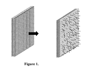
Description of the Drawings
FIG. 1 is a schematic, exploded diagram of the standalone melt-blown patch
whose
surface fibers are raised, and matrix loft increased by napping methods.
FIG. 2 illustrates a comparison of non-napped & different degrees of napped as
cross-
sectional images of substrates.
FIG. 3 illustrates aerial (left) and cross-sectional (right) SEM images of
equally coated
non-napped (top) and napped (bottom) substrates.
1
CA 03186741 2022-12-08
WO 2021/250547 PCT/IB2021/054986
FIG. 4 illustrates images of equally coated non-napped (top) and napped
(bottom)
substrates via micro-CT.
Detailed Description
The present invention is directed to a matrix that is particularly suited for
coating as the
napped surface has increased surface area for coating individual melt-blown
fibers in the
nonwoven matrix. A preferred high matrix loft is produced via a process of
napping that loosens
tightly entangled fibers, increasing matrix loft and overall volume that
enables greater
penetration depth for a subsequently applied coating layer.
One of the benefits of the present invention is the resulting wound dressing
has sidedness
since the surface that has been napped can be easily identified as the napped
and coated face that
should be applied onto a tissue surface.
The inventive wound dressing exhibits strong patch adhesion to tissue as the
napped
surface results in both higher amount of coated fibers and greater surface
roughness, that
together enhance adhesion at the patch-tissue interface.
Another advantage of the present invention is a low-profile patch that is easy
to handle,
with relatively low thickness and density that do not compromise functionality
when napped, and
can be easily be handled in smaller spaces. Patch may also need reduced
compression time to
seal.
Another advantage of the present invention is that the degree of napping can
be
modulated to allow for specific characteristics, e.g. decreasing stiffness
with increasing degree of
napping.
The present inventive wound dressing exhibits high tissue conformability as
the
combination of elastic-layered and roughened matrix allows for high compliance
with the tissue
if the tissue expands or moves.
In one embodiment, the present invention can be produced having tailored
absorption
time/ biocompatibility as the melt-blown nonwoven matrix can be fabricated
using
biocompatible and absorbable materials by, for example, pre-irradiation and/or
modulating fiber
diameter and polymer structure during melt extrusion and crystallization
respectively.
In one embodiment, a nonwoven base substrate is generated from an absorbable
and
biocompatible polyester material, such as Monocryl , a copolymer of glycolide
and epsilon-
caprolactone, by extrusion through a linear die containing several hundred
small orifices.
2
CA 03186741 2022-12-08
WO 2021/250547 PCT/IB2021/054986
Convergent streams of hot air attenuate the molten polymer to form extremely
fine-diameter
fibers. High-velocity air blows the fibers onto a collecting drum, forming one
sheet of the melt-
blown non-woven fabric. Process factors, such as drum speed and distance
between the drum and
surface of die, are selected to obtain preferred fiber diameter and
orientation of fibers on formed
web, which in-turn govern the resulting fiber diameter, pore-size and density
of the nonwoven
matrix.
The drum size, while dependent on the length of the polymer-extrusion die, is
arbitrary
and can be scaled up for large-scale manufacture of melt-blown sheets. The
drum speed is
inversely proportional to the non-woven matrix density per unit-area, and is
inherently associated
with fiber diameter, specific surface area and the overall porosity in the
layer. The collector
distance also affects matrix characteristics as increasing the gap between the
polymer-extrusion
die and the drum better randomizes fiber thickness and orientation.
The present invention identified preferred drum speeds in the range of 0.08-
0.41 m/s,
more preferably 0.12-0.37 m/s, most preferably 0.15-0.2 m/s, and distances in
the range of 10 to
40 inches, more preferably in the range of 15-35, most preferably in the range
of 20-30 in order
to tailor and generate nonwoven membranes that were microporous, water-
impermeable and
low-profile (thickness).
The ranges were established using Monocryl for which the material properties
(e.g.
1.67 intrinsic viscosity) may affect fiber characteristics to a small degree,
that in-turn play into
porosity, density and stiffness of the overall matrix. However, the ranges
should showcase the
same trends irrespective of material specifics (for example, increasing the
drum speed will
reduce patch density for both Monocryl and Vicryl , a Polyglactin 910, a
polyglycolic acid,
and at the very least, the ranges we determined are viable starting points).
Biodegradable
polymers of interest that can be melt-blown include and are not limited to
polyglycolic acid
(PGA), poly(lactic-co-glycolic acid) (PLGA), polylactic acid (PLA),
polydioxanone (PDS) and
caprolactone/glycolide polyesters, such as poly(caprolactone-co-glycolide).
The present invention, prior to napping, identified preferred thickness in the
range of
0.30-1.5 mm, more preferably 0.6-0.95 mm, most preferably 0.85-.90 mm. While
this starting
thickness can vary, post-napping, the present invention identified the
preferred increase in matrix
height in the range of approximately 50-250% the original thickness, more
preferably 55-175%,
most preferably 125-165%.
3
CA 03186741 2022-12-08
WO 2021/250547 PCT/IB2021/054986
The present invention, identified preferred density in the range of 140-250
mg/cm3, more
preferably 140-200 mg/cm3, most preferably 140-150 mg/cm3. The densities are
not expected to
change notably post-napping.
The present invention identified a preferred pore size distribution, based on
micro-CT
analysis, in the range of 0.01-0.5mm, with majority of the pores in the range
of 0.1-0.3mm.
Additionally from the micro-CT analysis, the invention identified the total
open porosity of the
matrix to be approximately 85%.
Meltblowing these polymers provides a unique advantage in generating ultra-
fine fibers.
The present invention of the melt-blown nonwoven identified fine fibers and a
diameter in the in
the range of 1-250 micrometers, preferably in the range of 1-90 microns.
In one embodiment, prior to napping, a standalone melt-blown patch is
generated by
extruding another sheet of melt-blown polyester-based nonwoven onto the
collecting drum
before crystallization of the former sheet. A plurality of discrete sheets are
deposited onto the
drum to create a multi-layered matrix. Then, the surface is modified to
increase surface area for
coatings and sidedness by napping. The napping effects are achieved by using
abrasive
techniques to mechanically raise the ends of fibers on the surface of the
patch and
simultaneously also increase the matrix loft as entanglements of fibers below
the surface are
loosened from the process.
Current napping methods employ both manual and automatic tools. For manual
napping,
a steel file card (e.g. 3.75") is used to brush the surface of the nonwoven
fabric unidirectionally
several times until fibers begin to dislodge from the surface (5-15 times is
preferred working
range for this method). For automatic napping, a bench-top drill press is used
with a crimped
wire wheel (e.g. 0.25" stem, 3"diameter) attachment. Other instruments such as
glass, wire
brushes and abrasive flap wheels can also be utilized the achieve different
degrees of napping.
Additionally, high-pressure air, vacuum or water-jets can also be utilized to
loosen the matrix. In
order to achieve extensive napping without destructive abrasion, matrices can
be subjected to
heat to soften the fibers prior to brushing. The napping increases cross-
sectional and specific
surface area for coating cross-linkable active molecules (Figure 1) that
ultimately provides
potential for superior structural integration of the hemostatic patch with
tissue for enhanced
adhesion.
4
CA 03186741 2022-12-08
WO 2021/250547 PCT/IB2021/054986
The degree of napping can be characterized by measurement of the increased
cross-
sectional height and area per density that results from the process. The most
preferred process of
moderately napping the surface raises the average fiber to provide a 161%
increase in matrix
height (Figure 2; Table 1).
Table 1. Increase in matrix height from napping
Density Matrix Height A
Condition (mg/cm2) (%)
Substrate A; No napping 13.6
Substrate A; Mildly Napped 13.2 54.70
Substrate B; No napping 13.3
Substrate B; Moderately
Napped 13.3 161.11
Substrate C; No napping 12.9
Substrate C; Highly Napped 12.8 252.81
The most preferred process of moderately napping the surface increases the
cross-
sectional area by 152% (Figure 2, Table 2). In all cases, density changes are
minimal.
Table 2.
Non-napped area Post-napped area
Condition (px2) (px2)
Matrix Area A (%)
Substrate A, Mildly Napped 26.17 41.15 57.24
Substrate B, Moderately 152.25
Napped 26.91 67.88
Substrate C, Highly Napped 24.31 129.86 434.18
Quantitative analysis of the preferred substrates demonstrated that moderate
napping
increased the matrix height, surface roughness and volume by 642%, 672% and
8999%,
respectively (Table 3).
Table 3.
Maximum Surface
Height Roughness, Volume
Napping (um) Sa (um) (nnA3)
None 1006 108 4.4E09
Mild 4635 759 2.7E11
CA 03186741 2022-12-08
WO 2021/250547 PCT/IB2021/054986
Moderate 7503 835 4.0E11
High 10465 1405 5.5E11
None VS Mild (%z\) 361 602 5992
None VS Moderate (% A) 646 672 8999
None VS High (%A) 940 1198 12465
Coating the matrix without napping resulted in poor penetration and
aggregation or
clumping of material, whereas napping displayed improved coating of individual
fibers and
better penetration into the matrix. Cross-sectional SEM microscopy revealed
napping alleviated
the issue of "flat films" where coating caked at the surface, blocking the
benefits of porous
structure and increasing stiffness (Figure 3).
Additionally, more cracks were observed in the non-napped coating. Image
analysis
confirmed the non-napped group only had a small number of pores and void space
at the surface
accounting for 12% of the total surface area, whereas the napped group had an
area of 27%.
Coating of individual fibers and less clumping in napped substrates
illustrated improved
pore volume and void space (15%) that will be beneficial for blood percolation
and enhanced
interlocking of coated fibers to tissue. To further corroborate these matrix
characteristics, micro-
CT imaging was conducted to understand the coating on napped surfaces. The
visualizations
reaffirmed not only how napping improved the matrix loft, but also resulted in
improved
penetration of the coating and increased porosity at the surface. Cross-
sectional analysis showed
napping disrupted the even film-like coating seen in non-napped conditions and
dispersed the
cross-linkers effectively without blocking the microporous structure of the
matrix substrate
(Figure 4). Lastly, porosity was improved and stiffness was reduced by 13.8%
and 50%,
respectively (Table 4).
Table 4.
Total Porosity Mean Stiffness
Sample Name
(%) (N/mm)
Non-napped,
41.4 0.02
coated
Napped, coated 55.2 0.01
Functional assessments were performed using a tissue peel test and a
heparinized, spleen
ex vivo bleeding model. For the qualified peel test the patch was applied to a
calf dermal tissue
and compressed in Tris Buffer Saline before peeling and measuring force at 90
.
6
CA 03186741 2022-12-08
WO 2021/250547 PCT/IB2021/054986
For the ex vivo evaluation of efficacy in hemostasis, the napped, coated, non-
napped
coated and non-napped, uncoated, each as non-woven substrate, were assessed
for reduction of
bleeding. Briefly, each patch was cut into 1"xl" squares placed over a lOmm
circular biopsy
defect in an ex-vivo spleen model (perfused with heparinized bovine blood) for
2 minutes with
tamponade. Quantitative analysis confirmed bleeding in the ex-vivo model was
minimized or
entirely arrested with the use of the coated, napped melt-blown patch.
These data confirmed the hemostatic patch was fully functional and
efficacious, in
addition to improved tissue adhesion. Different degrees of napping influenced
effectiveness of
hemostat. While all patches did reduce bleeding and eventually seal to arrest,
mild or high degree
of napping, as described previously, had reduced effectiveness compared to
moderate napping.
In one embodiment, a highly-adhesive hemostatic patch comprised of a napped
melt-
blown matrix substrate in combination with a cross-linkable coating can be
prepared from melt-
blown microfiber web using an absorbable and biocompatible polyester material,
such as
Monocryl , with a drum speed of 0.17 m/s, preferably within a range tested of
0.09-0.34 m/s,
and a collector distance of 25 inches, preferably within a range of 12-25
inches. Four layers can
be built directly on the collector drum, with a preferred range of 2-10
layers. The resulting
density of the four-layer construct is approximately 13mg/cm2 when using IV
equal to 1.6
Monocryl . These material characteristics and density are required prior to
napping.
A preferred degree of napping is achieved by abrasive techniques that loosen
fiber
entanglement, raise surface fibers and the overall matrix height by
approximately 161%, with a
preferred range of 55-253%, while the cross-sectional area is subsequently
increased by
approximately 152%, with a preferred range of 57-434%. The resultant substrate
can have
increased surface roughness and volume of approximately 676% and 8999%,
respectively.
Napping methods include both manual and automatic tools. Manual napping can be
achieved by, and is not limited to, a wire brush, streel file cards, glass or
similar tools/materials
with rough edges that can be used to create abrasion. To achieve the preferred
degree of napping,
a steel file card (3.75") is used to brush the surface of the nonwoven fabric
unidirectionally
several times until fibers begin to tear off surface (5-15 times is preferred
working range for this
method with 5 resulting in "mild" napping and 15 resulting in "high napping").
Alternatively,
automatic napping methods include and are not limited to a bench-top drill
press that is used with
a crimped wire wheel (e.g. 0.25" stem, 3"diameter) or other brush-based
attachments. Other
7
CA 03186741 2022-12-08
WO 2021/250547 PCT/IB2021/054986
power-instruments and attachments such as wire brushes and abrasive flap
wheels can also be
utilized the achieve different degrees of napping.
In order to achieve higher degrees of napping without destructive abrasion,
matrices can
be subjected to heat to soften the fibers prior to brushing. The degree of
heating can vary
depending on polymer; for Monocryl , the construct is heated to 50 C for 15
minutes prior to
napping.
Cross-linkable actives such as polyethylene glycol active esters (e.g. PEG-
succinimidyl
glutarate) are preferably coated in sequence with or without buffers and
additives to develop the
fully functional hemostat. To reduce the idea to practice, a 2-inch by 4-inch
melt-blown matrix
post-napping is either ultrasonically spray-coated (solubilized method) or dip-
coated (insoluble
method) with a light-layer of buffer that embeds deep into the porous
substrate: working
examples include a 1.25mg/cm2 of sodium borate, or 2mg/cm2 Bis-Tris or 1mg/cm2
of sodium
bicarbonate. Then, 15mg/cm2 of 4arm-PEG-Amine-HC1 (MW:10Kda) is ultrasonically
coated,
followed by 18mg/cm2 of 4arm-PEG-SG (MW:10Kda). The napped construct allows
for unique
deposition of the cross-linkable actives that results in enhanced tissue
adhesion.
Exemplary plasma derived (or related) hemostatic agents include proteins and
peptides,
and thus are not limited, to natural and can be in recombinant or synthetic
forms; prothrombin,
thrombin, fibrin, fibronectin, Factor (Factor) X/Xa , Factor VII/VIIa, Factor
IX/IXa, factor
XI/XIa, Factor XII/XIIa, tissue factor, von Willebrand factor, elastin,
albumin, platelet surface
glycoproteins, vasopressin and vasopressin group consisting of analogs,
epinephrine, selectin,
plasminogen activator inhibitor, platelet activating agents, synthetic
peptides, and any
combinations thereof having hemostatic activity.
The carrier sublayers can be in the form of non-woven materials. Exemplary
materials of
construction are synthetic polymers. The substrate may be comprised of
components selected
from aliphatic polyester polymers and/or copolymers of one or more monomers
selected from
the group consisting of D-lactic acid, L-lactic acid, lactide (including L-, D-
, meso forms),
glycolic acid, glycolide, caprolactone, p-dioxanone and trimethylene carbonate
and mixtures or
blends thereof.
The substrate may alternatively, or additionally, be comprised of layers of
fabric of
aliphatic polyester polymers, copolymers, or blends thereof. The aliphatic
polyesters are
typically synthesized in a ring opening polymerization of monomers including,
but not limited
8
CA 03186741 2022-12-08
WO 2021/250547 PCT/IB2021/054986
to, lactide (including L-, and D-, meso forms), glycolic acid, glycolide,
caprolactone, p-
dioxanone (1,4-dioxan-2-one), and trimethylene carbonate (1,3-dioxan-2-one).
The aliphatic
polyesters, in some cases, can be made by polycondensation of for instance, D-
lactic acid, L-
lactic acid and/or glycolic acid. In one form, the fabric comprises a
copolymer of glycolide and
lactide, in an amount ranging from about 70 to 95% by molar basis of glycolide
and the
remainder lactide.
The porous substrate of the dressing has openings or pores over at least a
portion of a
surface thereof. As described in more detail below, suitable materials for
forming the porous
substrate include, but are not limited to fibrous structures. In embodiments,
the pores may be in
sufficient number and size so as to interconnect across the entire thickness
of the porous
substrate.
One or more sublayers of the porous substrate can be at least 0.1 cm thick, in
certain
embodiments from about 0.2 to about 1.5 cm thick. The size of the pores in the
sublayers of the
porous substrate can be from about 2 micrometers to about 300 micrometers, in
embodiments
from about 50 micrometers to about 150 micrometers. It is envisioned that the
pores of the
sublayers of the substrate may be arranged in any manner in the substrate. For
example, the pores
may be configured in a random or uniform manner. In some embodiments, the
pores may be
formed with the use of calcium or copper alginate to create a honey-comb
shaped porous
substrate. In still other embodiments, the pores may be configured to create a
gradient in the
porous substrate. The gradient may further enhance the porous substrates
ability to absorb the
physiologic fluid and direct the migration of the physiological fluid carrying
the first co-reactive
component towards the second co-reactive component.
In one embodiment, the substrate has a first co-reactive component applied
onto a first
sublayer and a second co-reactive component applied thereto. The terms "first
co-reactive
component" and "second co-reactive component" each means a polymer, functional
polymer,
macromolecule, small molecule, or cross-linker that can take part in a
reaction to form a network
of cross-linked molecules, such as, a hydrogel.
In one embodiment, each of the first and second co-reactive components is
multifunctional, meaning that it comprises two or more electrophilic or
nucleophilic functional
groups, such that, for example, a nucleophilic functional group on the first
co-reactive
component may react with an electrophilic functional group on the second co-
reactive
9
CA 03186741 2022-12-08
WO 2021/250547 PCT/IB2021/054986
component to form a covalent bond. At least one of the first or second co-
reactive components
includes more than two functional groups, so that, as a result of
electrophilic-nucleophilic
reactions, the precursors combine to form cross-linked polymeric products.
Such reactions are
referred to as "cross-linking reactions".
In certain embodiments, each of the first and second co-reactive components
includes
only one category of functional groups, either only nucleophilic groups or
only electrophilic
functional groups, so long as both nucleophilic and electrophilic precursors
are used in the cross-
linking reaction. Thus, for example, if the first co-reactive component has
nucleophilic
functional groups such as amines, the second co-reactive component may have
electrophilic
functional groups such as N-hydroxysuccinimides. On the other hand, if first
co-reactive
component has electrophilic functional groups such as sulfosuccinimides, then
the second co-
reactive component may have nucleophilic functional groups such as amines or
thiols. Thus,
functional polymers such as proteins, poly(ally1 amine), styrene sulfonic
acid, or amine-
terminated di- or multifunctional poly(ethylene glycol) ("PEG") can be used.
The first and second co-reactive components may have biologically inert and
water
soluble cores. When the core is a polymeric region that is water soluble,
preferred polymers that
may be used include: polyether, for example, polyalkylene oxides such as
polyethylene glycol
("PEG"), polyethylene oxide ("PEO"), polyethylene oxide-co-polypropylene oxide
("PPO"), co-
polyethylene oxide block or random copolymers, and polyvinyl alcohol ("PVA");
poly(vinyl
pyrrolidinone) ("PVP"); poly(amino acids); poly (saccharides), such as
dextran, chitosan,
alginates, carboxymethylcellulose, oxidized cellulose, hydroxyethylcellulose,
hydroxymethylcellulose, hyaluronic acid; and proteins such as albumin,
collagen, casein, and
gelatin. The polyethers and more particularly poly(oxyalkylenes) or
poly(ethylene glycol) or
polyethylene glycol are especially useful. When the core is small molecular in
nature, any of a
variety of hydrophilic functionalities can be used to make the first and
second co-reactive
components water soluble. For example, functional groups like hydroxyl, amine,
sulfonate and
carboxylate, which are water soluble, maybe used to make the precursor water
soluble. In
addition, N-hydroxysuccinimide ("NHS") ester of subaric acid is insoluble in
water, but by
adding a sulfonate group to the succinimide ring, the NHS ester of subaric
acid may be made
water soluble, without affecting its reactivity towards amine groups.
CA 03186741 2022-12-08
WO 2021/250547 PCT/IB2021/054986
In certain embodiments, both the first and second co-reactive components may
be large
molecules that are capable of cross-linking. For example, in embodiments, one
of the precursors
may be a multi-functional PEG having a molecular weight of from about 2,000 to
about 20,000
Daltons. This multi-functional PEG, in embodiments possessing electrophilic
groups, may be
reacted with a collagen having a molecular weight of about 100,000 Daltons. In
other
embodiments, a gelatin having a molecular weight of from about 50,000 to about
100,000
Daltons may be used in place of the collagen.
In an alternative embodiment, the co-reactive components and buffering agent
are
provided on a patch. An exemplary sealing patch/pad comprises: PEG-NH2*HC1 and
PEG-NHS,
a buffering salt agent, preferably as an alkaline buffer (Borax) deposited on
an absorbable
substrate.
If it is desired that the biocompatible cross-linked polymer resulting from
the reaction of
the first and second co-reactive components be biodegradable or absorbable,
one or more of the
first and second co-reactive components may have biodegradable linkages
present between the
functional groups. The biodegradable linkage optionally also may serve as the
water soluble core
of one or more of the precursors. In the alternative, or in addition, the
functional groups of the
first and second co-reactive components may be chosen such that the product of
the reaction
between them results in a biodegradable linkage. For each approach,
biodegradable linkages may
be chosen such that the resulting biodegradable biocompatible cross-linked
polymer will
degrade, dissolve or be absorbed in a desired period of time. Preferably,
biodegradable linkages
are selected that degrade under physiological conditions into non-toxic
products.
The biodegradable linkage may be chelates or chemically or enzymatically
hydrolyzable
or absorbable. Illustrative chemically hydrolyzable biodegradable linkages
include polymers,
copolymers and oligomers of glycolide, d-lactide, lactide, caprolactone,
dioxanone, and
trimethylene carbonate. Illustrative enzymatically hydrolyzable biodegradable
linkages include
peptidic linkages cleavable by metalloproteinases and collagenases. Additional
illustrative
biodegradable linkages include polymers and copolymers of poly(hydroxy acid)s,
poly(orthocarbonate)s, poly(anhydride)s, poly(lactone)s, poly(amino acid)s,
poly(carbonate)s,
poly(saccharide)s and poly(phosphonate)s. In embodiments, the biodegradable
linkage may
contain ester linkages. Some non-limiting examples include esters of succinic
acid, glutaric acid,
propionic acid, adipic acid, or amino acids, as well as carboxymethyl esters.
11
CA 03186741 2022-12-08
WO 2021/250547 PCT/IB2021/054986
In embodiments, a multifunctional electrophilic polymer such as a multi-arm
PEG
functionalized with multiple NHS groups may be used as a first co-reactive
component, and a
multifunctional nucleophilic component such as trilysine may be used as a
second co-reactive
component. In other embodiments, a multifunctional electrophilic polymer such
as a multi-aim
PEG functionalized with multiple NHS groups may be used as a first co-reactive
component, and
a multifunctional nucleophilic polymer such as collagen and/or a collagen
derivative may be
used as a second co-reactive component. The multi-arm PEG functionalized with
multiple NHS
groups can for example have four, six or eight arms and have a molecular
weight of from about
5,000 to about 25,000. Many other examples of suitable first and second
precursors are described
in U.S. Pat. Nos. 6,152,943; 6,165,201; 6,179,862; 6,514,534; 6,566,406;
6,605,294; 6,673,093;
6,703,047; 6,818,018; 7,009,034; and 7,347,850, the entire content of each of
which is
incorporated herein by reference.
For the patch embodiment, the co-reactive components can be deposited upon the
matrix
as individual layers. Alternatively, the co-reactive components can be
deposited as a mixture.
The ordering of layers may change, but the preferred order sealing patch or
pad comprising PEG-
NH2*HC1 (or any other hydrohalide), PEG-NHS, and a buffering salt (such as
sodium
tetraborate, IVIES, TRIS, Bis-Tris, sodium bicarbonate), with the matrix, then
a layer of buffering
salt, a layer of protected PEG-amine and a layer of the PEG-NHS. Furthermore,
the number of
arms and molecular weight of materials may change, but 4-arm-10K-NH2*HC1 and 4-
arm-10K-
NHS are preferred variants from an efficacy and stability standpoint. The
embodiment was
evaluated with different order of coating. Performance and stability are
greatly impacted by the
location of the deposited buffer on the matrix using the spray-coating
process. When buffer was
deposited below both PEGs (i.e., furthest away from the tissue when matrix is
applied), the
performance and stability were optimal.
The first co-reactive component may be applied to the porous substrate using
any suitable
method known to those skilled in the art, including, but not limited to
spraying, brushing,
dipping, pouring, laminating, etc. In embodiments, the first co-reactive
component may be
applied as a coating on the substrate in any concentration, dimension and
configuration capable
of forming a hemostatic dressing. In embodiments, the first co-reactive
component coating may
penetrate the pores of the porous substrate. In embodiments, the first co-
reactive component may
12
CA 03186741 2022-12-08
WO 2021/250547 PCT/IB2021/054986
be applied to the porous substrate as a film that is laminated onto at least
one side of the
substrate.
The second co-reactive component likewise may be applied to the porous
substrate using
any suitable method known to those skilled in the art, including, but not
limited to spraying,
brushing, dipping, pouring, laminating, etc. In still other embodiments, the
second co-reactive
component may be applied to the porous substrate in solution followed by
evaporation or
lyophilization of the solvent. In embodiments, the second co-reactive
component may be applied
to the porous substrate as a coating on at least one side of the substrate or
as a film laminated
onto at least one side of the substrate.
During use, the patch dressing is oriented with the co-reactive components
applied
directly onto the tissue. In embodiments, the first and second portions may be
distinguishable
from one another by the addition of contrast dyes, surface texturing, coloring
or other visual
cues. Upon contact with tissue, such as, for example, injured tissue, the
dressing will soak up
physiological fluid and the first co-reactive hydrogel component will be
dissolved by the fluid.
As the fluid wicks into and migrates through the dressing, it will carry the
dissolved first co-
reactive component into the second co-reactive component and buffering agent.
Eventually, the
first and second co-reactive components will react to form a biocompatible
cross-linked material,
thereby assisting clot stabilization, tissue ingrowth and remodeling as the
scaffold degrades. In
some embodiments, the biocompatible cross-linked material produced by reaction
of the first and
second co-reactive components also provide the dressing with anti-adhesive
properties.
The following examples are provided for illustrative purposes only and are not
intended
to limit the scope of the present disclosure.
Matrix and Napping Process Example:
Meltblown microfiber web is extruded onto a drum using an absorbable and
biocompatible polyester material such as Monocryl with the most-preferred
drum speed of 0.17
m/s and drum distance to die of 25 inches. Four layers are built of this
configuration directly on
the collector drum with the resulting density of approximately 13mg/cm2 when
using IV equal to
1.6 Monocryl . These material characteristics and density are required prior
to napping.
After cutting to a 2-inch by 4-inch melt-blown matrix, the nonwoven patch is
gently
heated to 50 C for 15 minutes to soften the fibers and then napped by using a
4" steel file card to
13
CA 03186741 2022-12-08
WO 2021/250547 PCT/IB2021/054986
brush the surface unidirectionally until the overall matrix height is
increased by approximately
150%.
Coating Process Example:
A 2-inch by 4-inch melt-blown napped matrix is either ultrasonically spray-
coated
(solubilized method) or dip-coated (insoluble method) with a light-layer of
buffer that embeds
deep into the porous substrate. Working examples include a 1.25mg/cm2 of
sodium borate,
2mg/cm2 Bis-Tris or 1 mg/cm2 of sodium bicarbonate. Then, 15 mg/cm2 of 4-arm-
PEG-Amine-
HC1 (MW:10Kda) is ultrasonically coated, followed by 18mg/cm2 of 4arm-PEG-SG
(MW:10Kda).
The napped construct allows for unique deposition of the cross-linkable
actives, deep into
the matrix, that ultimately results in a highly effective hemostat with
enhanced adhesion.
14