Note: Descriptions are shown in the official language in which they were submitted.
INTRAVASCULAR PRESSURE AND FLOW DATA DIAGNOSTIC SYSTEMS, DEVICES,
AND METHODS
RELATED APPLICATIONS
100011 This application claims priority to U.S. Provisional Patent Application
No. 61/975,424
filed on April 4, 2014 and U.S. Provisional Patent Application No. 62/073,284
filed on October 31, 2014
[0002] The disclosure relates generally to intravascular measurements such as
pressure,
temperature and flow measurements and related diagnostic methods and devices.
BACKGROUND
[0003] Sensor and guide wire assemblies can be used to collect intravascular
data using
measurement sensors located at or near their distal tips. These devices are
typically used in
applications to measure internal properties of tissues and fluids such as
blood pressure. Sensor and
guide wire assemblies may be introduced into arteries, veins or other body
organs either by
themselves or through catheters that have been previously positioned within a
patient. These
assemblies can be used to measure pressure and other parameters.
10004] For example, such assemblies can be used along with one or more
pressure sensing devices
such as a delivery catheter to measure a Fractional Flow Reserve (FFR) using
pressure data. In
addition, Coronary Flow Reserve (CFR) measurements can be performed using a
thermodilution-
based approach. In such an approach a CFR value is obtained by injecting a
cold saline solution
into the coronary artery of interest and using a temperature sensor to measure
the onset of cold
saline injection into the artery to the return of the temperature to a
specific level.
[0005] The thermodilution method has a number of constraints. The stated
accuracy of this
method is as low as +/-30%. Further, the procedure is cumbersome/time-
consuming, requiring a
number of saline injections of a certain quality to produce enough data for
the system software to
calculate the CFR value. Performing FFR and CFR is performed as two separate
methods given
the nature of the thermalilution system, saline delivery, and subsequent
measurements required.
Date Recue/Date Received 2021-07-27
Date Recue/Date Received 2023-01-17
CA 02944111 2016-09-27
WO 2015nsem PC11182015/000675
2
[0006] FFR is used to provide a measure of stenosis severity in a coronary
artery. The typical
method to determine FFR is to measure a pressure drop in the coronary arteries
at hyperemia. A
hyperemia inducing substance is injected to create an increase in blood flow
in the coronary
system for a controlled period, The pressure drop is measured during this time
period and used as
an input in determining FFR.
1.0007.1 In part, the disclosure relates to methods, systems and devices
suitable for measuring
FFR, CPR, and other values and generating diagnostic outputs that overcome
some of the
challenges with existing methods.
SUMMARY
[0008] In part, the disclosure relates to methods, systems and devices to
simultaneously
perform intravascular pressure measurements while measuring blood flow values
or parameters
correlated with such flow. These embodiments can be based upon hot-film or hot-
wire
am:mom:try. Hot-film or hot-wire anemometry is a method of measuring the
cooling effect of a
flowing fluid (or gas) on a heated surface. When using a sensor as a hot-film
anemometer the
sensor is heated by electrical current, and the cooling effect of the flowing
blood is measured by
sampling the voltage across a resistor. The voltage across the resistor can be
measured as well as
other resistances, currents, and electrical parameters and signals. These
measured values can be
correlated with a flow parameter. In one embodiment, the voltage can be used
in two related
anemometry methods: Constant Temperature anemometry (CIA) and Constant
Excitation
Voltage (CVEX) anemometry.
[0009] In one embodiment, a semiconductor based sensor that includes a
first temperature
sensitive resistor and a second temperature sensitive resistor is used as part
of a pressure sensing
intravaseular device. Further, at least one of the first and second resistors
is also pressure
sensitive. The sensor can be delivered via a guide wire and can be used to
measure pressure
before and after a candidate stenosis while simultaneously obtaining flow
data, pressure data or
temperature data based upon changes in excitation voltage, current,
temperature or other sensor
parameters. Various control systems and calibration methods can be used to
support such
pressure and flow measurements.
Date Recue/Date Received 2023-01-17
CA 02044114 2016-09,27
WO 2015/150913 PCT/1112015/000675
3
[0010] In one embodiment, the disclosure relates to using a digital control
system while
performing simultaneous pressure and flow measurements using a semiconductor-
based pressure
sensor. The digital control system overcomes certain deficiencies of an analog
control system.
Specifically, some advantages of the digital control system include
calibration features and user
specified temperature selection features. The digital system also can be used
to improve the
signal relative to noise levels. The calibration features include reading
information digitally
encoded on a memory device associated with a given sensing probe and
regulating control
system stages in response thereto. The memory device can be attached to the
probe such as a
PROM, an EEPROM, an RFID, or other suitable memory storage device.
[0011] The temperature selection features include automatically obtaining the
temperature of the
blood vessel using one or more sensors and then changing an electrical
property of the sensing
system. This change to current, voltage, impedance, or another parameter is in
response to a user
specified temperature above the temperature of flowing blood such as an over-
temperature or
sensing temperature range. The over-temperature or sensing temperature range
provides a range
or value which can be reduced through cooling. The temperature reduction can
be measured as a
result of flowing blood. Alternatively, the degree to which an electrical
property, such as voltage
or current, needs to increase to maintain a constant over-temperature or
sensing temperature
range can be measured and correlated with blood flow. Thus, the over-
temperature can be
constant or can be a range which varies based on cooling.
[0012] In one embodiment, the disclosure relates to graphical user
interfaces and probe
interface or processing systems or display systems or integrated cardiology
display systems
(1CD) (each either separately or together, generally referred to as a
"measurement system") that
are in electronic communication with a guide wire-based probe simultaneously
relaying pressure
and flow related data thereto. In one embodiment, a suitable measurement
system such as an
ICD can include, without limitation, a RadiAnalyzer system, a RadiAnalyzer
Xpress system, a
Quantien system, an Aeris system, a Prestige guide wire-based probe system,
ComboMap
Pressure and Flow System, and other intravascular pressure sensing or FFR
determining devices
and systems. In one embodiment, the measurement system and the probe interface
or processing
systems and display system are the same device or collection of devices.
Date Recue/Date Received 2023-01-17
CA 029441.14 2016-09-27
WO 2015/150913 PC'171B2015/000675
4
[0013] In one embodiment, the interface device receives signals from the guide
wire-based
probe indicative of flow and pressure at one or more locations along a blood
vessel. Prior to
being introduced into a blood vessel, the guide wire-based probe sensor is
disposed in a catheter.
Further, prior to being introduced into a lumen of a blood vessel, the system
obtains a zero flow
reference value from inside the catheter. This zero flow reference can also be
used as an input
when calibrating the guide wire-based probe. Atmospheric pressure can also be
used as a zero
point during sensor calibration.
[0014] The timing of data collection and selection of the controlled
environment in which data
collection is performed provides a zero point or origin relative to which
other flow measurements
and/or pressure measurements can be evaluated. The zero point or calibration
point can be used
with other parameters and a transfer function to transform guide wire-based
probe signal data to
flow data suitable for display and subsequent data analysis using one or more
processors in a
measurement system.
[0015] In one embodiment. the transfer function T(x) is of the form T(x)=
a+b*ln(x), wherein
T(x) yields a temperature value in response to a flow value x. In one
embodiment, the transfer
function T(x) is of the form T(x)= a+b*In(x), wherein T(x) yields an
excitation voltage or
electrical power value in response to a flow value x. The flow value x is a
flow velocity in one
embodiment. In another embodiment, the flow value x is flow rate. In one
embodiment, the
transfer function T(x) is of the form T(W a + b*xAc.
[0016] In one embodiment, the ttansfer function is determined based upon data
fitting. In
particular, the data used to perform such fitting can include can include flow
vs temperature,
flow vs excitation voltage, flow velocity vs temperature, flow velocity vs
excitation voltage, and
others. One or more sensor specific parameter(s) useable by the transfer
function arc stored in
the guide wire-based probe memory storage. In one embodiment, the transfer
function is
determined based upon models, constraints, and other equations alone or in
combination with
data.
[0017] The time to collect data from a blood vessel and display pressure and
flow information
based on the collected data ranges from greater than about 0 zero seconds to
about I second. In
one embodiment, the data includes time varying electrical signals correlated
with changes in
Date Recue/Date Received 2023-01-17
CA 02944114 2016-09-27
WO 2015/150913 PC'171112015/000675
resistance. In one embodiment, the data includes time varying electrical
signals correlated with
changes in current.
[0018] In one embodiment, the potential differences applied to one or more
resistors disposed in
the sensing portion of the guide wire-based probe ranges from about greater
than about 0.1 to
about 15 volts. In one embodiment, the temperature changes measured in a blood
vessel using a
pressure (P), flow (Q), or temperature (T) sensing portion of the guide wire-
based probe ranges
from about greater than about 0 degrees C to about 5 degrees C. In one
embodiment, the
excitation voltage needed to create adequate over-temperature (i.e.
sensitivity to flow changes) is
greater than about 4 volts. This excitation voltage range applies to CVEX and
CTA
implementations in one embodiment.
[0019] In part, the disclosure relates to a method of collecting blood vessel
related data. The
method includes storing, in one or more memory devices, guide wire-based probe
data;
measuring a first electrical signal associated with a first resistor and a
second resistor disposed in
The blood vessel; measuring a second electrical signal associated with the
second resistor
disposed in the blood vessel; determining a transfer function using the guide
wire-based probe
data, the transfer function having a flow parameter as an output; determining
a blood pressure
value for the blood vessel using one or more of the first and second
electrical signals;
determining a blood temperature value for the blood vessel using one or more
of the first and
second electrical signals; determining a blood flow value for the blood vessel
using one or more
of the first and second electrical signals and the transfer function; and
displaying a pressure
versus flow curve for the blood vessel. In one embodiment, the transfer
function relates the flow
parameter and an excitation voltage. In one embodiment, the transfer function
relates the flow
value and a temperature of one or more of the first resistor and the second
resistor. In one
embodiment, the method further includes identifying one or more of an
occurrence of a
maximum flow, a minimum flow, and a relative extremum of flow and correlating
such an
occurrence with an intravascular or cardiac event.
[00201 In part, the disclosure relates to an intravascular pressure and flow
monitoring system.
The system includes one or more memory devices; and a computing device in
communication
with the memory device, wherein the memory device comprises instructions
executable by the
Date Recue/Date Received 2023-01-17
CA 02944114 2016-09-27
WO 2015/150913 PCT/182015/000675
6
computing device to cause the computing device to: determine one or more
intravascular
pressure values in response to a first electrical signal from a measurement
circuit formed from a
guide wire-based probe and an interface device; determine one or more
intravascular flow values
using a transfer function in response to a second electrical signal from the
measurement circuit
formed from the guide wire-based probe and the interface device; and display a
pressure versus
flow curve generated based on the one or more intravascular pressure values
and the
intravascular flow values, wherein the pressure versus flow curve changes over
time.
[00211 In one embodiment, the pressure versus flow curve is displayed on a
substantial real time
basis. In one embodiment, the transfer function T(x), wherein x is flow, is of
the form T(x)=
a+b*In(x), wherein a and b are constants. In one embodiment, the transfer
function T(x),
wherein x is flow, is of the form T(x)= a + b*xc, wherein a, b and c are
constants. In one
embodiment, the system further includes instructions which display one or more
cardiovascular
related values obtained during one or more points in time.
[0022] In one embodiment, the one or more cardiovascular related values are
selected from the
group consisting of a flow velocity, a pressure value, a maximum flow, a
minimum flow, a
relative extrernum of flow one or more fractional flow reserve (FFR) values,
coronary flow
reserve (CFR) values, coronary flow velocity reserve (CFVR) values,
instantaneous flow reserve
(1FR) values, and one or more index of myocardial resistance (IMR) values.
[0023] In one embodiment, the system further includes instructions which
display one or more
trajectories or signatures generated in response to intravascular probe data
with respect to one or
more positions in an artery. In one embodiment, the system further includes
instructions which
display a user interface that includes a flow velocity, a pressure value, a
maximum flow, a
minimum flow, a relative extremum of flow one or more fractional flow reserve
(FFR) values,
coronary flow reserve (CPR.) values, coronary flow velocity reserve (CFVR)
values,
instantaneous flow reserve (IFR) values, or one or more index of myocardial
resistance (IMR)
values generated using pressure and flow data from a intravascular probe.
[0024] In one embodiment, the system further includes instructions to
determine one or more
temperature values using a linear or other function in response to a second
electrical signal from
the measurement circuit formed from the guide wire-based probe and the
interface device,
Date Recue/Date Received 2023-01-17
CA 02944114 7016-09-27
WO 2015/150913 PCV1112015/000675
7
[0025] In part, the disclosure relates to an intravascular pressure and flow
monitoring adapter
kit. The kit includes a power supply unit comprising a first intravascular
pressure measurement
system output connection; and a second intravascular pressure measurement
system output
connection, wherein a power output range of the power supply unit ranges from
greater than
about .2 volts and less than about 12 volts, the power supply unit sized to
electrically connect to
an intravascular pressure measurement system. The kit may include one or more
electrical
components in electrical communication with the power supply. In one
embodiment of the kit,
the one or more electrical components are selected from the group consisting
of a filter, an
amplifier, a current source, a voltage source, and a control system
connection.
[0026] In one embodiment of the kit, the power output range is from about .3
volts to about 30
volts. In one embodiment of the kit, the kit further includes a non-transitory
storage medium
comprising instructions to cause a computing device of the intravascular
pressure monitoring
system to: store a transfer function in memory that outputs intravascular flow
values in response
to a excitation voltage or a temperature; and generate an intravascular flow
value in response to
(i) an excitation voltage or (ii) a difference between a fixed voltage and a
voltage across a
temperature dependent resistor from the power supply unit and the transfer
function.
[0027] In part, the disclosure relates to a method of calibrating a flow
monitoring device. The
method includes selecting an excitation voltage for a pressure sensor such
that a temperature of
the sensor and a temperature of blood in which the pressure sensor is disposed
substantially
match; determining an absolute temperature of blood in a blood vessel of
interest; and
measuring a flow value in the blood vessel using the pressure sensor. In one
embodiment, the
method includes determining an absolute temperature of blood in a blood vessel
of interest
comprises obtaining measurements during changes to a switch configuration in
an interface
System.
[0028] In part, the disclosure relates to an integrated cardiology system. The
system includes a
display system; a pressure and flow measurement system in electrical
communication with the
display system; a processor disposed in one of the display or pressure and
flow measurement
system; one or more panels generated using the processor and depicted on the
display, wherein
the one or more panels comprises flow values and pressure values obtained
using an
Date Recue/Date Received 2023-01-17
CA 02944114 2016-09-27
WO 2015/150913 Perf182015/000675
8
intravascular probe that comprises a pressure and flow sensor. In one
embodiment, the one or
more panels include a pressure versus flow curve that include one or more
trajectories generated
using intravascular pressure and flow data and further comprising an input to
receive data signals
from the intravascular probe, the intravascular probe comprising a temperature
sensor to measure
temperature changes correlated with flow values.
[0029] In one embodiment, a trajectory can include without limitation a
graphical representation
of transitions between states relevant to the cardiac cycle and vary based
upon stcnosis, pressure
changes in the arteries, and flow changes due to constrictions and other
artery or heart states. In
one embodiment, one or more panels include a signature, a trajectory, a slope,
a maximum point,
a minimum point, a ratio of measured values, a ratio of a measured value and a
derived value, a
ratio of a first derived value and a second derived value, an area, one or
more (FFR) values,
coronary flow reserve (CFR) values, coronary flow velocity reserve (CFVR)
values,
instantaneous flow reserve (IFR) values, and one or more myocardial resistance
(IMR) values.
[0030] In part, the disclosure relates to intravascular pressure and flow
monitoring system. The
system includes an intravascular pressure and flow interface system comprising
a wired interface
or a wireless interface to receive data from an intravascular probe; a display
system in electrical
communication with the intravascular pressure and flow interface system; one
or more memory
storage devices comprising instructions to output a user interface on the
display, the user
interface comprising one or more panels having fields for one or more flow
measurements; and a
processor in electrical communication with the intravascular pressure and flow
interface system,
the display system, and one or more memory storage devices, the processor
responsive to the
instructions such that the user interface is output on the display system.
[0031] In one embodiment, the system includes a calibration system configured
to convert one
of a measured temperature signal or an excitation voltage into a flow velocity
using a transfer
function. In one embodiment, the transfer function is of the form a¨b*lnx
and/or a+b*x'. In one
embodiment, the display system simultaneously outputs pressure and flow
velocity
measurements. In one embodiment, the display system simultaneously outputs
pressure and
absolute temperature measurements. In one embodiment, the display system
outputs one or
Date Recue/Date Received 2023-01-17
CA 02944114 ?016-09-27
WO 2015/150913 PC'171112015/000675
9
more parameters or indexes corresponding to one or more signals obtained at
the measurement
position of a probe sensor.
[0032] In one embodiment, the one or more parameters or indexes are selected
from the group
consisting of a signature, a trajectory, a slope, a maximum point, a minimum
point, a ratio of
measured values, a ratio of a measured value and a derived value, a ratio of a
first derived value
and a second derived value, an area, (FFR) values, coronary flow reserve (CFR)
values, coronary
flow velocity reserve (CFVR) values, instantaneous flow reserve (IFR) values,
and one or more
myocardial resistance (IMR) values. In one embodiment, the wired interface
comprises an
excitation voltage source, a first resistor, ,a second resistor, a first
switch and a second switch. In
one embodiment, the wireless interface comprises a plurality of current
sources, a plurality of
switches, a first resistor and a second resistor, wherein each current source
is in series with one
of the switches.
Coronary Flow Reserve Related Features and Embodiments
[0033] In part, the disclosure relates to methods and systems suitable for
determining one or
more Coronary Flow Reserve (CFR) and Fractional Flow Reserve (FFR) values
separately or
simultaneously using a thermoconvection device such as an intravascular
pressure and flow
sensor and an intravascular data collection and processing system. In
addition, in part, the
disclosure also relates to determining CFR values using an intravascular probe
having a pressure
sensor and Constant Temperature Anemometry (CTA) or Constant Excitation
Voltage (CVEX)
anenriometry.
10034] The disclosure also relates to a method of determining coronary flow
reserve data using
an intravascular pressure or flow sensor. The method includes sampling an
intravascular data
collection probe to obtain a one or more distal pressure values (Pd) from a
distal region of a
vessel and one or more thermoconvection data values; receiving one or more
aortic pressure
values (Pa), at an intravascular data processing system, obtained from a
proximal region of the
vessel; determining one or more fractional flow reserve (FFR) values from the
one or more distal
pressure values and the one or more aortic pressure values; determining one or
more coronary
flow reserve (CFR) values from the one or more themiocoiweetion data values;
and displaying
onc or more FFR values and one or more CFR values on a display unit.
Date Recue/Date Received 2023-01-17
CA 02944114 2016-09,27
WO 2015/150913 PCT/1112015/000675
[0035] In one embodiment, each CFR value is determined using a transfer
function. In one
embodiment, the transfer function is of the form T = a + c * 1n(2, wherein T
is the measured
temperature of the temperature variable resistor of the thermoconvection
device, Q is the flow,
and a and c are constants. In one embodiment, determining one or more coronary
flow reserve
(CFR) values comprises determining a measured temperature at hyperemic flow T
Op and a
measured temperature at baseline flow T. In one embodiment, each CFR value is
determined
Thyp Tbas
using a relationship between Thyp of and Tbas of the form b c
, the form being an algebraic
simplification of the inverse of the function of, wherein b is a generalized
base and c is a
constant. In one embodiment, the FFR values and the CFR value are displayed as
numerical
values and as time varying plots relative to a graphical user interface
comprising one or more
controls. In one embodiment, wherein one of the one or more controls comprises
an enable CFR
control that can be adjusted by a user to selected between an :PPR display
mode and a combined
FFR and CFR mode. In one embodiment, the method further includes tuning a
temperature
signal to find maximum, minimum, or other level. In one embodiment, the step
of tuning is
performed by adjusting a control until an auditory or visual cue indicative of
a tuned state occurs.
[0036] In part, the disclosure relates to a data collection method and/or
diagnostic methods using
collected data such as measured pressure, temperature, or flow values. The
method includes
setting a zero value for a distal pressure signal measured by an intravascular
thermoconvection
device; positioning intravascular thermoconvection device to delivery catheter
opening; setting
zero value of temperature signal measured by intravascular thermoconvection
device prior to
advancing into vascular system; advancing intravascular thermoconvection
device to a position
distal to the catheter opening; equalizing intravascular thermoconvection
device pressure signal
(Pd) to the aortic pressure (Pa) signal; tuning or optimizing, temperature
signal of intravascular
thermoconvection device, when in measurement location of interest sampling
intravascular
thermoconvection device to obtain baseline thermoconvection signal value;
sampling
intravascular thermoconvection device to obtain Pd values and thermoconvection
device values
for running FFR and CFR calculations. In one embodiment, the method further
includes
verifying pressure equalization and flow signal return to baseline level. In
one embodiment, the
method further includes displaying one or more FFR values and one or more CFR
values relative
Date Recue/Date Received 2023-01-17
at 02944114 2016-09-27
WO 2015/150913 PCT/182015/000675
11
to a graphic user interface comprising one or more axis and one or more
control inputs. In one
embodiment, the control inputs and user interface are implemented using a
touch screen.
[0037] In part, the disclosure relates to an intravascular data monitoring
system. The system
includes an intravascular data collection system comprising an interface to
receive data from an
intravascular probe; a display system in electrical communication with the
intravascular data
collection system; one or more memory storage devices comprising instructions
to output a user
interface on the display system, the user interface comprising one or more
regions for displaying
one or more CFR values or a plot thereof, the user interface comprising one or
more regions for
displaying one or more FFR values or a plot thereof; a processor in electrical
communication
with the intravascular data collection system, the display system, and one or
more memory
storage devices, the processor programmed to sample a plurality of proximal
pressure values
(Pa); sample a plurality of distal pressure values (Pd); sample a plurality of
thermoconvection
data values and determine the one or more CFR values and the one or more FFR
values using the
sampled Pa values, Pd values, and thermoconvection data values.
[0038] In part, the disclosure relates to a method of calibrating an
intravascular data collecting
system. The method includes setting a baseline value for a distal pressure
signal measured by an
intravascular thermoconvection device; positioning intravascular
thermoconvection device to
delivery catheter opening; setting baseline value of temperature signal
measured by intravascular
thermoconvection device prior to advancing into vascular system; advancing
intravascular
thermoconvection device to a position distal to the catheter opening;
equalizing intravascular
thermoconvection device pressure signal (Pd) to the aortic press= (Pa) signal;
calibrating
temperature signal of intravascular thermoconvection device, when in
measurement location of
interest; and sampling intravascular thermoconvection device to obtain Pd
values and
thermoconvection device values.
Stenosis Assessment and Flow Threshold/Peak Guided Measurement Embodiments
[0039] In part, the disclosure relates to intravascular pressure monitoring
systems and data
collection devices suitable for analysing pressure drops in a non-hyperemic
state or hyperemic state
and identifying one or more flow thresholds and collecting or otherwise
generating diagnostic data
Date Recue/Date Received 2023-01-17
C6 02944114 2016-09-27
WO 2015/150913 PCT/182015/000675
12
relative to the data collect at such a selected point in time. in one
embodiment, pressure ratios such
as distal to proximal ratios or pressure differences are collected at
different one or more flow
thresholds over time. The pressure and flow values measured at each flow
threshold can be used to
calculate the arithmetic mean of the pressure ratio/difference over a number
of heartbeats.
[0040] In part, the disclosure relates to a method of assessing a blood
vessel. The method
includes measuring a plurality of intravaseular blood flow values and a
plurality of blood
pressure values during one or more heartbeats using one or more sensors;
determining a flow
threshold for one or more heartbeats using one or more of the plurality of
intravascular blood
flow values, determining, a proximal pressure value (Pa) and a distal pressure
value (Pd) during
the flow threshold; calculating a first diagnostic parameter based upon the Pa
and Pd values at
the flow threshold for one or more heartbeats; displaying, on a user display,
the first diagnostic
parameter for one or more heart beats or a second diagnostic parameter
determined using the first
diagnostic parameter. In one embodiment, the first diagnostic parameter is a
pressure difference
Pa-Pd or a pressure ratio Pd/Pa. In one embodiment, the plurality of blood
pressure values
comprise one or more of proximal pressure values relative measured relative to
a stenosis and
one or more aortic pressure values.
[0041] In one embodiment, the first diagnostic parameter and the second
diagnostic parameter
are selected from the group consisting of Pa, Pd Pd/Pa, Pa-Pd, a flow
velocity, a pressure value,
a maximum flow, a minimum flow, a relative extremum of flow, a fractional flow
reserve (}PR)
value, coronary flow reserve (CFR) values, coronary flow velocity reserve
(CFVR) values,
instantaneous flow reserve (IFR) values, and one or more index of myocardial
resistance (IMR)
values. In one embodiment, the flow threshold is selected from the group
consisting of a
maximum flow during a cardiac cycle, a relative extremum flow value during a
cardiac cycle, a
fraction of the maximum flow during a cardiac cycle, a hyperemic max flow
value, and a non-
hyperemic flow value.
[0042] In one embodiment, measuring a plurality of intravaseular blood flow
values and a
plurality of blood pressure values further includes measuring a first
electrical signal associated
with a first resistor and a second resistor disposed in the blood vessel;
measuring a second
electrical signal associated with the second resistor disposed in the blood
vessel; determining
Date Recue/Date Received 2023-01-17
CA 02944114 2016-09-27
WO 2015/150913 K17182015/0110675
13
one or more of the blood pressure values of the plurality of intravascular
blood flow pressure
values -using one or more of the first and second electrical signals;
determining one or more
blood temperature values for the blood vessel using one or more of the first
and second electrical
signals; detemnning the plurality of intravascular blood flow values for the
blood vessel using
one or more of the first and second electrical signals.
10043] In one embodiment, one or more of the blood pressure values is an
aortic pressure value
or proximal pressure value. In one embodiment, the first diagnostic parameter
is a plot of a
value over time. in one embodiment, calculating a first diagnostic parameter
comprises
calculating an average of pressure ratios or pressure differences for a
plurality of cardiac cycles
per a determined flow threshold for each cardiac cycle. In one embodiment, the
first diagnostic
parameter is a mean of pressure ratios for a plurality of cardiac cycles or a
mean of the pressure
differences for a plurality of cardiac cycles.
[0044] In part, the disclosure relates to a method of assessing a blood
vessel. the method
includes receiving intravascular blood flow data and blood pressure data
obtained during one or
more cardiac cycles, intravascular blood flow data comprising a peak blood
flow value;
determining a flow threshold comprising the peak blood flow value;
determining, at the peak
blood flow for each of the one or more heartbeats, a first intravascular blood
pressure (Pa) and a
second intravascular blood pressure (Pd); calculating one or more of a
pressure difference
between Pa and Pd for each of the one or more cardiac cycles or one or more of
a pressure ratio
Pd/Pa for each of the one or more cardiac cycles; and displaying, on a user
display, diagnostic
information about the blood vessel, wherein the diagnostic information
comprises one or more of
the pressure ratio, the pressure difference, or a plot thereof. In one
embodiment, the calculating a
pressure difference includes calculating a mean of the pressure difference for
a plurality of
heartbeats.
[0045] In one embodiment, the method further includes receiving electrical
signals correlated
with temperature changes of an intravascular thermoconvection device in
thermal
communication with the blood vessel, the temperature changes correlated with
changes in a flow
during one or more cardiac cycles, the intravascular blood flow data
comprising the electrical
Date Recue/Date Received 2023-01-17
CA 02944114 2016-09-27
WO 2015/150913 PC'171B2015/000675
14
signals; and determining the peak blood flow value from the electrical signals
correlated with
temperature changes.
[0046] In one embodiment, the method further includes receiving electrical
signals correlated
with temperature changes of an intravascular thermoconvection device in
thermal
communication with the blood vessel, the temperature changes correlated with
changes in a flow
during one or more cardiac cycles, the intravascular blood flow data includes
the electrical
signals; and determining the peak blood flow value from the electrical signals
correlated with
temperature changes.
Additional Embodiments and Implementations
[0047] In part, the disclosure relates to an intravascular pressure and flow
monitoring system
that includes one or more memory devices; and a computing device in
communication with the
memory device, wherein the memory device comprises instructions executable by
the computing
device to cause the computing device to: determine one or more intravascular
pressure values in
response to a first electrical signal from a measurement circuit formed from a
guide wire-based
probe and an interface device; determine one or more intravascular flow values
using a transfer
function in response to a second electrical signal from the measurement
circuit formed from the
guide wire-based probe and the interface device; and display a pressure versus
flow curve
generated based on the one or more intravascular pressure values and the
intravascular flow
values, wherein the pressure versus flow curve changes over time. In one
embodiment, the
pressure versus flow curve is displayed on a substantial real time basis. In
one embodiment, the
transfer function T(x), wherein x is flow, is of the form T(x)= a+b*ln(x),
wherein a and b are
constants. In one embodiment, the transfer function T(x), wherein x is flow,
is of the form T(x)=
a + b*xc, wherein a, b and c are constants.
[0048] In one embodiment, the system further includes instructions which
display one or more
cardiovascular related values obtained during one or more points in time. In
one embodiment,
the one or more cardiovascular related values are selected from the group
consisting of a flow
velocity, a pressure value, a maximum flow, a minimum flow, a relative
extremum of flow one
or more fractional flow reserve (FFR) values, coronary flow reserve (CFR)
values, coronary flow
Date Recue/Date Received 2023-01-17
CA 02944114 ?016-09-27
WO 2015/150913 PCIAB2015/000675
velocity reserve (CFVR) values, instantaneous flow reserve (I FR) values, and
one or more index
of myocardial resistance (IMR) values.
[0049] In one embodiment, the system further includes instructions which
display one or more
trajectories or signatures generated in response to intravascular probe data
with respect to one or
more positions in an artery. In one embodiment, the system further includes
instructions which
display a user interface that includes a flow velocity, a pressure value, a
maximum flow, a
minimum flow, a relative extrermirn of flow one or more fractional flow
reserve (FFR) values,
coronary flow reserve (CFR) values, coronary flow velocity reserve (CFVR)
values,
instantaneous flow reserve (IFR) values, or one or more index of myocardial
resistance (IMR)
values generated using pressure and flow data from a intravascular probe.
100501 In one embodiment, the system further includes instructions to
determine one or more
temperature values using a linear or other function in response to a second
electrical signal from
the measurement circuit formed from the guide wire-based probe and the
interface devicc. In
one embodiment, the system further includes instructions to calibrate the
guide-wire based probe
by the following calibration method steps selecting an excitation voltage for
a pressure sensor
such that a temperature of the sensor and a temperature of blood in which the
pressure sensor is
disposed substantially match; determining an absolute temperature of blood in
a blood vessel of
interest; and measuring a flow value in the blood vessel using the pressure
sensor. In one
embodiment, determining an absolute temperature of blood in a blood vessel of
interest
comprises obtaining measurements during changes to a switch configuration in
an interface
system.
[0051] In one embodiment, the intravascular pressure and flow monitoring
system further
includes a display system; a pressure and flow measurement system in
electrical communication
with the display system and comprising the computing device; the computing
device disposed in
one of the display or pressure and flow measurement system; and one or more
panels generated
using the computing device and depicted on the display, wherein the one or
more panels
comprises flow values and pressure values obtained using an intravascular
probe that comprises
a pressure and flow sensor.
Date Recue/Date Received 2023-01-17
16
[0052] In one embodiment, one or more panels includes a pressure versus flow
curve
comprising one or more trajectories generated using intravascular pressure and
flow data and
further comprising an input to receive data signals from the intravascular
probe, the
intravascular probe comprising a temperature sensor to measure temperature
changes correlated
with flow values. In one embodiment, one or more panels include a signature, a
trajectory, a
slope, a maximum point, a minimum point, a ratio of measured values, a ratio
of a measured
value and a derived value, a ratio of a first derived value and a second
derived value, an area,
one or more (FFR) values, coronary flow reserve (CFR) values, coronary flow
velocity reserve
(CFVR) values, instantaneous flow reserve (IFR) values, and one or more
myocardial resistance
(IIVIR) values.
[0053] In one embodiment, the intravascular pressure and flow monitoring
system further
comprising instructions to process coronary flow reserve data using an
intravascular pressure
or flow sensor comprising: sampling an intravascular data collection probe to
obtain a one or
more distal pressure values (Pd) from a distal region of a vessel and one or
more
thermoconvection data values; receiving one or more aortic pressure values
(Pa), at an
intravascular data processing system, obtained from a proximal region of the
vessel;
determining one or more fractional flow reserve (FFR) values from the one or
more distal
pressure values and the one or more aortic pressure values; determining one or
more coronary
flow reserve (CFR) values from the one or more thermoconvecti on data values;
and displaying
one or more FFR values and one or more CFR values on a display unit, wherein
each CFR value
is determined using a transfer function. In one embodiment, the transfer
function is of the form
T = a + c * lnQ, wherein 7' is the measured temperature of the temperature
variable resistor of
the thermoconvection device, Q is the flow, and a and c are constants. In one
embodiment, each
CTR value is determined using a relationship between l'hyp of and Tbas of the
form b h)177. c-Tb",
the form being an algebraic simplification of the inverse of the transfer
function, wherein b is a
generalized base and c is a constant.
[0054] In part, the disclosure relates to a method of intravascular pressure
and flow monitoring.
The method includes measuring a plurality of intravascular blood flow values
and a plurality of
blood pressure values during one or more heartbeats using one or more sensors;
determining a
Date Recue/Date Received 2021-07-27
Date Recue/Date Received 2023-01-17
C A 079014114 :016-09-27
WO 2015/1511913 PC'I71112015/000675
17
flow threshold for one or more heartbeats using one or more of the plurality
of intravascular
blood flow values, determining, a proximal pressure value (Pa) and a distal
pressure value (Pd)
during the flow threshold; calculating a first diagnostic parameter based upon
the Pa and Pd
values at the flow threshold for one or more heartbeats; and displaying, on a
user display, the
first diagnostic parameter for one or more heart beats or a second diagnostic
parameter
determined using the first diagnostic parameter. In one embodiment, the first
diagnostic
parameter can be a pressure difference Pa-Pd or a pressure ratio Pd/Pa. In one
embodiment, the
plurality of blood pressure values comprise one or more of proximal pressure
values relative
measured relative to a stenosis and one or more aortic pressure values.
[0055] In one embodiment, the first diagnostic parameter and the second
diagnostic parameter
are selected from the group consisting of Pa, Pd , Pd/Pa, Pa-Pd, a flow
velocity, a pressure value,
a maximum flow, a minimum flow, a relative extremum of flow, a fractional flow
reserve (FFR)
value, coronary flow reserve (CFR) values, coronary flow velocity reserve
(CFVR) values,
instantaneous flow reserve (IFR) values, and one or more index of myocardial
resistance (IMR)
values. In one embodiment, the flow threshold is selected from the group
consisting of a
maximum flow during a cardiac cycle, a relative extremtun flow value during a
cardiac cycle, a
fraction of the maximum flow during a cardiac cycle, a hyperemic max flow
value, and a non-
hyperemic flow value. In one embodiment, measuring a plurality of
intravascular blood flow
values and a plurality of blood pressure values further includes measuring a
first electrical signal
associated with a first resistor and a second resistor disposed in the blood
vessel; measuring a
second electrical signal associated with the second resistor disposed in the
blood vessel;
determining one or more of the blood pressure values of the plurality of
intravascular blood flow
pressure values using one or more of the first and second electrical signals;
determining one or
more blood temperature values for the blood vessel using one or more of the
first and second
electrical signals; and determining the plurality of intravascular blood flow
values for the blood
vessel using one or more of the first and second electrical signals.
[0056] In one embodiment, calculating a first diagnostic parameter comprises
calculating an
average of pressure ratios or pressure differences for a plurality of cardiac
cycles per a
determined flow threshold for each cardiac cycle. In one embodiment, the first
diagnostic
parameter is a mean of pressure ratios for a plurality of cardiac cycles or a
mean of the pressure
Date Recue/Date Received 2023-01-17
18
differences for a plurality of cardiac cycles. In one embodiment, the method
includes
receiving electrical signals correlated with temperature changes of an
intravascular
thermoconvection device in thermal communication with the blood vessel, the
temperature
changes correlated with changes in a flow during one or more cardiac cycles,
the
intravascular blood flow data comprising the electrical signals; and
determining the peak
blood flow value from the electrical signals correlated with temperature
changes.
100571 In one embodiment, the method includes further comprising the steps of
calibrating
an intravascular data collection system comprising: setting a baseline value
for a distal
pressure signal measured by an intravascular thermoconvection device;
positioning
intravascular thermoconvection device to delivery catheter opening; setting
baseline value
of temperature signal measured by intravascular thermoconvection device prior
to advancing
into vascular system; advancing intravascular thermoconvection device to a
position distal
to the catheter opening; equalizing intravascular thermoconvection device
pressure signal
(Pd) to the aortic pressure (Pa) signal; calibrating temperature signal of
intravascular
thermoconvection device, when in measurement location of interest; and
sampling
intravascular thermoconvection device to obtain Pd values and thermoconvection
device
values.
In another embodiment, there is provided an intravascular pressure and flow
monitoring system comprising: a display; an intravascular guide wire-based
probe
comprising one or more sensors disposed on a distal end of a guidewire, the
one or more
sensors comprising an active resistor that is sensitive to temperature and
pressure, and a
passive resistor that is sensitive to temperature but is not sensitive to
pressure; one or more
memory devices; and a computing device in communication with the one or more
memory
devices. The one or more memory devices comprise instructions executable by
the
computing device to cause the computing device to: process a first electrical
signal
associated with the active resistor and the passive resistor; process a second
electrical signal
associated with the passive resistor; determine intravascular pressure values
based on the
Date Regue/Date Received 2022-05-24
Date Regue/Date Received 2023-01-17
I 8a
first electrical signal using a measurement circuit formed from the guide wire-
based probe
and an interface device; determine intravascular flow values using a transfer
function based
on the second electrical signal using the measurement circuit formed from the
guide wire-
based probe and the interface device; and generate, based on the intravascular
pressure
values and the intravascular flow values, a pressure versus flow curve that
changes over
time, and cause the display to display said pressure versus flow curve that
changes over time.
In a further embodiment, there is provided a system for collecting blood
vessel
related data, the system comprising: an intravascular guide wire-based probe
comprising a
temperature-sensitive resistor (Rp), the probe being disposed on a distal end
of a guidewire;
a probe interface unit comprising an excitation voltage source (VExc), and a
switch (S2)
located between the temperature-sensitive resistor (120 and the excitation
voltage source
(Voce); one or more memory devices; and a computing device in communication
with the
one or more memory devices. The one or more memory devices comprise
instructions that
are executable by the computing device to cause the computing device to: cause
the
excitation voltage source (VEXC) to apply an excitation voltage to the probe
while the probe
is located in a blood vessel, switch the switch between an ON state and an OFF
state at a
rate in a range of 400 to 600 Hz, sample a signal indicative of differences
between an offset
voltage (V.ropps) and a voltage (VI)) across the temperature-sensitive
resistor while the switch
is in both the ON state and the OFF state, the offset voltage (V-roffs)
corresponding to a
voltage that is substantially equal to the voltage (Vp) at a temperature of
about 37 C, and
calculate a temperature of the temperature-sensitive resistor based on the
sampled signal.
In a still further embodiment, there is provided a method of intravascular
pressure
and flow monitoring comprising: measuring a plurality of intravascular blood
flow values,
a plurality of proximal blood pressure values, and a plurality of distal blood
pressure values
daring each of a plurality of heart cycles using one or more sensors;
determining a plurality
of flow thresholds, including a flow threshold for each of the plurality of
heart cycles, using
the plurality of intravascular blood flow values; determining a proximal
pressure value
Date Recue/Date Received 2022-05-24
Date Recue/Date Received 2023-01-17
1 8b
(Pa)and a distal pressure value (Pd) at a time of each flow threshold;
calculating a first
diagnostic parameter based on the Pa and Pd values at the time of each flow
threshold; and
displaying, on a user display, the diagnostic parameter for the plurality of
heart cycles.
BRIEF DESCRIPTION OF DRAWINGS
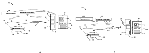
[0058] The figures are not necessarily to scale, emphasis instead generally
being placed
upon illustrative principles. The figures are to be considered illustrative in
all aspects and
are not intended to limit the disclosure, the scope of which is defined only
by the claims.
[0059] Figure IA is a schematic diagram of an intravascular probe suitable for
measuring
pressure, flow parameters and other parameters of interest in a wired
configuration with one
or more measurement systems.
[0060] Figure 1B is a schematic diagram of an intravascular probe suitable for
measuring
pressure, flow and other parameters of interest in a wireless configuration
with one or more
measurement systems.
Date Regue/Date Received 2022-05-24
Date Regue/Date Received 2023-01-17
CA 02944114 2016-09-27
WO 2015/150913 PCT/182015/000675
19
[0061] Figure 2A is a schematic diagram of a blood vessel having a pressure
and flow sensing
guide wire-based probe disposed therein in accordance with, an illustrative
embodiment of the
disclosure.
[0062] Figure 2B is a schematic diagram of a sensing region and related
components of an
exemplary guide wire-based probe embodiment suitable for simultaneous pressure
and flow
measurements in accordance with an illustrative embodiment of the disclosure.
[0063] Figure 2C is an image of a portion of a semiconductor substrate of a
guide wire-based
probe that includes active and passive resistors for pressure and flow sensing
in accordance with
an illustrative embodiment of the disclosure.
[0064] Figure 2D is a perspective view of a guide wire-based probe showing a
capsule
surrounding a sensor array in accordance with an illustrative embodiment of
the disclosure.
[0065] Figure 3 is a circuit diagram including various resistors and nodes as
a representation of
components of a probe, connections and contact pads relating thereto in
accordance with an
illustrative embodiment of the disclosure.
[0066] Figure 4A is a circuit diagram including various resistors and nodes as
a representation of
components of an intravascular probe and an interface or processing system in
a bridge
configuration in accordance with an illustrative embodiment of the disclosure.
[0067] Figure 4B is a circuit diagram including various resistors and nodes as
a representation of
components of an intravascular probe and an interface or processing system in
a bridge
configuration in accordance with an illustrative embodiment of the disclosure.
[0068] Figure 5A is a schematic diagram of a signal sampling system for use
with an
intravascular probe in conjunction with a measurement bridge such as shown in
Figure 4A.
100691 Figure 5B is a schematic diagram of a control system for a constant
temperature
anemometry (CTA) embodiment, using a guide wire-based probe in accordance with
an
illustrative embodiment of the disclosure.
[0070] Figure 5C is a schematic diagram of a constant temperature control
system that monitors
excitation voltage changes in accordance with an illustrative embodiment of
the disclosure.
Date Recue/Date Received 2023-01-17
CA 02944114 ?C116-09-27
WO 2015/150913 PCT/182015/000675
[0071] Figure 51) is a schematic diagram of a flow calculation system
implemented using a
constant excitation voltage (CVEX) in accordance with an illustrative
embodiment of the
disclosure.
[0072] Figure 5E is a schematic diagram of software signal processing diagram
for simultaneous
pressure and flow measurement using a CVEX in accordance with an illustrative
embodiment of
the disclosure.
100731 Figures 6A and 6B arc plots and representations of transfer functions
for CTA and CVEX
implementations of pressure and flow monitoring using an intravascular probe,
respectively, in
accordance with an illustrative embodiment of the disclosure.
[00741 Figure 7A shows a flow signal measured with an embodiment of the
disclosure (blue
color), compared to a reference flow signal (red color). The measurements were
performed
using a flow phantom.
[0075] Figure 7B shows a plot of a flow velocity pullback from distal LAD to
proximal LAD.
The recording was done in a beating isolated pig heart.
[0076] Figure 8A shows a pressure and flow versus time plot obtained using a
pressure and flow
sensing probe in which the sensor is placed in the proximal RCA in a pig heart
in accordance
with an illustrative embodiment of the disclosure.
[0077] Figure 813 shows pressure and flow versus time plots obtained using a
pressure and flow
sensing probe in which the sensor is placed in the proxitnal LAD in a pig
heart in accordance
with an illustrative embodiment of the disclosure.
[0078] Figure 8C shows a press= versus flow plot obtained using a pressure and
flow sensing
probe having a loop or trajectory obtained with regard to the RCA in
accordance with an
illustrative embodiment of the disclosure. The marked corresponding points are
shown in Figure
8D.
[0079] Figure 8D shows pressure and flow versus time plots of a flow velocity
profile obtained
with regard to the RCA using a pressure and flow sensing probe in accordance
with an
illustrative embodiment of the disclosure. The marked corresponding points are
shown in Figure
8C.
Date Recue/Date Received 2023-01-17
CA 02944114 2016-09-27
WO 2015/150913 PCTAB2015/000675
21
[0080] Figure 8E shows a pressure versus flow plot having a loop or trajectory
obtained with
regard to the LCA using a pressure and flow sensing probe in accordance with
an illustrative
embodiment of the disclosure. The marked corresponding points are shown in
Figure 8F.
[0081] Figure 8F shows a pressure and flow versus time plot of a flow velocity
profile obtained
with regard to the LCA in accordance with an illustrative embodiment of the
disclosure. The
marked corresponding points are shown in Figure 8E.
[0082] Figure 9A shows a plot of pressure and flow versus time in the proximal
left anterior
descending coronary artery in accordance with an illustrative embodiment of
the disclosure.
[0083] Figure 9B shows a plot of myocardial resistances versus time in
accordance with an
illustrative embodiment of the disclosure. The plot of Figure 9B is derived by
dividing the
pressure and the flow signal in Figure 9A.
[0084] Figures 10A and 10B show pressure versus flow plots (top) and pressure
versus time
plots (bottom) for a normal scenario, Figure 10A, and an abnormal scenario,
Figure 10B, in
accordance with an illustrative embodiment of the disclosure. The abnormal
scenario shown in
Figure 10B was created with an occluding balloon to cause a myocardial
infarction.
[0085] Figure 11 is a schematic diagram of an intravascular data collection
and display system
suitable for measuring CFR using an intravascular sensing device in accordance
with an
illustrative embodiment of the disclosure.
[0086] Figure I2A is a flow chart of an exemplary method of intravascular data
analysis and
display in accordance with an illustrative embodiment of the disclosure.
[0087] Figure 12B is a flow chart of an exemplary method of intravascular data
analysis and
display in accordance with an illustrative embodiment of the disclosure.
[0088] Figures 13A -13D are exemplary user interface and data display
screenshot during review
mode in accordance with an illustrative embodiment of the disclosure.
[0089] Figure 14 is graph showing the performance of a system in accordance
with an
illustrative embodiment of the disclosure used to determine CFR values
compared to reforence
CFR value.
[0090] Figures 15A-15D are exemplary user interface and data display
screenshots in
accordance with an illustrative embodiment of the disclosure.
Date Recue/Date Received 2023-01-17
(A 07944114 2016-09-27
WO 2015/150913 PC'171112015/000675
22
[0091] Figure 16 is diagnostic method relating to flow threshold detection in
accordance with an
illustrative embodiment of the disclosure.
[0092] Figures 17A and 17E1 are flow charts depicting method embodiments
relating to
intravascular data collection, analysis and display of diagnostic information
of interest in
accordance with an illustrative embodiment of the disclosure.
[0093] Figure 18 is a plot of presure ratio and pressure differences obtained
at a plurality of flow
thresholds obtained at different points in time using a sensing device versus
a pressure
measurement (top) and a flow measurement ! measurement correlated with a flow
value (bottom)
in accordance with an illustrative embodiment of the disclosure.
DETAILED DESCRIPTION
[0094] Various data collection and analysis systems are available to obtain
information with
regard to the coronary system. The data obtained using a device from a blood
vessel or derived
data from intravascular or extravascular measurements associated therewith can
be analyzed or
displayed to provide correlations and extrapolations to mist researchers and
clinicians. For
example, various measurement systems and intravascular probes are available to
determine
fractional flow reserve (FFR) with respect to a blood vessel using a pressure-
sensor based
device. intravascular ultrasound (IVUS) is an imaging modality that uses sound
waves to image
portions of a blood vessel, In turn, optical coherence tomography (OCT) is an
imaging modality
that uses an interferometer to obtain distance measurements relative to a
blood vessel or otiects
disposed therein.
[0095] intravascular data collection devices can be used to generate and
receive signals that
include diagnostic information relative to the blood vessel in which they arc
used. These devices
can include without limitation imaging devices, such as optical or ultrasound
probes, pressure
sensor devices, flow sensors, temperature sensors, ion and other chemical
sensors, and other
devices suitable for collecting data with regard to a blood vessel or other
components of a
cardiovascular system. Angiograph system 95 and other external sensors 97 in a
cath lab can
also be used to image a patient and provide data to a measurement system along
with data from
the other devices and systems 10,20. described herein such as for example in
Figures lA and 1B,
Date Recue/Date Received 2023-01-17
CA 02944114 2016-09-27
WO 2015/150913 PCT(182015/000675
23
[0096] Using such devices and systems Coronary Flow Reserve (CFR) and
Fractional Flow
Reserve (FFR) values can be determined separately or simultaneously as
described in more detail
herein. Further, pressure ratios and pressure differences can be selectively
measured at a specific
point or a plurality of specific points in time. These points can correspond
to flow thresholds
such as a peak flow or another flow extremum value or a value correlated with
or derived from a
flow value at a point corresponding to a periodic event in the cardiac cycle.
One example of
such a periodic event is the point of peak or maximum flow which occurs
repeatedly as the heart
expands and contracts although not necessarily to the same level of flow.
[0097] In part, the disclosure relates to methods, systems, and devices by
which intravascular
blood flow measurements and pressure measurements can be obtained and used to
generate
diagnostic feedback for a subject. As used herein, references to obtaining a
blood flow
measurement, measuring a blood flow value or parameter, and similar references
to blood flood
refer to a flow velocity value or correlated value rather than an absolute
flow value. Specifically,
various embodiments of the disclosure described herein simultaneously perform
intravascular
pressure measurements while obtaining blood flow information or parameters
correlated with
such flow. CTA and CVEX anemometry based methods can be used in one or more
embodiments to perform simultaneous flow and pressure measurements with regard
to a blood
vessel using a single guide wire-based probe that includes one or more optical
or electrical
sensors. The probe can include other sensors such as OCT, IVUS, and other data
collecting
sensors.
[0098] In a CTA embodiment, a constant temperature is maintained with
respect to the
temperature sensor. A control system is used to maintain the temperature and
can detect when
changes in the voltage required to maintain the temperature occur. As a
result, the cooling effect
of fluid flowing relative to the temperature sensor can be translated to a
time varying voltage
corresponding to a flow parameter. In contrast, for a CVEX embodiment, the
excitation voltage
of the temperature sensor is held constant and changes in resistance,
impedances, and other
voltages, current or time varying parameters arc measured as being indicative
of a flow
parameter.
Date Recue/Date Received 2023-01-17
CA 02944114 2016-09-27
WO 2015/150913 PCT/182015/000675
24
[0099] intravascular blood flow measurements can be used alone or in
combination with other
measurements to display diagnostic information of interest on a real time or
substantially real
time basis such in a time period greater than about 0 seconds to about 5
seconds. Various types
of data relating to a patient obtained using intravascular probes and other
catheter lab
measurement devices such as angiography system and room temperature, blood
oximetry, and
others can be integrated and displayed using an integrated cardiology display
system (ICD)
which can include one or more measurement systems. Additional details relating
to these
features are described herein.
[0100] Figures IA and 1B show systems 10, 20 different types of guide wire-
based devices
suitable for use in a catheter lab or other environment by which intravascular
blood flow
measurements can be obtained and displayed. In Figure 1A, an intravascular
probe 20 that
includes a wired connection 90 to an interface system 80 is shown. In
contrast, in Figure 1B, an
intravascular probe 35 that includes a wireless connection 91 to an interface
system 82 is shown.
Each of the devices shown in Figures 1A and 1B include a guide wire 40 and one
or more
sensors disposed on the distal end of a guide wire which constitute components
of an
intravascular probe. The distal end of the guide wire is sized for insertion
into a blood vessel
such as a coronary artery. The one or more sensors define a sensing region
suitable for sensing
or measuring one or more of a pressure value P, a flow value Q, a value
correlated with flow, a
temperature value T, and changes relating to any of the foregoing. The P Q T
sensing region 45
can correspond to the tip of the intravascular probe. The pressure sensors can
be electrical,
mechanical, or optical, as suitable for a given implementation,
101011 Each of Figures IA and 1B also show a magnified view of the probe tip
of each
respective type of intravascular probe. As shown in the magnified view the
guide wire 40 is
adjacent to a jacket or capsule 50 or other support structure which defines a
cavity above the
sensor array 43. The jacket or capsule can be a metal tube in one embodiment.
The sensor array
can include one or more sensors. in one embodiment, the sensor array 43
includes a pressure
sensitive resistor and a temperature sensitive resistor. In another
embodiment, the sensor array
includes an optical pressure sensor such as an optical fiber-based pressure
sensor. The sensory
array can include optical flow sensor, a mechanical flow sensor, and other
flow sensors.
Electrical connections or optical connections, depending on the type of
pressure and flow sensor,
Date Recue/Date Received 2023-01-17
CA 02944114 2016-09-27
WO 2015/150913 PCT(182015/000675
extend from the sensor array through the guide wire to the proximal connector.
The probe tip
can include one or more coils 55 such as for navigability or angiography
detection as shown in
Figure 21).
[0102] As shown in Figures IA and 1B, the proximal connectors 70, 72
differentiate the two
types of probes 30, 35. The proximal connector 70 of Figure IA is in
communication with the
probe tip at the distal end and connected to the guide wire at the proximal
end of the probe. The
proximal connector 70 as shown in Figure lA connects to a probe interface /
processing system
80 via a releasable wired connection 90. In contrast, the intravascular probe
35 of Figure LB has
a guide wire that terminates at a proximal connector 72 that includes a
transmitter 75.
[0103] With regard to a wireless embodiment of Figure 1B9 the transmitter
sends signals from
the probe to the probe interface / processing system 82 wirelessly while the
embodiment of
Figure IA uses a wired connection 90. The proximal connector and transmitter
also include a
power supply such as a battery in one embodiment. Each of the proximal
connectors includes
electrical or optical connections to the sensor array disposed in the probe
tip. The proximal
connector can also include interface circuitry that forms a wired or wireless
bridge with a
measurement system such as the probe interface / processing system.
[0104] In a wired connector-based system that uses an electrical pressure
sensor, the interfacing
electronics, such as for example those of Figure 4A, are located inside the
display system 87 or
another measurement system in one embodiment. The display system, the
interface / processing
system and other systems that have inputs to receive intravascular probe
signals can be separate
systems or combined in varying degrees as one or more systems such as an 1CD.
Analog-to-
digital conversion, signal processing and conversion of raw signal data into
calibrated data, and
Graphical User Interfaces for real-time presentation of pressure, flow, and
temperature data can
be implemented in one or more of the systems described herein that directly or
indirectly receive
probe signals or data including data generated from probe signals or data
received from a given
intravascular probe.
[0105] The interfaces or the interface unit arc connected to one or more
circuits or signal
processing or control elements. These circuits, elements, and other components
of a given
intravascular measurement system are used to convert the time varying
electrical signals from
Date Recue/Date Received 2023-01-17
CA 02944114 2016-09-27
WO 2015/150913 PC11.182015/000675
26
the guide wire-based probe to flow data and pressure data. The time varying
electrical signals
can be currents, voltages, resistance changes, temperature changes, or other
data correlated with
flow or pressure in a vessel. The interfaces and displays are formatted and
programmed to
display one or more panels. Panels can include sections of display such as
those used in
measurement systems for pressure data, ultrasound images, ang,iography images,
OCT images,
and other intravascular images and data. One or more such panels, such Di, D2,
and D3 can be
controlled and programmed using a display system or other measurement system
to display the
flow data as a real-time curve in the time domain. Pressure data can be
displayed simultaneously
with flow data. FFR values based on pressure data can also be displayed.
Various trajectories
and loops as described herein can also be displayed with points of interest.
[0106] The display system includes various panels, displays or GUIs such as
DI, D2, and D3.
These panels can represent suitable intravascular measurement data such as
imaging data or
pressure data or other data such as angiography data, ultrasound data or OCT
data. For example.
D1, D2, or D3 can show pressure versus flow curves and real-time FFR data
obtained using
pressure measurements for other measurements. DI, D2, or D3 can also show
other
intravascular data of interest including imaging data, relative extrema,
maximum flow, minimum
flow, maximum pressure, minimum pressure, myocardial resistance, flow data,
stem placement
images, and other details of interest.
[0107] In one embodiment, the flow data is displayed in the same way or a
compatible or
synchronized format as the pressure data. For example, an additional panel
such as DI or D2 or
D3 can be added to an existing user interface or data display screen for a
measurement system
such as an FFR system or a combination or multimodal intravascular data
collection system. The
extra panel or panels can display pressure and flow information simultaneously
such as via
pressure and flow curves or through other representations integrated with FFR
results. The
displays or interfaces can be part of or in electrical communication, such as
by wireletia
communication, with an interface unit for a guide wire-based probe, OCT, FFR,
WS, or other
intravascular data collection system. In one embodiment, a transfer function
or a calibration
function is used to calibrate the guide wire-based probe and uses the memory
stored parameters
as inputs as part of a calibration system 93. The calibration system 93 can be
part of a control
system 92 in one embodiment.
Date Recue/Date Received 2023-01-17
CA 02944114 2016-09-27
WO 2015/150913 Perf[82015/000675
27
[0108] DI, D2, and D3 can provide control user interfaces for the flow and
pressure
measurement system. In addition, the information or graphical user interface
panels Dl, D2, 133
and others can be used to display one or more of the plots or parameters
depicted in Figures 7A
to I OB. Although, DI, D2, and D3 are shown, these are by way of example and
not limitation.
bus, various additional or fewer displays, panels or subpanels can be used for
one or more of
the types of real time and stored data and user interfaces described herein.
101091 In a wireless probe-based system, in one embodiment, the interfacing
electronics are
located inside the proximal connector and transmitter. Analog-to-digital
conversion of signal
data obtained with regard to blood flowing in a vessel is performed within the
proximal
connector in one embodiment Further, conversion of raw data into calibrated
data can be
performed by circuit elements or a processor in the proximal connector and
transmitter or in the
display system. The Graphical User Interface (GUI) for real-time presentation
of pressure, flow,
and temperature data is implemented in the display system in one embodiment.
[0110] In part, embodiments of the disclosure relate to various features of
pressure sensing
devices, measurement systems, and software relating thereto suitable for
pressure monitoring and
flow monitoring. The pressure monitoring and flow monitoring can be performed
using a guide
wire-based probe with a semiconductor device that includes components that
undergo electrical
changes in response to flow and pressure changes. Simultaneous pressure and
flow
measurements are desirable for a number of clinical measurements such as
coronary flow reserve
(CFR), coronary flow velocity reserve (CFVR), fractional flow reserve (FFR)
and index of
myocardial resistance (IMR). In one embodiment, a max flow value or other flow
value of
interest, such as from a region of a P-Q plot, is identified using one of the
embodiments
described herein such that one or more of CFR, CFVR, FFR, or IMR can be
performed in
response to such a value. The embodiments described herein support methods of
performing
these procedures and measurements using a guide wire-based probe and
associated software and
electrical components of a measurement system.
[0111] A guide wire-based probe can be used in conjunction with a measurement
platform and
software-based methods as described herein to simultaneously measure pressure
and flow based
on electrical signal changes. These components provide a useful diagnostic
tool and various
interface types for displaying data on a real time basis. In light of the
ability to obtain such data
Date Recue/Date Received 2023-01-17
CA 02944114 2016-09-27
WO 2015/150913 PC17182015/000675
28
from electrical signals from a guide wire-based probe, the pressure and flow
data can be
displayed while the probe is in the patient. In turn, the pressure and flow
data can be plotted
together in real-time as a pressure versus flow or P-Q plot and used to
trigger events or as a
diagnostic tool as described herein. A stknt can be deployed along the guide
wire of the probe or
using other catheter deployable wires or devices to a location identified by
an angiography
system or other imaging system co-registered with the pressure and flow
probe's position in a
patient and its output data.
[01121 As shown in Figure 2A, a blood vessel 100 is shown, which can be, for
example, a
segment of a coronary artery. Blood flows in the vessel 100 and throughout the
rest of the artery
and undergoes flow changes and pressure drops as the blood encounters areas of
constriction in
the path of flow such as a stenosis 120, as shown. The wall 150 of the blood
vessel. 100
surrounds a lumen 200 through which blood flows. A pressure sensor-based
device 250, such as
a guide wire-based probe, can be inserted in the lumen 200 of the vessel 100
to obtain data with
regard to the vessel 100. The sensing region 300 of the device 250 in which
flow and pressure
sensing is performed is exposed to the blood, but can be surrounded by a
capsule, jacket or other
structural support or other structure. The capsule, jacket or other elongate
supporting region or
member provides structural support while allowing an opening for the sensing
region.
101131 A guide wire 320 that includes electrical leads in communication with
circuit elements in
region 300 can be used to introduce the device 250 into the blood vessel's
lumen 200 as shown
in Figure 2A. The guide wire 320 is part of the guide wire-based probe in one
embodiment.
Typically, the pressure sensor-based device 250 is disposed within a catheter
(not shown).
Measurements can be obtained within the catheter during which flow is
constrained or
substantially zero to provide a reference or calibration value for flow. The
guide wire may
terminate in a wireless proximal connector PC in one embodiment.
19114] In one embodiment, the electrical leads disposed in the guide wire and
the guide wire-
based probe terminate in a connector or a wireless device such that data can
be relayed to a probe
interface system 340 also referred to an interface unit. The probe interface
system or unit 340
can perform measurement calculations based on signals from the probe.
Alternatively, the
system 340 can receive signals encoding results of calculations performed
using circuitry or
Date Recue/Date Received 2023-01-17
CA 02944114 2016-09-27
WO 2015/15()913 PCT/182015/000675
29
processing elements disposed in the probe such as for example in the probe's
proximal
connector. In one embodiment, the interface unit 340 is a component or
subsystem of a
measurement unit. In one embodiment, the interface unit 340 is in
communication with a
measurement system 390 that can include a display unit or an LCD. Systems 340
and 390 can
include a power supply and other adapter components to provide or control the
over-temperature
or excitation voltages as described herein. Adapter components can be used to
retro-fit existing
pressure monitoring systems.
[0115] The measurement or interface unit 340 can include circuit elements
selected to balance
or operate with those disposed in the guide wire-based probe. The interface
unit can also include
software, control systems, and data analysis and display devices and
processors suitable for
graphing and displaying pressure, flow, and relative extremum relating to the
foregoing. In one
embodiment, the control systems are programmed to trigger pressure
measurements, flow
measurements or otherwise perform FFR, CFR, CFVR or other procedures or
calculations when
one of the following occurs: maximum flow, maximum pressure, relative maximum
flow,
relative maximum pressure, and other values or thresholds.
[0116] Figure 2B shows additional details relating to the sensing region 300
of the guide wire-
based probe of Figure 2A. The guide wire 320 typically includes a sensing
region 300, which
may be partially covered or otherwise supported by a capsule which is exposed
to flowing blood.
The sensing region can be cooled directly or indirectly by flowing blood. The
temperature of the
sensing region can be raised to a temperature equal to or greater than flowing
blood. Without
being limited to a particular mechanism, in one embodiment, flowing blood
contacts the sensing
region 300 and transfers heat therefrom. In another embodiment, the flowing
blood transfers
heat from the capsule and other mass of the guide wire or other supporting
structures disposed
around and in thermal communication with the sensing region 300. Thus, the
sensing region,
capsule, and surround materials can be sized and made from suitable thermally
conductive
materials to increase heat transfer to improve detection of blood flow
parameters.
[0117] In one embodiment, as shown in Figure 2B, the sensing region 300 of the
guide wire-
based probe is in fluid communication with the surrounding blood. This sensing
region is
bounded by a capsule or other barrier being disposed around the sensing
region. In one
Date Recue/Date Received 2023-01-17
CA 02944114 2016-09-27
WO 2015/150913 PCT/182015/000675
embodiment, the sensing region is directly cooled by flowing blood. In another
embodiment,
heat transfer through a capsule or other components of the probe supporting
the sensing region is
correlated with one or more blood flow parameters. Efficient heat transfer to
the sensing region
whether or not in direct contact with the surrounding blood increases the
thermal response and
accuracy of flow measurement. In one embodiment, the capsule is not present.
[0118] In one embodiment, a piezoelectric membrane 350 is disposed on a
semiconductor
substrate 400. The membrane 350 moves in response to pressure changes and
serves as an active
resistor or RA. In turn, a reference resistor, which is also referred to as a
passive resistor Rp is
also disposed or formed on the semiconductor substrate 400. The pressure
sensor and reference
or passive resistor can be electrically connected to one or more electrical
leads or other electrical
components in various configurations for different probe embodiments.
[0119] For example, electrical leads Ll and L2 are shown in electrical
communication with
resistors Rp and RA, respectively. As an alternate embodiment, Figure 2C shows
a
semiconductor chip portion 128 of a guide wire-based probe device that
includes a
semiconductor substrate and an active and passive resistor RA and Rp. In one
embodiment, one
or more electrical leads such as leads Ll and L2 connect to a measurement
system 340 through a
coupler or terminal end of a guide wire integrate as part of the guide wire-
based probe. A
connection to ground can also be another connection from the guide wire-based
probe as shown
in Figure 2B. The pressure sensor can also be implemented using an optical
sensor such as an
optical-fiber based etalon. The optical sensor can be integrated with other
hot wire anemometry
devices.
Temperature, Pressure, and Flow Data Collection
[0120] The pressure sensor-based device includes a miniature pressure sensor
mounted in the
tip of a guide wire as discussed herein. The pressure sensor is typically
disposed on or formed
from a semiconductor substrate as shown in Figures 2B and 2C. The pressure
sensor is part of a
sensor array that connects to a guide wire and support in part by a capsule as
shown in Figures
IA and 1B. In one embodiment, the sensor array includes one sensor suitable
for performing
flow measurement, pressure measurement, or both of the foregoing. An exemplary
representation of a sensing device such as an intravascular probe 220 having a
capsule 50 and a
Date Recue/Date Received 2023-01-17
CA 02944114 2016-09-27
WO 2015/150913 PC17182015/0110675
31
sensory array 43 are shown in Figure 2D. The guide wire-based probe 220 can
include two or
more measurement resistors, such as for example RA and Rp as part of the
sensor array. The
pressure sensor has a membrane with a tensile strength resistor in
communication with the
membrane as shown by RA in Figure 2C. When the pressure changes, it will
affect the sensor
membrane, and in turn change the resistance in the tensile strength resistor
also referred to as an
active resistor RA.
[0121] RA is typically sensitive to temperature. A similar reference resistor
or passive resistor
Rp is placed on the substrate 400 that is only sensitive to temperature. This
reference resistor
facilitates compensating for temperature changes that may change the pressure
signal or serve as
noise. As a result, in one embodiment, one guide wire-based probe resistor is
sensitive to
pressure and temperature (RA), and the other guide wire-based probe resistor
is sensitive to only
temperature (Rp).
[0122] The resistors are connected to measurement electronics, which can be
disposed within
or in electrical communication with an interface unit or measurement system by
micro-cables.
The cables selected are sized to fit in a guide-wire. Because the cables are
thin, r4, rp , and rp,
have significant resistance values, typically between about 40 ohms to about
70 ohms depending
on length. The micro-cables are also sensitive to temperature. An electrical
equivalent model is
shown in Figure 3. The sensor array is connected to an elongate guide wire
shown in schematic
form that terminates at a proximal connector. As shown in Figure IA and 1B,
the proximal
connector may include a transmitter in a wireless implementation or USC a
wired connection for
data signal transmission as an alternative implementation.
Guide wire-based Probe Capsule and Flow Directing Components
[0123] Figure 2D shows the tip 220 of a guide wire-based probe with a sensor
array 43 such as
that shown in Figures IA, 1B, 2A, 2B, and 2C exposed through an opening in a
capsule 50. The
capsule or jacket. which can be a tube with an opening in its side, abuts a
guide wire portion as
shown and defines an opening through which one or more components of the
sensor array are
visible. The role of the capsule is to provide structural support for guide
wire-based probe while
permitting flowing blood to pass relative to sensing region and remain in
contact with and cool
the tip of the probe. In one cmlx)diment, the sensitivity of the Vroas - Vp
signal increases in
Date Recue/Date Received 2023-01-17
CA 079014114 :,016-09-17
WO 2015/150913 PC'171112015/000675
32
response to a thinning or removal of the capsule. Accordingly, a combined
pressure and flow
measurement probe can be used with or without a capsule.
Electrical Interface Components
[01241 When using a guide wire-based probe that includes RA and RI, to measure
pressure and
flow, it transmits data to an interface or display system such as for example
a RadiAnalyzer, a
RadiAnalyzer Xpress, a Quantien, or an Aeris system. In one embodiment, the
guide wire-based
probe device, with its own arrangement of circuit elements, is shown in Figure
3, forms a bridge,
such as for example a Wheatstone bridge when connected with the electronics of
a measurement
system as shown in Figure 4A.
[0125] A shown in the circuit diagram of Figure 3, RA and Rp represents the
resistors on the
semiconductor substrate 40 and I.., rg and rp corresponds to the micro-cables,
including bond and
connector of a given guide wire-based probe. The resistance of the grounding
cable is rg. The
active and passive resistors, RA and Rp, can range from about 2200 ohms to
about 3200 ohms,
with a typical value of 2700 ohms. In one embodiment, the pressure sensitivity
of RA is at least
about 7.9 ppm/mmHg, and typically is about 10 ppm/mmHg. Accordingly, for a
typical guide
wire-based probe sensor the resistance to pressure sensitivity ratio is about
2700*10/1000000 =
27mci/mmHg. The temperature sensitivity of RA and Rp is at least 400 ppnit,
typical is 500
ppm/T. That means for a typical sensor the ratio of resistance to temperature
is about
27004'500/1000000 = about 1.350/ C. A change of 0.02 C corresponds to 1 mmHg
change of
the active resistor RA.
[01261 Figure 4A shows electronic components of an interface or measurement
system that
interface with the electronic components of the guide wire-based probe. The
active resistor and
the active resistor connection cable having resistance ra interface with node
A of the interface or
measurement system. In turn, node A is in electronic communication with
resistor R1 when the
guide wire-based probe is connected to the measurement device. VA is measured
from node A in
one embodiment. The passive resistor and the passive resistor connection
having resistance rp
interface with node P of the interface or measurement system. In turn, node P
is in electronic
Date Recue/Date Received 2023-01-17
CA 029441.14 ?C116-09-27
WO 2015/150913 PCT/182015/000675
33
communication with resistor R2 when the guide wire-based probe is connected to
the
measurement device. Vp is measured from node P in one embodiment.
[0127] The
grounding cable having resistance rg is connected to ground G. An excitation
voltage VExcis applied to resistors R1 and R2. RI and R2 arc disposed in one
or more systems
such as a probe interface system. In one embodiment, Vbxc is the Wheatstone
bridge excitation
voltage. In one embodiment, the bridge excitation voltage ranges from about I
volt to about 15
volts. RI and R2 can range from about 2000 to about 3000 ohms in one
embodiment. In one
embodiment, the Wheatstone bridge is excited by the guide wire-based probe
interface using the
excitation voltage VEXe and the potential difference between VA and Vp is
measured to derive the
pressure. The temperature is measured between the VTOFFs and Vp. A 10%
constraint is selected
such that the Amps is able to level the bridge. The ikroFt s voltage
corresponds to an offset
voltage that is substantially equal to Vp at about 37 C.
10128]
Still referring to Figure 4A, the VPopFs voltage, which is applied positively
and
negatively as VPOFFs+ and VPOFFS- as shown in Figure 4A, corresponds to an
offset voltage used
to level or balance nodes A and P on the Wheatstone bridge. VpopFs is an
offset voltage used to
balance the pressure channel in order to compensate for resistance difference
between RA and Rp.
The VPOFFS is adjusted to achieve zero potential between VA and Vp at zero
pressure in normal
barometric pressure (760 mmHg) assuming a temperature of about 37 C,
[01291 As
shown in Figure 4A, the connection of a guide wire-based probe's and a
measurement system's respective electrical components allow an excitation
voltage to be applied
in a bridge configuration or for other current or voltage levels to be
maintained as needed to
support a CFA or CVEX-based approach. Figure 5A shows how the voltages of the
measurement bridge in Figure 4A are sampled, converted and processed using a
DSP. The VA
and Vp can be amplified using a programmable gain prior to analog to digital
conversion.
[0130] A wireless interface system as shown in Figure 1B has an electrical
signal interface that
forms a measurement bridge with the transmitter of a wireless intrava,scular
probe or an
equivalent or similar interface to when connecting a guide wire-based probe to
a RadiAnalyzer,
Xpress, a Quantien or a ComboMap Pressure and Flow System. The guide wire-
based probe
interface unit or measurement system includes interface electronics that
sample the analog
Date Recue/Date Received 2023-01-17
CA 02944114 2016-09-27
WO 2015/150913 PCT/182015/000675
34
signals of a bridge such as a Wheatstone bridge or other balanceable circuit
arrange. An
example of such interface electronics is shown in Figure 4B.
[0131] Specifically, Figure 4B is a circuit diagram depicting interface
electronics suitable for
use with a wireless intravascular flow and pressure sensor. The dotted region
corresponds to
electrical components in the intravascular probe. For example, the embodiment
of Figure 1B is
compatible with the interface circuit of Figure 4B. The right side of the
figure shows how the
electronic components of the guide wire-based probe are connected to a
constant current
interface circuit. The constant current sources can be switched into contact
with the circuit using
the switches S 1 , S2, S3, and S4. The switches can be physical switches, a
multiplexer, or
implemented as software control of the current source. The resistors RI and R2
are fixed
precision resistors. In one embodiment one or more current sources I are
disposed in the
proximal connector. The VTOFFS voltage is substantially equal to Vp in one
embodiment. By
measuring the voltage difference VA - Vp, a measure of pressure can be
derived. The temperature
can be derived from the VTOFFS Vp voltage. The constant current circuit
creates an electrical
current induced heating of the guide wire-based probe chip sufficient for flow
measurement
using anemomeny methods such CVE)c CTA and others. Switching between the
current
sources allows pressure and temperature (flow) values to be obtained which can
then be
transmitted to a measurement system wirelessly.
Modffications to Legacy System Embodiments
[0132] In one embodiment, the disclosure relates to a system adapter for
adapting or otherwise
retrofitting a measurement system such as a pressure measurement interface
unit, such as for
example a unit originally design only for pressure monitoring. Thus, after the
addition of the
adapter, the sensing unit can then support simultaneous pressure and flow
=sing via a guide
wire-based probe. The adapter includes a power supply comprising an output
power range. In
one embodiment, the output power range is greater than about 5 volts. In one
embodiment, the
output power range is greater than about 10 volts. In one embodiment, the
output power range is
greater than or equal to about 12 volts. In one embodiment, the adapter is a
circuit board sized
for installation in the measurement system.
Date Recue/Date Received 2023-01-17
CA 02944114 ?016-09-27
WO 2015/150913 PCT/182015/000675
[01331 One or more circuit elements in a guide wire-based probe interface unit
can be modified
or replaced to achieve a greater guide wire-based probe excitation voltage in
a legacy pressure,
FFR, and other intravascular data collection systems. In one embodiment, power
supply
components can be added or modified such that the excitation voltage applied
to the sensing
region is greater than about 10 volts. In other embodiment, filters can be
incorporated with pass
bands set to remove noise scaled to the level of flow parameter measurement
signals.
[0134] Each pressure sensor-based unit has a memory storage such as an EEPROM.
In one
embodiment, one or more parameters obtained with regard to a guide wire-based
probe, such as
during its manufacture, are stored in a memory device. In one embodiment, the
memory device
is attached to the guide wire-based probe. The memory device can be an EEPROM,
an RFID, or
other suitable memory storage. The storage of one or more sensor parameters in
a memory
device associated with the pressure probe allows the parameters to be read as
needed. Once read
by a suitable scanner or interface device or component thereof, the parameters
can be used to
perform calibration of the guide wire-based probe prior to using it to collect
pressure and flow
information.
[0135] In one embodiment, the parameters stored on the probe include a zero
level or baseline
temperature, a zero level or baseline excitation voltage, and a sensitivity
factor associated with
the over-temperature. Pressure versus flow curves can also be output that show
changes of
pressure and flow over time and at locations in the vessel. The stored memory
parameters can be
used to scale or calibrate pressure versus flow curves. Pressure and flow data
can also be
displayed with image data such as optical coherence tomography images,
ultrasound images, and
angiography images.
[0136] In one embodiment, the memory is disposed at the connector end
containing individual
specific calibration parameters set during production. These stored parameters
are used by the
guide wire-based probe interface unit software to convert the sampled "raw"
voltages into a
correct pressure value in mmHg. Specifically, the pressure sensor is measured
in a final
measurement station during manufacturing process. The purpose is to measure
pressure
sensitivity of RA, temperature sensitivity of RA and Rp, balance the bridge
with VPorrs and
Worrs, sensor current, and temperature range. In one embodiment, relevant
parameters are then
Date Recue/Date Received 2023-01-17
CA 02994114 ?016-09-27
WO 2015/150913 PC111B2015/999675
36
stored in a memory such an RFID chip or an EEPROM or other memory. In one
embodiment,
the memory is located in the guide wire-based probe connector.
Flow Measurement
[0137] When using the pressure sensor-based as a hot-film anemometer the
sensor array, such
as the semiconductor sensor described herein, is heated by electrical current,
and the cooling
effect of the flowing blood is measured by sampling the voltage across the Rp
resistor, A circuit
showing a suitable configuration of Rp in which the voltage across Rr can be
measured as well as
other resistors and electrical connections is shown in Figure 4A. This voltage
can be used in two
related anemometry methods: CTA and CVEX anemometry.
[0138] One consideration associated with pressure sensor-based hot-film
anemometry is to
recognize that the electrical signals generated by the relevant resistors do
not distinguish between
flow changes and blood temperature changes. If any unusual blood temperature
changes occur,
they will be interpreted by the system as flow changes. As a result, a
feedback loop that
monitors the environment the subject is in as well as temperature reading of
the patient can be
obtained using other temperature sensors. Notwithstanding the foregoing,
typically the blood
temperature can be considered as constant give the duration of the procedure
and the time it takes
for blood tempera= to change.
Constant Temperature Anemometry (CTA)
[0139] As an example, the guide wire-based probe shown in Figures 1A, 1B, 2A
and 2C and
the components described herein can be used as a flow measuring device, as an
anemometer in a
constant temperature mode also referred to as Constant Temperature Anemometry
(CA). A
temperature sensitive resistor of the guide wire-based probe sensor chip is
heated, by applying a
controlled excitation voltage, to a certain temperature above ambient
temperature. The resistor is
exposed to the surrounding fluid or otherwise in communication with other
thermally conductive
materials that respond to changes in blood flow induce cooling and cause a
related cooling
change in the sensor. In turn, the flow of the fluid will have a variable
cooling effect on the
resistor. Higher flow increases the cooling effect, and lower flow decreases
cooling. A digital
system controls the excitation voltage such that the temperature sensitive
(Rp) is stable, at a pre-
defined level. Thus, the excitation voltage becomes a measure of the flow.
Date Recue/Date Received 2023-01-17
CA 02944114 2016-09-27
WO 2015/150913 PCIAB20151000675
37
[0140] The temperature of the pressure sensor-based Rp resistor is kept at
a constant level,
typically ranging from about 10 to about 20 degrees C above blood temperature.
This constant
temperature is controlled by the bridge excitation voltage. Higher blood flow
means a higher
cooling effect, leading to higher bridge excitation voltage. In contrast, a
lower flow leads to
lower excitation voltage. The excitation voltage thus becomes a measure of the
flow. The guide
wire-based probe interface unit software extracts the Rp temperature from the
V VTOFFs - Vp
voltage, and uses the temperature as the input to the excitation voltage
control system. The
control system is also implemented within the guide wire-based probe interface
unit software, as
a proportional (P) controller as shown in Figure 58.
CTA Control System Embodiments
[0141] Figure 5A shows the principle of measuring flow using the measurement
devices of
Figures 1-4, using a Constant Temperature Anemometty (CTA) approach. By
measuring the
VTOFFS Vp difference, as shown in Figure 5A, in the signal processing system
500 shown, and
dividing this signal with the applied excitation voltage (VEX), a signal which
is dependent only
on temperature changes of Rp and micro-cable resistances ra, rp, and rg of
Figure 4 is obtained.
This temperature dependent signal also referred to as Temp signal in Figures
513 is used as an
input to the VEX control of the DSP software. The control system is designed
to maintain a
constant temperature signal by controlling the VEX. A constant temperature
signal is the same
as a constant temperature of Rp, which is the fundamental idea of CTA where a
certain overheat
is created and maintained on a surface (in this case Rp) subjected to a
flowing fluid.
101421 As shown in Figure 5A, for pressure measurement, the voltage difference
between VA
and Vp is sampled. Since VA is both temperature and pressure sensitive,
ambient temperature
changes that affect the VA - Vp voltage difference are compensated for using
one or more signals.
In one embodiment, the VA - Vp voltage difference is temperature compensated
by processing it
with the Vreprs Vp voltage difference using a control system or circuit or
signal processing
device. The VIM'S' Vp is a temperature dependent signal.
[0143] As shown in Figure 5A, there are two signal channels that are measured,
pressure branch
(VA and Vp) 505 and temperature branch (VroFFs and Vp) 507. The pressure
branch measures
the difference between VA and Vp. The programmable gain 510 amplifies the
pressure signal.
Date Recue/Date Received 2023-01-17
CA 029441.14 2016-09-27
WO 2015/150913 Per/1132015/000675
38
The gain value can be selected on an individual basis for each guide wire-
based probe sensor and
stored in or generated from information stored in the memory of a given
pressure probe. In one
embodiment, the programmable gain value is calibrated so that one analog to
digital unit (ADU)
corresponds to about 0.1 mmHg. The analog to digital converter (ADC) 520
performs sampling
in response to the amplified VA - Vp pressure value and outputs a pressure
value in ADU units.
The gain value is read by the signal processing software of DSP 530 which also
loads the gain
value in the gain digital to analog converter (DAC) circuit as shown in Figure
5A.
[0144] Figure 5C shows a control system 580 for an intravascular measurement
system based
upon the CTA operating principles. The control system is programmed to keep
the temperature
of Rp constant. The control system measures temperature changes of Rp by
dividing the VTOFFS-
Vp signal with VEX, as depicted in Figure 5B, and using this signal, denoted
measured
temperature in Figure 5C as an input to the control system. The control system
compares the
input signal to a setpoint value corresponding to a certain Rp over-
temperature. The difference
between the setpoint and the measured Rp temperature is the error of the
system which is
multiplied with a factor k. The result of this multiplication is then added to
the excitation voltage
from the previous control iteration to produce a new excitation voltage which
is used to regulate
the sensing circuit in the probe tip.
[0145] Before the control system is initiated, a setpoint value based on the
temperature of the
fluid is established. Determining a setpoint is performed by converting the
measured
temperature signal at a VEX that ranges from about 0.5 to about 2 V into a
centigrade value by
using an AM-to-Centigrade conversion parameter stored in memory of the guide
wire-based
probe. The user defined over-temperature is then added to the measured
temperature of the fluid
to determine the setpoint in centigrade units. The temperature representation
of the setpoint is
then converted into an ADU (Analog-to-Digital Unit) value to be used as the
control system
setpoint. The AD1U-to-Centigrade conversion parameter stored in the
intravascular probe
memory storage or in another memory location is used to perform the conversion
to a setpoint in
ADU. A user can set the over-temperature using the interface of a measurement
system in one
embodiment
Date Recue/Date Received 2023-01-17
CA 029441.14 2016-09-27
WO 2015/150913 PCIY1B2015/000675
39
[0146] As shown in Figure 5C, a summer E 585 adds the sign inverted (-I)
measured
temperature 595 to the setpoint temperature 582 to produce the Error 587 of
the active control
system. This Error is then multiplied with a factor k as shown is processing
step or stage 590
and then added to the previous excitation voltage 591. As a result of this use
of a scaled error, a
new excitation voltage level is generated. This new excitation voltage can
then be applied to the
intravaseular pressure and flow monitoring probe 592. In turn, the probe 592
can be used to
sample intravascular data and generate a measured temperature 594.
[0147] The measured temperature depicted in Figure 5C is the VTOPPS - Vp
voltage divided by
the excitation voltage. This division by an excitation voltage results in a
signal which is
dependent only on temperature changes. The set point temperature is stored on
the pressure
sensor-based memory. For example, if the memory setpoint parameter is 10, the
system software
or the control system will keep the Rp temperature at 10 degrees C above the
blood temperature.
The excitation voltage ranges from about 5 volts to about 7 volts with regard
to a 10 degrees C
over-temperature. A higher over-temperature (greater than 10 degrees C) can
require using a
higher voltage range, i.e. greater than 7 volts, in one embodiment. These
excitation voltage
variations track changes in flow.
Constant Excitation Voltage
[0148] In one embodiment, a fixed excitation voltage, such about 5 volts, is
used to perform
flow and pressure measurements. In one embodiment, a CVEX-based approach does
not include
a control system algorithm because the VT0FFs - Vp voltage is used directly as
a measure of the
flow measured in a vessel. Instead, a flow value calculation algorithm is used
without
controlling for VEX changes as described above in terms of the control system
of Figure 5C. In
contrast, the Rp temperature changes with the flow. If there is a higher flow,
this results in a
lower Rp temperature. Conversely, if there is a lower flow this results in a
higher Rp
temperature. Figure 5D shows a flow measurement system 600 that processes
signals from a
guide wire-based probe while using a constant excitation voltage or CVEX.
[0149] As shown in Figure 5D, the DSP software embodiment of the disclosure
receives the
Temp signal (the sampled VTOFFS - VP signal) from the analog to digital
converter 630. The
VEX, which is constant, is set by the DSP software and converted to an analog
signal by a
Date Recue/Date Received 2023-01-17
CA 029441,14 2016-09-27
WO 2015/150913 PCIY1B2015/000675
Digital-to-Analog converter (DAC) 620. The flow calculation algorithm 615
receives the Temp
signal as an input from which it can generate flow values.
[0150] Figure 5E is a schematic diagram of software embodiment for a
simultaneous pressure
and flow measurement system 650 according to a CVEX method. As shown, the
probe interface
/ measurement system software 660 receives input voltage differences from the
analog to digital
converter 665. The VA - Vp difference signal and the - Vp
difference signal arc processed
to generate a pressure value which can be displayed as a value or a plot on a
console or other
display as described herein. In turn, the VTOFFS - Vp difference signal is
processed to generate a
flow value or a plot on a. console or other display as described herein. .
These values can be
displayed, plotted as pressure versus flow curves, and otherwise used as
inputs for FFR
calculations using a measurement system or calculated using a circuit or
processor integrated in
the probe itself.
CVEX and CTA Transfer Function Features and Embodiments
[0151] An applied excitation voltage (when using CTA) or the measured
temperature (when
using CVEX) can be converted to a flow value or a value correlated with flow
using a transfer
function. in one embodiment, the transfer function is determined by subjecting
the pressure
sensor-based to known reference flows and plotting the measurement signal
versus the reference
flow. A reference flow can be generated using a fluid flowing in a closed loop
system such as
water flowing in loop or curved tank. Figures 6A and 6B show typical
relationships between
measurement signals and reference flow.
101521 Figures 6A and OB shows curve fitting performed for both CTA and CVEX
to obtain
suitable transfer functions that relate flow, x , and temperature or
excitation voltage, respectively,
T(x). A pressure sensor-based unit can be calibrated by subjecting it to a
number of flow levels
and then, by curve fitting or other methods, determining a constant and the b
constant for its
individual T(x) = a + b*lnX function. T(x) outputs excitation voltage in the
CTA context and
T(x) outputs temperature in the CVEX context. The X value is flow or a flow
parameter in
either context. The transfer function constants (a and b) can be stored on the
pressure sensor-
based memory.
Date Recue/Date Received 2023-01-17
CA 02944111 2016-09-27
WO 2015/150913 PC171112015/000675
41
191531 Further, the inverse of the function a+b*lnX will then be used by the
guide wire-based
probe interface unit software to calculate the flow value as part of a flow
calculation algorithm.
The flow values can be tracked overtime to display points of maximum flow. The
points of
maximum flow or relative exttemum generated using pressure versus flow plots
or other
representations can be used to identify points in time or locations along a
blood vessel during
which images may be co-registered or measurements obtained such as a series of
FFR
measurements corresponding to different points in time and levels of flow that
span one or more
of a maximum value, a minimum value or a relative extrema.
[01541 There are several differences worth considering regarding CIA and
CVEX based
methods. Figures 6A and 6B show that CTA and CVEX share the same type of
relation between
flow and measured units (a + b*InX). Further, both. methods measure pulsating
flow with the
same accuracy (observed during lab testing). Accordingly, by taking flow
measurement into
account, the CVEX and CTA methods can be considered as equal. in one
embodiment,
measuring flow and pressure simultaneously appears to benefit from a CVEX
approach because
it simplifies pressure measurements. A CIA-based approach makes pressure
measurements
more complicated because of the varying excitation voltage. In one embodiment,
the hardware
and software of a CTA pressure and flow measurement system are selected to
measure pressure
accurately while simultaneously controlling the excitation voltage within
narrow error limits.
Adapting FFR and Other Measurement Platforms for Pressure and Flow
Measurements
101551 A system measuring pressure and flow simultaneously typically can be
performed
based upon the CVEX principle. Implementing CVEX flow measurement in a
pressure sensor-
based measurement system can be performed using an adapter, such as a circuit
board or circuit
and power supply, and some changes in the software and control flow.
101561 From a device or hardware standpoint, it is useful to increase
maximum excitation
voltage output to the pressure sensor-based measurement Wheatstone bridge. As
part of the
process of retrofitting an intravascular measurement system, such as a
pressure only
measurement system, for simultaneous pressure and flow monitoring, a guide
wire-based probe
interface unit board can be fitted with a 12V output power supply unit (PSU)
board. In addition
Date Recue/Date Received 2023-01-17
CA 02944114 2016-09-27
WO 2015/150913 PCIY1B2015/000675
42
one or more amplifiers in a guide wire-based probe interface unit are biased
with -'-12V and -12V
instead of the usual +/-5V used for pressure sensing.
[0157] In one embodiment, the guide wire-based probe interface software
samples the V-roFFs -
V? voltage at about sampling rate that ranges from about 400 Hz to about 600
Hz. This sampled
signal is stored in memory. In one embodiment, thc signal is stored at a
certain position of a
signal data array as a variable typically denoted TEMP. ' l'he CVEX
implementation converts the
voltage difference stored as the TEMP signal and converts it to a flow value,
using a transfer
function as described herein. A software representation of this signal
processing is shown in
Figure 5E. These hardware and software features are used to provide a system
which can use a
pressure sensor-based to measure pressure and flow simultaneously, using a
CVEX-based
approach.
[0158] The flow measurement systems described herein can be implemented
using certain
temperature related design modifications. These include implementing one or
more of a cable
temperature compensation and a flow insensitive absolute temperature
measurement.
Cable Temperature Compensation Method Embodiments
[0159] The switches SI and S2 of the guide wire-based probe measurement bridge
of Figure
4A (which is typically "ON" during standard guide wire-based based probe
interface unit
operation) can be used to switch off either of the branches of the bridge. If
S2 is switched off,
resistance changes (i.e. temperature changes) of the micro-cables ra, rp, and
rg can be measured
by sampling the voltage (VTopps Vp). In contrast, by switching S2 on and off
at a certain rate
(ranging from about 400 to about 600 Hz) and sampling Vrof is - Vp during both
S2 states sensor
array's temperature signal can be extracted:
Sensor Temperature (S2 ON Worn - Vp) - k*(S2_,OFE V-popps- Vp)
where (S2_0N_Viopps Vp) is the sampled signal during the 52 switching
element's "ON"
state, (S2 OFF Vrons Vp) is the sampled signal during the S2 switching
element's "Off' state,
and k is a compensation constant associated with a particular probe interface
unit. The k value
Date Recue/Date Received 2023-01-17
CA 02944114 7014-09-27
WO 2015/1511913 PC11182015/000675
43
can be stored in memory and accessible as needed by the sensor temperature
determination
method.
[0160] This
cable temperature compensation method, which determines the sensor
temperature, removes or reduces the impact of the cable temperature changes on
the flow signal.
Specifically, the method of compensating for cable effects using the "switched
ON" signal with
the "switched OFF" signal creates a signal which responds only to sensor
temperature changes
associated with the sensor array in the intravascular probe tip. This is
desirable because standard
handling and clinical use of a guide wire based probe induces temperature
changes in the micro-
cables.
[0161]
These induced temperature changes can degrade an uncompensated (S2 switching
element's "ON" state) signal as a result of cable resistance changes. Since
flow measurement
uses the VT0FFs Vp signal, the cable temperature compensation method described
herein yields a
signal which responds only to probe tip sensor temperature changes. Further,
if a stable blood
temperature is present, as would typically be the case for a patient resting
in a constant
temperature environment, the only temperature changes to the sensor would
correspond to
changes in a flow parameter. in one embodiment, the flow parameter is flow
velocity. In
another embodiment, the flow parameter is a flow rate.
Flow Insensitive Absolute Temperature Measurement Embodiments
[0162] By
using the cable temperature compensation described herein and lowering the
excitation voltage to about 0.5V, a system which measures blood temperature
changes (or the
temperature changes of whatever medium the probe is inserted into) results.
Selecting an
excitation voltage that ranges from greater than about zero to about 2 volts
is advantageous
because it results in a sensor array temperature that is approximately the
same as the surrounding
blood temperature. As a result, by selecting an excitation voltage that
results in sensor and fluid
temperature substantially matching, the pressure and flow monitoring system is
insensitive to
flow changes. This results because flow measurement is based on creating a
sensor array
temperature in response to an excitation voltage which can be affected by the
cooling of the
surrounding fluid as it flows relative to the probe tip. This allows the flow
of blood to be
measurable as opposed to simply measuring changes in blood temperature.
Date Recue/Date Received 2023-01-17
at 02944114 2016-09-27
WO 2015/150913 MT/182015/000675
44
[01 63j The sensor temperature signal defined above is measured at 0.5V
excitation voltage in
one embodiment. The absolute temperature of the fluid (blood) can then be
calculated using a
difference relationship between a first constant and the product of a second
constant and the
sensor temperature:
absolute temperature = C D(sensor temperature)
wherein C and D are constants specific to the individual interface or
processing systems or
related integrated systems that receive data from a given intravascular probe
and its sensory
array. The sensor temperature referenced in the absolute formula above can be
obtained using
the relationship above in which sensor temperature is given by (K_ON_VTopps -
Vp) ¨
le4(S2_OFF VrOFFs Vi). In one embodiment, the constants C and D are specific
to the
individual intravascular probe together with the individual interface or
processing systems.
These constants C and D can be encoded on a memory attached to each probe in
one
embodiment.
[0164] Cable compensation and absolute temperature measurements are useful
features in a
flow measurement system. In part, this is the case because signal influences
from the micro-
cable resistances are removed. Further, monitoring of the blood temperature
during flow
measurement is helpful since the flow measurement signal is sensitive to both
flow and blood
temperature changes. The user controls of the measurement system allow the
user to manually
switch between flow mode and absolute temperature mode.
FFR Measurements and Applications Thereof
[0165] In one embodiment, one or more pressure versus flow plots are obtained
following the
administration of hyperemic drugs. In one embodiment, one or more pressure
versus flow plots
are obtained without administering hyperemic drugs. In one embodiment, one or
more FFR,
pressure, flow, resistance, or other cardiovascular related measurements are
obtained following
the administration of hyperemic drugs. In one embodiment, one or more FFR,
pressure, flow,
resistance, or other cardiovascular related measurements are obtained without
administering
hyperemic drugs. In one embodiment, one or more FFR, pressure, flow,
resistance, or other
cardiovascular related measurements are obtained during periods of high or
maximum flow as
determined by measurements from an intravascular pressure and flow sensor.
Date Recue/Date Received 2023-01-17
CA 02944114 2016-09-27
WO 2015/150913 PCT/182015/000675
[0166] In one embodiment, an FFR value or a related value is obtained based on
the ratio of a
distal pressure to a proximal pressure (Pdistal/Pproximat) in a blood vessel.
In one embodiment,
an FFR value or a related value is obtained based on the ratio of an
obstructed max flow / an
unobstructed max flow, These various approaches to obtaining flow data such as
flow velocities
at points in the cardiovascular system can be displayed individually or along
with other
cardiovascular values as ratios or through other relationships indicative of a
state of a subject.
[0167] In one embodiment, the intrava.scular probe is moved along the length
of an artery and a
data set of one or more of pressure measurements, flow parameter measurements,
and positional
measurements are obtained over time. The elements of this data set can be
synchronized with
each other and registered with rvus, angiography, OCT, or other data from
additional sensors.
The data set can be processed to calculate a measure distal pressure such as a
distal coronary
pressure and a proximal pressure such as an aortic pressure. From these distal
and proximal
pressures a plurality of FFR i values can be obtained using the following
relationship:
FFR ----Pdistai 1/ Pproximal = an obstructed max flow / an unobstructed max
flow
The i value can be selected as the index of the set of elements obtained with
regard to the blood
vessel such a flow parameters, pressure values, etc. an obstructed max flow /
an unobstructed max
flow
[0168] FFR can also be evaluated as a maximum blood flow, a first flow, in the
presence of a
stenosis divided by the maximum flow if there was no stenosis, as a second
flow, as noted above.
The ratio of these obstructed versus unobstructed max flows yields an FFR
value in terms of a
flow ratio. These flows can be obtained using an infravaseular probe as
described herein.
101691 In one embodiment, the FFR values that are obtained during periods of
maximum or
minimum tlow are evaluated to determined FFR values based on events occurring
during their
respective measurement times and the positions along the blood vessel at which
they were
obtained, in one embodiment, the smallest FFR values obtained or the FFR
values obtained for a
maximum flow or an average maximum flow is displayed as theFFR value.
Date Recue/Date Received 2023-01-17
CA 02944114 2016-09-27
WO 2015/150913 PCIY1B2015/000675
46
Pressure and Flow Plots and Trajectories
[0170] In one embodiment, the disclosure relates to performing pattern
recognition with respect
to pressure versus flow plots or datasets generated using CIA or C'VEX based
methods. This
pattern recognition can be performed using a processor in a measurement device
or another
device that receives data generated from the intravascular pressure and flow
probe. The pressure
versus flow plots can be displayed on a real time or substantially real time
basis in response to
the simultaneous collection of pressure and flow data using a guide wire-based
probe. The
pattern recognition process can compare pressure versus flow trajectories or
patterns or subsets
of such curves and correlate them with conditions or patient states of
interest.
[0171] In one embodiment, patient data for a healthy population is used to
establish baseline
signatures such as tracings or trajectories of a P-Q plot for comparison to
individuals seeking
diagnostic information. Individual tracings can also be obtained for
individual patients and
compared to subsequent tracings to show efficacy of a given treatment regimen
or procedure.
The quality and duration of a recovery can also be evaluated using pressure
versus flow curves
and other plots and FFR values obtained before during and a procedure, such as
stenting, as
described herein.
[0172] The measurement or display system's graphical user interface (GUI)
displays the flow
data, as a real-time curve in the time domain. The pressure and flow data can
be plotted together
in real-time, producing a P-Q plot as shown in Figure 7A with additional
details relating to
pressure and flow ranges. The indicia G for green, B for blue, and R for red
are used to identify
curves as recited in the legends for the plots or described herein. G is
generally used to indicate
a curve relating to the right side of the heart. R is generally used to
indicate a curve relating to
the left side of the heart. B is generally used to indicate a curve relating
to blood pressure versus
time.
[0173] In one embodiment, the pressure versus flow curves include relative
extrema, inflexion
points, maximum values and minimum values correlated with one or more dynamic
events,
cardiac cycle events, cardiac conditions, degree of stenosis, pre-stent flow,
and post stent-flow,
degree of recovery following a cardiac event, comparisons to historic data,
and data obtained at
Date Recue/Date Received 2023-01-17
CA 02944114 2016-09-27
WO 2015/1511913 Pcr1u2015/090675
47
different points in time. In one embodiment, the different points in time can
be correlated with
the introduction of one or more drugs or treatment regimen such as a stent.
[0174] In one embodiment, the pressure versus flow curves can also be used to
calibrate pace
maker function. This can be done by obtaining pressure versus flow curves for
the patient and
monitoring them as they change over time and converge to one or more
trajectories or shapes. In
one embodiment, adjustments to a pacemaker can be made using historic and
current pressure
versus flow plots to cause the trajectories to track those of a healthy heart.
In this way,
pacemaker operating parameters can be tuned and calibrated. The pressure
versus flow curves
can also be used to establish pressure readings and flow readings before and
after renal
denervation to provide efficacy and diagnostic information. In one embodiment,
a guide wire-
based probe can be positioned at various locations in arteries such as near
trunks and branches
while collecting pressure and flow data. Locations in which flow changes from
one branch to
another can be used to categorize a given branch as potentially occluded.
These locations can be
identified using IVUS, OCT, angiography and other imaging modalities during a
procedure.
[0175] Various exemplary curves which can be obtained using a pressure and
flow sensing
probe are discussed in more detail below. These curves are generated using
sensor data obtained
in an artery at one or more locations using an intravascular probe comprising
a sensor suitable
for simultaneously measuring pressure, temperature, and flow. The sensor data
can be used to
generate one or more of the following a signature, a trajectory, a slope, a
maximum point, a
minimum point, a ratio of measured values, a ratio of a measured value and a
derived value, a
ratio of a first derived value and a second derived value, an area, a FFR
value, a CFR value, a
CFVR value, a 1FR value, a IMR values, an index, a patient state, and other
values and
representations of information as described herein.
[0176] Individual data elements and curves that evolve and change over time
can be used for
various diagnostic purposes as described herein. In one embodiment, the
trajectories or shapes
or areas (or other features) of the pressure versus flow curves can be fit to
historic data such as
those of a patient's age, weight, activity level, and one or more patient
conditions such as a heart
attack, a damaged valve, and other measuteable patient parameters, such that a
new patient's
Date Recue/Date Received 2023-01-17
CA 02944114 2016-09-27
WO 2015/150913 PC171B2015/000675
48
pressure versus flow curve can be compared relative to pressure versus flow
curves indicative of
a particular patient state to facilitate a diagnosis.
[0177] Figure 7A shows a plot of flow versus time using a reference flow
probe, identified by
R or a red color or other first indicia, and a guide-wired based pressure and
flow probe, identified
by B or blue color or other second indicia. The reference flow probe and the
guide-wire based
press= and flow probe embodiment were both subjected to pulsatile flow in a
water circulation
loop. The patterns of the reference and measured flow reveal a linear relation
between the
applied flow and the measured signal.
[0178] Figure 7B shows a plot of measured temperature (flow) signal increases
in response to
when the guide wire-based probe is pulled back from a distal Left Anterior
Descending Artery
(dLAD) position to a proximal Left Anterior Descending Artery KAM-position.
The pressure
signal is substantially constant during the pullback of the guide wire-based
probe through the
artery.
[0179] Figure 8A shows a pressure and flow versus time plot obtained using a
pressure and flow
sensing probe with a 40 kg swine. The proximal right coronary artery (pRCA)
(right side) is the
location being monitored with the sensing probe. Pressure and flow are shown
by the vertical
axis with time along the horizontal axis. The small bump at the bottom of the
curve is a small
back-flow. The dotted line shows pressure values which generally rise and fall
in a pattern
correlated with flow in the bottom curve. The phase of the pressure and flow
curves is aligned
with respect to the pRCA data shown in Figure 8A.
[0180] Figure 8B show pressure and flow versus time plots obtained using a
pressure and flow
sensing probe with a 40 kg swine. The pLAD is the location being monitored
with the sensing
probe. Pressure and flow are shown by the vertical axis with time along the
horizontal axis. The
small bump at the bottom of the curve is a small back-flow. In contrast with
Figure 8A, the
phase of the pressure and flow curves is shifted in Figure 8B with the
pressure signal's peaks
appearing as shifted to the right relative to the flow signals. As Figures 8A
and 8B show, the
flow peak is during Systole on the right side and occurs in Diastole on the
left side.
[0181] Figure 8C shows a pressure versus flow plot obtained using a pressure
and flow sensing
probe with a 40 kg swine. The proximal right coronary artery (pRCA) is the
location being
Date Recue/Date Received 2023-01-17
(A 02944114 "016-09-27
WO 2015/150913 PCIY182015/000675
49
monitored with the sensing probe in Figure 8C, The loop direction is in a
clockwise direction.
Various points of interest A, B, C and D are shown. Although, any point can be
selected as a
starting point, a trajectory or loop can be traced from the bottom left point
A. The shape or area
of the loop can be stored and compared to other loops to identify correlations
in one
embodiment. The shape or loop can also be monitored for contractions,
expansions, shifts, or
other changes before, during or after a procedure
[0182] For example, a path from point A, which corresponds to a low flow and
low pressure
state, the path along the loop can be traced up to point B as pressure
increases before moving
along the loop to the right along a substantially horizontal path of
increasing flow to point C.
From point C, pressure and flow decrease as point D is reached along an angled
path until the
loop returns to point A. Point A corresponds to a low flow and low pressure
state which
characterizes the start of Systole. Point B corresponds to state of max
pressure and minimum
flow occurring in Systole. Point C corresponds to maximum pressure and flow,
which occurs
during Systole. Point D corresponds to a transitional state between A and C
with a relative
extremum corresponding to a second highest flow in diastole. This flow at
point D is a
backflow.
[0183] Figure 8D show pressure and flow versus time plots obtained using a
pressure and flow
sensing probe with a 40 kg swine. The proximal RCA is the location being
monitored with the
sensing probe. The corresponding points A, B, C, and D from Figure 8C are also
shown in
Figure 8D. The points show rising pressure and flow as Systole commences and
then decreasing
pressure and flow in diastole following the contraction of the heart.
[0184] Figure 8E shows a pressure versus flow plot obtained using a pressure
and flow sensing
probe with a 40 kg swine. Figure 8F shows a pressure and flow versus time plot
corresponding
to the data of Figure 8E. The proximal left anterior descending coronary
artery (pLAD) is the
location being monitored with the sensing probe. The loop direction is in a
clockwise direction.
Various points of interest A, B, C, D, E and F are shown. The majority of the
points of interest
selected, B, C, D, and E, occur in Systole with points A and F occurring in
diastole. Point A
corresponds to the maximum flow in diastole following contraction of the heart
in Systole. Point
B corresponds to the minimum pressure value, which occurs at the onset of
systole. Point C
Date Recue/Date Received 2023-01-17
CA 02944114 2016-09-27
WO 2015/150913 PC171112015/000675
corresponds to relative local increase in pressure prior to reaching the
maximum pressure at point
D. Point E corresponds to the closing of the aortic valve and a decrease of
pressure in the left
ventricle. Accordingly, as shown in Figure 8E, when the pressure drops, the
blood flow in the
left coronary arteries again increases.
101851 Figure 9A shows pressure and flow versus time in the pLAD. Figure 9B
shows
myocardial resistances versus time. Instances of maximum flow and minimum
pressure arc
identified. Myocardial resistance is identified at its minimum at maximum
flow. For the
minimum pressure the myocardial resistance is also identified and plotted.
These resistance
values can be used to establish trajectories that change over time and
displayed to a user on a real
time or substantially real time basis along with F.FR values and other values
obtained using an
intravascular pressure and flow probe capable of simultaneously measuring the
foregoing
parameters. The measurements were obtained with a 40 kg swine.
[01861 Figure 10A and 10B show pressure and flow plots (top) along with a flow
versus time
plot (bottom) corresponding to a normal scenario (Figure 10A) and an abnormal
flow scenario
due to artificially created partial blockage of the marginal branch (Figure
10B). The pressure (F)
versus flow (Q) trajectory shown resembles a tilted figure eight or infinity
symbol as plotted in
the upper left hand corner of the pressure versus flow of Figure 10A. In this
case the normal
scenario of Figure 10A, the trajectory is substantially uniform at either
lobe. In contrast, in the
abnormal case of the corresponding plot in Figure 10B, in which after an
artificial blockage is
created, the trajectory is asymmetric with the left lobe contracted and the
right lobe distended.
These can be used as signatures to identify an abnormal cardiac event in one
embodiment.
Similarly, the double peak flow versus time curve of Figure 10A changes to a
more rounded and
higher amplitude curve in the abnormal scenario of Figure 10B. The amplitude
shift and the
rounding of the two peaks into one peak can be used as signatures to identify
an abnormal
cardiac event in one embodiment. The plots can also be tracked over time
before, after and
during procedures along with the other plots and parameters values described
herein to inform a
clinician or other user of interest.
Coronary Flow Reserve Diagnostic Systems and Methods
Date Recue/Date Received 2023-01-17
CA 02944114 2016-09-27
WO 2015/150913 PC11[82015/900675
51
[0187] In part, the disclosure relates to methods and systems suitable for
determining a coronary
flow reserve value in response to one or more of intravascular pressure and
flow data or data
otherwise correlated therewith. A sensing device (SD) such as a pressure or
flow sensor can be
used to determine a coronary flow reserve value over time using
thermoconvection data. In
parallel with a CFR measurement, the same sensing device can be sampled to
obtain distal
pressure values Pd that can be used with a reference pressure to
simultaneously determine FFR
values. The various measurement systems described herein such as ICDs can be
used to process
and display the CFR, FFR, and other parameters described herein. The
disclosure also relates to
various user interfaces and associated live and review modes by which CFR
values and other
values can be displayed and plotted.
[0188] Some exemplary sources of pressure data can include a pressure sensor
such as an
electrical or optical pressure transducer. Suitable pressure sensors can be
disposed on, in or
otherwise relative to a catheter, such as for example a delivery catheter, an
intravascular data
collection probe, a guide wire, and other suitable devices and systems. CFR
values and FFR
values can be simultaneously determined and displayed over time as numerical
values or time
varying curves on a GUI or used as inputs to generate other diagnostic data
relating to cardiac
system behavior.
[0189] In one embodiment, the disclosure relates to an intravascular data
collection method.
The method allows for diagnostic data and information to be collected and
generated. In one
embodiment, the method includes tuning or optimizing the temperature signal of
an intravascular
thermoconvection device when in measurement location of interest; sampling an
intravascular
thermoconvection device to obtain a baseline thermoconvection signal value;
and sampling an
intravascular thermoconvection device to obtain Pd values and thermoconvection
device values
for running FFR and CFR calculations. The outputs of the 1il4R and CFR
calculations can be
output on a display of cath lab device or other display such as a touch screen
device.
[0190] In part, the disclosure relates to methods and systems suitable for
determining one or
more Coronary Flow Reserve (CFR) and Fractional Flow Reserve (FFR) values
simultaneously
using a thermoconvection device such as an intravascular pressure and flow
sensor and an
intravascular data collection and processing system. In addition, in part, the
disclosure also
relates to determining CFR values using an intravascular probe having a
pressure sensor and
Date Recue/Date Received 2023-01-17
CA 02944114 2016-09-27
WO 2015/150913 PCT/182015/000675
52
Constant Temperature Anemometry (CTA) or Constant Excitation Voltage (CVEX)
anemometry.
[0191] In one embodiment, CFR is the ratio between hyperemic absolute flow and
baseline
absolute flow. Similarly, in one embodiment, OM:Cis defined as the ratio
between hyperemic
flow velocity and baseline flow velocity. CFR and CFVR are equal by value. The
references to
CFR in the attached can also be used to perform and display CFVR values as
well. As a result,
the usage of the term CFR can also be changed to CFVR as used herein to
describe CFVR
embodiments and measurements, which are also within the scope of the
disclosure.
[0192] Various data collection and analysis systems arc available to obtain
information with
regard to the coronary system. The data obtained using a device from a blood
vessel or derived
data from intravascular or extravascular measurements associated therewith can
be analyzed or
displayed to provide correlations and extrapolations to assist researchers and
clinicians. For
example, various measurement systems and intravascular probes are available to
determine
fractional flow reserve (FFR) and Coronary Flow Reserve (CFR). As described
herein, a
pressure-sensor based device can be used to obtain one or more CFR
measurements of a subject.
[0193] In one embodiment, pressure data (Pd) and thermoconvection data is
collected using an
intravascular data collection probe disposed in a subject's artery. Exemplary
intravascular data
collection probes include catheter-based or catheter delivered probes, guide
wire based probes,
imaging probes, ablation probes, ultrasound probes, interferometry-based
probes and other
suitable data collection probes as described herein.
[0194] In particular, a pressure sensor can be used to obtain data to measure
flow,
thermoconvection data, and other cardiac system parameters as described
herein. The systems,
methods and devices described herein, in part, relate to thermoconvection and
hot-film
anemotnetry technologies such Constant Temperature anemometry (CTA) and
Constant
Excitation Voltage (CVEX) anemometry in some embodiments. As described in more
detail
below a pressure sensor can be used to sample intravascular data and generate
CFR
measurements with advantages relative to existing thermodilution approaches.
[0195] In way of background, Coronary Flow Reserve measurement can be
implemented using
a pressure sensor such as a pressure wire or other pressure sensors which can
be operated as a
thermodilution device. As part of a legacy technique, a CFR value is obtained
by injecting a
Date Recue/Date Received 2023-01-17
CA 02944114 ?016-09-27
WO 2015/150913 PC171112015/000675
53
cold saline solution into the coronary artery of interest. In turn, the
temperature measuring
capability of an intravascular pressure sensor is used to measure the blood
temperature rise time
(from the onset of cold saline injection into the artery to the return of the
temperature to a
specific level). This rise time can be converted to a CFR value.
101961 Such legacy thermodilution methods suffer from a lack of accuracy and
can be
cumbersome to implement. For example, the slated accuracy of an exemplary
legacy
thermodilution method can result in CFR values that are less than /-30%
accurate. The
procedure is cumbersome/time-consuming because it typically requires multiple
saline injections
of a certain quality to produce enough data for the system software to
calculate the CFR value.
101971 The measurement systems, methods and devices described and depicted
herein can be
used to obtain a signal which is either a measure of pressure sensor chip
temperature or power.
The chip resistors are heated by electrical current to produce a certain over-
temperature
compared to the surrounding fluid (blood). The cooling effect of the flowing
blood on the chip
resistors is measured directly (as a temperature value) or indirectly (as the
electrical power
needed to keep the temperature of' the chip resistors stable).
101981 Figure 11 shows an exemplary system 710 suitable for measuring CFR. In
addition, the
system 710 can also be used to simultaneously measure CFR values and FFR
values. Some
non-limiting examples of intravascular data collection and analysis systems
710 or a component
thereof can include a RadiAnalyzer, a RadiAnalyzer Xpress, a Quantien, a
PressureWire system
(such as Aeris 1, Aeris 2 or Certus), an Optis system, a multimodal system
such as a
combination intravascular imaging and pressure monitoring system, a
hemodynamic display
having a pressure data input.
[0199] In one embodiment, a system suitable for perfbrming thermoconvection
CFR
measurements alone or simultaneously with FIR measurements such as system 710
of Figure
11 can include multiple components such as subsystems and devices. As an
example, such a
system can include a thermoconvection device 760. Examples of such
thennoconvection
devices can include pressure sensor-based devices such as those described
herein. As a specific
example, a therm000rtvection device can include a pressure sensor Aeris unit
adapted to
facilitate thermoconvection measurements. Such an Acris unit can bc modified
or programmed
Date Recue/Date Received 2023-01-17
CA 02944114 ?C116-09-27
WO 2015/150913 PCIIIB2015/000675
54
to use the measurement technology described herein. In
one embodiment, the
thermoconvection device includes a pressure sensor and is sized and configured
to measure
distal intravascular pressure and other parameters of interest to measure
flow, CFR or FFR or
other cardiac system parameters.
102001 As shown with respect to the system 710 of Figure 11, the exemplary
thermoconvection
CFR measurement system can also include a reference pressure device 765. The
reference
pressure device can be used to measure a reference pressure, such aortic
pressure in one
embodiment. A reference pressure device can include a pressure sensor of a
guide or delivery
catheter. Such a reference pressure device also receives proximal pressure
values (Pa) such as
aortic pressure values and transmits them as shown in system 710 as an input
for FFR
calculations.
[0201] The system can also include a signal processing and display unit 730
(such as the
Quantien system made by St. Jude Medical) which receives pressure and
thermoconvection
(flow) data from the thermoconvection device, either wirelessly or via cable.
The unit 730 also
receives pressure data from the reference pressure device, e.g. an aortic
pressure transducer. in
part, embodiments of the disclosure relate to various features of pressure
sensing devices,
measurement systems, and software relating thereto suitable for determining
ratios based upon
signals sampled from an intravascular data collection probe such as probes
described and
depicted herein. Unit 730 can include a display such as a touch screen display
or other display to
output a GUI along with measured CFR, FFR, and other data of interest from the
thermoconvection device 760 and reference pressure sensor 765 (such as an
aortic pressure
transducer). Thermoconvection data values are temperature values in one
embodiment, These
values can be generated based upon electrical changes triggered in the
thermoconvection device.
In one embodiment, the thermoconvection data values are given in degrees C or
other
temperature units.
102021 A guide wire-based probe with a semiconductor device that includes
components that
undergo electrical changes in response to pressure changes is an example of a
sensing device that
can be used to perform pressure monitoring, flow monitoring, sampling of
intravascular data for
CFR measurements, and sampling of intravascular data for FFR measurements. The
Date Recue/Date Received 2023-01-17
(A 07944114 2016-09-17
WO 2015/150913 PCT/1B2015/000675
embodiments described herein support methods of determining CFR values using a
thermoconvection device and various systems and methods ratio determination
and
measurements using a guide wire-based probe and associated software and
electrical components
of a data collection and analysis system 710. A wired probe or a wireless
probe can be used to
transmit Pd, Pa, and thermoconvection data from a sensor associated with a
given probe.
02031 System 710 can perform measurement calculations based on signals sampled
from the
intmvaseular probe. Alternatively, system 710 can receive signals encoding
results of
calculations performed using circuitry or processing elements disposed in the
probe such as, for
example, in the probe's proximal connector. System 710 can also include
software, control
systems, and data analysis and display devices and processors suitable for
graphing and
displaying pressure values, FFR values, CFR values, sampled Pa values, sampled
Ad values,
moving averages, and other values relating to the foregoing.
[9204] The data collection and analysis system 710 can include a processor
such a
microprocessor, a memory, and one or more software modules, circuits, or
hardware
components such as a CFR hardware component or CFR software module 740. System
710
can also include a FFR hardware component or FFR software module 750. These
components
or software routines are configured to receive intravascular data and
simultaneously determine
CFR and FFR values, if such information is selected for display on an
interface screen. The
CFR. hardware or software module can include one or more of the methods and
associated
empirically determined functions or mathematical relationships described
herein with regard to
determining a CFX, value using data sampled from a sensing device (SD) such as
a
thermoconvection device as described in with regard to temperature and flow
sensing guide
wire-based probes.
10205] In one embodiment, the thermoconvection data is the relative
temperature changes of
the heated measurement resistor, which is a measure of the flow changes around
the
resistor/device. Additional details relating to systems that can be used to
obtain
thermoconvection data and therrnoconvection devices that are based upon
pressure and flow
sensing devices are described and depicted herein.
Date Recue/Date Received 2023-01-17
CA 02944114 7016-09-27
WO 2015/150913 PC11182015/000675
56
[0206] The CFR measurement runs in parallel with the FFR measurement, For
example, the
CFR measurement software can be implemented as one or more software routines
or methods
as an extension to the pressure sensor signal processing software and/or the
one or more
software components running in hardware components and devices of the system
of Figure 11.
FFR data and Thermoconvection data CFR procedure
[0207] An exemplary set of steps of a combined FFR and Thermoconvection CFR
measurement procedure is described with regard to Figure 12B. These steps can
be performed
using the system of Figure 11 and those described herein. The associated
graphic user interface
screens associated with diagnostic data outputs are shown in Figures 13A-13D
and 5A-5D.
[0208] In Figure 12B, an exemplary series of method steps that can be
performed to determine
one or more CFR values and one or more FFR values for a subject is shown.
Steps 10 to 55
need not all be performed in a given embodiment. Further, some steps can be
performed in a
different order or simultaneously with other steps.
[0209] As part of this method, setting baseline value or otherwise
establishing a zero value of
pressure signal of a sensing device (SD) such as an intrav&scular pressure in
its tray, in saline or
another buffer is performed (Step 10). Advancing SD and positioning to
catheter opening (but
not into flowing blood) is performed (Step 15). Setting baseline value (zero
value) of SO
temperature signal (Step 20) is performed. Advancing SD to a position distal
to the catheter
opening and equalizing SD pressure signal (Pd) to the aortic pressure (Pa)
signal is performed
(Step 25). Advancing SD to position for CFR and FFR Assessment is performed
(Step 30).
Calibrating (tuning) temperature signal to find maximum, minimum, or other
level, by sound
and/or visual feedback is performed (Step 35). This calibration or tuning step
can be
implemented in software or can be software facilitated with visual or auditory
cues indicative of
a calibrated state being present to an operator when adjusting controls to
find a temperature
signal level. In one embodiment, tuning or calibrating refers to the
intravascular device being
physically moved radially inside the blood vessel in order to position the
device in optimal or
other desired flow level. Optimal flow is typically identified by the
temperature signal settling
on a maximum or minimum level. The temperature signal level is tuned to
achieve a relative
maximum or minimum value or other threshold value.
Date Recue/Date Received 2023-01-17
at 02944114 2016-09-27
WO 205/150913 PCT/182015/000675
57
[0210] Still referring to the method depicted in Figure 12B, the method can
also include
initiating a data ecording session of SD values and setting a CFR baseline
(Step 40). When
obtaining a CFR measure, in one embodiment, inducing hyperemia is performed
(Step 45).
Thus, in step 45 as an example, an introduction of a hyperemic agent such as
adenosine is
performed with regard to the subject being monitored to determine CFR and FFR
values.
Verifying pressure equalization and flow signal return to baseline is
performed (Step 50).
Finally, in one embodiment, once a desired set of sampled probe values has
been obtained or a
parallel diagnostic or treatment has concluded, terminating or stopping the
data recording
session occurs (Step 55). In one embodiment, the baseline level is level I or
another
established baseline value.
[0211] The Graphical User Interface (GUI) of a FFR measurement system such as
for example
a Quantien system or the system of Figure I lean incorporate one or more
processors or control
systems to provide user controls for zeroing the pressure sensor temperature
signal. In
addition, such user controls can be used to set the CFR baseline value, as
well as new graph
windows for signal tuning and CFR tracings. The system would also be extended
with a sound
interface/speaker for the audio tuning step.
[0212] In various screenshots, one or more regions or panels of 4 GUI or other
display output
of the system of Figure 11 or of the other systems described and depicted
herein, shows
recorded measurement for simultaneous FFR and CFR assessment. For example,
simultaneous
display of FFR and CFR data is depicted with Fv/Fv-B (Flow/Flow_baseline
ratio), which is
the output from the CFR calculation being displayed in various interface
figures along with
FFR values in review mode.
[0213] In part, the disclosure includes various features that represent
advances and new
diagnostic information and methodologies to users with regard to CFR and FFR.
One such
feature is the process of determining a CFR value based upon the following
relationship CFR =
bA((x_hyperemic-x_baseline)/e). Further, this CFR relationship can be
evaluated based on
measured pressure sensor temperature or power signals from an intravascular
pressure probe
such as herein. As noted herein with regard to Figure 11, it is advantageous
to run CFR
software-based method or hardware component 740 in parallel with pressure
sensor pressure
Date Recue/Date Received 2023-01-17
CA 02944114 2016-09-27
WO 2015/150913 PC'T/1B2015/000675
58
signal processing as part of FFR software-based method or hardware component
50 to facilitate
simultaneous FFR and CFR measurement and subsequently simultaneously display
the FFR
and CFR values. The method steps described herein, such as with regard to the
process flow of
Figures 12A and 12B and the associated user interfaces and plots of CFR data
and the
simultaneous plotting of FFR and CFR changes over time, are additional
features that offer
enhanced diagnostic information to a user.
[0214] Figure 14 shows the performance of CFR measurements performed with a
PressureWire Generation 8 unit, provided by St. Jude Medical, using
thernioconvection flow
measurement on an Aeris 2 platform, versus a reference CFR to serve as a
comparison known
CFR value. The measurement was performed in a 3 mm tube at three different
positions
(shown in red (position 1), blue (position 2), and green (position 3)). In one
embodiment,
during measure the PressureWire is rotated and a maximal flow signal level is
determined prior
to each CFR measurement. The results show that the maximum deviation is within
10% for all
positions. This compares favorable relative to the 30% error margin specified
for PressureWire
CFR measurements using the thermoclilution method.
Visual Representations of Intravascular and other Cardiac System Data
[0215] Various exemplary Graphical User Interface ((iIB) or display outputs
are depicted in
Figures 13A to 13D and 15A to 15D that include data sets generated using
intravascular
measurements such as one or more of a pressure, temperature, flow measurement,
or
measurements derived therefrom or correlated thereto. In one embodiment, there
is a plurality
of modes for the intravascular data processing and display system outputs. By
way of example,
a live mode and a review mode are discussed in more detail as follows.
Live mode- GU1
[0216] Figure 13A shows an exemplary OUT 805 in live mode with data being
displayed as it
is sampled by an intravascular probe and processed by a measurement system
such as system
710 or other 'CDs described herein. The GUI is typically displayed on a touch
screen and
various elements on the interface can be activated and deactivated and
otherwise interacted
with to change the display, calibrate the system, and make other
modifications. In the live
mode shown, real-time data curves and numerical values are displayed which
provide real time
Date Recue/Date Received 2023-01-17
at 02944114 2016-09-27
WO 2015/150913 PC'T/IB2015/000675
59
insights to a user reviewing such data, such as may be the case before,
during, or after another
diagnostic procedure of treatment.
[0217] As shown in Figure 13A, various graphs or plots eat be Shown
Wiillregard to measured
cardiac system data. The upper graph window shows phasic Pd (using one color
such as green
or second indicia), Pa (using another COlOT such as red or first indicia), and
their respective
average values. The white phasic curve 807 is the thermoconvcction (flow)
data, mapped to
the current flow ratio value (see Numerical information below). The average
value of the
phasic curve matches the present flow ratio value (1.88). The lower graph
window displays a
colored line 808 such a yellow line or another indicia, reflecting the current
Pd/Pa value (0.98).
The right-hand panel displays the following numerical information from top to
bottom:
= Pa average, with Pa maximum and Pa minimum shown with smaller digits.
= Pd average, with Pd maximum and Pd minimum shown with smaller digits.
a Pd/Pa value, based on average values of Pd and Pa.
= Fv/Fv-B (Flow/Flow baseline ratio), which is the output from the, CFR
Oaloulation.
one embodiment, the value is not called CFR in Live mode since the definition
of CFR is
the flow ratio relative to baseline flow at max/mum hyperemia. These
definitions can be
modified and changed based on expectations of users of the systems. In Ono
embodiment, maximum hyperemia is present during only a short portion of the
measurement cycle. In one embodiment, Pd/Pa is not identified as FFR in Live
mode.
[0218] The GUI of Figure 13A includes various user control buttons Rec
(record), Equalize,
Patient, Live, Review and Archive buttons and menus. The exemplary GUI 805 of
Figure 13A
also includes new controls and buttons with regard to the Enable CFR control
interface, The
Enable CFR control can be adjusted by the user to enable and disable FFR+CFR
mode, i.e. the
user can choose between FFR mode and FFR+CFR mode. The GUIs shown herein are
implemented using touchscreens or other controls and input devices in various
embodiments.
[0219] The Baseline control (shown in Figure 13A) can be used to open a new
menu where the
user can choose between Tune and Set Baseline. In turn, the Tune control can
be used to activate
thermoconvection signal tuning, using both visual information (a graph) and
audio (a sound
Date Recue/Date Received 2023-01-17
CA 02944114 2016-09-27
WO 2015/150913
PCI11B2015/0006 75
signal) to give the user a chance to optimize the thermoconwction signal, i.e.
to position the
device in an optimal vessel position. The Set Baseline control can be used to
instruct the
software to store the present average of the thermoconvection signal as Tbas
for the CFR
calculation.
Review mode
[0220] In Review mode as shown in Figures 13B, 13C, and 13D, measurement
recordings are
presented. The screen shows how the pressure and flow ratios start at 1
(baseline flow) and then
at the onset of hyperemia shift to their respective hyperemic levels, i.e.,
for Figure 13B, a
pressure ratio of around 0.8 and a flow ratio of 5Ø Note that the flow ratio
scale is shown on the
right-hand side of the top graph window, ranging from 0.0 to 10Ø On the
Review screen the
ratios are called FFR and CFR., assuming that the user uses the cursor to
slide across the
recording to identify maximum hyperemia (which is where the two ratios become
FFR and
CFR):
CFR Determination ¨ Thermoconvection Data
[0221] As described herein, a number of possible transfer functions from the
blood flow value
to the measured pressure sensor signal are listed. In one embodiment, a
transfer of the
following form is used: x = a + cslogbQ, where the base b of the logarithm is
unspecified, x is
the measured Pressure sensor temperature signal or chip power, a is an offset
value, c is a gain
factor, and Q is the value of the blood flow. Both a and c are dependent on
the position of the
pressure sensor chip in the blood vessel. The inverse of this function is used
to calculate a flow
value using the measured pressure sensor signal: Q = b(-ac). CFR
(Q_hyperemie/Q_baseline) can then be calculated as bA((x hyperemic-x
baseline)/e) as
described in more detail herein. In one implementation, the offset value (a)
is tuniecessary for
the CFR calculation.
[0222] The CFR calculation is based on one of the transfer functions described
above, namely:
7' = a + c * InQ (1)
where T is the measured temperature of the temperature variable resistor of
the
thermoconvecticm device, Q is the flow, and a and c are constants dependent on
the device and
Date Recue/Date Received 2023-01-17
at 02944114 2016-09-27
WO 2015/150913
PC'17182015/090675
61
the position of the thermoconvection device inside the flow vessel. To
calculate a flow value
given a measured thermoconvection device temperature, the inverse of (1) is
used:
T-a
Q = e¨c7. (2)
In one embodiment, the definition of CFR is:
(3)
Vbas
where 00,1, is hyperemic coronary flow and Qs,. is baseline (resting) coronary
flow. The CFR
index is thus a value of the maximum achievable blood flow increase ratio of
the coronary
systcm.
[0223] By inserting (2) in (3) we get
-a
¨Y-P¨
CFR ¨ e ________________________________________ c
ascil
c
102241 Where Tiwp is the measured temperature at hyperemic flow, and Tbas is
the measured
temperature at baseline flow. This requires that the thermoconvection device
is kept stable in a
specific measurement position during both baseline and hyperemic flow (or else
the a and
constants will not be valid for both hyperemia and baseline). Hyperemia can be
visualized in
Figures 15A-15D as the negative plateau region shown in the lower window of
the FFR data plot
and the positive plateau shown in the middle of the screen (see Figure 15A)
with respect to the
Fv/Fv-B (Flow/Flow_baseline ratio) plot. The vertical line shown around the
left third of the
screenshot of Figure5A represents the start of hyperemia. Simplification of
(4) using logarithm
laws gives:
Thyp -a Tbas-a)
CFR = e( r
[0225] Further simplification, leading to the final CFR function, as used by
the CFR calculation
software:
Thyp-Tbas
CFR = e c (5)
Date Recue/Date Received 2023-01-17
CA 02944114 2016-09-27
WO 2015/150913 PCT/1182015/000675
62
[0226] Note that the constant a is not necessary for calculating CFR. Constant
c however is
crucial, and is specific to the thernaoconvection device and the position of
the device in the flow
vessel. Also note that the CFR. value is a ratio of flow relative to the
baseline flow, meaning that
the T value acts as a constant during the specific CFR calculation. The value
of Tbas is
indirectly provided by the user; when the coronary flow is at resting level,
and the operator has
positioned the then-noconvection device in an optimal measurement position,
the GUI button Set
Baseline is pressed and the software stores the current thermoconvection value
(averaged) as
T.
[0227] The function can be written in a more general form by not using the
base of the natural
h)garithm (0, but the base of any logarithm (b):
T hyp Thar
CFR = b c (6)
[0228] Intravascular data collection devices can be used to generate and
receive signals that
include diagnostic information relative to the blood vessel in which they are
used. These devices
can include without limitation imaging devices, such as optical or ultrasound
probes, pressure
sensor devices, and other devices suitable for collecting data with regard to
a blood vessel or
other components of a cardiovascular system.
102291 In part, the disclosure relates to intravascular data collections
systems and related
methods by which intravascular data collected by an intravascular probe can be
transformed or
analyzed by a processor-based system. The results of such analysis and
transformation can be
displayed to an end user in various representations such as a display that is
in communication
with or part of a system such as a pressure monitoring system or intravascular
data collection
system. Examples of such systems are shown for example in Figures 1A-2B, 5A-
5E, 11, 13A-
13D, I 5A-15D and as otherwise depicted in whole or in part in other figures.
[0230] In one embodiment, the display consoles used to display user interfaces
such as touch
screen interfaces and one or more of a flow velocity, a maximum flow, a
minimum flow, a flow
threshold, a relative extremum of flow one or more fractional flow reserve
(FFR) values,
coronary flow reserve (CFR) values, coronary flow velocity reserve (CFVR)
values,
instantaneous flow reserve (IFR) values, and one or more index of myocardial
resistance (1MR)
Date Recue/Date Received 2023-01-17
CA 02944114 2016-09-27
WO 2015/150913 PCIII B2015/000675
63
values in one or more panels, user interface regions, or as values or as a
plot or graph re a
component of the system in one embodiment. The display device, console or the
cart or other
housing to which they are attached or in electrical or wireless communication
with and can
include one or more microprocessors to perform one or more of the steps
described herein and
process intravascular signals from a probe as recited herein.
[0231 J These figures and user interface screens can be used with
intravascular and angiography
images to make stein decisions, identify regions of interest from a diagnostic
standpoint, and
inform other cardiac system treatment decisions as diagnostic tools.
[0232] In one embodiment, a user of the systems, methods, and displays
disclosed herein can
review a given display of FFR and CFR values over time, before a procedure,
during a
procedure, or after a procedure to diagnose stenosis severity, stenosis
location, guide treatment
strategy, evaluate treatment effect, and assess the need for additional
therapy post procedure.
Flow Peak /Flow Threshold Measurement and Assessment Embodiments
[0233] A pressure or flow sensor Or other sensing device can be used in
conjunction with a system
such as an integrated cardiology display or other systems as described herein
to determine one or
more categories of intravascular data overtime using thermoconvection data or
flow data obtained
using a flow sensor or from other measurements correlated with flow data.
Mechanical, optical, and
other flow sensors can be used in addition to thermoconvection-based sensors.
The system can
include a signal processing and display unit (such as a RadiAnalyzer system, a
RadiAnalyzer Xpress
system, a Quantien system, an Aeris system, a Prestige guide wire-based probe
system,
ComboMap* Pressure and Flow System, and other intravascular pressure sensing
or FFR
determining devices and systems). In one embodiment, a pressure or flow sensor
is used that is part
of an intravascular probe. The system can also include or be in communication
with a reference
pressure device such as a catheter suitable for measuring a proximal or distal
pressure value. The
reference pressure device can be used to measure a reference pressure, such as
an aortic pressure in
one embodiment. A reference pressure device can include a pressure sensor of a
guide or delivery
catheter.
[0234]The same sensing device can be sampled to obtain distal pressure values
Pd that can be used
with a reference pressure to simultaneously determine-FFR values. Such a
reference pressure device
Date Recue/Date Received 2023-01-17
CA 02944114 2016-09-27
WO 2015/150913 PC'171112015/000675
64
also receives proximal pressure values (Pa) such as aortic pressure values and
transmits them for
subsequent analysis and calculations to a suitable system such as described
herein and as depicted in
exemplary embodiments in Figures 1A-2B, 5A-SE, 11, 13A-13D, 15A-15D.
[0235] In one aspect, the disclosure relates to using intravascular data
obtained to detect a flow
threshold such as a flow peak or other relative extrema, inflection point,
first derivative value, or
second derivative value as non-limiting examples during a cardiac cycle using
one or more
techniques described herein. In turn, the detected flow threshold such as a
flow peak (or other value
or point) can be selected as an indicator for a measurement system such as to
calculate a pressure
ratio/difference. Additional details relating to exemplary process steps are
described with regard to
Figures 16, 17A, 17B and the plot of exemplary pressure and flow curves along
with Pa and Pd
values on a per cardiac cycle basis.
[0236]ln part, embodiments of the disclosure relate to various features of
pressure sensing devices,
measurement systems, and software relating thereto suitable for determining
ratios based upon
signals sampled from an intravascular data collection probe. The signals,
which can be various
values such as pressure or flow thresholds (user specified via an interface or
automatically identified
by the measurement system) being used to select a point or a time period
during the cardiac cycle.
This point can be the max value of a set of flow values obtained with a probe.
The point or time
period selected is used to perform a measurement or select a previously
obtained measurement that
includes a distal pressure value or other parameters including without
limitation a flow velocity, a
maximum flow, a minimum flow, a flow threshold, a relative extremum of flow,
one or more
fractional flow reserve (FFR) values, coronary flow reserve (CFR) values,
coronary flow velocity
reserve (CFVR) values, instantaneous flow reserve (IFR) values, and one or
more index of
myocardial resistance (IMR) values. A flow threshold can be specified as a
level greater than or
equal to which a measure level of flow is categorized as a peak or high flow.
[0237]Alternatively, the largest flow value or absolute value of measure flow
can be identified as the
flow threshold or peak flow value at which point in a cardiac cycle one or
more pressure values or
other intravascular parameters of interest are measured. The selection of a
flow threshold provides a
point relative to which a pressure difference, a flow value, or other values
described herein such as
statistical values and other metrics can be obtained using a sensing device.
The diagnostic data
Date Recue/Date Received 2023-01-17
C/ 079014114 :016-09-27
WO 2015/150913 PC'171112015/000675
collected at each flow threshold, such as a peak or maximum flow, for example,
can then be
displayed as a value, plotted or otherwise processed and used to generate
correlated values which in
turn can be displayed or plotted.
[0238] Diagnostic methods suitable for performing stenosis assessment in
coronary arteries and
related thennoconvection systems and devices arc described herein. The systems
and methods can
be used to detect a flow peak (or other value or point as described herein)
which can serve as a guide
to identify points in time or an event in a cardiac cycle of interest. These
points in time or events
can be used as the basis for measuring a pressure ratio difference during the
event or point in time.
These methods and systems can be used to perform non-hyperemic measurements
such as a non-
hyperemic or resting FFR value.
[0239] In some of the currently used techniques to determine an FM independent
of flow data or
other metrics associated with a flow peak or other time point in the cardiac
cycle, assumptions are
made to determine an FFR value such as defining a region where high flow
normally appears. In
contrast with such a definitional approach, instead of simply specifying such
a region to select for
data collection, an anemometry-based or other flow measurement-based system
and method as
described herein can be used to accurately measure when high flow or peak flow
occurs or another
metric correlated with or derived using such a measured flow value. In some
embodiments, this is
referred to peak flow which can correspond to the occurrence of a maximum
blood flow which is
associated with a maximum blood flow peak or other relative extremum during
the heart cycle.
[0240] The position of interest (at a flow threshold such as a maximum blood
flow peak) can always
be found and the pressure drop is measured at that point in the heart cycle.
The appearance of the
flow peak can be located anywhere in the heart cycle and the ratio of the
cycle and thus is not
determined by pre-selecting a region based on an expectation relating to the
behavior of the heart
during systole and diastole. The pressure drop can be measured in resting
condition or/and in
hyperemia. The appearance of the flow peak can be located anywhere in the
heart cycle and the ratio
of the distal pressure (Pd) and the aortic pressure (Pa) can be measured at
this point (or several
points/samples of the neighborhood at this point). If pressure drop is of
interest, the difference (Pa-
Pd) is measured at the same point/points. As shown in Figure 18, the Paortic
curve shows a Pa
value at a peak flow and a Pd value at the vertical double headed arrows for
the eariac cycles shown.
Date Recue/Date Received 2023-01-17
CA 02944114 2016-09-27
WO 2015/150913 PC17182015/000675
66
These values can be found and used to calculate FFR, pressure differences and
other values on a per
cardiac cycle basis at the peak flow or other specified flow threshold. In one
embodiment, an
arithmetic mean or other statistical or data metric (mean, median, mode,
standard deviations, etc.) of
the measured values are calculated over a number of heart beats. This ratio is
Pd/Pa at the flow peak
or other flow threshold. At baseline (if no hyperemia is introduced), this is
the lowest Pd/Pa in the
heart cycle and represents non- hyperemic resting indices.
[0241] Figure 16 is a series of methods steps for a diagnostic method such a
blood vessel or
stenosis assessment method that uses a flow threshold and other measured
intravascular
parameters. As one step of the method, perfinming flow threshold detection
using an interface
or other intravascular measurement system and one or more sensing devices in
non-hyperemic
state (or in hyperemic state) is undertaken (Step Al). As a result, a sensing
device or system
component can output one or more peak flow values (or other identified value
or values) (Step
A2). Further, the system and associated control logic can determine pressure
ratios and/or
pressure differences in response to or at the occurrence of identified peak
flow values (or other
identified value or values) (Step A3). Using such pressure ratios or
differences, the system can
generate a statistical or other metric (e.g., arithmetic mean, mode,
deviation, or other metric or
statistical value) of the pressure ratio, pressure difference, or both over
one or more heartbeats
(Step A4).
[0242] In turn, the system can then display or output diagnostic information
(Step A5) such as
plots or ratios or differences obtained at a flow threshold such as peak or
maximum flow. Flow
values can be generated using an intravascular sensor and can be used to
provide an input to the
system to select the flow threshold. The system can display one or more of
numerical values of
pressure ratio (Pd/Pa) or value correlated therewith at identified value such
as maximum flow or
other identified value (Step A6). The system can display / categorize the
output as non-
hyperemic resting baseline (Step A6-I) if the data was collected in a non-
hyperemic state. The
system can display / categorize the output as hyperemic data (Step A6-2) is a
hyperemic agent
was used. In addition, the system can display one or more of numerical values
of pressure
difference values at identified value such as maximum flow or other identified
value (Step A7).
The system can display / categorize the output as non-hyperemic resting
baseline (Step 7-1). The
system can display / categorize the output as hyperemic data (Step 7-2).
Date Recue/Date Received 2023-01-17
CA 02944114 2016-09-27
WO 2015/150913 PC'171112015/000675
67
[0243] Figure 17A is another embodiment of a diagnostic method relating to
pressure value
ratios suitable for use with one or more of the pressure and flow sensing
systems described
herein. In Figure 17A, various steps are outlined relating to a Pd/Pa ratio as
shown. Initially, the
system is used for ttransmitting / providing intravascular data with regard to
a blood vessel of
interest (Step B1). Next, the step of determining a first flow value at a
first flow threshold during
a first time period or cardiac event (Step B2) is performed. Determining a
ratio of a distal
pressure to an aortic pressure (pd/pa) during the first flow threshold (Step
B3) is performed. In
addition, the system can be setup for determining one or more metrics for the
pressure ratio for a
plurality of time periods or cardiac events (Step B4). The system can then
display one or more
of the pd/pa, determined metrics, determined flow values or values derived
therefrom as
numerical values or plotted versus time or on a per cardiac event basis (Step
B5). Figure 18
shows an exemplary output display from the method of Figure 17A or a plot
suitable for
depicting flow thresholds and Pa and Pd values found at such thresholds such
as the Pa and Pd
value shown along vertical line A for the first cardiac cycle A.
[0244] Figure 17B is another embodiment of a diagnostic method relating to
pressure value
ratios suitable for use with one or more of the pressure and flow sensing
systems described
herein. In Figure 17B, various steps are outlined relating to a diagnostic
method relating to
pressure differences for an intravascular pressure value and another pressure
value of interest.
The system can be used to perform the step of transmitting / providing
intravascular data with
regard to a blood vessel of interest (Step B1). As another step, determining a
first flow value at a
first flow threshold during a first time period or cardiac event (Step B2) can
be performed. The
flow values measured with a flow sensor such as using temperature and voltage
as described
herein can be used to select the flow threshold at which measurements will be
obtained.
Determining pressure difference from aortic pressure to distal pressure (pa -
pd) during the first
flow threshold (Step Cl) can be performed.
[0245] As another step, determining one or more metrics for the pressure
difference for a
plurality of time periods or cardiac events (Step C2) can be performed such as
by user selection
or a predetermined selection in the system. The system displays one or more of
the determined
metrics, determined flow values or values derived therefrom as numerical
values or plotted
Date Recue/Date Received 2023-01-17
CA 02944114 2011-00-27
WO 2015/150913 Per/182015/000675
68
versus time or on a per cardiac event basis (Step C3). Figure 18 shows an
exemplary output
display from the method of Figure 17B.
[02461 With regard to Figure 18, two plots or tracings are generated using
measured intravascular
data such as through one or more of the methods of Figure 16, 17A and 17B.
Plot 910 (top of
figure) and plot 920 (bottom of figure) show pressure versus time and flow
values versus time
respectively for cardiac cycles A-E. That is, a series of pressure curves
versus time are shown over
multiple cardiac cycles. In the top figure, plot 910, the solid line
corresponds to the upper curve and
shows aortic pressure rising and falling over time as the heart goes through a
series of cardiac
cycles. The lower pressure curve corresponds to the distal pressure measured
in the blood vessel
shown by the dotted lines 100A, 100B, 100C, 100D, and 100E corresponding to
the flow thresholds.
The Pd and Pa values can be found at the intersection of the double headed
arrows and used to
determine a first parameter such as Pd-Pa or Pd/Pa or a second parameter
otherwise correlated with
the first parameter.
102471 Each vertical dotted line labelled A through E indicate a flow
threshold at which data is
collected and diagnostic data is generated on per cardiac cycle basis. The
vertical line shown
corresponds to a maximum flow. However, other flow thresholds can be used such
as X% of
maximum flow wherein X ranges from about 1 to about 100. There are five
cardiac cycles A-E
shown. The pressure differences shown by the double-headed arrows and
identified as PD-A, PD-B,
PD-C, PD-D, and PD-E are the pressure differences or pressure ratios
determined at the associated
time slice shown by the dotted vertical lines corresponding to a flow
threshold selected by the
system. As shown herein, a max or peak flow was the basis for the flow
threshold used to determine
where to measure the pressure ratios or pressure differences. The flow
thresholds shown by the
vertical lines can be selected as a max flow value on a per cycle basis as
determined using the
temperature based flow measurements described herein or other flow sensors.
[0248] For the five heartbeats shown, the double-healed vertical arrows
correspond to the difference
between the first pressure and the second pressure. The double-headed arrows
are aligned with the
vertical dotted lines that span the top figure and also continue down to the
bottom figure. These
dotted lines indicate the occurrence of a flow threshold such as peak flow.
These dotted lines can be
set using a control system and other flow threshold values of interest such as
measured flow values
Date Recue/Date Received 2023-01-17
CA 02944114 ?016-09-27
WO 2015/150913 PC'17IB2015/000675
69
or other values correlated therewith. The diagnostic values or plots thereof
that can be determined at
a flow threshold and displayed include without limitation one or more
fractional flow reserve (FFR)
values, coronary flow reserve (CFR) values, coronary flow velocity reserve
(CFVR) values,
instantaneous flow reserve (1FR) values, and one or more index of myocardial
resistance (Imit)
values.
[0249] The various pressure diffen,nces and pressure issues described herein
can be displayed as
numerical values for use during the diagnostic procedure or other procedures.
These differences and
ratios can also be used to generate various metrics such statistical values
and other metrics.
Examples of such statistical values include weighted average, average,
arithmetic mean, mode
frequency, median, standard deviations from a parameter such as a baseline
(whether hyperemic or
otherwise), and another statistical values relating to intravascular and
coronary system parameters.
Non-limiting Software Features and Embodiments for Implementing Pressure and
Flow Related
Data Collection and Analysis Methods and Systems
[0250] The following description is intended to provide an overview of device
hardware and
other operating components suitable for performing the methods of the
disclosure described
herein. This description is not intended to limit the applicable environments
or the scope of the
disclosure. Similarly, the hardware and other operating components may be
suitable as part of
the apparatuses described above. The disclosure can be practiced with other
system
configurations, including personal computers, multiprocessor systems,
microprocessor-based or
programmable electronic device, network PCs, minicomputers, mainframe
computers, and the
like. The disclosure can also be practiced in distributed computing
environments where tasks are
performed by remote processing devices that arc linked through a
communications network such
as in different rooms of a catheter or cath lab.
[0251] Some portions of the detailed description are presented in terms of
algorithms and
symbolic representations of operations on data bits within a computer memory.
These
algorithmic descriptions and representations can be used by those skilled in
the computer and
software related fields. in one embodiment, an algorithm is here, and
generally, conceived to be
a self-consistent sequence of operations leading to a desired result. The
operations performed as
methods stops or otherwise described herein are those requiring physical
manipulations of
Date Recue/Date Received 2023-01-17
CA 02944114 7016-09-27
WO 2015/150913 PC'Fit B2015/000675
physical quantities. Usually, though not necessarily, these quantities take
the form of electrical
or magnetic signals capable of being stored, transferred, combined,
transformed, compared, and
otherwise manipulated.
[9252] Unless specifically stated otherwise as apparent from the following
discussion, it is
appreciated that throughout the description, discussions utilizing terms such
as 'Processing" or
"computing" or "searching" or "detecting" or "measuring" or "calculating" or
"comparing"
"generating" or "sensing" or "determining" or "displaying," or Boolean logic
or other set related
operations or the like, refer to the action and processes of a computer
system, or electronic
device, that manipulates and transforms data represented as physical
(electronic) quantities
within the computer system's or electronic devices' registers and memories
into other data
similarly represented as physical quantities within electronic memories or
registers or other such
information storage, transmission or display devices.
112531 The present disclosure, in some embodiments, also relates to apparatus
for performing
the operations herein. This apparatus may be specially constructed for the
required purposes, or
it may comprise a general purpose computer selectively activated or
reconfigured by a computer
program stored in the computer. Various circuits and components thereof can be
used to perform
some of the data collection and transformation and processing described
herein.
[0254] The algorithms and displays presented herein are not inherently related
to any particular
computer or other apparatus. Various general purpose systems may be used with
programs in
accordance with the teachings herein, or it may prove convenient to construct
more specialized
apparatus to perform the required method steps. The required structure for a
variety of these
systems will appear from the description below. In addition, the present
disclosure is not
described with reference to any particular programming language, and various
embodiments may
thus be implemented using a variety of programming languages.
[0255] Embodiments of the disclosure may be embodied in many different forms,
including,
but in no way limited to, computer program logic for use with a processor
(e.g., a
microprocessor, microcontroller, digital signal processor, or general purpose
computer),
programmable logic for use with a programmable logic device, (e.g., a Field
Programmable Gate
Array (FPGA) or other programmable logic device), discrete components,
integrated circuitry
Date Recue/Date Received 2023-01-17
CA 02944114 2016-09-27
WO 2015/150913 PC17182015/000675
71
(e.g, an Application Specific Integrated Circuit (ASIC)), or any other means
including any
combination thereof. In a typical embodiment of the present disclosure, some
or all of the
processing of the data collected using an OCT probe and the processor-based
system is
implemented as a set of computer program instructions that is converted into a
computer
executable form, stored as such in a computer readable medium, and executed by
a
microprocessor under the control of an operating system. Thus, query,
response, transmitted
probe data, input data and other data and signal described herein are
transformed into processor
understandable instructions suitable for generating pressure and flow data,
detecting stenosis,
determining max flow values, calibrating using a CVEX-based transfer function,
calibrating
using a CIA-based transfer function, determining max flow values, determining
CFR values,
determining FFR values; displaying and plotting data and parameters as
described herein such in
regions of a GUI and otherwise performing analysis and comparisons based on
pressure versus
flow curves and flow measurements, and other features and embodiments
described above. Data
and parameters suitable tbr display as plotted curve, values, or as another
representation in a
graphical user interface can include without limitation fractional flow
reserve (FFR) values,
coronary flow reserve (CFR) values, coronary flow velocity reserve (CFVR)
values,
instantaneous flow reserve (IFR) values, flow thresholds, averages of flow
thresholds, and index
of myocardial resistance (IMR) values.
[0256] Computer program logic implementing all or part of the functionality
previously
described herein may be embodied in various forms, including, but in no way
limited to, a source
code form, a computer executable form, and various intermediate forms (e.g.,
forms generated by
an assembler, compiler, linker, or locator). Source code may include a series
of computer
program instructions implemented in any of various programming languages
(e.g., an object
code, an assembly language, or a high-level language such as Fortran, C, (>++,
JAVA, or HTML)
for use with various operating systems or operating environments. The source
code may define
and use various data structures and communication messages. The source code
may be in a
computer executable form (e.g., via an interpreter), or the source code may be
converted (e.g.,
via a translator, assembler, or compiler) into a computer executable form.
102571 The computer program may be fixed in any form (e.g., source code form,
computer
executable form, or an intermediate form) either permanently or transitorily
in a tangible storage
Date Recue/Date Received 2023-01-17
CA 02944114 2016-09-27
WO 2015/150913 PerilB2015,000675
72
medium, such as a semiconductor memory device (e.g., a RAM, ROM, PROM, EEPROM,
or
Flash-ProgramMable RAM), a magnetic memory device (e.g, a diskette or fixed
disk), an optical
memory device (e.g., a CD-ROM), a PC card (e.g., PCMCIA card), or other memory
device.
The computer program may be fixed in any form in a signal that is
transmittable to a computer
using any of various communication technologies, including, but in no way
limited to, analog
technologies, digital technologies, optical technologies, wireless
technologies (e.g., Bluetooth),
networking technologies, and internetworking technologies. The computer
program may be
distributed in any form as a removable storage medium with accompanying
printed or electronic
documentation (e.g., shrink-wrapped software), preloaded with a computer
system (e.g., on
system ROM or fixed disk), or distributed from a server or electronic bulletin
board over the
communication system (e.g., the Internet or World Wide Web).
[0258] Hardware logic (including programmable logic for use with a
programmable logic
device) implementing all or part of the functionality previously described
herein may be
designed using traditional manual methods, or may be designed, captured,
simulated, or
documented electronically using various tools, such as Computer Aided Design
(CAD), a
hardware description language (e.g., 1/11DL or AHDL), or a PLD programming
language (e.g.,
PALASM, ABEL, or CUPL).
[0259] Programmable logic may be fixed either permanently or transitorily
in a tangible
storage medium, such as a semiconductor memory device (e.g., a RAM, ROM, PROM,
EEPROM, or Flash-Programmable RAM), a magnetic memory device (e.g., 4 diskette
or fixed
disk), an optical memory device (e.g., a CD-ROM), or other memory device. The
programmable
logic may be fixed in a signal that is transmittable to a computer using any
of various
communication technologies, including, but in no way limited to, analog
technologies, digital
technologies, optical technologies, wireless technologies (e.g., Bluetooth),
networking
technologies, and internetworking technologies. The programmable logic may be
distributed as
a removable storage medium with accompanying printed or electronic
documentation (e.g.,
shrink-wrapped software), prcloaded with a computer system (e.g., on system
ROM or fixed
disk), or distributed from a server or electronic bulletin board over the
communication system
(e.g., the Internet or World Wide Web).
Date Recue/Date Received 2023-01-17
CA 02944114 2016-09-27
WO 2015/150913 PCT/1132015/000675
73
[0260] Various examples of suitable processing modules are discussed below in
more detail.
As used herein a module refers to software, hardware, or firmware suitable for
performing a
specific data processing or data transmission task. Typically, in a preferred
embodiment a
module refers to a software routine, program, or other memory resident
application suitable for
receiving, transforming, routing and processing instructions, or various types
of data such as
resistance changes, guide wire-based probe data, temperature data,
intravascular flow data,
intravascular pressure data, transfer function outputs calibration data,
excitation voltages, and
other information of interest.
[0261] Computers and computer systems described herein may include operatively
associated
computer-readable media such as memory for storing software applications used
in obtaining,
processing, storing and/or communicating data. It can be appreciated that such
memory can be
internal, external, remote or local with respect to its operatively associated
computer or computer
system.
[0262] Memory may also include any means for storing software or other
instructions
including, for example and without limitation, a hard disk, an optical disk,
floppy disk, DVD
(digital versatile disc), CD (compact disc), memory stick, flash memory, ROM
(read only
memory), RAM (random access memory), DRAM (dynamic random access memory), PROM
(programmable ROM), EEPROM (extended erasable PROM), and/or other like
computer-
readable media.
[0263] In general, computer-readable memory media applied in association with
embodiments
of the disclosure described herein may include any memory medium capable of
storing
instructions executed by a programmable apparatus. Where applicable, method
steps described
herein may be embodied or executed as instructions stored on a computer-
readable memory
medium or memory media. These instructions may be software embodied in various
programming languages such as C++, C, Java, and/or a variety of other kinds of
software
programming languages that may be applied to create instructions in accordance
with
embodiments of the disclosure.
[0264] A storage medium may be non-transitory or include a non-transitory
device.
Accordingly, a non-transitory storage medium or non-transitory device may
include a device that
Date Recue/Date Received 2023-01-17
CA 02044114 2016-09,27
WO 2015/150913 PC'171B2015/000675
74
is tangible, meaning that the device has a concrete physical form, although
the device may
change its physical state. Thus, for example, non-transitory refers to a
device remaining tangible
despite this change in state.
[0265] The
aspects, embodiments, features, and examples of the disclosure are to be
considered illustrative in all respects and arc not intended to limit the
disclosure, the scope of
which is defined only by thc claims. Other embodiments, modifications, and
usages will be
apparent to those skilled in the art without departing from the spirit and
scope of the claimed
disclosure.
[0266] The use of headings and sections in the application is not meant to
limit the disclosure;
each section can apply to any aspect, embodiment, or feature of the
disclosure.
[0267] Throughout the application, where compositions are described as having,
including, or
comprising specific components, or where processes are described as having,
including or
comprising specific process steps, it is contemplated that compositions of the
present teachings
also consist essentially of, or consist of, the recited components, and that
the processes of the
present teachings also consist essentially of, or consist of, the recited
process steps.
[0268] In
the application, where an element or component is said to be included in
and/or
selected from a list of recited elements or components, it should be
understood that the element
or component can be any one of the recited elements or components and can be
selected from a
group consisting of two or more of the recited elements or components.
Further, it should be
understood that elements and/or features of a composition, an apparatus, or a
method described
herein can be combined in a variety of ways without departing from the spirit
and scope of the
present teachings, whether explicit or implicit herein.
[0269] The use of the terms "include," "includes," "including," "have," "has,"
or "having"
should be generally understood as open-ended and non-limiting unless
specifically stated
otherwise.
[0270] The use of the singular herein includes the plural and vice versa)
unless specifically
stated otherwise. Moreover, the singular forms "a," "an," and "the" include
plural forms unless
the context clearly dictates otherwise. In addition, where the use of the term
-about" is before a
Date Recue/Date Received 2023-01-17
LA 02944114 2016-09-27
WO 2015/150913 PCW1R2015/000675
quantitative value, the present teachings also include the specific
quantitative value itself, unless
specifically stated otherwise.
[0271] It should be understood that the order of steps or order for performing
certain actions is
immaterial so long as the present teachings remain operable. Moreover, two or
more steps or
actions may be conducted simultaneously.
[0272] Where a range or list of values is provided, each intervening value
between the upper
and lower limits of that range or list of values is individually contemplated
and is encompassed
within the disclosure as if each value were specifically enumerated herein. In
addition, smaller
ranges between and including the upper and lower limits of a given range are
contemplated and
encompassed within the disclosure. The listing of exemplary- values or ranges
is not a disclaimer
of other values or ranges between and including the upper and lower limits of
a given range.
[0273] It
is to be understood that the figures and descriptions of the disclosure have
been
simplified to illustrate elements that are relevant for a clear understanding
of the disclosure,
while eliminating, for purposes of -clarity, other elements. Those of ordinary
skill in the art will
recogni7e, however, that these and other elements may be desirable. However,
because such
elements are well known in the art, and because they do not facilitate a
better understanding of
the disclosure, a discussion of such elements is not provided herein. It
should be appreciated that
the figures are presented for illustrative purposes and not as construction
drawings. Omitted
details and modifications or alternative embodiments are within the purview of
persons of
ordinary skill in the art.
[0274] It can be appreciated that, in certain aspects of the disclosure, a
single component may
be replaced by multiple components, and multiple components may be replaced by
a single
component, to provide an element or structure or to perform a given function
or functions.
Except where such substitution would not be operative to practice certain
embodiments of the
disclosure, such substitution is considered within the scope of the
disclosure.
[0275] The
examples presented herein arc intended to illustrate potential and specific
implementations of the disclosure. It can be appreciated that the examples are
intended primarily
for purposes of illustration of the disclosure for those skilled in the art.
There may be variations
to these diagrams or the operations described herein without departing from
the spirit of the
Date Recue/Date Received 2023-01-17
76
disclosure. For instance, in certain cases, method steps or operations may be
performed or
executed in differing order, or operations may be added, deleted or modified.
Date Recue/Date Received 2021-07-27
Date Recue/Date Received 2023-01-17