Note: Descriptions are shown in the official language in which they were submitted.
WO 2022/035538
PCT/US2021/041397
IN THE UNITED STATES PATENT AND TRADEMARK OFFICE
APPLICATION FOR U.S. LETTERS PATENT
Title:
ENDOSCOPIC INSTRUMENT
Inventors:
Benjamin Perry Hedges
Andrew W. Melton
Ryan Kellar
Zachary Dominguez
Stephen A. Sequin, Jr.
CARLSON, GASKEY & OLDS, P.C.
400 W. Maple, Ste. 350
Birmingham, MI 48009
(248) 988-8360
1
CA 03187133 2023- 1- 24
WO 2022/035538
PCT/US2021/041397
ENDOSCOPIC INSTRUMENT
CROSS-REFERENCE TO RELATED APPLICATION
[0001] This application claims priority to U.S. Provisional Application
63/065,037, filed July 13, 2020, which is incorporated herein in its entirety.
BACKGROUND
[0002] This disclosure relates to surgical instruments and methods, including
endoscopes and methods of performing an endoscopy.
SUMMARY
[0003] This disclosure relates to instrumentation and methods associated with
performing a surgical procedure, such as an endoscopy. The instrument may be
inserted
into a patient. One or more images may be obtained with the instrument.
[owl] An endoscope for producing images of a surgery in vivo according to an
implementation of the present disclosure includes, inter cilia, a hub and an
imaging rod
extending from the hub. The imaging rod may be configured to receive light and
direct
the light to an area adjacent to a distal end of the imaging rod. An imaging
sensor may
be located at the distal end portion of the imaging rod. The hub and the
imaging rod
may be attached to form a hub assembly that may have a center of mass within
the
imaging rod.
[0005] An endoscope for producing images of a surgery in vivo according to an
implementation of the present disclosure includes, inter alia, a
communications
assembly, a hub coupled to the communications assembly, and an imaging rod
extending from the hub. The hub and the imaging rod may be attached to form a
hub
assembly having a center of mass that may be established distally of the hub.
An
imaging sensor may be coupled to a distal end portion of the imaging rod.
[0006] An endoscope for producing images of a surgery in vivo according to an
embodiment of the present disclosure includes, inter cilia, a communications
assembly
and a hub assembly coupled to the communications assembly. The hub assembly
may
include an imaging rod, an imaging sensor and electronics coupled to the
imaging
sensor. The imaging rod may include a main body extending a first length
between a
proximal end portion and a distal end portion relative to a longitudinal axis.
The
imaging sensor may be arranged adjacent to the distal end portion of the
imaging rod.
2
CA 03187133 2023- 1- 24
WO 2022/035538
PCT/US2021/041397
The electronics may be arranged in an internal cavity of the imaging rod. The
hub
assembly may have a center of mass established within the imaging rod at a
second
length from the proximal end portion. The second length may be greater than or
equal
to 50 percent of the first length.
[0007] A method of performing an endoscopy according to an implementation
of the present disclosure includes, inter cilia, inserting a distal end
portion of an imaging
rod through an insertion point of a patient, the imaging rod extending from a
hub to the
distal end portion, and the hub and the imaging rod attached to establish a
hub assembly
having a center of mass established distally of the hub, and then inserting
the center of
mass through the insertion point. An imaging sensor may be located at the
distal end
portion of the imaging rod. The method may include obtaining an image by the
imaging
sensor at a position inward of the insertion point subsequent to the step of
inserting the
center of mass.
BRIEF DESCRIPTION OF THE DRAWINGS
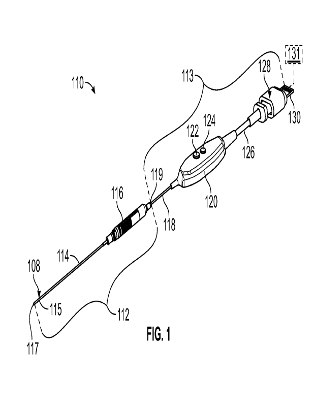
[00os] Figure 1 illustrates a perspective view of an exemplary endoscope
including a needle hub assembly and a cable assembly.
[0009] Figure 2A illustrates a perspective view of the needle hub assembly of
Figure 1.
room Figure 2B illustrates a side view of the needle hub assembly of Figure
1.
[mom Figure 3 illustrates an exploded view of the endoscope of Figure 1.
[own] Figure 4 illustrates a perspective view of the needle hub assembly of
Figure 1, including a support boot shown in phantom.
[00013] Figure 5 illustrates a perspective view of another exemplary
endoscope.
[00014] Figure 6 illustrates an exemplary electronical component.
[00015] Figure 7 illustrates another exemplary endoscope.
[00016] Figure 8 illustrates a method in a flowchart of performing a surgical
procedure.
[00017] Figure 9 illustrates an instrument positioned adjacent to an insertion
point of a patient.
[00018] Figure 10 illustrates a portion of the instrument inserted through the
insertion point of Figure 9.
3
CA 03187133 2023- 1- 24
WO 2022/035538
PCT/US2021/041397
[00019] Figure 11 illustrates the portion of the instrument withdrawn from
insertion point of Figure 10.
moan Figure 12 illustrates another exemplary instrument.
[00on] Figure 13 illustrates yet another exemplary instrument.
[00022] Figure 14 illustrates another exemplary instrument.
[00023] Like reference numbers and designations in the various drawings
indicate like elements.
DETAILED DESCRIPTION
[00024] This disclosure relates to instruments and methods that may be
utilized
during surgical procedures such as an endoscopy. An endoscopy generally
includes the
insertion of a tube into the body of a patient to observe an internal organ or
tissue.
[00025] An endoscope is provided with novel features. The endoscope is
designed to free up the hands of the surgeon or other assistants. The
endoscope may
have features allowing the endoscope to be inserted into the patient and to
remain in
position without being held by a surgeon or assistant. A unique combination of
the
size, weighting, and/or form factor may allow for these attributes. The
endoscope may
have an electronics housing and a rod extending therefrom for insertion into
the patient.
The rod may include a chip attached on the distal end including an imaging
sensor.
[000261 In some implementations, the chip may also include illumination
elements (e.g. LED elements, fiber optic bundle, light pipe, etc.) to generate
illumination in vivo. In other implementations, the illumination elements are
omitted.
Since the area may be flushed with fluid, the fluid may help to cool the chip
which
otherwise may provide heating concerns which would cause design concerns for
combining the imaging sensor and the LED illumination on the same chip. The
illumination elements may be configured to surround the imaging sensor and may
be
individually controllable. Groups of elements on the chip may have the same or
different wavelengths and may be intensity controlled based on a number of
factors
including using image features and/or sensor features for feedback. In some
implementations, one or more illumination sources may be included in a needle
hub
assembly as further described below.
[00027] The housing may be symmetric and balanced. For example, the housing
may be cylindrical. The housing may be concentric with the rod. The housing
and rod
may be fixedly attached to form a single manipulatable body. The housing may
have a
4
CA 03187133 2023- 1- 24
WO 2022/035538
PCT/US2021/041397
weight that is less 2x the weight of the rod, and in some implementations less
than 1.5x
the weight of the rod. The housing may in some instances beneficially have a
weight
that is less than the weight of the rod.
[00028] The housing may have a length that is less than 0.75x the length of
the
rod. The housing may in some instances beneficially have a length that is less
than
0.25x the length of the rod. In some instances, the components of the housing
may be
incorporated into the rod.
[00029] The housing may have a diameter that is less than 5x the diameter of
the
rod. The housing may in some instances beneficially have a diameter that is
the same
or less than the diameter of the rod.
[00030] The combined body of the housing and rod may have a center of mass
that is located distally from the housing, for example within the rod. As
such, the center
of mass may be configured to be within the body of the patient when inserted
during an
operation. Having the center of mass in the rod and/or inside the patient may
allow the
endoscope to remain more securely in place without intervention from the
surgeon or
assistant.
[00031] The housing may communicate wirelessly with a display or control
device. Alternatively, the housing may have a length of cable that is greater
than 1.5
feet from the housing to a control/display device allowing the body to be
positioned
with sufficient cable slack to negate or otherwise reduce any cable tension
affecting the
end of the housing and causing a force acting at an angle to the central axis
of the
endoscope that would cause the endoscope to tip off axis.
[00032] An endoscope for producing images of a surgery in vivo according to an
implementation of the present disclosure includes, inter alia, a hub and an
imaging rod
extending from the hub. The imaging rod may be configured to receive light and
direct
the light to an area adjacent to a distal end of the imaging rod. An imaging
sensor may
be located at the distal end portion of the imaging rod. The hub and the
imaging rod
may be attached to form a hub assembly that may have a center of mass within
the
imaging rod.
[00033] In a further implementation, the imaging rod may extend a first length
between the distal end portion and an interface between the imaging rod and
the hub.
The center of mass may be established within the imaging rod at a second
length from
the interface. The second length may be greater than or equal to 10 percent of
the first
length.
CA 03187133 2023- 1- 24
WO 2022/035538
PCT/US2021/041397
[00034] In a further implementation, the hub may include electronics
configured
to transmit image data from the imaging sensor.
[00035] In a further implementation, the second length may be greater than or
equal to 25 percent of the first length.
[00036] In a further implementation, the hub may include a light supply.
[00037] In a further implementation, the hub may include a power supply.
[00038] In a further implementation, the hub may include electronics
configured
to transmit image data wirelessly.
[00039] In a further implementation, the hub may include electronics
configured
to transmit image data digitally.
[00040] In a further implementation, the hub may include electronics
configured
to transmit image data across a coaxial cable in an analog signal.
[moan In a further implementation, the hub assembly may be symmetric with
respect to a reference plane extending along a longitudinal axis of the hub
assembly.
[00042] In a further implementation, the hub may be cylindrical.
[00043] In a further implementation, a length of the hub may be less than
0.75x
of a length of the imaging rod.
[00044] In a further implementation, a diameter of the hub may be less than 5x
of a diameter of the imaging rod.
[00045] In a further implementation, a weight of the hub may be less than 2x
of
a weight of the imaging rod.
[00046] In a further implementation, the hub may include electronics including
an electronic circuit, a light supply and an optical coupler. The electronic
circuit may
be configured to transmit image data from the imaging sensor. The light supply
may be
connected to the electronic circuit. The light supply may be configured to
generate
illumination in vivo, The optical coupler may be configured to communicate
light from
the light supply to a fiber optic. The fiber optic may be configured to
transmit light from
the hub to the distal end portion of the imaging rod. An enclosure may be
configured to
enclose the electronic circuit, the light supply and the optical coupler. A
hub coupler
may connect the imaging rod to the enclosure.
[00047] In a further implementation, a cable assembly may include a first
cable,
a second cable, a connector and a button yoke having one or more controls. The
first
cable may be coupled to a proximal end portion of the hub assembly. The button
yoke
may interconnect the first and second cables. The second cable may
interconnect the
6
CA 03187133 2023- 1- 24
WO 2022/035538
PCT/US2021/041397
button yoke and the connector. The connector may have a terminal configured to
interface with the external device.
m00481 An endoscope for producing images of a surgery in vivo according to an
implementation of the present disclosure includes, inter alia, a
communications
assembly, a hub coupled to the communications assembly, and an imaging rod
extending from the hub. The hub and the imaging rod may be attached to form a
hub
assembly having a center of mass that may be established distally of the hub.
An
imaging sensor may be coupled to a distal end portion of the imaging rod.
[00049] In a further implementation, the center of mass may be established
within the imaging rod.
[00050] In a further implementation, a light source may be within the imaging
rod adjacent to the distal end portion.
[000si] In a further implementation, the hub may include an enclosure
configured to enclose electronics.
[00052] In a further implementation, a plurality of light sources may be
configured in an array to surround the imaging sensor.
[00053] In a further implementation, the plurality of light sources may be
respective pathways configured to branch from a common light source.
[00054] In a further implementation, the light sources may be individually
controllable.
[000551 An endoscope for producing images of a surgery in vivo according to an
embodiment of the present disclosure includes, inter alia, a communications
assembly
and a hub assembly coupled to the communications assembly. The hub assembly
may
include an imaging rod, an imaging sensor and electronics coupled to the
imaging
sensor. The imaging rod may include a main body extending a first length
between a
proximal end portion and a distal end portion relative to a longitudinal axis.
The
imaging sensor may be arranged adjacent to the distal end portion of the
imaging rod.
The electronics may be arranged in an internal cavity of the imaging rod. The
hub
assembly may have a center of mass established within the imaging rod at a
second
length from the proximal end portion. The second length may be greater than or
equal
to 50 percent of the first length.
[00056] In a further implementation, the second length may be greater than 50
percent of the first length.
7
CA 03187133 2023- 1- 24
WO 2022/035538
PCT/US2021/041397
[00057] In a further implementation, the electronics may be arranged adjacent
to the distal end portion of the imaging rod.
[mom In a further implementation, the second length may be greater than 75
percent of the first length.
[00059] A method of performing an endoscopy according to an implementation
of the present disclosure includes, inter cilia, inserting a distal end
portion of an imaging
rod through an insertion point of a patient, the imaging rod extending from a
hub to the
distal end portion, and the hub and the imaging rod attached to establish a
hub assembly
having a center of mass established distally of the hub, and then inserting
the center of
mass through the insertion point. An imaging sensor may be located at the
distal end
portion of the imaging rod. The method may include obtaining an image by the
imaging
sensor at a position inward of the insertion point subsequent to the step of
inserting the
center of mass.
[00060] In a further implementation, the center of mass may be established
within the imaging rod.
[00061] In a further implementation, the hub may be outward of the insertion
point during the obtaining step.
[00062] In a further implementation, the method may include releasing the hub
assembly such that the center of mass may be in vivo. The method may include
releasing the hub assembly such that the hub may be substantially cantilevered
from the
imaging rod outward of the insertion point.
[00063] In a further implementation, a length of the hub may be less than
0.75x
of a length of the imaging rod. A weight of the hub may be less than 2x of a
weight of
the imaging rod.
[00064] In a further implementation, the method may include communicating
light to the imaging sensor prior to the obtaining step.
[00065] In a further implementation, the communicating step may include
communicating the light from the hub, then through the imaging rod, and then
towards
an area adjacent to the distal end portion of the imaging rod.
[00066] In a further implementation, the communicating step may include
communicating the light from a plurality of light sources adjacent to the
distal end
portion of the imaging rod. The light sources may be configured in an array to
surround
the imaging sensor.
8
CA 03187133 2023- 1- 24
WO 2022/035538
PCT/US2021/041397
[00067] In a further implementation, the communicating step may include
individually controlling the light sources to communicate the light.
[00068] In a further implementation, the insertion point may be established by
an incision in skin of the patient.
[00069] Figures 1-4 illustrate an exemplary endoscope 110, which may be
utilized to produce images of a surgery in vivo. Referring to Figure 1, the
endoscope
110 may include a needle hub assembly 112 and a cable (e.g., communications)
assembly 113. The needle hub assembly 112 may include a scope 114 (e.g., a
camera
or imaging rod) secured in a needle hub 116, as illustrated in Figures 1 and
2B. The
scope 114 extends from a distal end of the needle hub 116. The scope 114 and
needle
hub 116 can be dimensioned such that the needle hub assembly 112 is
substantially
symmetrical (e.g., mirror symmetry) with respect to a reference plane REF that
extends
along a longitudinal (e.g., central) axis X of the assembly 112 to divide the
assembly
112 into two opposed portions, as illustrated in Figures 2A-2B.
[00070] Various techniques may be utilized to dimension the hub assembly 112.
Referring to Figure 2B, with continuing reference to Figures 1 and 2A, the
scope 114
may extend a first length LI between the terminal (e.g., distal) end 117 and
an interface
121 between the scope 114 and the distal end of the hub 116 relative to the
longitudinal
axis X. The interface 121 may be established at or adjacent to a proximal end
of the
scope 114. In implementations in which the scope 114 is flexible, the first
length Li
corresponds to a maximum configurable length of the scope 114. In
implementations
in which the hub 116 is omitted, the first length Li may be established
between the
proximal and distal ends of the scope 114. In implementations, the first
length Li may
be between approximately 100 millimeters (mm) and 300 mm. The needle hub 116
may
extend a second length L2 between opposed proximal and distal ends of the
needle hub
116 relative to the longitudinal axis X. The scope 114 may establish a first
diameter
Di. The needle hub 116 may establish a second diameter D2. The first and
second
lengths Li, L2 and/or first and second diameters D1, D2 may be the same or may
differ.
In some implementations, the length L2 of the needle hub 116 is less than
0.75x of the
length LI of the scope 114, the diameter D2 of the hub 116 is less than 5x of
the
diameter D1 of the scope 114, and/or a weight of the hub 116 is less than 2x
of a weight
of the scope 114.
[00071] The hub 116 and scope 114 can be attached to form the hub assembly
112 having a center of mass CM. The center of mass CM may be established at a
9
CA 03187133 2023- 1- 24
WO 2022/035538
PCT/US2021/041397
longitudinal position relative to the longitudinal axis X. The longitudinal
position of the
center of mass CM may be aligned with a longitudinal position along the scope
114
relative to the longitudinal axis X. The center of mass CM may be established
distally
of the needle hub 116 relative to the longitudinal axis X. In implementations,
the center
of mass CM may be established within the scope 114 along the longitudinal axis
X. In
other implementations, the needle hub assembly 112 may be configured such that
the
center of mass may be established adjacent to, but offset from, the scope 114,
as
illustrated by center of mass CM' (Figure 2A). The endoscope 110 may be
configured
such that the center of mass CM may be inside or outside of the patient during
a surgical
procedure.
[00072] The hub 116 and/or scope 114 may be symmetric or asymmetric to
establish the center of mass CM. For example, the scope 114 may have a
curvilinear
geometry such that one or more portions of the scope 114 are offset from the
longitudinal axis X to establish an asymmetric configuration. As another
example,
component(s) within the hub 116 may be arranged such that a center of mass of
the
component(s) are offset from the longitudinal axis X.
[00073] The center of mass CM may be established at various positions relative
to the scope 114 and/or needle hub 116. The hub assembly 112 may be configured
such
that a portion of the hub assembly 112 including the center of mass CM may be
positioned in a patient to improve retention of the endoscope 110 without
intervention
by the surgeon or assistant, although another other portion of the hub
assembly 112 may
be positioned outside of the patient. The center of mass CM may established
at,
adjacent to, or distally of the distal end of the needle hub 116.
[00074] The center of mass CM may be established at a distance Lcm from a
proximal boundary of the first length Ll. The proximal boundary of the first
length Li
may be established by the interface 121 between the scope 114 and the distal
end of the
hub 116 or may be established by a proximal end of the scope 114 for
implementations
in which the hub 116 is omitted. The hub assembly 112 may be configured such
that a
center of mass CM' is offset from the scope 114. The distance Lcm may be less
than 10
percent of the first length LL The center of mass CM may established at the
interface
121 between the scope 114 and the distal end of the needle hub 116. In
implementations,
center of mass CM" may be established proximal of the interface 121 within the
hub
116. The hub assembly 112 may be configured such that the distance Lcm is
greater
than or equal to 10 percent of the first length Li, or more narrowly greater
than or equal
CA 03187133 2023- 1- 24
WO 2022/035538
PCT/US2021/041397
to approximately 25 percent of the first length Ll. In implementations, the
distance Lcm
may be less than or equal to approximately 50 percent of the first length Li.
For the
purposes of this disclosure, the terms "substantially," "approximately" and
"about"
mean 10 percent of the stated value or relationship unless otherwise
indicated.
Utilizing the techniques disclosed herein, including the disclosed dimensional
relationships and distributions, the surgeon or assistant may position the hub
assembly
112 in vivo in a manner that reduces a likelihood of movement Or intervention
while
the endoscope 110 is not being held.
[00075] Referring to Figure 3, with continuing reference to Figure 1, the
cable
assembly 113 may include a first cable 118 (e.g. a micro coaxial cable), a
button yoke
120, a second cable 126, and a connector 128. The connector 128 may include a
terminal 130 configured to communicate with an external device, such as a
display or
control device 131 (shown in dashed lines in Figure 1 for illustrative
purposes). In other
implementations, the endoscope 110 communicates wirelessly with the control
device
131. The needle hub assembly 112 or the cable assembly 113 can include a power
supply 133 that provides power to the various electrical components of the
endoscope
110 in operation (shown in dashed lines in Figure 3 coupled to electronic
circuit 138
for illustrative purposes). In other implementations, power is provided by an
external
device and is communicated by the terminal 130 to the various electrical
components.
[00076] The scope 114 can include an imaging sensor 108 located on, at or
otherwise adjacent to a distal end portion 115 of the scope 114 for obtaining
images of
a surgical site. The imaging sensor 108 may be a sensor assembly including a
sensor
and optics. The scope 114 can be configured to receive light and direct the
light to or
otherwise towards an area (e.g., scene or space being viewed by the surgeon)
adjacent
to the distal end portion 115 of the scope 114. The light may be reflected
from the area
back towards the sensor 108.
[00077] The scope 114 and each cable 118, 126 may be relatively rigid or
flexible. The distal end portion 115 of the scope 114 establishes a terminal
end 117
(e.g., tip) of the endoscope 110. At least a portion of the scope 114
including the distal
end portion 115 can be relatively flexible or bendable, and may comprise a
Nitinol
material, for example. Configuring the scope 114 to be relatively flexible can
facilitate
orienting the sensor 108 including bending or steering the sensor 108 around
corners
and viewing various angles of the surgical site, for example.
11
CA 03187133 2023- 1- 24
WO 2022/035538
PCT/US2021/041397
[00078] The first cable 118 may be a coaxial cable (e.g. a micro coax cable).
The
first cable 118 may be coupled to a proximal end portion 119 of the hub
assembly 112,
as illustrated in Figures 1 and 4. In some implementations, the first cable
118 may
communicate analog signals between the needle hub 116 and the button yoke 120.
The
button yoke 120 may interconnect the first cable 118 and the second cable 126.
The
second cable 126 may interconnect the button yoke 120 and the connector 128.
[00079] The button yoke 120 may have one or more controls (e.g. buttons,
dials,
levers, etc.) such as button 122 and button 124, for example. Each button 122,
124 may
have one or more functions, such as image and video capture. Each button 122.
124
may be programmable for a number of functions. Additionally, multiple
functions can
be accessed based on the number of times a button 122, 124 is pressed, the
amount of
time within which the button 122, 124 is pressed multiple times, and/or the
amount of
time for which a button 122, 124 remains depressed continuously.
[mow The first and second cables 118, 126 can have various dimensions. In
sonic implementations, the second cable 126 has a length of approximately 2
feet,
which may allow the button yoke 120 to rest on a surface when the endoscope
110 is in
use and may minimize or otherwise reduce the effect on the stationary position
of the
hub 116.
[00081] The needle hub 116 may include a hub coupler 134 that connects the
scope 114 to other components of the needle hub 116. The needle hub 116 may
include
various electronics 123 including a flexible circuit board 135, the electronic
circuit 138,
a light supply 137 and an optical coupler 136. The flexible circuit board 135
may extend
from the needle hub 116 through the scope 114 to the sensor 108. The flexible
circuit
board 135 may be connected to the electronic circuit 138. The electronic
circuit 138
may be in the form of a printed circuit board and may include one or more
chips. The
electronic circuit 138 can be configured to transmit image data across a
coaxial cable,
such as the first cable 118, in an analog signal. In implementations, one or
more of the
electronics 123 may be incorporated into the button yoke 120, including the
power
supply 133, flexible circuit board 135, optical coupler 136, light supply 137
and/or
electronic circuit 138, and a separate hub 116 including the enclosure 139 may
be
omitted.
moi)82] The scope 114 can be configured to receive light and direct the light
to
or towards an area adjacent to the distal end portion 115 of the scope 114.
The light
may be directly or indirectly communicated from the scope 114 to the sensor
108. For
12
CA 03187133 2023- 1- 24
WO 2022/035538
PCT/US2021/041397
example, the light may be reflected from the area back to or otherwise towards
the
sensor 108. The circuit 138 and/or flexible circuit board 135 may be connected
to the
light supply 137 (e.g., light source or illumination element). The light
source 137 may
be a light emitting diode (LED), for example, and can be configured and
utilized to
generate illumination in vivo. The optical coupler 136 may be configured to
communicate light from the light source 137 to a fiber optic (e.g., light
pipe) 103 (shown
in dashed lines in Figure 3 for illustrative purposes). The fiber optic 103
may be
configured to transmit the light from the needle hub 116 to the distal end
portion 115
of the scope 114. In other implementations, the fiber optic 103 may be omitted
and the
light source 137 may be positioned within the scope 114 distally of the needle
hub 116.
In implementations, a separate light source may be situated externally but
adjacent to
the distal end portion 115 of the scope 114 to illuminate the surgical site.
[00083] In some implementations, imaging sensor 308 and one or more light
sources 316 are integrated with or mounted to a common circuit board 323
(e.g., chip)
to establish an electrical component 325, as illustrated in Figure 6. The
light sources
316 can be configured in an array to surround the sensor 308 and may be
individually
controllable. In implementations, the light sources 316 may be respective
pathways
configured to branch from a singular, common light source 327 (shown in dashed
lines
for illustrative purposes). The light sources 316 may be utilized to improve
communication of light in a relatively compact arrangement. The common light
source
327 may be coupled to the circuit board 323 or another portion of the
endoscope.
Groups of light sources 316 on the circuit board 323 may have the same or
different
wavelengths and may be intensity controlled based on a number of factors
including
using image features and/or sensor features for feedback. The electrical
component 325
can be situated at any of the positions of the image sensors disclosed herein.
For
example, the electrical component 325 can be coupled or attached on or
adjacent to the
distal end portion 115 of the scope 114 (Figure 1). Combining the imaging
sensor 308
and light source(s) 316 on the same circuit board 323 may improve cooling
augmentation by fluid conveyed to a surgical site, such as fluid utilized to
flush the
surgical site during a surgical procedure.
[00084] The various electronics of the needle hub assembly 112 can be
configured to transmit image data wirelessly and/or digitally from the imaging
sensor
108 to an external device, such as the control device 131, and/or another
component of
the endoscope 110, such as the button yoke 120. Other sensors can be
incorporated into
13
CA 03187133 2023- 1- 24
WO 2022/035538
PCT/US2021/041397
the endoscope 110. For example, one or more sensors may be configured to sense
or
measure various conditions at the distal end portion 115 of the scope 114,
such as
temperature sensors, pressure sensors, etc. In some implementations, an
accelerometer
and/or gyroscope is positioned in the needle hub 116 and/or scope 114 to sense
a change
in position and/or orientation of the endoscope 110.
[00085] The terminal end 117 of the distal end portion 115 can be established
at
various angles relative to a central or longitudinal axis of the endoscope
110. For,
example, the terminal end 117 can be substantially perpendicular to the
longitudinal
axis X of the scope 114, as illustrated in Figure 1 and 2A-2B. In some
implementations,
a terminal end 117' of distal end portion 115' establishes an angle a that is
transverse
to a central or longitudinal axis X of the scope 114', as illustrated in
Figure 7. A sensor
image obtained by sensor 108' can be oriented at an angle (e.g., 30 degrees)
corresponding to the angle a, for example. Electronic circuit 138' or another
portion of
endoscope 110' can be programmed with or otherwise incorporate logic to
perform a
correction or translation of the captured image(s) to reorient the captured
image(s) as
the sensor 108' rotates during a procedure.
[00086] Referring still to Figure 3, the needle hub 116 may include an
enclosure
139 configured to enclose components of the needle hub 116. The enclosure 139
may
include a first shell 162 and a second shell 164 that cooperate to enclose the
electronics
and other components of the needle hub 116 (e.g., light source 137, circuit
138, and
optical coupler 136). The hub coupler 134 may couple the scope 114 to the
shells 162
and 164 of the enclosure 139. The hub coupler 134 may be a separate and
distinct
component or may be incorporated into the enclosure 139 and/or the scope 114.
[00087] A shield 230 may at least partially or completely surround the
enclosure
139, as illustrated in Figure 4. The shield 230 may take the form of a
flexible shield
and may be formed of a conductive material such as copper. Accordingly, the
shield
230 may take the form of a copper foil. A support boot 166 may support the
enclosure
139 and the first cable 118, as illustrated in Figure 4 (shown in phantom).
[mow For comparison purposes, Figure 5 illustrates another exemplary
endoscope 410. The endoscope 410 may have a handpiece 412 and a camera rod
414.
The handpiece 412 may be designed to be held by the surgeon or an assistant
and to
include all of the control electronics. In these implementations a center of
mass of the
endoscope 410 could he significantly back in the handpiece 412 and therefore
may be
14
CA 03187133 2023- 1- 24
WO 2022/035538
PCT/US2021/041397
held more extensively by the assistants rather than being staying static
without being
guided by the assistant.
[00089] Figure 8 illustrates an exemplary method of performing a surgical
procedure in a flowchart 540. The method 540 may be utilized to perform an
endoscopy.
The method 540 can be utilized with any of the instruments and assemblies
disclosed
herein, including the endoscopes 110, 110' and endoscopes 710, 810 (Figures 12-
13).
Obtained images may be utilized pre-operatively, inter-operatively, and/or
post-
operatively and may be utilized in conducting various surgical procedures,
such as an
arthroplasty to restore functionality to a joint. Fewer Or additional steps
than are recited
below could be performed within the scope of this disclosure, and the recited
order of
steps is not intended to limit this disclosure. Reference is made to
instrument (e.g.,
endoscope) 610 of Figures 9-11 for illustrative purposes.
[00090] Referring to Figure 9, with continuing reference to Figure 8, the
instrument 610 may include a hub assembly 612 coupled to a cable assembly 613.
The
hub assembly 612 may include a hub 616 and a scope 614 (e.g., a camera or
imaging
rod) coupled to the hub 616. The hub 616 may have a generally or substantially
tubular
geometry and may serve as a handle to position the hub assembly 612. The hub
assembly 612 may include an imaging sensor 608 located at a distal end portion
615 of
the imaging rod 614. The imaging rod 614 may extend from the hub 616 to the
distal
end portion 615. The hub 616 and the imaging rod 614 may be attached to
establish the
hub assembly 612 having a center of mass CM. The center of mass CM may be
established distally of the hub 616 relative to a longitudinal axis X of the
hub assembly
612 (Figure 10). The hub assembly 612 may be configured such that the center
of mass
CM may be established within or adjacent to the imaging rod 614. The hub
assembly
612 may be configured according to any of the techniques disclosed herein. In
implementations, a length of the hub 616 may be less than 0.75x of a length of
the
imaging rod 614, a weight of the hub 616 may be less than 2x of a weight of
the imaging
rod 614 and/or a diameter of the hub 616 may be less than 5x of a diameter of
the
imaging rod 614.
[00091] At step 542, the instrument 610 may be positioned relative to an
insertion point 611 in a body B of a patient at a surgical site S. The
insertion point 611
may be an incision made through skin, an orifice, or another opening in the
body B of
the patient. Method 540 may include forming the incision prior to step 542.
CA 03187133 2023- 1- 24
WO 2022/035538
PCT/US2021/041397
[00092] Referring to Figure 10, with continuing reference to Figures 8-9, step
542 may include moving the instrument 610 in a direction D1, and then
inserting a
portion of the instrument 610 through the insertion point 611 at step 544.
Step 544 may
occur such that the portion of the instrument 610 is situated in vivo. Step
544 may
include inserting at least the distal end portion 615 of the imaging rod 614
through the
insertion point 611 of the patient, and then then inserting the center of mass
CM of the
instrument 610 through the insertion point 611. A portion of the hub assembly
612
including the center of mass CM may be positioned within the patient, while
another
portion of the hub assembly 612 may be positioned outside of the patient, such
as the
hub 616 and/or a portion of the imaging rod 614 proximal of the center of mass
CM
including a proximal end of the imaging rod 614.
[00093] At step 546, the method 540 may include communicating light to the
imaging sensor 608. Various techniques may be utilized to communicate light to
the
imaging sensor 608. In implementations, step 546 may include communicating the
light
from the hub 616, then through the imaging rod 614, and then to an area of the
patient
adjacent the distal end portion 615 of the imaging rod 614. The light may be
reflected
from the area back to or otherwise towards the imaging sensor 608 (see also
hub 116,
imaging sensor 108 and imaging rod 114 of Figure 1). In implementations, step
546
may include communicating the light from one or more light sources adjacent
the distal
end portion 615 of the imaging rod 614. The light sources may be configured in
an array
to surround the imaging sensor 608 (see, e.g., imaging sensor 308 and light
sources 316
of Figure 6). Step 546 may include individually controlling the light sources
to
communicate the light at step 548.
[00094] At step 550, the surgeon or assistant may cause the instrument 610 to
obtain one or more images by the imaging sensor 608 at a position inward of
the
insertion point 611. Step 550 may occur subsequent to positioning the
instrument 610
at step 542 and/or communicating the light at step 546. The center of mass CM
of the
instrument 610 may be inward of the insertion point 611 or otherwise situated
in vivo,
and the hub 116 may be situated outward of the insertion point 611 or
otherwise situated
ex vivo during obtaining the image(s) at step 550. At step 552, the image(s)
may be
communicated to an external device (see, e.g., external device 131 of Figure
1).
[00095] At step 554, the surgeon or assistant may release control of the
instrument 610 while the distal end portion 615, imaging sensor 608 and/or
center of
mass CM of the instrument 610 are situated in vivo, as illustrated in Figure
10. Step
16
CA 03187133 2023- 1- 24
WO 2022/035538
PCT/US2021/041397
554 may include releasing control of the hub assembly 612 such that the center
of mass
CM may be situated in vivo and such that the hub 116 may be substantially
cantilevered
from the imaging rod 614 outward of the insertion point 611. For the purposes
of this
disclosure, the term "substantially" cantilevered means that no more than 10
percent of
the hub assembly 112 outward of the insertion point 611 is supported by means
other
than the imaging rod 614. Step 554 may include balancing the hub assembly 612
at or
otherwise adjacent to the insertion point 611 in response to releasing control
of the hub
assembly 612. The instrument 610 may remain in position without being held or
otherwise supported by a surgeon or assistant, which may improve flexibility
and
decrease time in performing other steps in a surgical procedure.
[00096] Referring to Figure 11, with continuing reference to Figure 8, at step
556
the portion of the instrument 610 that is situated in vivo may be moved in a
direction
D2 until the instrument 610 is withdrawn from the insertion point 611 and
removed
from the patient. Step 556 may include withdrawing the imaging sensor 608,
distal end
portion 615 and center of mass CM of the instrument 610 from the patient.
[00097] Figure 12 illustrates another exemplary instrument 710. The instrument
710 may be an endoscope utilized to obtain one or more images of a surgical
site. The
instrument 710 may include a needle hub assembly 712 coupled to a cable (e.g.,
communications) assembly 713 (shown in dashed lines for illustrative
purposes). In the
implementation of Figure 12, a separate hub is omitted from the hub assembly
712.
[00098] The hub assembly 712 may include a scope 714 (e.g., a camera or
imaging rod). The scope 714 may include a main body 729 extending along a
longitudinal axis X between a distal end portion 715 and a proximal end
portion 719 of
the hub assembly 712. The main body 729 may have a generally or substantially
tubular
geometry and may establish an internal cavity 725. The scope 714 may establish
a first
diameter Dl. The main body 729 may be dimensioned such that the first diameter
D1
is substantially constant between the distal end portion 715 and proximal end
portion
719 of the of the scope 714.
[00099] The scope 714 can include an imaging sensor 708 configured to obtain
images of a surgical site. The imaging sensor 708 may be arranged within the
internal
cavity 725 and may be arranged at the distal end portion 715 of the scope 714
to obtain
one or more images of a surgical site. The surgeon or assistant may utilize a
portion of
the scope 714 as a handle to situate the imaging sensor 708 at a desired
position and
orientation within the patient.
17
CA 03187133 2023- 1- 24
WO 2022/035538
PCT/US2021/041397
[mom The hub assembly 710 may include various electronics 760, including
any of the electronics disclosed herein, such as a flexible circuit board,
electronic
circuit, light supply, optical coupler and/or power supply (see, e.g., Figure
3). The
electronics 760 may be integrated onto a single chip to establish an
electronics unit,
which may integrate or may be coupled to the imaging sensor 708. The
electronics 760
may be arranged at various positions within the cavity 725 of the scope 714.
The
electronics 760 may be arranged proximal of a center of mass CM of the hub
assembly
712, such as at or adjacent to the proximal end portion 719 of the scope 714.
The
electronics 760 may include a light source within the scope 714 adjacent to
the proximal
end portion 719.
[mom] The center of mass CM may be established at a longitudinal position
between the distal and proximal end portions 715, 719 of the hub assembly 712,
including within the scope 714. The center of mass CM may be established at a
distance
Lcm from a proximal boundary of a first length Li of the scope 714. The hub
assembly
712 may be configured such that the center of mass CM is established according
to any
of the ratios of the distance Lcm and first length Li disclosed herein. In
implementations, the hub assembly 712 may be configured such that the distance
Lcm
is greater than or equal to 25 percent of the first length Li, or more
narrowly greater
than or equal to approximately 50 percent of the first length Ll. In
implementations,
the distance Lcm may be less than or equal to approximately 75 percent of the
first
length Ll.
[000102] Arranging at least some, a majority of, or all electronics 760 and/or
other internal components of the hub assembly 712 into the scope 714 may be
utilized
to shift a center of mass CM of the hub assembly 712 relatively more distally
relative
to the proximal end portion 719 of the hub assembly 712, which may improve
retention
of the instrument 710 without intervention by the surgeon or assistant.
[000103] Figure 13 illustrates another exemplary instrument 810. The
instrument
810 may be an endoscope utilized to obtain one or more images of a surgical
site. The
instrument 810 may include a hub assembly 812 coupled to a cable (e.g.,
communications) assembly 813. In the implementation of Figure 13, a separate
hub is
omitted.
[000104] The instrument 810 may include various electronics 860 arranged at
various positions within a cavity 825 of the scope 814. The electronics 860
may include
18
CA 03187133 2023- 1- 24
WO 2022/035538
PCT/US2021/041397
a first set of electronics 860-1 and a second set of electronics 860-2, which
may include
any of the electronics disclosed herein.
[0001051 The electronics 860 may be distributed within the scope 814 to
establish
a center of mass CM at various locations between a distal end portion 815 and
proximal
end portion 819 of the hub assembly 812. The center of mass CM may be
established
at a longitudinal position between the distal and proximal end portions 815,
819,
including within the scope 814. The first set of electronics 860-1 may be
arranged distal
of the center of mass CM of the hub assembly 812. The second set of
electronics 860-
2 may be arranged proximal of the center of mass CM. The first set of
electronics 860-
1 may be arranged at or adjacent to the distal end portion 815 of the scope
814. The
second set of electronics 860-2 maybe arranged at or adjacent to the proximal
end
portion 819 of the scope 814. In implementations, the second set of
electronics 860-2
is omitted such that substantially all electronics of the hub assembly 812 are
arranged
in a distal half of the imaging rod 814. The electronics 860-1 may include a
light source
such as a LED, which may be positioned adjacent to and proximal of the imaging
sensor
808.
[000106] The center of mass CM may be established at a longitudinal position
between the distal and proximal end portions 815, 819 of the hub assembly 812,
including within the scope 814. The center of mass CM may be established at a
distance
Lcm from a proximal boundary of a first length Li of the scope 814. The hub
assembly
812 may be configured such that the center of mass CM is established according
to any
of the ratios of the distance Lcm and first length Li disclosed herein. In
implementations, the hub assembly 812 may be configured such that the distance
Lcm
is greater than or equal to 25 percent of the first length Li, more narrowly
greater than
or equal to approximately 50 percent of the first length Li, or even more
narrowly
greater than or equal to approximately 75 percent of the first length Ll. In
implementations, the distance Lcm may be less than or equal to approximately
90
percent of the first length Ll.
[000107] Arranging at least some, a majority of, or all electronics 860 and/or
other
internal components of the hub assembly 812 adjacent to the distal end portion
815 of
the instrument 810 may be utilized to shift the center of mass CM of the hub
assembly
812 relatively more distally relative to the proximal end portion 819 of the
hub assembly
812, which may improve retention of the instrument 810 without intervention by
the
surgeon or assistant.
19
CA 03187133 2023- 1- 24
WO 2022/035538
PCT/US2021/041397
[000108] Referring to Figure 14, instrument 910 may include electronics 960.
One
or more of the electronics 960 may be incorporated into a cable assembly 913.
The
cable assembly 913 may include a button yoke 920, which may incorporate
electronics
960-2. The electronics 960-2 can include any of the electronics disclosed
herein,
including the power supply 133, flexible circuit board 135, optical coupler
136, light
supply 137 and/or electronic circuit 138 (Figure 3). A separate hub including
an
enclosure to enclose the electronics may be omitted. The instrument 910 may
include
electronics 9601 adjacent to a distal end portion 915 of an imaging rod 914,
or the
electronics 960-1 may be omitted and/or incorporated into the button yoke 920.
[000109] The novel devices and methods of this disclosure provide versatility
in
obtaining images of patient anatomy during an endoscopy. The disclosed
instruments
may be configured to allow the instrument to be inserted into the patient and
to remain
in position without being held or otherwise supported by a surgeon or
assistant. The
disclosed instruments may be configured to have a center of mass that improves
retention of the instrument without intervention from the surgeon or
assistant, which
can decrease complexity and time to perform a surgical procedure.
[mono] Although the different non-limiting embodiments are illustrated as
having specific components or steps, the embodiments of this disclosure are
not limited
to those particular combinations. It is possible to use some of the components
or
features from any of the non-limiting embodiments in combination with features
or
components from any of the other non-limiting embodiments.
[mum The foregoing description shall be interpreted as illustrative and not in
any limiting sense. A worker of ordinary skill in the art would understand
that certain
modifications could come within the scope of this disclosure. For these
reasons, the
following claims should be studied to determine the true scope and content of
this
disclosure.
CA 03187133 2023- 1- 24