Note: Descriptions are shown in the official language in which they were submitted.
CA 03188299 2022-12-26
WO 2022/011020 PCT/US2021/040699
SUBCUTANEOUS PORT FOR MINIMALLY INVASIVE IMPLANTATION
RELATED APPLICATIONS
[0001] This application claims priority to POT Application No.
POT/US20/41140, filed on
July 8, 2020, to U.S. Provisional Patent Application No. 63/123,028, filed on
December 9,
2020, and to U.S. Patent Application No. 17/146,253, filed on January 11,2021;
the entire
contents of each of which are hereby incorporated by reference herein. The
entire
disclosures of all the related applications set forth in this section are
hereby incorporated
by reference in their entireties.
BACKGROUND
[0002] The present disclosure relates to devices and methods for
facilitating and/or
improving repeated deliveries of fluids (e.g., fluids carrying nutrients,
medicament and/or
agents such as chemotherapy agents) into vasculature of a subject, and more
particularly,
but not exclusively, to vascular access ports and methods of minimally
invasive delivery
and deployment thereof in a body of a subject.
[0003] Repeated needle pricking for facilitating delivery or withdrawal of
fluids (e.g.,
medication or agents) to patient's vascular system causes harm to local
tissues and
decreases target blood vessel functionality and needle placement accuracy.
This
phenomenon is often evident in chronic diabetes, dialysis or chemotherapy
patients, for
example, who require continuous and repeated intravenous fluids administration
for
prolonged periods.
[0004] A vascular access port is a device that enables such repeated
pricking and fluid
administration while minimizing the accumulated harm caused by needle pricking
and
powered injections of fluid. The access port is subcutaneously implanted, in a
surgically
formed pocket in proximity to a large blood vessel, usually in the chest. It
is basically formed
of a port body enclosing a cavity, which is capped with a septum member
configured for
supporting the upper skin layers and for accepting repeated needle pricking
therethrough
for intravascular fluid deliveries sealed to the surrounding body tissues. The
port is attached
to a catheter (a thin, flexible tube) which provides fluid communication with
a large blood
vessel, such as the superior vena cava, in order to allow the injected fluid
to dilute in the
blood stream.
[0005] The implantation of a port is considered a minor procedure performed
under local
or general anesthesia by an interventional radiologist or a surgeon. First,
the surgeon
-1-
CA 03188299 2022-12-26
WO 2022/011020 PCT/US2021/040699
achieve access to the desired vein, a skin incision is made afterwards in the
access point.
Second larger incision is made above the desired location of the port, through
which a
pocket-like subcutaneous void is made using blunt device. The catheter is
extended
subcutaneously between the two incisions using a blunt tunneler. One end of
the catheter
is then inserted into the vein and its other end is coupled to the port.
Optionally, during
deployment the catheter is cut to a desired length.
[0006] Besides progress made in past years in access ports design, there is
still a need
to develop ports and methods of implantation and deployment thereof, which are
less
traumatic and invasive, and simpler to perform, potentially also by non-
surgical medical
personnel.
[0007] It should be noted that this Background is not intended to be an aid
in determining
the scope of the claimed subject matter nor be viewed as limiting the claimed
subject matter
to implementations that solve any or all of the disadvantages or problems
presented above.
The discussion of any technology, documents, or references in this Background
section
should not be interpreted as an admission that the material described is prior
art to any of
the subject matter claimed herein.
SUMMARY
[0008] The present disclosure relates to devices and methods for
facilitating and/or
improving repeated deliveries of fluids (e.g., fluids carrying nutrients,
medicament and/or
agents such as chemotherapy agents) into vasculature of a subject, and more
particularly,
but not exclusively, to vascular access ports and methods of minimally
invasive delivery
and deployment thereof in a body of a subject.
[0009] In certain embodiments, there is provided a subcutaneous port. The
subcutaneous port can comprise a port body enclosing a cavity, wherein the
cavity
comprises a first opening covered by a septum configured for repeated needle
penetrations
therethrough and a second opening configured for facilitating fluid
communication between
the cavity and a catheter. In some embodiments, the port body comprises a
rigid port
gripping portion configured to releasably engage with a medical clamp, and
wherein the
port body is configured to be pushed into a subcutaneous target implantation
site using the
medical clamp when the medical clamp is engaged with the port gripping
portion. In some
embodiments, the port gripping portion is configured to receive manual forces
and/or
torques from the medical clamp in at least one axis, wherein the manual forces
and/or
-2-
CA 03188299 2022-12-26
WO 2022/011020 PCT/US2021/040699
torques received at the port gripping portion are sufficient to releasably
affix the medical
damp to the port gripping portion,
[0010] In some embodiments, the port body includes a rigid port body member
surrounding the cavity and/or defining the first cavity opening, the rigid
port body member
comprising a front portion, a rear portion, and lateral portions extending
from opposing sides
thereof between the front portion and the rear portion, wherein the rear
portion comprises
the port gripping portion.
[0011] hi some embodiments, the manual forces and/or torques received at
the port
gripping portion are sufficient to form or enlarge a subcutaneous void and/or
passage in a
body of a subject using the subcutaneous port and/or to maneuver the
subcutaneous port
along the subcutaneous void and/or passage, without slipping from, or
releasing grip of, the
port gripping portion.
[0012] In some embodiments, the second cavity opening is in juxtaposition
with the port
gripping portion, and/or located below the port gripping portion, farther from
the first cavity
opening than the port gripping portion.
[0013] In some embodiments, the port gripping portion comprises a wall,
wherein the
wall comprises opposing first and second outer wall surfaces sized to
accommodate
clamping surfaces of the medical clamp.
[0014] In some embodiments, the port gripping portion is configured such
that the
manual forces are equal to or smaller than 10 kgf and/or the manual torques
are equal to
or smaller than about 0.25 Wm; when the clamping surfaces of the medical clamp
are
oriented and spaced apart with each other to substantially match a shape and a
thickness
of the wall.
[0015] In some embodiments, the port gripping portion is configured such
that manually
operable arms of the medical clamp are allowed to interlock when the clamping
surfaces of
the medical clamp are oriented and spaced apart with respect to each other to
match a
shape and a thickness of the wall.
[0016] In some embodiments, the first and second outer surfaces are
parallel.
[0017] In some embodiments, the port gripping portion comprises a clamp
engagement
feature.
-3-
CA 03188299 2022-12-26
WO 2022/011020 PCT/US2021/040699
[0018] In some embodiments, the damp engagement feature comprises one or
more of
tapered surfaces, roughened surfaces, scored surfaces, teeth, recesses, and
through-
holes.
[0019] hi some embodiments, the damp engagement feature is formed in a wall
comprising first and second outer surfaces.
[0020] hi some embodiments, each of the outer wall surfaces extends
vertically between
lateral portions of the port body.
[0021] hi some embodiments, each of the outer wall surfaces extends
horizontally
between a bottom portion and a top portion of the port body.
[0022] In some embodiments, an average or maximal thickness of the wall is
between
about 1 mm and about 4 mm, and/or an angle formed between the first and second
outer
wall surfaces is equal to or smaller than about 20 .
[0023] In some embodiments, the second cavity opening is in juxtaposition
with, and/or
located inferiorly to, the port gripping portion.
[0024] In some embodiments, the port body comprises a plurality of
components
coupled to each other, and wherein the port gripping portion is associated
with a first one
of the plurality of coupled components,
[0025] In some embodiments, the septum is associated with a second one of
the
plurality of coupled components different from the first one of the plurality
of coupled
components.
[0026] In some embodiments, the second cavity opening is associated with a
third one
of the plurality of coupled components different from the first and second
ones of the
plurality of coupled components.
[0027] In some embodiments, the port gripping portion includes a securing
structure or
mechanism configured to prevent lateral and/or rotational movement of the
medical clamp
on and relative to the port dripping portion, when the medical clamp is
engaged with and
affixed to the port gripping portion.
[0028] In some embodiments, the securing structure or mechanism includes
laterally
opposing bordering walls extending from at least one of the first and second
outer wall
surfaces, wherein the opposing bordering walls are laterally spaced from each
other so as
to snugly fit one of the clamping surfaces of the medical clamp.
-4-
CA 03188299 2022-12-26
WO 2022/011020 PCT/US2021/040699
[0029] In some embodiments, the securing structure or mechanism is
configured to
initiate lateral compression and/or locking of the medical clamp thereto when
the medical
clamp is engaged with and affixed to the port gripping portion.
[0030] In some embodiments, the port body is configured to create, enlarge
and/or be
inserted into a subcutaneous void and/or passage via a surgical opening at or
in proximity
to an axilla area in joining of an arm to a respective shoulder of a subject.
[0031] In some embodiments, the subcutaneous void and/or passage is
extendable
anteriorly over a pectoralis major of the subject.
[0032] In some embodiments, the port body is configured to be implanted
through the
subcutaneous void and/or passage at a target implantation site located
inferiorly to a
clavicle and/or anteriorly to a pectoralis major of the subject.
[0033] In certain embodiments, there is provided a surgical kit that can
comprise the
subcutaneous port and a catheter, wherein a first end of the catheter is
configured to be
inserted into vasculature of the subject via the surgical opening, and a
second end of the
catheter is connected or connectable to the subcutaneous port to form fluid
communication
between a lumen of the catheter and the cavity of the subcutaneous port.
[0034] In some embodiments, the first end of the catheter is configured to
be inserted to
the vasculature via an inferior portion of an axillary vein of the subject
inferiorly and/or
laterally to a pectoralis minor of the subject.
[0035] In some embodiments, the surgical kit further comprising a peel-
apart sheath
configured to be inserted into the axillary vein over a wire, and for
inserting the first end of
the catheter into the axillary vein through the peel-apart sheath after
removing the wire from
the axillary vein.
[0036] In some embodiments, the surgical kit further comprising a dilator
configured to
be inserted into the axillary vein when in the peel-apart sheath, wherein the
first end of the
catheter is configured for insertion through the peel-apart sheath after
removing the dilator
from the peel-apart sheath.
[0037] In some embodiments, the surgical kit further comprising a medical
clamp
configured to releasably engageable with the port gripping portion.
[0038] In some embodiments, the medical clamp is configured as medical
forceps and/or
is selected from Kelly forceps, a surgical needle holder, and locking forceps.
-5-
CA 03188299 2022-12-26
WO 2022/011020 PCT/US2021/040699
[0039] In certain embodiments, there is provided a method that can
comprise: forming a
surgical opening across skin layers in a subject; creating a subcutaneous void
and/or
passage beneath the skin layers via the surgical opening; damping a port
gripping portion
of a subcutaneous port with a medical damp; pushing the subcutaneous port with
the
medical damp through the surgical opening and the subcutaneous void and/or
passage to
a target implantation site; releasing the medical damp from the port gripping
portion; and
removing the medical damp from the subcutaneous void and/or passage.
[0040] In some embodiments, the method comprises creating or enlarging the
subcutaneous void and/or passage with the medical damp prior to the damping.
[0041] In some embodiments, the port gripping portion comprises a wall
comprising
opposing first and second outer wall surfaces, wherein the damping comprises
interlocking
manually operable arms of the medical clamp so as to apply continuous grip
against the
first and second wall surfaces of the port gripping portion.
[0042] In some embodiments, the method comprises forming the surgical
opening at an
axilla of the subject.
[0043] In some embodiments, the method comprises inserting a first end of a
catheter
to vasculature of the subject via the surgical opening and coupling a second
end of the
catheter to the subcutaneous port to form fluid communication between a lumen
of the
catheter and a cavity of the subcutaneous port.
[0044] In some embodiments, the first end of the catheter is inserted to
the vasculature
via axillary vein or jugular vein of the subject.
[0045] In some embodiments, any access to the vasculature and/or across the
skin
layers of the subject after the forming is made directly through the surgical
opening.
[0046] In some embodiments, the method further comprising at least one of:
accessing
into a vein of the subject with an access needle, inserting a wire into the
vein through the
access needle, removing the access needle from the vein, inserting a peel
apart sheath
and/or a dilator into the vein over the wire, removing the wire and/or the
dilator from the
vein, inserting a first end of a catheter into the vein through the peel apart
sheath, and
removing the peel apart sheath from the vein.
[0047] In some embodiments, the vein is an axillary vein or a jugular vein.
[0048] In some embodiments, the medical clamp is configured as medical
forceps and/or
selected from Kelly forceps, a surgical needle holder, and locking forceps.
-6-
CA 03188299 2022-12-26
WO 2022/011020 PCT/US2021/040699
[0049] In certain embodiments, there is provided a kit for creating
repeatable therapeutic
access to a subject. The kit can comprise: a subcutaneous port, the
subcutaneous port
comprising a port body enclosing a cavity, wherein the cavity comprises a
first opening
covered by a septum configured for repeated needle penetrations therethrough
and a
second opening configured for facilitating fluid communication between the
cavity and a
catheter; and a medical damp. In some embodiments, the port body comprises a
port
gripping portion configured to releasably engage with the medical damp, and
wherein the
port body is configured to be pushed into a subcutaneous target implantation
site using the
medical damp when the medical damp is engaged with the port gripping portion.
[0050] In some embodiments, the medical clamp comprises opposing pivotally
connected clamping arms.
[0051] In some embodiments, the port gripping portion comprises a wall,
wherein the
wall comprises first and second outer surfaces sized to accommodate distal
portions of the
opposing pivotally connected clamping arms.
[0052] All technical or/and scientific words, terms, or/and phrases, used
herein have the
same or similar meaning as commonly understood by one of ordinary skill in the
art to which
the invention pertains, unless otherwise specifically defined or stated
herein. Illustrative
embodiments of methods (steps, procedures), apparatuses (devices, systems,
components
thereof), equipment, and materials, illustratively described herein are
exemplary and
illustrative only and are not intended to be necessarily limiting. Although
methods,
apparatuses, equipment, and materials, equivalent or similar to those
described herein can
be used in practicing or/and testing embodiments of the invention, exemplary
methods,
apparatuses, equipment, and materials, are illustratively described below. In
case of
conflict, the patent specification, including definitions, will control.
BRIEF DESCRIPTION OF THE DRAWINGS
[0053] Various embodiments are discussed in detail in conjunction with the
Figures
described below, with an emphasis on highlighting the advantageous features.
These
embodiments are for illustrative purposes only and any scale that may be
illustrated therein
does not limit the scope of the technology disclosed. These drawings include
the following
figures, in which like numerals indicate like parts.
-7-
CA 03188299 2022-12-26
WO 2022/011020 PCT/US2021/040699
[0054] FIGs. 1A - 1C schematically illustrate respectively a side cross-
sectional view and
a top cross-sectional view of an exemplary deployed vascular access port, in
accordance
with some embodiments;
[0055] FIGs. 2A - 20 schematically illustrate different exemplary
variations of a port
gripping portions of the exemplary subcutaneous port shown in FIG. 1A,
according to some
embodiments;
[0056] FIGs. 3A - 3H schematically illustrate exemplary scenarios
representing steps in
an exemplary procedure for implanting the exemplary subcutaneous port shown in
FIG. 1A,
according to some embodiments;
[0057] FIGs. 4A - 4K schematically illustrate exemplary scenarios
representing steps in
an exemplary procedure for implanting the exemplary subcutaneous port shown in
FIG. 1A
via a single opening formed at subject's axilla, according to some
embodiments;
[0058] FIGs. 5A - 5F illustrate different views of an exemplary
subcutaneous port
comprising a port griping portion, according to some embodiments;
[0059] FIGs. 6A - 6B schematically illustrate frontal cross-sectional views
of a first
exemplary port gripping portion before and after clamping with an exemplary
medical
forceps, according to some embodiments;
[0060] FIGs. 7A - 7B schematically illustrate frontal cross-sectional views
of a second
exemplary port gripping portion before and after clamping with an exemplary
medical
forceps, according to some embodiments;
[0061] FIGs. 8A - 8B schematically illustrate frontal cross-sectional views
of a third
exemplary port gripping portion before and after clamping with an exemplary
medical
forceps, according to some embodiments;
[0062] FIGs. 9A - 9B illustrate axonometric views of another exemplary
subcutaneous
port comprising a port gripping portion, in accordance with some embodiments;
[0063] FIGs. 10A - 10B illustrate respectively a top view and an
axonometric view of the
subcutaneous port of FIG. 9A clamped with medical forceps, in accordance with
some
embodiments; and
[0064] FIG. 11 illustrates an axonometric view of an exemplary subcutaneous
port
comprising another exemplary configuration of a port gripping portion, in
accordance with
some embodiments.
DETAILED DESCRIPTION
-8-
CA 03188299 2022-12-26
WO 2022/011020 PCT/US2021/040699
[0065] The following description and examples illustrate some exemplary
implementations, embodiments, and arrangements of the disclosed invention in
detail.
Those of skill in the art will recognize that there are numerous variations
and modifications
of this invention that are encompassed by its scope. Accordingly, the
description of a
certain example embodiment should not be deemed to limit the scope of the
present
invention.
[0066] The present disclosure, in some embodiments thereof, relates to
devices and
methods for facilitating and/or improving repeated deliveries of fluids (e.g.,
fluids carrying
nutrients, medicament and/or agents such as chemotherapy agents) into
vasculature of a
subject, and more particularly, but not exclusively, to vascular access ports
and methods of
delivery and deployment thereof in a body of a subject. In some embodiments,
vascular
access ports of the present disclosure can improve safety and/or efficacy of
the surgical
implantation procedure of access port and catheter by reducing size or number
of surgical
processes (like cuts, incisions, and tunneling), their duration and/or
complexity, thereby also
provide a less traumatic experience and easier recovery for the patient,
[0067] As used herein, the term "vascular access port" refer to an implant
intended for
repeated transfer of fluids administered to and/or withdrawn from a subject.
"Repeated" in
this context may refer to more than 10 consecutive needle punctures,
optionally more than
100 consecutive needle punctures, optionally more than 1,000 consecutive
needle
punctures, optionally more than 10,000 consecutive needle punctures, or higher
or lower.
"Needle" in this context may refer to needles approved for fluid deliveries
through vascular
access ports, such as for intravenous administration.
[0068] The disclosures described herein are advantageous also when used in
conjunction with vascular access ports that have a septum member configured
for repeated
puncturing by a needle, but this particular feature is not a requirement and
other forms of
needle access openings or platforms may apply. Some vascular access ports
described
herein include one or more components configured, collectively, when properly
assembled
and deployed, for prolonged implantation in a live (e.g., human) subject and
for repeated
fluid transfer access, such as through a septum member. The vascular access
port includes
at least a structural object referred to herein as a "port body" which serves
as a facilitating
structure for fluid transfer access and/or as a support structure configured
for holding
components (e.g., a septum) applicable for fluid transfer access.
-9-
CA 03188299 2022-12-26
WO 2022/011020 PCT/US2021/040699
[0069] The port body may be structurally and/or functionally configured for
facilitating at
least the basic function of the vascular access port of repeated accumulating
and delivering
and/or withdrawing fluid to or from subject's vasculature. In some
embodiments, the port
body may optionally lack or be initially configured without one or more other
features,
optional or vital ones, for facilitating additional functions associated with
delivery,
deployment and/or prolonged use of a vascular access port. The port body may
be
connected to at least one other component for providing the vascular access
port additional
features or capabilities, for example improved or easier deliverability,
selective fixation to
body tissues surrounding the port body and/or increased stability in a chosen
implantation
site such as a preformed subcutaneous void,
[0070] In some embodiments, the port body forms a cavity beneath (e.g.,
inferiorly to)
the needle access opening and/or septum, which is sized, shaped and configured
for
repeatedly receiving a needle tip, for accumulating a chosen or predetermined
volume of
fluid (e.g., a liquid such as a solution, a suspension or a colloid), and/or
for fluid
administration to, and/or withdrawal from, a vasculature of the live subject.
In some
embodiments, a vascular access port may include a single cavity or several
distinct cavities,
optionally covered with one or several distinct septum members, provided as a
single
element or as several interconnectable members, some or all can be provided in
the port
body or in several portions or members of the vascular access port configured
each as a
separate port body.
[0071] Before or after implantation, a catheter may be attached to the
vascular access
port with a distal end that physically enters the vasculature of the patient.
Once connected,
a lumen of the catheter is provided in direct fluid communication with the
port body cavity.
A vascular access port as described herein, or a kit comprising it, may
include or not include
such a catheter and may include or not include a fitting for connecting to
such a catheter. A
vascular access port may have additional components and functionality not
associated with
fluid delivery or withdrawal. A vascular access port may be referred to herein
as simply a
"port" or an "implant". A "subcutaneous port" refers to a vascular access port
and optionally,
more generally, to any other medical implantable port, configured particularly
for
implantation beneath skin tissues and is accessible by way of needle puncture
or
penetration thereinside, percutaneously, through skin tissues covering it.
-10-
CA 03188299 2022-12-26
WO 2022/011020 PCT/US2021/040699
[0072] The vascular access port optionally includes a port gripping portion
configured
for facilitating effective continuous, optionally locked, damping or grasping
of the port using
a medical damp. The medical damp includes a distal damping head that is
selectively
operable using elongated arms extending distally therefrom, and the damping
head is
selectively changeable between an open (non-damping) configuration and a dosed
(damping) configuration. When in the dosed configuration, two damping surfaces
of two
opposing pivotally-connected damping head members forming the damping head,
are
pressed against each other from both sides of the port gripping portion. The
medical damp
may be provided to the user together with the port, optionally as a kit, or it
can be a generic
clamping or grasping device commonly used by medical practitioners performing
subcutaneous port implantations, optionally configured as medical forceps,
such as Kelly
forceps, surgical needle holder, surgical graspers, locking forceps, hemostat,
or another,
[0073] Deploying the vascular access port includes at least inserting the
port body into
a target implantation site in the subject body, such that a superior portion
of the port body
is accessible to repeated fluid transfer access. Insertion of the port (or
port body) can be
performed using the medical clamp by first applying it to continuously clamp
(and optionally
lock it in this clamping position) the port gripping portion, and then
manually push and/or
maneuver the port using the medical clamp arms,
[0074] Vascular access port deployment may include compacting of tissue
mass
surrounding periphery of the port body thereby increasing a volume of a void
formed in the
target implantation site between the periphery of the port body and the
compacted tissue
mass. In some embodiments, the void and/or a surgical passage thereto from an
incision
on subject's skin, can be performed using the same medical clamp (e.g., Kelly
forceps or
needle holder) before it is clamped to the port. The void can be a
subcutaneous void located
between or beneath skin tissue layers at the target implantation site.
Concurrently with
increasing the void volume, or immediately afterwards, the increased void
volume is
occupied with the vascular access port such as by increasing the volume of the
port body
or by connecting one or more solid shaped components (e.g., a port body
extension)
thereto. This also includes the situation that the tissue mass compaction may
be a direct
result of such increase in port body volume. The compacted tissue mass
normally affects a
continuous pressure on the deployed vascular access port and thereby increase
its fixation
and/or stability in the subcutaneous void. The port body may include an
inferior portion
-11-
CA 03188299 2022-12-26
WO 2022/011020 PCT/US2021/040699
which defines a cavity and a superior portion coupled with a septum member
covering the
cavity, and the vascular access port may be deployed such that the compacted
tissue mass
surrounds only an inferior portion and not the superior portion of the port
body.
[0075] FIGs. 1A - 1C schematically illustrate an exemplary vascular access
port 10,
configured as a subcutaneous port, before and after implantation in a subject
SUB (e.g., a
five human patient). Vascular access port 10, as shown in top view in FIG. 1A
and in side-
cut view in FIG. 1B, includes a port body 11 defining a cavity 12 and coupled
with a septum
member 13 that covers and seals cavity 12 from surroundings. Septum member 13
is
configured for repeated puncturing of needles, like needle 14 shown in FIG.
1B, without
compromising sealing of cavity 12 during needle placement therethrough and
after the
needle is withdrawn. Cavity 12 is optionally opened to a first cavity opening
that is enclosed
with septum member 13 and to a second cavity opening configured for
facilitating fluid
communication between the cavity and a lumen of a catheter when connected
thereto. In
some embodiments, the second cavity opening is located at a rear portion of
port 10, in
juxtaposition with, and/or located inferiorly to, a port gripping portion 21
configured for
gasping or damping by a medical damp.
[0076] Port 10 is implantable in a target implantation site IMS
subcutaneously beneath
skin layers SKL (including optionally within or beneath fat tissue) via a
single opening or
incision INS in a subject SUB. When fully deployed, vascular access port 10
has cavity 12
in fluid communication with vasculature VSC of subject SUB; normally a large
blood vessel
such as the Subclavian vein or one of the Vena Cavae; so that fluid
administrated into cavity
12 via needle 14 will flow directly to the subject's vascular system. A
catheter 15 with
catheter lumen 16 has a first catheter end 17 thereof positioned in and opened
to
vasculature VSC, and a second catheter end 18 thereof is connected to port
body 11 and
opened to cavity 12; catheter ends; 17 and 18, are opened to catheter lumen 16
and
facilitate fluid communication between cavity 12 and vasculature VSC. In some
embodiments, both port 10 and catheter 15 are introduced and implanted in
subject SUB
via the single opening or incision INS. FIG. 18 shows an optional deployment
scheme
where port 10 is positioned on an upper part the subject's chest in proximity
to access
opening made to Jugular vein, with first catheter end 17 positioned in the
superior vena
cava in proximity to subject's right atrium. Vascular access port 10 may be
provided
separately to catheter 15 with a connector configured for selective connection
-12-
CA 03188299 2022-12-26
WO 2022/011020 PCT/US2021/040699
therebetween, optionally within the body, or alternatively vascular access
port 10 and
catheter 15 are provided together as an assembly kit or as a unified device.
[0077] In some embodiments, port 10 may be substantially rigid such that
most or all
portions thereof surrounding cavity 12 are not deformable or shapeable under
normal
stresses originating during or after implantation subcutaneously in the
subject's body, and
in some other embodiments, at least one portion there if designed and
configured for flexing
or moving relative to other portions of port 10 (e,g., relative to port body
11 or rigid portions
thereof) before, during or after implantation. In some embodiments, port 10 is
configured as
a squeezable subcutaneous port capable of penetrating through a small opening,
such as
one formed by puncture or incision made to patient's skin, incapable of
accommodating
passage therethrough of port 10 in its maximal cross-sectional circumference
when in an
elastically relaxed state. Penetration through such an opening can be
accomplished by
forcing one or more portions of port 10 to elastically compress locally by the
opening neck
portion, when it is pushed distally through the opening.
[0078] In some such embodiments, port 10, and particularly port body 11,
includes a
rigid inner member 19, which forms cavity 12, and a flexible outer member 20
connected to
inner member 19 along at least one lateral periphery portion of the inner
member, thereby
forming a chosen predetermined spatial shape for port 10 (as shown in FIG, 1A,
for
example) when in an elastically relaxed state. Outer member 20 may be
configured with
elastic resistance to compression sufficient to maintain the predetermined
spatial shape
within a surgically formable subcutaneous void when under naturally occurring
subcutaneous stresses. Furthermore, outer member 20 is locally compressible
against
inner member 19, and configured to substantially maintain an overall volume by
enlarging
remotely to a compressed region thereof, thereby facilitating squeezing of
port 10 into the
subcutaneous void when pushed through a skin opening greater than a maximal
cross
sectional circumference of the inner member and smaller than a maximal cross
sectional
circumference of the predetermined spatial shape.
[0079] Port 10 includes a port gripping portion 21 having a relatively thin
wall bordered
with two opposing surfaces configured for effective continuous clamping or
grasping by a
medical clamp (such as surgical needle holder 119 shown in FIG. 7 and/or Kelly
forceps
230 shown in FIG. 9A, for example). Port gripping portion 21 is located at or
extended from
rear portion of body of port 10, or a rigid structural member thereof, and
optionally includes
-13-
CA 03188299 2022-12-26
WO 2022/011020 PCT/US2021/040699
a thin wall bounded by two opposing surfaces sized and shaped for effective
clamping by
the medical damp. In some embodiments, an average or maximal thickness of the
thin wall
is between about 1 mm and about 4 mm, or between about 1.5 mm and about 3 mm.
In
some embodiments, an angle formed between the opposing surfaces of the thin
wall is
equal to or smaller than about 20`), optionally equal to or smaller than about
10`), or
optionally equal to or smaller than about 5').
[0080] Port body 11 may include a superior portion or member which encloses
at least
superior portion of cavity 12 and/or contacts septum member 13, and this
superior portion
or member may narrow at rear portion thereof to form port gripping portion 21.
Port gripping
portion 21 is configured with sufficient rigidity and/or strength for
preventing mechanical
failure, when damped and manually maneuvered beneath skin layers of subject
SUB, and
is optionally made of metal alloy such as stainless steel or titanium alloy or
hard polymer
such as polyether ether ketone (PEEK). In some embodiments, port gripping
portion 21 is
shaped with opposing surfaces thereof so as to accommodate a desired clamping
orientation of medical clamp thereto,
[0081] Port gripping portion 21 is configured for transferring manual
forces and/or
torques through the medical clamp to port 10 in at least one axis, for example
a first axis of
grasping normal to the thin wall surfaces and optionally other axes in other
directions. The
manual forces and/or torques are optionally sufficient to lock the medical
clamp to port
gripping portion 21. In some embodiments, the manual forces and/or torques are
also
sufficient to form or enlarge a subcutaneous void and/or passage in the
subject's body using
clamped port 10 and/or to maneuver port 10 along the subcutaneous void and/or
passage,
without slipping from, or releasing grip of, the port gripping portion 21.
Port gripping portion
21 is optionally configured such that the manual forces are equal to or
smaller than about
20 kgf, optionally equal to or smaller than about 10 kgf, equal to or smaller
than about 5 kgf,
when the distal portions of the clamping head members forming medical clamp
are oriented
and spaced apart with each other to match shape and thickness of the thin
wall. Port
gripping portion 21 is optionally configured such that the manual torques are
equal to or
smaller than about 0.25 N*m, equal to or smaller than about 0.20 N*m, or equal
to or smaller
than about 0.15 N*m, when the distal portions of the clamping head members
forming
medical clamp are oriented and spaced apart with each other to match shape and
thickness
of the thin wall.
-14-
CA 03188299 2022-12-26
WO 2022/011020 PCT/US2021/040699
[0082] FIGs. 2A - 2C schematically illustrate different exemplary
variations of port
gripping portion 21 of port 10. FIG. 2A shows a first exemplary variation for
port gripping
portion 21 having a thin wall 22a with two opposing surfaces 23a being
substantially flat
and parallel with each other so as to effectively accommodate a first
exemplary medical
clamp head 24a configured for applying sufficient damping force when damping
head
members 25a thereof are oriented substantially parallel with each other, at
least with distal
portions thereof, when damping head members 25a are spaced apart at a distance
equal
to or slightly smaller than thickness of thin wall 22a. FIG. 2B shows a second
exemplary
variation for port gripping portion 21 having a thin wall 22b with two
opposing surfaces 23b
being substantially flat and tapered with each other so as to effectively
accommodate a
second exemplary medical damp head 24b configured for applying sufficient
damping force
when clamping head members 25b thereof are oriented substantially tapered with
each
other, at least with distal portions thereof, when clamping head members 25b
are spaced
apart at a distance equal to or slightly smaller than thickness of thin wall
22b, FIG. 20 shows
a third exemplary variation for port gripping portion 21 having a thin wall
22c with two
opposing surfaces 23c having each a non-flat (e.g., toothed) pattern. As such,
it can
effectively accommodate a third exemplary medical clamp head 24c configured
for locking
thereto in at least one axis when clamping head members 25c thereof engage
surfaces 23c
with mating non-flat patterns thereof when they are spaced apart at a distance
equal to or
slightly smaller than thickness of thin wall 22c.
[0083] FIGs. 3A - 3H schematically illustrate exemplary scenarios
representing possible
steps in an exemplary procedure for implanting vascular access port 10 with
catheter 15 in
subject SUB, via a single surgically made opening across patient's skin. The
exemplary
order of implantation first shows steps related to forming access into
vasculature VSC
followed by steps related to implantation of port 10 in a subcutaneous void in
the body of
subject SUB, and then other steps related to catheterization of catheter 15 by
positioning
first end thereof in the vasculature, however the procedure according to some
embodiments
can be performed in any different order such as by first implanting port 10
and then forming
access and implanting catheter first end 17, implanting port 10 after
catheterization, or
implanting them both in parallel at least in part. As shown in FIG, 3A, a
surgical opening
(optionally by way of incision INS) is formed across skin layers of subject
SUB, optionally
at the neck area or proximately superiorly (above) to the clavicle, and an
access needle 30
-15-
CA 03188299 2022-12-26
WO 2022/011020 PCT/US2021/040699
is introduced therethrough into nearby vasculature VSC (e.g.; the jugular
vein, as illustrated,
or the subclavian vein). In some embodiments, access needle 30 is applied with
tip thereof
to puncture and/or penetrate through the skin, and thereby optionally forming
the surgical
opening or part thereof, optionally before an incision is made with a scalpel
thereacross or
adjacent thereto. As shown in FIG. 3B, a guidewire 31 can then be inserted
through the
lumen of access needle 30 into vasculature VSC.
[0084] As shown in FIG, 30, port 10 can be damped with a medical damp 32
for
assisting in its implantation. In the event incision INS is not already
formed, the medical
practitioner can make the incision immediately before port implantation, or
increase size of
incision INS with a scalpel or with port 10 itself, for example. Before
damping on to port 10,
a subcutaneous void and/or passage SCV can be formed via incision INS using
medical
clamp 32 or with another instrumentation. The subcutaneous void and/or passage
SCV
extends from incision INS to target implantation site 1MS which is located
optionally at the
upper chest area and/or inferiorly (below) to the clavicle.
[0085] Port 10 can then be pushed into subcutaneous void and/or passage SCV
via
incision INS using medical clamp 32 clamped thereto (FIG. 3D). In some
embodiments, the
presented order is reversed and port 10 is implanted at implantation site IMS
before access
needle 30 and/or guidewire 31 is introduced into body of subject SUB. In some
embodiments, second end 18 of catheter 15 is already (fixedly or releasably)
connected to
port 10, as shown, however it may be provided disconnected and can be
connected before
or during delivery through incision INS, before or after clamping port 10 with
medical clamp
32. Following implantation of port 10 at the target implantation site 1MS,
first end 17 of
catheter 15 is optionally located outside body of subject SUB, however in some
other
embodiments first end 17 of catheter 15 can be put inside vasculature VSC
before port 10
is implanted. Once port 10 proper position is verified, medical clamp 32 can
be release
(undamped) from port 10 and withdrawn from subcutaneous void and/or passage
SCV.
[0086] Before or after implantation of port 10, access needle 30 can be
removed from
body of subject SUB while leaving guidewire 31 in the chosen path within
vasculature VSC
(FIG. 3E). Following that, a peel-apart sheath 33 (e.g.; that can be torn
along premade
weakening lines) can be inserted into vasculature VSC over guidewire 31.
Guidewire 31
may then be removed from vasculature VSC leaving peel-apart sheath 33 in-
place. In case
peel-apart sheath 33 was introduced with a dilator 34 extending along its
lumen, dilator 34
-16-
CA 03188299 2022-12-26
WO 2022/011020 PCT/US2021/040699
can also be withdrawn from within peel apart sheath 33, as shown in FIG. 3F,
Once lumen
of peel-apart sheath 33 is patent, first end 17 of catheter 15 is introduced
therethrough into
vasculature VSC and optionally positioned in the superior vena cava or into
the right atrium,
as shown sequentially in FIGs. 3G and 3H. After verifying port 10 and/or
catheter 15 are in
proper position and function, optionally under imaging, peel-apart sheath 33
is broken apart
and removed from body of subject SUB and incision INS is closed (e.g., by way
of suturing).
[0087] FIGs. 4A - 4K schematically illustrate exemplary scenarios
representing steps in
an exemplary procedure for implanting vascular access port 10 with catheter 15
in subject
SUB, via a single surgically made opening across patient's skin at axilla
(armpit) of subject
SUB. The exemplary order of implantation first shows steps related to forming
access into
subject's vasculature followed by steps related to implantation of port 10 in
a subcutaneous
void in the body of subject SUB, and then other steps related to
catheterization of catheter
15 by positioning first end thereof in the vasculature, however the procedure
according to
some embodiments can be performed in any different order such as by first
implanting port
and then forming access and implanting catheter first end 17, implanting port
10 after
catheterization, or implanting them both in parallel at least in part.
[0088] FIGs. 4A and 4B illustrate, respectively, partial in-body side view
and front view
of upper torso of subject SUB. A surgical opening (optionally by way of
incision INS) is
formed across skin layers of subject SUB at or in proximity to the axilla area
in joining of the
right or the left arm to the respective shoulder of subject SUB, optionally
adjacent to the
anterior axillary line. The access to axillary vein AXV of subject SUB, made
by penetrating
body of subject SUB through incision INS, is optionally inferiorly (below)
and/or laterally
and/or posteriorly to the pectoralis minor muscle. As shown in FIG. 40, an
access needle
40 can then be introduced through incision INS into axillary vein AXV,
optionally in inferior
(peripheral) portion thereof as shown. In some embodiments, access needle 40
is first
applied with a tip thereof to puncture and/or penetrate through the skin, and
thereby
optionally forming the surgical opening or part thereof, optionally before
incision INS is
made (e.g., using a scalpel) thereacross or adjacent thereto. A guidewire 41
can then be
inserted through lumen of access needle 40 into subject's vasculature through
axillary vein
AXV (FIG, 4D) and afterwards access needle 40 can be removed (FIG, 4E) leaving
guidewire 41 extending in axillary vein AXV.
-17-
CA 03188299 2022-12-26
WO 2022/011020 PCT/US2021/040699
[0089] As shown in FIG. 4F, a subcutaneous void and/or passage SCV can be
formed
via incision INS using a sharp and/or blunt surgical instrument, optionally
with a medical
clamp 42 (which may be similar or identical to medical damp 32, for example),
optionally
anteriorly to the pectoralis major muscle of subject SUB. The subcutaneous
void and/or
passage SCV extends from incision INS to target implantation site IMS which is
located
optionally at the upper chest area and/or inferiorly to the clavicle,
optionally anteriorly to the
pectoralis minor and to the pectoral major (as shown in FIG. 4A, for example)
and/or
optionally reaching between the third rib and the clavicle, optionally
adjacent to the top
portion of the second rib. In some embodiments, void and/or passage SCV is
formed across
or over the pectoralis major, and it can be performed subcutaneously above or
anteriorly to
(through or immediately below skin layers covering) the pectoralis major,
although it may
be performed at least partly through or below (posteriorly) the pectoralis
major. Port 10 can
then be clamped with medical clamp 42 for assisting in its implantation, In
the event incision
INS is not already formed, the medical practitioner can make the incision
immediately before
port implantation or increase size of incision INS with a scalpel or with port
10 itself, for
example.
[0090] As shown in FIG. 4G, port 10 can be pushed into subcutaneous void
and/or
passage SCV via incision INS, using medical clamp 42 clamped thereto, and
implanted in
target implantation site 1MS. In some embodiments, the presented order can be
reversed
and port 10 is implanted at implantation site IMS before access needle 40
and/or guidewire
41 is introduced into body of subject SUB. In some embodiments, second end 18
of catheter
15 is already (fixedly or releasably) connected to port 10, as shown, however
it may be
provided disconnected and can be connected before or during delivery through
incision
INS, before or after clamping port 10 with medical clamp 42. Following
implantation of port
at the target implantation site IMS, first end 17 of catheter 15 is optionally
located outside
body of subject SUB, however in some other embodiments first end 17 of
catheter 15 can
be put inside axillary vein AXV before port 10 is implanted.
[0091] Once port 10 proper position is verified, medical clamp 42 can be
release
(unclamped) from port 10 and withdrawn from subcutaneous void and/or passage
SCV, as
shown in FIG. 4H. Before or after implantation of port 10, a peel-apart sheath
43 can be
inserted into axillary vein AXV over guidewire 41. Guidewire 41 may then be
removed from
subject's vasculature leaving peel-apart sheath 43 in-place (FIG. 41). In case
peel-apart
-18-
CA 03188299 2022-12-26
WO 2022/011020 PCT/US2021/040699
sheath 43 was introduced with a dilator extending along its lumen, the dilator
can also be
withdrawn from within peel apart sheath 43. Once lumen of peel-apart sheath 43
is patent,
first end 17 of catheter 15 is introduced therethrough into axillary vein AXV
and optionally
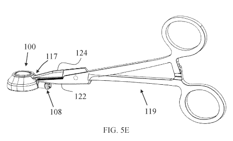
positioned in the superior vena cava SVC or into the right atrium RA, as shown
sequentially
in FIGs. 4J and 4K. Before insertion, catheter 15 can be cut to a chosen
length optionally
based on measurements of the path length from implantation site IMS to the
chosen
positioning of first catheter end 17 in patient's vasculature. Final
positioning of catheter first
tip 17 and/or port 10 can be applied by pushing or puffing port 10 in
subcutaneous void
and/or passage SCV. After verifying port 10 and/or catheter 15 are in proper
position and
function, optionally under imaging, peel-apart sheath 43 is broken apart and
removed from
body of subject SUB and incision INS is dosed (e.g., by way of suturing or
bonding).
[0092] FIGs. 5A - 5D illustrate different views of an exemplary squeezable
subcutaneous
port 100: in an assembled isometric view (FIG. 5A), in an exploded isometric
view (FIG.
5B), in a side cross-sectional view (FIG. 50) and in a frontal cross-sectional
view (FIG, 5D),
Port 100 is optionally an exemplary configuration of port 10 and may include
some or all
structural and/or functional features described with respect to port 10. Port
100 (optionally
particularly when in an elastically relaxed state, at least in part) may have
a maximal width
of 50 mm or less, optionally 25 mm or less; a maximal height of 30 mm or less,
optionally
15 mm or less; and a maximal length (with or without catheter connecting
means) of 50 mm
or less, optionally 30 mm or less. In some embodiments, port 100 is configured
to reshape
and/or deform to a narrower cross section for squeezing through surgical
openings (without
further widening or tearing when passing therethrough) having a maximal
opening
circumference of about 80 mm or less, optionally of about 60 mm or less,
optionally of about
40 mm or less, and/or formed by a surgical incision of about 20 mm or less in
length,
optionally about 15 mm or less in length, or optionally about 10 mm or less in
length.
[0093] Port 100 includes a rigid inner member 101 comprising a cavity 102
opened to a
first cavity opening 103 and to a second cavity opening 105. First cavity
opening 103 is
enclosed with a septum member 104 and configured for repeated needle
penetrations
therethrough into cavity 102. Second cavity opening 105 is configured for
facilitating fluid
communication between cavity 102 and a lumen of a catheter. Inner member 101
is
configured with sufficient rigidity to accommodate (safely and efficiently) a
chosen length of
-19-
CA 03188299 2022-12-26
WO 2022/011020 PCT/US2021/040699
a needle and to prevent the needle's tip from penetrating therethrough. Septum
member
104 is optionally oval, as shown, although it may have any other shape.
[0094] A cap member 106 is coupled over septum member 104 and over the
superior
portion of inner member 101 to form a unitary rigid encapsulated core body of
port 100.
Septum member 104 is restrained in-position and optionally compressed, at
least partly, by
and in-between cap member 106 and inner member 101. Inner member 101 and/or
cap
member 106 are optionally formed of hard plastic such as PEEK, or from metal
such as
titanium or stainless-steel alloys. Cap member 106 is optionally fixedly
connected to inner
member 101, such as by way of adhesives, compressing fitting and/or welding
(e.g,,
ultrasonic welding if the parts are made of plastic, or laser welding if the
parts are made of
metal). The encapsulated core body, once fully assembled, has sufficient
rigidity and yield
strength, and is configured to maintain internal pressures that are common
during injections
into cavity 102 (of optionally about 5 ml/sec injections at 300 psi, or higher
or lower). A
lumen extension 107 is coupled to inner member 101 with distal portion thereof
extending
towards cavity 102 through second cavity opening 105 and configured to provide
a fluid-
tight passage via proximal portion thereof to a catheter lumen. A connector
member 108 is
coupled over lumen extension 107 and is configured to facilitate selective
connection of a
catheter distal end with port 100, such as with a luer-fitting based
connection mechanism,
[0095] As shown in FIGs. 5E and 5F, port 100 includes a port gripping
portion 117
(optionally similar or identical in structure, function and/or dimensions to
port gripping
portion 21) provided at proximal end thereof and configured to facilitate
efficient and safe
grasping of port 100 with grasping means, such as medical clamp. Port gripping
portion 117
may be provided as a proximal extension of cap member 106, as shown, and
located above
(superiorly to) lumen extension 107 and connector member 108 (e.g., closer to
first cavity
opening 103). FIG, 5E illustrates port 100 grasped at port gripping portion
117 with an
exemplary medical clamp 119 (e.g., configured as a surgical needle holder).
Port gripping
portion 117 includes a wall 120 having opposing flat outer wall surfaces
extending
horizontally so that medical clamp 119 can be held by the medical practitioner
having its
arms arranged vertically (one over the other). Alternatively, wall 120 can be
arranged with
its flat surfaces in any other direction, including optionally vertically.
Wall 120 is optionally
configured in size, surface area of its flat surfaces, thickness and/or
durability and/or
strength to facilitate firm grasping by medical clamp 119 sufficiently to
push, squeeze-in,
-20-
CA 03188299 2022-12-26
WO 2022/011020 PCT/US2021/040699
and maneuver port 100 through a surgical opening smaller than its maximal
relaxed
dimensions, without releasing grip or mechanical failure. Medical damp 119 can
be used
to form or increase size of a subcutaneous void or passage before grasping on
to port 100
and delivering it into the subcutaneous void.
[0096] Port gripping portion 117 optionally includes a securing structure
configured to
prevent lateral and/or rotational movement of medical damp 119 on and relative
to wall
120, when the medical damp is engaged with and affixed thereto. As shown in
FIG. 5F,
port gripping portion 117 includes a recessed compartment 121 configured for
accommodating an inferior head member 122 of medical damp 119, wherein the
upper
surface of recessed compartment 121 is also the lower (inferior) wall surface
of wall 120.
Port gripping portion 117 also includes laterally opposing bordering walls 123
extending
from the upper (superior) outer wall surface of wall 120 and are configured
for
accommodating a superior head member 124 of medical clamp 119. Opposing
bordering
walls 123 may be laterally spaced from each other so as to snugly fit superior
head member
124 and/or clamping surface thereof,
[0097] FIGs. 6A - 6B schematically illustrate frontal cross-sectional views
of an
exemplary alternative configuration for a securing structure of port gripping
portion 117,
before and after clamping with an exemplary medical clamp having opposing head
members 127, each comprising projections or teeth 126. As shown, each one of
upper
surface lower surface of wall 120 includes a plurality of recesses 128 between
two bordering
walls 123. Recesses 128 are sized and shaped to snugly fit and accommodate
projections
126. This way lateral movements of medical clamp 125 can be diminished or
prevented.
Recesses 128 may be non-circular (e.g., star or cross shaped, for example)
thereby
preventing rotational movement of medical clamp 125 relative to and on wall
120.
[0098] In some embodiments, port gripping portion 117 includes a securing
structure or
mechanism configured to initiate lateral compression and/or locking of the
medical clamp
thereto when the medical clamp is engaged with and affixed to port gripping
portion 117.
FIGs. 7A - 7B schematically illustrate frontal cross-sectional views of
another exemplary
configuration for a securing structure of port gripping portion 117, before
and after clamping
with medical clamp 119. In this configuration, wall 120 is covered with a
cushion member
129 on each one of outer surfaces, the cushion members 129 are flexible and
configured
to compress into a volume shape mating to the clamping surfaces of head
members 122
-21-
CA 03188299 2022-12-26
WO 2022/011020 PCT/US2021/040699
and 124 while increasing in stiffness (optionally by maintaining or reducing
in volume)
and/or forming bordering wall portions 130 surrounding head members 122 and
124, as
shown. Port gripping portion 117 is configured such that cushion members 129
form shape
and stiffness sufficient to prevent lateral and/or rotational movement of
medical damp 119
on and relative to wall 120, when the medical damp is engaged with and affixed
thereto,
and optionally having arms thereof locked with each other.
[0099] FIGs. 8A - 8B schematically illustrate frontal cross-sectional views
of an
exemplary configuration for a securing mechanism of port gripping portion 117,
before and
after damping with medical damp 119. In this configuration, a lower
compartment 131 of
port gripping portion 117 (below lower/inferior surface of wall 120) is shaped
and/or
structured differently than an upper compartment 132 of port gripping portion
117 (above
upper/superior surface of wall 120), such that when head members 122 and 124
of medical
clamp 119 are pressed against wall 120 a securing mechanism is activated to
laterally press
and/or lock at least one of lower compartment 131 and upper compartment 132.
As shown
in this example, when inferior head member 122 engages and received in lower
compartment 131 it forces lower bordering walls 133 extending inferiorly from
wall 120 to
shift laterally outwardly. Lower bordering walls 133 then function as arms
pivoting about
coinciding portions with wall 120 and force upper bordering walls 134,
extending superiorly
from wall 120 and functioning as arm extensions of lower bordering walls 133,
to shift
laterally inwardly against superior head member 124. When activated to
function as
described, under normal clamping forces or pressures exerted by medical clamp
119, upper
bordering walls 134 are configured to press laterally and/or inferiorly
against superior head
member 124 sufficiently to prevent lateral and/or rotational movement of
medical clamp 119
on and relative to wall 120, when the medical clamp is engaged with and
affixed thereto,
and optionally having arms thereof locked with each other. In some
embodiments, cushion
members 135 are also provided in lower compartment 131 and/or upper
compartment 132
for affecting a more homogenous fitting and pressure transfer between head
members 122
and 124 and lower compartment 131 and/or upper compartment 132.
[0100] In some embodiments, inner member 101 can be functionally configured
or
applicable to serve as a vascular access port although it may be incapable,
insufficient, or
less compatible of providing one or more, optionally essential, features for
improving,
facilitating or easing implantation and/or long-term use of port 100. Port 100
includes a
-22-
CA 03188299 2022-12-26
WO 2022/011020 PCT/US2021/040699
flexible outer member 110 which provides, at least when it is in an
elastically relaxed state,
a final spatial shape and size, for providing one or more additional features,
including but
not limited to: stability and/or fixation in implantation site, transdermal
accessibility,
identification and/or locating of septum member 104 for repeated percutaneous
fluid
administration, protection to port body and/or overlaying skin layers, or
others,
[0101] Outer member 110 is connected to inner member 101 along at least one
lateral
periphery portion thereof, thereby forming a chosen predetermined spatial
shape of the
subcutaneous port when in an elastically relaxed state. Optionally, outer
member is
configured as a skirt or ring-like element encompassing most or all periphery
of inner
member 101, and optionally also periphery of cap member 106, in at least a
circumferential
segment thereof. In order to maintain sufficient rigid pushability of port 100
for its insertion
and implantation, the rigid inner member 101 extends longitudinally along most
or all length
of port 100, to function also as a rigid spine-like structure of port 100,
optionally in
combination with cap member 106. Inner member 101 includes a distal (front)
portion 113
extending distally relative to 102 cavity, having a rounded or pointed leading
edge 116
configured to facilitate or ease penetration of port 100 via the surgical
opening. Port 100
may be configured such that distal portion 113 is uncovered by outer member 20
which may
extend distally and transversely therefrom, although (as shown) it may be
covered with a
thin layer of outer member 110 such that sufficient rigid pushability is
substantially
uncompromised. Outer member 110 is optionally made of silicone or other
flexible and
elastic polymer or rubber, and is optionally extruded, casted or molded over
periphery of
inner member 101 or over periphery of the encapsulated core body (i.e., the
structure
formed by the interconnected inner member 101, septum member 104 and cap
member
106), optionally within boundary of a chosen shaped mold, when forming
subcutaneous port
100.
[0102] FIG& 9A - 9B illustrate axonometric views of another exemplary
vascular access
port 200 which includes a port body 201 and at least one port body extension
204
restrictedly movable along an at least one defined route 205 on port body 201
The at least
one port body extension 204 includes a first arm 208 located right to a median
plane of port
body 201 and a second arm 209 located left to the median plane. Port body
extensions 204,
particularly first and second arms 208 and 209, are each rotatably and
slidably connected
to port body 201 and configured to rotate around an axis of rotation and slide
on an at least
-23-
CA 03188299 2022-12-26
WO 2022/011020 PCT/US2021/040699
one of two opposing sides of the port body 201, along routes 205, when
changing from the
delivery configuration to the deployed configuration. Port body 201 has an
inferior portion
210 and a posterior portion 211, the posterior portion 211 is connected to a
septum member
202 and the inferior portion 210 surrounds a cavity 203 that is defined by
port body 201 and
located below and covered by septum member 202. Inferior portion also includes
a first
lateral surface spanning most or all right side of inferior portion 210 and a
second lateral
surface spanning most or all left side of the inferior portion 210. A rear end
214 of port body
201 is coupled to a catheter connector 215 configured for connecting to a
proximal end of
a catheter (such as catheter 15, for example) for facilitating fluid
communication between
cavity 203 and a lumen of the catheter.
[0103] Vascular access port 200 is selectively changeable from a delivery
configuration
(as shown in FIG. 9A) to a deployed configuration (as shown in FIG. 9B) by
moving first and
second arms 208 and 209 along a first and a second of routs 205, respectively,
When in
the delivery configuration, a front portion 207 of each port body extension
204 is positioned
axially distally to the port body 201, When changing to the deployed
configuration, port body
extensions 204 and the port body 201 are approximated along the median plane
of port
body 201 coincidently with laterally opposing portions 206 of port body
extension 204 being
parted transversely to the median plane, thereby reducing length-to-width
ratio of the
toggling vascular access port 200. When in the deployed configuration, the
port body
extensions 204 are fixedly and releasably connected to port body 201,
therefore allowing
selective reverting from the deployed configuration to the delivery
configuration.
Furthermore, rear end 214 of port body 201 is kept not covered with the port
body
extensions 204 also after changing to the deployed configuration, for avoiding
engagement
with catheter connector 215 and/or a catheter connected thereto, for example.
[0104] A port gripping portion 216 (which is optionally similar or
identical in structure,
function and/or dimensions to port gripping portion 21) is located on the rear
end 214 of port
body 201 superiorly to catheter connector 215 for allowing a user to
selectively move and/or
manipulate port 200 subcutaneously and in the target implantation site while
avoiding
engagement with catheter connector 215 and/or a catheter connected thereto,
for example.
A user can clamp port gripping portion 216 with medical forceps and push
toggling vascular
access port 200 when in the delivery configuration to the target implantation
site with the
medical forceps. Once in the target implantation site, port 200 can be changed
to the
-24-
CA 03188299 2022-12-26
WO 2022/011020 PCT/US2021/040699
deployed configuration by pushing port body 201 distally relative to port body
extensions
204 and/or pulling port body extensions 204, such as with pulling members 219
connected
to first and second arms 208 and 209 while resisting motion of port body 201
using the
forceps.
[0105] Port gripping portion 216 includes a thin wall portion 217
comprising opposing
lateral surfaces extending parallel to the median plane from both sides
thereof, the wall
portion 217 is configured for grasping and/or damping by medical forceps
including but not
limited to needle holder or Kelly forceps 230, as shown in FIGs. 10A and 10B.
In some
embodiments, wall portion 217 is about 0.5 mm to 3 mm (optionally particularly
about 1 mm
to 2 mm) thick and/or about 2 mm to 5 mm (optionally particularly about 3 mm
to 4 mm)
wide for allowing sufficient clamping contact area and buildup of sufficient
clamping,
grasping or locking force from both sides of wall portion 217 using medical
forceps.
[0106] Wall portion 217 can be configured as a septum dividing cavities 218
formed in
rear end 214 from both sides thereof. Cavities 218 are shaped and sized to
accommodate
a pair of tips of the medical forceps and to allow closing motion of the pair
of tips thereinside
towards the wall portion and grasping of the wall portion 217 with the pair of
tips from both
sides thereof. FIG. 11 illustrates an alternative exemplary configuration of
port gripping
portion 216 having a thin wall portion 217' similar to wall portion 217, yet
not bound by
cavities, rather allow greater room for forceps tips maneuverability.
[0107] Each of the following terms written in singular grammatical form:
'a', 'an', and 'the',
as used herein, means 'at least one', or 'one or more'. Use of the phrase 'one
or more'
herein does not alter this intended meaning of 'a', an, or 'the'. Accordingly,
the terms 'a',
'an', and 'the', as used herein, may also refer to, and encompass, a plurality
of the stated
entity or object, unless otherwise specifically defined or stated herein, or,
unless the context
clearly dictates otherwise. For example, the phrases: 'a unit', 'a device', an
assembly', 'a
mechanism', 'a component', 'an element', and 'a step or procedure', as used
herein, may
also refer to, and encompass, a plurality of units, a plurality of devices, a
plurality of
assemblies, a plurality of mechanisms, a plurality of components, a plurality
of elements,
and, a plurality of steps or procedures, respectively.
[0108] Each of the following terms: 'includes', 'including', 'has',
'having', 'comprises', and
'comprising', and, their linguistic / grammatical variants, derivatives,
or/and conjugates, as
used herein, means 'including, but not limited to, and is to be taken as
specifying the stated
-25-
CA 03188299 2022-12-26
WO 2022/011020 PCT/US2021/040699
component(s), feature(s), characteristic(s), parameter(s), integer(s), or
step(s), and does
not preclude addition of one or more additional component(s), feature(s),
characteristic(s),
parameter(s), integer(s), step(s), or groups thereof. Each of these terms is
considered
equivalent in meaning to the phrase 'consisting essentially of,
[0109] The term 'method', as used herein, refers to steps, procedures,
manners, means,
or/and techniques, for accomplishing a given task including, but not limited
to, those steps,
procedures, manners, means, or/and techniques, either known to, or readily
developed
from known steps, procedures, manners, means, or/and techniques, by
practitioners in the
relevant field(s) of the disclosed invention,
[0110] Throughout this disclosure, a numerical value of a parameter,
feature,
characteristic, object, or dimension, may be stated or described in terms of a
numerical
range format. Such a numerical range format, as used herein, illustrates
implementation of
some exemplary embodiments of the invention, and does not inflexibly limit the
scope of
the exemplary embodiments of the invention. Accordingly, a stated or described
numerical
range also refers to, and encompasses, all possible sub-ranges and individual
numerical
values (where a numerical value may be expressed as a whole, integral, or
fractional
number) within that stated or described numerical range. For example, a stated
or
described numerical range from 1 to 6 also refers to, and encompasses, all
possible sub-
ranges, such as from 1 to 3, 'from 1 to 4', from 1 to 5, from 2 to 4', from 2
to 6', from 3 to
6, etc., and individual numerical values, such as '1', '1.3', '2', '2.8, '3',
3.5', '4', 4.6', '5', '52',
and '6', within the stated or described numerical range of 'from 1 to 6'. This
applies
regardless of the numerical breadth, extent, or size, of the stated or
described numerical
range.
[0111] Moreover, for stating or describing a numerical range, the phrase
'in a range of
between about a first numerical value and about a second numerical value', is
considered
equivalent to, and meaning the same as, the phrase 'in a range of from about a
first
numerical value to about a second numerical value', and, thus, the two
equivalently
meaning phrases may be used interchangeably. For example, for stating or
describing the
numerical range of room temperature, the phrase 'room temperature refers to a
temperature
in a range of between about 20 C and about 25 00', and is considered
equivalent to, and
meaning the same as, the phrase 'room temperature refers to a temperature in a
range of
from about 20 00 to about 25 `)C'.
-26-
CA 03188299 2022-12-26
WO 2022/011020 PCT/US2021/040699
[0112] The term 'about', as used herein, refers to 10% of the stated
numerical value,
[0113] It is to be fully understood that certain aspects, characteristics,
and features, of
the invention, which are, for clarity, illustratively described and presented
in the context or
format of a plurality of separate embodiments, may also be illustratively
described and
presented in any suitable combination or sub-combination in the context or
format of a
single embodiment Conversely, various aspects, characteristics, and features,
of the
invention which are illustratively described and presented in combination or
sub-combination in the context or format of a single embodiment, may also be
illustratively
described and presented in the context or format of a plurality of separate
embodiments.
[0114] Although the invention has been illustratively described and
presented by way of
specific exemplary embodiments, and examples thereof, it is evident that many
alternatives,
modifications, or/and variations, thereof, will be apparent to those skilled
in the art.
Accordingly, it is intended that all such alternatives, modifications, or/and
variations, fall
within the spirit of, and are encompassed by, the broad scope of the appended
claims.
[0115] All publications, patents, and or/and patent applications, cited or
referred to in
this disclosure are herein incorporated in their entirety by reference into
the specification,
to the same extent as if each individual publication, patent, or/and patent
application, was
specifically and individually indicated to be incorporated herein by
reference. In addition,
citation or identification of any reference in this specification shall not be
construed or
understood as an admission that such reference represents or corresponds to
prior art of
the present invention. To the extent that section headings are used, they
should not be
construed as necessarily limiting.
[0116] The methods disclosed herein comprise one or more steps or actions
for
achieving the described method. The method steps and/or actions may be
interchanged
with one another without departing from the scope of the claims. In other
words, unless a
specific order of steps or actions is specified, the order and/or use of
specific steps and/or
actions may be modified without departing from the scope of the claims.
-27-