Note: Descriptions are shown in the official language in which they were submitted.
WO 2022/031804
PCT/US2021/044478
IMPROVED METH:ODS AND KITS FOR DETECTING SARS-COV-2 PROTEIN IN A
SAMPLE
RELATED APPLICATION INFORMATION
[00011 This application claims priority to U S. Application No.
63/060,922 filed August 4,
2020 and U.S. Application No. 63/156,775, filed on March 4, 2021, the contents
of which are
herein incorporated by reference.
SEQUENCE LISTING
[0002.I Incorporated by reference in its entirety herein is a computer-
readable
nucleotide/amino acid sequence listing submitted concurrently herewith and
identified as
follows: One 4,460 Byte ASCII (Text) file named "38688-601_ST25.TXT," created
on August 4,
2021.
TECHNICAL FIELD
[00031 The present disclosure relates methods, kits, and systems for
detecting the presence Of
determining the amount of at least one SARS-CoV-2 protein (e.g, antigen) in
one or more
samples obtained from a subject. In some aspects, the methods, kits and
systems employ at least
one polycation which improves the sensivity of detecting the presence or
determining the amount
of at least one SARS-CoV-2 nucleocapsid protein in one or more samples
obtained from a
subject.
BACKGROUND
[0004.1 Viruses of the family Coronaviridae possess a single-strand, positive-
sense RNA
genome ranging from 26 to 32 kilobases in length (reviewed by I.,u et al., The
Lancet, 395:565-
574 (February 22, 2020)). The Coronaviridae are further subdivided (initially
based on serology
but now based on phylogenetic clustering) into four groups, the alpha, beta,
gamma and delta
coronaviruses. Coronaviruses have been identified in several avian hosts, as
well as in various
mammals, including camels, bats, masked palm civets, mice, dogs, and cats.
100051 Among the several coronaviruses that are pathogenic to humans, most are
associated
with mild clinical symptoms, with three exceptions. Severe acute respiratory
syndrome (SARS)
coronavirus (SARS-CoV) is a novel betacoronavirus that emerged in Guangdong,
southern
1
CA 03188349 2023- 2-3
WO 2022/031804
PCT/US2021/044478
China, in November 2002 and resulted in more than 8000 human infections and
774 deaths in 37
countries in 2002-03. Middle East respiratory syndrome (MERS) coronavirus (RS-
CoV) was
first detected in Saudi Arabia in 2012 and was responsible for 2494 laboratory-
confirmed cases
of infection and 858 deaths from 2012-20. In December, 2019, a cluster of
pneumonia cases
caused by a newly identified 13-coronavirus were found to be epidemiologically-
associated with
the Huanan seafood market in Wuhan, China, where a number of non-aquatic
animals, such as
birds and rabbits were on sale before the outbreak. This coronavirus was named
January 2020
by the World Health Organization (WHO) as the 2019-novel coronavirus (2019-
nCov or
COVID-19), and February 2020 by the International Committee as SARS-CoV-2.
SARS-CoV-2
was declared a pandemic due to its rapid, uncontrolled and vast worldwide
spread.
100061 Coronavirus virions are spherical with diameters of approximately 125
nanometers, as
demonstrated in studies by cryo-electron tomography and cryo-electron
microscopy. A
prominent feature of coronaviruses is the club-shape spike projections
emanating from the
surface of the virion, giving the virion the appearance of a solar corona and
resulting in the
name, coronaviruses. Within the envelope of the coronavirus virion is the
helically-symmetrical
nucleocapsid, which binds to and creates a shell around the coronavirus RNA
genome. The
spike (S) and nucleocapsid (N) proteins are the main immunogens of the
coronavirus. The other
two main structural proteins of the coronavirus particles are the membrane (M)
and envelope (E)
proteins. All four proteins are encoded within the 3' end of the viral genome.
100071 The S protein (-150 k1.3a) is heavily N-linked glycosylated
and utilizes an N-terminal
signal sequence to gain access to the endoplasmic reticulum (ER). Homotrimers
of the virus-
encoding S protein make up the distinctive spike structure on the surface of
the virus. In many,
but not all, coronaviruses, the S protein is cleaved by a host cell furin-like
protease into two
separate polypepfides noted Si and S2. Si makes up the large receptor-binding
domain of the S
protein while S2 forms the stalk of the spike molecule. The trimeric S
glycoprotein mediates
attachment of the coronavirus virion to the host cell by interactions between
the S protein and its
receptor. In humans, angiotensin-converting enzyme 2 (ACE2) is the receptor
for SARS-CoV.
The sites of receptor binding domains (RBD) within the S I region of a
coronavirus S protein
vary depending on the virus, with some having the RBD at the N-terminus of S1
(e.g., murine
hepatitis virus) while others (e.g., SARS-CoV) have the RBD at the C-terminus
of Sl. The S-
2
CA 03188349 2023- 2-3
WO 2022/031804
PCT/US2021/044478
protein/receptor interaction is the primary determinant for the coronavirus to
infect a host species
and also governs the tissue tropism of the virus.
100081 The M protein is the most abundant structural protein in the
virion. It is a small (-25-
30 kDa) protein with 3 transmembrane domains and is believed to give the
virion its shape. It
has a small N-terminal glycosylated ectodomain and a much larger C-terminal
endodomain that
extends 6-8 nm into the viral particle. The E protein (-8-12 kDa) is found in
small quantities
within the virion. E proteins in coronaviruses are highly divergent but have a
common
architecture. Data suggests that the E protein is a transmembrane protein with
an N-terminal
ectodomain and a C-terminal endodomain that has ion channel activity.
Recombinant viruses
lacking the E protein are not always lethal -- although this is virus-type
dependent. The E
protein facilitates assembly and release of the virus, but also has other
functions (e.g., ion
channel activity in SARS-CoV E protein is not required for viral replication
but is required for
pathogenesis).
[00051 The N protein is the only protein present in the nucleocapsid.
It is composed of two
separate domains, an N-terminal domain (NTD) and a C-terminal domain (CTD),
both capable
of binding :RNA in vitro using different mechanisms, which may suggest that
optimal RNA
binding requires contributions from both domains. The N protein is heavily
phosphorylated, and
phosphoiylation has been suggested to trigger a structural change enhancing
the affinity for viral
versus non-viral RNA. The N protein binds the viral genome in a beads-on-a-
string type
conformation. Two specific RNA substrates have been identified for N protein;
the
transcriptional regulatory sequences and the genomic packaging signal. The
genomic packaging
signal has been found to bind specifically to the second, or C-terminal RNA
binding domain.
The N protein also binds nsp3, a key component of the replicase complex, and
the M protein.
These protein interactions likely help tether the viral genome to the
replicase-transcriptase
complex, and subsequently package the encapsidated genome into viral
particles.
100101 In February 2020, Lu et al. reported obtaining complete and partial
SARS-CoV-2
genome sequences using next-generation sequencing of bronchoalveolar lavage
fluid samples
and cultured isolates from nine patients from Wuhan diagnosed with viral
pneumonia but
negative for common respiratory pathogens. Lu et al., The Lancet, 395: 565-574
(February 22,
2020). Based on their analysis, Lu et al. further reported that SARS-CoV-2 was
closely related
(with 88% identity) to two bat-derived severe acute respiratory syndrome
(SARS)-like
3
CA 03188349 2023- 2-3
WO 2022/031804
PCT/US2021/044478
coronaviruses, bat-SL-CoVZC45 and bat-SL-CoVZXC21, collected in eastern China
in 2018.,
but was more distant from SARS-CoV (about 79%) and MERS-CoV (about 50%).
Additionally,
Zhou et al. confirmed that SARS-CoV-2 uses the same cellular entry receptor,
ACE2, as SARS-
CoV. Zhou et al., Nature, 579:270-273 (March 2020).
100111 SARS-CoV-2 primarily spreads through the respiratory tract, by
droplets, respiratory
secretions, and direct contact. Additionally, SARS-CoV-2 has been found in
fecal swabs and
blood, indicating the possibility of multiple routes of transmission. Zhang et
al., Microbes
9(1):386-9 (2020). SARS-CoV-2 is highly transmissible in humans, especially in
the elderly and
people with underlying diseases. Symptoms can appear 2 to 14 days after
exposure. Patients
present with symptoms such as fever, malaise, cough, and/or shortness of
breath. Most adults or
children with SARS-CoV-2 infection present with mild flu-like symptoms,
however, critical
patients rapidly develop acute respiratory distress syndrome, respiratory
failure, multiple organ
failure and even death.
100121 Because of the health risks imposed by SARS-CoV-2 transmission, there
is a need for
methods and kits to assess coronavirus transmission in humans, including
methods to determine
the presence and/or detect the amount of SARS-CoV-2 protein in one or more
samples obtained
from a subject.
SUMMARY
[00131 In one aspect, the present disclosure relates to an improvement of a
method of
detecting a presence or determining an amount of a SARS-CoV-2 nucleocapsid
protein in a
biological sample, wherein the method comprises detecting at least one complex
comprising a
first specific binding partner, said sample SARS-CoV-2 nucleocapsid protein,
and a second
specific binding partner comprising at least one detectable label and further
wherein the first
specific binding partner, second specific binding partner, or the first
specific binding partner and
the second specific binding partner comprise at least one anti-SARS-CoV
antibody, anti-SARS-
CoV-2 antibody, or fragment thereof that specifically binds to said sample
SARS-CoV-2
nucleocapsid protein, wherein the improvement comprises allowing the complex
to form in the
presence of at least one polycation having a molecular weight of at least
about 500 daltons or
greater prior to assessing the signal from the complex, wherein the amount of
detectable signal
4
CA 03188349 2023- 2-3
WO 2022/031804
PCT/US2021/044478
from the detectable label in the complex indicates the presence or amount of
SARS-CoV-2
nucleocapsid protein in the sample.
[0014.1 In some aspects, the biological sample in the above method is
whole blood, serum,
plasma, saliva, an oropharyngeal specimen, or a nasopharyngeal specimen.
Specifically, in some
aspects, the biological sample is whole blood. In other aspects, the
biological sample is plasma.
In yet other aspects, the biological sample is saliva. In still other aspects,
the biological sample
is an oropharyngeal specimen. In still yet other aspects, the biological
sample is a
nasopharyngeal specimen.
[0015.1 In still yet other aspects, the first specific binding partner
comprises at least one anti-
SARS-CoV antibody, anti-SARS-CoV-2 antibody, or fragment thereof, or the first
specific
binding partner and the second specific binding partner each comprise at least
one anti-SARS-
CoV antibody, anti-SARS-CoV-2 antibody or fragment thereof.
[0016.1 In still further aspects, the polycation in the above method
is at least one polylysine, at
least one polyomithine, at least one poly-I.:hi stidine, at least one poly-L-
arginine, at least one
polyethylenimine, at least one DEAE-Dextran, or combinations thereof. More
specifically, in
some aspects, the (i) the polylysine is poly-L-lysine hydrobromide, poly-D-
lysine hydrobromide,
poly-L-lysine hydrochloride, poly-L-lysine trifluoroacetate, poly(lysine,
alanine) 3:1
hydrobromide, poly(lysine, arginine) 2:1 hydrobromide, poly(lysine, alanine)
1:1 hydrobromide,
or poly(lysine, tiyptophan) 1:4 hydrobromide; (ii) the polyornithine is poly-L-
ornithine
hydrobromide or poly-DL-ornithine hydrobromide; (iii) the poly-L-histidine is
poly-L-histidine
hydrobromide; and (iv) the poly-L-arginine is poly-L-arginine hydrochloride or
poly-L-arginine
hydrobromide.
100171 In still further aspects, the improvement increases
sensitivity by at least about 5%, at
least about 10%, at least about 15%, at least about 20%, at least about 25%,
at least about 30%,
at least about 30%, at least about 35%, at least about 40%, at least about
45%, at least about
50%, at least about 55%, at least about 60%, at least about 65%, at least
about 70%, at least
about 75%, at least about 80%, at least about 85%, at least about 95%, at
least about 100%, at
least about 110%, at least about 120%, at least about 125%, at least about
130%, at least about
140%, at least about 150%, at least about 170%, at least about 180%, at least
about 190%, at
least about 200%, at least about 210%, at least about 220%, at least about
230%, at least about
240%, at least about 250%, at least about 260%, at least about 270%, at least
about 280%, at
CA 03188349 2023- 2-3
WO 2022/031804
PCT/US2021/044478
least about 290%, or at least about 300% when compared to methods which do not
use or
employ at least one polycation.
[0181 In still further aspects, the method can be selected from the
group consisting of an
immunoassay, a clinical chemistry assay, a point-of-care assay, and a lateral
flow assay. In yet
other aspects, the method can be performed using single molecule detection. In
still other
aspects, the method is adapted for use in an automated system or a semi-
automated system.
100191 in yet another aspect; the present disclosure relates to a
method of detecting a presence
or determining an amount of a SARS-CoV-2 nucleocapsid protein in a biological
sample in a
subject. The method can comprise:
a) contacting at least one biological sample from the subject, either
simultaneously
or sequentially, in any order, with:
at least one first specific binding partner comprising at least one anti-SARS-
CoV
antibody, anti-SARS-CoV-2 antibody, or fragment thereof thereof that
specifically binds to at
least one SARS-CoV-2 nucleocapsid protein in the sample,
at least one second specific binding partner comprising at least one
detectable
label, thereby producing one or more complexes comprising the first binding
member-SARS-
CoV-2 nucleocapsid protein-second specific binding partner, and
at least one polycation having a molecular weight of at least about 500
daltons or
greater; and
b) assessing a signal from the one or more complexes, wherein the amount of
detectable signal from the detectable label indicates the presence or amount
of SARS-CoV-2
nucleocapsid protein in the sample.
100201 In some aspects, the biological sample in the above method is whole
blood, serum,
plasma; saliva, an oropharyngeal specimen, or a nasopharyngeal specimen
Specifically, in some
aspects, the biological sample is whole blood. in other aspects, the
biological sample is plasma.
In yet other aspects, the biological sample is saliva. In still other aspects,
the biological sample
is an oropharyngeal specimen. In still yet other aspects, the biological
sample is a
nasopharyngeal specimen.
6
CA 03188349 2023- 2-3
WO 2022/031804
PCT/US2021/044478
1002.11 In still yet other aspects, the first specific binding partner
comprises at least one anti-
SARS-CoV antibody, anti-SARS-CoV-2 antibody, or fragment thereof, or the first
specific
binding partner and the second specific binding partner each comprise at least
one anti-SARS-
CoV antibody, anti-SARS-CoV-2 antibody or fragment thereof.
10022) In still further aspects, the polycation used in the above
method is at least one
polylysine, at least one polyornithine, at least one poly-L-histidine, at
least one poly-L-arginine,
at least one polyethylenimine, at least one DEAE-Dextran, or combinations
thereof. More
specifically, in some aspects, the (i) the polylysine is poly-L-lysine
hydrobromide, poly-D-lysine
hydrobromide, poly-L-lysine hydrochloride, poly-L-lysine trifluoroacetate,
poly(lysine, alanine)
3:1 hydrobromide, poly(lysine, arginine) 2:1 hydrobromide, poly(lysine,
alanine) 1:1
hydrobromide, or poly(lysine, tryptophan) 1:4 hydrobromide; (ii) the
polyornithine is poly-L-
ornithine hydrobromide or poly-DL-ornithine hydrobromide; (iii) the poly-L-
histidine is poly-L-
histidine hydrobromide; and (iv) the poly-L-arginine is poly-L-arginine
hydrochloride or poly-L-
arginine hydrobromide.
100231 In still further aspects, the improvement increases
sensitivity by at least about 5%, at
least about 10%, at least about 15%, at least about 20%, at least about 25%,
at least about 30%,
at least about 30%, at least about 35%, at least about 40%, at least about
45%, at least about
50%, at least about 55%, at least about 60%, at least about 65%, at least
about 70%, at least
about 75%, at least about 80%, at least about 85%, at least about 95%, at
least about 100%, at
least about 110%, at least about 120%, at least about 125%, at least about
130%, at least about
140%, at least about 150%, at least about 170%, at least about 180%, at least
about 190%, at
least about 200%, at least about 210%, at least about 220%, at least about
230%, at least about
240%, at least about 250%, at least about 260%, at least about 270%, at least
about 280%, at
least about 290%, or at least about 300% when compared to methods which do not
use or
employ at least one polycation.
[00241 In still yet other aspects, the method can be selected from
the group consisting of an
immunoassay, a clinical chemistry assay, a point-of-care assay, and a lateral
flow assay. In yet
other aspects, the method can be performed using single molecule detection. In
still other
aspects, the method is adapted for use in an automated system or a semi-
automated system.
100251
In yet another aspect, the present disclosure relates to a kit for
performing the
above method. Specifically, the kit comprises:
7
CA 03188349 2023- 2-3
WO 2022/031804
PCT/US2021/044478
a. at least one specific binding partner comprising at least one anti-SARS-
CoV
antibody, anti-SARS-CoV-2 antibody, or fragment thereof that specifically
binds to at least one
SARS-CoV-2 nucleocapsid protein;
b. at least one second specific binding partner comprising at least one
detectable
label; and
c. at least one polycation having a molecular weight of at least about 500
daltons or
greater.
[00261 in other aspects, the kit further comprises, or is configured
to be used with at least one
calibrator reagent, at least one control reagent, or at least one calibrator
reagent and at least one
control reagent. In some aspects, the kit further comprises at least one solid
support. In yet other
aspects, the polycation in the kit is at least one polylysine, at least one
polyornithine, at least one
poly-L-histidine, at least one poly-L-arginine, at least one
polyethylenimines, at least one DEAE-
Dextran, or combinations thereof. Specifically, in some aspects, the (i) the
polylysine is poly-L-
lysine hydrobromide, poly-D-lysine hydrobromide, poly-L-lysine hydrochloride,
poly-L-lysine
trifluoroacetate, poly(lysine, alanine) 3:1 hydrobromide, poly(lysine,
arginine) 2:1
hydrobromide, poly(lysine, alanine) 1:1 hydrobromide, or poly(lysine,
tryptophan) 1:4
hydrobromide; (ii) the polyornithine is poly-L-omithine hydrobromide or poly-
DL-orni thine
hydrobromide; (iii) the poly-L-histidine is poly-L-histidine hydrobromide; and
(iv) the poly-L-
arginine is poly-L-arginine hydrochloride or poly-L-arginine hydrobromide. In
some aspects,
the kit is adapted for use with an automated or semi-automated system.
100271 In yet another aspect, the disclosure relates to a system for
detecting a presence or
determining an amount of a SAR.S-CoV-2 nucleocapsid protein in a biological
sample in a
subject. The system comprises:
at least one first specific binding partner comprising at least anti-SARS-CoV
antibody,
anti-SARS-CoV-2 antibody, or fragment thereof that specifically binds to at
least one SARS-
CoV-2 nucleocapsid protein in the sample;
at least one second specific binding partner comprising at least one
detectable label,
thereby producing one or more complexes comprising the first binding member-
SARS-CoV-2
nucleocapsid protein-second specific binding partner;
8
CA 03188349 2023- 2-3
WO 2022/031804
PCT/US2021/044478
at least one polycation having a molecular weight of at least about 500
daltons or greater;
and
at least one device for detecting the at least one label from the complex.
[00281 in some aspects, in the system, the device for detecting the label from
the complex is
automated or semi-automated. In other aspects, in the system, the polycation
is at least one
polylysine, at least one polyornithine, at least one poly-L-histidine, at
least one poly-L-arginine,
at least one polyethylenimines, at least one DEAE-Dextran, or combinations
thereof. In some
aspects, the (i) the polylysine is poly-L-lysine hydrobromide, poly-D-lysine
hydrobromide, poly-
L-lysine hydrochloride, poly4,-lysine trifluoroacetate, poly(lysine, alanine)
3:1 hydrobromide,
poly(lysine, arginine) 2:1 hydrobromide, poly(lysine, alanine) 1:1
hydrobromide, or poly(lysine,
tryptophan) 1:4 hydrobromide; (ii) the polyornithine is poly-L-ornithine
hydrobromide or poly-
DL-ornithine hydrobromide; (iii) the poly-L-histidine is poly-L-histidine
hydrobromide; and (iv)
the poly-L-arginine is poly-L-arginine hydrochloride or poly-L-arginine
hydrobromide.
BRIEF DESCRIPTION OF THE DRAWINGS
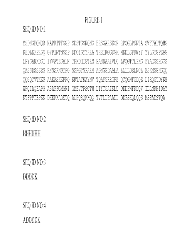
[0029.1 FIG. I provides a sequence listing comprising SEQ LD NOS. 1 ¨4.
DETAILED DESCRIPTION
[NA The present disclosure relates to methods, kits, and systems
to detect the presence of
or determine the amount, concentration and/or level of at least one SARS-CoV-2
protein (e.g.,
antigen), such as at least one SARS-CoV-2 nucleocapsid protein, in a sample.
In some aspects,
the methods, kits, and systems described herein are used to detect the
presence of or determine
the amount, concentration and/or level of at least one SARS-CoV-2 nucleocapsid
protein in
samples with improved sensitivity (e.g., have a higher signal to noise (S/N)
ratio) than other
methods, kits, and systems. In one aspect, the disclosure relates to an
improvement in a method
of detecting a presence or determining an amount of a SARS-CoV-2 nucleocapsid
protein in a
biological sample, wherein the method involves adding at least one polycation
having a
molecular weight of at least about 500 daltons or greater to the sample prior
to detecting the
presence or determing the amount of at least one SARS-CoV-2 nucleocapsid
protein in the
sample. The addition of at least one polycation having a molecular weight of
at least about 500
9
CA 03188349 2023- 2-3
WO 2022/031804
PCT/US2021/044478
daltons or greater to the sample results in an improvement in the sensitivity
(e.g., SIN ratio) of
detection in the method of at least about 5%, at least about 10%, at least
about 15%, at least
about 20%, at least about 25%, at least about 30%, at least about 30%, at
least about 35%, at
least about 40%, at least about 45%, at least about 50%, at least about 55%,
at least about 600/0,
at least about 65%, at least about 70%, at least about 75%, at least about
80%, at least about
85%, at least about 95%, at least about 100%, at least about 110%, at least
about 120%, at least
about 125%, at least about 130%, at least about 140%, at least about 150%, at
least about 170%,
at least about 180%, at least about 190%, at least about 200%, at least about
210%, at least about
220%, at least about 230%, at least about 240%, at least about 250%, at least
about 260%, at
least about 270%, at least about 280%, at least about 290%, or at least about
300% when
compared to methods which do not use or employ at least one polycation.
[0031.1 In another aspect, the disclosure relates to methods of
detecting the presence or
determining an amount of at least one SARS-CoV-2 nucleocapsid protein in a
biological sample
by contacting at least one sample obtained from a subject (either
simultaneously or sequentially,
in any order), with at least one first specific binding partner comprising at
at least one anti-
SARS-CoV antibody, anti-SARS-CoV-2 antibody, Of fragment thereof that
specifically binds to
at least one SARS-CoV-2 nucleocapsid protein in the sample, at least one
second specific
binding partner comprising a detectable label to produce one or more complexes
comprising the
first specific binding partner-SARS-CoV-2 nucleocapsid protein-second specific
binding partner,
and at least one polycation having a molecule weight of at least about 500
daltons or greater. A
signal from the one or more complexes are assessed (e.g., detected).
Specifically, the amount of
the detectable signal from the detectable label indicates the presence or
amount of SARS-CoV-2
nucleocapsid protein in the sample. The addition of at least one polycation
having a molecular
weight of at least about 5(X) daltons or greater in the method improves the
sensitivity (e.g., SIN
ratio) of the method by at least about 5%, at least about 10%, at least about
15%, at least about
20%, at least about 25%, at least about 30%, at least about 30%, at least
about 35%, at least
about 40%, at least about 45%, at least about 50%, at least about 55%, at
least about 60%, at
least about 65%, at least about 70%, at least about 75%, at least about 80%,
at least about 85%,
at least about 95%, at least about 100%, at least about 110%, at least about
120%, at least about
125%, at least about 130%, at least about 140%, at least about 150%, at least
about 170%, at
least about 180%, at least about 190%, at least about 200%, at least about
210%, at least about
1 0
CA 03188349 2023- 2-3
WO 2022/031804
PCT/US2021/044478
220%, at least about 230%, at least about 240%, at least about 250%, at least
about 260%, at
least about 270%, at least about 280%, at least about 290%, or at least about
300% when
compared to methods which do not use or employ at least one polycation.
[00321 The biological sample used in the methods of the present disclosure may
be obtained
from an asymptomatic subject or from a subject exhibiting one or more symptoms
of infection
with SARS-CoV-2. The methods of the present disclosure also include treating a
subject
identified as having a SARS-CoV-2 and optionally, monitoring such subjects,
such as before,
during and/or after receiving such treatments.
[00331 In another aspect, the present disclosure relates to kits for
performing such methods.
100341 In still yet another aspect, the present disclosure relates to
systems for detecting at
least one SARS-CoV-2 nucleocapsid protein in a biological sample.
[00351 Section headings as used in this section and the entire
disclosure herein are merely for
organizational purposes and are not intended to be limiting.
1. Definitions
[00361 Unless otherwise defined, all technical and scientific terms
used herein have the same
meaning as commonly understood by one of ordinary skill in the art In case of
conflict, the
present document, including definitions, will control. Preferred methods and
materials are
described below, although methods and materials similar or equivalent to those
described herein
can be used in practice or testing of the present disclosure. All
publications, patent applications,
patents and other references mentioned herein are incorporated by reference in
their entirety.
The materials, methods, and examples disclosed herein are illustrative only
and not intended to
be limiting.
[00371 The terms "comprise(s)," "include(s)," "having," "has," "can,"
"contain(s)," and
variants thereof, as used herein, are intended to be open-ended transitional
phrases, terms, or
words that do not preclude the possibility of additional acts or structures.
The singular forms
"a," "an" and "the" include plural references unless the context clearly
dictates otherwise. The
present disclosure also contemplates other embodiments "comprising,"
"consisting of' and
"consisting essentially of," the embodiments or elements presented herein,
whether explicitly set
forth or not.
11
CA 03188349 2023- 2-3
WO 2022/031804
PCT/US2021/044478
100381 For the recitation of numeric ranges herein, each intervening number
there between
with the same degree of precision is explicitly contemplated. For example, for
the range of 6-9,
the numbers 7 and 8 are contemplated in addition to 6 and 9, and for the range
6.0-7.0, the
number 6.0, 6.1, 6.2, 6.3, 6.4, 6.5, 6.6, 6.7, 6.8, 6.9, and 7.0 are
explicitly contemplated.
[00391 "At least" herein refers to type and/or quantity, unless it is
evident from the context in
which the clause is applied that it refers to only type, or only quantity, and
not both type and
quantity.
[00401 "Affinity matured antibody" is used herein to refer to an antibody with
one or more
alterations in one or more CDRs, which result in an improvement in the
affinity (i.e.,KD, kd or
ka) of the antibody for a target antigen compared to a parent antibody, which
does not possess the
alteration(s). Exemplary affinity matured antibodies will have nanomolar or
even picomolar
affinities for the target antigen. A variety of procedures for producing
affinity matured
antibodies is known in the art, including the screening of a combinatory
antibody library that has
been prepared using bio-di splay. For example, Marks et al., flinTechnologv,
10: 779-783 (1992)
describes affinity maturation by VH and VL domain shuffling. Random
mutagenesis of CDR
and/or framework residues is described by Barbas etal., Proc. Nat. Acad. Sci.
USA, 91: 3809-
3813 (1994); Schier et al., Gene, 169: 147-155 (1995); Yelton et
Immunol., 155: 1994-
2004 (1995); Jackson et al., J. Immunol., 154(7): 3310-3319 (1995); and
Hawkins c/a!, J. Mol.
Biol., 226: 889-896 (1992). Selective mutation at selective mutagenesis
positions and at contact
or hypermutation positions with an activity-enhancing amino acid residue is
described in U.S.
Patent No. 6,914,128 B1.
[00411 "Antibody" and "antibodies" as used herein refers to monoclonal
antibodies,
monospecific antibodies (e.g., which can either be monoclonal, or may also be
produced by other
means than producing them from a common germ cell), multi specific antibodies,
human
antibodies, humanized antibodies (fully or partially humanized), animal
antibodies such as, but
not limited to, a bird (for example, a duck or a goose), a shark, a whale, and
a mammal,
including a non-primate (for example, a cow, a pig, a camel, a llama, a horse,
a goat, a rabbit, a
sheep, a hamster, a guinea pig, a cat, a dog, a rat, a mouse, etc.) or a non-
human primate (for
example, a monkey, a chimpanzee, etc.), recombinant antibodies, chimeric
antibodies, single-
chain variable fragments ("say"), single chain antibodies, single domain
antibodies, Fab
fragments, F(ab') fragments, F(a1:02 fragments, disulfide-linked Fvs ("sdFv"),
and anti-idiotypic
12
CA 03188349 2023- 2-3
WO 2022/031804
PCT/US2021/044478
("anti-Id") antibodies, dual-domain antibodies, dual variable domain (i)VD) or
triple variable
domain (TVD) antibodies (dual-variable domain immunoglobulins and methods for
making them
are described in Wu, C., et al., Nature Biotechnology, 25(11):1290-1297 (2007)
and PCT
International Application WO 2001/058956, the contents of each of which are
herein
incorporated by reference), or domain antibodies (dAbs) (e.g., such as
described in Holt et al.
(2014) Trends in Biotechnology 21:484-490), and including single domain
antibodies sdAbs that
are naturally occurring, e.g., as in cartilaginous fishes and camelid, or
which are synthetic, e.g.,
nanobodies, VH:H, or other domain structure), and functionally active epitope-
binding fragments
of any of the above. In particular, antibodies include immunoglobulin
molecules and
immunologically active fragments of immunoglobulin molecules, namely,
molecules that contain
an analyte-binding site. Immunoglobulin molecules can be of any type (for
example, IgG, IgF,
1gM., IgD,1gA, and IgY), class (for example, IgGI, 1gG2, 1gG3, 1gG4, IgAl, and
1gA2), or
subclass. For simplicity sake, an antibody against an analyte is frequently
referred to herein as
being either an "anti-analyte antibody" or merely an "analyte antibody".
100421 "Antibody fragment" as used herein refers to a portion of an intact
antibody
comprising the antigen-binding site or variable region. The portion does not
include the constant
heavy chain domains (i.e., CH2, CH3, or CH4, depending on the antibody
isotype) of the Fc
region of the intact antibody. Examples of antibody fragments include, but are
not limited to,
Fab fragments, Fab' fragments, Fab`-SH fragments, F(a131)2 fragments, Pd
fragments, Fv
fragments, diabodies, single-chain Fv (scFv) molecules, single-chain
polypeptides containing
only one light chain variable domain, single-chain polypeptides containing the
three CDRs of the
light-chain variable domain, single-chain polypeptides containing only one
heavy chain variable
region, and single-chain polypeptides containing the three CDRs of the heavy
chain variable
region
10043) "Bead" and "particle" are used herein interchangeably and
refer to a substantially
spherical solid support. One example of a bead or particle is a microparticle.
Microparticles that
can be used herein can be any type known in the art. For example, the bead or
particle can be a
magnetic bead or magnetic particle. Magnetic beads/particles may be
ferromagnetic,
ferrimagnetic, paramagnetic, superparamagnetic or ferrofluidic. Exemplary
ferromagnetic
materials include Fe, Co, Ni, Gd, Dy, Cr02, MnAs, MnBi, Eu0, and NiO/Fe.
Examples of
ferrimagnetic materials include NiFe204, CoFe204, Fe304 (or FeOfe203). Beads
can have a solid
13
CA 03188349 2023- 2-3
WO 2022/031804
PCT/US2021/044478
core portion that is magnetic and is surrounded by one or more non-magnetic
layers. Alternately,
the magnetic portion can be a layer around a non-magnetic core. The
microparticles can be of
any size that would work in the methods described herein, e.g., from about
0.75 to about 5 nm, or
from about 1 to about 5 nm, or from about 1 to about 3 nm.
[00441 "Binding protein" is used herein to refer to a monomeric or multimeric
protein that
binds to and forms a complex with a binding partner, such as, for example, a
polypeptide, an
antigen, a chemical compound or other molecule, or a substrate of any kind. A
binding protein
specifically binds a binding partner. Binding proteins include antibodies, as
well as antigen-
binding fragments thereof and other various forms and derivatives thereof as
are known in the art
and described herein below, and other molecules comprising one or more antigen-
binding
domains that bind to an antigen molecule or a particular site (epitope) on the
antigen molecule.
Accordingly, a binding protein includes, but is not limited to, an antibody a
tetrameric
immunoglobulin, an IgG molecule, an IgG1 molecule, a monoclonal antibody, a
chimeric
antibody, a CDR-grafted antibody, a humanized antibody, an affinity matured
antibody, and
fragments of any such antibodies that retain the ability to bind to an
antigen.
[00451 "Bispecific antibody" is used herein to refer to a full-length
antibody that is generated
by quadroma technology (see Milstein etal., Nature, 305(5934): 537-540
(1983)), by chemical
conjugation of two different monoclonal antibodies (see, Staerz et al.,
Nature, 314(6012): 628-
631 (1985)), or by knob-into-hole or similar approaches, which introduce
mutations in the Fc
region (see Holliger etal., Proc. Natl. Acad. Sc!. USA, 90(14): 6444-6448
(1993)), resulting in
multiple different immunoglobulin species of which only one is the functional
bispecific
antibody. A bispecific antibody binds one antigen (or epitope) on one of its
two binding arms
(one pair of HC/LC), and binds a different antigen (or epitope) on its second
arm (a different pair
of FICA,C). By this definition, a bispecific antibody has two distinct antigen-
binding arms (in
both specificity and CDR sequences), and is monovalent for each antigen to
which it binds to.
[0046.1 As used herein, the term "coronavirus" refers to viruses that
belonging to the family
Coronaviridae that have a positive-sense, RNA genome ranging from 26 to 32
kilobases in
length. Coronaviruses having four main structural proteins: the spike
glycoprotein (S protein),
the membrane protein (M protein), the envelope protein (E protein) and the
nucleocapsid protein
(N protein). Coronavirus can be further subdivided into four groups, alpha,
beta, gamma and
delta coronaviruses. Examples of alpha coronaviruses include HCoV-229E and
HCoV-N1,63.
14
CA 03188349 2023- 2-3
WO 2022/031804
PCT/US2021/044478
Examples of beta COMIlaviruses are HCoV-0C43, HCoV-HKU I, Middle East
Respiratory
Syndrome (RS-CoV), severe acute respiratory syndrome (SARS) coronavirus (SARS-
CoV)
and SARS-CoV-2 (also known as 2019-nCov, COVID-19, coronavirus disease, and
Coronavirus
Disease 2019).
[00471 In one aspect, the present disclosure relates to13-
coronaviruses. In another aspect, the
p-coronaviruses are MERS-CoV, SARS-CoV and SARS-CoV-2. In still yet another
aspect, the
13-coronaviruses are SARS-CoV and SARS-CoV-2. In still yet another aspect, the
13-coronavirus
is SARS-CoV-2 The sequence of SARS-CoV-2 has been described in a number of
publications,
such as, for example, Lu et al., Lancet, 395:565-574 (February 2020) and
https://www.ncbi.nlm.nih.govigenbankisars-cov-2-seqs/, the contents of each
are herein
incorporated by reference.
[00481 "CDR" is used herein to refer to the "complementarily determining
region" within an
antibody variable sequence. There are three CDRs in each of the variable
regions of the heavy
chain and the light chain. Proceeding from the N-terminus of a heavy or light
chain, these
regions are denoted "CDR1", "CDR2", and "CDR3", for each of the variable
regions. The term
"CDR set" as used herein refers to a group of three CDRs that occur in a
single variable region
that binds the antigen. An antigen-binding site, therefore, may include six
CDRs, comprising the
CDR set from each of a heavy and a light chain variable region. A polypeptide
comprising a
single CDR, (e.g., a CDR1, CDR2, or CDR3) may be referred to as a "molecular
recognition
unit." Crystallographic analyses of antigen-antibody complexes have
demonstrated that the
amino acid residues of CDRs form extensive contact with bound antigen, wherein
the most
extensive antigen contact is with the heavy chain CDR3. Thus, the molecular
recognition units
may be primarily responsible for the specificity of an antigen-binding site.
In general, the CDR
residues are directly and most substantially involved in influencing antigen
binding.
100491 The exact boundaries of these CDRs have been defined differently
according to
different systems. The system described by Kabat (Kabat et al .õ5equences of
Proteins of
Immunological interest (National Institutes of Health, Bethesda, Md. (1987)
and (1991)) not
only provides an unambiguous residue numbering system applicable to any
variable region of an
antibody, but also provides precise residue boundaries defining the three
CDRs. These CDRs
may be referred to as "Kabat CDRs". Chothia and coworkers (Chothia and Lesk,
J. Mol.
196: 901-917 (1987); and Chothia etal., Nature, 342: 877-883 (1989)) found
that certain sub-
CA 03188349 2023- 2-3
WO 2022/031804
PCT/US2021/044478
portions within Kabat CDRs adopt nearly identical peptide backbone
conformations, despite
having great diversity at the level of amino acid sequence. These sub-portions
were designated
as "L1", "L2", and "L3", or "H1", "H2", and "H3", where the "L" and the "H"
designate the
light chain and the heavy chain regions, respectively. These regions may be
referred to as
"Chothia CDRs", which have boundaries that overlap with Kabat CDRs. Other
boundaries
defining CDRs overlapping with the Kabat CDRs have been described by Padlan,
FASEB 1, 9:
133-139 (1995), and MacCallum, J. Mol. Biol., 262(5): 732-745 (1996). Still
other CDR
boundary definitions may not strictly follow one of the herein systems, but
will nonetheless
overlap with the Kabat CDRs, although they may be shortened or lengthened in
light of
prediction or experimental findings that particular residues or groups of
residues or even entire
CDRs do not significantly impact antigen binding. The methods used herein may
utilize CDRs
defined according to any of these systems, although certain embodiments use
Kabat- or Chothia-
defined CDRs.
[00501 "Component," "components," or "at least one component," refer generally
to a capture
antibody, a detection or conjugate a calibrator, a control, a sensitivity
panel, a container, a buffer,
a diluent, a salt, an enzyme, a co-factor for an enzyme, a detection reagent,
a pretreatment
reagent/solution, a substrate (e.g., as a solution), a stop solution, and the
like that can be included
in a kit for assay of a test sample, such as a patient urine, whole blood,
serum or plasma sample,
in accordance with the methods described herein and other methods known in the
art. Some
components can be in solution or lyophilized for reconstitution for use in an
assay
1.0051.1
"Controls" as used herein generally refers to a reagent whose purpose is
to evaluate
the performance of a measurement system in order to assure that it continues
to produce results
within permissible boundaries (e.g., boundaries ranging from measures
appropriate for a research
use assay on one end to analytic boundaries established by quality
specifications for a
commercial assay on the other end). To accomplish this, a control should be
indicative of patient
results and optionally should somehow assess the impact of error on the
measurement (e.g., error
due to reagent stability, calibrator variability, instrument variability, and
the like). As used
herein, a "control subject" relates to a subject or subjects that has not been
infected with a
coronavirus, such as, a 13-coronavirus (i.e., SARS-CoV-2) or been exposed to
any subject that has
had a coronavirus, such as a13-coronavirus (i.e., SA.RS-CoV-2).
16
CA 03188349 2023- 2-3
WO 2022/031804
PCT/US2021/044478
100521 As used herein, the term "control zone" or "control line" is a
region of a test strip in
which a label can be observed to shift location, appear, change color, or
disappear to indicate that
an assay performed correctly. Detection or observation of the control zone
(e.g., of a control line)
may be done by any convenient means, depending upon the particular choice of
label, especially,
for example but not limited to, visually, fluorescently, by reflectance,
radiographically, and the
like. As will be described, the label may or may not be applied directly to
the control zone,
depending upon the design of the control being used.
[00531 "Determined by an assay" is used herein to refer to the
determination of a reference
level by any appropriate assay. The determination of a reference level may, in
some
embodiments, be achieved by an assay of the same type as the assay that is to
be applied to the
sample from the subject (for example, by an immunoassay, clinical chemistry
assay, a single
molecule detection assay, protein immunoprecipitation, immunoelectrophoresis,
chemical
analysis, SDS-PAGE and Western blot analysis, or protein immunostaining,
electrophoresis
analysis, a protein assay, a competitive binding assay, a functional protein
assay, or
chromatography or spectrometry methods, such as high-performance liquid
chromatography
(ITPLC) or liquid chromatography¨mass spectrometry (LC/MS)). The determination
of a
reference level may, in some embodiments, be achieved by an assay of the same
type and under
the same assay conditions as the assay that is to be applied to the sample
from the subject. As
noted herein, this disclosure provides exemplary reference levels (e.g.,
calculated by comparing
reference levels at different time points). It is well within the ordinary
skill of one in the art to
adapt the disclosure herein for other assays to obtain assay-specific
reference levels for those
other assays based on the description provided by this disclosure. For
example, a set of training
samples comprising samples obtained from subjects known to have been infected
by a
coronavirus, such as a fi-coronavirus, and samples obtained from human
subjects known not to
have been infected with a coronavirus, such as a fl-coronavirus, or been
exposed to a subject that
has been infected with a coronavirus, such as a13-coronavirus (i.e., SARS-CoV-
2), may be used
to obtain assay-specific reference levels. It will be understood that a
reference level "determined
by an assay" and having a recited level of "sensitivity" and/or "specificity"
is used herein to refer
to a reference level which has been determined to provide a method of the
recited sensitivity
and/or specificity when said reference level is adopted in the methods of the
disclosure. it is well
within the ordinary skill of one in the art to determine the sensitivity and
specificity associated
7
CA 03188349 2023- 2-3
WO 2022/031804
PCT/US2021/044478
with a given reference level in the methods of the disclosure, for example by
repeated statistical
analysis of assay data using a plurality of different possible reference
levels.
[00541 Practically, when discriminating between a subject as having been
infected by a
coronavirus, such as a13-coronavirus (i.e., SARS-CoV-2), or not having been
infected by a
coronavirus, such as a13-coronavirus, the skilled person will balance the
effect of raising a cutoff
on sensitivity and specificity. Raising or lowering a cutoff will have a well-
defined and
predictable impact on sensitivity and specificity, and other standard
statistical measures. It is
well known that raising a cutoff will improve specificity but is likely to
worsen sensitivity
(proportion of those with disease who test positive). In contrast, lowering a
cutoff will improve
sensitivity but will worsen specificity (proportion of those without disease
who test negative).
The ramifications for detecting or measuring a coronavirus, such as a [3-
coronavirus (i.e., SARS-
CoV-2), will be readily apparent to those skilled in the art. In
discriminating whether a subject
has or has not been infected by a coronavirus, such as a p-coronavirus, the
higher the cutoff,
specificity improves as more true negatives (i.e., subjects not having been
infected by a
coronavirus, such as p-coronavirus (e.g., SARS-CoV-2)) are distinguished from
those having
been infected by a coronavirus, such as a 13-coronavilus. But at the same
time, raising the cutoff
decreases the number of cases identified as positive overall, as well as the
number of true
positives, so the sensitivity must decrease. Conversely, the lower the cutoff,
sensitivity improves
as more true positives (i.e., subjects having been infected with a
coronavirus, such as a 0-
coronavirus) are distinguished from those who have not been infected (e.g., do
not have) with a
coronavirus, such as a13-coronavinis (i.e., SARS-CoV-2). :But at the same
time, lowering the
cutoff increases the number of cases identified as positive overall, as well
as the number of false
positives, so the specificity must decrease.
100551 Generally, a high sensitivity value helps one of skill rule
out disease or condition (such
as infection with a coronavirus, such as a13-coronavirus (i.e., SARS-CoV-2)),
and a high
specificity value helps one of skill rule in disease or condition. Whether one
of skill desires to
rule out or rule in disease depends on what the consequences are for the
patient for each type of
error. Accordingly, one cannot know or predict the precise balancing employed
to derive a test
cutoff without full disclosure of the underlying information on how the value
was selected. The
balancing of sensitivity against specificity and other factors will differ on
a case-by-case basis.
18
CA 03188349 2023- 2-3
WO 2022/031804
PCT/US2021/044478
This is why it is sometimes preferable to provide alternate cutoff (e.g.,
reference) values so a
physical' or practitioner can choose.
[00561 "Dual-specific antibody" is used herein to refer to a full-
length antibody that can bind
two different antigens (or epitopes) in each of its two binding arms (a pair
of HC/LC) (see PCT
publication WO 02/02773). Accordingly, a dual-specific binding protein has two
identical
antigen binding arms, with identical specificity and identical CDR sequences,
and is bivalent for
each antigen to which it binds.
[00571 "Dual variable domain" is used herein to refer to two or more antigen
binding sites on
a binding protein, which may be divalent (two antigen binding sites),
tetravalent (four antigen
binding sites), or multivalent binding proteins. :DVDs may be monospecific,
i.e., capable of
binding one antigen (or one specific epitope), or multispecific, i.e., capable
of binding two or
more antigens (i.e., two or more epitopes of the same target antigen molecule
or two or more
epitopes of different target antigens). A preferred DVD binding protein
comprises two heavy
chain DVD polypeptides and two light chain DVD polypeptides and is referred to
as a "DVD
immunoglobulin" or "DVD-Ig." Such a DVD-Ig binding protein is thus tetrameric
and
reminiscent of an IgG molecule, but provides more antigen binding sites than
an IgG molecule.
Thus, each half of a tetrameric DVD-Ig molecule is reminiscent of one half of
an IgG molecule
and comprises a heavy chain DVD polypeptide and a light chain DVD polypeptide,
but unlike a
pair of heavy and light chains of an IgG molecule that provides a single
antigen binding domain,
a pair of heavy and light chains of a DVD-Ig provide two or more antigen
binding sites.
[00581 Each antigen binding site of a DVD-Ig binding protein may be derived
from a donor
("parental") monoclonal antibody and thus comprises a heavy chain variable
domain (VH) and a
light chain variable domain (VL) with a total of six CDRs involved in antigen
binding per
antigen binding site. Accordingly, a DVD-Ig binding protein that binds two
different epitopes
(i.e., two different epitopes of two different antigen molecules or two
different epitopes of the
same antigen molecule) comprises an antigen binding site derived from a first
parental
monoclonal antibody and an antigen binding site of a second parental
monoclonal antibody.
100591 A description of the design, expression, and characterization of DVD-Ig
binding
molecules is provided in PCT Publication No. WO 2007/024715, U.S. Patent No.
7,612,181, and
Wu et al., Nature Biotech., 25: 1290-1297 (2007). A preferred example of such
DVD-Ig
molecules comprises a heavy chain that comprises the structural formula VD1-
(Xl)n-VD2-C-
19
CA 03188349 2023- 2-3
WO 2022/031804
PCT/US2021/044478
(X2)11, wherein VD1 is a first heavy chain variable domain, VD2 is a second
heavy chain
variable domain, C is a heavy chain constant domain, XI is a linker with the
proviso that it is not
CH:1, X2 is an Fc region, and n is 0 or 1, but preferably 1; and a light chain
that comprises the
structural formula VD1-(Xl)n-VD2-C-(X2)n, wherein VD1 is a first light chain
variable domain,
VD2 is a second light chain variable domain, C is a light chain constant
domain, X1 is a linker
with the proviso that it is not CH1, and X2 does not comprise an Fc region;
and n is 0 or 1, but
preferably 1. Such a DVD-Ig may comprise two such heavy chains and two such
light chains,
wherein each chain comprises variable domains linked in tandem without an
intervening constant
region between variable regions, wherein a heavy chain and a light chain
associate to form
tandem functional antigen binding sites, and a pair of heavy and light chains
may associate with
another pair of heavy and light chains to form a tetrameric binding protein
with four functional
antigen binding sites. In another example, a DVD-Ig molecule may comprise
heavy and light
chains that each comprise three variable domains (VD1, VD2, VD3) linked in
tandem without an
intervening constant region between variable domains, wherein a pair of heavy
and light chains
may associate to form three antigen binding sites, and wherein a pair of heavy
and light chains
may associate with another pair of heavy and light chains to form a tetrameric
binding protein
with six antigen binding sites.
[0060.1 In a preferred embodiment, a DVD-Ig binding protein not only binds the
same target
molecules bound by its parental monoclonal antibodies, but also possesses one
or more desirable
properties of one or more of its parental monoclonal antibodies. Preferably,
such an additional
property is an antibody parameter of one or more of the parental monoclonal
antibodies.
Antibody parameters that may be contributed to a DVD-Ig binding protein from
one or more of
its parental monoclonal antibodies include, but are not limited to, antigen
specificity, antigen
affinity, potency, biological function, epitope recognition, protein
stability, protein solubility,
production efficiency, immunogenicity, pharmacokinetics, bioavailability,
tissue cross reactivity,
and orthologous antigen binding.
[00611 A DVD-Ig binding protein binds at least one epitope of nucleocapsid
protein, spike
protein or nucleocapsid protein and spike protein from a coronavirus, such as
a ii-coronavirus.
Non-limiting examples of a DVD-Ig binding protein include a DVD-Ig binding
protein that
binds one or more epitopes of a nucleocapsid protein, spike protein, or
nucleocapsid protein and
spike protein of a p-coronavirus, such as SARS-CoV-2, a DVD-Ig binding protein
that binds an
CA 03188349 2023- 2-3
WO 2022/031804
PCT/US2021/044478
epitope of a human nucleocapsid protein, spike protein, or nucleocapsid
protein and spike protein
of a fi-coronavirus, such as SARS-CoV-2, and an epitope of a nucleocapsid
protein, spike
protein, or nucleocapside protein and spike protein of ali-coronavirus (i.e.,
such as SARS-CoV-
2) of another species (for example, mouse, rat, bat, etc.), and a DVD-Ig
binding protein that
binds an epitope of a human I3-coronavirus and an epitope of another target
molecule.
100621 "Dynamic range" as used herein refers to range over which an assay
readout is
proportional to the amount of target molecule or analyte in the sample being
analyzed.
[0063.1
"Epitope," or "epitopes," or "epitopes of interest" refer to a site(s) on
any molecule
that is recognized and can bind to a complementary site(s) on its specific
binding partner. The
molecule and specific binding partner are part of a specific binding pair. For
example, an
epitope can be on a polypeptide, a protein, a hapten, a carbohydrate antigen
(such as, but not
limited to, glycolipids, glycoproteins or lipopolysaccharides), or a
polysaccharide. Its specific
binding partner can be, but is not limited to, an antibody.
[00641 "Fragment antigen-binding fragment" or "Fab fragment" as used herein
refers to a
fragment of an antibody that binds to antigens and that contains one antigen-
binding site, one
complete light chain, and part of one heavy chain. Fab is a monovalent
fragment consisting of
the VL, VH:, CL and CH:1 domains. Fab is composed of one constant and one
variable domain
of each of the heavy and the light chain. The variable domain contains the
paratope (the antigen-
binding site), comprising a set of complementarity determining regions, at the
amino terminal
end of the monomer. Each arm of the Y thus binds an epitope on the antigen.
Fab fragments can
be generated such as has been described in the art, e.g., using the enzyme
papain, which can be
used to cleave an immunoglobulin monomer into two Fab fragments and an Fe
fragment, or can
be produced by recombinant means.
100651 "F(ab1)2 fragment" as used herein refers to antibodies generated by
pepsin digestion of
whole IgG antibodies to remove most of the Fe region while leaving intact some
of the hinge
region. F(abi)2 fragments have two antigen-binding F(ab) portions linked
together by disulfide
bonds, and therefore are divalent with a molecular weight of about 110 kDa.
Divalent antibody
fragments (F(.ab)2 fragments) are smaller than whole 1gG molecules and enable
a better
penetration into tissue thus facilitating better antigen recognition in
immunohistochemistry. The
use of F(abc)2 fragments also avoids unspecific binding to Fe receptor on live
cells or to Protein
A/G. F(a13)2 fragments can both bind and precipitate antigens.
21
CA 03188349 2023- 2-3
WO 2022/031804
PCT/US2021/044478
100661 "Framework" (FR) or "Framework sequence" as used herein may mean the
remaining
sequences of a variable region minus the CDRs. Because the exact definition of
a CDR
sequence can be determined by different systems (for example, see above), the
meaning of a
framework sequence is subject to correspondingly different interpretations.
The six CDRs
(CDR-Li, -L2, and -L3 of light chain and CDR-HI, -112, and -113 of heavy
chain) also divide the
framework regions on the light chain and the heavy chain into four sub-regions
(FRI., FR2, FR3,
and FR4) on each chain, in which CDR.1 is positioned between FR.1 and FR2,
CDR2 between
FR2 and FR3, and CDR3 between FR3 and FR4. Without specifying the particular
sub-regions
as FR!, FR2, FR3, or FR4, a framework region, as referred by others,
represents the combined
FRs within the variable region of a single, naturally occurring immunoglobulin
chain. As used
herein, a FR represents one of the four sub-regions, and FRs represents two or
more of the four
sub-regions constituting a framework region.
[0067.1 Human heavy chain and light chain FR sequences are known in the art
that can be
used as heavy chain and light chain "acceptor" framework sequences (or simply,
"acceptor"
sequences) to humanize a non-human antibody using techniques known in the art.
In one
embodiment, human heavy chain and light chain acceptor sequences are selected
from the
framework sequences listed in publicly available databases such as V-base
(hypertext transfer
protocol://vbase.mrc-cpe.cam.ac.uk/) or in the international ImMunoGeneTicse
(IIVIGTO)
information system (hypertext transfer
protocol://imgt.cines.fritexts/IMGTrepertoire/
LocusGenes/).
[0068.1 "Functional antigen binding site" as used herein may mean a
site on a binding protein
(e.g., an antibody) that is capable of binding a target antigen. The antigen
binding affinity of the
antigen binding site may not be as strong as the parent binding protein, e.g.,
parent antibody,
from which the antigen binding site is derived, but the ability to bind
antigen must be measurable
using any one of a variety of methods known for evaluating protein, e.g.,
antibody, binding to an
antigen. M:oreover, the antigen binding affinity of each of the antigen
binding sites of a
multivalent protein, e.g., multivalent antibody, herein need not be
quantitatively the same.
100691 The term "fusion protein" as used herein relates to a protein
or polypeptide comprising
at least one first protein or polyeptide joined or linked to at least one
second protein or
polypeptide. In some aspects, the at least one protein or polypeptide is
joined or linked to at least
one second protein or polypeptide through one or more linking peptide
sequences. An example
22
CA 03188349 2023- 2-3
WO 2022/031804
PCT/US2021/044478
of a fusion proteion is a chimeric protein. A fusion protein can be created
using routine
techniques known in the art such as recombinant DNA technology, through
joining or linking of
two or more genes that originally coded for separate proteins. Thus, a fusion
protein may
comprise a multimer of different or identical binding proteins which are
expressed as a single,
linear polypeptide.
100701 "Humanized antibody" is used herein to describe an antibody that
comprises heavy
and light chain variable region sequences from a non-human species (e.g., a
mouse) but in which
at least a portion of the VH and/or VI., sequence has been altered to be more
"human-like," i.e.,
more similar to human germline variable sequences. A "humanized antibody" is
an antibody or
a variant, derivative, analog, or fragment thereof, which immunospecifically
binds to an antigen
of interest and which comprises a framework (FR) region having substantially
the amino acid
sequence of a human antibody and a complementary determining region (CDR)
having
substantially the amino acid sequence of a non-human antibody. As used herein,
the term
"substantially" in the context of a CDR refers to a CDR having an amino acid
sequence at least
80%, at least 85%, at least 90%, at least 95%, at least 98%, or at least 99%
identical to the amino
acid sequence of a non-human antibody CDR. A humanized antibody comprises
substantially all
of at least one, and typically two, variable domains (Fab, Fab', F(ab)2, FabC,
Fs') in which all or
substantially all of the CDR regions correspond to those of a non-human
immunoglobulin
donor antibody) and all or substantially all of the framework regions are
those of a human
immunoglobulin consensus sequence. In an embodiment, a humanized antibody also
comprises
at least a portion of an immunoglobulin constant region (Fc), typically that
of a human
immunoglobulin. In some embodiments, a humanized antibody contains the light
chain as well
as at least the variable domain of a heavy chain. The antibody also may
include the CHI, hinge,
CH2, CH3, and CH4 regions of the heavy chain. In some embodiments, a humanized
antibody
only contains a humanized light chain. In some embodiments, a humanized
antibody only
contains a humanized heavy chain. In specific embodiments, a humanized
antibody only
contains a humanized variable domain of a light chain and/or humanized heavy
chain.
(007.1.1 A humanized antibody can be selected from any class of
immunoglobulins, including
IgM, IgG, IgD, igA, and IgE, and any isotype, including without limitation
IgGl, 1gG2, 1gG3,
and IgG4. A humanized antibody may comprise sequences from more than one class
or isotype,
23
CA 03188349 2023- 2-3
WO 2022/031804
PCT/US2021/044478
and particular constant domains may be selected to optimize desired effector
functions using
techniques well-known in the art.
[00721 The framework regions and CDRs of a humanized antibody need not
correspond
precisely to the parental sequences, e.g., the donor antibody CDR or the
consensus framework
may be mutagenized by substitution, insertion, and/or deletion of at least one
amino acid residue
so that the CDR or framework residue at that site does not correspond to
either the donor
antibody or the consensus framework. In a preferred embodiment, such
mutations, however, will
not be extensive. Usually, at least 80%, preferably at least 85%, more
preferably at least 90%,
and most preferably at least 95% of the humanized antibody residues will
correspond to those of
the parental FR and CDR sequences. As used herein, the term "consensus
framework" refers to
the framework region in the consensus immunoglobulin sequence. As used herein,
the term
"consensus immunoglobulin sequence" refers to the sequence formed from the
most frequently
occurring amino acids (or nucleotides) in a family of related immunoglobulin
sequences (see,
e.g., Winnaker, From Genes to Clones (Verlagsgesellschaft Weinheim, 1987)). A
"consensus
immunoglobulin sequence" may thus comprise a "consensus framework region(s)"
and/or a
"consensus CDR(s)". In a family of iminunoglobulins, each position in the
consensus sequence
is occupied by the amino acid occurring most frequently at that position in
the family. If two
amino acids occur equally frequently, either can be included in the consensus
sequence.
100731 "Identical" or "identity," as used herein in the context of
two or more polypeptide or
polynucleotide sequences, can mean that the sequences have a specified
percentage of residues
that are the same over a specified region. The percentage can be calculated by
optimally
aligning the two sequences, comparing the two sequences over the specified
region, determining
the number of positions at which the identical residue occurs in both
sequences to yield the
number of matched positions, dividing the number of matched positions by the
total number of
positions in the specified region, and multiplying the result by 100 to yield
the percentage of
sequence identity. In cases where the two sequences are of different lengths
or the alignment
produces one or more staggered ends and the specified region of comparison
includes only a
single sequence, the residues of the single sequence are included in the
denominator but not the
numerator of the calculation.
[00741 "Isolated polynucleotide" as used herein may mean a
polynucleotide (e.g., of genotnic,
cDNA, or synthetic origin, or a combination thereof) that, by virtue of its
origin, the isolated
24
CA 03188349 2023- 2-3
WO 2022/031804
PCT/US2021/044478
polynucleotide is not associated with all or a portion of a polynucleotide
with which the "isolated
polynucleotide" is found in nature; is operably linked to a polynucleotide
that it is not linked to
in nature, or does not occur in nature as part of a larger sequence. As used
herein, "isolated
polypeptide" refers to a polypeptide (e.g., of recombinant, synthetic or
chemical original or a
combination thereof), that, by virtue of its origin, the isolated polypeptide
is not associated with
all or a portion of a polypeptide and/or other protein(s) with which the
"isolated polypeptide" is
found in nature; is operably linked to a polypeptide and/or protein that it is
not linked to in
nature; or does not occur in nature as part of a larger sequence When
associated with a
particular species, virus or strain (e.g., "I3-coronavirus isolated
polypeptide" or "SARS-CoV-2
polypeptide"), the isolated polypeptide optionally can be made by recombinant
means rather than
by isolation from in vivo.
[00751 "Label" and "detectable label" as used herein refer to a moiety
attached to an antibody
or an analyte to render the reaction between the antibody and the analyte
detectable, and the
antibody or analyte so labeled is referred to as "detectably labeled." A label
can produce a signal
that is detectable by visual or instrumental means. Various labels include
signal-producing
substances, such as chromagens, fluorescent compounds, chemiluminescent
compounds,
radioactive compounds, and the like. Representative examples of labels include
moieties that
produce light, e.g., acridinium compounds, and moieties that produce
fluorescence, e.g.,
fluorescein. Other labels are described herein. In this regard, the moiety,
itself, may not be
detectable but may become detectable upon reaction with yet another moiety.
Use of the term
"detectably labeled" is intended to encompass such labeling.
[0076.1 Any suitable detectable label as is known in the art can be
used. For example, the
detectable label can be a radioactive label (such as 311., I4C, 32P, 33P, 35S,
90Y, 99Tc, I Inn,
1251, 1311, 1771,u, 166Ho, and 153Sm), an enzymatic label (such as horseradish
peroxidase,
alkaline peroxidase, glucose 6-phosphate dehydrogenase, and the like), a
chemiluminescent label
(such as acridinium esters, thioesters, or sulfonamides; luminol, isoluminol,
phenanthridinium
esters, and the like), a fluorescent label (such as fluorescein (e.g., 5-
fluorescein, 6-
carboxyfluorescein, 3'6-carboxyfluorescein, 5(6)-carboxyfluorescein, 6-
hexachloro-fluorescein,
6-tetrachlorofluorescein, fluorescein isothiocyanate, and the like)),
rhodamine, phycobiliproteins,
R-phycoerythrin, quantum dots (e.g., zinc sulfide-capped cadmium selenide), a
thermometric
label, or an immuno-polymerase chain reaction label. An introduction to
labels, labeling
CA 03188349 2023- 2-3
WO 2022/031804
PCT/US2021/044478
procedures and detection of labels is found in :Polak and Van Noorden,
Introduction to
Immunocytochernistry, 2nd ed., Springer Verlag, N.Y. (1997), and in Haugland,
Handbook of
Fluorescent Probes and Research Chemicals (1996), which is a combined handbook
and
catalogue published by Molecular Probes, Inc., Eugene, Oregon. A fluorescent
label can be used
in FPIA (see, e.g., U.S. Patent Nos. 5,593,896, 5,573,904, 5,496,925,
5,359,093, and 5,352,803,
which are hereby incorporated by reference in their entireties). An acridinium
compound can be
used as a detectable label in a homogeneous chemiluminescent assay (see, e.g.,
A.damczyk et al.,
Bioorg. Med. Chem. Lett. 16: 1324-1328 (2006); Adamczyk etal., Bioorg. Med.
Chem. Lett. 4:
2313-2317 (2004); Adamczyk et al., Biorg. Med. Chem. Lett. 14: 3917-
3921(2004); and
Adamczyk et al., Org. Lett. 5: 3779-3782 (2003)).
100771 In one aspect, the acridinium compound is an acridinium-9-carboxamide.
Methods for
preparing acridinium 9-carboxamides are described in Mattingly, J. Biolumin.
Chemilumin. 6:
107-114 (1991); Adamczyk et al., .1. Org. Chem. 63: 5636-5639 (1998); Adamczyk
etal.,
Tetrahedron 55: 10899-10914(1999); Adamczyk etal., Org. Lett. 1: 779-781
(1999); Adamczyk
etal., Blocoiyugate Chem. 11: 714-724 (2000); Mattingly etal., In Luminescence
Biotechnology: Instruments and Applications; Dyke, K. V. Ed.; CRC Press: Boca
Raton, pp. 77-
105 (2002); Adamczyk et al., Org. Lett. 5: 3779-3782 (2003); and U.S. Patent
Nos. 5,468,646,
5,543,524 and 5,783,699 (each of which is incorporated herein by reference in
its entirety for its
teachings regarding same).
100781 Another example of an acridinium compound is an acridinium-9-
carboxylate aryl
ester. An example of an acridinium-9-carboxylate aryl ester of formula II is
10-methy1-9-
(phenoxycarbonypacridinium fluorosulfonate (available from Cayman Chemical,
Ann Arbor,
MI). Methods for preparing acridinium 9-carboxylate aryl esters are described
in McCapra et
al., Photochem. Pholobiol. 4: 1111-21 (1965); :Razavi et al., Luminescence 15:
245-249 (2000);
Razavi et al., Luminescence 15: 239-244 (2000); and U.S. Patent No. 5,241,070
(each of which
is incorporated herein by reference in its entirety for its teachings
regarding same). Such
acridinium-9-carboxylate aryl esters are efficient chemiluminescent indicators
for hydrogen
peroxide produced in the oxidation of an analyte by at least one oxidase in
terms of the intensity
of the signal and/or the rapidity of the signal. The course of the
chemiluminescent emission for
the acridinium-9-carboxylate aryl ester is completed rapidly, i.e., in under 1
second, while the
acridinium-9-carboxamide chemiluminescent emission extends over 2 seconds.
Acridinium-9-
26
CA 03188349 2023- 2-3
WO 2022/031804
PCT/US2021/044478
carboxylate aryl ester, however, loses its chemi luminescent properties in the
presence of protein.
Therefore, its use requires the absence of protein during signal generation
and detection.
Methods for separating or removing proteins in the sample are well-known to
those skilled in the
art and include, but are not limited to, ultrafiltration, extraction,
precipitation, dialysis,
chromatography, and/or digestion (see, e.g., Wells, High Throughput
Bioanalytical Sample
Preparation. Methods and Automation Strategies, Elsevier (2003)). The amount
of protein
removed or separated from the test sample can be about 40%, about 45%, about
50%, about
55%, about 60%, about 65%, about 70%, about 75%, about 80%, about 85%, about
90%, or
about 95%. Further details regarding acridinium-9-carboxylate aryl ester and
its use are set forth
in U.S. Patent Application No. 11/697,835, filed April 9, 2007. Acridinium-9-
carboxylate aryl
esters can be dissolved in any suitable solvent, such as degassed anhydrous
N,N-
dimethylfonnamide (DMF) or aqueous sodium cholate.
[00791 "Linking sequence" or "linking peptide sequence" refers to a
natural or artificial
polypeptide sequence that is connected to one or more polypeptide sequences of
interest (e.g.,
full-length, fragments, etc.). The term "connected" refers to the joining of
the linking sequence
to the polypeptide sequence of interest. Such polypeptide sequences are
preferably joined by one
or more peptide bonds. Linking sequences can have a length of from about 4 to
about 50 amino
acids. Preferably, the length of the linking sequence is from about 6 to about
30 amino acids.
Natural linking sequences can be modified by amino acid substitutions,
additions, or deletions to
create artificial linking sequences. Linking sequences can be used for many
purposes, including
in recombinant Fabs. Exemplary linking sequences include, but are not limited
to: (i) Histidine
(His) tags, such as a 6X His tag, which has an amino acid sequence of HHHHIIH
(SEQ ID NO:
2), are useful as linking sequences to facilitate the isolation and
purification of polypeptides and
antibodies of interest; (ii) Enterokinase cleavage sites, like His tags, are
used in the isolation and
purification of proteins and antibodies of interest. Often, enterokinase
cleavage sites are used
together with His tags in the isolation and purification of proteins and
antibodies of interest.
Various enterokinase cleavage sites are known in the art. Examples of
enterokinase cleavage
sites include, but are not limited to, the amino acid sequence of DDDDK (SEQ
ID NO: 3) and
derivatives thereof (e.g., ADDDDK (SEQ ID NO: 4), etc.); (iii) Miscellaneous
sequences can be
used to link or connect the light and/or heavy chain variable regions of
single chain variable
region fragments. Examples of other linking sequences can be found in Bird et
al., Science 242:
27
CA 03188349 2023- 2-3
WO 2022/031804
PCT/US2021/044478
423-426 (1988); Huston et al., PNAS USA 85: 5879-5883 (1988); and McCafferty
etal., Nature
348: 552-554 (1990). Linking sequences also can be modified for additional
functions, such as
attachment of drugs or attachment to solid supports. In the context of the
present disclosure, the
monoclonal antibody, for example, can contain a linking sequence, such as a
His tag, an
enterokinase cleavage site, or both.
100801 "Monoclonal antibody" as used herein refers to an antibody obtained
from a
population of substantially homogeneous antibodies, i.e., the individual
antibodies comprising
the population are identical except for possible naturally occurring mutations
that may be present
in minor amounts. Monoclonal antibodies are highly specific, being directed
against a single
antigen. Furthermore, in contrast to polyclonal antibody preparations that
typically include
different antibodies directed against different determinants (epitopes), each
monoclonal antibody
is directed against a single determinant on the antigen. The monoclonal
antibodies herein
specifically include "chimeric" antibodies in which a portion of the heavy
and/or light chain is
identical with or homologous to corresponding sequences in antibodies derived
from a particular
species or belonging to a particular antibody class or subclass, while the
remainder of the
chain(s) is identical with or homologous to corresponding sequences in
antibodies derived from
another species or belonging to another antibody class or subclass, as well as
fragments of such
antibodies, so long as they exhibit the desired biological.
100811 "Multivalent binding protein" is used herein to refer to a
binding protein comprising
two or more antigen binding sites (also referred to herein as "antigen binding
domains"). A
multivalent binding protein is preferably engineered to have three or more
antigen binding sites,
and is generally not a naturally occurring antibody. The term "multispecific
binding protein"
refers to a binding protein that can bind two or more related or unrelated
targets, including a
binding protein capable of binding two or more different epitopes of the same
target molecule.
100821 "Nucleocapsid protein" or "N" protein as used interchangeably
herein, refers to one of
four main structural proteins of a coronavirus. The N protein is the only
protein present in the
nucleocapsid. It is composed of two separate domains, an N-terminal domain
(NTD) and a C-
terminal domain (CTD), both capable of binding RNA in vitro using different
mechanisms,
which may suggest that optimal RNA binding requires contributions from both
domains For
example, in SARS-CoV-2, the .NTD can be found at amino acids 1 to 209 of SEQ
ID NO.1. For
example, in SARS-CoV-2, the CTD can be found at amino acids 210 to 419 of SEQ
ID NO.1.
28
CA 03188349 2023- 2-3
WO 2022/031804
PCT/US2021/044478
100831 In some aspects described herein, a nucleocapsid protein is at
least a portion (e.g., at
least 5 amino acids, at least 10 amino acids, at least 15 amino acids, at
least 20 amino acids, at
least 25 amino acids, at least 30 amino acids, at least 40 amino acids, at
least 50 amino acids, at
least 60 amino acids, at least 70 amino acids, at least 80 amino acids, at
least 90 amino acids, at
least 100 amino acids, at least 105 amino acfids, at least 110 amino acids, at
least 115 amino
acids, at least 120 amino acids, at least 125 amino acids, at least 130 amino
acids, at least 135
amino acids, at least 140 amino acids, at least 145 amino acids, at least 150
amino acids, at least
160 amino acids, at least 165 amino acids, at least 170 amino acids, at least
175 amino acids, at
least 180 amino acids, at least 185 amino acids, at least 190 amino acids, at
least 195 amino
acids, at least 200 amino acids, at least 205 amino acids, at least 210 amino
acids, at least 215
amino acids, at least 220 amino acids, at least 225 amino acids, at least 230
amino acids, at least
235 amino acids, at least 240 amino acids, at least 245 amino acids, at least
250 amino acids, at
least 265 amino acids, at least 270 amino acids, at least 275 amino acids, at
least 280 amino
acids, at least 285 amino acids, at least 290 amino acids, at least 295 amino
acids, at least 300
amino acids, at least 305 amino acids, at least 310 amino acids, at least 315
amino acids, at least
320 amino acids, at least 325 amino acids, at least 330 amino acids, at least
335 amino acids, at
least 340 amino acids, at least 345 amino acids, at least 350 amino acids, at
least 355 amino
acids, at least 360 amino acids, at least 365 amino acids, at least 370 amino
acids, at least 375
amino acids, at least 380 amino acids, at least 385 amino acids, at least 390
amino acids, at least
395 amino acids, at least 400 amino acids, at least 405 amino acids, at least
410 amino acids, at
least 415 amino acids, or greater) or the entirety of a nucleocapsid protein
from a SARS-CoV-2
strain of a13-coronavirus comprising the sequence of SEQ ID NO:1.
100841 "Point-of-care device" refers to a device used to provide
medical diagnostic testing at
or near the point-of-care (namely, outside of a laboratory), at the time and
place of patient care
(such as in a hospital, physician's office, urgent or other medical care
facility, a patient's home, a
nursing home and/or a long-term care and/or hospice facility). Examples of
point-of-care
devices include those produced by Abbott Laboratories (Abbott Park, IL) (e.g.,
i-STAT and i-
STAT Alinity, Universal Biosensors (Rowvi Ile, Australia) (see US
2006/0134713), Axis-
Shield PoC AS (Oslo, Norway) and Clinical Lab Products (Los Angeles, USA).
108851 A "polycation" as used herein refers to an organic, inorganic,
synthetic, or naturally
occurring compound that has at least two positive charges. In some aspects,
the polycation has a
29
CA 03188349 2023- 2-3
WO 2022/031804
PCT/US2021/044478
molecular weight of about 500 daltons or greater. In some aspects, the
polycation has a
molecular weight of about 800 daltons or greater. In some aspects, the
polycation has a
molecular weight of about 1,000 daltons or greater. In some aspects, the
polycation has a
molecular weight of about 2,000 daltons or greater. In some aspects, the
polycation has a
molecular weight of about 3,000 daltons or greater, about 4,000 daltons or
greater, about 5,000
daltons or greater, about 6,000 daltons or greater, about 7,000 daltons or
greater, about 8,000
daltons or greater, about 9,000 daltons or greater, about 10,000 daltons or
greater, about 15,000
daltons or greater, about 20,000 daltons or greater, about 25,000 daltons or
greater, about 30,000
daltons or greater, about 35,000 daltons or greater, about 40,000 daltons or
greater, about 45,000
daltons or greater, about 50,000 daltons or greater, about 55,000 daltons or
greater, about 60,000
daltons or greater, about 65,000 daltons or greater, about 70,000 daltons or
greater, about 75,000
daltons or greater, about 80,000 daltons or greater, about 85,000 daltons or
greater, about 90,000
daltons or greater, about 100,000 daltons or greater, about 150,000 daltons or
greater, about
200,000 daltons or greater, about 250,000 daltons or greater, about 300,000 or
greater, about
350,000 daltons or greater, about 400,000 daltons or greater, about 450,000
daltons or greater, or
about 500,000 daltons or greater.
[00861 In some aspects, polycations that can be used in the present
disclosure include
polylysines (such as poly-L-lysines and poly-D-lysines) having a molecular
weight range of
about 1000 daltons to about 400,000 daltons, polyornithines (such as poly-L-
omithines or poly-
DL-omithines) haying a molecular weight range of about 5000 daltons to about
500,000 daltons,
poly-L-histidines having a molecular weight range of about 5000 daltons to
about 100,000
daltons, poly-L-arginines haying a molecular weight range of about 5000
daltons to about
100,000 daltons, polyethylenimines having a molecular weight range of about
800 daltons to
about 5,000 daltons, DEAF,-Dextrans having a molecular weight range of about
500 daltons to
about 500,000 daltons, or any combinations thereof. Examples of polylysines
that can be used
include poly-L-lysine hydrobromide, poly-D-lysine hydrobromide, poly-L-lysine
hydrochloride,
poly-L-lysine trifluoroacetate, poly(lysine,alanine) 3:1 hydrobromide,
poly(lysine, arginine) 2:1
hydrobromide, poly(lysine, alanine) 1:1 hydrobromide, or poly(lysine,
tryptophan) 1:4
hydrobromide. Examples of polyornithi nes that can be used include poly-L-
omithine
hydrobromide or poly-DL-omithine hydrobromide. Examples of poly-L-histidines
that can be
CA 03188349 2023- 2-3
WO 2022/031804
PCT/US2021/044478
used include poly-L-histidine hydrobromide. Examples of poly-L-arginines that
can be used
include poly-L-arginine hydrochloride and poly-L-arginine hydrobromide.
[00871 "Quality control reagents" in the context of immunoassays and
kits described herein,
include, but are not limited to, calibrators, controls, and sensitivity
panels. A "calibrator" or
"standard" typically is used (e.g., one or more, such as a plurality) in order
to establish
calibration (standard) curves for interpolation of the concentration of an
analyte, such as an
antibody or an analyte. Alternatively, a single calibrator, which is near a
reference level or
control level (e.g., "low", "medium", or "high" levels), can be used. Multiple
calibrators (i.e.,
more than one calibrator or a varying amount of calibrator(s)) can be used in
conjunction to
comprise a "sensitivity panel."
[0088] "Recombinant antibody" and "recombinant antibodies" refer to
antibodies prepared
by one or more steps, including cloning nucleic acid sequences encoding all or
a part of one or
more monoclonal antibodies into an appropriate expression vector by
recombinant techniques
and subsequently expressing the antibody in an appropriate host cell. The
terms include, but are
not limited to, recombinantly produced monoclonal antibodies, chimeric
antibodies, humanized
antibodies (fully or partially humanized), multi-specific Of multi-valent
structures formed from
antibody fragments, bifunctional antibodies, heteroconjugate Abs, DVD-1g0s,
and other
antibodies as described in (i) herein. (Dual-variable domain immunoglobulins
and methods for
making them are described in Wu, C., et al., Nature Biotechnology, 25:1290-
1297 (2007)). The
term "bifunctional antibody," as used herein, refers to an antibody that
comprises a first arm
having a specificity for one antigenic site and a second arm having a
specificity for a different
antigenic site, i.e., the bifunctional antibodies have a dual specificity.
10089] "Reference level" as used herein refers to an assay cutoff
value (or level) that is used
to assess diagnostic, prognostic, or therapeutic efficacy and that has been
linked or is associated
herein with various clinical parameters (e.g., presence of disease, stage of
disease, severity of
disease, progression, non-progression, or improvement of disease, etc.). As
used herein, the term
"cutoff' refers to a limit (e.g., such as a number) above which there is a
certain or specific
clinical outcome and below which there is a different certain or specific
clinical outcome.
100901 This disclosure provides exemplary reference levels However,
it is well-known that
reference levels may vary depending on the nature of the immunoassay (e.g.,
capture and
detection reagents employed, reaction conditions, sample purity, etc.) and
that assays can be
31
CA 03188349 2023- 2-3
WO 2022/031804
PCT/US2021/044478
compared and standardized. It further is well within the ordinary skill of one
in the art to adapt
the disclosure herein for other immunoassays to obtain immunoassay-specific
reference levels
for those other immunoassays based on the description provided by this
disclosure. Whereas the
precise value of the reference level may vary between assays, the findings as
described herein
should be generally applicable and capable of being extrapolated to other
assays.
100911 "Sample," "test sample," "specimen," "sample from a subject,"
"biological sample,"
and "patient sample" as used interchangeably herein may be a sample of blood,
such as whole
blood (including for example, capillary blood, venous blood, dried blood spot,
etc.), tissue, urine,
serum, plasma, amniotic fluid, lower respiratory specimens such as, but not
limited to, sputum,
endotracheal aspirate or bronchoalveolar lavage, cerebrospinal fluid,
placental cells or tissue,
endothelial cells, leukocytes, or monocytes. The sample can be used directly
as obtained from a
patient or can be pre-treated, such as by filtration, distillation,
extraction, concentration,
centrifugation, inactivation of interfering components, addition of reagents,
and the like, to
modify the character of the sample in some manner as discussed herein or
otherwise as is known
in the art. Additionally, the sample can be a nasopharyngeal or oropharyngeal
sample obtained
using one or more swabs that, once obtained, is placed in a sterile tube
containing a virus
transport media (V'I'M) or universal transport media (IYI7M), for testing.
[0092.1 A variety of cell types, tissue, or bodily fluid may be
utilized to obtain a sample. Such
cell types, tissues, and fluid may include sections of tissues such as biopsy
and autopsy samples,
oropharyngeal specimens, nasopharyngeal specimens, frozen sections taken for
histologic
purposes, blood (such as whole blood, dried blood spots, etc.), plasma, serum,
red blood cells,
platelets, interstitial fluid, cerebralspinal fluid, etc. Cell types and
tissues may also include
lymph fluid, cerebrospinal fluid, or any fluid collected by aspiration. A
tissue or cell type may be
provided by removing a sample of cells from a human and a non-human animal,
but can also be
accomplished by using previously isolated cells (e.g., isolated by another
person, at another time,
and/or for another purpose). Archival tissues, such as those having treatment
or outcome history,
may also be used. Protein or nucleotide isolation and/or purification may not
be necessary. In
some embodiments, the sample is a whole blood sample. In some embodiments, the
sample is a
capillary blood sample. in some embodiments, the sample is a dried blood spot.
In some
embodiments, the sample is a serum sample. In yet other embodiments, the
sample is a plasma
sample. In some embodiments, the sample is an oropharyngeal specimen. In other
32
CA 03188349 2023- 2-3
WO 2022/031804
PCT/US2021/044478
embodiments, the sample is a nasopharyngeal specimen. In other embodiments,
the sample is
sputum. In other embodiments, the sample is endotracheal aspirate. In still
yet other
embodiments, the sample is bronchoalveolar lavage.
[0093.1 "Sensitivity" of an assay as used herein refers to the proportion of
subjects for whom
the outcome is positive that are correctly identified as positive (e.g.,
correctly identifing those
subjects with a disease or medical condition for which they are being tested).
For example, this
might include correctly identifying subjects as having been infected with a
coronavirus, such as a
0-coronavirus, from those who do not have not been infected with a
coronavirus, such as a D-
coronavirus. In some aspects, the sensitivity of an assay can be determined by
evaluating
changes in the signal to noise (S/N) ratio of the assay. For example, in some
aspects, an increase
in a S/N ratio may indicate an improvement in the sensitivity of an assay for
a particular analyte
(e.g., SARS-CoV-2 nucleocapsid protein).
[0094.1 "Specificity" of an assay as used herein refers to the
proportion of subjects for whom
the outcome is negative that are correctly identified as negative (e.g.,
correctly identifying those
subjects who do not have a disease or medical condition for which they are
being tested). For
example, this might include correctly identifying subjects having being
infected with a
coronavirus, such as a ii-coronavirus, from those who have not been infected
with a coronavirus,
such as a 13-coronavirus.
100951 "Series of calibrating compositions" refers to a plurality of
compositions comprising a
known concentration of the analytes, such as one or more polypeptides (such as
one or more
peptides derived from SARS-CoV-2 nucleocapsid protein) wherein each of the
compositions
differs from the other compositions in the series by the concentration of the
analytes, such as one
or more SARS-CoV-2 proteins (e.g., one or more SARS-CoV-2 nucleocapsid
proteins)
100961 As used herein the term "single molecule detection" refers to
the detection and/or
measurement of a single molecule of an analyte in a test sample at very low
levels of
concentration (such as pWm1, or femtogram/mL levels). A number of different
single molecule
analyzers or devices are known in the art and include nanopore and nanowell
devices. Examples
of nanopore devices are described in International Patent Publication No. WO
2016/161402,
which is hereby incorporated by reference in its entirety. Examples of
nanowell device are
described in International Patent Publication No. WO 2016/161400, which is
hereby
incorporated by reference in its entirety.
33
CA 03188349 2023- 2-3
WO 2022/031804
PCT/US2021/044478
100971 "Solid phase" or "solid support" as used interchangeably
herein, refers to any material
that can be used to attach and/or attract and immobilize (1) one or more
capture agents or capture
specific binding partners, or (2) one or more detection agents or detection
specific binding
partners. The solid phase can be chosen for its intrinsic ability to attract
and immobilize a
capture agent. Alternatively, the solid phase can have affixed thereto a
linking agent that has the
ability to attract and immobilize the (1) capture agent or capture specific
binding partner, or (2)
detection agent or detection specific binding partner. For example, the
linking agent can include
a charged substance that is oppositely charged with respect to the capture
agent (e.g., capture
specific binding partner) or detection agent (e.g., detection specific binding
partner) itself or to a
charged substance conjugated to the (1) capture agent or capture specific
binding partner or (2)
detection agent or detection specific binding partner. In general, the linking
agent can be any
binding partner (preferably specific) that is immobilized on (attached to) the
solid phase and that
has the ability to immobilize the (1) capture agent or capture specific
binding partner, or (2)
detection agent or detection specific binding partner through a binding
reaction The linking
agent enables the indirect binding of the capture agent to a solid phase
material before the
performance of the assay or during the performance of the assay. For examples,
the solid phase
can be plastic, derivatized plastic, magnetic, or non-magnetic metal, glass or
silicon, including,
for example, a test tube, microtiter well, sheet, bead, microparticle, chip,
and other
configurations known to those of ordinary skill in the art.
100981 "Specific binding" or "specifically binding" as used herein
may refer to the interaction
of an antibody, a protein, or a peptide with a second chemical species,
wherein the interaction is
dependent upon the presence of a particular structure (e.g., an antigenic
determinant or epitope)
on the chemical species; for example, an antibody recognizes and binds to a
specific protein
structure rather than to proteins generally. If an antibody is specific for
epitope "A", the
presence of a molecule containing epitope A (or free, unlabeled A), in a
reaction containing
labeled "A" and the antibody, will reduce the amount of labeled A bound to the
antibody.
[0099.1 "Specific binding partner" or "Specific binding member", as
used interchangeably
herein, is a member of a specific binding pair. A specific binding pair
comprises two different
molecules, which specifically bind to each other through chemical or physical
means. Therefore,
in addition to antigen and antibody specific binding pairs of common
immunoassays, other
specific binding pairs can include biotin and avidin (or streptavidin),
carbohydrates and lectins,
34
CA 03188349 2023- 2-3
WO 2022/031804
PCT/US2021/044478
complementary nucleotide sequences, effector and receptor molecules, cofactors
and enzymes,
enzymes and enzyme inhibitors, and the like. Furtherinore, specific binding
pairs can include
members that are analogs of the original specific binding members, for
example, an analyte-
analog. Immunoreactive specific binding members include antigens, antigen
fragments, and
antibodies, including monoclonal and polyclonal antibodies as well as
complexes and fragments
thereof, whether isolated or recombinantly produced.
[01001 "Subject" and "patient" as used herein interchangeably refers
to any vertebrate,
including, but not limited to, a mammal (e.g., a bear, cow, cattle, pig,
camel, llama, horse, goat,
rabbit, sheep, hamster, guinea pig, cat, tiger, lion, cheetah, jaguar, bobcat,
mountain lion, dog,
wolf, coyote, rat, mouse, and a non-human primate (for example, a monkey, such
as a
cynomolgous or rhesus monkey, chimpanzee, etc.) and a human). In some
embodiments, the
subject may be a human, a non-human primate or a cat. In some embodiments, the
subject is a
human. The subject or patient may be undergoing other forms of treatment. In
some
embodiments, the subject is a human that may be undergoing other forms of
treatment. In some
embodiments, the subject is suspected to have, have had or has been exposed to
a subject that has
had or tested positive for infection with a coronavirus, such as a p-
coronavirus. In other
embodiments, the subject is completely asymptomatic and does not exhibit any
symptoms of a
coronavirus, such as a 13-coronavirus, and may or may not have been exposed to
a subject that
has or has been exposed or infected with a coronavirus, such as a 13-
coronavirus.
10101.] As used herein, a "system" refers to a plurality of real
and/or abstract components
operating together for a common purpose. In some embodiments, a "system" is an
integrated
assemblage of hardware and/or software components. In some embodiments, each
component of
the system interacts with one or more other components and/or is related to
one or more other
components. In some embodiments, a system refers to a combination of
components and
software for controlling and directing methods.
[01021 As used herein, the term "test strip" can include one or more
bibulous or non-bibulous
materials. If a test strip comprises more than one material, the one or more
materials are
preferably in fluid communication. One material of a test strip may be
overlaid on another
material of the test strip, such as for example, filter paper overlaid on
nitrocellulose.
Alternatively or in addition, a test strip may include a region comprising one
or more materials
followed by a region comprising one or more different materials. In this case,
the regions are in
CA 03188349 2023- 2-3
WO 2022/031804
PCT/US2021/044478
fluid communication and may or may not partially overlap one another. Suitable
materials for
test strips include, but are not limited to, materials derived from cellulose,
such as filter paper,
chromatographic paper, nitrocellulose, and cellulose acetate, as well as
materials made of glass
fibers, nylon, dacron, PVC, polyacrylamide, cross-linked dextran, agarose,
polyacrylate, ceramic
materials, and the like. The material or materials of the test strip may
optionally be treated to
modify their capillary flow characteristics or the characteristics of the
applied sample. For
example, the sample application region of the test strip may be treated with
buffers to correct the
pH, salt concentration, or specific gravity of an applied sample to optimize
test conditions.
[0103.1 The material or materials can be a single structure such as a sheet
cut into strips or it
can be several strips or particulate material bound to a support or solid
surface such as found, for
example, in thin-layer chromatography and may have an absorbent pad either as
an integral part
or in liquid contact. The material can also be a sheet having lanes thereon,
capable of spotting to
induce lane formation, wherein a separate assay can be conducted in each lane.
The material can
have a rectangular, circular, oval, triangular, or other shape provided that
there is at least one
direction of traversal of a test solution by capillary migration. Other
directions of traversal may
occur such as in an oval of circular piece contacted in the center with the
test solution. However,
the main consideration is that there be at least one direction of flow to a
predetermined site.
[0104.1 The support for the test strip, where a support is desired or
necessary, will normally be
water insoluble, frequently non-porous and rigid but may be elastic, usually
hydrophobic, and
porous and usually will be of the same length and width as the strip but may
be larger or smaller.
The support material can be transparent, and, when a test device of the
present technology is
assembled, a transparent support material can be on the side of the test strip
that can be viewed
by the user, such that the transparent support material forms a protective
layer over the test strip
where it may be exposed to the external environment, such as by an aperture in
the front of a test
device. A wide variety of non-mobilizable and non-mobilizable materials, both
natural and
synthetic, and combinations thereof, may be employed provided only that the
support does not
interfere with the capillary action of the material or materials, or non-
specifically bind assay
components, or interfere with the signal producing system. Illustrative
polymers include
polyethylene, polypropylene, poly(4-methylbutene), polystyrene, polymethacryl
ate,
poly(ethylene terephthalate), nylon, poly(vinyl butyrate), glass, ceramics,
metals, and the like.
Elastic supports may be made of polyurethane, neoprene, latex, silicone rubber
and the like.
36
CA 03188349 2023- 2-3
WO 2022/031804
PCT/US2021/044478
101051 "Treat," "treating" or "treatment" are each used
interchangeably herein to describe
reversing, alleviating, or inhibiting the progress of a disease and/or injury,
or one or more
symptoms of such disease, to which such term applies. Depending on the
condition of the
subject, the term also refers to preventing a disease, and includes preventing
the onset of a
disease, or preventing the symptoms associated with a disease. A treatment may
be either
performed in an acute or chronic way. The term also refers to reducing the
severity of a disease
or symptoms associated with such disease prior to affliction with the disease.
Such prevention or
reduction of the severity of a disease prior to affliction refers to
administration of a
pharmaceutical composition to a subject that is not at the time of
administration afflicted with the
disease. "Preventing" also refers to preventing the recurrence of a disease or
of one or more
symptoms associated with such disease. "Treatment" and "therapeutically,"
refer to the act of
treating, as "treating" is defined above.
[0106.1 "Variant" is used herein to describe a peptide or polypeptide
that differs from a
reference peptide or polypeptide in amino acid sequence by the insertion,
deletion, or
conservative substitution of amino acids, but retains at least one biological
activity.
Representative examples of "biological activity" include the ability to be
bound by a specific
antigen or antibody, or to promote an immune response. Variant is also used
herein to describe a
protein with an amino acid sequence that is substantially identical to a
referenced protein with an
amino acid sequence that retains at least one biological activity. A
conservative substitution of
an amino acid, i.e., replacing an amino acid with a different amino acid of
similar properties
(e.g., hydrophilicity, degree, and distribution of charged regions) is
recognized in the art as
typically involving a minor change. These minor changes can be identified, in
part, by
considering the hydropathic index of amino acids, as understood in the art.
Kyte et J. Mot
Biol. 157105-132 (1982). The hydropathic index of an amino acid is based on a
consideration
of its hydrophobicity and charge. It is known in the art that amino acids of
similar hydropathic
indexes can be substituted and still retain protein function. In one aspect,
amino acids having
hydropathic indexes of 2 are substituted. The hydrophilicity of amino acids
can also be used to
reveal substitutions that would result in proteins retaining biological
function. A consideration
of the hydrophilicity of amino acids in the context of a peptide permits
calculation of the greatest
local average hydrophilicity of that peptide, a useful measure that has been
reported to correlate
well with antigenicity and immunogenicity. U.S. Patent No. 4,554,101,
incorporated fully herein
37
CA 03188349 2023- 2-3
WO 2022/031804
PCT/US2021/044478
by reference. Substitution of amino acids having similar hydrophilicity values
can result in
peptides retaining biological activity, for example immunogenicity, as is
understood in the art.
Substitutions may be performed with amino acids having hydrophilicity values
within 2 of each
other. Both the hydrophobicity index and the hydrophilicity value of amino
acids are influenced
by the particular side chain of that amino acid. Consistent with that
observation, amino acid
substitutions that are compatible with biological function are understood to
depend on the
relative similarity of the amino acids, and particularly the side chains of
those amino acids, as
revealed by the hydrophobicity, hydrophilicity, charge, size, and other
properties. "Variant" also
can be used to refer to an antigenically-reactive fragment of an anti-analyte
antibody that differs
from the corresponding fragment of anti-analyte antibody in amino acid
sequence but is still
antigenically reactive and can compete with the corresponding fragment of anti-
analyte antibody
for binding with the analyte. "Variant" also can be used to describe a
polypeptide or a fragment
thereof that has been differentially processed, such as by proteolysis,
phosphorylation, or other
post-translational modification, yet retains its antigen reactivity.
101071 "Vector" is used herein to describe a nucleic acid molecule
that can transport another
nucleic acid to which it has been linked. One type of vector is a "plasmid",
which refers to a
circular double-stranded DNA loop into which additional DNA segments may be
ligated.
Another type of vector is a viral vector, wherein additional DNA segments may
be ligated into
the viral genome. Certain vectors can replicate autonomously in a host cell
into which they are
introduced (e.g., bacterial vectors having a bacterial origin of replication
and episomal
mammalian vectors). Other vectors (e.g., non-episomal mammalian vectors) can
be integrated
into the genome of a host cell upon introduction into the host cell, and
thereby are replicated
along with the host genome. Moreover, certain vectors are capable of directing
the expression of
genes to which they are operatively linked. Such vectors are referred to
herein as "recombinant
expression vectors" (or simply, "expression vectors"). In general, expression
vectors of utility in
recombinant DNA techniques are often in the form of plasmids. "Plasmid" and
"vector" may be
used interchangeably as the plasmid is the most commonly used form of vector.
However, other
forms of expression vectors, such as viral vectors (e.g., replication
defective retrovinises,
adenoviruses and adeno-associated viruses), which serve equivalent functions,
can be used. In
this regard, RNA versions of vectors (including RNA viral vectors) may also
find use in the
context of the present disclosure/
38
CA 03188349 2023- 2-3
WO 2022/031804
PCT/US2021/044478
101081 Unless otherwise defined herein, scientific and technical
terms used in connection with
the present disclosure shall have the meanings that are commonly understood by
those of
ordinary skill in the art. For example, any nomenclatures used in connection
with,
and techniques of, cell and tissue culture, molecular biology, immunology,
microbiology,
genetics and protein and nucleic acid chemistry and hybridization described
herein are those that
are well known and commonly used in the art. The meaning and scope of the
terms should be
clear; in the event, however of any latent ambiguity, definitions provided
herein take precedent
over any dictionary or extrinsic definition. Further, unless otherwise
required by context,
singular terms shall include pluralities and plural terms shall include the
singular.
2. Methods for detecting the presence of or determining the amount
of SARS-CoV-2
protein in a biological sample from a subject
10109] The present disclosure relates to methods for (a) detecting
the presence of at least one
SARS-CoV-2 protein, such as at least one SARS-CoV-2 nucleocapsid protein; or
(b)
determining or measuring the amount or level of at least one SARS-CoV-2
protein, such as at
least one SARS-CoV-2 nucleocapsid protein, in one or more biological samples
obtained from
one or more subjects In still yet other aspects, the methods described herein
can be used as an
aid in the diagnosis of a SARS-CoV-2 infection. For example, the methods
described herein can
be used in conjunction with clinical presentation and other laboratory tests
to aid in the diagnosis
of SARS-CoV-2 infection in a subject (e.g., who may or may not exhibit signs
and/or symptoms
of infection, or be suspected of having SARS-CoV-2).
101101 In some aspects, the detection in samples of at least one SARS-CoV-2
nucleocapsid
protein (e.g., antigen) signals a reaction to SARS-CoV-2, and thus the current
or past presence in
the subject of the virus. In one aspect, the methods relate to (a) detecting
the presence of at least
one SARS-CoV-2 nucleocapsid protein (e.g., antigen); or (b) determining or
measuring the
amount, level or concentration of at least one SARS-CoV-2 nucleocapsid protein
(e.g., antigen)
in one or more biological samples obtained from one or more subjects (e.g.,
who may or may not
exhibit signs and/or symptoms of infection and suspected of having SARS-CoV-
2). In one
aspect, the methods relate to detecting the presence of at least one SARS-CoV-
2 nucleocapsid
protein in one or more biological samples obtained from a subject (e.g., such
as a human, a non-
human primate, a cat, etc.). in another aspect, the methods relate to
determining or measuring
39
CA 03188349 2023- 2-3
WO 2022/031804
PCT/US2021/044478
the amount, level or concentration of at least one SARS-CoV-2 nucleocapsid
protein, in one or
more biological samples obtained from a subject (e.g., such as a human, a non-
human primate, a
cat, etc.). It should be understood that a "negative" result obtained using
the methods described
herein (e.g., where the presence of at least one SARS-CoV-2 nucleocapsid
protein is not
detected and/or the amount, level or concentration of at least one SARS-CoV-2
nucleocapsid
protein cannot be determined or is below a predetermined level or cutoff) does
not rule out prior
or current infection with SARS-CoV-2, particularly in those subjects who have
been in contact
with the virus (e.g., health care workers). Typically such subjects might
receive follow-up or
further testing with a molecular diagnostic to further rule out infection in
said individuals.
101111 In some embodiments, detecting the presence of or measuring the amount,
level or
concentration of at least SARS-CoV-2 nucleocapsid protein includes contacting
the sample,
either simultaneously or sequentially, in any order, with: (1) at least one
first specific binding
partner which specifically binds to at least one SARS-CoV-2 nucleocapsid
protein to form at
least one first specific binding partner-SARS-CoV-2 nucleocapsid complex; (2)
at least one
second specific binding partner comprising at least one detectable label
(e.g., detection reagent or
conjugate) that specifically binds to the SARS-CoV-2 nucleocapsid complex at a
different
location (e.g., epitope) than the at least one first specific binding partner
such that an at least one
first specific binding partner-SARS-CoV-2 nucleocapsid protein-second specific
binding partner
complex is formed; and (3) at least one polycation having a molecular weight
of at least about
500 daltons or greater, and detecting the presence or determining or measuring
the amount or
concentration of at least one SARS-CoV-2 nucleocapsid protein in the sample
based on the
signal generated by the detectable label in the first specific binding partner-
SARS-CoV-2
nucleocapsid protein-second specific binding partner complex.
101121 In some embodiments, the at least one first specific binding
partner (e.g., capture
reagent) comprises at least one anti-SARS-CoV antibody, anti-SARS-CoV-2
antibody or
fragment thereof that specifically binds to a first location (e.g., epitope)
on the at least one
SARS-CoV-2 nucleocapsid protein. In some embodiments, the at least one second
specific
binding partner (e.g., detection reagent or conjugate) comprises at least one
anti-SAR.S-CoV
antibody, anti-SARS-CoV-2 antibody, or fragment thereof or a recombinant
antigen, that binds
to a second location (e.g., epitope) on the SARS-CoV-2 nucleocapsid protein
that is different
than the first location of the first specific binding partner.
CA 03188349 2023- 2-3
WO 2022/031804
PCT/US2021/044478
101131 The "At least one First Specific Binding Partner" and the "At least one
Second
Specific Binding Partner"
[0114.1 In some aspects, the at least one first specific binding
partner (e.g., capture reagent)
comprises at least one anti-SARS-CoV, anti-SARS-CoV-2 antibody, or fragment
thereof that
specifically binds to a first location (e.g., epitope) on at least one SARS-
CoV-2 nucleocapsid
protein. The antibody or fragment thereof used as the at least one first
specific binding partner is
not critical and can be a polyclonal antibody, a monoclonal antibody, a
humanized antibody, a
chimeric antibody, a fully human antibody, a bispecific antibody, a multi-
specific antibody, a
single-chain variable fragment ("scFv"), a single chain antibody, a single
domain antibody, a Fab
fragment, a F(abl) fragment, a F(abs)2 fragment, a disulfide-linked Fv
("sdFv"), or an anti-
idiotypic ("anti-Id") antibody, dual-domain antibody, dual variable domain
(DVD) or triple
variable domain (TVD) antibody. An example of a first specific binding partner
that can be
used in the methods described above is scFv antibody N18 described in Zhao, et
al., Microbes
and Infection, 9:1026-1033 (2007), the contents of which are herein
incorporated by reference.
(0115) In some aspects, the at least one second specific binding
partner (e.g., detection
it:agent Of conjugate) comprises a label and (i) at least one anti-SARS-CoV,
antibody, anti-
SARS-CoV-2 antibody, or fragment thereof that specifically binds to a second
location (e.g.,
epitope) on at least one SARS-CoV-2 nucleocapsid protein that is different
than the first location
of the at least one first specific binding partner; and/or (ii) at least one
recombinant antigen.
10116] The antibody or fragment thereof (e.g., at least one anti-SARS-
CoV antibody, anti-
SARS-CoV-2 antibody, or fragment thereof) used as the at least one second
specific binding
partner is not critical and can be a polyclonal antibody, a monoclonal
antibody, a humanized
antibody, a chimeric antibody, a fully human antibody, a bispecific antibody,
a single-chain
variable fragment ("scFv"), a single chain antibody, a single domain antibody,
a Fab fragment, a
F(ab') fragment, a F(ab')2 fragment, a disulfide-linked FN./ ("sdFv"), or an
anti-idiotypic ("anti-
Id") antibody, dual-domain antibody, dual variable domain (DVD) or triple
variable domain
(TvD) antibody. An example of a second specific binding partner that can be
used in the
methods described above is human monoclonal antibody CR3009 described in van
den Brink et
al., Journal of Vir. 79(3):1635-1644 (Feb. 2005), the contents of which are
herein incorporated
by reference.
41
CA 03188349 2023- 2-3
WO 2022/031804 PCT/US2021/044478
101171 In some aspects, the at least one first specific binding
partner and the at least one
second specific binding partner is specific for and binds at least one
recombinant antigen. The
recombinant antigen comprises at least one P-coronavirus isolated polypeptide
or variant thereof
from a P-coronaviruses nucleocapsid protein or variant thereof. The
nucleocapsid protein of13-
coronaviruses comprises two separate domains (a) a N-terminal domain (NTD) or
N-terminal
binding domain (NBD) and (b) a C-terminal domain (CTD) or C-terminal binding
domain
(CBD). For example, FIG. 1 and SEQ ID NO:1 provide a nucleocapsid protein from
a strain of
human SARS-CoV-2. The MID can be found in amino acids 1-209 of FIG 1 and SEQ
ID
NO: 1. The CTD can be found in amino acids 210-419 of FIG. 1 and SEQ ID NO: 1.
101181 In some aspects, the methods described herein can detect the presence
of or measuring
the amount, level or concentration of at least one isolated SARS-CoV-2 variant
nucleocapsid
protein. The at least one variant can be in the NTD, CTD ,or NTD and CTD
domain. In some
aspects, an isolated SARS-CoV-2 variant nucleocapsid protein can comprise one
or more
substitutions in one or more of the following amino acid positions within SEQ
ID NO:1 as
shown below in Table 1.
101191 Table 1
Strain
Country EOhio
Protein Mutation COH, 26G/ COH.20G1 L00311131
P.1 B.1.1.28 UC301- B.1.1,7 501Y V2
677H 501Y 3
Nucleocapsid
ID3L
Nucleocapsid
S235 E X X
Nucleocapsid
M234i X X
Nucleoca ps
K373N X
N ucle nee ps id
0377Y X
N uole oca ps id
A376T X
101201 In other aspects, the isolated SARS-CoV-2 variant nucleocapsid
polypeptide can
comprise one or more of substitutions and/or deletions in one or more of the
following amino
acid positions within SEQ ID NO:1: (1) replacing aspartic acid with leucine at
amino acid
position 3 (D3L); (2) replacing proline with threonine at amino acid position
13 (P6T); (3)
replacing proline with leucine at amino acid position 13 (P 13L); (4)
replacing serine with
42
CA 03188349 2023-2-3
WO 2022/031804
PCT/US2021/044478
isoleucine at amino acid position 33 (S331); (5) replacing arginine with
serine at amino acid
position 92 (R92S); (6) replacing glycine with arginine at amino acid position
120 (G120R); (7)
replacing leucine with phenylalanine at amino acid position 139 (L139F); (8)
replacing alanine
with serine at amino acid position 152 (A152S); (9) replacing alanine with
serine at amino acid
position 156 (A156S); (10) replacing arginine with leucine at amino acid
position 191 (R1911..);
(11) replacing serine with leucine at amino acid position 194 (S194L); (12)
replacing serine with
asparagine at amino acid 202 (S202N); (13) replacing arginine with lysine at
amino acid 203
position (R203K); (14) replacing glycine with arginine at amino acid position
204 (G204R); (15)
replacing threonine with isoleucine at amino acid position 205 (T2051); (16)
replacing
methionine with isoleucine at amino acid position 234 (M2341); (17) replacing
serine with
phenylalanine at amino acid position 235 (S235F); (18) replacing glycine with
cysteine at amino
acid position 236 (G236C); (19) replacing praline with serine at amino acid
position 302
(P302S); (20) replacing praline with serine at amino acid position 344
(P344S); (21) replacing
aspartic acid with tyrosine at amino acid position 348 (D348Y); (22) replacing
threonine with
isoleucine at amino acid position 362 (T3621); (23) replacing lysine with
asparagine at amino
acid position 373 (K373N); (24) replacing alanine with threonine at amino acid
position 376
(A376T); (25) replacing aspartic acid with tyrosine at amino acid position 377
(1)377V); (26)
replacing threonine with isoleucine at amino acid position 393 (T3931); (27)
replacing aspartic
acid with asparagine at amino acid position 399 (D399N); or (28) any
combinations of (1)-(27),
either alone or combined with any other substitutions and/or deletions in SEQ
ID NO:1 other
than those recited in (1)-(27).
[01211 In other aspects, the isolated SARS-CoV-2 variant nucleocapsid
polypeptide can
comprise one or more substitutions and/or deletions in one or more positions
of amino acids 210
to 419 of SEQ ID NO:1: (1) replacing methionine with isoleucine at amino acid
position 234
(M234I); (2) replacing serine with phenylalanine at amino acid position 235
(S235F); (3)
replacing glycine with cysteine at amino acid position 236 (G236C); (4)
replacing praline with
serine at amino acid position 302 (P302S); (5) replacing praline with serine
at amino acid
position 344 (P344S); (6) replacing aspartic acid with tyrosine at amino acid
position 348
(D348Y); (7) replacing threonine with isoleucine at amino acid position 362
(T3620; (8)
replacing lysine with asparagine at amino acid position 373 (K373N); (9)
replacing alanine with
threonine at amino acid position 376 (A376T); (10) replacing aspartic acid
with tyrosine at
43
CA 03188349 2023- 2-3
WO 2022/031804
PCT/US2021/044478
amino acid position 377 (D377Y); (11) replacing threonine with isoleucine at
amino acid
position 393 (T3931); (12) replacing aspartic acid with asparagine at amino
acid position 399
(D399N); or (13) any combinations of (1)-(12), either alone or combined with
any other
substitutions and/or deletions in amino acids 210 to 419 of SEQ ID NO:1 other
than those recited
in (1)-(12).
[01221
in another aspect, the nucleocapsid protein or variant thereof against
which the first
specific binding partner and/or the second specific binding partner is
directed can have a length
of about 5 amino acids to about 500 amino acids, about 10 amino acids to about
500 amino acids,
about 15 amino acids to about 500 amino acids, about 20 amino acids to about
500 amino acids,
about 26 amino acids to about 500 amino acids, about 30 amino acids to about
500 amino acids,
about 40 amino acids to about 500 amino acids, about 50 amino acids to about
500 amino acids,
about 60 amino acids to about 500 amino acids, about 70 amino acids to about
500 amino acids,
about 75 amino acids to about 500 amino acids, about 80 amino acids to about
500 amino acids,
about 90 amino acids to about 500 amino acids, about 100 amino acids to about
500 amino acids,
about 5 amino acids to about 400 amino acids, about 10 amino acids to about
400 amino acids,
about 15 amino acids to about 400 amino acids, about 20 amino acids to about
400 amino acids,
about 26 amino acids to about 400 amino acids, about 30 amino acids to about
400 amino acids,
about 40 amino acids to about 400 amino acids, about 50 amino acids to about
400 amino acids,
about 60 amino acids to about 400 amino acids, about 70 amino acids to about
400 amino acids,
about 75 amino acids to about 400 amino acids, about 80 amino acids to about
400 amino acids,
about 90 amino acids to about 400 amino acids, about 100 amino acids to about
400 amino acids,
about 5 amino acids to about 300 amino acids, about 10 amino acids to about
300 amino acids,
about 15 amino acids to about 300 amino acids, about 20 amino acids to about
300 amino acids,
about 26 amino acids to about 300 amino acids, about 30 amino acids to about
300 amino acids,
about 40 amino acids to about 300 amino acids, about 50 amino acids to about
300 amino acids,
about 60 amino acids to about 300 amino acids, about 70 amino acids to about
300 amino acids,
about 75 amino acids to about 300 amino acids, about 80 amino acids to about
300 amino acids,
about 90 amino acids to about 300 amino acids, about 100 amino acids to about
300 amino acids,
about 5 amino acids to about 200 amino acids, about 10 amino acids to about
200 amino acids,
about 15 amino acids to about 200 amino acids, about 20 amino acids to about
200 amino acids,
44
CA 03100349 2023- 2- 3
WO 2022/031804
PCT/US2021/044478
about 26 amino acids to about 200 amino acids, about 30 amino acids to about
200 amino acids,
about 40 amino acids to about 200 amino acids, about 50 amino acids to about
200 amino acids,
about 60 amino acids to about 200 amino acids, about 70 amino acids to about
200 amino acids,
about 75 amino acids to about 200 amino acids, about 80 amino acids to about
200 amino acids,
about 90 amino acids to about 200 amino acids, about 100 amino acids to about
200 amino acids,
about 5 amino acids to about 100 amino acids, about 10 amino acids to about
100 amino acids,
about 15 amino acids to about 100 amino acids, about 20 amino acids to about
100 amino acids,
about 26 amino acids to about 100 amino acids, about 30 amino acids to about
100 amino acids,
about 40 amino acids to about 100 amino acids, about 50 amino acids to about
100 amino acids,
about 60 amino acids to about 100 amino acids, about 70 amino acids to about
100 amino acids,
about 75 amino acids to about 100 amino acids, about 80 amino acids to about
100 amino acids,
or about 90 amino acids to about 100 amino acids.
[01231 In some aspects, the nucleocapsid protein against which the
first specific binding
partner and/or the second specific binding partner is directed comprises the
amino acids 1-419
from a human SARS-CoV-2 (see, for example, SEQ ID NO:!) or a fragment or
variant thereof.
[01241 A "fragment" of SEQ ID NO.1 refers to a protein or polypeptide that
comprises a part
that is less than the entirety of SEQ ID NO: 1. A fragment of SEQ ID NO:1 can
comprise from
about 5 to about 415 contiguous amino acids. In another aspect, a fragment of
SEQ ID NO:1
comprises at least about 5 contiguous amino acids of SEQ ID NO:1, at least
about 10 contiguous
amino acids of SEQ ID NO:1, at least about 15 contiguous amino acids of SEQ ID
NO:!, at least
about 20 contiguous amino acids of SEQ ID NO:1, at least about 25 contiguous
amino acids of
SEQ ID NO:1, at least about 30 contiguous amino acids of SEQ ID NO:1, at least
about 35
contiguous amino acids of SEQ ID NO:1, at least about 40 contiguous amino
acids of SEQ ID
NO:1, at least about 45 contiguous amino acids of SEQ NO:1, at least about 50
contiguous
amino acids of SEQ ID NO:!, at least about 55 contiguous amino acids of SEQ ID
NO:!, at least
about 60 contiguous amino acids of SEQ ID NO: I, at least about 65 contiguous
amino acids of
SEQ ID NO:!, at least about 70 contiguous amino acids of SEQ ID NO:1, at least
about 75
contiguous amino acids of SEQ ID NO:1, at least about 80 contiguous amino
acids of SEQ ID
NO:1, at least about 85 contiguous amino acids of SEQ ID NO:1, at least about
90 contiguous
amino acids of SEQ NO:1, at least about 95 contiguous amino acids of
SEQ ID NO:!, at least
100 contiguous amino acids of SEQ 1D NO:1, at least about 105 contiguous amino
acids of SEQ
CA 03188349 2023- 2-3
WO 2022/031804
PCT/US2021/044478
ID NO:1, at least about 110 contiguous amino acids of SEQ ID NO:1, at least
about 115
contiguous amino acids of SEQ ID NO:1, at least about 120 contiguous amino
acids of SEQ ID
NO:1, at least about 125 contiguous amino acids of SEQ ID NO:1, at least about
130 contiguous
amino acids of SEQ ID NO:1, at least about 135 contiguous amino acids of SEQ
ID NO:1, at
least about 140 contiguous amino acids of SEQ 1D NO:1, at least about 145
contiguous amino
acids of SEQ ID NO:1, at least about 150 contiguous amino acids of SEQ ID
NO:1, at least
about 155 contiguous amino acids of SEQ ID NO:1, at least about 160 contiguous
amino acids of
SEQ 113 NO:1, at least about 165 contiguous amino acids of SEQ ED NO:1, at
least about 170
contiguous amino acids of SEQ ID NO:1, at least about 175 contiguous amino
acids of SEQ ID
NO:1, at least about 180 contiguous amino acids of SEQ ED NO:1, at least about
185 contiguous
amino acids of SEQ ID NO:1, at least about 190 contiguous amino acids of SEQ
ID NO:1, at
least about 95 contiguous amino acids of SEQ ID NO:1, at least 200 contiguous
amino acids of
SEQ ID NO:1, at least about 215 contiguous amino acids of SEQ ID NO:1, at
least about 220
contiguous amino acids of SEQ ID NO:1, at least about 225 contiguous amino
acids of SEQ ID
NO:1, at least about 230 contiguous amino acids of SEQ ID NO:1, at least about
235 contiguous
amino acids of SEQ ID NO.1, at least about 240 contiguous amino acids of SEQ
ID NO:1, at
least about 245 contiguous amino acids of SEQ ID NO:1, at least about 250
contiguous amino
acids of SEQ ID NO:1, at least about 255 contiguous amino acids of SEQ ID
NO:1, at least
about 260 contiguous amino acids of S:EQ :ED NO:1, at least about 265
contiguous amino acids of
SEQ ID NO:1, at least about 270 contiguous amino acids of SEQ ID NO:1, at
least about 275
contiguous amino acids of S:EQ 1D NO:1, at least about 280 contiguous amino
acids of SEQ ID
NO:1, at least about 285 contiguous amino acids of SEQ ID NO:1, at least about
290 contiguous
amino acids of SEQ ID NO:1, at least about 295 contiguous amino acids of SEQ
ID NO:1, at
least 300 contiguous amino acids of SEQ II) NO:1, at least about 305
contiguous amino acids of
SEQ ID NO:1, at least about 310 contiguous amino acids of SEQ ID NO:1, at
least about 315
contiguous amino acids of SEQ ED NO:1, at least about 320 contiguous amino
acids of SEQ ID
NO:1, at least about 325 contiguous amino acids of SEQ ID NO:1, at least about
330 contiguous
amino acids of SEQ ID NO:1, at least about 335 contiguous amino acids of SEQ
ID NO:1, at
least about 340 contiguous amino acids of SEQ ID NO:1, at least about 345
contiguous amino
acids of SEQ ID NO:1, at least about 350 contiguous amino acids of SEQ ID
NO:1, at least
about 355 contiguous amino acids of SEQ ID NO:1, at least about 360 contiguous
amino acids of
46
CA 03188349 2023- 2-3
WO 2022/031804
PCT/US2021/044478
SEQ ID NO:1, at least about 365 contiguous amino acids of SEQ ID NO:1, at
least about 370
contiguous amino acids of SEQ ID NO:1, at least about 375 contiguous amino
acids of SEQ ID
NO:1, at least about 380 contiguous amino acids of SEQ ID NO:1, at least about
385 contiguous
amino acids of SEQ ID NO:1, at least about 390 contiguous amino acids of SEQ
ID NO:1, at
least about 395 contiguous amino acids of SEQ 1D NO:1, at least 400 contiguous
amino acids of
SEQ ID NO:1, at least about 405 contiguous amino acids of SEQ ID NO:1, at
least 410
contiguous amino acids of SEQ 1D NO:1, or at least about 415 contiguous amino
acids of SEQ
ID NO: I .
[01251 In some aspects, the nucleocapsid protein against which the
first specific binding
partner and/or the second specific binding partner is directed comprises the
N'I'D of a
nucleocapsid protein of a 0-coronavirus (e.g., SARS-CoV or SARS-CoV-2) or any
fragments or
variants thereof. In other aspects, the nucleocapsid protein comprises amino
acids 1-209 of the
nucleocapsid protein of a1.3-coronavirus, such as, for example, SARS-CoV or
SARS-CoV-2, or
any fragments or variants thereof. In yet another aspect, the nucleocapsid
protein comprises
amino acids 1-209 from a human SARS-CoV-2 (See, for example, SEQ ID NO:1).
[01261 In some aspects, the nucleocapsid protein against which the
first specific binding
partner and/or the second specific binding partner is directed comprises the
CID of a
nucleocapsid protein of aril-coronavirus (e.g., SARS-CoV or SARS-CoV-2) or any
fragments or
variants thereof. In other aspects, the nucleocapsid protein comprises amino
acids 210-419 of
the nucleocapsid protein of a 13-coronavirus, such as, for example, SARS-CoV
or SARS-CoV-2,
or any fragments or variants thereof. In yet another aspect, the nucleocapsid
protein comprises
amino acids 210-419 from a human SARS-CoV-2 (See, for example, SEQ ID NO:1).
[01271 In some aspects, when the method uses only a first specific
binding partner and a
second specific binding partner, either specific binding partner can be
immobilized on a solid
support. For example in some aspects, the first specific binding partner can
be immobilized on
the solid support. In other aspects, when more than two specific binding
partners are used in the
method (e.g., a first specific binding partner, a second specific binding
partner, a third specific
binding partner, a fourth specific binding partner), any or all of the binding
members can be
immobilized on solid support). In some aspects, the at least one first
specific binding partner
and the at least one second specific binding partner may be immobilized on the
same solid
support. In other aspects, the at least one first specific binding partner and
the at least one
47
CA 03188349 2023- 2-3
WO 2022/031804
PCT/US2021/044478
second specific binding partner may be immobilized on different solid
supports. In other
aspects, when the at least one first specific binding partner and at least one
second specific
binding partner are immobilized on the same solid support, the amount or ratio
of at least one
first specific binding partner and at least one second specific binding
partner can be optimized
and at least one SARS-CoV-2 nucleocapsid protein to be detected or determined
(e.g., amount or
concentration determined). In some aspects, when a third specific binding
partner and fourth
specific binding partner are used as detection agents or conjugates, the
detectable label used for
the third specific binding partner and the fourth specific binding partner can
be the same label or
can be a different label. Still further, in yet further aspects, the third
specific binding partner and
the fourth specific binding partner can each be at least one one anti-SARS-CoV
antibody, anti-
SARS-CoV-2 antibody or fragment thereof.
[01281 At least one polycation having a molecular weight of at least about 500
dalions or
greater
[01291 In some aspects, the at least one polycation has a molecular
weight of about 500
daltons or greater. In yet other aspects, the at least one polycation has a
molecular weight of
about 800 daltons Or greater. In still other aspects, the at least one
polycation has a molecular
weight of about 1,000 daltons or greater. In still yet further aspects, the at
least one polycation
has a molecular weight of about 2,000 daltons or greater. In still yet other
aspects, the at least
one polycation has a molecular weight of about 3,000 daltons or greater, about
4,000 daltons or
greater, about 5,000 daltons or greater, about 6,000 daltons or greater, about
7,000 daltons or
greater, about 8,000 daltons or greater, about 9,000 daltons or greater, about
10,000 daltons or
greater, about 15,000 daltons or greater, about 20,000 daltons or greater,
about 25,000 daltons or
greater, about 30,000 daltons or greater, about 35,000 daltons or greater,
about 40,000 daltons or
greater, about 45,000 daltons or greater, about 50,000 daltons or greater,
about 55,000 daltons or
greater, about 60,000 daltons or greater, about 65,000 daltons or greater,
about 70,000 daltons or
greater, about 75,000 daltons or greater, about 80,000 daltons or greater,
about 85,000 daltons or
greater, about 90,000 daltons or greater, about 100,000 daltons or greater,
about 150,000 daltons
or greater, about 200,000 daltons or greater. about 250,000 daltons or
greater, about 300,000 or
greater, about 350,000 daltons or greater, about 400,000 daltons or greater,
about 450,000
daltons or greater, or about 500,000 daltons or greater.
48
CA 03188349 2023- 2-3
WO 2022/031804
PCT/US2021/044478
101301 In some aspects, the at least one polycation is at least one
polylysine (such as poly-L-
lysines and poly-D-lysines) having a molecular weight range of about 1000
daltons to about
400,000 daltons. In other aspects, the at least one polylysine has a molecular
weight range of
about 1000 daltons to about 100,000 daltons. In yet another aspect, the at
least one polylysine
has a molecular weight range of about 1000 daltons to about 50,000 daltons. In
still yet another
aspect, the at least one polylysine has a molecular weight range of about 1000
daltons to about
25,000 daltons. In yet still a further aspect, the at least one one polylysine
has a molecular
weight range of about 1000 daltons to about 15,000 daltons. In still yet
another aspect, the at
least one polylysine has a molecular weight range of about 1000 daltons to
about 10,000 daltons.
In still yet a further aspect, the at least one polylysine has a molecular
weight range of about
1000 daltons to about 5,000 daltons. Examples of polylysines that can be used
include poly-L-
lysine hydrobromide, poly-D-lysine hydrobromide, poly-.L-lysine hydrochloride,
poly-L-lysine
trifluoroacetate, poly(lysine,alanine) 3:1 hydrobromide, poly(lysine,
arginine) 2:1 hydrobromide,
poly(lysine, alanine) 1:1 hydrobromide, or poly(lysine, tryptophan) 1:4
hydrobromide.
101311 In some aspects, the at least one poly-cation is at least one
polyornithine (such as poly-
L-ornithines or poly-DL-ornithines) having a molecular weight range of about
5000 daltons to
about 500,000 daltons. Examples of polyornithines that can be used include
poly-L-ornithine
hydrobromide or poly-DL-ornithine hydrobromide.
101321 In some aspects, the at least one polycation is at least one
poly-L-histid.ine having a
molecular weight range of about 5000 daltons to about 100,000 daltons.
Examples of poly-L-
histidines that can be used include poly-L-histidine hydrobromide.
[01331 In yet other aspects, the at least one polycation is at least
one poly-L-arginine having a
molecular weight range of about 5000 daltons to about 100,000 daltons.
Examples of poly-L-
arginines that can be used include poly-L-arginine hydrochloride and poly-L-
arginine
hydrobromide.
[01341 In still yet other aspects, the at least one polycation is at
least one polyethylenimines
having a molecular weight range of about 800 daltons to about 5,000 daltons.
101351 In still yet further aspects, the at least one polycation is
at least one DEAE-Dextran
having a molecular weight range of about 500 daltons to about 500,000 daltons.
49
CA 03188349 2023- 2-3
WO 2022/031804
PCT/US2021/044478
101361 In yet other aspects, the at least one polycation is a
combination of one or more of at
least one polylysine, at least one polyomithine, at least one poly-L-
histidine, at least one poly-L-
arginine, or at least one polyethlenimines.
[01371 The amount of at least one polycation that can be used in the methods,
kits and
systems described herein is from about 0.1 to about 500 ng/mL. In yet another
aspect, the
amount of at least one polycation that can be used is from about 0.1 to about
250 ng/mL. In still
yet another aspect, the amount of polycation that can be used is from about
0.1 to about 150
ng/mL. In yet still another aspect, the amount of at least one polycation that
can be used is from
about 0.1 to about 100 ng/mL. In still yet another aspect, the amount of at
least one polycation
that can be used is from about 0.1 to about 50 ng/mL. In still yet another
aspect, the amount of at
least one polycation that can be used is from about 0.1 to about 25 ng/mL. In
yet another aspect,
the amount of at least one polycation that can be used is from about 0.5 to
about 250 ng/mL. In
still yet another aspect, the amount of polycation that can be used is from
about 0.5 to about 150
ng/mL. In yet still another aspect, the amount of at least one polycation that
can be used is from
about 0.5 to about 100 ng/mL. In still yet another aspect, the amount of at
least one polycation
that can be used is from about 0.5 to about 50 ng/mL. In still yet another
aspect, the amount of at
least one polycation that can be used is from about 0.5 to about 25 ng/mL. In
yet another aspect,
the amount of at least one polycation that can be used is from about I to
about 250 ng/mL. In
still yet another aspect, the amount of polycation that can be used is from
about 1 to about 150
ng/mL. In yet still another aspect, the amount of at least one polycation that
can be used is from
about Ito about 100 ng/mL. In still yet another aspect, the amount of at least
one polycation that
can be used is from about 1 to about 50 ng/mL. In still yet another aspect,
the amount of at least
one polycation that can be used is from about 1 to about 25 ng/mL. In yet
another aspect, the
amount of at least one polycation that can be used is from about 5 to about
250 ng/mL. In still
yet another aspect, the amount of polycation that can be used is from about 5
to about 150
ng/mL. In yet still another aspect, the amount of at least one polycation that
can be used is from
about 5 to about 100 ng/mL. In still yet another aspect, the amount of at
least one polycation that
can be used is from about 5 to about 50 ng/mL. In still yet another aspect,
the amount of at least
one polycation that can be used is from about 5 to about 25 ng/mL. In yet
another aspect, the
amount of at least one polycation that can be used is from about 10 to about
250 ng/mL. In still
yet another aspect, the amount of polycation that can be used is from about 10
to about 150
CA 03188349 2023- 2-3
WO 2022/031804
PCT/US2021/044478
ng/mL. In yet still another aspect, the amount of at least one polycation that
can be used is from
about 10 to about 100 ng/mL. In still yet another aspect, the amount of at
least one polycation
that can be used is from about 10 to about 50 ng/mL. In still yet another
aspect, the amount of at
least one polycation that can be used is from about 10 to about 25 ng/mL. In
yet still another
aspect, the amount of at least one polycation that can be used is from about 1
ng/m1.. to about 10
ng/mL. In yet still another aspect, the amount of at least one polycation that
can be used is from
about 1 ng/mI., to about 5 ng/mL. In yet still a further aspect, the amount of
at least one
polycation is about 1 ng/mL In another aspect, the amount of at least one
polycation is about 5
ng/mL. In still yet another aspect, the amount of at least one polycation is
about 10 ng/mL. In
still yet another aspect, the amount of at least one polycation is about 50
ng/mL. In yet still
another aspect, the amount of at least one polycation is about 100 ng/mL.
[01.381 it still yet other aspects, the time at which the at least one
polycation is added during
the performance of the method is not critical. For example, in some aspects,
the at least one
polycation may be added to the biological sample before the addition of either
the at least one
first specific binding partner or at least one second specific binding
partner. In other aspects, the
at least one polycation may be added simultaneously or sequentially with the
at least one first
specific binding partner or the at least one second specific binding partner.
In still yet other
aspects, the at least one polycation may be added after the addition of the at
least one second
specific binding partner. The inclusion or addition in the assay methods of at
least one polycation
as described herein has been found to improve the sensitivity of detection of
the methods
described above. Specifically, an improvement in sensitivity can be measured
by detemiining
the signal to noise (S/N) ratio of the methods described above with and
without the addition of at
least one polycation. Methods employing at least one polycation have a higher
S/N ratio than
methods that do not employ at least one poly/cation, and thus exhibit or
demonstrate higher
sensitivity. In some aspects, the addition of at least one polycation results
in an improvement in
the sensitivity (namely, a higher S/N ratio) in the above described methods of
at least about 5%,
at least about 10%, at least about 15%, at least about 20%, at least about
25%, at least about
30%, at least about 35%, at least about 40%, at least about 45%, at least
about 55%, at least
about 60%, at least about 70%, at least about 75%, at least about 80%, at
least about 85%, at
least about 95%, at least about 100%, at least about 110%, at least about
120%, at least about
125%, at least about 130%, at least about 140%, at least about 150%, at least
about 170%, at
51
CA 03188349 2023- 2-3
WO 2022/031804
PCT/US2021/044478
least about 180%, at least about 190%, at least about 200%, at least about
210%, at least about
220%, at least about 230%, at least about 240%, at least about 250%, at least
about 260%, at
least about 270%, at least about 280%, at least about 290%, or at least about
300% when
compared with methods or assays that do not employ at least one polycation.
[01391
In some aspects, the improvement in sensitivity is at least about 5% to
about 300%
when compared with methods or assays that do not employ at least one
polycation. In some other
aspects, the improvement in sensitivity is at least about 10% to about 300%
when compared with
methods or assays that do not employ at least one polycation. In yet other
aspects, the
improvement in sensitivity is at least about 15% to about 50% when compared
with methods or
assays that do not employ at least one polycation. In yet other aspects, the
improvement in
sensitivity is at least about 20% to about 300% when compared with methods or
assays that do
not employ at least one polycation. In still other aspects, the improvement in
sensitivity is at
least about 25% to about 300% when compared with methods or assays that do not
employ at
least one polycation. In still other aspects, the improvement in sensitivity
is at least about 30%
to about 300% when compared with methods or assays that do not employ at least
one
polycation. In some aspects, the improvement in sensitivity is at least about
5 /i to about 200%
when compared with methods or assays that do not employ at least one
polycation.
[01401
[01411
In some other aspects, the improvement in sensitivity is at least about
10% to about
200% when compared with methods or assays that do not employ at least one
polycation. In yet
other aspects, the improvement in sensitivity is at least about 15% to about
50% when compared
with methods or assays that do not employ at least one polycation. In yet
other aspects, the
improvement in sensitivity is at least about 20% to about 200% when compared
with methods or
assays that do not employ at least one polycation. In still other aspects, the
improvement in
sensitivity is at least about 25% to about 200% when compared with methods or
assays that do
not employ at least one polycation. In still other aspects, the improvement in
sensitivity is at
least about 30% to about 200% when compared with methods or assays that do not
employ at
least one polycation. In some aspects, the improvement in sensitivity is at
least about 5% to
about 100% when compared with methods or assays that do not employ at least
one polycation.
In some other aspects, the improvement in sensitivity is at least about 10% to
about 100% when
compared with methods or assays that do not employ at least one polycation. In
yet other
52
CA 03188349 2023- 2-3
WO 2022/031804
PCT/US2021/044478
aspects, the improvement in sensitivity is at least about 15% to about 100%
when compared with
methods or assays that do not employ at least one polycation. In yet other
aspects, the
improvement in sensitivity is at least about 20% to about 50% when compared
with methods or
assays that do not employ at least one polycation. In still other aspects, the
improvement in
sensitivity is at least about 25% to about 100% when compared with methods or
assays that do
not employ at least one polycation. In still other aspects, the improvement in
sensitivity is at
least about 30% to about 100% when compared with methods or assays that do not
employ at
least one polycation.
[0142.1 In some aspects, the improvement in sensitivity is at least
about 5% to about 50%
when compared with methods or assays that do not employ at least one
polycation. In some other
aspects, the improvement in sensitivity is at least about 10% to about 50%
when compared with
methods or assays that do not employ at least one polycation. In yet other
aspects, the
improvement in sensitivity is at least about 15% to about 50% when compared
with methods or
assays that do not employ at least one polycation. In yet other aspects, the
improvement in
sensitivity is at least about 20% to about 50% when compared with methods or
assays that do not
employ at least one polycation. In still other aspects, the improvement in
sensitivity is at least
about 25% to about 50% when compared with methods or assays that do not employ
at least one
polycation. In still other aspects, the improvement in sensitivity is at least
about 30% to about
50% when compared with methods or assays that do not employ at least one
polycation. In some
aspects, the improvement in sensitivity is at least about 5% to about 40% when
compared with
methods or assays that do not employ at least one polycation. In some other
aspects, the
improvement in sensitivity is at least about 10% to about 40% when compared
with methods or
assays that do not employ at least one polycation. In yet other aspects, the
improvement in
sensitivity is at least about 15% to about 40% when compared with methods or
assays that do not
employ at least one polycation. In yet other aspects, the improvement in
sensitivity is at least
about 20% to about 40% when compared with methods or assays that do not employ
at least one
polycation. In still other aspects, the improvement in sensitivity is at least
about 25% to about
40% when compared with methods or assays that do not employ at least one
polycation. In still
other aspects, the improvement in sensitivity is at least about 30% to about
40% when compared
with methods or assays that do not employ at least one polycation. In some
embodiments, the
biological sample is diluted or undiluted. The sample can be from about 1 to
about 25
53
CA 03188349 2023- 2-3
WO 2022/031804
PCT/US2021/044478
microliters, about 1 to about 24 microliters, about 1 to about 23 microliters,
about] to about 22
microliters, about 1 to about 21 microliters, about 1 to about 20 microliters,
about I to about 18
microliters, about 1 to about 17 microliters, about 1 to about 16 microliters,
about 15 microliters
or about 1 microliter, about 2 microliters, about 3 microliters, about 4
microliters, about 5
microliters, about 6 microliters, about 7 microliters, about 8 microliters,
about 9 microliters,
about 10 microliters, about 11 microliters, about 12 microliters, about 13
microliters, about 14
microliters, about 15 microliters, about 16 microliters, about 17 microliters,
about 18 microliters,
about 19 microliters, about 20 microliters, about 21 microliters, about 22
microliters, about 23
microliters, about 24 microliters or about 25 microliters. In some
embodiments, the sample is
from about 1 to about 150 microliters or less or from about 1 to about 25
microliters or less.
101431 Other methods of detection include the use of or can be adapted for use
on a nanopore
device or nanowell device, e.g., for single molecule detection. Examples of
nanopore devices
are described in International Patent Publication No. WO 2016/161402, which is
hereby
incorporated by reference in its entirety. Examples of nanowell device are
described in
International Patent Publication No. WO 2016/161400, which is hereby
incorporated by
reference in its entirety. Other devices and methods appropriate for single
molecule detection
also can be employed.
[0144.1
The nature of methods described herein is not critical and the test can be
any assay
known in the art such as, for example, immunoassays, lateral flow assays,
protein
immunoprecipitation, immunoelectrophoresis, chemical analysis, SDS-PAGE and
Western blot
analysis, or protein immunostaining, electrophoresis analysis, a protein
assay, a competitive
binding assay, a lateral flow assay, a functional protein assay, or
chromatography or
spectrometry methods, such as high-performance liquid chromatography (E1P1,C)
or liquid
chromatography---mass spectrometry (1,C/MS). Also, the assay can be employed
in a clinical
chemistry format such as would be known by one of ordinary skill in the art.
Such assays are
described in further detail herein in Sections 5-8. It is known in the art
that the values (e.g.,
reference levels, cutoffs, thresholds, specificities, sensitivities,
concentrations of calibrators
and/or controls etc.) used in an assay that employs specific sample type
(e.g., such as an
immunoassay that utilizes serum or a point-of-care device that employs whole
blood) can be
extrapolated to other assay formats using known techniques in the art, such as
assay
standardization. For example, one way in which assay standardization can be
performed is by
54
CA 03188349 2023- 2-3
WO 2022/031804
PCT/US2021/044478
applying a factor to the calibrator employed in the assay to make the sample
concentration read
higher or lower to get a slope that aligns with the comparator method. Other
methods of
standardizing results obtained on one assay to another assay are well known
and have been
described in the literature (See, for example, David Wild, Immunoassay
Handbook, 4th edition,
chapter 3.5, pages 315-322, the contents of which are herein incorporated by
reference).
3. Treatment and Monitoring of Subjects identified as having SARS-
CoV-2
101451 A subject identified according to the methods described above as having
at least one
SARS-CoV-2 nucleocapsid protein and/or having a certain amount, concentration
and/or level of
at least one SARS-CoV-2 nucleocapsid protein may be treated, monitored (e.g.,
by monitoring
SARS-CoV-2 nucleocapsid protein levels and/or anti-SARS-CoV-2 IgG and/or IgM
antibody
levels in the subject), treated and monitored and/or monitored and treated
using routine
techniques known in the art. In some aspects, the methods described herein
further include
treating the subject (e.g., such as a human) identified as having at least one
SARS-CoV-2
nucleocapsid protein and/or having a certain amount, concentration and/or
level of at least one
SARS-CoV-2 nucleocapsid protein in one or more biological samples obtained
from the subject.
101461 The treatment can take a variety of forms depending on whether or not
the subject is
asymptomatic or experiencing mild, moderate or severe infection with SARS-CoV-
2. For
example, subjects experiencing mild infection with SARS-CoV-2, will experience
a fever, cough
(with or without sputum production), anorexia, malaise, muscle pain, sore
throat, dyspnea, nasal
congestion, headache, diarrhea, nausea, and vomiting or any combination
thereof. Subjects
experiencing a moderate infection will experience a fever greater than 100.4 F
that lasts for
several days, chills, shortness of breath, lethargy, or any combination
thereof. Such subjects may
be suffering from pneumonia. Subjects experiencing severe infection will
experience trouble
breathing, persistant pain or pressure in the chest, confusion, inability to
rouse, bluish lips or
face, or any combination thereof. Such subjects may be suffering from severe
pneumonia.
101471 If the subject is asymptomatic or has mild symptoms, the subject may be
treated with
rest and sleep, by keeping warm, ingesting fluids (e.g., remaining hydrated)
minimizing social
interaction with other subjects (e.g., remain isolated or quarantined, such
as, for example, at
home), or any combination thereof. Additionally, the subject can be monitored
to see if
symptoms arise and/or worsen.
CA 03188349 2023- 2-3
WO 2022/031804
PCT/US2021/044478
101481 Subjects with moderate or severe symptoms of infection with SARS-CoV-2,
may be
treated with one or more drugs, vaccines, convalescent plasma therapy (e.g.,
receiving plasma
from blood taken from a subject that has survived an infection with SARS-CoV-
2, or respiratory
support or assistance (e.g., receiving supplemental oxygen through a nasal
cannula, nasal progns,
face mask, or non-invasive or invasive (e.g. intubation) ventilation) or
combinations thereof.
Examples of one or more drugs that can be used to treat a subject include, but
are not limited to,
remdesivir, hydroxychloroquine, chloroquine or combinations thereof Subjects
receiving any of
the aforementioned treatment can also further be monitored using routine
techniques known in
the art.
[(LW] In other aspects, a subject may be monitored prior to being treated for
SARS-CoV-2.
Such monitoring involves detecting, analyzing and/or interpreting changes in
the subject's
SARS-CoV-2 nucleocapsid protein levels and/or anti-SARS-CoV-2 IgG and/or IgM
antibody
levels over the course of time. For example, depending on a subject's SARS-CoV-
2
nucleocapsid protein levels and/or SARS-CoV-2 IgM antibody levels, a subject
may be
monitored prior to receiving any treatment to gauge whether the subject's
immune system is able
to fight the virus on its own without any treatment intervention. During the
course of the
monitoring, if the subject's SARS-CoV-2 nucleocapsid protein levels and/or
SARS-CoV-2 IgM
antibody levels increase, treatment can be commenced. Likewise, during
treatment, a subject's
SARS-CoV-2 IgM and/or IgG antibody levels can be monitored. if during
treatment the
subject's SARS-CoV-2 IgM antibody levels remain high and SARS-CoV-2 IgG
antibody levels
remain low, the subject can be continued to be treated for SARS-CoV-2 and
continued to
monitored until such time that the subject's SARS-CoV-2 IgM antibody levels
have lowered and
SARS-CoV-2 :IgG antibody levels increased.
4. Preparation/Productions :M:ethods for Antibodies for use as Specific
binding partners
a. Antibodies
101501 Antibodies may be prepared by any of a variety of techniques, including
those well
known to those skilled in the art. In general, antibodies can be produced by
cell culture
techniques, including the generation of monoclonal antibodies via conventional
techniques, or
via transfection of antibody genes, heavy chains, and/or light chains into
suitable bacterial or
mammalian cell hosts, to allow for the production of antibodies, wherein the
antibodies may be
56
CA 03188349 2023- 2-3
WO 2022/031804
PCT/US2021/044478
recombinant. The various forms of the term "transfection" are intended to
encompass a wide
variety of techniques commonly used for the introduction of exogenous DNA into
a prokaryotic
or eukaryotic host cell, e.g., electroporation, calcium-phosphate
precipitation, DEAE-dextran
transfection and the like. Although it is possible to express the antibodies
in either prokaryotic
or eukaryotic host cells, expression of antibodies in eukaryotic cells is
preferable, and most
preferable in mammalian host cells, because such eukaryotic cells (and in
particular mammalian
cells) are more likely than prokaryotic cells to assemble and secrete a
properly folded and
immunologically active antibody.
101511 Exemplary mammalian host cells for expressing the recombinant
antibodies include
Chinese Hamster Ovary (CHO cells) (including dhfr-CHO cells, described in
Urlaub and Chasin,
Proc. Natl. Acad. Set. USA, 77: 4216-4220 (1980)), used with a DHFR selectable
marker, e.g.. as
described in Kaufman and Sharp, J. Mol. Biol., 159: 601-621 (1982), NSO
myeloma cells, COS
cells, and SP2 cells. When recombinant expression vectors encoding antibody
genes are
introduced into mammalian host cells, the antibodies are produced by culturing
the host cells for
a period of time sufficient to allow for expression of the antibody in the
host cells or, more
preferably, secretion of the antibody into the culture medium in which the
host cells are grown.
Antibodies can be recovered from the culture medium using standard protein
purification
methods. In some aspects, the purification of the antibodies can be done in
CHO and/or HEK
cells using routine techniques known in the art.
101521 Host cells can also be used to produce functional antibody fragments,
such as Fab
fragments or scFv molecules. It will be understood that variations on the
above procedure may
be performed. For example, it may be desirable to transfect a host cell with
DNA encoding
functional fragments of either the light chain and/or the heavy chain of an
antibody.
Recombinant DNA technology may also be used to remove some, or all, of the DNA
encoding
either or both of the light and heavy chains that is not necessary for binding
to the antigens of
interest. The molecules expressed from such truncated DNA molecules are also
encompassed by
the antibodies. In addition, bifunctional antibodies may be produced in which
one heavy and one
light chain are a human13-coronavirus antibody (i.e., binds to one or more
epitopes on a13-
coronavirus, such as SARS-CoV or SARS-CoV-2) and the other heavy and light
chain are
specific for an antigen other than a human 13-coronavirus (e.g., such as SARS-
CoV or SARS-
57
CA 03188349 2023- 2-3
WO 2022/031804
PCT/US2021/044478
CoV-2) by crosslinking an antibody to a second antibody by standard chemical
crosslinking
methods.
[0153.1 In a preferred system for recombinant expression of an
antibody, or antigen-binding
portion thereof, a recombinant expression vector encoding both the antibody
heavy chain and the
antibody light chain is introduced into dhfr-CHO cells by calcium phosphate-
mediated
transfection. Within the recombinant expression vector, the antibody heavy and
light chain
genes are each operatively linked to CMV enhancer/AdMI.2 promoter regulatory
elements to
drive high levels of transcription of the genes. The recombinant expression
vector also carries a
DHFR gene, which allows for selection of CHO cells that have been transfected
with the vector
using methotrexate selection/amplification. The selected transformant host
cells are cultured to
allow for expression of the antibody heavy and light chains and intact
antibody is recovered from
the culture medium. Standard molecular biology techniques are used to prepare
the recombinant
expression vector, transfect the host cells, select for transformants, culture
the host cells, and
recover the antibody from the culture medium. Still further, the method of
synthesizing a
recombinant antibody may be by culturing a host cell in a suitable culture
medium until a
recombinant antibody is synthesized. The method can further comprise isolating
the
recombinant antibody from the culture medium.
[0154.1 Methods of preparing monoclonal antibodies involve the preparation of
immortal cell
lines capable of producing antibodies having the desired specificity. Such
cell lines may be
produced from spleen cells obtained from an immunized animal. The animal may
be immunized
with1.3-coronavirus (e.g., such as SARS-CoV or SARS-CoV-2) or a fragment
(e.g., such as from
the nucleocapsid and/or spike proteins) and/or variant thereof. The peptide
used to immunize the
animal may comprise amino acids encoding human Fc, for example the fragment
crystallizable
region or tail region of human antibody. The spleen cells may then be
immortalized by, for
example, fusion with a myeloma cell fusion partner. A variety of fusion
techniques may be
employed. For example, the spleen cells and myeloma cells may be combined with
a nonionic
detergent for a few minutes and then plated at low density on a selective
medium that supports
that growth of hybrid cells, but not myeloma cells. One such technique uses
hypoxanthine,
aminopterin, thymidine (HAT) selection. Another technique includes
electrofusion. After a
sufficient time, usually about I to 2 weeks, colonies of hybrids are observed.
Single colonies are
58
CA 03188349 2023- 2-3
WO 2022/031804
PCT/US2021/044478
selected and their culture supernatants tested for binding activity against
the polypeptide.
Hybridomas having high reactivity and specificity may be used.
[01551 :Monoclonal antibodies may be isolated from the supernatants of growing
hybridoma
colonies. In addition, various techniques may be employed to enhance the
yield, such as
injection of the hybridoma cell line into the peritoneal cavity of a suitable
vertebrate host, such
as a mouse. Monoclonal antibodies may then be harvested from the ascites fluid
or the blood.
Contaminants may be removed from the antibodies by conventional techniques,
such as
chromatography, gel filtration, precipitation, and extraction. Affinity
chromatography is an
example of a method that can be used in a process to purify the antibodies.
[01561 The proteolytic enzyme papain preferentially cleaves ]gG
molecules to yield several
fragments, two of which (the F(ab) fragments) each comprise a covalent
heterodimer that
includes an intact antigen-binding site. The enzyme pepsin is able to cleave
.IgG molecules to
provide several fragments, including the F(ab')2 fragment, which comprises
both antigen-binding
sites.
10157j The Fv fragment can be produced by preferential proteolytic cleavage of
an IgM, and
on rare occasions IgG or IgA immunoglobulin molecules. The Fv fragment may be
derived
using recombinant techniques. The Ey fragment includes a non-covalent VH:N.L.
heterodimer
including an antigen-binding site that retains much of the antigen recognition
and binding
capabilities of the native antibody molecule.
10158] The antibody, antibody fragment, or derivative may comprise a heavy
chain and a light
chain complementarity determining region ("CDR") set, respectively interposed
between a heavy
chain and a light chain framework ("FR") set which provide support to the CDRs
and define the
spatial relationship of the CDRs relative to each other. The CDR set may
contain three
hypervariable regions of a heavy or light chain V region.
101.591 Other suitable methods of producing or isolating antibodies of
the requisite specificity
can be used, including, but not limited to, methods that select recombinant
antibody from a
peptide or protein library (e.g., but not limited to, a bacteriophage,
ribosome, oligonucleotide,
RNA, cDNA, yeast or the like, display library); e.g., as available from
various commercial
vendors such as Cambridge Antibody Technologies (Cambridgeshire, UK),
MorphoSys
(Martinsreid/Planegg, Del.), Biovation (Aberdeen, Scotland, UK) Biolnvent
(Lund, Sweden),
using methods known in the art. See U.S. Patent Nos. 4,704,692; 5,723,323;
5,763,192;
59
CA 03188349 2023- 2-3
WO 2022/031804
PCT/US2021/044478
5,814,476; 5,817,483; 5,824,514; 5,976,862. Alternative methods rely upon
immunization of
transgenic animals (e.g., SCID mice, Nguyen et al. (1997) Microbiol. hninunol.
41:901-907;
Sandhu et al. (1996) Grit. Rev. Biotechnol. 16:95-118; Eren et al. (1998)
Inununol. 93:154-161)
that are capable of producing a repertoire of human antibodies, as known in
the art and/or as
described herein. Such techniques, include, but are not limited to, ribosome
display (Panes et al.
(1997) Proc. Natl. Acad Sci. USA, 94:4937-4942; Hanes et al. (1998) Proc.
Natl. Acad
USA, 95:14130-14135); single cell antibody producing technologies (e.g.,
selected lymphocyte
antibody method ("SLAM") (U.S. Patent No. 5,627,052, Wen et al. (1987)J.
Immunol. 17:887-
892; Babcook etal. (1996) Proc.. Natl. Acad. Sci. USA 93:7843-7848); gel
microdroplet and flow
cytometry (Powell etal. (1990) Biotechnol. 8:333-337; One Cell Systems,
(Cambridge, M:ass).;
Gray etal. (1995) J. Imm. Meth. 182:155-163; Kenny etal. (1995) Bio/Technol.
13:787-790); B-
cell selection (Steenbakkers et al. (1994)Molec. .thol. Reports 19:125-134
(1994)).
[01601 An affinity matured antibody may be produced by any one of a number of
procedures
that are known in the art. For example, see Marks etal., KioTechnology, 10:
779-783 (1992)
describes affinity maturation by VH and VL domain shuffling. Random
mutagenesis of CDR
and/or framework residues is described by Barbas et al., Proc. Nat. Acad. Sci.
USA, 91. 3809-
3813 (1994); Schier et al., Gene, 169: 147-155 (1995); Yelton et al., J:
Immunol., 155: 1994-
2004 (1995); Jackson et al., J. 'minimal., 154(7): 3310-3319 (1995); Hawkins
et al., J. Mol. Biol.,
226: 889-896 (1992). Selective mutation at selective mutagenesis positions and
at contact or
hypermutation positions with an activity enhancing amino acid residue is
described in U.S.
Patent No. 6,914,128 Bl.
[01611 Antibody variants can also be prepared using delivering a
polynucleotide encoding an
antibody to a suitable host such as to provide transgenic animals or mammals,
such as goats,
cows, horses, sheep, and the like, that produce such antibodies in their milk.
These methods are
known in the art and are described for example in U.S. Patent Nos. 5,827,690;
5,849,992;
4,873,316; 5,849,992; 5,994,616; 5,565,362; and 5,304,489.
[01621 Antibody variants also can be prepared by delivering a polynucleotide
to provide
transgenic plants and cultured plant cells (e.g., but not limited to tobacco,
maize, and duckweed)
that produce such antibodies, specified portions or variants in the plant
parts or in cells cultured
therefrom. For example, Cramer et al. (1999) Curr. Top. Microbiol. Innnunol.
240:95-118 and
references cited therein, describe the production of transgenic tobacco leaves
expressing large
CA 03188349 2023- 2-3
WO 2022/031804
PCT/US2021/044478
amounts of recombinant proteins, e.g., using an inducible promoter. Transgenic
maize have been
used to express mammalian proteins at commercial production levels, with
biological activities
equivalent to those produced in other recombinant systems or purified from
natural sources. See,
e.g., Hood et al., Adv. Exp. Med. Biol. (1999) 464:127-147 and references
cited therein.
Antibody variants have also been produced in large amounts from transgenic
plant seeds
including antibody fragments, such as single chain antibodies (scFv1s),
including tobacco seeds
and potato tubers. See, e.g., Conrad et al. (1998) Plant lt/tol. Biol. 38:101-
109 and reference cited
therein. Thus, antibodies can also be produced using transgenic plants,
according to known
methods.
101631 Antibody derivatives can be produced, for example, by adding exogenous
sequences to
modify immunogenicity or reduce, enhance or modify binding, affinity, on-rate,
off-rate, avidity,
specificity, half-life, or any other suitable characteristic. Generally, part
or all of the non-human
or human CDR sequences are maintained while the non-human sequences of the
variable and
constant regions are replaced with human or other amino acids.
101641 Small antibody fragments may be diabodies having two antigen-
binding sites, wherein
fragments comprise a heavy chain variable domain (VII) connected to a light
chain variable
domain (VL) in the same polypeptide chain (VH: VL). See for example, EP
404,097; WO
93/11161; and Hollinger et al., (1993) Proc. Nall Acad. Sci. USA 90:6444-6448.
By using a
linker that is too short to allow pairing between the two domains on the same
chain, the domains
are forced to pair with the complementary domains of another chain and create
two antigen-
binding sites. See also, U.S. Patent No. 6,632,926 to Chen et al. which is
hereby incorporated by
reference in its entirety and discloses antibody variants that have one or
more amino acids
inserted into a hypervariable region of the parent antibody and a binding
affinity for a target
antigen which is at least about two fold stronger than the binding affinity of
the parent antibody
for the antigen.
[01.651 The antibody may be a linear antibody. The procedure for making a
linear antibody is
known in the art and described in Zapata et al., (1995) Protein Eng.
8(10):1057-1062. Briefly,
these antibodies comprise a pair of tandem Fd segments (VH-CHI-VH-CHI) which
form a pair
of antigen binding regions. Linear antibodies can be bispecific or
rnonospecific.
101.661 The antibodies may be recovered and purified from recombinant cell
cultures by
known methods including, but not limited to, protein A purification, ammonium
sulfate or
61
CA 03188349 2023- 2-3
WO 2022/031804
PCT/US2021/044478
ethanol precipitation, acid extraction, anion or cation exchange
chromatography,
phosphocellulose chromatography, hydrophobic interaction chromatography,
affinity
chromatography, hydroxylapafite chromatography and lectin chromatography. High
performance liquid chromatography ("HPLC") can also be used for purification.
[01671 It may be useful to detectably label the antibody. Methods for
conjugating antibodies
to these agents are known in the art. For the purpose of illustration only,
antibodies can be
labeled with a detectable moiety such as a radioactive atom, a chromophore, a
fluorophore, or the
like. Such labeled antibodies can be used for diagnostic techniques, either in
vivo, or in an
isolated test sample. They can be linked to a cytokine, to a ligand, to
another antibody. Suitable
agents for coupling to antibodies to achieve an anti-tumor effect include
cytokines, such as
interleukin 2 (IL-2) and Tumor Necrosis Factor (TNF); photosensitizers, for
use in
photodynamic therapy, including aluminum (111) phthalocyanine tetrasulfonate,
hematoporphyrin, and phthalocyanine; radionuclides, such as iodine-131 (131I),
yttrium-90
(90Y), bismuth-212 (212Bi), bismuth-213 (21 MO, technetium-99m (99mTc),
rhenium-186
(186Re), and rhenium-188 (188Re); antibiotics, such as doxonibicin,
adriamycin, daunorubicin,
inethotrexate, daunomycin, neocaizinostatin, and cathoplatin; bacterial,
plant, and other toxins,
such as diphtheria toxin, pseudomonas exotoxin A, staphylococcal enterotoxin
A, abrin-A toxin,
ricin A (deglycosylated ricin A and native ricin A), TGF-alpha toxin,
cytotoxin from chinese
cobra (naja naja atra), and gel onin (a plant toxin); ribosome inactivating
proteins from plants,
bacteria and fungi, such as restrictocin (a ribosome inactivating protein
produced by Aspergillus
restrictus), saporin (a ribosome inactivating protein from Saponaria
officinalis), and RNase;
tyrosine kinase inhibitors; ly207702 (a difluorinated purine nucleoside);
liposomes containing
anti cystic agents (e.g., anti sense oligonucleotides, plasmids which encode
for toxins,
methotrexate, etc.); and other antibodies or antibody fragments, such as
F(ab).
101.681 Antibody production via the use of hybridoma technology, the selected
lymphocyte
antibody method (SLAM), transgenic animals, and recombinant antibody libraries
is described in
more detail below.
Monoclonal Antibodies Using Hybridoma Technology
[0169.1 Monoclonal antibodies can be prepared using a wide variety of
techniques known in
the art including the use of hybridoma, recombinant, and phage display
technologies, or a
combination thereof. For example, monoclonal antibodies can be produced using
hybridoma
62
CA 03188349 2023- 2-3
WO 2022/031804
PCT/US2021/044478
techniques including those known in the art and taught, for example, in Harlow
et al.,
Antibodies: A Laboratory Manual, second edition, (Cold Spring Harbor
Laboratory Press, Cold
Spring Harbor, 1988); Hamrnerling, et al., in Monoclonal Antibodies and T-Cell
Hybridornas,
(Elsevier, N.Y., 1981). It is also noted that the term "monoclonal antibody"
as used herein is not
limited to antibodies produced through hybridoma technology. The term
"monoclonal antibody"
refers to an antibody that is derived from a single clone, including any
eukaryotic, prokaryotic, or
phage clone, and not the method by which it is produced.
[01701 :Methods of generating monoclonal antibodies as well as antibodies
produced by the
method may comprise culturing a hybridoma cell secreting an antibody wherein,
preferably, the
hybridoma is generated by fusing splenocytes isolated from an animal, e.g., a
rat or a mouse,
immunized with a [3-coronavirus, such as SARS-CoV or SARS-CoV-2 (e.g., such as
a human,
mouse, rat, rabbit SARS-CoVor SARS-CoV-2), or a fragment or variant thereof
(collectively
referred to as a 13-coronavirus antigen") with myeloma cells and then
screening the hybridomas
resulting from the fusion for hybridoma clones that secrete an antibody able
to bind a
polypeptide. Briefly, rats can be immunized with a f3-coronavirus antigen. In
a preferred
embodiment, the P-coronavirus antigen is administered with an adjuvant to
stimulate the immune
response. Such adjuvants include complete or incomplete Freund's adjuvant,
RIB1 (muramyl
dipeptides) or ISCOM (immunostimulating complexes). Such adjuvants may protect
the
polypeptide from rapid dispersal by sequestering it in a local deposit, or
they may contain
substances that stimulate the host to secrete factors that are chemotactic for
macrophages and
other components of the immune system. Preferably, if a polypeptide is being
administered, the
immunization schedule will involve two or more administrations of the
polypeptide, spread out
over several weeks; however, a single administration of the polypeptide may
also be used.
101711 After immunization of an animal with af3-coronavirus antigen,
antibodies and/or
antibody-producing cells may be obtained from the animal. An anti- P-
coronavirus antibody-
containing serum is obtained from the animal by bleeding or sacrificing the
animal. The serum
may be used as it is obtained from the animal, an immunoglobulin fraction may
be obtained from
the serum, or the anti-fi-coronavirus antibodies may be purified from the
serum. Serum or
immunoglobulins obtained in this manner are polyclonal, thus having a
heterogeneous array of
properties.
63
CA 03188349 2023- 2-3
WO 2022/031804
PCT/US2021/044478
101721 Once an immune response is detected, e.g., antibodies specific
for the antigen 13-
coronavirus are detected in the rat serum, the rat spleen is harvested and
splenocytes isolated.
The splenocytes are then fused by well-known techniques to any suitable
myeloma cells, for
example, cells from cell line SP20 available from the American Type Culture
Collection (ATCC,
Manassas, Va., US). Hybiidomas are selected and cloned by limited dilution.
The hybridoma
clones are then assayed by methods known in the art for cells that secrete
antibodies capable of
binding to a I3-coronavirus (such as, for example, SARS-CoV or SARS-CoV-2).
Ascites fluid,
which generally contains high levels of antibodies, can be generated by
immunizing rats with
positive hybridoma clones.
101731 In another embodiment, antibody-producing immortalized hybridomas may
be
prepared from the immunized animal. After immunization, the animal is
sacrificed and the
splenic B cells are fused to immortalized myeloma cells as is well known in
the art. See, e.g.,
Harlow and Lane, supra. In a preferred embodiment, the myeloma cells do not
secrete
immunoglobulin polypeptides (a non-secretory cell line). After fusion and
antibiotic selection,
the hybridomas are screened using aP-coronavinis, or a portion thereof, or a
cell expressing af3-
coronavirus Of portion thereof. In a preferred embodiment, the initial
screening is performed
using an enzyme-linked immunosorbent assay (ELBA) or a radioimmunoassay (REA),
preferably an ELISA. An example of ELISA screening is provided in PCT
Publication No. WO
00/37504.
101741 13-coronavirus antibody-producing hybridomas are selected,
cloned, and further
screened for desirable characteristics, including robust hybridoma growth,
high antibody
production, and desirable antibody characteristics. Hybridomas may be cultured
and expanded
in vivo in syngeneic animals, in animals that lack an immune system, e.g.,
nude mice, or in cell
culture in vitro. Methods of selecting, cloning and expanding hybridomas are
well known to
those of ordinary skill in the art.
[01751 In a preferred embodiment, hybridomas are rat hybridomas. In
another embodiment,
hybridomas are produced in a non-human, non-rat species such as mice, sheep,
pigs, goats,
cattle, or horses. In yet another preferred embodiment, the hybridomas are
human hybridomas,
in which a human non-secretory myeloma is fused with a human cell expressing
an anti-(3-
coronavirus antibody.
64
CA 03188349 2023- 2-3
WO 2022/031804
PCT/US2021/044478
101761 Antibody fragments that recognize specific epitopes may be generated by
known
techniques. For example, Fab and F(a13')2 fragments may be produced by
proteolytic cleavage of
immunoglobulin molecules, using enzymes such as papain (to produce two
identical Fab
fragments) or pepsin (to produce an F(ab1)2 fragment). A F(ab)2 fragment of an
IgG molecule
retains the two antigen-binding sites of the larger ("parent") IgG molecule,
including both light
chains (containing the variable light chain and constant light chain regions),
the CH1 domains of
the heavy chains, and a disulfide-forming hinge region of the parent IgG
molecule. Accordingly,
an F(ab)2 fragment is still capable of crosslinking antigen molecules like the
parent IgG
molecule.
Monoclonal Antibodies Using SLAM:
[0177.1 In another aspect, recombinant antibodies are generated from
single, isolated
lymphocytes using a procedure referred to in the art as the selected
lymphocyte antibody method
(SLAM), as described in U.S. Patent No. 5,627,052; PCT Publication No. WO
92/02551; and
Babcook et al., Proc. Nall. Acad. Set. USA, 93: 7843-7848 (1996). In this
method, single cells
secreting antibodies of interest, e.g., lymphocytes derived from any one of
the immunized
animals are screened using an antigen-specific hemolytic plaque assay, wherein
the 13-
coronavirus antigen is coupled to sheep red blood cells using a linker, such
as biotin, and used to
identify single cells that secrete antibodies with specificity for a fi-
coronavinis Following
identification of antibody-secreting cells of interest, heavy- and light-chain
variable region
cDNAs are rescued from the cells by reverse transcriptase-Pak (RT-PCR) and
these variable
regions can then be expressed, in the context of appropriate immunoglobulin
constant regions
(e.g., human constant regions), in mammalian host cells, such as COS or CHO
cells. The host
cells transfected with the amplified immunoglobulin sequences, derived from in
vivo selected
lymphocytes, can then undergo further analysis and selection in vitro, for
example, by panning
the transfected cells to isolate cells expressing antibodies to a 13-
coronavinis. The amplified
immunoglobulin sequences further can be manipulated in vitro, such as by in
vitro affinity
maturation method. See, for example, PCT Publication No. WO 97/29131 and PCT
Publication
No. WO 00/56772.
Monoclonal Antibodies Using Transgenic Animals
1.0178.1 in another embodiment, antibodies are produced by immunizing a non-
human animal
comprising some, or all, of the human immunoglobulin locus with aii-
coronavirus antigen. In an
CA 03188349 2023- 2- 3
WO 2022/031804
PCT/US2021/044478
embodiment, the non-human animal is a XENOM:OUSE transgenic mouse, an
engineered
mouse strain that comprises large fragments of the human immunoglobulin loci
and is deficient
in mouse antibody production. See, e.g., Green et al., Nature Genetics, 7: 13-
21 (1994) and U.S.
Patent Nos. 5,916,771; 5,939,598; 5,985,615; 5,998,209; 6,075,181; 6,091,001;
6,114,598; and
6,130,364. See also PCT Publication Nos. WO 91/10741; WO 94/02602; WO
96/34096; WO
96/33735; WO 98/16654; WO 98/24893; WO 98/50433; WO 99/45031; WO 99/53049; WO
00/09560; and WO 00/37504. The XENOMOUSE transgenic mouse produces an adult-
like
human repertoire of fully human antibodies, and generates antigen-specific
human monoclonal
antibodies. The XENOMOUSE transgenic mouse contains approximately 80% of the
human
antibody repertoire through introduction of megabase sized, germline
configuration YAC
fragments of the human heavy chain loci and x light chain loci. See Mendez et
al., Nature
Genetics, 15: 146-156 (1997), Green and Jakobovits, J. Exp. Med., 188: 483-495
(1998), the
disclosures of which are hereby incorporated by reference.
Monoclonal Antibodies Using Recombinant Antibody Libraries
[01791 In vitro methods also can be used to make the antibodies, wherein an
antibody library
is screened to identify an antibody having the desired P-coronavirus-binding
specificity. Methods
for such screening of recombinant antibody libraries are well known in the art
and include
methods described in, for example, U.S. Patent No. 5,223,409 (Ladner et al);
PCT Publication
No. WO 92/18619 (Kang etal.); PCT Publication No. WO 91/17271 (Dower et al.);
PCT
Publication No. WO 92/20791 (Winter etal.); PCT Publication No. WO 92/15679
(Markland et
al.); PCT Publication No. WO 93/01288 (Breitling et al.); PCT Publication No.
WO 92/01047
(McCafferty et al.); PCT Publication No. WO 92/09690 (Garrard et al.); Fuchs
et al.,
Bio/Technology, 9: 1369-1372 (1991); Hay et al., Hum. Ant/bud. Hybridomas, 3:
81-85 (1992);
Huse et al., Science, 246: 1275-1281 (1989); McCafferty et al., Nature, 348:
552-554 (1990);
Griffiths etal., EMBO J., 12: 725-734 (1993); Hawkins et al., J. Mol. Biol.,
226: 889-896 (1992);
Clackson et al., Nature, 352: 624-628 (1991); Gram et al.., Proc. Natl. Acad.
Sci. USA, 89: 3576-
3580 (1992); Garrard et al., Bio/Technology, 9: 1373-1377 (1991); Hoogenboom
et al., Nucl.
Acids Res., 19: 4133-4137 (1991); Barbas et al., Proc. Nail. Acad. Sci. USA,
88: 7978-7982
(1991); U.S. Patent Application Publication No. 2003/0186374; and PCT
Publication No. WO
97/29131, the contents of each of which are incorporated herein by reference.
66
CA 03188349 2023- 2-3
WO 2022/031804
PCT/US2021/044478
101801 The recombinant antibody library may be from a subject immunized with
af3-
coronavirus antigen. Alternatively, the recombinant antibody library may be
from a naive
subject, i.e., one who has not been immunized with a13-coronavirus antigen,
such as a human
antibody library from a human subject who has not been immunized with a human
13-coronavirus
antigen. Antibodies are selected by screening the recombinant antibody library
with the peptide
comprising human P-coronavirus or fragment thereof to thereby select those
antibodies that
recognize ther3-coronavinis (e.g., SARS-CoV or SARS-CoV-2) of interest.
Methods for
conducting such screening and selection are well known in the art, such as
described in the
references in the preceding paragraph. To select antibodies having particular
binding affinities,
the art-known method of surface plasmon resonance can be used to select
antibodies having the
desired Koff rate constant. To select antibodies having a particular
neutralizing activity for a 0-
coronavirus, such as SARS-CoV or SARS-CoV-2, such as those with a particular
1050, standard
methods known in the art for assessing the inhibition of p-coronavirus
activity may be used.
(018.11 For example, antibodies can also be generated using various
phage display methods
known in the art. In phage display methods, functional antibody domains are
displayed on the
surface of phage particles which carry the polynucleotide sequences encoding
them. Such phage
can be utilized to display antigen-binding domains expressed from a repertoire
or combinatorial
antibody library (e.g., human or murine). Phage expressing an antigen binding
domain that binds
the antigen of interest can be selected or identified with antigen, e.g.,
using labeled antigen or
antigen bound or captured to a solid surface or bead. I'hage used in these
methods are typically
filamentous phage including fd and M13 binding domains expressed from phage
with :Fab, Fv, or
disulfide stabilized Fv antibody domains recombinantly fused to either the
phage gene III or gene
VIII protein. Examples of phage display methods that can be used to make the
antibodies include
those disclosed in Brinkmann et al . õI. Immunol. Methods, 182: 41-50(1995);
Ames et al., J.
Immunol. Methods, 184:177-186 (1995); Kettleborough et al., DM J. Immunol.,
24: 952-958
(1994); Persic et al., Gene, 187: 9-18 (1997); Burton et al., Advances in
Immunology, 57: 191-
280 (1994); PCT Publication No. WO 92/01047; PCT Publication Nos. WO 90/02809;
WO
91/10737; WO 92/01047; WO 92/18619; WO 93/11236; WO 95/15982; WO 95/20401; and
U.S.
Patent Nos. 5,698,426; 5,223,409; 5,403,484; 5,580,717; 5;427,908; 5,750,753;
5,821,047;
5,571,698; 5,427,908; 5,516,637; 5,780,225; 5,658,727; 5,733,743; and
5,969,108.
67
CA 03188349 2023- 2-3
WO 2022/031804
PCT/US2021/044478
101821 As described in the above references, after phage selection,
the antibody coding
regions from the phage can be isolated and used to generate whole antibodies
including human
antibodies or any other desired antigen binding fragment, and expressed in any
desired host,
including mammalian cells, insect cells, plant cells, yeast, and bacteria,
e.g., as described in
detail below. For example, techniques to recombinantly produce Fab, Fab', and
17(ab`)2 fragments
can also be employed using methods known in the art such as those disclosed in
PCT publication
No. WO 92/22324; Mullinax etal., BioTechniques, 12(6): 864-869 (1992); Sawai
et al., Am. J.
Reprod Immunol , 34: 26-34(1995); and Better et al., Science, 240- 1041-1043
(1988).
Examples of techniques which can be used to produce single-chain Fvs and
antibodies include
those described in U.S. Patent Nos. 4,946,778 and 5,258,498; Huston et al.,
Methods in
Enzymology, 203: 46-88 (1991); Shu etal., Proc. Natl. Acad. S'ci. USA, 90:
7995-7999 (1993);
and Skerra et al., Science, 240: 1038-1041 (1988).
[0183.1 Alternative to screening of recombinant antibody libraries by
phage display, other
methodologies known in the art for screening large combinatorial libraries can
be applied to the
identification of antibodies. One type of alternative expression system is one
in which the
recombinant antibody library is expressed as RNA-protein fusions, as described
in PCT
Publication No. WO 98/31700 (Szostak and Roberts), and in Roberts and Szostak,
Proc. Natl.
Acad. Sci. USA, 94: 12297-12302 (1997). In this system, a covalent fusion is
created between an
mRNA and the peptide or protein that it encodes by in vitro translation of
synthetic mRNAs that
carry puromycin, a peptidyl acceptor antibiotic, at their 3' end. Thus, a
specific mRNA can be
enriched from a complex mixture of mRNAs (e.g., a combinatorial library) based
on the
properties of the encoded peptide or protein, e.g., antibody, or portion
thereof, such as binding of
the antibody, or portion thereof, to the dual specificity antigen. Nucleic
acid sequences encoding
antibodies, or portions thereof, recovered from screening of such libraries
can be expressed by
recombinant means as described above (e.g., in mammalian host cells) and,
moreover, can be
subjected to further affinity maturation by either additional rounds of
screening of mRNA-
peptide fusions in which mutations have been introduced into the originally
selected sequence(s),
or by other methods for affinity maturation in vitro of recombinant
antibodies, as described
above. A preferred example of this methodology is PROfusion display
technology.
101841 In another approach, the antibodies can also be generated
using yeast display methods
known in the art. In yeast display methods, genetic methods are used to tether
antibody domains
68
CA 03188349 2023- 2-3
WO 2022/031804
PCT/US2021/044478
to the yeast cell wall and display them on the surface of yeast. In
particular, such yeast can be
utilized to display antigen-binding domains expressed from a repertoire or
combinatorial
antibody library (e.g., human or murine). Examples of yeast display methods
that can be used to
make the antibodies include those disclosed in U.S. Patent No. 6,699,658
(Wittrup et al.)
incorporated herein by reference.
Production of Recombinant Antibodies
[01851 Antibodies may be produced by any of a number of techniques known in
the art. For
example, expression from host cells, wherein expression vector(s) encoding the
heavy and light
chains is (are) transfected into a host cell by standard techniques. The
various forms of the term
"transfection" are intended to encompass a wide variety of techniques commonly
used for the
introduction of exogenous DNA into a prokaryotic or eukaryotic host cell,
e.g., electroporation,
calcium-phosphate precipitation, DEAE-dextran transfection, and the like.
Although it is
possible to express the antibodies in either prokaryotic or eukaryotic host
cells, expression of
antibodies in eukaryotic cells is preferable, and most preferable in mammalian
host cells,
because such eukaryotic cells (and in particular mammalian cells) are more
likely than
prokaryotic cells to assemble and secrete a properly folded and
immunologically active antibody.
[01861 Exemplary mammalian host cells for expressing the recombinant
antibodies include
Chinese Hamster Ovary (Cub cells) (including dhfr-CHO cells, described in
Urlaub and Chasin,
Proc. Acad. Sci. USA, 77: 4216-4220 (1980), used with a DHFR
selectable marker, e.g., as
described in Kaufman and Sharp, J. Mol. Biol., 159: 601-621 (1982), NSO
myeloma cells, COS
cells, and SP2 cells. When recombinant expression vectors encoding antibody
genes are
introduced into mammalian host cells, the antibodies are produced by culturing
the host cells for
a period of time sufficient to allow for expression of the antibody in the
host cells or, more
preferably, secretion of the antibody into the culture medium in which the
host cells are grown.
Antibodies can be recovered from the culture medium using standard protein
purification
methods. In some aspects, the antibodies can be purified in CHO and/or HEK
cells using routine
techniques known in the art.
[0187.1 Host cells can also be used to produce functional antibody fragments,
such as Fab
fragments or scFv molecules It will be understood that variations on the above
procedure may
be performed. For example, it may be desirable to transfect a host cell with
DNA encoding
69
CA 03188349 2023- 2-3
WO 2022/031804
PCT/US2021/044478
functional fragments of either the light chain and/or the heavy chain of an
antibody.
Recombinant DNA technology may also be used to remove some, or all, of the DNA
encoding
either or both of the light and heavy chains that is not necessary for binding
to the antigens of
interest. The molecules expressed from such truncated DNA molecules are also
encompassed by
the antibodies. In addition, bifunctional antibodies may be produced in which
one heavy and one
light chain are an antibody (i.e., binds to an IgG antibody, IgM antibody
and/or IgG or IgM
antibody) and the other heavy and light chain are specific for an antigen
other than an IgG
antibody, IgM antibody and/or an IgG and Igm antibody by crosslinking an
antibody to a second
antibody by standard chemical crosslinking methods.
[01881 In a preferred system for recombinant expression of an antibody, or
antigen-binding
portion thereof, a recombinant expression vector encoding both the antibody
heavy chain and the
antibody light chain is introduced into dhfr-CHO cells by calcium phosphate-
mediated
transfection. Within the recombinant expression vector, the antibody heavy and
light chain
genes are each operatively linked to CMV enhancer/AdMI." promoter regulatory
elements to
drive high levels of transcription of the genes. The recombinant expression
vector also carries a
DHFR gene, which allows for selection of CHO cells that have been transfected
with the vector
using methotrexate selection/amplification. The selected transformant host
cells are cultured to
allow for expression of the antibody heavy and light chains and intact
antibody is recovered from
the culture medium. Standard molecular biology techniques are used to prepare
the recombinant
expression vector, transfect the host cells, select for transformants, culture
the host cells, and
recover the antibody from the culture medium. Still further, the disclosure
provides a method of
synthesizing a recombinant antibody by culturing a host cell in a suitable
culture medium until a
recombinant antibody is synthesized. The method can further comprise isolating
the
recombinant antibody from the culture medium.
Humanized Antibody
[01891 The humanized antibody may be an antibody or a variant, derivative,
analog or portion
thereof which immunospecifically binds to an antigen of interest and which
comprises a
framework (FR) region having substantially the amino acid sequence of a human
antibody and a
complementary determining region (CDR) having substantially the amino acid
sequence of a
non-human antibody. The humanized antibody may be from a non-human species
antibody that
CA 03188349 2023- 2-3
WO 2022/031804
PCT/US2021/044478
binds the desired antigen having one or more complementarity determining
regions (CDRs) from
the non-human species and framework regions from a human immunoglobulin
molecule.
[01901 As used herein, the term "substantially" in the context of a
CDR refers to a C:DR
having an amino acid sequence at least 90%, at least 95%, at least 98% or at
least 99% identical
to the amino acid sequence of a non-human antibody CDR. A. humanized antibody
comprises
substantially all of at least one, and typically two, variable domains (Fab,
Fab', F(ab1)2, F'abC, Fv)
in which all or substantially all of the CDR regions correspond to those of a
non-human
immunoglobulin (i.e., donor antibody) and all or substantially all of the
framework regions are
those of a human immunoglobulin consensus sequence. According to one aspect, a
humanized
antibody also comprises at least a portion of an immunoglobulin constant
region (Fc), typically
that of a human immunoglobulin. In some embodiments, a humanized antibody
contains both the
light chain as well as at least the variable domain of a heavy chain. The
antibody also may
include the CH1, hinge, CH2, CH3, and CH4 regions of the heavy chain. In some
embodiments,
a humanized antibody only contains a humanized light chain. In some
embodiments, a
humanized antibody only contains a humanized heavy chain. In specific
embodiments, a
humanized antibody only contains a humanized variable domain of a light chain
and/or of a
heavy chain.
[01911 The humanized antibody can be selected from any class of
immunoglobulins,
including :IgM, IgG, IgD, IgA and IgE, and any isotype, including without
limitation IgGl, IgG2,
IgG3, and IgG4. The humanized antibody may comprise sequences from more than
one class or
isotype, and particular constant domains may be selected to optimize desired
effector functions
using techniques well-known in the art.
[01921 The framework and CDR regions of a humanized antibody need not
correspond
precisely to the parental sequences, e.g., the donor antibody CDR or the
consensus framework
may be mutagenized by substitution, insertion and/or deletion of at least one
amino acid residue
so that the CDR or framework residue at that site does not correspond to
either the donor
antibody or the consensus framework. In one embodiment, such mutations,
however, will not be
extensive. Usually, at least 90%, at least 95%, at least 98%, or at least 99%
of the humanized
antibody residues will correspond to those of the parental FR and CDR
sequences. As used
herein, the term "consensus framework" refers to the framework region in the
consensus
immunoglobulin sequence. As used herein, the term "consensus immunoglobulin
sequence"
71
CA 03188349 2023- 2-3
WO 2022/031804
PCT/US2021/044478
refers to the sequence formed from the most frequently occurring amino acids
(or nucleotides) in
a family of related immunoglobulin sequences (See e.g., Whittaker, From Genes
to Clones
(Verlagsgesellschaft, Weinheim, Germany 1987)). In a family of
immunoglobulins, each
position in the consensus sequence is occupied by the amino acid occurring
most frequently at
that position in the family. If two amino acids occur equally frequently,
either can be included in
the consensus sequence.
[01931 The humanized antibody may be designed to minimize unwanted
immunological
response toward rodent anti-human antibodies, which limits the duration and
effectiveness of
therapeutic applications of those moieties in human recipients. The humanized
antibody may
have one or more amino acid residues introduced into it from a source that is
non-human. These
non-human residues are often referred to as "import" residues, which are
typically taken from a
variable domain. Humanization may be performed by substituting hypervariable
region
sequences for the corresponding sequences of a human antibody. Accordingly,
such
"humanized" antibodies are chimeric antibodies wherein substantially less than
an intact human
variable domain has been substituted by the corresponding sequence from a non-
human species.
For example, see U.S. Patent No. 4,816,567, the contents of which are herein
incorporated by
reference. The humanized antibody may be a human antibody in which some
hypervariable
region residues, and possibly some FR residues are substituted by residues
from analogous sites
in rodent antibodies. Humanization or engineering of antibodies of the present
disclosure can be
performed using any known method, such as but not limited to those described
in U.S. Patent
Nos. 5,723,323; 5,976,862; 5,824,514; 5,817,483; 5,814,476; 5,763,192;
5,723,323; 5,766,886;
5,714,352; 6,204,023; 6,180,370; 5,693,762; 5,530,101; 5,585,089; 5,225,539;
and 4,816,567.
101941 The humanized antibody may retain high affinity for a fl-coronavirus
(such as SARS-
CoV and SARS-CoV-2) and other favorable biological properties. The humanized
antibody may
be prepared by a process of analysis of the parental sequences and various
conceptual humanized
products using three-dimensional models of the parental and humanized
sequences. Three-
dimensional immunoglobulin models are commonly available. Computer programs
are available
that illustrate and display probable three-dimensional conformational
structures of selected
candidate immunoglobulin sequences. Inspection of these displays permits
analysis of the likely
role of the residues in the functioning of the candidate immunoglobulin
sequence, i.e., the
analysis of residues that influence the ability of the candidate
immunoglobulin to bind its
72
CA 03188349 2023- 2-3
WO 2022/031804
PCT/US2021/044478
antigen. In this way, FR residues can be selected and combined from the
recipient and import
sequences so that the desired antibody characteristics, such as increased
affinity for f3-
coronavirus (such as SARS-CoV and SARS-CoV-2), is achieved. In general, the
hypervariable
region residues may be directly and most substantially involved in influencing
antigen binding.
As an alternative to humanization, human antibodies (also referred to herein
as "fully human
antibodies") can be generated. For example, it is possible to isolate human
antibodies from
libraries via PROfusi on and/or yeast related technologies. It is also
possible to produce
transgenic animals (e.g., mice that are capable, upon immunization, of
producing a full repertoire
of human antibodies in the absence of endogenous immunoglobulin production.
For example,
the homozygous deletion of the antibody heavy-chain joining region (hi) gene
in chimeric and
germ-line mutant mice results in complete inhibition of endogenous antibody
production.
Transfer of the human germ-line immunoglobulin gene array in such germ-line
mutant mice will
result in the production of human antibodies upon antigen challenge. The
humanized or fully
human antibodies may be prepared according to the methods described in U S.
Patent Nos.
5,770,429; 5,833,985; 5,837,243; 5,922,845; 6,017,517; 6,096,311; 6,111,166;
6,270,765;
6,303,755, 6,365,116; 6,410,690; 6,682,928; and 6,984,720, the contents each
of which me
herein incorporated by reference.
b. Recombinant antigens
[01951 Symihelic Prochtclion of Isolaied Nucleocapsid.from a fl-Coronavirus
101961 Once sequenced, polypeptides, such as a nucleocapsid protein or
fragment or variant
thereof of a 13-coronavirus (such as SARS-CoV or SARS-CoV-2); and/or (b) any
protein,
fragment or variant thereof that binds to an anti-nucleocapsid antibody or
antibody variant
thereof from a O-coronavirus, can be synthesized using methods known in the
art, such as, for
example, exclusive solid phase synthesis, partial solid phase synthesis,
fragment condensation,
and classical solution synthesis. See, e.g., Merrifield, .1. Am. Chem. Soc.
85: 2149 (1963). On a
solid phase, the synthesis typically begins from the C-terminal end of the
peptide using an alpha-
amino protected resin. A suitable starting material can be prepared, for
instance, by attaching the
required alpha-amino acid to a chloromethylated resin, a hydroxymethyl resin,
or a
benzhydrylamine resin. One such chloromethylated resin is sold under the
tradename BIO-
BEADS SX-1 by ho Rad Laboratories (Richmond, Calif.), and the preparation of
the
73
CA 03188349 2023- 2-3
WO 2022/031804
PCT/US2021/044478
hydroxymethyl resin is described by Bodonszky et al., Chem. lid. (London) 38:
1597 (1966).
The benzhydrylamine (BHA) resin has been described by Pietta and Marshall,
Chem. Comm 650
(1970) and is commercially available from Beckman instruments, Inc. (Palo
Alto, Calif.) in the
hydrochloride form. Automated peptide synthesizers are commercially available,
as are services
that make peptides to order.
101971 Thus, the polypeptides can be prepared by coupling an alpha-amino
protected amino
acid to the chloromethylated resin with the aid of, for example, cesium
bicarbonate catalyst,
according to the method described by Gisin, Hely. Chim. Ada. 56: 1467 (1973)
After the initial
coupling, the alpha-amino protecting group is removed by a choice of reagents
including
trifluoroacetic acid (TFA) or hydrochloric acid (HCI) solutions in organic
solvents at room
temperature.
[01981 Suitable alpha-amino protecting groups include those known to
be useful in the art of
stepwise synthesis of peptides. Examples of alpha-amino protecting groups are:
acyl type
protecting groups (e.g., formyl, trifluoroacetyl, and acetyl), aromatic
urethane type protecting
groups (e.g., benzyloxycarbonyl (Cbz) and substituted Cbz), aliphatic urethane
protecting groups
(e.g., t-butyloxycarbonyl (Boc), isopropyloxycarbonyl, and
cyclohexyloxycarbonyl), and alkyl
type protecting groups (e.g., benzyl and triphenylmethyl). Boc and Fmoc are
preferred protecting
groups. The side chain protecting group remains intact during coupling and is
not split off during
the deprotection of the amino-terminus protecting group or during coupling.
The side chain
protecting group must be removable upon the completion of the synthesis of the
final peptide and
under reaction conditions that will not alter the target peptide.
[01991 After removal of the alpha-amino protecting group, the remaining
protected amino
acids are coupled stepwise in the desired order. An excess of each protected
amino acid is
generally used with an appropriate carboxyl group activator such as
dicyclohexylcarbodiimide
(DCC) in solution, for example, in methylene chloride and dimethyl formamide
(DMF) mixtures.
[02001 After the desired amino acid sequence has been completed, the
desired peptide is
decoupled from the resin support by treatment with a reagent, such as TFA or
hydrogen fluoride
(HF), which not only cleaves the peptide from the resin, but also cleaves all
remaining side chain
protecting groups. When the chloromethylated resin is used, HF treatment
results in the
formation of the free peptide acids. When the benzhydrylamine resin is used,
HF treatment
results directly in the free peptide amide. Alternatively, when the
chloromethylated resin is
74
CA 03188349 2023- 2-3
WO 2022/031804
PCT/US2021/044478
employed, the side chain protected peptide can be decoupled by treatment of
the peptide resin
with ammonia to give the desired side chain protected amide or with an
alkylamine to give a side
chain protected alkylamide or dialkylamide. Side chain protection is then
removed in the usual
fashion by treatment with hydrogen fluoride to give the free amides,
alkylamides, or
dialkylarnides.
102011 These and other solid phase peptide synthesis procedures are well-known
in the art.
Such procedures are also described by Stewart and Young in Solid Phase Peptide
Syntheses (2nd
Ed., Pierce Chemical Company, 1984).
[0202.1 Recombinant Production of isolated Nucleocapsidfrom a 13-Coronavirus
102031 All or a portion of (a) a nucleocapsid protein or a fragment or variant
thereof from ap-
coronavirus; and/or (b) any protein, fragment or variant thereof that binds to
an anti-nucleocapsid
antibody or antibody variant thereof from ap-coronavirus, can be isolated or
purified, or
recombinantly produced, using methods known in the art. Such protein or
fragments may be
employed, e.g., as calibrators or controls, or as quality control reagents.
For example, an isolated
or purified nucleic acid molecule comprising a nucleotide sequence encoding
the polypeptide can
be expressed in a host cell, and the polypeptide can be isolated. The isolated
Or purified nucleic
acid molecule can comprise a nucleotide sequence encoding (a) a nucleocapsid
protein or a
fragment or variant thereof from a13-coronavirus; and/or (b) any protein,
fragment or variant
thereof that binds to an anti-nucleocapsid antibody or antibody variant
thereof from a13-
coronavirus. In one aspect, the isolated or purified nucleic acid can comprise
a nucleotide
sequence encoding a nucleocapsid protein having the amino acid sequence of SEQ
ID NO:1 or a
fragment or variant thereof. The isolated or purified nucleic acid molecule
can be a vector.
[02041 The isolated nucleic acid can be synthesized with an
oligonucleotide synthesizer, for
example. One of ordinary skill in the art will readily appreciate that, due to
the degeneracy of the
genetic code, more than one nucleotide sequence can encode a given amino acid
sequence. In
this regard, a nucleotide sequence encoding an amino acid sequence that is
substantially identical
to an amino acid sequence of a SEQ ID NO. specified herein can be used.
Codons, which are
favored by a given host cell, preferably are selected for recombinant
production. A nucleotide
sequence encoding the amino acid sequence of a specified SEQ ID NO. can be
combined with
other nucleotide sequences using polymerase chain reaction (PCR), ligation, or
ligation chain
reaction (LCR) to encode a mutated truncated nucleocapsid and/or spike
polypeptide. The
CA 03188349 2023- 2-3
WO 2022/031804
PCT/US2021/044478
individual oligonucleotides typically contain 5' or 3' overhangs for
complementary assembly.
Once assembled, the nucleotide sequence encoding (a) a nucleocapsid protein or
a fragment or
variant thereof from ap-coronavirus; and/or (b) any protein, fragment or
variant thereof that
binds to an anti-nucleocapsid antibody or antibody variant thereof from a 0-
coronavirus can be
inserted into a vector, operably linked to control sequences as necessary for
expression in a given
host cell, and introduced (such as by transformation or transfection) into a
host cell. The
nucleotide sequence can be further manipulated (for example, linked to one or
more nucleotide
sequences encoding additional immunoglobulin domains, such as additional
constant regions)
and/or expressed in a host cell.
[02051 Although not all vectors and expression control sequences may function
equally well
to express a polynucleotide sequence of interest and not all hosts function
equally well with the
same expression system, it is believed that those skilled in the art will be
able to make a selection
among these vectors, expression control sequences, optimized codons, and hosts
for use in the
present disclosure without any undue experimentation. For example, in
selecting a vector, the
host must be considered because the vector must be able to replicate in it or
be able to integrate
into the chromosome. The vector's copy number, the ability to control that
copy number, and the
expression of any other proteins encoded by the vector, such as antibiotic
markers, should also
be considered. In selecting an expression control sequence, a variety of
factors also can be
considered. These include, but are not limited to, the relative strength of
the sequence, its
controllability, and its compatibility with the nucleotide sequence encoding
(a) a nucleocapsid
protein or a fragment or variant thereof from a 13-coronavirus; and/or (b) any
protein, fragment or
variant thereof that binds to an anti-nucleocapsid antibody or antibody
variant thereof from a13-
coronavirus, particularly with regard to potential secondary structures. Hosts
should be selected
by consideration of their compatibility with the chosen vector, their codon
usage, their secretion
characteristics, their ability to fold the polypeptide correctly, their
fermentation or culture
requirements, their ability (or lack thereof) to glycosylate the protein, and
the ease of purification
of the products encoded by the nucleotide sequence, etc.
(02061 The recombinant vector can be an autonomously replicating vector,
namely, a vector
existing as an extrachromosomal entity, the replication of which is
independent of chromosomal
replication (such as a plasmid). Alternatively, the vector can be one which,
when introduced into
76
CA 03188349 2023- 2-3
WO 2022/031804
PCT/US2021/044478
a host cell, is integrated into the host cell genome and replicated together
with the
chromosome(s) into which it has been integrated.
[02071
The vector is preferably an expression vector in which the polynucleofide
sequence
encoding (a) a nucleocapsid protein or a fragment or variant thereof from af3-
coronavirus; and/or
(b) any protein, fragment or variant thereof that binds to an anti-
nucleocapsid antibody or
antibody variant thereof from a 13-coronavirus, is operably linked to
additional segments required
for transcription of the polynucleotide sequence. The vector is typically
derived from plasmid or
viral DNA A number of suitable expression vectors for expression in the host
cells mentioned
herein are commercially available or described in the literature. Useful
expression vectors for
eukaryotic hosts, include, but are not limited to, vectors comprising
expression control sequences
from SV40, bovine papilloma virus, adenovirus and cytomegalovirus. Specific
vectors include
pcDNA3.1 (-9\11.yg (Invitrogen Corp., Carlsbad, Calif.) and pC1-neo
(Stratagene, La Jolla,
Calif.). Examples of expression vectors for use in yeast cells include, but
are not limited to, the
2p. plasmid and derivatives thereof, the POT1 vector (see, e.g., U.S. Patent
No. 4,931,373), the
pJS037 vector (described in Okkels, Ann New York Acad. Sci. 782: 202-207
(1996)) and pPICZ
A, B or C (Invitrogen). Examples of expression vectors for use in insect cells
include, but are not
limited to, pVL941, pBG311 (Cate et al., (.7e11 45: 685-698 (1986)), and
pBluebac 4.5 and
pMelbac (both of which are available from Invitrogen).
[02081 Other vectors that can be used allow the nucleotide sequence encoding
(a) a
nucleocapsid protein or a fragment or variant thereof from a 13-coronavirus;
and/or (b) any
protein, fragment or variant thereof that binds to an anti-nucleocapsid
antibody or antibody
variant thereof from a 13-coronavirus, to be amplified in copy number. Such
amplifiable vectors
are well-known in the art. These vectors include, but are not limited to,
those vectors that can be
amplified by dihydrofolate reductase (MFR.) amplification (see, for example,
Kaufman, U.S
Patent No. 4,470,461; and Kaufman et al., MoL Cell Biol. 2: 1304-1319 (1982))
and glutamine
synthetase (GS) amplification (see, for example, U.S. Patent No. 5,122,464 and
EP Patent
Application Publication No. 0 338 841).
102091 The recombinant vector can further comprise a nucleotide sequence
enabling the
vector to replicate in the host cell in question. An example of such a
sequence for use in a
mammalian host cell is the SV40 origin of replication. Suitable sequences
enabling the vector to
77
CA 03188349 2023- 2-3
WO 2022/031804
PCT/US2021/044478
replicate in a yeast cell are the yeast plasmid 2i.t replication genes REP 1-3
and origin of
replication.
[02101 The vector can also comprise a selectable marker, namely, a
gene or polynucleotide,
the product of which complements a defect in the host cell, such as the gene
coding for DHFR or
the Schizosaccharomyces pombe TN gene (see, e.g., Russell, Gene 40: 125-130
(1985)), or one
which confers resistance to a drug, such as ampicillin, kanamycin,
tetracycline, chloramphenicol,
neomycin, hygromycin or methotrexate. For filamentous fungi, selectable
markers include, but
are not limited to, amdS, pyrG, arcB, niaD and sC.
[02111 Also present in the vector are "control sequences," which are any
components that are
necessary or advantageous for the expression of (a) the nucleocapsid protein
or a fragment or
variant thereof from a P-coronavirus; and/or (b) any protein, fragment or
variant thereof that
binds to an anti-nucleocapsid antibody or antibody variant thereof from a P-
coronavirus. Each
control sequence can be native or foreign to the nucleotide sequence encoding
(a) the
nucleocapsid protein or a fragment or variant thereof from a13-coronavirus;
and/or (b) any
protein, fragment or variant thereof that binds to an anti-nucleocapsid
antibody or antibody
variant thereof from a P-coronavirus. Such control sequences include, but are
not limited to, a
leader, a polyadenylation sequence, a propeptide sequence, a promoter, an
enhancer or an
upstream activating sequence, a signal peptide sequence, and a transcription
terminator. At a
minimum, the control sequences include at least one promoter operably linked
to the
polynucleotide sequence encoding (a) the nucleocapsid protein or a fragment or
variant thereof
from a P-coronavirus; and/or (b) any protein, fragment or variant thereof that
binds to an anti-
nucleocapsid antibody or antibody variant thereof from a P-coronavirus.
[02121 By "operably linked" is meant the covalent joining of two or more
nucleotide
sequences, by means of enzymatic ligation or otherwise, in a configuration
relative to one
another such that the normal function of the sequences can be performed. For
example, a
nucleotide sequence encoding a presequence or secretory leader is operably
linked to a
nucleotide sequence for a polypeptide if it is expressed as a preprotein that
participates in the
secretion of the polypeptide; a promoter or enhancer is operably linked to a
coding sequence if it
affects the transcription of the sequence; a ribosome binding site is operably
linked to a coding
sequence if it is positioned so as to facilitate translation. Generally,
"operably linked" means that
the nucleotide sequences being linked are contiguous and, in the case of a
secretory leader,
78
CA 03188349 2023- 2-3
WO 2022/031804
PCT/US2021/044478
contiguous and in the same reading frame. Linking is accomplished by ligation
at convenient
restriction sites. If such sites do not exist, then synthetic oligonucleotide
adaptors or linkers can
be used, in conjunction with standard recombinant :DNA methods.
[0213.1 A wide variety of expression control sequences can be used in the
context of the
present disclosure. Such useful expression control sequences include the
expression control
sequences associated with structural genes of the foregoing expression vectors
as well as any
sequence known to control the expression of genes of prokaryotic or eukaryotic
cells or their
viruses, and various combinations thereof. Examples of suitable control
sequences for directing
transcription in mammalian cells include the early and late promoters of SV40
and adenovirus,
for example, the adenovirus 2 major late promoter, the MT-1 (metallothionein
gene) promoter,
the human cytomegalovirus immediate-early gene promoter (CMV), the human
elongation factor
1 a (EF-1a) promoter, the Drosophila minimal heat shock protein 70 promoter,
the Rous Sarcoma
Virus (RSV) promoter, the human ubiquitin C (UbC) promoter, the human growth
hormone
terminator, SV40 or adenovirus Fib region polyadenylation signals and the
Kozak consensus
sequence (Kozak, J. Mot Biol. 196: 947-50 (1987)).
[02141 In order to improve expression in mammalian cells a synthetic
intron can be inserted
in the 5' untranslated region of a polynucleotide sequence encoding the
antibody or a fragment
thereof. An example of a synthetic intron is the synthetic intron from the
plasmid pCI-Neo
(available from Promega Corporation, Madison, Wis.).
102151 Examples of suitable control sequences for directing
transcription in insect cells
include, but are not limited to, the polyhethin promoter, the P10 promoter,
the baculovirus
immediate early gene 1 promoter, the baculovirus 39K delayed-early gene
promoter, and the
SV40 polyadenylation sequence.
102161 Examples of suitable control sequences for use in yeast host
cells include the
promoters of the yeast a-mating system, the yeast triose phosphate isomerase
(TPI) promoter,
promoters from yeast glycolytic genes or alcohol dehydrogenase genes, the
AD:112-4-c promoter
and the inducible GAL promoter.
[02171 Examples of suitable control sequences for use in filamentous
fungal host cells include
the ADH3 promoter and terminator, a promoter derived from the genes encoding
A.spergillus
oryzae TAKA amylase triose phosphate isomerase or alkaline protease, an A.
niger a-amylase, A.
79
CA 03188349 2023- 2-3
WO 2022/031804
PCT/US2021/044478
niger or A. nidulas glucoamylase, A. nidulans acetamidase, Rhizomucor miehei
aspartic
proteinase or lipase, the TPI1 terminator, and the ADH3 terminator.
[02181 The polynucleotide sequence may or may not also include a
polynucleotide sequence
that encodes a signal peptide. The signal peptide is present when (a) the
nucleocapsid protein or
a fragment or variant thereof from a 13-coronavirus; and/or (b) any protein,
fragment or variant
thereof that binds to an anti-nucleocapsid antibody or antibody variant
thereof from a13-
coronavirus. Such signal peptide, if present, should be one recognized by the
cell chosen for
expression of the polypeptide. The signal peptide can be homologous or
heterologous to (a) the
nucleocapsid protein or a fragment or variant thereof from a P-coronavirus;
and/or (b) any
protein, fragment or variant thereof that binds to an anti-nucleocapsid
antibody or antibody
variant thereof from a 13-coronavirus or can be homologous or heterologous to
the host cell, i.e., a
signal peptide normally expressed from the host cell or one which is not
normally expressed
from the host cell. Accordingly, the signal peptide can be prokaryotic, for
example, derived from
a bacterium, or eukaryotic, for example, derived from a mammalian, insect,
filamentous fungal,
or yeast cell.
[02191 The presence or absence of a signal peptide will, for example, depend
on the
expression host cell used for the production of (a) the nucleocapsid protein
or a fragment or
variant thereof from a 13-coronavirus; and/or (b) any protein, fragment or
variant thereof that
binds to an anti-nucleocapsid antibody or antibody variant thereof from a p-
coronavirus. For use
in filamentous fungi, the signal peptide can conveniently be derived from a
gene encoding an
Aspergillus sp. amylase or glucoamylase, a gene encoding a Rhizonmcor miehei
lipase or
protease or a Humicola lanuginosa lipase. For use in insect cells, the signal
peptide can be
derived from an insect gene (see, e.g., WO 90/05783), such as the lepidopteran
Manduca sexta
adipokinetic hormone precursor (see, e.g., U.S. Patent No. 5,023,328), the
honeybee melittin
(Invitrogen), ecdysteroid UDP glucosyltransferase (egt) (Murphy et al.,
Protein Expression and
Purification 4: 349-357 (1993), or human pancreatic lipase (hpl) (Methods in
Enzymology 284:
262-272 (1997)).
102201
Specific examples of signal peptides for use in mammalian cells include
murine ig
kappa light chain signal peptide (Col()ma, J Imm Methods. 152: 89-104 (1992)).
Suitable signal
peptides for use in yeast cells include the a-factor signal peptide from S.
cerevisiae (see, e.g.,
U.S. Patent No. 4,870,008), the signal peptide of mouse salivary amylase (see,
e.g., Hagenbuchle
CA 03188349 2023- 2- 3
WO 2022/031804
PCT/US2021/044478
et al., Nature 289: 643-646 (1981)), a modified carboxypeptidase signal
peptide (see, e.g., Valls
et al., Cell 48: 887-897 (1987)), the yeast BAR! signal peptide (see, e.g., WO
87/02670), and the
yeast aspartic protease 3 (YAPS) signal peptide (see, e.g., Egel-Mitani et
al., Yeast 6: 127-137
(1990)).
[022Ij In view of the above, the above-described isolated or purified
nucleic acid molecule,
which can be a vector, can be introduced into a host cell as described herein
below. Accordingly,
a host cell comprising the isolated or purified nucleic acid molecule is
provided.
[02221 Any suitable host can be used to produce (a) the nucleocapsid
protein or a fragment or
variant thereof from a 13-coronavirus; and/or (b) any protein, fragment or
variant thereof that
binds to an anti -nucleocapsid antibody or antibody variant thereof from a 0-
coronavirus,
including bacteria, fungi (including yeasts), plant, insect, mammal or other
appropriate animal
cells or cell lines, as well as transgenic animals or plants. A preferred host
cell is a Chinese
hamster ovary (CHO) cell. Examples of bacterial host cells include, but are
not limited to, gram-
positive bacteria, such as strains of Bacillus, fur example, B. hrevis or B.
suhtilis, Pseudomonas
or Streptomyces, or gram-negative bacteria, such as strains of E. co/i. The
introduction of a
vector into a bacterial host cell can, for instance, be effected by protoplast
transformation (see,
for example, Chang et al., Molec. Gen. Genet. 168: 111-115 (1979)), using
competent cells (see,
for example, Young et al., J. of Bacteriology 81: 823-829 (1961), or Dubnau et
al., J. ofMo/cc.
Biol. 56: 209-221(1971)), electroporation (see, for example, Shigekawa et al.,
Biotechniques 6:
742-751 (1988)), or conjugation (see, for example, Koehler et al., J. of
Bacteriology 169: 5771-
5278 (1987)).
[02231 Examples of suitable filamentous fungal host cells include,
but are not limited to,
strains of Aspergilhts, for example, A. oryzae, A. niger, or A. nidulans,
Fusarium or
Trichoderma. Fungal cells can be transformed by a process involving protoplast
formation,
transformation of the protoplasts, and regeneration of the cell wall using
techniques known to
those ordinarily skilled in the art. Suitable procedures for transformation of
Aspergillus host cells
are described in EP Patent Application No. 0 238 023 and U.S. Patent No.
5,679,543. Suitable
methods for transforming Fusarium species are described by Malardier et al.,
Gene 78: 147-156
(1989), and WO 96/00787. Yeast can be transformed using the procedures
described by Becker
and Guarente, In Abelson, J. N. and Simon, M. I., editors, Guide to Yeast
Genetics and
81
CA 03188349 2023- 2-3
WO 2022/031804
PCT/US2021/044478
Molecular Biology, Methods in Enzymology 194: 182-187, Academic Press, Inc.,
New York; Ito
et al, J. of Bacteriology 153: 163 (1983); and Hinnen et al., PNAS USA 75:
1.920 (1978).
[02241 Examples of suitable yeast host cells include strains of Saccharomyces,
for example, S:
cerevisiae, Schizosaccharomyces, Klyveromyces, Pichia, such as P. pastoris or
P. methanol/ca,
Hansenula, such as H. polymoipha or yarrowia. Methods for transforming yeast
cells with
heterologous polynucleotides and producing heterologous polypeptides therefrom
are disclosed
by Clontech Laboratories, Inc, Palo Alto, Calif., USA (in the product protocol
for the
Yeastmaker' Yeast Tranformation System Kit), and by Reeves et al., .F.EMS
Microbiology
Letters 99: 193-198 (1992), Manivasakam et al., Nucleic Acids Research 21:
4414-4415 (1993),
and Ganeva et al., .FFMS Microbiology Letters 121: 159-164 (1994).
[02251 Examples of suitable insect host cells include, but are not limited to,
a Lepidoptora
cell line, such as Spodoptera frugiperda (SD or Sf21) or Trichoplusia ni cells
(High Five) (see,
e.g., U.S. Patent No. 5,077,214). Transformation of insect cells and
production of heterologous
polypeptides are well-known to those skilled in the art.
102261 Examples of suitable mammalian host cells include Chinese hamster ovary
(CHO) cell
lines, simian (e.g., Green Monkey) cell lines (COS), mouse cells (for example,
NS/0), baby
hamster kidney (BHK) cell lines, human cells (such as human embryonic kidney
(HEK) cells
(e.g., HEK 293 cells (A.T.C.C. Accession No. CRL-1573)), myeloma cells that do
not otherwise
produce immunoglobulin protein, and plant cells in tissue culture. Preferably,
the mammalian
host cells are CHO cell lines and/or HEK (e.g., HEK 293) cell lines. Another
preferred host cell
is the B3.2 cell line (e.g., Abbott Laboratories, Abbott Bioresearch Center,
Worcester, Mass.), or
another dihydrofolate reductase deficient (DEUR') CHO cell line (e.g.,
available from
Invitrogen).
102271 M:ethods for introducing exogenous polynucleotides into
mammalian host cells include
calcium phosphate-mediated transfection, electroporation, DEAE-dextran
mediated transfection,
liposome-mediated transfection, viral vectors and the transfection method
described by Life
Technologies Ltd, Paisley, UK using Lipofectamine' 2000. These methods are
well-known in
the art and are described, for example, by Ausbel et al. (eds.), Current
Protocols in Molecular
Biology, John Wiley & Sons, New York, USA (1996). The cultivation of mammalian
cells is
conducted according to established methods, e.g., as disclosed in Jenkins,
Ed., Animal Cell
82
CA 03188349 2023- 2-3
WO 2022/031804
PCT/US2021/044478
Biotechnology, Methods and Protocols, Human Press Inc. Totowa, N.J., USA
(1999), and
Harrison and Rae, General Techniques of Cell Culture, Cambridge University
Press (1997).
[02281 In the production methods, cells are cultivated in a nutrient
medium suitable for
production of the (a) nucleocapsid protein or a fragment or variant thereof
from a f3-coronavirus;
and/or (b) any protein, fragment or variant thereof that binds to an anti-
nucleocapsid antibody or
antibody variant thereof from a 13-coronavirus using methods known in the art.
For example, cells
are cultivated by shake flask cultivation, small-scale or large-scale
fermentation (including
continuous, batch, fed-batch, or solid state fermentations) in laboratory or
industrial fermenters
performed in a suitable medium and under conditions allowing (a) the
nucleocapsid protein or a
fragment or variant thereof from a 13-coronavirus; and/or (b) any protein,
fragment or variant
thereof that binds to an anti-nucleocapsid antibody or antibody variant
thereof from a f3-
coronavirus to be expressed and/or isolated. The cultivation takes place in a
suitable nutrient
medium comprising carbon and nitrogen sources and inorganic salts, using
procedures known in
the art. Suitable media are available from commercial suppliers or can be
prepared according to
published compositions (e.g., in catalogues of the American Type Culture
Collection). If (a) the
nucleocapsid protein or a fragment or variant thereof from a 13-coronavirus;
and/or (b) any
protein, fragment or variant thereof that binds to an anti-nucleocapsid
antibody or antibody
variant thereof from a 13-coronavirus is secreted into the nutrient medium, it
can be recovered
directly from the medium.
102291 The resulting (a) nucleocapsid protein or a fragment or
variant thereof from a 13-
coronavirus; and/or any (b) protein, fragment or variant thereof that binds to
an anti-nucleocapsid
antibody or antibody variant thereof from a f3-coronavirus can be recovered by
methods known
in the art. For example, (a) the nucleocapsid protein or a fragment or variant
thereof from a 0-
coronavirus; and/or (b) any protein, fragment or variant thereof that binds to
an anti-nucleocapsid
antibody or antibody variant thereof from a 13-coronavirus can be recovered
from the nutrient
medium by conventional procedures including, but not limited to,
centrifugation, filtration,
extraction, spray drying, evaporation, or precipitation. The (a) nucleocapsid
protein or a
fragment or variant thereof from a 13-coronavirus; and/or (b) any protein,
fragment or variant
thereof that binds to an anti-nucleocapsid antibody or antibody variant
thereof from a 13-
coronavirus can be purified by a variety of procedures known in the art
including, but not limited
to, chromatography (such as, but not limited to, ion exchange, affinity,
hydrophobic,
83
CA 03188349 2023- 2-3
WO 2022/031804
PCT/US2021/044478
chromatofocusing, and size exclusion), electrophoretic procedures (such as,
but not limited to,
preparative isoelectric focusing), differential solubility (such as, but not
limited to, ammonium
sulfate precipitation), SDS-PAGE, or extraction (see, for example, Janson and
:Ryden, editors,
Protein Purification, VCH Publishers, New York (1989)). In some aspects, the
purification can
be done in CHO and/or HEK cells using routine techniques known in the art.
Examples of
antibodies that bind to the nucleocapsid protein or fragment or variant of
SARS-CoV-2 and can
be used in the methods described herein (either as capture and/or detection
agents) include (i) CR
3001, CR3002, CR3006, CR3013, CR3014 and CR3018 described in van den Brink et
al.,
Journal of Vir. 79(3):1635-1644 (Feb. 2005), the contents of which are herein
incorporated by
reference as well as the antibodies described in U.S. Patent No. 7,696,330;
and/or (ii) scFy
antibody N18 (described in Zhao, et al., Microbes and Infection, 9:1026-1033
(2007), the
contents of each of which are also herein incorporated by reference.
5. Variations on Methods
[02301 The disclosed methods detect the presence or determine the
amount or level of at least
one SARS-CoV-2 nucleocapsid protein present in a biological sample as
described herein. The
methods may also be adapted in view of other methods for analyzing analytes.
Examples of
well-known variations include, but are not limited to, immunoassay,
competitive inhibition
immunoassay (e.g., forward and reverse), enzyme multiplied immunoassay
technique (EMIT), a
competitive binding assay, bioluminescence resonance energy transfer (BRET),
one-step
antibody detection assay, homogeneous assay, heterogeneous assay, capture on
the fly assay,
single molecule detection assay, lateral flow assay, etc.
a. Immunoassay
102311 The analyte of interest, namely, at least one SARS-CoV-2 nucleocapsid
protein, may
be analyzed using at least one first specific binding partner (e.g., at least
one anti-SARS-CoV
antibody or anti-SARS-CoV-2 antibody) and at least one second specific binding
partner (e.g, at
least one anti-SARS-CoV antibody or anti-SARS-CoV-2 antibody) in an
immunoassay. The
presence or amount of the analyte, namely, at least one SARS-CoV-2
nucleocapsid protein, can
be determined using the binding of the at least one first specific binding
partner (e.g., at least one
anti-SARS-CoV antibody or anti-SARS-CoV-2 antibody) and at least one second
specific
binding partner to the analyte (e.g., at least one anti-SARS-CoV antibodyor
anti-SARS-CoV-2
84
CA 03188349 2023- 2-3
WO 2022/031804
PCT/US2021/044478
antibody). For example, at least anti-SARS-Co'V antibody or anti-SARS-CoV-2
antibody (e.g.,
at least one first specific binding partner) may specifically bind to the
analyte (e.g., at least one
SARS-CoV-2 nucleocapsid protein)). One or more anti-SARS-CoV or anti-SARS-CoV-
2
antibodies labeled with at least one detectable label (e.g., second specific
binding partner) can
used to detect the presence of determine the amount of the analyte in the
biological sample.
102321 The presence or amount of the analyte (e.g., a SARS-CoV-2 nucleocapsid
protein
present in a biological sample may be readily determined using an immunoassay.
For example,
in one aspect, one method that can be used is a chemiluminescent microparticle
immunoassay, in
particular one employing the ARCHITECT) or Alinity automated analyzer (Abbott
Laboratories, Abbott Park, IL.), as an example. Other methods that can be used
include, for
example, mass spectrometry, and immunohistochemistry (e.g., with sections from
tissue
biopsies). Additionally, methods of detection include those described in, for
example, U.S.
Patent Nos. 6,143,576; 6,113,855; 6,019,944; 5,985,579; 5,947,124; 5,939,272;
5,922,615;
5,885,527; 5,851,776; 5,824,799; 5,679,526; 5,525,524; and 5,480,792, each of
which is hereby
incorporated by reference in its entirety. Specific immunological binding of
the antibody to an
analyte (e.g., a SARS-CoV-2 nucleocapsid protein) can be detected via direct
labels, such as
fluorescent or luminescent tags, metals and radionuclides attached to the
antibody or via indirect
labels, such as alkaline phosphatase or horseradish peroxidase.
102331 The use of immobilized antibodies (e.g., at least one first
specific and/or second
specific binding partner) or antibody fragments thereof (e.g., at least one
second specific binding
partner) may be incorporated into the immunoassay. The antibodies may be
immobilized onto a
variety of supports, such as magnetic or chromatographic matrix particles, the
surface of an assay
plate (such as microtiter wells), pieces of a solid substrate material, and
the like. An assay strip
can be prepared by coating the antigen and/or antibody or plurality of
antibodies in an array on a
solid support. This strip can then be dipped into the test sample and
processed quickly through
washes and detection steps to generate a measurable signal, such as a colored
spot.
[02341 A homogeneous format may be used. For example, after the biological
sample is
obtained from a subject, a mixture is prepared. The mixture contains the test
sample being
assessed for the analyte (e.g., a SARS-CoV-2 nucleocapsid protein), a first
specific binding
partner, and a second specific binding partner. The order in which the test
sample, the first
specific binding partner, and the second specific binding partner are added to
form the mixture is
CA 03188349 2023- 2-3
WO 2022/031804
PCT/US2021/044478
not critical. The test sample is simultaneously contacted with the first
specific binding partner
and the second specific binding partner. In some embodiments, the first
specific binding partner
and any SARS-CoV-2 nucleocapsid protein contained in the test sample may form
a first specific
binding partner-analyte (e.g., SARS-CoV-2 nucleocapsid protein)-complex and
the second
specific binding partner may form a first specific binding partner-analyte of
interest (e.g., SARS-
CoV-2 nucleocapsid protein)-second specific binding partner complex. Moreover,
the second
specific binding partner is labeled with or contains a detectable label as
described above.
[0235.1 A heterogeneous format may be used. For example, after the
biological sample is
obtained from a subject, a first mixture is prepared. The mixture contains the
biological sample
being assessed for the analyte (e.g., SARS-CoV-2 nucleocapsid protein) and a
first specific
binding partner, wherein the first specific binding partner and any SARS-CoV-2
nucleocapsid
protein in the biological sample form a first specific binding partner-analyte
(SARS-CoV-2
nucleocapsid protein)-complex. The order in which the biological sample and
the first specific
binding partner are added to form the mixture is not critical.
102361 The first specific binding partner (e.g., at least one anti-
SARS-CoV antibody, at least
one anti-SARS-CoV-2 antibody, or fragment thereof) may be immobilized on a
solid phase. The
solid phase used in the immunoassay (for the first specific binding partner
and, optionally, the
second specific binding partner) can be any solid phase known in the art, such
as, but not limited
to, a magnetic particle, a bead, a test tube, a microtiter plate, a cuvette, a
membrane, a
scaffolding molecule, a film, a filter paper, a disc, and a chip. In those
embodiments where the
solid phase is a bead, the bead may be a magnetic bead or a magnetic particle.
Magnetic
beads/particles may be ferromagnetic, ferrimagnetic, paramagnetic,
superparamagnetic or
ferrofluidic. Exemplary ferromagnetic materials include Fe, Co, Ni, Gd, Dy,
Cr02, MnAs, MnBi,
Eu0, and NiO/Fe Examples of ferrimagnetie materials include NiFe204, CoFe204,
Fe304 (or
Fe0=Fe203). Beads can have a solid core portion that is magnetic and is
surrounded by one or
more non-magnetic layers. Alternately, the magnetic portion can be a layer
around a non-
magnetic core. The solid support on which the first specific binding partner
is immobilized may
be stored in dry form or in a liquid. The magnetic beads may be subjected to a
magnetic field
prior to or after contacting with the sample with a magnetic bead on which the
first specific
binding partner is immobilized.
86
CA 03188349 2023- 2-3
WO 2022/031804
PCT/US2021/044478
102371 After the mixture containing the first specific binding
partner-analyte (e.g, at least one
SARS-CoV-2 nucleocapsid protein) complex is formed, any unbound analyte (e.g.,
SARS-CoV-
2 nucleocapsid protein) is removed from the complex using any technique known
in the art. For
example, the unbound analyte (e.g., SARS-CoV-2 nucleocapsid protein) can be
removed by
washing. Desirably, in some instances, the first specific binding partner is
present in excess of
any analyte (e.g., SARS-CoV-2 nucleocapsid protein) present in the test
sample, such that all or
most analyte (e.g., SARS-CoV-2 nucleocapsid protein) that is present in the
test sample is bound
by the first specific binding partner.
[02381 After any unbound analyte (e.g., SARS-CoV-2 nucleocapsid protein) is
removed, a
second specific binding partner is added to the mixture to form a first
specific binding partner-
analyte of interest (e.g.. SARS-CoV-2 nucleocapsid protein)-second specific
binding partner
complex. M.oreover, the second specific binding partner is labeled with or
contains a detectable
label as described above.
[02391 The use of immobilized antibodiesor antibody fragments thereof may be
incorporated
into the immunoassay. The antibodies may be immobilized onto a variety of
supports, such as
magnetic or chromatographic matrix particles (such as a magnetic bead), latex
particles OF
modified surface latex particles, polymer or polymer film, plastic or plastic
film, planar
substrate, the surface of an assay plate (such as microtiter wells), pieces of
a solid substrate
material, and the like. The antibody (antibodies)or fragments thereof can be
bound to the solid
support by adsorption, by covalent bonding using a chemical coupling agent or
by other means
known in the art, provided that such binding does not interfere with the
ability of the antibody to
bind analyte (e.g., SARS-CoV-2 nucleocapsid protein). An assay strip can be
prepared by
coating the antibody or plurality of antibodies in an array on a solid
support. This strip can then
be dipped into the test sample and processed quickly through washes and
detection steps to
generate a measurable signal, such as a colored spot.
b. Forward Competitive Inhibition Assay
[02401 In a forward competitive format, an aliquot of labeled analyte
of interest (e.g., SARS-
CoV-2 nucleocapsid protein) having a fluorescent label, a tag attached with a
cleavable linker,
etc.) of a known concentration is used to compete with analyte of interest
(e.g., SARS-CoV-2
87
CA 03188349 2023- 2-3
WO 2022/031804
PCT/US2021/044478
nucleocapsid protein) in a biological sample for binding to antibody directed
against analyte of
interest.
[02411 In a forward competition assay, an immobilized specific
binding partner (such as an
antibody) can either be sequentially or simultaneously contacted with the
biological sample and a
labeled analyte of interest, labeled analyte of interest fragment or labeled
analyte of interest
variant thereof. The analyte of interest, analyte of interest fragment or
analyte of interest variant,
can be labeled with any detectable label, including a detectable label
comprised of tag attached
with a cleavable linker. In this assay, the antibody can be immobilized on to
a solid support.
Alternatively, the antibody can be coupled to an antibody, such as an
antispecies antibody, that
has been immobilized on a solid support, such as a microparticle or planar
substrate.
[0242] The labeled analyte of interest, the biological sample and the
antibody are incubated at
a p1-1 of from about 4.5 to about 10.0, at a temperature of from about 2 C to
about 45"C, and for
a period from at least one (1) minute to about eighteen (18) hours, from about
2-6 minutes, from
about 7-12 minutes, from a bout 5-15 minutes, or from about 3-4 minutes. Two
different species
of antibody and/or recombinant antigen-analyte of interest complexes may then
be generated.
Specifically, one of the antibody and/or recombinant antigen-analyte of
interest complexes
generated contains a detectable label (e.g., a fluorescent label, etc.) while
the other antibody
and/or recombinant antigen-analyte of interest complex does not contain a
detectable label. The
antibody and/or recombinant antigen-analyte of interest complex can be, but
does not have to be,
separated from the remainder of the biological sample prior to quantification
of the detectable
label. Regardless of whether the antibody and/or recombinant antigen-analyte
of interest
complex is separated from the remainder of the biological sample, the amount
of detectable label
in the antibody and/or recombinant antigen-analyte of interest complex is then
quantified. The
concentration of analyte of interest in the biological sample can then be
determined as described
above.
c. Reverse Competitive Inil ibit ion Assay
[0243] In a reverse competition assay, an immobilized analyte of
interest (e.g. a SARS-CoV-2
nucleocapsid protein) can either be sequentially or simultaneously contacted
with a test sample
and at least one labeled antibody.
88
CA 03188349 2023- 2-3
WO 2022/031804
PCT/US2021/044478
102441 The analyte of interest can be bound to a solid support, such
as the solid supports
discussed above.
[0245.1 The immobilized analyte of interest, biological sample and at
least one labeled
antibody are incubated under conditions similar to those described above. Two
different species
of analyte of interest-antibody complexes are then generated. Specifically,
one of the analyte of
interest-antibody and/or recombinant antigen complexes generated is
immobilized and contains a
detectable label (e.g., a fluorescent label, etc.) while the other analyte of
interest-antibody
complex is not immobilized and contains a detectable label. The non-
immobilized analyte of
interest-antibody complex and the remainder of the biological sample are
removed from the
presence of the immobilized analyte of interest-antibody complex through
techniques known in
the art, such as washing. Once the non-immobilized analyte of interest
antibody and/or
recombinant antigen complex is removed, the amount of detectable label in the
immobilized
analyte of interest-antibody complex is then quantified following cleavage of
the tag. The
concentration of analyte of interest in the test sample can then be determined
by comparing the
quantity of detectable label as described above.
d. One-Step Immunoassay or "Capture an the Fly" Assay
[02461 In a capture on the fly immunoassay, a solid substrate is pre-
coated with an
immobilization agent. The capture agent, the analyte (e.g., SARS-CoV-2
nucleocapsid protein)
and the detection agent are added to the solid substrate together, followed by
a wash step prior to
detection. The capture agent can bind the analyte (e.g., SARS-CoV-2
nucleocapsid protein) and
comprises a ligand for an immobilization agent. The capture agent and the
detection agents may
be antibodies or any other moiety capable of capture or detection as described
herein or known in
the art. The ligand may comprise a peptide tag and an immobilization agent may
comprise an
anti-peptide tag antibody. Alternately, the ligand and the immobilization
agent may be any pair
of agents capable of binding together so as to be employed for a capture on
the fly assay (e.g.,
specific binding pair, and others such as are known in the art). More than one
analyte may be
measured. :1:n some embodiments, the solid substrate may be coated with an
antibody and the
analyte to be analyzed is an antigen.
102471 In certain other embodiments, in a one-step immunoassay or "capture on
the fly", a
solid support (such as a microparticle) pre-coated with an immobilization
agent (such as biotin,
89
CA 03188349 2023- 2-3
WO 2022/031804
PCT/US2021/044478
streptavidin, etc.) and at least a first specific binding partner and a second
specific binding
partner (which function as capture and detection reagents, respectively) are
used. The first
specific binding partner comprises a ligand for the immobilization agent (for
example, if the
immobilization agent on the solid support is streptavidin, the ligand on the
first specific binding
partner may be biotin) and also binds to the analyte of interest e.g., SARS-
CoV-2 nucleocapsid
protein). The second specific binding partner comprises a detectable label and
binds to an
analyte of interest (e.g., SARS-CoV-2 nucleocapsid protein). The solid support
and the first and
second specific binding partners may be added to a test sample (either
sequentially or
simultaneously). The ligand on the first specific binding partner binds to the
immobilization
agent on the solid support to form a solid support/first specific binding
partner complex. Any
analyte of interest present in the sample binds to the solid support/first
specific binding partner
complex to form a solid support/first specific binding partner/analyte
complex. The second
specific binding partner binds to the solid support/first specific binding
partner/analyte complex
and the detectable label is detected. An optional wash step may be employed
before the
detection. In certain embodiments, in a one-step assay more than one analyte
may be measured.
In certain other embodiments, more than two specific binding partners can be
employed. In
certain other embodiments, multiple detectable labels can be added. In certain
other
embodiments, multiple analytes of interest can be detected, or their amounts,
levels or
concentrations, measured, determined or assessed.
102481 The use of a capture on the fly assay can be done in a variety of
formats as described
herein, and known in the art. For example, the format can be a sandwich assay
such as described
above, but alternately can be a competition assay, can employ a single
specific binding partner,
or use other variations such as are known.
e. Single :M:olecule Detection Assay
1024911 Single molecule detection assays and methods, such as the use
of a nanopore device or
nanowell device, can also be used. Examples of nanopore devices are described
in International
Patent Publication No. WO 2016/161402, which is hereby incorporated by
reference in its
entirety. Examples of nanowell device are described in International Patent
Publication No. WO
2016/161400, which is hereby incorporated by reference in its entirety. Other
devices and
methods appropriate for single molecule detection can also be employed.
CA 03188349 2023- 2-3
WO 2022/031804
PCT/US2021/044478
f. Lateral Flow Assays
102501 Lateral flow assays are generally provided in a device
comprising a lateral flow test
strip (e.g., nitrocellulose or filter paper), a sample application area (e.g.,
sample pad), a test
results area (e.g., a test line), an optional control results area (e.g., a
control line), and an analyte-
specific binding partner that is bound to a detectable label (e.g., a colored
particle or an enzyme
detection system) See, e.g., U.S. Patent Nos. 6,485,982; 6,187,598; 5,622,871;
6,565,808; and
6,809,687; and U.S. Patent App. Ser. No. 10/717,082, each of which is
incorporated herein by
reference.
[0251.1 In some aspects, the present disclosure provides assays for
detecting at least one
SARS-CoV-2 nucleocapsid protein (e.g., antigen) in a sample. In some aspects,
the technology
relates to analytical devices that are suitable for use in the home, clinic;
or hospital, and that are
intended to give an analytical result that is rapid with minimum degree of
skill and involvement
from the user. In some aspects, use of the devices described herein involves
methods in which a
user performs a sequence of operations to provide an observable test result.
102521 In some aspects, also provided is a test device comprising a
reagent-impregnated test
strip to provide a specific binding assay, e.g., an immunoassay. In some
embodiments, a sample
is applied to one portion of the test strip and is allowed to permeate through
the strip material,
usually with the aid of an eluting solvent such as water and/or a suitable
buffer (e.g., an
extraction buffer optionally comprising a detergent). In so doing, the sample
progresses into or
through a detection zone in the test strip wherein a first specific binding
partner (e.g., an
antibody or a fragment thereof and/or a recombinant antigen) for an analyte
(e.g., SARS-CoV-2
nucleocapsid protein) suspected of being in the sample is immobilized. Analyte
present in the
sample can therefore become bound within the detection zone. The extent to
which the analyte
becomes bound in that zone can be determined with the aid of labelled reagents
that can also be
incorporated in the test strip or applied thereto subsequently.
[02531 In some aspects, the analytical test device comprises a hollow
casing constructed of
moisture-impervious solid material containing a dry porous carrier that
communicates directly or
indirectly with the exterior of the casing such that a liquid test sample can
be applied to the
porous carrier. in some aspects, the device also comprises a labelled specific
binding partner for
an analyte and the labelled specific binding partner is freely mobile within
the porous carrier
when in the moist state. In some aspects, the device comprises unlabeled
specific binding partner
91
CA 03188349 2023- 2-3
WO 2022/031804
PCT/US2021/044478
for the same analyte and the unlabeled reagent is permanently immobilized in a
detection zone
on the carrier material and is therefore not mobile in the moist state. The
relative positioning of
the labelled reagent and detection zone being such that liquid sample applied
to the device can
pick up labelled reagent and thereafter permeate into the detection zone and
the device provides
the extent (if any) to which the labelled reagent becomes in the detection
zone to be observed.
102541 Another aspect relates to a device that comprises a porous
solid phase material
carrying in a first zone a labelled reagent that is retained in the first zone
while the porous
material is in the dry state but is free to migrate through the porous
material when the porous
material is moistened, for example, by the application of an aqueous liquid
sample suspected of
containing the analyte. In some embodiments, the porous material comprises in
a second zone,
which is spatially distinct from the first zone, an unlabeled specific binding
partner (e.g., a
recombinant antigen and//or antibody) having specificity for the analyte and
which is capable of
participating with the labelled reagent in either a "sandwich" or a
"competition" reaction. The
unlabeled specific binding partner is firmly immobilized on the porous
material such that it is not
free to migrate when the porous material is in the moist state. In some
aspects, the labelled
reagent is a specific binding partner (e.g., a second specific binding
plutner) for the analyte. The
labelled reagent, the analyte (if present), and the immobilized unlabeled
specific binding partner
(e.g., first specific binding partner) have specificities for different
epitopes on the analyte and
cooperate together in a reaction.
102551 In some embodiments, a device as described herein is contacted with an
aqueous
liquid sample suspected of containing the analyte, such that the sample
permeates by capillary
action through the porous solid phase material via the first zone into the
second zone and the
labelled reagent migrates therewith from the first zone to the second zone,
the presence of
analyte in the sample being determined.
102561 Examples of lateral flow assays that can be used in the present
disclosure include, for
example, Panbio , Binax and BinaxNOW (Al ere, Abbott Park, IL).
92
CA 03188349 2023- 2-3
WO 2022/031804
PCT/US2021/044478
6. Samples and Controls
a. Test or Biological Sample
102571
As used herein, "sample", "test sample", "biological sample" refer to
fluid sample
containing or suspected of containing at least one SARS-CoV-2 nucleocapsid
protein. The
sample may be derived from any suitable source. In some cases, the sample may
comprise a
liquid, fluent particulate solid, or fluid suspension of solid particles. In
some cases, the sample
may be processed prior to the analysis described herein. For example, the
sample may be
separated or purified from its source prior to analysis; however, in certain
embodiments, an
unprocessed sample containing at least one SARS-CoV-2 nucleocapsid protein may
be assayed
directly. In a particular example, the source of a SARS-CoV-2 nucleocapsid
protein is a
mammalian (e.g., human) bodily substance (e.g., bodily fluid, blood such as
whole blood
(including, for example, capillary blood, venous blood, etc.), serum, plasma,
urine, saliva, sweat,
sputum, semen, mucus, lacrimal fluid, lymph fluid, amniotic fluid,
interstitial fluid, lower
respiratory specimens such as, but not limited to, sputum, endotracheal
aspirate or
bronchoalveolarlavage, cerebrospinal fluid, feces, tissue, organ, one or more
dried blood spots,
or the like). Tissues may include, but are not limited to oropharyngeal
specimens,
nasopharyngeal specimens, skeletal muscle tissue, liver tissue, lung tissue,
kidney tissue,
myocardial tissue, brain tissue, bone marrow, cervix tissue, skin, etc. The
sample may be a liquid
sample or a liquid extract of a solid sample. In certain cases, the source of
the sample may be an
organ or tissue, such as a biopsy sample, which may be solubilized by tissue
disintegration/cell
lysis. Additionally, the sample can be a nasopharyngeal or oropharyngeal
sample obtained using
one or more swabs that, once obtained, is placed in a sterile tube containing
a virus transport
media (VTM) or universal transport media (UTM), for testing.
[02581 A wide range of volumes of the fluid sample may be analyzed. In a few
exemplary
embodiments, the sample volume may be about 0.5 nL, about 1 nL, about 3 nL,
about 0.01 L,
about 0.1 L, about 1 pL, about 5 pL, about 10 pL, about 100 pL, about 1 mL,
about 5 mL,
about 10 mL, or the like. In some cases, the volume of the fluid sample is
between about 0.01 L
and about 10 mL, between about 0.01 pL and about 1 mL, between about 0.01
.1_, and about 100
L, or between about 0.1 pL and about 10 L.
93
CA 03188349 2023- 2-3
WO 2022/031804
PCT/US2021/044478
102591 In some cases, the fluid sample may be diluted prior to use in
an assay. For example,
in embodiments where the source of a SARS-CoV-2 nucleocapsid protein is a
human body fluid
(e.g., blood, serum), the fluid may be diluted with an appropriate solvent
(e.g., a buffer such as
PBS buffer). A fluid sample may be diluted about 1-fold, about 2-fold, about 3-
fold, about 4-
fold, about 5-fold, about 6-fold, about 10-fold, about 100-fold, or greater,
prior to use. In other
cases, the fluid sample is not diluted prior to use in an assay. In some
aspects, the diluent may
optionally contain an antibody, such as an IgG antibody that is added to
remove any :IgG
antibodies from the sample.
[02601 In some cases, the sample may undergo pre-analytical processing or pre-
treatment.
Pre-analytical processing may offer additional functionality such as
nonspecific protein removal
and/or effective yet cheaply implementable mixing functionality. General
methods of pre-
analytical processing may include the use of electrokinetic trapping, AC
electrokinetics, surface
acoustic waves, isotachophoresis, dielectrophoresis, electrophoresis, or other
pre-concentration
techniques known in the art.
102611 In some cases, pre-treatment may involve adding an antibody, such as an
IgG and/or
IgM antibody to the biological sample prior to the addition of the at least
one first specific
binding partner and/or at least one second specific binding partner.
[02621 In some cases, the fluid sample may be concentrated prior to use in an
assay. For
example, in embodiments where the source of a SARS-CoV-2 nucleocapsid protein
is a human
body fluid (e.g., blood, serum), the fluid may be concentrated by
precipitation, evaporation,
filtration, centrifugation, or a combination thereof. A fluid sample may be
concentrated about 1-
fold, about 2-fold, about 3-fold, about 4-fold, about 5-fold, about 6-fold,
about 10-fold, about
100-fold, or greater, prior to use.
b. Controls and Calibrators
102631 It may be desirable to include a control (such as a positive
and/or negative control,
which are well known in the art). For example, a positive control can be
purified from in vivo or
any recombinant SARS-CoV-2 nucleocapsid protein or variant thereof that binds
to the first
specific binding partner (e.g., anti-SARS-CoV antibody, anti-SARS-CoV-2
antibody or any
fragment thereof) In some aspects, the positive control can be the full-length
SARS-CoV-2
nucleocapsid protein (such as a human SARS-CoV-2 nucleocapsid protein, such as
shown in
94
CA 03188349 2023- 2-3
WO 2022/031804
PCT/US2021/044478
SEQ ID .NO:1). Alternatively, the positive control can be a fragment or
variant of the full-length
SARS-CoV-2 nucleocapsid protein (such as a human SARS-CoV-2 nucleocapsid
protein, such
as shown in SEQ ID NO:1). In some aspects, the control can be a cell culture
derived virus
(that may or may not have been purified, e.g., lysates or cell culture
medium). Examples of
come cell culture derived viruses that can be used include Vero cells that
have been infected with
SARS-CoV or SARS-CoV-2.
[02641 The control may be analyzed separately from, or concurrently with, the
sample from
the subject as described above. The results obtained from the subject sample
can be compared to
the results or information obtained from the control sample. Standard curves
may be provided or
developed with use of the calibrators and controls, with which assay results
for the sample may
be compared. Such standard curves typically present levels of marker as a
function of assay
units (i.e., fluorescent signal intensity, if a fluorescent label is used).
[02651 It may also be desirable to include one or more calibrators
for use in calibrating of any
automated or semi-automated system for which the methods and kits described
herein are
adapted for use. The use of calibrators in such systems is well known in the
art. For example,
one or more calibrators can include the full-length SARS-CoV-2 nucleocapsid
protein (such as a
human SARS-CoV-2 nucleocapsid protein, such as shown in SEQ ID NO: l ).
Alternatively, the
calibrator can be a fragment or variant of the full-length SARS-CoV-2
nucleocapsid protein
(such as a human SARS-CoV-2 nucleocapsid protein, such as shown in SEQ ID NO:
l).
102661 In some aspects, the calibrator can be a cell culture derived
virus (that may or may not
have been purified, e.g., lysates or cell culture medium). Examples of come
cell culture derived
viruses that can be used include Vero cells that have been infected with SARS-
CoV or SARS-
CoV-2.
102671 The calibrator is optionally, part of a series of calibrators
in which each of the
calibrators differs from the other calibrators in the series by the
concentration of at least one
SARS-CoV-2 nucleocapsid protein (such as a human SARS-CoV-2 nucleocapsid
protein, such
as shown in SEQ ID NO:!).
7. Kit
102681 Provided herein is a kit, which may be used in the methods described
herein for
assaying or assessing a test sample for at least one SARS-CoV-2 nucleocapsid
protein (e.g.,
CA 03188349 2023- 2-3
WO 2022/031804
PCT/US2021/044478
antigen). The kit comprises at least one component for assaying the test
sample for a SARS-
CoV-2 nucleocapsid protein as well as instructions for assaying the test
sample at least one
SARS-CoV-2 nucleocapsid protein. For example, the kit can comprise
instructions for assaying
the test sample for a a SARS-CoV-2 nucleocapsid protein using an immunoassay,
e.g.,
chemiluminescent microparticle immunoassay. Instructions included in kits can
be affixed to
packaging material, can be included as a package insert, or can be viewed or
downloaded from a
particular website that is recited as part of the kit packaging or inserted
materials. While the
instructions are typically written or printed materials they are not limited
to such Any medium
capable of storing such instructions and communicating them to an end user is
contemplated by
this disclosure. Such media include, but are not limited to, electronic
storage media (e.g.,
magnetic discs, tapes, cartridges, chips), optical media (e.g., CD ROM), and
the like. As used
herein, the term "instructions" can include the address of an internet site
that provides the
instructions.
[02691 The at least one component for assaying the test sample for a SARS-CoV-
2
nucleocapsid protein may include at least one composition comprising one or
more anti-SARS-
CoV antibodies, anti-SARS-CoV-2 antibodies Or any fragments thereof that
specifically bind to a
SARS-CoV-2 nucleocapsid protein as described previously herein. The kit
further can include
anti-SARS-CoV or anti-SARS-CoV-2 antigens purified from in vivo or recombinant
antigens,
e.g., for use as calibrators or controls, or optionally, these can be provided
separately.
102701 Alternatively or additionally, the kit can comprise a
calibrator or control, e.g., purified,
and optionally frozen or lyophilized, as described previously herein, and/or
at least one container
(e.g., tube, microtiter plates or strips, which can be already coated with an
anti-SARS-CoV
antibody, anti-SARS-CoV-2 antibody or fragment thereof, and/or any recombinant
antigen (e.g.,
as calibrator or control) for conducting the assay, and/or a buffer, such as
an assay buffer or a
wash buffer, either one of which can be provided as a concentrated solution, a
substrate solution
for the detectable label (e.g., an enzymatic label), or a stop solution.
Preferably, the kit
comprises all components, i.e., reagents, standards, buffers, diluents, etc.,
which are necessary to
perform the assay. The instructions also can include instructions for
generating a standard curve.
10271) The kit may further comprise reference standards for quantifying a SARS-
CoV-2
nucleocapsid protein. The reference standards may be employed to establish
standard curves for
interpolation and/or extrapolation of a SARS-CoV-2 recombinant protein
concentration.
96
CA 03188349 2023- 2-3
WO 2022/031804
PCT/US2021/044478
102721 Any antibodies, such as recombinant antibodies and/or any
recombinant antigens,
which are provided in the kit, can incorporate a detectable label, such as a
fluorophore,
radioactive moiety, enzyme, biotin/avidin label, chromophore, cheini
luminescent label, or the
like, or the kit can include reagents for labeling the antibodies and/or
reagents for detecting the
SARS-CoV-2 antigen (e.g., detection antibodies) and/or for labeling the
analytes (e.g., SARS-
CoV-2 nucleocapsid protein) or reagents for detecting the analyte (e.g., SARS-
CoV-2
nucleocapsid protein). The antigens (e.g., polypeptides), antibodies,
calibrators, and/or controls
can be provided in separate containers or pre-dispensed into an appropriate
assay format, for
example, into microtiter plates,
102731 Optionally, the kit includes quality control components (for
example, sensitivity
panels, calibrators, and positive controls). Preparation of quality control
reagents is well-known
in the art and is described on insert sheets for a variety of immunodiagnostic
products.
Sensitivity panel members optionally are used to establish assay performance
characteristics, and
further optionally are useful indicators of the integrity of the immunoassay
kit reagents, and the
standardization of assays,
[02741 The kit can also optionally include other reagents required to
conduct a diagnostic
assay or facilitate quality control evaluations, such as buffers, salts,
enzymes, enzyme co-factors,
substrates, detection reagents, and the like. Other components, such as
buffers and solutions for
the isolation and/or treatment of a test sample (e.g., pretreatment reagents
or extraction buffers),
also can be included in the kit. The kit can additionally include one or more
other controls. One
or more of the components of the kit can be lyophilized, in which case the kit
can further
comprise reagents suitable for the reconstitution of the lyophilized
components.
102751 The various components of the kit optionally are provided in
suitable containers as
necessary, e.g., a microtiter plate. The kit can further include containers
for holding or storing a
sample (e.g., a container or cartridge for a urine, whole blood, plasma, or
serum sample). Where
appropriate, the kit optionally also can contain reaction vessels, mixing
vessels, and other
components that facilitate the preparation of reagents or the test sample. The
kit can also include
one or more instrument for assisting with obtaining a test sample, such as a
syringe, pipette,
forceps, measured spoon, or the like.
[02761 The kit can also include one or more sample
collection/acquisition instruments for
assisting with obtaining a test sample (e.g., microsampling devices, micro-
needles, or other
97
CA 03188349 2023- 2-3
WO 2022/031804
PCT/US2021/044478
minimally invasive pain-free blood collection methods; blood collection
tube(s); lancets;
capillary blood collection tubes; other single fingertip-prick blood
collection methods; buccal
swabs, nasal/throat swabs; 16-gauge or other size needle, surgical knife or
laser (e.g.,
particularly hand-held), syringes, sterile container, or canula, for
obtaining, storing, or aspirating
tissue samples).
102771
If the detectable label is at least one acridinium compound, the kit can
comprise at
least one acridinium-9-carboxamide, at least one acridinium-9-carboxylate aryl
ester, or any
combination thereof. If the detectable label is at least one acridinium
compound, the kit also can
comprise a source of hydrogen peroxide, such as a buffer, solution, and/or at
least one basic
solution. If desired, the kit can contain a solid phase, such as a magnetic
particle, bead, test tube,
microtiter plate, cuvette, membrane, scaffolding molecule, film, filter paper,
disc, or chip.
[02781 If desired, the kit can further comprise one or more components, alone
or in further
combination with instructions, for assaying the test sample for another
analyte, which can be a
biomarker, such as a biomarker of SARS-CoV-2 infection.
8. Adaptation of Kit and Method
102791 The kit (or components thereof), as well as the method for detecting
the presence or
determining the amount or level or concentration of a SARS-CoV-2 nucleocapsid
protein in a
test sample by an immunoassay as described herein, can be adapted for use in a
variety of
automated and semi-automated systems or platforms (including those wherein the
solid phase
comprises a microparticle), as described, e.g., U.S. Patent No. 5,063,081,
U.S. Patent
Application Publication Nos. 2003/0170881, 2004/0018577, 2005/0054078, and
2006/0160164
and as commercially marketed e.g., by Abbott Laboratories (Abbott Park, IL) as
Abbott Point of
Care (i-STAT or i-STAT Alinity, Abbott Laboratories) as well as those
described in U.S.
Patent Nos. 5,089,424 and 5,006,309, and as commercially marketed, e.g., by
Abbott
Laboratories (Abbott Park, IL) as ARCHITECT or the series of Abbott Alinity
devices. Such
systems include one or more devices and/or components that can be used to
detect one or more
labels in the resulting complexes formed in the methods described previously
herein.
[02801 Some of the differences between an automated or semi-automated system
as compared
to a non-automated system include the substrate to which the first specific
binding partner (e.g.,
recombinant antigen or capture reagent) is attached, and the length and timing
of the capture,
98
CA 03188349 2023- 2-3
WO 2022/031804
PCT/US2021/044478
detection, and/or any optional wash steps. Whereas a non-automated format may
require a
relatively longer incubation time with test sample and capture reagent (e.g.,
about 2 hours), an
automated or semi-automated format (e.g., ARCHITECT , Alinity, and any
successor platform,
Abbott Laboratories) may have a relatively shorter incubation time (e.g.,
approximately 18
minutes for ARCHITECTO). Similarly, whereas a non-automated format may
incubate a
detection antibody such as the conjugate reagent for a relatively longer
incubation time (e.g.,
about 2 hours), an automated or semi-automated format (e.g., ARCHITECT ,
Alinity, and any
successor platform) may have a relatively shorter incubation time (e.g.,
approximately 4 minutes
for the ARCHITECT , Alinity, and any successor platform).
102811
Other platforms available from Abbott Laboratories include, but are not
limited to,
AxSYMO, IMx (see, e.g., U.S. Patent No. 5,294,404, which is hereby
incorporated by
reference in its entirety), PRISM , E1A (bead), and Quan.tumTM 11, as well as
other platforms.
Additionally, the assays, kits, and kit components can be employed in other
formats, for
example, on electrochemical or other hand-held or point-of-care assay systems.
As mentioned
previously, the present disclosure is, for example, applicable to the
commercial Abbott Point of
Cate (i-STAT oni-STATO Alinity, Abbott Laboratories) electrochemical
immunoassay system
that performs sandwich immunoassays. Immunosensors and their methods of
manufacture and
operation in single-use test devices are described, for example in, U.S.
Patent No. 5,063,081,
U.S. Patent App. Publication Nos. 2003/0170881, 2004/0018577, 2005/0054078,
and
2006/0160164, which are incorporated in their entireties by reference for
their teachings
regarding same.
[0282.1 In particular, with regard to the adaptation of an assay to the i-STAT
or i-STAT
Alinity system, the following configuration is preferred. A microfabricated
silicon chip is
manufactured with a pair of gold amperometric working electrodes and a silver-
silver chloride
reference electrode. On one of the working electrodes, polystyrene beads (0.2
mm diameter)
with immobilized capture antibody are adhered to a polymer coating of
patterned polyvinyl
alcohol over the electrode. This chip is assembled into an i-STAT or i-STAT
Alinity
cartridge with a fluidics format suitable for immunoassay. On a portion of the
silicon chip, there
is a specific binding partner for a SARS-CoV-2 nucleocapsid protein, such as
at least one
specific binding partner as described herein (e.g., anti-SARS-CoV antibody,
anti-SARS-CoV-2
antibody, or fragment thereof) or one or more SARS-CoV or SARS-CoV-2 DVD-Igs
(or a
99
CA 03188349 2023- 2- 3
WO 2022/031804
PCT/US2021/044478
fragment thereof, a variant thereof, or a fragment of a variant thereof, that
can bind a SAR.S-
CoV-2 nucleocapsid protein, which can be detectably labeled. Within the fluid
pouch of the
cartridge is an aqueous reagent that includes p-aminophenol phosphate.
[0283.1 In operation, a sample from a subject suspected of having or being
exposed to SARS-
CoV-2 is added to the holding chamber of the test cartridge, and the cartridge
is inserted into the
i-STAT or i-STAT Alinity reader. A pump element within the cartridge pushes
the sample
into a conduit containing the chip. The sample is brought into contact with
the sensors allowing
the enzyme conjugate to dissolve into the sample. The sample is oscillated
across the sensors to
promote formation of the sandwich of approximately 2-12 minutes. In the
penultimate step of the
assay, the sample is pushed into a waste chamber and wash fluid, containing a
substrate for the
alkaline phosphatase enzyme, is used to wash excess enzyme conjugate and
sample off the
sensor chip. In the final step of the assay, the alkaline phosphatase label
reacts with p-
aminophenol phosphate to cleave the phosphate group and permit the liberated p-
aminophenol to
be electrochemically oxidized at the working electrode. Based on the measured
current, the
reader is able to calculate the amount of anti-P-coronavirus antibody in the
sample by means of
an embedded algorithm and factory-determined calibration curve. Adaptation of
a cartridge for
multiplex use, such as used for i-STAT or i-STAT Alinity, has been described
in the patent
literature, such as for example, U.S. Patent No. 6,438,498, the contents of
which are herein
incorporated by reference.
102841 The methods and kits as described herein necessarily encompass other
reagents and
methods for carrying out the immunoassay. For instance, encompassed are
various buffers such
as are known in the art and/or which can be readily prepared or optimized to
be employed, e.g.,
for washing, as a conjugate diluent, and/or as a calibrator diluent. An
exemplary conjugate
diluent is ARCHITECT or Alinity conjugate diluent employed in certain kits
(Abbott
Laboratories, Abbott Park, IL) and containing 2-(N-morpholino)ethanesulfonic
acid (MES), a
salt, a protein blocker, an antimicrobial agent, and a detergent. An exemplary
calibrator diluent
is ARCHITECT or Alinity human calibrator diluent employed in certain kits
(Abbott
Laboratories, Abbott Park, IL), which comprises a buffer containing .MES,
other salt, a protein
blocker, and an antimicrobial agent. Additionally, as described in U.S. Patent
Application No.
61/142,048 filed December 31, 2008, improved signal generation may be
obtained, e.g., in an i-
100
CA 03188349 2023- 2-3
WO 2022/031804
PCT/US2021/044478
STATO or i-STA`17 Alinity cartridge format, using a nucleic acid sequence
linked to the signal
antibody as a signal amplifier.
9. Analysis and I ntrepretation of Results
[02851 The results obtained using the methods of the present
disclosure (e.g., detecting the
presence of at least one SARS-CoV-2 nucleocapsid protein (e.g., antigen) or
determining the
amount, level or concentration of at least one one SARS-CoV-2 nucleocapsid
protein in a
biological sample) can be analyzed and interpreted individually or in
combination with other any
other results obtained prior to, during or after the results of the methods of
the present disclosure
are performed. The nature of the other results analyzed and interpreted with
the results of the
present disclosure are changeable. For example, in one aspect, if the methods
of present
disclosure are used to detect the presence of or determine the amount, level
or concentration of at
least one one SARS-CoV-2 nucleocapsid protein in a biological sample, these
results can be used
alone or in combination with concurrently, previously, or later obtained
results relating to
detecting the presence of or determining the amount, level or concentration of
at least one anti-
SAR S-CoV-2 IBM and/or lgG antibody in a biological sample obtained from the
same subject.
102861 The results relating to detecting the presence of or
determining the amount, level or
concentration of at least one anti-SARS-CoV-2 IgM and/or IgG antibody in a
biological sample
obtained from the subject may have been obtained at the same time, or minutes,
hours or days
before or after the results relating to detecting the presence of or
determining the amount, level
or concentration of at least one SARS-CoV-2 nucleocapsid protein (e.g.,
antigen) in the sample
were obtained. Analyzing the combined results regarding the presence of or
amount, level, or
concentration of at least one SARS-CoV-2 nucleocapsid protein and/or any SARS-
CoV-2 IgM
and/or IgG antibody levels can guide treatment and/or monitoring decisions to
be made by a
clinician.
102871 The present disclosure has multiple aspects, illustrated by
the following non-limited
examples.
10. Examples
[02881 It will be readily apparent to those skilled in the art that
other suitable modifications
and adaptations of the methods of the present disclosure described herein are
readily applicable
and appreciable, and may be made using suitable equivalents without departing
from the scope
10!
CA 03188349 2023- 2-3
WO 2022/031804
PCT/US2021/044478
of the present disclosure or the aspects and embodiments disclosed herein.
Having now
described the present disclosure in detail, the same will be more clearly
understood by reference
to the following examples, which are merely intended only to illustrate some
aspects and
embodiments of the disclosure, and should not be viewed as limiting to the
scope of the
disclosure. The disclosures of all journal references, U.S. patents, and
publications referred to
herein are hereby incorporated by reference in their entireties.
102891 Example I: Improved Sandwich Assay
[02901 Human SARS-CoV-2 antigen was assessed using a sandwich assay on the
ARCHITECT , automated analyzer (Abbott Laboratories, Abbott Park, IL)
utilizing the
following reagents: (1) Capture reagent: Dynabeads M-270 streptavidin
microparticles
(Therrnofisher Scientific, Waltham, MA) coated with scFv antibody N18
(described in Zhao, et
al., Microbes and infection, 9:1026-1033 (2007), the contents of which are
herein incorporated
by reference); (2) Detection reagent: Biotinylated human monoclonal antibody
CR3009
(described in van den Brink et al., Journal of Vir. 79(3):1635-1644 (Feb.
2005), the contents of
which are herein incorporated by reference); (3) a diluent solution
containing: (i) no poly-L-
lysine hydrobromide (molecular weight of about 1,000 to about 5,000 daltons)
("PLL"); (ii) 1
ng/mL poly-L-lysine hydrobromide (molecular weight of about 1,000 to about
5,000 daltons);
(iii) 10 ng/mL poly-L-lysine hydrobromide (molecular weight of about 1,000 to
about 5,000
daltons); (iv) 50 ng/mL hydrobromide (molecular weight of about
1,000 to about
5,000 daltons); or (v) 100 ng/mL poly-L-lysine hydrobromide (molecular weight
of about 1,000
to about 5,000 daltons); and (4) a calibrator comprising the full-length
(amino acids 1-419)
human SARS-CoV-2 nucleocapsid protein.
[02911 The capture reagent, detection reagent, and diluent solutions
containing 0 ng/mL
1 ng/mL PLL, 10 nWmL :NIL, 50 ng/mL PLL, or 100 ng/mL of PLL were combined
with (1)
nasopharyngeal swab in viral transport medium or buffer; (2) nasal swab in in
viral transport
medium or buffer; or (3) saliva with or without viral transport medium or
buffer. After an 18
minute incubation, the microparticles were washed and signal (S) detected from
the N18-SARS-
CoV-2 nucleocapsid protein-CR3009 complex, expressed in relative light units
(RLU), as shown
in Table A. The corresponding signal (S) to noise (N) ratio for each of the
four different
concentrations of poly-L-lysine tested are shown in Table B.
102
CA 03188349 2023- 2-3
WO 2022/031804 PCT/US2021/044478
Table A
Poly-L-Lysine Biotin Incorporation Ratio: 3.4 Biotin
Incorporation Ratio: 6.4
concentration No PLL PLL No PLL PLL
0 ng/m.I., 11674 124806 14171
126614
1 ng/mi, 22185 317028 24242
297020
ng/mL 92303 799763 104347
738801
50 ng/mL 418549 1314532 464428
1221717
100 ng/mL 746888 1444008 784920
1401570
Table B
Poly-L-Lysine Biotin Incorporation Ratio: 3.4 Biotin
Incorporation Ratio: 6.4
concentration No PLL PLL No PLL PLL
1 ng/mL 1.90 2.54 1.71
2.35
10 ng/mL 7.91 6.41 7.36
5.84
50 ng/mL 35.85 10.53 32.77
9.65
100 ng/m.L. 63.98 11.57 55.39
11.07
102921 As shown in Table B, 1 ng/mL of PLL improved the sensitivity of the
method by from
at least about 34% (i.e., biotin incorporation ratio of 3.4) to about 37%
(i.e., biotin incorporation
ratio of 6.4).
102931 Similar results as shown in Table A and B demonstrating an improvement
in
sensitivity were observed for SARS-CoV-2 nucleocapsid antigen assays performed
using other
anti-SARS-CoV antibodies as capture and detection reagents. Specifically,
improvements in the
sensitivity of from at least about 19% to about 280% in the above method were
observed using
PLL at various concentrations of 1 ng/mL, 10 ng/mL, 50 ng/mL and 100 ng/mL.
102941 Additionally, poly-D-lysine hydrobromide (PDL), polyethylenimines
having a
molecular weight of 800 daltons (PE.1800) and polyethylenimines having a
molecular weight of
1300 daltons (PEI1300) were also shown to increase the sensitivity of the
assay for nucleocapsid
antigen detection (with PDI.. performing better than PEI800, which in turn
performed better than
PEI1300) but not nearly as much as PLL. In contrast, cetyltrimethylammonium
bromide
103
CA 03188349 2023- 2-3
WO 2022/031804
PCT/US2021/044478
(C'I7AB), dodecyi trimetbylanunoniiim bromide (DTAB), 3-(N,N-
dimethymyristylammonio)propanesulfonate (SB3-14), Pluronic 17R4, and Pluracare
1307 were
not found to increase sensitivity.
Example 2 ¨ SARS-CoV-2 variants
102951
SARS-CoV-2 B.I.1.7 strain first identified in the United Kingdom is of utmost
concern for evaluation due to the observed link between increased
transmissibility and spike
gene mutation. While spike gene mutations primarily define the B.1.1.7
lineage, the presence of
additional mutations throughout the genome warrant further examination for
potential impact on
diagnostic lateral flow assay performance.
100011
Initial in silico examination of B.1.1.7 lineage sequences in GISAID (N=1787
accessed 12/21/2020) revealed no lineage-defining mutations of concern for the
lateral flow
sandwich assays described herein (e.g., lateral flow antigen assay 1 and
lateral flow antigen
assay 2). A virus culture (BEI NR-54011, EPI_ISL_751801) was heat inactivated
at 65 C for 30
minutes and tested in a dilution series. Multiple dilutions were detected with
the assays in the
expected ranges previously observed with other strains (Table C). The results
confirmed the in
silico prediction that these lateral flow antigen assays can reliably detect
the B.1.1.7 strain.
104
CA 03188349 2023- 2-3
WO 2022/031804
PCT/US2021/044478
Table C. Rapid antigen lateral flow sandwich assay results with B.1.1.7 virus
culture dilutions
I Lateral flow
Lateral flow antigen assay
antigen assay 1 2
Dilution . Log GF'./test*_ TCED5o/test _. Result Result
Neat 7.56 560 1/1 Positive 1/1 Positive
= ---4
-
1 6.56 56 4/4 Positive 3/3
Positive
2 6.26 28 4/4 Positive 3/3
Positive
3 5.96 14 3/4 Positive 3/4
Positive
*Log genome equivalents (GE)/test were calculated from a standard curve with
an R2 value of
0.99
100021 The B.1.351 lineage was first identified in South Africa and
has since spread to over a
dozen countries, with initial reports indicating this variant may escape
neutralizing antibodies.
The unique mutation profile of the B.1.351 lineage is primarily defined by
spike gene mutations
K417N, E484K, and N501Y, however, the presence of additional mutations
throughout the
genome may impact the performance of a variety of diagnostic lateral flow
assays.
100031
Initial in silico examination of B.1.351 lineage sequences in GISA1D
(N=195, as
accessed December 27, 2020) did not reveal any lineage-defining mutations of
concern for the
performance of the methods described herein. Two virus cultures (BEI NR-54008,
NR-54009)
were heat inactivated, tested in dilution series, and detected in the ranges
previously observed
with other strains (Table D). These results confirmed the in silico prediction
that the can reliably
detect the B.1.351 lineage.
Table D. Rapid antigen lateral flow sandwich assay results
NR-54008 NR-54009 --
Lateral flow Lateral flow Lateral flow
TLateral flow
Log antigen antigen Log antigen
antigen
GE*/test assay 1 assay 2
Ag GE/test assay 1 assay 2
3/3 3/3 3/3 6.25 5.98
3/3
1160 detected detected detected
detected
3/3 3/3 3/3 3/3
6.15 5.88
928 detected detected detected
detected
. ,
6/6 3/3 5 66 6/6 3/3
_ detected detected detected
detected 560
5.93 .
0/3 0/3 4.66 3/3 0/3
.
493
56 detected detected detected detected
105
CA 03188349 2023- 2- 3
WO 2022/031804
PCT/US2021/044478
* Log genome equivalents (GE)/mL were calculated from a GE/ml standard cuive
with an R2
value of 0.99 and a unit conversion plot of TCID50/mL vs GE/ml, with an R2
value of 0.99
[02961 The present disclosure has multiple aspects, illustrated by
the following non-limiting
examples.
102971 It is understood that the foregoing detailed description and
accompanying examples
are merely illustrative and are not to be taken as limitations upon the scope,
which is defined
solely by the appended claims and their equivalents.
[0298.1 Various changes and modifications to the disclosed embodiments will be
apparent to
those skilled in the art. Such changes and modifications, including without
limitation those
relating to the chemical structures, substituents, derivatives, intermediates,
syntheses,
compositions, formulations, or methods of use, may be made without departing
from the spirit
and scope thereof.
[02991 For reasons of completeness, various aspects are set out in the
following numbered
clauses:
103001 Clause 1. In an improvement of a method of detecting a presence or
determining an
amount of a SARS-CoV-2 nucleocapsid protein in a biological sample, wherein
the method
comprises detecting at least one complex comprising a first specific binding
partner, said sample
SARS-CoV-2 nucleocapsid protein, and a second specific binding partner
comprising at least
one detectable label and further wherein the first specific binding partner,
second specific
binding partner, or the first specific binding partner and the second specific
binding partner
comprise at least one anti-SAR.S-CoV antibody, anti-SARS-CoV-2 antibody, or
fragment thereof
that specifically binds to said sample SARS-CoV-2 nucleocapsid protein, the
improvement
comprising allowing the complex to form in the presence of at least one
polycation having a
molecular weight of at least about 500 daltons or greater prior to assessing
the signal from the
complex, wherein the amount of detectable signal from the detectable label in
the complex
indicates the presence or amount of SARS-CoV-2 nucleocapsid protein in the
sample.
103011 Clause 2. The improvement of clause 1, wherein the biological sample is
whole
blood, serum, plasma, saliva, an oropharyngeal specimen, or a nasopharyngeal
specimen.
[03021 Clause 3. The improvement of clauses 1 or 2, wherein the first
specific binding
partner comprises at least one anti-SARS-CoV antibody, anti-SARS-CoV-2
antibody, or
fragment thereof, or the first specific binding partner and the second
specific binding partner
106
CA 03188349 2023- 2-3
WO 2022/031804
PCT/US2021/044478
each comprise at least one anti-SARS-CoV antibody, anti-SARS-CoV-2 antibody or
fragment
thereof.
[03031 Clause 4. The improvement of any of clauses 1-3, wherein the
polycation is at least
one polylysine, at least one polyomithine, at least one poly-L-histidine, at
least one poly-L-
arginine, at least one polyethylenimine, at least one DEAE-Dextran, or
combinations thereof.
103041 Clause 5. The improvement of clause 4, wherein the (i) the
polylysine is poly-L-lysine
hydrobromide, poly-D-lysine hydrobromide, poly-L-lysine hydrochloride, poly-L-
lysine
trifluoroacetate, poly(lysine, alanine) 3:1 hydrobromide, poly(lysine,
arginine) 2:1
hydrobromide, poly(lysine, alanine) 1:1 hydrobromide, or poly(lysine,
tryptophan) 1:4
hydrobromide; (ii) the polyornithine is poly-L-ornithine hydrobromide or poly-
DL-ornithine
hydrobromide; (iii) the poly-L-histidine is poly-L-hisfidine hydrobromide; and
(iv) the poly-L-
arginine is poly-L-arginine hydrochloride or poly-L-arginine hydrobromide.
103051 Clause 6. The improvement of any of clauses 1-5, wherein the method is
selected
from the group consisting of an immunoassay, a clinical chemistry assay, a
point-of-care assay,
and a lateral flow assay.
[03061 Clause 7. The improvement of any of clauses 1-6, wherein the method is
performed
using single molecule detection.
103071 Clause 8. The improvement of any of clauses 1-7, wherein the method is
adapted for
use in an automated system or a semi-automated system.
103081 Clause 9. A method of detecting a presence or determining an amount of
a SARS-
CoV-2 nucleocapsid protein in a biological sample in a subject, the method
comprising:
c) contacting at least one biological sample from the
subject, either simultaneously
or sequentially, in any order, with:
at least one first specific binding partner comprising at least one anti-SARS-
CoV
antibody, anti-SARS-CoV-2 antibody, or fragment thereof thereof that
specifically
binds to at least one SARS-CoV-2 nucleocapsid protein in the sample,
at least one second specific binding partner comprising at least one
detectable
label, thereby producing one or more complexes comprising the first binding
member-SARS-CoV-2 nucleocapsid protein-second specific binding partner, and
107
CA 03188349 2023- 2-3
WO 2022/031804
PCT/US2021/044478
at least one polycation having a molecular weight of at least about 500 dal
tons or
greater; and
d) assessing a signal from the one or more complexes,
wherein the amount of
detectable signal from the detectable label indicates the presence or amount
of SARS-CoV-2
nucleocapsid protein in the sample.
103091 Clause 10. The method of clause 9, wherein the biological sample is
whole blood,
serum, plasma, saliva, an oropharyngeal specimen, or a nasopharyngeal
specimen.
103101 Clause 11. The method of clause 9 or clause 10, wherein the
first specific binding
partner comprises at least one anti-SARS-CoV antibody, anti-SARS-CoV-2
antibody, or
fragment thereof, or the first specific binding partner and the second
specific binding partner
each comprise at least one anti-SARS-CoV antibody, anti-SARS-CoV-2 antibody,
or fragment
thereof.
103.111 Clause 12. The method of any of clauses 9-11, wherein the
polycation is at least one
polylysine, at least one polyomithine, at least one poly-L-histidine, at least
one poly-L-arginine,
at least one polyethylenimines, at least one DEAE-Dextran, or combinations
thereof.
103121 Clause 13. The method of clause 12, wherein the (i) the
polylysine is poly4.-lysine
hydrobromide, poly-D-lysine hydrobromide, poly-L-lysine hydrochloride, poly-L-
lysine
trifluoroacetate, poly(lysine, alanine) 3:1 hydrobromide, poly(lysine,
arginine) 2:1
hydrobromide, poly(lysine, alanine) 1:1 hydrobromide, or poly(lysine,
tryptophan) 1:4
hydrobromide; (ii) the polyomithine is poly-L-omithine hydrobromide or poly-DL-
omithine
hydrobromide; (iii) the poly-L-histidine is poly-L-histidine hydrobromide; and
(iv) the poly-L-
arginine is poly-L-arginine hydrochloride or poly-L-arginine hydrobromide.
[03131 Clause 14. The method of any of clauses 9-13, wherein the method is
selected from
the group consisting of an immunoassay, a clinical chemistry assay, a point-of-
care assay, and a
lateral flow assay.
103141 Clause 15. The method of any of clauses 9-14, wherein the method is
performed using
single molecule detection.
103151 Clause 16. The method of any of clauses 9-15, wherein the method is
adapted for use
in an automated system or a semi-automated system.
103161 Clause 17. A kit for performing the method of clause 9, wherein the kit
comprises:
108
CA 03188349 2023- 2-3
WO 2022/031804
PCT/US2021/044478
d at least one specific binding partner comprising at least
one anti-SARS-CoV
antibody, anti-SARS-CoV-2 antibody, or fragment thereof that specifically
binds to at
least one SARS-CoV-2 nucleocapsid protein;
e. at least one second specific binding partner comprising at least one
detectable label;
and
f. at least one polycation having a molecular weight of at least about 500
daltons or
greater.
103171 Clause 18. The kit of clause 17, wherein the kit further
comprises, or is configured to
be used with at least one calibrator reagent, at least one control reagent, or
at least one calibrator
reagent and at least one control reagent.
103181 Clause 19. The kit of any of clause 17 or clause 18, wherein
the kit further comprises
at least one solid support.
[03191 Clause 20. The kit of any of clauses 17-19, wherein the
polycation is at least one
polylysine, at least one polyomithine, at least one poly-L-histidine, at least
one poly-L-arginine,
at least one polyethylenimines, at least one DEAE-Dextran, or combinations
thereof.
103201 Clause 21. The kit of clause 20, wherein the (i) the
polylysine is poly-L-lysine
hydrobromide, poly-D-lysine hydrobromide, poly-L-lysine hydrochloride, poly-L-
lysine
trifluoroacetate, poly(lysine, alanine) 3:1 hydrobromide, poly(lysine, argi
nine) 2:1
hydrobromide, poly(lysine, alanine) 1:1 hydrobromide, or poly(lysine,
tryptophan) 1:4
hydrobromide; (ii) the polyornithine is poly-L-ornithine hydrobromide or poly-
DL-ornithine
hydrobromide; (iii) the poly-L-histidine is poly-L-histidine hydrobromide; and
(iv) the poly-L-
argini ne is poly-L-arginine hydrochloride or poly-L-arginine hydrobromide.
[03211 Clause 22. The kit of any of clauses 17-21, wherein the kit is adapted
for use with an
automated or semi-automated system.
103221 Clause 23. A system for detecting a presence or determining an amount
of a SARS-
CoV-2 nucleocapsid protein in a biological sample in a subject, the method
comprising:
at least one first specific binding partner comprising at least anti-SARS-CoV
antibody,
anti-SARS-CoV-2 antibody, or fragment thereof that specifically binds to at
least one SARS-
CoV-2 nucleocapsid protein in the sample;
109
CA 03188349 2023- 2-3
WO 2022/031804
PCT/US2021/044478
at least one second specific binding partner comprising at least one
detectable label,
thereby producing one or more complexes comprising the first binding member-
SARS-CoV-2
nucleocapsid protein-second specific binding partner;
at least one polycation having a molecular weight of at least about 500
daltons or greater;
and
at least one device for detecting the at least one label from the complex.
103231 Clause 24. The system of clause 23, wherein the device for detecting
the label from the
complex is automated or semi-automated.
103241 Clause 25. The system of clause 23 or clause 24, wherein the
polycation is at least one
polylysine, at least one polyomithine, at least one poly-L-histidine, at least
one poly-L-arginine,
at least one polyethylenimines, at least one DEAE-Dextran, or combinations
thereof.
103251 Clause 26. The system of clause 25, wherein the (i) the
polylysine is poly-L-lysine
hydrobromide, poly-D-lysine hydrobromide, poly-L-lysine hydrochloride, poly-L-
lysine
trifluoroacetate, poly(lysine, alani ne) 3:1 hydrobromide, poly(lysine,
arginine) 2:1
hydrobromide, poly(lysine, alanine) 1:1 hydrobromide, or poly(lysine,
tryptophan) 1:4
hydrobromide; (ii) the polyomithine is poly-L-omithine hydrobromide or poly-DL-
omithine
hydrobromide; (iii) the poly-L-histidine is poly-L-histidine hydrobromide; and
(iv) the poly-L-
arginine is poly-L-arginine hydrochloride or poly-L-arginine hydrobromide.
[03261 Clause 27. The method of any of clauses 1-8 wherein the improvement
increases
sensitivity by at least about 5%, at least about 10%, at least about 15%, at
least about 20%, at
least about 25%, at least about 30%, at least about 30%, at least about 35%,
at least about 40%,
at least about 45%, at least about 50%, at least about 55%, at least about
60%, at least about
65%, at least about 70%, at least about 75%, at least about 80%, at least
about 85%, at least
about 95%, at least about 100%, at least about 110%, at least about 120%, at
least about 125%, at
least about 130%, at least about 140%, at least about 150%, at least about
170%, at least about
180%, at least about 190%, at least about 200%, at least about 210%, at least
about 220%, at
least about 230%, at least about 240%, at least about 250%, at least about
260%, at least about
270%, at least about 280%, at least about 290%, or at least about 300% when
compared to
methods which do not use or employ at least one polycation.
110
CA 03188349 2023- 2-3
WO 2022/031804
PCT/US2021/044478
1(13271 Clause 28. The method of any of clauses 946 wherein the improvement
increases
sensitivity by at least about 5%, at least about 100/o, at least about 15%, at
least about 20%, at
least about 25%, at least about 30%, at least about 30%, at least about 35%,
at least about 40%,
at least about 45%, at least about 50%, at least about 55%, at least about
600/0, at least about
65 A, at least about 70%, at least about 75%, at least about 80%, at least
about 85%, at least
about 95%, at least about 100%, at least about 110%, at least about 120%, at
least about 125%, at
least about 130%, at least about 140%, at least about 150%, at least about
170%, at least about
180%, at least about 190%, at least about 200%, at least about 210%, at least
about 220%, at
least about 230%, at least about 240%, at least about 250%, at least about
260%, at least about
270%, at least about 280%, at least about 290%, or at least about 300% when
compared to
methods which do not use or employ at least one polyeation.
1111
CA 03188349 2023- 2-3