Note: Descriptions are shown in the official language in which they were submitted.
1
ANTIBODY DRUG CONJUGATE
TECHNICAL FIELD
The present application relates to an antibody-drug conjugate comprising a
therapeutic antibody moiety, an
intermediate linker moiety and a cytotoxic drug moiety which are linked. The
present application further relates to
use of the antibody-drug conjugate in preparing a medicament for preventing
and treating cancer.
BACKGROUND
Antibody-drug conjugates (ADCs) are a class of drugs that combine the high
specificity of therapeutic antibodies
and the high killing activity of cytotoxic drugs, where the therapeutic
antibody moiety is linked to the cytotoxic
drug moiety via an intermediate linker moiety. Currently, at least eight ADC
drugs are marketed globally, among
which antibody moieties of brentuximab vedotin, polatuzumab vedotin and
enfortumab vedotin are directed
against targets CD30, CD79b and Nectin-4, respectively; antibody moieties of
trastuzumab emtansine and
trastuzumab deruxtecan are directed against target HER2; antibody moieties of
gemtuzumab ozogamicin and
inotuzumab ozogamicin are directed against targets CD33 and CD22,
respectively; antibody moiety of
sacituzumab govitecan is directed against target TROP2. For the cytotoxic drug
moieties, brentuximab vedotin,
polatuzumab vedotin and enfortumab vedotin adopt auristatin toxin molecules
acting on microtubules,
trastuzumab emtansine adopts maytansinoid toxin molecules acting on
microtubules, gemtuzumab ozogamicin
and inotuzumab ozogamicin adopts calicheamicin toxin molecules acting on DNA,
and the lastest marketed
trastuzumab deruxtecan and sacituzumab govitecan adopt camptothecin analog
toxin molecules. For the
intermediate linker moiety, trastuzumab emtansine adopts a non-cleavable
linker, while the remaining seven of the
above ADC drugs adopt cleavable linkers.
Camptothecin (CPT) analogs and derivatives exert anti-tumor activity by
binding to topoisomerase I, which
exhibits significant activity against a wide variety of tumor types. To
overcome the poor water solubility of CPT,
researchers have synthesized a variety of CPT derivatives, of which irinotecan
hydrochloride (CPT-11) is a
water-soluble prodrug that has been approved for the treatment of metastatic
colorectal cancer. However, CPT-11
must be catalyzed by carboxylesterase in vivo before it can be converted to
its active form SN-38 (formula l), this
conversion is extremely inefficient, and SN38 itself is difficult to be
prepared as drugs due to its poor solubility.
Exatecan (formula II), another water-soluble CPT derivative, had been
attempted for development as an
anti-tumor drug, however, the development had been ceased by 2004, and
exatecant does not need to be activated
by enzymes. In addition, compared with SN-38, which is the pharmacodynamic
ontology of irinotecan, exatecan
has a stronger inhibitory effect on the activity of topoisomerase I.
OH
fF
H2N
1
N N
0 0
0 -OH 0 OH
0 0
I I
ADC drugs combine the dual advantages of high potency of cytotoxic small
molecules and high selectivity of
antibodies to specific tumor cells, however, there is still a need to develop
highly potent and low-toxic ADC drugs
that can target more indications.
BRIEF SUMMARY
In one aspect, the present application provides an antibody-drug conjugate
containing a deuterated modification,
or a pharmaceutically acceptable salt or a solvate thereof, and specifically
relates to a deuterated modification of a
linker or a cytotoxic drug moiety.
In one aspect, the present application provides an antibody-drug conjugate of
general formula Ab-(L-U)n, or a
pharmaceutically acceptable salt or a solvate thereof, wherein Ab represents
an antibody moiety, L represents a
linker moiety, U represents a cytotoxic drug moiety, and n is an integer or a
decimal selected from the group
consisting of 1 to 10.
In one aspect, the present application provides an antibody-drug conjugate of
general formula Ab-(L-U)n, or a
pharmaceutically acceptable salt or a solvate thereof, wherein Ab (an antibody
moiety) can specifically bind to a
CA 03188508 2023- 2-6
2
tumor antigen (including a tumor specific antigen and a tumor-associated
antigen), which can be selected from
any tumor prevention or treatment target known in the art, for example, can be
selected from the group consisting
of HER2, EGFR, CD20, CD30, CD33, CD47, CD79b, VEGF, VEGFR, MET, RET, PD-1, PD-
L1, and the like.
In some embodiments, the present application provides an antibody-drug
conjugate of general formula Ab-(L-U)n,
or a pharmaceutically acceptable salt or a solvate thereof, wherein Ab (an
antibody moiety) may be modifiable,
for example, comprises changes, additions or subtractions of one or more amino
acids.
In some embodiments, the present application provides an antibody-drug
conjugate of general formula Ab-(L-U)n,
or a pharmaceutically acceptable salt or a solvate thereof, wherein the
antibody moiety Ab is an antibody capable
of specifically binding to HER2.
In some embodiments, the antibody moiety Ab of the antibody-drug conjugate of
general formula Ab-(L-U)n, or
the pharmaceutically acceptable salt or the solvate thereof provided herein,
is trastuzumab having a sequence
shown in Table Si below.
Table Si Trastuzumab sequence
Heavy chain EVQLVESGGG LVQPGGSLRL SCAASG FN I K DTY I HWVRQA PGKGLEWVAR
variable IYPTNGYTRY ADSVKGRFTI SADTSKNTAY LQMNSLRAED
TAVYYCSRWG
region GDGFYAMDYW GQGTLVTVSS (SEQ ID NO: 40)
Heavy chain GFNIKDTY I H (SEQ ID NO: 44)
CDR1
Heavy chain RIYPTNGYTRYADSVKG (SEQ ID NO: 28)
CD R2
Heavy chain WGGDGFYAMDYW (SEQ ID NO: 29)
CD R3
Light chain DIQMTQSPSS LSASVGDRVT ITCRASQDVN TAVAWYQQKP GKAPKLLIYS
variable ASFLYSGVPS RFSGSRSGTD FTLTISSLQP EDFATYYCQQ
HYTTPPTFGQ
region GTKVEIK (SEQ ID NO: 39)
Light chain CRASQDVNTAVAW (SEQ ID NO: 30)
CDR1
Light chain SASFLYS (SEQ ID NO: 33)
CD R2
Light chain QQHYTTPPT (SEQ ID NO: 32)
CD R3
In some embodiments, the antibody moiety Ab of the antibody-drug conjugate of
general formula Ab-(L-U)n
provided herein, or the pharmaceutically acceptable salt or the solvate
thereof, is pertuzumab having a sequence
shown in Table S2 below.
Table S2 Pertuzumab sequence
Heavy chain EVQLVESGGG LVQPGGSLRL SCAASGFTFT DYTMDWVRQA PGKGLEWVAD
variable VNPNSGGSIY NQRFKGRFTL SVDRSKNTLY LQMNSLRAED
TAVYYCARNL
region GPSFYFDYWG QGTLVTVSS (SEQ ID NO: 37)
Heavy chain GFTFTDYTMD (SEQ ID NO: 45)
CDR1
Heavy chain DVNPNSGGSIYNQRFKG (SEQ ID NO: 46)
CD R2
Heavy chain NLGPSFYFDY (SEQ ID NO: 47)
CD R3
Light chain DIQMTQSPSS LSASVGDRVT ITCKASQDVS IGVAWYQQKP
GKAPKLLIYS
variable ASY RYTGV PS RFSGSGSGTD FTLTISSLQP EDFATYYCQQ YY IY
PYTFGQ
region GTKVEIK (SEQ ID NO: 38)
Light chain KASQDVSIGVA (SEQ ID NO: 48)
CDR1
Light chain SASYRYT (SEQ ID NO: 49)
CD R2
Light chain QQYY IY PYT (SEQ ID NO: 50)
CD R3
In some embodiments, the present application provides an antibody-drug
conjugate of general formula Ab-(L-U)n,
or a pharmaceutically acceptable salt or a solvate thereof, wherein the
antibody moiety Ab comprises a first
antigen-binding fragment that is monovalent and specifically binds to an ECD4
epitope of HER2 on an
HER2-expressing cell, wherein the first antigen-binding fragment is an scFv
comprising a VH and a VL, the VH
having a K30 mutation, and/or the VL having an F53 mutation. In some
embodiments, the amino acid at position
CA 03188508 2023- 2-6
3
30 in the sequence of VH is mutated from K to an acidic amino acid, e.g., E.
In some embodiments, the amino
acid at position 53 in the sequence of VL is mutated from F to a neutral or
basic amino acid, e.g., Y, A, or R. For
example, the scFv may comprise or be a sequence having a K30 mutation and/or a
F53 mutation in the amino acid
sequence set forth in SEQ ID NO: 1.
In one aspect, the present application provides an antibody-drug conjugate of
general formula Ab-(L-U)n, or a
pharmaceutically acceptable salt or a solvate thereof, wherein the antibody
moiety Ab comprises a first
antigen-binding fragment that is monovalent and specifically binds to an ECD4
epitope of HER2 on an
HER2-expressing cell, wherein the first antigen-binding fragment is an scFv,
and comprises a heavy chain CDR1,
a heavy chain CDR2, a heavy chain CDR3, a light chain CDR1, a light chain CDR2
and a light chain CDR3, the
heavy chain CDR1, the heavy chain CDR2 and the heavy chain CDR3 comprising
amino acid sequences set forth
in SEQ ID NOs: 27, 28 and 29, respectively, and the light chain CDR1, the
light chain CDR2 and the light chain
CDR3 comprising amino acid sequences set forth in SEQ ID NOs: 30, 34 and 32,
respectively; wherein the
sequence set forth in SEQ ID NO: 27 is GFNIX2D1Y I H, where X2 is K or E; the
sequence set forth in SEQ ID
NO: 34 is SASX1LYS, where X1 is F or Y.
In one aspect, the present application provides an antibody-drug conjugate of
general formula Ab-(L-U)n, or a
pharmaceutically acceptable salt or a solvate thereof, wherein the antibody
moiety Ab comprises a first
antigen-binding fragment that is monovalent and specifically binds to an ECD4
epitope of HER2 on an
HER2-expressing cell, wherein the first antigen-binding fragment is an scFv,
and comprises a heavy chain CDR1,
a heavy chain CDR2, a heavy chain CDR3, a light chain CDR1, a light chain CDR2
and a light chain CDR3, the
heavy chain CDR1, the heavy chain CDR2 and the heavy chain CDR3 comprising
amino acid sequences set forth
in SEQ ID NOs: 43, 28 and 29, respectively, and the light chain CDR1, the
light chain CDR2 and the light chain
CDR3 comprising amino acid sequences set forth in SEQ ID NOs: 30, 31 and 32,
respectively.
In one aspect, the present application provides an antibody-drug conjugate of
general formula Ab-(L-U)n, or a
pharmaceutically acceptable salt or a solvate thereof, wherein the antibody
moiety Ab comprises a first
antigen-binding fragment that is monovalent and specifically binds to an ECD4
epitope of HER2 on an
HER2-expressing cell, wherein the first antigen-binding fragment is an scFv
and is selected from the group
consisting of:
i. the first antigen-binding fragment comprising a heavy chain variable region
and a light chain variable region
which comprise amino acid sequences set forth in SEQ ID NOs: 41 and 42,
respectively; and
ii. the first antigen-binding fragment comprising a heavy chain variable
region and a light chain variable region
which comprise amino acid sequences having at least 80% identity to the amino
acid sequences set forth in SEQ
ID NOs: 41 and 42, respectively;
wherein the sequence set forth in SEQ ID NO: 41 is:
EVQLV ESGGG LVQPGGS LRLSCAASG FNI X2DTY I HWVRQAPGKGLEWVARIY PTNGYTRYADSVKG
RFT
I SADTSK NTAY LQM NSLRAEDTAVYYCSRWGGDGFYAM DYWGQGTLVTVSS, where X2 is K or E;
the sequence set forth in SEQ ID NO: 42 is:
DIQMTQSPSSLSASVGDRVTITCRASQDVNTAVAWYQQKPGKAPKLUYSASX1LYSGVPSRFSGSRSGTDF
TLTISSLQPEDFATYYCQQHYTTPPTFGQGTKVEIK, where X1 is F or Y.
In one aspect, the present application provides an antibody-drug conjugate of
general formula Ab-(L-U)n, or a
pharmaceutically acceptable salt or a solvate thereof, wherein the antibody
moiety Ab comprises a first
antigen-binding fragment that is monovalent and specifically binds to an ECD4
epitope of HER2 on an
HER2-expressing cell, wherein the first antigen-binding fragment is an scFv
and comprises a heavy chain variable
region and a light chain variable region comprising amino acid sequences set
forth in SEQ ID NOs: 35 and 36,
respectively.
In one aspect, the present application provides an antibody-drug conjugate of
general formula Ab-(L-U)n, or a
pharmaceutically acceptable salt or a solvate thereof, wherein the antibody
moiety Ab comprises a first
antigen-binding fragment that is monovalent and specifically binds to an ECD4
epitope of HER2 on an
HER2-expressing cell, wherein the first antigen-binding fragment is an scFv,
and a VH and VL of the first
antigen-binding fragment is arranged from N-terminus to C-terminus in the
following order: VH-linker-VL.
In one aspect, the present application provides an antibody-drug conjugate of
general formula Ab-(L-U)n, or a
pharmaceutically acceptable salt or a solvate thereof, wherein the antibody
moiety Ab further comprises a second
antigen-binding fragment that is monovalent and specifically binds to an ECD2
epitope of HER2 on an
HER2-expressing cell, the second antigen-binding fragment being an Fab.
In one aspect, the present application provides an antibody-drug conjugate of
general formula Ab-(L-U)n, or a
pharmaceutically acceptable salt or a solvate thereof, wherein the antibody
moiety Ab further comprises a second
antigen-binding fragment that is monovalent and specifically binds to an ECD2
epitope of HER2 on an
HER2-expressing cell, wherein the second antigen-binding fragment is an Fab
and comprises a heavy chain
CDR1, a heavy chain CDR2, a heavy chain CDR3, a light chain CDR1, a light
chain CDR2 and a light chain
CA 03188508 2023- 2-6
4
CDR3, the heavy chain CDR1, the heavy chain CDR2 and the heavy chain CDR3
comprising amino acid
sequences set forth in SEQ ID NOs: 45, 46 and 47, respectively, and the light
chain CDR1, the light chain CDR2
and the light chain CDR3 comprising amino acid sequences set forth in SEQ ID
NOs: 48, 49 and 50, respectively.
In one aspect, the present application provides an antibody-drug conjugate of
general formula Ab-(L-U)n, or a
pharmaceutically acceptable salt or a solvate thereof, wherein the antibody
moiety Ab further comprises a second
antigen-binding fragment that is monovalent and specifically binds to an ECD2
epitope of HER2 on an
HER2-expressing cell, wherein the second antigen-binding fragment is an scFv,
and comprises a heavy chain
variable region and a light chain variable region comprising amino acid
sequences set forth in SEQ ID NOs: 37
and 38, respectively.
In some specific embodiments, the antibody moiety Ab of the antibody-drug
conjugate of general formula
Ab-(L-U)n, or the pharmaceutically acceptable salt or the solvate thereof
provided herein, is shown in Table S3.
Table S3 Antibody moiety of exemplary antibody-drug conjugate, or
pharmaceutically acceptable salt or solvate
thereof
Name Description First antigen-binding Second
fragment antigen-binding
(According to the Kabat fragment
numbering system)
Epitope-containing ECD4 ECD2
domain
Form scFV Fab
Original sequence Trastuzumab Pertuzumab
1 Sequence substitution VL: F53Y
2 Sequence substitution VH: K3OE
3 Sequence substitution VL: F53Y
VH: K3OE
In one aspect, the present application provides an antibody-drug conjugate of
general formula Ab-(L-U)n, or a
pharmaceutically acceptable salt or a solvate thereof, wherein the antibody
moiety Ab comprises an
immunoglobulin functional domain operably linked to a first antigen-binding
fragment and/or a second
antigen-binding fragment, the immunoglobulin functional domain comprising: i.
one or more of CL, CH1, CH2 or
CH3, or ii. an Fc.
In one aspect, the present application provides an antibody-drug conjugate of
general formula Ab-(L-U)n, or a
pharmaceutically acceptable salt or a solvate thereof, wherein the antibody
moiety Ab comprises an
immunoglobulin functional domain operably linked to a first antigen-binding
fragment and/or a second
antigen-binding fragment, the immunoglobulin functional domain comprising: i.
one or more of CL, CH1, CH2 or
CH3, or ii. an Fc, wherein the CL, CH1, CH2, CH3 and Fc are derived from CL,
CH1, CH2, CH3 and Fc of
human IgG, respectively.
In one aspect, the present application provides an antibody-drug conjugate of
general formula Ab-(L-U)n, or a
pharmaceutically acceptable salt or a solvate thereof, wherein the antibody
moiety Ab comprises an
immunoglobulin functional domain operably linked to a first antigen-binding
fragment and/or a second
antigen-binding fragment, the immunoglobulin functional domain comprising: i.
one or more of CL, CH1, CH2 or
CH3, or ii. an Fc, wherein the CL, CH1, CH2, CH3 or Fc has a modification or
does not have a modification;
preferably, the CH3 or Fc has a modification that is, e.g., an amino acid
substitution at position 435 or/and
position 436 according to the Kabat numbering system.
In one aspect, the present application provides an antibody-drug conjugate of
general formula Ab-(L-U)n, or a
pharmaceutically acceptable salt or a solvate thereof, wherein the antibody
moiety Ab comprises an
immunoglobulin functional domain operably linked to a first antigen-binding
fragment and/or a second
antigen-binding fragment, the immunoglobulin functional domain comprising: i.
one or more of CL, CH1, CH2 or
CH3, or ii. an Fc, wherein the Fc is a dimeric Fc comprising a first Fc
polypeptide and a second Fc polypeptide,
the first antigen-binding fragment is operably linked to the first Fc
polypeptide, and the second antigen-binding
fragment is operably linked to the second Fc polypeptide.
In one aspect, the present application provides an antibody-drug conjugate of
general formula Ab-(L-U)n, or a
pharmaceutically acceptable salt or a solvate thereof, wherein the antibody
moiety Ab comprises a constant region
operably linked to a first antigen-binding fragment and/or a second antigen-
binding fragment, wherein the
constant region may be a native sequence constant region or a mutated constant
region of an immunoglobulin,
e.g., one or more of native or mutated CL, CH1, CH2 and/or CH3 functional
domains, in some examples, these
functional domains are operably linked in a conventional manner in the art,
the constant region may be derived
from a constant region of a human immunoglobulin, such as from IgGl, IgG2,
IgG3 or IgG4, and in some
examples, the constant region may have modifications to improve its ability to
mediate effector functions.
CA 03188508 2023- 2-6
5
In one aspect, the present application provides an antibody-drug conjugate of
general formula Ab-(L-U)n, or a
pharmaceutically acceptable salt or a solvate thereof, wherein the antibody
moiety Ab comprises an
immunoglobulin functional domain or skeleton, e.g., an Fc, operably linked to
a first antigen-binding fragment
or/and a second antigen-binding fragment; the term Fc includes native sequence
Fc regions and variant Fc regions,
the Fc may be a human Fc, for example, it is derived from IgGl, IgG2, IgG3 or
IgG4, and the Fc may have a
modification so that its ability to mediate effector functions is improved.
For example, in some embodiments, the
skeleton has modifications, such as knob into hole, H435R, Y436F,
defucosylation and the like; in some
embodiments, the mutation sites of knob into hole in above skeleton include
such as Y349C, T366S, L368A,
Y407V, S354C, T366W and the like.
In one aspect, the present application provides an antibody-drug conjugate of
general formula Ab-(L-U)n, or a
pharmaceutically acceptable salt or a solvate thereof, wherein the antibody
moiety Ab comprises a skeleton
operably linked to a first antigen-binding fragment and/or a second antigen-
binding fragment. In some
embodiments, the skeleton described herein is a dimeric Fc comprising a first
Fc polypeptide and a second Fc
polypeptide. In some embodiments, the dimeric Fc described herein has a
modification. In some embodiments, the
dimeric Fc has an H435R modification or/and a Y436F modification, which may
occur in one or both of
polypeptide chain of the first Fc polypeptide and the second Fc polypeptide.
In some specific embodiments, the
dimeric Fc has an H435R modification or/and a Y436F modification, which only
occur in one Fc polypeptide
rather than in the other Fc polypeptide. In some embodiments, the dimeric Fc
has mutation sites of knob-into-hole,
such as Y349C, 1366S, L368A, Y407V, S354C, T366W and the like. In some
embodiments, one chain of the
dimeric Fc has 1366W or/and S354C, and the other chain has Y407V, Y349C, T366S
or/and L368A.
In one aspect, the present application provides an antibody-drug conjugate of
general formula Ab-(L-U)n, or a
pharmaceutically acceptable salt or a solvate thereof, wherein the antibody
moiety Ab is a bivalent bispecific
antibody comprising: a heavy chain comprising SEQ ID NO: 11, a heavy chain
comprising SEQ ID NO: 13, and a
light chain comprising SEQ ID NO: 15.
In some specific embodiments, the antibody moiety Ab of the antibody-drug
conjugate of general formula
Ab-(L-U)n, or the pharmaceutically acceptable salt or the solvate thereof
provided herein, is shown in Table S4.
Table S4 Antibody moiety of exemplary antibody-drug conjugate, or
pharmaceutically acceptable salt or solvate
thereof
Name Description Amino acid sequence
Heavy chain
GFNIEDTYIH
(SEQ ID NO: 43)
CDR1
Heavy chain
RIYPTNGYTRYADSVKG
(SEQ ID NO: 28)
CDR2
Heavy chain
WGGDGFYAMDYW
(SEQ ID NO: 29)
CDR3
Heavy chain EVQLVESGGG LVQPGGSLRLSCAASGFN I EDTY I HWVRQAPGKGL
First variable EWVARIY PTNGYTRYADSVKGRFTISADTSK NTAY
LQM NSLRAED
antigen- region TAVYYCSRWGGDGFYAMDYWGQGTLVTVSS
(SEQ ID NO: 35)
binding Light chain
fragment CDR1 C RA SQ DV NTAVAW
(SEQ ID NO: 30)
(scFV)
Light chain
SASY LYS
(SEQ ID NO: 31)
CDR2
Expi
Lightchain
CDR3 Her2-2/ QQHYTTPPT
(SEQ ID NO: 32)
23C-HE
R2-2 Light chain
DIQMTQSPSSLSASVGDRVTITCRASQDVNTAVAWYQQKPGKAPK
variable LLIYSASY
LYSGVPSRFSGSRSGTDFTLTISSLQPEDFATYYCQQHYT
region TPPTFGQGTKVE I K
(SEQ ID NO: 36)
EVQ LV ESGGG LVQPGGSLRLSCAASG FN I EDTY I HWVRQAPGKGL
EWVARIYPTNGYTRYADSVKGRFTISADTSKNTAY LQM NS LRA E
DTAVYY CS RWGG DGFYA M DYWGQGTLVTVSSGGGGSGGGGSG
GGGSGGGGSDI QMTQSPSSLSASVGDRVTITCRASQDVNTAVAW
YQQKPGKAPKLLIYSASY LYSGVPSRFSGSRSGTDFTLTISSLQPED
anti-Her2-scFv-VH-K30
FATYYCQQHYTTPPTFGQGTKVEIKGEPKSSDKTHTCPPCPAPELL
E-VL-F53Y-Fc
GGPSVFLFPPKPKDTLMISRTPEVICVVVDVSHEDPEVKFNVVYVD
GV EV HNA KTKPREEQY NSTY RVVSVLTV LHQDWLNGKEY KCKV
SNKALPAPI EKTISKAKGQPREPQVYTLPPCREEMTKNQVSLWCL
VKGFY PSDIAVEWESNGQPENNY KTTPPVLDSDGSFFLYSKLTVD
KSRWQQGNVFSCSVM HEALHNHYTQKSLSLSPGK
CA 03188508 2023- 2-6
6
(SEQ ID NO: 11)
Heavy chain EVQLVESGGGLVQPGGSLRLSCAASGFTFTDYTMDWVRQAPGKG
Second variable LEWVADVNPNSGGSIYNQRFKGRFTLSVDRSKNTLY LQM
NSLRAE
antigen- region DTAVYYCARNLGPSFYFDYWGQGTLVTVSS
(SEQ ID NO: 37)
binding
fragment Light chain
DIQMTQSPSSLSASVGDRVTITCKASQDVSIGVAWYQQKPGKAPKL
(Fab) variable LIY SASY
RYTGVPSRFSGSGSGTDFTLTISSLQPEDFATYYCQQYYIY
region PYTFGQGTKVEIK
(SEQ ID NO: 38)
EVQLVESGGGLVQPGGSLRLSCAASGFTFTDYTM DWV RQAPG KG
LEWVADVNPNSGGSIY NQRFKGRFTLSVDRSKNTLY LQM NSLRA
EDTAVYYCA RN LGPS FY F DY WGQGT LVTVSSASTKG PSV FPLA PS
SKSTSGGTAALGCLVKDY FPEPVTVSWNSGALTSGVHTFPAVLQS
SG LYSLSSVVTVPSSSLGTQTY I CNVN H KPSNTKVDK KVEPKSCDK
anti-Her2-domain2-HC-
THTCPPCPAPELLGGPSVFLFPPKPKDTLM I SRTPEVTCVVVDVSHE
Fc
DPEVKFNVVYVDGVEVHNAKTKPREEQYNSTYRVVSVLTVLHQD
WLNGKEYKCKVSNKALPAPIEKTISKAKGQPREPQVCTLPPSREE
MTKNQVSLSCAVKG FY PSDIAVEWESNGQPEN NY KTTPPVLDSD
GSFFLVSKLTVDKSRWQQGNVFSCSVMHEALHNRFTQKSLSLSPG
K
(SEQ ID NO: 13)
DI QMTQSPSSLSASVGDRVTITCKASQDVSIGVAWYQQKPGKAPK
LLIYSASYRYTGVPSRFSGSGSGTDFTLTISSLQPEDFATYYCQQYY
IYPYTFGQGTKVEIKRTVAAPSVFI FPPSDEQLKSGTASVVCLLNNF
anti-Her2-domain2-LC
YPREAKVQWKVDNALQSGNSQESVTEQDSKDSTYSLSSTLTLSKA
DY EK HKVYAC EVTHQG LSSPVTKSFN RG EC
(SEQ ID NO: 15)
Heavy chain
GFNIEDTYIH
(SEQ ID NO: 43)
CDR1
Heavy chain
RIYPTNGYTRYADSVKG
(SEQ ID NO: 28)
CDR2
Heavy chain
WGGDGFYAMDYW
(SEQ ID NO: 29)
CDR3
Heavy chain EVQLVESGGGLVQPGGSLRLSCAASGFNIEDTY I HWVRQAPGKGL
First variable EWVARIY PT NGYTRYA DSVKG RFT I SA DTSK
NTAY LQM NSLRAED
antigen- region TAVYYCSRWGGDGFYAMDYWGQGTLVTVSS (SEQ ID
NO: 35)
in Light chain
fragment CDR1 C RA SQ DV NTAVAW
(SEQ ID NO: 30)
Expi (scFV)
Light chain
Her2-3/ CDR2 SASFLYS
(SEQ ID NO: 33)
23C-HE Light chain
R2-3 CDR3 QQHYTTPPT
(SEQ I D NO: 32)
Light chain
DIQMTQSPSSLSASVGDRVTITCRASQDVNTAVAWYQQKPGKAPK
variable
LLIYSASFLYSGVPSRFSGSRSGTDFTLTISSLQPEDFATYYCQQHYT
region TPPTFGQGTKVEIK
(SEQ ID NO: 39)
Heavy chain EVQLVESGGGLVQPGGSLRLSCAASGFTFTDYTMDWVRQAPGKG
Second variable LEWVADVNPNSGGSIYNQRFKGRFTLSVDRSKNTLY LQM
NSLRAE
antigen- region DTAVYYCARNLGPSFYFDYWGQGTLVTVSS
(SEQ ID NO: 37)
binding
fragment Light chain
DIQMTQSPSSLSASVGDRVTITCKASQDVSIGVAWYQQKPGKAPKL
(Fab) variable LIY SASY
RYTGVPSRFSGSGSGTDFTLTISSLQPEDFATYYCQQYYIY
region PYTFGQGTKVEIK
(SEQ ID NO: 38)
Heavy chain
GFNIKDTYIH
(SEQ ID NO: 44)
CDR1
First Heavy chain
Expi RIYPTNGYTRYADSVKG
(SEQ ID NO: 28)
Her2-4/ antigen- CDR2
binding Heavy chain
23C-HE WGGDGFYAMDYW
(SEQ I D NO: 29)
R2-4 fragment CDR3
(scFV) Heavy chain EVQLVESGGGLVQPGGSLRLSCAASGFNIKDTY I
HWVRQAPGKGL
variable EWVARIY PT NGYTRYA DSVKG RFT I SA DTSK
NTAY LQM NSLRAED
region TAVYYCSRWGGDGFYAMDYWGQGTLVTVSS
(SEQ ID NO: 40)
CA 03188508 2023- 2-6
7
Light chain
C RA SQ DV NTAVAW
(SEQ ID NO: 30)
CDR1
Lightchain
SASY LYS
(SEQ ID NO: 31)
CDR2
Lightchain
QQHYTTPPT
(SEQ ID NO: 32)
CDR3
Light chain DI QMTQSPSSLSASVGDRVTITC RASQDVNTAVAWY
QQKPG KAPK
variable LLIYSASY
LYSGVPSRFSGSRSGTDFTLTISSLQPEDFATYYCQQHYT
region TPPTFGQGTKVE I K
(SEQ ID NO: 36)
Heavy chain EVQLVESGGG LVQPGGSLRLSCAASGFTFTDYTM DWVRQAPGKG
Second variable LEWVADVNPNSGGSIY NQRFKG RFTLSVDRSKNTLY
LQM NSLRAE
antigen- region DTAVYYCARNLGPS FY FDYWGQGTLVTVSS
(SEQ ID NO: 37)
binding
fragment Light chain
DIQMTQSPSSLSASVGDRVTITCKASQDVSIGVAWYQQKPGKAPKL
(Fab) variable LIY SASY
RYTGVPSRFSGSGSGTDFTLTISSLQPEDFATYYCQQYY IY
region PYTFGQGTKV El K (SEQ
ID NO: 38)
In one aspect, the present application provides an antibody-drug conjugate of
general formula Ab-(L-U)n, or a
pharmaceutically acceptable salt or a solvate thereof, wherein the cytotoxic
drug moiety U is conjugated to the
antibody moiety Ab via a linker moiety L. The linker moiety L disclosed herein
may be linked to the antibody
moiety by any method known in the art, preferably the linker moiety is linked
to the antibody moiety via a
sulfydryl group and/or amino group. In some more preferred embodiments, the
linker moiety disclosed herein is
linked to the antibody moiety via a sulfydryl group.
In one aspect, the present application provides an antibody-drug conjugate of
general formula Ab-(L-U)n, or a
pharmaceutically acceptable salt or a solvate thereof, wherein the cytotoxic
drug moiety U is conjugated to the
antibody moiety Ab via a linker moiety L, which may be a cleavable linker or a
non-cleavable linker; in some
embodiments, the linker moiety disclosed herein is a cleavable linker, which
may be of, e.g., a low pH-dependent
degradation type (including a hydrazone bond, a carbonate bond, and the like),
a proteolytic type (including a
peptide-based bond), a high glutathione concentration-dependent degradation
type (including a disulfide bond), or
the like; in other embodiments, the linker moiety disclosed herein is a non-
cleavable linker, for example, may be
maleimidocaproyl, and the like.
In one aspect, the present application provides an antibody-drug conjugate of
general formula Ab-(L-U)n, or a
pharmaceutically acceptable salt or a solvate thereof, wherein the antibody
moiety Ab is conjugated to one or
more cytotoxic drug moieties U, which may be selected from the group
consisting of, e.g., alkaloids,
antimetabolites, anti-tumor antibiotics, alkylating agents, platinum-based
drugs, and the like, preferably the
cytotoxic drug is a microtubule inhibitor (including maytansinoid, auristatin)
or a DNA-acting cytotoxic drug
(including cal icheamicin, duocarmycin, PBD (pyrrolobenzodiazepine), a
topoisomerase I inhibitor, and the like).
In some specific embodiments, the cytotoxic drug moiety U of the antibody-drug
conjugate of general formula
Ab-(L-U)n, or the pharmaceutically acceptable salt or the solvate thereof
provided herein, is a topoisomerase I
inhibitor.
In some specific embodiments, the cytotoxic drug moiety U of the antibody-drug
conjugate of general formula
Ab-(L-U)n, or the pharmaceutically acceptable salt or the solvate thereof
provided herein, is a camptothecin
analog topoisomerase I inhibitor.
In some preferred specific embodiments, the cytotoxic drug moiety U of the
antibody-drug conjugate of general
formula Ab-(L-U)n, or the pharmaceutically acceptable salt or the solvate
thereof provided herein, is selected
from the group consisting of SN-38, an SN-38 derivative, exatecan, and an
exatecan derivative.
In some embodiments, the present application provides an antibody-drug
conjugate of general formula Ab-(L-U)n,
or a pharmaceutically acceptable salt or a solvate thereof, wherein Ab
represents an antibody moiety, L represents
a linker moiety, U represents a camptothecin topoisomerase I inhibitor, and n
is an integer or a decimal selected
from 1 to 10, wherein the L moiety and/or the U moiety has a deuterated
modification. In some embodiments, n is
an integer or a decimal selected from the group consisting of 2 to 10, e.g., 2
to 9, 2 to 8, 3 to 9, 3 to 8, 4 to 9, 4 to
8, 5 to 9, and 5 to 8.
In some embodiments, the present application provides an antibody-drug
conjugate of general formula Ab-(L-U)n,
or a pharmaceutically acceptable salt or a solvate thereof, wherein Ab
represents an antibody moiety, L represents
a linker moiety, U represents a camptothecin topoisomerase I inhibitor, and n
is an integer or a decimal selected
from the group consisting of 1 to 10, wherein the L moiety and/or the U moiety
has a deuterated modification, and
the cytotoxic drug moiety U is selected from the group consisting of SN-38, an
SN-38 derivative, exatecan, and an
exatecan derivative.
In some embodiments, the antibody-drug conjugate of general formula Ab-(L-U)n,
or the pharmaceutically
CA 03188508 2023- 2-6
8
acceptable salt or the solvate thereof provided herein, comprises a structure
of formula III below:
D D
¨0>Cr 0
NH
0
/
0
OH 0
In some embodiments, the antibody-drug conjugate of general formula Ab-(L-U)n,
or the pharmaceutically
acceptable salt or the solvate thereof provided herein, comprises a structure
of formula IV below:
R1R1 D
X D
02r0
,NH
0
/
0
OHO IV
wherein Ri is selected from the group consisting of hydrogen (H) and deuterium
(D).
In some embodiments, the antibody-drug conjugate of general formula Ab-(L-U)n,
or the pharmaceutically
acceptable salt or the solvate thereof provided herein, comprises a structure
of formula IV-1 or IV-2 below:
D D D
wHN
/\
00 WHNXOO
,NH ,NH
0 0
/ /
0 0
OH 0 IV-1 OH 0 IV-2.
In some embodiments, the antibody-drug conjugate of general formula Ab-(L-U)n,
or the pharmaceutically
acceptable salt or the solvate thereof provided herein, comprises a structure
of formula V below:
0
0
H 0 Ri Ri D
A N 0 0
0 0 0 R2 R2 H
V
wherein Ri and R2 are each independently selected from the group consisting of
hydrogen (H) and deuterium (D),
and the left succininnide terminus of the structure is a site linking to the
antibody moiety and the right carbonyl
terminus is a site linking to the cytotoxic drug moiety.
In some embodiments, the antibody-drug conjugate of general formula Ab-(L-U)n,
or the pharmaceutically
acceptable salt or the solvate thereof provided herein, comprises a structure
of formula V-1, V-2, V-3, or V-4
below, and the structures of formulas V-1 to V-4 are linked to the antibody
moiety by the left succinimide
terminus and the cytotoxic drug moiety by the right carbonyl terminus,
respectively:
CA 03188508 2023- 2-6
9
0
0
H 0
H 0 D
N /\/N \\ N \\ /\ D
------ N
H0 N
H0 N 0 ¨0
H
0 V-1
0 tj
'-,-1,-- 0
H 0
H 0 D
------ N
H N
H N 0 0
H
0 0 0 D D V-2
0
-a,,õ-- 0
H 0
H 0 0 D D
----- N
H N
H
0 0 0 V-3
0
0 0 0 D D D
H H
D
1\1)(NX0 0
------. N
H N
H H
0 0 0 D D V-4.
In some embodiments, the antibody-drug conjugate of general formula Ab-(L-U)n,
or the pharmaceutically
acceptable salt or the solvate thereof provided herein, comprises a structure
of formula VI below:
0
0 14 0 ti 0 Ri Ri D
""LNIXO/ID-0
----- N [I
H N
H , p H
0 0 0R22. , ,NH
0
N
F N \ /
0
OHIO
VI
wherein,
111 and R2 are each independently selected from the group consisting of
hydrogen (H) and deuterium (D).
In some embodiments, the antibody-drug conjugate of general formula Ab-(L-U)n,
or the pharmaceutically
acceptable salt or the solvate thereof provided herein, comprises a structure
of formula VI-1, VI-2, VI-3, or VI-4
below:
CA 03188508 2023- 2-6
10
0
H 0
H 0 D
----- N `'
H N
H H
0 0 0 ,NH
0
N
F N \ /
0
OH 0 VI-
1
0
0
H 0
H 0 D
------ N `'
H N
H H
0 0 o D
0
N
0
OH 0 VI-
2
0
N /\/ki j Ed \ANX0)<rD 0
----- N i
H N
H H
0 0 0 ,NH
0
N
F N \ /
0
OH 0
VI-3
0
H 0
H 0 D D D
1\1)N 0/I-0
----- N `'
H N
H H
0 0 ci D D ,NH
0
N
F N \ /
0
OH 0 VI-
4.
In some specific embodiments, the present application provides an antibody-
drug conjugate, or a pharmaceutically
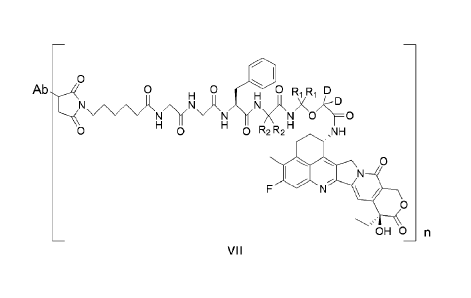
acceptable salt or a solvate thereof, having a structure of formula VII:
CA 03188508 2023- 2-6
11
0
Ab4 0 0 D
/\/1R11 NH Ji X kD
N X \N \=0
0 0 0 R2 R2 ,NH
0
N
0
OHO
VII
wherein,
Ab represents an antibody moiety comprising a first antigen-binding fragment
and a second antigen-binding
fragment, wherein the first antigen-binding fragment is an scFv, and comprises
a heavy chain CDR1, a heavy
chain CDR2, a heavy chain CDR3, a light chain CDR1, a light chain CDR2 and a
light chain CDR3, the heavy
chain CDR1, the heavy chain CDR2 and the heavy chain CDR3 comprising amino
acid sequences set forth in
SEQ ID NOs: 43, 28 and 29, respectively, and the light chain CDR1, the light
chain CDR2 and the light chain
CDR3 comprising amino acid sequences set forth in SEQ ID NOs: 30, 31 and 32,
respectively;
the second antigen-binding fragment is an Fab, and comprises a heavy chain
CDR1, a heavy chain CDR2, a heavy
chain CDR3, a light chain CDR1, a light chain CDR2 and a light chain CDR3, the
heavy chain CDR1, the heavy
chain CDR2 and the heavy chain CDR3 comprising amino acid sequences set forth
in SEQ ID NOs: 45, 46 and
47, respectively, and the light chain CDR1, the light chain CDR2 and the light
chain CDR3 comprising amino acid
sequences set forth in SEQ ID NOs: 48, 49 and 50, respectively;
n is an integer or a decimal selected from the group consisting of 1 to 10,
and
Ri and R2 are each independently selected from the group consisting of
hydrogen (H) and deuterium (D).
In some specific embodiments, the present application provides an antibody-
drug conjugate, or a pharmaceutically
acceptable salt or a solvate thereof, having a structure of formula VII-1,
VII-2, VII-3, or VII-4 below,
0
0 0 0
N N OMO
0 0 0 ,NH
0
N
0
OH 0 n
VII-1
CA 03188508 2023- 2-6
12
0
0
H
/\ ,1\1\A N) /\
N N 0 0
0 D
0
N
0
OH 0 __________________________________________________________________ n
VII-2
0
0
H OD D D
\ANX0/173 0
O
N
0 0 ,NH
0
N
0
OH 0 __________________________________________________________________ n
V11-3
0
0
/\ ,N \AN NNX(D/i7D 0
N
0 0 0 D D ,NH
0
N
0
OH 0 __________________________________________________________________ n
VII-4
wherein,
Ab represents an antibody moiety comprising a first antigen-binding fragment
and a second antigen-binding
fragment, wherein the first antigen-binding fragment is an scFv, and comprises
a heavy chain CDR1, a heavy
chain CDR2, a heavy chain CDR3, a light chain CDR1, a light chain CDR2 and a
light chain CDR3, the heavy
chain CDR1, the heavy chain CDR2 and the heavy chain CDR3 comprising amino
acid sequences set forth in
SEQ ID NOs: 43, 28 and 29, respectively, and the light chain CDR1, the light
chain CDR2 and the light chain
CDR3 comprising amino acid sequences set forth in SEQ ID NOs: 30, 31 and 32,
respectively;
the second antigen-binding fragment is an Fab, and comprises a heavy chain
CDR1, a heavy chain CDR2, a heavy
chain CDR3, a light chain CDR1, a light chain CDR2 and a light chain CDR3, the
heavy chain CDR1, the heavy
chain CDR2 and the heavy chain CDR3 comprising amino acid sequences set forth
in SEQ ID NOs: 45, 46 and
47, respectively, and the light chain CDR1, the light chain CDR2 and the light
chain CDR3 comprising amino acid
sequences set forth in SEQ ID NOs: 48, 49 and 50, respectively;
n is an integer or a decimal selected from the group consisting of 1 to 10.
CA 03188508 2023- 2-6
13
In one specific embodiment, the present application provides an antibody-drug
conjugate, or a pharmaceutically
acceptable salt or a solvate thereof, having a structure of formula VII-1
below,
Ab 0 0 0
N 0
0 ,NH
0
/
0
OH 0 n
vil-1
wherein,
Ab represents an antibody moiety comprising a first antigen-binding fragment
and a second antigen-binding
fragment, wherein the first antigen-binding fragment is an scFv, and comprises
a heavy chain CDR1, a heavy
chain CDR2, a heavy chain CDR3, a light chain CDR1, a light chain CDR2 and a
light chain CDR3, the heavy
chain CDR1, the heavy chain CDR2 and the heavy chain CDR3 comprising amino
acid sequences set forth in
SEQ ID NOs: 43, 28 and 29, respectively, and the light chain CDR1, the light
chain CDR2 and the light chain
CDR3 comprising amino acid sequences set forth in SEQ ID NOs: 30, 31 and 32,
respectively;
the second antigen-binding fragment is an Fab, and comprises a heavy chain
CDR1, a heavy chain CDR2, a heavy
chain CDR3, a light chain CDR1, a light chain CDR2 and a light chain CDR3, the
heavy chain CDR1, the heavy
chain CDR2 and the heavy chain CDR3 comprising amino acid sequences set forth
in SEQ ID NOs: 45, 46 and
47, respectively, and the light chain CDR1, the light chain CDR2 and the light
chain CDR3 comprising amino acid
sequences set forth in SEQ ID NOs: 48, 49 and 50, respectively;
n is an integer or a decimal selected from the group consisting of 1 to 10.
In one specific embodiment, the present application provides an antibody-drug
conjugate, or a pharmaceutically
acceptable salt or a solvate thereof, having a structure of formula VII below,
Ab 0 0 0 0 R Ri D
)(1\IX0)::' 0
N
H
,NH
0
/
0
OHO n
vil
wherein,
Ab is trastuzumab,
n is an integer or a decimal selected from the group consisting of 1 to 10,
and
111 and R2 are each independently selected from the group consisting of
hydrogen (H) and deuterium (D).
In one specific embodiment, the present application provides an antibody-drug
conjugate, or a pharmaceutically
acceptable salt or a solvate thereof, having a structure of formula VII-1
below,
CA 03188508 2023- 2-6
14
0
Ab 0 H 0 H 0
NN/\o/1173
N 0
0 0 0 ,N1H
0
/
0
OH 0 n
wherein,
Ab is trastuzumab,
n is an integer or a decimal selected from the group consisting of 1 to 10.
In one aspect, the present application provides a pharmaceutical composition
comprising the antibody-drug
conjugate, or the pharmaceutically acceptable salt or the solvate thereof
according to the present application, and a
pharmaceutically acceptable carrier.
In one aspect, the present application provides use of the antibody-drug
conjugate, or the pharmaceutically
acceptable salt or the solvate thereof according to the present application,
in preparing a medicament for
preventing and treating cancer.
In one aspect, the present application provides use of a pharmaceutical
composition comprising the antibody-drug
conjugate, or the pharmaceutically acceptable salt or the solvate thereof
according to the present application, and a
pharmaceutically acceptable carrier in preparing a medicament for preventing
and treating cancer.
In one aspect, the present application provides an antibody-drug conjugate, or
a pharmaceutically acceptable salt
or a solvate thereof for use in preventing and treating cancer.
In one aspect, the present application provides a method for treating or
preventing cancer, the method comprising
administering to a patient in need thereof a therapeutically effective amount
of the antibody-drug conjugate, or the
pharmaceutically acceptable salt or the solvate thereof according to the
present application, or a pharmaceutical
composition comprising the antibody-drug conjugate, or the pharmaceutically
acceptable salt or the solvate
thereof according to the present application, and a pharmaceutically
acceptable carrier.
In some embodiments, the antibody-drug conjugate, or the pharmaceutically
acceptable salt or the solvate thereof
according to the present application, may be used for preventing or treating
HER2 positive cancer, HER2 negative
cancer (including triple-negative breast cancer), and cancer that shows HER2
expression as I HC2+ detected by
immunohistochemical assay.
In some aspects, the present application provides a linker-drug intermediate
compound having a structure of
formula VI below, for use in obtaining an antibody-drug conjugate that links
an antibody to the intermediate
compound:
0
0 H 0 0R R D
i\jANX0 N
H 0
0 0 0 R2 R2 NH
0
/
0
OHO VI
wherein,
111 and R2 are each independently selected from the group consisting of
hydrogen (H) and deuterium (D).
CA 03188508 2023- 2-6
15
In some aspects, the present application provides a linker-drug intermediate
compound having a structure of
formula VI-1, VI-2, VI-3 or VI-4 below, for use in obtaining an antibody-drug
conjugate that links an antibody to
the intermediate compound:
0
H 0
H 0 D
N /\ ,N\A N N \AN1/\02;0
----- N `'
H H H
0 0 0 ,NH
0
N
F N \ /
0
OH 0 VI-
1
0
H 0
H 0 D
N/i;
----- N `'
H N N 0 0
0 0 H 0 D DH ,NH
0
N
F N \ /
0
OH 0 VI-
2
0
H 0
H 0 D D D
N /\ ,N\A Nj X 2<rj
-----. N `'
H N H N 0 0
H
0 0 0 ,NH
0
N
F N \ /
0
OH 0 VI-
3
0
H H
D D
D
N>NX0/1-0
---- N `'
H N
H
0 0 H ODD ,NH
0
N
F N \ /
0
OH 0 VI-
4.
CA 03188508 2023- 2-6
16
In some aspects, the present application provides a linker compound having a
structure of formula V below, for
use in obtaining an antibody-drug conjugate that links a drug to an antibody
via the linker:
0
NX µN/ \O/IFO N ' N
H H H
wherein Ri and R2 are each independently selected from the group consisting of
hydrogen (H) and deuterium (D),
and the left succinimide terminus of the structure is a site linking to the
antibody moiety and the right carbonyl
terminus is a site linking to the cytotoxic drug moiety.
In some aspects, the present application provides a linker compound having a
structure of formula V-1, V-2, V-3,
or V-4 below, for use in obtaining an antibody-drug conjugate that links a
drug to an antibody via the linker, and
the structures of formulas V-1 to V-4 are linked to the antibody moiety by the
left succinimide terminus and the
cytotoxic drug moiety by the right carbonyl terminus, respectively:
0
H D
N o NI,\(11V1 j jN/\0
N D
o H N
H H 0
O 0 V-
1
0
-1,-,-, 0
H 0 H 0 D
----% N "
H N
H N 0
H 0
O 0 D D
V-2
0
H 0 H 0 D D D
N /\(N j NjNX0 D
N
H N
H H 0
O 0 V-
3
0
H
N /\ /NINA Nx)-NX0 D
N N 0
H oil H H
VA.
In some aspects, the present application provides a compound having a
structure of formula IV(a) below:
R1 R 1 D
D
H2NX 0/Ir 0
,NH
0
N
F N \ /
0
.õ,
OH 0 IV(a)
wherein Ri is selected from the group consisting of hydrogen (H) and deuterium
(D).
In some aspects, the present application provides a compound having a
structure of formula IV(a)-1 or formula
IV(a)-2 below:
CA 03188508 2023- 2-6
17
H2N/\0/1173 0
,NH
0
/
0
OH 0 IV(a)-1
DDD
X
H2N0 0
,NH
0
/
0
OH 0 IV(a)-2.
In some aspects, the present application provides a compound having a
structure of formula 111(a) below:
D D
HO
,NH
0
/
0
OH 0 111(a).
The present application provides an antibody-drug conjugate, or a
pharmaceutically acceptable salt or a solvate
thereof having improved pharmacokinetic properties. An improvement in
pharmacokinetic properties will result in
a reduction in toxicity of the target compound, an increase in safety and/or
tolerability, an increase in efficacy and
an improvement in the final therapeutic window.
BRIEF DESCRIPTION OF THE DRAWINGS
FIG. 1 shows the killing rates of anti-HER2 bispecific antibodies (Expi HER2-
1, Expi HER2-2, 23C2 HER2-1
and 23C2 HER2-2) and the combination of trastuzumab + pertuzumab against BT474
tumor cells;
FIG. 2 shows the killing rates of anti-HER2 bispecific antibody, trastuzumab,
T-DM1, and the combination of
trastuzumab + pertuzumab against NCI-N87 tumor cells;
FIG. 3 shows the killing rates of anti-HER2 bispecific antibody, trastuzumab,
T-DM1, and the combination of
trastuzumab + pertuzumab against J I MT-1 tumor cells;
FIG. 4 shows the results of inhibition of anti-HER2 bispecific antibody,
trastuzumab, and the combination of
trastuzumab + pertuzumab on proliferation of BT474 tumor cells;
FIG. 5 shows the effect of anti-HER2 bispecific antibody, PBS vehicle control,
and the combination of
trastuzumab + pertuzumab (Per + Ira) on changes in mouse tumor volume in a
gastric cancer N87 mouse
xenograft tumor model;
FIG. 6 shows the effect of anti-HER2 bispecific antibody, PBS vehicle control,
and the combination of
trastuzumab + pertuzumab on changes in mouse body weight in the gastric cancer
N87 mouse xenograft tumor
efficacy;
FIG. 7 shows a structure of some exemplary anti-HER2 bispecific antibodies,
wherein a dimeric Fc is depicted
with one chain shown in black (a first Fc polypeptide) and another chain shown
in gray (a second Fc polypeptide),
and one antigen-binding domain (a first antigen-binding fragment) is shown
hatched and the other antigen-binding
CA 03188508 2023- 2-6
18
domain (a second antigen-binding fragment) is shown white; wherein the first
antigen-binding fragment is an scFv
and fused with the first Fc polypeptide, and the second antigen-binding
fragment is an Feb and fused with the
second Fc polypeptide;
FIG. 8 shows the endocytic activity of drug conjugates of different antibodies
(monoclonal antibody-DDDXD and
bispecific a ntibody-DDDXD) in NCI-N87 tumor cells; and
FIG. 9 shows the endocytic activity of drug conjugates of different antibodies
(monoclonal antibody-DDDXD and
bispecific antibody-DDDXD) in SK-BR-3 tumor cells.
Explanation and definitions
Unless otherwise stated, the following terms used herein shall have the
following meanings. A certain term, unless
otherwise specifically defined, should not be considered uncertain or unclear,
but construed according to its
common meaning in the field. Reference is made to, for example, Singleton et
al., Dictionary of Microbiology and
Molecular Biology, 2nd ed., j. Wiley & Sons (New York, NY 1994); Sambrook et
al., Molecular Cloning: A
Laboratory Manual, Cold Springs Harbor Press (Cold Springs Harbor, NY 1989);
Davis et al., Basic Methods in
Molecular Biology, Elsevier Science Publishing Inc., New York, USA (2012);
Abbas et al., Cellular and Molecular
Immunology, Elsevier Science Health Science div (2009); He Wei et al., Medical
Immunology, (2nd ed), People's
Medical Publishing House, 2010. When referring to a trade name herein, it is
intended to refer to its corresponding
commercial product or its active ingredient.
The term "substituted" means that any one or more hydrogen atoms on a specific
atom are substituted by
substituents, as long as the valence of the specific atom is normal and the
resulting compound is stable. When the
substituent is oxo (namely =0), it means that two hydrogen atoms are
substituted, and oxo is not available on an
aromatic group.
The term "optional" or "optionally" means that the subsequently described
event or circumstance may, but not
necessarily, occur. The description includes instances where the event or
circumstance occurs and instances where
the event or circumstance does not occur. A certain group being "optionally
substituted" means that the group may
be substituted or unsubstituted, for example, ethyl being "optionally"
substituted with halogen means that the
ethyl may be unsubstituted (CH2CH3), monosubstituted (for example, CH2CH2F),
polysubstituted (for example,
CHFCH2F, CH2CHF2 and the like), or fully substituted (CF2CF3). It will be
understood by those skilled in the art
that for any groups comprising one or more substituents, any substitutions or
substituting patterns which may not
exist or cannot be synthesized spatially are not introduced.
Cm_, used herein means that the portion has an integer number of carbon atoms
in the given range. For example,
"Ci 6" means that the group may have 1 carbon atom, 2 carbon atoms, 3 carbon
atoms, 4 carbon atoms, 5 carbon
atoms, or 6 carbon atoms.
When any variable (e.g., R) occurs more than once in the constitution or
structure of a compound, the variable is
independently defined in each case. Therefore, for example, if a group is
substituted with 2 R, the definition of
each R is independent.
When a connecting group has a number of 0, for example, -(CH2)o-, it means
that the connecting group is a
covalent bond.
When a variable is a single bond, it means that the two groups are directly
connected. For example, in A-L-Z,
when L represents a single bond, it means that the structure is actually A-Z.
The term "halo-" or "halogen" refers to fluorine, chlorine, bromine and
iodine.
The term "hydroxy" refers to -OH group.
The term "cyano" refers to -CN group.
The term "sulfydryl" refers to -SH group.
The term "amino" refers to -NH2 group.
The term "nitro" refers to -NO2 group.
The term "alkyl" refers to hydrocarbyl with a general formula of Col-12o,1.
The alkyl can be linear or branched. For
example, the term "C1_6 alkyl" refers to alkyl containing 1 to 6 carbon atoms
(for example, methyl, ethyl,
n-propyl, isopropyl, n-butyl, isobutyl, sec-butyl, tert-butyl, n-pentyl, 1-
methylbutyl, 2-methylbutyl, 3-methyl butyl,
neopentyl, hexyl, 2-methylpentyl and the like). The alkyl moieties (namely
alkyl) of alkoxy, alkylamino,
dialkylamino, alkylsulfonyl and alkylthio are similarly defined as above.
The term "alkoxyl" refers to -0-alkyl.
The term "a I kyla m i no" refers to-NH-alkyl.
The term "d ia I kyla m i no" refers to -N(alkyl)2.
The term "a I kylsulfonyl" refers to-S02-alkyl.
The term "alkylthio" refers to -S-alkyl.
CA 03188508 2023- 2-6
19
The term "alkenyl" refers to linear or branched unsaturated aliphatic
hydrocarbyl consisting of carbon atoms and
hydrogen atoms with at least one double bond. Non-limiting examples of alkenyl
include, but are not limited to,
ethenyl, 1-propenyl, 2-propenyl, 1-butenyl, isobutenyl, 1,3-butadienyl, and
the like.
The term "alkynyl" refers to linear or branched unsaturated aliphatic
hydrocarbyl consisting of carbon atoms and
hydrogen atoms with at least one triple bond. Non-limiting examples of alkynyl
include, but are not limited to,
ethynyl (-CCH), 1-propinyl (-CC-CH3), 2-pro pi nyl (-CH2-CCH), 1,3-butadiynyl
(-CC-CCH), and the like.
The term "cycloalkyl" refers to a carbon ring that is fully saturated and may
exist in the form of a monocyclic,
bridged cyclic, or spiro cyclic structure. Unless otherwise specified, the
carbon ring is generally a 3-10 membered
ring. Non-limiting examples of cycloalkyl include, but are not limited to,
cyclopropyl, cyclobutyl, cyclopentyl,
cyclohexyl, norbornyl(bicyclo[2.2.1]heptyl), bicyclo[2.2.2]octyl, adamantyl,
and the like.
The term "cycloalkenyl" refers to a non-aromatic carbon ring that is not fully
saturated and may exist in the form
of a monocyclic, bridged cyclic, or Spiro cyclic structure. Unless otherwise
specified, the carbon ring is generally
a 5-8 membered ring. Non-limiting examples of cycloalkenyl include, but are
not limited to, cyclopentenyl,
cyclopentadienyl, cyclohexenyl, cyclohexadienyl, cycloheptenyl,
cycloheptadienyl, and the like.
The term "heterocyclyl" refers to a fully saturated or partially unsaturated
(but not fully unsaturated
heteroaromatic group) nonaromatic ring which may exist in the form of a
monocyclic, bridged cyclic, or Spiro
cyclic structure. Unless otherwise specified, the heterocyclyl is usually a 3-
7 membered ring containing 1-3
heteroatoms (preferably 1 or 2 heteroatoms) independently selected from the
group consisting of sulfur, oxygen,
and/or nitrogen. Non-limiting examples of heterocyclyl include, but are not
limited to, oxiranyl, tetrahydrofuranyl,
dihydrofuranyl, pyrrolidinyl, N-methylpyrrolidinyl, dihydropyrrolyl,
piperidinyl, piperazinyl, pyrazolidinyl,
4H-pyranyl, morpholinyl, thiomorpholinyl, tetrahydrothienyl, and the like.
The term "heterocycloalkyl" refers to a fully saturated cyclic group that may
exist in the form of a monocyclic,
bridged cyclic, or Spiro cyclic structure. Unless otherwise specified, the
heterocyclyl is usually a 3-7 membered
ring containing 1-3 heteroatoms (preferably 1 or 2 heteroatoms) independently
selected from the group consisting
of sulfur, oxygen, and/or nitrogen. Examples of 3 membered heterocycloalkyl
include, but are not limited to,
oxiranyl, thiiranyl, and aziranyl; non-limiting examples of 4 membered
heterocycloalkyl include, but are not
limited to, azetidinyl, oxetanyl, and thietanyl; examples of 5 membered
heterocycloalkyl include, but are not
limited to, tetrahydrofuranyl, tetrahydrothienyl, pyrrolidinyl,
isoxazolidinyl, oxazolidinyl, isothiazolidinyl,
thiazolidinyl, imidazolidinyl, and tetrahydropyrazolyl; examples of 6 membered
heterocycloalkyl include, but are
not limited to, piperidinyl, tetrahydropyranyl, tetrahydrothiopyranyl,
morpholinyl, piperazinyl, 1,4-thioxanyl,
1,4-dioxanyl, thiomorpholinyl, 1,3-dithianyl, and 1,4-dithianyl; examples of 7
membered heterocycloalkyl
include, but are not limited to, azacycloheptanyl, oxacycloheptanyl and
thiocycloheptanyl. Preferably, the
heterocycloalkyl is a monocyclic heterocycloalkyl having 5 or 6 ring atoms.
The term "aryl" refers to an aromatic monocyclic or fused polycyclic group of
carbon atoms with the conjugated
pi-electron system. For example, aryl may have 6-20 carbon atoms, 6-14 carbon
atoms or 6-12 carbon atoms.
Non-limiting examples of aryl include, but are not limited to, phenyl,
naphthyl, anthryl,
1,2,3,4-tetrahydronaphthalene, and the like.
The term "heteroaryl" refers to a monocyclic or fused polycyclic system
containing at least one ring atom selected
from the group consisting of N, 0 and S, with the remaining ring atoms being
C, and having at least one aromatic
ring. Preferably, the heteroaryl has a single 4-8 membered ring, in
particular, a 5-8 membered ring, or is a plurality
of fused rings comprising 6-14 ring atoms, in particular 6-10 ring atoms. Non-
limiting examples of heteroaryl
include, but are not limited to, pyrrolyl, furanyl, thienyl, imidazolyl,
oxazolyl, pyrazolyl, pyridinyl, pyrimidinyl,
pyrazinyl, quinolyl, isoquinolyl, tetrazolyl, triazolyl, triazinyl,
benzofuranyl, benzothienyl, indolyl, isoindolyl and
the like.
The "derivative": a compound formed by substituting atoms or atom groups in
the molecule of the parent
compound with other atoms or atom groups is referred to as a derivative of the
parent compound.
Any atom of a compound labeled and synthesized herein may represent any stable
isotope of the atom, if not
specifically designated. Unless otherwise specified, when a position in a
structure is defined as H, i.e., hydrogen
(H-1), this position contains only the naturally occurring isotope. Similarly,
unless otherwise specified, when a
position in a structure is defined as D, i.e., deuterium (H-2), this position
contains an isotope having an amount
that is at least 3340 times greater than the amount of the naturally occurring
isotope (0.015%) (i.e., at least 50.1%
deuterium isotope), when one or more positions in the structure of the labeled
synthetic compound are defined as
D, i.e., deuterium (H-2), the content of the compound represented by the
structure may be at least 52.5%, at least
60%, at least 67.5%, at least 75%, at least 82.5%, at least 90%, at least 95%,
at least 97%, at least 98.5%, at least
99%, or at least 99.5%. The deuterated ratio of a compound labeled and
synthesized herein refers to a ratio of the
amount of the labeled synthetic isotope to the amount of the naturally
occurring isotope. The deuterated ratio per
designated deuterium atom of the compound labeled and synthesized herein may
be at least 3500 times (52.5%),
at least 4000 times (60%), at least 4500 times (67.5%), at least 5000 times
(75%), at least 5500 times (82.5%), at
CA 03188508 2023- 2-6
20
least 6000 times (90%), at least 6333.3 times (95%), at least 6466.7 times
(97%), at least 6566.7 times (98.5%), at
least 6600 times (99%), at least 6633.3 times (99.5%). lsotopologues herein
refer to compounds that differ only in
isotopic composition in terms of chemical structure. The compound labeled and
synthesized herein has the same
chemical structure, with only isotopic changes in the atomic composition of
its molecules. Therefore, the
deuterium-containing compound at a specific position labeled and synthesized
herein also contains very little
hydrogen isotope at this position, and the amount of hydrogen isotopologue at
a certain position in the compound
labeled and synthesized herein depends on many factors, including the
deuterium isotopic purity of the deuterated
agent (D20, D2, NaBD4, LiAID4, and the like) and the effectiveness of
introducing deuterium isotope synthesis
methods. However, as previously mentioned, the total amount of such hydrogen
isotopologue at a certain position
will be less than 49.9%. The total amount of hydrogen isotopologue at a
certain position in the compound labeled
and synthesized herein will be less than 47.5%, 40%, 32.5%, 25%, 17.5%, 10%,
5%, 3%, 1%, or 0.5%.
In the present application, any individual atom not designated as deuterium is
present at its natural isotopic
abundance.
The term "treating" or "treatment" means administering the compound or
formulation described herein to prevent,
ameliorate, or eliminate a disease or one or more symptoms associated with the
disease, including: (i) preventing
the occurrence of the disease or disease state in a mammal, particularly when
such a mammal is predisposed to the
disease state but has not yet been diagnosed with it; (ii) inhibiting a
disease or disease state, i.e., arresting its
development; (iii) alleviating a disease or disease state, i.e., causing its
regression.
The term "therapeutically effective amount" refers to an amount of the
compound of the present application for (i)
treating or preventing a specific disease, condition or disorder; (ii)
alleviating, ameliorating or eliminating one or
more symptoms of a specific disease, condition or disorder, or (iii)
preventing or delaying onset of one or more
symptoms of a specific disease, condition or disorder described herein. The
amount of the compound of the
present application composing the "therapeutically effective amount" varies
dependently on the compound, the
disease state and its severity, the route of administration, and the age of
the mammal to be treated, but can be
determined routinely by those skilled in the art in accordance with their
knowledge and the present disclosure.
The term "pharmaceutically acceptable" is used herein for those compounds,
materials, compositions, and/or
dosage forms which are, within the scope of sound medical judgment, suitable
for use in contact with the tissues
of human beings and animals without excessive toxicity, irritation, allergic
response, or other problems or
complications, and commensurate with a reasonable benefit/risk ratio.
A pharmaceutically acceptable salt, for example, may be a metal salt, an
ammonium salt, a salt formed with an
organic base, a salt formed with an inorganic acid, a salt formed with an
organic acid, a salt formed with a basic or
acidic amino acid, and the like.
The term "solvate" refers to a substance formed by association of a compound
with a solvent molecule.
The term "pharmaceutical composition" refers to a mixture consisting of one or
more of the compounds or the
salts thereof disclosed herein and a pharmaceutically acceptable excipient.
The pharmaceutical composition is
intended to facilitate the administration of the compound to an organic
entity.
The term "pharmaceutically acceptable excipients" refers to those which do not
have a significant irritating effect
on an organic entity and do not impair the biological activity and properties
of the active compound. Suitable
excipients are well known to those skilled in the art, for example
carbohydrate, wax, water-soluble and/or
water-swellable polymers, hydrophilic or hydrophobic material, gelatin, oil,
solvent, water and the like.
The compounds and intermediates disclosed herein may also exist in different
tautomeric forms, and all such
forms are included within the scope of the present application. The term
"tautomer" or "tautomeric form" refers to
structural isomers of different energies that can interconvert via a low
energy barrier. For example, a proton
tautomer (also referred to as prototropic tautomer) includes interconversion
via proton transfer, such as keto-enol
isomerism and imine-ena mine isomerism. A specific example of a proton
tautomer is an imidazole moiety where a
proton can transfer between two ring nitrogens. A valence tautomer includes
the interconversion via
recombination of some bonding electrons.
The term "antibody" is used in its broadest sense and specifically encompasses
intact monoclonal antibodies,
polyclonal antibodies, multispecific antibodies (e.g., bispecific antibodies)
formed from at least two intact
antibodies, multifunctional antibodies, and antibody fragments so long as they
possess the desired biological
activity.
The term "humanized antibody" refers to an antibody comprising CDRs derived
from a non-human antibody, and
the remainder of the antibody molecule is derived from one or more human
antibodies.
The term "mutant" is used to refer to a peptide comprising an amino acid
sequence derived from the amino acid
sequence of the peptide as follows: substitution of one or two or more amino
acids with amino acids different
from the original peptide, deletion of one or two or more wild-type amino
acids, insertion of one or two or more
amino acids that do not exist in the wild type, and/or addition of amino acids
that do not exist in the wild type to
CA 03188508 2023- 2-6
21
the amino terminus (N-terminus) and/or the carboxy terminus (C-terminus) of
the wild type (hereinafter,
collectively referred to as "mutation"). In the present application,
"insertion" may also be included in "addition".
The term "CDR" (complementarity-determining region), also known as
"hypervariable region", refers to each
region of an antibody variable domain which is highly variable in sequence
and/or forms a structurally defined
loop. Natural four-chain antibodies typically comprise six CDRs, three in the
heavy chain variable region and
three in the light chain variable region.
The term "variable region": the antibody structural unit is composed of two
pairs of polypeptide chains, each pair
having one heavy chain and one light chain, and the N-terminal domain of each
chain defining a region of about
100 to 110 or more amino acids primarily responsible for antigen recognition
is the variable region.
The term "Fab" means comprising the constant domain (CL) of the light chain
and the first constant domain
(CH1) of the heavy chain, together with the variable domains VL (light chain
variable region) and VH (heavy
chain variable region) in the light chain and heavy chain, respectively. The
variable domain comprises
complementarity-determining regions (CDRs) that are involved in antigen-
binding.
The term "scFv" includes the VH and VL domains of an antibody, wherein these
domains are present in a single
polypeptide chain. In some embodiments, scFv further comprises a polypeptide
linker between the VH and VL
domains that enables the scFv to form the required structure for antigen-
binding.
The term "[CD" refers to an extracellular domain. HER receptors are receptor
protein tyrosine kinases belonging
to the human epidermal growth factor receptor (HER) family and include EGFR,
HER2, HER3, and HER4
receptors, wherein the HER2 receptor generally comprises an extracellular
domain that may bind HER ligand, a
lipophilic transmembrane domain, a conserved intracellular tyrosine kinase
domain, and a carboxy-terminal
signaling domain with several tyrosine residues that can be phosphorylated,
and the extracellular domain of HER2
comprises four domains that are ECD1, ECD2, ECD3 and ECD4, respectively.
The term "antibody moiety" refers to an antibody moiety in an antibody-drug
conjugate, which, in certain
embodiments, is linked to an intermediate linker moiety via a specific
functional group, and the antibody moiety
can specifically bind to an antigen.
The term "linker moiety" refers to a part of the antibody-drug conjugate which
links an antibody moiety with a
cytotoxic drug moiety and may be cleavable or uncleavable, wherein the
cleavable linker refers to a part which
may be cleaved in a target cell so as to release the cytotoxic drug.
The term "cytotoxic drug moiety" refers to a cytotoxic drug moiety in an
antibody-drug conjugate, and in certain
specific embodiments, the cytotoxic drug moiety is linked to an intermediate
linker moiety via a functional group,
so that cytotoxic drug molecules can be liberated in tumor cells to exert an
anti-tumor effect.
Generic term "trastuzumab" refers to a recombinant humanized monoclonal
antibody that selectively acts on the
extracellular site of human epidermal growth factor receptor-4 (HER4) and can
be used to treat HER2 positive
cancer, an example of which is the commercially available therapeutic
monoclonal antibody product under the
trade name HERCEPTI NO.
Generic term "pertuzumab" refers to a recombinant humanized monoclonal
antibody that selectively acts on the
extracellular site of human epidermal growth factor receptor-2 (HER2) and can
be used to treat HER2 positive
cancer.
The term "HER2" is a second member of the EGFR family having a tyrosine kinase
activity, wherein HER2
expression levels can be detected by immunohistochemical assay, HER2 positive
refers to I HC3+, HER2 negative
refers to I HC1+/0, and for I HC2+, ISH assay should be performed for further
clarification.
The term "cancer" refers to a physiological condition in mammals that is
typically characterized by unregulated
cell growth.
The term "triple-negative breast cancer" is a breast cancer that is negative
for expression of estrogen receptors,
progesterone receptors, and human epidermal growth factor receptor-2.
As used herein, unless otherwise stated, the terms "comprise", "comprises" and
"comprising" or equivalents
thereof (contain, contains, containing, include, includes, including) are open-
ended statements and mean that
elements, components and steps that are not specified may be included in
addition to those listed.
As used herein, unless otherwise indicated, all numbers expressing the amounts
of ingredients, measurements, or
reaction conditions used herein are to be understood as being modified in all
instances by the term "about". The
term "about" when connected to a percentage may mean, for example, 0.1%,
preferably, 0.05%, and more
preferably, 0.01%.
Unless otherwise specified clearly herein, singular terms encompass plural
referents, and vice versa Similarly,
unless otherwise specified clearly herein, the word "or" is intended to
include "and".
As used herein, the percent identity (degree of homology) between sequences
can be determined by comparing the
two sequences, for example, using freely available computer programs (e.g.,
BLASTp or BLASTn with default
CA 03188508 2023- 2-6
22
settings) typically used for this purpose on the World Wide Web (e.g.,
www.ncbi.nlm.nih.gov).
DETAILED DESCRIPTION
For clarity, the present application is further described with the following
examples, which are, however, not
intended to limit the scope of the present application. The reagents used
herein are commercially available and can
be used without further purification.
Trastuzumab and Pertuzumab used in the examples of the present application
were prepared according to the
conventional methods for antibodies, wherein vectors were constructed first,
and eukaryotic cells were transfected
and then purified for expression, with the sequence of Trastuzumab being
referenced to WHO DRUG
INFORMATION INN RL78, and the sequence of Pertuzumab being referenced to the
examples of W00100245.
DS-8201 is the active ingredient of Enhertu, a commercially available
formulation from Daiichi Sankyo Co., Ltd,
and has the same structure as Trastuzumab-DXD prepared in the present
application (see Example 15 for
structure).
Example 1. Construction, Expression and Purification of Anti-Her2 scFv-Fc and
Variants Thereof
In constructing anti-Her2 scFv-Fc, human IgG1 was used as the Fc portion, and
the variable region sequence of
the anti-Her2 arm was a sequence based on the monoclonal antibody Herceptine.
The light and heavy chain
variable regions of the monoclonal antibody Herceptin were linked in series
by a designed linker 1 (i.e.,
(GGGGS)3) to form anti-Her2 scFv-Fc (SEQ ID NO: 1), the amino acid sequence of
which is as follows:
EVQLVESGGGLVQPGGSLRLSCAASGFNI KDTY I HWV RQAPG KGLEWVA RIY PTNGYTRYA DSVKG
RFT!
SADTSKNTAY LQMNSLRAEDTAVYYCSRWGGDGFYAM DYWGQGTLVTVSSGGGGSGGGGSGGGGSDI
QMTQSPSSLSASVGDRVTITCRASQDVNTAVAWYQQKPGKAPKLLIYSASFLYSGVPSRFSGSRSGTDFTL
TI SSLQPE DFATYY CQQHYTTPPTFGQGTKVE I KG EPKSSDKTHTCPPCPAPE L LGGPSVF
LFPPKPKDTL M I
SRTPEVTCVVVDVSH EDPEVK FNVVYVDGV EV HNAKTKPRE EQY NSTY RVVSVLTVLHQDWLNGKEY K
CKVSNKALPAPI EKTISKAKGQPREPQVYTLPPSRE EMTKNQVS LTCLVKG FY PSDIAVEWESNGQPENNY
KTTPPVLDSDGS FFLY S KLTV DKSRWQQG NV FSCSVM H EA LH N HYTQKSLS LSPG K
Further, point mutations were constructed in the anti-Her2 scFv-Fc sequence to
construct the following variants:
anti-Her2-scFv-VL-F53Y-Fc (SEQ ID NO: 3): derived from wild-type anti-Her2
scFv-Fc, with an F53Y mutation
in the VL region; the amino acid sequence is as follows:
EVQLVESGGGLVQPGGSLRLSCAASGFNI KDTY I HWV RQAPG KGLEWVA RIY PTNGYTRYA DSVKG
RFT!
SADTSKNTAY LQMNSLRAEDTAVYYCSRWGGDGFYAM DYWGQGTLVTVSSGGGGSGGGGSGGGGSDI
QMTQSPSSLSASVGDRVTITCRASQDVNTAVAWYQQKPGKAPKLLIYSASYLYSGVPSRFSGSRSGTDFTL
TI SSLQPE DFATYY CQQHYTTPPTFGQGTKVE I KG EPKSSDKTHTCPPCPAPE L LGGPSVF
LFPPKPKDTL M I
SRTPEVTCVVVDVSH EDPEVK FNVVYVDGV EV HNAKTKPRE EQY NSTY RVVSVLTVLHQDWLNGKEY K
CKVSNKALPAPI EKTISKAKGQPREPQVYTLPPSRE EMTKNQVS LTCLVKG FY PSDIAVEWESNGQPENNY
KTTPPVLDSDGS FFLY S KLTVDKSRWQQG NV FSCSVM H EA LH N HYTQKSLS LSPG K.
anti-Her2-scFv-VL-F53A-Fc (SEQ ID NO: 5): derived from wild-type anti-Her2
scFv-Fc, with an F53A mutation
in the VL region; the amino acid sequence is as follows:
EVQLVESGGGLVQPGGSLRLSCAASGFNI KDTY I HWV RQAPG KGLEWVA RIY PTNGYTRYA DSVKG
RFT!
SADTSKNTAY LQMNSLRAEDTAVYYCSRWGGDGFYAM DYWGQGTLVTVSSGGGGSGGGGSGGGGSDI
QMTQSPSSLSASVGDRVTITCRASQDVNTAVAWYQQKPGKAPKLLIYSASALYSGVPSRFSGSRSGTDFTL
TI SSLQPE DFATYY CQQHYTTPPTFGQGTKVE I KG EPKSSDKTHTCPPCPAPE L LGGPSVF
LFPPKPKDTL M I
SRTPEVTCVVVDVSH EDPEVK FNVVYVDGV EV HNAKTKPRE EQY NSTY RVVSVLTVLHQDWLNGKEY K
CKVSNKALPAPI EKTISKAKGQPREPQVYTLPPSRE EMTKNQVS LTCLVKG FY PSDIAVEWESNGQPENNY
KTTPPVLDSDGS FFLY S KLTVDKSRWQQG NV FSCSVM H EA LH N HYTQKSLSLSPG K.
anti-Her2-scFv-VL-F53R-Fc (SEQ ID NO: 7): derived from wild-type anti-Her2
scFv-Fc, with an F53R mutation
in the VL region; the amino acid sequence is as follows:
EVQLVESGGGLVQPGGSLRLSCAASGFNI KDTY I HWV RQAPG KGLEWVA RIY PTNGYTRYA DSVKG
RFT!
SADTSKNTAY LQMNSLRAEDTAVYYCSRWGGDGFYAM DYWGQGTLVTVSSGGGGSGGGGSGGGGSDI
QMTQSPSSLSASVGDRVTITCRASQDVNTAVAWYQQKPGKAPKLLIYSASRLYSGVPSRFSGSRSGTDFTL
TI SSLQPE DFATYY CQQHYTTPPTFGQGTKVE I KG EPKSSDKTHTCPPCPAPE L LGGPSVF
LFPPKPKDTL M I
SRTPEVTCVVVDVSH EDPEVK FNVVYVDGV EV HNAKTKPRE EQY NSTY RVVSVLTVLHQDWLNGKEY K
CKVSNKALPAPI EKTISKAKGQPREPQVYTLPPSRE EMTK NQVS LTCLVKG FY PSDIAVEWESNGQPENNY
KTTPPVLDSDGS FFLY S KLTVDKSRWQQG NV FSCSVM H EA LH N HYTQKSLS LSPG K.
anti-Her2-scFv-VH-K30E-Fc (SEQ ID NO: 9): derived from wild-type anti-Her2
scFv-Fc, with a K3OE mutation
in the VH region; the amino acid sequence is as follows:
EVQLVESGGGLVQPGGSLRLSCAASGFNI EDTY I HWVRQAPGKGLEWVARIY PINGYTRYADSV KG RFT!
SADTSKNTAY LQMNSLRAEDTAVYYCSRWGGDGFYAM DYWGQGTLVTVSSGGGGSGGGGSGGGGSDI
QMTQSPSSLSASVGDRVTITCRASQDVNTAVAWYQQKPGKAPKLLIYSASFLYSGVPSRFSGSRSGTDFTL
TI SSLQPE DFATYY CQQHYTTPPTFGQGTKVE I KG EPKSSDKTHTCPPCPAPE L LGGPSVF
LFPPKPKDTL M I
CA 03188508 2023- 2-6
23
SRTPEVTCVVVDVSH EDPEVK FNVVYVDGV EV HNAKTKPRE EQY NSTY RVVSV LTVL HQDWLNG KEY
K
CKVSNKALPAPI E KTISKAKGQPREPQVYTLPPSRE EMTK NQVS LTCLVKG FY PSDIAVEWESNGQPENNY
KTTPPVLDSDGS FFLY S K LTVDKSRWQQG NV FSCSVM H EA LH N HYTQKSLSLSPG K.
The DNA sequences of anti-Her2 scFv-Fc and the variants thereof (SEQ ID NOs:
2, 4, 6, 8, and 10) were
synthesized and each cloned into the pcDNA3.1 expression vector. The
expression vector of anti-Her2 scFv-Fc or
the variants thereof was transfected into ExpiCHO cells (CHO-S, Thermo) by
using an ExpiCHOTM expression kit
(Thermo Fisher, Cat. No. A29133). The cells were cultured in ExpiCHO
expression medium in a humidified
atmosphere in an incubator at 37 C with 8% CO2 on an orbital shaker spinning
at 130 rpm. The culture
supernatant was collected, and protein purification was performed using
protein A magnetic beads (Genscript, Cat.
No. L00273). The protein concentration was measured using a UV-Vis
spectrophotometer (NanoDrop lite,
Thermo Scientific).
Table 1: Sequence information about anti-Her2 scFv-Fc and the variants thereof
Amino acid Encoding DNA
Mutation site in
Name sequence sequence
(SEQ ID NO) (SEQ ID NO) variable
region
anti-Her2 scFv-Fc 1 2 None
anti-Her2-scFv-VL-F53Y-Fc 3 4 F53Y
anti-Her2-scFv-VL-F53A-Fc 5 6 F53A
anti-Her2-scFv-VL-F53R-Fc 7 8 F53R
anti-Her2-scFv-VH-K3OE-Fc 9 10 K3OE
Example 2. Aggregate Verification and Human Her2 Antigen-Binding Assay of Anti-
Her2 scFv-Fc and
Variants Thereof
For the expressed and purified anti-Her2 scFv-Fc and variants thereof, the
affinity of the test molecules for human
Her2 protein was determined by Biacore T200 (GE) as follows:
an amount of anti-Her2 scFv-Fc or a variant thereof was captured by a chip
conjugated to Anti-hIgG, and then an
antigen human HER2 protein (Sino Biological, Cat. No. 10004-H08H) was allowed
to flow over the chip surface.
The response signals were detected in real time using Biacore T200, and
association and dissociation curves were
obtained. The buffer used in the experiment was Biacore universal buffer (137
mM NaCI, 2.7 mM KCI, 10 mM
Na2HPO4.12H20, 1.8 mM KH2PO4, 0.05% surfactant P20 (GE, Cat. No. BR-1000-54),
pH 7.4). Anti-hIgG
(captured by human antibody capture kit, GE, Cat. No. 29-2346-00) was
conjugated to a CMS chip surface at a
response value of up to about 9000 RU, and the response value after anti-Her2
scFv-Fc or a variant thereof
captured was about 200 RU. Then the signal values of the interaction of
different concentrations of human HER2
protein (100 nM, 50 nM, 25 nM, 12.5 nM, 6.25 nM, and 3.125 nM) with anti-Her2
scFv-Fc or the variant thereof
were measured. The flow rate in the flow cell was at 50 uL/min, the
association was performed for 240 s, the
dissociation was performed for 1400 s, the regeneration was performed using 3
M MgCl2 (GE) for 60 s, and the
baseline was stable. Results were obtained by calculation according to the
affinity and kinetics 1:1 binding mode
in biacore evaluation software. The affinity of anti-Her2 scFv-Fc or the
variants for the antigen human Her2
protein is shown in Table 2. Compared to anti-Her2 scFv-Fc (KD = 0.77 nM),
F53A mutation had a greater effect
on the binding affinity of the variant anti-Her2-scFv-VL-F53A-Fc (KD = 1.26
nM) for the antigen human Her2
protein. The variant anti-Her2-scFv-VL-F53Y-Fc (KD = 0.8 nM) and anti-Her2
scFv-Fc (KD = 0.77 nM) showed
similar binding affinity for the antigen human Her2 protein. The variant anti-
Her2-scFv-VH-K3OE-Fc has a
binding KD of 0.54 nM for the antigen human Her2 protein. It is known that the
introduction of mutations F53Y
and K3OE does not reduce the binding affinity for the antigen human Her2
protein.
Components of anti-Her2 scFv-Fc or the variants thereof were separated by gel
column chromatography, wherein
the components were eluted out in descending order according to their
molecular weights. The gel
chromatography column used was an ACQUITY UPLC Protein BEH SEC Column 200 A,
1.7 rim, 4.6 x 300 mm,
and the column temperature was at 25 C. The mobile phase was 50 mmol/L
phosphate-buffered saline-200
mmol/L sodium chloride at pH 7.0 (2.33 g of sodium dihydrogen phosphate
dihydrate, 12.53 g of disodium
hydrogen phosphate dodecahydrate and 11.69 g of sodium chloride were weighed
into about 800 mL of ultrapure
water and completely dissolved by stirring; ultrapure water was added until
the volume reached 1000 mL; the
mixture was well mixed and then filtered through a 0.22 rim filter membrane).
The sample was diluted with the
mobile phase to obtain a 10 mg/mL test solution, 2 liL of which was precisely
measured out and injected into a
liquid chromatograph (adjusting the volume of injection so that 20 Fig of
protein was injected if the sample
concentration was less than 10 mg/mL) for detected at 280 nm. The flow rate
was at 0.30 mL/min, and isocratic
elution was performed for 15 min. Data was processed, and the results were
quantitatively analyzed using the area
normalization method. The peak area percentages for the aggregate,
immunoglobulin monomer and low molecular
weight impurities were calculated. The aggregate peak appeared before the main
peak, which represents the
immunog lobul in monomer, and the low molecular weight impurity peaks appeared
after the main peak. The main
CA 03188508 2023- 2-6
24
peak and aggregate content percentages in anti-Her2 scFv-Fc or the variants
thereof are shown in Table 2. The
variants anti-Her2-scFv-VL-F53Y-Fc and anti-Her2-scFv-VH-K30E-Fc have
significantly reduced aggregate,
wherein the aggregate content was reduced from 6.31% (in anti-Her2 scFv-Fc) to
4.94% and 3.39%, respectively.
Table 2: Aggregate content and binding affinity of anti-Her2 scFv-Fc and
variants thereof for antigen Her2
N anti-Her2 anti-Her2-scFv- anti-Her2-scFv- anti-
Her2-scFv- anti-Her2-scFv
ame
scFv-Fc VL-F53Y-Fc VL-F53A-Fc VL-F53R-Fc -VH-K30E-Fc
Aggregate 6.31% 4.94% 6.96% 7.83%
3.39%
Monomer 93.43% 94.83% 92.80% 91.90%
96.32%
Low molecular
0.25% 0.24% 0.23% 0.26% 0.28%
weight fragments
Antigen Her2
(KD) 0.77 nM 0.8 nM 1.26 nM Not measured
0.54 nM
Example 3. Construction, Expression and Purification of Anti-Her2 Bispecific
Antibodies
Anti-Her2 bispecific antibodies were produced as human IgG1 by knobs-into-
holes (Ridgway, et al., 1996) Fc
engineering. H435R and Y436F mutations (Jendeberg et al., 1997) were designed
in the Fc region sequence of
one heavy chain to reduce the affinity of Fc for protein A, which was
favorable for removing the homodimers
formed during the assembly of bispecific antibodies in the protein A affinity
purification (Patent US5945311A). It
can be seen from Example 2 that anti-Her2-scFv-VL-F53Y-Fc mutation and anti-
Her2-scFv-VH-K30E-Fc
mutation could significantly reduce the anti-Her2-scFv aggregate while the
affinity for Her2 antigen remained
unchanged. The antigen-binding domain of one anti-Her2 arm of the anti-Her2
bispecific antibodies in this
Example was in scFv (VH-linker-VL structure) form, and the variable region
sequences comprised a mutation
K3OE (b-anti-Her2-scFv-VH-K30E-Fc, SEQ ID NO: 21), a mutation F53Y (b-anti-
Her2-scFv-VL-F53Y-Fc, SEQ
ID NO: 23), or both of the point mutations (anti-Her2-scFv-VH-K30E-VL-F53Y-Fc,
SEQ ID NO: 11). The
antigen-binding domain of another anti-Her2 arm of the anti-Her2 bispecific
antibodies in this Example was in
Fab form, including anti-Her2-domain2-HC-Fc (SEQ ID NO: 13) and anti-Her2-
domain2-LC (SEQ ID NO: 15).
Additionally, an anti-Her2 bispecific antibody without mutations was
constructed in scFv form (VH-linker-VL
structure or VL-linker-VH structure) as a control.
Table 3: Sequence information about anti-Her2 bispecific antibodies
Nucleotide
Amino acid sequence
Name Composition (SE ID NO
sequence
Q )
(SEQ ID NO)
DI QMTQSPSSLSASVGDRVTITCRASQDV NTAVAVVY
QQKPGKAPKLLIYSASFLYSGVPSRFSGSRSGTDFTL
TI SS LQPEDFATYYCQQHYTTPPTFGQGTKV El KGGS
GGGSGGGSGGGSGGGSGEVQLVESGGGLVQPGGSL
RLSCAASG FN I KDTY I HWV RQAPG KG LEWVARIY PT
NGYTRYADSVKG RFT! SADTSKNTAY LQM NSLRAE
anti-Her2-scFv- DTAVYYCSRWGGDGFYAM DYWGQGTLVTVSSAA
18
VL-VH-Fc EPKSSDKTHTCPPCPAPELLGGPSVFLFPPKPKDTLM
ISRTPEVTCVVVDVSHEDPEVKFNVVYVDGVEVHNA
KTKPREEQY NSTY RVVSV LTV LHQDWLNGKEY KC
KVSNKALPAPI EKTISKAKGQPREPQVYTLPPSRDEL
TK NQVSLICLV KG FY PSDIAVEWESNGQPENRY MT
WPPVLDSDGSFFLYSKLTVDKSRWQQGNVFSCSVM
Expi
HEALHNHYTQKSLSLSPGK (SEQ ID NO: 17)
Her2-1
EVQLVESGGG LVQPGGSLRLSCAASGFTFTDYTM D
WVRQAPGKGLEWVADVNPNSGGSIY NQRFKGRFTL
SVDRSKNTLY LQM NSLRAEDTAVYYCARNLGPSFY
FDYWGQGTLVTVSSASTKG PSVFPLAPSSKSTSGGT
AALGCLVKDY FPEPVTVSWNSGALTSGVHTFPAVL
S. Q SG LYSLSSVVTVPSSSLGTQTY I CNVNHKPSNTKV
anti-Her2-domai
DKKVEPKSCDKTHTCPPCPAPELLGGPSVFLFPPKPK 20
n2-HC-Fc-2
DTLM ISRTPEVTCVVVDVSHEDPEVKFNWYVDGVE
VHNAKTKPREEQY NSTY RVVSV LTV LHQDWLNGK
EY KC KVSN KA LPAPI E KTI SKAKGQPREPQVYVY PP
SRDELTKNQVSLTCLVKG FY PSDIAVEWESNGQPEN
NY KTTPPV L DSDGSFA LVSK LTV DKSRWQQG NVFS
CSVMHEALHNRFTQKSLSLSPG (SEQ ID NO: 19)
CA 03188508 2023- 2-6
25
DI QMTQSPSSLSASVGDRVTITCKASQDVSIGVAWY
QQKPGKAPKLLIYSASYRYTGVPSRFSGSGSGTDFTL
I. T SS LQPEDFATYYCQQYY IY PYTFGQGTKV El KRTV
anti-Her2-doma 1
AAPSVFI FPPSDEQLKSGTASVVCLLNNFYPREAKVQ 16
n2-LC
WKVDNALQSGNSQESVTEQDSKDSTYSLSSTLTLSK
ADY EKHKVYACEVTHQGLSSPVTKSFNRGEC
(SEQ ID NO: 15)
EVQLVESGGG LVQPGGSLRLSCAASGFN I EDTY I HW
VRQAPGKGLEWVARIYPTNGYTRYADSVKGRFTIS
ADTSKNTAY LQMNSLRAEDTAVYYCSRWGGDGFY
AM DYWGQGTLVTVSSGGGGSGGGGSGGGGSGGG
GSDIQMTQSPSSLSASVGDRVTITCRASQDVNTAVA
WYQQKPGKAPKLLIYSASYLYSGVPSRFSGSRSGTD
a nti-Her2-scFv-
FTLTISSLQPEDFATYYCQQHYTTPPTFGQGTKVEI K
VH-K30E-VL-F 12
GEPKSSDKTHTCPPCPAPELLGGPSVFLFPPKPKDTL
53Y -Fc
Ml SRTPEVTCVVVDVSHEDPEVKFNVVYVDGVEVHN
AKTKPREEQYNSTYRVVSVLTVLHQDWLNGKEY K
CKVSNKALPAPI EKTISKAKGQPREPQVYTLPPCREE
MTKNQVSLWC LVKG FY PSDIAV EWESNGQ PEN NY
KTTPPV LDSDGS FF LY SK LTVDKSRWQQG NV FSCSV
M HEALHNHYTQKSLSLSPGK (SEQ ID NO: 11)
EVQLVESGGGLVQPGGSLRLSCAASGFTFTDYTMD
WVRQAPGKGLEWVADVNPNSGGSIY NQRFKGRFTL
Expi SVDRSKNTLYLQM NSLRAEDTAVYYCARNLGPSFY
Her2-2 FDYWGQGTLVTVSSASTKGPSVFPLAPSSKSTSGGT
AALGCLVKDY FPEPVTVSWNSGALTSGVHTFPAVL
S. Q SG LY SLSSVVTVPSSSLGTQTY I CNV N H KPSNTKV
anti-Her2-doma 1
DKKVEPKSCDKTHTCPPCPAPELLGGPSVFLFPPKPK 14
n2-HC-Fc
DTLM ISRTPEVTCVVVDVSHEDPEVKFNWYVDGVE
VHNAKTKPREEQY NSTYRVVSVLTVLHQDWLNGK
EY KCKVSN KA LPAPI EKTISKAKGQPREPQVCTLPPS
REEMTKNQVSLSCAVKGFY PSDIAVEWESNGQPEN
NY KTTPPV LDSDGSFFLVSK LTV DKSRWQQG NV FS
CSVM HEALHNRFTQKSLSLSPGK (SEQ ID NO: 13)
DI QMTQSPSSLSASVGDRVTITCKASQDVSIGVAWY
QQKPGKAPKLLIYSASYRYTGVPSRFSGSGSGTDFTL
I. T SS LQPEDFATYYCQQYY IY PYTFGQGTKV El KRTV
anti-Her2-doma =1
AAPSVFI FPPSDEQLKSGTASVVCLLNNFY PREAKVQ 16
n2-LC
WKVDNALQSGNSQESVTEQDSKDSTYSLSSTLTLSK
ADY EKHKVYACEVTHQGLSSPVTKSFNRGEC
(SEQ ID NO: 15)
EVQLVESGGG LVQPGGSLRLSCAASGFN I EDTY I HW
VRQAPGKGLEWVARIYPTNGYTRYADSVKGRFTIS
ADTSKNTAY LQMNSLRAEDTAVYYCSRWGGDGFY
AM DYWGQGTLVTVSSGGGGSGGGGSGGGGSGGG
GSDIQMTQSPSSLSASVGDRVTITCRASQDVNTAVA
WYQQKPGKAPK LLIYSASFLYSGVPSRFSGSRSGTD
b-anti-Her2-scF FTLTISSLQPEDFATYYCQQHYTTPPTFGQGTKVEI K
22
v-VH-K30E-Fc GEPKSSDKTHTCPPCPAPELLGGPSVFLFPPKPKDTL
Expi MI SRTPEVTCVVVDVSHEDPEVKFNVVYVDGVEVHN
Her2-3 AKTKPREEQYNSTYRVVSVLTVLHQDWLNGKEY K
CKVSNKALPAPI EKTISKAKGQPREPQVYTLPPCREE
MTKNQVSLWC LVKG FY PSDIAV EWESNGQ PEN NY
KTTPPVLDSDGSFFLYSKLTVDKSRWQQGNVFSCSV
M HEALHNHYTQKSLSLSPGK (SEQ ID NO: 21)
anti-Her2-domai
SEQ ID NO: 13 14
n2-HC-Fc
anti-Her2-domai
SEQ ID NO: 15 16
n2-LC
CA 03188508 2023- 2-6
26
EVQLVESGGG LVQPGGSLRLSCAASG FN I KDTY I HW
V RQAPG KG L EWVARIY PTNGYTRYADSV KGRFTI S
ADTSKNTAY LQMNSLRAEDTAVYYCSRWGGDGFY
AM DYWGQGTLVTVSSGGGGSGGGGSGGGGSGGG
GSDIQMTQSPSSLSASVGDRVTITCRASQDVNTAVA
WYQQKPGKAPKLLIYSASY LYSGVPSRFSGSRSGTD
b-anti-Her2-scF FTLTISSLQPEDFATYY CQQHYTTPPTFGQGTKVEI K
24
v-VL-F53Y-Fc G EPKSSDKTHTCPPCPAPELLGGPSVFLFPPKPKDTL
Expi MI SRTPEVTCVVVDVSHEDPEVKFNVVYVDGVEVHN
Her2-4 AKTKPREEQY NSTYRVVSVLTVLHQDWLNGKEY K
C KVSNKALPAPI EKTISKAKGQPREPQVYTLPPCREE
MTKNQVS LWC LV KG FY PS DIAV EWESN GQ PEN NY
KTTPPVLDSDGSFFLYSKLTVDKSRWQQGNVFSCSV
MHEALHNHYTQKSLSLSPGK (SEQ ID NO: 23)
anti-Her2-donnai
SEQ ID NO: 13 14
n2-HC-Fc
anti-Her2-domai
SEQ ID NO: 15 16
n2-LC
EVQLVESGGG LVQPGGSLRLSCAASG FN I KDTY I HW
V RQAPG KG L EWVARIY PTNGYTRYADSV KGRFTI S
ADTSKNTAY LQMNSLRAEDTAVYYCSRWGGDGFY
AM DYWGQGTLVTVSSGGGGSGGGGSGGGGSGGG
GSDIQMTQSPSSLSASVGDRVTITCRASQDVNTAVA
WYQQKPGKAPKLLIYSASFLYSGVPSRFSGSRSGTD
anti-Her2-scFv- FTLTISSLQPEDFATYYCQQHYTTPPTFGQGTKVEI K
26
VH-VL-Fc G EPKSSDKTHTCPPCPAPELLGGPSVFLFPPKPKDTL
Expi MI SRTPEVTCVVVDVSHEDPEVKFNVVYVDGVEVHN
Her2-5 AKTKPREEQY NSTYRVVSVLTVLHQDWLNGKEY K
C KVSNKALPAPI EKTISKAKGQPREPQVYTLPPCREE
MTKNQVS LWC LV KG FY PS DIAV EWESN GQ PEN NY
KTTPPVLDSDGSFFLYSKLTVDKSRWQQGNVFSCSV
MHEALHNHYTQKSLSLSPGK (SEQ ID NO: 25)
anti-Her2-domai
SEQ ID NO: 13 14
n2-HC-Fc
anti-Her2-domai
SEQ ID NO: 15 16
n2-LC
The DNA sequences of anti-Her2 bispecific antibodies (SEQ ID NOs: 12, 14 and
16) were synthesized and each
cloned into the pcDNA3.1 expression vector. The expression vectors of anti-
Her2-scFv-VH-K30E-VL-F53Y-Fc
(SEQ ID NO: 11), anti-Her2-domain2-HC-Fc (SEQ ID NO: 13) and anti-Her2-domain2-
LC (SEQ ID NO: 15)
were co-transfected into ExpiCHO cells in a transfection ratio of 1:1:1.5
using a CHOgro high yield expression
system (Cat. No. MIR 6270). The transfection density was 6 x 10⁶ cells/mL. The
medium was CHOgro
expression medium (Cat. No. MIR 6200, manufacturer: Mirus). The culture was
continued until day 10 after
transfection, and the cell culture supernatant was collected by
centrifugation. Protein purification was performed
using protein A magnetic beads (Genscript, Cat. No. L00273). The protein
concentration was measured using a
UV-V is spectrophotometer (NanoDrop lite, Thermo Scientific). This sample was
designated as Expi Her2-2. With
reference to this method, other bispecific antibodies, Expi Her2-1, Expi Her2-
3, Expi Her2-4, and Expi Her2-5,
were obtained by expression and purification.
Example 4. Preparation and Verification of Fucose Knockout Bispecific
Antibodies
The interaction of IgG1 with FcgRI Ila can be improved by knocking out the
fucose expression-related gene
FUT8, and thereby the ADCC of the antibody is enhanced (Shields et al., 2002;
Yamane-Ohnuki et al., 2004). In
this example, fucose knockout anti-Her2 bispecific antibodies were prepared
using FUT8- knockout CHO-S cells
(designated as CHO FUT8-/- cells). The DNA sequences of anti-Her2 bispecific
antibodies (SEQ ID NOs: 12, 14
and 16) were synthesized and each cloned into the pcDNA3.1 expression vector,
respectively. The expression
vectors of anti-Her2-scFv-VH-K30E-VL-F53Y-Fc, anti-Her2-domain2-HC-Fc and anti-
Her2-domain2-LC were
co-transfected into the FUT8- knockout CHO-S cells in a transfection ratio of
1:1:1.5 using a CHOgro high
yield expression system (Cat. No. MIR 6270). The transfection density was 6 x
106 cells/mL. The medium was
CHOgro expression medium (Cat. No. MIR 6200, manufacturer: Mirus). The
culture was continued until day 10
after transfection, and the cell culture supernatant was collected by
centrifugation. Protein purification was
performed using protein A magnetic beads (Genscript, Cat. No. L00273). The
protein concentration was measured
CA 03188508 2023- 2-6
27
using a UV-Vis spectrophotometer (NanoDrop lite, Thermo Scientific). This
sample was designated as 23C2
Her2-2. With reference to this method, the sequences of Expi Her2-1, Expi Her2-
3, Expi Her2-4, and Expi Her2-5
were expressed in the CHO FUT8-/- cells to obtain corresponding fucose
knockout anti-Her2 bispecific antibodies
23C2 Her2-1, 23C2 Her2-3, 23C2 Her2-4, and 23C2 Her2-5.
The anti-Her2 bispecific antibody samples expressed by the CHO FUT8-/- cells
and CHO-S cells were processed
using a GlycoWorks RapiFluor-MS N-Glycan kit (Waters, Milford, Mass., USA).
The N-glycans were released
from protein and labeled. After column chromatography separation, analysis was
performed using an FLR
detector (Waters, Milford, Mass., USA), and the structure and content of N-
glycan could be obtained. The glycan
content percentages are shown in Table 4. In the normal anti-Her2 bispecific
antibody Expi HER2-2, the
percentage of de-fucosylated glycans was 21.85%, and in the anti-Her2
bispecific antibody 23C2 HER2-2
expressed by FUT8- knockout CHO-S cells, the percentage of de-fucosylated
glycans was 99.40%.
Table 4: Glycan content analysis of anti-Her2 bispecific antibodies
Content (%)
Component name
Expi HER2-2 23C2 HER2-
2
A1(M3B) 1.81 9.84
A1G(4)1 0.16 0.17
A2 1.14 70.33
A2[3]G(4)1 I 2.34
A2[6]G(4)1 I 4.19
A2[3]BG(4)1 I 0.11
A2G(4)2 I 0.55
A2G(4)2S(3)1 I 0.21
A2G(4)25(3,3)2 I 0.46
F(6)A1G(4)1
F(6)A1[3]G(4)1S(3)1
F(6)A1 12.77
F(6)A2 60.69 0.60
F(6)A2[3]G(4)1 1.20
F(6)A2[6]G(4)1 0.70
F(6)A2G(4)2
F(6)A2G(4)25(3)1
M3 I 0.26
M4 I 0.07
M4A1G(4)1 I 0.36
M4 D1 I 0.08
M5 12.31 5.67
M5A1G(4)1 I 0.05
M6 D1 2.11 0.75
M6 D3 0.73 0.18
M7 D1 0.80 0.44
M8 0.20 0.15
De-fucosylated glycans 21.85 99.40
Di-sialylated glycans I 0.46
High mannose glycans 20.07 7.24
Mono-sialylated glycans 0.18 0.21
Non-sialylated glycans 99.82 99.33
Example 5. Aggregate Verification of Bispecific Antibodies
This example relates to aggregate verification of anti-her2 bispecific
antibodies 23C2 Her2-1, 23C2 Her2-2, 23C2
Her2-3, 23C2 Her2-4, and 23C2 Her2-5.
The anti-her2 bispecific antibodies were separated by gel column
chromatography, and the aggregate content was
verified, wherein the components were eluted out in descending order according
to their molecular weights. The
gel chromatography column used was an ACQUITY UPLC Protein BEH SEC Column 200
A, 1.7 pm, 4.6 x 300
CA 03188508 2023- 2-6
28
mm, and the column temperature was at 25 C. The mobile phase was 50 mmol/L
phosphate-buffered saline-200
mmol/L sodium chloride at pH 7.0 (2.33 g of sodium dihydrogen phosphate
dihydrate, 12.53 g of disodium
hydrogen phosphate dodecahydrate and 11.69 g of sodium chloride were weighed
into about 800 mL of
ultrapure water and completely dissolved by stirring; ultrapure water was
added until the volume reached 1000
mL; the mixture was well mixed and then filtered through a 0.22 rim filter
membrane). The sample was diluted
with the mobile phase to obtain a 10 mg/mL test solution, 2 FIL of which was
precisely measured out and injected
into a liquid chromatograph (adjusting the volume of injection so that 20 lig
of protein was injected if the sample
concentration was less than 10 mg/mL) for detected at 280 nm. The flow rate
was at 0.30 mL/min, and isocratic
elution was performed for 15 min. Data was processed, and the results were
quantitatively analyzed using the area
normalization method. The peak area percentages for the aggregate,
immunoglobulin monomer and low molecular
weight impurities were calculated. The aggregate peak appeared before the main
peak, which represents the
immunog lobul in monomer, and the low molecular weight impurity peaks appeared
after the main peak.
Table 5: Aggregate content of anti-Her2 bispecific antibodies
Sample name Aggregate Monomer Low molecular weight fragments
23C2 Her2-2 8.51% 91.02% 0.47%
23C2 Her2-1 19.55% 80.05% 0.40%
The results show that after scFVs were assembled into bispecific antibodies,
the assembled bispecific antibodies
still produced a large amount of aggregate; however, the aggregate content in
the bispecific antibodies can be
reduced by introducing mutations. The aggregate content in 23C2 Her2-2 can be
reduced to 8.51% compared to
that in 23C2 Her2-1 comprising no mutation (Table 5).
Example 6. Antigen-Binding Assay of Anti-Her2 Bispecific Antibodies
For the expressed and purified anti-Her2 bispecific antibodies, the affinity
of the test molecules for HER2 protein
was determined by Biacore 1200 (GE) as follows:
An amount of an anti-Her2 bispecific antibody was captured by a chip coupled
to Anti-hIgG, and then human
Her2 (Sino Biological, Cat. No. 10004-H08H) was allowed to flow over the chip
surface. The response signals
were detected in real time using Biacore T200, and association and
dissociation curves were obtained. The buffer
used in the experiment was Biacore universal buffer (137 mM NaCI, 2.7 mM KCI,
10 mM Na2HPO4.12H20, 1.8
mM KH2PO4, 0.05% surfactant P20, pH 7.4). Anti-hIgG (captured by human
antibody capture kit, GE, Cat. No.
29-2346-00) was conjugated to a CMS chip surface at a response value of up to
about 9000 RU, and the response
value of the captured anti-Her2 bispecific antibody was about 200 RU. Then the
signal values of the interaction of
different concentrations of Her2 protein (100 nM, 50 nM, 25 nM, 12.5 nM, 6.25
nM, and 3.125 nM) with the
anti-Her2 bispecific antibody were measured. The flow rate in the flow cell
was at 50 pLimin, the association was
performed for 240 s, the dissociation was performed for 1400 s, the
regeneration was performed using 3 M MgCl2
(GE) for 60 s, and the baseline was stable.
The results were obtained by calculation according to biacore evaluation
software. The binding affinity of the
anti-Her2 bispecific antibody 23C2 Her2-2, as well as the controls trastuzumab
and pertuzumab, for antigen Her2
is shown in Table 6. The binding KD of the anti-Her2 bispecific antibody 23C2
Her2-2 for antigen Her2 is
6.11E-10 M, the binding KD of trastuzumab for antigen Her2 is 1.22E-09 M, and
the binding KD of pertuzumab
for antigen Her2 is 2.35E-09 M. The anti-Her2 bispecific antibody 23C2 Her2-2
showed higher affinity than
trastuzumab and pertuzumab for antigen Her2.
Table 6: Affinity of anti-Her2 bispecific antibodies for human Her2 antigen
ka (1/Ms) kd (1/s) KD (M)
23C2 Her2-2 1.79E+05 1.09E-04 6.11E-10
Trastuz u ma b control
1.73E+05 2.12E-04 1.22E-09
sample
Pertuzumab control
1.15E+05 2.69E-04 2.35E-09
sample
Example 7. Antigen-Binding Assay of Anti-Her2 Bispecific Antibodies
With reference to the procedures in Example 6, the affinity of the expressed
and purified anti-Her2 bispecific
antibodies 23C2 Her2-1, 23C2 Her2-2, 23C2 Her2-3, 23C2 Her2-4, and 23C2 Her2-5
for HER2 protein was
determined by Biacore T200 (GE).
The result of the affinity determination of the anti-Her2 bispecific antibody
23C2 Her2-1 for the antigen human
Her2 protein is shown in Table 7. It can be seen from the results in Tables 6
and 7 that the binding affinity of the
anti-Her2 bispecific antibodies for the antigen human Her2 protein can be
improved by introducing F53Y and
K3OE mutations.
Table 7: Affinity of anti-Her2 bispecific antibodies for human Her2 antigen
CA 03188508 2023- 2-6
29
Sample name 23C2 Her2-1
Antigen Her2 KD 4.02 nM
Example 8. Killing of Her2 Positive Target Cell BT474 by Anti-Her2 Bispecific
Antibodies
The killing effects of the anti-Her2 bispecific antibodies on target cells
(BT474 Her2+++, source: the Cell Bank of
Type Culture Collection Committee of the Chinese Academy of Sciences) were
studied using NK cells provided
by human PBMCs (peripheral blood mononuclear cells), and the in vitro activity
of the anti-Her2 bispecific
antibodies was assessed by EC50 value.
The specific procedures are as follows: BT474 cells were adjusted to a cell
density of 3 x 10⁵ cells/mL using 1640
medium containing 2% FBS (fetal bovine serum) and seeded in a 96-well cell
culture plate (eppendorf, Cat. No.
0030730199) at 50 laL per well. Different concentrations of the anti-Her2
bispecific antibodies (1000 ng/mL, 333
ng/mL, 111 ng/mL, 37 ng/mL, 12.3 ng/mL, 4.11 ng/mL, 1.37 ng/mL, 0.46 ng/mL,
0.15 ng/mL and 0.05 ng/mL)
were prepared using 1640 medium and added to the above 96-well cell culture
plate at 50 pt per well. Human
PBMCs were adjusted to a cell density of 1.5 x 10⁶cells/mL using 1640 medium
and added at 100 pt per well. An
administration group (target cell + effector cell + antibody), a target cell
group (B1474 cell), an effector cell group
(human PBMC), a target cell + effector cell group, a blank control group
(medium) and a lysis solution control
group, and a target cell maximum release group (target cell + lysis solution)
were set, with the effector-to-target
cell ratio being 10:1. 45 min prior to the assay, 20 pL/well of lysis solution
(Promega, Cat. No. G1 82A) was added
to the target cell maximum release group and the lysis solution control group.
After 45 min, the cell lysis rates
were measured using a CytoTox968 nonradioactive cytotoxicity assay (Promega,
G1780).
Rate of lysis (%) = (0Dadministration group - ODtarget cell + effector cell
group)/(0Dtarget cell maximum release group - ODtarget cell group) X
100%
FIG. 1 shows the killing rate of the anti-Her2 bispecific antibodies against
BT474 Her2+++ tumor cells. The
ADCC-enhanced anti-Her2 bispecific antibodies (represented as 23C2 HER2-1 and
23C2 HER2-2 in FIG. 1) had
better killing effects on BT474 tumor cells than the combination of
trastuzumab and pertuzumab and than the
anti-Her2 bispecific antibodies (Expi HER2-1 and Expi HER2-2) expressed by CHO-
S cells; wherein the EC50 of
the combination of trastuzumab and pertuzumab (1:1) is 8.627 ng/mL, the EC50
of Expi HER2-1 is 38.05 ng/mL,
and the EC50 of Expi HER2-2 is 35.17 ng/mL, so that the Expi HER2-2 is
superior to Expi HER2-1; the EC50 of
the ADCC-enhanced 23C2 HER2-1 is 4.728 ng/mL, and the EC50 of ADCC-enhanced
23C2 HER2-2 is 3.658
ng/mL, so that the ADCC-enhanced 23C2 HER2-2 is superior to 23C2 HER2-1.
Example 9. Killing of Her2 Positive Target Cell NCI-N87 by Anti-Her2
Bispecific Antibodies
The killing effects of the anti-Her2 bispecific antibodies on target cells
(NCI-N87 Her2++, source: the Cell Bank
of Type Culture Collection Committee of the Chinese Academy of Sciences) were
studied using NK cells
provided by human PBMCs (peripheral blood mononuclear cells), and the in vitro
activity of the anti-Her2
bispecific antibodies was assessed by EC50 value.
The specific procedures are as follows: NCI-N87 cells were adjusted to a cell
density of 3 x 10⁵ cells/mL using
1640 medium containing 2% FBS (fetal bovine serum) and seeded in a 96-well
cell culture plate (eppendorf, Cat.
No. 0030730199) at 50 ItL per well. Different concentrations of the anti-Her2
bispecific antibodies or control
drugs (8.1 nM, 2.7 nM, 0.9 nM, 0.3 nM, 0.1 nM, 0.03 nM, 0.01 nM, 0.003 nM,
0.001 nM and 0.0004 nM) were
prepared using medium for experiment and added to the above 96-well cell
culture plate at 50 !IL per well. Human
PBMCs were adjusted to a cell density of 1.5 x 10⁶ cells/mL using medium for
experiment and added at 100 !IL
per well. An administration group (target cell + effector cell + antibody or
control drug), a target cell group
(NCI-N87 cell), an effector cell group (human PBMC), a target cell + effector
cell group, a blank control group
(medium) and a lysis solution control group, and a target cell maximum release
group (target cell + lysis solution)
were set, with the effector-to-target cell ratio being 10:1. 45 min prior to
the assay, 20 L/well of lysis solution
(Promega, Cat. No. G182A) was added to the target cell maximum release group
and the lysis solution control
group. After 45 min, the cell lysis rates were measured using a CytoTox968
nonradioactive cytotoxicity assay
(Promega, G1780). Trastuzumab, T-DM1 (trastuzumab-emtansine conjugate, under
trade name Kadcylag), the
trastuzumab and pertuzumab (1:1), and Expi HER2-1 were used as the control
drugs.
Rate of lysis (%) = (0Dadministration group - ODtarget cell - effector cell
group)/(0Dtarget cell maximum release group - Olitarget cell group) X
100%
FIG. 2 shows the killing rate of anti-Her2 bispecific antibodies against NCI-
N87 tumor cells. The
ADCC-enhanced anti-Her2 bispecific antibody 23C2 HER2-2 had a better killing
effect on NCI-N87 tumor cells
than the combination of trastuzumab and pertuzumab, trastuzumab, T-DM1, and
Expi HER2-1; wherein the EC50
of the ADCC-enhanced 23C2 HER2-2 is 0.02447 nM, the EC50 of the combination of
trastuzumab and
pertuzumab is 0.08267 nM, the EC50 of Expi HER2-1 is 0.1048 nM, the EC50 of T-
DM 1 is 0.07392 nM, and the
EC50 of trastuzumab is 0.07468 nM.
Example 10. Killing of Trastuzumab-Resistant Cell J I MT-1 by Anti-Her2
Bispecific Antibodies
CA 03188508 2023- 2-6
30
The killing effects of the anti-Her2 bispecific antibodies on target cells (J
IMT-1, source: AddexBio, Cat. No.
C0006005) were studied using NK cells provided by human PBMCs (peripheral
blood mononuclear cells), and
the in vitro activity of the anti-Her2 bispecific antibodies was assessed by
EC50 value.
The specific procedures are as follows: J I MT-1 cells were adjusted to a cell
density of 3 x 10⁵ cells/mL using
1640 medium containing 2% FBS (fetal bovine serum) and seeded in a 96-well
cell culture plate (eppendorf, Cat.
No. 0030730199) at 50 p,L per well. Different concentrations of the anti-Her2
bispecific antibodies or control
drugs (8.1 nM, 2.7 nM, 0.9 nM, 0.3 nM, 0.1 nM, 0.03 nM, 0.01 nM, 0.003 nM,
0.001 nM and 0.0004 nM) were
prepared using medium for experiment and added to the above 96-well cell
culture plate at 50 ILLL per well. Human
PBM Cs were adjusted to a cell density of 1.5 x 10⁶ cells/mL using medium for
experiment and added at 100 )1,1_,
per well. An administration group (target cell + effector cell + antibody or
control drug), a target cell group
(BT474 cell), an effector cell group (human PBMC), a target cell + effector
cell group, a blank control group
(medium) and a lysis solution control group, and a target cell maximum release
group (target cell + lysis solution)
were set, with the effector-to-target cell ratio being 20:1. 45 min prior to
the assay, 20 laL/well of lysis solution
(Promega, Cat. No. G182A) was added to the target cell maximum release group
and the lysis solution control
group. After 45 min, the cell lysis rates were measured using a CytoTox96
nonradioactive cytotoxicity assay
(Promega, G1780). Trastuzumab, T-DM1, the combination of trastuzumab and
pertuzumab (1:1), and Expi
HER2-1 were used as the control drugs.
Rate of lysis (%) = (0Dadministration group - 0 Dtarget cell - effector cell
group)/(0Dtarget cell maximum release group - ODtarget cell group) X
100%
FIG. 3 shows the killing rate of the anti-Her2 bispecific antibodies against
JIMT-1 tumor cells. The
ADCC-enhanced anti-Her2 bispecific antibody 23C2 HER2-2 had a better killing
effect on J I MT-1 tumor cells
than the combination of trastuzumab and pertuzumab, trastuzumab, T-DM1, and
Expi HER2-1; wherein the EC50
of the ADCC-enhanced 23C2 HER2-2 is 0.01006 nM, the EC50 of the combination of
trastuzumab and
pertuzumab is 0.06727 nM, the EC50 of Expi HER2-1 is 0.08066 nM, the EC50 of T-
DM 1 is 0.08357 nM, and the
EC50 of trastuzumab is 0.07443 nM; in addition, the anti-Her2 bispecific
antibody 23C2 HER2-2 showed a higher
cell lysis rate.
Example 11. Inhibition of Proliferation of BT474 Her2 +++ Tumor Cells by Anti-
Her2 Bispecific Antibodies
23C2 Her2-2, trastuzumab and pertuzumab were diluted with DM EM/F12 medium (GI
BCO, Cat. No. 11330-032)
containing 2% FBS (fetal bovine serum, manufacturer: GI BCO, Cat. No. 10099-
141) to a final concentration of
3.2 pg/mL, and then serially diluted in a ratio of 1:1 until 9 concentrations
(1.6 pg/mL, 0.8 pg/mL, 0.4 ug/mL, 0.2
ug/mL, 0.1 pg/mL, 0.05 pg/mL, 0.025 ug/mL, 0.0125 pg/mL and 0.00625 pg/mL)
were obtained. BT474
Her2+++ cells growing at log phase were collected, adjusted to a density of 1
x 105 cells/mL, and plated at 100 pit
per well, and a blank well without any cells was set as a control. The above
serially diluted samples were added at
50 ILL per well. The plate was incubated in an incubator at 37 C with 5% CO
for 5 days. The culture medium
was discarded, and CCK-8 (Dojindo, Japan, Cat. No. CK04) working solution was
added at 100 I, per well. The
plate was incubated for 4-5 h for color development and placed in a microplate
reader (manufacturer: Thermo,
Model: VarioskanFlash). The absorbance values at a wavelength of 450 nm were
read and recorded using a
reference wavelength of 630 nm. The proliferation inhibition rates against the
tumor cells were calculated.
The results are shown in FIG. 4. The proliferation inhibition rate of the ADCC-
enhanced anti-Her2 bispecific
antibody 23C2 Her2-2 against BT474 tumor cells is 78.38%, which is better than
that of trastuzumab (54.12%)
and that of the combination of trastuzumab and pertuzumab (53.7%).
Example 12. Inhibition of NCI-N87 Her2++ Gastric Cancer Nude Mice Xenograft
Tumor by Anti-Her2
Bispecific Antibodies
The in vivo efficacy of the a nti-Her2 bispecific antibodies was assessed in a
mouse xenog raft model using
NCI-N87 Her2++ gastric cancer cells (the Cell Bank of Type Culture Collection
Committee of the Chinese
Academy of Sciences). NCI-N87 Her2++ gastric cancer cells were prepared at a
concentration of 5 x 10⁷ cells/mL
and inoculated into nude mice (from Changzhou Cavens Laboratory Animal Ltd.,
14-17 g, male, housed in an SPF
environment) at the right side armpit at 0.1 mL/mouse. The diameter of the
nude mouse xenograft tumor was
measured using a vernier caliper, and the animals were randomized into 5
groups when the tumors grew to 100-
250 nne:
Group 1: control
Group 2: Expi Her2-1, 10 mg/kg
Group 3: 23C2 Her2-2, 5 mg/kg
Group 4: 23C2 Her2-2, 10 mg/kg
Group 5: Per + Tra (the combination of trastuzumab and pertuzumab), 5 mg/kg +
5 mg/kg
Each administration group was intravenously injected with a corresponding dose
of the drug twice a week for
about 3 consecutive weeks (6 injections). Each group was dosed at 10 mL/kg
except that the two drugs in group 5
(the combination group) were each administered at 5 mL/kg. Group 1 was dosed
i.v. with PBS (Hyclone; Cat. No.
CA 03188508 2023- 2-6
31
sh30256.01) at 10 mL/kg at the same time. In the combination group,
trastuzumab was administered at least 30
min after pertuzumab was administered.
The anti-tumor effects of the test drugs were dynamically monitored by
measuring the tumor volume. The tumor
volume was measured 2-3 times a week, and meanwhile the mice were weighed; the
data were recorded. The
general behavior of the mice was observed every day.
Detection index:
Tumor volume (TV), calculated as follows: TV = 1/2 x a x b2, where a and b
represent the length and width of the
tumor, respectively.
In this experiment, administration was started on dO and performed 6 times (on
dO, d3, d7, d10, d14 and d17). By
d21 of the experiment, no animals died. The mean body weight of the mice in
each group showed an upward trend
(FIG.6). The drugs had no significantly toxic effect.
By d21, the effect of each test sample of group 2(10 mg/kg), group 3 (5
mg/kg), group 4 (10 mg/kg), and Per +
Tra combination group (5 mg/kg + 5 mg/kg) on the volume of NCI-N87 gastric
cancer nude mouse xenograft
tumor is shown in Table 8 and FIG. 5. 23C2 Her2-2 group (10 mg/kg) had a
better inhibitory effect on the
NCI-N87 gastric cancer nude mouse xenograft tumor than Expi Her2-1 (10 mg/kg)
and the Per + Tra combination
group (5 mg/kg + 5 mg/kg).
Table 8: Effects of anti-HER2 bispecific antibodies on NCI-N87 gastric cancer
nude mouse xenograft tumor
volume (mean SD)
Number of
Groups Dose Route of animals (mice) TV (mm
3)
(mg/kg) administration
dO d21 dO d3 d6 d9 d12
d15 d18 d21
Control - iv. 6 6 204 40 284 58 363 78 481 109 586
161 724 261 808 241 863 251
Expi
iv. 6 6 204 23 235 46 207 35 179 30 195 24 204 44 195 50 178 35
Her2-1
23C2
5 iv. 6 6 204 31 246 37 250 47 277 67 318
79 348 86 353 84 358 101
Her2-2
23C2
10 iv. 6 6 204 44 201 39 154 39 136 33 151
35 173 46 165 35 144 29
Her2-2
Per+Tra 5+5 iv. 6 6 204 34 224 48 199 59 202 65 205
45 231 93 218 94 218 91
Example 13 Synthesis of MC-GGFG-DXD
CA 03188508 2023- 2-6
32
0 H I 0
-. Thrr\l'OH 1 - 0
/ H Pyridine, THF 0
0 H
ON 0
\ 0 \C----\- N" -ri-d()1,r H0,2L,r-
1 H 0 0
________________________
----- 0 N g )
0 N
Lead tetraaceta-te r-- - 10 ---) NaOH,
H20, DME '/)-------T:j
. J
___I----
\\J
1
Step 1 Step 2
,-
SM 5
A5
B5
r r
0 H 0 O OH
..,_ 0AN-Nõ0 OH A
Pd /C \ ,,,_ H 6 Ms0H ,N :r -
HATU, DTPEA, DMF
H2, ethanol, H2N
ethyl acetate + Step 4
/ )
Step 3
\
F
C5
SM4
)- 0 N ,N,O,A
0
¨ TT H2Nnr
--
,./
F F
Step 5
D5 E 5
I,
I 0 0 \
H H
N IT 0
0
__-__144------------------r N,--11- N ---õ-- N
0 H 0 -
--1- ' 0
0'-)NH
b 0 OH H 0
HO
I
-,_-, N
HATU, DIPEA, DMF ,I
Step 6 MC-GGFG-DXD F
Step 1 Synthesis of A5 (UN-[(9H-fluoren-9-ylmethoxy)carbonyl]glycyl}amino)
methyl acetate)
20 g of SM5 (N-[(9H-fluoren-9-ylmethoxy)carbonyl]glycylglycine) was weighed
into a 1 L three-necked flask,
300 mL of tetrahydrofuran and 100 mL of toluene were added, and the mixture
was stirred uniformly and then
added with 30 g of lead tetraacetate and 5.4 g of pyridine. The mixture was
heated to 65 C and reacted for 4 h.
The reaction solution was cooled to room temperature and the solid was
filtered off. The organic phase was
concentrated to dryness at 40 C. 300 mL of ethyl acetate and 300 mL of water
were added to the concentrated dry
organic phase. The organic phase was stirred for 20 min. The ethyl acetate
phase was separated off. 100 mL of a
saturated sodium chloride solution was added to the ethyl acetate phase, and
the mixture was stirred for 20 min.
The ethyl acetate phase was separated off and concentrated to dryness. The
concentrate was subjected to silica gel
column chromatography (petroleum ether:ethyl acetate = 1: 1) to obtain 13 g of
A5. The yield was 63%.
ESI-MS:m/z=391.1 [M+Na]t
Step 2 Synthesis of B5 ([({N-[(9H-fluoren-9-
ylmethoxy)carbonyl]glycyllamino)methoxAbenzyl acetate)
1.0 g of AS was weighed into a 250 mL single neck flask, 15 mL of DM E
(ethylene glycol dimethyl ether) was
added, 0.897 g benzyl glycolate was added, and the mixture was cooled to 0 C
in an ice-water bath. A solution
was prepared from 0.27 mL of water and 0.108 g of NaOH, The prepared NaOH
solution was added into the
reaction solution. After 1 h of reaction at 0 C, 0.078 g of glacial acetic
acid was added to the reaction solution.
100 mL of water and 100 mL of ethyl acetate were added, the mixture was
stirred for 20 min at room temperature,
and the organic phase was separated off and concentrated to dryness. The
reaction solution was subjected to silica
gel column chromatography (petroleum ether:ethyl acetate = 1:1) to obtain 0.68
g of B5. The yield was 53%.
CA 03188508 2023- 2-6
33
ESI-MS:m/z=497.1 [M+Na]t
Step 3 Synthesis of C5 ([({N-[(9H-fluoren-9-ylmethoxy)carbonyl]glycylla
mino)methoMacetic acid)
0.68 g of B5 was weighed into a 250 mL hydrogenation flask, 20 mL of ethanol
and 10 mL of ethyl acetate were
added, and 0.34 g wet palladium on carbon (palladium content 10%) was added.
The reaction solution was purged
with hydrogen through the hydrogen balloon. The reaction system was reacted at
room temperature for 1 h. The
palladium on carbon was filtered off with celite. The filtrate was
concentrated to dryness at 40 C. 425 mg of C5
was obtained. The yield was 77%. ESI-MS:m/z=407.1 [M+Na]t
Step 4 Synthesis of
D5
([(IN-[(9H-fluoren-9-ylmethoxy)carbonyl]g lycyl)amino)methoMacetyl-N-[(2-
{[(1S,9S)-9-ethy1-5-fluoro-9-hydr
oxy-4-methyl-10,13-d ioxo-2,3,9,10,13,15-hexahydro-1H,12H-
benzo[de]pyrano[3',4':6'7] ndo I izino[1,2-Nquinol
n-1-y1 Ia m i no} -2-oxoethoxy) methyl ]g lyci na m ide)
50 mg of SM4 (exatecan mesylate) and 50 mg of C5 were weighed into a 100 mL
single-neck flask, 2 mL of DM F
(N,N-dimethylformamide) was added, the reaction system was cooled to 0 C, and
54 mg of HATU
(2-(7-azabenzotriazol-1-y1)-N,N,N',N'-tetramethyluronium hexafluorophosphate)
and 30 mg of DI EA
(N,N-diisopropylethylamine) were added. The reaction system was warmed to room
temperature. The reaction
system was reacted at room temperature for 3 h. 100 mL of dichloromethane and
100 mL of water were added.
The organic phase was stirred for 20 min. The organic phase was separated off
and concentrated to dryness. The
reaction solution was subjected to silica gel column chromatography
(dichloromethane:methanol = 10:1) to obtain
88 mg of D5. The yield was 84%. ESI-MS:m/z=802.4 [M +H]t
Step 5 Synthesis of
E5
({ [(g lycypa m ino]methoxy}acetyl-N-[(2-{[(1S,9S)-9-ethy1-5-fluoro-9-hydroxy-
4-methy1-10,13-d ioxo-2,3,9,10,13,
15-hexahydro-1H,12H-benzo[de]pyrano[3',4':6'7]i ndo I izino[1,2-b]gui no I i n-
1-yl]arni no}-2-oxoethoxy) methyl ]g ly
cinamide)
78 mg of D5 was weighed into a 100 mL single-neck flask, 4 mL of
tetrahydrofuran was added, the reaction
system was cooled to 0 C and 78 mg of ethylenediamine was added. The reaction
system was warmed to room
temperature. The reaction system was reacted for 5 h. The reaction solution
was concentrated to dryness at 40 C.
55 mg of E5 was obtained. The yield was 98%. ESI-MS:m/z=580.3 [M +H]-.
Step 6 Synthesis of MC-GGFG-DXD
55 mg of E5 was weighed into a 100 mg single-neck flask, 2 mL of N,N-
dimethylformamide was added, 48 mg of
MC-GGF-OH (maleimidocaproyl-glycyl-glycyl-phenylalanine) was added, and the
reaction system was cooled to
0 C. 54 mg of HATU was added and 30 mg of DIEA was added. The reaction system
was warmed to room
temperature. The reaction system was reacted for 1 h. 100 mL of ethyl acetate
and 100 mL of water were added
thereto, and the mixture was stirred for 20 min. The organic phase was
separated off. The organic phase was
concentrated to dryness at 40 C. The reaction solution was subjected to
silica gel column chromatography (ethyl
acetate:methanol = 10:1) to obtain 26 mg of MC-GGFG-DXD. The yield was 27%.
ESI-MS: m/z=1034.51
([M+Fl]). 1-1-1-NMR (500 MHz, DMSO-d6) 8.62 (1H, t, J =6.5Hz), 8.50 (1H, d, J
=9.0Hz), 8.29 (1H, t, J =6.0Hz),
8.12 (1H, d, J =8.0Hz), 8.06 (1H, t, J =6.0Hz), 8.00 (1H, t, J =6.0Hz),
7.76(1H, d, J =11Hz), 7.30(1H, s), 7.25-7.15
(5H, m), 6.90 (2H,$), 6.52 (1H,brs), 5.61-5.57 (1H,m), 5.42-5.40 (2H, m), 5.19-
5.16 (2H, m), 4.64 (2H, d,
J =7.0Hz), 4.49-4.44 (1H, m), 4.05-4.01 (2H, m), 3.76-3.51 (6H, m), 3.37-
3.32(2H, m), 3.21-3.11 (2H, m), 3.02
(1H,dd,J =4.5,14.0), 2.77 (1H,dd,J =9.5,13.5), 2.37 (3H, s), 2.21-2.15(2H, m),
2.09 (2H, t, J =7.5Hz), 1.91-1.81
(2H, m), 1.49-1.42 (4H, m), 1.20-1.14 (2H, m), 0.87 (3H, t, J =6.5Hz).
Example 14 Synthesis of Deuterated MC-GGFG-DXD (MC-GGFG-DDDXD), Deuterated DXD
(DDDXD)
and DXD
CA 03188508 2023- 2-6
34
o o
HO
o\ -\
D D
Ethyl diazoacetate Step 1
A 0
H
Fmoc,N + D D
-L(:).------ -
>" __ ,
H A
0
Step 3
H ? H C
Fmoc ..----, ,N1.,--'-, = Fmoc, N 01.r
N T1 OH N IT
H H 0 Step 2 0 0 _.-/
N-fluorenylmethoxycarbonyl- B
glycyl-glycine
,---, NH2 MSOH
N 0 .ri N H
N
)--- 7-----µ ,)- --\ Fmoc,N,---N , H ¨N----( ()
F' .
-( H 11
HO
'r.
2H20 Ho 1
I
0 H
Fmoc 0 ,,..'= ,N , ,N,0 A
.., T N fl A D D OH Exatecan mesylate
dihydrate
H 0 ________________________ ..-
_________________ ,..-
F
Step 4 D Step 5 E
0
11 H
0 0 H
\ /____0
o 0
H 2'\--N--------------,,r-
''yN --,----*-N---- ,-N
- il H 11
H2N' I X 'NH ;--N \ ____4r, 0 0
0 D D HO 0
G 0
'OH
' I
N
____________________________________________________________________________
.._
Step 6
1
I ' Step 7
F
F
1
-,---:õ ,
0 0
H 11
)"\---- -"--,
N N '-KN NH j
H H 0
o o 0,N,-rrrN 0,
l'rs1H r¨N \ 3
0
HO
r
MC-GGFG-DDDXD F
Step 1 Synthesis of intermediate A
80 g of ethyl diazoacetate was placed in a 3 L single-neck flask under
nitrogen atmosphere, and 800 mL of
dichloromethane and 800 mL of a 1% deuterated acetic acid solution (8 g of
deuterated acetic acid dissolved in
800 mL of deuterium water) were added. The reaction solution was stirred at
room temperature for 75 h under a
light-shielding condition. The organic phase was separated and collected, the
aqueous phase was extracted 2 times
with dichloromethane (200 mL x 2), and the organic phases were combined. The
organic phase was washed with
200 mL of deuterium water, the resulting organic phase was dried over
anhydrous sodium sulfate, the sodium
sulfate was removed by filtration, and the filtrate was concentrated to
dryness under reduced pressure at 20 C to
obtain 49.25 g of intermediate A. The yield was 66%. 1H NM R (500 M Hz, CDCI3)
8 4.27 (q, J= 7.1 Hz, 1H), 1.31
(t, J = 7.1 Hz, 2H).
Step 2 Synthesis of intermediate B
50 g of N-fluorenylmethoxycarbonyl-glycyl-glycine was weighed into a 2 L round-
bottom flask, 750 mL of
tetrahydrofuran and 150 mL of glacial acetic acid were added, the mixture was
stirred at 40 C for 20 min, 100 g
of lead tetraacetate was added, and the reaction system was warmed to 80 C
and then reacted for 3 h. The
reaction solution was cooled to room temperature and filtered under vacuum,
and the filter cake was washed with
250 mL of ethyl acetate. The filtrate was concentrated to dryness. The
filtrate was dissolved by adding 330 mL of
CA 03188508 2023- 2-6
35
dichloromethane and 670 mL of ethyl acetate to obtain organic phase. The
organic phase was washed 3 times with
30% aqueous potassium bicarbonate (500 mL x 3). The organic phase was dried
over anhydrous sodium sulfate
and filtered. The filtrate was concentrated to dryness under reduced pressure,
and dissolved by adding 100 mL of
dichloromethane; 100 mL of n-hexane was further added thereto, the mixture was
stirred at room temperature until
a solid was precipitated, and then 300 mL of a mixed solution of n-hexane and
dichloromethane
(n-hexane:dichloromethane = 1:1) was added thereto and stirred overnight. The
reaction solution was filtered, and
the filter cake was dried in a vacuum oven at 40 C for 4 h to obtain 37.3 g
of intermediate B. The yield was 72%.
LCMS ([S1) m/z: 391.09 [M
Step 3 Synthesis of intermediate C
78 g of intermediate B was weighed into a 3000 mL single-neck flask, 800 mL of
dichloromethane was added
thereto, and the mixture was added with 45 g of compound A. The 3000 mL single-
neck flask was placed into an
ice water bath, and the reaction system was cooled to 0 C. 16 g of lithium
tert-butoxide was dissolved in 400 mL
of dichloromethane to prepare a lithium tert-butoxide solution. The lithium
tert-butoxide solution was added to a
3000 mL round-bottom flask. The reaction system was reacted at 0 C for 3 h.
The reaction solution was warmed
to room temperature, added with 800 mL of water and stirred. The organic phase
was separated and collected. The
aqueous phase was extracted with 400 mL of dichloromethane. The organic phases
were combined. The organic
phase was washed with 800 mL of saturated brine for 1 time, dried over
anhydrous sodium sulfate and filtered; the
filtrate was concentrated under reduced pressure. The reaction solution was
subjected to silica gel column
chromatography (petroleum ether:ethyl acetate =3:1) to obtain 61 g of
intermediate C. The yield was 70%. LCMS
([S1) m/z: 437.34 [M+Na].
Step 4 Synthesis of compound D
31.2 g of compound C was added to 270 mL of deuterated methanol and 70 mL of
heavy water, and the mixture
was stirred in an ice bath, followed by addition of 5.5 g of NaOH, and warmed
to room temperature and stirred
overnight. 300 mL of ethyl acetate and 300 mL of water were added to the
reaction solution for extraction, and 20
mL of glacial acetic acid was added to the aqueous layer to adjust pH to 2-3;
a solid precipitated out and filtered
under vacuum to obtain 20.3 g of compound D. The yield was 69.8%. LCMS ([S1)
m/z: 409.08 EM + Na].
Step 5 Synthesis of compound E
2.0 g of exatecan mesylate dihydrate and 1.63 g of compound D were weighed
into a 100 mL round-bottom flask.
40 mL of N,N-dimethylformamide was added thereto, and the mixture was stirred.
The reaction solution was
cooled to 0 C. 2.0 g of 2-(7-azabenzotriazol-1-y1)-N,N,N',N'-
tetramethyluronium hexafluorophosphate and 1.82 g
of N,N-di isopropylethylamine were added successively thereto. The reaction
system was reacted at 0 C for 3 h.
The reaction solution was poured into 120 mL of ice water. The reaction
solution was stirred for 1 h and filtered.
The filter cake was dissolved with dichloromethane. The reaction solution was
subjected to column
chromatography (100 g of 100-200 mesh silica gel, dichloromethane:methanol =
30:1, 2 L) to obtain 2.6 g of
compound E. The yield was 92%. LCMS ([S1) m/z: 804.84 EM +Hr.
Step 6 Preparation of compound F
0.38 g of 1,8-diazabicyclo[5.4.0]undec-7-ene was weighed into a 100 mL round-
bottom flask. 20 mL of
tetrahydrofuran was added to the round-bottom flask and the mixture was
stirred. The reaction solution was
cooled to 0 C. 2.0 g of compound E was weighed out and dissolved in 20 mL of
tetrahydrofuran. The prepared
solution of compound E was slowly added to a 100 mL round-bottom flask. The
reaction system was naturally
warmed to room temperature and reacted for 3 h. The reaction solution was
filtered under nitrogen atmosphere.
1.45 g of compound F was obtained. The yield was 98%. LCMS ([S1) m/z: 582.39
[M +H]t
Step 7 Preparation of compound H
1.00 g of compound F and 0.97 g of compound G were weighed into a 100 mL round-
bottom flask, and 10 mL of
N,N-dimethylformamide was added thereto. The reaction solution was cooled to -
20 C, and added with 0.34 g of
1-hydroxybenzotriazole and 0.49 g of 1-(3-dimethylanninopropy1)-3-
ethylcarbodiimide hydrochloride. The
reaction system was reacted at -20 C for 3 h. 20 mL of DCM and 20 mL of water
were added into the reaction
solution, the mixture was stirred for 0.5 h and left standing for liquid
separation, and the organic phase was
collected. The organic phase was dried over anhydrous sodium sulfate and
filtered. The filtrate was concentrated
to dryness under reduced pressure. The reaction solution was subjected to
silica gel column chromatography
(dichloromethane:methanol = 15:1) to obtain 400 mg of compound H. The yield
was about 22%. MS m/s: 1037.08
[m+H]t H-NM R(500M Hz, DMSO-d6) 8.62 (1H, t, J=6.5), 8.49 (1H, d, J=8.5), 8.29
(1H, t, j=5.5), 8.12 (1H, d,
J =8.0), 8.06 (1H, t, J =5.5), 8.00 (1H, t, J =5.5), 7.74 (1H, d, J=10.5),
7.30 (1H, s), 7.27-7.12 (5H, m), 6.98 (2H, s),
6.51 (1H, brs), 5.61-5.58(1H, m), 5.45-5.37 (2H, m), 5.22-5.13 (2H, m), 4.64
(2H, d, J=6.5),4.49-4.45 (1H, m),
3.76-3.57 (6H, m), 3.37-3.32 (2H, m), 3.24-3.09 (2H, m), 3.02 (1H, dd,
J=4.5,14.0), 2.77 (1H, dd, J=9.5,13.5),
2.36 (3H,$), 2.23-2.14 (2H, m) 2.09 (2H, t, J=7.5), 1.91-1.79 (2H, m), 1.49-
1.42 (4H, m), 1.20-1.14(2H, m),
0.87 (3H, t, J =7.5).
CA 03188508 2023- 2-6
36
0
D
I N IRII \A Ny-LN/\0)<r3 0
H
----- N/)/
H N
H
0 0 0 D D ,NH
0
N
F N \ /
0
OH 0
0
----- 0 H 0 0 D DD
/ N \ A Ed \ A X L: )
----- N [
H N
H N 0 1-0
H
0 0 0 NH
0
N
F N \ /
0
OH 0
0
0 D DD
ININX(:)21r3 0
----. N [
H N
0 0 HODDH NH
0
N
0
-,.õ-
OH 0
NH2 2H20
0
----..
N
7
N \ /
F 0
Ms0H HO , 0
-- 0 0
0
0 0 HO NH N \
0
HOxJ-L,o.,-, HOyOH Exatecan mesylate dihydrate
D D =
, _.,_. ,
I
HO
DD DO N
Step 1 Step 2
A I
F
J
DDDXD
Step 1 Synthesis of intermediate I
0.85 g of compound A was added to 9 mL of deuterated methanol and 1 mL of
heavy water, and the mixture was
CA 03188508 2023- 2-6
37
stirred in an ice bath, followed by addition of 0.32 g of sodium hydroxide,
and stirred at room temperature
overnight. The reaction solution was concentrated under reduced pressure at 40
C to obtain compound I, which
was directly used in the next step without purification.
Step 2 Synthesis of compound J (DDDXD)
0.30 g of exatecan mesylate dihydrate and 0.056 g of intermediate I were added
to 3 mL of
N,N-dimethylformamide. The mixture was stirred in an ice bath, followed by
addition of 0.32 g of
1H-benzotriazol-1-yl-oxytripyrrolidinyl hexafluorinephosphate and 0.22 g of
N,N-diisopropylethylamine, and
stirred at room temperature for 4 h. The reaction solution was subjected to
liquid chromatography to prepare 0.18
g of compound j. The yield was 64.3%. LC-MS (ESI) m/z: 496.37 [M+H]+. 11-1-NMR
(500 MHz, DMSO-d6) 8.39
(d, J = 8.9Hz, 1H), 7.70 (d, J = 10.9Hz, 1H), 7.29 (s, 1H), 6.52 (s, 1H), 5.58-
5.54 (m, 1H), 5.47 (s, 1H), 5.40 (s,
2H), 5.17-5.02 (m, 2H), 3.24-3.03 (m, 2H), 2.34 (s, 3H), 2.25-2.09 (m, 2H),
1.92-1.80 (m, 2H), 0.87 (t, J =
7.3Hz, 3H).
In addition, DXD was prepared by reference to the method disclosed in Example
76 of the specification of Patent
W02014057687.
0 0 0
H0 0
N
HO
DXD
Example 15 Preparation of Antibody-Drug Conjugates
Reagents:
Solution A: PBS buffer at pH 7.4
Solution B: 10 mM aqueous TCEP (tris(2-carboxyethyl)phosphine hydrochloride)
Solution C: DMSO (dimethyl sulfoxide)
Solution D: histidine buffer (containing 0.89 mg/mL L-histidine and 4.04 mg/mL
L-histidine hydrochloride
monohydrate)
Solution E: 700 mg/mL sucrose solution (formulated with solution D)
Solution F: 20 mg/mL Tween 80 (formulated with solution D)
Antibody: trastuzumab, 23C2 Her2-2
Linker-payload (linker-cytotoxic drug moiety): MC-GGFG-DXD and MC-GGFG-DDDXD
Table 9: Experimental conditions and groups:
Antibody (Ni): TCEP (N2) Antibody (Ni): compound (N3)
1:6/1:6.6 1:11.6/1:9.6
Serial number Groups
1 Saturated conjugation of Trastuzumab to MC-GGFG-DXD
(N1:N2 = 1:6, N1:N3 = 1:11.6)
2 Saturated conjugation of Trastuzumab to MC-GGFG-DDDXD
(N1:N2 = 1:6, N1:N3 = 1:11.6)
3 Saturated conjugation of 23C2 Her2-2 to MC-GGFG-DXD
(Nl:N2 = 1:6.6, Nl:N3 = 1:9.6)
4 Saturated conjugation of 23C2 Her2-2 to MC-GGFG-DDDXD
(N1:N2 = 1:6.6, N1:N3 =
1:9.6)
Procedures:
1. Antibody replacement
a. an ultrafiltration centrifuge tube with 30 KB was fully wet using the
solution A;
b. the antibody was replaced into solution A;
c. an appropriate amount of solution A was added to adjust antibody
concentration to 5 mg/mL (23C2
Her2-2) and 7.5 mg/mL (trastuzumab).
2. Antibody reduction
a. the molar weight of the antibody was calculated and recorded as Ni;
b. an appropriate amount of solution B was added into the antibody solution to
ensure that the molar weight
of TCEP in the reaction system was N2;
c. the ultrafiltration centrifuge tube was wrapped with aluminum foil, placed
on a rotary culture instrument
and shaken at low speed (20 rpm) and reacted for 1 h at 37 C in the dark.
3. Conjugation
CA 03188508 2023- 2-6
38
a. an appropriate amount of linker-payload was taken and dissolved in DMSO to
adjust a final concentration
to 10 mg/mL;
b. DMSO was added into the antibody solution to make the antibody
concentration be 5 wt%, and then the
mixture was added with an appropriate amount of linker-payload solution to
make the molar concentration be
N3;
c. the ultrafiltration centrifuge tube was wrapped with aluminum foil, placed
on a rotary culture instrument
and shaken at low speed (20 rpm) and reacted for 2 h at 20 C in the dark.
4. Conjugation termination
a. an ultrafiltration centrifuge tube was wet by using the solution D;
b. the antibody was replaced into the solution D, an appropriate amount of
solutions E and F were added, and
the concentration of sucrose and Tween 80 was adjusted to 90 mg/mL and 0.3
mg/mL, respectively, and the
mixture was frozen and stored at -80 C.
Determination of DAR value (mean number of drug linkages per molecule of
antibody) of antibody-drug
conjugates
DAR values were determined by LC-MS method. 50 ug of the prepared ADC sample
was added with 1 ELL of
glycosidase PNGaseF (RHINO BIO, China) and incubated at 37 C for 20 h. The
mass spectrometer used in the
experiment was a high resolution Xevo G2-XS (Waters, USA). The concentration
of the sample was adjusted to 5
uM, and mass spectrum data were collected in a positive ion mode by adopting a
direct sampling method. The
collected non-denaturing mass spectral data were analyzed and processed using
the software UNIFI 1.8.2.169
(Waters, USA).
Determination of protein concentration of antibody-drug conjugates
Protein concentration was detected by lowry method. Trastuzumab and 23C2 Her2-
2 were used as standard
substance. The absorbance values of the standard substance and the prepared
ADC sample at 0D650 wavelength
were detected by using a microplate reader, a standard curve wad fitted, the
absorbance value of the sample was
substituted into the standard curve, and the protein concentration was
calculated.
The following antibody-drug conjugates were prepared and assayed by the above
method:
0
trastuzumab>K 0 0 0
N N 0/0
0 0 0 ,NH
0
/
0
OHO n
Trastuzumab-DXD, antibody concentration: 4.25 mg/mL, DAR: 7.6.
O
trastuzumab, 0 0 0 D D
N N 0 0
0 0 0 ,NH
0
/
0
OHO n
CA 03188508 2023- 2-6
39
Trastuzumab-DDDXD, antibody concentration: 4.29 mg/mL, DAR: 7.7.
0
23C2 Her2-2 0 H H
N N 0/\r 0
0 0 0 ,NH
0
N
0
OHO n
23C2 Her2-2-DXD, antibody concentration: 4.35 mg/mL, DAR: 5.7.
o
23C2 Her2-2j 0 H 0 D D
N N 0 0
0 0 0 ,NH
0
N
0
OHO n
23C2 Her2-2-DDDXD, antibody concentration: 4.16 mg/mL, DAR: 5.8.
Example 16 In Vitro Enzymatic Activity of Deuterated DXD (DDDXD)
1. Reagent material preparation
a. 1% agarose electrophoresis gel was formulated;
b. gelred dye soaking solution was formulated and stored in the dark;
c. a working solution of topoisomerase I was formulated by ultrapure water and
buffer.
2. Sample formulation
a. compound (DDDXD) was re-dissolved and diluted in DMSO (serially diluted 10-
fold from the initial
concentration of 200 pM to 5 concentrations).
3. Reaction system
a. positive control: 15 L of ultrapure water + 2 pL of 10x DNA Topoismerasel
Buffer + 2 !IL 0.1% BSA + 1 pi,
pBR322DNA;
b. negative control: 14 pi., of ultrapure water + 2 1.11., of 10x DNA
Topoismerasel Buffer + 2 iL of 0.1% BSA + 1
.tLofpBR322DNA+ 1 fuL of topoisomerase I working solution;
c. sample group: 12 ELL of ultrapure water + 2 ILL of 10x DNA Topoismerasel
Buffer + 2 pi, of 0.1% BSA + 1 uL
of pBR322DNA + 1 pi of topoisomerase I working solution + 2 pi., of compound.
4. Procedures
a. the above reaction system was placed in a water bath at 37 C for 30 min;
b. 2 L of loading buffer was added into each tube system to terminate the
reaction;
c. agarose gel electrophoresis was performed for 1.5 h under the voltage of 2-
2.5 V/cm;
d. the gel after electrophoresis was stained with gelred dark bubbles for 1.5
h and photographed with a gel imager.
Example 17 Antigen Binding Assays of Antibody-Drug Conjugates
With reference to the method of Example 6, the measured affinities of
Trastuzumab, 23C2 HER2-2,
CA 03188508 2023- 2-6
40
Trastuzumab-DDDXD and 23C2 HER2-2-DDDXD for HER2 protein are shown in Table 10
below:
Sample ka (1/Ms) kd (1/s)
KD (M)
Trastuzumab 1.23E+05 1.12E-04
9.16E-10
23C2 HER2-2 1.73E+05 6.06E-05
3.49E-10
Trastuzumab-DDDXD 1.28E+05 1.19E-04
9.25E-10
23C2 HER2-2-DDDXD 1.28E+05 3.26E-05
2.55E-10
The results show that the anti-Her2 bispecific antibody had a stronger antigen
binding activity compared with
trastuzumab, and the anti-Her2 bispecific antibody ADC had a stronger antigen
binding activity compared with
trastuzumab ADC.
Example 18 Endocytosis Assay of Antibody-Drug Conjugates
The experimental method: 1 vial of cells in the logarithmic growth phase (NCI-
N87 cells and SK-BR-3 cells)
were collected, the cell density was adjusted to 2.5 x 10⁶ cells/mL, and the
cells were added to a 96-well plate at
20 ILL/well. ADC samples Trastuzumab-DDDXD and 23C2 HER2-2-DDDXD prepared in
Example 15 were
pre-diluted to a concentration of 40 pg/mL and labeled as Si, and then
subjected to 3-fold gradient dilution to
obtain corresponding samples S1-S9. In addition, DS-8201, control IgG1 (a non-
HER2 target-specific IgGl; Sino
Biological, Cat No. HG1K), and DDDXD Conjugate IgGl-DDDXD were used as
controls. The sample solution
diluted in gradient and labeled endocytosis reagent (sartorius, 90564) were
added to the cell plate at 20 ILL/well
and incubated at 37 C for 15 min. The 96-well cell culture plate was taken
and added with the two cells at 20
ILL/well, and then the cell culture plate was incubated at 37 C for 2 h. The
96-well cell culture plate was removed
and placed in a flow cytometer to measure signal intensity.
Results of endocytosis experiments on NCI-N87 and SK-BR-3 two HER2 positive
cells are shown in FIGs. 8 and
9, which show that the endocytosis of the anti-Her2 bispecific antibody ADC
was stronger than that of the
trastuzumab ADC.
Example 19 CellularActivity of Deuterated DXD and Antibody-Drug Conjugates
DXD and DDDXD were pre-diluted to 140,000 ng/mL in culture medium and labeled
as Si, and then five-fold
serially diluted to obtain their corresponding reference samples S1-S9; the
final drug concentration range was
35000 ng/mL to 0.0896 ng/mL with 9 concentrations in total. HER2 positive
tumor cells NCI-N87 in the
logarithmic growth phase were collected, adjusted to a density of 1 x 10⁵
cells/mL, and plated at 100 ILL per well,
and a blank well without any cells was set as a control. The above serially
diluted two samples were added at 50
ILL per well. The plate was incubated in an incubator at 37 C with 5% CO2 for
5 days. The culture medium was
discarded, and CCK-8 (Dojindo, Japan, Cat. No. CK04) working solution was
added at 100 ILL per well. The plate
was incubated for 4-5 h for color developing and placed in a microplate reader
(manufacturer: Thermo, Model:
VarioskanFlash). The absorbance values at a wavelength of 450 nm were read and
recorded using a reference
wavelength of 630 nm. The proliferation inhibition against the tumor cells
were calculated.
The results of the NCI-N87 cell experiment are shown in Table 11 below:
Sample IC50 (nM)
DXD 4.45
DDDXD 4.01
DDDXD showed stronger tumor cell proliferation inhibitory activity than DXD.
The antibody to be detected and the antibody-drug conjugate prepared in
Example 15 were pre-diluted to 20
pg/mL by using a culture medium and labeled as Si, and then five-fold serially
diluted to obtain their
corresponding samples S1-S9. The final concentration range of the drug was
5000 ng/mL to 0.0128 ng/mL with 9
concentrations in total. The HER2 positive tumor cells (NCI-N87, B1474 and SK-
BR-3) in the logarithmic
growth phase were collected, each adjusted to a density of 2 x 10⁴ cells/mL,
and plated at 100 ILL per well, and a
blank well without any cells was set as a control. The serially diluted
samples were added at 50 ILL per well. The
plate was incubated in an incubator at 37 C with 5% CO2. The culture medium
was discarded, 100 ILL of CTG
detection medium (Promega, Cat. No. G7572) was added to each well, and the
plate was incubated for 10 min for
color developing and placed in a microplate reader (manufacturer Thermo,
model: VarioskanFlash) to read the
chemiluminescence value. The proliferation inhibition rates against the tumor
cells were calculated.
The results of the NCI-N87 cell experiment are shown in Table 12 below:
Sample Inhibition rate %
Trastuzumab 34.51
Trastuzumab-DDDXD 70.81
23C2 HER2-2 75.73
23C2 HER2-2-DDDXD 90.08
The results of the BT474 cell experiment are shown in Table 13 below:
CA 03188508 2023- 2-6
41
Sample Inhibition rate %
Trastuzumab 67.91
Trastuzumab-DDDXD 47.82
23C2 HER2-2 75.56
23C2 HER2-2-DDDXD 73.76
The results of the SK-BR-3 cell experiments are shown in Table 14 below:
Sample Inhibition rate %
Trastuzumab 2036.
Trastuzumab-DDDXD 67.15
23C2 HER2-2 62.89
23C2 HER2-2-DDDXD 75.56
It can be seen from the above results that the cell killing ability of the
anti-Her2 bispecific antibody ADC was
better than that of trastuzumab ADC.
Example 20 In vitro Stability Assay in Liver Microsome
Each incubation system contained phosphate buffered saline (PBS, pH 7.4),
liver microsomal protein, substrate
(acetonitrile solution of the sample to be tested) and NADPH, and incubation
was performed in a 37 C water
bath, and the reaction was terminated by adding the same volume of ice-cold
acetonitri le after 0, 5, 15, 30 and 60
min. Negative controls were incubated with heat-inactivated liver microsomes
of the corresponding species. The
remaining content of the original substrate was detected by LC/MS/MS method.
Example 21 In Vivo Pharmacokinetic Experiments Of Antibody-Drug Conjugates
The in vivo metabolic pathways and pharmacokinetic parameters of the antibody-
drug conjugate of the present
application were determined by reference to the methods described in Yoko
Nagai, et al., Comprehensive
Preclinical Pharmacokinetic Evaluations of Trastuzumab Deruxtecan (DS-8201a),
an HER2-targeting
antibody-drug conjugate, in Cynomolgus Monkeys, Xenobiotica, 2019, 49(9), 1086-
1096.
Example 22 Inhibitory Effect Of Antibody-Drug Conjugates On J IMT-1 Her2
Positive Breast Cancer Nude
Mouse Xenograft Tumor
J I MT-1 breast cancer cells were prepared at a concentration of 2 x 107 mL x
0.1 mL/mouse, and inoculated under
aseptic conditions into the right side armpit of nude mice. Animals were
randomly divided into 3 groups after
subcutaneous xenograft tumor inoculation until the tumor volume was around 100-
300 mile: model group: solvent
(comprising L-histidine 0.89 mg/mL, L-histidine hydrochloride 4.04 mg/mL,
polysorbate 80 0.3 mg/mL, sucrose
90 mg/mL), 6 animals; 23C2 Her2-2-DXD group: 1.68 mg/kg, qw, iv. 6 animals;
23C2 Her2-2-DDDXD group:
1.59 mg/kg, qw, iv. 6 animals. Measuring the tumor volume 2-3 times per week,
weighing the mouse, and
recording data; animal performance was observed daily.
The relative weight (RWt) was calculated using the following formula:
wt Wt
RWC(%) = x 100% 100% PWt (%) ¨ ___________________________________ x 100%
wto
wherein Wto is the animal body weight at the time of cage administration
(i.e., dO) and Wt is the animal body
weight at each measurement.
The tumor growth inhibition (TGI) was calculated using the following formula:
7'W
TO(%) = ¨ X 100%
TWo
wherein TW is the tumor weight of administration group and TWo is the tumor
weight of model group.
The body weights are shown in Table 15 below:
Groups Dosage Frequency of Route of Number Relative
body weight change (%) Mean SD
mg/kg administration administration of
animals
dO d24 d6 d12 d18
d24
Model group qw iv. 6 6 5.8 3.1 9.1
3.9 9.6 3.8 15.8 5.4
23C2 Her2-2-DXD 1.68 qw iv. 6 6 6.7 6.6 9.9
5.6 10.2 4.3 14.5 4.7
23C2 Her2-2-DDDXD 1.59 qw iv. 6 6 8.8 3.5 11.5
5.0 12.4 2.9 15.0 5.4
The drug effect is shown in Table 16 below:
Groups Dosage Frequency of Route of Number of
Tumor weight TGI (%)
mg/kg administration administration animals
M ean SD
dO d24
Model group qw iv. 6 6 0.594 0.166
23C2 Her2-2-DXD 1.68 qw iv. 6 6 0.189 0.072
68.2%
23C2 Her2-2-DDDXD 1.59 qw iv. 6 6 0.145 0.056
75.6%
The results show that: the 23C2 Her2-2-DXD and 23C2 Her2-2-DDDXD had
significant drug effects on a J I MT-1
CA 03188508 2023- 2-6
42
mouse xenograft tumor model of human breast cancer cells, and the 23C2 Her2-2-
DDDXD had stronger tumor
inhibition effect compared with the 23C2 Her2-2-DXD.
According to the content disclosed in the present application, the methods of
the present application have been
described in terms of preferred embodiments. However, it will be apparent to
those skilled in the art that changes
and recombinations may be applied to the products, elements, methods and the
steps or the sequence of steps of
the methods described herein without departing from the concept, spirit and
scope of the present application.
All patents, patent applications and other publications are explicitly
incorporated herein by reference for the
purpose of description and disclosure. These publications are provided solely
because they were disclosed prior to
the filing date of the present application. All statements as to the dates of
these documents or description as to the
contents of these documents are based on the information available to the
applicant and do not constitute any
admission as to the correctness of the dates or the content of these
documents. Moreover, in any country or region,
any reference to these publications herein is not to be construed as an
admission that the publications form part of
the commonly recognized knowledge in the art. The disclosed contents of all
documents cited herein are hereby
incorporated by reference to the extent that they provide exemplary,
procedural and other details supplementary to
those described herein.
CA 03188508 2023- 2-6